Antioxidant Potential of an Extract of Calendula officinalis Flowers in ...
Antioxidant Potential of an Extract of Calendula officinalis Flowers in ...
Antioxidant Potential of an Extract of Calendula officinalis Flowers in ...

You also want an ePaper? Increase the reach of your titles
YUMPU automatically turns print PDFs into web optimized ePapers that Google loves.
Pharmaceutical Biology<br />
2006, Vol. 44, No. 9, pp. 691–697<br />
<strong>Antioxid<strong>an</strong>t</strong> <strong>Potential</strong> <strong>of</strong> <strong>an</strong> <strong>Extract</strong> <strong>of</strong> <strong>Calendula</strong> <strong>of</strong>fic<strong>in</strong>alis <strong>Flowers</strong><br />
<strong>in</strong> Vitro <strong>an</strong>d <strong>in</strong> Vivo<br />
K.C. Preethi, Girija Kutt<strong>an</strong>, <strong>an</strong>d Ramadas<strong>an</strong> Kutt<strong>an</strong><br />
Amala C<strong>an</strong>cer Research Centre, Kerala, India<br />
Abstract<br />
An extract <strong>of</strong> <strong>Calendula</strong> <strong>of</strong>fic<strong>in</strong>alis L<strong>in</strong>n. (Compositae)<br />
was evaluated for its <strong>an</strong>tioxid<strong>an</strong>t potential <strong>in</strong> vitro <strong>an</strong>d<br />
<strong>in</strong> vivo. <strong>Calendula</strong> <strong>of</strong>fic<strong>in</strong>alis extract was found to scavenge<br />
superoxide radicals generated by photoreduction<br />
<strong>of</strong> rib<strong>of</strong>lav<strong>in</strong> <strong>an</strong>d hydroxyl radicals generated by Fenton<br />
reaction <strong>an</strong>d <strong>in</strong>hibited <strong>in</strong> vitro lipid peroxidation. Concentrations<br />
needed for 50% <strong>in</strong>hibition (IC 50 ) were 500,<br />
480, <strong>an</strong>d 2000 mg=mL, respectively. <strong>Extract</strong> scavenged<br />
ABTS radicals <strong>an</strong>d DPPH radicals <strong>an</strong>d IC 50 were 6.5<br />
<strong>an</strong>d 100 mg=mL, respectively. IC 50 values were compared<br />
with that <strong>of</strong> g<strong>in</strong>ger extract, which is a st<strong>an</strong>dard <strong>an</strong>tioxid<strong>an</strong>t<br />
extract. The drug also scavenged nitric oxide,<br />
<strong>an</strong>d the IC 50 was found to be 575 mg=mL. <strong>Extract</strong> also<br />
produced dose-dependent scaveng<strong>in</strong>g <strong>of</strong> nitric oxide <strong>in</strong><br />
culture. The oral adm<strong>in</strong>istration <strong>of</strong> <strong>Calendula</strong> extract<br />
<strong>in</strong>hibited superoxide generation <strong>in</strong> macrophages <strong>in</strong> vivo<br />
by 12.6% <strong>an</strong>d 38.7% at doses <strong>of</strong> 100 <strong>an</strong>d 250 mg=kg<br />
b.wt. Oral adm<strong>in</strong>istration <strong>of</strong> <strong>Calendula</strong> <strong>of</strong>fic<strong>in</strong>alis to mice<br />
for 1 month signific<strong>an</strong>tly <strong>in</strong>creased catalase activity. The<br />
extract produced signific<strong>an</strong>t <strong>in</strong>crease <strong>in</strong> glutathione levels<br />
<strong>in</strong> blood <strong>an</strong>d liver. Glutathione reductase was found to<br />
be <strong>in</strong>creased, whereas glutathione peroxidase was found<br />
to be decreased after adm<strong>in</strong>istration <strong>of</strong> <strong>Calendula</strong> extract.<br />
These results <strong>in</strong>dicated <strong>Calendula</strong> <strong>of</strong>fic<strong>in</strong>alis has signific<strong>an</strong>t<br />
<strong>an</strong>tioxid<strong>an</strong>t activity <strong>in</strong> vitro <strong>an</strong>d <strong>in</strong> vivo.<br />
Keywords: ABTS, <strong>an</strong>tioxid<strong>an</strong>t, <strong>Calendula</strong> <strong>of</strong>fic<strong>in</strong>alis,<br />
DPPH, free radicals, glutathione.<br />
<strong>of</strong> several diseases such as liver cirrhosis (Slater, 1987),<br />
<strong>in</strong>flammation, atherosclerosis (Halliwell & Gutterridge,<br />
1985), diabetes, c<strong>an</strong>cer (Dreher & Junod, 1996), neurodegenerative<br />
disease (Knight, 1997), <strong>an</strong>d so forth. The l<strong>in</strong>k<br />
between free radicals <strong>an</strong>d disease processes has led to<br />
considerable research <strong>in</strong>to nontoxic drugs that c<strong>an</strong> scavenge<br />
the free radicals. Several pl<strong>an</strong>t extracts <strong>an</strong>d pl<strong>an</strong>t<br />
products have been shown to possess signific<strong>an</strong>t <strong>an</strong>tioxid<strong>an</strong>t<br />
potential (Soudham<strong>in</strong>i & Kutt<strong>an</strong>, 1989; Jose &<br />
Kutt<strong>an</strong>, 1995; Sabu & Kutt<strong>an</strong>, 2003). <strong>Calendula</strong> <strong>of</strong>fic<strong>in</strong>alis<br />
L<strong>in</strong>n. (Compositae), <strong>an</strong> herb employed <strong>in</strong> traditional<br />
medic<strong>in</strong>e, has been reported to have several pharmacological<br />
activities. Topical application <strong>of</strong> <strong>Calendula</strong><br />
<strong>of</strong>fic<strong>in</strong>alis extract was found to possess signific<strong>an</strong>t <strong>an</strong>ti<strong>in</strong>flammatory<br />
activity (Della Logia et al., 1994). It also<br />
possessed wound-heal<strong>in</strong>g activity (Rao et al., 1991). In<br />
cl<strong>in</strong>ical trials, <strong>Calendula</strong> <strong>of</strong>fic<strong>in</strong>alis extract was found to<br />
possess preventive activity aga<strong>in</strong>st acute dermatitis<br />
dur<strong>in</strong>g irradiation (Pommier et al., 2004) <strong>Calendula</strong> <strong>of</strong>fic<strong>in</strong>alis<br />
flower extract was reported to possess <strong>an</strong> <strong>an</strong>tigenotoxic<br />
effect (Perez-Carreon et al., 2002). These<br />
reported pharmacological activities might be related to<br />
the <strong>an</strong>tioxid<strong>an</strong>t activity <strong>of</strong> <strong>Calendula</strong> extract, although<br />
this was not fully subst<strong>an</strong>tiated. In fact, there are only<br />
very few reports on the <strong>an</strong>tioxid<strong>an</strong>t activity <strong>of</strong> <strong>Calendula</strong><br />
extract (Cordova et al., 2002; Herold et al., 2003), <strong>an</strong>d <strong>in</strong><br />
this report we have done a systematic <strong>in</strong>vestigation on<br />
the <strong>an</strong>tioxid<strong>an</strong>t potential <strong>of</strong> this extract <strong>in</strong> vitro as well<br />
as <strong>in</strong> vivo.<br />
Introduction<br />
Free radicals generated either exogenously or endogenously<br />
<strong>in</strong>side the body have been implicated <strong>in</strong> causation<br />
Materials <strong>an</strong>d Methods<br />
Nitro blue tetrazolium (NBT), glutathione (GSH),<br />
glutathione oxidized (GSSG), nicot<strong>in</strong>amide aden<strong>in</strong>e<br />
Accepted: June 24, 2006<br />
Address correspondence to: Dr. Ramadas<strong>an</strong> Kutt<strong>an</strong>, Amala C<strong>an</strong>cer Research Centre, Amala Nagar, Thrissur 680555, Kerala, India.<br />
E-mail: amalaresearch@rediffmail.com<br />
DOI: 10.1080/13880200601009149<br />
# 2006 Informa Healthcare
692 K.C. Preethi et al.<br />
d<strong>in</strong>ucleotide phosphate reduced (NADPH), <strong>an</strong>d 5-5 0 -<br />
dithiobis (2-nitro benzoic acid) (DTNB) were purchased<br />
from Sisco Research Laboratories Pvt. Ltd (Mumbai,<br />
India). 2,2-Diphenyl-1-picryl hydrazyl (DPPH) <strong>an</strong>d<br />
2,2-azo bis-3-ethylbenzthiazol<strong>in</strong>e-6-sulfonicacid (ABTS)<br />
were purchased from Sigma Aldrich (St. Louis, MO,<br />
USA). RPMI 1640 was purchased from Himedia Lab<br />
(Mumbai, India). Fetal calf serum (FCS) was obta<strong>in</strong>ed<br />
from Biological Industries (Kibbutz Beit Haemek,<br />
Israel). Phorbol-12-myristate-13-acetate (PMA) was a<br />
gift from Dr. Allen Conney. All other chemicals <strong>an</strong>d<br />
reagents used were <strong>of</strong> <strong>an</strong>alytical grade.<br />
Preparation <strong>of</strong> the extract<br />
Fresh <strong>Calendula</strong> flower tops were used for extraction <strong>of</strong><br />
the active components. They were collected from<br />
Government Bot<strong>an</strong>ical Gardens, Ooty, Nilgiris, dur<strong>in</strong>g<br />
J<strong>an</strong>uary 2005 <strong>an</strong>d were authenticated with the voucher<br />
specimen. <strong>Extract</strong>ion was done as per st<strong>an</strong>dard pharmacopoeia<br />
(Committee on Pharmacopoeia, 1954). <strong>Calendula</strong><br />
flowers (700 g) were extracted with 450 mL ethyl<br />
alcohol. For this, the material was placed <strong>in</strong> a widemouth<br />
bottle <strong>an</strong>d the alcohol was added. The jar was<br />
stoppered <strong>an</strong>d sealed to prevent evaporation. It was<br />
placed <strong>in</strong> a dark room at room temperature <strong>an</strong>d shaken<br />
every day for 2 weeks. Thereupon the clear liquid was<br />
dec<strong>an</strong>ted <strong>an</strong>d the residue was pressed out through cle<strong>an</strong><br />
l<strong>in</strong>en, which was added to the dec<strong>an</strong>ted liquid. Volume<br />
was made up to 1 L with alcohol. One hundred milliliters<br />
<strong>of</strong> this t<strong>in</strong>cture <strong>of</strong> <strong>Calendula</strong> <strong>of</strong>fic<strong>in</strong>alis was evaporated to<br />
dryness <strong>in</strong> a shaker water bath at 42 C. The yield was<br />
found to be 1.1 g. One gram <strong>of</strong> the dried extract was<br />
redissolved <strong>in</strong> known amount <strong>of</strong> distilled water <strong>an</strong>d used<br />
for all experiments.<br />
Experimental <strong>an</strong>imals<br />
Female Swiss alb<strong>in</strong>o mice (20–25 g) were obta<strong>in</strong>ed from<br />
the <strong>an</strong>imal house <strong>of</strong> Amala C<strong>an</strong>cer Research Centre.<br />
They were housed <strong>in</strong> well-ventilated cages <strong>an</strong>d fed with<br />
normal mouse chow (Sai Durga Feeds <strong>an</strong>d Food,<br />
B<strong>an</strong>galore, India) <strong>an</strong>d water ad libitum. All the <strong>an</strong>imal<br />
experiments were done after approval from the <strong>in</strong>stitutional<br />
<strong>an</strong>imal ethical committee.<br />
In vitro <strong>an</strong>tioxid<strong>an</strong>t activity<br />
Determ<strong>in</strong>ation <strong>of</strong> superoxide radical scaveng<strong>in</strong>g activity<br />
Superoxide radical scaveng<strong>in</strong>g activity was determ<strong>in</strong>ed<br />
by the NBT reduction method (Mc Cord & Fridovich,<br />
1969). The reaction mixture conta<strong>in</strong>ed EDTA (6 mM),<br />
NaCN (3 mg), rib<strong>of</strong>lav<strong>in</strong> (2 mM), NBT (50 mM), various<br />
concentrations <strong>of</strong> the extract (0.22 to 2.2 mg), <strong>an</strong>d phosphate<br />
buffer (67 mM, pH 7.8) <strong>in</strong> a f<strong>in</strong>al volume <strong>of</strong> 3 mL.<br />
The tubes were uniformly illum<strong>in</strong>ated with <strong>an</strong> <strong>in</strong>c<strong>an</strong>descent<br />
lamp for 15 m<strong>in</strong>, <strong>an</strong>d the optical density was<br />
measured at 560 nm before <strong>an</strong>d after illum<strong>in</strong>ation. The<br />
percentage <strong>in</strong>hibition <strong>of</strong> superoxide generation was evaluated<br />
by compar<strong>in</strong>g the absorb<strong>an</strong>ce values <strong>of</strong> the control<br />
<strong>an</strong>d experimental tubes.<br />
Determ<strong>in</strong>ation <strong>of</strong> hydroxyl radical scaveng<strong>in</strong>g activity<br />
Hydroxyl radical scaveng<strong>in</strong>g activity was measured by<br />
study<strong>in</strong>g the competition between deoxyribose <strong>an</strong>d test<br />
compound for hydroxyl radicals generated from the<br />
Fe 3þ =ascorbate=EDTA=H 2 O 2 system. The hydroxyl radical<br />
attacks deoxyribose, which results <strong>in</strong> thiobarbituric<br />
acid react<strong>in</strong>g subst<strong>an</strong>ce (TBARS) formation (Elizabeth<br />
& Rao, 1990). The reaction mixture conta<strong>in</strong>ed deoxyribose<br />
(2.8 mM), FeCl 3 (0.1 mM), EDTA (0.1 mM), H 2 O 2<br />
(1 mM), ascorbic acid (0.1 mM), KH 2 PO 4 -KOH buffer<br />
(20 mM, pH 7.4), <strong>an</strong>d various concentrations <strong>of</strong> the<br />
extract (0.22 to 2.2 mg) <strong>in</strong> a f<strong>in</strong>al volume <strong>of</strong> 1 mL. The<br />
reaction mixture was <strong>in</strong>cubated for 1 h at 37 C. Deoxyribose<br />
degradation was measured as TBARS <strong>an</strong>d percentage<br />
<strong>in</strong>hibition was calculated (Ohkawa et al., 1979).<br />
Determ<strong>in</strong>ation <strong>of</strong> <strong>in</strong>hibition <strong>of</strong> lipid peroxidation<br />
Reaction mixture (0.5 mL) conta<strong>in</strong><strong>in</strong>g rat liver homogenate<br />
(0.1 mL, 25% w=v) <strong>in</strong> Tris-HCl buffer (40 mM,<br />
pH 7.0), KCl (30 mM), ferrous ion (0.16 mM), <strong>an</strong>d ascorbic<br />
acid (0.06 mM) were <strong>in</strong>cubated for 1 h at 37 C <strong>in</strong> the<br />
presence (0.22 to 2.2 mg) <strong>an</strong>d absence <strong>of</strong> the extracts.<br />
The lipid peroxide formed was measured by TBARS<br />
formation (Ohkawa et al., 1979). Incubation mixtures<br />
(0.4 mL) were treated with sodium dodecyl sulfate<br />
(SDS; 8.1%, 0.2 mL), thiobarbituric acid (TBA; 0.8%,<br />
1.5 mL), <strong>an</strong>d acetic acid (20%, 1.5 mL, pH 3.5). The total<br />
volume was then made up to 4 mL with distilled water<br />
<strong>an</strong>d kept <strong>in</strong> a water bath at 100 C for 1 h. After cool<strong>in</strong>g,<br />
1 mL <strong>of</strong> distilled water <strong>an</strong>d 5 mL <strong>of</strong> a mixture <strong>of</strong> n-but<strong>an</strong>ol<br />
<strong>an</strong>d pyrid<strong>in</strong>e (15:1 v=v) were added <strong>an</strong>d vortexed.<br />
After centrifugation, the absorb<strong>an</strong>ce <strong>of</strong> the org<strong>an</strong>ic layer<br />
was measured at 532 nm. The percentage <strong>in</strong>hibition <strong>of</strong><br />
lipid peroxidation was determ<strong>in</strong>ed by compar<strong>in</strong>g the<br />
results <strong>of</strong> the test compound with those <strong>of</strong> the control<br />
not treated with the extract.<br />
Determ<strong>in</strong>ation <strong>of</strong> DPPH radical scaveng<strong>in</strong>g activity<br />
In this method, a commercially available <strong>an</strong>d stable free<br />
radical DPPH (2,2-diphenyl-1-picryl hydrazyl) soluble <strong>in</strong><br />
meth<strong>an</strong>ol was used. DPPH <strong>in</strong> its radical form has <strong>an</strong><br />
absorption peak at 515 nm, which disappeared on reduction<br />
by <strong>an</strong> <strong>an</strong>tioxid<strong>an</strong>t compound (Aqu<strong>in</strong>o et al., 2001).<br />
Different concentrations <strong>of</strong> the extract (22 to 550 mg)<br />
were added to 1.5 mL <strong>of</strong> freshly prepared DPPH solution<br />
(0.25 g=L <strong>in</strong> meth<strong>an</strong>ol). Absorb<strong>an</strong>ce was measured at
<strong>Antioxid<strong>an</strong>t</strong> activity <strong>of</strong> <strong>Calendula</strong> 693<br />
515 nm, 20 m<strong>in</strong> after the reaction was started. The percentage<br />
<strong>in</strong>hibition <strong>of</strong> DPPH þ <strong>in</strong> the reaction medium<br />
was calculated by compar<strong>in</strong>g with the control.<br />
estimation <strong>of</strong> nitric oxide by the Griess reaction (Green<br />
et al., 1982). The percentage <strong>in</strong>hibition <strong>of</strong> nitric oxide formation<br />
is calculated by compar<strong>in</strong>g with that <strong>of</strong> control.<br />
Determ<strong>in</strong>ation <strong>of</strong> ABTS radical scaveng<strong>in</strong>g activity<br />
ABTS radical scaveng<strong>in</strong>g activity <strong>of</strong> the extract was<br />
determ<strong>in</strong>ed us<strong>in</strong>g ferryl myoglob<strong>in</strong>=ABTS protocol<br />
(Alzoreky & Nakahara, 2001). The stock solutions <strong>of</strong><br />
500 mM ABTS diammonium salt, 400 mM myoglob<strong>in</strong><br />
(MbIII), 740 mM potassium ferricy<strong>an</strong>ide, <strong>an</strong>d 450 mM<br />
H 2 O 2 were prepared <strong>in</strong> phosphate-buffered sal<strong>in</strong>e (PBS;<br />
pH 7.4). Metmyoglob<strong>in</strong> was prepared by mix<strong>in</strong>g equal<br />
amounts <strong>of</strong> myoglob<strong>in</strong> <strong>an</strong>d potassium ferricy<strong>an</strong>ide solutions.<br />
The reaction mixture (2 mL) conta<strong>in</strong>ed ABTS<br />
(150 mM), MbIII (2.25 mM), vary<strong>in</strong>g concentrations <strong>of</strong><br />
<strong>Calendula</strong> extract (11 to 110 mg), <strong>an</strong>d PBS. The reaction<br />
was <strong>in</strong>itiated by add<strong>in</strong>g 75 mM H 2 O 2 , <strong>an</strong>d lag time <strong>in</strong> seconds<br />
was recorded before absorb<strong>an</strong>ce <strong>of</strong> ABTS þ at<br />
734 nm beg<strong>an</strong> to <strong>in</strong>crease.<br />
Determ<strong>in</strong>ation <strong>of</strong> nitric oxide radical scaveng<strong>in</strong>g activity<br />
Nitric oxide generated from sodium nitroprusside was<br />
measured by the Griess reagent (Green et al., 1982).<br />
Stock solution (10 mM) <strong>of</strong> sodium nitroprusside was prepared<br />
<strong>in</strong> PBS (pH 7.4). Various concentrations <strong>of</strong> the<br />
extract (0.25 to 2 mg) <strong>an</strong>d sodium nitroprusside (1 mM)<br />
<strong>in</strong> PBS <strong>in</strong> a f<strong>in</strong>al volume <strong>of</strong> 3 mL were <strong>in</strong>cubated at<br />
25 C for 150 m<strong>in</strong>. After <strong>in</strong>cubation, 0.5 mL <strong>of</strong> the solution<br />
was removed <strong>an</strong>d diluted with 0.5 mL <strong>of</strong> the Griess<br />
reagent (1% sulf<strong>an</strong>ilamide, 2% orthophosphoric acid,<br />
<strong>an</strong>d 0.1% naphthyl ethylene diam<strong>in</strong>e dihydrochloride).<br />
The absorb<strong>an</strong>ce <strong>of</strong> the chromophore formed dur<strong>in</strong>g<br />
the diazotization <strong>of</strong> nitrite with sulf<strong>an</strong>ilamide <strong>an</strong>d subsequent<br />
coupl<strong>in</strong>g with naphthyl ethylene diam<strong>in</strong>e dihydrochloride<br />
was read at 546 nm. The percentage<br />
<strong>in</strong>hibition <strong>of</strong> nitric oxide was calculated by compar<strong>in</strong>g<br />
with the control.<br />
Determ<strong>in</strong>ation <strong>of</strong> <strong>in</strong>hibition <strong>of</strong> nitric oxide<br />
produced by macrophages<br />
Macrophages were elicited by <strong>in</strong>ject<strong>in</strong>g 5% sodium<br />
case<strong>in</strong>ate <strong>in</strong>traperitoneally <strong>in</strong> Swiss alb<strong>in</strong>o mice. Peritoneal<br />
macrophages were isolated from peritoneum with<br />
5 mL <strong>of</strong> sterile PBS. Macrophages were washed with<br />
PBS <strong>an</strong>d resuspended <strong>in</strong> RPMI 1640 medium with 10%<br />
FCS. The macrophages (1 10 6 cells=well) were plated<br />
<strong>in</strong> 96-well culture plate <strong>an</strong>d <strong>in</strong>cubated for 2 h at 37 C<strong>in</strong><br />
5% CO 2 . After <strong>in</strong>cubation, nonadherent cells were<br />
removed <strong>an</strong>d adherent macrophages were <strong>in</strong>cubated <strong>in</strong><br />
complete culture medium, <strong>in</strong> the presence <strong>an</strong>d absence<br />
<strong>of</strong> different concentrations <strong>of</strong> the extract (10 to 150 mg)<br />
for 24 h at 37 C with 5% CO 2 . After 24 h, the plate<br />
was centrifuged, <strong>an</strong>d the supernat<strong>an</strong>t was used for the<br />
Determ<strong>in</strong>ation <strong>of</strong> the effect <strong>of</strong> <strong>Calendula</strong> <strong>of</strong>fic<strong>in</strong>alis on<br />
PMA-<strong>in</strong>duced superoxide radical generation <strong>in</strong><br />
peritoneal macrophages<br />
Female Swiss alb<strong>in</strong>o mice (4–6 weeks) weigh<strong>in</strong>g 20–25 g<br />
were used. Animals were divided <strong>in</strong>to three groups (three<br />
<strong>an</strong>imals=group). All the <strong>an</strong>imals were <strong>in</strong>jected (i.p.) with<br />
sodium case<strong>in</strong>ate (5%) to elicit macrophages. Group I<br />
was kept as control. Groups II <strong>an</strong>d III were treated with<br />
s<strong>in</strong>gle dose <strong>of</strong> <strong>Calendula</strong> extract (100 <strong>an</strong>d 250 mg=kg<br />
b.wt., respectively). On the fifth day after 1 h <strong>of</strong> drug<br />
adm<strong>in</strong>istration, peritoneal macrophages elicited by<br />
sodium case<strong>in</strong>ate were activated <strong>in</strong> vivo by <strong>in</strong>ject<strong>in</strong>g<br />
PMA (100 ng=<strong>an</strong>imal). After 3 h, peritoneal macrophages<br />
were harvested. The effect <strong>of</strong> <strong>Calendula</strong> extract<br />
on the <strong>in</strong>hibition <strong>of</strong> superoxide generation <strong>in</strong> the macrophages<br />
was measured by <strong>in</strong>hibition <strong>in</strong> the reduction <strong>of</strong><br />
NBT to formaz<strong>an</strong> by the method <strong>of</strong> Dwivedi et al.<br />
(1992). The percentage <strong>in</strong>hibition was determ<strong>in</strong>ed by<br />
compar<strong>in</strong>g the absorb<strong>an</strong>ce values <strong>of</strong> untreated <strong>an</strong>d<br />
treated <strong>an</strong>imals.<br />
Determ<strong>in</strong>ation <strong>of</strong> <strong>in</strong> vivo <strong>an</strong>tioxid<strong>an</strong>t activity<br />
Thirty-two Swiss alb<strong>in</strong>o female mice were divided <strong>in</strong>to<br />
four groups <strong>of</strong> eight <strong>an</strong>imals <strong>an</strong>d they were treated orally<br />
with <strong>Calendula</strong> <strong>of</strong>fic<strong>in</strong>alis extract at different doses for<br />
30 days.<br />
Group I: Normal<br />
Group II: <strong>Calendula</strong> <strong>of</strong>fic<strong>in</strong>alis extract 50 mg=kg b.wt.<br />
Group III: <strong>Calendula</strong> <strong>of</strong>fic<strong>in</strong>alis extract 100 mg=kg b.wt.<br />
Group IV: <strong>Calendula</strong> <strong>of</strong>fic<strong>in</strong>alis extract 250 mg=kg b.wt.<br />
At the end <strong>of</strong> the experiment, <strong>an</strong>imals were sacrificed,<br />
<strong>an</strong>d blood was collected by heart puncture <strong>an</strong>d liver was<br />
excised <strong>an</strong>d washed <strong>in</strong> ice-cold Tris-HCl buffer (0.1 M,<br />
pH 7.4), <strong>an</strong>d cytosolic samples <strong>of</strong> liver homogenate were<br />
prepared by centrifug<strong>in</strong>g at 10,000 rpm for 30 m<strong>in</strong> at 4 C.<br />
The total prote<strong>in</strong> was estimated by the method <strong>of</strong><br />
Lowry et al. (1951). Hemoglob<strong>in</strong> was estimated by the<br />
cy<strong>an</strong>methemoglob<strong>in</strong> solution us<strong>in</strong>g Drabk<strong>in</strong>’s method<br />
(Drabk<strong>in</strong> et al., 1932). The follow<strong>in</strong>g parameters were<br />
assayed <strong>in</strong> both blood <strong>an</strong>d liver to assess the oxidative<br />
stress. Superoxide dismutase activity was measured by<br />
the NBT reduction method <strong>of</strong> McCord <strong>an</strong>d Fridovich<br />
(1969). Catalase activity was estimated by the method<br />
<strong>of</strong> Aebi (1974) by measur<strong>in</strong>g the rate <strong>of</strong> decomposition<br />
<strong>of</strong> hydrogen peroxide at 240 nm. Glutathione activity<br />
was assayed by the method <strong>of</strong> Moron et al. (1979) based<br />
on the reaction with DTNB. An assay <strong>of</strong> glutathione peroxidase<br />
followed the method <strong>of</strong> Hafem<strong>an</strong> et al. (1974)
694 K.C. Preethi et al.<br />
based on the degradation <strong>of</strong> H 2 O 2 <strong>in</strong> the presence <strong>of</strong><br />
GSH. The method <strong>of</strong> Racker et al. (1955) was followed<br />
to assay the activity <strong>of</strong> glutathione reductase, where the<br />
amount <strong>of</strong> reduced form <strong>of</strong> NADP consumed dur<strong>in</strong>g<br />
the conversion <strong>of</strong> GSSG to GSH was measured. Lipid<br />
peroxides were measured <strong>in</strong> liver by us<strong>in</strong>g the TBA<br />
method <strong>of</strong> Ohkawa et al. (1979).<br />
Statistical <strong>an</strong>alysis<br />
Data was expressed as me<strong>an</strong> st<strong>an</strong>dard deviation (SD).<br />
Signific<strong>an</strong>ce levels for comparison <strong>of</strong> differences were<br />
determ<strong>in</strong>ed us<strong>in</strong>g Student’s t-test.<br />
Results<br />
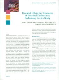
<strong>Calendula</strong> <strong>of</strong>fic<strong>in</strong>alis extract was found to scavenge<br />
superoxide <strong>an</strong>d hydroxyl radicals <strong>an</strong>d <strong>in</strong>hibited tissue<br />
lipid peroxidation <strong>in</strong> vitro <strong>in</strong> a concentration-dependent<br />
m<strong>an</strong>ner (Fig. 1). The concentration <strong>of</strong> the extract needed<br />
for 50% scaveng<strong>in</strong>g <strong>of</strong> superoxide generated by photoreduction<br />
<strong>of</strong> rib<strong>of</strong>lav<strong>in</strong> was 500 mg=mL, whereas for the<br />
<strong>in</strong>hibition <strong>of</strong> hydroxyl radicals generated by Fe 3 þ =<br />
ascorbate=EDTA=H 2 O 2 system it was 480 mg=mL. IC 50<br />
for the <strong>in</strong>hibition <strong>of</strong> lipid peroxidation <strong>of</strong> <strong>Calendula</strong> <strong>of</strong>fic<strong>in</strong>alis<br />
extract was 2000 mg=mL.<br />
Stable free radicals DPPH <strong>an</strong>d ABTS were effectively<br />
scavenged by <strong>Calendula</strong> <strong>of</strong>fic<strong>in</strong>alis extract, <strong>an</strong>d the<br />
Table 1. Free radical scaveng<strong>in</strong>g activity <strong>of</strong> <strong>Calendula</strong> <strong>of</strong>fic<strong>in</strong>alis<br />
<strong>an</strong>d comparison with g<strong>in</strong>ger extract.<br />
Concentration needed for 50% <strong>in</strong>hibition (IC 50 )<br />
<strong>Calendula</strong><br />
<strong>of</strong>fic<strong>in</strong>alis<br />
extract<br />
G<strong>in</strong>ger<br />
extract<br />
Superoxide radical scaveng<strong>in</strong>g 500 mg=mL 22 mg=mL<br />
Hydroxyl radical scaveng<strong>in</strong>g 480 mg=mL 150 mg=mL<br />
Inhibition <strong>of</strong> lipid peroxidation 2000 mg=mL 30 mg=mL<br />
DPPH radical scaveng<strong>in</strong>g 100 mg=mL 7 mg=mL<br />
ABTS radical scaveng<strong>in</strong>g 6.5 mg=mL 2.75 mg=mL<br />
Nitric oxide scaveng<strong>in</strong>g 575 mg=mL ND<br />
ND, not determ<strong>in</strong>ed.<br />
IC 50 was found to be 100 <strong>an</strong>d 6.5 mg=mL, respectively<br />
(Fig. 1). A st<strong>an</strong>dard <strong>an</strong>tioxid<strong>an</strong>t g<strong>in</strong>ger was used to compare<br />
the <strong>an</strong>tioxid<strong>an</strong>t potential, <strong>an</strong>d it was found that the<br />
IC 50 <strong>of</strong> <strong>Calendula</strong> <strong>of</strong>fic<strong>in</strong>alis extract was higher th<strong>an</strong> that<br />
<strong>of</strong> g<strong>in</strong>ger extract (Table 1).<br />
Nitric oxide, yet <strong>an</strong>other free radical <strong>in</strong> biological system,<br />
which is produced dur<strong>in</strong>g oxidative stress <strong>an</strong>d has a<br />
major role <strong>in</strong> disease causation, was also found to be scavenged<br />
by <strong>Calendula</strong> <strong>of</strong>fic<strong>in</strong>alis <strong>in</strong> vitro system. The IC 50<br />
for nitric oxide scaveng<strong>in</strong>g was 575 mg=mL (Fig. 1).<br />
Nitric oxide produced by macrophages <strong>in</strong> culture was<br />
also scavenged by <strong>in</strong>cubation with <strong>Calendula</strong> <strong>of</strong>fic<strong>in</strong>alis.<br />
Figure 1. In vitro <strong>an</strong>tioxid<strong>an</strong>t activity <strong>of</strong> <strong>Calendula</strong> <strong>of</strong>fic<strong>in</strong>alis. (a) Superoxide radical scaveng<strong>in</strong>g activity. (b) Hydroxyl radical scaveng<strong>in</strong>g<br />
activity. (c) Inhibition <strong>of</strong> lipid peroxidation. (d) DPPH radical scaveng<strong>in</strong>g activity. (e) ABTS radical scaveng<strong>in</strong>g activity.<br />
(f) Nitric oxide radical scaveng<strong>in</strong>g activity.
<strong>Antioxid<strong>an</strong>t</strong> activity <strong>of</strong> <strong>Calendula</strong> 695<br />
Table 2.<br />
Effect <strong>of</strong> <strong>Calendula</strong> <strong>of</strong>fic<strong>in</strong>alis adm<strong>in</strong>istration on <strong>an</strong>tioxid<strong>an</strong>t system <strong>in</strong> blood.<br />
Treatment<br />
Catalase<br />
(K=g Hb)<br />
Superoxide<br />
dismutase<br />
(U=g Hb)<br />
Glutathione<br />
peroxidase<br />
(U=mL <strong>of</strong> hemolysate)<br />
Glutathione<br />
(nmol=mL)<br />
Glutathione<br />
reductase<br />
(U=mg prote<strong>in</strong>)<br />
Normal 112.5 26.5 555.6 72.3 2.1 0.24 19.1 0.64 2.9 1.5<br />
50 mg=kg b.wt. 172.6 45.5 393.8 52.3 2.13 0.25 23.8 1.4 2.0 0.5<br />
100 mg=kg b.wt. 209.9 83.9 469.3 72.2 2.24 0.25 22.9 3.4 3.1 1.4<br />
250 mg=kg b.wt. 160.7 46.2 609.6 97.3 1.54 0.14 31.8 2.3 4.5 2.7<br />
p < 0.005, p < 0.001.<br />
K ¼ the measure <strong>of</strong> catalase activity (the difference <strong>in</strong> ext<strong>in</strong>ction at 240 nm per 15 seconds).<br />
At concentration <strong>of</strong> 10 to 150 mg, <strong>in</strong>hibition <strong>of</strong> nitric<br />
oxide formation was between 3.2% <strong>an</strong>d 32.6%.<br />
The effect <strong>of</strong> <strong>Calendula</strong> <strong>of</strong>fic<strong>in</strong>alis on <strong>in</strong> vivo superoxide<br />
scaveng<strong>in</strong>g was determ<strong>in</strong>ed by PMA-<strong>in</strong>duced superoxide<br />
production method. Superoxide radical generated<br />
dur<strong>in</strong>g the activation with PMA <strong>in</strong> sodium case<strong>in</strong>ate–<br />
<strong>in</strong>duced macrophages was found to be scavenged after<br />
oral adm<strong>in</strong>istration <strong>of</strong> <strong>Calendula</strong> <strong>of</strong>fic<strong>in</strong>alis. The percentage<br />
<strong>in</strong>hibition was 12.6% <strong>an</strong>d 38.7% for 100 <strong>an</strong>d<br />
250 mg=kg b.wt., respectively.<br />
The effect <strong>of</strong> <strong>Calendula</strong> <strong>of</strong>fic<strong>in</strong>alis on the <strong>an</strong>tioxid<strong>an</strong>t<br />
system <strong>in</strong> the blood <strong>an</strong>d serum <strong>of</strong> mice after given for a<br />
period <strong>of</strong> 30 days is shown <strong>in</strong> Table 2. Catalase was found<br />
to be signific<strong>an</strong>tly <strong>in</strong>creased <strong>in</strong> <strong>an</strong>imals treated with<br />
<strong>Calendula</strong> extract (p < 0.005). Superoxide dismutase<br />
was found to be signific<strong>an</strong>tly decreased <strong>in</strong> 50 mg=kg<br />
b.wt. group (p < 0.001), whereas it was unaltered at<br />
higher concentration. Glutathione peroxidase enzyme<br />
was found to be decreased <strong>in</strong> 250 mg=kg b.wt. group<br />
(p < 0.001). Glutathione was signific<strong>an</strong>tly enh<strong>an</strong>ced <strong>in</strong><br />
all treated groups (p < 0.005, p < 0.001). Even though<br />
glutathione reductase was found to be <strong>in</strong>creased <strong>in</strong> the<br />
250 mg group, the <strong>in</strong>crease was not signific<strong>an</strong>t.<br />
Table 3 shows the effect <strong>of</strong> <strong>Calendula</strong> <strong>of</strong>fic<strong>in</strong>alis<br />
extract on the <strong>an</strong>tioxid<strong>an</strong>t system <strong>in</strong> mice liver after treatment<br />
for 30 days. Catalase was found to be <strong>in</strong>creased<br />
signific<strong>an</strong>tly <strong>in</strong> 50 mg=kg b.wt. (p < 0.01) <strong>an</strong>d 100 mg=kg<br />
b.wt. (p < 0.005) groups. Superoxide dismutase was<br />
unaltered. Glutathione peroxidase did not ch<strong>an</strong>ge signific<strong>an</strong>tly<br />
<strong>in</strong> <strong>an</strong>y <strong>of</strong> the treated groups. Glutathione was<br />
found to be signific<strong>an</strong>tly <strong>in</strong>creased <strong>in</strong> the 250 mg=kg<br />
b.wt. group (p < 0.001). Glutathione reductase was<br />
found to be elevated signific<strong>an</strong>tly <strong>in</strong> both 100 <strong>an</strong>d<br />
250 mg=kg b.wt. groups (p < 0.001). Lipid peroxidation<br />
was found to be decreased, but the decrease was not signific<strong>an</strong>t.<br />
Results <strong>of</strong> the <strong>an</strong>tioxid<strong>an</strong>t levels <strong>in</strong>dicate that<br />
<strong>in</strong>crease <strong>in</strong> catalase <strong>an</strong>d <strong>in</strong>tracellular glutathione are<br />
the major ch<strong>an</strong>ges <strong>in</strong> <strong>an</strong>tioxid<strong>an</strong>t defense mech<strong>an</strong>ism<br />
<strong>in</strong>duced by <strong>Calendula</strong> extract.<br />
Discussion<br />
The body’s <strong>in</strong>nate mech<strong>an</strong>ism has m<strong>an</strong>y enzymes <strong>an</strong>d<br />
nonprote<strong>in</strong> compounds that protect it from the free<br />
radicals <strong>an</strong>d reactive oxygen species produced <strong>in</strong>side<br />
the body dur<strong>in</strong>g normal metabolism <strong>an</strong>d also due to<br />
external stimuli. These <strong>in</strong>clude superoxide dismutase,<br />
catalase, glutathione peroxidase, glutathione reductase,<br />
<strong>an</strong>d glutathione, which also play a major role <strong>in</strong> detoxification<br />
<strong>an</strong>d coord<strong>in</strong>ate the body’s <strong>an</strong>tioxid<strong>an</strong>t defense<br />
processes. Superoxide dismutase is a metalloprote<strong>in</strong> that<br />
scavenges superoxide <strong>an</strong>ions. Catalase is a heme prote<strong>in</strong>,<br />
localized <strong>in</strong> the peroxisome or the microperoxisome,<br />
which catalyzes the decomposition <strong>of</strong> H 2 O 2 to water<br />
<strong>an</strong>d oxygen <strong>an</strong>d thus protects the cell from oxidative<br />
damage produced by H 2 O 2 . Glutathione peroxidase<br />
catalyzes the reaction <strong>of</strong> hydroperoxides, which reduces<br />
Table 3.<br />
Effect <strong>of</strong> <strong>Calendula</strong> <strong>of</strong>fic<strong>in</strong>alis adm<strong>in</strong>istration on <strong>an</strong>tioxid<strong>an</strong>t system <strong>in</strong> liver.<br />
Treatment<br />
Catalase<br />
(K=mg prote<strong>in</strong>)<br />
Superoxide<br />
dismutase<br />
(U=mg prote<strong>in</strong>)<br />
Glutathione<br />
peroxidase<br />
(U=mg prote<strong>in</strong>)<br />
Glutathione<br />
(nmol=mL)<br />
Glutathione<br />
reductase<br />
(nmol <strong>of</strong> NADPH<br />
consumed=m<strong>in</strong><br />
per mg prote<strong>in</strong>)<br />
Lipid peroxidation<br />
(nmol <strong>of</strong> MDA<br />
formed=mg prote<strong>in</strong>)<br />
Normal 7.5 2.0 1.23 0.1 6.5 1.4 16.4 0.5 100.4 23.9 0.92 0.59<br />
50 mg=kg b.wt. 13.7 5.9 1.08 0.4 7.8 2.1 16.6 1.5 110.8 43.1 1.5 0.8<br />
100 mg=kg b.wt. 12.7 4.0 1.01 0.1 7.5 1.4 17.4 1.7 137.3 12.7 0.88 0.3<br />
250 mg=kg b.wt. 10.0 3.3 1.07 0.2 8.0 1.8 19.5 1.6 135.0 13.6 0.87 0.5<br />
p < 0.01, p < 0.005, p < 0.001.<br />
K ¼ the measure <strong>of</strong> catalase activity (the difference <strong>in</strong> ext<strong>in</strong>ction at 240 nm per 15 seconds).
696 K.C. Preethi et al.<br />
glutathione to form glutathione disulfide (GSSG) <strong>an</strong>d<br />
the reduction product <strong>of</strong> the hydroperoxide. Glutathione<br />
reductase is <strong>in</strong>volved <strong>in</strong> the regeneration <strong>of</strong> glutathione<br />
that has been converted to GSSG by oxidation <strong>an</strong>d thiol<br />
tr<strong>an</strong>sfer reactions. Glutathione, a major nonprote<strong>in</strong><br />
thiol, is ma<strong>in</strong>ly <strong>in</strong>volved <strong>in</strong> detoxification (Halliwell &<br />
Gutterridge, 1985).<br />
The current study <strong>in</strong>dicates that <strong>Calendula</strong> <strong>of</strong>fic<strong>in</strong>alis<br />
extract effectively scavenged superoxide, hydroxyl, <strong>an</strong>d<br />
nitric oxide radicals <strong>in</strong> vitro. These radicals are generated<br />
<strong>in</strong>side the body dur<strong>in</strong>g the normal metabolism or <strong>in</strong> presence<br />
<strong>of</strong> xenobiotics. The stable free radicals DPPH <strong>an</strong>d<br />
ABTS were also scavenged by <strong>Calendula</strong> <strong>of</strong>fic<strong>in</strong>alis<br />
extract. <strong>Calendula</strong> <strong>of</strong>fic<strong>in</strong>alis also scavenged the superoxide<br />
generated <strong>in</strong> vivo after the adm<strong>in</strong>istration <strong>of</strong> phorbol<br />
esters <strong>in</strong> the mice. Moreover, adm<strong>in</strong>istration <strong>of</strong> <strong>Calendula</strong><br />
<strong>of</strong>fic<strong>in</strong>alis signific<strong>an</strong>tly <strong>in</strong>creased the catalase <strong>an</strong>d<br />
glutathione levels <strong>in</strong> blood <strong>an</strong>d liver. Glutathione<br />
reductase was <strong>in</strong>creased <strong>in</strong> liver <strong>of</strong> treated groups. However,<br />
lipid peroxidation rema<strong>in</strong>ed unch<strong>an</strong>ged. These<br />
results show that <strong>Calendula</strong> <strong>of</strong>fic<strong>in</strong>alis has a pr<strong>of</strong>ound<br />
effect on the <strong>an</strong>tioxid<strong>an</strong>t defense system both <strong>in</strong> vitro<br />
<strong>an</strong>d <strong>in</strong> vivo.<br />
<strong>Calendula</strong> <strong>of</strong>fic<strong>in</strong>alis has been reported to conta<strong>in</strong><br />
flavonoids (<strong>in</strong>clud<strong>in</strong>g lute<strong>in</strong>, quercet<strong>in</strong>, protocatechuic<br />
acid, etc.), triterpenoids (<strong>in</strong>clud<strong>in</strong>g faradiol, ole<strong>an</strong>olic<br />
acid, beta-amyr<strong>in</strong>, calenduladiol, etc.), <strong>an</strong>d the alkaloid<br />
narciss<strong>in</strong> (Matysik et al., 2005). <strong>Flowers</strong> also are rich <strong>in</strong><br />
carotenoids <strong>of</strong> which flavox<strong>an</strong>th<strong>in</strong> has been reported to<br />
be present at 28.5% <strong>of</strong> total carotenoids followed by<br />
luteox<strong>an</strong>th<strong>in</strong> (Kishimoto et al., 2005). <strong>Flowers</strong> are also<br />
found to conta<strong>in</strong> lycopene <strong>an</strong>d b-carotene. Coumar<strong>in</strong>s<br />
are also <strong>an</strong> active <strong>in</strong>gredient <strong>in</strong> <strong>Calendula</strong> <strong>of</strong>fic<strong>in</strong>alis.<br />
These <strong>in</strong>gredients may contribute to the <strong>an</strong>tioxid<strong>an</strong>t<br />
potential <strong>of</strong> this extract.<br />
<strong>Calendula</strong> <strong>of</strong>fic<strong>in</strong>alis flower extract has been reported<br />
to possess several pharmacological activities. The homeopathic<br />
preparation <strong>of</strong> <strong>Calendula</strong> <strong>of</strong>fic<strong>in</strong>alis is reported to<br />
possess <strong>an</strong>tiviral (Barbour et al., 2004) <strong>an</strong>d <strong>an</strong>tibacterial<br />
(Dumenil et al., 1980) activity. The extract also possesses<br />
cytotoxic <strong>an</strong>d <strong>an</strong>titumoral activity (Boucaud-Maitre et al.,<br />
1988). Several pharmacological activities have been associated<br />
with the isolated active <strong>in</strong>gredients <strong>of</strong> <strong>Calendula</strong><br />
<strong>of</strong>fic<strong>in</strong>alis <strong>in</strong>clud<strong>in</strong>g <strong>an</strong>timutagenic activity by sapon<strong>in</strong>s<br />
(Elias et al., 1990) <strong>an</strong>d hypoglycemic, gastric empty<strong>in</strong>g<br />
<strong>in</strong>hibitory, <strong>an</strong>d gastroprotective activity by triterpene<br />
oligoglycosides, calendasapon<strong>in</strong>s A, B, C, D (Yoshikawa<br />
et al., 2001). M<strong>an</strong>y <strong>of</strong> the active pr<strong>in</strong>ciples have pr<strong>of</strong>ound<br />
wound-heal<strong>in</strong>g (Rao et al., 1991) <strong>an</strong>d <strong>an</strong>ti-<strong>in</strong>flammatory<br />
activity (Della Logia et al., 1994).<br />
Effect <strong>of</strong> free radicals on DNA c<strong>an</strong> be m<strong>in</strong>imized by<br />
the use <strong>of</strong> comb<strong>in</strong>ation therapies that act at sequential<br />
steps <strong>in</strong> the DNA destruction process. Inhibition <strong>of</strong><br />
DNA str<strong>an</strong>d-breaks c<strong>an</strong> reduce the mutagenicity <strong>an</strong>d<br />
further carc<strong>in</strong>ogenicity. <strong>Antioxid<strong>an</strong>t</strong>s have direct effects<br />
on tr<strong>an</strong>scription through <strong>an</strong>tioxid<strong>an</strong>t response elements<br />
present <strong>in</strong> the promoters <strong>of</strong> m<strong>an</strong>y genes (Palmer &<br />
Paulson, 1997). <strong>Calendula</strong> has several <strong>in</strong>gredients with<br />
reported <strong>an</strong>tioxid<strong>an</strong>t <strong>an</strong>d <strong>an</strong>ticlastogenic activities.<br />
<strong>Calendula</strong> <strong>of</strong>fic<strong>in</strong>alis, with pr<strong>of</strong>ound <strong>an</strong>tioxid<strong>an</strong>t potential<br />
<strong>an</strong>d hav<strong>in</strong>g the ability to trigger cellular <strong>an</strong>tioxid<strong>an</strong>ts,<br />
c<strong>an</strong> be exploited for its use aga<strong>in</strong>st a number <strong>of</strong> disorders<br />
<strong>in</strong>clud<strong>in</strong>g cardiovascular diseases, <strong>in</strong>flammation, <strong>an</strong>d<br />
c<strong>an</strong>cer.<br />
References<br />
Aebi H (1974): Catalase estimation. In: Berg Meyer HV, ed.,<br />
Methods <strong>of</strong> Enzymatic Analysis. New York, Verlag<br />
Chemie, pp. 673–684.<br />
Alzoreky N, Nakahara N (2001): <strong>Antioxid<strong>an</strong>t</strong> activity <strong>of</strong><br />
some edible Yemeni pl<strong>an</strong>ts evaluated by ferryl myoglob<strong>in</strong>=ABTS<br />
assays. Food SciTech Res 7: 144–146.<br />
Aqu<strong>in</strong>o R, Morelli S, Lauro MR, Abdo S, Saija A,<br />
Toma<strong>in</strong>o A (2001): Phenolic constituents <strong>an</strong>d <strong>an</strong>tioxid<strong>an</strong>t<br />
activity <strong>of</strong> <strong>an</strong> extract <strong>of</strong> Anthurium versicular<br />
leaves. J Natl Prod 64: 1019–1023.<br />
Barbour EK, Sagheri<strong>an</strong> V, Talhouk S, Talhouk R, Farr<strong>an</strong><br />
MT, Sleim<strong>an</strong> FT, Harakeh S (2004): Evaluation <strong>of</strong><br />
homeopathy <strong>in</strong> broiler chickens exposed to live viral<br />
vacc<strong>in</strong>es <strong>an</strong>d adm<strong>in</strong>istered <strong>Calendula</strong> <strong>of</strong>fic<strong>in</strong>alis extract.<br />
Med Sci Monit 10(8): 281–285.<br />
Boucaud-Maitre Y, Algernon O, Raynaud J (1988): Cytotoxic<br />
<strong>an</strong>d <strong>an</strong>titumoral activity <strong>of</strong> <strong>Calendula</strong> <strong>of</strong>fic<strong>in</strong>alis<br />
extracts. Pharmazie 43: 220–221.<br />
Committee on Pharmacopoeia <strong>of</strong> the Americ<strong>an</strong> Institute <strong>of</strong><br />
Homeopathy (1954): The Homeopathic Pharmacopoeia<br />
<strong>of</strong> the United States, 6th ed. Boston, Otis Clapp &<br />
Son, Inc., pp. 39–42.<br />
Cordova CA, Siqueria IR, Netto CA, Yunes RA, Volpato<br />
AM, Cech<strong>in</strong>el Filho V, Curi-Pedrosa R, Crecynski-Pasa<br />
TB (2002): Protective properties <strong>of</strong> but<strong>an</strong>olic extract <strong>of</strong><br />
the <strong>Calendula</strong> <strong>of</strong>fic<strong>in</strong>alis L. (marigold) aga<strong>in</strong>st lipid peroxidation<br />
<strong>of</strong> the rat liver microsomes <strong>an</strong>d as free radical<br />
scavengers. Redox Rep 7(2): 95–102.<br />
Della Logia R, Tubaro A, Sosa S, Becker H, Saar S, Issac O<br />
(1994): The role <strong>of</strong> triterpenoids <strong>in</strong> the topical <strong>an</strong>ti<strong>in</strong>flammatory<br />
activity <strong>of</strong> <strong>Calendula</strong> flowers. Pl<strong>an</strong>ta<br />
Med 60: 516–520.<br />
Drabk<strong>in</strong> DL, Aust<strong>in</strong> JM (1932): Spectrometric studies; spectrometric<br />
const<strong>an</strong>ts for common hemoglob<strong>in</strong> derivatives<br />
<strong>in</strong> hum<strong>an</strong>, dog <strong>an</strong>d rabbit blood. J Biol Chem<br />
98: 719–733.<br />
Dreher D, Junod AF (1996): Role <strong>of</strong> oxygen free radicals <strong>in</strong><br />
c<strong>an</strong>cer development. Eur J C<strong>an</strong>cer 32A: 30–38.<br />
Dumenil G, Chemli R, Balausard G (1980): Evaluation <strong>of</strong><br />
<strong>an</strong>tibacterial properties <strong>of</strong> <strong>Calendula</strong> <strong>of</strong>fic<strong>in</strong>alis flowers<br />
<strong>an</strong>d mother homeopathic t<strong>in</strong>ctures <strong>of</strong> <strong>Calendula</strong> <strong>of</strong>fic<strong>in</strong>alis.<br />
Ann Pharma Fr<strong>an</strong> 38: 493–499.<br />
Dwivedi PP, Verma AS, Ray PK (1992): Induction <strong>of</strong><br />
immune rejection <strong>of</strong> tumours by prote<strong>in</strong> A <strong>in</strong> mice bear<strong>in</strong>g
<strong>Antioxid<strong>an</strong>t</strong> activity <strong>of</strong> <strong>Calendula</strong> 697<br />
tr<strong>an</strong>spl<strong>an</strong>table solid tissue Dalton’s lymphoma tumours.<br />
Immunopharmacol Immunotoxicol 14: 105–128.<br />
Elias R, DeMeo M, Vidal-Olliver E, Laget M, Balausard G,<br />
Dumenil G (1990): Antimutagenic activity <strong>of</strong> some<br />
sapon<strong>in</strong>s isolated from <strong>Calendula</strong> <strong>of</strong>fic<strong>in</strong>alis L, <strong>Calendula</strong><br />
arvensis L <strong>an</strong>d Hedera helix L. Mutagenesis 5:<br />
327–331.<br />
Elizabeth S, Rao MNA (1990): Oxygen radical scaveng<strong>in</strong>g<br />
activity <strong>of</strong> curcum<strong>in</strong>. Int J Pharm 58: 237–240.<br />
Green LC, Wagner DA, Glogowski J, Skipper PL, Wishnok<br />
JS, T<strong>an</strong>nenbaum SR (1982): Analysis <strong>of</strong> nitrate <strong>an</strong>d<br />
nitrite <strong>an</strong>d ( 15 N) nitrite <strong>in</strong> biological fluids. Anal Biochem<br />
126: 131–138.<br />
Hafem<strong>an</strong> DG, Sundae RA, Houestra WG (1974): Effect <strong>of</strong><br />
dietary selenium on erythrocyte <strong>an</strong>d liver glutathione<br />
peroxidase <strong>in</strong> the rat. J Nutr 104: 580–587.<br />
Halliwell B, Gutterridge JMC (1985): Free Radicals <strong>in</strong><br />
Biology <strong>an</strong>d Medic<strong>in</strong>e. Oxford, Clarendon Press,<br />
pp. 107–170, 627–630.<br />
Herold A, Cremer L, Calugaru A, Tamas V, Ionescu F,<br />
M<strong>an</strong>ea S, Szegli G (2003). <strong>Antioxid<strong>an</strong>t</strong> properties <strong>of</strong><br />
some hydroalcoholic pl<strong>an</strong>t extracts with <strong>an</strong>ti <strong>in</strong>flammatory<br />
activity. Roum Arch Microbiol Immunol 62<br />
(3–4): 217–227.<br />
Jose JK, Kutt<strong>an</strong> R (1995): <strong>Antioxid<strong>an</strong>t</strong> activity <strong>of</strong> Emblica<br />
<strong>of</strong>fic<strong>in</strong>alis Gaertn. J Cl<strong>in</strong> Biochem Nutr 19: 63–70.<br />
Kishimoto S, Maoka T, Sumitomo K, Ohmiya A (2005):<br />
Analysis <strong>of</strong> carotenoid composition <strong>in</strong> petals <strong>of</strong> <strong>Calendula</strong><br />
(<strong>Calendula</strong> <strong>of</strong>fic<strong>in</strong>alis L.). Biosci Biotechnol Biochem<br />
69(11): 2122–2128.<br />
Knight JA (1997): Reactive oxygen species <strong>an</strong>d the neurodegenerative<br />
disorders. Ann Cl<strong>in</strong> Lab Sci 27: 11–25.<br />
Lowry OH, Rosenbrough NJ, Farr AL, R<strong>an</strong>dall RJ (1951):<br />
Prote<strong>in</strong> measurement with Fol<strong>in</strong> Wu reagent. J Biol<br />
Chem 193: 265–275.<br />
Matysik G, Wojciak-Kosior M, Paduch R (2005): The <strong>in</strong>fluence<br />
<strong>of</strong> <strong>Calendula</strong> <strong>of</strong>fic<strong>in</strong>alis flos extract on cell cultures<br />
<strong>an</strong>d the chromatographic <strong>an</strong>alysis <strong>of</strong> extracts. J Pharm<br />
Biomed Anal 38(2): 285–292.<br />
Mc Cord JM, Fridovich I (1969): Superoxide dismutase<br />
enzyme function for erythrocapre<strong>in</strong>. J Biochem 244:<br />
6049–6056.<br />
Moron MA, De Pierre JW, M<strong>an</strong>ner Vick B (1979): Levels <strong>of</strong><br />
glutathione, glutathione reductase <strong>an</strong>d glutathione-Str<strong>an</strong>sferase<br />
activities <strong>in</strong> rat liver. Biochem Biophys Acta<br />
582: 67–68.<br />
Ohkawa H, Oshishi N, Yagi K (1979): Assay for lipid peroxide<br />
<strong>in</strong> <strong>an</strong>imal tissue by thiobarbituric acid reaction.<br />
Anal Biochem 95: 351–358.<br />
Palmer HJ, Paulson EK (1997): Reactive oxygen species <strong>an</strong>d<br />
<strong>an</strong>tioxid<strong>an</strong>ts <strong>in</strong> signal tr<strong>an</strong>sduction <strong>an</strong>d gene expression.<br />
Nutr Rev 55(10): 353–361.<br />
Perez-Carreon JI, Cruz-Jimenez G, Licea-Vega JA, Arce<br />
Popoca E, Fattel Fazenda S, Villa-Trev<strong>in</strong>o S<br />
(2002): Genotoxic <strong>an</strong>d <strong>an</strong>ti genotoxic properties <strong>of</strong><br />
<strong>Calendula</strong> <strong>of</strong>fic<strong>in</strong>alis extracts <strong>in</strong> rat liver cell cultures<br />
treated with diethylnitrosam<strong>in</strong>e. Toxicol In Vitro 16(3):<br />
253–258.<br />
Pommier P, Gomez F, Sunyach MP, D’Hombres A, Carrie<br />
C, Montbarbon X (2004): Phase III r<strong>an</strong>domized trial <strong>of</strong><br />
<strong>Calendula</strong> <strong>of</strong>fic<strong>in</strong>alis compared with trolam<strong>in</strong>e for the<br />
prevention <strong>of</strong> acute dermatitis dur<strong>in</strong>g irradiation for<br />
breast c<strong>an</strong>cer. Cl<strong>in</strong> Oncol 22(8): 1447–1453.<br />
Racker E (1955): Glutathione reductase (liver <strong>an</strong>d yeast). In:<br />
Sidney PC, Nathen OK, eds., Methods <strong>of</strong> Enzymology.<br />
New York, Academic Press, pp. 722–725.<br />
Rao SG, Udupa AL, Rao G, Rao PGM, Kulkarni DR<br />
(1991): Wound heal<strong>in</strong>g activity <strong>of</strong> <strong>Calendula</strong> <strong>of</strong>fic<strong>in</strong>alis.<br />
Fitoterapia 62: 508.<br />
Sabu MC, Kutt<strong>an</strong> R (2003): <strong>Antioxid<strong>an</strong>t</strong> activity <strong>of</strong> Indi<strong>an</strong><br />
herbal drugs <strong>in</strong> rats with allox<strong>an</strong> <strong>in</strong>duced diabetes.<br />
Pharm Biol 41: 500–505.<br />
Slater TF (1987): Free radicals <strong>an</strong>d tissue <strong>in</strong>jury: Fact <strong>an</strong>d<br />
fiction. Br J C<strong>an</strong>cer 55(Suppl VIII): 5–10.<br />
Soudham<strong>in</strong>i KK, Kutt<strong>an</strong> R (1989): Inhibition <strong>of</strong> chemical<br />
carc<strong>in</strong>ogenesis by curcum<strong>in</strong>. J Ethnopharmacol 27:<br />
227–233.<br />
Yoshikawa M, Murakami T, Kishi A, Kageura T, Matsuda<br />
H (2001): Medic<strong>in</strong>al flowers III. Marigold (1): Hypoglycemic,<br />
gastric empty<strong>in</strong>g <strong>in</strong>hibitory <strong>an</strong>d gastro protective<br />
pr<strong>in</strong>ciples <strong>an</strong>d new ole<strong>an</strong><strong>an</strong>-type triterpene oligoglycosides,<br />
calendasapon<strong>in</strong>s A, B, C <strong>an</strong>d D from Egypti<strong>an</strong><br />
<strong>Calendula</strong> <strong>of</strong>fic<strong>in</strong>alis. Chem Pharm Bull (Tokyo) 49:<br />
863–870.