ACOG Practice Bulletin No. 101
ACOG Practice Bulletin No. 101
ACOG Practice Bulletin No. 101
Create successful ePaper yourself
Turn your PDF publications into a flip-book with our unique Google optimized e-Paper software.
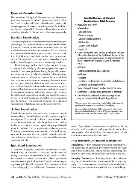
Types of Examinations<br />
The American College of Obstetricians and Gynecologists<br />
uses the terms “standard” (also called basic), “limited,”<br />
and “specialized” (also called detailed) to describe<br />
various types of ultrasound examinations performed during<br />
the second or third trimesters. First-trimester ultrasound<br />
examination is distinct and is discussed separately.<br />
Standard Examination<br />
Ultrasonography is an accurate method of determining gestational<br />
age, fetal number, viability, and placental location.<br />
A standard obstetric ultrasound examination in the second<br />
or third trimester includes an evaluation of fetal presentation,<br />
amniotic fluid volume, cardiac activity, placental position,<br />
fetal biometry, and fetal number, plus an anatomic<br />
survey. The maternal cervix and adnexa should be examined<br />
as clinically appropriate when technically feasible.<br />
Fetal anatomy, as described in this document, may<br />
be assessed adequately by ultrasonography after approximately<br />
18 weeks of gestation. It may be possible to document<br />
normal structures before this time, although some<br />
structures can be difficult to visualize because of fetal<br />
size, position, and movement; maternal abdominal scars;<br />
and increased maternal abdominal wall thickness. A second-<br />
or third-trimester ultrasound examination may pose<br />
technical limitations for an anatomic evaluation because<br />
of suboptimal imaging. When this occurs, the report of<br />
the ultrasound examination should document the nature<br />
of this technical limitation. A follow-up examination<br />
may be helpful. The essential elements of a standard<br />
examination of fetal anatomy are listed in the box.<br />
Limited Examination<br />
A limited examination does not replace a standard examination<br />
and is performed when a specific question requires<br />
investigation. For example, a limited examination in the<br />
second or third trimester could be performed to confirm<br />
fetal heart activity in a patient experiencing vaginal bleeding<br />
or to establish fetal presentation in a laboring patient.<br />
A limited examination also may be performed in any<br />
trimester to evaluate interval growth, estimate amniotic<br />
fluid volume, evaluate the cervix, and assess the presence<br />
of cardiac activity.<br />
Essential Elements of Standard<br />
Examination of Fetal Anatomy<br />
• Head, Face and Neck*<br />
—Cerebellum<br />
—Choroid plexus<br />
—Cisterna magna<br />
—Lateral cerebral ventricles<br />
—Midline falx<br />
—Cavum septi pellucidi<br />
—Upper lip<br />
• Chest—Heart (The basic cardiac examination includes<br />
a four-chamber view of the fetal heart. As part of the<br />
cardiac screening examination, an attempt should be<br />
made, if technically feasible, to view the outflow<br />
tracts.)<br />
• Abdomen<br />
—Stomach (presence, size, and situs)<br />
—Kidneys<br />
—Bladder<br />
—Umbilical cord insertion site into the fetal abdomen<br />
—Umbilical cord vessel number<br />
• Spine—Cervical, thoracic, lumbar, and sacral spine<br />
• Extremities—Legs and arms (presence or absence)<br />
• Sex—Medically indicated in low-risk pregnancies<br />
only for the evaluation of multiple gestations<br />
*A measurement of the nuchal fold may be helpful during a specific<br />
age interval to suggest an increased risk of aneuploidy.<br />
American College of Radiology. ACR practice guideline for the performance<br />
of obstetrical ultrasound. In: ACR practice guidelines and<br />
technical standards, 2007. Reston (VA): ACR; 2007. p. 1025–1033.<br />
Specialized Examination<br />
A detailed or targeted anatomic examination is performed<br />
when an anomaly is suspected on the basis of<br />
history, laboratory abnormalities, or the results of either<br />
the limited or standard examination. Other specialized<br />
examinations might include fetal Doppler ultrasonography,<br />
biophysical profile, amniotic fluid assessment, fetal<br />
echocardiography, or additional biometric measurements.<br />
Specialized examinations are performed by an<br />
operator with experience and expertise in such ultrasonography<br />
who determines the components of the<br />
examination on a case-by-case basis.<br />
First-Trimester Ultrasound Examination<br />
Indications. A first-trimester ultrasound examination is<br />
an ultrasound examination performed before 13 weeks<br />
and 6 days of gestation. Indications for performing firsttrimester<br />
ultrasound examinations are listed in the box.<br />
Imaging Parameters. Scanning in the first trimester<br />
may be performed either transabdominally or transvaginally.<br />
If a transabdominal examination is not definitive,<br />
a transvaginal scan or transperineal scan should be per-<br />
452 <strong>ACOG</strong> <strong>Practice</strong> <strong>Bulletin</strong> Ultrasonography in Pregnancy OBSTETRICS & GYNECOLOGY


