ACOG Practice Bulletin No. 101
ACOG Practice Bulletin No. 101
ACOG Practice Bulletin No. 101
You also want an ePaper? Increase the reach of your titles
YUMPU automatically turns print PDFs into web optimized ePapers that Google loves.
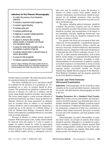
Indications for First-Trimester Ultrasonography<br />
• To confirm the presence of an intrauterine<br />
pregnancy<br />
• To evaluate a suspected ectopic pregnancy<br />
• To evaluate vaginal bleeding<br />
• To evaluate pelvic pain<br />
• To estimate gestational age<br />
• To diagnosis or evaluate multiple gestations<br />
• To confirm cardiac activity<br />
• As adjunct to chorionic villus sampling,<br />
embryo transfer, or localization and removal<br />
of an intrauterine device<br />
• To assess for certain fetal anomalies, such as<br />
anencephaly, in patients at high risk<br />
• To evaluate maternal pelvic or adnexal masses or<br />
uterine abnormalities<br />
• To screen for fetal aneuploidy<br />
• To evaluate suspected hydatidiform mole<br />
American College of Radiology. ACR practice guideline for the performance<br />
of obstetrical ultrasound. In: ACR practice guidelines and<br />
technical standards, 2007. Reston (VA): ACR; 2007. p. 1025–1033.<br />
formed whenever possible. The following factors should<br />
be considered during the examination.<br />
The uterus, including the cervix, and adnexa should<br />
be evaluated for the presence of a gestational sac. If a<br />
gestational sac is seen, its location should be documented.<br />
The gestational sac should be evaluated for the<br />
presence or absence of a yolk sac or embryo, and the<br />
crown–rump length should be recorded, when possible.<br />
The crown–rump length is a more accurate indicator of<br />
gestational (menstrual) age than is mean gestational sac<br />
diameter. However, the mean gestational sac diameter<br />
may be recorded when an embryo is not identified.<br />
Caution should be used in presumptively diagnosing a<br />
gestational sac in the absence of a definite embryo or<br />
yolk sac. Without these findings, intrauterine fluid collection<br />
could represent a pseudogestational sac associated<br />
with an ectopic pregnancy.<br />
Presence or absence of cardiac activity should be<br />
reported. With transvaginal scans, cardiac motion should<br />
be observed when the embryo is 5 mm or greater in<br />
length. An embryo should be visible by transvaginal<br />
ultrasonography with a mean gestational sac diameter of<br />
20 mm or greater. If an embryo less than 5 mm in length<br />
is seen without cardiac activity, a subsequent scan at a<br />
later time may be needed to assess the presence or<br />
absence of cardiac activity. Fetal number should be<br />
reported. Amnionicity and chorionicity should be documented<br />
for all multiple gestations when possible.<br />
Embryonic or fetal anatomy should be assessed according<br />
to gestational age.<br />
The uterus, including adnexal structures, should be<br />
evaluated. The presence, location, and size of adnexal<br />
masses should be recorded. The presence of leiomyomas<br />
should be recorded, and measurements of the largest or<br />
any potentially clinically significant leiomyomas may<br />
be recorded. The cul-de-sac should be evaluated for the<br />
presence or absence of fluid.<br />
For patients who desire an assessment of their individual<br />
risk of fetal aneuploidy, a standardized measurement<br />
of the nuchal translucency during a specific age<br />
interval is necessary. Nuchal translucency measurements<br />
should be used (in conjunction with serum biochemistry)<br />
to determine the risk of Down syndrome, trisomy 13, trisomy<br />
18, or other anatomic abnormalities, such as heart<br />
defects. In this setting, it is important that the practitioner<br />
measure the nuchal translucency according to established<br />
guidelines for measurement. In addition, a quality<br />
assessment program is recommended to ensure accurate<br />
results. Organizations currently providing guidelines and<br />
ongoing quality assessment include the Nuchal<br />
Translucency Quality Review program of the Maternal–<br />
Fetal Medicine Foundation and the program sponsored<br />
by the Fetal Medicine Foundation.<br />
Second- and Third-Trimester<br />
Ultrasound Examination<br />
Indications. Ultrasonography can be of benefit in many<br />
situations in the second and third trimesters. Indications<br />
for second- and third-trimester ultrasonography are listed<br />
in the box.<br />
Imaging Parameters for a Standard Fetal Examination.<br />
Fetal cardiac activity, fetal number, and fetal presentation<br />
should be reported. Any abnormal heart rates or<br />
rhythms should be reported. Multiple gestations require<br />
the documentation of additional information: chorionicity,<br />
amnionicity, comparison of fetal sizes, estimation of<br />
amniotic fluid volume (increased, decreased, or normal)<br />
on each side of the membrane, and fetal genitalia (when<br />
visualized).<br />
Ultrasonography can detect abnormalities in amniotic<br />
fluid volume. An estimate of amniotic fluid volume<br />
should be reported. Although it is acceptable for experienced<br />
examiners to qualitatively estimate amniotic fluid<br />
volume, semiquantitative methods also have been described<br />
for this purpose (eg, amniotic fluid index, single<br />
deepest pocket, two-diameter pocket).<br />
VOL. 113, NO. 2, PART 1, FEBRUARY 2009 <strong>ACOG</strong> <strong>Practice</strong> <strong>Bulletin</strong> Ultrasonography in Pregnancy 453


