Understanding Brachial Plexus Palsy - The Royal Children's Hospital
Understanding Brachial Plexus Palsy - The Royal Children's Hospital
Understanding Brachial Plexus Palsy - The Royal Children's Hospital

Create successful ePaper yourself
Turn your PDF publications into a flip-book with our unique Google optimized e-Paper software.
<strong>Understanding</strong><br />
BRACHIAL<br />
PLEXUS<br />
PALSY<br />
Departments of Physiotherapy,<br />
Occupational <strong>The</strong>rapy<br />
and Plastic Surgery,<br />
<strong>Royal</strong> Children’s <strong>Hospital</strong>, Melbourne
THIS pamphlet has been prepared to help<br />
you learn about brachial plexus injuries.<br />
If you have any further questions specific to your<br />
child after reading this pamphlet, please consult your<br />
doctor, physiotherapist or occupational therapist.<br />
You can contact the Department of Plastic and<br />
Maxillofacial Surgery on the following telephone<br />
numbers or email address:<br />
(03) 9345 6636 or (03) 9345 5347<br />
plastic.surgery@rch.org.au<br />
1
C5<br />
C6<br />
T1 & C8<br />
C7<br />
<strong>Brachial</strong> plexus<br />
Clavicle (collarbone)<br />
C7<br />
Overview<br />
<strong>The</strong> brachial plexus is a large network of<br />
nerves that extend from the neck into the arm. (Fig. 1)<br />
<strong>The</strong> five large nerves (given the symbols C5, C6,<br />
C7, C8 & T1) exit from the spinal cord between<br />
Figure 1. Location of brachial plexus.<br />
Upper trunk<br />
Middle trunk<br />
Lower trunk<br />
C5<br />
C6<br />
C7<br />
C8<br />
Clavicle (collarbone)<br />
Musculocutaneous nerve<br />
the bones in the neck (the vertebrae). (Fig. 2)<br />
<strong>The</strong>se nerves provide movement and feeling<br />
to the arm and hand. It is through the nerves of<br />
the brachial plexus that the brain sends electrical<br />
signals to the individual muscles of the arm and<br />
hand. One nerve is made up of thousands of nerve<br />
fibres. (Fig. 3) <strong>The</strong>se nerve fibres carry the electrical<br />
T1<br />
First rib<br />
Figure 2. Detail of the nerve network of the brachial plexus.<br />
Peripheral nerve<br />
Nerve covering (sheath)<br />
Nerve bundle<br />
Nerve fibre<br />
Median nerve<br />
Radial nerve<br />
Ulnar nerve<br />
signals from the brain to the arm. If nerve fibres are<br />
injured, the muscle that the nerve serves does not<br />
Figure 3. Detail of nerve anatomy.<br />
2
eceive electrical signals from the brain to make<br />
it work. Instead, the muscle is inactive and begins<br />
to deteriorate. <strong>The</strong> arm may not grow normally<br />
and muscles and joints may tighten. <strong>The</strong> skin<br />
may also have reduced feeling.<br />
Most brachial plexus injuries occur during<br />
birth. <strong>The</strong> brachial plexus is often damaged when<br />
it is under tension. Most hospitals report one to<br />
two babies being born with a brachial plexus<br />
injury per 1000 births.<br />
<strong>The</strong> nerves of the brachial plexus have some<br />
ability to repair themselves. As long as the outer<br />
sheath or covering of the nerve is preserved, the<br />
torn, the nerve cannot grow back and the muscle<br />
will not work.<br />
Rapid return of muscle function is a positive<br />
sign. Most nerve regrowth and noticeable muscle<br />
function recovery will occur during the first year<br />
of life, with some less noticeable improvements in<br />
the second year. Most children who spontaneously<br />
recover well in the first few months are able to use<br />
their affected arm to do almost all activities they<br />
want. However, some muscle weakness usually<br />
remains. <strong>The</strong> movements of the affected arm may<br />
not look the same as the non-affected arm doing<br />
the same movement.<br />
damaged nerve fibres can regrow down to a muscle.<br />
Nerve fibres regrow at a rate of about 1mm per<br />
day, or an inch per month. <strong>The</strong>refore it can take<br />
many months for regrowing nerve fibres to reach<br />
the muscles in the lower arm and hand. If the<br />
entire nerve (including the outer sheath) has been<br />
3
• A rupture is when the nerve is torn, but not<br />
from where it attaches to the spinal cord.<br />
This usually occurs beyond the vertebrae in the<br />
neck. A rupture requires surgery to reconnect<br />
the ends of the nerve.<br />
Types of brachial plexus injuries (Fig. 4)<br />
• A neuroma forms when torn nerve fibres have<br />
attempted to regrow and heal themselves, but scar<br />
tissue has grown in and around the injury. This<br />
Normal nerve<br />
Avulsion<br />
scar tissue makes it impossible for the nerve to<br />
conduct electrical signals to the muscles. Surgery<br />
removes the scar tissue around the nerve and<br />
Rupture<br />
between the ends of a completely ruptured nerve.<br />
Neuroma<br />
Figure 4. Types of nerve injury.<br />
• An avulsion is when the nerve is torn from<br />
Rupture and<br />
neuroma formation<br />
Avulsion<br />
where it attaches to the spinal cord. No recovery<br />
is expected with an avulsion injury. It cannot<br />
be repaired with surgery.<br />
Figure 5. Typical brachial plexus injury.<br />
4
• Axonotomesis occurs when the fibres inside the<br />
nerve have been broken but the nerve covering<br />
is still intact. Recovery by regrowth of the nerve<br />
fibres is often very good but it takes time<br />
(1mm per day) for the nerve to regrow from<br />
the site of the injury to its paralysed muscle.<br />
• Neuropraxis occurs when the nerve has been<br />
damaged (e.g. sprained) but not torn. In this<br />
case, the nerve fibres can recover on their own.<br />
Improvement in movement of the arm should<br />
be seen within three months.<br />
A typical brachial plexus injury may have a combination<br />
of the above. (Fig. 5)<br />
How do brachial plexus injuries occur (Fig. 6)<br />
In many cases the baby is larger than average.<br />
However, newborns of all sizes can suffer a<br />
brachial plexus injury, and prediction of babies<br />
likely to be affected is often extremely difficult.<br />
During childbirth, the baby’s shoulders can<br />
unexpectedly become trapped in the mother’s<br />
pelvis after delivery of the head. By this stage in<br />
labour, it is important that the baby is delivered<br />
promptly to avoid brain damage as a consequence<br />
of oxygen deprivation. In order to release the<br />
shoulders, the head is pulled downward, thereby<br />
unavoidably stretching the brachial plexus.<br />
Weakness of the arm is immediately obvious if<br />
significant injury has occurred. Associated complications<br />
can include a broken clavicle (collar bone),<br />
a broken humerus (upper arm bone), and<br />
Horner’s Syndrome (characterised by drooping<br />
Figure 6. Nerve injury during birth.<br />
of the eyelid and a slightly smaller pupil).<br />
5
How can you tell how severe the injury is<br />
<strong>The</strong>re is no single test which can determine<br />
the extent of the brachial plexus injury. Instead,<br />
your child’s arm movement will be assessed and<br />
monitored over a period of time by your doctor<br />
and physiotherapist. If your child is being<br />
considered for surgery, MRI (magnetic resonance<br />
imaging) may be used to diagnose avulsions of<br />
the brachial plexus. It has been found that MRI<br />
recovery of this injury dictates the final outcome.<br />
<strong>The</strong> faster the return of muscle function, the greater<br />
likelihood of complete recovery. Your physiotherapist<br />
will rate your child’s progress. <strong>The</strong> majority of<br />
children with brachial plexus injuries recover with<br />
physiotherapy alone. About 10% require exploration<br />
and repair of damaged nerves aiming to achieve a<br />
better, but not complete recovery.<br />
can define the integrity of nerve roots where they<br />
leave the spinal cord. It does not show ruptures<br />
of the plexus in the neck reliably.<br />
Time is the most important factor in the<br />
recovery of brachial plexus injuries. <strong>The</strong> rate of<br />
7
Physiotherapy and Occupational <strong>The</strong>rapy for brachial plexus injuries<br />
Physiotherapy should be started early in the<br />
newborn with a brachial plexus injury. You will be<br />
given an exercise sheet and be instructed by your<br />
physiotherapist how to perform daily exercises.<br />
Physiotherapy cannot make the nerve grow faster but<br />
it aims to reduce problems with joint stiffness. <strong>The</strong>se<br />
“range of movement” exercises aim to keep the muscles<br />
and joints flexible and ready to work if and when the<br />
nerves and muscle function improve. As your child gets<br />
older, weakness of some muscle groups and imbalances<br />
between muscle groups with opposite effects can cause<br />
tightness of muscles and joints requiring specific<br />
exercises or splinting by an Occupational <strong>The</strong>rapist.<br />
8
Surgery for brachial plexus injuries<br />
Your child will be regularly monitored by a<br />
physiotherapist to record any progress in muscle<br />
strength. Surgery may be chosen when adequate<br />
muscle function has not been recovered by nine<br />
months of age. <strong>The</strong> decision to operate is often<br />
made earlier if there is little recovery by three to four<br />
months of age. Primary surgical treatment includes<br />
fibres of nerve may grow through the scar producing<br />
some movement in the arm. Children selected for surgery<br />
are those who are not expected to continue to improve to<br />
a worthwhile extent. Surgery is recommended when it is<br />
believed that the chances of achieving further recovery are<br />
better with removal of the neuroma and nerve grafting<br />
than waiting for spontaneous nerve regrowth.<br />
removing scar tissue and nerve grafting. Unimportant<br />
sensory nerves are removed from the legs and placed<br />
between the nerve ends using microsurgery. (Fig. 7)<br />
Accessory nerve<br />
Nerve grafts (x5)<br />
Nerve transfer<br />
Suprascapular nerve<br />
Even those children who have a very severe brachial<br />
plexus injury will show some recovery by six to nine<br />
months. Small fibres of nerve may be intact or small<br />
Figure 7. Typical surgical repair of brachial plexus injury.<br />
9
<strong>The</strong> RCH <strong>Brachial</strong> <strong>Plexus</strong> Clinic<br />
When older, some children continue to have<br />
major movement problems that limit the use of<br />
their arm and may benefit from secondary surgery.<br />
Secondary surgery involves procedures that are<br />
applied directly to the muscles, tendons, joints<br />
and bones of the affected arm. <strong>The</strong>re are several<br />
procedures for the shoulder, elbow, wrist and hand.<br />
Shoulder muscles which have developed tightness<br />
may need to be surgically released during the first<br />
few years in order to prevent or treat shoulder<br />
<strong>The</strong> <strong>Royal</strong> Children’s <strong>Hospital</strong> <strong>Brachial</strong> <strong>Plexus</strong><br />
Clinic is run by a multi-disciplinary team. Your<br />
child will be seen by a physiotherapist from the<br />
clinic on a monthly basis and regularly by the<br />
clinic doctor throughout the first year of life.<br />
Surgery will be recommended where appropriate.<br />
Children who have ongoing problems or have<br />
been operated on are followed up by the<br />
physiotherapist and occupational therapist<br />
until school age and beyond.<br />
dislocations and/or abnormal rotation of the arm.<br />
Surgical correction of elbow, forearm, wrist and hand<br />
deformities are usually carried out in later childhood.<br />
10
Range of motion exercises for infants with obstetric brachial plexus palsy<br />
Range of motion exercises are movements<br />
done with your child’s arm to ensure that the<br />
joints maintain full movement. <strong>The</strong>y should be<br />
performed slowly and held at the end of the range<br />
for at least ten seconds. <strong>The</strong> exercises should be<br />
done at least three times a day with each exercise<br />
being repeated three times unless otherwise<br />
directed by your therapist. <strong>The</strong>re will be many<br />
more opportunities to do these stretching exercises<br />
such as during baths and times when your baby<br />
is being nursed, held or changed.<br />
12
1. Shoulder exercises<br />
A. Gently grasp the child’s forearm and raise the<br />
arm slowly over the head, keeping the arm close<br />
to the ear and hold.<br />
B. This exercise resembles a “high five”. Raise the<br />
shoulder out half way and bend the elbow 90°.<br />
Maintaining this position, rotate the arm back so<br />
that the arm touches the bed and hold.<br />
13
2. Elbow exercises<br />
A. Keeping the palm turned up, straighten the elbow<br />
and hold. <strong>The</strong>n bend the elbow and hold.<br />
C. Keep the elbow bent at 90° with the upper<br />
arm against the body. Turn the forearm out to<br />
the side and hold. This is probably the most<br />
important exercise.<br />
14
3. Wrist and fingers exercises<br />
A. Gently bend the wrist backwards and hold,<br />
then straighten the fingers and hold.<br />
B. Use the same wrist position as above.<br />
Straighten the thumb and hold.<br />
B. Keep the elbow bent at 90° with the upper arm<br />
against the body. Start with the palm down. Turn<br />
the forearm until the palm is up and hold. <strong>The</strong>n, turn<br />
the forearm until the palm is down and hold.<br />
15
4. Activity exercises<br />
A. Place the child on their side with the affected<br />
arm highest. Place a large rolled up towel snugly<br />
at the child’s back and another at their front.<br />
Put toys in front to encourage activity of the<br />
uppermost affected arm. This position makes<br />
reaching easier because the child does not have<br />
B. Place the child on the floor on their tummy with<br />
their arms forward. Encourage them to lean on the<br />
affected arm and reach for a toy with the opposite<br />
arm. <strong>The</strong>n reverse the exercise so they are reaching<br />
the toy with the affected arm. This allows practise of<br />
both supporting and reaching with the affected arm.<br />
to lift against gravity.<br />
C. Place your hands on the child’s arms or elbows<br />
and assist them in a two handed activity such as<br />
reaching for a toy or clapping. This encourages<br />
co-ordination between the unaffected and<br />
affected arms.<br />
16
D. Place the child on the floor and then suspend<br />
or hold a toy above them. Encourage reaching<br />
upwards, particularly with the affected arm. <strong>The</strong><br />
child must be able to reach the toy and you may<br />
need to gently hold back the unaffected arm at<br />
times. This encourages reaching skills.<br />
E. Increase body awareness by rubbing a variety<br />
of textures against the child’s skin; velvet for soft<br />
sensations and coarser material like a bath towel<br />
for rough ones. This may not be tolerated by some<br />
children because of sensitivity, but in others it will<br />
increase awareness of the affected arm.<br />
17
Obstetrical brachial plexus injuries: glossary of terms<br />
Abduction<br />
Adduction<br />
Avulsion<br />
<strong>Brachial</strong> <strong>Plexus</strong><br />
Clavicle<br />
Contracture<br />
Dislocation<br />
Dystocia<br />
Erb’s <strong>Palsy</strong><br />
Extension<br />
External Rotation<br />
Flexion<br />
Horner’s Syndrome<br />
Humerus<br />
Internal Rotation<br />
A movement of the shoulder where the arm moves out to the side, away from the body.<br />
A movement of the shoulder where the arm moves in towards the body.<br />
When a nerve is disconnected from the spinal cord; no recovery is expected. At present it is not<br />
possible to surgically repair the nerve back into the spinal cord.<br />
<strong>Brachial</strong> refers to the arm; plexus means network. <strong>The</strong> brachial plexus is the name given to the network<br />
of nerves that provide movement and feeling to the arm. It is made up of five nerve roots (C5, C6, C7,<br />
C8 & T1) that exit the spinal cord and travel between the bones (vertebrae) of the spine. <strong>The</strong> nerves are<br />
called C5, C6, C7, C8 and T1. ‘C’ stands for cervical (neck), ‘T’ stands for thoracic (chest) and the<br />
number tells you which spinal cord segment the nerve comes from.<br />
Also called the collarbone; an elongated, slender bone running horizontally at the root of the neck,<br />
in the upper part of the chest.<br />
Shortening of muscles, tendons and ligaments about joints causing stiffness and limitation<br />
of movement.<br />
Displacement of a bone from a joint, eg. shoulder dislocation occurs when the upper arm bone<br />
(humerus) comes out of the shoulder joint.<br />
Pathologic or difficult labour, which can be caused by an obstruction or constriction of the birth<br />
passage or an abnormal size, shape, position or condition of the foetus.<br />
This is the name given to the injury when only the first 2 or 3 (C5, C6 +/- C7) of the five nerves that<br />
make up the brachial plexus are injured. This usually results in paralysis of the shoulder and elbow<br />
muscles. This is the most common type of brachial plexus injury at birth.<br />
In the upper limb, the shoulder, elbow, wrist and small joints of the fingers all move into extension.<br />
Extension is the straightening out of a joint.<br />
A movement of the shoulder which turns the arm out away from the body. It is this movement which<br />
is the most difficult for a baby with a brachial plexus injury. This movement is required when bringing<br />
your hand to your mouth, for example.<br />
In the upper limb, the shoulder, elbow, wrist and small joints in the fingers all move into flexion.<br />
Flexion is the opposite of extension, ie. bending the joint.<br />
Caused when the T1 nerve root of the brachial plexus is injured. It is characterised by drooping of the<br />
eyelid and a slightly smaller pupil on the same side as the brachial plexus injury.<br />
<strong>The</strong> bone of the upper arm, between the shoulder and elbow.<br />
A movement of the shoulder which turns the arm in towards the body. This movement is used when<br />
bringing your hand behind your back or when bringing your hand towards your opposite shoulder,<br />
for example. It is the muscles that produce the movement of internal rotation, which are most at risk<br />
of tightening up and forming contractures. <strong>The</strong>refore these muscles need to be stretched regularly and<br />
these range of movement exercises will be taught to you by your physiotherapist.<br />
18
Klumpke’s <strong>Palsy</strong><br />
MRI<br />
Nerve Graft<br />
Neuroma<br />
Neuropraxia<br />
Pronation<br />
Radius<br />
ROM<br />
Rupture<br />
Scapula<br />
Subluxation<br />
Supination<br />
This is the name given when only the last 2 (C8 and T1) of the five nerves of the brachial plexus are<br />
injured. This usually results in paralysis of the hand. Klumpke’s <strong>Palsy</strong> is rarely seen in newborns.<br />
MRI (Magnetic Resonance Imaging) is used to diagnose avulsions of the brachial plexus from<br />
the spinal cord. It is reported as being very accurate. This is a technique which requires a general<br />
anaesthetic. <strong>The</strong> patient is put into a machine which creates a very detailed picture of the inside<br />
of their body.<br />
This is a length of nerve taken from elsewhere in the body and microsurgically attached to both ends<br />
of a torn nerve after it has been trimmed back to healthy nerve fibres (ie. neuroma = scar tissue needs<br />
to be removed). Usually the sural nerve from the leg is used. Apart from the long scar on the back of<br />
the leg, the only side effect of removing it is an area of numbness on the outer aspect of the foot.<br />
<strong>The</strong> nerve has attempted to heal itself, but instead scar tissue has developed around and within the<br />
injured nerve forming a neuroma. This scar tissue prevents electrical signals passing through this part<br />
of the nerve. Surgery is required to remove the scar tissue. When scar tissue has replaced the interior<br />
of the nerve, this section of the nerve must be cut out and replaced by nerve grafts.<br />
<strong>The</strong> nerve has been damaged but not torn. A neuropraxia heals itself.<br />
Pronation is a movement which occurs in the forearm. You pronate your forearm when you turn your<br />
palm away from your face or downwards. For example, when you pick a pen up off the table you need<br />
to pronate your forearm.<br />
<strong>The</strong> bone on the thumb side of the forearm.<br />
Range of motion exercises (ROM) are designed to keep the affected muscles and joints flexible<br />
and to prevent any stiffness. An exercise sheet will be given to you by your physiotherapist and<br />
it is recommended that these exercises are repeated three times a day.<br />
When a nerve is completely torn in the neck. All ruptured nerves develop neuromas on the ends. This<br />
can be repaired. <strong>The</strong> surgical procedure involves bypassing the torn area of nerve with a nerve graft<br />
taken from another part of the body. As the nerve is still attached to the spinal cord (not an avulsion)<br />
there is some hope of nerve regeneration.<br />
Also called the shoulder blade; the flat, triangular bone in the back of the shoulder.<br />
Partial dislocation.<br />
Supination is a movement which occurs in the forearm and it is the opposite movement of pronation.<br />
Supination is the movement which turns your palm upwards. For example you supinate your forearm<br />
when you bring a biscuit to your mouth.<br />
19
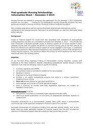
040088 Design/photography by the Educational Resource Centre, Women’s & Children’s Health, reprinted January 2004<br />
This publication is provided with<br />
the compliments of the<br />
Mark and Chapter<br />
Freemasons of Victoria<br />
Freemasonry is an ancient and respectable<br />
institution, embracing individuals of every<br />
nation, of every faith and every condition<br />
of life. It can be defined as a benevolent,<br />
charitable, educational and ethical society.<br />
It strives to teach every moral and social<br />
virtue and exhorts its membership to practice<br />
the universal principles of brotherly love,<br />
relief and truth.<br />
Masonic Centre of Victoria<br />
300 Albert Street,<br />
East Melbourne, Victoria, Australia 3002<br />
Telephone (03) 9419 8687