
You also want an ePaper? Increase the reach of your titles
YUMPU automatically turns print PDFs into web optimized ePapers that Google loves.
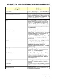
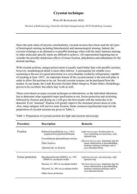
<strong>Cryostat</strong> <strong>technique</strong><br />
WOLF D. KUHLMANN, M.D.<br />
Division of Radiooncology, Deutsches Krebsforschungszentrum, 69120 Heidelberg, Germany<br />
Since the early times of enzyme cytochemistry, cryostat sections have been used for all types<br />
of histological staining including histochemical and immunological staining. Indeed, the<br />
cryostat <strong>technique</strong> is an alternative to paraffin histology when with the latter immuno-staining<br />
or other molecular specific stains are difficult to achieve. All experimental beginning has to<br />
consider the possibly deleterious effects of tissue fixation, dehydration and embedment for the<br />
desired stainings.<br />
With cryostat sections, antigen preservation is usually much better than with paraffin sections,<br />
however, morphological detail is most often inferior. A prerequisite for reliable cryosectioning<br />
is the use of a good microtome in a cryo-chamber cooled by refrigeration, capable<br />
of reaching at least -30°C. An important feature of the cryomicrotome is the anti-roll plate in<br />
order to allow flat sections to be cut. Several cryostat systems can be purchased from the<br />
market. In our hands, the Linde Kryostat (System Dittes-Duspiva; Walter Dittes, Heidelberg)<br />
proves to be excellent, but others may work as well.<br />
There exist almost as many cryostat <strong>technique</strong>s as laboratories, so the individual laboratory<br />
has to determine what sequential steps (prefixation or not, freeze protection and sectioning<br />
followed by fixation and drying etc.) will give the best results with the molecules to be<br />
detected. Even “minimal” fixation will greatly improve the structural preservation of cells.<br />
Also, many antigens will survive some fixation. Some common experimental steps for the<br />
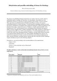
preparation of cryostat sections are given in Table 1.<br />
Table 1: Preparation of cryostat sections for light and electron microscopy<br />
Procedure Description Remarks<br />
Fixation Buffered formaldehyde (e.g. 1-4%)<br />
prepared from paraformaldehyde<br />
Formaldehyde-glutaraldehyde mixture<br />
Other fixatives<br />
Alternatively: no fixation<br />
Light microscopy: fixation prior to<br />
cryo-sectioning not necessary, only<br />
optional step<br />
Electron microscopy: important step for<br />
tissue stabilization in preembedding<br />
immuno-staining <strong>technique</strong> and in<br />
cryo-ultramicrotomy<br />
Freeze protection Dimethylsulfoxide (DMSO), f.e. 10%<br />
DMSO in cacodylate buffer<br />
Sucrose, f.e. 30% in cacodylate buffer<br />
Glycerol, f.e. 30% in cycodylate buffer<br />
Other freeze protection<br />
Light microscopy: not necessary, only<br />
optional step<br />
Electron microscopy: important step for<br />
tissue stabilization in preembedding<br />
immuno-staining <strong>technique</strong> and in<br />
cryo-ultramicrotomy
Snap-freezing<br />
Liquid nitrogen cooled isopentane:<br />
immersion of tissue block mounted on<br />
a tissue holder with a drop of Tissue<br />
Tek® as binding matrix 1)<br />
Direct snap-freezing in liquid nitrogen<br />
Frozen tissue blocks are transferred into<br />
the cryochamber or stored in liquid<br />
nitrogen<br />
Cryo-sectioning<br />
Light microscopy: sections are cut at<br />
4-8 µm thickness<br />
Electron microscopy: sections are cut at<br />
10-40 µm thickness for preembedding<br />
immuno-staining<br />
Electron microscopy: cryo-ultramicrotomy<br />
for obtaining ultrathin frozen<br />
sections<br />
Light microscopy: cryochamber at -30°C<br />
Electron microscopy: cryochamber at<br />
-30°C for preparing thick frozen<br />
sections (preembedding immunostaining)<br />
Electron microscopy: cryochamber at<br />
-90°C to -120°C for ultrathin cryosectioning<br />
Collection of<br />
sections<br />
Light microscopy: frozen sections are<br />
mounted in the cryochamber on clean,<br />
BSA or silane conditioned glass slides<br />
and transferred to room temperature;<br />
slides can be stored for several days<br />
at -20 to -80°C before staining<br />
Electron microscopy: frozen cut sections<br />
are dropped straight as free-floating<br />
sections into vials with 10% DMSO<br />
Electron microscopy: ultrathin frozen<br />
sections are collected dry on nickel<br />
grids or spread on 50% DMSO and<br />
transferred with a wire loop onto a<br />
drop of water at room temperature;<br />
optionally: sections are picked up with<br />
a loop containing a drop of 2.3 mol/L<br />
sucrose or 1% methyl cellulose; the<br />
loop is then touched onto the surface of<br />
a grid to deposit the sections<br />
Light microscopy: cryosections may be<br />
either air-dried (variable times), then<br />
fixed (in acetone or ethanol) at 4°C<br />
or fixed at -20°C for 5, 10 or 15 min<br />
Electron microscopy: sections for<br />
preembedding immuno-staining<br />
are kept free-floating in the vials<br />
during all subsequent steps until final<br />
resin embedment<br />
Electron microscopy: ultrathin sections<br />
are cut at a thickness of about 50-70<br />
nm; sections are picked up and<br />
transferred for direct imaging or<br />
immuno-staining by a variety of<br />
methods<br />
Fixation<br />
Light microscopy: air-dried sections are<br />
fixed in acetone at 4°C or at -20°C for<br />
5 or 10 or 15 min, then allowed to dry<br />
Alternatively: other type of fixation<br />
Light microscopy: different methods<br />
may be tried depending on the<br />
molecules to be studied<br />
Electron microscopy: usually no further<br />
fixation needed; postfixation (OsO 4 )<br />
of immuno-stained thick frozen<br />
sections is useful for further resin<br />
embedment<br />
Immuno-staining<br />
Light microscopy: see Immuno-staining<br />
with paraffin embedded tissue sections<br />
and Immunofluorescence staining of<br />
cryostat sections<br />
Electron microscopy: see Preembedding<br />
immuno-staining for electron<br />
microscopy<br />
Electron microscopy: see Miscellaneous<br />
immuno-staining <strong>technique</strong>s<br />
Light microscopy: some differences in<br />
immuno-staining of paraffin and<br />
cryostat sections, e.g. antigen retrieval<br />
steps in paraffin preparations, enzyme<br />
inhibition schedules<br />
Electron microscopy: staining of<br />
ultrathin<br />
frozen sections is quite different from<br />
procedures of preembedding immunostaining<br />
of thick frozen sections
1)<br />
Tissu Tek is a formulation of water-soluble compounds (polyvinyl alcohol, polyethelene glycol and other<br />
non-reactive ingredients (Sakura Inc.)<br />
Frozen sections for light microscopy<br />
The concept of first cutting cryo-sections which are then fixed is different from the classical<br />
paraffin embedment procedure (where fixation is first and followed by embedment). This can<br />
involve problems inasmuch as soluble antigens may leak away from the cryo-sections before<br />
sufficient tissue stabilization is achieved.<br />
Usually, snap-frozen tissue blocks are cut on a standard cryostat with a clean blade, and<br />
sections are mounted on acetone cleaned or specially coated glass slides. Coating is done for<br />
better adhesion of sections, f.e. with poly-L-lysine, bovine serum albumin, silane or other<br />
molecules. Unfixed sections may be immediately stored at -80ºC. Frozen sections are thawed<br />
at room temperature and immersed immediately into a fixative. For fixation purposes, widely<br />
employed solutions are (a) cold acetone for 5 min; (b) cold methanol for 5 min; (c) 70%<br />
ethanol for 15-30 sec; (d) 4% formaldehyde freshly prepared from paraformaldehyde for 5<br />
min; or (d) mixtures from ethanol and formaldehyde either followed by acetone or not. One<br />
one has to reconcile that acetone is not a real fixative like aldehydes. Acetone will solve fatty<br />
structures, and associated antigens may solve, too.<br />
After fixation, slides are rinsed briefly in phosphate buffered saline at pH 7.4 and subjected to<br />
immuno-staining. Cryo-sections being fixed with acetone alone are quite vulnerable with<br />
respect to surfactants (Tween etc.) or substances for blocking endogenous enzyme activities<br />
(methanol etc.). Endogenous peroxidase activity can be inhibited (with the exception of<br />
neutrophilic granulocytes) by use of 0.1% sodium azide plus 0.3% hydrogen peroxide in<br />
phosphate buffered saline (PBS). Optionally, inhibition of peroxidase activity can be tried by<br />
a glucose oxidase blocking procedure, i.e. incubation of cryo-sections in PBS containing D-<br />
glucose, the enzyme GOD and sodium azide.<br />
The use of distilled water as final step after all immunohistological procedures including<br />
chromogen development should be avoided because distilled water can generally be harmful<br />
to acetone fixed cryo-sections; use tap water instead.<br />
Cryo-<strong>technique</strong>s for electron microscopy<br />
Cryo-<strong>technique</strong>s are a group of related procedures for the stabilisation of biological<br />
specimens for microscopic observations. Tissue samples (lightly fixed or not and<br />
cryoprotected) are frozen to maintain morphological structure and composition as it exists<br />
under physiological conditions. Thus, cryopreserved samples exhibit the best antigenicity,<br />
essential for successful immuno-electron microscopy.<br />
The method of ultrathin frozen sectioning has been primarily proposed as an alternative to<br />
conventional resin embedded tissue sections for ultrastructural studies. The concept is to<br />
avoid morphological changes and denaturation effects of both dehydration and resin<br />
embedment procedures for enzyme cytochemical and immunocytochemical research. The<br />
advantage of cryo-ultramicrotomy for such approaches over resin ultramicrotomy is selfunderstanding.<br />
From the very beginning of cryo-ultramicrotomy (FERNÁNDEZ-MORÁN H,
1952; BERNHARD W and NANCY MT, 1964; DOLLHOPF FL and SITTE H, 1969; HODSON S and<br />
MARSHALL J, 1970; KOLEHMAINEN-SEVÉUS L, 1970; TOKUYASU KT, 1973; TOKUYASU KT<br />
and SINGER SJ, 1976; TOKUYASU KT, 1986) to the point where this <strong>technique</strong> has become an<br />
appropriate method for ultrastructural studies, it has taken about 20 years of development.<br />
Examples of cryo-<strong>technique</strong>s for immunocytochemical studies include<br />
• Cryo-ultramicrotomy: ultrathin frozen sections (50-70 nm thick) are cut on an<br />
ultramicrotome in a cryochamber. Sections are thawed on grids for transmission<br />
electron microscopy and subsequently immuno-stained, for example with ferritin or<br />
gold conjugated antibodies.<br />
• Cryo-substitution: this is a cryo-variant of conventional resin embedment. The method<br />
avoids denaturing effects of room temperature dehydration and resin embedment.<br />
Samples are frozen, then dehydration and resin embedment are performed in a cryosubstitution<br />
unit, for example at -90°c in acetone. At this temperature the water ice is<br />
replaced by acetone in a sublimation type process which may minimize adverse effects<br />
on antigenicity. The sample is then infiltrated with low viscosity resins (such as the<br />
Lowicryl series) and cured at low temperatures with UV light. Ultrathin sections are<br />
cut from the embedded tissue blocks with conventional ultramicrotomes for<br />
subsequent<br />
immuno-staining.<br />
Apart from the above cryo-<strong>technique</strong>s we have to mention cryo-electron microscopy which is<br />
used to observe molecules, viruses and tissues in their native state without fixation or<br />
staining. A typical application is called frozen hydrated transmission electron microscopy. A<br />
thin layer of unfixed aqueous sample is frozen to give a sheet of vitreous ice with the sample<br />
suspended in it. The preparation is observed at low temperature in the electron microscope<br />
equipped with a special cold stage. Samples prepared in this way are very sensitive to the<br />
electron beam. All immuno-stainings may be done prior to freezing. In a similar way, samples<br />
are prepared for cryo-SEM (scanning electron microscopy) where specimens are rapidly<br />
frozen, sputter coated and transferred to the SEM for imaging.<br />
Selected publications for further readings<br />
Fernández-Morán H (1952)<br />
Bernhard W and Nancy MT (1964)<br />
Dollhopf FL and Sitte H (1969)<br />
Hodson S and Marshall J (1970)<br />
Kolehmainen-Sevéus L (1970)<br />
Kuhlmann WD and Viron A (1972)<br />
Tokuyasu KT (1973)<br />
Tokuyasu KT and Singer SJ (1976)<br />
Tokuyasu KT (1986)<br />
Full version of citations in chapter References.<br />
© Prof. Dr. Wolf D. Kuhlmann, Heidelberg 20.09.2008