AIDS Kit I: Simulation of HIV-1 Detection - Frederiksen
AIDS Kit I: Simulation of HIV-1 Detection - Frederiksen
AIDS Kit I: Simulation of HIV-1 Detection - Frederiksen

You also want an ePaper? Increase the reach of your titles
YUMPU automatically turns print PDFs into web optimized ePapers that Google loves.
EDVOTEK • P.O. Box 1232 • West Bethesda, MD 20827-1232<br />
®<br />
The Biotechnology<br />
Education Company ®<br />
<strong>AIDS</strong> <strong>Kit</strong> I:<br />
<strong>Simulation</strong> <strong>of</strong><br />
<strong>HIV</strong>-1 <strong>Detection</strong><br />
271<br />
EDVO-<strong>Kit</strong> #<br />
Storage:<br />
Store entire experiment in the refrigerator.<br />
Experiment Objective:<br />
The objective <strong>of</strong> this experiment is to understand the molecular biology<br />
<strong>of</strong> the human immunodeficiency virus and the pathogenesis <strong>of</strong> acquired<br />
immune deficiency syndrome. The experimental concepts and methodology<br />
involved with enzyme linked immunosorbent (ELISA) assays will<br />
be introduced in the context <strong>of</strong> the clinical screening <strong>of</strong> serum samples<br />
for antibodies to the <strong>HIV</strong> virus.<br />
1-800-EDVOTEK • (301) 251-5990 • 24-hour FAX: (301) 340-0582<br />
http://www.edvotek.com • email: edvotek@aol.com<br />
All components are intended for educational research only. They are not to be used for<br />
diagnostic or drug purposes, nor administered to or consumed by humans or animals.<br />
EVT 909168AM
2<br />
EDVO-<strong>Kit</strong> # 271: <strong>AIDS</strong> <strong>Kit</strong> I: <strong>Simulation</strong> <strong>of</strong> <strong>HIV</strong>-1 <strong>Detection</strong><br />
EDVOTEK • The Biotechnology<br />
Education Company ®<br />
Major Section Headings<br />
Page<br />
Experiment Components 3<br />
Requirements 3<br />
Background Information 4<br />
Experimental Procedures 9<br />
Study Questions 12<br />
Instructor's Guide<br />
General Information 13<br />
Pre-Lab Preparations 13<br />
Avoiding Common Pitfalls 16<br />
Idealized Schematic <strong>of</strong> Results 16<br />
Answers to Study Questions 17<br />
This experiment does not contain <strong>HIV</strong> virus or its components.<br />
None <strong>of</strong> the components have been prepared from human sources.<br />
Duplication <strong>of</strong> this document, in conjunction with use <strong>of</strong> accompanying reagents, is permitted for classroom/laboratory use only. This<br />
document, or any part, may not be reproduced or distributed for any other purpose without the written consent <strong>of</strong> EDVOTEK, Inc.<br />
Copyright © 1992,1993,1994,1996,1997,1998, EDVOTEK, Inc., all rights reserved.<br />
EVT 909168AM
EDVO-<strong>Kit</strong> # 271: <strong>AIDS</strong> <strong>Kit</strong> I: <strong>Simulation</strong> <strong>of</strong> <strong>HIV</strong>-1 <strong>Detection</strong><br />
3<br />
EDVOTEK • The Biotechnology<br />
Education Company ®<br />
Experiment Components<br />
Storage:<br />
Store entire experiment<br />
in the refrigerator.<br />
All components <strong>of</strong> this experiment<br />
are intended for educational<br />
research only. They are not to be<br />
used for diagnostic or drug<br />
purposes, nor administered to or<br />
consumed by humans or animals.<br />
This kit is designed for 10 lab groups<br />
A<br />
B<br />
C<br />
D<br />
E<br />
F<br />
G<br />
H<br />
Contents<br />
<strong>HIV</strong> Antigens (simulated)<br />
Positive Control (primary antibody)<br />
Donor 1 Serum (simulated)<br />
Donor 2 Serum (simulated)<br />
Anti-IgG-peroxidase conjugate (secondary antibody)<br />
Hydrogen peroxide, stabilized<br />
Aminosalicylic acid (peroxide co-substrate)<br />
Phosphate buffered saline concentrate<br />
2 Microtiter plates<br />
50 Transfer pipets<br />
60 Microtest tubes with attached caps<br />
25 1 ml pipets<br />
2 plastic tubes, 50 ml<br />
This experiment does not contain <strong>HIV</strong> virus or its components.<br />
None <strong>of</strong> the components have been prepared from human sources.<br />
Requirements<br />
• Distilled or deionized water<br />
• Beakers<br />
• 37°C Incubation oven<br />
• Disposable lab gloves<br />
• Safety goggles<br />
• Automatic micropipets and tips recommended<br />
Make sure glassware is clean, dry and free <strong>of</strong> soap residue.<br />
For convenience, additional disposable transfer pipets (Cat. # 632) can<br />
be purchased for liquid removal and washing steps.<br />
Duplication <strong>of</strong> this document, in conjunction with use <strong>of</strong> accompanying reagents, is permitted for classroom/laboratory use only. This<br />
document, or any part, may not be reproduced or distributed for any other purpose without the written consent <strong>of</strong> EDVOTEK, Inc.<br />
Copyright © 1992,1993,1994,1996,1997,1998, EDVOTEK, Inc., all rights reserved.<br />
EVT 909168AM
4<br />
EDVO-<strong>Kit</strong> # 271: <strong>AIDS</strong> <strong>Kit</strong> I: <strong>Simulation</strong> <strong>of</strong> <strong>HIV</strong>-1 <strong>Detection</strong><br />
EDVOTEK • The Biotechnology<br />
Education Company ®<br />
BACKGROUND INFORMATION<br />
Background Information<br />
Acquired immune deficiency syndrome (<strong>AIDS</strong>) is a disease characterized<br />
by the progressive deterioration <strong>of</strong> an individual's immune system. The<br />
immunological impairment allows infectious agents such as viruses, bacteria,<br />
fungi and parasites to invade the body and propagate unchecked. In<br />
addition, the incidence <strong>of</strong> certain cancers dramatically increases in these<br />
patients because <strong>of</strong> their compromised immune system. <strong>AIDS</strong> is a serious<br />
threat to human health and is a global problem. Intensive research is being<br />
done to advance methods <strong>of</strong> detection, clinical treatment and prevention.<br />
Retroviruses<br />
The <strong>AIDS</strong> etiologic agent (<strong>HIV</strong>-1) is the human immunodeficiency virus<br />
type 1, which is a retrovirus. Retroviruses contain an RNA genome and<br />
the RNA dependent DNA polymerase also termed reverse transcriptase.<br />
Members <strong>of</strong> the retrovirus family are involved in the pathogenesis <strong>of</strong> certain<br />
types <strong>of</strong> leukemias and other sarcomas in humans and animals. The<br />
structure and replication mechanism <strong>of</strong> <strong>HIV</strong> is very similar to other<br />
retroviruses. <strong>HIV</strong> is unique in some <strong>of</strong> its properties since it specifically<br />
targets the immune system, is very immunoevasive, forms significant<br />
amounts <strong>of</strong> progeny virus in vivo during the later stages <strong>of</strong> the disease and<br />
can be transmitted during sexual activity.<br />
Glycoprotein<br />
Reverse<br />
transcriptase<br />
Protease<br />
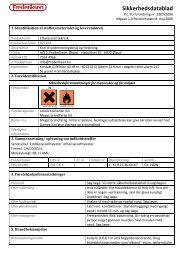
The <strong>HIV</strong> viral particle is surrounded by a lipid bilayer derived from the<br />
host cell membrane during budding. The viral proteins are identified by<br />
the prefix gp (glycoprotein) or p (protein) followed<br />
Protein<br />
coat<br />
RNA<br />
by a number indicating the approximate molecular<br />
weight in kilodaltons. The lipid bilayer contains gp<br />
160, gp 120 and gp 41. The gp 41 anchors gp 120 in<br />
the bilayer. Beneath the bilayer is a capsid consisting<br />
<strong>of</strong> p17 and p18. Within this shell is the viral core.<br />
The walls <strong>of</strong> the core consists <strong>of</strong> p24 and p25. Within<br />
the core are two identical RNA molecules 9000 nucleotides<br />
in length. Hydrogen bonded to each genomic<br />
RNA is a cellular tRNA molecule. The core also contains<br />
approximately 50 molecules <strong>of</strong> reverse transcriptase.<br />
Lipid<br />
bilayer<br />
Core<br />
Integrase<br />
Large quantities <strong>of</strong> virus can be grown in tissue culture<br />
for diagnostic and research purposes. Several<br />
<strong>of</strong> the viral proteins have been cloned and expressed<br />
in relatively large quantities.<br />
Duplication <strong>of</strong> this document, in conjunction with use <strong>of</strong> accompanying reagents, is permitted for classroom/laboratory use only. This<br />
document, or any part, may not be reproduced or distributed for any other purpose without the written consent <strong>of</strong> EDVOTEK, Inc.<br />
Copyright © 1992,1993,1994,1996,1997,1998, EDVOTEK, Inc., all rights reserved.<br />
EVT 909168AM
EDVO-<strong>Kit</strong> # 271: <strong>AIDS</strong> <strong>Kit</strong> I: <strong>Simulation</strong> <strong>of</strong> <strong>HIV</strong>-1 <strong>Detection</strong><br />
5<br />
EDVOTEK • The Biotechnology<br />
Education Company ®<br />
BACKGROUND INFORMATION<br />
Background Information,<br />
continued<br />
Mechanism <strong>of</strong> <strong>HIV</strong> Infection<br />
An individual can be infected with <strong>HIV</strong> through an abrasion in a mucosal<br />
surface (e.g. vaginal and rectal walls), a blood transfusion or by<br />
intravenous injection with a contaminated needle. Virus or virally infected<br />
cells are found in bodily fluids such as semen and blood. During<br />
the early stages <strong>of</strong> infection in an immunocompetent person the<br />
<strong>HIV</strong> virus elicits humoral and cellular immunity responses that result<br />
in a variety <strong>of</strong> circulating IgG molecules directed at several viral<br />
epitopes. However, since the virus has a high mutation rate the variants<br />
survive and produce progeny having a similar capacity to escape<br />
immunosurveillance.<br />
Unlike other cellular DNA polymerases, <strong>HIV</strong> DNA polymerase (reverse<br />
transcriptase) has a high error rate (1 in 10 4 ). These frequent<br />
mutations continually change the viral protein epitopes. This is believed<br />
to be the main mechanism <strong>of</strong> <strong>HIV</strong> immunoevasion. The most<br />
important target for the virus are hematopoietic cells such as bone<br />
marrow derived monocytes, myelocytes and immune system lymphocytes.<br />
Infection <strong>of</strong> immune system effector cells such as T cells and<br />
macrophages ultimately produce the most pr<strong>of</strong>ound clinical consequences.<br />
gp 120 binds to the CD4 receptors on the surface <strong>of</strong> T helper<br />
(T H<br />
) cells. These receptors are membrane bound glycoproteins involved<br />
in T cell maturation from precursor cells. T H<br />
cells are required for the<br />
body's overall immunological responses. The viral lipid bilayer fuses<br />
with that <strong>of</strong> the cell's membranes and the viral protein capsid becomes<br />
internalized via receptor mediated endocytosis. Subsequently, the rest<br />
<strong>of</strong> the CD4 receptors are internalized and gp 120 appears on the T cell<br />
surface.<br />
<strong>HIV</strong> Replication and Transcription<br />
Through a complex mechanism involving several events, the reverse<br />
transcriptase synthesizes a double stranded DNA copy <strong>of</strong> the genomic<br />
RNA template. The tRNA molecule acts as the primer for the first<br />
strand synthesis. The reverse transcriptase, RNAse H activity, degrades<br />
the RNA strand <strong>of</strong> the RNA-DNA duplex and the polymerase activity<br />
synthesizes a complementary DNA strand. The DNA reverse transcripts<br />
(copy DNA) migrate into the cell nucleus where they become<br />
covalently integrated into the cellular genomic DNA. The integration<br />
is catalyzed by the <strong>HIV</strong> integrase. The copy DNA integrates via specific,<br />
self-complimentary sequences at both ends called long terminal<br />
repeats (LTRs). These sequences also have important functions in viral<br />
transcription. The integrated copy DNA is called proviral DNA or<br />
the provirus. The provirus enters a period <strong>of</strong> latency that can last for<br />
several years. The proviral DNA is replicated along with the cellular<br />
DNA and can be inherited through many generations. The <strong>HIV</strong> proviral<br />
DNA contains the major genes common to all non-transducing<br />
Duplication <strong>of</strong> this document, in conjunction with use <strong>of</strong> accompanying reagents, is permitted for classroom/laboratory use only. This<br />
document, or any part, may not be reproduced or distributed for any other purpose without the written consent <strong>of</strong> EDVOTEK, Inc.<br />
Copyright © 1992,1993,1994,1996,1997,1998, EDVOTEK, Inc., all rights reserved.<br />
EVT 909168AM
6<br />
EDVO-<strong>Kit</strong> # 271: <strong>AIDS</strong> <strong>Kit</strong> I: <strong>Simulation</strong> <strong>of</strong> <strong>HIV</strong>-1 <strong>Detection</strong><br />
EDVOTEK • The Biotechnology<br />
Education Company ®<br />
BACKGROUND INFORMATION<br />
Background Information,<br />
continued<br />
retroviruses. These genes are gag, pol and env. <strong>HIV</strong> also contains five<br />
or six other genes that are much smaller. Retroviral transcription is a<br />
complex process producing a variety <strong>of</strong> RNAs. Promotion <strong>of</strong> transcripts<br />
is controlled in the LTR and transcriptional termination signals are located<br />
in each major gene. Those RNA transcripts that remain unspliced<br />
become packaged in the new viral particles. The gag gene is translated<br />
into a polypeptide that is cleaved by a viral protease into four<br />
proteins that form the inner shells. Specific protease inhibitors are clinically<br />
being used to inhibit protein processing and control the further<br />
spread <strong>of</strong> the <strong>HIV</strong> virus in patients suffering from <strong>AIDS</strong>. The pol gene<br />
encodes the reverse transcriptase and the integrase which is responsible<br />
for the genomic incorporation <strong>of</strong> copy DNA. The env gene encodes<br />
the surface glycoproteins the viral particles acquire as they bud<br />
from the cells.<br />
Immunological Response:<br />
Macrophages are circulating monocytes and are involved in the nonspecific<br />
engulfment <strong>of</strong> foreign material and normal cellular debris.<br />
These materials are degraded in the lysosomes <strong>of</strong> the cells. Peptides<br />
from foreign degraded proteins are transported to the macrophage<br />
surface where they remain bound by specialized receptors. Immunologically<br />
inactive T H<br />
cells interact with these surface bound antigenreceptor<br />
complexes which enables them to become fully activated. <strong>HIV</strong><br />
infects macrophages by binding to the cells' CD4 receptors. Fully activated<br />
T H<br />
cells secrete several types <strong>of</strong> protein factors collectively known<br />
as lymphokines. Several <strong>of</strong> these factors are the interleukins which<br />
stimulate antibody secretions from B cells enable macrophage activation,<br />
stimulate general T cell growth and activate cytotoxic T cells.<br />
Cytotoxic T cells are involved in the actual destruction <strong>of</strong> foreign cells<br />
and body cells infected with different viruses. Inactive T H<br />
cells that<br />
have been infected by <strong>HIV</strong> remain in a relatively quiescent state similar<br />
to uninfected cells. When a T H<br />
cell containing provirus undergoes<br />
antigenic activation the integrated copy DNA becomes open to the transcription<br />
<strong>of</strong> viral RNA.<br />
Viral replication causes the destruction <strong>of</strong> the T H<br />
cells. Infected T H<br />
cells<br />
also form syncytia, i.e. fused cells. Syncytia occur since the gp 120 on<br />
the infected T cell surface binds to CD4 receptors on other T H<br />
cells.<br />
Cell to cell transmission <strong>of</strong> virus can occur in syncytia without the need<br />
for a free viral intermediate. Replication <strong>of</strong> virus also proceeds in activated<br />
macrophages which causes cell death and release <strong>of</strong> infectious<br />
viral particles. These and other events ultimately cause complete immune<br />
system collapse. The long latency period after <strong>HIV</strong> infection can<br />
be understood in terms <strong>of</strong> T H<br />
cell activation. Only certain T H<br />
cells are<br />
capable <strong>of</strong> responding to a particular antigen. <strong>HIV</strong> infected but<br />
asymptomatic individuals will experience the usual exposure to chemicals,<br />
viruses and bacteria. Each infection activates a subpopulation T H<br />
cells containing the provirus which eventually leads to the death <strong>of</strong><br />
Duplication <strong>of</strong> this document, in conjunction with use <strong>of</strong> accompanying reagents, is permitted for classroom/laboratory use only. This<br />
document, or any part, may not be reproduced or distributed for any other purpose without the written consent <strong>of</strong> EDVOTEK, Inc.<br />
Copyright © 1992,1993,1994,1996,1997,1998, EDVOTEK, Inc., all rights reserved.<br />
EVT 909168AM
EDVO-<strong>Kit</strong> # 271: <strong>AIDS</strong> <strong>Kit</strong> I: <strong>Simulation</strong> <strong>of</strong> <strong>HIV</strong>-1 <strong>Detection</strong><br />
7<br />
EDVOTEK • The Biotechnology<br />
Education Company ®<br />
BACKGROUND INFORMATION<br />
Background Information,<br />
continued<br />
these cells. After successive waves <strong>of</strong> infections the population <strong>of</strong> T H<br />
and macrophage cells become depleted and clinical <strong>AIDS</strong> develops.<br />
There are a very small number <strong>of</strong> individuals who have coexisted with<br />
<strong>HIV</strong> for over 15 years. Although the reasons for this coexistence with<br />
<strong>HIV</strong> is not understood, the immune system appears to remain intact.<br />
Such individuals tend to eat well, exercise and practice stress reduction<br />
techniques. Genetic analysis may determine whether or not subtle<br />
genetic differences in the immune system are significant factors.<br />
Description <strong>of</strong> the <strong>HIV</strong> Screening <strong>Simulation</strong><br />
well<br />
Human<br />
Serum<br />
(IgG)<br />
<strong>HIV</strong><br />
Antigen<br />
well<br />
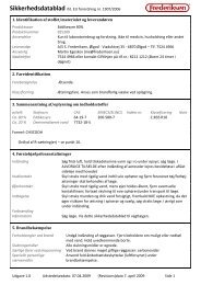
Schematic for<br />
ELISA<br />
Horseradish<br />
peroxidase<br />
Anti-IgG<br />
(Rabbit)<br />
Human<br />
Serum<br />
(IgG)<br />
<strong>HIV</strong><br />
Antigen<br />
Enzyme linked immunosorbent assay (ELISA) tests were originally<br />
developed for antibody measurement. These immunoassays have also<br />
been adapted to successfully detect samples that contain antigens. This<br />
ELISA simulation experiment has been designed to detect a hypothetical<br />
patient's circulating IgG directed towards the viral (<strong>HIV</strong>) antigen.<br />
ELISAs are done in microtiter plates which are generally made <strong>of</strong> polystyrene<br />
or polyvinyl chloride. The plates are somewhat transparent<br />
and contain many small wells, in which liquid samples are deposited.<br />
First, the antigens are added to the wells<br />
where some remain adsorbed by hydrophobic<br />
association to the walls after washing<br />
away the excess. The antigens can be<br />
the whole <strong>HIV</strong> lysate, specific <strong>HIV</strong> proteins,<br />
or a mixture <strong>of</strong> the two. There is no<br />
specificity involved with the adsorption<br />
process although some substances may exhibit<br />
low binding to the walls. In certain<br />
cases the antigens can be covalently crosslinked<br />
to the plastic using UV light. After<br />
S<br />
(colorless)<br />
well<br />
Horseradish<br />
peroxidase<br />
Anti-IgG<br />
(Rabbit)<br />
Human<br />
Serum<br />
(IgG)<br />
<strong>HIV</strong><br />
Antigen<br />
P<br />
(color)<br />
washing away unadsorbed material, the<br />
unoccupied sites on the walls <strong>of</strong> the plastic<br />
wells are blocked with proteins, typically<br />
gelatin or bovine serum albumin.<br />
Infection by <strong>HIV</strong>-1 causes the individual<br />
to mount an antibody response which<br />
eventually results in plasma IgG molecules<br />
that bind to different <strong>HIV</strong> proteins (and/<br />
or different areas or the same polypeptide).<br />
In this experiment, if these antibodies are<br />
present in the plasma sample they will<br />
bind to the adsorbed antigens in the well<br />
and remain there after washing.<br />
A solution containing the IgG antibody<br />
that binds to any kind <strong>of</strong> human IgG, is<br />
Duplication <strong>of</strong> this document, in conjunction with use <strong>of</strong> accompanying reagents, is permitted for classroom/laboratory use only. This<br />
document, or any part, may not be reproduced or distributed for any other purpose without the written consent <strong>of</strong> EDVOTEK, Inc.<br />
Copyright © 1992,1993,1994,1996,1997,1998, EDVOTEK, Inc., all rights reserved.<br />
EVT 909168AM
8<br />
EDVO-<strong>Kit</strong> # 271: <strong>AIDS</strong> <strong>Kit</strong> I: <strong>Simulation</strong> <strong>of</strong> <strong>HIV</strong>-1 <strong>Detection</strong><br />
EDVOTEK • The Biotechnology<br />
Education Company ®<br />
BACKGROUND INFORMATION<br />
Background Information,<br />
continued<br />
then added to the wells. If the primary antibody has remained in a<br />
well, then the secondary antibody will bind to it and also remain attached<br />
after washing. These secondary antibodies are usually raised<br />
in rabbits and goats immunized with human IgG fractions. The second<br />
IgG antibodies are purified and covalently cross linked to horseradish<br />
peroxidase. This modification does not significantly affect the<br />
binding specificity and affinity <strong>of</strong> the antibody or the enzymatic activity<br />
<strong>of</strong> the peroxidase.<br />
After washing, a solution containing hydrogen peroxide and<br />
aminosalicylate is added to each well. Peroxidase possesses a high<br />
catalytic activity and can exceed turnover rates <strong>of</strong> 10 6 per second. Consequently,<br />
amplification <strong>of</strong> a positive sample can occur over several<br />
orders <strong>of</strong> magnitude. Many hydrogen donor co-substrates can be used<br />
by peroxidase. These co-substrates include o-diansidine,<br />
aminoantipyrine, aminosalicylic acid and numerous phenolic compounds<br />
that develop color upon oxidation. The substrate solution<br />
added is nearly colorless. Peroxidase converts the peroxide to H 2<br />
O +<br />
O 2<br />
using the salicylate as the hydrogen donor. The oxidized salicylate<br />
is brown and can be easily observed in wells containing anti-<strong>HIV</strong>-1<br />
IgG (positive plasma).<br />
It should be noted that polyclonal antibody preparations to a given<br />
antigen can have variable binding affinities due to differences in the<br />
immunological responses between animals. Different immunizations<br />
with the same antigen in the same animal can also produce variable<br />
binding affinities. The use <strong>of</strong> monoclonal antibodies directed against a<br />
single epitope eliminates this variability. Western blot analysis <strong>of</strong> positive<br />
samples are used to confirm infection by <strong>HIV</strong>.<br />
Duplication <strong>of</strong> this document, in conjunction with use <strong>of</strong> accompanying reagents, is permitted for classroom/laboratory use only. This<br />
document, or any part, may not be reproduced or distributed for any other purpose without the written consent <strong>of</strong> EDVOTEK, Inc.<br />
Copyright © 1992,1993,1994,1996,1997,1998, EDVOTEK, Inc., all rights reserved.<br />
EVT 909168AM
EDVO-<strong>Kit</strong> # 271: <strong>AIDS</strong> <strong>Kit</strong> I: <strong>Simulation</strong> <strong>of</strong> <strong>HIV</strong>-1 <strong>Detection</strong><br />
9<br />
EDVOTEK • The Biotechnology<br />
Education Company ®<br />
EXPERIMENTAL PROCEDURES<br />
EXPERIMENT OBJECTIVE:<br />
The objective <strong>of</strong> this experiment is to understand the molecular biology<br />
<strong>of</strong> the human immunodeficiency virus and the pathogenesis <strong>of</strong><br />
acquired immune deficiency syndrome. The experimental concepts<br />
and methodology involved with enzyme linked immunosorbent<br />
(ELISA) assays will be introduced in the context <strong>of</strong> the clinical screening<br />
<strong>of</strong> serum samples for antibodies to the virus.<br />
LABORATORY SAFETY<br />
Gloves and goggles should be worn routinely as good laboratory practice.<br />
Student Experimental Procedures<br />
GENERAL INSTRUCTIONS AND PROCEDURES<br />
Row 1<br />
Row 2<br />
Row 3<br />
Row 4<br />
Remember!<br />
Equilibrate a 37°C<br />
incubation oven before<br />
starting the experiment.<br />
Labeling the Microtiter Plate:<br />
• Place the microtiter plate vertically as shown in Figure 2.<br />
• Mark the plate with your initials or lab group number and<br />
number the rows 1-4 down the side.<br />
Labeling the Plastic Transfer pipets:<br />
Label 5 transfer pipets as follows:<br />
• ( - ) (negative)<br />
• (+ ) (positive)<br />
• DS 1 (Donor Serum 1)<br />
• DS 2 (Donor Serum 2)<br />
• PBS (Phosphate Buffered Saline)<br />
Use the appropriately labeled plastic transfer pipet for liquid removals<br />
and washes as outlined in the experimental procedures starting on<br />
page 10.<br />
Figure 2<br />
Duplication <strong>of</strong> this document, in conjunction with use <strong>of</strong> accompanying reagents, is permitted for classroom/laboratory use only. This<br />
document, or any part, may not be reproduced or distributed for any other purpose without the written consent <strong>of</strong> EDVOTEK, Inc.<br />
Copyright © 1992,1993,1994,1996,1997,1998, EDVOTEK, Inc., all rights reserved.<br />
EVT 909168AM
10<br />
EDVO-<strong>Kit</strong> # 271: <strong>AIDS</strong> <strong>Kit</strong> I: <strong>Simulation</strong> <strong>of</strong> <strong>HIV</strong>-1 <strong>Detection</strong><br />
EDVOTEK • The Biotechnology<br />
Education Company ®<br />
EXPERIMENTAL PROCEDURES<br />
Student Experimental<br />
Procedures, continued<br />
INSTRUCTIONS FOR ADDING LIQUIDS AND<br />
WASHING WELLS<br />
Adding Reagents to wells:<br />
• For adding reagents to the wells, use the same 1 ml pipet.<br />
Useful Hint!<br />
The general<br />
procedure for adding<br />
reagents to the wells<br />
will conserve the use <strong>of</strong><br />
a large number <strong>of</strong> pipets in this<br />
simulated classroom ELISA.<br />
If available, reagents should be<br />
dispensed with an automatic<br />
micropipet using disposable tips.<br />
• RINSE THE PIPET THOROUGHLY with distilled water before<br />
using the pipet for adding the next reagent.<br />
Liquid Removal and Washes:<br />
• When instructed in the experimental procedures, remove liquids<br />
with the appropriately labeled transfer pipet, and then<br />
wash the wells as follows:<br />
A. Use the transfer pipet labeled "PBS", to add PBS buffer to<br />
the wells in all rows. Add PBS buffer until each well is<br />
almost full.<br />
The capacity <strong>of</strong> each well is approximately 0.2 ml. Do not allow<br />
the liquids to spill over into adjacent wells.<br />
B. With the appropriately labeled transfer pipet, remove all<br />
the liquid (PBS buffer) from the wells in each row. Dispose<br />
the liquid in the beaker labeled "waste".<br />
EXPERIMENTAL STEPS FOR THE ENZYME LINKED<br />
IMMUNOSORBENT ASSAY (ELISA)<br />
1. To all 12 wells, add 0.1 ml <strong>of</strong> “<strong>HIV</strong>” (viral antigens).<br />
WEAR SAFETY GOGGLES<br />
AND GLOVES<br />
2. Incubate for 5 minutes at room temperature.<br />
3. Remove all the liquid (viral antigens) with a transfer pipet.<br />
4. Wash each well once with PBS buffer as described above ("Liquid<br />
Removal and Washes").<br />
In research labs, following this step, all sites on the microtiter plate are<br />
saturated with a blocking solution consisting <strong>of</strong> a protein mixture, such<br />
as BSA. We have designed this experiment to eliminate this step to save<br />
time.<br />
Duplication <strong>of</strong> this document, in conjunction with use <strong>of</strong> accompanying reagents, is permitted for classroom/laboratory use only. This<br />
document, or any part, may not be reproduced or distributed for any other purpose without the written consent <strong>of</strong> EDVOTEK, Inc.<br />
Copyright © 1992,1993,1994,1996,1997,1998, EDVOTEK, Inc., all rights reserved.<br />
EVT 909168AM
EDVO-<strong>Kit</strong> # 271: <strong>AIDS</strong> <strong>Kit</strong> I: <strong>Simulation</strong> <strong>of</strong> <strong>HIV</strong>-1 <strong>Detection</strong><br />
11<br />
EDVOTEK • The Biotechnology<br />
Education Company ®<br />
EXPERIMENTAL PROCEDURES<br />
Student Experimental<br />
Procedures, continued<br />
Reminders:<br />
ADDING REAGENTS:<br />
Be sure to rinse the 1 ml pipet<br />
thoroughly before adding a new<br />
reagent. (Steps 1, 5, 9, & 13).<br />
Alternatively, if you are using<br />
automatic micropipets, use a fresh tip<br />
for each reagent.<br />
LIQUID REMOVALS:<br />
Use the appropriately labeled transfer<br />
pipet to remove all liquid from the wells<br />
in each row (steps 3, 7, & 11) and<br />
after washes (steps 4, 8 & 12)<br />
Transfer pipet (-) Row 1<br />
Transfer pipet (+) Row 2<br />
Transfer pipet DS 1 Row 3<br />
Transfer pipet DS 2 Row 4<br />
Dispose the liquid in the beaker<br />
labeled "waste".<br />
WASHES:<br />
For all rows, use the transfer pipet<br />
labeled "PBS" to add PBS until each<br />
well is almost full. (Steps 4, 8, & 12)<br />
5. Add reagents as outlined below:<br />
Remember to rinse the 1 ml pipet thoroughly with distilled water<br />
before adding a new reagent. If you are using automatic micropipets,<br />
use a clean micropipet tip for each reagent.<br />
• Add 0.1 ml <strong>of</strong> PBS Buffer to the three wells in Row 1.<br />
(This is the negative control.)<br />
• Add 0.1 ml <strong>of</strong> "+" (positive) to the three wells in Row 2.<br />
(This is the positive control.)<br />
• Add 0.1 ml <strong>of</strong> Donor Serum “DS1” to the 3 wells in Row 3.<br />
• Add 0.1 ml <strong>of</strong> Donor Serum “DS2” to the 3 wells in Row 4.<br />
6. Incubate at 37°C for 15 minutes .<br />
7. Remove all the liquid from each well with the appropriately labeled<br />
transfer pipet.<br />
8. Wash each well once with PBS buffer (as described under "Liquid<br />
Removal and Washes").<br />
9. Add 0.1 ml <strong>of</strong> the anti-IgG peroxidase conjugate (2°Ab) to all 12<br />
wells.<br />
10. Incubate at 37°C for 15 minutes.<br />
At this time you can obtain the substrate to be used in step 13. Since the<br />
substrate must be prepared just prior to use, your instructor will prepare<br />
it towards the end <strong>of</strong> the incubation in step 10.<br />
11. Remove all the liquid from each well with the appropriately labeled<br />
transfer pipet.<br />
Quick Reference:<br />
The positive control, which<br />
contains IgG directed against <strong>HIV</strong><br />
antigens, is the primary antibody.<br />
Positive serum samples will also<br />
contain anti-<strong>HIV</strong> IgG, while<br />
negative serum samples will not<br />
contain anti-<strong>HIV</strong> IgG.<br />
12. Wash each well once with PBS buffer (as described under "Liquid<br />
Removal and Washes").<br />
13. Add 0.1 ml <strong>of</strong> the substrate to all 12 wells.<br />
14. Incubate at 37°C for 5 minutes.<br />
15. Remove the plate for analysis.<br />
16. If color is not fully developed after 5 minutes, incubate at 37°C for<br />
a longer period <strong>of</strong> time.<br />
Duplication <strong>of</strong> this document, in conjunction with use <strong>of</strong> accompanying reagents, is permitted for classroom/laboratory use only. This<br />
document, or any part, may not be reproduced or distributed for any other purpose without the written consent <strong>of</strong> EDVOTEK, Inc.<br />
Copyright © 1992,1993,1994,1996,1997,1998, EDVOTEK, Inc., all rights reserved.<br />
EVT 909168AM
12<br />
EDVO-<strong>Kit</strong> # 271: <strong>AIDS</strong> <strong>Kit</strong> I: <strong>Simulation</strong> <strong>of</strong> <strong>HIV</strong>-1 <strong>Detection</strong><br />
EDVOTEK • The Biotechnology<br />
Education Company ®<br />
Study Questions<br />
1. Describe the mechanism <strong>of</strong> ELISA. Why is ELISA so sensitive<br />
Why is it necessary to block unoccupied binding sites in the microtiter<br />
wells Why is it important to have a positive control<br />
2. Why can the onset <strong>of</strong> <strong>AIDS</strong> take several years<br />
3. Why is anti-<strong>HIV</strong>-1 IgG screened instead <strong>of</strong> the virus itself<br />
4. Why does the destruction <strong>of</strong> T H<br />
cells compromise the entire immune<br />
system How does <strong>HIV</strong> target T H<br />
cells<br />
5. Why are there so many immunological variants <strong>of</strong> <strong>HIV</strong><br />
6. The elimination <strong>of</strong> several steps in the ELISA could be accomplished<br />
if the primary antibody was made into an enzyme conjugate. Why<br />
is this generally not done What can cause a false positive in an<br />
ELISA<br />
Duplication <strong>of</strong> this document, in conjunction with use <strong>of</strong> accompanying reagents, is permitted for classroom/laboratory use only. This<br />
document, or any part, may not be reproduced or distributed for any other purpose without the written consent <strong>of</strong> EDVOTEK, Inc.<br />
Copyright © 1992,1993,1994,1996,1997,1998, EDVOTEK, Inc., all rights reserved.<br />
EVT 909168AM
EDVO-<strong>Kit</strong> # 271: <strong>AIDS</strong> <strong>Kit</strong> I: <strong>Simulation</strong> <strong>of</strong> <strong>HIV</strong>-1 <strong>Detection</strong><br />
13<br />
EDVOTEK • The Biotechnology<br />
Education Company ®<br />
INSTRUCTOR'S GUIDE<br />
General Information<br />
APPROXIMATE TIME REQUIREMENTS FOR PRE-<br />
LAB AND EXPERIMENTAL PROCEDURES<br />
1. Pre-lab preparation <strong>of</strong> biologicals and reagents takes approximately<br />
one and one-half hours.<br />
2. The student experimental activity requires approximately 60 minutes.<br />
Pre-Lab Preparations<br />
PREPARATIONS BEFORE THE LAB<br />
Microtiter Plates<br />
1. As shown in Figure 1, orient the microtiter plates so that the numbers<br />
1-12 are at the top and the letters A-H are on your left.<br />
Row 1<br />
Row 2<br />
Row 3<br />
Row 4<br />
Row 1<br />
Row 2<br />
Row 3<br />
Row 4<br />
A<br />
B<br />
C<br />
D<br />
E<br />
F<br />
G<br />
H<br />
1 2 3 4 5 6 7 8 9 10 11 12<br />
cutting lines depicted by dashed lines<br />
2. Cut each plate on the dotted lines as<br />
shown in the figure. Each piece will be 3<br />
wells on one axis and 4 wells on the other<br />
axis. Each lab group will receive one<br />
piece.<br />
Dispensing Components A through D:<br />
3. Use a FRESH 1 ml pipet (provided) for<br />
dispensing each <strong>of</strong> the components A-D<br />
directly from the component tubes provided<br />
in this experiment kit. Label<br />
microtest tubes and dispense volumes as<br />
outlined in the chart "Quick Reference:<br />
Preparation <strong>of</strong> experiment reagents"<br />
which appears on Page 15.<br />
Figure 1<br />
Duplication <strong>of</strong> this document, in conjunction with use <strong>of</strong> accompanying reagents, is permitted for classroom/laboratory use only. This<br />
document, or any part, may not be reproduced or distributed for any other purpose without the written consent <strong>of</strong> EDVOTEK, Inc.<br />
Copyright © 1992,1993,1994,1996,1997,1998, EDVOTEK, Inc., all rights reserved.<br />
EVT 909168AM
14<br />
EDVO-<strong>Kit</strong> # 271: <strong>AIDS</strong> <strong>Kit</strong> I: <strong>Simulation</strong> <strong>of</strong> <strong>HIV</strong>-1 <strong>Detection</strong><br />
EDVOTEK • The Biotechnology<br />
Education Company ®<br />
INSTRUCTOR'S GUIDE<br />
Pre-Lab Preparations,<br />
continued<br />
PREPARATIONS ON THE DAY OF THE LAB<br />
Preparation <strong>of</strong> Phosphate Buffered Saline<br />
1. Add all <strong>of</strong> the Phosphate Buffered Saline concentrate (H) to 270 ml<br />
<strong>of</strong> distilled water. Mix.<br />
2. Label this diluted Phosphate Buffered saline as “PBS”.<br />
3. Dispense 25 ml into small beakers for each <strong>of</strong> the 10 lab groups.<br />
Preparation <strong>of</strong> Anti-IgG Peroxidase Conjugate<br />
(Secondary Antibody)<br />
4. Add 0.3 ml <strong>of</strong> diluted Phosphate Buffered Saline (PBS) to the concentrated<br />
Anti-IgG peroxidase conjugate (E). Mix thoroughly by<br />
tapping and inverting the tube.<br />
5. Transfer 15 ml <strong>of</strong> diluted Phosphate Buffered Saline (PBS) to a 50<br />
ml plastic tube provided.<br />
6. Transfer the entire contents <strong>of</strong> tube E prepared in step 4 to the 50<br />
ml tube containing 15 ml <strong>of</strong> PBS. Label the tube "2°Ab" (Secondary<br />
Antibody). Mix.<br />
7. Dispense 1.4 ml <strong>of</strong> the diluted Anti-IgG peroxidase conjugate for<br />
each group.<br />
PREPARATION OF PEROXIDASE SUBSTRATE<br />
DURING THE LAB EXPERIMENT<br />
Quick Reference:<br />
The substrate is prepared for the<br />
peroxidase enzyme, which is<br />
attached to the anti-IgG<br />
peroxidase conjugate (secondary<br />
antibody).<br />
Prepare the substrate 15 - 30<br />
minutes before students require it<br />
for plate development (last<br />
incubation).<br />
Prepare 15 - 30 minutes before the last incubation:<br />
1. Dispense 13.5 ml <strong>of</strong> diluted Phosphate buffered saline (PBS) to the<br />
second 50 ml tube provided.<br />
2. Add Aminosalicylic acid (G) to the 13.5 ml <strong>of</strong> PBS. Cap and mix<br />
thoroughly by shaking and/or vortexing. There is usually undissolved<br />
material remaining.<br />
3. Then add 1.5 ml <strong>of</strong> Hydrogen peroxide (F). Cap and mix.<br />
4. Dispense 1.4 ml <strong>of</strong> the peroxidase substrate for each group.<br />
Duplication <strong>of</strong> this document, in conjunction with use <strong>of</strong> accompanying reagents, is permitted for classroom/laboratory use only. This<br />
document, or any part, may not be reproduced or distributed for any other purpose without the written consent <strong>of</strong> EDVOTEK, Inc.<br />
Copyright © 1992,1993,1994,1996,1997,1998, EDVOTEK, Inc., all rights reserved.<br />
EVT 909168AM
EDVO-<strong>Kit</strong> # 271: <strong>AIDS</strong> <strong>Kit</strong> I: <strong>Simulation</strong> <strong>of</strong> <strong>HIV</strong>-1 <strong>Detection</strong><br />
15<br />
EDVOTEK • The Biotechnology<br />
Education Company ®<br />
INSTRUCTOR'S GUIDE<br />
Quick Reference Tables<br />
Preparation <strong>of</strong> Experiment reagents<br />
Dispense for<br />
Label each group<br />
A* <strong>HIV</strong> Antigens <strong>HIV</strong> 1.4 ml<br />
B* Positive control + 0.4 ml<br />
C* Donor Serum # 1 DS 1 0.4 ml<br />
D* Donor Serum # 2 DS 2 0.4 ml<br />
E + PBS Anti-IgG-peroxidase-conjugate 2°Ab 1.4 ml<br />
PBS + F + G Peroxidase-enzyme substrate Substrate 1.4 ml<br />
H + water Phosphate Buffered Saline PBS 25.0 ml<br />
* Components A - D can be dispensed before the actual day <strong>of</strong> the lab and stored in the<br />
refrigerator. If these components are dispensed on the day <strong>of</strong> the lab, leave at room<br />
temperature.<br />
STUDENT MATERIALS<br />
Each Lab Group Should Receive:<br />
1 piece <strong>of</strong> microtiter plate<br />
1 tube labeled "<strong>HIV</strong>"<br />
1 tube labeled "+"<br />
1 tube labeled "DS 1"<br />
1 tube labeled "DS 2"<br />
1 tube labeled "2°Ab"<br />
1 1 ml pipet (or 1 automatic micropipet with tips)<br />
1 pipet pump<br />
5 Transfer pipets<br />
1 beaker containing 16 ml <strong>of</strong> PBS<br />
1 beaker containing approximately 100 ml <strong>of</strong> distilled water<br />
1 empty beaker labeled "waste"<br />
1 tube labeled "Substrate" (just before the last incubation)<br />
Duplication <strong>of</strong> this document, in conjunction with use <strong>of</strong> accompanying reagents, is permitted for classroom/laboratory use only. This<br />
document, or any part, may not be reproduced or distributed for any other purpose without the written consent <strong>of</strong> EDVOTEK, Inc.<br />
Copyright © 1992,1993,1994,1996,1997,1998, EDVOTEK, Inc., all rights reserved.<br />
EVT 909168AM
16<br />
EDVO-<strong>Kit</strong> # 271: <strong>AIDS</strong> <strong>Kit</strong> I: <strong>Simulation</strong> <strong>of</strong> <strong>HIV</strong>-1 <strong>Detection</strong><br />
EDVOTEK • The Biotechnology<br />
Education Company ®<br />
INSTRUCTOR'S GUIDE<br />
Avoiding Common Pitfalls<br />
If you don't find answers to your<br />
questions in this section, call our<br />
Technical Service Department<br />
(1-800-338-6835)<br />
Mon - Fri<br />
9:00 am to<br />
5:00 pm EST<br />
24-hour FAX: (301) 340-0582<br />
email: edvotek@aol.com<br />
1. Students should be advised to be very careful when transferring<br />
solutions into and out <strong>of</strong> the microliter plate wells.<br />
2. Use only clean or appropriately labeled pipets and avoid contaminating<br />
adjacent wells.<br />
3. Do not attempt to empty the microliter wells by shaking it out.<br />
This will not work - it will result in contaminating adjacent wells.<br />
4. Wash the wells gently and slowly, without force.<br />
5. As an optional activity, the plates can be read in a microplate reader<br />
at 405 nm.<br />
Please have the following<br />
information:<br />
• The kit number and title<br />
• <strong>Kit</strong> lot number on box or tube<br />
• The literature version number<br />
(in lower right corner)<br />
• Approximate purchase date<br />
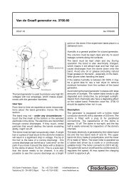
Expected Results<br />
Donor 2 should show positive for <strong>HIV</strong>.<br />
The color should look similar to the positive<br />
control.<br />
A<br />
B<br />
1 2 3<br />
C<br />
D<br />
Duplication <strong>of</strong> this document, in conjunction with use <strong>of</strong> accompanying reagents, is permitted for classroom/laboratory use only. This<br />
document, or any part, may not be reproduced or distributed for any other purpose without the written consent <strong>of</strong> EDVOTEK, Inc.<br />
Copyright © 1992,1993,1994,1996,1997,1998, EDVOTEK, Inc., all rights reserved.<br />
EVT 909168AM
EDVO-<strong>Kit</strong> # 271: <strong>AIDS</strong> <strong>Kit</strong> I: <strong>Simulation</strong> <strong>of</strong> <strong>HIV</strong>-1 <strong>Detection</strong><br />
17<br />
EDVOTEK • The Biotechnology<br />
Education Company ®<br />
INSTRUCTOR'S GUIDE<br />
Answers to Study Questions<br />
1. Adsorbed antigen is bound by an IgG. The bound IgG is itself<br />
bound by an IgG from another species. The second IgG is conjugated<br />
to a marker such as peroxidase which can produce colored<br />
products from certain substrates. If sites not occupied by antigens<br />
are not blocked, the primary and secondary antibodies will<br />
be non-specifically adsorbed as well, producing false positives.<br />
A positive control assures that the reagents and plates are working<br />
optimally. The sensitivity <strong>of</strong> the ELISA is due to the high<br />
catalytic turnover rates <strong>of</strong> the enzyme linked conjugate. One conjugate<br />
can generate millions <strong>of</strong> product molecules in a few minutes.<br />
2. <strong>HIV</strong> remains as a provirus until the T-cell is activated by a specific<br />
antigen.<br />
3. The <strong>HIV</strong> has a proviral phase. During the early stages <strong>of</strong> the disease<br />
very little circulating virus is present. In addition, the viral<br />
mutation rate is too high to dependably monitor with a given<br />
preparation <strong>of</strong> antibodies.<br />
4. T H<br />
cells produce lymphokines which are required for T-cytotoxic,<br />
B-cell and macrophage growth and maturation. The virus binds<br />
to T H<br />
cell CD4 receptors.<br />
5. High error rate <strong>of</strong> reverse transcriptase.<br />
6. For every antigen the corresponding IgG would have to be purified<br />
and conjugated. This is too labor intensive and not cost effective.<br />
False positives can be caused by the cross reactivity <strong>of</strong> an<br />
antibody. Occasionally, two unrelated antigens will be recognized<br />
by the same antibody. Inadequate blocking <strong>of</strong> the microtiter well<br />
will also give a false positive.<br />
Duplication <strong>of</strong> this document, in conjunction with use <strong>of</strong> accompanying reagents, is permitted for classroom/laboratory use only. This<br />
document, or any part, may not be reproduced or distributed for any other purpose without the written consent <strong>of</strong> EDVOTEK, Inc.<br />
Copyright © 1992,1993,1994,1996,1997,1998, EDVOTEK, Inc., all rights reserved.<br />
EVT 909168AM