Rockin' Round the Clock - School of Veterinary Medicine - Louisiana ...
Rockin' Round the Clock - School of Veterinary Medicine - Louisiana ...
Rockin' Round the Clock - School of Veterinary Medicine - Louisiana ...

Create successful ePaper yourself
Turn your PDF publications into a flip-book with our unique Google optimized e-Paper software.
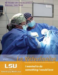
Clinical Case<br />
Said Dr. Riggs, “In this case, <strong>the</strong> horse was diagnosed with<br />
grade IV left laryngeal hemiplegia obstructing <strong>the</strong> airway when<br />
he was worked. No o<strong>the</strong>r abnormalities were noted.”<br />
The Optomed DRS endoscope system is installed in five<br />
steps. The first step is <strong>the</strong> installation <strong>of</strong> <strong>the</strong> endoscope into<br />
one <strong>of</strong> <strong>the</strong> horse’s nostrils. The second step requires <strong>the</strong><br />
endoscope to be attached to a special DRS bridle. In <strong>the</strong> third<br />
step, <strong>the</strong> processor is attached to <strong>the</strong> saddle pad, and in <strong>the</strong><br />
fourth step, <strong>the</strong> transmitter-recorder is also attached to <strong>the</strong><br />
saddle pad. For <strong>the</strong> last step, <strong>the</strong> examination <strong>of</strong> <strong>the</strong> upper<br />
respiratory tract is displayed on <strong>the</strong> receiver’s screen in real<br />
time. It takes about five minutes and two people to install.<br />
Mick underwent a pros<strong>the</strong>tic laryngoplasty (“tie-back”) and<br />
laser ventriculocordectomy (removal <strong>of</strong> <strong>the</strong> vocal cord and<br />
ventricle on <strong>the</strong> left side. For <strong>the</strong> tie-back procedure, an<br />
incision is made in <strong>the</strong> throat just behind <strong>the</strong> mandible. A<br />
heavy suture is placed between two areas <strong>of</strong> cartilage in <strong>the</strong><br />
larynx to mimic <strong>the</strong> non-functioning cricoarytenoideus dorsalis<br />
muscle (muscle <strong>of</strong> <strong>the</strong> larynx), which is responsible for holding<br />
<strong>the</strong> larynx open during breathing. This suture holds <strong>the</strong><br />
cartilage out <strong>of</strong> <strong>the</strong> airway and enables <strong>the</strong> horse to brea<strong>the</strong><br />
without obstruction or noise when it exercises.<br />
Mick has a good prognosis for return to jumping without<br />
exercise intolerance or noise. According to Lauren, he is<br />
currently exercising and doing great.<br />
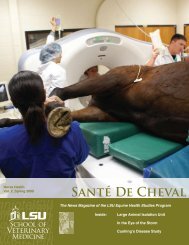
IMAGES FROM TOP LEFT: Dr. Laura Riggs places <strong>the</strong><br />
endoscope on Mick. After <strong>the</strong> saddle is placed on Mick, <strong>the</strong><br />
camera is inserted into his airway via his nose. Dr. Colin<br />
Mitchell and veterinary student Emily Collins assist Dr. Riggs<br />
with <strong>the</strong> video monitor. <strong>Veterinary</strong> technician Nick McClure<br />
exercises Mick in order to examine his airway while he’s in<br />
motion. Dr. Riggs, interns, residents, and students observe<br />
Mick and <strong>the</strong> receiver screen.<br />
ABOVE: The top image shows a horse with a normal airway.<br />
The middle image shows Mick’s airway prior to treatment. The<br />
bottom image shows Mick at his first post-surgery event in<br />
February.<br />
9