View - Coccidia.icb.usp.br - USP
View - Coccidia.icb.usp.br - USP
View - Coccidia.icb.usp.br - USP

You also want an ePaper? Increase the reach of your titles
YUMPU automatically turns print PDFs into web optimized ePapers that Google loves.
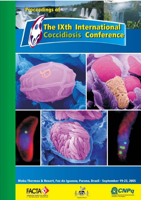
Front cover figure legends:<<strong>br</strong> />
A. Scanning electron micrograph (SEM) of an oocyst in the gut lumen showing partial loss of the veil and<<strong>br</strong> />
with a microgamete adhering to the outer layer of the oocyst wall.<<strong>br</strong> />
B. SEM of a fracture through a caecum infected with E. tenella showing a microgametocyte with<<strong>br</strong> />
numerous mature microgametes in a cell adjacent to a macrogamete.<<strong>br</strong> />
C and E. Tachyzoite of T. gondii (C) and merozoite of E. tenella (E) and labelled with anti-enoyl reductase<<strong>br</strong> />
showing the peri-nuclear location of the single or multiple apicoplasts (green). .<<strong>br</strong> />
D. SEM of a mature schizont of Eimeria tenella showing the individual merozoites.<<strong>br</strong> />
The original images were kindly supplied by Dr. David J. P. Ferguson (Oxford University, UK), copied and<<strong>br</strong> />
printed with permission. Copyright is retained by the author.
Proceedings of<<strong>br</strong> />
The IX th International<<strong>br</strong> />
Coccidiosis Conference<<strong>br</strong> />
Foz de Iguassu, Parana, Brazil<<strong>br</strong> />
September 19-23, 2005
FACTA - APINCO Foundation of Poultry Science and Technology<<strong>br</strong> />
WPSA-Brazilian Branch<<strong>br</strong> />
Av. Andrade Neves, 2501 - Castelo - Campinas - SP - Brazil - 13070-001<<strong>br</strong> />
Tel: +55 19 3243-6555 • Fax: +55 19 3243-8542 • facta@facta.org.<strong>br</strong><<strong>br</strong> />
Site: http://www.facta.org.<strong>br</strong><<strong>br</strong> />
PUBLISHING STAFF<<strong>br</strong> />
Coordination: Arthur Gruber/Ariel Antonio Mendes<<strong>br</strong> />
Organization: Nilza S. Marcondes/Glaucia C. de Campos Faria/Carla R. Palermo<<strong>br</strong> />
Proof-reading: Bettina Gertum Becker<<strong>br</strong> />
Graphic creation, edition, and production: AGROS Comunicação – 55 19 3241-9292<<strong>br</strong> />
Secretariat/Registration: Nilza S. Marcondes/Glaucia C. de Campos Faria/Carla R. Palermo<<strong>br</strong> />
Financial Department: Nilza S. Marcondes
FACTA - APINCO FOUNDATION OF POULTRY SCIENCE AND TECHNOLOGY<<strong>br</strong> />
FACTA COUNCIL<<strong>br</strong> />
José Flávio Neves Mohallem – Granja Planalto Ltda - Brazil<<strong>br</strong> />
Antonio Mário Penz Junior – UFRGS – Brazil<<strong>br</strong> />
Aroldo Silva Amorim Filho – Asa Alimentos Ltda - Brazil<<strong>br</strong> />
Edir Nepomuceno da Silva – Unicamp - Brazil<<strong>br</strong> />
Élsio Antônio Pereira de Figueiredo – Em<strong>br</strong>apa - Brazil<<strong>br</strong> />
Fernando Bacci – Granja São José Ltda - Brazil<<strong>br</strong> />
Ivan Pupo Lauandos – Agroceres - Brazil<<strong>br</strong> />
José Carlos Godoy – APINCO - Brazil<<strong>br</strong> />
Luiz Antonio Bossi – Granja Alvorada Ltda - Brazil<<strong>br</strong> />
Paulo Tabajara Chaves Costa – UFSM - Brazil<<strong>br</strong> />
Pedro Benur Bohrer – Seara Alimentos - Brazil<<strong>br</strong> />
Roberto Kaefer – Globoaves Agroavícola - Brazil<<strong>br</strong> />
Zoé Silveira D´Ávila – UBA - Brazil<<strong>br</strong> />
EXECUTIVE COMMITTEE<<strong>br</strong> />
Ariel Antonio Mendes – President<<strong>br</strong> />
Antonio Guilherme Machado de Castro – Marketing Director<<strong>br</strong> />
Edir Nepomuceno da Silva – Events Director<<strong>br</strong> />
Irenilza de Alencar Naas – Especial Projects Director<<strong>br</strong> />
José Di Fábio – Financial Director<<strong>br</strong> />
Luiz Almiro Carvalho Sesti – Executive Director<<strong>br</strong> />
Marcos Macari – Courses and Publishing Director<<strong>br</strong> />
TECHNICAL COMMITTEE<<strong>br</strong> />
Alberto Bernardino - Fort-Dodge Saúde Animal - Brazil<<strong>br</strong> />
Antonio Mário Penz Junior – UFRGS – Brazil<<strong>br</strong> />
Antonio Guilherme Machado de Castro – Instituto Biológico - Brazil<<strong>br</strong> />
Ariel Antonio Mendes – Unesp - Brazil<<strong>br</strong> />
Byron Grieco Moreira da Silva – Intervet - Brazil<<strong>br</strong> />
Diogo Tsuyoshi Ito – Nutron - Brazil<<strong>br</strong> />
Élsio Antônio Pereira de Figueiredo – Em<strong>br</strong>apa - Brazil<<strong>br</strong> />
Edir Nepomuceno da Silva – Unicamp - Brazil<<strong>br</strong> />
Fabio Tavares Zancan – PUC – Brazil<<strong>br</strong> />
Ga<strong>br</strong>iel Jorge Neto – Nutron - Brazil<<strong>br</strong> />
Godofredo Miltenburg – Delta Assessoria - Brazil<<strong>br</strong> />
Inaldo Sales Patrício – Consultor - Brazil<<strong>br</strong> />
Irenilza de Alencar Naas - Unicamp - Brazil<<strong>br</strong> />
João Batista Luchesi – Multimix - Brazil<<strong>br</strong> />
João Tomelin – UBA - Brazil<<strong>br</strong> />
José Di Fábio – JF Laboratório - Brazil<<strong>br</strong> />
Luiz Almiro Carvalho Sesti - Multimix - Brazil<<strong>br</strong> />
Marcelo A. Fagnani Zuanaze – Biovet - Brazil<<strong>br</strong> />
Marcos Macari - Unesp - Brazil<<strong>br</strong> />
Nelson José Beraquet – Ital - Brazil<<strong>br</strong> />
Paulo César Martins – Euri<strong>br</strong>id - Brazil<<strong>br</strong> />
Tércio Michelan Filho – Agroceres - Brazil<<strong>br</strong> />
Teresa Cristina Ruocco Dari – Nutron - Brazil<<strong>br</strong> />
ADMINISTRATION COMMITTEE<<strong>br</strong> />
Nilza Santos Marcondes – Financial Executive<<strong>br</strong> />
Glaucia C. de Campos Faria – Convention Organizer<<strong>br</strong> />
Carla Rizzo Palermo – Communication Assistant<<strong>br</strong> />
IXth International Coccidiosis Conference € 5
IX TH INTERNATIONAL COCCIDIOSIS CONFERENCE<<strong>br</strong> />
General Organizer<<strong>br</strong> />
Arthur Gruber - Universidade de São Paulo (<strong>USP</strong>) - Brazil<<strong>br</strong> />
International Scientific Committee<<strong>br</strong> />
David Chapman<<strong>br</strong> />
Fiona Tomley<<strong>br</strong> />
Hyun S. Lillehoj<<strong>br</strong> />
Kazumi Sasai<<strong>br</strong> />
Mark Jenkins<<strong>br</strong> />
Martin W. Shirley<<strong>br</strong> />
Nick Smith<<strong>br</strong> />
University of Arkansas – USA<<strong>br</strong> />
Institute for Animal Health – UK<<strong>br</strong> />
US Department of Agriculture – USA<<strong>br</strong> />
Osaka Prefecture University - Japan<<strong>br</strong> />
US Department of Agriculture – USA<<strong>br</strong> />
Institute for Animal Health – UK<<strong>br</strong> />
University of Technology Sydney – Australia<<strong>br</strong> />
Organizing Committee<<strong>br</strong> />
Alda Maria B. N. Madeira<<strong>br</strong> />
Arthur Gruber<<strong>br</strong> />
Rosângela Zacarias Machado<<strong>br</strong> />
Solange Maria Gennari<<strong>br</strong> />
Ariel Antonio Mendes<<strong>br</strong> />
Byron Grieco Moreira da Silva<<strong>br</strong> />
Carlos O. Cardoso Pulici<<strong>br</strong> />
Clóvis de Oliveira<<strong>br</strong> />
Dieter Suída<<strong>br</strong> />
Eduardo Villas Boas Leffer<<strong>br</strong> />
Fernando José Bertazzo<<strong>br</strong> />
José Di Fabio<<strong>br</strong> />
Luiz Adolfo Bier Neto<<strong>br</strong> />
Marcel Louis Joineau<<strong>br</strong> />
Marcelo A. Fagnani Zuanaze<<strong>br</strong> />
Marco Antonio C. de Almeida<<strong>br</strong> />
Maurício Luiz do Nascimento<<strong>br</strong> />
Paulo José Ubices de Moraes<<strong>br</strong> />
Rogério B. Petri<<strong>br</strong> />
Universidade de São Paulo (<strong>USP</strong>) - Brazil<<strong>br</strong> />
Universidade de São Paulo (<strong>USP</strong>) - Brazil<<strong>br</strong> />
Universidade do Estado de São Paulo (UNESP) - Brazil<<strong>br</strong> />
Universidade de São Paulo (<strong>USP</strong>) - Brazil<<strong>br</strong> />
FACTA – Fundação APINCO de Ciência e Tecnologia Avícolas - Brazil<<strong>br</strong> />
Azko Nobel Ltda – Div. Intervet - Brazil<<strong>br</strong> />
Imuvet Comercial Ltda - Brazil<<strong>br</strong> />
Merial Saúde Animal Ltda - Brazil<<strong>br</strong> />
Novus do Brasil Comércio e Importação Ltda - Brazil<<strong>br</strong> />
Schering Plough - Brazil<<strong>br</strong> />
Eli Lilly do Brasil Ltda - Brazil<<strong>br</strong> />
JF Laboratório - Brazil<<strong>br</strong> />
Inovoject do Brasil - Brazil<<strong>br</strong> />
Impextraco - Brazil<<strong>br</strong> />
Laboratório Biovet S.A. - Brazil<<strong>br</strong> />
Alpharma Animal Health - Divisão Brasil - Brazil<<strong>br</strong> />
Phi<strong>br</strong>o Animal Health - Brazil<<strong>br</strong> />
Laboratórios Pfizer Ltda - Brazil<<strong>br</strong> />
Bayer do Brasil S/A - Brazil<<strong>br</strong> />
Secretariat<<strong>br</strong> />
Carla Rizzo Palermo – FACTA - Brazil<<strong>br</strong> />
Glaucia C. de Campos Faria – FACTA - Brazil<<strong>br</strong> />
Nilza Santos Marcondes – FACTA - Brazil<<strong>br</strong> />
IXth International Coccidiosis Conference • 7
Sponsors<<strong>br</strong> />
The organizers of the IXth International Coccidiosis Conference acknowledge the following companies<<strong>br</strong> />
for their financial support and generosity:<<strong>br</strong> />
• Akzo Nobel – Intervet Division<<strong>br</strong> />
• Alpharma Inc.<<strong>br</strong> />
• Em<strong>br</strong>ex Inc.<<strong>br</strong> />
• Imuvet Comercial Ltda.<<strong>br</strong> />
• Janssen Animal Health<<strong>br</strong> />
• Laboratório BioVet S/A<<strong>br</strong> />
• Laboratorios Inmuner<<strong>br</strong> />
• Laboratórios Pfizer Ltda<<strong>br</strong> />
• Merial Saúde Animal Ltda<<strong>br</strong> />
• Novartis Animal Health<<strong>br</strong> />
• Novus International Inc.<<strong>br</strong> />
• Phi<strong>br</strong>o Animal Health<<strong>br</strong> />
• Schering-Plough<<strong>br</strong> />
• Vetech Laboratories Inc.<<strong>br</strong> />
Support<<strong>br</strong> />
CNPq – Conselho Nacional de Desenvolvimento Científico e Tecnológico (National Research Council) - Brazil<<strong>br</strong> />
IXth International Coccidiosis Conference • 9
Introduction<<strong>br</strong> />
It is a great honor to host the IX th International Coccidiosis Conference (IX th ICC) and welcome the<<strong>br</strong> />
delegates at the Mabu Thermas & Resort, in the city of Foz do Iguassu, Brazil. At its ninth edition, and<<strong>br</strong> />
being held each four years, since 1973, the Conference has become the most traditional meeting in<<strong>br</strong> />
coccidiosis research. This meeting has aggregated academic and private company researchers, product<<strong>br</strong> />
managers, and field professionals, providing an opportunity for information interchange of both basic and<<strong>br</strong> />
applied sciences.<<strong>br</strong> />
We have decided to hold the IXth ICC in the city of Foz do Iguassu. World widely known for its famous<<strong>br</strong> />
Iguassu Falls, Foz do Iguassu represents Brazil’s one of the most important touristic sites. A wonderful<<strong>br</strong> />
nature, including two National Parks, in Brazilian and Argentinean sides, ecological tours, and a very<<strong>br</strong> />
diverse wild life, composes a rich scenario for this meeting. We hope all delegates enjoy the conference<<strong>br</strong> />
and also have a pleasant stay in this natural paradise.<<strong>br</strong> />
In this ninth edition of the Conference, we tried to maintain the spirit that has characterized the former<<strong>br</strong> />
events. Traditional approaches for coccidiosis control and diagnosis will be presented side-by-side with<<strong>br</strong> />
highly innovative technologies. We received contributed papers of many different topics, including vaccines,<<strong>br</strong> />
chemotherapy, basic immunology, molecular and cell biology, pathology, genomics and post-genomics.<<strong>br</strong> />
Taking into account the increasing importance of vaccines in coccidiosis control, we have prepared specific<<strong>br</strong> />
sessions to discuss new approaches and technologies for vaccination. We have also invited a group of<<strong>br</strong> />
selected academic researchers for the plenary lectures, to present the state-of-art of research and<<strong>br</strong> />
development. A new age has arisen since large scale genome sequencing became a mainstream approach<<strong>br</strong> />
to unravel the complexity of living organisms. For the first time in history, we now have access to the<<strong>br</strong> />
complete genome information of some coccidian parasites like Toxoplasma gondii, Eimeria tenella and<<strong>br</strong> />
two species of Cryptosporidium. Where is this information going to lead us How will it contribute for new<<strong>br</strong> />
control measures This is an issue that will also be discussed and, we hope, will open up new trends for<<strong>br</strong> />
the disease control. Finally, the increasing importance of new regulations on the use of anticoccidial<<strong>br</strong> />
drugs and vaccines stimulated us to include a symposium on current and future perspectives of legislation.<<strong>br</strong> />
This symposium will be followed by an organized public debate, where the major trends of this important<<strong>br</strong> />
issue will be openly discussed.<<strong>br</strong> />
Social activities were also programmed to complement the scientific programme, allowing for delegates<<strong>br</strong> />
to know each other and create an environment of professional and personal integration.<<strong>br</strong> />
We wish you a pleasant stay in Foz do Iguassu, and a very fruitful Conference.<<strong>br</strong> />
Arthur Gruber<<strong>br</strong> />
Conference Organizer<<strong>br</strong> />
Ariel Antonio Mendes<<strong>br</strong> />
President of FACTA<<strong>br</strong> />
IXth International Coccidiosis Conference • 11
Contents<<strong>br</strong> />
Contents<<strong>br</strong> />
Professor Peter L. Long: an Appreciation .................................................................................... 21<<strong>br</strong> />
Martin W. Shirley<<strong>br</strong> />
Plenary Lectures<<strong>br</strong> />
Eight decades of research on Eimeria in poultry ......................................................................... 29<<strong>br</strong> />
Martin W. Shirley - martin.shirley@bbsrc.ac.uk<<strong>br</strong> />
Genome sequence of the avian coccidiosis-causing agent, Eimeria tenella ............................... 36<<strong>br</strong> />
Matt Berriman - mb4@sanger.ac.uk<<strong>br</strong> />
Proteomics of Eimeria : a focus on host cell invasion ................................................................. 37<<strong>br</strong> />
Fiona Tomley - fiona.tomley@bbsrc.ac.uk<<strong>br</strong> />
The role of morphology and microscopy<<strong>br</strong> />
in coccidian research in this genomic/proteomic age ................................................................. 41<<strong>br</strong> />
David J. P. Ferguson - david.ferguson@ndcls.ox.ac.uk<<strong>br</strong> />
New roles for proteases in the infection biology of coccidian parasites ................................... 51<<strong>br</strong> />
Vern B. Carruthers - vcarruth@jhsph.edu<<strong>br</strong> />
Horizontally transferred genes as new<<strong>br</strong> />
targets for drug development in Cryptosporidium parvum ....................................................... 60<<strong>br</strong> />
Boris Striepen - striepen@cb.uga.edu<<strong>br</strong> />
Immune response to <strong>Coccidia</strong> ...................................................................................................... 63<<strong>br</strong> />
Hyun S. Lillehoj - hlilleho@anri.barc.usda.gov<<strong>br</strong> />
Protective immune responses involved in host<<strong>br</strong> />
resistance to Brazilian isolates of Toxoplasma gondii:<<strong>br</strong> />
implications on vaccine development employing recombinant viral vectors ............................ 84<<strong>br</strong> />
Ricardo T. Gazzinelli - ritoga@cpqrr.fiocruz.<strong>br</strong><<strong>br</strong> />
Immune responses to Neospora caninum and prospects for vaccination .................................. 91<<strong>br</strong> />
Elisabeth A. Innes - Lee.Innes@moredun.ac.uk<<strong>br</strong> />
Perspectives for the control of coccidiosis in poultry by chemotherapy and vaccination ........ 99<<strong>br</strong> />
H. David Chapman - dchapman@uark.edu<<strong>br</strong> />
Anticoccidial drug discovery:<<strong>br</strong> />
approaches towards the identification of novel chemotherapeutic agents ............................ 104<<strong>br</strong> />
Paul Liberator - paul_liberator@merck.com<<strong>br</strong> />
Vaccination against coccidial parasites. The method of choice .............................................. 105<<strong>br</strong> />
Arno N. Vermeulen - arno.vermeulen@intervet.com<<strong>br</strong> />
Symposium “Legislation on drug and vaccine use”<<strong>br</strong> />
A US perspective on the current and future regulation of anticoccidial drugs and vaccines.. 117<<strong>br</strong> />
Paul Duquette - Paul.Duquette@pahc.com<<strong>br</strong> />
IXth International Coccidiosis Conference • 13
Contents<<strong>br</strong> />
Feed Safety: Evolution in Legislation, Implementation and Harmonisation ........................... 126<<strong>br</strong> />
Yvan Dejaegher - dejaegher.yvan@skynet.be<<strong>br</strong> />
Current and futures perspectives on the regulation of anticoccidial drugs and vaccines ....... 128<<strong>br</strong> />
João Palermo Neto - jpalermo@<strong>usp</strong>.<strong>br</strong><<strong>br</strong> />
Symposium “Histomonas meleagridis: Update on Life Cycle, Control and Diagnostics”<<strong>br</strong> />
Histomonas meleagridis: life cycle and epidemiology.............................................................. 131<<strong>br</strong> />
Maarten De Gussem - dgussem@janbe.jnj.com<<strong>br</strong> />
The diagnostics of Histomonas meleagridis .............................................................................. 132<<strong>br</strong> />
Wil Landman - w.landman@gdvdieren.nl<<strong>br</strong> />
Histomoniasis, an ancient disease threatening the European poultry production.<<strong>br</strong> />
What are the current and future options for control .............................................................. 133<<strong>br</strong> />
Koen De Gussem - koen.degussem@alpharma.com<<strong>br</strong> />
Enteric flagellated protozoa in turkeys. ..................................................................................... 134<<strong>br</strong> />
Steven Clark - steven.clark@alpharma.com<<strong>br</strong> />
Contributed Papers: Oral Presentations<<strong>br</strong> />
Vaccines<<strong>br</strong> />
Guidelines for evaluating the efficacy and safety of live<<strong>br</strong> />
anticoccidial vaccines, and obtaining approval for their use in chickens and turkeys<<strong>br</strong> />
H. David Chapman - dchapman@uark.edu ........................................................................................................................... 137<<strong>br</strong> />
Selection of a precocious line of the rabbit coccidium Eimeria piriformis<<strong>br</strong> />
and characterization of the life cycle of both the parent strain and the precocious line<<strong>br</strong> />
Michal Pakandl - pakandi@paru.cas.cz .................................................................................................................................. 137<<strong>br</strong> />
Long Term Viability of Cryopreserved Eimeria Sporocysts<<strong>br</strong> />
Rebecca M. Poston - rposton@em<strong>br</strong>ex.com .......................................................................................................................... 138<<strong>br</strong> />
Non-interference of INOVOCOX , a live coccidial oocyst vaccine, with Marek’s<<strong>br</strong> />
Disease Vaccine or Bursaplex®, A live bursal disease vaccine, after in ovo administration<<strong>br</strong> />
Vivian W. Doelling - vdoeling@em<strong>br</strong>ex.com ............................................................................................................................ 138<<strong>br</strong> />
Uniformity of Infection after INOVOCOX Delivery AT E18 Using the INOVOJECT ® System<<strong>br</strong> />
Rebecca M. Poston - rposton@em<strong>br</strong>ex.com .......................................................................................................................... 139<<strong>br</strong> />
In Ovo and Dietary Modulation of Host Intestinal Immunity<<strong>br</strong> />
and its Subsequent Effects on Coccidiosis<<strong>br</strong> />
Rami A. Dalloul - rdalloul@anri.barc.usda.gov ........................................................................................................................ 139<<strong>br</strong> />
Effects of dietary short chain fatty acids on experimentally induced<<strong>br</strong> />
coccidiosis and necrotic enteritis in <strong>br</strong>oilers vaccinated against coccidiosis<<strong>br</strong> />
Mauro Antongiovanni - mauro.antogiovanni@unifi.it ............................................................................................................. 140<<strong>br</strong> />
The Effect of Light Intensity During Brooding<<strong>br</strong> />
on the Productivity and Health of Paracox-5 Vaccinated Broilers<<strong>br</strong> />
Luciano Gobbi - luciano.gobbi@spcorp.com ........................................................................................................................... 141<<strong>br</strong> />
Efficacy of gel spray as a delivery system for turkey coccidiosis<<strong>br</strong> />
vaccine consisting of Eimeria adenoeides and E. meleagrimites<<strong>br</strong> />
Roberto Soares - rbsoares@vetechinc.com ............................................................................................................................ 141<<strong>br</strong> />
Comparison Of Performance Of Ross Broiler Breeder Males To Full,<<strong>br</strong> />
Half-Dose And 10X Dose Of Coccivac ® -D Day-of-Age Vaccination<<strong>br</strong> />
Stephen W. Davis - steve@coloradoqualityresearch.com ....................................................................................................... 142<<strong>br</strong> />
Safety And Efficacy Of Advent ® Coccidiosis Vaccine<<strong>br</strong> />
Marco A. Quiroz - marco.quiroz@novusint.com ..................................................................................................................... 143<<strong>br</strong> />
Performance of Broilers Using A Live Vaccine Against Avian Coccidiosis<<strong>br</strong> />
Alejandro Juan José Dubois - alejudubois@hotmail.com ......................................................................................................... 144<<strong>br</strong> />
14 • IXth International Coccidiosis Conference
Contents<<strong>br</strong> />
Efficacy of Coccivac against field isolates of chicken Eimeria<<strong>br</strong> />
Steve Fitz-Coy - steve-fitz-coy@spcorp.com .......................................................................................................................... 145<<strong>br</strong> />
Growout Performance Following Vaccination with Inovocox As Compared to Salinomycin<<strong>br</strong> />
Alison Martin - amartin@em<strong>br</strong>ex.com .................................................................................................................................... 145<<strong>br</strong> />
Anticoccidial Sensitivity Profile of <strong>Coccidia</strong>l Strains in Inovocox<<strong>br</strong> />
Alison Martin - amartin@em<strong>br</strong>ex.com .................................................................................................................................... 146<<strong>br</strong> />
Maternal Protection against Eimeria challenge of CoxAbic® vaccinated chickens<<strong>br</strong> />
Amnon Michael - amnon.michael@teva.co.il ......................................................................................................................... 146<<strong>br</strong> />
Use of avian immunoglobulins in the control of coccidiosis<<strong>br</strong> />
Andrea Rodríguez Ropon - arodriguez@grupoidisa.com ........................................................................................................ 147<<strong>br</strong> />
Acquisition of immunity to Eimeria maxima in newly hatched chickens given 100 oocysts<<strong>br</strong> />
H. David Chapman - dchapman@uark.edu ........................................................................................................................... 148<<strong>br</strong> />
Inovocox Onset Of Immunity After in Ovo Delivery to Broilers<<strong>br</strong> />
Vivian W. Doelling - vdoelling@em<strong>br</strong>ex.com ........................................................................................................................... 148<<strong>br</strong> />
Immune reactions to different doses of Eimeria acervulina in day-old <strong>br</strong>oilers<<strong>br</strong> />
Willem Swinkels - willem.swinkels@wur.nl .............................................................................................................................. 149<<strong>br</strong> />
Genomics and Post-genomics<<strong>br</strong> />
HAPPY mapping – a tool for genome finishing<<strong>br</strong> />
Paul H. Dear - phd@mrc-lmb.cam.ac.uk ................................................................................................................................ 149<<strong>br</strong> />
Survey of Eimeria spp. Transcripts Using Open Reading Frame ESTs (Orestes)<<strong>br</strong> />
Alda M.B.N. Madeira - albackx@<strong>usp</strong>.<strong>br</strong> .................................................................................................................................. 150<<strong>br</strong> />
Molecular Characterization and Comparative Analysis of Mitochondrial Genomes of Eimeria spp.<<strong>br</strong> />
Arthur Gruber - argruber@<strong>usp</strong>.<strong>br</strong> .......................................................................................................................................... 150<<strong>br</strong> />
Molecular and Phylogenetic Features of<<strong>br</strong> />
Two Distinct dsRNA Virus Families Infecting Eimeria spp. of Domestic Fowl<<strong>br</strong> />
Jane Silveira Fraga - jane.fraga@gmail.com .......................................................................................................................... 151<<strong>br</strong> />
Long SAGE (Serial Analysis of Gene Expression) In Eimeria Tenella – A Preliminary Study<<strong>br</strong> />
Jeniffer Novaes - jeffys@ajato.com.<strong>br</strong> .................................................................................................................................... 151<<strong>br</strong> />
DNA Microarray Gene Expression Analyses in Attenuated and Virulent E. tenella<<strong>br</strong> />
Tomas Mikuš - mikus@<strong>br</strong>i.cz ................................................................................................................................................... 152<<strong>br</strong> />
Progress in the development of stable transfection in Eimeria tenella<<strong>br</strong> />
Fiona Tomley - fiona.tomley@bbsrc.ac.uk .............................................................................................................................. 152<<strong>br</strong> />
Immunology<<strong>br</strong> />
Common and Unique Gene Expression<<strong>br</strong> />
of Avian Macrophages in Response to Three Major Eimeria Species<<strong>br</strong> />
Rami A. Dalloul - rdalloul@anri.barc.usda.gov ........................................................................................................................ 153<<strong>br</strong> />
Molecular cloning and expression profiles of chicken NK-lysin during Eimeria infections<<strong>br</strong> />
Yeong H. Hong - yhong@anri.barc.usda.gov .......................................................................................................................... 153<<strong>br</strong> />
Development of future strategies for vaccinating chickens against coccidiosis using<<strong>br</strong> />
recombinant antibodies, genes and peptides.<<strong>br</strong> />
Hyun S. Lillehoj - hlilleho@anri.barc.usda.gov ......................................................................................................................... 154<<strong>br</strong> />
Genetics, immunity and DNA fingerprinting in the identification<<strong>br</strong> />
of protective antigens of Eimeria maxima<<strong>br</strong> />
Damer Peter Blake - damer.blake@bbsrc.ac.uk .................................................................................................................... 154<<strong>br</strong> />
Application of functional genomics, immunology and<<strong>br</strong> />
molecular biology tools to explore host immune response to Eimeria<<strong>br</strong> />
Hyun S. Lillehoj - hlilleho@anri.barc.usda.gov ......................................................................................................................... 155<<strong>br</strong> />
Proteins from the wall- forming bodies of Eimeria maxima: function and immunogenicity<<strong>br</strong> />
Nicholas Smith - nick.smith@uts.edu.au ................................................................................................................................. 155<<strong>br</strong> />
IXth International Coccidiosis Conference • 15
Contents<<strong>br</strong> />
Mixed Cocciodiosis Control: Drugs and Vaccines<<strong>br</strong> />
Restoration of Field Eimeria Anticoccidial Sensitivity with Coccivac-B, a Live Coccidiosis Vaccine<<strong>br</strong> />
Greg Mathis - southern_poultry_res@msn.com ..................................................................................................................... 156<<strong>br</strong> />
Rotational Programs Combining Drugs and Coccidiosis<<strong>br</strong> />
Vaccine Improve Performance and Profitability in a Commercial Broiler Farm<<strong>br</strong> />
Marco A. Quiroz - marco.quiroz@novusint.com ..................................................................................................................... 156<<strong>br</strong> />
Comparison of drug-sensitivity and species composition<<strong>br</strong> />
of Eimeria isolated from poultry farms<<strong>br</strong> />
Mark C. Jenkins - mjenkins@anri.barc.usda.gov .................................................................................................................... 157<<strong>br</strong> />
Higher incidence of Eimeria spp. field isolates sensitive<<strong>br</strong> />
to diclazuril and monensin after live coccidiosis vaccination with Paracox -5<<strong>br</strong> />
Wil J. M. Landman - w.landman@gdvdieren.nl ...................................................................................................................... 157<<strong>br</strong> />
Coccidiosis in Turkeys: patterns of drug response to in feed anticoccidials<<strong>br</strong> />
Marcelo Lang - marcelo.lang@spcorp.com ............................................................................................................................ 158<<strong>br</strong> />
Diagnosis and Epidemiology<<strong>br</strong> />
Sequence analysis of the 18S RNA gene of Isospora belli and identification of Caryospora-like oocysts<<strong>br</strong> />
Somchai Jongwutiwes - fmedsjw@md2.md.chula.ac.th ........................................................................................................ 158<<strong>br</strong> />
Detection and identification of<<strong>br</strong> />
Neospora caninum in a naturally infected buffalo fetus (Bubalus bubalis) from Brazil<<strong>br</strong> />
Solange Maria Gennari - sgennari@<strong>usp</strong>.<strong>br</strong> ............................................................................................................................. 159<<strong>br</strong> />
Molecular epidemiology of cryptosporidiosis among human immunodeficiency<<strong>br</strong> />
virus-infected patients in Thailand: analysis of the 18S RNA and the Cpg60/45/15 loci<<strong>br</strong> />
Somchai Jongwutiwes - fmedsjw@md2.md.chula.ac.th ........................................................................................................ 160<<strong>br</strong> />
Molecular Genotyping Of Eimeria tenella and E. acervulina Through microsatellite markers<<strong>br</strong> />
Arthur Gruber - argruber@<strong>usp</strong>.<strong>br</strong> .......................................................................................................................................... 161<<strong>br</strong> />
Counting coccidia: a genomic approach<<strong>br</strong> />
Damer Peter Blake - damer.blake@bbsrc.ac.uk .................................................................................................................... 162<<strong>br</strong> />
Digital Image Analysis In The Diagnosis Of Chicken Coccidiosis<<strong>br</strong> />
Arthur Gruber - argruber@<strong>usp</strong>.<strong>br</strong> .......................................................................................................................................... 162<<strong>br</strong> />
Molecular biology and Biochemistry<<strong>br</strong> />
The Macrophage Inhibitory Factor of Eimeria species<<strong>br</strong> />
Katharzyna Miska - kmiska@anri.barc.usda.gov ................................................................................................................... 163<<strong>br</strong> />
The developmental expression of Eimeria Heat shock protein 90<<strong>br</strong> />
Katharzyna Miska - kmiska@anri.barc.usda.gov ................................................................................................................... 163<<strong>br</strong> />
Acetyl CoA Carboxylase in Avian Eimeria<<strong>br</strong> />
Patricia C. Allen - pallen@anri.barc.usda.gov ......................................................................................................................... 164<<strong>br</strong> />
The Satellite DNA Of Eimeria tenella: a Quantitative and Qualitative Study<<strong>br</strong> />
Arthur Gruber - argruber@<strong>usp</strong>.<strong>br</strong> .......................................................................................................................................... 164<<strong>br</strong> />
Characterization of Serine Proteases in Developmental Stages of Eimeria tenella.<<strong>br</strong> />
Raymond H. Fetterer - rfettere@anri.barc.usda.gov ............................................................................................................. 165<<strong>br</strong> />
Cell Biology<<strong>br</strong> />
Plastid Replication in Apicomplexan Parasites<<strong>br</strong> />
Boris Striepen - striepen@cb.uga.edu .................................................................................................................................... 165<<strong>br</strong> />
Identification and function of the EtMIC4-EtMIC5<<strong>br</strong> />
protein complex. A novel microneme complex of Eimeria tenella<<strong>br</strong> />
Fiona Tomley - fiona.tomley@bbsrc.ac.uk .............................................................................................................................. 166<<strong>br</strong> />
Characterisation of Apical Mem<strong>br</strong>ane Antigens in Eimeria tenella<<strong>br</strong> />
Fiona Tomley - fiona.tomley@bbsrc.ac.uk .............................................................................................................................. 166<<strong>br</strong> />
16 • IXth International Coccidiosis Conference
Contents<<strong>br</strong> />
Investigation of the role of proteoglycans during invasion of Eimeria tenella sporozoites<<strong>br</strong> />
Fiona Tomley - fiona.tomley@bbsrc.ac.uk .............................................................................................................................. 167<<strong>br</strong> />
Characteristic of Cryptosporidium Apical Antigens Using Chicken Monoclonal Antibodies<<strong>br</strong> />
Against Eimeria<<strong>br</strong> />
M. Matsubayashi - matsu@oyg.ac.jp ...................................................................................................................................... 167<<strong>br</strong> />
Chemotherapy<<strong>br</strong> />
Effects of Natustat supplementation on Chickens Challenged<<strong>br</strong> />
with Eimeria acervulina, Eimeria maxima and Eimeria tenella<<strong>br</strong> />
Cepta F Duffy - cduffy@alltech.com ...................................................................................................................................... 168<<strong>br</strong> />
The impact of the crowding effect of chicken Eimeria on the efficacy of anticoccidials: a hypothesis<<strong>br</strong> />
Maarten De Gussem - mdgussen@janbe.jnj.com ................................................................................................................... 168<<strong>br</strong> />
Coccidiosis control in poultry: importance of the quality of anticoccidial premixes<<strong>br</strong> />
Maarten De Gussem - mdgussen@janbe.jnj.com ................................................................................................................... 169<<strong>br</strong> />
Anticoccidial sensitivity profiles of recently obtained Dutch, German,<<strong>br</strong> />
and Spanish Eimeria spp. isolates<<strong>br</strong> />
Herman W. Peek - h.peek@gdvdieren.nl ............................................................................................................................... 170<<strong>br</strong> />
Treatment of Mice with the anticoccidial drug Toltrazuril does not interfere with the<<strong>br</strong> />
development of a specific cellular intestinal immune response to Eimeria falciformis<<strong>br</strong> />
Thomas Pogonka - thomas.pogonka@rz.hu-berlin.de ........................................................................................................... 171<<strong>br</strong> />
The impact of coccidial drug-resistance on the commercial performance of <strong>br</strong>oilers<<strong>br</strong> />
Raymond Barry Williams - ray.coxitec@texco.net ................................................................................................................... 171<<strong>br</strong> />
Pathology<<strong>br</strong> />
Chicken coccidiosis: a fresh look at lesions<<strong>br</strong> />
Raymond Barry Williams - ray.coxitec@texco.net ................................................................................................................... 172<<strong>br</strong> />
The Stealth Chicken Eimeria: E. mivati - a new perspective<<strong>br</strong> />
Steve Fitz-Coy - steve.fitz-coy@spcorp.com .......................................................................................................................... 173<<strong>br</strong> />
The Role of E. mitis in Chicken Coccidiosis: a Study of Pathogenicity<<strong>br</strong> />
Luciano Gobbi - luciano.gobbi@spcorp.com ........................................................................................................................... 173<<strong>br</strong> />
The comparison and validation of methods for monitoring coccidiosis in <strong>br</strong>oilers<<strong>br</strong> />
Christiaan le Roux - corne.loots@spcorp.com ......................................................................................................................... 174<<strong>br</strong> />
Comparison of E. acervulina oocyst counts in single droppings of <strong>br</strong>oilers and in droppings<<strong>br</strong> />
collected during 24 hours<<strong>br</strong> />
Francisca C. Velkers - f.c.velkers@vet.uu.nl............................................................................................................................ 174<<strong>br</strong> />
Reasons for Field Problems with Eimeria maxima: E. acervulina versus E. maxima<<strong>br</strong> />
Greg Mathis - southern_poultry_res@msn.com.<strong>br</strong> ................................................................................................................ 175<<strong>br</strong> />
Contributed Papers: Posters<<strong>br</strong> />
Diagnosis and Epidemiology<<strong>br</strong> />
Comparison between the Modified Ziehl-Neelsen and Fluorescent Antibody Tests for the<<strong>br</strong> />
detection of Cryptosporidium spp. oocysts in faeces<<strong>br</strong> />
Ralph Marshall - r.n.marshall@vla.dfra.gsi.gov.uk ................................................................................................................... 179<<strong>br</strong> />
Occurrence of Toxoplama gondii antibodies in fattening pigs from the State of São Paulo, Brazil<<strong>br</strong> />
Solange Maria Gennari - sgennari@<strong>usp</strong>.<strong>br</strong> ............................................................................................................................. 179<<strong>br</strong> />
Prevalence of <strong>Coccidia</strong> in French Calves: Results of Several Surveys<<strong>br</strong> />
Alain Richard - alain.richard@alpharma.com .......................................................................................................................... 180<<strong>br</strong> />
Study on the ability of water supply to contaminate poultry flocks with coccidia<<strong>br</strong> />
Jean Michel Reperant - jm.reperant@afssa.fr ....................................................................................................................... 181<<strong>br</strong> />
Evaluation of a rapid screening assay<<strong>br</strong> />
for Cryptosporidium identification (DOT-ELISA) using monoclonal antibody<<strong>br</strong> />
Elisabeth Natal de Gaspari - egaspari@ial.sp.gov.<strong>br</strong> ............................................................................................................... 181<<strong>br</strong> />
IXth International Coccidiosis Conference • 17
Contents<<strong>br</strong> />
Eimeria Species Identification In Faeces And Litter Samples From Chickens<<strong>br</strong> />
Anita Haug - anita.haug@vetinst.no ...................................................................................................................................... 182<<strong>br</strong> />
Determination of the Sensitivity of the Serial Scraping<<strong>br</strong> />
Method of Intestinal Mucosa in the Diagnosis of Subclinical Coccidiosis<<strong>br</strong> />
Mauricio De Franceschi - mauriciodf@datamarkets.com.ar .................................................................................................. 183<<strong>br</strong> />
Identification of <strong>Coccidia</strong> Species in Broiler Chickens in Argentina<<strong>br</strong> />
Hernán González - sp@mail.unlu.edu.ar ................................................................................................................................ 183<<strong>br</strong> />
Development of a histological scoring model for use in interpreting the immune response<<strong>br</strong> />
David Grant Richards - medichick@ava.com.au ..................................................................................................................... 184<<strong>br</strong> />
Progress Toward The Development Of Serological Tests For Diagnosis Of Coccidiosis In Chickens<<strong>br</strong> />
David Grant Richards - medichick@ava.com.au ..................................................................................................................... 185<<strong>br</strong> />
Characterization of cryptic field isolates of Eimeria spp. from chickens<<strong>br</strong> />
David Grant Richards - medichick@ava.com.au ..................................................................................................................... 185<<strong>br</strong> />
Pathology<<strong>br</strong> />
Experimental production of necrotic enteritis and its use for studies on the relationships<<strong>br</strong> />
between necrotic enteritis and coccidiosis in chickens<<strong>br</strong> />
Ralph Marshall - r.n.marshall@vla.dfra.gsi.gov.uk ................................................................................................................... 186<<strong>br</strong> />
Clinical And Pathological Changes In Chickens (Gallus domesticus) Experimentally Infected<<strong>br</strong> />
with Eimeria acervulina (TIZZER, 1929) oocysts<<strong>br</strong> />
Rosangela Z. Machado - zacarias@fcav.unesp.<strong>br</strong> .................................................................................................................. 186<<strong>br</strong> />
Association between <strong>Coccidia</strong> and Intestinal Helmints in Broiler Chickens<<strong>br</strong> />
Mauricio Enrique de Franceschi - sip@mail.unlu.edu.ar ......................................................................................................... 187<<strong>br</strong> />
Phenotypic characterization of chicken selection lines infected with Eimeria tenella<<strong>br</strong> />
Giovani Rota Bertani - gbertani@cnpsa.em<strong>br</strong>apa.<strong>br</strong> .............................................................................................................. 187<<strong>br</strong> />
Necrotic Enteritis Association with Eimeria acervulina and E. maxima<<strong>br</strong> />
Greg Mathis - southern_poultry_res@msn.com ..................................................................................................................... 188<<strong>br</strong> />
Interaction of E. coli O157:H7 with Cryptosporidium parvum in 6-week-old lambs<<strong>br</strong> />
Ralph Marshall - r.n.marshall@vla.dfra.gsi.gov.uk ................................................................................................................... 189<<strong>br</strong> />
Application of Eimeria maxima to a Clostridium perfringens-associated necrotic enteritis<<strong>br</strong> />
model for studying bacterial competitive exclusion in poultry<<strong>br</strong> />
Ralph Marshall - r.n.marshall@vla.dfra.gsi.gov.uk ................................................................................................................... 189<<strong>br</strong> />
Immunology<<strong>br</strong> />
Cryptosporidium parvum cellular recruitment during the inflammatory response in mice<<strong>br</strong> />
Elisabeth Natal de Gaspari - egaspari@ial.sp.gov.<strong>br</strong> ............................................................................................................... 190<<strong>br</strong> />
Vaccines<<strong>br</strong> />
The Use Of A Viability Assay To Formulate A New Coccidiosis Vaccine<<strong>br</strong> />
Marco A. Quiroz - marco.quiroz@novusint.com ..................................................................................................................... 190<<strong>br</strong> />
Comparison of the onset of an immune response using three oocyst vaccination strategies<<strong>br</strong> />
David Grant Richards - medichick@ava.com.<strong>br</strong> ...................................................................................................................... 191<<strong>br</strong> />
Quantization of Plasma D-Xylose Concentration Following of Vaccination against Coccidiosis<<strong>br</strong> />
and Experimental Subclinical Coccidiosis Induced by Eimeria maxima in Broilers<<strong>br</strong> />
Seyed Shapoor Reza Shojaei - vetparasitologist@yahoo.com ................................................................................................ 191<<strong>br</strong> />
Chemotherapy<<strong>br</strong> />
Relationship of E. acervulina Ionophore Resistance to Field Problems with E. maxima<<strong>br</strong> />
Greg Mathis - southern_poultry_res@msn.com ..................................................................................................................... 192<<strong>br</strong> />
Efficacy of decoquinate at different administration<<strong>br</strong> />
strategies against cryptosporidiosis in natural infected cashmere goat kids<<strong>br</strong> />
Alain Richard - alain.richard@alpharma.com .......................................................................................................................... 193<<strong>br</strong> />
Efficacy of toltrazuril and amprolium against turkey coccidiosis caused by a mixed E.<<strong>br</strong> />
adenoeides and E. meleagrimitis infection<<strong>br</strong> />
Ro<strong>br</strong>echt Froyman - ro<strong>br</strong>echt.froyman@bayerhealthcare.com ............................................................................................. 194<<strong>br</strong> />
Author Index .................................................................................................................... 195<<strong>br</strong> />
18 • IXth International Coccidiosis Conference
The IX th International<<strong>br</strong> />
Coccidiosis Conference<<strong>br</strong> />
is dedicated to Dr. Peter Long
Professor Peter L. Long: an Appreciation<<strong>br</strong> />
Professor Peter L. Long: an Appreciation<<strong>br</strong> />
M. W. Shirley<<strong>br</strong> />
Institute for Animal Health, Compton Laboratory, Compton, Newbury, Berks. RG 20 7NN, UK, shirley@bbsrc.ac.uk<<strong>br</strong> />
If asked to think of the most influential coccidiologist of the past four decades, the chances are that the<<strong>br</strong> />
name of Peter Long will feature at, or near, the top of most people’s lists. If asked to think of the most<<strong>br</strong> />
influential coccidiologist of the past four decades who is also the most approachable and sociable, the<<strong>br</strong> />
likelihood is that Peter will be at the top of everyone’s list! Peter has the rare gift of scientific excellence<<strong>br</strong> />
combined with a warm demeanour and fondness for bon homie that has endeared him to all who have<<strong>br</strong> />
had the pleasure of his acquaintance.<<strong>br</strong> />
I first became a colleague in 1967 when I started work at the Houghton Poultry Research Station (HPRS)<<strong>br</strong> />
where Peter was a principal scientist looking to recruit a new member of his team. At that time he had<<strong>br</strong> />
not yet gained his PhD but he was established comfortably as a coccidiologist of increasing international<<strong>br</strong> />
renown and was a natural successor to Clifford Horton-Smith who had also previously graced HPRS with<<strong>br</strong> />
distinction and charisma. A symmetry in the three generations was to emerge - Clifford had been a<<strong>br</strong> />
mentor to Peter as Peter was to become to me. Moreover, there was to be further symmetry in the way<<strong>br</strong> />
that the academic career of Peter developed subsequently with how mine was to shape up under his<<strong>br</strong> />
guidance.<<strong>br</strong> />
Edward Tyzzer was working on his Magnum Opus to be published in 1929 when Peter was born in<<strong>br</strong> />
London in 1928. Peter developed into a good sportsman with a special love of cricket – a game at which<<strong>br</strong> />
he was especially skilled and which he played on-and-off for several decades. He supplemented his<<strong>br</strong> />
cricket with both hockey and squash and his competitive nature at sports was mirrored by his determination<<strong>br</strong> />
to succeed professionally.<<strong>br</strong> />
He began his professional life working on poultry parasites at the Central Veterinary Laboratory at<<strong>br</strong> />
Wey<strong>br</strong>idge, Surrey as a laboratory technician with Dr Clifford Horton-Smith. Only four years later, however,<<strong>br</strong> />
he was conscripted into the Royal Army Medical Corps and served in Egypt, Malta and Palestine.<<strong>br</strong> />
In 1949, following demobilisation, Peter and Clifford were reunited at the Ashford Hospital, Middlesex<<strong>br</strong> />
and then a more durable union followed with their relocation to the HPRS: Peter becoming the research<<strong>br</strong> />
technician for Horton-Smith within the Parasitology department working on a programme of research<<strong>br</strong> />
directed principally towards coccidiosis of poultry. Without any formal academic qualifications, Peter<<strong>br</strong> />
studied part-time to become a Member of the Institute of Medical Laboratory Technology in 1952 and<<strong>br</strong> />
obtained Fellow status by thesis in 1956.<<strong>br</strong> />
A steady stream of publications was beginning to appear and a strong desire to publish his data was to<<strong>br</strong> />
define his entire working life. Some early publications included a very useful treatise on E. maxima [Long,<<strong>br</strong> />
P. L. (1959). A study of Eimeria maxima Tyzzer, 1929, A coccidium of the fowl (Gallus gallus). Annals of<<strong>br</strong> />
Tropical Medicine and Parasitology, 53, 325–333] that remains one of the few definitive studies on this<<strong>br</strong> />
major avian species. By now Peter was progressing through the ranks from his original grading of “Assistant<<strong>br</strong> />
Experimental Officer” such that by 1963 he was able to spend a sabbatical year at Cornell University in<<strong>br</strong> />
the USA working with the eminent poultry pathologist, Professor P. P. Levine. This very positive scientific<<strong>br</strong> />
experience left an indelible mark on his subsequent career and much later he was to return to the USA<<strong>br</strong> />
to the University of Georgia where he spent the final 10 years of his active working life.<<strong>br</strong> />
Peter’s taste for working a<strong>br</strong>oad was strengthened in 1963 and 1964 with two visits to the Lebanon,<<strong>br</strong> />
where he worked on poultry coccidiosis and taught parasitology at the Institut de Recherches Agronomiques.<<strong>br</strong> />
IXth International Coccidiosis Conference • 21
Professor Peter L. Long: an Appreciation<<strong>br</strong> />
In 1964, and through further part time studies, Peter became a member of the Institute of Biology in the<<strong>br</strong> />
UK and in 1971 Peter was awarded a PhD by Brunel University for his work on the avian coccidia and in<<strong>br</strong> />
1977 was awarded a DSc. His association with Brunel University led to a strong friendship with Professor<<strong>br</strong> />
Roland J. Terry, who subsequently paid a professional visit to Peter when he was at Athens.<<strong>br</strong> />
Throughout the 1960’s and 1970’s Peter maintained a high output of publications from the parasitologists<<strong>br</strong> />
at HPRS with an impressive list of personal papers that laid much of the groundwork for later developments<<strong>br</strong> />
in the control and study of the avian coccidia including:<<strong>br</strong> />
• two of the few papers on Eimeria to be published in the journal Nature [Long, P. L. (1965). Development<<strong>br</strong> />
of Eimeria tenella in avian em<strong>br</strong>yos. Nature 208, 509-10 and Long, P. L. (1970c). Development<<strong>br</strong> />
(schizogony) of Eimeria tenella in the liver of chickens treated with corticosteroid. Nature 225, 290-1]<<strong>br</strong> />
• pioneering studies on the growth of different species of Eimeria in avian em<strong>br</strong>yos that would eventually<<strong>br</strong> />
help in the development of live attenuated vaccines [Long, P. L. (1966). The growth and attenuation of<<strong>br</strong> />
some species of Eimeria in avian em<strong>br</strong>yos. Parasitology 56, 575-81 and Long, P. L. (1972b). Eimeria<<strong>br</strong> />
tenella: reproduction, pathogenicity and immunogenicity of a strain maintained in chick em<strong>br</strong>yos by<<strong>br</strong> />
serial passage. J Comp Pathol 82, 429-37]<<strong>br</strong> />
• the isolation and identification of different species [e.g. Long, P. L. (1973c). Studies on the relationship<<strong>br</strong> />
between Eimeria acervulina and Eimeria mivati. Parasitology 67, 143-55 and Long, P. L. (1974). Experimental<<strong>br</strong> />
infection of chickens with two species of Eimeria isolated from the Malaysian jungle fowl.<<strong>br</strong> />
Parasitology 69, 337-47]<<strong>br</strong> />
• studies on immunity, often with Elaine Rose [Long, P. L., and Rose, M. E. (1972). Immunity to coccidiosis:<<strong>br</strong> />
effect of serum antibodies on cell invasion by sporozoites of Eimeria in vitro. Parasitology 65, 437-45<<strong>br</strong> />
• studies on chemotherapy [Long, P. L., and Millard, B. J. (1968). Eimeria: effect of meticlorpindol and<<strong>br</strong> />
methyl benzoquate on endogenous stages in the chicken. Exp Parasitol 23, 331-8]<<strong>br</strong> />
• the first studies to provide quantitative data on the incidence of Eimeria species in poultry houses<<strong>br</strong> />
[Long, P. L., Tompkins, R. V., and Millard, B. J. (1975). Coccidiosis in <strong>br</strong>oilers: evaluation of infection by<<strong>br</strong> />
the examination of <strong>br</strong>oiler house litter for oocysts. Avian Pathology 4, 287-294].<<strong>br</strong> />
This was indeed a golden period for Peter and his acclaim grew within HPRS, where he was appointed<<strong>br</strong> />
successor to Dr Donald L. Lee as Head of Parasitology in 1972; nationally, for example, he was awarded<<strong>br</strong> />
the Tom Newman Medal for Poultry Research from the British Poultry Breeders and Hatcheries Association<<strong>br</strong> />
in 1971 and made an honorary member of the British Veterinary Poultry Association in 1972; and<<strong>br</strong> />
internationally where he consolidated his position as arguably the leading scientist working on the biology<<strong>br</strong> />
of avian coccidia.<<strong>br</strong> />
In 1973 Peter worked with Datus M. Hammond to edit “The coccidia”, a book that was to become the<<strong>br</strong> />
first in a series of volumes on the biology of the coccidia and coccidiosis. These books, most especially the<<strong>br</strong> />
1982 volume entitled “The biology of the coccidia” have stood the test of time and continue to be some<<strong>br</strong> />
of the most used reference texts. The most recent volume, now over 15 years old and entitled “Coccidiosis<<strong>br</strong> />
in man and domestic animals”, is a further reflection of Peter’s commitment and ability to work with<<strong>br</strong> />
others to effectively deliver knowledge of the avian coccidia into the public domain.<<strong>br</strong> />
Within the UK Peter helped to establish the forum that is now known as the European Coccidiosis<<strong>br</strong> />
Discussion Group and he was one of the founding members of what was to become the British Society for<<strong>br</strong> />
Parasitology, which later made him an honorary member.<<strong>br</strong> />
In 1979, Peter left the HPRS to succeed Professor W. Malcolm Reid as Professor of Parasitology and<<strong>br</strong> />
Chairman of the Faculty of Parasitology at the University of Georgia. In 1983, Peter was further recognised<<strong>br</strong> />
22 • IXth International Coccidiosis Conference
Professor Peter L. Long: an Appreciation<<strong>br</strong> />
by the University by the title, which he held until 1990 when he retired back to the UK, of D.W. Brooks<<strong>br</strong> />
Distinguished Professor of Poultry Science.<<strong>br</strong> />
During his years at HPRS, Peter was most ably supported scientifically and technically by Brian Millard<<strong>br</strong> />
and, if there were an equivalent anywhere else in the world, he most definitely found that person in Joyce<<strong>br</strong> />
Johnson. Peter and Joyce developed a wonderful working rapport and the transition of his science from<<strong>br</strong> />
HPRS to the University of Georgia seemed almost seamless with virtually no interruption to the stream of<<strong>br</strong> />
papers.<<strong>br</strong> />
Peter’s later contributions to coccidiosis research continued to reflect his desire to improve and understand<<strong>br</strong> />
the control of disease and his early work in Georgia was pivotal to the first understandings of the relationship<<strong>br</strong> />
between pathological changes in the intestines and body weight changes of vaccinated or naive chickens<<strong>br</strong> />
after challenge, findings that were to become important considerations in the evaluation of vaccines [e.g.<<strong>br</strong> />
Long, P. L., Johnson, J., and Wyatt, R. D. (1980). Eimeria tenella: clinical effects in partially immune and<<strong>br</strong> />
susceptible chickens. Poult Sci 59, 2221-4]. In addition he pursued work in the USA on the development<<strong>br</strong> />
of precocious, attenuated lines of Eimeria as well as addressing issues of disease control more generally.<<strong>br</strong> />
In total, Peter published more than 200 scientific papers, countless popular articles and very many book<<strong>br</strong> />
chapters. He also helped stage the Coccidiosis Conference in Georgia in 1985.<<strong>br</strong> />
The scientific excellence of Peter and his contribution to research on coccidia and coccidiosis has been<<strong>br</strong> />
recognised many times and, in addition to the Tom Newman Award, he was the first recipient of the<<strong>br</strong> />
Gordon Memorial Medal (1983); the Merck Award for Research from the American Poultry Science<<strong>br</strong> />
Association (1984); and the Creative Research Medal from the University of Georgia, Athens.<<strong>br</strong> />
Throughout his career Peter influenced and mentored many colleagues including several PhD students<<strong>br</strong> />
(about whom he still speaks with great warmth and pride) as well as many more established researchers<<strong>br</strong> />
across the globe. He enjoyed interacting with, or working alongside, such luminaries as Alan Pierce,<<strong>br</strong> />
Akira Arakawa, Stan Ball, Peter Bedrnik, David Chapman, Aggie Fernando, Datus Hammond, Thomas<<strong>br</strong> />
Jeffers, Joyce Johnson, Len Joyner, P. P. Levine Lazlo Pellerdy, Larry McDougald, Malcolm Reid, Elaine<<strong>br</strong> />
Rose, Erich Scholtyseck and Ray Williams, to name just a few.<<strong>br</strong> />
When David Chapman and I published a recent history of the Houghton strain, a strain that is now so<<strong>br</strong> />
firmly embedded within research into Eimeria spp. as the parasite used for the derivation of a genome<<strong>br</strong> />
sequence, we dedicated the paper to both Peter Long and Elaine Rose. Without the direct inputs of Peter<<strong>br</strong> />
on both the science and the people, the current landscape of coccidiosis research would almost certainly<<strong>br</strong> />
be very different from what we have now and probably less at the cutting-edge of microbiological research.<<strong>br</strong> />
Many of us working on Eimeria parasites might care to reflect upon the words of Isaac Newton to Robert<<strong>br</strong> />
Hooke on 5th. Fe<strong>br</strong>uary 1676, that “If I have seen further, it is by standing on the shoulders of giants”.<<strong>br</strong> />
Peter Long is a giant in the history of coccidiosis research.<<strong>br</strong> />
A SELECTION OF OTHER PUBLICATIONS BY PETER LONG THAT FURTHER ILLUSTRATES HIS VERY SIGNIFICANT<<strong>br</strong> />
CONTRIBUTION TO STUDIES ON THE AVIAN COCCIDIA<<strong>br</strong> />
Long, P. L. (1967). Studies on Eimeria mivati in chickens and a comparison with Eimeria acervulina. J Comp Pathol<<strong>br</strong> />
77, 315-25.<<strong>br</strong> />
Long, P. L., and Millard, B. J. (1967). The effect of Meticlorpindol on Eimeria infections of the fowl. Vet Rec 81, 11-<<strong>br</strong> />
5.<<strong>br</strong> />
Long, P. L. (1968). The effect of <strong>br</strong>eed of chickens on resistance to Eimeria infections. Br Poult Sci 9, 71-8.<<strong>br</strong> />
Long, P. L. (1968). The pathogenic effects of Eimeria praecox and E. acervulina in the chicken. Parasitology 58, 691-<<strong>br</strong> />
700.<<strong>br</strong> />
Long, P. L., and Horton-Smith, C. (1968). <strong>Coccidia</strong> and coccidiosis in the domestic fowl. Adv Parasitol 6, 313-25.<<strong>br</strong> />
IXth International Coccidiosis Conference • 23
Professor Peter L. Long: an Appreciation<<strong>br</strong> />
Long, P. L. (1969). Observations on the growth of Eimeria tenella in cultured cells from the parasitized chorioallantoic<<strong>br</strong> />
mem<strong>br</strong>anes of the developing chick em<strong>br</strong>yo. Parasitology 59, 757-65.<<strong>br</strong> />
Long, P. L. (1970). Coccidiosis: development of new techniques in coccidiostat evaluation. Exp Parasitol 28, 151-<<strong>br</strong> />
5.<<strong>br</strong> />
Long, P. L. (1970). Eimeria tenella: chemotherapeutic studies in chick em<strong>br</strong>yos with a description of a new method<<strong>br</strong> />
(chorioallantoic mem<strong>br</strong>ane foci counts) for evaluating infections. Z Parasitenkd 33, 329-38.<<strong>br</strong> />
Long, P. L. (1970). Some factors affecting the severity of infection with Eimeria tenella in chicken em<strong>br</strong>yos. Parasitology<<strong>br</strong> />
60, 435-47.<<strong>br</strong> />
Long, P. L. (1970). Studies on the viability of sporozoites of Eimeria tenella. Z Parasitenkd 35, 1-6.<<strong>br</strong> />
Long, P. L., and Rose, M. E. (1970). Extended schizogony of Eimeria mivati in betamethasone-treated chickens.<<strong>br</strong> />
Parasitology 60, 147-55.<<strong>br</strong> />
Long, P. L., and Milne, B. S. (1971). The effect of an interferon inducer on Eimeria maxima in the chicken. Parasitology<<strong>br</strong> />
62, 295-302.<<strong>br</strong> />
Long, P. L. (1971). Schizogony and gametogony of Eimeria tenella in the liver of chick em<strong>br</strong>yos. J Protozool 18, 17-<<strong>br</strong> />
20.<<strong>br</strong> />
Long, P. L. (1972). Eimeria mivati: reproduction, pathogenicity and immunogenicity of a strain maintained in chick<<strong>br</strong> />
em<strong>br</strong>yos by serial passage. J Comp Pathol 82, 439-45.<<strong>br</strong> />
Long, P. L. (1972). Observations on the oocyst production and viability of Eimeria mivati and E. tenella in the<<strong>br</strong> />
chorioallantois of chicken em<strong>br</strong>yos incubated at different temperatures. Z Parasitenkd 39, 27-37.<<strong>br</strong> />
Long, P. L., Burns Brown, W., and Goodship, G. (1972). The effect of methyl <strong>br</strong>omide on coccidial oocysts determined<<strong>br</strong> />
under controlled conditions. Vet Rec 90, 562-7.<<strong>br</strong> />
Long, P. L. (1973). Endogenous stages of a ‘chick em<strong>br</strong>yo-adapted’ strain of Eimeria tenella. Parasitology 66, 55-<<strong>br</strong> />
62.<<strong>br</strong> />
Long, P. L. (1973). The growth of avian coccidia in the developing chick em<strong>br</strong>yo and in cultured cells . Folia Vet Lat<<strong>br</strong> />
3, 89-109.<<strong>br</strong> />
Long, P. L., Fernando, M. A., and Remmler, O. (1974). Experimental infections of the domestic fowl with a variant<<strong>br</strong> />
of Eimeria praecox from the Ceylon jungle fowl. Parasitology 69, 1-9.<<strong>br</strong> />
Long, P. L., and Rowell, J. G. (1975). Sampling <strong>br</strong>oiler house litter for coccidial oocysts. Br Poult Sci 16, 583-92.<<strong>br</strong> />
Long, P. L., Millard, B. J., Joyner, L. P., and Norton, C. C. (1976). A guide to laboratory techniques used in the study<<strong>br</strong> />
and diagnosis of avian coccidiosis. Folia Vet Lat 6, 201-17.<<strong>br</strong> />
Long, P. L., and Millard, B. J. (1976). The detection of occult coccidial infections by inoculating chickens with<<strong>br</strong> />
corticosteroid drugs. Z Parasitenkd 48, 287-90.<<strong>br</strong> />
Long, P. L., and Millard, B. J. (1976). Studies on site finding and site specificity of Eimeria praecox, Eimeria maxima<<strong>br</strong> />
and Eimeria acervulina in chickens. Parasitology 73, 327-36.<<strong>br</strong> />
Long, P. L., and Rose, M. E. (1976). Growth of Eimeria tenella in vitro in macrophages from chicken peritoneal<<strong>br</strong> />
exudates. Z Parasitenkd 48, 291-4.<<strong>br</strong> />
Long, P. L., Millard, B. J., and Shirley, M. W. (1977). Strain variations within Eimeria meleagrimitis from the turkey.<<strong>br</strong> />
Parasitology 75, 177-82.<<strong>br</strong> />
Long, P. L., and Millard, B. J. (1978). Coccidiosis in <strong>br</strong>oilers: the effect of monensin and other anticocidial drug<<strong>br</strong> />
treatments on oocyst output. Avian Pathology 7, 373-381.<<strong>br</strong> />
Long, P. L., and Millard, B. J. (1978). Studies on Eimeria grenieri in the guinea fowl (Numida meleagris). Parasitology<<strong>br</strong> />
76, 1-9.<<strong>br</strong> />
Long, P. L., and Millard, B. J. (1979). Immunological differences in Eimeria maxima: effect of a mixed immunizing<<strong>br</strong> />
inoculum on heterologous challenge. Parasitology 79, 451-7.<<strong>br</strong> />
Long, P. L., and Millard, B. J. (1979). Rejection of Eimeria by foreign hosts. Parasitology 78, 239-47.<<strong>br</strong> />
Long, P. L., and Millard, B. J. (1979). Studies on Eimeria dispersa Tyzzer 1929 in turkeys. Parasitology 78, 41-52.<<strong>br</strong> />
24 • IXth International Coccidiosis Conference
Professor Peter L. Long: an Appreciation<<strong>br</strong> />
Long, P. L., Millard, B. J., and Lawn, A. M. (1979). An unusual local reaction to an intracellular protozoon parasite<<strong>br</strong> />
Eimeria dispersa. Z Parasitenkd 60, 193-5.<<strong>br</strong> />
Long, P. L. (1970). Anticoccidial drugs: factors affecting pathogenicity of avian coccidia. Exp Parasitol 28, 4-10.<<strong>br</strong> />
Long, P. L., Johnson, J., and Reyna, P. (1980). The use of sentinel birds to monitor coccidial infection in litter pens.<<strong>br</strong> />
Avian Dis 24, 435-45.<<strong>br</strong> />
Long, P. L., Johnson, J., and Wyatt, R. D. (1980). Eimeria tenella: clinical effects in partially immune and susceptible<<strong>br</strong> />
chickens. Poult Sci 59, 2221-4.<<strong>br</strong> />
Long, P. L., Johnson, J., and Wyatt, R. D. (1981). Pathological and clinical effects of Eimeria tenella in partially<<strong>br</strong> />
immune chickens. J Comp Pathol 91, 581-7.<<strong>br</strong> />
Long, P. L., and Jeffers, T. K. (1982). Studies on the stage of action of ionophorous antibiotics against Eimeria. J<<strong>br</strong> />
Parasitol 68, 363-71.<<strong>br</strong> />
Long, P. L., Johnson, J., and Gore, T. C. (1982). Attenuation of a strain of Eimeria mivati of U.S. origin by serial<<strong>br</strong> />
em<strong>br</strong>yo passage. Avian Dis 26, 305-13.<<strong>br</strong> />
Long, P. L. (1984). Gordon memorial lecture. Coccidiosis control: past, present and future. Br Poult Sci 25, 3-18.<<strong>br</strong> />
Long, P. L., and Joyner, L. P. (1984). Problems in the identification of species of Eimeria. J Protozool 31, 535-41.<<strong>br</strong> />
Long, P. L., Johnson, J., and Baxter, S. (1985). Eimeria tenella: relative survival of drug-resistant and drug-sensitive<<strong>br</strong> />
populations in floor pen chickens. Poult Sci 64, 2403-5.<<strong>br</strong> />
Long, P. L., Johnson, J., and McKenzie, M. E. (1988). Anticoccidial activity of combinations of narasin and nicarbazin.<<strong>br</strong> />
Poult Sci 67, 248-52.<<strong>br</strong> />
IXth International Coccidiosis Conference • 25
Plenary<<strong>br</strong> />
Lectures
Plenary Lectures<<strong>br</strong> />
Eight decades of research on Eimeria in poultry<<strong>br</strong> />
M. W. Shirley 1 and H. David Chapman 2<<strong>br</strong> />
1<<strong>br</strong> />
Institute for Animal Health, Compton Laboratory, Compton, Newbury, Berks. RG 20 7NN, UK, shirley@bbsrc.ac.uk<<strong>br</strong> />
2<<strong>br</strong> />
Department of Poultry Science, University of Arkansas, Fayetteville, AR, 72701, USA.<<strong>br</strong> />
OVERVIEW<<strong>br</strong> />
For around 80 years, the amount of knowledge gathered on the avian coccidial parasites and the disease<<strong>br</strong> />
of coccidiosis has been impressive and research remains at the cutting-edge of what is being undertaken<<strong>br</strong> />
in Microbiology more generally. Research in to avian coccidiosis has been exceptionally well served and<<strong>br</strong> />
the starting point for knowledge gathering of the disease and the parasites of poultry stems from pioneering<<strong>br</strong> />
studies conducted in the 1920’s and 1930’s by investigators who, with limited experimental facilities,<<strong>br</strong> />
were able to establish many basic principles, such as life cycles, epidemiology and control of the Eimeria<<strong>br</strong> />
parasite, that are still relevant today.<<strong>br</strong> />
In the years that followed, research was conducted at institutions in the academic, government, and<<strong>br</strong> />
private sectors with the specialized facilities, especially access to buildings that prevented chances of<<strong>br</strong> />
extraneous infections of the hosts. Much of the work in the early period focused on the obvious practical<<strong>br</strong> />
control of Eimeria spp. and was supplemented by the acquisition of much basic information on the<<strong>br</strong> />
biology of the parasites, including host-parasite relationships, genetics, immunology, and pathology and<<strong>br</strong> />
some of this information was to lay the foundation for the introduction of a series of strategies to control<<strong>br</strong> />
the parasites by vaccination of the hosts.<<strong>br</strong> />
Building on the early discoveries, the control of coccidiosis by chemotherapy was introduced and, by<<strong>br</strong> />
eliminating mass out<strong>br</strong>eaks of disease, it became a critical lynchpin in the expansion of the international<<strong>br</strong> />
poultry industry. Today, chemotherapy remains the dominant method of control but the use of live vaccines<<strong>br</strong> />
has a similar long history through the early work of Allen Edgar and others. The dramatic attenuation of<<strong>br</strong> />
the life cycle of E. tenella, as first described by Thomas Jeffers in the mid 1970’s, was to lead to the<<strong>br</strong> />
selection of “precocious” lines of all species and these parasites, with their ab<strong>br</strong>eviated life cycles and<<strong>br</strong> />
marked attenuation of virulence, proved to be a significant development as they led to a new generation<<strong>br</strong> />
of safe coccidiosis vaccines. Towards the end of the 20 th century much interest was being directed<<strong>br</strong> />
towards the greater use of vaccination for the control of coccidiosis and, in contrast to an opposite trend<<strong>br</strong> />
in anticoccidial drugs – especially within Europe - many new products were introduced.<<strong>br</strong> />
Some of the research conducted in recent years has helped to maintain work on Eimeria at the cutting<<strong>br</strong> />
edge of microbiological science and is providing a wealth of knowledge on the biology of the genome(s)<<strong>br</strong> />
of the parasite. A highlight has been a project to determine a whole genome sequence for Eimeria<<strong>br</strong> />
tenella and this work is currently nearing completion and being supplemented by many complementary<<strong>br</strong> />
studies focusing on gene expression. With access to a genome sequence, future researchers are now<<strong>br</strong> />
offered tremendous opportunities to investigate the biology of eimerian parasites and, for example, seek<<strong>br</strong> />
those elusive protective antigens that will enable new vaccines and new drugs to be developed.<<strong>br</strong> />
This short account of coccidiosis research during the past 80 years is intended to provide only a superficial<<strong>br</strong> />
skim and the names of most of the scientists who contributed to a large body of knowledge on the<<strong>br</strong> />
parasites and the diseases they cause will not be given. We will also omit the names of very many other<<strong>br</strong> />
coccidiologists who, whilst primarily studying other coccidia such as Eimeria species from other hosts such<<strong>br</strong> />
as Toxoplasma, etc., nonetheless have contributed much to the body of relevant science on the avian<<strong>br</strong> />
coccidia. Apologies to them all.<<strong>br</strong> />
IXth International Coccidiosis Conference • 29
Plenary Lectures<<strong>br</strong> />
SOLID BEGINNINGS<<strong>br</strong> />
Present knowledge of the disease coccidiosis of poultry is founded firmly on pioneering studies carried out<<strong>br</strong> />
by Walter Johnson at the Oregon State University Experiment Station and Ernest Tyzzer at the Harvard<<strong>br</strong> />
Medical School in the 1920’s (Johnson, 1923; 1923/1924; Tyzzer, 1929; Tyzzer et al., 1932). Prior to that<<strong>br</strong> />
time coccidiosis was thought to be caused by just one species, Eimeria avium, that could infect many<<strong>br</strong> />
hosts (avian and mammal); clinical effects were confused with those caused by other organisms such as<<strong>br</strong> />
Histomonas and intestinal bacteria, and a range of effects were thought possible, ranging from leg<<strong>br</strong> />
problems to “white diarrhea” (see Chapman, 2003; 2004). Between them, Johnson and Tyzzer described<<strong>br</strong> />
most of the now recognized species, viz. E. acervulina, E. maxima, E. mitis, E. necatrix, E. praecox and<<strong>br</strong> />
E. tenella and correctly identified their specific lesions. They also considered the phenomena of site and<<strong>br</strong> />
host specificity; the self-limiting nature of the eimerian life cycle; the relationship between magnitude of<<strong>br</strong> />
dose and severity of infection; the phenomenon of acquired immunity, and the epizootiology of coccidiosis<<strong>br</strong> />
infections. Although both Johnson and Tyzzer emphasized the importance of sanitation and hygiene in<<strong>br</strong> />
reducing environmental contamination with the infective stage of the parasites, they realized that eradication<<strong>br</strong> />
was an unrealistic goal for the control of coccidiosis and advocated exposure of the host to low numbers<<strong>br</strong> />
of oocysts to allow them to acquire protective immunity. Indeed Johnson took this further and at the end<<strong>br</strong> />
of his life was working on the possibility of immunizing birds by intentionally introducing controlled<<strong>br</strong> />
numbers of oocysts in the feed. In order to undertake critical studies on pure parasites, both Johnson and<<strong>br</strong> />
Tyzzer appreciated the importance of isolating species from single oocysts and of using sterilized cages<<strong>br</strong> />
sterility for the propagation of Eimeria infections (Tyzzer, 1932) and some of their methodologies form the<<strong>br</strong> />
foundation of modern procedures for working with these parasites (Chapman and Shirley, 2003).<<strong>br</strong> />
Anyone with an interest in the biology of the avian coccidia is most definitely encouraged to read the two<<strong>br</strong> />
seminal texts by Tyzzer [Tyzzer (1929) Coccidiosis in gallinaceous birds. American Journal of Hygiene 10:<<strong>br</strong> />
269-383, and Tyzzer et al. (1932) Coccidiosis in gallinaceous birds II. A comparative study of species of<<strong>br</strong> />
Eimeria of the chicken. American Journal of Hygiene 15: 319-393] because they contain wonderful<<strong>br</strong> />
descriptions of the parasites, exquisite drawings and data that are relevant today.<<strong>br</strong> />
CONSOLIDATION<<strong>br</strong> />
The efforts of Johnson and Tyzzer had put an understanding of coccidiosis on a solid footing, and in the<<strong>br</strong> />
years that followed their findings were widely communicated to the developing poultry industry. At this<<strong>br</strong> />
time the industry was progressing from small backyard flocks of a few dozen chickens to poultry farms<<strong>br</strong> />
with houses holding several hundred birds. The introduction of such large scale husbandry practices<<strong>br</strong> />
began to tip the balance in favour of the highly efficient transmission of coccidia between hosts and a<<strong>br</strong> />
rising incidence of disease was the spur for numerous, initially unsuccessful, studies to investigate possible<<strong>br</strong> />
remedies. Nutritional experiments were carried out at the Bureau of Animal Industry in Washington and<<strong>br</strong> />
elsewhere and pathological/physiological investigations were done at the University of Wisconsin. P. P.<<strong>br</strong> />
Levine at Cornell University made several important contributions, including the first description of E.<<strong>br</strong> />
<strong>br</strong>unetti, but it was his report of the activity of the compound sulphanilamide that was to determine a<<strong>br</strong> />
primary direction of coccidiosis research for the next few decades (Levine, 1939). Interestingly, Levine<<strong>br</strong> />
believed that use of drugs should be an adjunct to management and that medication should not take the<<strong>br</strong> />
place of proper husbandry (Levine, 1945), a viewpoint that is still relevant today. Levine’s work was the<<strong>br</strong> />
first of many studies in several countries in the 1940’s to define optimal conditions for the use of<<strong>br</strong> />
sulphonamides. The most significant study, however, that had the greatest impact on future control<<strong>br</strong> />
methods, was the demonstration by Delaplane and colleagues at the Rhode Island Agricultural Experiment<<strong>br</strong> />
Station that sulphaquinoxaline, administered at low concentrations in the feed, gave an effective control<<strong>br</strong> />
of coccidiosis (Delaplane et al., 1947).<<strong>br</strong> />
30 • IXth International Coccidiosis Conference
Plenary Lectures<<strong>br</strong> />
This era of early research culminated in 1949 in the first conference entirely devoted to coccidiosis. It was<<strong>br</strong> />
organized by the New York Academy of Sciences (Anon, 1949) and leading researchers from government,<<strong>br</strong> />
university and industry in the USA attended, including one international delegate from the UK, Clifford<<strong>br</strong> />
Horton-Smith. The significance of the new era of chemotherapy was evident since half (11 of 22)<<strong>br</strong> />
presentations on poultry coccidiosis were concerned with drugs.<<strong>br</strong> />
THE 1950S-1970S<<strong>br</strong> />
Increasing recognition of the significance of coccidiosis and its deleterious consequences for the developing<<strong>br</strong> />
poultry industry required a more detailed and better basic understanding of the nature of the parasites<<strong>br</strong> />
and the disease. In retrospect, it is remarkable how much was accomplished prior to the 1950s, since<<strong>br</strong> />
animal facilities at that time were limited and, although the chicken remains inexpensive and relatively<<strong>br</strong> />
easy to maintain, the ubiquity of coccidial oocysts and the consequent difficulty of raising birds in the<<strong>br</strong> />
absence infection introduced a major constraint upon research.<<strong>br</strong> />
Many of the laboratories that contributed to the very early work on the avian coccidia had by now<<strong>br</strong> />
relinquished their interest in this field and the mantle for research was passed to those institutions<<strong>br</strong> />
(government, university, and private) with the resources and specialized facilities necessary for this type<<strong>br</strong> />
of research. Such laboratories included the Houghton Poultry Research Station (HPRS) and the Central<<strong>br</strong> />
Veterinary Laboratory at Wey<strong>br</strong>idge in the UK, the Parasite Research Laboratory at Beltsville and the<<strong>br</strong> />
Department of Poultry Science at the University of Georgia in the USA<<strong>br</strong> />
Some of the leading established scientists of the day were Peter Long, Elaine Rose, John Ryley and Len<<strong>br</strong> />
Joyner in the UK; David Doran, Michael Ruff, Chin Chung Wang, Larry McDougald and W. Malcolm Reid<<strong>br</strong> />
in the USA; Aggie Fernando in Canada; Peter Bedrnik in the Czech Republic; Tamara Beyer and Theresa<<strong>br</strong> />
Shibalova in the USSR; other luminaries they interacted with included Datus Hammond, Clarence Speer,<<strong>br</strong> />
JP Dubey, Ron Fayer and Erich Scholtyseck (to name only a few). A new generation of coccidiologists was<<strong>br</strong> />
also entering the scene in the 1970’s and included Pat Allen, Patricia Augustine, Harry Danforth, Thomas<<strong>br</strong> />
Jeffers, Dennis Schmatz and Ray Williams.<<strong>br</strong> />
A <strong>br</strong>oad range of scientific activities was undertaken at all the academic institutions and they included a<<strong>br</strong> />
fuller understanding of the life cycles (including descriptions of prepatent and patent times; endogenous<<strong>br</strong> />
stages), growth of parasites in vitro and in em<strong>br</strong>yonating eggs, genetic recombination between strains,<<strong>br</strong> />
the nature of host immune responses and the immunizing abilities of the different parasite life cycle<<strong>br</strong> />
stages, pathology, chemotherapy, epidemiology and diagnosis of Eimeria infections. For many of the<<strong>br</strong> />
more fundamental studies, E. tenella became the most popular species for a number of reasons including<<strong>br</strong> />
its importance in the field, ease of propagation, high virulence and distinctive lesions, ability to grow cell<<strong>br</strong> />
culture and in the chorioallantoic mem<strong>br</strong>anes of em<strong>br</strong>yonating eggs, robustness of the sporozoites for<<strong>br</strong> />
biochemical analyses, etc. A number of reference strains were introduced in to common usage from the<<strong>br</strong> />
1950’s and it was the well characterized Houghton strain of E. tenella, derived from oocysts recovered<<strong>br</strong> />
from a chicken that was sent to the HPRS for post mortem investigation in 1949, that was chosen for the<<strong>br</strong> />
ongoing internationally-coordinated genome sequencing project (Shirley et al., 2004).<<strong>br</strong> />
A defining characteristic of research on Eimeria most especially during the 1950’s to 1970’s was the<<strong>br</strong> />
duality of programmes in the government and university settings with those in the commercial sector. An<<strong>br</strong> />
obvious <strong>br</strong>oad distinction between the two research environments was that academic scientists had a<<strong>br</strong> />
stronger focus on the biology of the parasites, with some long-term studies on immunity and the potential<<strong>br</strong> />
for developing new vaccines and those in the commercial sector were working towards the development<<strong>br</strong> />
of better anticoccidials. The introduction of modern <strong>br</strong>oad spectrum anticoccidial drugs from research<<strong>br</strong> />
done within companies such as Merck and Eli Lilly was to have a profound effect on the control of<<strong>br</strong> />
coccidiosis and the recognition by Eli Lilly of the anticoccidial efficacy of the ionophores was to dramatically<<strong>br</strong> />
IXth International Coccidiosis Conference • 31
Plenary Lectures<<strong>br</strong> />
change prospects for the control of coccidiosis – an impact that remains to day. This period also saw some<<strong>br</strong> />
of the first attempts to introduce comprehensive biochemical studies of the coccidia, both in the academic<<strong>br</strong> />
and commercial sectors from which an understanding of the mode of action of the ionophores was to<<strong>br</strong> />
emerge. Sadly, the basis of how most other drugs exerted their lethal effects was not addressed and/or<<strong>br</strong> />
resolved to the same level of detail. Moreover, although relatively little biochemistry of the avian coccidia<<strong>br</strong> />
was to be done after the 1970’s, the availability of an annotated genome sequence for E. tenella now<<strong>br</strong> />
provides an enhanced opportunity for studies on metabolism, etc.<<strong>br</strong> />
The 1960’s heralded the first work that examined the ability of Eimeria species to develop in cell culture<<strong>br</strong> />
and in the chorioallantoic mem<strong>br</strong>anes (CAM) of the em<strong>br</strong>yonating egg. The findings that the life cycles<<strong>br</strong> />
of E. tenella and a few other species of avian Eimeria could be completed within the CAM following<<strong>br</strong> />
injection of sporozoites in to the allantoic cavity led to an evaluation of the effects of serial passage and,<<strong>br</strong> />
from that work, the emergence of the first attenuated lines that had potential as the basis of live attenuated<<strong>br</strong> />
vaccines.<<strong>br</strong> />
Despite the success of a complete life cycle of E. tenella and some other species within the CAM,<<strong>br</strong> />
attempts to maintain the parasites in cell culture foundered and that specific challenge still remains<<strong>br</strong> />
today.<<strong>br</strong> />
Whilst control of avian coccidiosis was being achieved through the introduction of a series of prophylactic<<strong>br</strong> />
anticoccidial drugs, the work of Allen Edgar had led to the introduction of the first commercial vaccine,<<strong>br</strong> />
viz. “Coccivac” in the 1950’s, a history reviewed comprehensively by Williams (2002). In comparison to<<strong>br</strong> />
the use of anticoccidials, Coccivac® was (and still remains) a relatively minor product for control of<<strong>br</strong> />
disease in <strong>br</strong>oilers, but along with a similar product, Immucox®, it represented a victory for an immunological<<strong>br</strong> />
approach and the prior acceptance of Coccivac® by the poultry industry was significant many years later<<strong>br</strong> />
when a new generation of attenuated vaccines was introduced. Their entry in to the marketplace stemmed<<strong>br</strong> />
from a truly significant piece of work around the middle of the 1970’s by Thomas Jeffers (Jeffers 1975)<<strong>br</strong> />
who investigated the genetic stability of the prepatent time of E. tenella. His efforts to serially select the<<strong>br</strong> />
first oocysts to be produced during an infection were rewarded with the first ever “precocious line” and<<strong>br</strong> />
he thus laid the basis for the subsequent, and very comprehensive, work done elsewhere that led, around<<strong>br</strong> />
the end of the 1980’s, to the introduction of the first live attenuated vaccines against the avian coccidia.<<strong>br</strong> />
THE MODERN ERA<<strong>br</strong> />
The ‘politics’ around in-feed medication of livestock have continued to evolve (especially within Europe)<<strong>br</strong> />
and an overall more negative view on the use of prophylactic chemotherapy is proving to be a spur for an<<strong>br</strong> />
increasing interest in the immunological control of avian coccidiosis. Not surprisingly, since Paracox® and<<strong>br</strong> />
Livacox® vaccines were developed in the early 1980’s and introduced commercially towards the end of<<strong>br</strong> />
the 1980’s, a slew of other live attenuated vaccines has been introduced worldwide (e.g. ADVENT®,<<strong>br</strong> />
Eimerivac®, Eimeriavax; Gelcox®, Inovocox® Nobilis® CoxATM, etc.). The new vaccines make use of<<strong>br</strong> />
different combinations of the seven recognized species and different methods of administration and this<<strong>br</strong> />
range of products is now supplemented with the first subunit vaccine (CoxAbic) that was developed from<<strong>br</strong> />
the efforts and insights of Michael Wallach.<<strong>br</strong> />
In contrast to the enhanced research activities towards the development of vaccines, there is now<<strong>br</strong> />
significantly less research interest in new anticoccidials. A number of factors contribute to this and, in<<strong>br</strong> />
addition to the poorer political climate for the use of in-feed antibiotics and other drugs, new drugs are<<strong>br</strong> />
increasingly expensive to get to market and drug-resistance remains an inevitable threat to the financial<<strong>br</strong> />
investment. Thus whilst the marketplace for anticoccidials is still dominated by the use of drugs, the<<strong>br</strong> />
greatest financial investment (albeit limited) for future products within the commercial sector lay not now<<strong>br</strong> />
32 • IXth International Coccidiosis Conference
Plenary Lectures<<strong>br</strong> />
with the large multinationals but with smaller companies (and some might be described as “bijoux”) that<<strong>br</strong> />
are interested in live, and other, vaccines.<<strong>br</strong> />
One noteworthy aspect of the current use of many live vaccines is that research has shown that introduction<<strong>br</strong> />
of drug-sensitive vaccinal strains (most were isolated before the onset of global chemotherapy) in to the<<strong>br</strong> />
field leads to a decrease in the numbers of drug-resistant parasites (e.g. Chapman, 1994). Thus ability of<<strong>br</strong> />
coccidiologists to help restore drug-sensitivity in the field through the tactical deployment of drug-sensitive<<strong>br</strong> />
strains is a considerable benefit arising from the use of some vaccines and may be unique in the field of<<strong>br</strong> />
microbiology.<<strong>br</strong> />
The study of host immune responses to coccidial infection has also continued to run as a significant strand<<strong>br</strong> />
of international research from the 1970’s to the present day and the highly productive efforts of Elaine<<strong>br</strong> />
Rose and Hyun Lillehoj are especially noteworthy.<<strong>br</strong> />
The so-called modern era might also be thought of as the “molecular” era and an understanding of the<<strong>br</strong> />
biology of the parasites has been extended to new levels of detail and technical sophistication. Examples<<strong>br</strong> />
of areas where progress has, and is still being, made include a better understanding of parasite motility;<<strong>br</strong> />
the mechanisms of host cell invasion allied to the function of sub-cellular organelles, especially rhoptries<<strong>br</strong> />
and micronemes; the basis of virulence; host specificity and the process of differentiation as the life cycle<<strong>br</strong> />
progresses through the different asexual and sexual stages.<<strong>br</strong> />
Diagnosis of infections and the definitive identification of the different species was once limited by<<strong>br</strong> />
consideration of the appearance of gross lesions, etc., but has now been transformed (at least in the<<strong>br</strong> />
laboratory setting) through the availability of DNA markers that permit rapid and unequivocal discrimination,<<strong>br</strong> />
not only between different species but also between some strains.<<strong>br</strong> />
A highly significant outcome of current research is the close integration of many laboratories worldwide<<strong>br</strong> />
to form an “Eimeria Genome Consortium” that is working with a major genome sequencing facility in the<<strong>br</strong> />
UK (The Sanger Institute) to derive a sequence for the 55 million units (bases) of DNA that make up the<<strong>br</strong> />
14 chromosomes within the nucleus of E. tenella. This global consortium is reflective both of the way in<<strong>br</strong> />
which large research projects are undertaken and funded and of the changing scientific grouping whereby<<strong>br</strong> />
smaller research groups around the world with distinctive and, sometimes, unique, expertise are able to<<strong>br</strong> />
tackle specific scientific problems. In the context of the Eimeria genome Consortium it is worth recording<<strong>br</strong> />
that membership comprises three laboratories in the UK; one in the USA; one in Brazil, one in Malaysia<<strong>br</strong> />
and one in China. The inputs of the group at the Institute for Animal Health in the UK (Martin Shirley and<<strong>br</strong> />
Fiona Tomley), Arthur Gruber at the University of Sao Paolo, Brazil and Wan Kiew-Lian at the Universiti<<strong>br</strong> />
Kebangsaan, Malaysia were at the core of the project to derive and annotate a whole genome sequence<<strong>br</strong> />
through funding of more than £1M (~$1.8M) from the Biotechnology and Biological Sciences Research<<strong>br</strong> />
Council (BBSRC) in the UK where the work was done at the Sanger Institute near Cam<strong>br</strong>idge (http://<<strong>br</strong> />
www.sanger.ac.uk/Projects/E_tenella/), coordinated by Al Ivens and Matt Berriman. Complementing to<<strong>br</strong> />
this whole genome sequencing and annotation work are more detailed efforts on specific chromosomes<<strong>br</strong> />
(Malaysia), studies on expressed sequence tags (Brazil; Kate Miska and colleagues at the USDA, Beltsville<<strong>br</strong> />
and Jianping Cai, Guangdong Academy of Agricultural Sciences, China) and physical (HAPPY) mapping<<strong>br</strong> />
of chromosomes (Paul Dear, laboratory of Molecular Biology, Cam<strong>br</strong>idge, UK). The sum total of this<<strong>br</strong> />
activity is that critical resources are now in place to assist the next generation of scientists address further<<strong>br</strong> />
important questions. Even after 80 years some fundamental questions remain to be answered. For example,<<strong>br</strong> />
“what are the molecules within the parasite that induce protective immune responses within the host”,<<strong>br</strong> />
“how do the avian species locate their preferred sites of development within the intestine”, “why does<<strong>br</strong> />
asexual reproduction end and sexual reproduction begin”, “what are metabolic pathways that the<<strong>br</strong> />
parasites use throughout their life cycles, both inside and outside of the chicken”. Answers to these<<strong>br</strong> />
questions might be needed to make the leap to the next generation of control measures.<<strong>br</strong> />
IXth International Coccidiosis Conference • 33
Plenary Lectures<<strong>br</strong> />
Perhaps one of the real surprises from work on the genomes of Eimeria spp. and related parasites was the<<strong>br</strong> />
finding that they contain three genomes; viz. nuclear, mitochondrial, and a plastid (vestigial plant-like<<strong>br</strong> />
genome). A chance now to look at the roles that the other genomes play in facilitating the life style of<<strong>br</strong> />
parasitism (especially the plastid) offers further hopes for better control.<<strong>br</strong> />
Finally, the ability to genetically engineer Eimeria parasites is now being achieved with some marked<<strong>br</strong> />
success. This considerable technical development by Fiona Tomley and colleagues (this meeting) potentially<<strong>br</strong> />
paves the way forward for the development of live vaccines in which it might be envisaged that one<<strong>br</strong> />
species of Eimeria could be engineered to deliver protective antigens of a number of species.<<strong>br</strong> />
HAS EIGHT DECADES OF RESEARCH ON EIMERIA IN POULTRY MADE A DIFFERENCE<<strong>br</strong> />
It might be argued with some confidence that research on Eimeria has been one of the success stories<<strong>br</strong> />
associated with livestock production.<<strong>br</strong> />
• The challenge of <strong>br</strong>inging under good control seven genetically complex species of Eimeria has been<<strong>br</strong> />
met through chemotherapy and vaccination. Control strategies have been developed both through<<strong>br</strong> />
knowledge gained empirically and that accumulated from studies on the basic biology of the parasites.<<strong>br</strong> />
• Both the commercial and academic sectors have played a significant part in improving control of<<strong>br</strong> />
coccidiosis<<strong>br</strong> />
• The two sectors have worked synergistically together through collaborative scientific and technical<<strong>br</strong> />
projects; meetings on coccidiosis arranged jointly; scientific exchanges between laboratories – both<<strong>br</strong> />
information and personal visits<<strong>br</strong> />
• A steady stream of complementary publications has been delivered from both sectors in to the more<<strong>br</strong> />
popular press to ensure that the poultry industry is well informed<<strong>br</strong> />
• Government funding in the UK and USA has provided a long term commitment to research on avian<<strong>br</strong> />
coccidiosis<<strong>br</strong> />
• A constant stream of effective scientists in to the field has ensured that work on Eimeria spp. remains<<strong>br</strong> />
near to the cutting-edge of microbiological sciences<<strong>br</strong> />
REFERENCES<<strong>br</strong> />
Anonymous. (1949). Coccidiosis. Annals of the New York Academy of Sciences 52: 429-624.<<strong>br</strong> />
Chapman, H. D. (1994). Sensitivity of field isolates of Eimeria to monensin following the use of a coccidiosis<<strong>br</strong> />
vaccine in <strong>br</strong>oiler chickens. Poultry Science 73: 476-478.<<strong>br</strong> />
Chapman, H. D. (2003). Origins of coccidiosis research in the fowl - the first fifty years. Avian Diseases 47: 1-20.<<strong>br</strong> />
Chapman, H. D. (2004). Walter T. Johnson (1892 to 1937): pioneer of coccidiosis research in the fowl. Avian<<strong>br</strong> />
Pathology 33: 107-116.<<strong>br</strong> />
Chapman, H. D. and Shirley, M.W. (2003). The Houghton strain of Eimeria tenella: a review of the type strain<<strong>br</strong> />
selected for genome sequencing. Avian Pathology 32: 115-127.<<strong>br</strong> />
Delaplane, J. F., R. M. Batchelder, and T. C. Higgins. (1947). Sulfaquinoxaline in the prevention of Eimeria tenella<<strong>br</strong> />
infections in chickens. North American Veterinarian 28: 19-24.<<strong>br</strong> />
Jeffers, T. K. (1975). Attenuation of Eimeria tenella through selection for precociousness. Journal of Parasitology<<strong>br</strong> />
61: 1083-1090.<<strong>br</strong> />
Johnson, W.T. (1923). Avian coccidiosis. Poultry Science 2: 146-163.<<strong>br</strong> />
Johnson, W.T. (1923/1924). Eimeria avium and the diagnosis of avian coccidiosis. Poultry Science 3: 41-57.<<strong>br</strong> />
Levine, P. P. (1939). The effect of sulfanilamide on the course of experimental avian coccidiosis. Cornell Veterinarian<<strong>br</strong> />
29: 309-320.<<strong>br</strong> />
34 • IXth International Coccidiosis Conference
Plenary Lectures<<strong>br</strong> />
Levine, P. P. (1945). Specific diagnosis and chemotherapy of avian coccidiosis. Journal of the American Veterinary<<strong>br</strong> />
Medical Association 106: 88-103.<<strong>br</strong> />
Shirley M. W., Ivens, A., Gruber, A., Madeira, A. M. B., Wan, K-L., Dear, P. H. and Tomley, F. M. (2004). The Eimeria<<strong>br</strong> />
genome projects: a sequence of events. Parasitology Today 20: 199-201.<<strong>br</strong> />
Tyzzer, E.E. (1929). Coccidiosis in gallinaceous birds. American Journal of Hygiene 10: 269-383.<<strong>br</strong> />
Tyzzer, E.E. (1932). Criteria and methods in the investigation of avian coccidiosis. Science 75: 324-328.<<strong>br</strong> />
Tyzzer, E.E., Theiler, H. & Jones, E.E. (1932). Coccidiosis in gallinaceous birds II. A comparative study of species of<<strong>br</strong> />
Eimeria of the chicken. American Journal of Hygiene 15: 319-393.<<strong>br</strong> />
Williams, R.B. (2002). Fifty years of anticoccidial vaccines for poultry (1952-2002). Avian Diseases, 46: 775-802.<<strong>br</strong> />
IXth International Coccidiosis Conference • 35
Plenary Lectures<<strong>br</strong> />
Genome sequence of the avian coccidiosis-causing<<strong>br</strong> />
agent, Eimeria tenella<<strong>br</strong> />
Bart Barrell 1 , Matt Berriman 1 , Arthur Gruber 2 , Al Ivens 1 , Marie-Adèle Rajandream 1 , Pierre Rivailler 3 , Martin Shirley 3 ,<<strong>br</strong> />
Fiona Tomley 3 .<<strong>br</strong> />
1<<strong>br</strong> />
Wellcome Trust Sanger Institute, Genome Campus, Hinxton, CB10 1SA, UK<<strong>br</strong> />
2<<strong>br</strong> />
University of Sao Paulo, Faculty of Vet.Medicine and Zootechny, Av. Prof. Orlando Marques de Paiva 87, Sao Paulo - SP -<<strong>br</strong> />
Brazil - 05508-000<<strong>br</strong> />
3<<strong>br</strong> />
Institute for Animal Health, Compton, Newbury, Berkshire, RG20 7NN, UK<<strong>br</strong> />
Eimeria spp are major pathogens of intensely reared livestock. Drug resistance in the parasite is increasing<<strong>br</strong> />
and no new drugs have been introduced for more than 10 years. Live vaccination is effective but difficult<<strong>br</strong> />
to implement due to the large number of chicken required in the manufacture. Recombinant vaccines are<<strong>br</strong> />
desirable but their discovery will be preceded by a detailed molecular knowledge of the parasite.<<strong>br</strong> />
Against this backdrop, a project to sequence the entire genome of Eimeria tenella was started in 2002.<<strong>br</strong> />
Whole genome shotgun sequencing has produced contiguous sequences totalling 58 Mb in length.<<strong>br</strong> />
Predicting the genes is the next challenge. Although distantly related to other Apicomplexan parasites<<strong>br</strong> />
that have been subjected to genome sequencing, Eimeria is the most complex yet. The base composition<<strong>br</strong> />
of the genome is unusual and the gene structures are complex. Gene predictions can be <strong>br</strong>owsed,<<strong>br</strong> />
searched or downloaded via the GeneDB database (http://www.genedb.org/Eimeria), allowing scientists<<strong>br</strong> />
working on Eimeria to quickly access genes of interest to their own research, such as enzymes, ligands,<<strong>br</strong> />
transporters or surface proteins. Detailed annotation of the genes will allow aspects of the organism’s<<strong>br</strong> />
biology to be reconstructed in silico, for instance to gain insights into pathways that would take years to<<strong>br</strong> />
characterise in vitro or in vivo.<<strong>br</strong> />
The genome sequence provides a framework to underpin scientific research, not only accelerating hypothesis<<strong>br</strong> />
driven research but also generating hypotheses for further investigation.<<strong>br</strong> />
36 • IXth International Coccidiosis Conference
Plenary Lectures<<strong>br</strong> />
Proteomics of Eimeria : a focus on host cell invasion<<strong>br</strong> />
Liz Bromley and Fiona Tomley<<strong>br</strong> />
Institute for Animal Health, Compton, Berkshire, RG20 7NN, UK<<strong>br</strong> />
Apicomplexan parasites are serious pathogens of man and domestic livestock for which there is an urgent<<strong>br</strong> />
need to develop novel, sustainable therapies based on new drugs or vaccines. The genomes of many<<strong>br</strong> />
species have been sequenced, or are nearing completion, including those of Plasmodium falciparum,<<strong>br</strong> />
Cryptosporidium parvum, Theileria parva, Theileria annulata, Toxoplasma gondii and Eimeria tenella.<<strong>br</strong> />
Detailed analysis of these genomes will be invaluable for developing an understanding of the biology and<<strong>br</strong> />
biochemistry of the Apicomplexa and for guiding the selection of novel, effective targets for drug and<<strong>br</strong> />
vaccine design. However, whilst genome sequences give good indications of the genes that are present<<strong>br</strong> />
within each parasite, they provide no clues about the expression or localisation of gene products, for<<strong>br</strong> />
example whether they are expressed at all, targeted to specific sub-cellular location, switched on or off at<<strong>br</strong> />
particular points during the parasite lifecycles or whether their level of expression is modulated by certain<<strong>br</strong> />
conditions. It is precisely these types of questions that proteomics, defined as the study of the full protein<<strong>br</strong> />
content of an organism, or an organelle, is designed to answer.<<strong>br</strong> />
For obligate intracellular parasites such as the Apicomplexa, the molecular interactions between parasite<<strong>br</strong> />
and host-cell surfaces define uniquely each host-parasite relationship. The essential nature of host cell<<strong>br</strong> />
recognition, attachment and invasion, the repeated rounds of invasion that occur during the course of<<strong>br</strong> />
infections and the accessibility of the extracellular parasite make these interactions priority targets for<<strong>br</strong> />
intervention. The process of invasion is more or less conserved between most apicomplexans and consists<<strong>br</strong> />
<strong>br</strong>oadly of (1) contact of parasite with host cell (2) reorientation of the parasite to make apical contact (3)<<strong>br</strong> />
tight attachment of the parasite apex to the host cell plasma mem<strong>br</strong>ane (PM) (4) rapid invasion of the<<strong>br</strong> />
parasite accompanied by deformation of the host PM to form a parasitophorous vacuole (5) pinching off<<strong>br</strong> />
of the PV from the PM. The process is driven by a parasite actinomyosin contractile motor and because<<strong>br</strong> />
host cell surface proteins are excluded from the PV mem<strong>br</strong>ane, parasites within the PV remain isolated<<strong>br</strong> />
from the host cell endocytic pathway. In recent years, a working hypothesis of apicomplexan invasion has<<strong>br</strong> />
evolved in which four classes of parasite proteins, in addition to the motor proteins, are implicated. These<<strong>br</strong> />
are GPI-linked surface antigens and a variety of soluble and mem<strong>br</strong>ane-bound proteins derived from the<<strong>br</strong> />
microneme (MIC), rhoptry (ROP) and dense granule (GRA) secretory organelles. However, as with the<<strong>br</strong> />
process of invasion itself there are many paradoxes that remain to be explored concerning apicomplexan<<strong>br</strong> />
secretory organelles. Chief amongst these is the fact that the number and type of organelles varies<<strong>br</strong> />
enormously between parasites, and between different developmental stages of the same parasite, and<<strong>br</strong> />
may even be absent. Thus micronemes range from a handful (Plasmodium merozoites and sporozoites)<<strong>br</strong> />
through to many hundred (Eimeria merozoites) per cell, rhoptries from zero (Plasmodium ookinetes) or<<strong>br</strong> />
one (Cryptosporidium sporozoites) to around a dozen (Eimeria sporozoites) and dense granules from<<strong>br</strong> />
possibly zero (Eimeria sporozoites) to around a dozen or more (Toxoplasma tachyzoites).<<strong>br</strong> />
We have focused attention in our laboratory on defining the proteomes of the microneme and rhoptry<<strong>br</strong> />
organelles of Eimeria tenella with a view to understanding the precise function of these organelles. Using<<strong>br</strong> />
sequences of 36 characterised microneme, rhoptry and dense granule proteins from T. gondii and 21<<strong>br</strong> />
from Plasmodium spp., we initially screened E. tenella genome and EST databases in silico to look for<<strong>br</strong> />
orthologues and homologues. Whilst we found some degree of conservation between the microneme<<strong>br</strong> />
proteins of the three species, largely due to the possession of common adhesive domains, there was little<<strong>br</strong> />
evidence for conservation of either rhoptry or dense granule proteins (for example, see Table below).<<strong>br</strong> />
IXth International Coccidiosis Conference • 37
Plenary Lectures<<strong>br</strong> />
COMPARISON OF ORGANELLE PROTEINS BETWEEN T. GONDII AND E. TENELLA<<strong>br</strong> />
Using proteomics tools we have now characterised the protein content of gradient-purified preparations<<strong>br</strong> />
of micronemes and rhoptries from E.tenella sporozoites. The methods for purifying these organelles is<<strong>br</strong> />
long-established in our laboratory (Kawazoe et al, 1992, Parasitology 104, 1-9) and whilst there is some<<strong>br</strong> />
debate about whether dense granules are present in these stages of Eimeria, these organelles would<<strong>br</strong> />
most likely be isolated in the same fraction as the rhoptries. Organellar proteins were separated by two<<strong>br</strong> />
dimensional gel electrophoresis, which fractionates proteins on the basis of both their pI and molecular<<strong>br</strong> />
mass. After staining with a modified silver stain, protein spots were excised, digested with trypsin and<<strong>br</strong> />
analysed by various methods including matrix-assisted laser desorption/ionisation time-of-flight (MALDI-<<strong>br</strong> />
TOF) mass spectrometry (MS), and tandem electrospray MS. Interpretation of the mass spectra and<<strong>br</strong> />
comparison of these data with in silico protein predictions derived from the Eimeria genome and other<<strong>br</strong> />
databases using Mascot software resulted in identification of a large proportion of the protein spots.<<strong>br</strong> />
Pipeline: Microneme and rhoptry proteomes of E. tenella<<strong>br</strong> />
38 • IXth International Coccidiosis Conference
Plenary Lectures<<strong>br</strong> />
Whilst 2D gels have the advantage over 1D gels of allowing differentiation between several proteins of<<strong>br</strong> />
the same molecular mass, they are acknowledged to be unsuitable for the separation of certain types of<<strong>br</strong> />
protein, including very large or low abundance proteins, hydrophobic proteins and proteins of extreme pI.<<strong>br</strong> />
Each large format 2D gel was loaded with parasite organelle protein purified from 5-10 infected chickens.<<strong>br</strong> />
However even when the technique was fully optimised with help from 2D gel experts it was clear that the<<strong>br</strong> />
amounts of proteins visible on the 2D gels did not reflect the quantities of protein loaded, particularly for<<strong>br</strong> />
the rhoptry fractions. Therefore we also separated batches rhoptry proteins by 1D SDS PAGE and found<<strong>br</strong> />
that many more of the larger proteins were separated so these were also analysed by Tandem MS.<<strong>br</strong> />
The proteins identified using these approaches fell into three major groups. In the first group were exogenous<<strong>br</strong> />
non-Eimeria contaminants, such as trypsin inhibitor, that were introduced during subcellular fractionation.<<strong>br</strong> />
Second were Eimeria proteins that from their homologies to other known proteins are not expected to<<strong>br</strong> />
localise to the apical organelles and were most likely structural contaminants. The third group consisted<<strong>br</strong> />
of Eimeria proteins that most likely encode novel organellar proteins. This final group included 8 putative<<strong>br</strong> />
novel microneme proteins and 28 putative novel rhoptry proteins. We have selected a number of these<<strong>br</strong> />
novel proteins for further study, as demonstrated by some examples outlined below.<<strong>br</strong> />
ROPF is a protein which shares some limited homology to hypothetical proteins from other apicomplexan<<strong>br</strong> />
parasites so may represent a novel class of organellar proteins from these parasites. ROPF is an 200kDa<<strong>br</strong> />
protein excised from a SDS PAGE gel and the sequences of tryptic peptides from this band all matched to<<strong>br</strong> />
a single contiguous genome sequence and to hypothetical proteins from two species of Plasmodium.<<strong>br</strong> />
Interestingly, peptides from proteins migrating above and below ROPF on SDS PAGE in the range 180-<<strong>br</strong> />
220kDa were also found to hit the same region of the genome Due to the overlap of the peptides from<<strong>br</strong> />
different proteins along the length of the gene we have concluded that these proteins are differently<<strong>br</strong> />
modified forms of expressed gene product. Sequencing the full-length cDNA of ROPF has revealed that it<<strong>br</strong> />
occupies >9kb of the genome and is organised over 22 exons. We have found evidence of extensive<<strong>br</strong> />
alternative splicing, which accounts for several of the isoforms and are currently sequencing clones from<<strong>br</strong> />
a ROPF mini-cDNA li<strong>br</strong>ary to determine the exact sequence differences between the isoforms.<<strong>br</strong> />
ROPJ is a putative rhoptry protein that shares homology with another set of Plasmodium hypothetical<<strong>br</strong> />
proteins, as well as scoring hits onto the genome and/ or ESTs of T. gondii, N. caninum and T. annulata,<<strong>br</strong> />
suggesting that this protein belongs to a well-conserved family of rhoptry proteins from across the whole<<strong>br</strong> />
apicomplexan phylum. We have sequenced the full-length cDNA of ROPJ, revealing that the gene is<<strong>br</strong> />
spread over 5kb of the genome, over 12 exons one of which is alternatively spliced between different<<strong>br</strong> />
cDNAs. ROPJ contains multiple predicted transmem<strong>br</strong>ane domains and a putative C terminal tyrosinebased<<strong>br</strong> />
rhoptry sorting signal.<<strong>br</strong> />
EtSUB is a subtlisin, a type of serine protease named after its similarity to a protease which was found to<<strong>br</strong> />
be secreted by the bacterium Bacillus subtilis. The active site of subtilisins consists of a characteristic<<strong>br</strong> />
catalytic triad of residues: aspartic acid, histidine and serine. The catalytic region of EtSUB has homology<<strong>br</strong> />
with T. gondii subtilisin TgSUB2, which itself is homologous to the P. falciparum subtilisin PfSUB2. The<<strong>br</strong> />
apicomplexan subtilisins are of interest to us as they are variously localised to all three apical organelles:<<strong>br</strong> />
the microneme (TgSUB1), the rhoptry (TgSUB2) and the dense granules (PfSUB1 and PfSUB2). Whilst<<strong>br</strong> />
EtSUB appears to be most similar to the rhoptry enzyme, TgSUB2 we have not yet definitively localised<<strong>br</strong> />
the enzyme to these organelles. We have sequenced the full length cDNA of EtSUB, which has revealed<<strong>br</strong> />
a transcript of approximately 1.1kb encoding an enzyme of ~114kDa. Again the gene is complex, being<<strong>br</strong> />
organised over 16 exons with one region of alternative splicing.<<strong>br</strong> />
These three examples of putative rhoptry proteins highlight some of the characteristic features of E.<<strong>br</strong> />
tenella genes. They are typically complex, being organised over multiple exons and employing alternative<<strong>br</strong> />
IXth International Coccidiosis Conference • 39
Plenary Lectures<<strong>br</strong> />
splicing as a mechanism for gene expression (see http://www.tigr.org/tigr-scripts/tgi/<<strong>br</strong> />
splnotes.plspecies=e_tenella for other examples of alternative splicing).<<strong>br</strong> />
In conclusion, many novel proteins of E. tenella have been identified through proteomic analyses of<<strong>br</strong> />
subcellular fractions of parasites enriched for either microneme or rhoptry secretory organelles. The relatively<<strong>br</strong> />
complex nature of these organelles is readily apparent and the analysis of organelle-specific proteomes of<<strong>br</strong> />
one parasite can lead to the identification of previously unknown homologues in other parasites. It is clear<<strong>br</strong> />
that an understanding of the molecular structure, processing, cellular context and precise interactions of<<strong>br</strong> />
target molecules will be critical to the rational development of effective intervention strategies against<<strong>br</strong> />
apicomplexan parasites. Also, significant insights into the molecular evolution of this complex and important<<strong>br</strong> />
group of pathogens can be obtained by comparative analysis of their organelle-specific proteomes. In<<strong>br</strong> />
collaboration with experts on Plasmodium and Toxoplasma we are now expanding work in this area with<<strong>br</strong> />
the intention of (i) using our expertise in cellular fractionation to purify organelles from other parasites<<strong>br</strong> />
where organelles are less abundant (ii) generating organellar proteomes from important apicomplexan<<strong>br</strong> />
parasites to build a comprehensive picture of the evolution, diversity and function of secretory organelles<<strong>br</strong> />
across the phylum and (iii) exploiting the more tractable reverse-genetic systems of Toxoplasma gondii<<strong>br</strong> />
and Plasmodium berghei to begin functional analysis of organellar proteins that are conserved between<<strong>br</strong> />
different genera, focusing initially on the genes that we have already identified in the current work. We<<strong>br</strong> />
are also currently undertaking a much more comprehensive, high-throughput proteomics study in which<<strong>br</strong> />
we aim to define whole cell proteomes for several different developmental stages of E. tenella using both<<strong>br</strong> />
2DGE and liquid-chromatography separations, a process that will not only help to build up knowledge<<strong>br</strong> />
regarding stage-specific gene regulation in this parasite but will also served to help and support the<<strong>br</strong> />
mammoth task of accurate and comprehensive genome annotation.<<strong>br</strong> />
We would like to thank the large number of people who have contributed to this work including Rich<<strong>br</strong> />
Oakes, Pierre Rivailler and others from the parasitology group at IAH; Mike Dunn and colleagues at<<strong>br</strong> />
Proteome Sciences, London; Jonathan Wastling and colleagues from University of Liverpool, Bob Sinden<<strong>br</strong> />
and colleagues from Imperial College and by no means least the entire Eimeria genome consortium for<<strong>br</strong> />
providing the essential sequence data that underpins the work.<<strong>br</strong> />
40 • IXth International Coccidiosis Conference
Plenary Lectures<<strong>br</strong> />
The role of morphology and microscopy in coccidian<<strong>br</strong> />
research in this genomic/proteomic age<<strong>br</strong> />
David J. P. Ferguson<<strong>br</strong> />
Nuffield Department of Pathology, Oxford University,<<strong>br</strong> />
John Radcliffe Hospital, Oxford, OX3 9DU, UK<<strong>br</strong> />
The history of the <strong>Coccidia</strong> is intimately involved with microscopy. The parasites are below the resolution<<strong>br</strong> />
of the human eye (with the exception of giant tissue cysts of certain Sarcocystis species) therefore<<strong>br</strong> />
microscopy is required for visualisation and identification of the parasite. The <strong>Coccidia</strong> belong to the<<strong>br</strong> />
phylum Apicomplexa, a widespread and successful group of protozoan parasites which includes many of<<strong>br</strong> />
medical and veterinary importance. The family <strong>Coccidia</strong> is characterised by a life cycle involving faecal/<<strong>br</strong> />
oral transmission with asexual and sexual development within the alimentary tract of the definitive host.<<strong>br</strong> />
To survive the rigors of the external environment the parasites have evolved a cystic stage (oocyst) that is<<strong>br</strong> />
released with the faeces and can survive in the external environment. The appearance and size of these<<strong>br</strong> />
oocysts are characteristic of a given species and microscopy of faecal samples has been used for many<<strong>br</strong> />
years to diagnose infections and to identify the species involved. This single host species life cycle is<<strong>br</strong> />
typical of those parasites of the genus Eimeria responsible for coccidiosis in chickens. However, a subgroup<<strong>br</strong> />
of the <strong>Coccidia</strong> (including the genera Toxoplasma, Neospora, Hammondia, Besnotia and Sarcocystis)<<strong>br</strong> />
has evolved a more elaborate life cycle involving intermediate hosts where tissue cysts are formed in the<<strong>br</strong> />
muscle or <strong>br</strong>ain. In these cyst forming <strong>Coccidia</strong>, infection can be transmitted to the definitive host when<<strong>br</strong> />
ingested with the tissue of the intermediate host. There are obvious advantages for this complex life cycle<<strong>br</strong> />
for coccidian parasites of carnivores.<<strong>br</strong> />
The vital role of microscopy, especially electron microscopy, can be inferred from the name of the phylum<<strong>br</strong> />
– the Apicomplexa. This name is derived from the ultrastructural identification of a characteristic group of<<strong>br</strong> />
organelles at the apical end of the infectious stages. Through the 1970s and 80s straightforward electron<<strong>br</strong> />
microscopy added greatly to knowledge and understanding of the <strong>Coccidia</strong> and the developmental processes<<strong>br</strong> />
involved in their life cycle. Unfortunately many of the papers were published before the modern<<strong>br</strong> />
electronic age and therefore are not readily accessible. More recently there has been a renaissance of<<strong>br</strong> />
both light and electron microscopy with the development of new molecular techniques. A major advance<<strong>br</strong> />
was the application of immunocytochemistry in which antibodies can be used to identify and localise<<strong>br</strong> />
specific molecules during the parasite life cycle. Now light and electron microscopy, in conjunction with<<strong>br</strong> />
modern genomic and proteomic techniques, plays a vital part in a multidisciplinary approach to improve<<strong>br</strong> />
our understanding of <strong>Coccidia</strong>n parasites. A few areas where microscopy and immunocytochemistry have<<strong>br</strong> />
added to our knowledge of the <strong>Coccidia</strong> will be illustrated.<<strong>br</strong> />
APICAL COMPLEX<<strong>br</strong> />
The specific group of organelles found in the anterior of the infectious stages that give rise to the name<<strong>br</strong> />
Apicomplexa consist of three distinct structures; the micronemes (small cigar-shaped structures), the<<strong>br</strong> />
rhoptries (bulbous structures with ducts running to the anterior) and the dense granules (spherical structures)<<strong>br</strong> />
that can also be found in other regions of the parasite (Fig 1a). In the <strong>Coccidia</strong>, the infectious stages also<<strong>br</strong> />
possess an additional organelle, the conoid, which is a truncated cone comprised of microtubules (Fig<<strong>br</strong> />
1a). The relative and absolute numbers of the various organelles can vary between species and even<<strong>br</strong> />
between the infectious stages in a single species. For example the merozoites of Eimeria tenella have<<strong>br</strong> />
large numbers of micronemes but few dense granules in contrast to the tachyzoites Toxoplasma gondii<<strong>br</strong> />
that have numerous dense granules but few micronemes. Within the <strong>Coccidia</strong>, this group of organelles<<strong>br</strong> />
are believed to play a co-ordinated role in the infection of new host cells. The contents of the micronemes<<strong>br</strong> />
are released first and are involved identification and adherence to suitable host cells. The content of the<<strong>br</strong> />
rhoptries are released during the invasion process and some proteins are incorporated into the<<strong>br</strong> />
parasitophorous vacuole. Finally the dense granules are secreted into and are believed to modify the<<strong>br</strong> />
IXth International Coccidiosis Conference • 41
Plenary Lectures<<strong>br</strong> />
parasitophorous vacuole to facilitate parasite development (reviewed by Carruthers 2002, Mercier et al<<strong>br</strong> />
2005). Using molecular techniques, numerous new proteins are continually being identified (Zhou et al<<strong>br</strong> />
2005, Bradley et al 2005) and by using immuno-light and electron microscopy it has been possible to<<strong>br</strong> />
identify the location of certain of these molecules to one or other of the apical organelles. This can assist<<strong>br</strong> />
in understanding their biological function. By immuno-light microscopy, molecules can be localised to the<<strong>br</strong> />
apical region (Fig 1b) and using immuno-electron microscopy it is possible to identify the exact organelle<<strong>br</strong> />
they are located in (Fig 1c). It is possible to localise unknown apical proteins to the micronemes (MIC<<strong>br</strong> />
proteins), the rhoptries (ROP proteins) or the dense granules (GRA proteins). It is also possible using<<strong>br</strong> />
double or triple labelling, to show that different proteins are located within the same or different organelles.<<strong>br</strong> />
For example it can be confirmed that GRA2, 4 and 6 are located within the same dense granule (Fig 1d)<<strong>br</strong> />
and it is also possible to differentiate between organelles by their protein content with rhoptries labelled<<strong>br</strong> />
with anti-ROP2 and dense granules by anti-NTPase, a dense granule protein (Fig 1e). In elegant studies<<strong>br</strong> />
using T. gondii, which is most amenable to molecular manipulation, it has been possible to transfect<<strong>br</strong> />
parasites with fluorescent (GFP) proteins and identify the factors which specifically targets proteins to<<strong>br</strong> />
each of the various organelles (Gubbels and Striepen, 2004).<<strong>br</strong> />
Figure 1<<strong>br</strong> />
Figure 1a. Electron micrograph of a <strong>br</strong>adyzoite of T. gondii showing the characteristic compliment of apical organelles<<strong>br</strong> />
consisting of the conoid (C), micronemes (MN), rhoptries (R) and dense granules. N – nucleus. Bar is 1µm.<<strong>br</strong> />
b. Immuno-fluorescent image of a double labelled merozoite of E. tenella showing labelling of the anterior with anti-<<strong>br</strong> />
MIC2 (Mic; red) and the small peri-nuclear apicoplast labelled with anti-enoyl reductase (A; green). Nucleus labelled with<<strong>br</strong> />
DAPI (N; blue). Bar is 1µm.<<strong>br</strong> />
c. Immuno-electron micrograph of a tachyzoite of T. gondii labelled with anti-GRA6 showing the gold particles are limited to<<strong>br</strong> />
the dense granules (DG) while the micronemes (MN) and rhoptries (R) and negative. Bar is 100 nm.<<strong>br</strong> />
d. A triple labelled section using anti-GRA2 (5 nm gold), anti-GRA4 (10 nm gold) and anti-GRA6 (15 nm gold) showing a dense<<strong>br</strong> />
granules labelled with all three markers. Bar is 100 nm.<<strong>br</strong> />
e. Cross section through the anterior of a merozoite of T. gondii double labelled with anti-ROP2 (10 nm gold) and anti-<<strong>br</strong> />
NTPase (5 nm gold) showing the specific labelling of the rhoptries (R) and dense granules (DG). Bar is 100nm<<strong>br</strong> />
(Copyright for all images is retained by D J P Ferguson, Oxford University)<<strong>br</strong> />
42 • IXth International Coccidiosis Conference
Plenary Lectures<<strong>br</strong> />
APICOPLAST<<strong>br</strong> />
More recently, the apicomplexan parasites have also been found to possess a second unique feature.<<strong>br</strong> />
They contain a non-photosynthetic plastid, which has been termed the apicoplast (reviewed by Wilson<<strong>br</strong> />
2002, Waller and McFadden 2005). This residual plastid is believed to be derived from an ancient secondary<<strong>br</strong> />
endosymbiotic acquisition from a red alga (Wilson 2002). The plastid genome was first identified in<<strong>br</strong> />
Plasmodium sp. but has subsequently been shown to be present in all members of the Apicomplexa with<<strong>br</strong> />
the exception of Cryptosporidium sp. (Xu et al 2004). It has been shown that the apicoplast is essential for<<strong>br</strong> />
parasite survival and parasites without an apicoplast are non-viable. In the absence of a plastid, parasites<<strong>br</strong> />
displayed an unusual delayed death phenotype (He et al 2001). That is, the parasites without a plastid<<strong>br</strong> />
could continue to divide within the host cell vacuole as long as one plastid containing organism was<<strong>br</strong> />
present. Indeed they were able to escape and invade new host cells but were unable to undergo proliferation<<strong>br</strong> />
within the new host cell resulting in death of the parasite (He et al, 2001). These findings are extremely<<strong>br</strong> />
exciting since the prokaryotic nature of this organelle could represent new and unique targets for drug<<strong>br</strong> />
intervention. The role of the apicoplast is still incompletely understood but it is known to be involved in<<strong>br</strong> />
type II fatty acid biosynthesis and isoprenoid biosynthesis (reviewed Wilson 2002, Waller and McFadden<<strong>br</strong> />
2005). By electron microscopy it was shown that this genome was located within an organelle characterised<<strong>br</strong> />
by being limited by multiple mem<strong>br</strong>anes (McFadden et al 1996, Kohler et al 1997) (Fig 2c). This organelle<<strong>br</strong> />
was first identified by electron microscopy in the 1960s (Sheffield and Melton 1968) although its function<<strong>br</strong> />
remained unknown. The exact number of mem<strong>br</strong>anes is still controversial (Kohler 2005) but the presence<<strong>br</strong> />
of multiple (3/4) mem<strong>br</strong>anes allows identification of the organelle and differentiates it from the other<<strong>br</strong> />
genome containing organelles (the nucleus and the mitochondrion) that are limited by two mem<strong>br</strong>anes<<strong>br</strong> />
(Fig 2c, e). It has been found that many of the plastid specific proteins are encoded by nuclear genes<<strong>br</strong> />
(lateral gene transfer) and these proteins have to be targeted back to the apicoplast using a specific bipartite<<strong>br</strong> />
targeting sequence. Initial morphological studies of the apicoplast examined, in vitro, asexual<<strong>br</strong> />
development of parasites transfected with the green fluorescent protein targeted to the apicoplast (Striepen<<strong>br</strong> />
et al 2000, Vaishnava et al 2005). More recently, in vivo studies using antibodies to an apicoplast specific<<strong>br</strong> />
protein (enoyl reductase) have examined the changes in the apicoplast during both asexual and sexual<<strong>br</strong> />
development in T. gondii (Ferguson et al 2005) and E. tenella. In the infectious (sporozoite, merozoite,<<strong>br</strong> />
tachyzoite and <strong>br</strong>adyzoite) stages the apicoplast is normally present as a single small spherical structure<<strong>br</strong> />
adjacent to the nucleus (Fig 1b, 2a) although multiple plastids have been observed in the merozoites of<<strong>br</strong> />
E. tenella (Fig 2b). Asexual proliferation (schizogony) results in the simultaneous formation of numerous<<strong>br</strong> />
daughters. To ensure their viability, each merozoite must receive a nucleus and at least one apicoplast<<strong>br</strong> />
and mitochondrion. This requires a tightly co-ordinated process of nuclear, apicoplast and mitochondrial<<strong>br</strong> />
division and segregation. The possible mechanisms are still under investigation. There are conflicting<<strong>br</strong> />
observations with certain studies showing a close relationship between nuclear and apicoplast division<<strong>br</strong> />
(Striepen et al 2000, Vaishnava et al 2005) while others show evidence that nuclear and apicoplast<<strong>br</strong> />
division are independent events (Ferguson et al 2005) (Fig 2d). However, organelle segregation to the<<strong>br</strong> />
daughters may involve the nuclear pole/centriolar complex in all cases. When sexual development was<<strong>br</strong> />
examined, there was found to be little change in the apicoplast during microgametogony and the<<strong>br</strong> />
microgametes lacked an apicoplast. In contrast there is a marked increase in the size and activity of the<<strong>br</strong> />
apicoplast during macrogametogony perhaps reflecting the increased metabolic activity (Fig 2f). This<<strong>br</strong> />
means that there will be maternal inheritance of the apicoplast with the sporozoites, formed within the<<strong>br</strong> />
oocyst, receiving their apicoplast from the macrogamete (Ferguson et al 2005).<<strong>br</strong> />
IXth International Coccidiosis Conference • 43
Plenary Lectures<<strong>br</strong> />
Figure 2<<strong>br</strong> />
Figure 2 a and b. Tachyzoite of T. gondii (a) and merozoite of E. tenella (b) labelled with anti-enoyl reductase showing the<<strong>br</strong> />
peri-nuclear location of the single or multiple apicoplasts (arrows; green). Bar is 1µm.<<strong>br</strong> />
c. High power electron micrograph of an apicoplast in E. tenella showing the four limiting mem<strong>br</strong>anes (arrowheads). Bar is<<strong>br</strong> />
100 nm.<<strong>br</strong> />
d. Immuno-fluorescent image of multinucleate schizont of T. gondii in the cat intestine showing a large elongated and<<strong>br</strong> />
<strong>br</strong>anched apicoplast (arrows). Bar is 1µm.<<strong>br</strong> />
e. Electron micrograph of part of a schizont similar to that in d showing the enlarged multimem<strong>br</strong>aned apicoplast (A) and<<strong>br</strong> />
the nucleus (N) and mitochondrion Mi) limited by two mem<strong>br</strong>anes. Bar is 100 nm<<strong>br</strong> />
f. Immuno-fluorescent image of macrogamete of T. gondii in the cat intestine showing a large elongated and <strong>br</strong>anched<<strong>br</strong> />
apicoplast (arrows). Bar is 1µm.<<strong>br</strong> />
(Copyright for all images is retained by D J P Ferguson, Oxford University)<<strong>br</strong> />
ASEXUAL DEVELOPMENT.<<strong>br</strong> />
Asexual development within the <strong>Coccidia</strong> is often considered as a single process, which has been termed<<strong>br</strong> />
schizogony, only varying in the number of daughters formed. However when the process was examined<<strong>br</strong> />
in different species by electron microscopy, subtle differences could be identified showing at least four<<strong>br</strong> />
variations in the process exhibited by different parasites. There is classical schizogony which is undergone<<strong>br</strong> />
by the vast majority of coccidian species including the genera Eimeria. It can be summarised as involving<<strong>br</strong> />
a proliferative phase in which there are repeated cycles of DNA replication and nuclear division giving rise<<strong>br</strong> />
to a multinucleate stage followed by a differentiation phase where daughter formation is initiated at the<<strong>br</strong> />
parasite surface and the nuclei and other organelles segregate to the daughters as they bud into the<<strong>br</strong> />
parasitophorous vacuole (Fig 3). A variation of this process is undergone by the coccidian stages of T.<<strong>br</strong> />
gondii in the cat gut where there is an identical proliferative phase but during the differentiation phase<<strong>br</strong> />
daughter formation is initiated and completed within the mother cell cytoplasm (Ferguson et al 1974,<<strong>br</strong> />
2005) and this process has been termed endopolygeny (Fig 3). A third variation is that undergone by<<strong>br</strong> />
certain asexual stages of Sarcocystis spp. where there are repeated cycles of DNA replication but with no<<strong>br</strong> />
nuclear division. In this case the initiation of multiple daughter formation occurs within the mother cell<<strong>br</strong> />
cytoplasm and coincides with fragmentation of the polyploid nucleus into a number of haploid nuclei<<strong>br</strong> />
each of which enters a developing daughter in a similar manner to that seen in endopolygeny (Speer and<<strong>br</strong> />
44 • IXth International Coccidiosis Conference
Plenary Lectures<<strong>br</strong> />
Dubey 1999, 2001, Vaishnava et al 2005). The fourth variation is the one that has been most extensively<<strong>br</strong> />
studied since it is that undergone by the exo-enteric (tachyzoites and <strong>br</strong>adyzoites) forms of T. gondii and<<strong>br</strong> />
has been termed endodyogeny. In this case only two daughters are formed with the initiation of daughter<<strong>br</strong> />
formation within the mother cell cytoplasm occurring before completion of nuclear division (Fig 3). The<<strong>br</strong> />
process of daughter formation is similar to that observed during endopolygeny but only two daughters are<<strong>br</strong> />
formed (Sheffield and Melton1968, Striepen et al 2000). These three variations from classical schizogony<<strong>br</strong> />
are the exception and it may be significant that they are found within the sub-group of cyst forming<<strong>br</strong> />
<strong>Coccidia</strong>, which have had to adapt to development in intermediate hosts. Although these subtle variations<<strong>br</strong> />
can only be identified by electron microscopy, an appreciation of these differences can help to reconcile<<strong>br</strong> />
certain apparent conflicting results in relation to the division and segregation of the nuclei and apicoplasts<<strong>br</strong> />
during daughter formation in T. gondii, Sarcocystis sp., and E. tenella (Striepen et al 2000, Vaishnava et<<strong>br</strong> />
al 2005, Ferguson et al 2005)<<strong>br</strong> />
Figure 3<<strong>br</strong> />
Figure 3a. A montage of asexual development illustrating certain of the ultrastructural differences associate with the<<strong>br</strong> />
process of endodyogeny, endopolygeny and classical schizogony within the <strong>Coccidia</strong>.<<strong>br</strong> />
(Copyright for all images is retained by D J P Ferguson, Oxford University)<<strong>br</strong> />
SEXUAL DEVELOPMENT<<strong>br</strong> />
Microgamete formation<<strong>br</strong> />
Within the <strong>Coccidia</strong>, microgametogony results in the formation of relatively low numbers of microgametes.<<strong>br</strong> />
The relative number of microgametocytes varies between species. In the case of T. gondii, where there<<strong>br</strong> />
are relatively few microgametocytes to the number of macrogametes, it has been questioned if there are<<strong>br</strong> />
sufficient microgametes to ensure that efficient fertilisation can take place (Ferguson 2003). By electron<<strong>br</strong> />
microscopy it is interesting to note just how similar microgametes are to mammalian sperm. They consist<<strong>br</strong> />
of a compact, electron dense nucleus, a mitochondrion and are powered by two large flagella (Fig 4a, b).<<strong>br</strong> />
IXth International Coccidiosis Conference • 45
Plenary Lectures<<strong>br</strong> />
Figure 4<<strong>br</strong> />
Figure 4. Scanning (a) and transmission electron microscopy (b) of the mature microgamete of T. gondii showing the nucleus<<strong>br</strong> />
(N), mitochondrion (Mi) and two flagella (F). Bar is 100 µm.<<strong>br</strong> />
(Copyright for all images is retained by D J P Ferguson, Oxford University)<<strong>br</strong> />
MACROGAMETE AND OOCYST FORMATION<<strong>br</strong> />
The survival of this group of parasites depends on the ability of the oocysts formed from the macrogametes<<strong>br</strong> />
to survive for extended periods in the external environment. Not only that, but the oocyst have to undergo<<strong>br</strong> />
sporulation to form eight infectious sporozoites, which is the only example of extra-cellular development<<strong>br</strong> />
by coccidian parasites. This places two essential requirements on the developing macrogamete. The first<<strong>br</strong> />
is that it must acquire and store all the ingredients necessary to allow sporulation while cut off from<<strong>br</strong> />
additional nutrients in the hostile external environment. Thus the macrogamete contains large amounts<<strong>br</strong> />
of storage material in the form of polysaccharide granules and lipid droplets (Fig 5a). The second is for the<<strong>br</strong> />
macrogametocyte to synthesise and store the material required to form the oocyst wall. The oocyst wall<<strong>br</strong> />
is a complex structure consisting of a loose outer veil, which is lost during excretion, and a wall consisting<<strong>br</strong> />
of two distinct layers, a thick electron dense outer layer and a thinner more electron lucent inner layer<<strong>br</strong> />
(Fig 5g). The structure is pretty consistent across the <strong>Coccidia</strong> (Ferguson et al, 1975, 1977, 2003). The<<strong>br</strong> />
report of a thin (10 nm thick) outer layer and a thicker (90nm) inner layer observed for oocysts of E.<<strong>br</strong> />
tenella isolated from faeces (Stotish et al 1978) may represent an example of where part of the wall has<<strong>br</strong> />
been lost during processing. Any disruption in the process of oocyst wall formation would have serious<<strong>br</strong> />
implications for parasite transmission. To understand this process, genes for certain components of the<<strong>br</strong> />
oocyst wall have been identified (Belli et al 2002a, b, 2003, Wallach, 2002). Using antibodies to these<<strong>br</strong> />
and to other proteins it has been possible to start to dissect the changes occurring during macrogamete<<strong>br</strong> />
maturation and oocyst wall formation by combining electron microscopy and immunocytochemistry (Mouafo<<strong>br</strong> />
et al. 2002, Ferguson et al, 2003). An accidental and unexpected finding was that an antibody to the<<strong>br</strong> />
specific apple domains of the MIC4 protein of T. gondii (TgMIC4, Brecht et al 2001) cross reacted with a<<strong>br</strong> />
subset of granules in the early macrogametocyte of T. gondii (Ferguson et al, 2000) but also with small<<strong>br</strong> />
granules in the early macrogametocytes of E. maxima (Ferguson et al, 2003) and E. tenella. In all three<<strong>br</strong> />
cases, the contents of these granules were secreted into the parasitophorous vacuole and appeared to<<strong>br</strong> />
associate with the formation of the loose outer veil (Fig 6d). These previously unidentified granules have<<strong>br</strong> />
46 • IXth International Coccidiosis Conference
Plenary Lectures<<strong>br</strong> />
been termed the veil forming bodies (VFB) (Ferguson et al 2003). It is interesting to speculate that the veil<<strong>br</strong> />
contains a protein with apple domains and, since these domains are associated with protein-protein or<<strong>br</strong> />
protein-carbohydrate interactions, they could be involved in microgamete recognition. The second type<<strong>br</strong> />
of granule is the large electron dense mem<strong>br</strong>ane bound spherical structures, which have been termed the<<strong>br</strong> />
wall forming bodies type 1 (WFB1) (Fig 5a, g). In addition, there is a third structure consisting of irregular<<strong>br</strong> />
shaped electron dense deposits located with the rough endoplasmic reticulum that have been identified<<strong>br</strong> />
as the wall forming bodies type 2 (WFB2) (Fig 5a, g). Using immuno-light and electron microscopy is was<<strong>br</strong> />
possible to identify and follow the changes in these structures during oocyst wall formation. Antibodies to<<strong>br</strong> />
affinity purified gametocyte antigens (anti-APGA, Belli et al 2002a, b) stained the WFB1 strongly and the<<strong>br</strong> />
WFB2 weakly (Fig 5c) and it was possible to follow the secretion of the WFB1 to form the outer layer of<<strong>br</strong> />
the oocyst wall (Fig 5e, f). In contrast, the anti-gam56 (Belli et al 2002b) and anti-gam82 (Belli et al,<<strong>br</strong> />
2003a) antibodies only stained WFB2 (Fig 5d), the contents of which were released to form the inner layer<<strong>br</strong> />
of the oocyst wall (Fig 5f). The question of how material located in the endoplasmic reticulum could be<<strong>br</strong> />
secreted remained. However, by immuno-EM, it was observed that, only after the release of the contents<<strong>br</strong> />
of WFB1, the WFB2 material retained in the rER was transferred to the surface via the Golgi body (Ferguson<<strong>br</strong> />
et al, 2003). This sophisticated control mechanism allows independent and sequential secretion of the<<strong>br</strong> />
three components thus allowing efficient formation of the stratified oocyst wall. These results were obtained<<strong>br</strong> />
using antibodies to Eimeria maxima proteins but there is likely to be close homology between the genes<<strong>br</strong> />
within the genus Eimeria and indeed it was found that these antibodies cross reacted with the homologous<<strong>br</strong> />
proteins of E. tenella and gave a similar staining pattern.<<strong>br</strong> />
Figure 5<<strong>br</strong> />
Figure 5a. Electron micrograph through a mature macrogamete of E. maxima showing the central nucleus and the<<strong>br</strong> />
cytoplasm packed with numerous polysaccharide granules (PG) and lipid droplet (L) in addition to the WFB1 (W1) and the<<strong>br</strong> />
WFB2 (W2). Bar is 1µm.<<strong>br</strong> />
b. Sections through an early oocyst with a fully formed oocyst wall. Note that while the polysaccharide granules (PG) and<<strong>br</strong> />
lipid droplets (L) are still present but both types of WFB had disappeared. Bar is 1µm.<<strong>br</strong> />
c. Immuno-fluorescent images through a mature macrogamete stained with anti-APGA showing strong labelling of the<<strong>br</strong> />
WFB1 (W1) and lighter staining of the WFB2 (W2). Bar is 1µm.<<strong>br</strong> />
d. Similar organism to that in c stained with anti-gam56 showing that only the WBF2 (W2) are stained. The WFB1 (W1) were<<strong>br</strong> />
visualised by Evans blue staining (red). Bar is 1µm.<<strong>br</strong> />
IXth International Coccidiosis Conference • 47
Plenary Lectures<<strong>br</strong> />
e. Early stage of oocyst wall formation showing secretion of the WFB1 to initiate formation of the outer layer of the oocyst<<strong>br</strong> />
wall (W1). Stained with anti-APGA. W2 – WFB2. Bar is 1µm.<<strong>br</strong> />
f. Late stage in oocyst wall formation in which both the inner (I) and outer (O) layers are formed. Stained with anti-APGA. Bar is 1µm.<<strong>br</strong> />
g. Detail from the macrogamete in a showing the large mem<strong>br</strong>ane bound WFB1 (W1) and the smaller irregular clumps of<<strong>br</strong> />
material representing the WBF2 (W2) located within the rER. Bar is 0.5 µm.<<strong>br</strong> />
h. Detail of the fully formed oocyst wall consisting of the loosely attached outer veil (V) along with the electron dense outer<<strong>br</strong> />
layer (O) and the thinner and less electron dense inner layer (I). Bar is 0.5 µm.<<strong>br</strong> />
(Copyright for all images is retained by D J P Ferguson, Oxford University)<<strong>br</strong> />
The process of fertilisation is yet to be observed in detail but it would appear that oocyst wall formation<<strong>br</strong> />
occurs prior to release from the host cell (Fig 6b). An extensive search failed to show microgametes<<strong>br</strong> />
associated with the macrogametes even when located in adjacent cells (Fig 6a). However, a high proportion<<strong>br</strong> />
of oocysts in the lumen were found to have attached microgametes (Fig 6c). These were located beneath<<strong>br</strong> />
the veil adjacent to the outer layer of the wall (Fig 6d). It is still not clear when and how the microgamete<<strong>br</strong> />
completes fertilisation.<<strong>br</strong> />
Figure 6<<strong>br</strong> />
Figure 6a. Scanning electron micrograph (SEM) of a fracture through a caecum infected with E. tenella showing a<<strong>br</strong> />
microgametocyte with numerous mature microgametes (Mg) in a cell adjacent to a macrogamete (Ma). Bar is1 µm.<<strong>br</strong> />
b. SEM of the luminal surface of a crypt showing an oocyst (characterised by the surface folds) escaping from and epithelial<<strong>br</strong> />
cell (arrow). Bar is 1 µm.<<strong>br</strong> />
c. SEM of an oocyst in the gut lumen showing partial loss of the veil (V) and with a microgamete (Mg) adhering to the outer<<strong>br</strong> />
layer of the oocyst wall (O). Bar is1 µm.<<strong>br</strong> />
d. Transmission electron micrograph through the surface of an oocyst showing the microgamete (Mg) located between the<<strong>br</strong> />
veil (labelled with anti-TgMIC5) (V) and the outer layer (O) of the oocyst wall. Bar is 100µm.<<strong>br</strong> />
(Copyright for all images is retained by D J P Ferguson, Oxford University)<<strong>br</strong> />
48 • IXth International Coccidiosis Conference
Plenary Lectures<<strong>br</strong> />
This summarises a few examples where microscopy, combined with immunocytochemistry, has helped in<<strong>br</strong> />
our understanding of the <strong>Coccidia</strong>. There are still many areas where further research, using molecular<<strong>br</strong> />
techniques and microscopy, will provide new insights and perhaps identify areas of weakness in this<<strong>br</strong> />
important group of parasites.<<strong>br</strong> />
ACKNOWLEDGEMENTS.<<strong>br</strong> />
The work described in this paper has only been possible because of the active collaboration of a large<<strong>br</strong> />
number of people from the research groups of Rima McLeod (Chicago University, USA), Craig Roberts<<strong>br</strong> />
(Strathclyde University, UK), John Boothroyd (Stanford University), USA, Fiona Tomley and Martin Shirley<<strong>br</strong> />
(Compton, UK), Sabina Belli and Nick Smith (University of Technology, Sydney), Dominique Soldati (Geneva,<<strong>br</strong> />
Switzerland), J-F Du<strong>br</strong>emetz (Montpellier, France) and M-F Ces<strong>br</strong>on-Delauw (La Tronche, France). DJPF<<strong>br</strong> />
was supported by an equipment grant from the Wellcome Trust. The work on the structure of the<<strong>br</strong> />
macrogamete and oocyst was supported by ABIC Ltd.<<strong>br</strong> />
REFERENCES:<<strong>br</strong> />
Belli SI, Lee M, Thebo P, Wallach MG, Schwartsburd B, Smith NC, 2002a. Biochemical characterisation of the 56<<strong>br</strong> />
and 82 kDa immunodominant gametocyte antigens from Eimeria maxima. Int J Parasitol. 32, 805-16.<<strong>br</strong> />
Belli SI, Wallach MG, Smith NC. 2003. Cloning and characterization of the 82 kDa tyrosine-rich sexual stage<<strong>br</strong> />
glycoprotein, GAM82, and its role in oocyst wall formation in the apicomplexan parasite, Eimeria maxima.<<strong>br</strong> />
Gene 307, 201-12.<<strong>br</strong> />
Belli SI, Witcombe D, Wallach MG, Smith NC, 2002b. Functional genomics of gam56: characterisation of the role of<<strong>br</strong> />
a 56 kilodalton sexual stage antigen in oocyst wall formation in Eimeria maxima. Int J Parasitol.32,1727-37.<<strong>br</strong> />
Bradley PJ, Ward C, Cheng SJ, Alexander DL, Coller S, Coombs GH, Dunn JD, Ferguson DJP, Sanderson SJ, Wastling<<strong>br</strong> />
JM, Boothroyd JC. Proteomic analysis of rhoptry organelles reveals many novel constituents for hostparasite<<strong>br</strong> />
interactions in Toxoplasma gondii. J Biol. Chem. Jul 7; [Epub ahead of print]<<strong>br</strong> />
Brecht S, Carruthers VB, Ferguson DJP, Giddings OK, Wang G, Jackle U, Sibley LD, Soldati D. 2001. The Toxoplasma<<strong>br</strong> />
microneme protein MIC4 is an adhesin composed of six conserved apple domains J. Biol. Chem. 276, 4119-27.<<strong>br</strong> />
Carruthers VB. 2002. Host cell invasion by the opportunistic pathogen Toxoplasma gondii. Acta Trop. 81:111-22.<<strong>br</strong> />
Ferguson DJP. 2002 Toxoplasma gondii and sex: essential or optional extra. Trends in Parasitology 18: 355-359.<<strong>br</strong> />
Ferguson DJP, Belli SI, Smith NC, Wallach MG. 2003. The development of the macrogamete and oocyst wall in<<strong>br</strong> />
Eimeria maxima: Immuno-light and electron microscopy. Inter J Parasitol 33: 1329-1340, 2003.<<strong>br</strong> />
Ferguson DJP, Birch-Andersen A, Hutchison WM, Siim JChr. 1977. Ultrastructural studies on the endogenous<<strong>br</strong> />
development of Eimeria <strong>br</strong>unetti IV. Formation and structure of the oocyst wall. Acta Path Microbiol Scand,<<strong>br</strong> />
Sect B 85: 201-211.<<strong>br</strong> />
Ferguson DJP, Brecht S, Soldati D. 2000. The microneme protein MIC4, or a MIC4-like protein, is expressed within the<<strong>br</strong> />
macrogamete and associated with oocyst wall formation in Toxoplasma gondii. Int. J Parasitol. 30,203-09.<<strong>br</strong> />
Ferguson DJP, Hutchison WM, Dunachie JF and Siim J Chr. 1974. Ultrastructural study of early stages of asexual<<strong>br</strong> />
multiplication and microgametogony of Toxoplasma gondii in the small intestine of the cat. Acta Path<<strong>br</strong> />
Microbiol Scand, Sect B 82: 167-181.<<strong>br</strong> />
Ferguson DJP, Hutchison WM, Siim, JChr. 1975. The ultrastructural development of the macrogamete and formation<<strong>br</strong> />
of the oocyst wall of Toxoplasma gondii. Acta Path. Microbiol. Scand., Sect. B 83, 491-505.<<strong>br</strong> />
Gubbels MJ, Striepen B. 2004. Studying the cell biology of apicomplexan parasites using fluorescent proteins.<<strong>br</strong> />
Microsc Microanal.10:568-79.<<strong>br</strong> />
He, CY, Shaw MK, Pletcher CH, Striepen B, Tilney LG, Roos DS. 2001. A plastid segregation defect in the protozoan<<strong>br</strong> />
parasite Toxoplasma gondii. EMBO J. 20:330-9.<<strong>br</strong> />
Kohler S. 2005. Multi-mem<strong>br</strong>ane-bound structures of Apicomplexa: I. the architecture of the Toxoplasma gondii<<strong>br</strong> />
apicoplast. Parasitol Res. 96:258-72.<<strong>br</strong> />
IXth International Coccidiosis Conference • 49
Plenary Lectures<<strong>br</strong> />
Kohler S, Delwiche CF, Denny PW, Tilney LG, Webster P, Wilson PRG, Palmer JD, Roos DS. 1997. A plastid of<<strong>br</strong> />
probable green algal origin in Apicomplexan parasites. Science 275:1485-9.<<strong>br</strong> />
McFadden, GI, Reith ME, Munholland J, Lang-Unnasch N. 1996. Plastid in human parasites. Nature 381:482.<<strong>br</strong> />
Mercier C, Adjogble KD, Daubener W, Delauw MF. 2005. Dense granules: Are they key organelles to help understand<<strong>br</strong> />
the parasitophorous vacuole of all apicomplexa parasites<<strong>br</strong> />
Int J Parasitol. 35:829-49.<<strong>br</strong> />
Mouafo AN, Weck-Heimann A, Du<strong>br</strong>emetz J-F, Entzeroth R. 2002. Monoclonal antibodies specific for the two<<strong>br</strong> />
types of wall-forming bodies of Eimeria tenella macrogametes (<strong>Coccidia</strong>, Apicomplexa). Parasitol. Res. 88,<<strong>br</strong> />
217-24.<<strong>br</strong> />
Sheffield, HG., Melton ML. 1968. The fine structure and reproduction of Toxoplasma gondii. J. Parasitol. 54: 209-<<strong>br</strong> />
226.<<strong>br</strong> />
Speer CA, Dubey JP. 1999. Ultrastructure of shizonts and merozoites of Sarcocystis falcatula in the lungs of<<strong>br</strong> />
budgerigars (Melopsittacus undulatus). J Parasitol. 85:630-7.<<strong>br</strong> />
Speer CA, Dubey JP. 2001. Ultrastructure of schizonts and merozoites of Sarcocystis neurona. Vet Parasitol.<<strong>br</strong> />
95:263-71.<<strong>br</strong> />
Stotish RL, Wang CC, Meyenhofer M. 1978. Structure and composition of the oocyst wall of Eimeria tenella. J<<strong>br</strong> />
Parasitol. 64:1074-81.<<strong>br</strong> />
Striepen B, Crawford MJ, Shaw MK, Tilney LG, Seeber F, Roos DS. 2000. The plastid of Toxoplasma gondii is<<strong>br</strong> />
divided by association with the centrosomes. J. Cell Biol. 151:1423-34.<<strong>br</strong> />
Vaishnava1 S, Morrison DP, Rajshekhar Y. Gaj RY, Murray JM, Entzeroth R, Howe DK, Striepen B. 2005. Plastid<<strong>br</strong> />
segregation and cell division in the apicomplexan parasite Sarcocystis neurona. J Cell Sci. (in press)<<strong>br</strong> />
Wallach, M. 2002. The development of CoxAbic® a novel vaccine against coccidiosis. World Poultry 18, 2-4<<strong>br</strong> />
Waller RF, McFadden GI.2005. The apicoplast: a review of the derived plastid of apicomplexan parasites. Curr<<strong>br</strong> />
Issues Mol Biol. 7:57-79.<<strong>br</strong> />
Wilson, RJM. 2002. Progress with parasite plastids. J. Mol. Biol. 319:257-274.<<strong>br</strong> />
Xu P, Widmer G, Wang Y, Ozaki LS, Alves JM, Serrano MG, Puiu D, Manque P, Akiyoshi D, Mackey AJ, Pearson WR,<<strong>br</strong> />
Dear PH, Bankier AT, Peterson DL, A<strong>br</strong>ahamsen MS, Kapur V, Tzipori S, Buck GA. 2004. The genome of<<strong>br</strong> />
Cryptosporidium hominis. Nature. 431:1107-12.<<strong>br</strong> />
Zhou XW, Kafsack BF, Cole RN, Beckett P, Shen RF, Carruthers VB. 2005. The opportunistic pathogen Toxoplasma<<strong>br</strong> />
gondii deploys a diverse legion of invasion and survival proteins. J Biol Chem. 2005 Jul 7; [Epub ahead of<<strong>br</strong> />
print]<<strong>br</strong> />
50 • IXth International Coccidiosis Conference
Plenary Lectures<<strong>br</strong> />
New roles for proteases in the infection biology of<<strong>br</strong> />
coccidian parasites<<strong>br</strong> />
Fabiola Parussini 1 , Chin Fen Teo 1 , Jill M. Harper 1 , Emily Binder 2 , Kami Kim 2 , Vern B. Carruthers 1<<strong>br</strong> />
1<<strong>br</strong> />
W. Harry Feinstone Department of Molecular Microbiology and Immunology, Johns Hopkins Bloomberg School of Public<<strong>br</strong> />
Health, 615 N. Wolfe Street, Baltimore, MD 21205 USA, e-mail: vcarruth@jhsph.edu<<strong>br</strong> />
2<<strong>br</strong> />
Departments of Medicine and of Microbiology and Immunology, Albert Einstein College of Medicine, 1300 Morris Park<<strong>br</strong> />
Ave, Bronx, NY 10461 USA<<strong>br</strong> />
SUMMARY<<strong>br</strong> />
<strong>Coccidia</strong>n parasites including Toxoplasma gondii cause widespread infections and disease in humans and<<strong>br</strong> />
animals. The need for new therapeutic agents along with the fascinating biology of these parasites has<<strong>br</strong> />
fueled a keen interest in understanding how key steps in the life cycle are regulated. Proteolysis is<<strong>br</strong> />
intimately associated with cell and tissue invasion by these obligate intracellular parasites and recent<<strong>br</strong> />
studies have begun to identify the proteases involved in these processes. Based on clues from inhibitor<<strong>br</strong> />
experiments and cleave site mapping studies, we and others are using emerging genome information<<strong>br</strong> />
and molecular genetics to identify and validate proteases that regulate secretory organelle biogenesis<<strong>br</strong> />
and invasion protein activity. These studies are revealing roles for a variety of proteases including cathepsins,<<strong>br</strong> />
subtilases, and rhomboids in cell and tissue invasion. The identification of highly selective inhibitors for<<strong>br</strong> />
these proteases has the potential to not only further dissect their role in infection but also to potentially<<strong>br</strong> />
prevent or ameliorate disease.<<strong>br</strong> />
1. INTRODUCTION<<strong>br</strong> />
<strong>Coccidia</strong>n parasites are responsible for several important animal and human diseases. Neospora caninum<<strong>br</strong> />
is a leading cause of spontaneous abortion in bovines, leading to significant economic losses in the dairy<<strong>br</strong> />
industry (1). Eimeria infection of commercial livestock remains an important source of production losses<<strong>br</strong> />
(2), particularly in the poultry industry where high density facilities provide an ideal environment for<<strong>br</strong> />
rampant spread of disease. Toxoplasma gondii commonly infects animals and humans, causing birth<<strong>br</strong> />
defects, ocular disease, and neurological complications, particularly in immunocompromised individuals<<strong>br</strong> />
(3). Collectively these parasitic infections have multi-billion dollar yearly economic impact due to production<<strong>br</strong> />
losses and the medical care and institutionalization of afflicted people.<<strong>br</strong> />
Although a variety of antibiotics are currently used to treat coccidian infections, these anti-coccidial<<strong>br</strong> />
compounds remain hampered by limitations. Eimeria drug resistance or tolerance is an ongoing problem<<strong>br</strong> />
in poultry production facilities. Although drug resistance in Toxoplasma infections is uncommon, front line<<strong>br</strong> />
treatments such as antifolate combination therapy (e.g., pyrimethamine and sulfadiazine) are often poorly<<strong>br</strong> />
tolerated or cause severe allergic reactions in patients. These limitations, combined with the fascinating<<strong>br</strong> />
properties of coccidian parasites, have accelerated interest in investigating mechanisms governing the<<strong>br</strong> />
infection biology of these pathogens.<<strong>br</strong> />
Most of the attention has focused on unique aspects of coccidian parasites that are distinct from their host<<strong>br</strong> />
or other eukaryotes because these exceptional features and properties represent both potential targets<<strong>br</strong> />
for therapeutic intervention and opportunities to expand the boundaries of biological insight. Cell invasion<<strong>br</strong> />
is a unique and essential property of coccidian parasites that has received particularly intense attention<<strong>br</strong> />
because of its dramatic features. Typically complete within 10-20 seconds, coccidian zoites power their<<strong>br</strong> />
way into susceptible host cells by using a unique form of locomotion, gliding motility. Molecular<<strong>br</strong> />
interrogations of gliding motility and host cell invasion have recently revealed that proteases play central<<strong>br</strong> />
IXth International Coccidiosis Conference • 51
Plenary Lectures<<strong>br</strong> />
roles several distinct aspects of these processes. This article is intended to highlight new and emerging<<strong>br</strong> />
insight into the function of motility and invasion proteases in coccidian parasites.<<strong>br</strong> />
2. INVASION ORGANELLES AND PROTEINS<<strong>br</strong> />
Because of its experimental tractability, Toxoplasma has been most extensively investigated coccidian<<strong>br</strong> />
parasite for invasion studies. Like other coccidians, Toxoplasma actively invades host cells in a process<<strong>br</strong> />
that is largely parasite driven and distinct from the facilitated endocytosis mechanisms used by many<<strong>br</strong> />
intracellular bacteria and viruses. The parasite uses gliding motility to approach a target host cell and slide<<strong>br</strong> />
along its surface. Gliding motility is driven by the parasite’s intrapellicular actin-myosin motor system (4),<<strong>br</strong> />
which connects both with the inner mem<strong>br</strong>ane complex (5) and transmem<strong>br</strong>ane adhesive proteins secreted<<strong>br</strong> />
from micronemes (6). Micronemes are small cigar-shaped secretory organelles that discharge their contents<<strong>br</strong> />
by fusion at the extreme apical end of the parasite (7). Basal secretion of micronemes occurs continuously<<strong>br</strong> />
as the parasite glides over surfaces by treadmilling microneme adhesive proteins from the apical tip to<<strong>br</strong> />
posterior pole. Upon reaching a suitable site for invasion, the parasite further relies of sequential discharge<<strong>br</strong> />
of secretory organelles to mediate entry (8). In the first event, microneme contents accumulate on the<<strong>br</strong> />
parasite apical surface where they bind host receptors during a step that coincides with the formation of<<strong>br</strong> />
the moving junction, a tight apposition of the parasite and host plasma mem<strong>br</strong>anes. Immediately thereafter,<<strong>br</strong> />
the contents of the club-shaped rhoptries are extruded through an apical duct where they translocate into<<strong>br</strong> />
the host cell. Underneath the site of invasion, rhoptry proteins and lipids reconfigure into small vesicles<<strong>br</strong> />
(evacuoles) that fuse with the parasitophorous vacuole (PV) as the parasite plunges into the cell (9).<<strong>br</strong> />
Similar to gliding motility, entry is powered by the actin-myosin depend translocation of microneme<<strong>br</strong> />
adhesins backward across the parasite surface to the posterior end where they accumulate in a “caplike”<<strong>br</strong> />
structure before being shed during the last few seconds of penetration (10). Although the PV is<<strong>br</strong> />
created by pushing in the host plasma mem<strong>br</strong>ane, the moving junction excludes most of the host surface<<strong>br</strong> />
components and this is thought to be a key survival strategy for avoiding acidification and fusion with the<<strong>br</strong> />
endocytic or lysosomal systems. Within a few minutes of entry, a third class of secretory organelles called<<strong>br</strong> />
dense granules (DG) are released by fusion with the apical mem<strong>br</strong>ane. DG proteins extensively modify<<strong>br</strong> />
the PV and are thought to participate in nutrient acquisition. Although DGs resemble regulated dense<<strong>br</strong> />
core secretory granules of higher eukaryotes, their secretion is not as tightly controlled as micronemes<<strong>br</strong> />
and rhoptries. This, along with evidence that DGs are a “default” pathway through which proteins traffic<<strong>br</strong> />
in the absence of specific forward targeting signals (11), suggests that DGs are not classic regulated<<strong>br</strong> />
secretory organelles. Interestingly, among the three classes of secretory proteins associated with invasion,<<strong>br</strong> />
DG proteins are the only group that is not proteolytically processed. Thus, proteolysis is tightly associated<<strong>br</strong> />
with micronemes and rhoptries, the two pathways that are most intimately coupled with parasite invasion.<<strong>br</strong> />
3. UNDERSTANDING THE ROLE OF PROTEASES IN CELL AND TISSUE INVASION: FROM PROTEOLYTIC EVENTS AND<<strong>br</strong> />
INHIBITORS TO CANDIDATE ENZYMES<<strong>br</strong> />
Two main tacks have been used to uncover evidence of proteases involvement in coccidian invasion.<<strong>br</strong> />
One approach has been to identify proteolytically processed invasion proteins and map their cleavage<<strong>br</strong> />
sites. Although usually not definitive, cleavage site information can often provide clues about the<<strong>br</strong> />
type of protease involved based on the known recognition specificities of different classes of<<strong>br</strong> />
proteases. A second approach is the use of class specific protease inhibitors. For example, the<<strong>br</strong> />
serine protease inhibitors 3,4 dichloroisocumarin (3,4 DCI) and 4-(2-Aminoethyl) benzenesulphonyl<<strong>br</strong> />
fluoride (AEBSF) have been reported to block Toxoplasma invasion of human fi<strong>br</strong>oblast cells (12).<<strong>br</strong> />
Other serine protease inhibitors similarly block Eimeria invasion (13,14). Cysteine proteases have<<strong>br</strong> />
also been implicated in invasion based on findings that cysteine protease inhibitor (PRT2253F)<<strong>br</strong> />
with activity against cathepsin B inhibits Toxoplasma cell entry (15). Although, limited by the<<strong>br</strong> />
52 • IXth International Coccidiosis Conference
Plenary Lectures<<strong>br</strong> />
possibility of non-specific “off target” effects, these studies at least provided initial evidence for<<strong>br</strong> />
the role of proteolysis in coccidian invasion. The identification and analysis of proteolytic substrates<<strong>br</strong> />
and the use of protease inhibitors have revealed that proteases function in several distinct steps of<<strong>br</strong> />
invasion (Fig. 1). This section will discuss how these approaches, along with the recent completion<<strong>br</strong> />
of the Toxoplasma genome, have recently led to the identification of candidate enzymes involved<<strong>br</strong> />
in each step.<<strong>br</strong> />
Figure 1. Sites of invasion protein proteolysis<<strong>br</strong> />
Microneme and rhoptry invasion proteins are subjected to a series of proteolytic processing steps including: (1) Proteolytic maturation en route<<strong>br</strong> />
to secretory organelles; (2) Mobilization to the parasite surface during parasite attachment; (3) Primary processing that trims microneme<<strong>br</strong> />
proteins while they are expressed on the parasite surface; and (4) Shedding by which the microneme products are eventually released from the<<strong>br</strong> />
parasite surface after they translocate toward the posterior end. Protease inhibitors that block each of these steps are listed.<<strong>br</strong> />
3.1 PROTEOLYTIC MATURATION OF INVASION PROTEINS<<strong>br</strong> />
While performing pulse-chase metabolic labeling experiments, Achbarou (16) and Soldati (17) noted that<<strong>br</strong> />
nascent microneme (TgMIC3) and rhoptry (TgROP1) proteins are proteolytically processed within minutes<<strong>br</strong> />
of their initial translation. It is now appreciated that proteolytic maturation is a widespread phenomenon<<strong>br</strong> />
associated with most invasion proteins destined for secretion via the micronemes or rhoptries (Fig. 2).<<strong>br</strong> />
Such proteins are initially synthesized as preproproteins. The “pre” segment is the signal peptide, which<<strong>br</strong> />
is removed cotranslationally by signal peptidase during import into the endoplasmic reticulum. The “pro”<<strong>br</strong> />
peptide is subsequently removed as the protein transits through the secretory pathway. Rhoptry proteins<<strong>br</strong> />
appear to undergo processing in the nascent rhoptries during parasite cell division by endodyogeny (17,18).<<strong>br</strong> />
To investigate the sub-cellular site of microneme processing, we generated antibodies to the propeptides<<strong>br</strong> />
of TgMIC5 and TgM2AP. Immunofluorescent staining of extracellular or intracellular parasites revealed<<strong>br</strong> />
that these precursors occupy the trans-Golgi network and an early endosome compartment defined by<<strong>br</strong> />
co-staining with Rab51. No staining of mature micronemes was seen, however, some parasites showed<<strong>br</strong> />
partial localization of TgMIC5 within the DGs suggesting this may be an alternative route for secretion of<<strong>br</strong> />
immature microneme proteins. Collectively, our findings imply that microneme protein maturation occurs<<strong>br</strong> />
within or just beyond the early endosome, and not within in mature micronemes.<<strong>br</strong> />
IXth International Coccidiosis Conference • 53
Plenary Lectures<<strong>br</strong> />
Figure 2. Selective proteolysis of Toxoplasma invasion proteins.<<strong>br</strong> />
Microneme and rhoptry proteins are often multiply processed both within the secretory pathway and on the parasite surface whereas DG<<strong>br</strong> />
proteins are not subjected to postranslational processing.<<strong>br</strong> />
What roles do propeptides play in the biogenesis of secretory organelles Although this remains an<<strong>br</strong> />
incompletely resolved question, recent evidence suggests that propeptides assist in the trafficking and<<strong>br</strong> />
regulation of their cognate proteins. For example, the TgROP1 propeptide and a segment containing the<<strong>br</strong> />
TgROP4 propeptide both supported the trafficking of heterologous proteins to the rhoptries (19,20). It<<strong>br</strong> />
should be noted, however, that TgROP1 can also use alternative sorting signals since a propeptide deletion<<strong>br</strong> />
mutant was still correctly targeted to the rhoptries (17). Also, based on analysis of a non-cleavable site<<strong>br</strong> />
directed mutant, processing of TgROP1 is not necessary for its trafficking or for rhoptry biogenesis (21).<<strong>br</strong> />
However, since ROP1 is not an essential protein (22) and only one of many that are trafficked to the<<strong>br</strong> />
rhoptries, it may not be ideal for assessing a general role of propeptides in organellar biogenesis. On the<<strong>br</strong> />
other hand, protease inhibitors are capable of interfering with the processing of multiple substrates cleaved<<strong>br</strong> />
by a single protease and therefore are expected to have more profound effects. Consistent with this<<strong>br</strong> />
notion, Shaw et al. (23) showed that subtilisin inhibitor III and cathepsin inhibitor III block parasite replication<<strong>br</strong> />
54 • IXth International Coccidiosis Conference
Plenary Lectures<<strong>br</strong> />
and cause marked abnormalities of secretory compartments including the rhoptries. Based in part on this<<strong>br</strong> />
study, Miller and coworkers (24) investigated a role for TgSUB2 in propeptide processing. TgSUB2 is a<<strong>br</strong> />
member of the subtilase family of serine proteases. Although Toxoplasma harbors at least 12 subtilase<<strong>br</strong> />
genes, most of these have not been investigated in any detail. TgSUB2 undergoes autocatalytic processing<<strong>br</strong> />
en route to the rhoptries and mutation of the natural autocleavage site revealed a key role for and acidic<<strong>br</strong> />
residue at the P1 position (cleavage site residues are designated P4-P3-P2-P1 / P1’-P2’-P3’-P4’, where /<<strong>br</strong> />
indicates the scissile site). Interestingly, an acidic P1 residue is also required for ROP1 processing and<<strong>br</strong> />
TgSUB2 co-immunoprecipitates with ROP1. Also, ROP2, 4, and 8 have similar putative cleavage sites,<<strong>br</strong> />
suggesting they are candidate substrates for TgSUB2. However, a cysteine protease of the cathepsin<<strong>br</strong> />
family, TgCPB (also called TgCP1 or Toxopain 1), has also been implicated in rhoptry protein processing<<strong>br</strong> />
based on its trafficking to the rhoptries and on the observations that a cathepsin B inhibitor partially blocks<<strong>br</strong> />
ROP2,3,4 processing, disrupts rhoptry biogenesis, and interferes with parasite invasion and infection (15,25).<<strong>br</strong> />
Thus, it appears that Toxoplasma expresses at least two distinct rhoptry maturases that participate in the<<strong>br</strong> />
processing of rhoptry proteins and in rhoptry biogenesis.<<strong>br</strong> />
Propeptides appear to also play a central role in the trafficking and regulation of microneme proteins.<<strong>br</strong> />
TgMIC3 uses a short N-terminal propeptide to mask the carbohydrate binding activity of its lectin-like<<strong>br</strong> />
domain (26). This presumably prevents inappropriate binding to parasite glycoproteins within the secretory<<strong>br</strong> />
pathway. A propeptide deletion mutant (∆pro) of TgMIC3 also fails to reach the micronemes and is<<strong>br</strong> />
instead retained within the secretory pathway along with its partner protein TgMIC8 (M. Le<strong>br</strong>un, personal<<strong>br</strong> />
communication). Similarly, we have shown that ∆proTgMIC5 and ∆proTgM2AP are retained within or<<strong>br</strong> />
near the early endosome, in addition to other sites. These findings indicate that microneme propeptides<<strong>br</strong> />
function in the trafficking of their cognate proteins, possibly by binding to cargo receptors. Interestingly,<<strong>br</strong> />
TgM2AP’s partner protein TgMIC2 also contains sorting signals in its C-terminal cytosolic domain (27)<<strong>br</strong> />
suggesting that multiple forward targeting elements are required for correct sorting to the micronemes.<<strong>br</strong> />
These signals may work at distinct sites, however, since deletion of TgMIC2 does not cause retention of<<strong>br</strong> />
TgM2AP but instead results in the misdirection of TgM2AP to the parasitophorous vacuole. Such findings<<strong>br</strong> />
are not universal though because deletion of the TgMIC6 propeptide has no effect on sorting of TgMIC6<<strong>br</strong> />
or its partner proteins (TgMIC1 and TgMIC4) to the micronemes (28). To determine the type of protease<<strong>br</strong> />
involved in the proteolytic maturation of microneme proteins, we tested a series of protease inhibitors<<strong>br</strong> />
using the processing of nascent microneme proteins as an indicator of activity. Whereas serine or aspartyl<<strong>br</strong> />
protease inhibitors had no effect, cysteine protease inhibitors delayed the processing of TgM2AP and<<strong>br</strong> />
TgMIC3. Because cathepsin L inhibitor II was among the most effective inhibitors, we hypothesized that<<strong>br</strong> />
microneme proteins are processed by a cathepsin L-like enzyme. Analysis of the Toxoplasma genome<<strong>br</strong> />
database revealed that this parasite likely only has a single cathepsin L-like gene, termed TgCPL. TgCPL<<strong>br</strong> />
features a 30 kDa catalytic domain preceded by a proregion that includes a transmem<strong>br</strong>ane anchor,<<strong>br</strong> />
which is a unique feature of apicomplexan cathepsin L proteases including the falcipain family in Plasmodium<<strong>br</strong> />
(29). After purification, recombinant proTgCPL undergoes autoactivation in vitro under mildly acidic<<strong>br</strong> />
conditions. Although definitive evidence is still forthcoming, several findings suggest a link between<<strong>br</strong> />
TgCPL and microneme protein maturation. First, the results of immunofluorescence and immunoelectron<<strong>br</strong> />
microscopy experiments suggest that TgCPL occupies a novel apical compartment intermediately positioned<<strong>br</strong> />
between the trans-Golgi network and the mature micronemes. Second, recombinant TgCPL can cleave<<strong>br</strong> />
recombinant proTgM2AP at or very near the correct cleavage site. Third, based on microarray screening<<strong>br</strong> />
of synthetic peptide substrate li<strong>br</strong>aries, recombinant TgCPL shows a preference for leucine and structurally<<strong>br</strong> />
similar residues in the P2 position, which is consistent with the cleavage sites of TgM2AP, TgMIC3,<<strong>br</strong> />
TgMIC6, and TgAMA1, a microneme protein recently shown to be necessary for Toxoplasma invasion<<strong>br</strong> />
(30). Moreover, mutation of the TgM2AP P2 leucine residue to aspartic acid results in markedly less<<strong>br</strong> />
efficient cleavage and partial use of an alternative cleavage site 2 amino acids upstream of the normal<<strong>br</strong> />
IXth International Coccidiosis Conference • 55
Plenary Lectures<<strong>br</strong> />
cut site. Additional studies to further test the role of TgCPL in microneme protein maturation are underway.<<strong>br</strong> />
A parallel may also exist in Sarcocystis muris since this parasite expresses both a cathepsin L like enzyme<<strong>br</strong> />
(SmTP1; (31)) and a microneme protein (16/17 kDa antigen; (32)) that undergoes proteolytic maturation<<strong>br</strong> />
at a cleavage site with a P2 leucine.<<strong>br</strong> />
3.2 MOBILIZATION OF INVASION PROTEINS<<strong>br</strong> />
While screening a small li<strong>br</strong>ary of cysteine protease inhibitors in an invasion assay, we noted that two<<strong>br</strong> />
compounds substantially impaired invasion while simultaneously disrupting microneme protein release.<<strong>br</strong> />
These compounds LHVS and ZL3VS both feature a vinyl sulfone warhead for electrophilic attack of the<<strong>br</strong> />
active site cysteine of a thiolprotease. LHVS and ZL3VS showed low micromolar dose-dependent inhibition<<strong>br</strong> />
of parasite entry in two different invasion assays, and had similar potency for blocking microneme protein<<strong>br</strong> />
release. We focused on LHVS for subsequent experiments because it was slightly more potent than<<strong>br</strong> />
ZL3VS. Using fluorescent differential 2-dimensional gel electrophoresis (2D-DIGE), we showed that LHVS<<strong>br</strong> />
impaired the release of a variety of microneme proteins but did not affect secretion of DG proteins or<<strong>br</strong> />
proteins released from other internal sites. LHVS also disrupted gliding motility since treated parasites<<strong>br</strong> />
fewer and shorter trails compared to solvent or control compound treated parasites. We initially reasoned<<strong>br</strong> />
that these compounds may block gliding and invasion by preventing the proteolytic shedding of microneme<<strong>br</strong> />
proteins from the parasite surface. However, the recent evidence that serine proteases of the rhomboid<<strong>br</strong> />
family are responsible for microneme protein shedding (see below) is inconsistent with this notion. Also,<<strong>br</strong> />
unlike the serine protease inhibitor 3,4 DCI, LHVS treatment did not result in the accumulation of microneme<<strong>br</strong> />
proteins such as TgMIC2 and TgM2AP on the parasite surface after stimulating secretion with a calcium<<strong>br</strong> />
agonist. Based on these findings, we conclude that a cysteine protease is required at an earlier step,<<strong>br</strong> />
perhaps facilitating the mobilization of microneme contents to the parasite surface. Interestingly, a<<strong>br</strong> />
fluorescent derivative of LHVS (Bodipy-LHVS) covalently labels a single 30 kDa band that immunoprecipitates<<strong>br</strong> />
with TgCPL antibodies. Labeling was block by pretreatment with LHVS, suggesting that the recognition is<<strong>br</strong> />
highly specific. Moreover, bodypy-LHVS illuminates the same apical compartment occupied by TgCPL.<<strong>br</strong> />
Whether TgCPL’s role in the mobilization of microneme proteins is related to its putative function as a<<strong>br</strong> />
maturase remains under investigation.<<strong>br</strong> />
3.3 SURFACE TRIMMING (PRIMARY PROCESSING)<<strong>br</strong> />
After being discharged from the micronemes, several invasion proteins are subjected to proteolytic cleavages<<strong>br</strong> />
that do not affect their association with the parasite surface. These events are termed primary processing<<strong>br</strong> />
or “trimming” because they occur before microneme proteins are proteolytically liberated from the surface<<strong>br</strong> />
(see below). Amino or carboxy terminal peptides of low structural complexity are often the targets of<<strong>br</strong> />
trimming, which often occurs at multiple sites in the same substrate. For example, an amino-terminal<<strong>br</strong> />
peptide extending from the globular A-domain of TgMIC2 is removed with with at least three<<strong>br</strong> />
endoproteolytic cleavages by microneme protein protease 2 (MPP2) activity (33). This processing was<<strong>br</strong> />
recently shown to activate the TgMIC2 A-domain for binding to ICAM1, which the parasite uses in part to<<strong>br</strong> />
traverse cell barriers to reach deep tissues where it replicates (34). Therefore, disrupting MPP2 activity is<<strong>br</strong> />
predicted to interfere with the pathogenesis of infection. MPP2 also cleaves TgMIC4 near its carboxy<<strong>br</strong> />
terminus and it trims off the carboxy terminal “coiled” domain from TgM2AP, along with another putative<<strong>br</strong> />
proteolytic activity MPP3 (35). Although all of the MPP2 cleavage sites for TgMIC2 and TgM2AP have<<strong>br</strong> />
been defined, this information was not particularly revealing, apart from suggesting that MPP2 prefers<<strong>br</strong> />
small to medium sized uncharged amino acids in the P1-P4 sites. Whereas MPP3 activity is resistant to all<<strong>br</strong> />
of the inhibitors tested, MPP2 activity is blocked by the tripeptide aldehyde compounds ALLN and ALLM<<strong>br</strong> />
and by the serine protease inhibitors chymostatin and PMSF (35). Although it was recently proposed (36)<<strong>br</strong> />
that surface trimming is responsible for activating adhesive proteins for tight binding to host receptors,<<strong>br</strong> />
56 • IXth International Coccidiosis Conference
Plenary Lectures<<strong>br</strong> />
little evidence exists to support this idea. Critical to testing this hypothesis is the identification and<<strong>br</strong> />
characterization of the protease(s) responsible for MPP2 and MPP3 activities. Intriguing new findings<<strong>br</strong> />
from collaborative studies in between our labs (Carruthers and Kim) suggest a potential <strong>br</strong>eakthrough in<<strong>br</strong> />
this quest.<<strong>br</strong> />
While investigating phenotypic changes stemming from the targeted deletion of the TgSUB1<<strong>br</strong> />
gene, we noted from western blots that the trimming of TgMIC2, TgM2AP, and TgMIC4 was<<strong>br</strong> />
markedly diminished in the TgSUB1 knockout (KO) parasites compared to a control parasite line.<<strong>br</strong> />
This microneme derived subtilase uses a glycosylphosphatidyl inositol (GPI) anchor to transiently<<strong>br</strong> />
occupy the parasite surface before being proteolytically shed into the culture supernatant where<<strong>br</strong> />
it becomes a major component of the excreted/secreted antigen (ESA) fraction (37). To more<<strong>br</strong> />
widely examine changes in the processing of microneme proteins, we performed 2D-DIGE on ESA<<strong>br</strong> />
fractions collected from control parasites and TgSUB1KO. This analysis vividly showed the near<<strong>br</strong> />
complete absence of TgMIC2, TgM2AP, and TgMIC4 processing products, with the corresponding<<strong>br</strong> />
accumulation of precursor species. Since this pattern closely resembles that of ESAs collected<<strong>br</strong> />
from ALLN treated parasites (35), we tentatively conclude that TgSUB1 is MPP2. Although ALLN<<strong>br</strong> />
was initially reported to be a selective inhibitor of calpains (calcium dependent cysteine proteases),<<strong>br</strong> />
this compound also has activity against some serine proteases including the proteosome. Also,<<strong>br</strong> />
chymostatin and PMSF inhibition of MPP2 is also consistent with it being TgSUB1. Since subtilases<<strong>br</strong> />
are often activated by high calcium concentrations, this may be how TgSUB1 is regulated upon<<strong>br</strong> />
reaching the parasite surface and the extracellular environment. Intriguingly, preliminary mouse<<strong>br</strong> />
infection experiments suggest that TgSUB1KO parasites are moderately attenuated in virulence,<<strong>br</strong> />
with some mice surviving a normally lethal infectious dose. Further studies will be necessary to<<strong>br</strong> />
determine whether this is due to an effect on cell entry, tissue invasion, or both. Regardless, these<<strong>br</strong> />
studies may have wider implications since orthologous subtilases are expressed a variety of<<strong>br</strong> />
apicomplexans including Neospora (38,39) and Plasmodium (40).<<strong>br</strong> />
3.4 SURFACE SHEDDING (SECONDARY PROCESSING)<<strong>br</strong> />
In contrast to surface antigens (SAGs) which continuously occupy the parasite surface, invasion proteins<<strong>br</strong> />
derived from the microneme are only transiently associated with the parasite plasma mem<strong>br</strong>ane.<<strong>br</strong> />
Consequently, steady state levels of microneme proteins on extracellular tachyzoites are generally low.<<strong>br</strong> />
Microneme proteins are most readily detected on the parasite surface during invasion, where they can be<<strong>br</strong> />
seen accumulating on the extracellular portion of the parasite as they translocate backwards driven by<<strong>br</strong> />
the actin-myosin motor system. Our early studies on TgMIC2 indicated that this protein was shed into the<<strong>br</strong> />
culture supernatant as a smaller species that was devoid of its carboxy-terminal cytosolic domain and<<strong>br</strong> />
transmem<strong>br</strong>ane anchor. This finding coupled with the observation that TgMIC2 is not seen on intracellular<<strong>br</strong> />
parasites suggested that microneme proteins are proteolytically released from the parasite surface during<<strong>br</strong> />
invasion. MPP1, the hypothetical protease responsible for shedding is unaffected by a wide range of<<strong>br</strong> />
protease inhibitors (33), suggesting it may be an unusual enzyme. This notion was subsequently corroborated<<strong>br</strong> />
when Dominique Soldati’s group demonstrated that TgMIC6 is cleaved within its transmem<strong>br</strong>ane anchor<<strong>br</strong> />
near the extracellular interface. Although this suggested the existence of an intramem<strong>br</strong>anous protease,<<strong>br</strong> />
David Sibley’s group simultaneously reported that mutation of two lysine residues outside the transmem<strong>br</strong>ane<<strong>br</strong> />
anchor abolished shedding and disrupted invasion (41). To address this apparent discrepancy, Zhou et al.<<strong>br</strong> />
(35) used mass spectroscopy to determine that TgMIC2 is also cleaved intramem<strong>br</strong>anously at a site<<strong>br</strong> />
precisely corresponding to that of TgMIC6. Consolidating these findings, it appears likely that while<<strong>br</strong> />
cleavage occurs within the transmem<strong>br</strong>ane anchor, sequences outside of the anchor are required for<<strong>br</strong> />
protease recognition or for supporting a favorable structural configuration for cleavage.<<strong>br</strong> />
Intramem<strong>br</strong>ane proteolysis is a recently described phenomenon performed by integral mem<strong>br</strong>ane proteases<<strong>br</strong> />
that usually do not cleave until another protease has processed the substrate at another site (42). However,<<strong>br</strong> />
inhibition of MPP2 processing of TgMIC2 had no effect on its shedding, implying that MPP1 does not<<strong>br</strong> />
IXth International Coccidiosis Conference • 57
Plenary Lectures<<strong>br</strong> />
require a primary processing event. This clue helped focus attention on the rhomboid family of<<strong>br</strong> />
intramem<strong>br</strong>ane serine proteases, which can cleave their substrates without prior processing. Also, rhomboids<<strong>br</strong> />
typically cleave near the extracellular portion of the transmem<strong>br</strong>ane anchor in a region populated by<<strong>br</strong> />
small, helix <strong>br</strong>eaking amino acids such as alanine and glycine (43), properties that are consistent with the<<strong>br</strong> />
TgMIC2 and TgMIC6 cleavage sites. Similar highly conserved putative cleavage sites are seen a multitude<<strong>br</strong> />
of transmem<strong>br</strong>ane microneme proteins expressed by Neospora, Eimeria, and Plasmodium, among other<<strong>br</strong> />
apicomplexans (44). Rhomboids are widely present throughout the phylum (44). Heterologous expression<<strong>br</strong> />
of fusion proteins with microneme transmem<strong>br</strong>ane anchors showed susceptibility to cleavage by human<<strong>br</strong> />
and drosophila rhomboids (43). Analysis of the Toxoplasma genome revealed the presence of six rhomboidlike<<strong>br</strong> />
genes (ROM1, 2, 3, 4, 5, and 6) (44-46). Since ROM6 is highly homologous to a mitochondrial<<strong>br</strong> />
rhomboid involved in organellar fusion, this was eliminated as a candidate for MPP1. Also, ROM3 is not<<strong>br</strong> />
expressed in tachyzoites, reducing the likelihood that it encodes MPP1. Localization studies of the remaining<<strong>br</strong> />
candidates revealed that ROM1 is expressed in the micronemes, ROM2 is in the Golgi, ROM4 occupies<<strong>br</strong> />
the entire parasite surface, while ROM5 is most abundant on the posterior surface (45,46). Since MPP1 is<<strong>br</strong> />
constitutively active on the parasite surface (33,47), ROM4 and ROM5 are currently the best candidates.<<strong>br</strong> />
While ROM5 is the only ROM capable of cleaving a full length fusion protein of TgMIC2 in a heterologous<<strong>br</strong> />
expression system (45), this protease is not as well conserved among the Apicomplexa as is ROM4 (46).<<strong>br</strong> />
Determining precisely which ROM is MPP1 will probably require conditional expression experiments.<<strong>br</strong> />
4. CONCLUSIONS<<strong>br</strong> />
Building on data from protease inhibitor and cleavage site mapping, we and others are using the effectively<<strong>br</strong> />
complete genome sequences of Toxoplasma and other related parasites to begin matching proteolytic<<strong>br</strong> />
events with the associated enzymes. Emerging insight from these developments suggest that proteases<<strong>br</strong> />
play distinct roles in the trafficking, mobilization, and regulation of invasion proteins. Cysteine proteases<<strong>br</strong> />
(TgCPB and TgCPL) along with the subtilase TgSUB2 appear to participate in the proteolytic maturation<<strong>br</strong> />
of microneme and rhoptry substrates during the biogenesis of these organelles. Additionally, TgSUB1 and<<strong>br</strong> />
integral mem<strong>br</strong>ane protease of the rhomboid family function on the parasite surface where they regulate<<strong>br</strong> />
proteins involved in cell entry and tissue invasion. Future challenges not only include uncovering a deeper<<strong>br</strong> />
understanding of the biological roles of coccidian proteases but also the identification of selective inhibitors<<strong>br</strong> />
designed to interfere with their function for therapeutic gain.<<strong>br</strong> />
LITERATURE CITED<<strong>br</strong> />
Waldner, C. L., Janzen, E. D., and Ribble, C. S. (1998) J Am Vet Med Assoc 213, 685-690<<strong>br</strong> />
Ruff, M. D. (1999) Vet Parasitol 84, 337-347<<strong>br</strong> />
Montoya, J. G., and Liesenfeld, O. (2004) Lancet 363, 1965-1976<<strong>br</strong> />
Do<strong>br</strong>owolski, J. M., and Sibley, L. D. (1996) Cell 84, 933-939<<strong>br</strong> />
Gaskins, E., Gilk, S., DeVore, N., Mann, T., Ward, G., and Beckers, C. (2004) J Cell Biol 165, 383-393<<strong>br</strong> />
Jewett, T. J., and Sibley, L. D. (2003) Mol Cell 11, 885-894<<strong>br</strong> />
Carruthers, V. B., and Sibley, L. D. (1999) Mol Microbiol 31, 421-428<<strong>br</strong> />
Carruthers, V. B., and Sibley, L. D. (1997) Eur J Cell Biol 73, 114-123<<strong>br</strong> />
Hakansson, S., Charron, A. J., and Sibley, L. D. (2001) Embo J 20, 3132-3144<<strong>br</strong> />
Carruthers, V. B., Giddings, O. K., and Sibley, L. D. (1999) Cell Microbiol 1, 225-235<<strong>br</strong> />
Karsten, V., Qi, H., Beckers, C. J., Reddy, A., Du<strong>br</strong>emetz, J. F., Webster, P., and Joiner, K. A. (1998) J Cell Biol 141,<<strong>br</strong> />
1323-1333<<strong>br</strong> />
Conseil, V., Soete, M., and Du<strong>br</strong>emetz, J. F. (1999) Antimicrob Agents Chemother 43, 1358-1361<<strong>br</strong> />
Adams, J. H., and Bushell, G. R. (1988) Int J Parasitol 18, 683-685<<strong>br</strong> />
Fuller, A. L., and McDougald, L. R. (1990) J Parasitol 76, 464-467<<strong>br</strong> />
Que, X., Ngo, H., Lawton, J., Gray, M., Liu, Q., Engel, J., Brinen, L., Ghosh, P., Joiner, K. A., and Reed, S. L. (2002) J<<strong>br</strong> />
Biol Chem 277, 25791-25797<<strong>br</strong> />
58 • IXth International Coccidiosis Conference
Plenary Lectures<<strong>br</strong> />
Achbarou, A., Mercereau-Puijalon, O., Autheman, J. M., Fortier, B., Camus, D., and Du<strong>br</strong>emetz, J. F. (1991) Mol<<strong>br</strong> />
Biochem Parasitol 47, 223-233<<strong>br</strong> />
Soldati, D., Lassen, A., Du<strong>br</strong>emetz, J. F., and Boothroyd, J. C. (1998) Mol Biochem Parasitol 96, 37-48<<strong>br</strong> />
Carey, K. L., Jongco, A. M., Kim, K., and Ward, G. E. (2004) Eukaryot Cell 3, 1320-1330<<strong>br</strong> />
Bradley, P. J., and Boothroyd, J. C. (2001) Int J Parasitol 31, 1177-1186<<strong>br</strong> />
Bradley, P. J., Li, N., and Boothroyd, J. C. (2004) Mol Biochem Parasitol 137, 111-120<<strong>br</strong> />
Bradley, P. J., Hsieh, C. L., and Boothroyd, J. C. (2002) Mol Biochem Parasitol 125, 189-193<<strong>br</strong> />
Kim, K., Soldati, D., and Boothroyd, J. C. (1993) Science 262, 911-914<<strong>br</strong> />
Shaw, M. K., Roos, D. S., and Tilney, L. G. (2002) Microbes Infect 4, 119-132<<strong>br</strong> />
Miller, S. A., Thathy, V., Ajioka, J. W., Blackman, M. J., and Kim, K. (2003) Mol Microbiol 49, 883-894<<strong>br</strong> />
Que, X., Wunderlich, A., Joiner, K. A., and Reed, S. L. (2004) Infect Immun 72, 2915-2921<<strong>br</strong> />
Cerede, O., Du<strong>br</strong>emetz, J. F., Bout, D., and Le<strong>br</strong>un, M. (2002) Embo J 21, 2526-2536<<strong>br</strong> />
Di Cristina, M., Spaccapelo, R., Soldati, D., Bistoni, F., and Crisanti, A. (2000) Mol Cell Biol 20, 7332-7341<<strong>br</strong> />
Reiss, M., Viebig, N., Brecht, S., Fourmaux, M. N., Soete, M., Di Cristina, M., Du<strong>br</strong>emetz, J. F., and Soldati, D. (2001)<<strong>br</strong> />
J Cell Biol 152, 563-578<<strong>br</strong> />
Rosenthal, P. J. (2004) Int J Parasitol 34, 1489-1499<<strong>br</strong> />
Mital, J., Meissner, M., Soldati, D., and Ward, G. E. (2005) Mol Biol Cell<<strong>br</strong> />
Hansner, T., Freyer, B., Mehlhorn, H., and Ruger, W. (1998) Parasitol Res 84, 578-582<<strong>br</strong> />
Klein, H., Mehlhorn, H., and Ruger, W. (1996) Parasitol Res 82, 468-474<<strong>br</strong> />
Carruthers, V. B., Sherman, G. D., and Sibley, L. D. (2000) J Biol Chem 275, 14346-14353<<strong>br</strong> />
Barragan, A., Brossier, F., and Sibley, L. D. (2005) Cell Microbiol 7, 561-568<<strong>br</strong> />
Zhou, X. W., Blackman, M. J., Howell, S. A., and Carruthers, V. B. (2004) Mol Cell Proteomics 3, 565-576<<strong>br</strong> />
Carruthers, V. B., and Blackman, M. J. (2005) Mol Microbiol 55, 1617-1630<<strong>br</strong> />
Miller, S. A., Binder, E. M., Blackman, M. J., Carruthers, V. B., and Kim, K. (2001) J Biol Chem 276, 45341-45348<<strong>br</strong> />
Louie, K., Nordhausen, R., Robinson, T. W., Barr, B. C., and Conrad, P. A. (2002) J Parasitol 88, 1113-1119<<strong>br</strong> />
Louie, K., and Conrad, P. A. (1999) Mol Biochem Parasitol 103, 211-223<<strong>br</strong> />
Blackman, M. J., Fujioka, H., Stafford, W. H., Sajid, M., Clough, B., Fleck, S. L., Aikawa, M., Grainger, M., and<<strong>br</strong> />
Hackett, F. (1998) J Biol Chem 273, 23398-23409<<strong>br</strong> />
Brossier, F., Jewett, T. J., Lovett, J. L., and Sibley, L. D. (2003) J Biol Chem 278, 6229-6234<<strong>br</strong> />
Brown, M. S., Ye, J., Rawson, R. B., and Goldstein, J. L. (2000) Cell 100, 391-398<<strong>br</strong> />
Urban, S., and Freeman, M. (2003) Mol Cell 11, 1425-1434<<strong>br</strong> />
Dowse, T. J., and Soldati, D. (2005) Trends Parasitol 21, 254-258<<strong>br</strong> />
Brossier, F., Jewett, T. J., Sibley, L. D., and Urban, S. (2005) Proc Natl Acad Sci U S A 102, 4146-4151<<strong>br</strong> />
Dowse, T. J., Pascall, J. C., Brown, K. D., and Soldati, D. (2005) Int J Parasitol 35, 747-756<<strong>br</strong> />
Opitz, C., Di Cristina, M., Reiss, M., Ruppert, T., Crisanti, A., and Soldati, D. (2002) Embo J 21, 1577-1585<<strong>br</strong> />
IXth International Coccidiosis Conference • 59
Plenary Lectures<<strong>br</strong> />
Horizontally transferred genes as new targets for<<strong>br</strong> />
drug development in Cryptosporidium parvum<<strong>br</strong> />
Boris Striepen<<strong>br</strong> />
Center for Tropical and Emerging Global Diseases & Department of Cellular Biology, University of Georgia, Athens, GA<<strong>br</strong> />
30602, e-mail: striepen@cb.uga.edu<<strong>br</strong> />
Cryptosporidium parvum has emerged as one of the most troublesome waterborne infections in the<<strong>br</strong> />
industrialized world. A number of large out<strong>br</strong>eaks have occurred in the U.S. with the largest in Milwaukee<<strong>br</strong> />
causing 403,000 cases of acute gastrointestinal diseases. Cryptosporidiosis is also an important factor in<<strong>br</strong> />
severe diarrheal disease in children around the world who lack access to contamination free drinking<<strong>br</strong> />
water. Crytosporidiosis is a gastrointestinal disease, characterized by watery diarrhea, abdominal cramps,<<strong>br</strong> />
nausea and fever. The disease is self-limiting with symptoms usually subsiding after 2 to 3 weeks. In<<strong>br</strong> />
contrast, immunosuppressed patients suffer from prolonged chronic disease resulting in severe dehydration<<strong>br</strong> />
and weight loss which can become live-threatening. It is these patients for which effective antimicrobial<<strong>br</strong> />
therapy is most urgently needed.<<strong>br</strong> />
A wide variety of antimicrobial agents has been tested in vitro, in animal models and in clinical trials.<<strong>br</strong> />
Despite considerable effort no fully effective therapy has been established yet. However, two drugs have<<strong>br</strong> />
emerged which show a consistent albeit modest benefit in placebo controlled studies: paromomycin and<<strong>br</strong> />
even more promising nitazoxaninde, which has now received FDA approval for the treatment of<<strong>br</strong> />
immunocompetent children. The resistance of C. parvum against drugs, which are highly effective against<<strong>br</strong> />
related apicomplexan parasites has puzzled and frustrated researchers and clinicians alike. Two general<<strong>br</strong> />
models can be developed to explain this resistance. The first model is the extracytoplasmatic model. It<<strong>br</strong> />
argues that the peculiar extracytoplasmatic subcellular localization of the parasite within its host cell<<strong>br</strong> />
severely limits access of drugs to the parasite. In addition efflux pumps could further rid the parasite of<<strong>br</strong> />
toxic compounds protecting susceptible target enzymes. The second model, the metabolism model,<<strong>br</strong> />
proposes that the metabolism of Cryptosporidium differs from other Apicomlexa much more than initially<<strong>br</strong> />
appreciated and that drugs active against other apicomplexa fail because their targets are absent or<<strong>br</strong> />
divergent in C. parvum. Obviously, these models are not mutually exclusive and both mechanisms could<<strong>br</strong> />
act synergistically.<<strong>br</strong> />
The metabolic hypothesis put forward in this talk explains C. parvum’s resistance to typical anti-apicomplexan<<strong>br</strong> />
drugs as a reflection of its phylogenetic and metabolic uniqueness. One of the most striking differences<<strong>br</strong> />
between C. parvum and other apicomplexan pathogens is the absence of fully functional mitochondria<<strong>br</strong> />
and plastids. An experimental study by Zhu and colleagues had predicted the absence of an apicoplast in<<strong>br</strong> />
C. parvum based on PCR and hy<strong>br</strong>idization experiments, which failed to detect the presence of sequences<<strong>br</strong> />
conserved among all plastid genomes. Indeed the completed and now published C. parvum and hominis<<strong>br</strong> />
genome sequences do neither contain the organellar genome nor the extensive set of nuclear encoded<<strong>br</strong> />
plastid targeted genes described for P. falciparum and T. gondii. The secondary endosymbiosis that let to<<strong>br</strong> />
the presence of the apicoplast is generally viewed as and early event in the evolution of Apicomplexa and<<strong>br</strong> />
Alveolata. The observation that several genes in the C. parvum genome show strong phylogentic<<strong>br</strong> />
relationships to plants and algae support this view, and suggest that C. parvum is derived from a lineage<<strong>br</strong> />
which once harbored an algal endosymbiont which was lost later. The lack of a plastid has important<<strong>br</strong> />
metabolic and pharmacological consequences as it e.g. explains C. parvum’s resistance to macrolide<<strong>br</strong> />
antibiotics like clindamycin which specifically target protein synthesis in the plastid and which are quite<<strong>br</strong> />
effective in T. gondii.<<strong>br</strong> />
60 • IXth International Coccidiosis Conference
Plenary Lectures<<strong>br</strong> />
C. parvum is a challenging experimental system, and the lack of continuous culture models and transfection<<strong>br</strong> />
technology has posed limitations on the molecular analysis of this pathogen. However, C. parvum has<<strong>br</strong> />
one of the Apicomplexa’s most accessible genome. The genome is small and introns are rare making<<strong>br</strong> />
gene prediction straightforward. The analysis of the genome sequence has just begun, but it already<<strong>br</strong> />
uncovered a surprising number of metabolic differences between C. parvum and other apicomplexans.<<strong>br</strong> />
Our work has been focused on nucleotide biosynthesis. Nucleotide biosynthesis has been a main stay of<<strong>br</strong> />
antiprotozoal treatment. Antifolates are highly active against T. gondii and P. falciparum. C. parvum on<<strong>br</strong> />
the other hand is resistant to pyrimethamine/sulfadiazine. DHFR-TS is one of the few potential C. parvum<<strong>br</strong> />
drug targets that has been studied in detail. Structural as well as kinetic analysis recently published by<<strong>br</strong> />
other investigators suggests that the Cryptosporidium enzyme is quite different from previously characterized<<strong>br</strong> />
fused enzymes from kinetoplastids and apicomplexans. In addition to differences in DHRF-TS the general<<strong>br</strong> />
pattern of pyrimidine nucleotide synthesis and salvage could equally modulate the efficiency of antifolates.<<strong>br</strong> />
Recent genomic and experimental work has uncovered a surprising diversity of pyrimidine biosynthetic<<strong>br</strong> />
pathways within the Apicomplexa. P. falciparum is entirely dependent on de novo synthesis of pyrimidines<<strong>br</strong> />
making DHRF-TS an essential enzyme. T. gondii posses the ability to salvage uracil using uracilphosphoribosyltransferase.<<strong>br</strong> />
However as recently reported by Fox and Bzik, this salvage pathway is not<<strong>br</strong> />
sufficient to sustain the parasite in the absence of de novo synthesis . C. parvum finally, has lost all six<<strong>br</strong> />
genes for the enzymes in this pathway indicating that it is unable to synthesize pyrimidines de novo. The<<strong>br</strong> />
parasite depends entirely on salvage, and three pyrimidine salvage enzymes have been identified, two of<<strong>br</strong> />
them uridine kinase-uracil phosphoribosyltransferase (UK-UPRT) and thymidine kinase (TK) are not found<<strong>br</strong> />
in any other apicomplexan. Phylogenetic analysis suggests that both enzymes were obtained from other<<strong>br</strong> />
organisms by horizontal gene transfer, UK-UPRT from an algal endosymbiont which has since been lost,<<strong>br</strong> />
and TK from a proteobacterium. The presence of TK in C. parvum provides an additional and potentially<<strong>br</strong> />
alternative source of dTMP for this parasite and could reduce its sensitivity to inhibition of DHFR and the<<strong>br</strong> />
subsequent starvation of the thymidylate synthase reaction. Equally the finding of UK-UPRT explains the<<strong>br</strong> />
difference in susceptibility to cytosine arabinoside between C. parvum and T. gondii. While T. gondii is<<strong>br</strong> />
highly resistant to this prodrug, which has to be activated by cytosine or uridine kinase, C. parvum was<<strong>br</strong> />
surprisingly susceptible in a large drug screen conducted by Woods and Upton.<<strong>br</strong> />
Both UK-UPRT and TK could be new targets to pursue for C. parvum. Especially TK might hold promise<<strong>br</strong> />
based on the successful exploitation of this target in the therapy of Herpes viruses. A large variety of<<strong>br</strong> />
compounds subverting the viral TK has been generated, and the relationship between enzyme structure<<strong>br</strong> />
and drug sensitivity and resistance is well understood. The divergent phylogenetic origin of the parasite<<strong>br</strong> />
enzyme from a proteobacterium might allow for the identification of compounds with selective specificity<<strong>br</strong> />
for the parasite versus the human enzyme. But will nucleoside analogs known to subvert this enzyme be<<strong>br</strong> />
able to reach their target within the parasites Experiments using 5-deoxy<strong>br</strong>omo-uridine in C. parvum<<strong>br</strong> />
infected tissue cultures indeed suggest that this class of compounds gains access to the parasite.<<strong>br</strong> />
Horizontal gene transfers into the C. parvum nucleotide metabolism are not limited to the pyrimidine<<strong>br</strong> />
pathway. C. parvum has obtained its inosine 5’-monophosphate dehydrogenase (IMPDH), an enzyme<<strong>br</strong> />
central to the purine salvage pathway, from an e-proteobacterium whereas P. falciparum and T. gondii<<strong>br</strong> />
harbor enzymes of clear eukaryotic phylogeny. As TK, IMPDH is a well established target of antiviral and<<strong>br</strong> />
immunosuppressive therapy. Furthermore prokaryotic and eukaryotic IMPDH differ in structure and<<strong>br</strong> />
mechanism which should facilitate the identification of C. parvum specific inhibitors. Kinetic analysis of<<strong>br</strong> />
recombinant C. parvum IMPDH has shown pronounced differences between parasite and human enzyme,<<strong>br</strong> />
most importantly a 1000 fold difference in susceptibility to mycophenolic acid. This protein has also been<<strong>br</strong> />
crystallized recently and the results from the ongoing structural study should greatly enhance the ability to<<strong>br</strong> />
design parasite specific inhibitors.<<strong>br</strong> />
IXth International Coccidiosis Conference • 61
Plenary Lectures<<strong>br</strong> />
Genomic and experimental studies show a highly streamlined salvage pathway for C. parvum which<<strong>br</strong> />
relies on adenosine as sole source of purine (earlier biochemical studies on crude parasite lysates had also<<strong>br</strong> />
predicted adenine, hypoxanthine, xanthine and guanine salvage, however the genes for these enzymes<<strong>br</strong> />
seem not to be present in the genome. IMPDH is at the center of this streamlined pathway and an<<strong>br</strong> />
essential enzyme of the multi-step conversion of AMP to GMP. Treatment of infected tissue cultures with<<strong>br</strong> />
the IMPDH inhibitors mycophenolic acid and ribavirin consequently results in dose dependent inhibition of<<strong>br</strong> />
C. parvum development.<<strong>br</strong> />
One of these drugs, ribavirin, has also been tested in a neonatal mouse model of cryptosporidiosis and<<strong>br</strong> />
treatment with 50 mg/kg for one week resulted in a 90% reduction of parasite load when compared to<<strong>br</strong> />
untreated controls. Interestingly, in these experiments the drug was injected into the peritoneum, rather<<strong>br</strong> />
than given orally suggesting that uptake from the intestinal lumen through the apical mem<strong>br</strong>ane of the<<strong>br</strong> />
host cell might not be necessary for this compound. Ribavirin, like the aforementioned drug targeting TK,<<strong>br</strong> />
is a nucleoside analog. This class of drugs does not freely diffuse across mem<strong>br</strong>anes, but subverts the<<strong>br</strong> />
nucleoside transporters of their target cell to get access. But where is the parasite nucleoside transporter<<strong>br</strong> />
localized Several nucleoside transporters have been characterized in related intracellular parasites and<<strong>br</strong> />
most seem to localized over the entire surface of the parasite. A potential nucleoside transporter with<<strong>br</strong> />
similarity to the T. gondii adenosine transporter has been identified in C. parvum and could provide an<<strong>br</strong> />
important molecular reagent to further address nutrient and drug transport in C. parvum.<<strong>br</strong> />
A series of recent genomic, biochemical and cell biological studies has produced considerable support for<<strong>br</strong> />
the metabolism hypothesis for C. parvum’s drug resistance. C. parvum’s metabolism differs dramatically<<strong>br</strong> />
from its better studied cousins P. falciparum and T. gondii. In all cases where the target of a widely used<<strong>br</strong> />
anti-apicomplexan drug has been characterized in molecular detail, it was either absent in C. parvum<<strong>br</strong> />
(e.g. clindamycin and atovaquone) or the enzyme was highly divergent and resistant (pyrimethamine).<<strong>br</strong> />
Limited drug access might still remain as an important challenge to treatment, but the data warrants a<<strong>br</strong> />
fresh look at a new and metabolically more appropriate set of drugs and targets. The divergence of C.<<strong>br</strong> />
parvum from the generic eukaryotic metabolism might after all present an Achilles heel, and the presence<<strong>br</strong> />
of numerous bacterial enzymes provides an exciting set of candidate targets for parasite specific inhibition.<<strong>br</strong> />
Transgenic models might provide urgently needed assay systems to validate targets identified by genome<<strong>br</strong> />
mining and screen compounds.<<strong>br</strong> />
Acknowledgements: The abstract format prevented me from citing published work by colleagues<<strong>br</strong> />
appropriately for which I apologize. Work in the author’s laboratory is currently funded by grants from the<<strong>br</strong> />
National Institutes of Health (AI 48475 & AI 55268), the American Heart Association, and Merck Research<<strong>br</strong> />
Laboratories. I would like to thank Liz Hedstrom, Jessica Kissinger and Jan Mead for many discussions.<<strong>br</strong> />
62 • IXth International Coccidiosis Conference
Plenary Lectures<<strong>br</strong> />
Immune response to <strong>Coccidia</strong><<strong>br</strong> />
Hyun S. Lillehoj<<strong>br</strong> />
Animal Parasitic Disease laboratory<<strong>br</strong> />
Animal and Natural Resources Institute<<strong>br</strong> />
Beltsville Agricultural Research Center<<strong>br</strong> />
United States Department of Agriculture<<strong>br</strong> />
Agricultural Research Service<<strong>br</strong> />
Beltsville, MD 20705<<strong>br</strong> />
SUMMARY<<strong>br</strong> />
In view of rapid development of new biotechnologies in veterinary science, novel control strategies using<<strong>br</strong> />
genomics, molecular biology and immunology will offer an alternative way to prevent the spread of<<strong>br</strong> />
coccidiosis in the future. Increasing understanding of the protective role of local intestinal immune responses<<strong>br</strong> />
and the identification of various effector molecules against coccidia provide optimism that novel means<<strong>br</strong> />
to control coccidiosis will be feasible in the near future. The aim of this presentation is to review the<<strong>br</strong> />
current progress in our understanding of the host immune response to Eimeria and to discuss potential<<strong>br</strong> />
strategies which are currently being developed for coccidiosis control. Due to complexities of the host<<strong>br</strong> />
immune system and the parasite life cycle, comprehensive understanding of host-parasite interactions<<strong>br</strong> />
that lead to protective immunity should precede before we can develop successful prevention and disease<<strong>br</strong> />
control strategies. Recent progress in Eimeria and poultry genome sequencing and rapidly developing<<strong>br</strong> />
functional genomics technology is facilitating the identification and characterization of host and parasite<<strong>br</strong> />
genes which are involved in immunoprotection and immunopathology in avian coccidiosis. Recent studies<<strong>br</strong> />
have provided much evidence that molecular and immunological-based strategies such as recombinant<<strong>br</strong> />
vaccines and dietary immunomodulation enhance gut immunity. Thus, successful application of new<<strong>br</strong> />
knowledge on host-parasite immunobiology, gut immunity and genomics in commercial settings will lead<<strong>br</strong> />
to the development of novel disease prevention strategies against coccidiosis in the near future.<<strong>br</strong> />
INTRODUCTION<<strong>br</strong> />
Avian coccidiosis is the major parasitic disease of poultry with a substantial economic burden estimated to<<strong>br</strong> />
cost the industry greater than $800 million in annual losses (Williams, 1998). In-feed medication for<<strong>br</strong> />
prevention and treatment contributes a major portion of these losses in addition to mortality, malabsorption,<<strong>br</strong> />
inefficient feed utilization and impaired growth rate in <strong>br</strong>oilers, and a temporary reduction of egg production<<strong>br</strong> />
in layers. Eimeria spp. possess a complex life cycle comprising of both sexual and asexual stages, and<<strong>br</strong> />
their pathogenicity varies in birds of different genetic backgrounds (Lillehoj, 1988). In the natural host,<<strong>br</strong> />
immunity is species-specific, such that chickens immune to one species of Eimeria are susceptible to<<strong>br</strong> />
others. Additionally, Eimeria spp. exhibits different tissue and organ specificity in the infected host.<<strong>br</strong> />
Understanding the interplay between the host and the parasites in the intestine is crucial for the design of<<strong>br</strong> />
novel control approaches against coccidiosis.<<strong>br</strong> />
While natural infection with Eimeria spp. induces immunity, vaccination procedures on a commercial<<strong>br</strong> />
scale have shown limited effectiveness and current disease control remains largely dependent on routine<<strong>br</strong> />
use of anti-coccidial drugs in most countries (Dalloul and Lillehoj, 2005). Recently several different live<<strong>br</strong> />
vaccines have been commercially developed and these are mostly composed of either virulent or attenuated<<strong>br</strong> />
parasite strains. Major disadvantages of live parasite vaccines are labor-intensive production and high<<strong>br</strong> />
cost due to inclusion of multiple parasite species in the vaccine. Although live oocyst vaccines represent<<strong>br</strong> />
a limited but useful alternative to anticoccidial drugs, a recombinant vaccine composed of parasite antigens/<<strong>br</strong> />
IXth International Coccidiosis Conference • 63
Plenary Lectures<<strong>br</strong> />
antigen-encoding genes that elicit coccidia-specific immunity would be eminently preferable. While it<<strong>br</strong> />
would be cost-effective to produce recombinant vaccines (proteins or DNA), the difficulty remains to<<strong>br</strong> />
identify the antigens or genes which are responsible for eliciting protective immunity and to devise the<<strong>br</strong> />
most efficient delivery method for these recombinant vaccines to be delivered and presented to the bird’s<<strong>br</strong> />
immune system. Also, such subunit vaccines would eliminate the danger of emerging resistant strains<<strong>br</strong> />
encountered with live vaccines, but unfortunately until efficient vaccines become commercially available,<<strong>br</strong> />
the poultry industry is forced to rely upon prophylactic chemotherapy to control coccidiosis. Further, the<<strong>br</strong> />
introduction of alternative prevention/treatment measures such as non-chemical feed supplements that<<strong>br</strong> />
effectively enhance productivity and non-specific immunity may help limit the use of anticoccidials. However,<<strong>br</strong> />
the lack of efficient vaccines and the increasing incidence of drug resistant strains and escalating public<<strong>br</strong> />
anxiety over chemical residues in meat and eggs mandate the development of alternative control methods.<<strong>br</strong> />
For developing a successful recombinant vaccine strategy, we need to better understand the chicken<<strong>br</strong> />
immune system and the means to elicit effective immunity against coccidia.<<strong>br</strong> />
PATHOGENESIS AND IMMUNOGENICITY OF EIMERIA<<strong>br</strong> />
The pathogenicity of coccidia depends largely on the successful replication of developing parasites inside<<strong>br</strong> />
the host. Theoretical estimates indicate that a single oocyst of a virulent species such as E. tenella could<<strong>br</strong> />
yield 2,520,000 invasive parasites after the 2 nd merogony stage (Levine, 1982). E. maxima is thought to<<strong>br</strong> />
undergo a minimum of four generations of schizogony (McDonald et al., 1986). Most major enteric<<strong>br</strong> />
protozoa including coccidia invade the intestinal mucosa and induce a certain degree of epithelial cell<<strong>br</strong> />
damage, inflammation and villous atrophy (Pout, 1967). The signs of coccidiosis depend on the degree of<<strong>br</strong> />
the damage and inflammation and include watery, whitish diarrhea (E. acervulina) or hemorrhagic diarrhea<<strong>br</strong> />
(E. tenella), petechial hemorrhages and the marked production of mucus (E. maxima), dehydration,<<strong>br</strong> />
weight loss, rectal prolapse and dysentery. The profuse bleeding in the ceca is a characteristic feature of<<strong>br</strong> />
E. tenella infection due to its extensive destruction of the mucosa with histological lesions (Witlock et al.,<<strong>br</strong> />
1975).<<strong>br</strong> />
In general, young animals are more susceptible to coccidiosis and more readily display signs of disease,<<strong>br</strong> />
whereas older chickens are relatively resistant to infection (Lillehoj, 1998). Young animals which recover<<strong>br</strong> />
from coccidiosis may later be able to partly compensate for the loss of body growth, but their growth<<strong>br</strong> />
potential remains severely compromised. The magnitude of clinical signs resulting from Eimeria infection<<strong>br</strong> />
is significantly influenced by host genetic factors. In two genetically divergent strains of in<strong>br</strong>ed chicken<<strong>br</strong> />
lines, SC (B 2 B 2 ) and TK (B 15 B 21 ), the different degrees of disease pathogenesis depended upon the genetics<<strong>br</strong> />
of host when they are infected with E. tenella or E. acervulina (Lillehoj, 1998). In general, SC chickens<<strong>br</strong> />
are more resistant than TK chickens to coccidiosis. Because of their genetic differences in disease susceptibility<<strong>br</strong> />
to coccidiosis, extensive research has been carried out regarding the underlying immunological mechanisms<<strong>br</strong> />
controlling intestinal cell-mediated immune responses. More detailed information concerning the genetic<<strong>br</strong> />
control of immune response to Eimeria can be found in previous reviews (Lillehoj, 1991 and 1998; Lillehoj<<strong>br</strong> />
and Lillehoj, 2000, Lillehoj et al., 2004).<<strong>br</strong> />
Infection with Eimeria induces protective immunity that is long-lasting and exquisitely specific to that<<strong>br</strong> />
particular Eimeria species. While a large number of oocysts is generally required to generate a good<<strong>br</strong> />
immune response against Eimeria, some exceptions have been noted, e.g. E. maxima is highly<<strong>br</strong> />
immunogenic and requires only a small number of oocysts to induce almost complete immunity. The<<strong>br</strong> />
early endogenous stages of Eimeria life cycle are considered to be more immunogenic than the later<<strong>br</strong> />
sexual stages (Rose and Hesketh, 1976; Rose et al., 1984) although gamete antigens of E. maxima were<<strong>br</strong> />
shown to be immunogenic and induce protection against a challenge infection with the live parasites<<strong>br</strong> />
(Wallach et al. 1990 and 1995). However, activation of immune T cells released IFN-γ which inhibited the<<strong>br</strong> />
intracellular development of coccidia (Lillehoj and Choi, 1998) indicating that host protective immunity is<<strong>br</strong> />
64 • IXth International Coccidiosis Conference
Plenary Lectures<<strong>br</strong> />
against the exponential growth phase of the parasite life cycle. Because of the complexity of hostparasite<<strong>br</strong> />
immunobiology which involve many different cell types and soluble factors, further studies are<<strong>br</strong> />
necessary to obtain insights on host immunity eliciting complete protection.<<strong>br</strong> />
CHICKEN IMMUNE SYSTEM<<strong>br</strong> />
Chickens have evolved sophisticated immune system much like mammals. Major defense mechanisms<<strong>br</strong> />
include a non-specific immune response which is activated immediately following exposure to potential<<strong>br</strong> />
pathogens. Non-specific immunity is mediated by macrophages, granulocytes, natural killer (NK) cells,<<strong>br</strong> />
serum proteins and soluble factors and precedes the development of antigen-specific memory immune<<strong>br</strong> />
response mediated by lymphocytes. Lymphocytes are generated in the primary lymphoid organs such as<<strong>br</strong> />
thymus and bursa of Fa<strong>br</strong>icius where they acquire functional identity whereas it is in the secondary<<strong>br</strong> />
lymphoid organs such as lymph nodes, spleen and mucosal associated lymphoid tissues where they<<strong>br</strong> />
differentiate into effector cells upon encounter with antigens and potential pathogens. Lymphoid organs<<strong>br</strong> />
are organized into different compartments where lymphocytes and non-lymphoid cells form a<<strong>br</strong> />
microenvironment suitable for effective immune responses. In summary the major cellular components of<<strong>br</strong> />
the avian immune system include thymus-derived T lymphocytes, bursa-derived B lymphocytes, macrophages<<strong>br</strong> />
and NK cells.<<strong>br</strong> />
B lymphocytes: B lymphocytes play an important role in host defense against many infectious diseases<<strong>br</strong> />
by producing antibodies which are specific for the eliciting antigen. Once produced, antibodies becomes<<strong>br</strong> />
effector molecules which can either block the invasion of host cells by pathogens, neutralize toxins or kill<<strong>br</strong> />
extracellular pathogens through antibody-dependent cell-mediated cytotoxicity (ADCC). Unlike other<<strong>br</strong> />
animals, chicken B cells develop in the bursa of Fa<strong>br</strong>icius, a gut associated primary lymphoid tissue<<strong>br</strong> />
located near the cloaca. During em<strong>br</strong>yonic development, pre-bursal stem cells enter the bursal rudiment<<strong>br</strong> />
in a single wave (Houssaint et al., 1976) where they undergo a maturation process which involves<<strong>br</strong> />
generation of antibody diversity by gene conversion and somatic diversification to generate different<<strong>br</strong> />
classes of immunoglobulins (IgM, IgG and IgA.) Bursal cells migrate out of bursa to the periphery a few<<strong>br</strong> />
days prior to hatch. Chickens generate strong antibody responses to both T cell-dependent and independent<<strong>br</strong> />
antigens. After an initial encounter with an antigen, B cells secrete the IgM isotype of antibody which<<strong>br</strong> />
later switches to IgG or IgA upon the secondary exposure. As in mammals, the secondary response is<<strong>br</strong> />
characterized not only by isotype switching but also an increase in magnitude compared with the initial<<strong>br</strong> />
response (Ratcliff, 1989). The activation of naïve B cells in vivo requires a direct interaction with helper T<<strong>br</strong> />
cells typically expressing CD4 and this interaction is restricted by antigen recognition in the context of<<strong>br</strong> />
class II genes of the major histocompatibility complex (MHC).<<strong>br</strong> />
T lymphocytes: Thymus-derived lymphocytes in chickens are divided into 3 separate subpopulations on<<strong>br</strong> />
the basis of their cell surface antigen expression and biological function. Unlike mammals that possess<<strong>br</strong> />
two different types of antigen-recognizing receptors (TCRαβ and TCRγδ), chicken T cells express 3 distinct<<strong>br</strong> />
T cell receptors, TCR1, TCR2 and TCR3. The TCR on a given T lymphocyte subset can be a heterodimer<<strong>br</strong> />
consisting either of a γ and δ chain (TCR1), an α and Vβ1 chain (TCR2) or an α and Vβ2 chain (TCR3)<<strong>br</strong> />
(Cooper et al., 1991; Gobel, 1996). As in mammals, immature T lymphocytes undergo differentiation in<<strong>br</strong> />
the thymus in chickens: CD4-CD8- thymocytes give rise to CD4+CD8+ which develop into CD4+CD8- or<<strong>br</strong> />
CD4-CD8+ T-cells. CD4 and CD8 T subsets express all 3 types of TCRs (Davidson and Boyd, 1992).<<strong>br</strong> />
Mature CD4 or CD8 single-positive T-cells leave the thymus to populate secondary immune organs, and<<strong>br</strong> />
also travel in the circulatory and lymphatic systems. In the blood, CD4-CD8-TCR1+ T cells, as well as<<strong>br</strong> />
CD4+CD8- and CD4-CD8+ T cells, expressing either TCR2 or TCR3 have been identified. The same T cell<<strong>br</strong> />
populations are found in the spleen where CD4-CD8+TCR1+ T cells also exist (Sowder et al., 1988;<<strong>br</strong> />
Gobel, 1996). With differentiation, functionally distinct T cell subsets express certain cell surface proteins.<<strong>br</strong> />
T cells expressing both CD4 and CD8 molecules are considered immature T cells and constitute the<<strong>br</strong> />
IXth International Coccidiosis Conference • 65
Plenary Lectures<<strong>br</strong> />
majority of cells in the thymus. Single-positive T cells, expressing either CD4 or CD8 are mature T cells.<<strong>br</strong> />
Most CD4+CD8- cells are helper or inflammatory T cells responding to exogenous antigen in association<<strong>br</strong> />
with MHC class II molecules, whereas CD4-CD8+ cells represent cytotoxic cells which respond to<<strong>br</strong> />
endogenous antigen in association with MHC class I molecules. The αβ TCR (TCR2 and TCR3) are known<<strong>br</strong> />
to mediate MHC-restricted antigen recognition by single-positive T cells, whereas the physiological role<<strong>br</strong> />
of T cells expressing TCR1 is not well defined (Gobel, 1996).<<strong>br</strong> />
Macrophages: Macrophages and dendritic cells represent components of the mononuclear phagocyte<<strong>br</strong> />
system (van Furth et al., 1972) and are involved in processing and presenting antigens to lymphocytes.<<strong>br</strong> />
Macrophages are highly heterogeneous cells present in primary and secondary lymphoid tissues and are<<strong>br</strong> />
important cells involved in host defense. Their functions are primarily phagocytosis, cytotoxic activity<<strong>br</strong> />
against tumors and production of chemokines and cytokines which mediate inflammatory responses. In<<strong>br</strong> />
chickens, interdigitating cells (IDC) which are found in situ in all T cell areas of all lymphoid tissues are<<strong>br</strong> />
characterized by dendritic extensions and express high level of MHC class II antigens and are probably<<strong>br</strong> />
the main antigen presenting cells to T helper cells in vivo during primary immune responses (Jeurissen et<<strong>br</strong> />
al., 1994). Macrophages express many cell surface antigens that have been detected by mouse monoclonal<<strong>br</strong> />
antibodies including K1 (Lillehoj et al., 1993) and KUL01 (Mast and Goddeeris,1995). Recent advances in<<strong>br</strong> />
our understanding of how these cells recognize diverse antigens led to the discovery of highly conserved<<strong>br</strong> />
pattern recognition receptors (PRRs) which are involved in recognition of highly conserved molecular<<strong>br</strong> />
structures on microbial components called pathogen-associated molecular patterns (PAMPs). The Tolllike<<strong>br</strong> />
receptor (TLR) family is mem<strong>br</strong>ane-bound PRRs that play critical roles in activating the innate immune<<strong>br</strong> />
response and phagocytosis (Underhill and Ozinsky, 2002). Activation of TLR by binding to a particular<<strong>br</strong> />
ligand leads to activation of the NF-κB signal transduction pathway inducing a wide variety of host genes<<strong>br</strong> />
involved in innate immunity, such as antimicrobial peptides, cytokines, chemokines and nitric oxide<<strong>br</strong> />
synthase (Barton and Medzhitov, 2002). Although in humans more than ten TLRs have been identified,<<strong>br</strong> />
only a limited number of homologues have been characterized in chickens. Thus, it appears that TLRs<<strong>br</strong> />
have been conserved through evolution and expressed in various immune related tissues and cell lines<<strong>br</strong> />
(Iqbal et al., 2005). Similar to the mammalian gene products, the secondary protein structure of chicken<<strong>br</strong> />
TLR1, 3, 5, 7, and 10 consist of several leucine rich domains, a transmem<strong>br</strong>ane domain, and Toll/interleukin-<<strong>br</strong> />
1 receptor domains (Yilmaz et al., 2005). Understanding of how TLRs regulate immune response to<<strong>br</strong> />
pathogens in poultry will be important for future development of new strategies for disease control.<<strong>br</strong> />
NK cells: NK cells are non-lymphoid, heterogeneous and nonphagocytic cells which mediate immediate<<strong>br</strong> />
response against infection. Along with macrophages, they are important in defense against pathogens<<strong>br</strong> />
and tumors in unimmunized hosts. NK cells in chickens have been identified from freshly obtained spleen<<strong>br</strong> />
of SPF chickens (Schat et al., 1986) and have been identified in many tissues including blood and the<<strong>br</strong> />
intestine (Lillehoj and Chai, 1988). In mammals, NK cells express the ⎛ chain of CD3, the IL-2 receptor,<<strong>br</strong> />
CD2, and CD16. Chicken NK cells have been identified using different monoclonal antibodies such as<<strong>br</strong> />
K108 (Chung and Lillehoj, 1991) and 28-4 and they do not express CD3, CD4 or TCR (Gobel et al. 1996).<<strong>br</strong> />
Rather, NK cells have been classified as TCR0 cells since they do not express TCRαβ or TCRγδ. NK cells<<strong>br</strong> />
have been implicated in resistance against MDV-induced tumors (Lam and Linna, 1979), and in intestinal<<strong>br</strong> />
defense against coccidia (Lillehoj, 1989) and rotavirus (Myers and Schat, 1990).<<strong>br</strong> />
INNATE AND AQUIRED IMMUNE RESPONSE TO EIMERIA<<strong>br</strong> />
Because the life cycle of Eimeria parasites is complex and comprised of intracellular, extracellular, asexual,<<strong>br</strong> />
and sexual stages, host immune responses are quite diverse and complex. After invasion of the host<<strong>br</strong> />
intestine, Eimeria elicit both nonspecific and specific immune responses which involve many facets of<<strong>br</strong> />
cellular and humoral immunity (Lillehoj, 1991; Lillehoj 1998; Lillehoj and Lillehoj, 2000; Dalloul and<<strong>br</strong> />
Lillehoj, 2005) Nonspecific factors include physical barriers, phagocytes, leukocytes, chemokines and<<strong>br</strong> />
66 • IXth International Coccidiosis Conference
Plenary Lectures<<strong>br</strong> />
complement components. Antigen-specific immunity is mediated by antibodies, lymphocytes, and cytokines.<<strong>br</strong> />
Due to the specific invasion and intracellular development of coccidia in the intestine, understanding of<<strong>br</strong> />
the gut-associated lymphoid tissues (GALT) is important. The GALT serve three main functions in host<<strong>br</strong> />
defense against enteric pathogens: processing and presentation of antigens, production of intestinal<<strong>br</strong> />
antibodies, and activation of cell-mediated immunity (CMI). In the naïve host, coccidia activate local<<strong>br</strong> />
dendritic cells and macrophages eliciting various chemokines and cytokines (Lillehoj, 1998). In immune<<strong>br</strong> />
hosts, parasites enter the gut early after infection, but are prevented from further development, indicating<<strong>br</strong> />
that acquired immunity to coccidiosis may involve mechanisms that inhibit the natural progression of<<strong>br</strong> />
parasite development (Rose et al., 1984; Trout and Lillehoj, 1996; Lillehoj and Choi, 1998; Yun et al.,<<strong>br</strong> />
2000a). Recent studies demonstrated the role of several cytokines produced locally during coccidiosis<<strong>br</strong> />
(Yun et al., 2000b; Min et al., 2003; Lillehoj et al., 2004; Dalloul and Lillehoj, 2005) which are responsible<<strong>br</strong> />
for enhancing protective immunity against Eimeria (Lillehoj et al., 1998; Yun et al., 2000c).<<strong>br</strong> />
Antibody responses: Following coccidiosis, both circulating and secretory antibodies specific for coccidia<<strong>br</strong> />
parasites are detected in serum, bile and intestine (Lillehoj and Ruff, 1987; Lillehoj, 1988; Yun et al.,<<strong>br</strong> />
2000c). However, antibody titers in serum and intestine do not correlate with the level of protection after<<strong>br</strong> />
oral infection with coccidia (Dalloul et al., 20003; Lillehoj and Ruff, 1987). Convincing evidence on the<<strong>br</strong> />
minimum involvement of humoral antibody to limit coccidian infection came from agammaglobulinemic<<strong>br</strong> />
chicken models where it was observed that chickens Bursectomized by hormonal and chemical means<<strong>br</strong> />
were resistant to reinfection with coccidia (Rose and Long, 1970; Lillehoj, 1987). Three isotypes of antibodies<<strong>br</strong> />
are recognized in birds, IgM, IgA, and IgY. IgY is considered the orthologue of the mammalian IgG (Leslie<<strong>br</strong> />
at al., 1969), even though the cDNA encoding the IgY heavy chain shows similarity to mammalian IgE<<strong>br</strong> />
(Parvari et al., 1998). The presence of other antibody classes such as IgD or IgE in chickens has not yet<<strong>br</strong> />
been documented. The role of parasite specific antibodies both in serum and mucosal secretions has<<strong>br</strong> />
been extensively studied in coccidiosis (Girard et al., 1997; Lillehoj and Ruff, 1987). Maternal IgY is<<strong>br</strong> />
concentrated in the yolk sac of the egg (Rose et al., 1974) where it is transported to the em<strong>br</strong>yo during<<strong>br</strong> />
late development by a mechanism similar to that found in mammals (West el al., 2004), and is thus<<strong>br</strong> />
considered to be of some relevance in maternal passive immunity (Wallach et al., 1992). When hens<<strong>br</strong> />
were hyperimmunized with gametocyte surface antigens of E. maxima, passively transferred antibodies<<strong>br</strong> />
in young birds protected against challenge with sporulated E. maxima oocysts by reducing fecal oocyst<<strong>br</strong> />
production (Wallach et al., 1992). The efficiency of maternally transferred antibodies in protection against<<strong>br</strong> />
field infections needs to be verified using large scale trials. Considering the short life span of maternal<<strong>br</strong> />
antibodies in young chicks, it may not be feasible to maintain extremely high levels of antibodies in birds<<strong>br</strong> />
for a long time.<<strong>br</strong> />
Lately, immunotherapy using whole antibody molecules or single chain fragments of the variable region<<strong>br</strong> />
(ScFv) with antigen binding activity has been gaining interest as a potential immunotherapy against<<strong>br</strong> />
infectious agents. Currently available immunological control strategies consist of sub-acute infection with<<strong>br</strong> />
virulent or live attenuated parasites. The main obstacle to the development of an antibody-based strategy<<strong>br</strong> />
against avian coccidiosis however, is the existence of many different Eimeria species. With recent progress<<strong>br</strong> />
in molecular biology and sequence information on chicken immunoglobulin genes, it is now possible to<<strong>br</strong> />
generate recombinant chicken antibodies (Min et al., 2001; Park et al., 2004). There are potentially two<<strong>br</strong> />
different approaches using antibodies against coccidiosis. One is to produce hyperimmune serum against<<strong>br</strong> />
major immunogenic proteins of coccidia and passively administer it to 18 day-old em<strong>br</strong>yos or to feed<<strong>br</strong> />
orally to young chicks at hatch. In a previous study, a surface protein 3-1E, which was identified from the<<strong>br</strong> />
merozoites of E. acervulina, was used as a potential subunit vaccine for avian coccidiosis and found to be<<strong>br</strong> />
protective against challenge infection with the homologous Eimeria (Lillehoj et al., 2000). Moreover, a<<strong>br</strong> />
DNA vaccine prepared from the gene coding for the protein was partially protective against challenge<<strong>br</strong> />
with E. acervulina (Lillehoj et al., 2000). These results prompted us to investigate the potential use of<<strong>br</strong> />
IXth International Coccidiosis Conference • 67
Plenary Lectures<<strong>br</strong> />
chicken antibodies against 3-1E in protection against coccidiosis. In a recent report (Ngyen et al., 2003),<<strong>br</strong> />
we tested the protective effect of chicken egg antibody (IgY) powder which was prepared from hens<<strong>br</strong> />
hyperimmunized with purified 3-1E recombinant protein in a challenge model with E. acervulina and E.<<strong>br</strong> />
tenella. Chickens which were fed standard diet with IgY powder containing antibodies against 3-1E (3-<<strong>br</strong> />
1E/IgY) were better protected against oral challenge with E. tenella or E. acervulina oocysts compared<<strong>br</strong> />
with those fed with standard diet supplemented with IgY-containing powder only. These results clearly<<strong>br</strong> />
indicated that 3-1E represents an important target antigen for coccidiosis prevention and that passive<<strong>br</strong> />
immunization of chickens with antigen-specific IgY powder is a promising method to confer protection<<strong>br</strong> />
against coccidiosis.<<strong>br</strong> />
Another approach to generate therapeutic antibody is to develop recombinant antibodies against protective<<strong>br</strong> />
epitopes. Using the chicken B cell line R27H4, we previously developed several hy<strong>br</strong>idomas producing<<strong>br</strong> />
coccidia-specific antibodies (Lillehoj et al., 1994). One of them, 6D-12-G10, was reactive with an Eimeria<<strong>br</strong> />
protein suggested to be involved in binding to a host cell receptor (Sasai et al. 1996). Unfortunately, the<<strong>br</strong> />
amount of antibody secreted by this hy<strong>br</strong>idoma into culture medium was insufficient for further biochemical<<strong>br</strong> />
and physiological characterization of the antigen. To circumvent the problems associated with low yield,<<strong>br</strong> />
we produced an scFv fragment derived from the V H<<strong>br</strong> />
and V L<<strong>br</strong> />
genes encoding the 6D-12-G10 antibody. The<<strong>br</strong> />
single chain Fv antibody was expressed in E. coli and the recombinant gene product bound whole parasites<<strong>br</strong> />
(Min et al., 2002) by immunoblot, immunofluorescence assay (IFA) and enzyme-linked immunosorbent<<strong>br</strong> />
assay (ELISA). Chickens fed recombinant scFv antibodies showed reduced fecal oocysts upon challenge<<strong>br</strong> />
infection with live coccidia (unpublished data). Using similar approaches, we also generated other scFv<<strong>br</strong> />
antibodies detecting coccidia proteins (Park et al., 2004). Like the native monoclonal antibodies from<<strong>br</strong> />
which they were derived, these recombinant antibodies showed binding activity against Eimeria antigens<<strong>br</strong> />
and were secreted at 5 mg/L into culture medium, indicating that soluble, stable and functional chicken<<strong>br</strong> />
ScFv can be produced in large volume. Thus, recombinant antibody technology has advantages over<<strong>br</strong> />
hy<strong>br</strong>idomas, which generally produce low quantities of antibodies (
Plenary Lectures<<strong>br</strong> />
(Isobe and Lillehoj, 1993), and cell depletion using mouse monoclonal antibodies against CD8+ or αβTCRexpressing<<strong>br</strong> />
cells (Trout and Lillehoj, 1996). In all of these studies, the a<strong>br</strong>ogation of T cell function impaired<<strong>br</strong> />
host protective immunity against coccidiosis. Additional evidence for the protective role of T cells came<<strong>br</strong> />
from adoptive transfer studies where peripheral blood lymphocytes (PBL) and spleen cells from E. maximaimmune<<strong>br</strong> />
chickens protected syngeneic recipients against a live parasite challenge infection (Rose and<<strong>br</strong> />
Hesketh, 1982). Lillehoj and Choi (1998) and Miller et al. (1994), using an in vitro culture, showed that<<strong>br</strong> />
splenocytes from E. tenella-immune chickens inhibited the intracellular development of E. tenella in<<strong>br</strong> />
kidney cells. The nature of these cells was not determined, but may be NK cells since they did not show<<strong>br</strong> />
any MHC restriction in their action. Direct evidence for the presence of Eimeria-specific T cells was<<strong>br</strong> />
demonstrated by an in vitro antigen-driven lymphoproliferation assay (Rose and Hesketh, 1984; Lillehoj,<<strong>br</strong> />
1986; Vervelde et al.,1996).<<strong>br</strong> />
T lymphocytes: In the gut, intraepithelial lymphocytes (IEL) represent an important component of the<<strong>br</strong> />
GALT (Guy-Grand et al., 1974). A unique feature of IEL is that γδ T cells are predominant, whereas the<<strong>br</strong> />
vast majority of mature T lymphocytes in the peripheral blood and lymphoid organs use the CD3-associated<<strong>br</strong> />
αβ TCR heterodimer for antigen recognition (Goodman and Lefrancois, 1988; Bonneville et al., 1988).<<strong>br</strong> />
Following primary and secondary infections with E. acervulina, an increased percentage of intraepithelial<<strong>br</strong> />
γδ T cells was observed in the duodenum (Choi and Lillehoj, 2000). The percentage of γδ T cells was<<strong>br</strong> />
significantly elevated by day 8 following primary infection with E. acervulina in SC chicken whereas a<<strong>br</strong> />
significant increase was seen as early as day 4 in TK chickens (Choi et al., 1999). Concurrent with the<<strong>br</strong> />
increase of γδ T cells, a significant enhancement of IL-2 mRNA transcripts was found (Choi and Lillehoj,<<strong>br</strong> />
2000). The percentage of αβ T cells was elevated in IEL by day 4 after primary infection with E. acervulina<<strong>br</strong> />
in SC chickens whereas a significant increase of αβ T cells was not seen until 6 d post secondary infection<<strong>br</strong> />
in SC chickens (Choi et al., 1999).<<strong>br</strong> />
The importance of CD8+ T cells has been shown in many intracellular parasitic infections including<<strong>br</strong> />
toxoplasmosis (Hakim et al., 1991) and malaria (Weiss et al., 1990). In avian coccidiosis, the selective<<strong>br</strong> />
elimination of CD8+ cells by anti-CD8+ monoclonal antibody resulted in exacerbation of the disease, as<<strong>br</strong> />
evidenced by increased oocyst shedding after infection with E. tenella or E. acervulina (Trout and Lillehoj,<<strong>br</strong> />
1996). Significant increase of T cells expressing CD8+ molecules was noted in the intestinal IEL population<<strong>br</strong> />
following challenge infections of chicken with E. acervulina (Lillehoj and Bacon, 1991). Two-color<<strong>br</strong> />
immunofluorescence staining revealed that the majority of CD8+ cells in the duodenum intraepithelium<<strong>br</strong> />
of immune chickens co-expressed the αβ TCR. In both SC and TK chickens, the ratio of CD8+ to CD4+ T<<strong>br</strong> />
lymphocytes in IEL was elevated by day 4 following primary and secondary infections with E. acervulina.<<strong>br</strong> />
These cells continued to increase in SC chickens but showed a marked decrease in TK chickens following<<strong>br</strong> />
the secondary infection (Choi et al., 1999). When two MHC congenic chickens with a different disease<<strong>br</strong> />
susceptibility to coccidiosis were compared, the higher increase of ab TCR+CD8+ and gd TCR+CD8+<<strong>br</strong> />
cells was associated with B2B2 chickens which are less susceptible. Similarly, Bessay et al. (1996) observed<<strong>br</strong> />
a significant increase in the proportion of CD4+, CD8+ and TCR γδ cells in duodenal IEL from day 4 to day<<strong>br</strong> />
8 post-infection with E. acervulina. In contrast, the proportion of CD8+ cells decreased significantly in the<<strong>br</strong> />
blood and spleen on days 4 and 6 post-infection. After E. tenella infection, the proportion of CD4+ cells<<strong>br</strong> />
increased on day 8 post-infection and CD8+ cells on days 6 and 8 post-infection in cecal IEL. At the same<<strong>br</strong> />
time, the proportion of CD4+ cells decreased in the spleen on day 8 post-infection and CD8+ cells<<strong>br</strong> />
decreased in the blood on day 6. In chickens infected with E. mivati, the percentages of splenic lymphocytes<<strong>br</strong> />
bearing CD8+, γδ TCR, class II MHC, or surface IgM antigens, were decreased in the dexamethasonetreated<<strong>br</strong> />
chickens when compared to the normal chickens (Isobe and Lillehoj, 1993). Significantly higher<<strong>br</strong> />
numbers of total oocyst output in the dexamethasone-treated chickens following primary and secondary<<strong>br</strong> />
infections with E. mivati indicated the significance of CD8+ cells in primary as well as secondary immune<<strong>br</strong> />
responses.<<strong>br</strong> />
IXth International Coccidiosis Conference • 69
Plenary Lectures<<strong>br</strong> />
In the peripheral blood, a transient but sharp increase in the proportion of CD8-expressing T cells was<<strong>br</strong> />
found in White Leghorn chickens at 8 days after a primary infection with E. tenella (Breed et al., 1996;<<strong>br</strong> />
1997a,b). This increase was found to be concurrent with a marked increase in IFN-γ as well as nitric oxide<<strong>br</strong> />
(NO) production upon in vitro stimulation of PBL by T cell mitogen and E. tenella sporozoite antigen<<strong>br</strong> />
(Breed et al., 1997a). In E. maxima infection, both CD4+ and CD8+ cells were seen in the small intestine<<strong>br</strong> />
of Light Sussex chickens, but the proportion of CD8+ cells was higher (Rothwell et al., 1995). CD4+ cells<<strong>br</strong> />
represent a minor population of the IEL. During E. acervulina infection, CD4+ cells increased at day 7<<strong>br</strong> />
after primary and day 14 after secondary infection (Lillehoj, 1994). Bessay et al. (1996) examined the T-<<strong>br</strong> />
lymphocyte subsets in the intestine following E. tenella and E. acervulina infections. Following E. acervulina<<strong>br</strong> />
infection, a significant increase in the proportion of CD4+ was observed in duodenal IEL from day 4 to day<<strong>br</strong> />
8, and in the blood and spleen on day 8 post-infection. In E. tenella infection, CD4+ cells increased on<<strong>br</strong> />
day 8 post-infection in the cecal IEL but the proportion of CD4+ cells dropped in the spleen on day 8 postinfection.<<strong>br</strong> />
In the ceca, the number of CD4+ cell increased significantly at day 2 after E. tenella infection<<strong>br</strong> />
and in immune chickens, mainly CD4+ and CD8+ T cells infiltrated the lamina propria (Vervelde et al.,<<strong>br</strong> />
1996). A significantly higher number of sporozoites were found within or next to CD3+, CD8+, and αβ<<strong>br</strong> />
TCR+ cells in immune chickens. In a study aimed at elucidating the immunologic differences between<<strong>br</strong> />
resistant SC (B2B2) and susceptible TK (B15B21) chickens, duodenal CD4+ T lymphocytes increased<<strong>br</strong> />
significantly and rapidly at day 4 after primary and secondary infections with E. acervulina in SC as<<strong>br</strong> />
compared to TK chickens (Choi et al., 1999). The role of CD4+ T cells in coccidiosis may involve the<<strong>br</strong> />
production of soluble cytokines such as IFN-γ (Yun et al., 2000a,b,c). Using a quantitative RT-PCR, increased<<strong>br</strong> />
IFN-γ mRNA expression was observed in the cecal tonsil lymphocytes in E. tenella-infected SC chickens,<<strong>br</strong> />
and the selective depletion of CD4+ cells, but not CD8+ cells, reduced IFN-γ production.<<strong>br</strong> />
Non-T Cells:The role of NK-cells in parasitic diseases has been well documented (Lillehoj et al., 2004).<<strong>br</strong> />
The chicken gut IEL are known to contain subpopulations of cells that can mediate NK cell activities as<<strong>br</strong> />
demonstrated in 4 hr 51 Cr release assays using different avian tumor cell targets (Chai and Lillehoj, 1988).<<strong>br</strong> />
The NK cell activity was higher in the jejunum and ileum than in the duodenum and cecum. Following<<strong>br</strong> />
infection with Eimeria parasites, the NK cell activities of both splenic and intestinal IEL decreased to a<<strong>br</strong> />
subnormal level during the early phase of infection (Lillehoj, 1989). NK cell activity returned to normal or<<strong>br</strong> />
slightly higher than normal levels about 1 week after the primary inoculation. Significant increases in the<<strong>br</strong> />
splenic and intestinal IEL NK cell activities were seen during the early phase of secondary infection. This<<strong>br</strong> />
increase in the IEL NK cell activity shortly after secondary infection was accompanied by a substantial<<strong>br</strong> />
increase in the number of IEL expressing the asialo-GM1 antigen, a NK marker (Lillehoj, 1989). In a<<strong>br</strong> />
recent study, we have identified a major effector molecule from IEL which shows lytic activity against<<strong>br</strong> />
sporozoites (unpublished observation). Chicken NK cells, defined phenotypically as CD8+ cells lacking T-<<strong>br</strong> />
or B lineage specific markers, constitute approximately 30% of CD8+ intestinal IEL, but < 1% of splenocytes<<strong>br</strong> />
or PBL (Gobel et al., 2001). Using the 28-4 monoclonal antibody, specific for CD8+CD3- IEL and an<<strong>br</strong> />
antibody for CD3, IEL were separated into CD3+ IEL T cells and the 28-4+ cells, both co-expressing the<<strong>br</strong> />
CD8 antigen. The 28-4+ IEL were able to lyse the NK-sensitive target cells. These results define the two<<strong>br</strong> />
major phenotypically and functionally distinct IEL subpopulations, and imply an important role of NK cells<<strong>br</strong> />
in the mucosal immune system (Gobel et al., 2001). Using mouse antibodies K-14 and K-108, Chung and<<strong>br</strong> />
Lillehoj (1991) identified NK cells which stain 6 to 17% of splenic lymphocytes, 11 to 14% of PBL, and<<strong>br</strong> />
fewer than 5% of thymic and bursal lymphocytes.<<strong>br</strong> />
Chicken macrophages identified using the monoclonal antibody K1, express MHC class II antigens (Kaspers<<strong>br</strong> />
et al., 1993) and are involved in different phases of the host immune response to coccidia (Lillehoj et al.,<<strong>br</strong> />
2004). In E. tenella-immune chickens, more leukocytes were present in the lamina propria and leukocytes<<strong>br</strong> />
infiltrated the ceca more rapidly than in the naive chickens (Vervelde et al., 1996). By immunocytochemical<<strong>br</strong> />
staining, most infiltrating leukocytes were macrophages and T cells. Macrophages pretreated with the<<strong>br</strong> />
70 • IXth International Coccidiosis Conference
Plenary Lectures<<strong>br</strong> />
culture supernatants of Con A-stimulated spleen cells or T cells exerted cytostatic effects on the growth of<<strong>br</strong> />
E. tenella sporozoites (Lillehoj et al., 1989; Dimier et al., 1998). Pretreatment of macrophages with<<strong>br</strong> />
culture supernatants of Con A-stimulated spleen cells induced NO synthesis, and the addition of NG<<strong>br</strong> />
monomethyl-L-arginine, a NO synthase inhibitor, also overcame the inhibition of E. tenella replication in<<strong>br</strong> />
macrophage cultures suggesting possible involvement of NO or toxic oxygen intermediates in inhibiting E.<<strong>br</strong> />
tenella growth (Dimier-Poisson et al., 1999).<<strong>br</strong> />
Cytokine and chemokine responses: Extensive experimental evidence supports the notion that immunity<<strong>br</strong> />
mediated by lymphocytes and their secreted products such as cytokines mediate antigen specific protection<<strong>br</strong> />
against challenge infection with Eimeria (Lillehoj and Lillehoj, 2000; Lillehoj et al., 2004). For example,<<strong>br</strong> />
correlation of disease resistance and enhanced proliferation of T lymphocytes and recruitment of intestinal<<strong>br</strong> />
T cells into the duodenum following primary and secondary E. acervulina infections have been<<strong>br</strong> />
documented (Lillehoj, 1989 and 1998; Lillehoj et al., 2004). In the intestine, lymphocytes, macrophages,<<strong>br</strong> />
dendritic cells and other effector cells act in harmony to secrete cytokines and proinflammatory molecules<<strong>br</strong> />
which direct the appropriate immune responses to eliminate the invading parasite and to induce the<<strong>br</strong> />
development of memory responses. Recently, using T lymphocyte and macrophage cDNA microarrays,<<strong>br</strong> />
host genes related to the immune response in the gut have been identified and their role in protection<<strong>br</strong> />
against coccidiosis is being investigated (Min et al., 2003 and 2005; Dalloul et al., in preparation).<<strong>br</strong> />
Although the importance of cytokines in mediating innate and acquired immunity against coccidiosis has<<strong>br</strong> />
been suggested and documented, the nature of many chicken cytokines has not been well characterized<<strong>br</strong> />
due to slow progress in characterizing chicken cytokine genes (Lillehoj et al., 2003). In contrast to the<<strong>br</strong> />
plethora of mammalian cytokines, only a few chicken homologues have been described, the main ones<<strong>br</strong> />
being IFN-γ, TGF, TNF, IL-1, IL-2, IL-6, IL-8 and IL-15 (Lillehoj, 2004). Of late, a series of new chicken<<strong>br</strong> />
cytokines have been described including IL-17 (Min and Lillehoj, 2002), IL-18 (Gobel et al., 2003), IL-16<<strong>br</strong> />
(Min and Lillehoj, 2003), IL-12(Degen et al., 2004), and Th2 type cytokines such as IL-3, IL-4, IL-13 and<<strong>br</strong> />
GM-CSF (Avery et al., 2004), IL-10 (Rothwell et al., 2004) and IL-5 (Koskela et al., 2004).<<strong>br</strong> />
Many different types of chemokines and cytokines are produced following primary and secondary infection<<strong>br</strong> />
with Eimeria (Lillehoj et al., 2003). Interferons have been shown to have various immunomodulating<<strong>br</strong> />
effects on a wide variety of tissues. Interferons are classified into type I (IFN-α,-β, -ω, and -τ) and type II<<strong>br</strong> />
(IFN-γ). The chicken gene encoding IFN-γ has been cloned and its biological function studied by many<<strong>br</strong> />
laboratories (Digby and Lowenthal, 1995; Song et al., 1997). IFN-γ production during coccidiosis was<<strong>br</strong> />
examined using a quantitative RT-PCR (Choi et al., 1999; Yun et al., 2000c), and recently using gene<<strong>br</strong> />
expression profiling (Min et al., 2003). After E. acervulina infection, IFN-γ mRNA expression was detected<<strong>br</strong> />
in the cecal tonsils and spleen but not in the duodenum of SC chickens (Choi et al., 1999). In E. tenellainfected<<strong>br</strong> />
chickens, IFN-γ transcripts were detected in the spleens, cecal tonsils, and IEL following the<<strong>br</strong> />
primary and the secondary infections with E. tenella. The marked increase in the transcripts of IFN-γ was<<strong>br</strong> />
shown at day 6 after primary infection in the cecal tonsils. Laurent et al. (2001) recently showed that IFNγ<<strong>br</strong> />
expression in the cecum and jejunum of White Leghorn (PA12) chickens increased over 200-fold above<<strong>br</strong> />
the control at day 7 after primary infection with E. tenella and E. maxima. The effects of pretreatment of<<strong>br</strong> />
chicken macrophages or fi<strong>br</strong>oblasts with crude culture supernatants containing IFN-g on E. tenella<<strong>br</strong> />
sporozoites were examined in various in vitro systems (Lillehoj et al., 1989; Lillehoj and Choi, 1998;<<strong>br</strong> />
Dimier et al., 1998). Multiple intramuscular injections (three times) of the supernatant of recombinant<<strong>br</strong> />
chicken IFN-γ at one day prior to, and two and four days after infection with E. acervulina,, conferred<<strong>br</strong> />
significant protection as measured by body weight loss and oocyst shedding in both SC and TK strains<<strong>br</strong> />
(Lillehoj and Choi, 1998). Furthermore, E. tenella sporozoites were inhibited to undergo intracellular<<strong>br</strong> />
development in a chicken cell line stably transfected with the chicken IFN-γ gene. Treatment of chicken<<strong>br</strong> />
cells with recombinant IFN-γ inhibited the intracellular development of E. tenella without affecting sporozoite<<strong>br</strong> />
invasion of host cells (Lillehoj and Choi, 1998). These results provide the first direct evidence that IFN-γ<<strong>br</strong> />
IXth International Coccidiosis Conference • 71
Plenary Lectures<<strong>br</strong> />
exerts an inhibitory effect against Eimeria and provides a rational basis for the use of this cytokine as a<<strong>br</strong> />
vaccine adjuvant against coccidiosis.<<strong>br</strong> />
Interleukin-2 (IL-2) is a potent growth factor for a variety of cell types including T cell differentiation, B cell<<strong>br</strong> />
development and NK cell activation (Lillehoj et al., 1992; Farner et al., 1997). The chicken IL-2 gene has<<strong>br</strong> />
been cloned (Sundick and Gill-Dixon, 1997) and its biological function characterized (Choi and Lillehoj,<<strong>br</strong> />
2000; Lillehoj et al., 2001). After primary and secondary infections with E. acervulina, a significant<<strong>br</strong> />
enhancement of IL-2 mRNA transcripts was observed in the spleen and intestine (Choi and Lillehoj,<<strong>br</strong> />
2000). The protective effect of IL-2 on vaccination of chickens with the recombinant 3-1E coccidia gene<<strong>br</strong> />
was recently demonstrated by DNA vaccination (Lillehoj et al., 2000; Min et al., 2001). Co-injection of<<strong>br</strong> />
the IL-2 gene with the 3-1E or MIC2 antigen or gene enhanced the host response to the vaccination<<strong>br</strong> />
procedure (Ding et al., 2005a,b; Lillehoj et al., 2004, 2005).<<strong>br</strong> />
IL-16 was originally described as a lymphocyte chemoattractant factor synthesized by CD8+ and CD4+ T<<strong>br</strong> />
cells and released in response to antigens, mitogens, histamine or serotonin (Cruikshank et al., 2000).<<strong>br</strong> />
Further analysis indicated that IL-16 is generated by B cells, mast cells, epithelial cells, macrophages,<<strong>br</strong> />
fi<strong>br</strong>oblasts, and eosinophils (Cruikshank et al., 2000). Initially, IL-16 is produced as a 67 kDa pro-IL-16<<strong>br</strong> />
(Baier et al., 1997) that subsequently is cleaved by caspase-3 producing a 17 kDa secreted form of the<<strong>br</strong> />
chemokine that aggregates to form biogically active homotetramers (Zhang et al., 1998). IL-16 is<<strong>br</strong> />
chemoattractive for CD4+ T cells, eosinophils, and monocytes through a mechanism involving binding to<<strong>br</strong> />
CD4 (Zhang et al., 1998), although recent data suggest that CD4 is not the only receptor for IL-16<<strong>br</strong> />
function (Mathy et al., 2000). In addition to its chemotactic function, IL-16 induces the expression of the<<strong>br</strong> />
IL-2 receptor alpha chain and MHC class II molecules (Cruikshank et al., 1987). Recently, a cDNA from<<strong>br</strong> />
an expressed sequence tags (EST) cDNA li<strong>br</strong>ary, prepared from intestinal IEL of Eimeria-infected chickens<<strong>br</strong> />
and containing a full-length open reading frame (ORF) of pro-IL-16, was characterized (Min and Lillehoj,<<strong>br</strong> />
2004). The encoded protein, predicted to consist of 607 amino acids, showed 86% sequence homology<<strong>br</strong> />
to duck pro-IL-16 and 49-52% homology to various mammalian homologues. By Northen blot analysis, IL-<<strong>br</strong> />
16 transcripts were identified in chicken lymphoid tissues, but not in the non-lymphoid tissues examined.<<strong>br</strong> />
A recombinant protein containing the COOH-terminal 149 amino acids of pro-IL-16 when expressed in<<strong>br</strong> />
COS-7 cells showed chemoattractant activity for splenic lymphocytes.<<strong>br</strong> />
IL-17 was cloned originally from an activated T cell hy<strong>br</strong>idoma produced by the fusion of a mouse cytotoxic<<strong>br</strong> />
T cell clone with a rat T cell hy<strong>br</strong>idoma, and referred to as CTLA-8 (cytotoxic T lymphocyte-associated<<strong>br</strong> />
antigen 8)(Rouvier et al., 1993). IL-17 was produced in a mixture of glycosylated (22 kDa) and nonglycosylated<<strong>br</strong> />
(15 kDa) forms and secreted by activated CD4+ T cells as covalently bound homodimers<<strong>br</strong> />
(Fossiez et al., 1996). Whereas IL-17 transcripts were restricted to activated T cells, their receptors were<<strong>br</strong> />
found to be expressed ubiquitously in a variety of mammalian tissues and cell lines (Yao et al., 1995,1997).<<strong>br</strong> />
Functional studies indicated that IL-17 is involved in a <strong>br</strong>oad range of cellular activities. For example, IL-<<strong>br</strong> />
17 stimulated osteoclastogenesis (Kotake et al., 1999), granulopoiesis (Schwarzenberger, et al., 1998),<<strong>br</strong> />
and T cell proliferation by suboptimal concentrations of phytohemagglutinin (Yao et al., 1995). Chicken<<strong>br</strong> />
IL-17 was cloned from an EST cDNA li<strong>br</strong>ary prepared from intestinal IEL of Eimeria-infected chickens (Min<<strong>br</strong> />
and Lillehoj, 2002). It contained a 507 bp ORF predicted to encode a protein of 169 amino acids with a<<strong>br</strong> />
molecular mass of 18.9 kDa, a 27 residue NH2-terminal signal peptide, a single potential N-linked<<strong>br</strong> />
glycosylation site, and 6 cysteine residues conserved with mammalian IL-17s. Chicken IL-17 shared 37-<<strong>br</strong> />
46% amino acid sequence identity with the previously described mammalian homologues and also was<<strong>br</strong> />
homologous to ORF 13 of Herpes virus saimiri (HVS 13). By Northen blot analysis, IL-17 transcripts were<<strong>br</strong> />
identified in a reticuloendotheliosis virus-transformed chicken lymphoblast cell line (CU205) and Con A-<<strong>br</strong> />
stimulated splenic lymphocytes, but not other chicken cell lines or normal tissues. Conditioned medium<<strong>br</strong> />
from COS-7 cells transfected with the chicken IL-17 cDNA induced IL-6 production by chicken em<strong>br</strong>yonic<<strong>br</strong> />
fi<strong>br</strong>oblasts suggesting a functional role for the cytokine in avian immunity.<<strong>br</strong> />
72 • IXth International Coccidiosis Conference
Plenary Lectures<<strong>br</strong> />
Tumor necrosis factor (TNF)-α or β have not been well characterized in poultry at present. However,<<strong>br</strong> />
macrophages obtained during and immediately following an infection with E. maxima or E. tenella produced<<strong>br</strong> />
a TNF-like activity in a biphasic fashion, whereby the first peak was associated with the pathogenesis of<<strong>br</strong> />
disease and the second peak with the development of a protective immunity (Byrnes et al., 1993). The<<strong>br</strong> />
production of significantly greater amounts of TNF during day 3-6 after inoculation correlated with the<<strong>br</strong> />
appearance of the most characteristic local and systemic pathophysiological changes induced by coccidia<<strong>br</strong> />
(Byrnes et al., 1993). Zhang et al. (1995a,b) investigated the effect of a TNF-like activity on the pathogenesis<<strong>br</strong> />
of coccidiosis in in<strong>br</strong>ed chickens. The TNF-like factor was produced by peripheral blood macrophages in<<strong>br</strong> />
time- and dose-dependent manners following primary, but not secondary, E. tenella infection. Treatment<<strong>br</strong> />
of chickens with antibody against TNF resulted in a partial a<strong>br</strong>ogation of E. tenella induced body weight<<strong>br</strong> />
loss in SC chickens.<<strong>br</strong> />
Transforming growth factor (TGF)-β is a pleiotropic anti-inflammatory cytokine that stimulates the repair<<strong>br</strong> />
of damaged mucosal epithelial integrity following injury (Robinson et al., 2000). Lymphocytes that<<strong>br</strong> />
secrete TGF−β downregulate host immune and inflammatory responses, especially in the intestinal mucosa<<strong>br</strong> />
(Strober et al., 1997). The expression of TGF−β2, 3, and 4 was investigated using cDNA probes and<<strong>br</strong> />
antibodies specific for the different TGF−β isoforms in chickens (Jakowlew et al., 1997). After infection<<strong>br</strong> />
with E. acervulina, expression of TGF−β4 mRNA which is equivalent to TGF−β1 in mammals, increased 5-<<strong>br</strong> />
to 8-fold in intestinal IEL and 2.5-fold in spleen cells, whereas the expression of mRNA for TGF−β2 and<<strong>br</strong> />
TGF−β3 remained constant in these cells. Administration of TGF−β to T. gondii-infected severe combined<<strong>br</strong> />
immunodeficiency (SCID) mice resulted in an earlier mortality and shortening of the survival time of mice<<strong>br</strong> />
given exogenous IL-12. Administration of anti-TGF−β to SCID mice beginning 4 hr prior to infection and<<strong>br</strong> />
every 2 days thereafter prolonged the survival time significantly. These data demonstrated the ability of<<strong>br</strong> />
TGF−β to antagonize IL-12-induced IFN-g production by SCID mice and suggested a role for TGF−β in the<<strong>br</strong> />
regulation of T cell-independent resistance mechanism to T. gondii (Hunter et al., 1995).<<strong>br</strong> />
IL-6 is a pleotropic lymphokine originally described as a T cell-derived lymphokine that induced the<<strong>br</strong> />
maturation of B cells into antibody-producing plasma cells (Narazaki and Kishimoto, 1994). Chicken IL-6<<strong>br</strong> />
shows about 35% sequence identity to human IL-6 (Schneider et al., 2001). Bacterially expressed chicken<<strong>br</strong> />
IL-6 carrying a histidine tag in place of the signal peptide was biologically active and induced the proliferation<<strong>br</strong> />
of the IL-6-dependent murine hy<strong>br</strong>idoma cell line 7TD1 (Schneider et al., 2001). Production of chicken IL-<<strong>br</strong> />
6-like factor activity was detected by a murine IL-6 7TD1 bioassay in serum taken from chickens infected<<strong>br</strong> />
with E. tenella during the course of a primary infection (Lynagh et al., 2000). IL-6 activity was detected<<strong>br</strong> />
during the first few hours post-infection indicating a possible role of this cytokine in the development of<<strong>br</strong> />
acquired immunity.<<strong>br</strong> />
In vitro production of IL-1 by macrophages obtained from Eimeria-infected chickens was observed during<<strong>br</strong> />
and immediately following infection with E. maxima or E. tenella (Byrnes et al., 1993). Lymphocytes from<<strong>br</strong> />
Eimeria-infected chickens produced a higher level of IL-1 following stimulation than cells from noninfected<<strong>br</strong> />
birds. RT-PCR measurement of IL-1 production demonstrated a 27- to 80-fold increase in the IL-<<strong>br</strong> />
1b transcript levels at day 7 after infection with E. tenella and E. maxima (Laurent et al., 2001). The<<strong>br</strong> />
precise role of IL-1 in the development of resistance against coccidiosis needs to be better characterized<<strong>br</strong> />
in view of its documented role in various infections. Chemokines are important mediators of cell migration<<strong>br</strong> />
during inflammation and in normal leukocyte trafficking. These proteins are generally active at the<<strong>br</strong> />
nanomolar concentration and are produced by a wide variety of cell types in response to exogeneous<<strong>br</strong> />
irritants and endogeneous mediators such as IL-1, TNF, PDGF, and IFN-g (Oppenheim, 1991). Chemokines<<strong>br</strong> />
are grouped into four structural families characterized by the position of their amino-terminal cysteine<<strong>br</strong> />
residues (Zlotnik and Yoshie, 2000). The CXC class, which has one amino acid separated by two cysteine<<strong>br</strong> />
residues, and the CC class, which possess two consecutive cysteine residues, is the most common<<strong>br</strong> />
chemokines. IL-8 and K60 are CXC chemokines (Kaiser et al., 1999; Sick et al., 2000) and K203 is a CC<<strong>br</strong> />
IXth International Coccidiosis Conference • 73
Plenary Lectures<<strong>br</strong> />
chemokine recently cloned from chickens (Sick et al., 2000). The K203 cDNA cloned from the chicken<<strong>br</strong> />
macrophage cell line HD-11 stimulated with LPS, revealed 50% sequence identity to the mammalian<<strong>br</strong> />
macrophage inflammatory protein 1b (MIP-1b) (Sick et al., 2000). Laurent et al. (2001) showed that<<strong>br</strong> />
mRNA levels of the CC chemokines K203 and MIP-1β were upregulated 200- and 80-fold, respectively, in<<strong>br</strong> />
the cecum in response to E. tenella infection, and 100- and 5-fold in the jejunum in response to E.<<strong>br</strong> />
maxima infection. Interestingly, no discernible changes were observed in the mRNA levels of the CXC<<strong>br</strong> />
chemokines IL-8, and K60.<<strong>br</strong> />
The role of various cytokines and chemokines needs to be better studied to understand how these<<strong>br</strong> />
different factors interact to eliminate parasites from the host and to develop memory responses against<<strong>br</strong> />
later infections. To accomplish this, we generated an EST cDNA li<strong>br</strong>ary from IEL of Eimeria-infected<<strong>br</strong> />
chickens (Min et al., 2005) and have identified two new chicken cytokines, IL-16 (Min and Lillehoj, 2003)<<strong>br</strong> />
and IL-17 (Min and Lillehoj 2002). Both cytokines were elevated in Eimeria-infected tissues, they may be<<strong>br</strong> />
involved in regulating local immune response to coccidia. Analysis of 30 different cytokines and chemokines<<strong>br</strong> />
during coccidia infection indicated that cytokines invloved in mediating Th1 responses seem to be dominant<<strong>br</strong> />
during early times after coccidiosis (unpublished data) as best manifested by the proven involvement of<<strong>br</strong> />
IFN-γ (Lillehoj and Choi, 1998; Lillehoj et al., 2000; Lillehoj et al., 2004; Lowenthal et al., 1997). The role<<strong>br</strong> />
of Th2 type cytokines should also be investigated in coccidiosis in order to obtain better insights on<<strong>br</strong> />
protective immunity. In toxoplasma infection, mice defective in IL-10, an anti-inflammatory cytokine,<<strong>br</strong> />
showed enhanced susceptibility to disease suggesting the role of IL-10 in downregulating inflammatory<<strong>br</strong> />
response to prevent host immunopathology (Gazzinelli et al., 1996). Recent evidence indicated that IL-10<<strong>br</strong> />
is produced during coccidiosis (Rothwell et al., 2004), but its role in disease pathogenesis has not been<<strong>br</strong> />
investigated. Future studies to delineate cytokine regulation of local immunity to coccidia will lead to<<strong>br</strong> />
better understanding of host-parasite immunobiology and novel control strategies against coccidiosis.<<strong>br</strong> />
FUTURE DIRECTIONS IN STUDYING HOST IMMUNE RESPONSE GENES CONTROLLING COCCIDIOSIS RESISTANCE<<strong>br</strong> />
USING DNA MICROARRAY<<strong>br</strong> />
With increased information on poultry genomics and the availability of several tissue-specific cDNA EST<<strong>br</strong> />
li<strong>br</strong>aries, high throughput gene expression analysis is possible to study host immune response to Eimeria<<strong>br</strong> />
(Min et al. 2005). DNA microarray is a revolutionary tool for genomic study of interested traits in a high<<strong>br</strong> />
throughput manner. By immobilizing thousands of DNA sequences in individual spots on a solid phase,<<strong>br</strong> />
DNA microarray allows simultaneous analysis of a large number of genes in a single step, thereby identifying<<strong>br</strong> />
genes whose expression levels are altered during natural biological processes or experimental treatments,<<strong>br</strong> />
or vary due to genetic differences (Eisen and Brown, 1999). In one approach, the sample of interest, such<<strong>br</strong> />
as mRNA isolated from a certain tissue, is used to synthesize cDNA labeled with a fluorescent dyes. The<<strong>br</strong> />
labeled cDNA probe is then hy<strong>br</strong>idized to the array and a post-hy<strong>br</strong>idization image is developed. The<<strong>br</strong> />
color density of individual nucleic acid species reflects the relative amount of labeled cDNA hy<strong>br</strong>idized to<<strong>br</strong> />
the DNA immobilized at the known position on the array. By comparing different samples tested in wellcontrolled<<strong>br</strong> />
conditions, changes in expression levels of individual genes can be detected. Once genes of<<strong>br</strong> />
interest are identified, Northern blotting or RT-PCR can be used to confirm genes with differential expression.<<strong>br</strong> />
The genes with significant differences can be used as potential candidate genes influencing disease<<strong>br</strong> />
susceptibility traits.<<strong>br</strong> />
Development of chicken intestinal cDNA microarray to investigate host immunity against coccidia:<<strong>br</strong> />
We have established a normalized chicken intestinal cDNA li<strong>br</strong>ary using pooled intestinal tissues from<<strong>br</strong> />
coccidia-infected chickens (Min et al., 2005). The li<strong>br</strong>ary was prepared from intestinal epithelial cells and<<strong>br</strong> />
lymphocytes at 0, 1, 2, 3, and 4 days post-infection with Eimeria. According to the normalization control,<<strong>br</strong> />
the redundancy in this li<strong>br</strong>ary has been reduced by 37-fold. Individual clones (n=34,078) were randomly<<strong>br</strong> />
picked and sequenced, generating 14,409 chicken-specific ESTs that could be grouped into 9,446 unique<<strong>br</strong> />
74 • IXth International Coccidiosis Conference
Plenary Lectures<<strong>br</strong> />
contigs. The majority of contigs (7,595; 80.4%) consisted of single ESTs and the remaining 1,851 contigs<<strong>br</strong> />
were composed of clusters of 2 or more overlapping/identical ESTs (average of 3.7 ESTs per cluster<<strong>br</strong> />
contig). Most cluster contigs (1,567; 84.7%) contained 2-4 ESTs comprising 30.6% (4,418) of the total<<strong>br</strong> />
number of ESTs. The contig average readable sequence length was 418 bp with almost half (45.8%)<<strong>br</strong> />
falling within the range of 400-600 bp and the average sequence length per contig was 712 bp and<<strong>br</strong> />
71.2% (1,318) of all contigs ranged from 0.5-1.0 kb.<<strong>br</strong> />
Using the Basic Local Alignment Search Tool (BLAST) program to perform sequence-similarity searches<<strong>br</strong> />
against the GenBank nucleic acid sequence database, we analyzed the 18 cluster contigs containing 15<<strong>br</strong> />
or more ESTs, which could be regarded as abundant transcripts and therefore were most likely to match<<strong>br</strong> />
previously described genes. This group of contigs constituted 1.0% of the total number of clusters and<<strong>br</strong> />
3.6% of the total ESTs. Ten sequences were highly homologous to previously described chicken genes.<<strong>br</strong> />
They included NK-lysin, apolipoprotein AIV, fatty acid binding protein, acid ribosomal phosphoprotein, a-<<strong>br</strong> />
tublin, GAPDH, and ferritin heavy chain. Comparison of our intestine cDNA sequence data with chicken<<strong>br</strong> />
DNA sequences in GenBank identified 125 clones which encoded novel genes. Of these, 110 genes were<<strong>br</strong> />
unknown and the remaining genes showed weak homologies to CC chemokine receptor type 8, cell<<strong>br</strong> />
adhesion receptor CD36, unc-51-like kinase 1, death-associated protein kinase 2, molybdenum cofactor<<strong>br</strong> />
sulfurase, lysosomal alpha-glucosidase precursor, FYVE and coiled-coil domain containing 1, NADHubiquinone<<strong>br</strong> />
oxidoreductase 23 kDa subunit, cytochrome b, proline rich protein 2, XE7 protein, glypican-5<<strong>br</strong> />
precursor, nuclear receptor ROR gamma, and LPS-induced TNF-á factor. These EST sequences from Eimeriastimulated<<strong>br</strong> />
intestinal IEL transcripts will be used to study global gene expression profiling and to identify<<strong>br</strong> />
novel immune-related genes during avian coccidiosis and in other enteric diseases of poultry.<<strong>br</strong> />
Analysis of cell-mediated immune response to Eimeria using T lymphocyte cDNA microarray:<<strong>br</strong> />
In view of importance of T cell-mediated immunity in coccidiosis resistance, we initially selected<<strong>br</strong> />
450 clones encoding immune response associated genes from a cDNA li<strong>br</strong>ary prepared from<<strong>br</strong> />
mitogen-activated T lymphocytes (Min et al., 2003). To assess the changes in intestinal gene<<strong>br</strong> />
expression of chickens infected with E. acervulina or E. maxima, IEL were collected from the<<strong>br</strong> />
duodenum or ileum at 1, 2, 3, and 4 days following primary or secondary infection. In general, E.<<strong>br</strong> />
acervulina primary and secondary infection resulted in up- or down-regulation of more transcripts<<strong>br</strong> />
compared with E. maxima infection and primary infection by either parasite induced changes in a<<strong>br</strong> />
greater number of transcripts compared with secondary infection (Min et al., 2003). Specifically,<<strong>br</strong> />
E. acervulina and E. maxima infection affected the levels of 99 and 51 gene transcripts respectively<<strong>br</strong> />
following primary infection and 46 and 25 transcripts following secondary infection. Conversely,<<strong>br</strong> />
E. acervulina and E. maxima decreased the levels 88 and 56 gene transcripts respectively following<<strong>br</strong> />
primary infection and 22 and 37 transcripts following secondary infection. When considering all<<strong>br</strong> />
time points examined following primary or secondary infection with E. acervulina or E. maxima,<<strong>br</strong> />
the quantities of 5 gene transcripts were commonly induced (CMRF35 leukocyte immunoglobulinlike<<strong>br</strong> />
receptor, zinc finger gene, PmbA homolog, granulysin precursor, cyclophilin A) and 8 were<<strong>br</strong> />
repressed (alpha-actinin, hypothetical protein F39H12.5, spleen mitotic checkpoint BUB3,<<strong>br</strong> />
interferon-induced granylate-binding protein 2, transcription factor NF-YC subunit, transport<<strong>br</strong> />
associated protein 3, alpha-adaptin, homobox protein HOX-D8).<<strong>br</strong> />
Since the changes in cytokine genes following Eimeria infection is an indication of local cellmediated<<strong>br</strong> />
immunity (Lillehoj et al., 2003), we included in our microarray analysis 12 cytokine<<strong>br</strong> />
genes to monitor changes in their corresponding transcripts subsequent to Eimeria infection. The<<strong>br</strong> />
transcript levels for IL-8, IL-15, and lymphotactin genes were increased at all time points examined<<strong>br</strong> />
following primary or secondary infection with E. acervulina or E. maxima, whereas IL-18 and<<strong>br</strong> />
osteopontin gene transcripts were repressed (Min et al., 2003). With the exception of TGF-b4,<<strong>br</strong> />
changes in the levels of all cytokine transcripts examined were similar when comparing primary<<strong>br</strong> />
infection of E. acervulina with E. maxima. Similarly, after secondary infection, all transcript levels<<strong>br</strong> />
IXth International Coccidiosis Conference • 75
Plenary Lectures<<strong>br</strong> />
except those for IL-6 and TGF-β4 showed comparable changes when comparing E. acervulina<<strong>br</strong> />
with E. maxima.<<strong>br</strong> />
CONCLUDING REMARKS<<strong>br</strong> />
In view of increasing consumer’s concern about drug residues in the food supply and impending regulations<<strong>br</strong> />
on the use of growth promoting drugs in poultry production, the industry will eventually look for alternative<<strong>br</strong> />
method for coccidiosis control. Problems associated with antigenic variation of field strains and the cost of<<strong>br</strong> />
producing multiple-species live vaccines pose limits on the current vaccination approaches. Thus, novel<<strong>br</strong> />
strategies to control coccidiosis are needed, but this will only be realized after a systematic and detailed<<strong>br</strong> />
analysis of host-parasite interactions at the molecular and cellular levels are completed. In particular,<<strong>br</strong> />
fundamental knowledge on the basic immunobiology from initial parasite invasion to intracellular<<strong>br</strong> />
development and ultimate elimination from the host is very limited. Increasing evidence shows the magnitude<<strong>br</strong> />
of complexity involved in host immune responses to Eimeria. Additional basic research is needed<<strong>br</strong> />
to ascertain the detailed immunological and physiological processes mediating protective immunity. The<<strong>br</strong> />
need to continue seeking more effective ways to minimize the impact of poultry coccidiosis is undisputable,<<strong>br</strong> />
but critical resources are severely lacking making it difficult to effect timely progress. Encouraging results<<strong>br</strong> />
obtained from recent molecular and immunological studies that show the ability of dietary modulation on<<strong>br</strong> />
intestinal immunity and enhanced disease resistance against enteric pathogens of economic importance<<strong>br</strong> />
need to be further explored. One encouraging finding is the feasibility to induce protective immunity<<strong>br</strong> />
against live parasites using recombinant vaccines delivered in ovo. The performance of these novel<<strong>br</strong> />
vaccines will have to be verified in field evaluations in commercial settings.<<strong>br</strong> />
REFERENCES<<strong>br</strong> />
Avery, S., L. Rothwell, W.G.J. Degen, V.E.J.C. Schijns, J.R. Young, J. Kaufman, and P. Kaiser. Characterization of the<<strong>br</strong> />
first nonmammalian T2 cytokine gene cluster: The cluster contains functional single-copy genes for IL-3, IL-<<strong>br</strong> />
4, IL-13, and GM-CSF, a gene for IL-5 that appears to be a pseudogene, and a gene encoding another<<strong>br</strong> />
cytokine -like transcript, KK34. J. Interf. Cytok. Res. 24:600-610. 2004.<<strong>br</strong> />
Baier, M., N. Bannert, A. Werner, K. Lang, and R. Kurth. Molecular cloning, sequence, expression, and processing<<strong>br</strong> />
of the interleukin 16 precursor. Proc. Natl. Acad. Sci. USA 94:5273-5277. 1997.<<strong>br</strong> />
Barton G. M., and R. Medzhitov. Control of adaptive immune responses by Toll-like receptors. Curr Opin Immunol<<strong>br</strong> />
14:380–383. 2002.<<strong>br</strong> />
Bessay, M., Y. Le Vern, D. Kerboeuf, P. Yvore, and P. Quere. Changes in intestinal intra-epithelial and systemic T-cell<<strong>br</strong> />
subpopulations after an Eimeria infection in chicken: comparative study between E. acervulina and E.<<strong>br</strong> />
tenella. Vet. Res. 27:503-514. 1996.<<strong>br</strong> />
Breed, D. G., J. Dorrestein, and A. N. Vermeulen. Immunity to Eimeria tenella in chickens: phenotypical and<<strong>br</strong> />
functional changes in peripheral blood T-cell subsets. Avian Dis. 40:37-48. 1996.<<strong>br</strong> />
Breed, D. G. J., J. Dorrestein, T. P. M. Schetters, L. V. D. Waart, and E. Rijke. Peripheral blood lymphocytes from<<strong>br</strong> />
Eimeria tenella infected chickens produce g-interferon after stimulation in vitro. Parasit. Immunol. 19:127-<<strong>br</strong> />
135. 1997a.<<strong>br</strong> />
Breed, D. G. J., T. P. M. Schetters, N. A. P. Verhoeven, and A. N. Vermeulen. Characterization of phenotype related<<strong>br</strong> />
responsiveness of peripheral blood lymphocytes from Eimeria tenella infected chicken. Parasit. Immunol.<<strong>br</strong> />
19: 563-569. 1997b.<<strong>br</strong> />
Byrnes, S., R. Eaton, and M. Kogut. In vitro interleukin-1 and tumor necrosis factor-a production by macrophages<<strong>br</strong> />
from chicken infected with Eimeria maxima or Eimeria tenella. Int. J. Parasitol. 23:639-645. 1993.<<strong>br</strong> />
Chai, J.Y., and H. S. Lillehoj. Isolation and functional characterization of chicken intestinal intraepithelial lymphocytes<<strong>br</strong> />
showing natural killer cell activity against tumor target cells. Immunology 63:111•117. 1988.<<strong>br</strong> />
Choi, K.D., and H. S. Lillehoj. Role of chicken IL-2 on γδ T-cells and Eimeria acervulina-induced changes in intestinal<<strong>br</strong> />
IL-2 mRNA expression and γδ T-cells. Vet. Immunol. Immunopathol. 73:309-321. 2000.<<strong>br</strong> />
76 • IXth International Coccidiosis Conference
Plenary Lectures<<strong>br</strong> />
Choi, K. D., H. S. Lillehoj, and D. S. Zarlenga. Changes in local IFN-gamma and TGF-beta4 mRNA expression and<<strong>br</strong> />
intraepithelial lymphocytes following Eimeria acervulina infection. Vet. Immunol. Immunopathol. 71:263-<<strong>br</strong> />
275. 1999.<<strong>br</strong> />
Chung, K. S., and H. S. Lillehoj. Development and functional characterization of monoclonal antibodies recognizing<<strong>br</strong> />
chicken lymphocytes with natural killer cell activity. Vet. Immunol. Immunopathol. 28:351-363. 1991.<<strong>br</strong> />
Cooper, M.D., Chen, C.H., Bucy, R.P., and C. B. Thompson. Avian T-cell ontogeny. Adv. Immunol. 50: 87–117.<<strong>br</strong> />
1991.<<strong>br</strong> />
Cooper, M.D. Characterization of avian killer cells and their intracellular CD3 protein complex. European J.<<strong>br</strong> />
Immunology. 24:1685—1691. 1994.<<strong>br</strong> />
Cruikshank, W. W., K. Lim, A. C. Theodore, J. Cook, G. Fine, P. F. Weller, and D. M. Center. IL-16 inhibition of CD3-<<strong>br</strong> />
dependent lymphocyte activation and proliferation. J. Immunol. 157:5240-5248. 1996.<<strong>br</strong> />
Cruikshank, W. W., H. Kornfeld, and D. M. Center. Interleukin-16. J. Leukoc. Biol. 67:757-766. 2000.<<strong>br</strong> />
Dalloul, R.A., H.S. Lillehoj, T.A. Shellem, and J.A. Doerr. Enhanced mucosal immunity against Eimeria acervulina in<<strong>br</strong> />
<strong>br</strong>oilers fed a Lactobacillus-based probiotic. Poult. Sci. 82:62-66. 2003.<<strong>br</strong> />
Dalloul, R.A., and H. S. Lillehoj. Recent advances in immunomodulation and vaccination strategies against coccidiosis.<<strong>br</strong> />
Avian Diseases. 49:1-8. 2005.<<strong>br</strong> />
Degen, W.G.J., N. van Daal, H.I. van Zuilekom, J. Burnside, and V.E.J.C. Schijns. Identification and molecular cloning<<strong>br</strong> />
of functional chicken IL-12. J. Immunol. 172:4371-4380. 2004.<<strong>br</strong> />
Digby, M. R., and J. W. Lowenthal. Cloning and expression of the chicken interferon-g gene. J. Interf. Cytok. Res.<<strong>br</strong> />
15:939-945. 1995.<<strong>br</strong> />
Dimier, I. H., P. Quere, M. Naciri, and D. T. Bout. Inhibition of Eimeria tenella development in vitro mediated by<<strong>br</strong> />
chicken macrophages and fi<strong>br</strong>oblasts treated with chicken cell supernatants with IFN-g activity. Avian Dis.<<strong>br</strong> />
42:239-247. 1998.<<strong>br</strong> />
Dimier-Poisson, I. H., Z. Soundouss, M. Naciri, D. T. Bout, and P. Quere. Mechanisms of the Eimeria tenella growth<<strong>br</strong> />
inhibitory activity induced by concanavalin A and reticuloendotheliosis virus supernatants with interferong<<strong>br</strong> />
activity in chicken macrophages and fi<strong>br</strong>oblasts. Avian Dis. 43:65-74. 1999.<<strong>br</strong> />
Ding, X., H.S. Lillehoj, M.A. Quiroz, E. Bevensee, and E.P. Lillehoj. Protective immunity against Eimeria acervulina<<strong>br</strong> />
following in ovo immunization with a recombinant subunit vaccine and cytokine genes. Infect. Immun. 72:<<strong>br</strong> />
6939-6944. 2004.<<strong>br</strong> />
Ding, X. C., H.S. Lillehoj, R. Dalloul, W. Min, Sato, T., and E. P. Lillehoj. In ovo Vaccination with the Eimeria tenella<<strong>br</strong> />
EtMIC2 Gene Induces Immunity Against Coccidiosis. Vaccine. 23:3733-3740. 2005.<<strong>br</strong> />
Eisen, M. B., and P. O. Brown. DNA arrays for analysis of gene expression. Methods Enzymol. 303:179-205. 1999.<<strong>br</strong> />
Farner, N. L., J. A. Hank, and P. M. Sondel. Interleukin-2: molecular and clinical aspects. Pages 29-40 in Cytokines<<strong>br</strong> />
in Health and Diseases. D. G. Remick and J. S. Friedland, ed. Marcel Dekker, New York, NY. 1997.<<strong>br</strong> />
Fossiez, F., O. Djossou, P. Chomarat, L. Flores-Romo, S. Ait-Yahia, C. Maat, J. J. Pin, P. Garrone, E. Garcia, S. Saeland,<<strong>br</strong> />
D. Blanchard, C. Gaillard, B. Das Mahapatra, E. Rouvier, P. Golstein, J. Banchereau, and S. Lebecque. T cell<<strong>br</strong> />
interleukin-17 induces stromal cells to produce proinflammatory and hematopoietic cytokines. J. Exp.<<strong>br</strong> />
Med. 183:2593-2603. 1996.<<strong>br</strong> />
Gazzinelli, R. T., Wysocha, M., Hieny, S., Scharton-Kersten, T., Cheever, A., Kuhn, R., Muller, W., Trinchieri, G., and<<strong>br</strong> />
A. Sher. In the absence of endogenous IL-10, mice acutely infected with Toxoplasma gondii succumb to a<<strong>br</strong> />
lethal immune response dependent on CD4+ T cells and accompanied by overproduction of IL-12, IFN-γ<<strong>br</strong> />
and TNF-α. Journal of Immunology 157:798-805. 1996.<<strong>br</strong> />
Girard, F., G. Fort, P. Yvore, and P. Quere. Kinetics of specific immunoglobulin A, M and G production in the<<strong>br</strong> />
duodenal and caecal mucosa of chickens infected with Eimeria acervulina or Eimeria tenella. Int. J. Parasitol.<<strong>br</strong> />
27:803-809. 1997<<strong>br</strong> />
Gobel, T.W.F. The T-dependent immune system. in: Davison, T.F., Morris, T.R., Payne, L.N. Eds. , ¨Poultry Immunology,<<strong>br</strong> />
Poultry Science Symposium Series Vol. 24, Carfax Publishing Co. Abingdon, UK pp. 31–45. 1996.<<strong>br</strong> />
IXth International Coccidiosis Conference • 77
Plenary Lectures<<strong>br</strong> />
Gobel, T. W., B. Kaspers, and M. Stangassinger. NK and T cells constitute two major, functionally distinct intestinal<<strong>br</strong> />
epithelial lymphocyte subsets in the chicken. Int. Immunol. 13:757-762. 2001.<<strong>br</strong> />
Gobel, T.W., K. Schneider, B. Schaerer, I. Mejri, F. Puehler, S. Weigend, P. Staeheli, and B. Kaspers. IL-18 stimulates<<strong>br</strong> />
the proliferation and IFN-gamma release of CD4+ T cells in the chicken: Conservation of a Th1-like system<<strong>br</strong> />
in a nonmammalian species. J. Immunol. 171:1809-1815. 2003.<<strong>br</strong> />
Goodman, T., and L. Lefrancois. Expression of the gamma-delta T-cell receptor on intestinal CD8+ intraepithelial<<strong>br</strong> />
lymphocytes. Nature 333:855-858. 1988.<<strong>br</strong> />
Guy-Grand, D., C. Griscelli, and P. Vassalli. The gut-associated lymphoid system: nature and properties of the large<<strong>br</strong> />
dividing cells. Eur. J. Immunol. 4:435-443. 1974.<<strong>br</strong> />
Hakim, F. T., R. T. Gazzinelli, E. Denkers, S. Hieny, G. M. Shearer, and A. Sher. CD8+ T cells from mice vaccinated<<strong>br</strong> />
against Toxoplasma gondii are cytotoxic for parasite-infected or antigen-pulsed host cells. J. Immunol.<<strong>br</strong> />
147:2310-2316. 1991.<<strong>br</strong> />
Houssaint, E., Belo, M. and N. M. LeDouarin. Investigations of cell lineage tissue interactions in the developing<<strong>br</strong> />
bursa of Fa<strong>br</strong>icius through interspecies chimeras. Developmental Biology, 53:250-264. 1976.<<strong>br</strong> />
Hunter, C. A., L. Bermudez, H. Beernink, W. Waegell, and J. S. Remington. Transforming growth factor-β inhibits<<strong>br</strong> />
interleukin-12-induced production of interferon-g by natural killer cells: a role for transforming growth<<strong>br</strong> />
factor-β in the regulation of T cell-independent resistance to Toxoplasma gondii. Eur. J. Immunol. 25:994-<<strong>br</strong> />
1000. 1995.<<strong>br</strong> />
Iqbal, M., Philbin, V.J. and A. L. Smith. Expression patterns of chicken toll-like receptor mRNA in tissues, immune<<strong>br</strong> />
cell subsets and cell lines. Veterinary Immunology and immunopathology 104: 117-127. 2005<<strong>br</strong> />
Isobe, T., and H. S. Lillehoj. Dexamethasone suppresses T cell-mediated immunity and enhances disease susceptibility<<strong>br</strong> />
to Eimeria mivati infection. Vet. Immunol. Immunopathol. 39:431-446. 1993.<<strong>br</strong> />
Jakowlew, S. B., A. Mathias, and H. S. Lillehoj. Transforming growth factor-β isoforms in the developing chicken<<strong>br</strong> />
intestine and spleen: increase in transforming growth factor β4 with coccidia infection. Vet. Immunol.<<strong>br</strong> />
Immunopathol. 55:321-339. 1997.<<strong>br</strong> />
Jeurissen S. H. M., Vervelde, L. and E. M.Janse. Structure and function of lymphoid tissues of the chicken. Poultry<<strong>br</strong> />
Science Reviews. 5:183-207. 1994.<<strong>br</strong> />
Kaiser, P., S. Hughes, and N. Bumstead. The chicken 9E3/CEF4 CXC chemokine is the avian orthologue of IL8 and<<strong>br</strong> />
maps to chicken chromosome 4 syntenic with genes flanking the mammalian chemokine cluster.<<strong>br</strong> />
Immunogenetics 49:673-684. 1999.<<strong>br</strong> />
Kaspers, B., H. S. Lillehoj, and E. P. Lillehoj. Chicken macrophages and thrombocytes share a common cell surface<<strong>br</strong> />
antigen defined by a monoclonal antibody. Vet. Immun. Immunopathol. 36:333-346. 1993.<<strong>br</strong> />
Kim, J. K., Min, W., Lillehoj, H. S., Kim, S. W., Sohn, E. J., Song, K. D. and Han, J. Y. Generation and characterization<<strong>br</strong> />
of recombinant scFv antibodies detecting Eimeria acervulina surface antigens. Hy<strong>br</strong>idoma 20:175-181.<<strong>br</strong> />
2001.<<strong>br</strong> />
Koskela, K., P. Kohonen, H. Salminen, T. Uchida, J.M. Buerstedde, and O. Lassila. Identification of a novel cytokinelike<<strong>br</strong> />
transcript differentially expressed in avian gammadelta T cells. Immunogenetics 55:845-854. 2004.<<strong>br</strong> />
Kotake, S., N. Udagawa, N. Takahashi, K. Matsuzaki, K. Itoh, S. Ishiyama, S. Saito, K. Inoue, N. Kamatani, M. T.<<strong>br</strong> />
Gillespie, T. J. Martin, and T. Suda. IL-17 in synovial fluids from patients with rheumatoid arthritis is a<<strong>br</strong> />
potent stimulator of osteoclastogenesis. J. Clin. Invest. 103:1345-1352. 1999.<<strong>br</strong> />
Lam, KM, and T. J. Linna. Transfer of natural resistance to Marek’s disease (JMV) with non-immune spleen cells. I.<<strong>br</strong> />
Studies of cell population transferring resistance. International J. Cancer, 24:662-667. 1979.<<strong>br</strong> />
Laurent, F., R. Mancassola, S. Lacroix, R. Menezes, and M. Naciri. Analysis of Chicken Mucosal Immune Response<<strong>br</strong> />
to Eimeria tenella and Eimeria maxima Infection by Quantitative Reverse Transcription-PCR. Infect. Immun.<<strong>br</strong> />
69:2527-2534. 2001.<<strong>br</strong> />
Leslie, G. A., and L. W. Clem. Phylogeny of immunoglobulin structure and function. 3. Immunoglobulins of the<<strong>br</strong> />
chicken. J. Exp. Med. 130:1337-1352. 1969<<strong>br</strong> />
78 • IXth International Coccidiosis Conference
Plenary Lectures<<strong>br</strong> />
Levine, N. D. Taxonomy and life cycles of coccidian, In: (edited by Long, P. L.) The Biology of the <strong>Coccidia</strong>, Univeristy<<strong>br</strong> />
Park Press, Baltimore,pp 1-33. 1982.<<strong>br</strong> />
Lillehoj, H. S. Effects of immunosuppression on avian coccidiosis: cyclosporin A but not hormonal bursectomy<<strong>br</strong> />
a<strong>br</strong>ogates host protective immunity. Infect. Immun. 55:1616-1621. 1987<<strong>br</strong> />
Lillehoj, H.S., and M.D. Ruff. Comparison of disease susceptibility and subclass-specific antibody response in SC<<strong>br</strong> />
and FP chickens experimentally inoculated with Eimeria tenella, E. acervulina, or E. maxima. Avian Dis.<<strong>br</strong> />
31:112-119. 1987.<<strong>br</strong> />
Lillehoj, H.S. Influence of inoculation dose, inoculation schedule, chicken age, and host genetics on disease<<strong>br</strong> />
susceptibility and development of resistance to Eimeria tenella infection. Avian Dis. 32:437-444. 1988.<<strong>br</strong> />
Lillehoj, H. S., and Chai, J. Y. Comparative natural killer cell activities of thymic, bursal, splenic, and intestinal<<strong>br</strong> />
intraepithelial lymphocytes. Dev. Comp. Immunol. 12:629•643. 1988.<<strong>br</strong> />
Lillehoj, H.S. Intestinal intraepithelial and splenic natural killer cell responses to eimerian infections in in<strong>br</strong>ed<<strong>br</strong> />
chickens. Infect. Immun. 57:1879-1884. 1989<<strong>br</strong> />
Lillehoj, H. S., S. Y. Kang, L. Keller, and M. Sevoian. Eimeria tenella and E. acervulina: lymphokines secreted by an<<strong>br</strong> />
avian T cell Lymphoma or by sporozoite-stimulated immune T lymphocytes protect chickens against avian<<strong>br</strong> />
coccidiosis. Exp. Parasitol. 69:54-64. 1989.<<strong>br</strong> />
Lillehoj, H.S. Cell-mediated immunity in parasitic and bacterial diseases. In: Avian Cellular Immunology. J. M.<<strong>br</strong> />
Sharma, ed. CRC Press, Boca Raton, FL. pp. 155-182. 1991.<<strong>br</strong> />
Lillehoj, H. S., and L. D. Bacon. Increase of intestinal intraepithelial lymphocytes expressing CD8+ antigen following<<strong>br</strong> />
challenge infection with Eimeria acervulina. Avian Dis. 35:294-301. 1991.<<strong>br</strong> />
Lillehoj, H. S., B. Kaspers, M. C. Jenkins, and E. P. Lillehoj. Avian interferon and interleukin-2. A review by comparison<<strong>br</strong> />
with mammalian homologues. Poul. Sci. Rev. 4:67-85. 1992.<<strong>br</strong> />
Lillehoj, H. S., T. Isobe, and D. Weinstock. Tissue distribution and cross-reactivities of new monoclonal antibody<<strong>br</strong> />
detecting chicken T lymphocytes and macrophages: in: Coudert, F. (Ed) Avian Immunology in Progress,<<strong>br</strong> />
pp37-42 (Paris, INRA Editions). 1993.<<strong>br</strong> />
Lillehoj, H. S. Analysis of Eimeria acervulina induced-changes in the intestinal T lymphocyte subpopulations in<<strong>br</strong> />
two chicken strains showing different levels of susceptibility to coccidiosis. Res. Vet. Sci. 56:1-7. 1994.<<strong>br</strong> />
Lillehoj, H. S., K.S., Sasai, and H. Matsuda. Development and characterization of chicken-chicken B-cell hy<strong>br</strong>idomas<<strong>br</strong> />
secreting monoclonal antibodies that detect sporozoite and merozoite antigens of Eimeria. Poultry Sci.<<strong>br</strong> />
73:1685-1693. 1994.<<strong>br</strong> />
Lillehoj, H.S., and J.M. Trout. Avian gut-associated lymphoid tissues and intestinal immune responses to Eimeria<<strong>br</strong> />
parasites. Clin. Microbiol. Rev. 9:349-360. 1996.<<strong>br</strong> />
Lillehoj, H.S. Role of T lymphocytes and cytokines in coccidiosis. Int. J. Parasitol. 28:1071-1081. 1998.<<strong>br</strong> />
Lillehoj, H.S., and K.D. Choi. Recombinant chicken interferon-gamma-mediated inhibition of Eimeria tenella<<strong>br</strong> />
development in vitro and reduction of oocyst production and body weight loss following Eimeria acervulina<<strong>br</strong> />
challenge infection. Avian Dis. 42:307-314. 1998.<<strong>br</strong> />
Lillehoj, H.S., and E.P. Lillehoj. Avian coccidiosis. A review of acquired intestinal immunity and vaccination strategies.<<strong>br</strong> />
Avian Dis. 44:408-425. 2000.<<strong>br</strong> />
Lillehoj, H.S., K.D. Choi, M.C. Jenkins, V.N. Vakharia, K.D. Song, J.Y. Han, and E.P. Lillehoj. A recombinant Eimeria<<strong>br</strong> />
protein inducing interferon-gamma production: comparison of different gene expression systems and<<strong>br</strong> />
immunization strategies for vaccination against coccidiosis. Avian Dis. 44:379-389. 2000.<<strong>br</strong> />
Lillehoj, H. S., W. Min, K. D. Choi, U. S. Babu, J. Burnside, T. Miyamoto, B. M. Rosenthal, and E. P. Lillehoj. Molecular,<<strong>br</strong> />
cellular, and functional characterization of chicken cytokines homologous to mammalian IL-15 and IL-2.<<strong>br</strong> />
Vet. Immunol. Immunopathol. 82:229-244. 2001.<<strong>br</strong> />
Lillehoj, H.S., R.A. Dalloul, and W. Min. Enhancing intestinal immunity to coccidiosis. World Poult. 19 (Coccidiosis<<strong>br</strong> />
4):18-21. 2003.<<strong>br</strong> />
IXth International Coccidiosis Conference • 79
Plenary Lectures<<strong>br</strong> />
Lillehoj, H.S., W. Min, and R.A. Dalloul. Recent progress on the cytokine regulation of intestinal immune responses<<strong>br</strong> />
to Eimeria. Poult. Sci. 83:611-623. 2004<<strong>br</strong> />
Lillehoj, H.S., X. Ding, R.A. Dalloul, T. Sato, A. Yasuda, and E.P. Lillehoj. Em<strong>br</strong>yo vaccination against Eimeria tenella<<strong>br</strong> />
and E. acervulina infections using recombinant proteins and cytokine adjuvants. J. Parasitology.91:666-<<strong>br</strong> />
673.2005a.<<strong>br</strong> />
Lillehoj, H.S., X. Ding, M.A. Quiroz, E. Bevensee, and E.P. Lillehoj. Resistance to intestinal coccidiosis following DNA<<strong>br</strong> />
immunization with the cloned 3-1E Eimeria gene plus IL-2, IL-15, and IFN-g. Avian Diseases.49:112-117.<<strong>br</strong> />
2005b.<<strong>br</strong> />
Lowenthal, J.W., J.J. York, T.E. O’Neil, S. Rhodes, S.J. Prowse, D.G. Strom, and M.R. Digby. In vivo effects of chicken<<strong>br</strong> />
interferon-gamma during infection with Eimeria. J. Interf. Cytok. Res. 17:551-558. 1997.<<strong>br</strong> />
Lynagh, G. R., M. Bailey, and P. Kaiser. Interleukin-6 is produced during both murine and avian Eimeria infections.<<strong>br</strong> />
Vet. Immunol. Immunopathol. 76:89-102. 2000.<<strong>br</strong> />
Mast, J. and B. M., Goddeeris, B. M. Monoclonal antibodies reactive with the chicken monocytes/macrophage<<strong>br</strong> />
lineage. In:Davison, TF., Bumstead, N. and Kaiser, P. (Eds) Advances in Avian Immunology Research. Pp39-<<strong>br</strong> />
48 (Abingdon, Oxford, Carfax).1995.<<strong>br</strong> />
Mathy, N. L., N. Bannert, S. G. Norley, and R. Kurth. Cutting edge: CD4 is not required for the functional activity of<<strong>br</strong> />
IL-16. J. Immunol. 164:4429-4432. 2000.<<strong>br</strong> />
McDonald, V.; Shirley, M. W. and Bellatti, M. A. Eimeria maxima: characteristics of attenuated lines obtained by<<strong>br</strong> />
selection for precocious development in the chicken. Exp. Parasitol. 61:192-200. 1986<<strong>br</strong> />
Miller, T. K., D. D. Bowman, and K. A. Schat. Inhibition of the in vitro development of Eimeria tenella in chick kidney<<strong>br</strong> />
cells by immune chicken splenocytes. Avian Dis. 38:418-427. 1994.<<strong>br</strong> />
Min, W., J. K. Kim, H. S. Lillehoj, E. Sohn, J. Y. Han, K. D. Song, and E. Lillehoj. Characterization of recombinant scFv<<strong>br</strong> />
antibody reactive with an apical antigen of Eimeria acervulina. Biotechnology Letters 23:949-955. 2001.<<strong>br</strong> />
Min, W., H.S. Lillehoj, J. Burnside, K.C. Weining, P. Staeheli, and J.J. Zhu. Adjuvant effects of IL-1beta, IL-2, IL-8, IL-<<strong>br</strong> />
15, IFN-alpha, IFN-gamma TGF-beta4 and lymphotactin on DNA vaccination against Eimeria acervulina.<<strong>br</strong> />
Vaccine 20:267-274. 2001.<<strong>br</strong> />
Min, W., and H.S. Lillehoj. Isolation and characterization of chicken interleukin-17 cDNA. J. Inter. Cyto. Res.<<strong>br</strong> />
22:1123-1128. 2002.<<strong>br</strong> />
Min, W., H. S. Lillehoj, S. Kim, J. J. Zhu, H. Beard, N. Alkharouf, and B.F. Matthews. Profiling local gene expression<<strong>br</strong> />
changes associated with Eimeria maxima and Eimeria acervulina using cDNA microarray. Appl. Microbiol.<<strong>br</strong> />
Biotechnol. 62:392-399. 2003<<strong>br</strong> />
Min, W., and H.S. Lillehoj. Identification and characterization of chicken interleukin-16 cDNA. Dev. Comp. Immunol.<<strong>br</strong> />
28:153-162. 2004.<<strong>br</strong> />
Min, W., R.A. Dalloul, and H.S. Lillehoj. Application of biotechnological tools for coccidia vaccine development. J.<<strong>br</strong> />
Vet Science. 5:279-288. 2004.<<strong>br</strong> />
Min, W., H. S. Lillehoj, C. M. Ashwell, C. van Tassell, L. L. Matukumalli, J. Han, and E. P. Lillehoj. EST analysis of<<strong>br</strong> />
Eimeria-activated intestinal intra-epithelial lymphocytes in chickens. Molecular Biotechnology. 30:143-<<strong>br</strong> />
150. 2005.<<strong>br</strong> />
Myers, T. J. and K. A. Schat. Natural killer cell activity of chicken intraepithelial leukocytes against rota-virus<<strong>br</strong> />
infected target cells. Veterinary Immunology and Immunopathology 26:157-170. 1990<<strong>br</strong> />
Nguyen, S., H. S. Lillehoj, J. Donohue, A. Yokohama, and Y. Kodama, Y. Passive Protection against Two Eimeria<<strong>br</strong> />
Species in Chickens by Orally Administered Antibodies Specific for a Single Eimeria Protein. American<<strong>br</strong> />
Association of Avian Pathologists, American Veterinary Medical Association, p21. 2004.<<strong>br</strong> />
Narazaki, M., and T. Kishimoto. Interleukin-6 (IL-6). Pages 56-61: in Guidebook to Cytokines and their Receptors.<<strong>br</strong> />
N. Nicola, ed. A Sam<strong>br</strong>ook and Tooze Pub., Oxford University Press, Oxford, UK. 1994.<<strong>br</strong> />
Oppenheim, J. J., C. O. Zachariae, N. Mukaida, and K. Matsushima. Properties of the novel proinflammatory<<strong>br</strong> />
supergene “intercrine” cytokine family. Ann. Rev. Immunol. 9:617-648. 1991.<<strong>br</strong> />
80 • IXth International Coccidiosis Conference
Plenary Lectures<<strong>br</strong> />
Park, K., D. C. Park, H. Kim, B. K. Han, J. Y. Han, H. S. Lillehoj and J. K. Kim. Development and Characterization of a<<strong>br</strong> />
Recombinant Chicken Single-Chain Fv Detecting Eimeria acervulina sporozoite antigen. Biotechnology<<strong>br</strong> />
Letter, Biotechnology Letter, 27:289-295. 2005.<<strong>br</strong> />
Parvari, R., A. Avivi, F. Lentner, E. Ziv, S. Tel-Or, Y. Burstein, and I. Schechter. Chicken immunoglobulin gamma-heavy<<strong>br</strong> />
chains: limited VH gene repertoire, combinatorial diversification by D gene segments and evolution of the<<strong>br</strong> />
heavy chain locus. EMBO J. 7:739-744. 1988<<strong>br</strong> />
Pout, D. D. Villous atrophy and coccidiosis. Nature 213:306-307. 1967.<<strong>br</strong> />
Ratcliffe, M. J. H. Development of avian B lymphocyte lineage. CRC critical Reviews in Poultry Biology, 2: 207-234.<<strong>br</strong> />
1989.<<strong>br</strong> />
Robinson, P., P. C. Okhuysen, C. L. Chappell, D. E. Lewis, I. Shahab, S. Lahoti, and A. C. White Jr. Transforming<<strong>br</strong> />
growth factor b1 is expressed in the jejunum after experimental Cryptosporidium parvum infection in<<strong>br</strong> />
humans. Infect. Immun. 68:5405-5407. 2000.<<strong>br</strong> />
Rose, M. E., and P. L. Long. Resistance to Eimeria infections in the chicken: the effects of thymectomy, bursectomy,<<strong>br</strong> />
whole body irradiation and cortisone treatment. Parasitol. 60:291-299. 1970.<<strong>br</strong> />
Rothwell, L., R. A. Gramzinski, M. E. Rose, and P. Kaiser. Avian coccidiosis: changes in intestinal lymphocyte<<strong>br</strong> />
populations associated with the development of immunity to Eimeria maxima. Parasit. Immunol. 17:525-<<strong>br</strong> />
533. 1995.<<strong>br</strong> />
Rouvier, E., M. F. Luciani, M.G. Mattei, F. Denizot, and P. Golstein. CTLA-8, cloned from an activated T cell, bearing<<strong>br</strong> />
AU-rich messenger RNA instability sequences, and homologous to a herpesvirus saimiri gene. J. Immunol.<<strong>br</strong> />
150:5445-5456. 1993.<<strong>br</strong> />
Rose, M. E. and P. Hesketh. Immunity to coccidiosis: stages of the life-cycle of Eimeria maxima which induce, and<<strong>br</strong> />
are affected by, the response of the host. Parasitology 73:25-57. 1976.<<strong>br</strong> />
Rose, M. E.; A. M. Lawn, and B. J. Millard. The effect of immunity on the early events in the life-cycle of Eimeria<<strong>br</strong> />
tenella in the caecal mucosa of the chicken. Parasitol. 88:199-210. 1984.<<strong>br</strong> />
Rothwell, L., J. R. Young, R. Zoorob, C.A. Whittaker, P. Hesketh, A. Archer, A.L. Smith, and P. Kaiser. Cloning and<<strong>br</strong> />
characterization of chicken IL-10 and its role in the immune response to Eimeria maxima. J. Immunol.<<strong>br</strong> />
173:2675-2682. 2004.<<strong>br</strong> />
Schneider, K., R. Klaas, B. Kaspers, and P. Staeheli. Chicken interleukin-6. cDNA structure and biological properties.<<strong>br</strong> />
Eur. J. Biochem. 268:4200-4206. 2001.<<strong>br</strong> />
Sasai, K., H. S. Lillehoj, Matsuda, H., and W. P. Wergin. Characterization of a chicken monoclonal antibody that<<strong>br</strong> />
recognizes the apical complex of Eimeria acervulina sporozoites and partially inhibits sporozoite invasion<<strong>br</strong> />
of CD8+ T lymphocytes in vitro. J. Parasitol. 82:82-87. 1996.<<strong>br</strong> />
Schat, K. A, B. W. Calnek, and D. Weinstock. Cultivation and characterization of avian lymphocytes with natural<<strong>br</strong> />
killer cell activity. Avian Pathology 15:539-556. 1986.<<strong>br</strong> />
Schwarzenberger, P., V. La Russa, A. Miller, P. Ye, W. Huang, A. Zieske, S. Nelson, G. J. Bagby, D. Stoltz, R. L. Mynatt,<<strong>br</strong> />
M. Spriggs, and J. K. Kolls. IL-17 stimulates granulopoiesis in mice: use of an alternate, novel gene therapyderived<<strong>br</strong> />
method for in vivo evaluation of cytokines. J. Immunol. 161:6383-6389. 1998.<<strong>br</strong> />
Shirley, M.W., A. Ivens, A. Gruber, A. M. B. N. Madeira, K.-L. Wan, P. H. Dear, and F. M. Tomley. The Eimeria genome<<strong>br</strong> />
projects: a sequence of events. Trends Parasitol. 20:199-201. 2004.<<strong>br</strong> />
Sick, C., K. Schneider, P. Staeheli, and K. C. Weining. Novel chicken CXC and CC chemokines. Cytokine 12:181-<<strong>br</strong> />
186. 2000.<<strong>br</strong> />
Song, K. D., H. S. Lillehoj, K. D. Choi, D. Zarlenga, and J. Y. Han. Expression and functional characterization of<<strong>br</strong> />
recombinant chicken interferon-g. Vet. Immunol. Immunopathol. 58:321-333. 1997.<<strong>br</strong> />
Sowder, J.T., C.-L.H. Chen, L. L. Ager, M. M. Chan, and M. D. Cooper. A large subpopulation of avian T-cells express<<strong>br</strong> />
a homologue of the mammalian Tgd receptor. J. Exp. Med. 167: 315–322. 1988.<<strong>br</strong> />
Strober, W., B. Kelsall, I. Fuss, T. Marth, B. Ludviksson, R. Ehrhardt, and M. Neurath. Reciprocal IFN-g and TGF-b<<strong>br</strong> />
responses regulate the occurrence of mucosal inflammation. Immunol. Today 18:61-64. 1997.<<strong>br</strong> />
IXth International Coccidiosis Conference • 81
Plenary Lectures<<strong>br</strong> />
Sundick, R. S., and C. Gill-Dixon. A cloned chicken lymphokine homologous to both mammalian IL-2 and IL-15. J.<<strong>br</strong> />
Immunol. 159:720-725. 1997.<<strong>br</strong> />
Trout, J. M., and H. S. Lillehoj. T lymphocyte roles during Eimeria acervulina and Eimeria tenella infections. Vet.<<strong>br</strong> />
Immunol. Immunopathol. 53:163-172. 1996<<strong>br</strong> />
Underhill DM, and A. Ozinsky. Phagocytosis of microbes:complexity in action. Annu Rev Immunol 20:825–852.<<strong>br</strong> />
2002.<<strong>br</strong> />
van Furth, R., Z. A. Cohn, J. G. Hirsch, J. H. Humphrey, W. G. Spector, and H. L. Langevoort. The mononuclear<<strong>br</strong> />
phagocyte system: a new classification of macrophages, monocytes, and their precursor cells. Bulletin<<strong>br</strong> />
World Health Organization, 46:845-852. 1972.<<strong>br</strong> />
Vervelde, L., A. N. Verleulen, and S. H. M. Jeurissen. In situ characterization of leucocyte subpopulations after<<strong>br</strong> />
infection with Eimeria tenella in chickens. Parasit. Immunol. 18:247-256. 1996.<<strong>br</strong> />
Wallach, M.; G. Pillemer, S. Yarus, A. Halabi, T. Pugatsch, and D. Mencher. Passive immunization of chickens<<strong>br</strong> />
against Eimeria maxima infection with a monoclonal antibody developed against a gametocyte antigen.<<strong>br</strong> />
Infect. Immun. 58:557-562. 1990.<<strong>br</strong> />
Weining, K. C., C. Sick, B. Kaspers, and P. Staeheli. A chicken homolog of mammalian interleukin-1 β: cDNA<<strong>br</strong> />
cloning and purification of active recombinant protein. Eur. J. Biochem. 258:994-1000. 1998.<<strong>br</strong> />
Weiss, W. R., S. Mellouk, R. A. Houghten, M. Sedegah, S. Kumar, M. F. Good, J. A. Berzofsky, L. H. Miller, and S. L.<<strong>br</strong> />
Hoffman. Cytotoxic T cells recognize a peptide from the circumsporozoite protein on malaria-infected<<strong>br</strong> />
hepatocytes. J. Exp. Med. 171:763-773. 1990.<<strong>br</strong> />
Wallach, M., A. Halabi, G. Pillemer, O. Sar-Shalom, D. Mencher, M. Gilad, U. Bendheim, H. D. Danforth, and P.C.<<strong>br</strong> />
Augustine. Maternal immunization with gametocyte antigens as a means of providing protective immunity<<strong>br</strong> />
against Eimeria maxima in chickens. Infect. Immun. 60:2036-2039. 1992.<<strong>br</strong> />
Weber, F. H., K.C. Genteman, M. A. LeMay, D.O. Lewis, Sr., and N.A. Evans. Immunization of <strong>br</strong>oiler chicks by in ovo<<strong>br</strong> />
injection of infective stages of Eimeria. Poult. Sci. 83:392-399. 2004.<<strong>br</strong> />
West, J., Anthony P., A.B. Herr, and P. J. Bjorkman. The chicken yolk sac IgY receptor, a functional equivalent of the<<strong>br</strong> />
mammalian MHC-related Fc receptor, is a phospholipase A2 receptor homolog. Immunity 20:601-610.<<strong>br</strong> />
2004.<<strong>br</strong> />
Williams, R.B. Epidemiological aspects of the use of live anticoccidial vaccines for chickens. Int. J. Parasitol.<<strong>br</strong> />
28:1089-1098. 1998.<<strong>br</strong> />
Witlock, D. R., W. B. Lushbaugh, H. D. Danforth, and M. D. Ruff, Scanning electron microscopy of the cecal mucosa<<strong>br</strong> />
in Eimeria-tenella-infected and uninfected chickens. Avian Dis. 19:293-304. 1975.<<strong>br</strong> />
Yao, Z., W. C. Fanslow, M. F. Seldin, A. M. Rousseau, S. L. Painter, M. R. Comeau, J. I. Cohen, and M. K. Spriggs.<<strong>br</strong> />
Herpesvirus Saimiri encodes a new cytokine, IL-17, which binds to a novel cytokine receptor. Immunity<<strong>br</strong> />
3:811-821. 1995.<<strong>br</strong> />
Yao, Z., M. K. Spriggs, J. M. Derry, L. Strockbine, L. S. Park, T. VandenBos, J. D. Zappone, S. L. Painter, and R. J.<<strong>br</strong> />
Armitage. Molecular characterization of the human interleukin (IL)-17 receptor. Cytokine 9:794-800. 1997.<<strong>br</strong> />
Yun, C. H., H. S. Lillehoj, and K.D. Choi. Eimeria tenella infection induces local gamma interferon production and<<strong>br</strong> />
intestinal lymphocyte subpopulation changes. Infect. Immun. 68:1282-1288. 2000a.<<strong>br</strong> />
Yun, C.H., H. S. Lillehoj, and E.P. Lillehoj. Intestinal immune responses to coccidiosis. Dev. Comp. Immunol.<<strong>br</strong> />
24:303-324. 2000b.<<strong>br</strong> />
Yun, C.H., H. S. Lillehoj, J. Zhu, and W. Min. Kinetic differences in intestinal and systemic interferon-gamma and<<strong>br</strong> />
antigen-specific antibodies in chickens experimentally infected with Eimeria maxima. Avian Dis. 44:305-<<strong>br</strong> />
312. 2000c.<<strong>br</strong> />
Zhang, S., H. S. Lillehoj, and M. D. Ruff. Chicken tumor necrosis-like factor. I. In vitro production by macrophages<<strong>br</strong> />
stimulated with Eimeria tenella or bacterial lipopolysaccharide. Poult. Sci. 74:1304-1310. 1995a.<<strong>br</strong> />
Zhang, S., H. S. Lillehoj, and M. D. Ruff. In vivo role of tumor necrosis-like factor in Eimeria tenella infection. Avian<<strong>br</strong> />
Dis. 39:859-866. 1995b<<strong>br</strong> />
82 • IXth International Coccidiosis Conference
Plenary Lectures<<strong>br</strong> />
Zhang, Y., D. M. Center, D. M. Wu, W. W. Cruikshank, J. Yuan, D. W. Andrews, and H. Kornfeld. Processing and<<strong>br</strong> />
activation of pro-interleukin-16 by caspase-3. J. Biol. Chem. 273:1144-1149. 1998.<<strong>br</strong> />
Zhao, C., T. Nguyen, L. Liu, R. E. Sacco, K. A. Brogden, and R. I. Lehrer. Gallinacin-3, an inducible epithelial b-<<strong>br</strong> />
defensin in the chicken. Infect. Immun. 69:2684-2691.2001.<<strong>br</strong> />
Zhu, J.J., H.S. Lillehoj, P.C. Allen, C.P. Van Tassell, T.S. Sonstegard, H.H. Cheng, D. Pollock, M. Sadjadi, W. Min, and<<strong>br</strong> />
M.G. Emara. Mapping quantitative trait loci associated with resistance to coccidiosis and growth. Poult.<<strong>br</strong> />
Sci. 82:9-16. 2003<<strong>br</strong> />
Zlotnik, A., and O. Yoshie. Chemokines: a new classification system and their role in immunity. Immunity 12:121-<<strong>br</strong> />
127. 2000.<<strong>br</strong> />
Yilmaz, A., S. Shen, D. L. Adeison, S. Xavier, and J. Zhu. Identification and sequence analysis of chicken Toll-like<<strong>br</strong> />
receptors. Immunogenetics 56:743-753. 2005.<<strong>br</strong> />
IXth International Coccidiosis Conference • 83
Plenary Lectures<<strong>br</strong> />
Protective immune responses involved in host<<strong>br</strong> />
resistance to Brazilian isolates of Toxoplasma gondii:<<strong>br</strong> />
implications on vaccine development employing<<strong>br</strong> />
recombinant viral vectors<<strong>br</strong> />
Ricardo T. Gazzinelli, *# Braulia Caetano, *# Blima Fux, *# Marianne Garcia, *# Alexandre V. Machado, * Adriana M.<<strong>br</strong> />
Ferreira, £ Ricardo W. A. Vitor, £ Maria Norma Melo, £ Oscar Bruna-Romero *#F<<strong>br</strong> />
Departamento de Bioquímica e Imunologia, * Departamento de Microbiologia, F Departamento de Parasitologia, £ Instituto<<strong>br</strong> />
de Ciências Biológicas, Universidade Federal de Minas Gerais; and Laboratório de Imunopatologia, Centro de Pesquisas<<strong>br</strong> />
René Rachou, Fundação Oswaldo Cruz #<<strong>br</strong> />
Toxoplasma gondii is an intracellular protozoan parasite that is widespread in nature, with a high prevalence<<strong>br</strong> />
in a variety of warm-blooded animals, including humans. It is estimated that toxoplasmosis exists in a<<strong>br</strong> />
chronic, asymptomatic form in between five hundred million to one billion people (1). Toxoplasmosis<<strong>br</strong> />
establishes itself as a lifelong, chronic infection, with transmission occurring in two ways: (i) consumption<<strong>br</strong> />
or handling of uncooked meat bearing tissue cysts; and (ii) ingestion of food or water contaminated with<<strong>br</strong> />
oocysts shed in cat feces (1). Although infections are usually benign, high morbidity and even mortality<<strong>br</strong> />
can be seen in immunocompromised patients, particularly in cancer and transplant patients under<<strong>br</strong> />
immunosuppressive therapy or HIV infected individuals (2). In addition, infection with T. gondii is also a<<strong>br</strong> />
major cause of economical loss in veterinary medicine (3,4).<<strong>br</strong> />
In humans, toxoplasmosis is a common cause of infectious retinitis in both immunocompetent and<<strong>br</strong> />
immunosupressed persons, accounting for 30-50% of all cases of posterior uveitis (5). Toxoplasmic<<strong>br</strong> />
retinochoroiditis is believed to result mainly from periodic reactivation of latent cysts associated with<<strong>br</strong> />
congenital infection. However, recent studies in France (6), Canada (7), Brazil (8-10) and the United<<strong>br</strong> />
States (11) indicate that ocular disease after postnatal acquired infection is also prevalent in healthy<<strong>br</strong> />
adults. Typical features of retinochoroiditis include unilateral focal retinitis at the border of a pre-existing<<strong>br</strong> />
pigmented retinochoroid lesion and an overlying vitritis. In some cases, the lesions may be atypical,<<strong>br</strong> />
consisting of large areas of retinal necrosis or retinochoroiditis without a preexisting scar (12).<<strong>br</strong> />
Approximately, 50% of the adult population in Brazil is chronically infected with the parasite, this prevalence<<strong>br</strong> />
is similar to other countries in Latin America (8,10,13-15). However, in some areas of the country the<<strong>br</strong> />
prevalence is significantly higher, reaching up 95% of the population, e.g. Erechim, Rio Grande do Sul<<strong>br</strong> />
State (8,9). In regard to the ocular disease in the seropositive individuals, a high frequency has been<<strong>br</strong> />
described in different areas of Brazil. Studies performed in Erechim (8,9) show 17.7% of the infected<<strong>br</strong> />
population developed ocular lesions, whereas in Melquiades, Minas Gerais State, where 49% of prevalence<<strong>br</strong> />
was observed, 12.5% of the seropositive individuals presented signs of ocular disease (10). We believe<<strong>br</strong> />
that the high frequency of ocular toxoplasmosis in Brazilian populations may reflect the unique combination<<strong>br</strong> />
of infective parasite strains, host genetic background, and/or epidemiological aspects such as infection<<strong>br</strong> />
early in life as well as continuous exposure to infective parasites.<<strong>br</strong> />
The main objectives of the studies performed in our laboratories are: (i) to define the genetic structure of<<strong>br</strong> />
T. gondii strains isolated in the Brazilian territory; (ii) to define the immunological components involved in<<strong>br</strong> />
resistance of the intermediary host to infection with Brazilian T. gondii isolates; (iii) to elaborate subunit<<strong>br</strong> />
vaccines that protect the intermediary host against infection with Brazilian T. gondii strains. Ultimately,<<strong>br</strong> />
we expect that this information may contribute to the rational design of an effective vaccine to be used<<strong>br</strong> />
in prophylaxis of T. gondii in humans and other intermediary hosts.<<strong>br</strong> />
84 • IXth International Coccidiosis Conference
Plenary Lectures<<strong>br</strong> />
I – GENETIC ANALYSIS OF BRAZILIAN T. GONDII ISOLATES BY MULTILOCUS PCR-RFLP<<strong>br</strong> />
T. gondii strains can be divided in three main lineages based on various genetic markers (16). Studies in<<strong>br</strong> />
mice have shown that infection with each of the three lineages of T. gondii results in different outcomes:<<strong>br</strong> />
Type I strains are highly virulent, whereas types II and III strains are relatively avirulent (16). Type I differ<<strong>br</strong> />
genetically by 1% or less from type II and type III (17). However, the main determinants that dramatically<<strong>br</strong> />
affect the virulence of different T. gondii strains in the host and pathogenesis of toxoplasmosis are poorly<<strong>br</strong> />
understood. Type II strains of T. gondii appear to be dominant in the US territory and are frequently<<strong>br</strong> />
isolated from AIDS patients with toxoplasmic encephalitis (17). Interestingly, different studies suggest the<<strong>br</strong> />
involvement of Type I and Type I/III strains in the development of ocular disease. Consistently, Type I strain<<strong>br</strong> />
was identified as responsible for a toxoplasmosis epidemic out<strong>br</strong>eak associated with a high rate of<<strong>br</strong> />
development of acquired ocular disease (18). In addition, a recent study indicates a high frequency of<<strong>br</strong> />
Type I as well as Type I/III recombinant isolates from ocular lesions in patients from US (19).<<strong>br</strong> />
Polymerase chain reaction - restriction fragment length polymorphism (PCR-RFLP) at eight independent<<strong>br</strong> />
loci was used to determine the type (I, II or III) lineage of 20 Toxoplasma gondii strains isolated from<<strong>br</strong> />
humans and animals in Brazil. RH (type I, highly virulent), ME49 (type II, avirulent) and VEG (type III,<<strong>br</strong> />
avirulent) were used as reference strains. Differently from expected frequencies, all Brazilian strains<<strong>br</strong> />
showed to have recombinant genotypes, with typical alleles of types I, II or III at almost all loci assessed.<<strong>br</strong> />
The cB21-4 locus, a microsatellite marker, showed a higher allelic polymorphism with seven alleles among<<strong>br</strong> />
strains under analysis. Data have also shown that many Brazilian T. gondii strains presented a new<<strong>br</strong> />
haplotype at the L363 locus. When results of the eight loci were combined, 14 schizodemes were<<strong>br</strong> />
characterized out of the 20 T. gondii strains isolated in Brazil. The phenogram representing PCR-RFLP<<strong>br</strong> />
data separated Brazilian strains into two distinct genetic groups associated with murine virulence phenotype,<<strong>br</strong> />
termed group I-A and group I-B. Strains from group I-A (AS28, BV and N) that were highly virulent in<<strong>br</strong> />
BALB/c mice, were clustered with RH reference strain. Only those strains presented the haplotype I at the<<strong>br</strong> />
L363 locus, suggesting that this could be a possible marker of highly virulent strains. Strains from group I-<<strong>br</strong> />
B (cystogenic strains) showed a more heterogeneous behavior regarding virulence: a few of them (EGS,<<strong>br</strong> />
RAR, SAF, D5 and D6) were virulent, others (C4, P and D8) avirulent and most of them (D1, D2, D3, D4,<<strong>br</strong> />
D7, EFP, CH1, CH2 and CH3) intermediate virulent in mice. A significant linkage disequili<strong>br</strong>ium was<<strong>br</strong> />
observed in the population surveyed. However, the role of sexual recombination in the population structure<<strong>br</strong> />
of T. gondii in Brazil seems to be more central than in Europe and North America, where most studies<<strong>br</strong> />
have been performed.<<strong>br</strong> />
The fact that all Brazilian T. gondii strains are more closely related to the type I lineage is noteworthy.<<strong>br</strong> />
From the studied strains, 85% showed a certain degree of virulence (highly virulent, virulent and intermediate<<strong>br</strong> />
virulence) and only 15% of the strains were avirulent (20). These findings contrasts with studies performed<<strong>br</strong> />
in the US and Europe, where most strains are avirulent of type II or type III (17). Although speculative at<<strong>br</strong> />
this point, we would like to suggest that the high frequency of virulent strains closely related to type I<<strong>br</strong> />
lineage may be in part responsible for the high frequency of acquire ocular toxoplasmosis commonly<<strong>br</strong> />
found in Brazil (8-10).<<strong>br</strong> />
It seems that sexual reproduction may plays a central role in the population structure of T. gondii in Brazil,<<strong>br</strong> />
although we have detected a significant correlation between PCR-RFLP, RAPD-PCR and SSR-PCR genetic<<strong>br</strong> />
distances, suggesting linkage disequili<strong>br</strong>ium in this population. It is also possible that the epidemiology of<<strong>br</strong> />
toxoplasmosis may be different in Brazil, with ingestion of oocysts being the main mechanism of T. gondii<<strong>br</strong> />
transmission in our country, which could explain the high percentage (100%) of recombinant strains<<strong>br</strong> />
observed in our study. Circumstantial evidence of the potential importance of oocyst transmission in Brazil<<strong>br</strong> />
was the finding that the consumption of untreated or unfiltered water was implicated as a source of T.<<strong>br</strong> />
gondii infection for human populations in north Rio de Janeiro State, a disease endemic area (15). Moreover,<<strong>br</strong> />
IXth International Coccidiosis Conference • 85
Plenary Lectures<<strong>br</strong> />
an out<strong>br</strong>eak of clinical toxoplasmosis in humans was epidemiologically linked to drinking water from a<<strong>br</strong> />
municipal water reservoir in Santa Isabel do Ivai, Paraná State (21). Whether asexual or sexual stage<<strong>br</strong> />
transmission is responsible for the majority of Toxoplasma infections, it should be possible to identify an<<strong>br</strong> />
oocyst-specific immunogenic peptide or whole protein that would identify the stage of the parasite<<strong>br</strong> />
responsible for causing infection. The identification of factors that contribute to the high percentage of<<strong>br</strong> />
genetic recombination in Brazilian T. gondii population would be of clinical and epidemiological importance<<strong>br</strong> />
and should be explored in the future.<<strong>br</strong> />
II – ROLE OF CYTOKINES AND MHC HAPLOTYPE IN MOUSE RESISTANCE TO INFECTION WITH NATURAL RECOMBINANT<<strong>br</strong> />
(TYPE I/III) STRAINS OF T. GONDII.<<strong>br</strong> />
Different studies performed in murine experimental show the important role of cytokines such as IL-12,<<strong>br</strong> />
TNF-α and IFN-γ and generation of reactive nitrogen intermediates (RNI) as mediators of host resistance<<strong>br</strong> />
to early T. gondii infection (22). Thus, animals deficient in IL-12, IFN-γ, inducible nitric oxide synthase<<strong>br</strong> />
(iNOS), treated with neutralizing antibodies anti-cytokines or specific inhibitors of iNOS are more susceptible<<strong>br</strong> />
to infection with T. gondii (23-27). Acquired immunity to T. gondii is associated with a Th1-type response<<strong>br</strong> />
(24). During chronic infection, neutralization of either IFN-γ or TNF-α results in the reactivation of disease<<strong>br</strong> />
and the development of toxoplasmic encephalitis (TE) (28,29). Further, other host genetic factors, including<<strong>br</strong> />
MHC alleles are important determinants of host resistance and susceptibility to early infection, as well as<<strong>br</strong> />
controlling cyst numbers and encephalitis at later stages of infection with T. gondii in mice (30,31).<<strong>br</strong> />
Consistently, both CD4+ T as well as CD8+ T lymphocytes are important components in host resistance to<<strong>br</strong> />
this parasite (28,30).<<strong>br</strong> />
Considering the immunological studies described above, we decided to characterize the importance of<<strong>br</strong> />
such immunological mechanisms in host resistance to three natural recombinant T. gondii strains (Type I/<<strong>br</strong> />
III) isolated in Brazil, named P-Br (32), D8 and C4, which are cystogenic in mice. The IL-12/IFN-γ axis and<<strong>br</strong> />
iNOS were defined as main determinants of resistance during the acute infection with the Brazilian<<strong>br</strong> />
strains. Different from the Type II strain of T. gondii (ME-49), per-oral infection with the Type I/III strains<<strong>br</strong> />
led only to a light inflammatory infiltrate and no major lesions in the intestine of the C57BL/6 mice. In<<strong>br</strong> />
addition, the BALB/c (resistant to ME-49) and C57BL/6 (susceptible to ME-49) mice were shown, respectively,<<strong>br</strong> />
to be more susceptible and resistant to cyst formation and toxoplasmic encephalitis, when infected with<<strong>br</strong> />
Type I/III strains. Consistently, a congenic BALB/c strain containing MHC haplotype “b” was highly susceptible<<strong>br</strong> />
to ME-49 (Type II strain), but not to the recombinant Type I/III strains. Together, our results indicate that<<strong>br</strong> />
MHC haplotype “b” is a major determinant of susceptibility to cyst formation and toxoplasmic encephalitis<<strong>br</strong> />
induced during infection with Type II, but not with Type I/III strains of T. gondii.<<strong>br</strong> />
III – PROTECTION AGAINST CHALLENGE WITH A NATURAL RECOMBINANT (TYPE I/III) STRAIN OF T. GONDII IN MICE<<strong>br</strong> />
VACCINATED WITH RECOMBINANT VIRUSES CODING THE MAIN TACHYZOITE SURFACE ANTIGENS.<<strong>br</strong> />
Vaccine development is based on the observation that exposure to T. gondii can elicit a life-long immune<<strong>br</strong> />
response, capable of protecting the host against a secondary challenge with the parasite (22). In early<<strong>br</strong> />
stages of infection, this response is characterized by activation of innate mechanisms mediated by<<strong>br</strong> />
macrophages, that internalize free tachyzoites and produce IL-12, and by NK cells, which secrete IFN-γ<<strong>br</strong> />
(22). This last cytokine controls the replication of the parasite and, in conjunction with IL-12, drives the<<strong>br</strong> />
differentiation of CD4 + T lymphocytes specific for parasite antigens to a Th1 cytokine profile (24). In<<strong>br</strong> />
addition, CD8 + T cells are crucial to control parasite replication in the chronic phase of the disease,<<strong>br</strong> />
preventing reactivation of infection. Both CD4 + and CD8 + T cells secrete IFN-γ, and CD8 + T cells develop<<strong>br</strong> />
cytotoxic activity against infected cells (30,31).<<strong>br</strong> />
Initial attempts to induce protection against toxoplasmosis involved the use of live attenuated tachyzoites<<strong>br</strong> />
from mutant strains of T. gondii, like the heat sensitive ts-4 (33). Despite the high efficiency in terms of<<strong>br</strong> />
86 • IXth International Coccidiosis Conference
Plenary Lectures<<strong>br</strong> />
activation of CD4 + and CD8 + T cells and the efficacy of protection, vaccines based on live tachyzoites are<<strong>br</strong> />
not applicable to humans, due to the risk of pathogenic side effects. Thus, most recent protocols focus on<<strong>br</strong> />
development of recombinant vaccines. As target antigens, there is a great interest in tachyzoite surface<<strong>br</strong> />
proteins, particularly SAG1 (34-38). In addition, a few studies have also been performed with SAG2 and<<strong>br</strong> />
SAG3 (39). The SAG proteins, the most abundant on the tachyzoite surface, are anchored to the parasite<<strong>br</strong> />
mem<strong>br</strong>ane by glicosylphosphatidylinositol (GPI) structures and are believed to be involved in the process<<strong>br</strong> />
of host cell invasion. Further, their sequences are highly conserved among different strains of T. gondii,<<strong>br</strong> />
sharing high degree of homology between type I (pathogenic and lethal to mice) and type II/III (cystogenic)<<strong>br</strong> />
strains (32,40).<<strong>br</strong> />
Genes coding for T. gondii surface antigens SAG1 and SAG2 have been cloned and expressed in<<strong>br</strong> />
recombinant procariotic and eucaryotic systems, and both recombinant protein and plasmid have been<<strong>br</strong> />
used to immunize mice and other species. It was observed that recombinant proteins generally induce<<strong>br</strong> />
high titers of IgG1 antibodies, which have poor effect against challenge with pathogenic strains of T.<<strong>br</strong> />
gondii (38), and that improvement of the response depends on combination with proper adjuvants. On<<strong>br</strong> />
the other hand, vaccines based on plasmids coding for SAG1 and SAG2 were able in many cases,<<strong>br</strong> />
depending on administration route and formulation, to induce cellular immune response with Th1 profile,<<strong>br</strong> />
inducing better protection (34).<<strong>br</strong> />
In this context, viral vectors such as adenoviruses can improve the immune responses in comparison to<<strong>br</strong> />
naked plasmid vaccines, since they are more efficient in transferring the genetic sequences and inducing<<strong>br</strong> />
protein expression (41). Adenoviruses infect a great variety of cells, including key immune cells, like<<strong>br</strong> />
dendritic cells, leading to an efficient activation of CD4 + and CD8 + T cells (42) and are capable of driving<<strong>br</strong> />
the immune response to a desired Th1 type (43). We generated recombinant adenoviruses encoding<<strong>br</strong> />
three genetically modified surface antigens (SAG) of the parasite Toxoplasma gondii, i.e. AdSAG1, AdSAG2<<strong>br</strong> />
and AdSAG3 (44). Modifications included the removal of their GPI-anchoring motifs and, in some cases,<<strong>br</strong> />
the exchange of the native signal peptide by influenza virus haemaglutinin signal sequence (HASS).<<strong>br</strong> />
Adenovirus immunization of BALB/c mice elicited potent antibody responses against each protein, displaying<<strong>br</strong> />
a characteristic bias to a Th1 profile when the IgG2a/IgG1 antibody ratio was determined. Furthermore,<<strong>br</strong> />
the presence of parasite-specific IFN-γ producing CD8 + T cells was analyzed in the same animals by<<strong>br</strong> />
ELISPOT. Splenocytes from immunized mice secreted IFN-γ after recognition of antigen-presenting cells<<strong>br</strong> />
infected with each recombinant adenovirus. However, only AdSAG2 was able to induce in vivo sufficient<<strong>br</strong> />
numbers of antigen-specific lymphocytes to be readily distinguished from the control animals vaccinated<<strong>br</strong> />
with an irrelevant virus. We finally tested the capacity of the immune responses detected to protect mice<<strong>br</strong> />
against a challenge with live T. gondii parasites. Although no major protection was observed against<<strong>br</strong> />
tachyzoites of the highly virulent RH strain, a significant reduction in <strong>br</strong>ain cyst loads was observed in<<strong>br</strong> />
animals challenged with cysts of the P-Br strain. Thus, up to 80% of the parasitic forms were eliminated<<strong>br</strong> />
from animals vaccinated with a mix of the three recombinant viruses. Since adenoviruses seemed capable<<strong>br</strong> />
of inducing <strong>br</strong>oad Th1-biased protective immune responses against T. gondii antigens, other parasite<<strong>br</strong> />
antigens should be tested alone or in combination with the ones described here to further develop a<<strong>br</strong> />
protective vaccine against toxoplasmosis.<<strong>br</strong> />
Considering that the regular route of infection with T. gondii is through the mucosa of the digestive<<strong>br</strong> />
tract, we decided to direct our studies towards a vaccine that induces a strong mucosal immunity.<<strong>br</strong> />
For that we are considering the use a heterologous prime-boost protocol with SAG2 encoding<<strong>br</strong> />
adenovirus and influenza virus. It is noteworthy that both viruses infect cells from the digestive<<strong>br</strong> />
tract and can be use to elicit mucosal immunity. Thus, we have also constructed influenza vector<<strong>br</strong> />
(45) expressing the T. gondii surface antigens, and the experiments with heterologous primeboost<<strong>br</strong> />
with recombinant virus are being performed.<<strong>br</strong> />
IXth International Coccidiosis Conference • 87
Plenary Lectures<<strong>br</strong> />
IV – CONCLUSIONS<<strong>br</strong> />
We conclude that the great majority of the T. gondii Brazilian isolates are natural recombinants generated<<strong>br</strong> />
from lineages Type I and III. Importantly, the majority of these isolates are virulent in mice. Whereas the<<strong>br</strong> />
IL-12/IFN-γ and iNOS were critical for host resistance to these isolates, we found that the mouse MHC<<strong>br</strong> />
haplotype “b” is not a determinant of susceptibility to infection, as previously determined by parasites of<<strong>br</strong> />
Type II lineage. Finally, adenovirus coding modified genes of SAG1, SAG2 and SAG3 led to induction of<<strong>br</strong> />
protective immunity, as indicated by 80% reduction of cyst numbers in mice challenged with a natural<<strong>br</strong> />
recombinant Type I/III Brazilian isolate.<<strong>br</strong> />
V – REFERENCES<<strong>br</strong> />
Dubey JP. Advances in the life cycle of Toxoplasma gondii. Int J Parasitol 1998; 28:1019.<<strong>br</strong> />
Seitz HM, Trammer T. Opportunistic infections caused by protozoan parasites. J Exp Clin Med 1998; 23:249<<strong>br</strong> />
Buston D, Innes EA. A commercial vaccine for ovine toxoplasmosis. Parasitology. 1995;110 Suppl:S11.<<strong>br</strong> />
Freyre A, Bonimo J, Falcon J, Castells D, Correa O, Casaretto A. The incidence and economic significance of ovine<<strong>br</strong> />
toxoplasmosis in Uruguay. Vet Parasitol 1997; 73:13.<<strong>br</strong> />
McCannel C, Holland GN, Helm CJ, Cornell PJ, Winston JV, Rimmer TG. Causes of uveitis in the general practice of<<strong>br</strong> />
ophthalmology. Am J Ophthalmol 1996; 121:35.<<strong>br</strong> />
Couvreur J, Thulliez P. Acquired toxoplasmosis of ocular or neurologic site: 49 cases. Presse Med 1996; 25:438.<<strong>br</strong> />
Burnett AJ, Shortt SG, Isaac-Renton J, King A, Werker D, Bowie WR. Multiple cases of acquired toxoplasmosis<<strong>br</strong> />
retinitis presenting in an out<strong>br</strong>eak. Ophthalmology 1998; 105:1032.<<strong>br</strong> />
Glasner PD, Silveira C, Kruszon-Moran D, Martins MC, Burnier Junior M, Silveira, S, Camargo ME, Nussenblatt RB,<<strong>br</strong> />
Kaslow RA, Belfort Junior R. An unusually high prevalence of ocular toxoplasmosis in southern Brazil. Am<<strong>br</strong> />
J Ophthalmol 1992; 114: 136.<<strong>br</strong> />
Silveira C, Belfort RJr, Muccioli C, A<strong>br</strong>eu MT, Martins MT, Victora C, Nussenblatt RB, Holland GN. A follow-up study<<strong>br</strong> />
of Toxoplasma gondii infection in southern Brazil. Am J Ophthalmol 2001; 131:351.<<strong>br</strong> />
Portela RW, Bethony J, Costa MI, Gazzinelli A, Vitor RW, Hermeto FM, Correa-Oliveira R, Gazzinelli RT. A<<strong>br</strong> />
multihousehold study reveals a positive correlation between age, severity of ocular toxoplasmosis, and<<strong>br</strong> />
levels of glycoinositolphospholipid-specific immunoglobulin A. J Infect Dis 2004; 190:175.<<strong>br</strong> />
Montoya JG, Remington JS. Toxoplasmic chorioretinitis in the setting of acute acquired toxoplasmosis. Clin Infect<<strong>br</strong> />
Dis 1996; 23:277.<<strong>br</strong> />
Holland GN. Reconsidering the pathogenesis of ocular toxoplasmosis. Am J Ophthalmol 1999; 128:502.<<strong>br</strong> />
Giraldi N, Vidotto O, Navarro IT, Garcia JL, Ogawa L, Kobylka E. Toxoplasma gondii antibody and stool parasites in<<strong>br</strong> />
public school children, Rolandia, Parana, Brazil. Rev Soc Bras Med Trop 2002; 35:215.<<strong>br</strong> />
Gomez-Marin JE, Montoya-De-Londono MT, Castano-Osorio JC, Heine FA, Duque AM, Chemla C, Aubert D,<<strong>br</strong> />
Bonhomme A, Pinon JM. Frequency of specific anti-Toxoplasma gondii IgM, IgA and IgE in colombian<<strong>br</strong> />
patients with acute and chronic ocular toxoplasmosis. Mem Inst Oswaldo Cruz 2000; 95:89.<<strong>br</strong> />
Bahia-Oliveira LMG, Jones JL, Azevedo-Silva J, Alves CCF, Oréfice F,<<strong>br</strong> />
Addiss DG. Highly endemic, waterborne toxoplasmosis in north Rio de Janeiro<<strong>br</strong> />
State, Brazil. Emerg Infect Dis 2003; 9:55.<<strong>br</strong> />
Sibley LD, Boothroyd JC. Virulent strains of Toxoplasma gondii comprise a single clonal lineage. Nature 1992; 359:<<strong>br</strong> />
82.<<strong>br</strong> />
Howe DK, Sibley LD. Toxoplasma gondii comprises three clonal lineages: correlation of parasite genotype with<<strong>br</strong> />
human disease. J Infect Dis 1995; 172:1561.<<strong>br</strong> />
Burnett AJ, Short SG, Isaac-Renton J, King A, Werker D and Bowie WR. Multiple cases of acquired toxoplasmosis<<strong>br</strong> />
retinitis presenting in an out<strong>br</strong>eak. Ophtalmol 1998; 105:1032.<<strong>br</strong> />
Grigg ME, Ganatra J, Boothroyd JC, and Margolis TP. Unusual abundance of atypical strains associated with<<strong>br</strong> />
human ocular toxoplasmosis. J Infect Dis 2001; 184:633.<<strong>br</strong> />
88 • IXth International Coccidiosis Conference
Plenary Lectures<<strong>br</strong> />
Ferreira AM, Vitor RWA, Gazzinelli RT, Melo MN. Genetic analysis of natural recombinant Brazilian Toxoplasma<<strong>br</strong> />
gondii strains by mulitlocus PCR-RFLP 2005; Infect Genet Evol (in press)<<strong>br</strong> />
Dubey JP, Navarro IT, Sreekumar C, Dahl E, Freire RL, Kawabata HH,<<strong>br</strong> />
Vianna MC, Kwok OC, Shen SK, Thulliez P, Lehmann T. Toxoplasma<<strong>br</strong> />
gondii infections in cats from Paraná, Brazil: seroprevalence, tissue distribution, and<<strong>br</strong> />
biologic and genetic characterization of isolates. J Parasitol 2004; 90:721.<<strong>br</strong> />
Denkers EY, Gazzinelli RT. Regulation and function of T-cell mediated immunity during Toxoplasma gondii infection.<<strong>br</strong> />
Clin Microbiol Rev 1998; 11:569.<<strong>br</strong> />
Suzuki Y, Orellana MA, Schreiber RD, Remington JS. Interferon – gamma: the major mediator of resistance against<<strong>br</strong> />
Toxoplasma gondii. Science 1988; 240:516.<<strong>br</strong> />
Gazzinelli RT, Wysocka M, Hayashi S, Denkers EY, Hieny S, Caspar P, Trinchieri G, Sher A. Parasite-induced IL-12<<strong>br</strong> />
stimulates early IFN-γ synthesis and resistance during acute infection with Toxoplasma gondii. J Imunnol<<strong>br</strong> />
1994;153:2533.<<strong>br</strong> />
Hayashi S, Chan CC, Gazzinelli R, Roberge FG. Contribution of nitric oxide to the host parasite equili<strong>br</strong>ium in<<strong>br</strong> />
toxoplasmosis. J Immunol 1996; 4:1476.<<strong>br</strong> />
Scharton-Kersten TM, Wynn TA, Denkers EY, Bala S, Grunvald E, Hieny S, Gazzinelli RT, Sher A. In the absence of<<strong>br</strong> />
endogenous IFN-gamma, mice develop unimpaired IL-12 responses to Toxoplasma gondii while failing to<<strong>br</strong> />
control acute infection. J Immunol 1996; 157: 4045.<<strong>br</strong> />
Yap G, Pesin M, Sher A. Cutting edge: IL-12 is required for the maintenance of IFN-gamma production in T cells<<strong>br</strong> />
mediating chronic resistance to the intracellular pathogen, Toxoplasma gondii. J Immunol 2000; 165:628.<<strong>br</strong> />
Gazzinelli RT, Xu Y, Cheever A, Sher A. Simultaneous depletion of CD4+ and CD8+ T lymphocytes is required to<<strong>br</strong> />
reactivated chronic infection with Toxoplasma gondii. J Immunol 1992; 149:175.<<strong>br</strong> />
Gazzinelli RT, Eltoum I, Wynn TA, Sher A. Acute cere<strong>br</strong>al toxoplasmosis is induced by in vivo neutralization of TNFalpha<<strong>br</strong> />
and correlates with the down-regulated expression of inducible nitric oxide synthase and other<<strong>br</strong> />
markers of macrophage activation. J Immunol 1993; 7:3672.<<strong>br</strong> />
Brown CR, McLeod R. Class I MHC genes and CD8+ T cells determine cysts number in Toxoplasma gondii<<strong>br</strong> />
infection. J Immunol 1990; 145:3438.<<strong>br</strong> />
Blackwell JM, Roberts CW, Alexander J. Influence of genes within the MHC on mortality and <strong>br</strong>ain cysts development<<strong>br</strong> />
in mice infected with Toxoplasma gondii: Kinetics of immune regulation in BALB H-2 congenic mice.<<strong>br</strong> />
Parasite Immunol 1993; 15:317.<<strong>br</strong> />
Fux B, Rodrigues CV, Portela RW, Silva NM, Su C, Sibley D, Vitor RWA,<<strong>br</strong> />
Gazzinelli RT. Role of cytokines and major histocompatibility complex<<strong>br</strong> />
restriction in mouse resistence to infection with a natural recombinant strain (type I-III)<<strong>br</strong> />
of Toxoplasma gondii. Infect Immun 2003; 71:6392.<<strong>br</strong> />
Gazzinelli RT, Hakim FT, Hieny S, Shearer GM, Sher A. Synergic role of CD4+ and CD8+ T lynfocytes in IFN-γ<<strong>br</strong> />
production and protective immunity induced by attenuated T. gondii vaccine. J Immunol 1991; 146:286.<<strong>br</strong> />
Angus CW, Klivington-Evans D, Dubey JP, Kovacs JA Immunization with a DNA plasmid encoding the SAG1 (P30)<<strong>br</strong> />
protein of Toxoplasma gondii is immunogenic and protective in rodents. J Infect Dis 2000; 181:317.<<strong>br</strong> />
Bonenfant C, Dimier-Poisson I, Velge-Roussel F, Buzoni-Gatel D, Del Giudice G, Rappuoli R, Bout D. Intranasal<<strong>br</strong> />
immunization with SAG1 and nontoxic mutant heat-labile enterotoxins protects mice against Toxoplasma<<strong>br</strong> />
gondii. Infect. Immun. 2001; 69:1605.<<strong>br</strong> />
Couper KN, Nielsen HV, Petersen E, Roberts F, Roberts CW, Alexander J. DNA vaccination with the immunodominant<<strong>br</strong> />
tachyzoite surface antigen (SAG-1) protects against adult acquired Toxoplasma gondii infection but does<<strong>br</strong> />
not prevent maternofoetal transmission. Vaccine 2003; 21:2813.<<strong>br</strong> />
Haumont M, Delhaye L, Garcia L, Jurado M, Mazzu P, Daminet V, Verlant V, Bollen A, Biemans R, Jacquet A.<<strong>br</strong> />
Protective immunity against toxoplasmosis with recombinant SAG1 protein in a Guinea Pig model. Infect<<strong>br</strong> />
Immun 2000; 68:4948.<<strong>br</strong> />
IXth International Coccidiosis Conference • 89
Plenary Lectures<<strong>br</strong> />
Petersen E, Nielsen HV, Christiansen L, Spenter J Immunization with E. coli produced recombinant T. gondii SAG1<<strong>br</strong> />
with alum as adjuvant protect against lethal infection with Toxoplasma gondii. Vaccine 1998; 16:1283.<<strong>br</strong> />
Mishima M, Xuan X, Shioda A, Omata Y, Fujisak K, Nagasawa H, Mikami T. Modified protection against Toxoplasma<<strong>br</strong> />
gondii lethal infection and <strong>br</strong>ain cyst formation by vaccination with SAG2 and SRS1. J Vet Med Sci 2001;<<strong>br</strong> />
63:433.<<strong>br</strong> />
Lekutis C, Ferguson DJP, Grigg ME, Camps M, Boothroyd JC. Surface antigens of Toxoplasma gondii: variations on<<strong>br</strong> />
a theme. Int J Parasitol 2001; 31:1285.<<strong>br</strong> />
MCGrory WJ, Bautista DS, Graham FL. A simple technique for the rescue of Early Region 1 mutations into infectious<<strong>br</strong> />
human adenovirus type 5. Virology 1988; 163:614.<<strong>br</strong> />
Mercier S, Gahery-Segard H, Monteil M, Lengagne R, Guillet J-G, Eloit M, Denesvre C. Distinct roles of adenovirusvector<<strong>br</strong> />
transduced dendritic cells, myoblasts and endothelial cells in mediating an immune response against<<strong>br</strong> />
a transgene product. J Virol 2002; 76:2899.<<strong>br</strong> />
Bruna-Romero O, Gonzalez-Aseguinolaza G, Hafalla JC, Tsuji M, Nussenzweig RS Complete, long-lasting protection<<strong>br</strong> />
against malaria of mice primed and boosted with two distinct viral vectors expressing the same plasmodial<<strong>br</strong> />
antigen. Proc Nat Acad Sci USA 2001; 98:11491.<<strong>br</strong> />
Caetano BC, Bruña-Romero O, Fux B, Gazzinelli RT. Vaccination with replication-deficient recombinant adenoviruses<<strong>br</strong> />
coding for the main surface antigens of Toxoplasma gondii induces immune response and protection<<strong>br</strong> />
against infection in mice. (submitted)<<strong>br</strong> />
Machado AV, van der Werf S, Escriou N. Expression of a foreign gene by stable recombinant influenza viruses<<strong>br</strong> />
harboring a dicistronic genomic segment with an internal promoter. Virology 2003; 313:235.<<strong>br</strong> />
90 • IXth International Coccidiosis Conference
Plenary Lectures<<strong>br</strong> />
Immune responses to Neospora caninum and<<strong>br</strong> />
prospects for vaccination<<strong>br</strong> />
Elisabeth A. Innes, Stephen E. Wright, Paul Bartley, Stephen Maley, David Buxton.<<strong>br</strong> />
Moredun Research Institute, Pentlands Science Park, Edinburgh EH26 OPZ<<strong>br</strong> />
E.mail: lee.innes@moredun.ac.uk<<strong>br</strong> />
Reproductive failure in cattle is of major economic and welfare concern to producers worldwide. There<<strong>br</strong> />
are many different causes of reproductive loss, including infectious disease and an accurate diagnosis of<<strong>br</strong> />
the condition may be problematic. Neospora caninum is a recently recognised protozoan parasite that<<strong>br</strong> />
has been linked with causing bovine abortion in many countries worldwide (Dubey, 2003). The parasite is<<strong>br</strong> />
closely related to Toxoplasma gondii, an important zoonotic pathogen, also known to cause congenital<<strong>br</strong> />
disease (Buxton, 1990). In this paper we will discuss the disease in cattle with emphasis on understanding<<strong>br</strong> />
the host-parasite relationship leading to devising strategies to control bovine neosporosis.<<strong>br</strong> />
THE DISEASE<<strong>br</strong> />
Epidemiological studies in several countries have shown that cattle infected with Neospora caninum are<<strong>br</strong> />
three to seven times more likely to have an abortion compared with uninfected cattle, with the highest<<strong>br</strong> />
risk during a first pregnancy (Thurmond and Hietala 1997a; Moen et al, 1998; Wouda et al, 1998). Adult<<strong>br</strong> />
cattle rarely show clinical symptoms following infection and disease manifests in the placenta and developing<<strong>br</strong> />
foetus (Innes et al 2002; Buxton et al 2002). Clinical consequences of infection include abortion of the<<strong>br</strong> />
foetus, birth of a weak calf sometimes showing neurological symptoms or birth of a clinically normal but<<strong>br</strong> />
persistently infected calf (Dubey and Lindsay, 1996). The clinical outcome is likely to be related to the<<strong>br</strong> />
timing of infection during pregnancy (Innes et al, 2002). Economic losses associated with the disease<<strong>br</strong> />
include costs associated with loss of calf, fertility problems and increased calving interval, reduced milk<<strong>br</strong> />
production, reduced value of stock and increased likelihood of culling (Thurmond and Hietala, 1997b;<<strong>br</strong> />
Trees et al 1999; Dubey, 2003).<<strong>br</strong> />
TRANSMISSION AND LIFE-CYCLE STAGES<<strong>br</strong> />
Neospora caninum may be transmitted to cattle via consumption of feed or water contaminated with the<<strong>br</strong> />
oocyst stage of the parasite or by vertical transmission of the tachyzoite stage from dam to foetus during<<strong>br</strong> />
pregnancy (Dubey, 2003). Dogs have recently been identified as a definitive host of the parasite (McAllister<<strong>br</strong> />
et al 1998; Basso et al 2001). Oocysts may be shed in the faeces of acutely infected dogs that may<<strong>br</strong> />
acquire the infection through the consumption of infected bovine placentas (Dijkstra et al 2001). The<<strong>br</strong> />
oocyst stage of the parasite is thought to persist in the environment but currently little is known about the<<strong>br</strong> />
environmental conditions that may favour oocyst survival or the frequency of oocyst shedding by dogs<<strong>br</strong> />
(Dubey et al 2003). Following infection, the tachyzoite stage of the parasite actively invades host cells<<strong>br</strong> />
and multiplies by a process called endodyogeny resulting in many tachyzoites which burst from the cell<<strong>br</strong> />
ready to invade new cells and resume rapid multiplication (Dubey and Lindsay, 1996). Using this process<<strong>br</strong> />
the parasite can disseminate via the circulation throughout the host (Okeoma et al 2004). The parasite<<strong>br</strong> />
can only multiply within host cells and it is thought that under pressure from the immune response of the<<strong>br</strong> />
host, the parasite differentiates into the slower multiplying <strong>br</strong>adyzoite stage. Bradyzoites are usually<<strong>br</strong> />
observed within tissue cysts in neural tissues (<strong>br</strong>ain and spinal cord) and this is thought to be how the<<strong>br</strong> />
parasite may cause persistent infection in cattle (Dubey and Lindsay, 1996). Vertical transmission from<<strong>br</strong> />
dam to foetus may occur following an exogenous challenge during pregnancy or may result following<<strong>br</strong> />
recrudescence of an existing persistent infection. A characteristic of bovine neosporosis is the high rate of<<strong>br</strong> />
IXth International Coccidiosis Conference • 91
Plenary Lectures<<strong>br</strong> />
vertical transmission estimated at between 78-95% (Pare et al 1996; Davison et al 1999). Interestingly,<<strong>br</strong> />
transplacental transmission can occur over consecutive pregnancies and congenitally infected heifers can<<strong>br</strong> />
transmit the parasite to their own offspring (Bjorkman et al 1996).<<strong>br</strong> />
DO CATTLE DEVELOP NATURAL IMMUNITY<<strong>br</strong> />
Cattle which have experienced an abortion due to neosporosis have a significantly decreased chance of<<strong>br</strong> />
having a repeat abortion due to the same infectious agent (Anderson et al 1995; Wouda et al 1998),<<strong>br</strong> />
implying that cattle can develop a degree of protective immunity against the parasite. Further evidence<<strong>br</strong> />
for this came from an investigation of a point source out<strong>br</strong>eak showing that those cattle which had<<strong>br</strong> />
evidence of prior exposure to N. caninum were less likely to abort compared with those undergoing a<<strong>br</strong> />
primary infection (McAllister et al 2000). While cattle may be able to develop some protective immunity<<strong>br</strong> />
to help prevent abortion, this immunity does not protect so well against vertical transmission as evidenced<<strong>br</strong> />
by the high rates of repeated vertical transmission seen in natural infection. Therefore a vaccination<<strong>br</strong> />
strategy to prevent/reduce abortion may a more feasible goal than to prevent vertical transmission.<<strong>br</strong> />
In any host-parasite relationship a vast array of different immune responses are induced against the<<strong>br</strong> />
various life-cycle stages of the parasite. Some of these immune responses will be protective to the host,<<strong>br</strong> />
others protective to the parasite, some may cause pathology in the host and others may be largely<<strong>br</strong> />
irrelevant. In the following sections we will discuss the different roles of the host immune response and<<strong>br</strong> />
how this contributes to our understanding of the host-parasite relationship, disease pathogenesis and<<strong>br</strong> />
immunological strategies to control the disease.<<strong>br</strong> />
HOST PROTECTIVE IMMUNE RESPONSES<<strong>br</strong> />
The tachyzoite stage of N. caninum actively invades and multiplies within various cells of the host (Hemphill,<<strong>br</strong> />
1999). The intracellular location of the parasite suggests that cell-mediated immune responses are likely<<strong>br</strong> />
to play a significant role in protective immunity (Marks et al 1998). Interferon gamma (IFNg) and tumour<<strong>br</strong> />
necrosis factor alpha (TNFa) are known to significantly inhibit intracellular multiplication of N. caninum<<strong>br</strong> />
(Innes et al 1995; Yamane et al 2000). The cytokines IFNg and interleukin 12 (IL-12) were shown to be<<strong>br</strong> />
important components of protective immunity using mouse models of infection (Khan et al 1997; Bazler<<strong>br</strong> />
et al 1999) and IFNg knockout mice showed a significantly increased vulnerability to N. caninum infection<<strong>br</strong> />
(Dubey et al 1998). The importance of CD4+ T-cells in protective immunity was highlighted in a study<<strong>br</strong> />
where mice were treated in vivo with antibodies to deplete CD4+ or CD8+ T-cells prior to challenge with<<strong>br</strong> />
N. caninum (Tanaka et al 2000). In the group of mice where CD4+T-cells were depleted, all mice died<<strong>br</strong> />
within 30 days of the challenge, in contrast no mice died within this time period in the control group or<<strong>br</strong> />
the group where CD8+T-cells had been depleted (Tanaka et al 2000). In addition, N. caninum-specific<<strong>br</strong> />
CD4+ T-cells, from infected cattle, were able to directly lyse parasite infected autologous target cells invitro<<strong>br</strong> />
(Staska et al 2003).<<strong>br</strong> />
While we still know comparatively little concerning induction, function and regulation of protective immune<<strong>br</strong> />
mechanisms against N. caninum parasites in cattle current data would support an important role for<<strong>br</strong> />
CD4+T-cells and pro-inflammatory cytokines such as IFNg.<<strong>br</strong> />
CHANGES TO THE HOST-PARASITE RELATIONSHIP AT DIFFERENT STAGES OF PREGNANCY<<strong>br</strong> />
Neosporosis is a disease that manifests during pregnancy where the developing foetus is particularly<<strong>br</strong> />
vulnerable. Various changes occur in the maternal immune response to enable the dam to support the<<strong>br</strong> />
pregnancy and prevent immunological rejection of the semi-allogeneic foetus (Raghupathy, 1997). These<<strong>br</strong> />
natural changes in the immune system may favour the parasite and help to explain disease pathogenesis<<strong>br</strong> />
in pregnancy. Relevant to our understanding of bovine neosporosis are studies examining cytokine regulation<<strong>br</strong> />
in pregnancy, in particular at the materno-foetal interface (Innes et al 2002). The pro-inflammatory cytokines<<strong>br</strong> />
92 • IXth International Coccidiosis Conference
Plenary Lectures<<strong>br</strong> />
such as IFNγ and IL-12 are involved in the generation of Th1-type immune responses that may be damaging<<strong>br</strong> />
to the pregnancy (Tangri et al 1993; Wegman et al 1993; Entrican, 2002). The cytokine environment of<<strong>br</strong> />
the placenta favours more regulatory Th2-type cytokines such as IL-10, IL-4 and transforming growth<<strong>br</strong> />
factor beta (TGF-β) whose role is to counteract the inflammatory responses induced by the Th1-type<<strong>br</strong> />
cytokines (Entrican et al 2002).<<strong>br</strong> />
Therefore the natural immuno-modulation occurring in the pregnant dam resulting in a bias towards Th2-<<strong>br</strong> />
type immune responses may limit her ability to control N. caninum multiplication and the Th1-type<<strong>br</strong> />
immune responses, known to protect against N. caninum may be detrimental to the pregnancy. A similar<<strong>br</strong> />
example of pregnancy related changes to the immune system affecting the host-parasite relationship is<<strong>br</strong> />
seen with Leishmania major infection in mice where the protective immune response is also associated<<strong>br</strong> />
with a Th1-type immune response. During pregnancy there was a reduction in the IFNγ response and an<<strong>br</strong> />
increase in production of the more regulatory cytokines IL-4 and IL-10 that resulted in the pregnant mice<<strong>br</strong> />
being less able to control the infection compared to non-pregnant controls (Krishnan et al 1996).<<strong>br</strong> />
A study examining cell-mediated immune responses in pregnant cattle infected with N. caninum<<strong>br</strong> />
noted that there was a significant reduction in the antigen-specific cell-proliferation and IFNγ<<strong>br</strong> />
response around mid-gestation compared to pre-pregnancy or early gestation (Innes et al 2001).<<strong>br</strong> />
Levels of progesterone in pregnant cattle are also known to increase steadily from early to midgestation<<strong>br</strong> />
(Pope et al 1969) and progesterone in known to bias a T-cell response towards a Th2<<strong>br</strong> />
phenotype (Kalinski et al 1997). These studies indicate the changing dynamics of the maternal<<strong>br</strong> />
immune response as gestation progresses that may influence the response of the parasite within<<strong>br</strong> />
the host. Epidemiological studies have suggested that most recorded cases of Neospora-associated<<strong>br</strong> />
abortion occur between 4-6 months of gestation (Anderson et al 1991; Thurmond and Hietala,<<strong>br</strong> />
1997; Moen et al 1998; Gonzales et al 1999). The changes in the maternal immune response<<strong>br</strong> />
around this time may influence recrudescence of a persistent infection or the ability of the dam to<<strong>br</strong> />
control a new infection. Recrudescence of T. gondii infection is known to occur in HIV infected<<strong>br</strong> />
patients when the T-cell and IFNγ response are diminished (Luft et al 1984).<<strong>br</strong> />
Several studies using controlled experimental infections with N. caninum have shown that the timing of<<strong>br</strong> />
placental and foetal infection is important in determining the outcome, in general the earlier in gestation<<strong>br</strong> />
this occurs the more severe the consequences for the foetus (Barr et al, 1994; Buxton et al 1998, Williams<<strong>br</strong> />
et al 2000; Maley et al 2003; Macaldowie et al 2004). In the study outlined previously examining temporal<<strong>br</strong> />
changes to the maternal immune system during pregnancy (Innes et al 2001) the dams showed<<strong>br</strong> />
significantly higher antigen specific cell-proliferation and IFNg responses in early compared to mid-pregnancy.<<strong>br</strong> />
Therefore an infection occurring at this stage of pregnancy may invoke a strong Th1-type immune response<<strong>br</strong> />
that may in itself prove detrimental to the pregnancy. Recent data examining lesions in the placenta of<<strong>br</strong> />
cattle experimentally infected with N. caninum in early gestation has shown a strong maternal inflammatory<<strong>br</strong> />
response in those dams where foetal death had occurred (Macaldowie et al 2004). Further examination<<strong>br</strong> />
of the placental tissues has shown the presence of NK cells, CD4+, CD8+ and δγ T-cells and IFNγ<<strong>br</strong> />
associated with foetal death, as these responses were not seen in those infected cattle carrying live<<strong>br</strong> />
foetuses or in the uninfected control cattle (Maley et al, manuscript in preparation). It is known from other<<strong>br</strong> />
studies that direct administration of IFNγ can induce spontaneous abortion in pregnant mice (Chaout et al<<strong>br</strong> />
1990).<<strong>br</strong> />
Therefore while we know that Th1-type immune responses may be protective to the dam against N.<<strong>br</strong> />
caninum infection, this type of immune response induced in placental tissue may be highly detrimental to<<strong>br</strong> />
the foetus.These observations highlight how immune cytokines may have both a beneficial and detrimental<<strong>br</strong> />
effect on the host depending on their concentration and tissue location.<<strong>br</strong> />
IXth International Coccidiosis Conference • 93
Plenary Lectures<<strong>br</strong> />
DEVELOPMENT OF FOETAL IMMUNITY<<strong>br</strong> />
A further important influence determining the outcome of infection is the relative immunocompetence of<<strong>br</strong> />
the foetus at the time of challenge. The immune system of the foetus matures progressively throughout<<strong>br</strong> />
gestation (Osburn et al 1982). Studies examining foetal immune responses in cattle infected with N.<<strong>br</strong> />
caninum in early gestation have shown mitogenic responses in foetal spleen and thymus cells around day<<strong>br</strong> />
100 of gestation but there was no evidence of antigen specific cellular or humoral immune responses at<<strong>br</strong> />
this stage (Innes et al, manuscript in preparation). Evidence of specific cell-mediated and humoral immune<<strong>br</strong> />
responses occurs around 4-7 months of gestation (Andrianarivo et al 2001; Almeria et al 2003; Bartley et<<strong>br</strong> />
al 2004). The increasing immunocompetence of the foetus as pregnancy progresses will enable the<<strong>br</strong> />
foetus to better control the parasite infection resulting in reduced disease severity.<<strong>br</strong> />
Therefore the dynamics of the host-parasite relationship changes throughout pregnancy. Important factors<<strong>br</strong> />
influencing severity of disease in bovine neosporosis include the timing of the infection during pregnancy,<<strong>br</strong> />
the relative immunocompetence of the foetus and the various consequences of the maternal immune<<strong>br</strong> />
response being host protective, parasite protective and in causing immunopathology.<<strong>br</strong> />
CONTROL STRATEGIES<<strong>br</strong> />
As dogs are known to play an important role in the transmission of the parasite and oocysts may persist<<strong>br</strong> />
for some time in the environment it is important to introduce farm management procedures to prevent or<<strong>br</strong> />
minimise faecal contamination of feedstuffs and water (Dubey, 2003). Efficient disposal of infected placentas,<<strong>br</strong> />
foetuses or still born calves will also help to minimise sources of contamination. Testing of animals<<strong>br</strong> />
prior to introducing them to the herd and culling of infected cattle may be an option depending on the<<strong>br</strong> />
level of infection within the herd. Various pharmaceutical agents have been tested in vitro and in-vivo and<<strong>br</strong> />
have shown some efficacy against the tachyzoite stage of the parasite (Lindsay et al 1994; Gottstein et al<<strong>br</strong> />
2001). However, there are no drugs available that are effective in curing cattle of N. caninum infection<<strong>br</strong> />
and there may be problems arising from drug residues in milk from lactating cows (Dubey, 2003).<<strong>br</strong> />
There is currently much interest in developing a control strategy against bovine neosporosis based on<<strong>br</strong> />
vaccination. The targets for such a control strategy would include prevention of Neospora-associated<<strong>br</strong> />
abortion and ideally prevention of vertical transmission of the parasite.<<strong>br</strong> />
INDUCTION OF PROTECTIVE IMMUNITY<<strong>br</strong> />
Encouraging studies in this area have shown that experimental infection of naïve animals prior to mating<<strong>br</strong> />
induced protective immunity against both abortion and vertical transmission of the parasite following<<strong>br</strong> />
challenge during pregnancy ( Innes et al 2001, Buxton et al 2001). In addition, persistently infected cattle<<strong>br</strong> />
were protected against a challenge that induced foetopathy in naïve control animals (Williams et al<<strong>br</strong> />
2003).<<strong>br</strong> />
A live vaccine preparation is likely to stimulate appropriate CMI responses against intracellular pathogens<<strong>br</strong> />
as it more closely mimics what is happening during natural infection and the parasite antigens are presented<<strong>br</strong> />
to the immune system in the correct context. There is interest in developing attenuated strains of the<<strong>br</strong> />
parasite that may be useful as vaccine preparations (Lindsay et al 1999). A highly successful commercially<<strong>br</strong> />
available vaccine to prevent toxoplasmosis in sheep utilises a live attenuated strain of T. gondii (Buxton<<strong>br</strong> />
and Innes, 1995). Drawbacks of live vaccines include a limited shelf-life and safety concerns therefore<<strong>br</strong> />
attention has also focussed on development of killed vaccines. The major challenges in designing an<<strong>br</strong> />
effective killed vaccine against an intracellular pathogen are to select relevant antigens and to deliver<<strong>br</strong> />
these antigens to the host to stimulate appropriate and long-lasting protective immune responses.<<strong>br</strong> />
94 • IXth International Coccidiosis Conference
Plenary Lectures<<strong>br</strong> />
SELECTION OF RELEVANT ANTIGENS<<strong>br</strong> />
Understanding protective host immune responses may be helpful in selection of relevant antigens. Antigens<<strong>br</strong> />
recognised by immune sera and also immune T-cells may prove to be useful vaccine candidates (Marks et<<strong>br</strong> />
al 1998; Hemphill, 1999; Staska et al 2005; Tuo et al 2005). In addition parasite antigens known to be<<strong>br</strong> />
involved in host cell invasion and survival are likely to be important (Hemphill 1999). Due to the complex<<strong>br</strong> />
interaction of the parasite and the bovine host involving different life-cycle stages a killed vaccine may<<strong>br</strong> />
have to comprise a cocktail of different antigens (Innes et al 2002).<<strong>br</strong> />
ANTIGEN DELIVERY STRATEGIES<<strong>br</strong> />
Live antigen delivery systems have been used to elicit immune responses against a wide range of pathogens.<<strong>br</strong> />
Recombinant virus vectors have been shown to stimulate specific CMI responses against other intracellular<<strong>br</strong> />
protozoan parasites (Honda et al 1998; Schneider et al 1998; Oliveira-Ferreira et al 2000).<<strong>br</strong> />
Recombinant vaccinia viruses constructed to express the antigens Nc-SRS2 or NcSAG1 were able to<<strong>br</strong> />
induce protective immunity against acute N. caninum infection in non-pregnant mice (Nishikawa et al<<strong>br</strong> />
2001a) and were also able to induce protection against abortion in a pregnant mouse model (Nishikawa<<strong>br</strong> />
et al 2001b). In both cases the best protection was achieved using the recombinant vaccinia virus expressing<<strong>br</strong> />
the NcSRS2 antigen.<<strong>br</strong> />
Crude lysate antigen prepared from N. caninum tachyzoites has been tested using different adjuvant<<strong>br</strong> />
preparations in attempts to induce protective immunity in mice. The use of non-ionic surfactant vesicles<<strong>br</strong> />
as an adjuvant exacerbated encephalitis and clinical neurological disease in immunised mice (Bazler et al<<strong>br</strong> />
2000) and administration of antigen with Quil A or ISCOMs resulted in enhanced protection (Lunden et al<<strong>br</strong> />
2002). Administration of a crude tachyzoite lysate with ImmuMAXSRä adjuvant protected against vertical<<strong>br</strong> />
transmission of N. caninum in a pregnant mouse model (Liddell et al 1999). Protective immunity was also<<strong>br</strong> />
induced in mice using specific recombinant antigens, NcSRS2 incorporated into ISCOMs (Pinitkiatisakul<<strong>br</strong> />
et al 2005) and NcMIC3 antigen with the Ribi adjuvant system (Cannas et al 2003a).<<strong>br</strong> />
DNA VACCINATION<<strong>br</strong> />
With DNA vaccines the host is injected with DNA incorporated into a plasmid containing sequences<<strong>br</strong> />
encoding the antigens of interest. An advantage of DNA vaccination is the way that the plasmid is taken<<strong>br</strong> />
up and processed by antigen presenting cells resulting in the induction of both cell-mediated and humoral<<strong>br</strong> />
immune responses (Reyes-Sandoval and Ertl, 2001). This is of particular importance when trying to design<<strong>br</strong> />
vaccines against intracellular pathogens. Cytokines and immunostimulatory DNA sequences can be coexpressed<<strong>br</strong> />
to help modulate the type of immune response required (Sakai et al 2003).<<strong>br</strong> />
Mice vaccinated intramuscularly (im) with a eukaryotic expression plasmid containing NcSRS2 or NcSAG1<<strong>br</strong> />
cDNA inserts and then boosted using the recombinant antigens were better protected against N. caninum<<strong>br</strong> />
challenge than those mice receiving only recombinant antigen (Cannas et al 2003b). A further study<<strong>br</strong> />
showed direct immunisation of Balb/c mice with plasmid DNA encoding NcGRA7 or NcsHSP33 protected<<strong>br</strong> />
against congenital infection with N. caninum (Liddell et al 2003).<<strong>br</strong> />
CpGs (oligodinucleotides) are known to activate Th1 type immune responses and pro-inflammatory<<strong>br</strong> />
cytokines and are thought to be useful adjuvants to enhance the immune response to vaccines<<strong>br</strong> />
against intracellular infections (Klinman, 2003, Mutwiri et al 2003). Addition of the CpG adjuvant<<strong>br</strong> />
to the vaccination of mice with plasmid DNA expressing NcGRA7 significantly improved protection<<strong>br</strong> />
(Jenkins et al 2004).<<strong>br</strong> />
IXth International Coccidiosis Conference • 95
Plenary Lectures<<strong>br</strong> />
KILLED VACCINE TRIALS IN CATTLE<<strong>br</strong> />
A killed N. caninum preparation combined with a POLYGEN adjuvant was used to vaccinate heifers at<<strong>br</strong> />
35 and 63 days of gestation (Andrianarivo et al 2000). The cattle were challenged with a combined i.v/<<strong>br</strong> />
i.m inoculation of live N. caninum tachyzoites four weeks after the second inoculation. Following vaccination,<<strong>br</strong> />
the cattle developed specific humoral and cell-mediated immune responses and after challenge there<<strong>br</strong> />
was a boost to the antibody response but not to the cell-mediated immune response. All of the challenged<<strong>br</strong> />
heifers, either vaccinates or controls had infected foetuses indicating that under the challenge conditions<<strong>br</strong> />
used in this study the vaccine preparation had not successfully protected the cattle (Andrianarivo et al<<strong>br</strong> />
2000).<<strong>br</strong> />
A commercial vaccine, Bovilis ® Neoguard, Intervet comprising a killed Neospora tachyzoite preparation<<strong>br</strong> />
formulated with an adjuvant, SPUR® is currently commercially available in certain countries. The vaccine<<strong>br</strong> />
is administered sc on two occasions, 3-4 weeks apart in the first trimester of pregnancy. Data on the<<strong>br</strong> />
efficacy of the vaccine under field trial conditions showed that the vaccine had some protective effect<<strong>br</strong> />
against abortions occurring at 5-6 months of gestation in cattle in Costa Rica whereas, a similar study in<<strong>br</strong> />
dairy cattle from New Zealand resulted in no definite conclusions on the ability of the vaccine to protect<<strong>br</strong> />
cattle (Schetters et al 2004).<<strong>br</strong> />
CONCLUDING REMARKS<<strong>br</strong> />
Recent data from controlled experimental infections of pregnant cattle is helping us to understand the<<strong>br</strong> />
complex dynamics of the host-parasite relationship in bovine neosporosis and to determine why some<<strong>br</strong> />
cattle abort their foetuses while others produce clinically healthy, albeit congenitally infected calves.<<strong>br</strong> />
Additional studies looking at induction of protective immune responses has given encouragement to the<<strong>br</strong> />
possibility of controlling the disease by vaccination. However there are still several challenges to overcome.<<strong>br</strong> />
It is important that the vaccine is designed in such way as to induce protective immune responses without<<strong>br</strong> />
exacerbating pathology. In addition, further work needs to be done to determine the immunological<<strong>br</strong> />
implications of cattle becoming infected with the parasite in-utero when their immune systems are still<<strong>br</strong> />
developing and being born persistently infected with the parasite. Does this somehow compromise their<<strong>br</strong> />
ability to develop effective immunity against N. caninum later in life and does this in part explain the high<<strong>br</strong> />
rates of repeated vertical transmission observed in natural infection This would have important implications<<strong>br</strong> />
in devising a vaccination strategy as it may prove to be more efficacious to target the vaccine to naïve<<strong>br</strong> />
cattle and cull out those that are congenitally infected.<<strong>br</strong> />
ACKNOWLEDGEMENTS<<strong>br</strong> />
The authors would like to acknowledge the support of the Scottish Executive Environmental and Rural<<strong>br</strong> />
Affairs Department.<<strong>br</strong> />
REFERENCES<<strong>br</strong> />
Almeria, S. et al (2003) Parasite Immunol. 25, 383-392<<strong>br</strong> />
Anderson, M.L. et al (1991) J. Am. Vet. Med. Assoc. 198, 241-244<<strong>br</strong> />
Anderson, M.L. et al (1995) J. Am. Vet. Med. Assoc. 207, 1206-1210<<strong>br</strong> />
Andrianarivo, A.G. et al (2000) Int. J. Parasitol. 30, 985-990<<strong>br</strong> />
Andrianarivo, A.G. et al (2001) Parasitol Res. 87, 817-825<<strong>br</strong> />
Basso, W. et al (2001) J. Parasitol. 87, 612-618<<strong>br</strong> />
Barr, B.C. et al (1994) J. Vet. Diagn. Invest. 6, 207-215<<strong>br</strong> />
Bartley, P.M. et al (2004) J. Comp. Path. 130, 81-91<<strong>br</strong> />
Baszler, T.V. et al (1999) Int. J. Parasitol. 29, 1635-1646<<strong>br</strong> />
Baszler, T.V. et al (2000) Clin. Diag. Lab. Immunol. 7, 893-898<<strong>br</strong> />
96 • IXth International Coccidiosis Conference
Plenary Lectures<<strong>br</strong> />
Bjorkman, C. et al (1996) J. Am. Vet. Med. Assoc. 208, 1441-1444<<strong>br</strong> />
Buxton, D. (1990) The Practitioner 234, 42-44<<strong>br</strong> />
Buxton, D., Innes, E.A. (1995) Parasitol. 110, 11-16<<strong>br</strong> />
Buxton, D. et al (1998) J. Comp. Path. 118, 267-279<<strong>br</strong> />
Buxton, D et al (2001) Parasite Immunol. 23, 85-91<<strong>br</strong> />
Buxton, D. et al (2002) Trends in Parasitol. 18, 546-552<<strong>br</strong> />
Cannas, A. et al (2003a) J. Parasitol. 89, 44-50<<strong>br</strong> />
Cannas, A. et al (2003b) Parasitol. 126, 303-312<<strong>br</strong> />
Chaouat, G. et al (1990) J. Reprod. Fertil. 89, 447-453<<strong>br</strong> />
Davison, H.C. et al (1999) Int. J. Parasitol. 29, 1683-1689<<strong>br</strong> />
Dijkstra, T. et al (2001) Int. J. Parasitol. 31, 747-752<<strong>br</strong> />
Dubey, J.P., Lindsay, D.S. (1996) Vet. Parasitol. 67, 1-59<<strong>br</strong> />
Dubey, J.P. et al (1998) Int. J. Parasitol. 28, 1293-1304<<strong>br</strong> />
Dubey, J.P. (2003) J. Parasitol. 89, S42-S56<<strong>br</strong> />
Entrican, G. (2002) J. Comp. Path. 126, 79-94<<strong>br</strong> />
Gonzales, L. et al (1999) Vet. Rec. 144, 145-150<<strong>br</strong> />
Gottstein, B. (2001) Parasitol. Res. 87, 43-48<<strong>br</strong> />
Hemphill, A. (1999) Adv. Parasitol. 43, 47-104<<strong>br</strong> />
Honda, Y. et al (1998) Vaccine 16, 1276-1282<<strong>br</strong> />
Innes, E.A. et al (1995) J.Comp. Path. 113, 95-100<<strong>br</strong> />
Innes, E.A. et al (2001) Int. J. Parasitol. 31, 1523-1534<<strong>br</strong> />
Innes, E.A. et al (2002) Trends in Parasitol.18, 497-504<<strong>br</strong> />
Jenkins, M. et al (2004) Infect. Imm. 72, 1817-1819<<strong>br</strong> />
Kalinski, P. et al (1997) J. Immunol. 159, 28-35<<strong>br</strong> />
Khan, I. et al (1997) Exp. Immunol. 85, 24-34<<strong>br</strong> />
Klinman, D.M. (2003) Expert Rev. Vaccines 2, 305-315<<strong>br</strong> />
Krishnan, L. et al (1996) J. Immunol. 156, 644-652<<strong>br</strong> />
Liddell, S. et al (1999) J. Parasitol. 85, 1072-1075<<strong>br</strong> />
Liddell, S. et al (2003) J. Parasitol. 89, 496-500<<strong>br</strong> />
Lindsay, D.S. et al (1994) Am. J. Vet. Res. 55, 976-981<<strong>br</strong> />
Lindsay, D.S. et al (1999) J. Parasitol. 85, 64-67<<strong>br</strong> />
Luft, B.J. et al (1984) J. Am. Med. Assoc. 252, 913-917<<strong>br</strong> />
Lunden, A. et al (2002) Int. J. Parasitol. 32, 867-876<<strong>br</strong> />
Macaldowie, C. et al (2004) J. Comp. Path. 131, 142-156<<strong>br</strong> />
Maley, S.W. et al (2003) J. Comp. Path. 129, 186-195<<strong>br</strong> />
Marks, J. et al (1998) Parasite Immunol. 20, 303-309<<strong>br</strong> />
McAllister, M.M. et al (1998) Int. J. Parasitol. 28, 1473-1478<<strong>br</strong> />
McAllister, M.M. et al (2000) JAVMA 217, 881-887<<strong>br</strong> />
Moen, A.R. et al (1998) Theriogenology 49, 1301-1309<<strong>br</strong> />
Mutwiri, G. et al (2003) Vet. Immunol. Immunopathol. 91, 89-103<<strong>br</strong> />
Nishikawa, Y. et al (2001a) Vaccine 19, 1381-1390<<strong>br</strong> />
Nishikawa, Y. et al (2001b) Vaccine 19, 1710-1716<<strong>br</strong> />
Okeoma, C.M. et al (2004) Vet. Parasitol. 122, 307-315<<strong>br</strong> />
Oliveira-Ferreira, J. et al (2000) Vaccine. 18, 1863-1869<<strong>br</strong> />
Osburn, B.I. et al (1982) J. Am. Vet. Med. Assoc. 181, 1049-1052<<strong>br</strong> />
Pare, J. et al (1996) Can. J. Vet. Res. 60, 133-139<<strong>br</strong> />
Pinitkiatisakul, S. et al (2005) Vet. Parasitol. 129, 25-34<<strong>br</strong> />
Pope, G.S. et al (1969) J. Reprod. Fertil. 20, 369-381<<strong>br</strong> />
IXth International Coccidiosis Conference • 97
Plenary Lectures<<strong>br</strong> />
Raghupathy, R. (1997) Immunol. Today 18, 478-482<<strong>br</strong> />
Reyes-Sandoval, A., Ertl, H.C. (2001) Curr. Mol. Med. 1, 217-243<<strong>br</strong> />
Sakai, T. et al (2003) Vaccine 21, 1432-1444<<strong>br</strong> />
Schetters, T. et al (2004) Vet. Parasitol. 125, 137-146<<strong>br</strong> />
Schneider, J. et al (1998) Nat. Med. 4, 397-402<<strong>br</strong> />
Staska, L.M. et al (2003) Infect. Immun.71, 3272-3279<<strong>br</strong> />
Staska, L.M. et al (2005) Infect. Immun. 73, 1321-1329<<strong>br</strong> />
Tanaka, T. et al (2000) Vet. Parasitol. 90, 183-191<<strong>br</strong> />
Tangri, S., Ragupathy, R. (1993) Biol. Reprod. 49, 850-856<<strong>br</strong> />
Thurmond, M., Hietala, S. (1997a) Am. J. Vet. Res. 58, 1381-1385<<strong>br</strong> />
Thurmond, M., Hietala, S. (1997b) JAVMA 210, 672-674<<strong>br</strong> />
Trees, A.J. et al (1999) Int. J. Parasitol. 29, 1195-1200<<strong>br</strong> />
Tuo, W. et al (2005) Infect. Immun. 73, 5093-5100<<strong>br</strong> />
Wegman, T.G. et al (1993) Immunol. Today 14, 353-356<<strong>br</strong> />
Williams, D.J.L. et al (2000) Parasitol. 121, 347-358<<strong>br</strong> />
Williams, D.J.L et al (2003) Int. J. Parasitol. 33, 1059-1065<<strong>br</strong> />
Wouda, W. et al (1998) Theriogenology 49, 1311-1316<<strong>br</strong> />
Yamane, I. et al (2000) J. Vet. Med. Sci. 62, 347-351<<strong>br</strong> />
98 • IXth International Coccidiosis Conference
Plenary Lectures<<strong>br</strong> />
Perspectives for the control of coccidiosis in poultry<<strong>br</strong> />
by chemotherapy and vaccination<<strong>br</strong> />
H. David Chapman<<strong>br</strong> />
Department of Poultry Science, University of Arkansas, Fayetteville, AR, 72701, USA. dchapman@uark.edu<<strong>br</strong> />
INTRODUCTION<<strong>br</strong> />
Despite the introduction of live vaccines, in most countries prophylactic chemotherapy is still the preferred<<strong>br</strong> />
method for the control of coccidiosis (Chapman, 2000). Significant improvements in the performance of<<strong>br</strong> />
commercially reared poultry have been made during the last half of the twentieth century and a recent<<strong>br</strong> />
study has shown that in the USA these improvements have continued, and are measurable even for the<<strong>br</strong> />
relatively short time period of 1995-2001 (Chapman et al., 2003). It has often been assumed that these<<strong>br</strong> />
improvements would not have been possible without the introduction of a succession of ever more<<strong>br</strong> />
effective anticoccidial agents to control coccidiosis. In recent years, however, few new drugs have been<<strong>br</strong> />
introduced. The most widely used compounds, ionophorous antibiotics, have been utilized with apparent<<strong>br</strong> />
success for more than thirty years; these drugs have been used extensively in commercial <strong>br</strong>oilers and are<<strong>br</strong> />
also employed to some degree during the rearing of <strong>br</strong>oiler <strong>br</strong>eeder and replacement layer stock (Chapman,<<strong>br</strong> />
1993; 2001). Synthetic drugs (chemicals) have been used to a lesser extent; in the USA nicarbazin was<<strong>br</strong> />
introduced in 1955 and is still used in <strong>br</strong>oiler production (Chapman, 1994a)! In the 1970s many other<<strong>br</strong> />
highly efficacious synthetic drugs were introduced only to be withdrawn because of the development of<<strong>br</strong> />
drug resistance.<<strong>br</strong> />
At first it was thought that resistance would not develop to the ionophores but many recent studies have<<strong>br</strong> />
shown this not to be the case. For example, Mathis (1999) examined the sensitivity of recent isolates of<<strong>br</strong> />
E. acervulina, E. maxima, and E. tenella to monensin, salinomycin, narasin and lasalocid and found that<<strong>br</strong> />
these drugs were only marginally or poorly effective; similar conclusions have been made by others (e.g.<<strong>br</strong> />
Chapman and Hacker, 1994). Anecdotal information provided to the author by those involved in rearing<<strong>br</strong> />
poultry indicates that coccidiosis is not generally considered to be a major problem. How can this be true<<strong>br</strong> />
if the principal drugs used are no longer as effective as in the past Possible reasons are discussed in this<<strong>br</strong> />
article.<<strong>br</strong> />
ACQUIRED DRUG RESISTANCE<<strong>br</strong> />
Conventional methods for evaluating drug efficacy involve challenging birds with isolates of Eimeria<<strong>br</strong> />
obtained from the field using a sufficiently large dose of oocysts to induce a depression of weight gain<<strong>br</strong> />
and/or lesions in susceptible birds (Chapman, 1998; Holdsworth et al., 2004). Such studies can indicate<<strong>br</strong> />
whether a drug is able to control an infection capable of causing clinical disease and if not the isolates<<strong>br</strong> />
may be classified as showing various degrees of “resistance” to the drug. Some authors have questioned<<strong>br</strong> />
whether current experimental methods for determining resistance are appropriate (Watkins, 1997).<<strong>br</strong> />
Experiments with isolates considered “resistant” to ionophores showed lack of control by these drugs<<strong>br</strong> />
whether a large or small dose of oocysts was administered, and weight gain or oocyst production in the<<strong>br</strong> />
feces respectively used to assess efficacy (Chapman and Shirley, 1989). An explanation for the efficacy of<<strong>br</strong> />
ionophores under commercial conditions where ionophore resistant strains are present must be sought<<strong>br</strong> />
elsewhere. A possibility is that these drugs do not prevent the acquisition of immunity and that this may<<strong>br</strong> />
develop before birds are exposed to potentially pathogenic numbers of oocysts (Chapman, 1999a). Indeed,<<strong>br</strong> />
reliance upon immunity development in flocks medicated with ionophores is a major strategy used to<<strong>br</strong> />
control coccidiosis by poultry producers in the USA (see below). Whether resistance will translate into<<strong>br</strong> />
IXth International Coccidiosis Conference • 99
Plenary Lectures<<strong>br</strong> />
drug failure in the field will depend upon the extent of exposure to infective oocysts in the commercial<<strong>br</strong> />
poultry house. Such exposure will in turn depend upon many environmental and management factors.<<strong>br</strong> />
Our knowledge of the extent to which resistance is present in the field would be increased if methods<<strong>br</strong> />
were available for identifying resistant strains; such information may be forthcoming in the future (Sangster<<strong>br</strong> />
et al., 2002).<<strong>br</strong> />
MANAGEMENT AND ENVIRONMENT<<strong>br</strong> />
The pioneering studies of Johnson and Tyzzer in the 1920s laid the groundwork for our understanding of<<strong>br</strong> />
coccidiosis and the important management and environmental factors that affect the incidence and<<strong>br</strong> />
epizootiology of this disease (Chapman, 2003). Of the many improvements in husbandry and housing<<strong>br</strong> />
that have occurred over the years, one of the most significant may have been the comparatively recent<<strong>br</strong> />
introduction of nipple type drinking systems. These systems require careful management but have the<<strong>br</strong> />
potential to reduce litter wetness that is known to be a major factor in the occurrence of clinical coccidiosis.<<strong>br</strong> />
The introduction of efficient ventilation systems and enclosed housing has facilitated better environmental<<strong>br</strong> />
control and may also have helped reduce the likelihood of coccidiosis. An example of the effect of<<strong>br</strong> />
management upon the effectiveness of coccidiosis control programs was given by Williams; higher parasite<<strong>br</strong> />
numbers were produced by vaccinated birds where “clean-out” between flocks was demonstrably<<strong>br</strong> />
inadequate (Chapman et al., 2002).<<strong>br</strong> />
OTHER DISEASES AND PATHOGENS<<strong>br</strong> />
There is some published evidence that coccidiosis is more prevalent in flocks where other diseases,<<strong>br</strong> />
bacterial and viral, are present. The introduction of modern vaccination programs for a variety of such<<strong>br</strong> />
diseases may therefore have contributed to the decreased occurrence of clinical coccidiosis. Many<<strong>br</strong> />
antibiotics have been introduced to improve the performance of commercial poultry and are generally<<strong>br</strong> />
thought to work by suppressing the growth of pathogenic bacteria; these are often used along with<<strong>br</strong> />
anticoccidial drugs. A recent study has clearly demonstrated improved performance of flocks where<<strong>br</strong> />
growth promoters are included in the feed in combination with anticoccidial drugs (Chapman and Johnson,<<strong>br</strong> />
2002).<<strong>br</strong> />
HOST GENETICS<<strong>br</strong> />
It is known that different <strong>br</strong>eeds of chicken may vary in their susceptibility to infection with Eimeria<<strong>br</strong> />
species. Although <strong>br</strong>eeding programs to select for resistance to coccidiosis have not been undertaken by<<strong>br</strong> />
the commercial poultry industry, selection for “fitness” will include resistance to common environmental<<strong>br</strong> />
pathogens of which Eimeria is one component. It would be of interest to establish whether the modern<<strong>br</strong> />
<strong>br</strong>oiler is less susceptible to coccidiosis than its antecedents.<<strong>br</strong> />
IONOPHORES AND IMMUNITY<<strong>br</strong> />
Recent studies have shown that a factor in the efficacy of ionophores has been their lack of interference<<strong>br</strong> />
with the development of immunity (Chapman, 1999b). The poultry industry has taken advantage of this<<strong>br</strong> />
by increasing the withdrawal period of medication prior to slaughter with considerable savings in the costs<<strong>br</strong> />
of medication. This can, however, result in poorer performance, particularly in situations where birds may<<strong>br</strong> />
be exposed to heavy infections late in life (Chapman et al., 2004).<<strong>br</strong> />
VACCINES<<strong>br</strong> />
It seems logical that if immunity is desired then this should be achieved by use of live vaccines. Guidelines<<strong>br</strong> />
to assist those interested in designing studies to evaluate the efficacy of new vaccines have recently been<<strong>br</strong> />
published (Chapman, et al., 2005b). Vaccination has become more practical with the development of<<strong>br</strong> />
100 • IXth International Coccidiosis Conference
Plenary Lectures<<strong>br</strong> />
new methods for vaccinating birds in the hatchery. It has recently been shown however that even with<<strong>br</strong> />
highly immunogenic species of Eimeria, such as E. maxima, reinfection is necessary for the establishment<<strong>br</strong> />
of solid immunity at 4 weeks of age when birds are given a small dose of oocysts following hatch<<strong>br</strong> />
(Chapman et al., 2005a). Exposure to large numbers of oocysts prior to 4 weeks could result in clinical<<strong>br</strong> />
coccidiosis before birds acquire protective immunity. Thus with vaccines as well as drugs success of<<strong>br</strong> />
vaccination is likely to depend upon environmental and management factors that affect numbers of<<strong>br</strong> />
infective oocysts in commercial poultry houses.<<strong>br</strong> />
In recent years several new methods of vaccinating chickens have been introduced but in few cases has<<strong>br</strong> />
the route of infection been clearly demonstrated and the proportion of birds infected established; the<<strong>br</strong> />
latter is relatively easily determined (Chapman and Cherry, 1997).<<strong>br</strong> />
RESTORATION OF DRUG SENSITIVITY<<strong>br</strong> />
Use of live vaccines comprising drug sensitive strains results in the restoration of sensitivity to ionophores<<strong>br</strong> />
such as monensin (Chapman, 1994b); similar conclusions were reached for salinomycin and diclazuril<<strong>br</strong> />
(e.g. Mathis, 2003). It is difficult to demonstrate that restoration of sensitivity has resulted in long term<<strong>br</strong> />
improvements in flock performance, nevertheless programs involving the rotation of vaccines with traditional<<strong>br</strong> />
chemotherapy have been advocated (Chapman et al., 2002) and are used by the poultry industry.<<strong>br</strong> />
CURRENT COCCIDIOSIS CONTROL IN THE USA<<strong>br</strong> />
In the USA accurate information on the use of feed additives, including anticoccidial drugs, in the feed of<<strong>br</strong> />
poultry is available from a commercial database that covers almost the entire <strong>br</strong>oiler industry (Agri. Stats<<strong>br</strong> />
Inc.). Data from 1995-1999 indicate drug usage had a characteristic cyclical annual pattern (Chapman,<<strong>br</strong> />
2001). Programs comprising a single or two different ionophores were used extensively (80-90% of<<strong>br</strong> />
<strong>br</strong>oiler complexes) in the summer and fall (June-November) followed by a decrease (70-50% of complexes)<<strong>br</strong> />
in winter and spring (December-May). During the latter period there was an increase in the use of shuttle<<strong>br</strong> />
programs (20-50% of complexes) in which a synthetic drug and ionophore were employed. Overall, more<<strong>br</strong> />
than 95% of <strong>br</strong>oilers were given an anticoccidial drug in the feed for some part of their life.<<strong>br</strong> />
Data from 2004/2005 indicate a different pattern of drug usage. Shuttle programs in which a synthetic<<strong>br</strong> />
drug was employed during winter and spring were still widespread (20-60% of complexes). However,<<strong>br</strong> />
during the summer and fall the use of a single or two ionophores had declined to approximately 50-60%.<<strong>br</strong> />
The remaining complexes (10-30%) used no anticoccidial medication during this period and it is speculated<<strong>br</strong> />
that the reason was the adoption of coccidiosis vaccines. In the USA, vaccines are primarily used in the<<strong>br</strong> />
summer months; it will be interesting to see if this pattern persists in the future.<<strong>br</strong> />
THE FUTURE<<strong>br</strong> />
Crystal ball gazing is not without risk as the premature anticipation of recombinant DNA vaccines illustrates.<<strong>br</strong> />
The dearth of new drugs to control coccidiosis makes it tempting to speculate that the “age of<<strong>br</strong> />
chemotherapy”, having been extremely successful and lasting almost sixty years, (since the demonstration<<strong>br</strong> />
in 1947 that sulphonamides can be used prophylactically) may be nearing its end. As already indicated,<<strong>br</strong> />
in the USA where accurate data are available, the use of drugs is still extensive; in Brazil, a country with<<strong>br</strong> />
a <strong>br</strong>oiler industry similar in size to that of the USA, approximately 92-95 % of <strong>br</strong>oilers receive an anticoccidial<<strong>br</strong> />
drug in the feed (Viana, personal communication). Although coccidiosis vaccines have been available<<strong>br</strong> />
since the 1950s their use in the USA is limited although as indicated above this situation may be changing<<strong>br</strong> />
(attenuated vaccines that are apparently widely used in Europe and elsewhere are not presently available).<<strong>br</strong> />
It may be premature therefore to consider that the ‘age of vaccination” is upon us. An unknown factor is<<strong>br</strong> />
the extent to which national authorities, in response to perceived consumer pressure, may impose restrictions<<strong>br</strong> />
on the use of anticoccidial drugs in poultry feeds. Vaccination is now considered a more realistic proposition<<strong>br</strong> />
IXth International Coccidiosis Conference • 101
Plenary Lectures<<strong>br</strong> />
than in the past but this does not mean that it is without problems and many questions remain to be<<strong>br</strong> />
answered. In the case of attenuated vaccines, not the least will be the logistics of providing sufficient<<strong>br</strong> />
oocysts to vaccinate billions of <strong>br</strong>oilers worldwide. A likely scenario in the coming decades is a combination<<strong>br</strong> />
of approaches in which chemotherapy and vaccination are integrated in programs designed to achieve<<strong>br</strong> />
sustainable coccidiosis control for the foreseeable future.<<strong>br</strong> />
References<<strong>br</strong> />
Chapman, H. D. (1993). Twenty-one years of monensin for the control of coccidiosis (a review). Barta, J.R. &<<strong>br</strong> />
Fernando, M.A. (Eds.). Proceedings of the VIth International Coccidiosis Conference (pp. 37-44). 21-25<<strong>br</strong> />
June. Guelph, Ontario, Canada.<<strong>br</strong> />
Chapman, H. D. (1994a). A review of the biological activity of the anticoccidial drug nicarbazin and its application<<strong>br</strong> />
for the control of coccidiosis in poultry. Poult. Sci. Rev. 5: 231-243.<<strong>br</strong> />
Chapman, H. D. (1994b). Sensitivity of field isolates of Eimeria to monensin following the use of a coccidiosis<<strong>br</strong> />
vaccine in <strong>br</strong>oiler chickens. Poult. Sci. 73: 476-478.<<strong>br</strong> />
Chapman, H. D. (1998). Evaluation of the efficacy of anticoccidial drugs against Eimeria species in the fowl. Int. J.<<strong>br</strong> />
Parasitol. 28: 1141-1144.<<strong>br</strong> />
Chapman, H. D. (1999a). The development of immunity to Eimeria species in <strong>br</strong>oilers given anticoccidial drugs.<<strong>br</strong> />
Avian Pathol. 28: 155-162.<<strong>br</strong> />
Chapman, H. D. (1999b). Anticoccidial drugs and their effects upon the development of immunity to Eimeria<<strong>br</strong> />
infections in poultry. Avian Pathol. 28: 521-535.<<strong>br</strong> />
Chapman, H. D. (2000). Practical use of vaccines for the control of coccidiosis in the chicken. W’lds Poult. Sci. J. 56:<<strong>br</strong> />
7-20.<<strong>br</strong> />
Chapman, H. D. (2001). Use of anticoccidial drugs in <strong>br</strong>oiler chickens in the USA: analysis for the years 1995-1999.<<strong>br</strong> />
Poult. Sci. 80: 572-580.<<strong>br</strong> />
Chapman, H. D. (2003). Origins of coccidiosis research in the fowl - the first fifty years. Avian Dis. 47: 1-20.<<strong>br</strong> />
Chapman, H. D. & Cherry T.E. (1997). Eyespray vaccination: infectivity and development of immunity to Eimeria<<strong>br</strong> />
acervulina and Eimeria tenella. J. Appl. Poult. Res. 6: 274-278.<<strong>br</strong> />
Chapman, H. D., Cherry, T. E., Danforth, H. D., Richards, G., Shirley, M. W., & Williams, R. B. (2002). Sustainable<<strong>br</strong> />
coccidiosis control in poultry production: the role of live vaccines. Int. J. Parasitol. 32: 617-629.<<strong>br</strong> />
Chapman, H. D. & Hacker, A.B. (1994). Sensitivity of field isolates of Eimeria from two <strong>br</strong>oiler complexes to<<strong>br</strong> />
anticoccidial drugs in the chicken. Poult. Sci. 73: 1404-1408.<<strong>br</strong> />
Chapman, H. D. & Johnson, Z. B. (2002). Use of antibiotics and roxarsone in <strong>br</strong>oiler chickens in the USA: analysis<<strong>br</strong> />
for the years 1995-2000. Poult. Sci. 81: 356-364.<<strong>br</strong> />
Chapman, H. D., Johnson, Z. B. & McFarland, J. L. (2003). Improvements in the performance of commercial <strong>br</strong>oilers<<strong>br</strong> />
in the USA: analysis for the years 1997 to 2001. Poult. Sci. 82:50-53.<<strong>br</strong> />
Chapman, H. D., Matsler, P. & LaVorgna, M. W. (2004). The effects of salinomycin and roxarsone upon the<<strong>br</strong> />
performance of <strong>br</strong>oilers when included in the feed for four, five, or six weeks and infected with Eimeria<<strong>br</strong> />
species during the starter or grower phase of production. Poult. Sci. 83:761-764.<<strong>br</strong> />
Chapman, H. D., Matsler, P. L., Muthavarapu, V. K. & Chapman, M. E. (2005b). Acquisition of immunity to Eimeria<<strong>br</strong> />
maxima in newly hatched chickens given 100 oocysts. Avian Dis. (In Press).<<strong>br</strong> />
Chapman, H. D. Roberts, B. Shirley, M. W. & Williams, R. B. (2005a). Guidelines for evaluating the efficacy and<<strong>br</strong> />
safety of live anticoccidial vaccines, and obtaining approval for their use in chickens and turkeys. Avian<<strong>br</strong> />
Pathol. (In press).<<strong>br</strong> />
Chapman, H. D. & Shirley, M.W. (1989). Sensitivity of field isolates of Eimeria species to monensin and lasalocid in<<strong>br</strong> />
the chicken. Res. Vet. Sci. 46: 114-117.<<strong>br</strong> />
Holdsworth, P. A., Conway, D. P., McKenzie, M. E., Dayton, A. D., Chapman, H. D., Mathis, G. F., Skinner, J. T., &<<strong>br</strong> />
Mundt, H. C. (2004). World Association for the Advancement of Veterinary Parasitology (W.A.A.V.P.)<<strong>br</strong> />
guidelines for evaluating the efficacy of anticoccidial drugs in poultry. Vet. Parasitol. 121:189-212.<<strong>br</strong> />
102 • IXth International Coccidiosis Conference
Plenary Lectures<<strong>br</strong> />
Mathis, G. F. (1999). Anticoccidial sensitivity of recent field isolates of chicken coccidia. Poult. Sci. 78 (Supplement<<strong>br</strong> />
1). p.116.<<strong>br</strong> />
Mathis, G. F. (2003). Examination of the restoration of sensitivity to Clinacox by using Coccivac-B. Proceedings of<<strong>br</strong> />
the 13 th Congress of the Worlds Veterinary Poultry Association (p. 211). 19-23 July. Denver, CO, USA.<<strong>br</strong> />
Sangster, N., Batterham, P., Chapman, H. D., Duraisingh, M., LeJam<strong>br</strong>e, L., Shirley, M. W., Upcroft, J., & Upcroft, P.<<strong>br</strong> />
(2002). Resistance to antiparasitic drugs: the role of molecular diagnosis. Int. J. Parasitol. 32: 637-653.<<strong>br</strong> />
Watkins, K. L. Are sensitivity test results good predictors of anticoccidial field efficacy In: Control of coccidiosis<<strong>br</strong> />
into the next millennium. Shirley, M. W., Tomley, F. & Freeman, B. M. (Eds.). VIIth International Coccidiosis<<strong>br</strong> />
Conference (p. 53). 1-5 September 1997, Institute for Animal Health, Compton, Newbury, Berks, UK p.53.<<strong>br</strong> />
IXth International Coccidiosis Conference • 103
Plenary Lectures<<strong>br</strong> />
Anticoccidial drug discovery: Approaches towards the<<strong>br</strong> />
identification of novel chemotherapeutic agents<<strong>br</strong> />
Jennifer W. Anderson 1 , Tesfaye Biftu 2 , Christine Brown 3 , Robert G k Donald 1 , Anne Gurnett 1 , Penny Sue Leavitt 1 , John<<strong>br</strong> />
Mathew 3 , Bakela Nare 1 , Dennis Schmatz 1 , Tamas Tamas 1 , Donald Thompson 3 , Tanya Zhong 1 and Paul Liberator 1<<strong>br</strong> />
Departments of Human and Animal Infectious Disease Research 1 , Medicinal Chemistry 2 and Pharmacology 3 , Merck & Co.,<<strong>br</strong> />
Inc. Rahway, New Jersey USA<<strong>br</strong> />
paul_liberator@merck.com<<strong>br</strong> />
New chemotherapeutic agents that efficiently control avian coccidiosis have not been introduced to the<<strong>br</strong> />
poultry industry for three decades. Despite the creative use of shuttle programs, the biological cycling of<<strong>br</strong> />
Eimeria parasites and the high density housing that is typical for poultry operations today have predisposed<<strong>br</strong> />
the industry to drug resistance. The identification of novel antiparasitic agents has become essential for<<strong>br</strong> />
continued chemotherapeutic control of avian coccidiosis. A new anticoccidial agent must have the following<<strong>br</strong> />
features: (i) novel molecular target to minimize the potential for cross-resistance in field isolates, (ii)<<strong>br</strong> />
minimal resistance induction potential, (iii) potent spectrum of activity against all Eimeria spp. commonly<<strong>br</strong> />
encountered in the field, (iv) an acceptable therapeutic index without genotoxicity, (v) no tissue residue<<strong>br</strong> />
issues, and (vi) simple chemistry to be able to provide sufficient bulk material while meeting the stringent<<strong>br</strong> />
economic requirements imposed by the industry.<<strong>br</strong> />
The list of potential drug targets promises to grow as a result of the availability and comparative analysis<<strong>br</strong> />
of apicomplexan parasite genomes, including that of E. tenella. Until this time, our discovery efforts have<<strong>br</strong> />
been based in empiric screens, scoring for inhibition of parasite growth in cell culture. The ability to<<strong>br</strong> />
conduct “clinical” efficacy studies early in the program using a minimal amount of a compound with<<strong>br</strong> />
empiric whole cell in vitro activity is an enormous advantage for anticoccidial discovery. Conventional<<strong>br</strong> />
biochemical tools are used to help identify potential targets or pathways responsible for the antiparasitic<<strong>br</strong> />
activity of empiric hits. Critically important to the discovery process, and in conjunction with chemical<<strong>br</strong> />
validation, is genetic validation of the molecular target. Using T. gondii as a model parasite, demonstration<<strong>br</strong> />
that a potential gene target is essential rather than functionally redundant or dispensable in the parasite<<strong>br</strong> />
life cycle is required to warrant medicinal chemistry involvement. Synthetic modification of the primary<<strong>br</strong> />
hit to develop suitable pharmacokinetic and safety properties, with a focus on simple inexpensive chemistry<<strong>br</strong> />
then follows. In this presentation the identification of parasite cGMP-dependent protein kinase (PKG) as<<strong>br</strong> />
a molecular target and optimization of PKG inhibitors will be used to illustrate the discovery process.<<strong>br</strong> />
104 • IXth International Coccidiosis Conference
Plenary Lectures<<strong>br</strong> />
Vaccination against coccidial parasites. The method of<<strong>br</strong> />
choice<<strong>br</strong> />
Arno N. Vermeulen<<strong>br</strong> />
Intervet International PO Box 31 5830 AA Bosmeer The Netherlands arno.vermeulen@intervet.com<<strong>br</strong> />
INTRODUCTION<<strong>br</strong> />
The family of <strong>Coccidia</strong>n parasites comprising genera such as Eimeria, Toxoplasma, Neospora and Sarcocystis<<strong>br</strong> />
is more or less particular, because to date effective vaccines have been developed against each of its<<strong>br</strong> />
members. Having vaccines available might suggest that problems are solved and other diseases can be<<strong>br</strong> />
tackled; however, this is an oversimplification of the complex nature of immunity and disease control in<<strong>br</strong> />
different animal species and against different members of each genus.<<strong>br</strong> />
Present vaccines may be improved with respect to better control of parasitemia and easier and more costeffective<<strong>br</strong> />
production and distribution.<<strong>br</strong> />
A major disadvantage of an immunological approach is that for each species to be controlled a separate<<strong>br</strong> />
and specific vaccine needs to be developed. This may seem obvious, but it is a drawback when compared<<strong>br</strong> />
to the development of a drug.<<strong>br</strong> />
Most anti-coccidial drugs developed in the second half of the 20 th century are active against multiple<<strong>br</strong> />
species and even genera and can act in different, albeit not all, hosts (examples are ionophores and antifolates).<<strong>br</strong> />
The occurrence of resistance makes it still necessary to develop new drugs, preferentially targeting common<<strong>br</strong> />
pathways of <strong>Coccidia</strong> or even Apicomplexa, of which some are even absent in verte<strong>br</strong>ate hosts. Coombs<<strong>br</strong> />
(1) reviews such targets exemplified as the functional apicoplast, the shikimate pathway, mannitol cycle<<strong>br</strong> />
and the polyamine metabolism. Recently developed organotypic cultures of neural tissue may be of<<strong>br</strong> />
benefit in the selection of active compounds (2). It is to be foreseen that although more and better<<strong>br</strong> />
vaccines become available, several animal species suffering from the parasites mentioned above, will<<strong>br</strong> />
remain dependent on drug therapy because vaccines can’t handle the many different species involved<<strong>br</strong> />
(10 species of Eimeria are attacking young lambs) (3).<<strong>br</strong> />
Reduction of the use of drugs in consumption animals however also drives the generation of biological<<strong>br</strong> />
solutions (vaccines).<<strong>br</strong> />
EIMERIA VACCINES<<strong>br</strong> />
Eimeriosis, often designated as coccidiosis, is the disease caused by Eimeria parasites resulting in severe<<strong>br</strong> />
mucosal damage, weight loss and sometimes even death. The disease is widespread and many species<<strong>br</strong> />
occur in poultry, livestock and small animals such as rabbits. Especially the intensification in poultry<<strong>br</strong> />
production (<strong>br</strong>oiler farms) could only be achieved by the concurrent development and application of<<strong>br</strong> />
effective drugs (4).<<strong>br</strong> />
Since chickens readily develop immunity from natural infection, vaccines were developed based on<<strong>br</strong> />
virulent strains of the most frequently occurring species E. tenella, E. acervulina and E. maxima. These<<strong>br</strong> />
vaccines were shown to be effective when administered at low dose, early in life.<<strong>br</strong> />
Attenuation of strains could be achieved by adaptation to development in fertilized eggs. Passage of<<strong>br</strong> />
strains through eggs rendered them less pathogenic, although for instance E.acervulina could not be<<strong>br</strong> />
adapted to the egg (5). Livacox ® contains an em<strong>br</strong>yo-adapted E. tenella line. Precociousness is a<<strong>br</strong> />
IXth International Coccidiosis Conference • 105
Plenary Lectures<<strong>br</strong> />
characteristic of most Eimeria referring to a naturally occurring population of parasites that complete their<<strong>br</strong> />
lifecycle from sporozoite to oocyst 20-30 hrs faster than their fellow parasites from the same parent. This<<strong>br</strong> />
is a selectable trait and sometimes this is accompanied with a decrease in proliferative capacity and<<strong>br</strong> />
pathogenicity. Paracox® was the first line of vaccines that utilized this feature for live vaccines with a<<strong>br</strong> />
better safety profile (6). Recent publications report on newly developed precocious vaccines in different<<strong>br</strong> />
parts of the world (4,7,8). Kawazoe et al. (7) reported that as a result of the selection for precociousness<<strong>br</strong> />
one strain appeared less pathogenic, whereas the other did not differ from the parent line in that respect.<<strong>br</strong> />
This emphasizes the fact that the trait precociousness is not unambiguously linked to the aspect of low<<strong>br</strong> />
pathogenicity.<<strong>br</strong> />
Strains selected for naturally occurring low pathogenicity were included in NobilisCox ATM® (9). Whereas<<strong>br</strong> />
most vaccines allegedly contain sensitive strains, the latter comprises strains with a defined tolerance for<<strong>br</strong> />
specific drugs. Thereby it allows the concomitant use of certain ionophores until immunity is fully developed.<<strong>br</strong> />
The strains are, on the other hand, fully susceptible to chemical drugs such as diclazuril and toltrazuril,<<strong>br</strong> />
which allows removal if preferred. Li et al. (8) recently selected ionophore-tolerant, precocious strains for<<strong>br</strong> />
similar purpose, and Kawazoe et al. (7) demonstrated that a certain degree of ionophore tolerance is a<<strong>br</strong> />
natural feature of different Eimeria strains, even if they did not have contact with these drugs before.<<strong>br</strong> />
Antigenic variability becomes more and more evident, especially when live E. maxima vaccine strains are<<strong>br</strong> />
applied (10). Only Paracox® and NobilisCox ATM® have included antigenically different strains. The two<<strong>br</strong> />
strains in the latter vaccine appear to act synergistically (4).<<strong>br</strong> />
Vaccine failures were often attributed to highly virulent strains. However, with improved administration of<<strong>br</strong> />
live oocysts, chickens develop immunity more readily as a consequence of early cycling of the parasites.<<strong>br</strong> />
Vaccine spray on day-old birds is nowadays widely considered as the best method to convey the infection<<strong>br</strong> />
to the chicks. In ovo application of live coccidial vaccines (such as Inovocox®, Em<strong>br</strong>ex) may also turn out<<strong>br</strong> />
to be effective. It at least ensures that all birds receive the same dose. Now that more and more vaccines<<strong>br</strong> />
of similar kind enter the scene, objective criteria need to be set for these products as presently there are<<strong>br</strong> />
no studies regarding safety and efficacy requirements.<<strong>br</strong> />
Being realistic, we estimate that the vaccination of forty billion <strong>br</strong>oilers each year will be very difficult if<<strong>br</strong> />
using live oocysts produced by chickens. Thus, there is a great need for mass production and mass<<strong>br</strong> />
application of effective vaccines. Subunit vaccines are generally not suited for such purpose, although a<<strong>br</strong> />
maternal vaccine Coxabic® was licensed. This vaccine is based on E. maxima gametocyte-derived antigens<<strong>br</strong> />
gam56 and gam82, which should protect the offspring by the action of maternal antibodies present in the<<strong>br</strong> />
yolk. An Emax250kD antigen was recently cloned, and predominantly recognized by maternal sera<<strong>br</strong> />
infected with E. maxima (11). It is not clear what the relation between these approaches is, but they<<strong>br</strong> />
could work in concert.<<strong>br</strong> />
Recombinant vaccines should be the only long term solution for the problem in the future, especially since<<strong>br</strong> />
more drugs will be banned and requirements for live vaccines will become stricter.<<strong>br</strong> />
It was shown that target antigens are involved in essential parasite functions. Surface-expressed and<<strong>br</strong> />
apical complex-associated proteins are prime targets for antibody attack (12). Effective protection using<<strong>br</strong> />
invasion-related proteins has not been demonstrated so far. In our lab, we have extensively tested EtMic1<<strong>br</strong> />
and EtMic5; protein or DNA-based antigens EtMic2 and EtMic4 have been tested and showed some<<strong>br</strong> />
protective effect (13,14). Presentation is of essential importance and may influence the level of the effect<<strong>br</strong> />
observed.<<strong>br</strong> />
The discovery of an array of 23 different genes (variants), encoding a SAG family of GPI-anchored proteins<<strong>br</strong> />
(no homology to Toxoplasma this time.) expressed on the surface of E. tenella merozoites, cannot be<<strong>br</strong> />
106 • IXth International Coccidiosis Conference
Plenary Lectures<<strong>br</strong> />
currently be evaluated in relation to vaccine development, but may underline the complexity of our quest<<strong>br</strong> />
to find a vaccine based on only few components covering the major species of Eimeria in chickens (15).<<strong>br</strong> />
Immunogenic soluble/cytoplasmic proteins, such as LDH and enzymes from the anti-oxidant pool (SOD<<strong>br</strong> />
and 1Cys-peroxidoxin), have been shown to be promising candidates, since they are recognized by T-<<strong>br</strong> />
cells, CD4+, and CD8+, producing IFN-γ. Partial protection was elicited in vaccinated chickens using<<strong>br</strong> />
Salmonella typhimurium expressed genes (16).<<strong>br</strong> />
As said above, the presentation of all candidate antigens to the host immune system is decisive for<<strong>br</strong> />
inducing good levels of protection.<<strong>br</strong> />
Lillehoj and coworkers have concentrated their work on this issue as reviewed in (13). A series of cytokine<<strong>br</strong> />
genes were reported to enhance the effect of the E.acervulina 3-1E or EtMic2 vaccination, although<<strong>br</strong> />
there was apparently no consensus cytokine that promoted protective effects for both antigens.<<strong>br</strong> />
DNA application is more frequently reported. Song et al (17) and Min et al. (18) used pcDNA3-1E plasmid<<strong>br</strong> />
and co-injected it with cytokine genes in day-old chicks; however, protection was minimal (60%<<strong>br</strong> />
reduction in oocyst output.<<strong>br</strong> />
DNA plasmid deposition was applied using Salmonella typhimurium bacteria (14), achieving a 50%<<strong>br</strong> />
oocyst reduction after challenge with a pcDNA5401 plasmid.<<strong>br</strong> />
Scientifically, these approaches will stimulate our thinking about immune mechanisms involved, the role<<strong>br</strong> />
of cytokines in maturation of an adaptive response, and antigen processing. Practical cost-effective solutions<<strong>br</strong> />
for a mass application of Eimeria vaccines will most probably not come from these approaches. Viral<<strong>br</strong> />
vector vaccines are, in my opinion, the best solution, but still a number of obstacles have to be overcome.<<strong>br</strong> />
Herpes and Pox viruses are able to harbor the insert sizes needed to express multiple genes, which are<<strong>br</strong> />
necessary to control the multiple species involved. Fowl pox and Herpes virus of turkeys (HVT) are possible<<strong>br</strong> />
candidates (4,20). We have designed cassettes containing three Eimeria genes, each under the control of<<strong>br</strong> />
its own promoter in HVT, whereby each of the genes was expressed efficiently.<<strong>br</strong> />
This indicates that we have the tools to construct vector vaccines for multiple antigen expression. Proof of<<strong>br</strong> />
principle to find the effective combination needs further study.<<strong>br</strong> />
TOXOPLASMA GONDII VACCINES<<strong>br</strong> />
In contrast to the vast experience with vaccination against Eimeria in chickens, only limited data are<<strong>br</strong> />
published on the induction of protection against Toxoplasma-related disease in mammals.<<strong>br</strong> />
Toxoplasma infections occur in nearly all warm-blooded animals, including man. Apart from felids that<<strong>br</strong> />
can act as definitive host (sexual cycle producing oocysts) and intermediate host, the remaining hosts are<<strong>br</strong> />
only intermediate hosts acquiring the infection by ingestion of oocysts or tissue cysts contained in food<<strong>br</strong> />
(muscle, <strong>br</strong>ain or organs of chronically infected animals). The tachyzoites are the pathogenic stage of the<<strong>br</strong> />
parasite causing high fever, strong inflammatory reactions, and inducing abortion or birth of the offspring<<strong>br</strong> />
with neurological symptoms by the passage from the pregnant host to the fetus. Due to the innate<<strong>br</strong> />
immunity response of the host, the tachyzoite stage converts into <strong>br</strong>adyzoites embedded in tissue cysts,<<strong>br</strong> />
after which a life long immunity is established.<<strong>br</strong> />
Immunedepression may cause recrudescence, but in man this is only seen in HIV patients or in patients<<strong>br</strong> />
under post-transplantation treatments, and this seems to be caused by vast impairment of macrophage<<strong>br</strong> />
function in the <strong>br</strong>ain (21). There is vertical transmission of the parasite, but in sheep, for instance, solid<<strong>br</strong> />
immunity is induced after the primary infection and subsequent pregnancies do not induce recrudescence<<strong>br</strong> />
(22). Some conflicting papers (23) detected high percentage of familial abortion in sheep, but no association<<strong>br</strong> />
to Toxoplasma was evidenced.<<strong>br</strong> />
IXth International Coccidiosis Conference • 107
Plenary Lectures<<strong>br</strong> />
When a primary infection is acquired during pregnancy, the em<strong>br</strong>yo is seriously at risk due to a very<<strong>br</strong> />
efficient infection of placenta and fetal tissue.<<strong>br</strong> />
In humans it is generally believed that most ocular toxoplasmosis is caused by congenital infection, but<<strong>br</strong> />
recent studies have shown that ocular lesions are seen mostly in postnatally acquired infection (24,25).<<strong>br</strong> />
In animals, the main health problems are abortions in sheep and goats (26). In UK and Spain, the<<strong>br</strong> />
prevalence of Toxoplasma in sheep is high and it is responsible for 25% of abortions (26,28). This is<<strong>br</strong> />
probably true in most sheep-producing countries. Cattle seem to be less efficiently infected by Toxoplasma<<strong>br</strong> />
gondii (27), and cats usually do not develop clinical symptoms. Prevalence in pigs is very variable, and<<strong>br</strong> />
very much dependent on management and facility sanitation. In Argentina, indoor reared pigs only had<<strong>br</strong> />
4% seroprevalence, whereas outdoor reared sows were 100% positive (29).<<strong>br</strong> />
It is generally accepted that the main source for human infection is eating infected pork or lamb. Since<<strong>br</strong> />
indoor farming started to be practiced in Europe, the prevalence in pigs and humans has dramatically<<strong>br</strong> />
decreased, with only a population of young women at risk, since most are seronegative up to childbearing<<strong>br</strong> />
age (30).<<strong>br</strong> />
A risk factor for acquiring Toxoplasma infection also seems to be pregnancy itself. Dramatic changes in<<strong>br</strong> />
the CD4/CD8 ratio and reduced functionality of macrophages and NK cells especially in the third trimester<<strong>br</strong> />
of gestation render a state of high susceptibility for infections such as T. gondii (31). In a recent multicenter<<strong>br</strong> />
study in Europe 62% of women seroconverted during pregnancy, with 18.5% of the children acquired<<strong>br</strong> />
congenital toxoplasmosis (32).<<strong>br</strong> />
Vaccine research for farm animals has focused on the control of abortions in sheep and goats, and the<<strong>br</strong> />
only commercial vaccines available are live vaccines comprising the S48 strain of T. gondii (Ovilis®Toxovax,<<strong>br</strong> />
Intervet) (Toxovax, Agvax New Zealand) (33). This strain is peculiar, as it does no longer produce tissue<<strong>br</strong> />
cysts ,and it is regarded as a deficient strain, that can be safely used as a live vaccine for livestock. The<<strong>br</strong> />
product is registered for use in sheep, and reduces the risk of abortion due to T. gondii infection (33). It is<<strong>br</strong> />
applied either intramuscularly or subcutaneously no later than three weeks prior to mating. In a challenge<<strong>br</strong> />
study it was shown that the duration of immunity was of at least 18 months (59). This could indicate that<<strong>br</strong> />
the parasite deposited antigenic materials at remote sites, which are able to sustain the immunity. The<<strong>br</strong> />
S48 strain was attenuated by serial passages for over 30 yrs. Mutagenesis has also been used and has<<strong>br</strong> />
resulted in temperature-sensitive variants, which optimal proliferation temperature is 28-32 C. These<<strong>br</strong> />
strains (TS4 mutant) (34) could be used as a vaccines, but have not been developed commercially.<<strong>br</strong> />
A <strong>br</strong>adyzoite-based vaccine was developed using a mutant T263, deficient in sexual replication. This<<strong>br</strong> />
product could be used as a vaccine to reduce oocyst production in cats. It needed storage in liquid<<strong>br</strong> />
nitrogen and was applied orally to cats by a straw.<<strong>br</strong> />
Vaccines for humans probably will not be based on live attenuated parasites. Recombinant approaches<<strong>br</strong> />
could result in defined products of sustainable quality. In this respect, progress can be made along the<<strong>br</strong> />
lines of immunology and the functional role of proteins in the life cycle of the parasite.<<strong>br</strong> />
The completion of the Toxoplasma genome, the availability of extensive EST databases, and further<<strong>br</strong> />
detailed studies of stage conversion mechanisms have elucidated different host-parasite interactions that<<strong>br</strong> />
could be of value for the future development of vaccines based on individual proteins, genes or combinations<<strong>br</strong> />
of these.<<strong>br</strong> />
Some success is reported from latest studies using DNA-encoding granule-dense antigens in conjunction<<strong>br</strong> />
with SAG1 DNA-plasmids (35). Almost 90% reduction of mortality after a lethal challenge was achieved<<strong>br</strong> />
in mice, and a significant reduction of tissue cyst establishment was produced. Similarly, the use of SAG1<<strong>br</strong> />
DNA-plasmids reduced cysts numbers, but no effect was detected on vertical transmission (36). The latter<<strong>br</strong> />
indicates that even few tissue cysts can be responsible for the generation of fetus infection.<<strong>br</strong> />
108 • IXth International Coccidiosis Conference
Plenary Lectures<<strong>br</strong> />
Although such studies are performed in animal models, this is certainly encouraging for future development<<strong>br</strong> />
of vaccines for women during their reproductive phase of live. However, the progress mentioned in<<strong>br</strong> />
molecular systems driving stage conversion, such as the role of Hsp90, especially in the conversion of<<strong>br</strong> />
tissue cysts to tachyzoites, may also provide alternative ways to fight the disease. Also, drugs that could<<strong>br</strong> />
interfere in this process could be developed. The main challenge will be how to cross the blood-<strong>br</strong>ain<<strong>br</strong> />
barrier (37).<<strong>br</strong> />
NEOSPORA CANINUM VACCINES<<strong>br</strong> />
Neospora caninum has been recognized as the most commonly diagnosed cause of abortion in cattle,<<strong>br</strong> />
after its discovery in 1984 (38). Its life cycle is very similar to that of T. gondii, with asexual multiplication<<strong>br</strong> />
through tachyzoites and <strong>br</strong>adyzoites in different mammals – though mainly ruminants – and sexual<<strong>br</strong> />
multiplication in canids, such as the dog. It was demonstrated that coyotes are part of a sylvatic cycle<<strong>br</strong> />
(39). Although extensively studied, there is no indication that human infections occur on any significant<<strong>br</strong> />
scale. Very low seroprevalence is detected in man and no relation to any pregnancy problems could be<<strong>br</strong> />
found (38).<<strong>br</strong> />
Cattle do mount an effective immune response protecting the fetus from aborting, although repeated<<strong>br</strong> />
abortions may occur in 5% of the animals. Previously infected animals have a greater chance of abortion<<strong>br</strong> />
than seronegative animals due to recrudescence of existing infection during pregnancy, whereas exogenous<<strong>br</strong> />
infections from dog-spread oocysts are relatively rare (40). The efficiency of transplacental transmission is<<strong>br</strong> />
over 90%, which makes this disease hard to control, since no drugs are available against the acute<<strong>br</strong> />
phase, and certainly not the tissue cyst stage.<<strong>br</strong> />
The negative effects of N. caninum infection are not only abortion, but also em<strong>br</strong>yo mortality, reduction<<strong>br</strong> />
in milk production, higher culling rate, birth of calves with congenital abnormalities, and decreased<<strong>br</strong> />
growth rate (60). The consequences of N. caninum infection depend on the time of gestation in which<<strong>br</strong> />
the parasitemia occurs (44). Infection during late gestation seldom results in abortion, but in the birth of<<strong>br</strong> />
congenitally infected calves or calves with congenital abnormalities.<<strong>br</strong> />
Control measures are focused on reducing the chance of infection during gestation and on preventing the<<strong>br</strong> />
vertical spread of infection by not <strong>br</strong>eeding infected cows. Intensive systems of screening of serum or milk<<strong>br</strong> />
tank antibody levels have been developed to identify farms at risk, but these systems require guidance<<strong>br</strong> />
and are very costly.<<strong>br</strong> />
Vaccination therefore seems to provide the best tool for long lasting control of neosporosis.<<strong>br</strong> />
As with the use of live vaccines against Toxoplasma-induced abortions, it was also shown that experimental<<strong>br</strong> />
infection of naïve animals prior to pregnancy can reduce or even prevent abortion if challenged at<<strong>br</strong> />
10 weeks of gestation (41), and can even reduce vertical transmission if animals were challenged around<<strong>br</strong> />
130 days of gestation (42). However, no effect was seen on the recrudescing infection during midgestation<<strong>br</strong> />
in already infected animals (41).<<strong>br</strong> />
This indicates that vaccination with live tachyzoites is feasible in seronegative animals. However, since no<<strong>br</strong> />
attenuated strain of Neospora caninum that would not induce a chronic infection is available, no such<<strong>br</strong> />
vaccine was developed. Some naturally low pathogenicity strains may be used for this purpose, as reported<<strong>br</strong> />
for the Nowra strain in Australia (43). Moreover, the main target is to develop a vaccine that prevents<<strong>br</strong> />
vertical transmission of the parasite in previously infected animals and not only in naïve animals.<<strong>br</strong> />
Since the disease is associated to immunological changes that occur during gestation, the main focus of<<strong>br</strong> />
research has been on the immunological responses of both the cow and the fetus during the whole<<strong>br</strong> />
gestational period in relation to the effects needed to control an active or reactivated infection of N.<<strong>br</strong> />
caninum. Innes et al (44) reviewed the state of the art in this subject, and concluded that the reduction of<<strong>br</strong> />
IXth International Coccidiosis Conference • 109
Plenary Lectures<<strong>br</strong> />
maternal T-cell mediated immunity or the tilting of the balance towards the Th-2 type of response reduces<<strong>br</strong> />
the ability of the mother to generate IFN-gamma, TNF-alpha and other typical responses of the antiinflammatory<<strong>br</strong> />
repertoire, which clears the way for the parasite to proliferate more or less uncontrolled and<<strong>br</strong> />
pass to the foetus.<<strong>br</strong> />
The timing of this passage during gestation determines the outcome of infection. Early infection leads to<<strong>br</strong> />
high innate IFN-g response, resulting in abortion or mummification. Infection during mid-gestation is<<strong>br</strong> />
accompanied by a reduction in maternal response and the outcome is mainly abortion. When the infection<<strong>br</strong> />
occurs late in gestation, the immune system of the fetus is already able to cope with the infection and a<<strong>br</strong> />
healthy but infected calf is born (44).<<strong>br</strong> />
Congenital acquired infection leads to life-long infection as a postnatal infection would, but the immune<<strong>br</strong> />
status of the animal may differ according to the timing of infection acquisition. There may be a point<<strong>br</strong> />
during which tolerance converts into responsiveness.<<strong>br</strong> />
In Bovine Viral Diarrhoea (BVD), disease-tolerant calves are known to carry BVD virus when infected before<<strong>br</strong> />
gestational day 120; these calves do not produce antibodies, but shed virus during their entire life (45).<<strong>br</strong> />
The immunological studies have been mainly carried out in rodent models for the availability of different<<strong>br</strong> />
cytokines, antibodies, and knockout strains. These models confirm the consensus hypothesis that the key<<strong>br</strong> />
cytokines during pregnancy are IL-4 and IL-10, which modulate the response towards a Th-2 bias (46).<<strong>br</strong> />
Cattle were vaccinated with killed tachyzoites, and this was found to be effective in reducing the chance<<strong>br</strong> />
of abortion. The commercial vaccine resulting from these studies (Bovilis® Neoguard or in USA called<<strong>br</strong> />
NeoGuard, Intervet) consists of killed tachyzoites with Havlogen adjuvant in an oil-in-water emulsion<<strong>br</strong> />
given subcutaneously to 1-3 months pregnant cattle (47). Romero et al. (48) applied this vaccine in farms<<strong>br</strong> />
in Costa-Rica and found a 50% reduction in the risk of abortion in vaccinated animals (total n=876). Most<<strong>br</strong> />
abortions occurred during 5-6 months of gestation, which is consistent with earlier observations. Heuer et<<strong>br</strong> />
al. (47) used the same vaccine in New Zealand, and demonstrated similar efficacy. Interestingly, Heuer<<strong>br</strong> />
was able to detect a vaccination effect in seropositive cattle as well as seronegative cattle, although<<strong>br</strong> />
sample size was too small to be significant. Killed tachyzoite preparations were also efficacious in reducing<<strong>br</strong> />
transplacental transmission in pregnant mouse studies (49), whereas no data are reported that this is also<<strong>br</strong> />
true for cattle.<<strong>br</strong> />
Rodent studies showed that recombinant antigens can be effective either as proteinaceous vaccine for<<strong>br</strong> />
SRS2 (50), for combinations of SAG1, SRS2 and DG1 and DG2 (51) or as plasmid-DNA for SAG1 and SRS2<<strong>br</strong> />
(52), or presented by vaccinia virus for SRS2 (53). The extrapolation of such studies to the problems in<<strong>br</strong> />
cattle needs to be further studied.<<strong>br</strong> />
In conclusion, N. caninum is an important cause of abortion and congenital infection of cattle. Although<<strong>br</strong> />
a killed vaccine is available, which reduces the chance of abortion, prevention of transplacental infection<<strong>br</strong> />
is the final challenge. Such vaccine could derive from defined antigens, which should be tested in the<<strong>br</strong> />
final host. A vaccine for dogs has limitations, since other canids or stray dogs can also transmit the<<strong>br</strong> />
infection, and are sources which are hard to control.<<strong>br</strong> />
SARCOCYSTIS NEURONA VACCINE FOR HORSES<<strong>br</strong> />
Sarcocystis neurona is the causative agent of a neurological disease in horses known as EPM (equine<<strong>br</strong> />
protozoal myeloencephalitis), observed especially in the Americas. The definitive host for this coccidian<<strong>br</strong> />
parasite is the opossum. Horses acquire the disease from sporocysts spread by roaming opossums. Although<<strong>br</strong> />
until recently parasites were detected only in horses that were severely immunocompromised (54), Rossano<<strong>br</strong> />
et al (55) described the culture of viable merozoites from the blood of an immunocompetent horse<<strong>br</strong> />
artificially infected with sporocysts obtained from opossum. The incidence of clinical EPM in the USA is<<strong>br</strong> />
110 • IXth International Coccidiosis Conference
Plenary Lectures<<strong>br</strong> />
rather low (
Plenary Lectures<<strong>br</strong> />
Vermeulen,A.N. (1998) Progress in recombinant vaccine development against coccidiosis. A review and prospects<<strong>br</strong> />
into the next millennium. Int.J.Parasitol. 28, 1121-1130.<<strong>br</strong> />
Dalloul,R.A. & Lillehoj,H.S. (2005) Recent advances in immunomodulation and vaccination strategies against<<strong>br</strong> />
coccidiosis. Avian Dis. 49, 1-8.<<strong>br</strong> />
Du,A. & Wang,S. (2005) Efficacy of a DNA vaccine delivered in attenuated Salmonella typhimurium against Eimeria<<strong>br</strong> />
tenella infection in chickens. International Journal for Parasitology 35, 777-785.<<strong>br</strong> />
Tabares,E., Ferguson,D., Clark,J., Soon,P.E., Wan,K.L., & Tomley,F. (2004) Eimeria tenella sporozoites and merozoites<<strong>br</strong> />
differentially express glycosylphosphatidylinositol-anchored variant surface proteins. Molecular and<<strong>br</strong> />
Biochemical Parasitology 135, 123-132.<<strong>br</strong> />
Kuiper, C. M., Roosmalen-Vos, S. v., Beek-Verhoeven, N. vd., Schaap, T. C., and Vermeulen, A. N. Eimeria tenella<<strong>br</strong> />
anti-oxidant proteins: differentially expressed enzymes with immunogenic properties. Proceedings of the<<strong>br</strong> />
VIIIth International Coccidiosis Conference, Palm Cove, Australia.J.T.Ellis, A.M.Johnson, D.A.Morrison,<<strong>br</strong> />
N.C.Smith eds. 102-103. 2001.<<strong>br</strong> />
Song,K.D., Lillehoj,H.S., Choi,K.D., Yun,C.H., Parcells,M.S., Huynh,J.T., & Han,J.Y. (2000) A DNA vaccine encoding<<strong>br</strong> />
a conserved Eimeria protein induces protective immunity against live Eimeria acervulina challenge. Vaccine<<strong>br</strong> />
19, 243-252.<<strong>br</strong> />
Min,W., Lillehoj,H.S., Burnside,J., Weining,K.C., Staeheli,P., & Zhu,J.J. (2001) Adjuvant effects of IL-1[beta], IL-2, IL-<<strong>br</strong> />
8, IL-15, IFN-[alpha], IFN-[gamma] TGF-[beta]4 and lymphotactin on DNA vaccination against Eimeria<<strong>br</strong> />
acervulina. Vaccine 20, 267-274.<<strong>br</strong> />
Wu,S.Q., Wang,M., Liu,Q., Zhu,Y.J., Suo,X., & Jiang,J.S. (2004) Construction of DNA vaccines and their induced<<strong>br</strong> />
protective immunity against experimental Eimeria tenella infection. Parasitol.Res. 94, 332-336.<<strong>br</strong> />
Cronenberg,A.M., van Geffen,C.E., Dorrestein,J., Vermeulen,A.N., & Sondermeijer,P.J. (1999) Vaccination of <strong>br</strong>oilers<<strong>br</strong> />
with HVT expressing an Eimeria acervulina antigen improves performance after challenge with Eimeria.<<strong>br</strong> />
Acta Virol. 43, 192-197.<<strong>br</strong> />
Gazzinelli,R.T., Eltoum,I., Wynn,T.A., & Sher,A. (1993) Acute cere<strong>br</strong>al toxoplasmosis is induced by in vivo<<strong>br</strong> />
neutralization of TNF-alpha and correlates with the down-regulated expression of inducible nitric oxide<<strong>br</strong> />
synthase and other markers of macrophage activation. J.Immunol. 151, 3672-3681.<<strong>br</strong> />
Munday,B.L. (1972) Transmission of Toxoplasma infection from chronically infected ewes to their lambs. Br.Vet.J.<<strong>br</strong> />
128, lxxi-lxxii.<<strong>br</strong> />
Williams,R.H., Morley,E.K., Hughes,J.M., Duncanson,P., Terry,R.S., Smith,J.E., & Hide,G. (2005) High levels of<<strong>br</strong> />
congenital transmission of Toxoplasma gondii in longitudinal and cross-sectional studies on sheep farms<<strong>br</strong> />
provides evidence of vertical transmission in ovine hosts. Parasitology 130, 301-307.<<strong>br</strong> />
Holland,G.N. (2003) Ocular toxoplasmosis: a global reassessment. Part I: epidemiology and course of disease.<<strong>br</strong> />
Am.J.Ophthalmol. 136, 973-988.<<strong>br</strong> />
Silveira,C., Belfort,R., Jr., Muccioli,C., A<strong>br</strong>eu,M.T., Martins,M.C., Victora,C., Nussenblatt,R.B., & Holland,G.N. (2001)<<strong>br</strong> />
A follow-up study of Toxoplasma gondii infection in southern Brazil. Am.J.Ophthalmol. 131, 351-354.<<strong>br</strong> />
Buxton,D. (1998) Protozoan infections (Toxoplasma gondii, Neospora caninum and Sarcocystis spp.) in sheep and<<strong>br</strong> />
goats: recent advances. Vet.Res. 29, 289-310.<<strong>br</strong> />
Munday,B.L. (1978) Bovine toxoplasmosis: experimental infections. Int.J.Parasitol. 8, 285-288.<<strong>br</strong> />
Pereira-Bueno,J., Quintanilla-Gozalo,A., Perez-Perez,V., Alvarez-Garcia,G., Collantes-Fernandez,E., & Ortega-<<strong>br</strong> />
Mora,L.M. (2004) Evaluation of ovine abortion associated with Toxoplasma gondii in Spain by different<<strong>br</strong> />
diagnostic techniques. Veterinary Parasitology 121, 33-43.<<strong>br</strong> />
Venturini,M.C., Bacigalupe,D., Venturini,L., Rambeaud,M., Basso,W., Unzaga,J.M., & Perfumo,C.J. (2004)<<strong>br</strong> />
Seroprevalence of Toxoplasma gondii in sows from slaughterhouses and in pigs from an indoor and an<<strong>br</strong> />
outdoor farm in Argentina. Veterinary Parasitology 124, 161-165.<<strong>br</strong> />
Kortbeek,L.M., De Melker,H.E., Veldhuijzen,I.K., & Conyn-Van Spaendonck,M.A. (2004) Population-based<<strong>br</strong> />
Toxoplasma seroprevalence study in The Netherlands. Epidemiol.Infect. 132, 839-845.<<strong>br</strong> />
112 • IXth International Coccidiosis Conference
Plenary Lectures<<strong>br</strong> />
Avelino,M.M., Campos,D., Jr., Parada,J.B., & Castro,A.M. (2004) Risk factors for Toxoplasma gondii infection in<<strong>br</strong> />
women of childbearing age. Braz.J.Infect.Dis. 8, 164-174.<<strong>br</strong> />
Gilbert,R. & Gras,L. (2003) Effect of timing and type of treatment on the risk of mother to child transmission of<<strong>br</strong> />
Toxoplasma gondii. BJOG: An International Journal of Obstetrics and Gynaecology 110, 112-120.<<strong>br</strong> />
Buxton,D. & Innes,E.A. (1995) A commercial vaccine for ovine toxoplasmosis. Parasitology 110 Suppl, S11-S16.<<strong>br</strong> />
Dubey,J.P. (1996) Strategies to reduce transmission of Toxoplasma gondii to animals and humans. Vet.Parasitol.<<strong>br</strong> />
64, 65-70.<<strong>br</strong> />
Mevelec,M.N., Bout,D., Desolme,B., Marchand,H., Magne,R., Bruneel,O., & Buzoni-Gatel,D. (2005) Evaluation of<<strong>br</strong> />
protective effect of DNA vaccination with genes encoding antigens GRA4 and SAG1 associated with GM-<<strong>br</strong> />
CSF plasmid, against acute, chronical and congenital toxoplasmosis in mice. Vaccine 23, 4489-4499.<<strong>br</strong> />
Couper,K.N., Nielsen,H.V., Petersen,E., Roberts,F., Roberts,C.W., & Alexander,J. (2003) DNA vaccination with the<<strong>br</strong> />
immunodominant tachyzoite surface antigen (SAG-1) protects against adult acquired Toxoplasma gondii<<strong>br</strong> />
infection but does not prevent maternofoetal transmission. Vaccine 21, 2813-2820.<<strong>br</strong> />
Echeverria,P.C., Matrajt,M., Harb,O.S., Zappia,M.P., Costas,M.A., Roos,D.S., Du<strong>br</strong>emetz,J.F., & Angel,S.O. (2005)<<strong>br</strong> />
Toxoplasma gondii Hsp90 is a Potential Drug Target Whose Expression and Subcellular Localization are<<strong>br</strong> />
Developmentally Regulated. Journal of Molecular Biology 350, 723-734.<<strong>br</strong> />
Dubey,J.P. & Lindsay,D.S. (1996) A review of Neospora caninum and neosporosis. Veterinary Parasitology 67, 1-59.<<strong>br</strong> />
Gondim,L.F.P., McAllister,M.M., Pitt,W.C., & Zemlicka,D.E. (2004) Coyotes (Canis latrans) are definitive hosts of<<strong>br</strong> />
Neospora caninum. International Journal for Parasitology 34, 159-161.<<strong>br</strong> />
Davison,H.C., Otter,A., & Trees,A.J. (1999) Estimation of vertical and horizontal transmission parameters of Neospora<<strong>br</strong> />
caninum infections in dairy cattle. International Journal for Parasitology 29, 1683-1689.<<strong>br</strong> />
Williams,D.J.L., Guy,C.S., Smith,R.F., Guy,F., McGarry,J.W., McKay,J.S., & Trees,A.J. (2003) First demonstration of<<strong>br</strong> />
protective immunity against foetopathy in cattle with latent Neospora caninum infection. International<<strong>br</strong> />
Journal for Parasitology 33, 1059-1065.<<strong>br</strong> />
Innes,E.A., Wright,S.E., Maley,S., Rae,A., Schock,A., Kirvar,E., Bartley,P., Hamilton,C., Carey,I.M., & Buxton,D. (2001)<<strong>br</strong> />
Protection against vertical transmission in bovine neosporosis. International Journal for Parasitology 31,<<strong>br</strong> />
1523-1534.<<strong>br</strong> />
Miller,C., Quinn,H., Ryce,C., Reichel,M.P., & Ellis,J.T. (2005) Reduction in transplacental transmission of Neospora<<strong>br</strong> />
caninum in out<strong>br</strong>ed mice by vaccination. International Journal for Parasitology 35, 821-828.<<strong>br</strong> />
Innes,E.A., Andrianarivo,A.G., Bjorkman,C., Williams,D.J.L., & Conrad,P.A. (2002) Immune responses to Neospora<<strong>br</strong> />
caninum and prospects for vaccination. Trends in Parasitology 18, 497-504.<<strong>br</strong> />
McClurkin,A.W., Littledike,E.T., Cutlip,R.C., Frank,G.H., Coria,M.F., & Bolin,S.R. (1984) Production of cattle<<strong>br</strong> />
immunotolerant to bovine viral diarrhea virus. Can.J.Comp Med. 48, 156-161.<<strong>br</strong> />
Quinn,H.E., Miller,C.M.D., & Ellis,J.T. (2004) The cell-mediated immune response to Neospora caninum during<<strong>br</strong> />
pregnancy in the mouse is associated with a bias towards production of interleukin-4. International<<strong>br</strong> />
Journal for Parasitology 34, 723-732.<<strong>br</strong> />
(2004) Intervet symposium: Bovine neosporosis. Veterinary Parasitology 125, 137-146.<<strong>br</strong> />
Romero,J.J., Perez,E., & Frankena,K. (2004) Effect of a killed whole Neospora caninum tachyzoite vaccine on the<<strong>br</strong> />
crude abortion rate of Costa Rican dairy cows under field conditions. Vet.Parasitol. 123, 149-159.<<strong>br</strong> />
Jenkins,M.C. (2001) Advances and prospects for subunit vaccines against protozoa of veterinary importance.<<strong>br</strong> />
Veterinary Parasitology 101, 291-310.<<strong>br</strong> />
Pinitkiatisakul,S., Mattsson,J.G., Wikman,M., Friedman,M., Bengtsson,K.L., Stahl,S., & Lunden,A. (2005)<<strong>br</strong> />
Immunisation of mice against neosporosis with recombinant NcSRS2 iscoms. Veterinary Parasitology 129,<<strong>br</strong> />
25-34.<<strong>br</strong> />
Cho,J.H., Chung,W.S., Song,K.J., Na,B.K., Kang,S.W., Song,C.Y., & Kim,T.S. (2005) Protective efficacy of vaccination<<strong>br</strong> />
with Neospora caninum multiple recombinant antigens against experimental Neospora caninum infection.<<strong>br</strong> />
Korean J.Parasitol. 43, 19-25.<<strong>br</strong> />
IXth International Coccidiosis Conference • 113
Plenary Lectures<<strong>br</strong> />
Cannas,A., Naguleswaran,A., Muller,N., Eperon,S., Gottstein,B., & Hemphill,A. (2003) Vaccination of mice against<<strong>br</strong> />
experimental Neospora caninum infection using Nc. Parasitology 126, 303-312.<<strong>br</strong> />
Nishikawa,Y., Xuan,X., Nagasawa,H., Igarashi,I., Fujisaki,K., Otsuka,H., & Mikami,T. (2001) Prevention of vertical<<strong>br</strong> />
transmission of Neospora caninum in BALB/c mice by recombinant vaccinia virus carrying NcSRS2 gene.<<strong>br</strong> />
Vaccine 19, 1710-1716.<<strong>br</strong> />
Long,M.T., Mines,M.T., Knowles,D.P., Tanhauser,S.M., Dame,J.B., Cutler,T.J., MacKay,R.J., & Sellon,D.C. (2002)<<strong>br</strong> />
Sarcocystis neurona: parasitemia in a severe combined immunodeficient (SCID) horse fed sporocysts.<<strong>br</strong> />
Experimental Parasitology 100, 150-154.<<strong>br</strong> />
Rossano,M.G., Schott II,H.C., Murphy,A.J., Kaneene,J.B., Sellon,D.C., Hines,M.T., Hochstatter,T., Bell,J.A., &<<strong>br</strong> />
Mansfield,L.S. (2005) Parasitemia in an immunocompetent horse experimentally challenged with Sarcocystis<<strong>br</strong> />
neurona sporocysts. Veterinary Parasitology 127, 3-8.<<strong>br</strong> />
Dubey,J.P., Lindsay,D.S., Saville,W.J.A., Reed,S.M., Granstrom,D.E., & Speer,C.A. (2001) A review of Sarcocystis<<strong>br</strong> />
neurona and equine protozoal myeloencephalitis (EPM). Veterinary Parasitology 95, 89-131.<<strong>br</strong> />
Cutler,T.J., MacKay,R.J., Ginn,P.E., Gillis,K., Tanhauser,S.M., LeRay,E.V., Dame,J.B., & Greiner,E.C. (2001)<<strong>br</strong> />
Immunoconversion against Sarcocystis neurona in normal and dexamethasone-treated horses challenged<<strong>br</strong> />
with S. neurona sporocysts. Veterinary Parasitology 95, 197-210.<<strong>br</strong> />
Duarte,P.C., Daft,B.M., Conrad,P.A., Packham,A.E., Saville,W.J., MacKay,R.J., Barr,B.C., Wilson,W.D., Ng,T., Reed,S.M.,<<strong>br</strong> />
& Gardner,I.A. (2004) Evaluation and comparison of an indirect fluorescent antibody test for detection of<<strong>br</strong> />
antibodies to Sarcocystis neurona, using serum and cere<strong>br</strong>ospinal fluid of naturally and experimentally<<strong>br</strong> />
infected, and vaccinated horses. J.Parasitol. 90, 379-386.<<strong>br</strong> />
Buxton, D., Thomson K.M., Maley, S., Wright, S., Bos, H.J. (1993) Experimental challenge of sheep 18 months after<<strong>br</strong> />
vaccination with a live (S48) Toxoplasma gondii vaccine. The Veterinary Record 133, 310-312.<<strong>br</strong> />
Trees AJ, Davison HC, Innes EA, Wastling JM. (1999)Towards evaluating the economic impact of bovine neospororsis.<<strong>br</strong> />
Int J Parasitol 29:1195-1200<<strong>br</strong> />
114 • IXth International Coccidiosis Conference
Symposium<<strong>br</strong> />
“Legislation on<<strong>br</strong> />
drug and vaccine use”
Symposium “Legislation on drug and vaccine use”<<strong>br</strong> />
A US perspective on the current and future regulation<<strong>br</strong> />
of anticoccidial drugs and vaccines<<strong>br</strong> />
Paul Duquette,<<strong>br</strong> />
Phi<strong>br</strong>o Animal Health (paul.duquette@pahc.com)<<strong>br</strong> />
65 Challenger Road, Third Floor, Ridgefield Park, New Jersey 07660, USA<<strong>br</strong> />
ABSTRACT<<strong>br</strong> />
The US <strong>br</strong>oiler industry is currently thriving, with more than 8.5 billion <strong>br</strong>oilers produced per year. This<<strong>br</strong> />
success is partly due to the availability of multiple anticoccidial drugs, both chemicals and polyether<<strong>br</strong> />
ionophores, and anticoccidial vaccines. In the US, the Center for Veterinary Medicine (CVM), Food and<<strong>br</strong> />
Drug Administration, is responsible for approval of new anticoccidial drugs, while the United States<<strong>br</strong> />
Department of Agriculture (USDA) is responsible for approval of anticoccidial vaccines. The CVM<<strong>br</strong> />
anticoccidial drug approval process is quite rigorous, requiring demonstration of manufacturing capability,<<strong>br</strong> />
efficacy, target animal safety, human food safety, and environmental safety. The USDA approval process<<strong>br</strong> />
for anticoccidial vaccines, which differs somewhat from the CVM approval process, also requires<<strong>br</strong> />
demonstration of manufacturing capability, efficacy and safety. Although many anticoccidial drugs are<<strong>br</strong> />
currently available (and expected to remain available), few, if any, new drugs are under development,<<strong>br</strong> />
due to the high cost and length of the approval process. Only a few anticoccidial vaccines are currently<<strong>br</strong> />
available, but new vaccines are under development. The US and worldwide <strong>br</strong>oiler industry should continue<<strong>br</strong> />
to thrive as long as currently used anticoccidial drugs are used prudently, anticoccidial vaccines are<<strong>br</strong> />
used when and where effective, and necrotic enteritis and other diseases that exacerbate coccidiosis are<<strong>br</strong> />
controlled.<<strong>br</strong> />
INTRODUCTION<<strong>br</strong> />
The <strong>br</strong>oiler industry in the US is currently a thriving industry. US <strong>br</strong>oiler producers currently produce more<<strong>br</strong> />
than of 8.5 billion <strong>br</strong>oilers per year. The industry relies on various feed additives, including growth promotants<<strong>br</strong> />
used to enhance productivity and maintain the health status of the animals, therapeutic antibiotics used<<strong>br</strong> />
for disease treatment, and anticoccidials. Without these products, all of which are approved by FDA after<<strong>br</strong> />
extensive testing to demonstrate efficacy and safety, it would be impossible for the industry to maintain<<strong>br</strong> />
the quality of its products and to survive in its current form.<<strong>br</strong> />
In order to understand the current and future of the industry in general, and anticoccidials in particular, it<<strong>br</strong> />
is important to have a historical perspective.<<strong>br</strong> />
HISTORY OF THE US BROILER INDUSTRY AND ANTICOCCIDIALS<<strong>br</strong> />
In his campaign for the presidency in 1928, Herbert Hoover used a campaign slogan “a chicken in every<<strong>br</strong> />
pot and a car in every garage”. Hoover’s intent was to promise affluence to the US public. However, his<<strong>br</strong> />
slogan gives an important perspective regarding the <strong>br</strong>oiler industry. In 1928, only the affluent, or farmers<<strong>br</strong> />
who grew their own, could afford to eat chicken, which was considered a “high priced” protein source.<<strong>br</strong> />
Although numbers for <strong>br</strong>oiler production aren’t available for 1928, they are available beginning in 1940.<<strong>br</strong> />
In 1940, 143 million <strong>br</strong>oilers were raised for consumption in the US. This number grew to 631 million in<<strong>br</strong> />
1950, 1.8 billion in 1960, 3 billion in 1970, 4 billion in 1980, 5.8 billion in 1990 and 8.3 billion in 2000,<<strong>br</strong> />
and reached over 8.8 billion in 2004. During this time period, average live weight of <strong>br</strong>oilers increased<<strong>br</strong> />
from 3 pounds to over 5 pounds, feed conversion decreased from 4 pounds of feed per pound of <strong>br</strong>oiler<<strong>br</strong> />
to under 2 pounds, mortality decreased from 10% to 5%, and market age decreased from 12 to less than<<strong>br</strong> />
IXth International Coccidiosis Conference • 117
Symposium “Legislation on drug and vaccine use”<<strong>br</strong> />
7 weeks. [Note that the production data listed above was obtained from various United States Department<<strong>br</strong> />
of Agriculture (USDA) publications. For more information on the US poultry industry, see the USDA web<<strong>br</strong> />
site at http://www.ams.usda.gov.]<<strong>br</strong> />
The tremendous improvements in the <strong>br</strong>oiler industry can be attributed to many factors. Intensive <strong>br</strong>eeding<<strong>br</strong> />
programs have resulted in highly improved genetics. Management practices include knowledge of nutrient<<strong>br</strong> />
requirements and improved feedstuffs. Antibiotic feed additives, especially those that control necrotic<<strong>br</strong> />
enteritis, allow for intensive rearing conditions with reduced mortality, while growth promotants improve<<strong>br</strong> />
feed efficiency and growth rate.<<strong>br</strong> />
Of course, without anticoccidial drugs and vaccines, the growth of the US <strong>br</strong>oiler industry described above<<strong>br</strong> />
would have been impossible, as coccidia are ubiquitous and extremely deleterious to the growth and<<strong>br</strong> />
survival of <strong>br</strong>oilers. Fortunately, introduction of the sulfur/sulfonimide drugs in the 1930s started an era of<<strong>br</strong> />
treating and preventing coccidiosis. Since that time, the emphasis has been on prevention, with the<<strong>br</strong> />
introduction of the chemical anticoccidial drugs in the 1940s through 1970s, introduction of the first<<strong>br</strong> />
polyether ionophores in the 1970s, and the more recent introduction of anticoccidial vaccines (although<<strong>br</strong> />
vaccines have been used in <strong>br</strong>oiler <strong>br</strong>eeders since the 1950s, formulations for <strong>br</strong>oiler production are<<strong>br</strong> />
relatively new). Growth of the US <strong>br</strong>oiler industry demonstrates a high correlation with the discovery of<<strong>br</strong> />
new anticoccidials.<<strong>br</strong> />
THE CURRENT STATUS OF ANTICOCCIDIALS AND ANTICOCCIDIAL REGULATION IN THE US<<strong>br</strong> />
Anticoccidial drugs and vaccines are regulated by two US agencies. The Center for Veterinary Medicine<<strong>br</strong> />
(CVM), Food and Drug Administration (FDA), regulates anticoccidial drugs, while the United States<<strong>br</strong> />
Department of Agriculure (USDA) regulates anticoccidial vaccines. As such, they will be discussed separately,<<strong>br</strong> />
below.<<strong>br</strong> />
ANTICOCCIDIAL DRUGS<<strong>br</strong> />
The US Food, Drug and Cosmetic Act mandates that a new animal drug may not be sold in interstate<<strong>br</strong> />
commerce unless it is the subject of a New Animal Drug Application (NADA). In order to obtain an<<strong>br</strong> />
NADA, a drug product sponsor must demonstrate that the drug is safe and effective, and can be<<strong>br</strong> />
manufactured in a manner that preserves its identity, strength, purity and quality. The Center for Veterinary<<strong>br</strong> />
Medicine (CVM), Food and Drug Administration (FDA) is responsible for the review and approval of<<strong>br</strong> />
NADAs. See U.S. Code: Title 21-Food and Drugs Part 514 – New Animal Drug Applications at http://<<strong>br</strong> />
www.access.gpo.gov/nara/cfr/waisidx_98/21cfr514_98.html for more information on the NADA process.<<strong>br</strong> />
Twenty anticoccidial drugs/drug combinations are currently codified (approved) for use in <strong>br</strong>oilers in the<<strong>br</strong> />
US. Table 1 lists the approved US anticoccidial drugs, their approved withdrawal time, and their tissue<<strong>br</strong> />
tolerances/safe concentrations.<<strong>br</strong> />
118 • IXth International Coccidiosis Conference
Symposium “Legislation on drug and vaccine use”<<strong>br</strong> />
Table 1. Anticoccidial drugs approved for use in <strong>br</strong>oilers in the US. a<<strong>br</strong> />
a<<strong>br</strong> />
b<<strong>br</strong> />
c<<strong>br</strong> />
Data from United States Code of Federal regulations, 21 CFR Parts 556 (tolerances) and 558 (approvals<<strong>br</strong> />
and withdrawal times), 2005. For further information, see http://www.gpoaccess.gov/cfr/index.html.<<strong>br</strong> />
C = chemical; I = polyether ionophore.<<strong>br</strong> />
US FDA sets tissue tolerances and/or safe concentrations, which may differ from European MRLs.<<strong>br</strong> />
Tolerance refers to a concentration of a marker residue in the target tissue selected to monitor for total<<strong>br</strong> />
residues of the drug in the target animal, and safe concentrations refers to the concentrations of total<<strong>br</strong> />
residues considered safe in edible tissues. Tolerances are shown in normal font, while safe concentrations<<strong>br</strong> />
are shown in italics. In cases where no safe concentration is listed separately, the tolerance is the safe<<strong>br</strong> />
concentration.<<strong>br</strong> />
Although all of the drugs listed in Table 1 are approved, many of them are no longer marketed. The US<<strong>br</strong> />
CVM approval process for animal health drugs used in food animal species (including anticoccidial drugs)<<strong>br</strong> />
is quite rigorous and comprehensive. Submissions for approval can be phased (each “technical section”<<strong>br</strong> />
submitted separately) or complete (all “technical sections” submitted simultaneously). For further information<<strong>br</strong> />
on phased submissions, see Guidance for Industry (GFI) #132 located on the CVM website at http://<<strong>br</strong> />
www.fda.gov/cvm/Documents/dguide132.doc]; for further information on complete submissions, see GFI<<strong>br</strong> />
#41 http://www.fda.gov/cvm/Guidance/Guideline41.htm). Whichever submission format is chosen, the<<strong>br</strong> />
following technical sections are required:<<strong>br</strong> />
IXth International Coccidiosis Conference • 119
Symposium “Legislation on drug and vaccine use”<<strong>br</strong> />
• Chemistry, Manufacturing, and Controls<<strong>br</strong> />
• Effectiveness<<strong>br</strong> />
• Target Animal Safety<<strong>br</strong> />
• Human Food Safety<<strong>br</strong> />
• Environmental Impact<<strong>br</strong> />
• Labeling<<strong>br</strong> />
• Freedom of Information Summary<<strong>br</strong> />
• “All other information”<<strong>br</strong> />
Each of these sections will be discussed <strong>br</strong>iefly.<<strong>br</strong> />
The Chemistry, Manufacturing, and Controls (CMC) section should contain complete information regarding<<strong>br</strong> />
the manufacture of the new animal drug active ingredient and the new animal drug product. It should<<strong>br</strong> />
contain information on personnel, facilities, components and composition, manufacturing procedures,<<strong>br</strong> />
analytical specifications and methods, control procedures, stability, containers and closures, Good<<strong>br</strong> />
Manufacturing Practice (GMP) compliance, and other aspects of the chemistry and manufacturing processes.<<strong>br</strong> />
CVM has published multiple GFIs that describe different aspects of the CMC section process,<<strong>br</strong> />
including those listed in Table 2.<<strong>br</strong> />
Table 2. CVM Guidance on Chemistry, Manufacturing and Controls. a<<strong>br</strong> />
120 • IXth International Coccidiosis Conference
Symposium “Legislation on drug and vaccine use”<<strong>br</strong> />
a<<strong>br</strong> />
All CVM GFI can be found on the CVM website at http://www.fda.gov/cvm/guidance/published.htm.<<strong>br</strong> />
The Effectiveness section must contain full reports of all studies that show whether or not the new<<strong>br</strong> />
animal drug is effective for its intended use. CVM has published Guidance for Industry (GFI) that specifically<<strong>br</strong> />
outlines the studies necessary to demonstrate effectiveness of anticoccidial drugs. Specific studies described<<strong>br</strong> />
in this GFI include battery studies (with both single Eimeria species and mixed cultures), floor pen challenge<<strong>br</strong> />
studies, and field trials. For further information, see GFI #40, at http://www.fda.gov/cvm/Guidance/<<strong>br</strong> />
dguide40.pdf.<<strong>br</strong> />
The Target Animal Safety technical section must contain full reports of all studies that show whether or<<strong>br</strong> />
not the new animal drug is safe to the target species. In addition, any target animal safety issues that<<strong>br</strong> />
become apparent in efficacy studies must be reported in this section. Target animal safety study design<<strong>br</strong> />
for poultry anticoccidial drugs is specifically mentioned in section IX of GFI # 33, located at http://<<strong>br</strong> />
www.fda.gov/cvm/Guidance/Guideline33.htm.<<strong>br</strong> />
The Human Food Safety technical section is the most comprehensive technical section, and must contain<<strong>br</strong> />
information on residue toxicology, residue chemistry, residue analytical methods, pharmacokinetics and<<strong>br</strong> />
bioavailability. CVM has recently (June 2005) updated GFI #3, General Principles for Evaluating the Safety<<strong>br</strong> />
of Compounds Used in Food-Producing Animals, including references to the following Guidelines: Guidance<<strong>br</strong> />
for Metabolism Studies and for Selection of Residues for Toxicological Testing; Guidance for Toxicological<<strong>br</strong> />
Testing; Guidance for Establishing a Safe Concentration; Guidance for Approval of a Method of Analysis<<strong>br</strong> />
for Residues; Guidance for Establishing a Withdrawal Period; Guidance for New Animal Drugs and Food<<strong>br</strong> />
Additives Derived from a Fermentation; and, Guidance for the Human Food Safety Evaluation of Bound<<strong>br</strong> />
Residues Derived from Carcinogenic New Animal Drugs. In addition, CVM has adopted various guidelines<<strong>br</strong> />
(GL) developed by the International Cooperation on Harmonisation of Technical Requirements for<<strong>br</strong> />
Registration of Veterinary Medicinal Products (VICH) regarding safety studies for residues, including those<<strong>br</strong> />
listed in Table 3.<<strong>br</strong> />
Table 3. VICH safety study guidelines adopted by CVM. a<<strong>br</strong> />
a<<strong>br</strong> />
These guidelines are available on the CVM http://www.fda.gov/cvm/guidance/published.htm.<<strong>br</strong> />
IXth International Coccidiosis Conference • 121
Symposium “Legislation on drug and vaccine use”<<strong>br</strong> />
It should be noted that, for anticoccidial compounds that have anti-infective properties (such as the<<strong>br</strong> />
polyether ionophores), CVM has recently begun requiring studies on microbiology, including the effects of<<strong>br</strong> />
residues on human intestinal microflora (GFI #159) and evaluation of the safety with regard to their<<strong>br</strong> />
microbiological effects on bacteria of human health concern antimicrobial resistance (GFI #152). GFI<<strong>br</strong> />
#152, which describes a “qualitative” approach to antibacterial resistance risk assessment, may be<<strong>br</strong> />
superceded by a more “quantitative” approach. Although no examples of a quantitative risk assessment<<strong>br</strong> />
are available for the polyether ionophores, CVM has published a draft risk assessment on virginiamycin,<<strong>br</strong> />
a product used in poultry to control necrotic enteritis and improve growth rate and feed efficiency. The<<strong>br</strong> />
CVM document (http://www.fda.gov/cvm/Documents/SREF_RA_FinalDraft.pdf) demonstrates the CVM<<strong>br</strong> />
approach to risk assessment (and also demonstrates that the continued use of virginiamycin poses no<<strong>br</strong> />
significant risk to human health).<<strong>br</strong> />
The Environmental Impact section must, by regulation, contain either an environmental assessment<<strong>br</strong> />
(EA), or a request for categorical exclusion. A claim of categorical exclusion must include a statement of<<strong>br</strong> />
compliance with the categorical exclusion criteria and must state that to the sponsor’s knowledge, no<<strong>br</strong> />
extraordinary circumstances exist. “Environmental Impact Considerations” and directions for preparing<<strong>br</strong> />
an EA can be found in 21 CFR Part 25. In addition, CVM has adopted 2 VICH environmental guidelines,<<strong>br</strong> />
GL6 [CVM GFI #89 Guidance for Industry - Environmental Impact Assessments (EIA’s) For Veterinary<<strong>br</strong> />
Medicinal Products (VMP’s) - Phase I, http://www.fda.gov/cvm/Guidance/guide89.doc] and GL38 [GFI<<strong>br</strong> />
#166 Draft Guidance for Industry - Environmental Impact Assessments (EIA’s) for Veterinary Medicinal<<strong>br</strong> />
Products (VMP’s) - Phase II, http://www.fda.gov/cvm/Guidance/dguide166.doc].<<strong>br</strong> />
The Labeling section should include facsimile copies of container labels, package inserts and any other<<strong>br</strong> />
labeling that will be used with the products. For medicated feeds, copies of representative labeling for the<<strong>br</strong> />
Type B and Type C medicated feeds, referred to as “Blue Bird” labeling, should also be included. Facsimile<<strong>br</strong> />
labeling is nearly final labeling that adequately reproduces the package size (actual or to scale); graphics;<<strong>br</strong> />
pictures; type size, font, and color of text; and, the substance of the text to demonstrate to the reviewing<<strong>br</strong> />
Division that the final printed labeling will be in compliance with applicable regulations. Labeling should<<strong>br</strong> />
address any user safety concerns identified during the review process. CVM has not published final<<strong>br</strong> />
guidance on labeling,; however, labeling requirements are codified in 21 CFR Part 201 (http://<<strong>br</strong> />
www.access.gpo.gov/nara/cfr/waisidx_98/21cfr201_98.html).<<strong>br</strong> />
The completed Freedom of Information Summary (FOI) Summary should include the specific language<<strong>br</strong> />
relevant to a technical section that was agreed upon during the review of the individual technical section<<strong>br</strong> />
(e.g., the tolerance and withdrawal time for a new animal drug intended for use in food-producing<<strong>br</strong> />
animals) and should be accepted by the Division responsible for the evaluation of the target animal safety<<strong>br</strong> />
technical section. For further information on FOI summaries, see GFI # 16 (FOI Summary Guideline, http:/<<strong>br</strong> />
/www.fda.gov/cvm/Guidance/Guideline16.htm).<<strong>br</strong> />
The All Other Information section must include all other information, not included in any of the other<<strong>br</strong> />
technical sections, that is pertinent to an evaluation of the safety or effectiveness of the new animal drug<<strong>br</strong> />
for which approval is sought. All other information includes, but is not limited to, any information derived<<strong>br</strong> />
from other marketing (domestic or foreign) and favorable and unfavorable reports in the scientific literature.<<strong>br</strong> />
It should be noted that, in the US, anticoccidial drugs cannot be used in combination with any other drugs<<strong>br</strong> />
until the combination is approved by CVM (see CVM Guidelines for Drug Combinations for Use in Animals,<<strong>br</strong> />
GFI # 24, at http://www.fda.gov/cvm/Guidance/Guideline24.htm.<<strong>br</strong> />
For further information on the animal drug approval process in the US, visit the CVM website and associated<<strong>br</strong> />
links at http://www.fda.gov/cvm/nadaappr.htm.<<strong>br</strong> />
122 • IXth International Coccidiosis Conference
Symposium “Legislation on drug and vaccine use”<<strong>br</strong> />
ANTICOCCIDIAL VACCINES<<strong>br</strong> />
The U.S. Department of Agriculture (USDA) is authorized, under the 1913 Virus-Serum-Toxin Act (see<<strong>br</strong> />
http://www.aphis.usda.gov/vs/cvb/vsta.htm) as amended by the 1985 Food Security Act, to ensure that<<strong>br</strong> />
all veterinary biologics produced in, or imported into, the United States are not worthless, contaminated,<<strong>br</strong> />
dangerous, or harmful. Federal law prohibits the shipment of veterinary biologics unless they are<<strong>br</strong> />
manufactured in compliance with regulations contained in Title 9 of the Code of Federal Regulations,<<strong>br</strong> />
Parts 101 to 118. Veterinary biologics for commercial use must be produced at a USDA-approved<<strong>br</strong> />
establishment, and be demonstrated to be pure, safe, potent, and efficacious.<<strong>br</strong> />
Three anticoccidial vaccines (Table 4) are currently USDA approved for use in <strong>br</strong>oilers in the US.<<strong>br</strong> />
Table 4. Anticoccidial vaccines approved for use in <strong>br</strong>oilers in the US. a<<strong>br</strong> />
Note in the above table that all currently approved vaccines contain wild type live oocysts, but vary in the<<strong>br</strong> />
Eimeria species they contain and their delivery method.<<strong>br</strong> />
The USDA vaccine approval process is outlined as follows (see http://www.aphis.usda.gov/vs/cvb/lpd/<<strong>br</strong> />
lpdfaqs.htm):<<strong>br</strong> />
In order to manufacture and sell veterinary biologics in the US, domestic manufacturers are required to<<strong>br</strong> />
possess a valid U.S. Veterinary Biologics Establishment License and an individual U.S. Veterinary Biologics<<strong>br</strong> />
Product License for each product produced for sale.<<strong>br</strong> />
Foreign manufacturers of veterinary biologics may export veterinary biologics to the United States, provided<<strong>br</strong> />
that the manufacturer’s legal representative (permittee) residing in the United States possesses a valid<<strong>br</strong> />
U.S. Veterinary Biological Product Permit to import these products for general distribution and sale.<<strong>br</strong> />
The following documentation must be submitted to USDA for a biologics establishment license:<<strong>br</strong> />
• Application for United States Veterinary Biologics Establishment License<<strong>br</strong> />
• Articles of Incorporation for applicant and any subsidiaries if a corporation<<strong>br</strong> />
• Water quality statement<<strong>br</strong> />
• An application for at least one United States Veterinary Biological Product License<<strong>br</strong> />
• Personnel Qualifications Statement for supervisory personnel<<strong>br</strong> />
• Blueprints, plot plans, and legends<<strong>br</strong> />
The following documentation must be submitted to USDA for a biologics product license:<<strong>br</strong> />
• Application for a United States Veterinary Biological Product License<<strong>br</strong> />
• Outline of Production and, if applicable, Special Outlines<<strong>br</strong> />
• Master Seed data, including test results for purity, safety, identity, and genetic characterization<<strong>br</strong> />
• Master Cell Stock data, including test results for purity and identity<<strong>br</strong> />
• Product safety data from laboratory animal and contained host animal studies<<strong>br</strong> />
• Host animal immunogenicity/efficacy data<<strong>br</strong> />
• Field safety data<<strong>br</strong> />
• Labels for all containers, cartons, and enclosures (circulars)<<strong>br</strong> />
IXth International Coccidiosis Conference • 123
Symposium “Legislation on drug and vaccine use”<<strong>br</strong> />
Potency test validation data, including correlation with host animal efficacy 3 consecutively produced prelicense<<strong>br</strong> />
serials that have tested satisfactorily for purity, safety, and potency Guidelines (Veterinary Services<<strong>br</strong> />
Memorandums) on preparation and submission of the above information can be found at the USDA<<strong>br</strong> />
website, http://www.aphis.usda.gov/vs/cvb/vsmemos.htm.<<strong>br</strong> />
In addition to the approved vaccines listed in Table 2, vaccines may be produced and used without USDA<<strong>br</strong> />
approval, provided that:<<strong>br</strong> />
• The product was manufactured by veterinarians and intended solely for use with their clients’ animals<<strong>br</strong> />
under a veterinarian-client-patient relationship.<<strong>br</strong> />
• The product was manufactured by individuals or companies for use only in their own animals.<<strong>br</strong> />
• The product was manufactured in States with USDA-approved veterinary biologics regulatory programs,<<strong>br</strong> />
for sale only in those States.<<strong>br</strong> />
THE FUTURE OF ANTICOCCIDIALS AND ANTICOCCIDIAL REGULATION IN THE US<<strong>br</strong> />
The Animal Health Institute (AHI) has recently published a news release entitled “Antibiotic Use in<<strong>br</strong> />
Animals Rises in 2004” (see http://www.ahi.org/mediaCenter/documents/Antibioticuse2004.pdf) that shows<<strong>br</strong> />
that ionophore/arsenical (grouped to abide by disclosure agreements) sales in the US increased from<<strong>br</strong> />
8,644,638 pounds in 2003 to 9,444,107 pounds in 2004. At first glance, this appears to be good news for<<strong>br</strong> />
the animal health industry,; however, these data demonstrate increased reliance on existing compounds,<<strong>br</strong> />
as no new compounds have been approved in the past few years. In fact, the last new anticoccidial drug<<strong>br</strong> />
approval in the US occurred in 1999, when diclazuril was approved. Prior to diclazuril, semduramicin was<<strong>br</strong> />
approved in 1994. The approval of new anticoccidial drugs has come to a standstill due to the high cost<<strong>br</strong> />
and length of time it takes for approval with the current approval requirements: estimates are that it<<strong>br</strong> />
currently costs more than $25 million and at least 10 years to obtain CVM approval of a new anticoccidial<<strong>br</strong> />
drug (information based on AHI sponsor input). Industry information suggests that no new anticoccidial<<strong>br</strong> />
drugs are under development. The good news is that currently marketed anticoccidial drugs will remain<<strong>br</strong> />
on the market, as there is no indication that CVM has any concerns with these compounds.<<strong>br</strong> />
If no new anticoccidial drugs will become available, what will happen to the US and worldwide<<strong>br</strong> />
<strong>br</strong>oiler industry It appears that new anticoccidial vaccine technology may fill some of the void.<<strong>br</strong> />
Dalloul and Lillehoj (2005) have recently published a paper describing functional genomic technology<<strong>br</strong> />
and recent advances in live and recombinant vaccine development that may result in <strong>br</strong>oiler vaccination<<strong>br</strong> />
strategies that work better than current vaccines. Bedrnik (2004) reviews information on multiple<<strong>br</strong> />
anticoccidial vaccines that have been or are being developed worldwide. MacDougald et al. (2005)<<strong>br</strong> />
have recently patented vaccine for coccidiosis in chickens prepared from four attenuated Eimeria<<strong>br</strong> />
species: E. acervulina, E. maxima, E. mitis and E. tenella (see http://www.<strong>usp</strong>to.gov/ patent number<<strong>br</strong> />
6,908,620). Industry sources suggest that at least 4 new anticoccidial vaccines are currently under<<strong>br</strong> />
development for the US market.<<strong>br</strong> />
However, anticoccidial vaccines are not the complete answer as they are not always effective and do<<strong>br</strong> />
nothing to control necrotic enteritis, and anticoccidial drugs must continue to be used. The <strong>br</strong>oiler industry<<strong>br</strong> />
must use these products prudently to avoid over use and possible resistance issues. Shuttle programs must<<strong>br</strong> />
be adopted to “rest” popular anticoccidial drugs. Both chemical and polyether ionophore anticoccidial<<strong>br</strong> />
drugs should be used as seasonal conditions and coccidial challenge allow. Efforts should be made to<<strong>br</strong> />
control necrotic enteritis and other disease conditions that may exacerbate coccidiosis.<<strong>br</strong> />
The US and worldwide <strong>br</strong>oiler industry should continue to thrive as long as currently used anticoccidial<<strong>br</strong> />
drugs are used prudently, anticoccidial vaccines are used when and where effective, and necrotic enteritis<<strong>br</strong> />
and other diseases that exacerbate coccidiosis are controlled. The anticoccidial drugs are amongst the<<strong>br</strong> />
124 • IXth International Coccidiosis Conference
Symposium “Legislation on drug and vaccine use”<<strong>br</strong> />
most important tools that allow the <strong>br</strong>oiler producer to produce healthy birds, because they are the safest<<strong>br</strong> />
and most effective method to control coccidiosis. In turn, the consumer benefits through low-priced, highquality<<strong>br</strong> />
animal protein, produced using drugs that have been proven safe and effective in rigorous scientific<<strong>br</strong> />
studies.<<strong>br</strong> />
REFERENCES<<strong>br</strong> />
[Note: much of the information presented in this paper was gathered from official US government websites,<<strong>br</strong> />
which have been referenced throughout this document. Key websites used, and referenced publications,<<strong>br</strong> />
are listed below.]<<strong>br</strong> />
Websites<<strong>br</strong> />
United States Department of Agriculture, Agriculture Marketing Service at http://www.ams.usda.gov<<strong>br</strong> />
United States Code of Federal regulations, 21 CFR Parts 556 (tolerances) and 558 (approvals and withdrawal<<strong>br</strong> />
times), 2005 at http://www.gpoaccess.gov/cfr/index.html<<strong>br</strong> />
United States Food and Drug Administration, Center for Veterinary Medicine guidance documents at http://<<strong>br</strong> />
www.fda.gov/cvm/guidance/published.htm<<strong>br</strong> />
United States Department of Agriculture, Center for Veterinary Biologics, Veterinary Services Memorandums<<strong>br</strong> />
(guidance documents) at http://www.aphis.usda.gov/vs/cvb/vsmemos.htm<<strong>br</strong> />
United States Patent and Trademark Office at http://www.<strong>usp</strong>to.gov/<<strong>br</strong> />
Animal Health Institute at http://www.ahi.org/<<strong>br</strong> />
Publications<<strong>br</strong> />
Bedrnik, P., 2004. Control of Poultry Coccidiosis in the 21st Century. Praxis veterinaria 52 (1-2):49 – 54.<<strong>br</strong> />
Dalloul, R.A. and H.S. Lillehoj, 2005. Recent Advances in Immunemodulation and Vaccination Strategies Against<<strong>br</strong> />
Coccidiosis. Avian Diseases 49:1-8.<<strong>br</strong> />
IXth International Coccidiosis Conference • 125
Symposium “Legislation on drug and vaccine use”<<strong>br</strong> />
Feed Safety: Evolution in Legislation, Implementation<<strong>br</strong> />
and Harmonisation<<strong>br</strong> />
Ir. Yvan Dejaegher<<strong>br</strong> />
info@bemefa.be<<strong>br</strong> />
Director general Belgian Feed Compounders Association (Bemefa – www.bemefa.be)<<strong>br</strong> />
Deputy Chairman Ovocom (www.ovocom.be)<<strong>br</strong> />
Chairman IFSA<<strong>br</strong> />
AUGUST 2005<<strong>br</strong> />
As a result of several contaminations, especially in feed ingredients destined for the compound feed<<strong>br</strong> />
industry, various initiatives were undertaken. On the one hand authorities published new legislation with<<strong>br</strong> />
additional requirements. On the other hand the private sector invested a lot in a pro-active approach<<strong>br</strong> />
assuring feed ingredients at source being the most cost effective action for the feed chain as a whole.<<strong>br</strong> />
First, the General Food Law was published. Following principles were established:<<strong>br</strong> />
1. the responsibility of the producer<<strong>br</strong> />
2. the obliged notification to the authorities concerning withdrawal and recall<<strong>br</strong> />
3. traceability.<<strong>br</strong> />
The legislation on feed additives also had to be revised thoroughly. The principle of the positive list is<<strong>br</strong> />
maintained. All additives on this positive list are divided into 5 categories. For each additive a procedure<<strong>br</strong> />
for approval has to be sublitted. In case of zootechnical additives (coccidiostatica included), the legislator<<strong>br</strong> />
states explicitly that there has to be a “Brand Specific Approval” (BSA). The state of the art for the<<strong>br</strong> />
coccodiostatica : a transitional period is ongoing.<<strong>br</strong> />
The regulation on Feed Hygiene, will come into force on January1st of 2006. All actors worldwide in the<<strong>br</strong> />
feed/food chain, will at least be obliged to be registered. All actors of the feed/food chain will have to<<strong>br</strong> />
implement a self-control system which has the HACCP-principles built in (except for the primary production).<<strong>br</strong> />
In addition, certain minimum conditions concerning the obliged notification and the traceability, have to<<strong>br</strong> />
be fulfilled. The necessity for financial guarantees is still under examination.<<strong>br</strong> />
The private sector accelerated investing in a pro-active feed chain approach from the year 2000 on. In<<strong>br</strong> />
several member countries (the Netherlands, the UK, Belgium, Germany,…) projects were started, with<<strong>br</strong> />
the intention to write out and implement a quality system for control on food safety in the whole feed<<strong>br</strong> />
chain. Most of these projects were guided by the feed industry under severe pressure of the retailers. All<<strong>br</strong> />
actors of the feed chain ( feed ingredient producers, traders, premix manufacturers, feed manufacturers,<<strong>br</strong> />
transport,…). Each EU-member country anticipated on the political and social bottlenecks. The standards<<strong>br</strong> />
(codes of good practices) were elaborated and introduced for accreditation per individual member country<<strong>br</strong> />
at the level of the national accreditation boards. Independent certification bodies could certify the individual<<strong>br</strong> />
companies and therefore sustain the credibility of the quality system .<<strong>br</strong> />
These individual, national initiatives resulted in the fact that the importers all over the world were confronted<<strong>br</strong> />
with different quality systems applied by the different countries. The lack of communication was apparent<<strong>br</strong> />
. Furthermore, even the countries with a quality system, approved by an accreditation body, had problems<<strong>br</strong> />
with the export of their certified commodities/services.<<strong>br</strong> />
Several member countries took the initiative to strive for bilateral agreements between quality systems.<<strong>br</strong> />
These latter were compared through well developed comparison tables and were put through a<<strong>br</strong> />
benchmarking procedure. That way, the biggest bottlenecks for the incoming and outgoing fluxes with<<strong>br</strong> />
126 • IXth International Coccidiosis Conference
Symposium “Legislation on drug and vaccine use”<<strong>br</strong> />
the neighbouring countries were eliminated. Still, the suppliers from countries spread all over the world<<strong>br</strong> />
kept complaining about the several quality-requirements : they were still confronted with different quality<<strong>br</strong> />
systems and -demands.<<strong>br</strong> />
Four countries, Belgium, Germany, the Netherlands and the UK, already involved in bilateral agreements,<<strong>br</strong> />
decided to establish an international platform with the main purpose of harmonising the quality systems<<strong>br</strong> />
worldwide. Therefore, a legal entity was created, IFSA (International feed Safety Alliance). IFSA wrote a<<strong>br</strong> />
completely new standard (IFIS), meant in the first place for the processed feed ingredients . The standard<<strong>br</strong> />
contains four important chapters: Quality management System, resources and good hygienic practices,<<strong>br</strong> />
transport and product safety management. This horizontal standard is a convenient heading to classify<<strong>br</strong> />
several “sector notes” under. The purpose of these sector notes is to lay the emphasis on some sector<<strong>br</strong> />
specific requirements. The risk analysis, which is sector specific, is translated into the registration of the<<strong>br</strong> />
risks and the determination and implementation of the necessary monitoring.<<strong>br</strong> />
Of course, beside the standard, some other certification rules need to be figured out. The rules of certification<<strong>br</strong> />
contain:<<strong>br</strong> />
• The sort of certification (ISO guide 65: EN45011)<<strong>br</strong> />
• The job description for the auditors<<strong>br</strong> />
• The necessary training for the auditors<<strong>br</strong> />
• The checklists<<strong>br</strong> />
• The audit conditions<<strong>br</strong> />
• The fees that go with the auditing and the fees for tha maintenance of the quality system.<<strong>br</strong> />
Like already mentioned IFSA’s standard (IFIS) covers in the first place aims the processed feed ingredients.<<strong>br</strong> />
This is the scope to start with. The producers of these processed feed ingredients were closely involved<<strong>br</strong> />
with the development of the standard and especially the sector notes. It is expected that all suppliers<<strong>br</strong> />
worldwide are certified against the IFIS-standard towards 01/01/2007.<<strong>br</strong> />
Other stakeholders have requested the extension of the IFSA initiative to activities such as trade, transport,<<strong>br</strong> />
additives, premixes, …IFSA is working on the evaluation and implementation of these extensions.<<strong>br</strong> />
Finally, in several member countries, there was a fuss about the notion of ‘zero-tolerance’. Methods of<<strong>br</strong> />
analysis made tremendous progress which resulted in LOD (Limits of Detection) at ppb-level. LOD’s of less<<strong>br</strong> />
than 5 ppb’s are very common now. Production processes are not developed to provide a zero-crosscontamination.<<strong>br</strong> />
Food Safety Agencies, during their monitoring program, detected residues of additives<<strong>br</strong> />
and medicines in meat, milk, eggs as non-target product. This means, that for instance coccidiostatica<<strong>br</strong> />
used in the feed for target animals, can end up in the feed for non-target animals, because of the<<strong>br</strong> />
dragging (cross contamination), in spite of all necessary control measurements (flushing) within the quality<<strong>br</strong> />
system of the feed manufacturers.<<strong>br</strong> />
The compound feed industry developed the necessary methods to measure the cross contamination in<<strong>br</strong> />
the compound feed production process. Only few countries in the EU, have developed methods: Belgium,<<strong>br</strong> />
the Netherlands, France en Germany. Apart from the cross contamination, the compound feed manufacturer<<strong>br</strong> />
has to know the legal norms to be respected at feed level. These norms have to guarantee that the norms<<strong>br</strong> />
on finished products (meat, milk and eggs) are not exceeded . So they postulate guarantees that the<<strong>br</strong> />
national health won’t be put in danger. Therefore, threshold levels were developed based on scientific<<strong>br</strong> />
research (ADI as basis and if possible transfer studies (carry over) from feed to food, the ALARA principle<<strong>br</strong> />
and benchmark procedures on the ADI). This proposition has been transmitted to the European Commission<<strong>br</strong> />
and the EFSA.<<strong>br</strong> />
IXth International Coccidiosis Conference • 127
Symposium “Legislation on drug and vaccine use”<<strong>br</strong> />
Current and futures perspectives on the regulation of<<strong>br</strong> />
anticoccidial drugs and vaccines.<<strong>br</strong> />
Prof. Dr. João Palermo Neto<<strong>br</strong> />
Applied Pharmacology and Toxicology, School of Veterinary Medicine,<<strong>br</strong> />
University of São Paulo, São Paulo, Brazil.<<strong>br</strong> />
Coccidiosis is one of the most relevant diseases concerning economic losses it causes on livestock production.<<strong>br</strong> />
The world losses attributable to avian coccidiosis were estimated in 1,5 billion dollar per year. In Brazil, a<<strong>br</strong> />
30 million dollar damage per year was assigned to this disease. Many of those figures are based on grants<<strong>br</strong> />
spent with anticoccidials. In this respect, what about the future of anticoccidial drugs in EU<<strong>br</strong> />
According to the Regulation (EC) n o 1831/2003 of the European Parliament and of the Council on additives<<strong>br</strong> />
for use in animal nutrition, coccidiostats and histomonostats are feed additives (articles 5 & 6). Thus, their<<strong>br</strong> />
placement on the market and use within the Community and in other countries that export to EU must<<strong>br</strong> />
follow the conditions laid down in this Regulation. Regulation articles 10 & 11 state “... with a view on the<<strong>br</strong> />
phasing out of the use of coccidiostats and histomonostats as feed additives by 31 December 2012, the<<strong>br</strong> />
Commission shall submit to the European Parliament and to the Council before 1 January 2008 a report on<<strong>br</strong> />
the use of these substances as feed additives and available alternatives, accompanied, where appropriate,<<strong>br</strong> />
by legislative proposals”. As far as it is of our concern, no decisions have been taken on the coccidiostats<<strong>br</strong> />
yet. They require prescriptions in Sweden and it seems they are pushing for the rest of Europe to go down<<strong>br</strong> />
this route. However, taking into account the economic importance of coccidiosis, the most likely outcome<<strong>br</strong> />
by 2012 will be the transition of anticoccidials to a new veterinary product status; perhaps they will be<<strong>br</strong> />
classified by medicine legislation when reviewed. Anyway, the impact on both pharmaceutical companies<<strong>br</strong> />
and livestock production should be small as the requirements for feed additives of this type are now very<<strong>br</strong> />
similar to the medicines. It should not be forgotten, however, that vaccines against coccidiosis are becoming<<strong>br</strong> />
more and more sophisticated and also more consistent. For instance, it was recently observed that interleukins,<<strong>br</strong> />
such as INF-γ, IL-2 and IL-15, strengthened the antigenic effects of recombinant DNA vaccines. Thus,<<strong>br</strong> />
although anticoccidial use might be kept in Europe for the control of coccidiosis, the establishment of<<strong>br</strong> />
vaccines in this scenario should increase, becoming more relevant worldwide.<<strong>br</strong> />
128 • IXth International Coccidiosis Conference
Symposium<<strong>br</strong> />
“Histomonas meleagridis:<<strong>br</strong> />
Update on Life Cycle,<<strong>br</strong> />
Control and Diagnostics”
Symposium “Histomonas meleagridis:<<strong>br</strong> />
Update on Life Cycle, Control and Diagnostics”<<strong>br</strong> />
Histomonas meleagridis: life cycle and epidemiology<<strong>br</strong> />
Maarten De Gussem<<strong>br</strong> />
Janssen Animal Health<<strong>br</strong> />
mdgussem@janbe.jnj.com<<strong>br</strong> />
H. meleagridis is a member of the class Sarcomastigophora. This protozoal parasite of poultry bears<<strong>br</strong> />
characteristics both of both amoeba (Sarcodina) and flagellates (Mastigophora). H. meleagridis is a<<strong>br</strong> />
pleomorph organism, as morphology is adapted to the location in the final or intermediate host.<<strong>br</strong> />
When present in caecal cavities or in cultures, one or two flagellae can be found. This form is also able to<<strong>br</strong> />
form pseudopodes, which are used for feeding with bacteria, cell de<strong>br</strong>is and other small particles. Their<<strong>br</strong> />
size is more or less 10 to 20 µm. Reproduction is through simple binary division.<<strong>br</strong> />
When present in tissues such as the caecal wall or the liver, only the intercellular amoebal form will be<<strong>br</strong> />
found. In the intermediate host Heterakis gallinarum another, smaller form is found. Fresh Histomonas is<<strong>br</strong> />
inactivated within hours when outside its final or intermediate hosts. Unlike Eimeria species for example,<<strong>br</strong> />
no cyst, that allows survival longer than approximately one day, is formed. The inability of Histomonas,<<strong>br</strong> />
whenever not protected by the eggs of the intermediate host Heterakis gallinarum, to pass the pH barrier<<strong>br</strong> />
of the stomach emphasizes the importance of Heterakis in the life-cycle of Histomonas.<<strong>br</strong> />
Only when a turkey sits on the caecal dropping within hours after discharge, and cloacal drinking allows<<strong>br</strong> />
the re-introduction into a new bird’s caeca, direct transmission can be successful.<<strong>br</strong> />
Histomonas meleagridis is an obligatory parasite. When introduced in the caeca of its host, either by<<strong>br</strong> />
transmission through the intermediate host or directly through cloacal drinking, they start dividing in the<<strong>br</strong> />
lumen.<<strong>br</strong> />
When Heterakis is present in the caecum, the adult worms can again ingest the histomonads. An active<<strong>br</strong> />
invasion of the ovum causes the inclusion in the intermediate host..<<strong>br</strong> />
After loosing its flagellum the histomonads invade the caecal wall, using pseudopodia. In this phase, the<<strong>br</strong> />
invasive forms release proteolytic enzymes, which aid in phagocytosis of partly digested host tissue. The<<strong>br</strong> />
histomonads are transported towards the liver and other organs, most probably via the portal vein system.<<strong>br</strong> />
Pathogenicity largely depends on whether the histomonads reach the liver or not. This depends more on<<strong>br</strong> />
host factors than on strain factors, although pathogenicity differences between strains are evident. The<<strong>br</strong> />
host factors include species, <strong>br</strong>eed, strain, age, intestinal flora and concurrent infections such as coccidiosis.<<strong>br</strong> />
IXth International Coccidiosis Conference • 131
Symposium “Histomonas meleagridis:<<strong>br</strong> />
Update on Life Cycle, Control and Diagnostics”<<strong>br</strong> />
The diagnostics of Histomonas meleagridis<<strong>br</strong> />
Wil J.M. Landman, DVM, PhD<<strong>br</strong> />
Animal Health Service Ltd., P.O. Box 9, 7400 AA Deventer, the Netherlands<<strong>br</strong> />
E-mail: w.landman@gdvdieren.nl<<strong>br</strong> />
Histomoniasis is a parasitic disease mainly affecting Galliformes, most prominently turkeys (Meleagris<<strong>br</strong> />
gallopavo). Its causative agent is the protozoan Histomonas meleagridis, which may induce necrotic foci<<strong>br</strong> />
in liver and necrotic typhlitis. In severe cases other organs may also be affected. A presumptive diagnosis<<strong>br</strong> />
can be made relatively easy based on macroscopic lesions, especially when subacute to chronic liver<<strong>br</strong> />
lesions characterized by a circular or oval depressed area of necrosis of about one centimeter in diameter<<strong>br</strong> />
occur in combination with inflammation, ulceration and cecal necrosis. Because of the variable aspect of<<strong>br</strong> />
lesions, especially in early stages and/or severe infestations, confusion with other diseases is possible.<<strong>br</strong> />
Therefore, diagnosis should be confirmed by histopathology. Routine staining techniques such as H&E<<strong>br</strong> />
easily lead to identification of the tissue form of H. meleagridis. Individual or clustered histomonads may<<strong>br</strong> />
appear as round or ovoid eosinophilic bodies laying in lacunae at the periphery of lesions. Other stains like<<strong>br</strong> />
periodic acid-Shiff’s and Grocott’s stain may be useful to differentiate histomonads from fungi. Fluorescent<<strong>br</strong> />
labeled antibody techniques can be used to demonstrate H. meleagridis in tissue samples and culture. As<<strong>br</strong> />
a confirmation test histomonads may also be cultured in special media such as Dwyer’s medium and<<strong>br</strong> />
modifications. A requirement for the culture is that samples of cecal contents are gathered from readily<<strong>br</strong> />
sacrificed birds before cooling. The medium, which consists of 85% medium 199 with Hank’s salts, 10%<<strong>br</strong> />
heat-inactivated horse serum, 5% chicken em<strong>br</strong>yo extract and 10 to 12 mg of rice powder per 12.5 ml<<strong>br</strong> />
medium, is inoculated and incubated at 40°C. Histomonads may also be demonstrated in cecal fluid and<<strong>br</strong> />
scrapings using a phase contrast microscope. Applying a warm stage will enable observation of typical<<strong>br</strong> />
‘rocking’ movement of histomonads. More recently, a number of molecular techniques have further<<strong>br</strong> />
expanded above mentioned list of diagnostic possibilities. Several PCR techniques have been mentioned<<strong>br</strong> />
including nested and quantitative PCR. In all cases, primers were designed based on the nucleotide<<strong>br</strong> />
sequence of small subunit ribosomal RNAs. PCR techniques have been successful at detecting H. meleagridis<<strong>br</strong> />
in organs as well as faeces samples. Finally, in situ hy<strong>br</strong>idization for H. meleagridis has been described.<<strong>br</strong> />
This technique, regarded as complementary to the histological analysis, enables detection of specific<<strong>br</strong> />
RNA sequences and at the same time histomorphological analysis of tissue samples.<<strong>br</strong> />
132 • IXth International Coccidiosis Conference
Symposium “Histomonas meleagridis:<<strong>br</strong> />
Update on Life Cycle, Control and Diagnostics”<<strong>br</strong> />
Histomoniasis, an ancient disease threatening the<<strong>br</strong> />
European poultry production. What are the current<<strong>br</strong> />
and future options for control <<strong>br</strong> />
Koen De Gussem<<strong>br</strong> />
Alpharma<<strong>br</strong> />
koen.degussem@alpharma.com<<strong>br</strong> />
Histomoniasis is a disease with a (re)-increasing importance. The disease is caused by Histomonas<<strong>br</strong> />
meleagridis, a protozoan parasite that can infect different poultry-species.<<strong>br</strong> />
Research had ceased more or less some thirty years ago, due to the fact that the disease was very well<<strong>br</strong> />
controlled (both preventative and curative) with products now banned.<<strong>br</strong> />
Histomoniasis is jeopardising the liveability of the European turkey-production. At present in Europe, no<<strong>br</strong> />
products, either preventative nor therapeutic, to control Histomonas meleagridis in turkeys are available.<<strong>br</strong> />
But also in <strong>br</strong>eeders and in layers in newer, so called sustainable, housing systems, histomoniasis is a reemerging<<strong>br</strong> />
disease for some years now in the EU.<<strong>br</strong> />
There are some indications that in the USA and other parts of the world, the incidence and severity of the<<strong>br</strong> />
disease is increasing in <strong>br</strong>eeders.<<strong>br</strong> />
It is noteworthy that histomoniasis has become a huge problem not only because of the direct economic<<strong>br</strong> />
damage, but also animal welfare is seriously at stake.<<strong>br</strong> />
Since the real overall impact of this disease remains hard to measure, there surely is a need for improved<<strong>br</strong> />
diagnosis tools. A lot of possible ‘alternative’ control means have been introduced, but until now without<<strong>br</strong> />
consistent satisfactory results. Hence the gap which apparently can be filled only with registered<<strong>br</strong> />
pharmaceutical products.<<strong>br</strong> />
At least one pharmaceutical company is working on the registration of a FDA-approved product, while<<strong>br</strong> />
the possibility of vaccines is seriously considered by various research-groups. Several companies are<<strong>br</strong> />
believed to screen ‘older’ molecules for anti-histomoniasis activity.<<strong>br</strong> />
IXth International Coccidiosis Conference • 133
Symposium “Histomonas meleagridis:<<strong>br</strong> />
Update on Life Cycle, Control and Diagnostics”<<strong>br</strong> />
Enteric Flagellated Protozoa in Turkeys<<strong>br</strong> />
Steven Clark<<strong>br</strong> />
Alpharma Animal Health, Inc., Fort Lee, New Jersey, USA steven.clark@alpharma.com<<strong>br</strong> />
Koen De Gussem<<strong>br</strong> />
Alpharma Animal Health, Inc. Antwerp, Belgium koen.de-gussem@alpharma.com<<strong>br</strong> />
John Barnes<<strong>br</strong> />
North Carolina State University, College of Veterinary Medicine, Raleigh, North Carolina, USA John_Barnes@ncsu.edu<<strong>br</strong> />
Several types of protozoa are associated with enteric disease of turkeys. Protozoal enteritis can present<<strong>br</strong> />
with general signs, including dehydration, loss of appetite (off-feed), loose droppings and watery intestinal<<strong>br</strong> />
contents. Flagellated protozoa include Cochlosoma, Tetratrichomonas, Histomonas and Hexamita.<<strong>br</strong> />
<strong>Coccidia</strong> Cryptosporidia and Eimeria are non-flagellated protozoa of turkeys. Cochlosoma and Hexamita<<strong>br</strong> />
have recently been associated with enteritis, primarily in young turkeys, especially in the summer months.<<strong>br</strong> />
There are field reports of co-infections with Cochlosoma and Tetratrichomonas, or Cochlosoma and<<strong>br</strong> />
Hexamita, or flagellated protozoa and Eimeria.<<strong>br</strong> />
Cochlosoma anatis is an enteric flagellate protozoan. It is about the same size as the turkey’s red blood<<strong>br</strong> />
cell. Microscopically, it is will have a rapid, spiral movement. Cochlosoma organisms are found in the villi<<strong>br</strong> />
of the jejunum (upper small intestine), and attaches to the mucosa by means of a sucker-apparatus. C.<<strong>br</strong> />
anatis is associated with significant production losses in turkeys.<<strong>br</strong> />
Hexamita meleagridis, now called Spironucleus meleagridis, is another enteric protozoa. Hexamita is<<strong>br</strong> />
smaller than a red blood cell. It has a characteristic torpedo, fast, straight movement and has a narrow,<<strong>br</strong> />
elongate appearance on the microscope slide. Hexamita is found in the crypts or lumen of the upper<<strong>br</strong> />
small intestine (duodenum, jejunum, and ileum).<<strong>br</strong> />
Tetratrichomonas gallinarum, another common flagellate protozoa of turkeys, is larger than a red blood<<strong>br</strong> />
cell. Microscopically, Tetratrichomonas has a jerky motion and is found in the ceca. Trichomonads have<<strong>br</strong> />
an undulating mem<strong>br</strong>ane and are plump, pyriform in shape. Unless found in high numbers,<<strong>br</strong> />
Tetratrichomonas, commonly referred to as “Trich”, probably does not cause clinical disease.<<strong>br</strong> />
Blackhead (Histomonas) is caused by Histomonas meleagridis, a flagellated protozoan. The bird ingests<<strong>br</strong> />
em<strong>br</strong>yonated ova of cecal worms (Heterakis gallinarum) that contain Histomonas.<<strong>br</strong> />
Protozoan infections can be diagnosed in the laboratory from fresh intestinal mucosa scrapings; each<<strong>br</strong> />
organism has a distinctive microscopic appearance. Recent research has reported the negative effects<<strong>br</strong> />
Cochlosoma has on bird performance. Biosecurity and sanitation are essential steps in controlling and<<strong>br</strong> />
preventing flagellated protozoan infections. Chlortetracycline, oxytetracycline and tetracycline have FDAapproved<<strong>br</strong> />
label claims for the control of hexamitiasis. In the USA, both nitarsone and roxarsone have<<strong>br</strong> />
been used successfully in the field and research settings to control enteritis caused by flagellated protozoan.<<strong>br</strong> />
134 • IXth International Coccidiosis Conference
Contributed Papers:<<strong>br</strong> />
Oral Presentations
Contributed Papers – Oral Presentations<<strong>br</strong> />
GUIDELINES FOR EVALUATING THE EFFICACY<<strong>br</strong> />
AND SAFETY OF LIVE ANTICOCCIDIAL<<strong>br</strong> />
VACCINES, AND OBTAINING APPROVAL FOR<<strong>br</strong> />
THEIR USE IN CHICKENS AND TURKEYS<<strong>br</strong> />
H. David Chapman 1 , B. Roberts 2 , M.W. Shirley 3 and<<strong>br</strong> />
R. B. Williams 4<<strong>br</strong> />
1<<strong>br</strong> />
Department of Poultry Science, University of Arkansas,<<strong>br</strong> />
Fayetteville, AR 72701, USA, 2 Great Missenden,<<strong>br</strong> />
Buckinghamshire, UK, 3 Institute for Animal Health,<<strong>br</strong> />
Compton Laboratory, Compton, Newbury, Berkshire, RG20<<strong>br</strong> />
7NN, UK, 4 Coxitec Consulting, Hertfordshire, UK.<<strong>br</strong> />
Corresponding author: dchapman@uark.edu<<strong>br</strong> />
Guidelines have recently been published in Avian<<strong>br</strong> />
Pathology that are intended as an aid in the design,<<strong>br</strong> />
implementation and interpretation of laboratory,<<strong>br</strong> />
floor-pen and field studies for the assessment of<<strong>br</strong> />
the efficacy and safety of live anticoccidial vaccines<<strong>br</strong> />
for immunisation of chickens and turkeys against<<strong>br</strong> />
Eimeria species. In addition to efficacy and safety<<strong>br</strong> />
requirements, manufacture, quality control, and<<strong>br</strong> />
licensing considerations are discussed. The<<strong>br</strong> />
guidelines do not address sub-unit vaccines, but<<strong>br</strong> />
many of the principles described will be relevant to<<strong>br</strong> />
such vaccines if they are developed in the future.<<strong>br</strong> />
Guidelines are available in some countries for avian<<strong>br</strong> />
vaccines of bacterial or viral origin, but specific<<strong>br</strong> />
standards for anticoccidial vaccines in poultry have<<strong>br</strong> />
not, as far as we know, been produced. Information<<strong>br</strong> />
is provided on general requirements of registration<<strong>br</strong> />
authorities (based upon regulations applicable in<<strong>br</strong> />
the European Union and the USA) for obtaining<<strong>br</strong> />
marketing authorisations for vaccines. These<<strong>br</strong> />
guidelines may assist poultry specialists in providing<<strong>br</strong> />
specific information for administrators involved in<<strong>br</strong> />
the decision-making process leading to registration<<strong>br</strong> />
of new vaccines, and are intended to facilitate the<<strong>br</strong> />
worldwide adoption of consistent, standard<<strong>br</strong> />
procedures.<<strong>br</strong> />
SELECTION OF A PRECOCIOUS LINE OF THE<<strong>br</strong> />
RABBIT COCCIDIUM EIMERIA PIRIFORMIS<<strong>br</strong> />
AND CHARACTERIZATION OF THE LIFE CYCLE<<strong>br</strong> />
OF BOTH THE PARENT STRAIN AND THE<<strong>br</strong> />
PRECOCIOUS LINE<<strong>br</strong> />
M. Pakandl 1 and Alena Jelínková 2<<strong>br</strong> />
1<<strong>br</strong> />
Institute of Parasitology, Academy of Sciences of the Czech<<strong>br</strong> />
Republic, Branišovská 31,<<strong>br</strong> />
370 05 Èeské Budìjovice, Czech Republic<<strong>br</strong> />
2<<strong>br</strong> />
Faculty of Biology, University of South Bohemia, Èeské<<strong>br</strong> />
Budìjovice, Czech Republic<<strong>br</strong> />
e-mail: pakandl@paru.cas.cz<<strong>br</strong> />
The principal method used to attenuate coccidian<<strong>br</strong> />
strains is selection for early development of oocysts<<strong>br</strong> />
(precociousness). In rabbit coccidia, the precocious lines<<strong>br</strong> />
(PL) have already been obtained from five species<<strong>br</strong> />
(Eimeria coecicola, E. flavescens, E. intestinalis, E. magna<<strong>br</strong> />
and E. media), but not from E. piriformis. The aim<<strong>br</strong> />
of this work was to select PL of E. piriformis and to<<strong>br</strong> />
characterize endogenous development of both of original<<strong>br</strong> />
strain (OS) and PL. SPF rabbits were inoculated<<strong>br</strong> />
with OS oocysts and the first newly developed<<strong>br</strong> />
oocysts recovered from the intestine were used for<<strong>br</strong> />
the infection of other rabbits. The prepatent period<<strong>br</strong> />
(PP) was shortened after 12 passages from 194 to<<strong>br</strong> />
170 hours and remained stable after 5 passages<<strong>br</strong> />
without any selection pressure. Histology and<<strong>br</strong> />
transmission electron microscopy were used to<<strong>br</strong> />
characterize the endogenous development and<<strong>br</strong> />
oocyst morphology of PL and OS. PL oocysts<<strong>br</strong> />
exhibited peculiar morphology. Besides refractile<<strong>br</strong> />
bodies (RB) within sporozoites, one huge RB<<strong>br</strong> />
merged with a residual body was present inside each<<strong>br</strong> />
sporocyst. The parasite developed in the proximal<<strong>br</strong> />
colon and in lesser extent in other parts of the large<<strong>br</strong> />
intestine. All stages were seen in the epithelium of<<strong>br</strong> />
crypts. In OS, four asexual generations preceded<<strong>br</strong> />
gamogony and, like in other rabbit coccidia, two<<strong>br</strong> />
types of meronts were observed: meronts of the<<strong>br</strong> />
type A that develop in polynucleate merozoites in<<strong>br</strong> />
which endomerogony takes place, and meronts of<<strong>br</strong> />
the type B that form uninucleate merozoites. The<<strong>br</strong> />
endogenous development of PL was identical with<<strong>br</strong> />
that of OS except the last merogony which was<<strong>br</strong> />
absent. This apparently resulted in earlier<<strong>br</strong> />
appearance of gamogony and shortening of PP.<<strong>br</strong> />
These observations of the life cycle of E. piriformis<<strong>br</strong> />
substantially correct its descriptions made 50-60<<strong>br</strong> />
years ago, which have not been verified until<<strong>br</strong> />
recently.<<strong>br</strong> />
This work was performed in cooperation with<<strong>br</strong> />
BIOPHARM, Research Institute of Biopharmacy and<<strong>br</strong> />
Veterinary Drugs, Czech Republic.<<strong>br</strong> />
IXth International Coccidiosis Conference • 137
Contributed Papers – Oral Presentations<<strong>br</strong> />
LONG TERM VIABILITY OF CRYOPRESERVED<<strong>br</strong> />
EIMERIA SPOROCYSTS<<strong>br</strong> />
R. M. Poston, L. K. Griffin, C. Heggen-Peay, J. E.<<strong>br</strong> />
Hutchins, A. Martin, and V. W. Doelling<<strong>br</strong> />
Em<strong>br</strong>ex, Inc., P.O. Box 13989, Research Triangle Park, NC<<strong>br</strong> />
27709<<strong>br</strong> />
rposton@em<strong>br</strong>ex.com<<strong>br</strong> />
As Eimeria oocysts have limited viability and cannot<<strong>br</strong> />
be effectively cryopreserved, sporocysts from individual<<strong>br</strong> />
Eimeria strains are the desired life stage for<<strong>br</strong> />
cryopreservation. However, little data are available<<strong>br</strong> />
in the literature indicating the effects of long-term<<strong>br</strong> />
frozen storage on the viability of different Eimeria<<strong>br</strong> />
strains. Our laboratory has used standard sporocyst<<strong>br</strong> />
preparation and cryopreservation techniques on<<strong>br</strong> />
several Eimeria strains including E. acervulina, E.<<strong>br</strong> />
maxima, and E. tenella. After freezing, sporocysts<<strong>br</strong> />
were held in the vapor phase of liquid nitrogen for<<strong>br</strong> />
long-term storage prior to testing. To determine<<strong>br</strong> />
viability of the frozen stock cultures, sporocysts were<<strong>br</strong> />
thawed, washed one time, and administered by oral<<strong>br</strong> />
gavage to <strong>br</strong>oiler chickens at one of two doses. Fecal<<strong>br</strong> />
material from inoculated birds was collected during<<strong>br</strong> />
the appropriate patent period and oocyst output<<strong>br</strong> />
per bird determined. All species tested were viable<<strong>br</strong> />
after three years and oocyst output trended with<<strong>br</strong> />
sporocyst dosage used for gavage. Oocyst output<<strong>br</strong> />
data from sporocysts stored for three months in the<<strong>br</strong> />
vapor phase of liquid nitrogen were comparable to<<strong>br</strong> />
oocyst output from stocks frozen for three years.<<strong>br</strong> />
These data indicate frozen sporocyst stocks from<<strong>br</strong> />
Eimeria acervulina, E. tenella, and E. maxima can<<strong>br</strong> />
be maintained with acceptable viability for at least<<strong>br</strong> />
three years.<<strong>br</strong> />
NON-INTERFERENCE OF INOVOCOX TM , A LIVE<<strong>br</strong> />
COCCIDIAL OOCYST VACCINE, WITH MAREK’S<<strong>br</strong> />
DISEASE VACCINE OR BURSAPLEX ® , A LIVE<<strong>br</strong> />
BURSAL DISEASE VACCINE, AFTER IN OVO<<strong>br</strong> />
ADMINISTRATION<<strong>br</strong> />
V. W. Doelling, R. M. Poston, A. Martin, L. M.<<strong>br</strong> />
Charniga, L. K. Griffin, D. B. Link and A. P. Avakian<<strong>br</strong> />
Em<strong>br</strong>ex, Inc., P.O. Box 13989, Research Triangle Park, NC<<strong>br</strong> />
27709<<strong>br</strong> />
vdoelling@em<strong>br</strong>ex.com<<strong>br</strong> />
Studies were conducted to determine the<<strong>br</strong> />
compatibility of Inovocox, a live coccidiosis<<strong>br</strong> />
vaccine, with other in ovo vaccines. Safety and<<strong>br</strong> />
efficacy of Inovocox and a commercial live Marek’s<<strong>br</strong> />
Disease (MD) HVT-SB1 vaccine or Bursaplex ® , a<<strong>br</strong> />
commercial live infectious bursal disease (IBD)<<strong>br</strong> />
vaccine, was determined after co-administration in<<strong>br</strong> />
ovo. Inovocox consists of four strains of coccidia<<strong>br</strong> />
including Eimeria acervulina, E. tenella, and two<<strong>br</strong> />
strains of E. maxima. For each study, in ovo<<strong>br</strong> />
treatments included sham-vaccination with diluent,<<strong>br</strong> />
Inovocox alone, MD or IBD vaccine alone, or<<strong>br</strong> />
Inovocox combined with MD or IBD vaccine. Each<<strong>br</strong> />
vaccine preparation was administered in 100 ul of<<strong>br</strong> />
Marek’s diluent at 18 days of em<strong>br</strong>yonation to<<strong>br</strong> />
<strong>br</strong>oiler or SPF eggs. To test Inovocox efficacy, <strong>br</strong>oilers<<strong>br</strong> />
were placed in floor pens and challenged with mixed<<strong>br</strong> />
Eimeria strains at 21 days of age. Efficacy was<<strong>br</strong> />
assessed by weight gain and intestinal lesion scores<<strong>br</strong> />
at six days post challenge. Viral vaccine efficacy was<<strong>br</strong> />
tested in SPF birds in isolation units. Groups receiving<<strong>br</strong> />
MD or IBD viral challenge were monitored for<<strong>br</strong> />
challenge-related mortality. Birds were also<<strong>br</strong> />
necropsied for relevant gross lesions at 49 days post<<strong>br</strong> />
challenge for MD and 3 days post challenge for IBD.<<strong>br</strong> />
There were no differences in percent hatch and no<<strong>br</strong> />
treatment related mortalities in any treatment group.<<strong>br</strong> />
Weight gain and lesion scores in the Inovocox<<strong>br</strong> />
vaccinated treatment groups demonstrated<<strong>br</strong> />
significant protection when Inovocox was<<strong>br</strong> />
administered alone or in combination with MD<<strong>br</strong> />
vaccine or Bursaplex. Both the MD and Bursaplex<<strong>br</strong> />
vaccines demonstrated significant protection when<<strong>br</strong> />
administered with Inovocox or alone. Together, these<<strong>br</strong> />
results demonstrate that co-administration of<<strong>br</strong> />
Inovocox with either MDV or Bursaplex is safe and<<strong>br</strong> />
does not interfere with vaccine efficacy.<<strong>br</strong> />
138 • IXth International Coccidiosis Conference
Contributed Papers – Oral Presentations<<strong>br</strong> />
UNIFORMITY OF INFECTION AFTER<<strong>br</strong> />
INOVOCOX DELIVERY AT E18 USING THE<<strong>br</strong> />
INOVOJECT ® SYSTEM<<strong>br</strong> />
R. M. Poston, L. K. Griffin, J. R. Upton, H. E. Boyette, A.<<strong>br</strong> />
Martin, and V. W. Doelling<<strong>br</strong> />
Em<strong>br</strong>ex, Inc., P.O. Box 13989, Research Triangle Park, NC<<strong>br</strong> />
27709<<strong>br</strong> />
rposton@em<strong>br</strong>ex.com<<strong>br</strong> />
This study investigated the uniformity of infection<<strong>br</strong> />
among individual chicks vaccinated at 18 days of<<strong>br</strong> />
em<strong>br</strong>yonation with a controlled dose of Inovocox.<<strong>br</strong> />
Inovocox is a live oocyst vaccine consisting of four<<strong>br</strong> />
strains of coccidia including Eimeria acervulina, E.<<strong>br</strong> />
tenella, and two strains of E. maxima. A 50 ul dose<<strong>br</strong> />
of Inovocox in Marek’s diluent was administered to<<strong>br</strong> />
Cobb x Cobb <strong>br</strong>oiler em<strong>br</strong>yos using the Inovoject®<<strong>br</strong> />
system. Percent hatch of vaccinates was normal. At<<strong>br</strong> />
hatch, 60 in ovo vaccinated chicks were placed in<<strong>br</strong> />
individual cages facilitating fecal collection from<<strong>br</strong> />
individual birds. Fecal collections were performed<<strong>br</strong> />
over days 4-9. Oocyst presence and relative oocyst<<strong>br</strong> />
sizes in feces were determined using Whitlock<<strong>br</strong> />
flotation chambers. The percentage of birds<<strong>br</strong> />
demonstrating infection was 90% with<<strong>br</strong> />
representative sizes of all vaccine strains present.<<strong>br</strong> />
These data indicate that in ovo administration of a<<strong>br</strong> />
live oocyst vaccine to <strong>br</strong>oiler em<strong>br</strong>yos results in a<<strong>br</strong> />
consistent, uniform infection during the first week<<strong>br</strong> />
of life when administered via this specific injection<<strong>br</strong> />
system.<<strong>br</strong> />
IN OVO AND DIETARY MODULATION OF HOST<<strong>br</strong> />
INTESTINAL IMMUNITY AND ITS<<strong>br</strong> />
SUBSEQUENT EFFECTS ON COCCIDIOSIS<<strong>br</strong> />
Rami A. Dalloul, 1,2, * Hyun S. Lillehoj 1,# and John A. Doerr 2<<strong>br</strong> />
1<<strong>br</strong> />
Animal Parasitic Diseases Laboratory, Animal and Natural<<strong>br</strong> />
Resources Institute, USDA-ARS, Beltsville, MD 20705, USA<<strong>br</strong> />
2<<strong>br</strong> />
College of Agriculture and Natural Resources, University<<strong>br</strong> />
of Maryland, College Park, MD 20742, USA<<strong>br</strong> />
Various agents of known influence on the mucosal<<strong>br</strong> />
immune system were evaluated as immunomodulators<<strong>br</strong> />
in poultry in response to coccidial infections. These<<strong>br</strong> />
included vitamin A, probiotics, and synthetic CpG<<strong>br</strong> />
oligodeoxynucleotides (ODNs). Vitamin A deficiency<<strong>br</strong> />
impaired the gut immune defenses of <strong>br</strong>oiler chickens,<<strong>br</strong> />
which are best characterized by alteration in<<strong>br</strong> />
intraepithelial lymphocyte (IEL) subpopulations, mainly<<strong>br</strong> />
CD4 + T cells, resulting in increased susceptibility to<<strong>br</strong> />
coccidiosis. Conversely, supplementing <strong>br</strong>oiler diets<<strong>br</strong> />
with a Lactobacillus-based probiotic stimulated the<<strong>br</strong> />
local immune system and improved resistance to<<strong>br</strong> />
Eimeria, as shown in a 4-fold reduction in oocyst<<strong>br</strong> />
shedding. Further investigation on the probiotic effects<<strong>br</strong> />
demonstrated that probiotic-fed chickens shed fewer<<strong>br</strong> />
oocysts than chickens without probiotic even in vitamin<<strong>br</strong> />
A-deficient birds, thus confirming improved resistance<<strong>br</strong> />
to coccidiosis. Studies with other immunoprotective<<strong>br</strong> />
agents involved in vitro, in vivo, and in ovo testing of<<strong>br</strong> />
a number of CpG ODNs. Several CpG ODNs including<<strong>br</strong> />
D19 and 2006 induced strong IL-6 and nitric oxide<<strong>br</strong> />
secretion by HD11 cells. Intracellular killing of<<strong>br</strong> />
Salmonella enteritidis was also increased in ODN 2006-<<strong>br</strong> />
activated HD11 cells. Also, in vivo CpG treatment with<<strong>br</strong> />
ODN 2006 enhanced birds’ resistance to coccidiosis<<strong>br</strong> />
in a normally susceptible chicken strain (TK), as shown<<strong>br</strong> />
by reduced oocyst shedding and improved weight gain<<strong>br</strong> />
during E. acervulina infection. Further, in ovo<<strong>br</strong> />
vaccination with CpG ODNs and an Eimeria<<strong>br</strong> />
recombinant microneme protein (MIC2), alone or in<<strong>br</strong> />
combination, showed positive effects on susceptibility<<strong>br</strong> />
to coccidiosis. In ovo injection of ODNs D19 and 2006<<strong>br</strong> />
alone reduced oocyst shedding, but did not affect<<strong>br</strong> />
weight gain during E. acervulina infection. When coadministered<<strong>br</strong> />
with MIC2, both ODNs reduced oocyst<<strong>br</strong> />
shedding; however, only ODN D19 plus MIC2<<strong>br</strong> />
consistently improved weight gain. Also, vaccinating<<strong>br</strong> />
with ODN 2006 or MIC2 protein curtailed oocyst<<strong>br</strong> />
shedding, but did not enhance weight gain in E.<<strong>br</strong> />
tenella-infected birds. Collectively, these agents<<strong>br</strong> />
demonstrated immunoenhancing and adjuvant effects<<strong>br</strong> />
during Eimeria infections. However, regulation of<<strong>br</strong> />
immune responses is extremely complex and complete<<strong>br</strong> />
knowledge of how the immune system responds<<strong>br</strong> />
to Eimeria infections requires more research.<<strong>br</strong> />
*Presenting author<<strong>br</strong> />
#<<strong>br</strong> />
Corresponding author: hlilleho@anri.barc.usda.gov<<strong>br</strong> />
IXth International Coccidiosis Conference • 139
Contributed Papers – Oral Presentations<<strong>br</strong> />
EFFECTS OF DIETARY SHORT CHAIN FATTY<<strong>br</strong> />
ACIDS ON EXPERIMENTALLY INDUCED<<strong>br</strong> />
COCCIDIOSIS AND NECROTIC ENTERITIS IN<<strong>br</strong> />
BROILERS VACCINATED AGAINST<<strong>br</strong> />
COCCIDIOSIS.<<strong>br</strong> />
Conclusions. Although vaccinated, all the birds<<strong>br</strong> />
developed gut coccidian lesions and NE. Avilamycin<<strong>br</strong> />
was linked with a high number of coccidian OPG.<<strong>br</strong> />
The birds fed diet 4 had better live performances.<<strong>br</strong> />
S. Perrucci (1) , G. Brajon (2) , G. Fichi (1) , G. Rossi (3) , M.<<strong>br</strong> />
Kang (4) , F. Corrias (2) , A. Piazza (2) , A. Buccioni (5) , F.<<strong>br</strong> />
Petacchi (5) , G. Tosi (6) , M. Antongiovanni (5)<<strong>br</strong> />
(1)<<strong>br</strong> />
Dipartimento di Patologia Animale, Profilassi ed Igiene<<strong>br</strong> />
degli Alimenti, University of Pisa (Italy);<<strong>br</strong> />
(2)<<strong>br</strong> />
Istituto Zooprofilattico Sperimentale delle Regioni Lazio<<strong>br</strong> />
e Toscana – Sezione di Firenze (Italy);<<strong>br</strong> />
(3)<<strong>br</strong> />
Dipartimento di Scienze Veterinarie, University of<<strong>br</strong> />
Camerino (Italy).<<strong>br</strong> />
(4)<<strong>br</strong> />
Department of Veterinary Medicine, University of<<strong>br</strong> />
Qinghai (China);<<strong>br</strong> />
(5)<<strong>br</strong> />
Dipartimento di Scienze Zootecniche, University of<<strong>br</strong> />
Florence (Italy).<<strong>br</strong> />
(6)<<strong>br</strong> />
Istituto Zooprofilattico Sperimentale per La Lombardia e<<strong>br</strong> />
l’Emilia Romagna – Sezione di Forlì (Italy).<<strong>br</strong> />
Corresponding author’s E-mail: perrucci@vet.unipi.it<<strong>br</strong> />
Introduction. In the poultry industry, necrotic<<strong>br</strong> />
enteritis (NE) manifests itself when a large<<strong>br</strong> />
Clostridium perfrigens (CP) colonization coincides<<strong>br</strong> />
with significant damages to the gut epithelium,<<strong>br</strong> />
often caused by coccidia. Since antibiotics and<<strong>br</strong> />
coccidiostats are to be banned in the European<<strong>br</strong> />
Union next year, short chain fatty acids (SCFA) could<<strong>br</strong> />
replace them.<<strong>br</strong> />
Objective. The aim of the present work was to verify<<strong>br</strong> />
the onset of experimentally induced NE in vaccinated<<strong>br</strong> />
chickens (Paracox-5, sprayed in the hatchery), either<<strong>br</strong> />
fed SCFA for 21 days only or avilamycin for 42 days.<<strong>br</strong> />
Birds were orally challenged on day 21 with CP<<strong>br</strong> />
(5x10 5 CFU/ml) and Eimeria acervulina, maxima and<<strong>br</strong> />
tenella (10 4 ; 10 3 and 5x10 3 oocysts, respectively).<<strong>br</strong> />
The groups (40 chickens each) were: 1) control; 2)<<strong>br</strong> />
avilamycin, 10 ppm; 3) butyric triglyceride (BT),<<strong>br</strong> />
0.2%; 4) BT, 0.2% + caprylic and capric triglycerides,<<strong>br</strong> />
0.15% and 5) BT, 0.2% + free caprylic and capric<<strong>br</strong> />
acids, 0.1%.<<strong>br</strong> />
From day 28 until day 42, three birds per group<<strong>br</strong> />
were euthanised each week and their intestines<<strong>br</strong> />
examined for coccidia, CP, NE lesions, and number<<strong>br</strong> />
of oocysts for gram of faeces (OPG).<<strong>br</strong> />
Main findings. All the examined birds presented<<strong>br</strong> />
NE lesions after Eimeria spp. and CP. The lesion score<<strong>br</strong> />
was higher in the first two weeks following infection,<<strong>br</strong> />
and no mortality was recorded. The most severe<<strong>br</strong> />
lesions were associated with a high degree of subepithelial/interstitial<<strong>br</strong> />
coccidian colonization. Clostridia,<<strong>br</strong> />
when largely present, were associated with necrotic<<strong>br</strong> />
material or into severely damaged crypts. OPG<<strong>br</strong> />
resulted highest with avilamycin at day 28.<<strong>br</strong> />
140 • IXth International Coccidiosis Conference
Contributed Papers – Oral Presentations<<strong>br</strong> />
THE EFFECT OF LIGHT INTENSITY DURING<<strong>br</strong> />
BROODING ON THE PRODUCTIVITY AND<<strong>br</strong> />
HEALTH OF PARACOX-5 VACCINATED BROILERS<<strong>br</strong> />
Luciano Gobbi and Raffaele Pezzotti<<strong>br</strong> />
Schering-Plough Animal Health, Milan, Italy.<<strong>br</strong> />
luciano.gobbi@spcorp.com<<strong>br</strong> />
Producers often try to increase bird activity during<<strong>br</strong> />
<strong>br</strong>ooding to stimulate water and feed intake,<<strong>br</strong> />
increase growth, reduce early mortality, and improve<<strong>br</strong> />
subsequent flock uniformity.<<strong>br</strong> />
Some experimental studies show that in young<<strong>br</strong> />
chickens, high intensity light (>70 lux sqm.) significantly<<strong>br</strong> />
increases activity as compared to low lux regimens<<strong>br</strong> />
(
Contributed Papers – Oral Presentations<<strong>br</strong> />
poults. We again concluded that poult vaccination<<strong>br</strong> />
by gel spray prevents weight gain reduction and<<strong>br</strong> />
also decreases morbidity as compared to<<strong>br</strong> />
unprotected and challenged controls.<<strong>br</strong> />
1<<strong>br</strong> />
To whom correspondence should be addressed:<<strong>br</strong> />
elee@vetechinc.com<<strong>br</strong> />
COMPARISON OF PERFORMANCE OF ROSS<<strong>br</strong> />
BROILER BREEDER MALES TO FULL, HALF-<<strong>br</strong> />
DOSE AND 10X DOSE OF COCCIVAC ® -D DAY-<<strong>br</strong> />
OF-AGE VACCINATION<<strong>br</strong> />
S.W. Davis 1 , R.A. Phillips 2 and C. Broussard 2<<strong>br</strong> />
1<<strong>br</strong> />
Colorado Quality Research, Wellington, Colorado, USA<<strong>br</strong> />
steve@coloradoqualityresearch.com<<strong>br</strong> />
2<<strong>br</strong> />
Schering-Plough Animal Health, Union, New Jersey, USA<<strong>br</strong> />
Coccivac ® -D is commonly administered to<<strong>br</strong> />
commercial <strong>br</strong>oiler <strong>br</strong>eeder replacement pullet and<<strong>br</strong> />
cockerel flocks at a half-dose of the manufacturer’s<<strong>br</strong> />
recommended dosage in the theory that the lower<<strong>br</strong> />
dosage results in improved flock livability and<<strong>br</strong> />
uniformity compared to full dose administration.<<strong>br</strong> />
This commercial practice of using the half-dose of<<strong>br</strong> />
Coccivac ® -D is most prevalent in flocks of Ross<<strong>br</strong> />
<strong>br</strong>oiler <strong>br</strong>eeder males. The objective of this study<<strong>br</strong> />
was to determine if there are differences in the Ross<<strong>br</strong> />
344 male <strong>br</strong>oiler <strong>br</strong>eeder in its response to varying<<strong>br</strong> />
doses of Coccivac-D day-of-age vaccination.<<strong>br</strong> />
Environmental conditions including floor, feeder and<<strong>br</strong> />
water space, temperature, lighting, vaccination and<<strong>br</strong> />
feeding programs were designed to replicate typical<<strong>br</strong> />
commercial <strong>br</strong>oiler <strong>br</strong>eeder production in the USA<<strong>br</strong> />
industry. Treatments were compared by 21 day gross<<strong>br</strong> />
coccidiosis scores, 28 and 70 day body weights,<<strong>br</strong> />
uniformity and feed conversion and 70 day livability<<strong>br</strong> />
results. The 21 day gross coccidiosis lesion scores<<strong>br</strong> />
resulted in significantly (P>.05) lower scores in the<<strong>br</strong> />
10X dose treatment compared to the half-dose plus<<strong>br</strong> />
Amprol treatment. Body weights and the individual<<strong>br</strong> />
bird weight uniformity results were similar and<<strong>br</strong> />
showed no differences between treatments at 28<<strong>br</strong> />
and 70 days. There was also no significant difference<<strong>br</strong> />
between treatments in 70 day livability results. This<<strong>br</strong> />
study concluded that there were no measurable<<strong>br</strong> />
performance improvements gained by administering<<strong>br</strong> />
a half-dose of Coccivac ® -D to Ross 344 <strong>br</strong>oiler<<strong>br</strong> />
<strong>br</strong>eeder males compared to the manufacturer’s<<strong>br</strong> />
recommended dosage. The investigation shows that<<strong>br</strong> />
multiple doses (10X) of the vaccine resulted in less<<strong>br</strong> />
21 day coccidiosis lesions compared to the half-dose<<strong>br</strong> />
application. These results may indicate that the<<strong>br</strong> />
lower dosage practice may inhibit rapid and uniform<<strong>br</strong> />
flock coccidiosis immunity that could lead to rolling<<strong>br</strong> />
reactions and delayed flock immunity.<<strong>br</strong> />
142 • IXth International Coccidiosis Conference
Contributed Papers – Oral Presentations<<strong>br</strong> />
SAFETY AND EFFICACY OF ADVENT ®<<strong>br</strong> />
COCCIDIOSIS VACCINE<<strong>br</strong> />
Julia J. Dibner, Marco A. Quiroz, and Chris D. Knight<<strong>br</strong> />
Novus International, Inc., 20 Research Park Dr., St. Charles,<<strong>br</strong> />
MO 63304<<strong>br</strong> />
marco.quiroz@novusint.com<<strong>br</strong> />
This paper describes a series of experiments to determine<<strong>br</strong> />
the safety and efficacy of a new coccidiosis<<strong>br</strong> />
vaccine. ADVENT ® Coccidiosis Control contains<<strong>br</strong> />
sporulated viable oocysts from the three<<strong>br</strong> />
commercially relevant species in <strong>br</strong>oilers, E.<<strong>br</strong> />
acervulina (strain VND-A10), E. maxima (strain VND-<<strong>br</strong> />
M27), and E. tenella (strain VND-T49). The strains<<strong>br</strong> />
have been selected to have the robust protection<<strong>br</strong> />
necessary for today’s <strong>br</strong>oilers without including any<<strong>br</strong> />
unnecessary species, they were also selected for<<strong>br</strong> />
<strong>br</strong>oad immunogenicity and undiminished oocyst<<strong>br</strong> />
yield. A unique methodology has been developed<<strong>br</strong> />
that inactivates extraneous agents without the need<<strong>br</strong> />
to use any hazardous substances such as potassium<<strong>br</strong> />
dichromate which is used as preservative in other<<strong>br</strong> />
coccidiosis vaccines. Ability of the sterilent and the<<strong>br</strong> />
processing methodology to inactivate viruses was<<strong>br</strong> />
evaluated using two viruses; chicken anemia virus<<strong>br</strong> />
(CAV) and infectious bursal disease virus (IBDV),<<strong>br</strong> />
selected for their stability, resistance to disinfectants,<<strong>br</strong> />
and importance to the poultry industry. It was<<strong>br</strong> />
demonstrated that the sterilent and procedures used<<strong>br</strong> />
were sufficient in eliminating at least 4-5 log 10<<strong>br</strong> />
of<<strong>br</strong> />
IBDV and CAV. It was proven that the procedures<<strong>br</strong> />
also inactivate extraneous bacterial contaminants<<strong>br</strong> />
such as Salmonella spp. Extensive screening for<<strong>br</strong> />
anticoccidial drugs resistance confirmed sensitivity<<strong>br</strong> />
of the strains to ionophores and chemical drugs.<<strong>br</strong> />
An in vitro potency method – the VIACYST ® Assay -<<strong>br</strong> />
– was used to determine the viability of sporocysts<<strong>br</strong> />
of each of the three Eimeria strains. Studies confirm<<strong>br</strong> />
that the in vitro viability assay correlates to the ability<<strong>br</strong> />
to confer resistance to a coccidiosis challenge and<<strong>br</strong> />
permits accurate and consistent vaccine formulation.<<strong>br</strong> />
Different studies confirmed the safety of the<<strong>br</strong> />
minimum immunization dose for each species as<<strong>br</strong> />
well as a dose approximately 10 times that for each<<strong>br</strong> />
species administered ether via spray cabinet or spray<<strong>br</strong> />
on the feed. Oral immunization with ADVENT results<<strong>br</strong> />
in resistance to significant challenges of oocysts: at<<strong>br</strong> />
least 300,000 viable sporulated oocysts of E.<<strong>br</strong> />
acervulina, and 40,000 viable sporulated oocysts<<strong>br</strong> />
of E. maxima or E. tenella. Doses resulting in<<strong>br</strong> />
immunity have been evaluated both on the basis of<<strong>br</strong> />
lesion score reduction and post challenge <strong>br</strong>oiler<<strong>br</strong> />
performance. The special sanitation and purification<<strong>br</strong> />
process along with the exclusive viability assay used<<strong>br</strong> />
in the formulation of the ADVENT vaccine results in<<strong>br</strong> />
coccidiosis control that is consistent from lot to lot<<strong>br</strong> />
and vial to vial. The resulting vaccine provides<<strong>br</strong> />
protection without excess immune stimulation and<<strong>br</strong> />
is a consistent and pure source of contemporary<<strong>br</strong> />
coccidial oocysts.<<strong>br</strong> />
®<<strong>br</strong> />
ADVENT is a trademark of Viridus Animal Health, LLC, and<<strong>br</strong> />
is registered in the United States and other countries<<strong>br</strong> />
®<<strong>br</strong> />
VIACYST is a service mark of Viridus Animal Health, LLC,<<strong>br</strong> />
and is registered in the United States and other countries.<<strong>br</strong> />
IXth International Coccidiosis Conference • 143
Contributed Papers – Oral Presentations<<strong>br</strong> />
PERFORMANCE OF BROILERS USING A LIVE<<strong>br</strong> />
VACCINE AGAINST AVIAN COCCIDIOSIS<<strong>br</strong> />
Miguel Angel Müller and Alejandro Juan José Dubois*<<strong>br</strong> />
Avian Health and Production Department, College of<<strong>br</strong> />
Veterinarian Sciences<<strong>br</strong> />
Universidad Nacional del Litoral, Esperanza - Santa Fe -<<strong>br</strong> />
Argentina<<strong>br</strong> />
*Corresponding author. E-mail: alejudubois@hotmail.com<<strong>br</strong> />
Avian Coccidiosis is a parasitic disease which affects<<strong>br</strong> />
the digestive tract in birds, causing considerably high<<strong>br</strong> />
economic losses. Anticoccidial drugs, such as<<strong>br</strong> />
chemicals and ionophores, are administered to<<strong>br</strong> />
prevent it, but there is a risk of resistance<<strong>br</strong> />
development by the different Emeria species, and<<strong>br</strong> />
residues are likely to be present in poultry meat for<<strong>br</strong> />
human consumption, in addition to many other<<strong>br</strong> />
disadvantages.<<strong>br</strong> />
Vaccine prophylaxis is an alternative method to<<strong>br</strong> />
control this disease.<<strong>br</strong> />
Farm trials were carried out in Argentina on a<<strong>br</strong> />
500,000 <strong>br</strong>oilers population vaccinated on their first<<strong>br</strong> />
day of age with Inmuner â Gel-Coc, by a specially<<strong>br</strong> />
designed massive application method called “Lluvia<<strong>br</strong> />
– Gel”. This vaccine aims at the prevention of Avian<<strong>br</strong> />
Coccidiosis in <strong>br</strong>oilers and is formulated with liveattenuated<<strong>br</strong> />
oocysts of E. acervulina, E. <strong>br</strong>unetti, E.<<strong>br</strong> />
maxima, and E. tenella.<<strong>br</strong> />
The most important productive parameters were<<strong>br</strong> />
analysed from an economic point of view and<<strong>br</strong> />
compared to objective parameters, which were<<strong>br</strong> />
compared with the average performance indexes<<strong>br</strong> />
obtained during a production year without using<<strong>br</strong> />
vaccines.<<strong>br</strong> />
At the same time, successive comparative trials<<strong>br</strong> />
focused on production were performed.<<strong>br</strong> />
Performance rates reached in <strong>br</strong>oilers flocks<<strong>br</strong> />
vaccinated with Inmuner â Gel-Coc on the first<<strong>br</strong> />
day of age were compared to the ones obtained<<strong>br</strong> />
in medicated flocks which had received<<strong>br</strong> />
ionophores or chemical anticoccidial drugs<<strong>br</strong> />
through the meal.<<strong>br</strong> />
Performance indexes measured were flock average<<strong>br</strong> />
weight, feed intake, daily weight gain, feed<<strong>br</strong> />
conversion ratio, and mortality. Each of them was<<strong>br</strong> />
weekly analyzed.<<strong>br</strong> />
Simultaneously, systematic and periodic<<strong>br</strong> />
coccidiological monitoring was carried out in each<<strong>br</strong> />
of these flocks, as well as and histological studies<<strong>br</strong> />
in order to evaluate possible anatomic-physiological<<strong>br</strong> />
development and/or anatomic-pathological changes<<strong>br</strong> />
caused by the replication of different species of<<strong>br</strong> />
Eimeria in the intestine.<<strong>br</strong> />
Production data obtained in each of these trials<<strong>br</strong> />
showed a better performance of the vaccinated<<strong>br</strong> />
groups as compared to the medicated ones.<<strong>br</strong> />
Therefore, we conclude that this vaccine against<<strong>br</strong> />
Avian Coccidiosis does not interfere with the most<<strong>br</strong> />
important production parameters from an economic<<strong>br</strong> />
point of view. Moreover, the fact that no Coccidiosis<<strong>br</strong> />
clinical signs were found in the vaccinated flocks<<strong>br</strong> />
demonstrate the protective effect of the vaccine and<<strong>br</strong> />
its lack of harmful effects.<<strong>br</strong> />
144 • IXth International Coccidiosis Conference
Contributed Papers – Oral Presentations<<strong>br</strong> />
EFFICACY OF COCCIVAC AGAINST FIELD<<strong>br</strong> />
ISOLATES OF CHICKEN EIMERIA<<strong>br</strong> />
Steve H. Fitz-Coy<<strong>br</strong> />
Schering-Plough Animal Health, Union NJ<<strong>br</strong> />
steve.fitz-coy@spcorp.com<<strong>br</strong> />
This report includes a series of tests to compare the<<strong>br</strong> />
antigenicity of coccidia isolated from recent field<<strong>br</strong> />
samples against those in Coccivac. Based on the<<strong>br</strong> />
data, the antigens in Coccivac provided good<<strong>br</strong> />
protection against field isolates.<<strong>br</strong> />
The objective of the study was to determine the<<strong>br</strong> />
incidence of field strains of Eimeria that show<<strong>br</strong> />
differences in antigenic properties from those<<strong>br</strong> />
antigens in the Coccivac vaccine. Field isolates<<strong>br</strong> />
collected from 60 <strong>br</strong>oiler and <strong>br</strong>eeder pullet farms<<strong>br</strong> />
across the USA. The chickens (n=70/group) were<<strong>br</strong> />
inoculated per os with one dose of Coccivac-B or<<strong>br</strong> />
Coccivac-D during the first week of life. Inoculated<<strong>br</strong> />
birds were placed into floor pens on clean wood<<strong>br</strong> />
shavings, then grown to approximately 35 days of<<strong>br</strong> />
age. Following immunization, birds were tagged<<strong>br</strong> />
and placed into assigned treatment groups.<<strong>br</strong> />
Attempts were not made to purify the inocula, but<<strong>br</strong> />
to standardize the E. maxima level to 100,000<<strong>br</strong> />
sporulated oocysts per bird. Birds from the<<strong>br</strong> />
immunized group and their un-immunized hatchmates<<strong>br</strong> />
were challenged with inocula of field isolates.<<strong>br</strong> />
The un-immunized hatch-mates served as the<<strong>br</strong> />
positive controls. Birds kept in the challenge phase<<strong>br</strong> />
between 144 to 156 hr, the following processes<<strong>br</strong> />
were followed: euthanasia, necropsy and intestines<<strong>br</strong> />
examined grossly for coccidial lesions using a 0 to 4<<strong>br</strong> />
scoring system. Mucosal scrapings taken from the<<strong>br</strong> />
duodenal loop, jejunum, ileum and ceca were then<<strong>br</strong> />
placed on microscope slides for microscopic<<strong>br</strong> />
evaluations. Microscopic evaluation (scored 0 to 4)<<strong>br</strong> />
was made using a compound light microscope.<<strong>br</strong> />
Severity of infection (0 = no parasite, 1 = 1 to 10<<strong>br</strong> />
per field, 2 = 11 to 20 per field, 3 = 21 to 40 per<<strong>br</strong> />
field and 4 = > 50 per field for E. maxima).<<strong>br</strong> />
Birds immunized with Coccivac and challenged with<<strong>br</strong> />
field isolates demonstrated substantial immunity as<<strong>br</strong> />
determined by the level of parasitism in the<<strong>br</strong> />
immunized birds vs the non-immunized groups for<<strong>br</strong> />
E. acervulina, E. tenella and E. maxima. The average<<strong>br</strong> />
level of protection provided by Coccivac against the<<strong>br</strong> />
three predominant species of field isolates of Eimeria<<strong>br</strong> />
(E. acervulina, E. maxima and E. tenella) were: 97%,<<strong>br</strong> />
86% and 91%, respectively.<<strong>br</strong> />
GROWOUT PERFORMANCE FOLLOWING<<strong>br</strong> />
VACCINATION WITH INOVOCOX AS<<strong>br</strong> />
COMPARED TO SALINOMYCIN<<strong>br</strong> />
A. Martin, V. W. Doelling, L. M. Charniga, G. F.<<strong>br</strong> />
Mathis 2 , C. Heggen-Peay, and R. M. Poston<<strong>br</strong> />
1<<strong>br</strong> />
Em<strong>br</strong>ex Inc., P. O. Box 13989, Research Triangle Park, NC<<strong>br</strong> />
27709<<strong>br</strong> />
2<<strong>br</strong> />
Southern Poultry Research, Athens, GA 30607<<strong>br</strong> />
amartin@em<strong>br</strong>ex.com<<strong>br</strong> />
Floor pen studies were conducted to assess the<<strong>br</strong> />
performance of <strong>br</strong>oilers after in ovo vaccination with<<strong>br</strong> />
Inovocox, a live oocyst vaccine containing Eimeria<<strong>br</strong> />
acervulina, E. tenella, and two strains of E. maxima.<<strong>br</strong> />
Broiler eggs were vaccinated with Inovocox in ovo<<strong>br</strong> />
or sham injected with diluent. Chicks were vent<<strong>br</strong> />
sexed and 25 males and 25 females placed in floor<<strong>br</strong> />
pens at 0.75 sq. ft. per bird. Vaccinated chicks were<<strong>br</strong> />
fed a diet without anticoccidials throughout the<<strong>br</strong> />
study. Non-vaccinated (sham injected) control chicks<<strong>br</strong> />
were fed the identical diet containing 66 ppm<<strong>br</strong> />
salinomycin through 35 days of age, and nonmedicated<<strong>br</strong> />
finisher diet thereafter. In one study<<strong>br</strong> />
chicks were placed on clean litter, in the other on<<strong>br</strong> />
built up litter. Body weight and adjusted feed<<strong>br</strong> />
conversion were compared at 21, 35, 42, and 49<<strong>br</strong> />
days of age. On clean litter at 49 days of age,<<strong>br</strong> />
salinomycin bird weights averaged 2.396 kg,<<strong>br</strong> />
compared with 2.403 kg for the vaccinated birds.<<strong>br</strong> />
Feed conversion was equivalent at all ages. On built<<strong>br</strong> />
up litter at 49 days of age, salinomycin birds<<strong>br</strong> />
weighed 2.241 kg, compared to 2.252 kg for the<<strong>br</strong> />
vaccinated birds. Again there was no difference in<<strong>br</strong> />
feed conversion at any age tested. Birds vaccinated<<strong>br</strong> />
with Inovocox demonstrated equivalent<<strong>br</strong> />
performance to birds fed salinomycin.<<strong>br</strong> />
IXth International Coccidiosis Conference • 145
Contributed Papers – Oral Presentations<<strong>br</strong> />
ANTICOCCIDIAL SENSITIVITY PROFILE OF<<strong>br</strong> />
COCCIDIAL STRAINS IN INOVOCOX<<strong>br</strong> />
A. Martin, R. M. Poston, G. F. Mathis 2 , J. E. Hutchins, J.<<strong>br</strong> />
R. Upton, and V. W. Doelling<<strong>br</strong> />
1<<strong>br</strong> />
Em<strong>br</strong>ex Inc., P. O. Box 13989, Research Triangle Park, NC<<strong>br</strong> />
27709<<strong>br</strong> />
2<<strong>br</strong> />
Southern Poultry Research, Athens, GA 30607<<strong>br</strong> />
amartin@em<strong>br</strong>ex.com<<strong>br</strong> />
These studies were conducted to determine the<<strong>br</strong> />
anticoccidial sensitivity profiles of the vaccine strains<<strong>br</strong> />
comprising Inovocox. This live vaccine is delivered<<strong>br</strong> />
in ovo to late stage em<strong>br</strong>yos, and is composed of<<strong>br</strong> />
Eimeria acervulina, E. tenella, and two strains of E.<<strong>br</strong> />
maxima. Two studies were conducted. Broiler chicks<<strong>br</strong> />
were placed in cages and fed unmedicated diet until<<strong>br</strong> />
12 days of age. Two cages were then assigned to<<strong>br</strong> />
each treatment group as follows: Non-medicated<<strong>br</strong> />
non-challenged, non-medicated challenged,<<strong>br</strong> />
salinomycin (SAL), monensin (MON), nicarbazin<<strong>br</strong> />
(NIC), or diclazuril (DIC). After two days on the<<strong>br</strong> />
medicated diets, chicks were challenged with<<strong>br</strong> />
oocysts. Intestinal lesions and body weight gain<<strong>br</strong> />
were assessed 6 days later. E. acervulina and E.<<strong>br</strong> />
tenella were sensitive to all anticoccidials tested, as<<strong>br</strong> />
demonstrated by both lesion scores and body<<strong>br</strong> />
weight gain. Mild lesions were evident in some birds<<strong>br</strong> />
medicated with DIC. This anticoccidial primarily<<strong>br</strong> />
affects the sexual development of E. maxima after<<strong>br</strong> />
the point of lesion development, so mild lesions are<<strong>br</strong> />
not unexpected in drug sensitive strains. Improved<<strong>br</strong> />
weight gain in the birds on DIC treatment indicated<<strong>br</strong> />
sensitivity of both E. maxima strains. The chicks<<strong>br</strong> />
medicated with SAL, MON, or NIC and challenged<<strong>br</strong> />
with either of the two E. maxima strains had no<<strong>br</strong> />
significant lesions. Both E. maxima strains were<<strong>br</strong> />
sensitive to all of the anticoccidials as determined<<strong>br</strong> />
by body weight gain. These studies demonstrate<<strong>br</strong> />
that all four vaccine strains comprising Inovocox are<<strong>br</strong> />
sensitive to the most commonly used anticoccidials.<<strong>br</strong> />
MATERNAL PROTECTION AGAINST EIMERIA<<strong>br</strong> />
CHALLENGE OF COXABIC ® VACCINATED<<strong>br</strong> />
CHICKENS<<strong>br</strong> />
Avner Finger & Amnon Michael,<<strong>br</strong> />
ABIC Biological Laboratories TEVA, ISRAEL<<strong>br</strong> />
CoxAbic ® , the subunit vaccine was tested for its<<strong>br</strong> />
ability to protect young <strong>br</strong>oiler chicks by way of<<strong>br</strong> />
Maternal Immunity. Broiler <strong>br</strong>eeder pullets were<<strong>br</strong> />
twice vaccinated with the vaccine during rearing.<<strong>br</strong> />
Eggs collected from these and from unvaccinated<<strong>br</strong> />
hens were hatched and groups of the offspring<<strong>br</strong> />
chicks were raised on litter floor cages. On day 8<<strong>br</strong> />
after hatch, a simulated natural exposure was<<strong>br</strong> />
applied by infecting orally 1 chick from every 13<<strong>br</strong> />
chicks with 50 sporulated oocysts each of four<<strong>br</strong> />
species of coccidia – E. maxima, acervulina, tenella<<strong>br</strong> />
and mitis. Groups of chicks were kept on wire<<strong>br</strong> />
floored cages unexposed to coccidia and served as<<strong>br</strong> />
negative controls. On day 35 groups of the chicks<<strong>br</strong> />
were orally challenged with one of the four Eimeria<<strong>br</strong> />
species (80,000 oocysts of E. acervulina, 25,000<<strong>br</strong> />
maxima, 120,000 mitis and 25,000 tenella). Oocysts<<strong>br</strong> />
were counted in the litter (OPG) after the exposure<<strong>br</strong> />
of the seeder chicks and after challenge. Body<<strong>br</strong> />
weights were recorded before and after challenge.<<strong>br</strong> />
OPG indicated that the simulated natural exposure<<strong>br</strong> />
to coccidia resulted in infection of the birds through<<strong>br</strong> />
cycling parasites. Chicks from vaccinated and<<strong>br</strong> />
unvaccinated dams became actively immune by way<<strong>br</strong> />
of the exposure as elicited by their resistance to the<<strong>br</strong> />
challenge at 35 days of age in comparison to the<<strong>br</strong> />
negative control chickens. The difference between<<strong>br</strong> />
the two groups was expressed in their body weights.<<strong>br</strong> />
There was a consistent gap of approximately 25%<<strong>br</strong> />
in favor of the CoxAbic ® chicks, which was recorded<<strong>br</strong> />
after the early exposure through rearing until the<<strong>br</strong> />
end of the trial at 55 days of age.<<strong>br</strong> />
The trial showed that maternal immunity protects<<strong>br</strong> />
<strong>br</strong>oiler chicks from the effects of early exposure to<<strong>br</strong> />
coccidian parasites. This early natural exposure to<<strong>br</strong> />
the ubiquitous parasite is the way that chicks<<strong>br</strong> />
become actively immune to coccidiosis, but in birds<<strong>br</strong> />
without the protection of maternal antibodies (or<<strong>br</strong> />
of coccidiostats in their feed), this early exposure<<strong>br</strong> />
results in a significant and economically important<<strong>br</strong> />
weight reduction, from which CoxAbic ® chicks are<<strong>br</strong> />
spared.<<strong>br</strong> />
146 • IXth International Coccidiosis Conference
Contributed Papers – Oral Presentations<<strong>br</strong> />
USE OF AVIAN IMMUNOGLOBULINS IN THE<<strong>br</strong> />
CONTROL OF COCCIDIOSIS<<strong>br</strong> />
GA. Morales 1 , LD. García 1 , RA. Rodríguez 1 , DE. Lucio 1<<strong>br</strong> />
1<<strong>br</strong> />
Investigación Aplicada S.A. de C.V.<<strong>br</strong> />
Tehuacán Puebla, México<<strong>br</strong> />
Coccidiosis research should be directed towards the<<strong>br</strong> />
control of the parasites through immunological<<strong>br</strong> />
methods, including the generation of new vaccines<<strong>br</strong> />
or other methods with special emphasis in avoiding<<strong>br</strong> />
detrimental lesions, in diminishing time to develop<<strong>br</strong> />
an effective immune response as well as in avoiding<<strong>br</strong> />
the drug-resistance problem derived from<<strong>br</strong> />
chemotherapy. Transfer of humoral immunity from<<strong>br</strong> />
hyperimmunized hens to susceptible chicken may<<strong>br</strong> />
be an attractive alternative. The aim of the present<<strong>br</strong> />
research was to demonstrate the usefulness of avian<<strong>br</strong> />
immunoglobulins in the control of coccidiosis.<<strong>br</strong> />
MATERIAL AND METHODS: Two ml of specific liquid<<strong>br</strong> />
immunoglobulins against Eimeria spp. obtained<<strong>br</strong> />
from egg yolks of hyperimmunized SPF hens were<<strong>br</strong> />
administered by oral route to thirty chickens (2<<strong>br</strong> />
groups of 15 each). A second group of thirty birds<<strong>br</strong> />
received the immunoglobulins in a dry form at a<<strong>br</strong> />
dose of 100g/T of feed, and a control group of thirty<<strong>br</strong> />
birds received food without any anticoccidial drug<<strong>br</strong> />
plus placebo. The treatments were administered<<strong>br</strong> />
during two weeks before challenge with 150,000<<strong>br</strong> />
oocysts of E acervulina and E maxima by oral route.<<strong>br</strong> />
The treatments continued for one more week and<<strong>br</strong> />
the birds were sacrificed and submitted to necropsy.<<strong>br</strong> />
Weight gain, feed conversion, oocyst count in the<<strong>br</strong> />
gut, and lesion scores were determined. Birds were<<strong>br</strong> />
housed under controlled conditions along the whole<<strong>br</strong> />
assay.<<strong>br</strong> />
RESULTS: The weight gain during the last week was<<strong>br</strong> />
of 273 and 220 g for the group which received<<strong>br</strong> />
immunoglobulins in the feed, whereas the group<<strong>br</strong> />
of liquid immunoglobulins had 281 and 310 g and<<strong>br</strong> />
the control group 193 g. Feed conversion was of<<strong>br</strong> />
1.82 and 1.85 for the birds that received liquid<<strong>br</strong> />
treatment as compared with 1.66 and 1.74 in the<<strong>br</strong> />
feed plus immunoglobulins group. The control<<strong>br</strong> />
group showed feed conversion of 1.98. The average<<strong>br</strong> />
lesion score was 1.67 and 1.87 in the liquid<<strong>br</strong> />
treatment group, 2.06 and 2.13 in the feed plus<<strong>br</strong> />
immunoglobulin group and 2.8 in the control group.<<strong>br</strong> />
In both treated groups, a 126,000 oocyst count was<<strong>br</strong> />
recorded as compared to 192,000 oocysts in the<<strong>br</strong> />
control group.<<strong>br</strong> />
DISCUSSION The liquid treatment group showed the<<strong>br</strong> />
best performance in weight gain as compared to<<strong>br</strong> />
feed plus immunoglobulin and control groups.<<strong>br</strong> />
Lesion scores of the liquid treatment and the feed<<strong>br</strong> />
plus immunoglobulin groups were lower as<<strong>br</strong> />
compared with the control group. A significant<<strong>br</strong> />
reduction (66%) in oocyst production was observed<<strong>br</strong> />
in the treated groups as compared to the control<<strong>br</strong> />
group. The specific antibodies against coccidia<<strong>br</strong> />
provided effective protection decreasing intestinal<<strong>br</strong> />
damage and oocyst production. Antibody therapy<<strong>br</strong> />
may be an innocuous tool in the control of<<strong>br</strong> />
coccidiosis in birds.<<strong>br</strong> />
IXth International Coccidiosis Conference • 147
Contributed Papers – Oral Presentations<<strong>br</strong> />
ACQUISITION OF IMMUNITY TO EIMERIA<<strong>br</strong> />
MAXIMA IN NEWLY HATCHED CHICKENS<<strong>br</strong> />
GIVEN 100 OOCYSTS.<<strong>br</strong> />
H. D. Chapman, P. L. Matsler, and M. E. Chapman<<strong>br</strong> />
Department of Poultry Science, University of Arkansas,<<strong>br</strong> />
Fayetteville, AR 72701<<strong>br</strong> />
Corresponding author email address: dchapman@uark.edu<<strong>br</strong> />
The acquisition of immunity to E. maxima by chicks<<strong>br</strong> />
infected 18 hr post-hatch with a single dose of 100<<strong>br</strong> />
oocysts was investigated. In the first experiment,<<strong>br</strong> />
birds were moved each day to clean cages in order<<strong>br</strong> />
to prevent the possibility of secondary infection<<strong>br</strong> />
resulting from ingestion of oocysts passed in their<<strong>br</strong> />
feces. Immunity was measured at 4 wk of age by<<strong>br</strong> />
calculation of oocyst production following challenge<<strong>br</strong> />
with 500 oocysts or weight gain following challenge<<strong>br</strong> />
with 100,000 oocysts. Large numbers of oocysts<<strong>br</strong> />
were produced by infected birds following challenge<<strong>br</strong> />
although numbers were significantly less than those<<strong>br</strong> />
from birds that had been reared in the absence of<<strong>br</strong> />
infection (susceptible controls). The weight gain of<<strong>br</strong> />
infected birds following challenge was significantly<<strong>br</strong> />
greater than that of susceptible controls but less<<strong>br</strong> />
than that of unchallenged controls. Thus only partial<<strong>br</strong> />
protection had been acquired, whether parasite<<strong>br</strong> />
replication or bodyweight gain was used to assess<<strong>br</strong> />
the extent of immunity development. In a second<<strong>br</strong> />
experiment, acquisition of immunity at 4 wk by<<strong>br</strong> />
chicks infected 18 hr post-hatch with 100 oocysts<<strong>br</strong> />
of E. maxima and reared in floor-pens in contact<<strong>br</strong> />
with their droppings was investigated. Infected birds<<strong>br</strong> />
produced no oocysts following challenge and<<strong>br</strong> />
weight gains were not significantly different from<<strong>br</strong> />
the unchallenged controls indicating that full<<strong>br</strong> />
immunity had developed by 4 wk. It is concluded<<strong>br</strong> />
that if oocysts of Eimeria species are used to<<strong>br</strong> />
vaccinate day-old chicks, re-infection by oocysts<<strong>br</strong> />
present in the litter is necessary for the establishment<<strong>br</strong> />
of protective immunity.<<strong>br</strong> />
INOVOCOX ONSET OF IMMUNITY AFTER IN<<strong>br</strong> />
OVO DELIVERY TO BROILERS<<strong>br</strong> />
V. W. Doelling, R. M. Poston, C. L. Heggen-Peay, G. F.<<strong>br</strong> />
Mathis 2 , A. Martin, J. R. Upton, and L. M. Charniga.<<strong>br</strong> />
1<<strong>br</strong> />
Em<strong>br</strong>ex Inc., P. O. Box 13989, Research Triangle Park, NC<<strong>br</strong> />
27709<<strong>br</strong> />
2<<strong>br</strong> />
Southern Poultry Research, Athens, GA 30607<<strong>br</strong> />
vdoelling@em<strong>br</strong>ex.com<<strong>br</strong> />
Broiler eggs were vaccinated in ovo at 18 days of<<strong>br</strong> />
em<strong>br</strong>yonation with Inovocox, a coccidiosis vaccine<<strong>br</strong> />
consisting of live sporulated oocysts of E. acervulina,<<strong>br</strong> />
E. tenella and two strains of E. maxima. Vaccine<<strong>br</strong> />
safety was demonstrated by comparable percent<<strong>br</strong> />
hatch for vaccinates and non-vaccinated controls.<<strong>br</strong> />
At hatch, chicks were vent-sexed and male chicks<<strong>br</strong> />
were selected. Birds were placed in 5 floorpens per<<strong>br</strong> />
treatment with 45 birds per pen. To assess efficacy,<<strong>br</strong> />
a subset of birds (5 per pen; 25 per treatment group)<<strong>br</strong> />
were weighed and orally challenged with mixed<<strong>br</strong> />
Eimeria sp (homologous strains) on days 14, 21/22,<<strong>br</strong> />
28, and 35. Six days post challenge, birds were<<strong>br</strong> />
individually weighed and lesions evaluated in the<<strong>br</strong> />
upper, mid, and cecal regions of the intestines.<<strong>br</strong> />
Results indicated a progression in development of<<strong>br</strong> />
immunity over time. Following challenge, statistical<<strong>br</strong> />
improvement in weight gain and some decrease in<<strong>br</strong> />
lesion formation was observed as early as day 14.<<strong>br</strong> />
By day 21/22, full immunity was achieved with<<strong>br</strong> />
improved weight gain and lower lesion scores for<<strong>br</strong> />
all Eimeria species demonstrated in vaccinated birds<<strong>br</strong> />
when compared with non-vaccinated birds. These<<strong>br</strong> />
data establish that in ovo vaccination with live<<strong>br</strong> />
oocysts is safe and efficacious in <strong>br</strong>oiler chickens.<<strong>br</strong> />
148 • IXth International Coccidiosis Conference
Contributed Papers – Oral Presentations<<strong>br</strong> />
IMMUNE REACTIONS TO DIFFERENT DOSES<<strong>br</strong> />
OF EIMERIA ACERVULINA IN DAY-OLD<<strong>br</strong> />
BROILERS.<<strong>br</strong> />
W.J.C Swinkels, W.A. Boersma, JMJ Rebel.<<strong>br</strong> />
ASG Lelystad p.o. box 65 8200AB Lelystad, The<<strong>br</strong> />
Netherlands. E-mail: willem.swinkels@wur.nl<<strong>br</strong> />
T-cell responses are the dominant immune reaction<<strong>br</strong> />
in <strong>br</strong>oilers infected with Eimeria. Different factors<<strong>br</strong> />
that may influence the kind of T-cell responses to<<strong>br</strong> />
an Eimeria infection are e.g.: species of Eimeria, age<<strong>br</strong> />
of the host, amount of parasite, infection history.<<strong>br</strong> />
In young chicks, the intestine is still developing. This<<strong>br</strong> />
process means not only an increase in length, but<<strong>br</strong> />
also that lymphocyte population in the gut develops<<strong>br</strong> />
and differentiates. In chicks infected at young age,<<strong>br</strong> />
the immune response may be different in quality as<<strong>br</strong> />
compared to the response in chicks in which the<<strong>br</strong> />
gut is fully developed. We investigated the (T-cell)<<strong>br</strong> />
immune responses of young <strong>br</strong>oilers to a primary E.<<strong>br</strong> />
acervulina infection in relation to the number of<<strong>br</strong> />
parasites used for infection.<<strong>br</strong> />
In our experiment, we infected day-old <strong>br</strong>oilers with<<strong>br</strong> />
a low (500 oocysts) and a high (50.000 oocysts)<<strong>br</strong> />
dose of E.acervulina. We used a species-specific realtime<<strong>br</strong> />
PCR to quantify total amount of parasites in<<strong>br</strong> />
the intestine as the number of oocysts in faeces<<strong>br</strong> />
may not be representative for the exposure of the<<strong>br</strong> />
gut immune system. We characterized T-cell subsets<<strong>br</strong> />
in the duodenum by means of fluorescence antibody<<strong>br</strong> />
cell sorting (FACS), performed lymphocyte<<strong>br</strong> />
proliferation assays with spleen lymphocytes with<<strong>br</strong> />
E. acervulina antigens, and characterized the mRNA<<strong>br</strong> />
profiles of different cytokines (TGF-beta2, 4, IFNgamma,<<strong>br</strong> />
IL2, 6, 8 and 18) in the duodenum by<<strong>br</strong> />
means of real-time PCR.<<strong>br</strong> />
From day 5 p.i., <strong>br</strong>oilers infected with a high dose<<strong>br</strong> />
of E.acervulina had significantly lower body weight<<strong>br</strong> />
than the control group. There was no increase in<<strong>br</strong> />
CD4 + T-cells, but a strong increase in CD8 + T-cells in<<strong>br</strong> />
the duodenum at day 7 and 9 p.i. was observed in<<strong>br</strong> />
<strong>br</strong>oilers infected with a high dose E. acervulina at<<strong>br</strong> />
one day of age. Strong IL8 and IL18 mRNA responses<<strong>br</strong> />
were observed after infection with both doses, but<<strong>br</strong> />
no IFN-gamma and TGF-beta mRNA responses were<<strong>br</strong> />
found in the duodenum. The specific proliferative<<strong>br</strong> />
T-cell responses to a low infectious dose were not<<strong>br</strong> />
significantly different as compared to the control<<strong>br</strong> />
group. In conclusion, based on the kinetics of<<strong>br</strong> />
observed phenomena, a primary infection with a<<strong>br</strong> />
high dose of E. acervulina in day-old <strong>br</strong>oilers seems<<strong>br</strong> />
to generate an immune response that shows a peak<<strong>br</strong> />
at the time of oocyst excretion, whereas the immune<<strong>br</strong> />
response to a low dose is less explicit.<<strong>br</strong> />
HAPPY MAPPING – A TOOL FOR GENOME<<strong>br</strong> />
FINISHING<<strong>br</strong> />
Alan T. Bankier, Helen F. Spriggs, Bernard A. Konfortov,<<strong>br</strong> />
Justin A. Pachebat and Paul H. Dear<<strong>br</strong> />
The Laboratory of Molecular Biology, Hills Road, Cam<strong>br</strong>idge CB2 2QH<<strong>br</strong> />
The majority of small genome sequences, including<<strong>br</strong> />
those of most parasites, are assembled using a<<strong>br</strong> />
shotgun strategy. However, shotgun methods alone<<strong>br</strong> />
are scarcely ever capable of producing a complete<<strong>br</strong> />
and finished genome sequence: cloning biases,<<strong>br</strong> />
sequencing problems and repetitive regions leave<<strong>br</strong> />
many gaps and potential mis-assemblies. For these<<strong>br</strong> />
reasons, a genome that has only been shotgunned<<strong>br</strong> />
will be left in a large number of un-connected contigs.<<strong>br</strong> />
A collection of contigs is sufficient for finding many of<<strong>br</strong> />
the genes in the organism, but is inadequate for other<<strong>br</strong> />
purposes. No reliable estimate of the degree of<<strong>br</strong> />
completeness of the sequence can be made, and many<<strong>br</strong> />
genes lying at (or beyond) contig ends will have been<<strong>br</strong> />
missed or mis-predicted. It becomes impossible to<<strong>br</strong> />
comprehensively catalogue the organism’s genes, or to<<strong>br</strong> />
infer the absence of a specific orthologue simply from<<strong>br</strong> />
its absence in the contig set. Nor can any conclusions<<strong>br</strong> />
about genome organisation or long-range synteny be<<strong>br</strong> />
drawn. In short, a shotgun project tends to produce a<<strong>br</strong> />
pile of unbound pages rather than a genomic atlas.<<strong>br</strong> />
Genome finishing (ordering and joining contigs to<<strong>br</strong> />
produce a complete sequence) is therefore one of the<<strong>br</strong> />
most challenging but important aspects of sequencing.<<strong>br</strong> />
It is usually frustrating because the resources used for<<strong>br</strong> />
finishing – additional sequencing templates and largerinsert<<strong>br</strong> />
clones – are essentially similar to those used in<<strong>br</strong> />
the shotgun, and tend to have the same limitations.<<strong>br</strong> />
HAPPY mapping is rapid in vitro technique that can be<<strong>br</strong> />
used to make extremely accurate maps, which allow<<strong>br</strong> />
shotgun contigs to be located precisely in the genome.<<strong>br</strong> />
Once this has been done, it becomes far simpler to close<<strong>br</strong> />
the remaining sequence gaps. Even where gaps cannot<<strong>br</strong> />
be closed (for example, because a short region is<<strong>br</strong> />
unsequenceable), the map provides a framework within<<strong>br</strong> />
which the sizes and locations of the gaps are known.<<strong>br</strong> />
HAPPY mapping works by the direct analysis of<<strong>br</strong> />
genomic DNA, and does not involve cloning or the<<strong>br</strong> />
creation of any prior resources. Its freedom from<<strong>br</strong> />
‘biological’ steps means that it is applicable to any<<strong>br</strong> />
genome, and is not adversely influenced by ‘difficult’<<strong>br</strong> />
sequences, repetitive regions or other peculiarities of<<strong>br</strong> />
the genome. The cost and time required to make an<<strong>br</strong> />
accurate HAPPY map of a genome is normally a<<strong>br</strong> />
fraction of that expended on shotgun sequencing, and<<strong>br</strong> />
enables rapid completion of the genome sequence.<<strong>br</strong> />
The presentation will give details of HAPPY mapping,<<strong>br</strong> />
and of how it has been applied successfully to a range<<strong>br</strong> />
of genomes including Dictyostelium,<<strong>br</strong> />
Cryptosporidium, Eimeria and others.<<strong>br</strong> />
IXth International Coccidiosis Conference • 149
Contributed Papers – Oral Presentations<<strong>br</strong> />
SURVEY OF EIMERIA SPP. TRANSCRIPTS USING<<strong>br</strong> />
OPEN READING FRAME ESTS (ORESTES)<<strong>br</strong> />
J. Novaes 1 , A.Y. Kashiwabara 2 , L. Varuzza 1 ; L.T. Nagao 1 ,<<strong>br</strong> />
A.P.S. Manha 1 , S. Fernandez 1 , A.M. Durham 2 , A. Gruber 1<<strong>br</strong> />
and A.M.B.N. Madeira 1,*<<strong>br</strong> />
1<<strong>br</strong> />
Faculty of Veterinary Medicine and Zootechny, 2 Institute of<<strong>br</strong> />
Mathematics and Statistics, University of São Paulo, Brazil<<strong>br</strong> />
*<<strong>br</strong> />
albackx@<strong>usp</strong>.<strong>br</strong><<strong>br</strong> />
EST sequencing of different developmental stages is a<<strong>br</strong> />
simple approach to unravel the transcriptome of a<<strong>br</strong> />
parasite. ORESTES (ORF ESTs) is a cDNA synthesis<<strong>br</strong> />
method involving low-stringency RT-PCR using arbitrary<<strong>br</strong> />
primers. Since this technique is based upon a high<<strong>br</strong> />
number of amplification cycles, even rare transcripts<<strong>br</strong> />
can be amplified. In addition, the cDNAs are biased<<strong>br</strong> />
towards the central part of the mRNAs, which represents<<strong>br</strong> />
the most informative region of the transcripts. In order<<strong>br</strong> />
to derive data complementary to the currently available<<strong>br</strong> />
Eimeria tenella sequences, we utilized ORESTES to<<strong>br</strong> />
generate cDNAs of different developmental stages of<<strong>br</strong> />
E. tenella, including sporozoites, second-generation<<strong>br</strong> />
merozoites, and unsporulated, sporulating and<<strong>br</strong> />
sporulated oocysts. In addition, for comparative<<strong>br</strong> />
purposes, we have also generated ORESTES reads for<<strong>br</strong> />
two additional and relevant species of chicken Eimeria:<<strong>br</strong> />
E. acervulina and E. maxima. All cDNA clones were<<strong>br</strong> />
sequenced in a 96-capillary DNA sequencer (ABI-3700)<<strong>br</strong> />
and submitted to an automated sequence processing<<strong>br</strong> />
pipeline (EGene system - Durham et al. – Bioinformatics<<strong>br</strong> />
21: 2812-3, 2005). We have generated a total of 14,025<<strong>br</strong> />
reads of E. tenella, 16,151 reads of E. acervulina and<<strong>br</strong> />
13,667 reads of E. maxima. Clustering was performed<<strong>br</strong> />
using CAP3 assembly program. A total of 3,755, 3,310,<<strong>br</strong> />
and 3,051 clusters were obtained for E. tenella, E.<<strong>br</strong> />
acervulina and E. maxima, respectively. The E. tenella<<strong>br</strong> />
ORESTES reads were also clustered with ESTs already<<strong>br</strong> />
available for E. tenella, totaling 42,288 sequences. A<<strong>br</strong> />
total of 7,296 clusters (3,196 contigs and 4,100 singlets)<<strong>br</strong> />
were obtained, a number which is possibly not far from<<strong>br</strong> />
reflecting the transcriptome size of the parasite. EST<<strong>br</strong> />
sequencing of different developmental stages would<<strong>br</strong> />
help to clarify this point. In order to investigate<<strong>br</strong> />
differential gene expression, cluster sequences of distinct<<strong>br</strong> />
developmental stages were submitted to similarity<<strong>br</strong> />
searches against one another using BlastN. Preliminary<<strong>br</strong> />
results show that only a small fraction of the transcripts<<strong>br</strong> />
of any stage are shared by other stages. This finding<<strong>br</strong> />
suggests that any stage expresses only a small set of<<strong>br</strong> />
transcripts that is switched to another small set upon<<strong>br</strong> />
differentiation. We intend to validate this hypothesis<<strong>br</strong> />
through an extensive SAGE analysis of different<<strong>br</strong> />
developmental stages of E. tenella.<<strong>br</strong> />
Financial support: FAPESP, CNPq, CAPES and Pró-<<strong>br</strong> />
Reitoria de Pesquisa <strong>USP</strong><<strong>br</strong> />
MOLECULAR CHARACTERIZATION AND<<strong>br</strong> />
COMPARATIVE ANALYSIS OF<<strong>br</strong> />
MITOCHONDRIAL GENOMES OF EIMERIA SPP.<<strong>br</strong> />
C.M. Romano 1 , P.M.A. Zanotto 2 , S. Fernandez 1 , A.M.B.N.<<strong>br</strong> />
Madeira 1 ; M. Pakandl 2 , M.W. Shirley 3 & A. Gruber 1,*<<strong>br</strong> />
1<<strong>br</strong> />
Faculty of Veterinary Medicine and Zootechny, 2 Institute of<<strong>br</strong> />
Biomedical Sciences, University of São Paulo, Brazil;<<strong>br</strong> />
2<<strong>br</strong> />
Institute of Parasitology, Academy of Sciences of the Czech<<strong>br</strong> />
Republic, Czech Republic; 3 Institute for Animal Health,<<strong>br</strong> />
Compton, United Kingdom * argruber@<strong>usp</strong>.<strong>br</strong><<strong>br</strong> />
The mitochondrial genome of Apicomplexa has been<<strong>br</strong> />
characterized in Plasmodium spp. and Theileria spp. In<<strong>br</strong> />
the former genus, it corresponds to a linear molecule<<strong>br</strong> />
composed by 15-150 tandemly repeated copies of a 6<<strong>br</strong> />
kb element, comprising three cytochrome genes (cytb,<<strong>br</strong> />
coxI and coxIII) and some stretches of rRNA genes. In<<strong>br</strong> />
order to characterize the mitochondrial genomes of<<strong>br</strong> />
Eimeria spp., our group has determined the complete<<strong>br</strong> />
mtDNA sequences of the seven Eimeria species that<<strong>br</strong> />
infect the domestic fowl, plus two species of domestic<<strong>br</strong> />
rabbit: E. coecicola and E. flavescens. In addition, the<<strong>br</strong> />
mitochondrial genomes of five distinct strains of E.<<strong>br</strong> />
tenella were also sequenced. All mtDNA sequences<<strong>br</strong> />
showed a size of circa 6 kb and contained the<<strong>br</strong> />
cytochrome genes reported for other apicomplexan<<strong>br</strong> />
parasites. A multiple sequence alignment, using<<strong>br</strong> />
sequences derived from the nine Eimeria species,<<strong>br</strong> />
revealed a cross-similarity of around 90%. A comparison<<strong>br</strong> />
of the mitochondrial genomes of Eimeria spp. and other<<strong>br</strong> />
apicomplexan parasites showed a conservation of the<<strong>br</strong> />
gene order and orientation among different species,<<strong>br</strong> />
but not across distinct genera. These “shuffling” events<<strong>br</strong> />
could be related to the replication mechanism of these<<strong>br</strong> />
genomes, primarily based on recombination events and<<strong>br</strong> />
rolling circle activity. Another interesting finding was<<strong>br</strong> />
the high AT content, of circa 65%, with a highly<<strong>br</strong> />
conserved codon usage, strongly biased towards the<<strong>br</strong> />
use of A/T bases at the third position. Regarding intraspecific<<strong>br</strong> />
variability, the five E. tenella strains revealed a<<strong>br</strong> />
high conservation, with only two consecutive thymines<<strong>br</strong> />
being deleted in strains MC (Brazilian) and Wisconsin<<strong>br</strong> />
(North American), when compared to strains H, TA and<<strong>br</strong> />
Wey (isolated in the UK). This high level of conservation<<strong>br</strong> />
has also been observed in mtDNAs of P. falciparum<<strong>br</strong> />
strains, and may reflect a very recent common origin of<<strong>br</strong> />
the strains. A phylogenetic reconstruction of the<<strong>br</strong> />
different Eimeria species, using both mitochondrial<<strong>br</strong> />
nucleotide sequences and 18S rRNA, showed a good<<strong>br</strong> />
agreement between these markers. Using cytochrome<<strong>br</strong> />
b sequences in a phylogenetic analysis of Apicomplexa,<<strong>br</strong> />
the <strong>Coccidia</strong>, Piroplasmida and Haemosporida classes<<strong>br</strong> />
were clearly grouped into distinct clades, as should be<<strong>br</strong> />
expected according to their taxonomy.<<strong>br</strong> />
Financial support: FAPESP and CNPq<<strong>br</strong> />
150 • IXth International Coccidiosis Conference
Contributed Papers – Oral Presentations<<strong>br</strong> />
MOLECULAR AND PHYLOGENETIC FEATURES<<strong>br</strong> />
OF TWO DISTINCT DSRNA VIRUS FAMILIES<<strong>br</strong> />
INFECTING EIMERIA SPP. OF DOMESTIC FOWL<<strong>br</strong> />
J.S. Fraga 1 , P.M.A. Zanotto 2 , S. Fernandez 2 , A.M.<<strong>br</strong> />
Katsuyama 1 & A. Gruber 1,*<<strong>br</strong> />
1<<strong>br</strong> />
Faculty of Veterinary Medicine and Zootechny, 2 Institute of<<strong>br</strong> />
Biomedical Sciences, University of São Paulo, Brazil.<<strong>br</strong> />
*<<strong>br</strong> />
argruber@<strong>usp</strong>.<strong>br</strong><<strong>br</strong> />
Virus-like particles (VLPs) classified in the Totiviridae<<strong>br</strong> />
family have been described in distinct protozoan<<strong>br</strong> />
hosts. Here we report the complete genome<<strong>br</strong> />
sequencing of five distinct viruses of E. <strong>br</strong>unetti, E.<<strong>br</strong> />
maxima, E. mitis and E. praecox. A comparative<<strong>br</strong> />
analysis permitted to classify them into two groups:<<strong>br</strong> />
Group 1, composed by Eb-RV1, Ep-RV1 and Emt-<<strong>br</strong> />
RV1, presenting a genome size of 5.3-6.2 kb; and<<strong>br</strong> />
group 2, composed by Ep-RV2 and Em-RV1, with a<<strong>br</strong> />
genome size of 4.2-4.3 kb. Cesium chloride<<strong>br</strong> />
gradients of parasite lysates were performed and<<strong>br</strong> />
fractions containing dsRNA bands were analyzed<<strong>br</strong> />
by electron microscopy (EM). Group 1 revealed<<strong>br</strong> />
icosaedric particles similar to the morphology of<<strong>br</strong> />
Totiviridae. Despite experimental evidences that<<strong>br</strong> />
Group 2 dsRNA genomes are encapsidated, we were<<strong>br</strong> />
not able to evidentiate viral particles by EM. Finally,<<strong>br</strong> />
0.7 kb dsRNA bands were also observed in coinfection<<strong>br</strong> />
with two distinct Group 1 Eimeria viruses.<<strong>br</strong> />
These small elements are encapsidated and may<<strong>br</strong> />
correspond to satellite viruses. Similarity searches<<strong>br</strong> />
of Group 1 sequences showed significant hits to<<strong>br</strong> />
Totiviridade, whereas Group 2 sequences presented<<strong>br</strong> />
only a small similarity block to an unclassified virus<<strong>br</strong> />
of Zygosaccharomyces bailii. No similarity was<<strong>br</strong> />
observed between viruses of groups 1 and 2. Thus,<<strong>br</strong> />
Group 2 viruses may represent a novel family of<<strong>br</strong> />
protozoan viruses. Phylogenetic inference of Group<<strong>br</strong> />
1 viruses confirmed that these viruses constitute a<<strong>br</strong> />
monophyletic group and are more closely related<<strong>br</strong> />
to fungal viruses than to other protozoan viruses.<<strong>br</strong> />
These results may suggest that a progenitor of these<<strong>br</strong> />
viruses might have been a non-infectious virus of a<<strong>br</strong> />
cell type that predated the differentiation of<<strong>br</strong> />
protozoa and fungi. However, the finding of two<<strong>br</strong> />
distinct and non-related virus groups, infecting the<<strong>br</strong> />
same protozoan host cells, may provide evidence<<strong>br</strong> />
that more than one single progenitor may have<<strong>br</strong> />
originated the viruses currently found in Eimeria.<<strong>br</strong> />
Financial support: FAPESP and CNPq<<strong>br</strong> />
LONG SAGE (SERIAL ANALYSIS OF GENE<<strong>br</strong> />
EXPRESSION) IN EIMERIA TENELLA – A<<strong>br</strong> />
PRELIMINARY STUDY<<strong>br</strong> />
J. Novaes 1 , L. Varuzza 1 ; L.T. Nagao 1 , T.J.P. So<strong>br</strong>eira 1 ,<<strong>br</strong> />
A.Y. Kashiwabara 2 , C.A.B. Pereira 2 , A.M. Durham 2 , A.<<strong>br</strong> />
Gruber 1 and A.M.B.N. Madeira 1,*<<strong>br</strong> />
1<<strong>br</strong> />
Faculty of Veterinary Medicine and Zootechny, 2 Institute of<<strong>br</strong> />
Mathematics and Statistics, University of São Paulo, Brazil<<strong>br</strong> />
*<<strong>br</strong> />
albackx@<strong>usp</strong>.<strong>br</strong><<strong>br</strong> />
Eimeria tenella is one of the most relevant causing<<strong>br</strong> />
agent of poultry coccidiosis. Since this species is<<strong>br</strong> />
amenable to laboratory manipulation, including in<<strong>br</strong> />
vitro cell culture and transfection, it became the<<strong>br</strong> />
model species for coccidiosis research. Genome and<<strong>br</strong> />
EST sequencing projects are underway in several<<strong>br</strong> />
laboratories throughout the world, including ours.<<strong>br</strong> />
Identification of the complete set of transcripts<<strong>br</strong> />
expressed in a genome is essential for genome<<strong>br</strong> />
annotation and gene function studies. Another central<<strong>br</strong> />
topic to better understand the biology of the<<strong>br</strong> />
parasite is the transcriptome expression level and,<<strong>br</strong> />
most important, the differential gene expression. In<<strong>br</strong> />
order to address this point, we decided to perform<<strong>br</strong> />
serial analysis of gene expression (SAGE) on secondgeneration<<strong>br</strong> />
merozoites and sporozoites, both<<strong>br</strong> />
representing invasive developmental stages of the<<strong>br</strong> />
parasite. In this work, we employed the long SAGE<<strong>br</strong> />
method, a modified protocol that generates 21-bp<<strong>br</strong> />
tags, thus allowing a better tag mapping than the<<strong>br</strong> />
conventional SAGE. We have obtained so far a total<<strong>br</strong> />
of 8,096 tags (3,457 unique tags) from a merozoitederived<<strong>br</strong> />
li<strong>br</strong>ary. Analysis of individual tag abundance<<strong>br</strong> />
showed that 2,464 tags (30%) occurred only once,<<strong>br</strong> />
425 tags occurred 2 times (10%), 206 tags 3 times<<strong>br</strong> />
(8%), 322 tags repeat from 4 to 20 times (30%), 29<<strong>br</strong> />
tags from 21 to 36 times (10%) and 11 tags from 38<<strong>br</strong> />
to 186 times (12%). These results suggest that a very<<strong>br</strong> />
small number of genes is highly expressed. In another<<strong>br</strong> />
preliminary analysis, the 3,457 unique tags were<<strong>br</strong> />
mapped onto the E. tenella genome sequence. From<<strong>br</strong> />
this set, 46.6% (1,612 tags) did not present any hit<<strong>br</strong> />
on the genome. A frequency distribution of the<<strong>br</strong> />
unique tags presenting genome hits revealed that<<strong>br</strong> />
85% (1,561 tags) mapped to a single site, 12% (218<<strong>br</strong> />
tags) to two sites, whereas 3% (66 tags) mapped to<<strong>br</strong> />
multiple sites. We intend to map the tags on EST<<strong>br</strong> />
clusters in order to improve the corresponding gene<<strong>br</strong> />
assignment. We are now constructing other SAGE<<strong>br</strong> />
li<strong>br</strong>aries from sporozoite and merozoite mRNAs,<<strong>br</strong> />
whose tags will be incorporated into the study.<<strong>br</strong> />
Statistical modeling and analysis will be used to<<strong>br</strong> />
evaluate the differential gene expression.<<strong>br</strong> />
Financial support: FAPESP, CNPq, CAPES and Pró-<<strong>br</strong> />
Reitoria de Pesquisa <strong>USP</strong><<strong>br</strong> />
IXth International Coccidiosis Conference • 151
Contributed Papers – Oral Presentations<<strong>br</strong> />
DNA MICROARRAY GENE EXPRESSION<<strong>br</strong> />
ANALYSES IN ATTENUATED AND VIRULENT E.<<strong>br</strong> />
TENELLA<<strong>br</strong> />
T. Mikuš 1 , M. Poplštein 1 , L. Sigutová 1 , A. Ivens 2 , Z.<<strong>br</strong> />
Houdek 1 , J. Mucksová 1 , P. Trefil 1 , and P. Bedrník 1<<strong>br</strong> />
1<<strong>br</strong> />
BIOPHARM, Research Institute of Biopharmacy and<<strong>br</strong> />
Veterinary Drugs, a.s., Jilove near Prague, Czech Republic<<strong>br</strong> />
2<<strong>br</strong> />
Pathogen Microarrays Group, Wellcome Trust Sanger<<strong>br</strong> />
Institute, Hinxton CB10 1SA, United Kingdom<<strong>br</strong> />
Eimeria tenella is one of the most pathogenic<<strong>br</strong> />
coccidian species of domestic poultry. Interactions<<strong>br</strong> />
of this parasite with the host cells and/or host<<strong>br</strong> />
immune system are predominantly mediated<<strong>br</strong> />
through mem<strong>br</strong>ane structures or secreted proteins.<<strong>br</strong> />
Abundant proteins synthesised during critical<<strong>br</strong> />
periods of the infection process could be potential<<strong>br</strong> />
vaccine candidates or important factors of<<strong>br</strong> />
pathogenicity. We have used microarrays,<<strong>br</strong> />
comprising 2,700 long oligos designed from the<<strong>br</strong> />
currently available EST/ORESTES dataset, to study<<strong>br</strong> />
differential transcriptional activity in a subset of the<<strong>br</strong> />
developmental stages of E. tenella. Several<<strong>br</strong> />
developmentally regulated genes were identified.<<strong>br</strong> />
Interestingly, virulent and attenuated E. tenella<<strong>br</strong> />
(strain M 98 and the em<strong>br</strong>yo-adapted line CH-E-A,<<strong>br</strong> />
respectively) appeared to exhibit differing profiles<<strong>br</strong> />
of gene expression in some developmental stages.<<strong>br</strong> />
Changes in gene expression induced upon<<strong>br</strong> />
interaction with epithelial cells (MDBK cells or<<strong>br</strong> />
primary chicken kidney cells) were also analysed.<<strong>br</strong> />
The differential transcriptional activities of the<<strong>br</strong> />
selected developmentally regulated candidate genes<<strong>br</strong> />
in both the virulent and attenuated strains were<<strong>br</strong> />
confirmed using Northern blot analyses. We are thus<<strong>br</strong> />
confident the resources we have developed provide<<strong>br</strong> />
a useful tool for the study of differential gene<<strong>br</strong> />
expression in Eimeria.<<strong>br</strong> />
Corresponding author:<<strong>br</strong> />
RNDr. Tomáš Mikuš, Ph.D<<strong>br</strong> />
BIOPHARM, Research Institute of Biopharmacy and<<strong>br</strong> />
Veterinary Drugs, a.s<<strong>br</strong> />
Jilové near Prague 254 49 Czech Republic<<strong>br</strong> />
Tel. +420 2 613 95 230<<strong>br</strong> />
Fax +420 2 419 50 503<<strong>br</strong> />
Email tomas.mikus@<strong>br</strong>i.cz<<strong>br</strong> />
PROGRESS IN THE DEVELOPMENT OF STABLE<<strong>br</strong> />
TRANSFECTION IN EIMERIA TENELLA<<strong>br</strong> />
Julie Clark and Fiona Tomley<<strong>br</strong> />
Institute for Animal Health, Compton.<<strong>br</strong> />
Many practical difficulties have to be overcome to<<strong>br</strong> />
develop transfection techniques for Eimeria tenella<<strong>br</strong> />
because these parasites do not invade and develop<<strong>br</strong> />
in vitro with sufficiently high efficiencies to allow<<strong>br</strong> />
the screening for or propagation of transfected<<strong>br</strong> />
parasites. Recently we have made significant<<strong>br</strong> />
progress in (1) the efficiency of electroporation and<<strong>br</strong> />
transient transfection of E. tenella; (2) the<<strong>br</strong> />
development of more reliable methods for infecting<<strong>br</strong> />
chickens with electroporated sporozoites; and (3)<<strong>br</strong> />
the administration of drugs to establish an effective<<strong>br</strong> />
selection barrier in vivo.<<strong>br</strong> />
In addition, we have for the first time expressed<<strong>br</strong> />
successfully at a high level the fluorescent marker<<strong>br</strong> />
protein YFP within E. tenella. These advances mean<<strong>br</strong> />
that we can now dose chickens with significant<<strong>br</strong> />
numbers of potentially transfected parasites, select<<strong>br</strong> />
progeny oocysts with an effective drug barrier and<<strong>br</strong> />
FACs sort these parasites before re-propagating<<strong>br</strong> />
them in vivo. Using these approaches stable<<strong>br</strong> />
integration of YFP into the E. tenella genome has<<strong>br</strong> />
been achieved.<<strong>br</strong> />
152 • IXth International Coccidiosis Conference
Contributed Papers – Oral Presentations<<strong>br</strong> />
COMMON AND UNIQUE GENE EXPRESSION<<strong>br</strong> />
OF AVIAN MACROPHAGES IN RESPONSE TO<<strong>br</strong> />
THREE MAJOR EIMERIA SPECIES<<strong>br</strong> />
Rami A. Dalloul, 1, * Hyun S. Lillehoj 1,# Travis W. Bliss, 2<<strong>br</strong> />
Yeong H. Hong 1 and Calvin L. Keeler 2<<strong>br</strong> />
1<<strong>br</strong> />
Animal Parasitic Diseases Laboratory, Animal and Natural<<strong>br</strong> />
Resources Institute, USDA-ARS, Beltsville, MD 20705, USA<<strong>br</strong> />
2<<strong>br</strong> />
Department of Animal and Food Sciences, College of<<strong>br</strong> />
Agriculture and Natural Resources, University of Delaware,<<strong>br</strong> />
Newark, DE 19716, USA<<strong>br</strong> />
Host immune responses to Eimeria are very complex and<<strong>br</strong> />
involve both innate and adaptive immunities. The chicken<<strong>br</strong> />
intestine is an intricate and dynamic ecosystem where<<strong>br</strong> />
immune elements interact with antigenic components<<strong>br</strong> />
of pathogens as well as with those of nutrients,<<strong>br</strong> />
exogenous compounds and microflora. These immune<<strong>br</strong> />
responses are also influenced by the host genetic makeup<<strong>br</strong> />
and their interactions are essential in the defense against<<strong>br</strong> />
enteric pathogens like Eimeria. The outcomes of these<<strong>br</strong> />
complex interactions determine resistance to infection,<<strong>br</strong> />
and the development of genomic tools and techniques<<strong>br</strong> />
allows for analysis of such multiple and complex host<<strong>br</strong> />
responses. Macrophages and intraepithelial lymphocytes<<strong>br</strong> />
(IELs) are critical components of the intestinal immune<<strong>br</strong> />
system and have been shown to play significant roles in<<strong>br</strong> />
both the innate and acquired immune responses to<<strong>br</strong> />
Eimeria. In order to better understand the immunobiology<<strong>br</strong> />
of host-parasite interaction, avian macrophage and IEL<<strong>br</strong> />
cDNA microarrays were used. Using avian macrophage<<strong>br</strong> />
microarray containing 4,906 unique gene elements<<strong>br</strong> />
obtained from chicken macrophage cDNA li<strong>br</strong>ary and<<strong>br</strong> />
spotted in triplicates, we identified important host genes<<strong>br</strong> />
which expression were enhanced following infection of<<strong>br</strong> />
macrophages with sporozoites of E. tenella, E. acervulina,<<strong>br</strong> />
and E. maxima. Genes which showed significant<<strong>br</strong> />
expression were confirmed using real-time quantitative<<strong>br</strong> />
PCR. Host genes, which expression changed significantly<<strong>br</strong> />
after infection with Eimeria, included many genes<<strong>br</strong> />
encoding cytokines and chemokines that are associated<<strong>br</strong> />
with inflammatory responses. Among key cytokine genes<<strong>br</strong> />
that increased shortly after infection IFN-γ, IL-2, IL-15, IL-<<strong>br</strong> />
16 IL-17, and IL-18 were included. Furthermore, we<<strong>br</strong> />
prepared a cDNA li<strong>br</strong>ary from intestinal IELs of Eimeriainfected<<strong>br</strong> />
chickens and constructed a small intestinal<<strong>br</strong> />
microarray containing over 10,000 unique elements<<strong>br</strong> />
spotted in duplicates. Investigations are currently under<<strong>br</strong> />
way to study kinetic gene expression in mucosal scrapings<<strong>br</strong> />
of birds infected by one of the three Eimeria species.<<strong>br</strong> />
Further analysis of these expression patterns will<<strong>br</strong> />
contribute to a better understanding of the mechanisms<<strong>br</strong> />
underlying resistance and susceptibility to coccidiosis.<<strong>br</strong> />
*Presenting author<<strong>br</strong> />
#<<strong>br</strong> />
Corresponding author: hlilleho@anri.barc.usda.gov<<strong>br</strong> />
MOLECULAR CLONING AND EXPRESSION<<strong>br</strong> />
PROFILES OF CHICKEN NK-LYSIN DURING<<strong>br</strong> />
EIMERIA INFECTIONS<<strong>br</strong> />
Yeong H. Hong*, Hyun S. Lillehoj # , Rami A. Dalloul,<<strong>br</strong> />
Kate B. Miska, Wenbin Tuo, and Sung H. Lee<<strong>br</strong> />
Animal Parasitic Diseases Laboratory, Animal and Natural<<strong>br</strong> />
Resources Institute, USDA-ARS, Beltsville, MD 20705, USA<<strong>br</strong> />
NK-lysin is known to have antibacterial and<<strong>br</strong> />
antitumor activities and is expressed by NK cells as<<strong>br</strong> />
well as T- lymphocytes. In a previous report, the<<strong>br</strong> />
contig 171 (NK-lysin- like sequence), composed of<<strong>br</strong> />
87 ESTs, occurred with the highest prevalence in an<<strong>br</strong> />
Eimeria-induced intestinal cDNA li<strong>br</strong>ary. This study<<strong>br</strong> />
was conducted to clone, express, and characterize<<strong>br</strong> />
contig 171 as the chicken NK-lysin, which consists<<strong>br</strong> />
of an 868 bp DNA sequence and an open reading<<strong>br</strong> />
frame of 140 amino acids with a predicted molecular<<strong>br</strong> />
mass of 15.2 kDa. Comparison of deduced amino<<strong>br</strong> />
acid sequences showed less than 20% identity to<<strong>br</strong> />
granulysin and other mammalian NK-lysins. When<<strong>br</strong> />
infected with E. maxima, chicken total intraepithelial<<strong>br</strong> />
lymphocytes (IELs), CD4+ and CD8+ IELs showed<<strong>br</strong> />
higher expression levels of NK-lysin mRNA than<<strong>br</strong> />
CD8- IELs as quantified by real-time PCR. Upon<<strong>br</strong> />
infections with three Eimeria species, chicken NKlysin<<strong>br</strong> />
RNA expression level normalized to GAPDH<<strong>br</strong> />
exhibited differential and interesting patterns.<<strong>br</strong> />
Further, chicken NK-lysin was expressed into COS-<<strong>br</strong> />
7 cells and its antitumor activity was demonstrated<<strong>br</strong> />
in killing LSCC-RP9 macrophage transformed cells.<<strong>br</strong> />
Also, chicken NK-lysin showed sequence similarity<<strong>br</strong> />
to other mammalian cDNA with cytotoxic activity<<strong>br</strong> />
to tumor cells. In these studies, we cloned and<<strong>br</strong> />
characterized chicken NK-lysin and studied its<<strong>br</strong> />
expression patterns in mRNA of Eimeria-infected<<strong>br</strong> />
birds. As in other animals, chicken NK-lysin may play<<strong>br</strong> />
an important role in antimicrobial defenses against<<strong>br</strong> />
enteric pathogens such as Eimeria, especially at the<<strong>br</strong> />
early stages of such infections.<<strong>br</strong> />
*Presenting Author<<strong>br</strong> />
#<<strong>br</strong> />
Corresponding Author<<strong>br</strong> />
E-mail: hlilleho@anri.barc.usda.gov<<strong>br</strong> />
IXth International Coccidiosis Conference • 153
Contributed Papers – Oral Presentations<<strong>br</strong> />
DEVELOPMENT OF FUTURE STRATEGIES FOR<<strong>br</strong> />
VACCINATING CHICKENS AGAINST<<strong>br</strong> />
COCCIDIOSIS USING RECOMBINANT<<strong>br</strong> />
ANTIBODIES, GENES AND PEPTIDES.<<strong>br</strong> />
H.S. Lillehoj, R. A. Dalloul and D. W. Park<<strong>br</strong> />
Animal Parasitic Diseases Laboratory, Animal and Natural<<strong>br</strong> />
Resources Institute, U.S. Department of Agriculture,<<strong>br</strong> />
Beltsville, MD. 20705, (email address of presenting author:<<strong>br</strong> />
hlilleho@anri.barc.usda.gov )<<strong>br</strong> />
Avian coccidiosis is caused by several different<<strong>br</strong> />
species of the intestinal protozoan parasites, Eimeria.<<strong>br</strong> />
Ability to develop novel control strategies against<<strong>br</strong> />
coccidiosis will reduce major economic losses to the<<strong>br</strong> />
poultry industry worldwide. Using functional<<strong>br</strong> />
genomics and molecular biology tools, we have now<<strong>br</strong> />
better understanding of host innate and acquired<<strong>br</strong> />
immunity to Eimeria. This paper will discuss various<<strong>br</strong> />
new strategies that we have taken to induce<<strong>br</strong> />
protective immunity to avian coccidiosis.<<strong>br</strong> />
Furthermore, novel vaccine delivery technologies are<<strong>br</strong> />
being applied to poultry to induce protective<<strong>br</strong> />
immunity against coccidioisis using recombinant<<strong>br</strong> />
proteins, genes and immunomodulators. For<<strong>br</strong> />
example, we have demonstrated that recombinant<<strong>br</strong> />
coccidian protein or recombinant DNA carrying a<<strong>br</strong> />
coccidian gene, when injected either into 18-dayold<<strong>br</strong> />
em<strong>br</strong>yos using a commercial in ovo injector,<<strong>br</strong> />
Intelliject, or injected intramuscularly using a gene<<strong>br</strong> />
gun, reduced oocyst shedding and enhanced body<<strong>br</strong> />
weight gain. Addition of chicken cytokines or CpG<<strong>br</strong> />
oligodinucleotides further enhanced the vaccineinduced<<strong>br</strong> />
protection against coccidiosis. These results<<strong>br</strong> />
indicated that the type and dose of DNA vaccine<<strong>br</strong> />
and adjuvant influence the quality of the local<<strong>br</strong> />
immune response to DNA vaccination against<<strong>br</strong> />
coccidiosis. Another method that we are developing<<strong>br</strong> />
utilize recombinant antibodies whose specificities<<strong>br</strong> />
are directed against apical complex proteins involved<<strong>br</strong> />
in host cell invasion. Recombinant antibodies<<strong>br</strong> />
directed against apical complex antigen of Eimeria<<strong>br</strong> />
blocked sporozoite invasion of host cells and<<strong>br</strong> />
reduced oocyst shedding when injected in ovo at<<strong>br</strong> />
18-day of em<strong>br</strong>yonic development. Taken together,<<strong>br</strong> />
we have demonstrated how new generation<<strong>br</strong> />
vaccines and immunomodulators could be used to<<strong>br</strong> />
enhance host resistance against avian coccidiosis.<<strong>br</strong> />
GENETICS, IMMUNITY AND DNA<<strong>br</strong> />
FINGERPRINTING IN THE IDENTIFICATION OF<<strong>br</strong> />
PROTECTIVE ANTIGENS OF EIMERIA MAXIMA<<strong>br</strong> />
Damer P. Blake, Adrian L. Smith and Martin W. Shirley.<<strong>br</strong> />
Institute for Animal Health, Compton, Berkshire, RG20 7NN, UK.<<strong>br</strong> />
Email: damer.blake@bbsrc.ac.uk<<strong>br</strong> />
Many protozoan genomes are the subject of extensive<<strong>br</strong> />
sequencing projects, but the identification of genes that<<strong>br</strong> />
encode antigens capable of stimulating a protective<<strong>br</strong> />
immune response remains a demanding task. The rational<<strong>br</strong> />
identification of genuinely immunoprotective gene<<strong>br</strong> />
products encoded by unsequenced genomes is even<<strong>br</strong> />
more daunting. The majority of screens for vaccine<<strong>br</strong> />
candidates protective against protozoan infection have<<strong>br</strong> />
been largely empirical, based upon the induction of an<<strong>br</strong> />
immune response rather than protection. However,<<strong>br</strong> />
consideration of the genetics of immune escape by<<strong>br</strong> />
different strains of Eimeria maxima has raised the<<strong>br</strong> />
possibility of the targeted identification of a gene (or<<strong>br</strong> />
genes) under immune selection. Central to our strategy<<strong>br</strong> />
is the concept that the inheritance of genetic markers is<<strong>br</strong> />
influenced by their proximity within the genome to loci<<strong>br</strong> />
under deleterious selection. Initially multiple independent<<strong>br</strong> />
hy<strong>br</strong>id populations were created through the mating of<<strong>br</strong> />
two strains of E. maxima that each induce a lethal strainspecific<<strong>br</strong> />
protective immune response in the host and show<<strong>br</strong> />
a differential response to the anticoccidial drug<<strong>br</strong> />
robenidine. Escape by a sub-population within the<<strong>br</strong> />
progeny of this cross from a double-barrier consisting of<<strong>br</strong> />
immune and chemotherapeutic selection lethal to either<<strong>br</strong> />
parent proved that loci encoding molecules stimulating<<strong>br</strong> />
strain-specific protective immunity or resistance to<<strong>br</strong> />
robenidine segregate independently. Comparison of<<strong>br</strong> />
parasite replication in the presence of the selective doublebarrier,<<strong>br</strong> />
either individual component of the barrier or no<<strong>br</strong> />
barrier, implicates a highly restricted number of key<<strong>br</strong> />
protective-antigen-encoding loci. The selection of<<strong>br</strong> />
recombinant parasites through the double-barrier<<strong>br</strong> />
imposed genetic-bottleneck was accompanied by the<<strong>br</strong> />
elimination of ~3% of the polymorphic DNA markers<<strong>br</strong> />
that defined the parent strain used to immunise the host.<<strong>br</strong> />
Hy<strong>br</strong>idisation studies with Southern-blotted digested and<<strong>br</strong> />
undigested karyotypes resolved by PFGE and a BAC li<strong>br</strong>ary<<strong>br</strong> />
derived from the parent strain under immune selection<<strong>br</strong> />
have revealed two clusters of these DNA markers<<strong>br</strong> />
correlated with selection that cover independent regions<<strong>br</strong> />
of ~220 Kb (13 markers) and ~400 Kb (8 markers) within<<strong>br</strong> />
the genome. Our identification of polymorphic DNA<<strong>br</strong> />
markers that associate with immune-mediated killing and<<strong>br</strong> />
are physically co-localised within the genome support<<strong>br</strong> />
our strategy to identify loci under selection in Eimeria<<strong>br</strong> />
spp. and should be applicable to other loci under selection<<strong>br</strong> />
in other Apicomplexa including Plasmodium spp..<<strong>br</strong> />
154 • IXth International Coccidiosis Conference
Contributed Papers – Oral Presentations<<strong>br</strong> />
APPLICATION OF FUNCTIONAL GENOMICS,<<strong>br</strong> />
IMMUNOLOGY AND MOLECULAR BIOLOGY<<strong>br</strong> />
TOOLS TO EXPLORE HOST IMMUNE<<strong>br</strong> />
RESPONSE TO EIMERIA.<<strong>br</strong> />
H. S. Lillehoj 1 , R. A. Dalloul 1 , Y. H. Hong 1 , T. W. Bliss 2 , C.<<strong>br</strong> />
L. Keeler 2 , I. Ben Chouikha 1 , D.W. Park 1 , and J.Y. Han 3<<strong>br</strong> />
Animal Parasitic Diseases Laboratory 1 , Animal and Natural<<strong>br</strong> />
Resources Institute, U.S. Department of Agriculture,<<strong>br</strong> />
Beltsville, MD. 20705, Department of Animal and Food<<strong>br</strong> />
Sciences 2 , University of Delaware, Newark, DE., School of<<strong>br</strong> />
Agricultural Biotechnology 3 , Seoul National University,<<strong>br</strong> />
Seoul 151-742, Korea (email address of presenting author:<<strong>br</strong> />
hlilleho@anri.barc.usda.gov)<<strong>br</strong> />
Avian coccidiosis is caused by several different<<strong>br</strong> />
Eimeria species which infect different regions of the<<strong>br</strong> />
intestine inducing a specie-specific immunity.<<strong>br</strong> />
Coccidiosis usually stimulates a number of<<strong>br</strong> />
immunological defense mechanisms, namely<<strong>br</strong> />
antibody- and cell-mediated. Recent technological<<strong>br</strong> />
advance in molecular genomics is opening a novel<<strong>br</strong> />
way for the discovery of molecular/cell biological<<strong>br</strong> />
pathways associated with complex biological<<strong>br</strong> />
phenomenon and is facilitating the development<<strong>br</strong> />
of an alternative strategy to combat coccidiosis.<<strong>br</strong> />
Using high-throughput molecular genomics<<strong>br</strong> />
approaches, we have identified host genes involved<<strong>br</strong> />
in the disease process and resistance. Since<<strong>br</strong> />
intraepithelial lymphocytes (IELs) play a critical role<<strong>br</strong> />
in protective immune response to Eimeria, a list of<<strong>br</strong> />
genes expressed by intestinal IEL of Eimeria-infected<<strong>br</strong> />
chickens was compiled using the expressed<<strong>br</strong> />
sequence tag (EST) strategy. The 14,409 ESTs<<strong>br</strong> />
consisted of 1,851 clusters and 7,595 singletons,<<strong>br</strong> />
which revealed 9,446 unique genes in the data set.<<strong>br</strong> />
This EST li<strong>br</strong>ary will be a valuable resource for<<strong>br</strong> />
profiling global gene expression in normal and<<strong>br</strong> />
pathogen-infected chickens for the identification of<<strong>br</strong> />
host immune-related genes. We have also carried<<strong>br</strong> />
out fine mapping of quantitative trait locus (QTL)<<strong>br</strong> />
which control coccidiosis resistance and identified<<strong>br</strong> />
a QTL on chromosome 1 that significantly affects<<strong>br</strong> />
Eimeria oocyst shedding and three QTLs that<<strong>br</strong> />
influence body weight of chickens during<<strong>br</strong> />
coccidiosis. These results provide the foundation for<<strong>br</strong> />
further investigation to validate the QTL. In addition,<<strong>br</strong> />
we have investigated local host immunity to Eimeria<<strong>br</strong> />
using tissue-specific cDNA microarrays. The results<<strong>br</strong> />
of these studies clearly indicate that the intricate<<strong>br</strong> />
and complex interactions of host local innate<<strong>br</strong> />
immune response and parasites determine the<<strong>br</strong> />
outcome of host response to coccidiosis.<<strong>br</strong> />
PROTEINS FROM THE WALL- FORMING<<strong>br</strong> />
BODIES OF EIMERIA MAXIMA: FUNCTION<<strong>br</strong> />
AND IMMUNOGENICITY<<strong>br</strong> />
N.C. Smith 1 , S.I. Belli 1 , M. Katrib 1 , D.M. Witcombe 1 , K.<<strong>br</strong> />
Mai, C. Skene 1 , M.T. Gleeson 1 , M.G. Wallach 1 , D.J.P.<<strong>br</strong> />
Ferguson 2 , C. Luxford 3 , M. Davies 3 and A. Finger 4<<strong>br</strong> />
1<<strong>br</strong> />
Institute for the Biotechnology of Infectious Diseases,<<strong>br</strong> />
University of Technology, Sydney, Australia; 2 Nuffield<<strong>br</strong> />
Department of Pathology, University of Oxford, UK; 3 The<<strong>br</strong> />
Heart Research Institute, Sydney, Australia; 4 Abic Veterinary<<strong>br</strong> />
Products Ltd, Netanya, Israel.<<strong>br</strong> />
Wall- forming bodies type 1 of Eimeria maxima are<<strong>br</strong> />
large, electron- dense vacuoles that stain positively<<strong>br</strong> />
with antibodies in an enriched preparation of native<<strong>br</strong> />
forms of 56, 82 and 230 kDa gametocyte proteins<<strong>br</strong> />
(APGA). Wall- forming bodies type 2 also react<<strong>br</strong> />
positively with these antibodies, but are less<<strong>br</strong> />
electron- dense than wall- forming bodies type 1,<<strong>br</strong> />
have whorled or donut-shaped profiles and remain<<strong>br</strong> />
enclosed in the rough endoplasmic reticulum. At<<strong>br</strong> />
the initiation of oocyst wall formation, wall- forming<<strong>br</strong> />
bodies type 1 first align at the periphery of the<<strong>br</strong> />
macrogamete, then release their contents to form<<strong>br</strong> />
an electron- dense outer oocyst wall that reacts with<<strong>br</strong> />
anti-APGA antibodies. Wall- forming bodies type 2<<strong>br</strong> />
subsequently give rise to an electron- lucent inner<<strong>br</strong> />
wall. Proteomic and Western blot analysis has<<strong>br</strong> />
revealed that the 56 and 82 kDa proteins are<<strong>br</strong> />
processed into smaller, tyrosine-rich proteins that<<strong>br</strong> />
can cross-link and harden to form the oocyst wall<<strong>br</strong> />
of Eimeria maxima. This observation helps to explain<<strong>br</strong> />
the molecular basis of action of the subunit vaccine,<<strong>br</strong> />
CoxAbic®, which includes native 56 and 82 kDa<<strong>br</strong> />
proteins and stimulates strong antibody responses<<strong>br</strong> />
to protect the hatchlings of vaccinated hens from<<strong>br</strong> />
coccidiosis. Genes encoding these two<<strong>br</strong> />
immunodominat components of CoxAbic® were<<strong>br</strong> />
cloned and the recombinant proteins expressed in<<strong>br</strong> />
E. coli and purified. Both recombinant proteins are<<strong>br</strong> />
immunogenic, eliciting dose-dependent antibody<<strong>br</strong> />
responses comparable to that provoked by<<strong>br</strong> />
CoxAbic® and able to recognise the native proteins<<strong>br</strong> />
in that vaccine. Furthermore, the recombinant<<strong>br</strong> />
proteins are able to inhibit the binding of anti-APGA<<strong>br</strong> />
antibodies to APGA, indicating that the recombinant<<strong>br</strong> />
versions have some promise as components of a<<strong>br</strong> />
second-generation subunit vaccine against poultry<<strong>br</strong> />
coccidiosis.<<strong>br</strong> />
IXth International Coccidiosis Conference • 155
Contributed Papers – Oral Presentations<<strong>br</strong> />
RESTORATION OF FIELD EIMERIA<<strong>br</strong> />
ANTICOCCIDIAL SENSITIVITY WITH<<strong>br</strong> />
COCCIVAC-B, A LIVE COCCIDIOSIS VACCINE<<strong>br</strong> />
Greg F. Mathis, Southern Poultry Research, Inc. and C.<<strong>br</strong> />
Broussard Schering-Plough Animal Health<<strong>br</strong> />
Vaccination was examined as an alternative strategy<<strong>br</strong> />
to rotation of in-feed anticoccidials to maximize the<<strong>br</strong> />
sensitivity of Eimeria spp. to anticoccidial<<strong>br</strong> />
compounds. The study used anticoccidial sensitivity<<strong>br</strong> />
battery tests (AST’s) to determine the baseline<<strong>br</strong> />
diclazuril sensitivity of field Eimeria isolates from<<strong>br</strong> />
seven <strong>br</strong>oiler complexes that had recently used<<strong>br</strong> />
diclazuril. Based on weight gain and lesion scores,<<strong>br</strong> />
25% or fewer of the isolates submitted by each<<strong>br</strong> />
complex demonstrated good sensitivity to diclazuril.<<strong>br</strong> />
Following the baseline sampling, four of the<<strong>br</strong> />
complexes rotated to non-diclazuril in-feed<<strong>br</strong> />
anticoccidial programs and three of the complexes<<strong>br</strong> />
rotated a Coccivac-B vaccination as the sole<<strong>br</strong> />
coccidiosis control program for two <strong>br</strong>oiler growout<<strong>br</strong> />
cycles. Eimeria isolates were subsequently<<strong>br</strong> />
collected from the identical houses on all seven<<strong>br</strong> />
complexes and diclazuril AST results were compared<<strong>br</strong> />
to the baseline AST results. Following the two growout<<strong>br</strong> />
cycles, sensitivity on the four complexes that<<strong>br</strong> />
rotated to in-feed anticoccidials remained essentially<<strong>br</strong> />
unchanged. The three complexes that rotated to<<strong>br</strong> />
Coccivac-B vaccination demonstrated marked<<strong>br</strong> />
improvement in diclazuril sensitivity, with good<<strong>br</strong> />
sensitivity in 60 to 100% of the samples from each<<strong>br</strong> />
complex. Earlier examinations of reversal of<<strong>br</strong> />
sensitivity using Coccivac-B used the ionophore<<strong>br</strong> />
salinomycin as the baseline. Sensitivity is easier to<<strong>br</strong> />
determine with a chemical anticoccidial than an<<strong>br</strong> />
ionophorous anticoccidial. This study used a<<strong>br</strong> />
chemical anticoccidial and clearly demonstrated that<<strong>br</strong> />
vaccination with Coccivac-B has the potential to<<strong>br</strong> />
replace resistant coccidia with sensitive coccidia.<<strong>br</strong> />
ROTATIONAL PROGRAMS COMBINING DRUGS<<strong>br</strong> />
AND COCCIDIOSIS VACCINE IMPROVE<<strong>br</strong> />
PERFORMANCE AND PROFITABILITY IN A<<strong>br</strong> />
COMMERCIAL BROILER FARM<<strong>br</strong> />
Tim Cherry A , Marco A. Quiroz B , and Joey Bray A<<strong>br</strong> />
A<<strong>br</strong> />
Stephen F. Austin State University, Nacogdoches Texas, USA<<strong>br</strong> />
B<<strong>br</strong> />
Novus International, Inc., 20 Research Park Dr., St. Charles,<<strong>br</strong> />
MO 63304, USA tcherry@sfasu.edu<<strong>br</strong> />
It is documented that production performance will<<strong>br</strong> />
improve after the use of coccidiosis vaccines due to<<strong>br</strong> />
the replacement of drug resistant oocysts with a<<strong>br</strong> />
more drug sensitive oocyst population. A study<<strong>br</strong> />
consisting of seven grow-outs was conducted on<<strong>br</strong> />
the SFASU Broiler Research Farm to determine the<<strong>br</strong> />
effect of ADVENT ® Coccidiosis Control, a live oocyst<<strong>br</strong> />
coccidiosis vaccine, on performance, and to determine<<strong>br</strong> />
if there was an improvement in performance<<strong>br</strong> />
when going back on a coccidiostat program with a<<strong>br</strong> />
more drug sensitive population of coccidial oocyst.<<strong>br</strong> />
ADVENT contains sporulated viable oocysts from the three<<strong>br</strong> />
commercially relevant species in <strong>br</strong>oilers, E. acervulina<<strong>br</strong> />
(strain VND-A10), E. maxima (strain VND-M27), and E.<<strong>br</strong> />
tenella (strain VND-T49). Extensive screening for<<strong>br</strong> />
anticoccidial drugs resistance confirmed sensitivity of the<<strong>br</strong> />
strains to ionophores and chemical drugs.<<strong>br</strong> />
Five consecutive <strong>br</strong>oiler flocks were grown on a<<strong>br</strong> />
commercial <strong>br</strong>oiler farm. The study consisted of<<strong>br</strong> />
comparing performance parameters on a four house<<strong>br</strong> />
solid-wall modern <strong>br</strong>oiler farm. Houses 1 & 2 were<<strong>br</strong> />
deemed Farm 1 and houses 3 & 4 were deemed Farm 2<<strong>br</strong> />
for comparison purposes. Farm 1 used a typical chemical/<<strong>br</strong> />
ionophore and 3-nitro coccidiostat program and farm 2<<strong>br</strong> />
used a coccidiosis vaccine program. Both Farms employed<<strong>br</strong> />
the use of BMD-60. Averaging the adjusted feed<<strong>br</strong> />
conversion on five consecutive flocks, farm 1 had a 0.03<<strong>br</strong> />
adjusted feed conversion advantage over farm 2.<<strong>br</strong> />
For the sixth flock, both farm 1 and farm 2 were<<strong>br</strong> />
placed on the same anti-coccidial program. Farm 2<<strong>br</strong> />
had a 0.11 advantage for adjusted feed conversion.<<strong>br</strong> />
On the seventh flock, both farm 1 and farm 2 were<<strong>br</strong> />
placed on the same anti-coccidial program. Farm 2<<strong>br</strong> />
had a 0.07 advantage for adjusted feed conversion.<<strong>br</strong> />
<strong>Coccidia</strong>l lesion scoring (Johnson and Reid Method) was<<strong>br</strong> />
done on five birds from each house on days 14, 21, 28,<<strong>br</strong> />
35, and 42 through out all the grow-outs. The<<strong>br</strong> />
improvement in Adjusted Feed Conversion on Farm 2 for<<strong>br</strong> />
the 6 th and 7 th flock can be directly related to the decrease<<strong>br</strong> />
seen in coccidial lesion scores as compared to farm 1.<<strong>br</strong> />
The improvement in performance in the subsequent<<strong>br</strong> />
two flocks after the five consecutive grow-outs using<<strong>br</strong> />
ADVENT coccidiosis vaccine in the same houses may<<strong>br</strong> />
be the result of shifting the oocyst population to a<<strong>br</strong> />
drug sensitive population from the vaccine.<<strong>br</strong> />
®<<strong>br</strong> />
ADVENT is a trademark of Viridus Animal Health, LLC,<<strong>br</strong> />
and is registered in the United States and other countries<<strong>br</strong> />
156 • IXth International Coccidiosis Conference
Contributed Papers – Oral Presentations<<strong>br</strong> />
COMPARISON OF DRUG-SENSITIVITY AND<<strong>br</strong> />
SPECIES COMPOSITION OF EIMERIA<<strong>br</strong> />
ISOLATED FROM POULTRY FARMS UTILIZING<<strong>br</strong> />
EITHER ANTI-COCCIDIAL DRUGS OR A LIVE<<strong>br</strong> />
OOCYST VACCINE TO CONTROL AVIAN<<strong>br</strong> />
COCCIDIOSIS.<<strong>br</strong> />
M.C. Jenkins 1 , S. Klopp 2 , G. Wilkins 1 , K. Miska 1 .<<strong>br</strong> />
1<<strong>br</strong> />
Animal Parasitic Diseases Laboratory, ARS, USDA,<<strong>br</strong> />
Beltsville, MD, USA. 2 Townsends, Inc., Georgetown, DE,<<strong>br</strong> />
USA. mjenkins@anri.barc.usda.gov.<<strong>br</strong> />
The purpose of this study was to compare drug<<strong>br</strong> />
resistance and species composition of Eimeria<<strong>br</strong> />
isolated from two types of poultry operations that<<strong>br</strong> />
differed in the means of controlling avian coccidiosis<<strong>br</strong> />
(i.e. drugs vs. live oocyst vaccine). Poultry litter was<<strong>br</strong> />
collected from 10 different <strong>br</strong>oiler operations; 5<<strong>br</strong> />
utilizing anti-coccidial drugs, 5 utilizing a live oocyst<<strong>br</strong> />
vaccine. The number of species present in vaccinecontrol<<strong>br</strong> />
facilities was greater than in drug-control<<strong>br</strong> />
operations, with all 6 species present in the former,<<strong>br</strong> />
and 3-4 species present in the latter. The<<strong>br</strong> />
pathogenicity of oocysts propagated from the<<strong>br</strong> />
vaccine-using farms was greater than in the drugutilizing<<strong>br</strong> />
farms, in that 3.5 times fewer oocysts were<<strong>br</strong> />
needed to cause similar levels of weight depression.<<strong>br</strong> />
Eimeria propagated from drug-utilizing operations<<strong>br</strong> />
displayed greater anti-coccidial resistance, as<<strong>br</strong> />
measured by lower weight gains, higher intestinal<<strong>br</strong> />
lesion scores, and poorer feed conversion ratios,<<strong>br</strong> />
compared to Eimeria recovered from farms using a<<strong>br</strong> />
live oocyst vaccine. Only one drug, Avatec, provided<<strong>br</strong> />
greater than 90% weight gain protection against<<strong>br</strong> />
coccidiosis in <strong>br</strong>oilers challenged with oocysts<<strong>br</strong> />
recovered from drug-utilizing farms. In contrast,<<strong>br</strong> />
three drugs-Coban, Biocox, and Avatec, provided<<strong>br</strong> />
greater than 90% protection against challenge with<<strong>br</strong> />
oocysts recovered from vaccine farms. Quite<<strong>br</strong> />
unexpected was the partial resistance to two drugs,<<strong>br</strong> />
Clinicox and Monteban, observed in oocysts isolated<<strong>br</strong> />
from vaccine-using farms. These results suggest that<<strong>br</strong> />
conventional drug treatment leads to lower Eimeria<<strong>br</strong> />
species diversity coincident with slightly greater drug<<strong>br</strong> />
resistance. The mechanism by which non-vaccine<<strong>br</strong> />
drug-resistant species are introduced into vaccineutilizing<<strong>br</strong> />
farms remains to be determined.<<strong>br</strong> />
HIGHER INCIDENCE OF EIMERIA SPP. FIELD<<strong>br</strong> />
ISOLATES SENSITIVE TO DICLAZURIL AND<<strong>br</strong> />
MONENSIN AFTER LIVE COCCIDIOSIS<<strong>br</strong> />
VACCINATION WITH PARACOX-5<<strong>br</strong> />
H.W. Peek and W.J.M. Landman<<strong>br</strong> />
w.landman@gdvdieren.nl<<strong>br</strong> />
Animal Health Service Ltd., P.O. Box 9, 7400 AA<<strong>br</strong> />
Deventer, the Netherlands<<strong>br</strong> />
Twenty Eimeria spp. field isolates originating from<<strong>br</strong> />
Danish, German, Greek, Italian, Portuguese and<<strong>br</strong> />
Rumanian <strong>br</strong>oiler farms were subjected to<<strong>br</strong> />
anticoccidial drug sensitivity testing. The<<strong>br</strong> />
anticoccidial drugs tested were diclazuril (Clinacox ® )<<strong>br</strong> />
and monensin (Elancoban ® ) at a concentration of<<strong>br</strong> />
1.1 mg/kg and 103 mg/kg respectively. The assay<<strong>br</strong> />
was performed in experimental battery cages. Broiler<<strong>br</strong> />
chickens fed with anticoccidial drugs were infected<<strong>br</strong> />
with a defined number of sporulated oocysts of the<<strong>br</strong> />
Eimeria spp. field isolates. An infected unmedicated<<strong>br</strong> />
control group was given the same inoculum and<<strong>br</strong> />
served as positive control while an uninfected<<strong>br</strong> />
unmedicated group was used as negative control.<<strong>br</strong> />
<strong>Coccidia</strong>l lesion scores, oocysts shedding and weight<<strong>br</strong> />
gain were used as parameters. Based on the<<strong>br</strong> />
percentages of reduction of mean lesion scores of<<strong>br</strong> />
the infected medicated birds compared to the<<strong>br</strong> />
infected non-medicated chicks, the results show that<<strong>br</strong> />
resistance is common amongst European coccidiosis<<strong>br</strong> />
field isolates, especially E. acervulina (68 and 53%<<strong>br</strong> />
resistance for diclazuril and monensin, respectively).<<strong>br</strong> />
Resistance is less frequent amongst E. maxima (38<<strong>br</strong> />
and 50% resistance to diclazuril and monensin,<<strong>br</strong> />
respectively) and E. tenella isolates (23 and 38%<<strong>br</strong> />
resistance to diclazuril and monensin, respectively).<<strong>br</strong> />
Statistical analysis by the Chi-Square test showed a<<strong>br</strong> />
highly significant influence of the coccidiosis<<strong>br</strong> />
prevention program (live coccidiosis vaccination with<<strong>br</strong> />
Paracox-5 versus anticoccidial drugs in feed) on<<strong>br</strong> />
the sensitivity pattern of Eimeria spp. field isolates<<strong>br</strong> />
to both diclazuril (P = 0.000) and monensin (P =<<strong>br</strong> />
0.001). When looking at coccidia species and<<strong>br</strong> />
anticoccidial drug level, a significant effect of<<strong>br</strong> />
vaccination on the sensitivity profile of E. acervulina<<strong>br</strong> />
to monensin (P = 0.018), E. tenella for diclazuril (P<<strong>br</strong> />
= 0.007) and E. maxima to diclazuril (P = 0.009)<<strong>br</strong> />
was found – thus, in half of the cases. The present<<strong>br</strong> />
study shows that sensitivity to the anticoccidial drugs<<strong>br</strong> />
diclazuril and monensin is more frequent in Eimeria<<strong>br</strong> />
spp. field isolates originating from <strong>br</strong>oiler farms<<strong>br</strong> />
where a coccidiosis vaccination policy was followed.<<strong>br</strong> />
IXth International Coccidiosis Conference • 157
Contributed Papers – Oral Presentations<<strong>br</strong> />
COCCIDIOSIS IN TURKEYS: PATTERNS OF<<strong>br</strong> />
SENSITIVITY TO DICLAZURIL FOLLOWING<<strong>br</strong> />
VACCINATION WITH COCCIVAC-T VACCINE<<strong>br</strong> />
Marcelo Lang 1 ; Steve Fitz-Coy<<strong>br</strong> />
1<<strong>br</strong> />
corresponding author<<strong>br</strong> />
A series of trials were conducted to determine the<<strong>br</strong> />
efficacy of Clinacox (diclazuril) in turkey flocks<<strong>br</strong> />
against isolates collected from farms using different<<strong>br</strong> />
control programs for coccidiosis. Isolates of coccidia<<strong>br</strong> />
were collected from farms that:<<strong>br</strong> />
(1) Used only Coccivac-T vaccine in the year prior<<strong>br</strong> />
to collection<<strong>br</strong> />
(2) Used Clinacox for six months, followed by an<<strong>br</strong> />
ionophore drug prior to collection<<strong>br</strong> />
(3) Used Clinacox for six months, followed by<<strong>br</strong> />
Coccivac-T prior to collection.<<strong>br</strong> />
Three groups of poults did not receive medication<<strong>br</strong> />
and were challenged with sporulated oocysts<<strong>br</strong> />
coming from each of the three sources (infected,<<strong>br</strong> />
unmedicated controls, IUC). Three groups of poults<<strong>br</strong> />
received feed with diclazuril and were inoculated<<strong>br</strong> />
with oocysts coming from one of the above sources.<<strong>br</strong> />
Another group received diclazuril-free feed and was<<strong>br</strong> />
not challenged, serving as the unmedicated,<<strong>br</strong> />
unchallenged control (UUC).<<strong>br</strong> />
Growth rate at the end of 6 days post-challenge<<strong>br</strong> />
was depressed by an average of 37% on the IUC<<strong>br</strong> />
groups as compared to the UUC group. The average<<strong>br</strong> />
gross lesion score of IUC birds was 2.0 and a<<strong>br</strong> />
microscopic parasite score of 3.27 (range 0 to 4,<<strong>br</strong> />
the higher the score the greater the severity) was<<strong>br</strong> />
assessed, compared to no lesions and zero parasite<<strong>br</strong> />
burden in the UUC birds.<<strong>br</strong> />
Index scores were determined using growth, gross<<strong>br</strong> />
lesions, microscopic parasitism and livability; A higher<<strong>br</strong> />
index score indicates better efficacy. The average indexes<<strong>br</strong> />
for the isolates obtained from source 1 were 40 and<<strong>br</strong> />
96, for IUC and diclazuril medicated birds, respectively.<<strong>br</strong> />
The average score for the samples collected from source<<strong>br</strong> />
2 were 35 and 60, for IUC and diclazuril medicated<<strong>br</strong> />
birds, respectively. The average score for the samples<<strong>br</strong> />
collected from source 3 were 35 and 86, for IUC and<<strong>br</strong> />
diclazuril medicated birds, respectively.<<strong>br</strong> />
The infectivity of the isolates was severe and similar on<<strong>br</strong> />
the IUC birds regardless of source. On the other hand,<<strong>br</strong> />
the response of the isolates to medication with diclazuril<<strong>br</strong> />
was different. Birds challenged with isolates from<<strong>br</strong> />
sources 1 and 3 (where Coccivac-T vaccine had been<<strong>br</strong> />
used) showed better response to medication than birds<<strong>br</strong> />
challenged with isolates from source 2, where diclazuril<<strong>br</strong> />
had been used, but no vaccination was applied.<<strong>br</strong> />
Using Coccivac-T vaccine in rotation with diclazuril appeared<<strong>br</strong> />
to provide a sparing effect on the efficacy of the drug.<<strong>br</strong> />
SEQUENCE ANALYSIS OF THE 18S RNA GENE<<strong>br</strong> />
OF ISOSPORA BELLI AND IDENTIFICATION OF<<strong>br</strong> />
CARYOSPORA-LIKE OOCYSTS<<strong>br</strong> />
Chaturong Putaporntip, Malee Charoenkorn, Somchai<<strong>br</strong> />
Jongwutiwes<<strong>br</strong> />
Department of Parasitology, Faculty of Medicine,<<strong>br</strong> />
Chulalongkorn University, Bangkok 10330, Thailand; e-mail:<<strong>br</strong> />
fmedcpt@md2.md.chula.ac.th<<strong>br</strong> />
Isospora belli is an important coccidian protozoan<<strong>br</strong> />
that parasitizes both immunocompetent and<<strong>br</strong> />
immunocompromised patients, especially HIV-1<<strong>br</strong> />
infected patients with low CD4+ lymphocyte counts.<<strong>br</strong> />
During 2002-2004, 38 patients infected with I. belli<<strong>br</strong> />
were identified by examination of 78,743 stool<<strong>br</strong> />
specimens submitted to the parasitology laboratory<<strong>br</strong> />
of King Chulalongkorn Memorial Hospital in<<strong>br</strong> />
Bangkok. Isosporiasis patients comprised 35 Thai<<strong>br</strong> />
from diverse regions of the country and 3 recent<<strong>br</strong> />
immigrants from Cambodia, Laos and Pakistan. Out<<strong>br</strong> />
of these, 30 were HIV-positive, 3 received prolonged<<strong>br</strong> />
corticosteroid therapy for other diseases and 5<<strong>br</strong> />
immunocompetent individuals. In order to verify if<<strong>br</strong> />
genetic heterogeneity or cryptic species occur in<<strong>br</strong> />
Isospora infecting humans, we determined the<<strong>br</strong> />
morphometry of oocysts from all patients and<<strong>br</strong> />
performed sequencing analysis of the small subunit<<strong>br</strong> />
ribosomal RNA gene (SSU rDNA, 18S RNA) spanning<<strong>br</strong> />
1,680 base pairs from 26 isolates. Morphometric<<strong>br</strong> />
study of oocysts revealed that oocyst dimension in<<strong>br</strong> />
this study varied from 17 to 37 (28.3±3.0) mm in<<strong>br</strong> />
length, 8 to 21 (13.5±1.9) mm in width and the<<strong>br</strong> />
shape index (length by width) 1.3 to 3.3 (2.1±0.3).<<strong>br</strong> />
Although the oocysts exhibited shape and size<<strong>br</strong> />
variations both within and among isolates in this<<strong>br</strong> />
study, the shape indices of all oocysts observed were<<strong>br</strong> />
consistent with that of I. belli being more than 1.2<<strong>br</strong> />
(range=1.3-3.3), which were distinct from those for<<strong>br</strong> />
I. natanlensis and other species infecting nonhuman<<strong>br</strong> />
mammals (
Contributed Papers – Oral Presentations<<strong>br</strong> />
all 3 isolates representing 9-29% of all mature<<strong>br</strong> />
oocysts (n=15). Meanwhile, the SSU rDNA<<strong>br</strong> />
sequences revealed minimal sequence variation<<strong>br</strong> />
containing 2 sequence types. Thus, no correlation<<strong>br</strong> />
between distinct I. belli strain and disease severity<<strong>br</strong> />
was observed. Phylogenetic tree showed that I. belli<<strong>br</strong> />
in this study was within the same clade as I.<<strong>br</strong> />
ohioensis, I. suis, I. orlovi, I. felis, Toxoplasma gondii<<strong>br</strong> />
and Sarcocystis sp. In conclusion, unlike<<strong>br</strong> />
Cryptosporidium infecting humans that comprises<<strong>br</strong> />
both zoonotic and anthroponotic species, our study<<strong>br</strong> />
demonstrated that human isosporiasis is caused by<<strong>br</strong> />
a single species belonging to I. belli based on<<strong>br</strong> />
morphometric and molecular evidences.<<strong>br</strong> />
DETECTION AND IDENTIFICATION OF<<strong>br</strong> />
NEOSPORA CANINUM IN A NATURALLY<<strong>br</strong> />
INFECTED BUFFALO FETUS (BUBALUS<<strong>br</strong> />
BUBALIS) FROM BRAZIL<<strong>br</strong> />
Aline A. R. Rodrigues, Rodrigo M. Soares, Daniela P.<<strong>br</strong> />
Chiebao, Solange M. Gennari*<<strong>br</strong> />
Departamento de Medicina Veterinária Preventiva e Saúde<<strong>br</strong> />
Animal, Faculdade de Medicina Veterinária e Zootecnia,<<strong>br</strong> />
Universidade de São Paulo, Av. Prof. Orlando Marques de<<strong>br</strong> />
Paiva, 87, Cidade Universitária, São Paulo, SP, CEP 05508-<<strong>br</strong> />
000.<<strong>br</strong> />
* Author for correspondence: Phone: +551130917654, Fax:<<strong>br</strong> />
+551130917928, sgennari@<strong>usp</strong>.<strong>br</strong> (S. M. Gennari)<<strong>br</strong> />
The water buffalo (Bubalus bubalis) is an important<<strong>br</strong> />
natural host for Neospora caninum. In this study,<<strong>br</strong> />
we aimed at detecting and identifying N. caninum<<strong>br</strong> />
from fetuses obtained from pregnant water<<strong>br</strong> />
buffaloes sacrificed at slaughter house in order to<<strong>br</strong> />
confirm the congenital transmission of the parasite<<strong>br</strong> />
in this intermediate host. Specimens of <strong>br</strong>ain and<<strong>br</strong> />
thoracic fluid from nine fetuses with gestational ages<<strong>br</strong> />
ranging from two to five months were obtained<<strong>br</strong> />
from females water buffaloes raised under grazing<<strong>br</strong> />
condition. The <strong>br</strong>ain specimens of one fetus proved<<strong>br</strong> />
positive by two PCR assays, one of them directed<<strong>br</strong> />
to Neospora-specific Nc5 gene and the other to the<<strong>br</strong> />
common toxoplasmatiid ITS-1 sequence. The PCR<<strong>br</strong> />
products of gene 5 and ITS-1 were sequenced and<<strong>br</strong> />
revealed the identity of PCR products, which were<<strong>br</strong> />
confirmed as N. caninum. All fetal fluid samples<<strong>br</strong> />
examined by indirect fluorescent antibody test were<<strong>br</strong> />
negative for the presence of N. caninum antibodies<<strong>br</strong> />
(antibody titer < 25). The absence of antibodies in<<strong>br</strong> />
the N. caninum positive fetus may be due to lack of<<strong>br</strong> />
fetal immunocompetence, or a short interval<<strong>br</strong> />
between infection and sample collection. In bovine<<strong>br</strong> />
fetuses younger than six months, lack of fetal<<strong>br</strong> />
immunocompetence may explain the low sensitivity<<strong>br</strong> />
of fetal serology for the detection of N. caninum<<strong>br</strong> />
infection. In conclusion, this is the first detection<<strong>br</strong> />
and molecular identification of N. caninum in<<strong>br</strong> />
fetuses of naturally infected water buffaloes and<<strong>br</strong> />
confirms the serological observations that many of<<strong>br</strong> />
these animals may already be infected at calving,<<strong>br</strong> />
and also that congenital transmission of N. caninum<<strong>br</strong> />
occurs in buffaloes. However, the actual role played<<strong>br</strong> />
by this protozoan as a causative agent of<<strong>br</strong> />
reproductive disorders in buffaloes remains to be<<strong>br</strong> />
elucidated.<<strong>br</strong> />
IXth International Coccidiosis Conference • 159
Contributed Papers – Oral Presentations<<strong>br</strong> />
MOLECULAR EPIDEMIOLOGY OF<<strong>br</strong> />
CRYPTOSPORIDIOSIS AMONG HUMAN<<strong>br</strong> />
IMMUNODEFICIENCY VIRUS-INFECTED<<strong>br</strong> />
PATIENTS IN THAILAND: ANALYSIS OF THE<<strong>br</strong> />
18S RNA AND THE CPG60/45/15 LOCI<<strong>br</strong> />
Somchai Jongwutiwes 1 , Chaturong Putaporntip 1 ,<<strong>br</strong> />
Hiroji Kanbara 2<<strong>br</strong> />
1<<strong>br</strong> />
Department of Parasitology, Faculty of Medicine,<<strong>br</strong> />
Chulalongkorn University, Bangkok 10330, Thailand and<<strong>br</strong> />
Department of Protozoology, Institute of Tropical Medicine,<<strong>br</strong> />
Nagasaki University, Nagasaki 852-8523, Japan; e-mail:<<strong>br</strong> />
fmedsjw@md2.md.chula.ac.th<<strong>br</strong> />
Diarrheal disease caused by Cryptosporidium sp. is<<strong>br</strong> />
globally prevalent, affecting both immunocompetent<<strong>br</strong> />
and immunocompromised hosts. The consequences<<strong>br</strong> />
of cryptosporidiosis in human immunodeficiency virus<<strong>br</strong> />
(HIV)-infected patients can lead to more significant<<strong>br</strong> />
morbidity and mortality than in individuals with normal<<strong>br</strong> />
immune status. Because an effective anticryptosporidial<<strong>br</strong> />
agent is currently unavailable,<<strong>br</strong> />
prevention of disease transmission is of primary<<strong>br</strong> />
importance. Several epidemiological studies indicate<<strong>br</strong> />
that human cryptosporidiosis is caused by diverse<<strong>br</strong> />
species of Cryptosporidium, one of which deploys<<strong>br</strong> />
exclusive anthroponotic transmission cycle and others<<strong>br</strong> />
are considered as zoonosis. To examine the extent of<<strong>br</strong> />
species of Cryptosporidium infecting humans in<<strong>br</strong> />
Thailand, we recruited 67 isolates from HIV-infected<<strong>br</strong> />
patients who sought medical treatment at King<<strong>br</strong> />
Chulalongkorn Memorial Hospital in Bangkok during<<strong>br</strong> />
1997-2003. Species determination was performed<<strong>br</strong> />
by analysis of partial sequences of the 18S RNA gene<<strong>br</strong> />
encompassing species-specific domain and compared<<strong>br</strong> />
with those in the GenBank database. The Cpg60/<<strong>br</strong> />
45/15 gene was used for subgenotypic analysis of<<strong>br</strong> />
Cryptosporidium. After PCR amplification and<<strong>br</strong> />
purification, sequences were determined directly from<<strong>br</strong> />
the PCR-amplified products and/or from subclones,<<strong>br</strong> />
which were done from both directions. Results<<strong>br</strong> />
revealed that the 18S RNA genes defined 8 sequence<<strong>br</strong> />
types belonging to C. hominis (38.6%), C.<<strong>br</strong> />
meleagridis, (28.6%), C. parvum, (12.9%), C. canis,<<strong>br</strong> />
(10%), C. felis, (2.8%), C. muris, (1.4%),<<strong>br</strong> />
Cryptosporidium Pig1 genotype (1.4%), and a novel<<strong>br</strong> />
sequence (4.3%). Mixed infections between C.<<strong>br</strong> />
hominis and C. meleagridis were observed in 3<<strong>br</strong> />
isolates (4.3%). Phylogenetic construction has placed<<strong>br</strong> />
the novel sequence type of the 18S RNA gene close<<strong>br</strong> />
to that from an environmental isolate from water in<<strong>br</strong> />
USA. Meanwhile, PCR amplification targeting the<<strong>br</strong> />
Cpg65/45/15 gene of Cryptosporidium was<<strong>br</strong> />
successful for C. hominis, C. parvum and C.<<strong>br</strong> />
meleagridis, while the rest yielded negative results.<<strong>br</strong> />
The Cpg64/45/15 sequences obtained from C.<<strong>br</strong> />
hominis belonged to groups Ia, Id, and Ie, while those<<strong>br</strong> />
from C. parvum displayed a novel type II sequence<<strong>br</strong> />
and all C. meleagridis in this study possessed type<<strong>br</strong> />
IIIa sequence. Sequence comparison with those<<strong>br</strong> />
previously reported showed that the Cpg65/45/15<<strong>br</strong> />
gene was highly polymorphic, rendering it an<<strong>br</strong> />
attractive marker for subgenotyping or strain<<strong>br</strong> />
differentiation of C. hominis, C. parvum and C.<<strong>br</strong> />
meleagridis. In addition, no significant correlation<<strong>br</strong> />
between species or subgenotypes of Cryptosporidium<<strong>br</strong> />
and clinical symptoms of the patients was found in<<strong>br</strong> />
this analysis. Hence, human acquisition of<<strong>br</strong> />
Cryptosporidium can plausibly be from diverse<<strong>br</strong> />
sources and the zoonotic transmission cycles of this<<strong>br</strong> />
coccidian protozoan in Thailand are common among<<strong>br</strong> />
HIV-infected patients.<<strong>br</strong> />
160 • IXth International Coccidiosis Conference
Contributed Papers – Oral Presentations<<strong>br</strong> />
MOLECULAR GENOTYPING OF EIMERIA<<strong>br</strong> />
TENELLA AND E. ACERVULINA THROUGH<<strong>br</strong> />
MICROSATELLITE MARKERS<<strong>br</strong> />
A.P. Viotti 1 , S. Fernandez 1,2 , T.J.P. So<strong>br</strong>eira 1 , J. Solis 2 & A. Gruber 1,*<<strong>br</strong> />
1<<strong>br</strong> />
Faculty of Veterinary Medicine and Zootechny, University<<strong>br</strong> />
of São Paulo; 2 Laboratório Biovet S/A, Brazil<<strong>br</strong> />
*<<strong>br</strong> />
argruber@<strong>usp</strong>.<strong>br</strong><<strong>br</strong> />
the detection of the unauthorized use of commercial<<strong>br</strong> />
strains, discrimination between field and vaccine<<strong>br</strong> />
strains, purity control of vaccine strains on<<strong>br</strong> />
production, and field monitoring of vaccine<<strong>br</strong> />
propagation and crossing with field strains.<<strong>br</strong> />
Financial support: FAPESP and Laboratório Biovet S/A<<strong>br</strong> />
Chicken coccidiosis is caused by seven species of<<strong>br</strong> />
the genus Eimeria. Species identification can be<<strong>br</strong> />
accurately attained using either classical biological<<strong>br</strong> />
and morphological features, or molecular targets<<strong>br</strong> />
for PCR amplification. On the other hand, strain<<strong>br</strong> />
differentiation remains a complex task due the lack<<strong>br</strong> />
of good morphological/biological discriminative<<strong>br</strong> />
features. In addition, the only available molecular<<strong>br</strong> />
assays rely on DNA fingerprinting (Southern<<strong>br</strong> />
blotting) and RAPD. The former technique is tedious<<strong>br</strong> />
and time-consuming, and the latter presents low<<strong>br</strong> />
reliability and reproducibility across different<<strong>br</strong> />
laboratories. The aim of the present work was the<<strong>br</strong> />
development of reliable microsatellite markers for<<strong>br</strong> />
the intra-specific discrimination of two of the most<<strong>br</strong> />
relevant Eimeria species: E. acervulina and E. tenella.<<strong>br</strong> />
In order to select potential candidate loci, we<<strong>br</strong> />
employed genomic sequences of the E. tenella<<strong>br</strong> />
Genome Project (http://www.sanger.ac.uk/Projects/<<strong>br</strong> />
E_tenella/) and also E. acervulina open reading frame<<strong>br</strong> />
EST (ORESTES) sequences generated in our<<strong>br</strong> />
laboratory (http://www.lbm.fmvz.<strong>usp</strong>.<strong>br</strong>/eimeria/).<<strong>br</strong> />
The sequences were analyzed with the program<<strong>br</strong> />
Tandem Repeats Finder, and the best candidates<<strong>br</strong> />
were selected using TRAP, the Tandem Repeat<<strong>br</strong> />
Analysis Program (So<strong>br</strong>eira, T.J.P.; Durham, A.M. &<<strong>br</strong> />
Gruber, A. – manuscript in preparation). TRAP was<<strong>br</strong> />
developed in our laboratory as a bioinformatic tool<<strong>br</strong> />
for selection, classification and quantification of<<strong>br</strong> />
tandemly repeated sequences. The best candidate<<strong>br</strong> />
markers were chosen and primers pairs flanking the<<strong>br</strong> />
microsatellite repeats were designed. From a total<<strong>br</strong> />
of 57 markers tested for E. acervulina, 16 presented<<strong>br</strong> />
both species-specificity and intra-specific<<strong>br</strong> />
polymorphism, with an average of 2.8 alleles per<<strong>br</strong> />
locus. For E. tenella, 79 markers were screened,<<strong>br</strong> />
yielding a total of 15 polymorphic loci, with an<<strong>br</strong> />
average of 2.1 alleles per locus. Taking these data<<strong>br</strong> />
together, the microsatellite markers obtained so far<<strong>br</strong> />
allow for the theoretical differentiation of more than<<strong>br</strong> />
7 million haplotypes for E. acervulina and circa<<strong>br</strong> />
70,000 for E. tenella. We intend to extend this<<strong>br</strong> />
microsatellite development to E. maxima, another<<strong>br</strong> />
relevant pathogenic species of the domestic fowl,<<strong>br</strong> />
and to reach a theoretical differentiation of at least<<strong>br</strong> />
1 million haplotypes for each species. Among the<<strong>br</strong> />
possible applications of this technology, we can cite<<strong>br</strong> />
IXth International Coccidiosis Conference • 161
Contributed Papers – Oral Presentations<<strong>br</strong> />
COUNTING COCCIDIA: A GENOMIC<<strong>br</strong> />
APPROACH<<strong>br</strong> />
Damer P. Blake, Patricia Hesketh, Andrew Archer,<<strong>br</strong> />
Martin W. Shirley and Adrian L. Smith.<<strong>br</strong> />
Institute for Animal Health, Compton, Berkshire, RG20<<strong>br</strong> />
7NN, UK.<<strong>br</strong> />
Email: damer.blake@bbsrc.ac.uk<<strong>br</strong> />
Quantification of infection with the Eimeria spp.<<strong>br</strong> />
through monitoring pathological severity or magnitude<<strong>br</strong> />
of oocyst excretion has greatly informed our<<strong>br</strong> />
understanding of the eimerian life cycle. However,<<strong>br</strong> />
attempts to quantify fluctuations in parasite<<strong>br</strong> />
reproduction within the host have largely relied<<strong>br</strong> />
upon labour-intensive microscopic analysis, creating<<strong>br</strong> />
a ‘grey box’. The development, validation and<<strong>br</strong> />
application of a quantitative real-time PCR assay has<<strong>br</strong> />
opened this biological ‘box’, permitting the sensitive<<strong>br</strong> />
and reproducible enumeration of intracellular<<strong>br</strong> />
parasite genomes present throughout the course<<strong>br</strong> />
of infection. Generic and species-specific<<strong>br</strong> />
quantitative PCR assays that target the conserved<<strong>br</strong> />
5S ribosomal RNA coding sequence of nine avian<<strong>br</strong> />
and murine Eimeria species and the E. maxima MIC1<<strong>br</strong> />
gene have been validated as capable of detecting<<strong>br</strong> />
>0.01 and >1 E. maxima genomes respectively. The<<strong>br</strong> />
generic 5S ribosomal RNA assay has been shown<<strong>br</strong> />
to enumerate the genomes of E. acervulina, E. mitis,<<strong>br</strong> />
E. tenella, E. pragensis and E. vermiformis with a<<strong>br</strong> />
comparable sensitivity and efficiency to that<<strong>br</strong> />
recorded for E. maxima over at least seven orders<<strong>br</strong> />
of magnitude. Preparation of samples collected in<<strong>br</strong> />
vivo with a commercial DNA extraction kit allowed<<strong>br</strong> />
the rapid enumeration of parasite genomes within<<strong>br</strong> />
an excised tissue section in just two working days.<<strong>br</strong> />
Parasite genomic DNA could first be detected within<<strong>br</strong> />
the host three days after infection with only 100<<strong>br</strong> />
sporulated E. maxima oocysts.<<strong>br</strong> />
Application of these complimentary generic and<<strong>br</strong> />
species-specific assays to study the dynamics of E.<<strong>br</strong> />
maxima replication within hosts representing<<strong>br</strong> />
‘susceptible’ and ‘resistant’ genotypes indicate a<<strong>br</strong> />
<strong>br</strong>ief period of differential killing four to five days<<strong>br</strong> />
after infection, suggesting that the third and fourth<<strong>br</strong> />
generation schizonts, but not the gametocytes, are<<strong>br</strong> />
targeted by the strain-specific host immune<<strong>br</strong> />
response. Other potential applications for the<<strong>br</strong> />
quantitative PCR assays described here include<<strong>br</strong> />
investigating the efficacy and point of action of<<strong>br</strong> />
existing and novel chemotherapeutic compounds<<strong>br</strong> />
and vaccine candidates, together with the continued<<strong>br</strong> />
characterisation of the eimerian life cycle.<<strong>br</strong> />
DIGITAL IMAGE ANALYSIS IN THE DIAGNOSIS<<strong>br</strong> />
OF CHICKEN COCCIDIOSIS<<strong>br</strong> />
C.A.B. Castañón 1,2 , J.S. Fraga 1 , S. Fernandez 1 , L.F. Costa 2 &<<strong>br</strong> />
A. Gruber 1,*<<strong>br</strong> />
1<<strong>br</strong> />
Faculty of Veterinary Medicine and Zootechny, 2 Physics<<strong>br</strong> />
Institute of São Carlos, University of São Paulo, Brazil<<strong>br</strong> />
*argruber@<strong>usp</strong>.<strong>br</strong><<strong>br</strong> />
Coccidiosis of the domestic fowl is an enteric disease<<strong>br</strong> />
caused by seven distinct species of the genus Eimeria.<<strong>br</strong> />
Species discrimination is classically performed using<<strong>br</strong> />
morphological/pathological parameters like oocyst size<<strong>br</strong> />
and shape, aspect and location of the intestinal lesions,<<strong>br</strong> />
etc. However, accuracy of species assignment by visual<<strong>br</strong> />
inspection is severely restricted by the slight differences<<strong>br</strong> />
and overlap of characteristics among the different<<strong>br</strong> />
species. This work aimed at developing a non-subjective<<strong>br</strong> />
process for the morphological oocyst identification and<<strong>br</strong> />
classification through computational analysis of<<strong>br</strong> />
microscope digital images. Our methodology involves<<strong>br</strong> />
three steps: (i) image acquisition and preprocessing, (ii)<<strong>br</strong> />
image characterization, and (iii) image classification.<<strong>br</strong> />
Initially, digital oocyst images were obtained from pure<<strong>br</strong> />
strains of each Eimeria species, using a 4-megapixel<<strong>br</strong> />
CCD camera. Each image was preprocessed to define<<strong>br</strong> />
the parametric contour of the oocyst wall. In addition,<<strong>br</strong> />
the images were submitted to an automated feature<<strong>br</strong> />
extraction and classification system, using shape and<<strong>br</strong> />
textural characteristics (oocyst length and width,<<strong>br</strong> />
perimeter, area, curvature, texture and symmetry), and<<strong>br</strong> />
comprising a set of 13 distinct features [1]. For species<<strong>br</strong> />
discrimination, we used the multivariate normal density<<strong>br</strong> />
as the discriminative function for the multidimensional<<strong>br</strong> />
Bayesian learning classifier [1]. A total of 2,177 oocyst<<strong>br</strong> />
micrographs of the seven species were captured and<<strong>br</strong> />
30% of the images were used as a training set for the<<strong>br</strong> />
generation of the classification model. The rest of the<<strong>br</strong> />
images, taking 100 randomly generated groups, were<<strong>br</strong> />
submitted to two iterations for Bayesian learning. The<<strong>br</strong> />
rate of correct species assignment varied from 64.9%<<strong>br</strong> />
(E. necatrix) to 97.7% (E. <strong>br</strong>unetti), with and overall<<strong>br</strong> />
rate of 86.8%. A standalone program was implemented<<strong>br</strong> />
in C ++ programming language and installed on a web<<strong>br</strong> />
server. Thus, a remote user can upload an image and<<strong>br</strong> />
obtain a real-time electronic diagnosis of the Eimeria<<strong>br</strong> />
species through the internet. The approach proposed<<strong>br</strong> />
here may represent an auxiliary tool for the differential<<strong>br</strong> />
diagnosis of chicken coccidiosis, with the advantage of<<strong>br</strong> />
not requiring biological sample transportation between<<strong>br</strong> />
the poultry farm and the reference laboratory.<<strong>br</strong> />
References<<strong>br</strong> />
[1] Luciano Costa and Roberto Cesar., Shape Analysis and<<strong>br</strong> />
Classification, Theory and Practice, CRC Press, Boca Raton (2001).<<strong>br</strong> />
Financial support: CAPES, CNPq and FAPESP<<strong>br</strong> />
162 • IXth International Coccidiosis Conference
Contributed Papers – Oral Presentations<<strong>br</strong> />
THE MACROPHAGE INHIBITORY FACTOR OF<<strong>br</strong> />
EIMERIA SPECIES<<strong>br</strong> />
K. B. Miska, H. S. Lillehoj, P. A. Allen, R. H. Fetterer and<<strong>br</strong> />
M. C. Jenkins.<<strong>br</strong> />
USDA/ARS, Animal Parasitic Diseases Laboratory<<strong>br</strong> />
10300 Baltimore Ave, Beltsville, MD 20705 U.S.A<<strong>br</strong> />
Development of drug resistance and the paucity of<<strong>br</strong> />
new chemotherapeutic agents have increased the<<strong>br</strong> />
need for new drugs as well as vaccine targets against<<strong>br</strong> />
poultry coccidiosis. One approach used in identifying<<strong>br</strong> />
novel targets is the random screening of transcripts<<strong>br</strong> />
expressed during the invasive stages of the parasite<<strong>br</strong> />
life cycle. While screening a cDNA li<strong>br</strong>ary derived<<strong>br</strong> />
from merozoites of Eimeria acervulina a single fulllength<<strong>br</strong> />
clone was isolated, and this shared between<<strong>br</strong> />
35-38% amino acid identities with the Macrophage<<strong>br</strong> />
Inhibitory Factors (MIFs) of verte<strong>br</strong>ates. To further<<strong>br</strong> />
characterize Eimeria MIF the full-length Eimeria<<strong>br</strong> />
tenella cDNA was also cloned and sequenced. The<<strong>br</strong> />
amino acid identity between the two Eimeria MIFs<<strong>br</strong> />
is 64%. The mRNA expression pattern of Eimeria<<strong>br</strong> />
MIF was determined using quantitative RT-PCR on<<strong>br</strong> />
RNA collected from several stages of the parasite<<strong>br</strong> />
life cycle. MIF expression profiles in both E.<<strong>br</strong> />
acervulina and E. tenella were found to be almost<<strong>br</strong> />
identical, with high levels of transcripts present in<<strong>br</strong> />
merozoites, while developing oocysts and<<strong>br</strong> />
sporozoites expressed only small amounts of MIF<<strong>br</strong> />
mRNA. Using Western analysis, a 12 kDa protein,<<strong>br</strong> />
corresponding to the molecular weight of MIF, was<<strong>br</strong> />
shown to be highly expressed in merozoites, while<<strong>br</strong> />
expression in other stages were significantly lower.<<strong>br</strong> />
It also appears that MIF is secreted by E. acervulina<<strong>br</strong> />
merozoites. The amount of secreted MIF is increased<<strong>br</strong> />
proportionately with the temperature in which<<strong>br</strong> />
merozoites were incubated. Because Eimeria MIF is<<strong>br</strong> />
strongly associated with merozoites its potential as<<strong>br</strong> />
a vaccine candidate should be evaluated in the<<strong>br</strong> />
future.<<strong>br</strong> />
THE DEVELOPMENTAL EXPRESSION OF<<strong>br</strong> />
EIMERIA HEAT SHOCK PROTEIN 90<<strong>br</strong> />
K.B. Miska, H. S. Lillehoj, R.H. Fetterer<<strong>br</strong> />
USDA/ARS, Animal Parasitic Diseases Laboratory<<strong>br</strong> />
10300 Baltimore Ave, Beltsville, MD 20705 U.S.A<<strong>br</strong> />
Heat shock protein 90 (Hsp90) represents one of<<strong>br</strong> />
the most abundant and evolutionarily conserved<<strong>br</strong> />
proteins. Because in most species studied Hsp90<<strong>br</strong> />
was found to be essential for proper cell function,<<strong>br</strong> />
a study investigating Eimeria Hsp90 was<<strong>br</strong> />
initiated.The full-length Eimeria acervulina and<<strong>br</strong> />
Eimeria maxima Hsp90 cDNAs were isolated and<<strong>br</strong> />
sequenced. From evolutionary analysis and<<strong>br</strong> />
sequence identity, it is likely that the Eimeria Hsp90<<strong>br</strong> />
sequences described so far encode the cytosolic<<strong>br</strong> />
versions of the protein. Although at nucleotide and<<strong>br</strong> />
amino acid level Eimeria Hsp90s are highly similar,<<strong>br</strong> />
their expression profiles differ considerably. The E.<<strong>br</strong> />
acervulina Hsp90 transcripts are developmentally<<strong>br</strong> />
regulated, with little or no expression in developing<<strong>br</strong> />
and in fully sporulated oocysts. The analysis of<<strong>br</strong> />
Hsp90 protein in E. acervulina corroborates with RT-<<strong>br</strong> />
PCR data, which show that the highest protein levels<<strong>br</strong> />
are expressed by sporozoites and merozoites, with<<strong>br</strong> />
little or no expression in oocysts. While E. tenella<<strong>br</strong> />
and E. maxima Hsp90 transcripts were present in<<strong>br</strong> />
every stage tested, the level of expression differed<<strong>br</strong> />
among stages. The mRNA expression profile was<<strong>br</strong> />
very similar to the protein expression, with the<<strong>br</strong> />
highest amounts of protein expressed in sporozoites<<strong>br</strong> />
and merozoites. We have also observed that E.<<strong>br</strong> />
tenella Hsp90 is not actively secreted by sporozoites<<strong>br</strong> />
or merozoites. The significance of differential Hsp90<<strong>br</strong> />
expression profiles in these species is currently<<strong>br</strong> />
unknown; however, it is clear that the molecular<<strong>br</strong> />
mechanisms occurring during oocyst development<<strong>br</strong> />
may be quite different between Eimeria.<<strong>br</strong> />
IXth International Coccidiosis Conference • 163
Contributed Papers – Oral Presentations<<strong>br</strong> />
ACETYL CoA CARBOXYLASE IN AVIAN<<strong>br</strong> />
EIMERIA.<<strong>br</strong> />
P.C. Allen, K.B. Miska, M.C. Jenkins, R.F. Fetterer.<<strong>br</strong> />
USDA/ARS, Beltsville, Maryland, USA.<<strong>br</strong> />
pallen@anri.barc.usda.gov<<strong>br</strong> />
Acetyl coA carboxylase, catalyzes the first committed<<strong>br</strong> />
step in the biosynthesis of fatty acids. In some<<strong>br</strong> />
apicomplexan parasites such as Toxoplasma gondii<<strong>br</strong> />
and Plasmodium sp., a multi-domain form of this<<strong>br</strong> />
enzyme (ACC 1) has been identified as a nuclearencoded<<strong>br</strong> />
protein that is transported to and<<strong>br</strong> />
apparently functions in the apicoplast organelle<<strong>br</strong> />
found in zoite stages of parasites life cycles. The<<strong>br</strong> />
unique genetic origin of this enzyme and its<<strong>br</strong> />
apparent requirement for parasite development<<strong>br</strong> />
suggests its potential as a target for control of<<strong>br</strong> />
apicomplexan parasites. We have therefore<<strong>br</strong> />
undertaken the characterization of ACC1 in avian<<strong>br</strong> />
Eimeria. BLAST searches of both nucleotide and<<strong>br</strong> />
protein data files from T. gondii ACC1 (tACC1) precursor<<strong>br</strong> />
against the Sanger Institute Eimeria tenella<<strong>br</strong> />
genome data base found a putative homolog<<strong>br</strong> />
Eimeria ACC1 (eACC1) gene to be contained in<<strong>br</strong> />
Contig 5754, and the carbamyl phosphate synthase,<<strong>br</strong> />
(5’end), biotin binding, and carboxyl transferase (3’<<strong>br</strong> />
end) domains were identified both through<<strong>br</strong> />
bioinformatics as well as RT-PCR. The low genetic<<strong>br</strong> />
complexity of the central portion of the eACC1 gene<<strong>br</strong> />
made its complete sequencing difficult. A 224 nt<<strong>br</strong> />
sequence that is 73 nt upstream from the first exon<<strong>br</strong> />
common to tACC1 and eACC1 was identified as a<<strong>br</strong> />
possible signal sequence for eACC1. However, no<<strong>br</strong> />
tripartite leader sequence, such as is found in tACC1<<strong>br</strong> />
precursor that allows transport of the protein into<<strong>br</strong> />
an apicoplast, was defined. These observations,<<strong>br</strong> />
along with the lack of evidence for an Eimerian<<strong>br</strong> />
apicoplast in the ultrastructure literature suggest<<strong>br</strong> />
major differences between tACC1 and eACC1.<<strong>br</strong> />
Expression of eACC1 protein was documented<<strong>br</strong> />
through detection on polyacrylamide gels of a<<strong>br</strong> />
biotin-containing protein band of about 250 kd.<<strong>br</strong> />
This band appears most intense in merozoite extracts<<strong>br</strong> />
as compared to extracts of oocysts and sporozoites.<<strong>br</strong> />
Determinations of differential gene and protein<<strong>br</strong> />
expression of eACC1 among stages of development<<strong>br</strong> />
are ongoing through use of RT-PCR and Western<<strong>br</strong> />
blots of extracts.<<strong>br</strong> />
THE SATELLITE DNA OF EIMERIA TENELLA: A<<strong>br</strong> />
QUANTITATIVE AND QUALITATIVE STUDY<<strong>br</strong> />
T.J.P. So<strong>br</strong>eira 1,* , A. Gruber 1,** , A.M. Durham 2 & The<<strong>br</strong> />
Eimeria tenella Genome Consortium 3<<strong>br</strong> />
1<<strong>br</strong> />
Faculty of Veterinary Medicine and Zootechny, 2 Institute of<<strong>br</strong> />
Mathematics and Statistics, University of São Paulo, Brazil;<<strong>br</strong> />
A full list of authors is available at the web address http://<<strong>br</strong> />
www.lbm.fmvz.<strong>usp</strong>.<strong>br</strong>/eimeria/consortium/members.html<<strong>br</strong> />
*<<strong>br</strong> />
CNPq fellow; ** argruber@<strong>usp</strong>.<strong>br</strong><<strong>br</strong> />
Eimeria tenella genome has a complexity of 58 Mb,<<strong>br</strong> />
distributed in 14 chromosomes ranging from 1 to<<strong>br</strong> />
more than 7 Mb. One of the most interesting features<<strong>br</strong> />
of this genome is the very high tandem repeat<<strong>br</strong> />
content, with the triplet (GCA) and heptamer<<strong>br</strong> />
(TTTAGGG) repeats constituting the most<<strong>br</strong> />
predominant repetitive units. The genome<<strong>br</strong> />
sequencing started in 2002 and a set of circa 800,000<<strong>br</strong> />
shotgun reads was generated at the Wellcome Trust<<strong>br</strong> />
Sanger Institute, and made available on the internet<<strong>br</strong> />
at the address ftp://ftp.sanger.ac.uk/pub/pathogens/<<strong>br</strong> />
Eimeria/tenella. Aiming at characterizing and<<strong>br</strong> />
quantitating the whole satellite content of E. tenella<<strong>br</strong> />
genome, our group in Brazil developed TRAP, the<<strong>br</strong> />
Tandem Repeat Analysis Program (So<strong>br</strong>eira, T.J.P.;<<strong>br</strong> />
Durham, A.M. & Gruber, A. – manuscript in<<strong>br</strong> />
preparation). TRAP is a companion tool for Tandem<<strong>br</strong> />
Repeats Finder (Benson, 1999), a popular worldwide<<strong>br</strong> />
used application for ab initio tandem repeat finding.<<strong>br</strong> />
The program provides a unified set of analyses for<<strong>br</strong> />
the selection, classification and quantification of<<strong>br</strong> />
tandemly repeated sequences. The E. tenella genome<<strong>br</strong> />
assembly file (version of May 24, 2005) was<<strong>br</strong> />
downloaded from the Sanger’s FTP site and processed<<strong>br</strong> />
by TRF version 3.21. TRF output files were analyzed<<strong>br</strong> />
by TRAP, selecting repeat loci with at least two repeat<<strong>br</strong> />
units, a minimum repeat period of 2 bp and a<<strong>br</strong> />
maximum period of 1,000 bp. The repetitive content<<strong>br</strong> />
of the genome was calculated using different identity<<strong>br</strong> />
percentages (id%), where id% values represent<<strong>br</strong> />
percentages of matches between adjacent repeat<<strong>br</strong> />
units overall. The whole genome satellite content<<strong>br</strong> />
varied from 1.9% at 100% identity to 16.8% at 70%<<strong>br</strong> />
identity. From this latter result, 9.0% corresponded<<strong>br</strong> />
to microsatellite (repeat period size of 2-6 bp), 7.5%<<strong>br</strong> />
to minisatellite (period size of 7-100 bp) and 0.3%<<strong>br</strong> />
to satellite (period size longer than 100 bp)<<strong>br</strong> />
sequences. The five most prevalent repeat units were<<strong>br</strong> />
GCA, TTTAGGG, TAAA, GCTA and AAATT, with the<<strong>br</strong> />
two former units corresponding to 8.8% of the<<strong>br</strong> />
genome content. A complete catalogue of the repeat<<strong>br</strong> />
units and statistics will be publicly available on the<<strong>br</strong> />
internet upon publication of the corresponding paper.<<strong>br</strong> />
In the meantime, the authors can provide a username<<strong>br</strong> />
and password under request.<<strong>br</strong> />
164 • IXth International Coccidiosis Conference
Contributed Papers – Oral Presentations<<strong>br</strong> />
CHARACTERIZATION OF SERINE PROTEASES<<strong>br</strong> />
IN DEVELOPMENTAL STAGES OF EIMERIA<<strong>br</strong> />
TENELLA.<<strong>br</strong> />
R.H. Fetterer, K.B. Miska, P.C. Allen, H. S. Lillehoj and<<strong>br</strong> />
R. C. Barfield.<<strong>br</strong> />
Animal Parasitic Disease Laboratory, Animal and Natural<<strong>br</strong> />
Resources Institute, USDA, ARS, Beltsville, Maryland. USA.<<strong>br</strong> />
rfettere@anri.barc.usda.gov<<strong>br</strong> />
Because of the importance of enzymes of the serine<<strong>br</strong> />
class as targets for novel controls of human diseases,<<strong>br</strong> />
the current study investigates the occurrence and<<strong>br</strong> />
function of serine proteases in Eimeria tenella<<strong>br</strong> />
developmental stages. Using gel electrophoresis<<strong>br</strong> />
with casein imbedded gels (zymograms), bands of<<strong>br</strong> />
proteases activity with relative molecular weights<<strong>br</strong> />
(Mr) of 15, 24, 40 and 90 kDa were observed in<<strong>br</strong> />
soluble extracts of oocysts following 0, 12, 24, 48<<strong>br</strong> />
and 72 hr of sporulation. Inhibition of the activity<<strong>br</strong> />
by serine protease inhibitors suggests that the<<strong>br</strong> />
observed proteolysis was the result of a serine<<strong>br</strong> />
protease. Purification of the proteolytic activity from<<strong>br</strong> />
unsporulated (0 hr) oocysts by affinity and anion<<strong>br</strong> />
exchange chromatography yielded a protein with<<strong>br</strong> />
an Mr of 24 kDa. Assessment of proteolytic activity<<strong>br</strong> />
from in vivo derived merozoites (MZs) demonstrated<<strong>br</strong> />
a similar pattern of proteolytic activity as seen for<<strong>br</strong> />
oocysts. Excretory/secretory (ES) products obtained<<strong>br</strong> />
from MZ after 3hr in culture at 41 C were enriched<<strong>br</strong> />
10 fold in serine protease activity compared to MZ<<strong>br</strong> />
extract. In contrast, proteolytic activity from soluble<<strong>br</strong> />
extracts of sporozoites (SZs) contained only a single<<strong>br</strong> />
band of serine proteases activity (24 kDa), but ES<<strong>br</strong> />
had little protease activity. The invasion of E. tenella<<strong>br</strong> />
SZs into cultured cells was inhibited by the<<strong>br</strong> />
irreversible serine protease inhibitor, AEBSF, in a<<strong>br</strong> />
dose- dependent manner. AEBSF (1mM) also<<strong>br</strong> />
inhibited the release of the microneme protein MIC2<<strong>br</strong> />
by SZs into the cell culture media suggesting that<<strong>br</strong> />
AEBSF prevents cell invasion by inhibition of a serine<<strong>br</strong> />
protease involved in the release or processing of<<strong>br</strong> />
micronemes. Searches of the E. tenella genome for<<strong>br</strong> />
enzymes homologous to the serine protease class<<strong>br</strong> />
indicated that trypsin-like enzymes are absent, but<<strong>br</strong> />
enzymes related to bacterial subtilisins and a<<strong>br</strong> />
rhomboid protease are present in the genome.<<strong>br</strong> />
Consistent with this finding, analysis of transcripts<<strong>br</strong> />
expressed by E. tenlla oocysts using substractive<<strong>br</strong> />
hy<strong>br</strong>idization methods indicate the presence of two<<strong>br</strong> />
serine protease, a subtilisin and a rhomboid<<strong>br</strong> />
protease. These findings together suggest that<<strong>br</strong> />
serine proteases are widely distributed in E. tenella<<strong>br</strong> />
developmental stages. The enzymes function during<<strong>br</strong> />
sporulation is unknown, but a serine protease may<<strong>br</strong> />
be important for sporozoite cell invasion.<<strong>br</strong> />
PLASTID REPLICATION IN APICOMPLEXAN<<strong>br</strong> />
PARASITES<<strong>br</strong> />
Shipra Vaishnava 1 , Marc-Jan Gubbels 2 , Raj Gaji 3 , John<<strong>br</strong> />
Murray 4 , Daniel Howe 1 and Boris Striepen 2<<strong>br</strong> />
1<<strong>br</strong> />
Department of Cellular Biology and 2 Center for Tropical and<<strong>br</strong> />
Emerging Global Diseases, University of Georgia, Athens, GA<<strong>br</strong> />
30602, USA; 3 Gluck Equine Research Center, Department of<<strong>br</strong> />
Veterinary Science, University of Kentucky, Lexington 40546,<<strong>br</strong> />
USA; 4 Department of Cell and Development Biology,<<strong>br</strong> />
University of Pennsylvania, PA 19104, USA<<strong>br</strong> />
Apicomplexan parasites harbor a secondary plastid<<strong>br</strong> />
which is essential for development and pathogenesis.<<strong>br</strong> />
Several plastid localized metabolic and housekeeping<<strong>br</strong> />
enzymes have emerged as promising drug targets in<<strong>br</strong> />
Toxoplasma and Plasmodium. We are studying the<<strong>br</strong> />
replication and segregation of this important<<strong>br</strong> />
organelle and its genome. Our genomic analysis<<strong>br</strong> />
reveals that Apicomplexa have lost all elements of<<strong>br</strong> />
the conserved chloroplast/cyanobacterial division<<strong>br</strong> />
machinery including FtsZ, and several Min and Arc<<strong>br</strong> />
genes. We hypothesize that in contrast to plants and<<strong>br</strong> />
algae the plastid in Apicomplexa, is segregated using<<strong>br</strong> />
a genuinely eukaryotic mechanism, association with<<strong>br</strong> />
the centrosomes of the mitotic spindle. We have<<strong>br</strong> />
tested this hypothesis in T. gondii and S. neurona,<<strong>br</strong> />
which we show to have highly organized linear<<strong>br</strong> />
plastids. We show tight association of the plastid and<<strong>br</strong> />
spindle poles throughout the cell cycle. Using<<strong>br</strong> />
pharmacological experiments ablating the parasite’s<<strong>br</strong> />
microtubules we show that spindles are essential for<<strong>br</strong> />
plastid organization. Using several molecular markers<<strong>br</strong> />
we further show that fission of the single plastid<<strong>br</strong> />
occurs late and concurs with daughter cell formation.<<strong>br</strong> />
Using transgenic S. neurona and in-vivo microscopy<<strong>br</strong> />
and laser bleaching experiments we have further<<strong>br</strong> />
tested our model of organellar fission in living cells.<<strong>br</strong> />
FISH analysis employing probes specific for the 35<<strong>br</strong> />
kb plastid genome suggest that the organellar<<strong>br</strong> />
genome might be positioned and segregated by<<strong>br</strong> />
centrosomes as well. In contrast to the fission<<strong>br</strong> />
machinery the genome replication apparatus of<<strong>br</strong> />
apicomplexans seems of prokaryotic origin. To<<strong>br</strong> />
develop robust molecular markers for the genome’s<<strong>br</strong> />
position we have cloned the putative T. gondii plastid<<strong>br</strong> />
histone-like HU, PolA and helicase genes. We show<<strong>br</strong> />
that the T. gondii HU protein complements the<<strong>br</strong> />
respective mutant in E. coli. Antibodies raised against<<strong>br</strong> />
the recombinant protein show that HU is indeed<<strong>br</strong> />
localized to the plastid and associates with the plastid<<strong>br</strong> />
genome. Overexpression of a HU-YFP fusion protein<<strong>br</strong> />
in T. gondii has a pronounced dominant negative<<strong>br</strong> />
effect on organellar genome replication yielding<<strong>br</strong> />
plastids devoid of a genome.<<strong>br</strong> />
IXth International Coccidiosis Conference • 165
Contributed Papers – Oral Presentations<<strong>br</strong> />
IDENTIFICATION AND FUNCTION OF THE<<strong>br</strong> />
ETMIC4-ETMIC5 PROTEIN COMPLEX. A NOVEL<<strong>br</strong> />
MICRONEME COMPLEX OF EIMERIA TENELLA<<strong>br</strong> />
Javier Periz, Andrew Gill, Lawrence Hunt and Fiona<<strong>br</strong> />
Tomley<<strong>br</strong> />
Institute for Animal Health, Compton<<strong>br</strong> />
Eimeria tenella, like other members of the<<strong>br</strong> />
Apicomplexa, shares a conserved mechanism of<<strong>br</strong> />
motility and invasion which relies on the sequential<<strong>br</strong> />
exocytosis of adhesive proteins from secretory<<strong>br</strong> />
organelles such as micronemes. We have<<strong>br</strong> />
investigated the function of EtMIC4, a 240 kDa<<strong>br</strong> />
transmem<strong>br</strong>ane microneme protein of Eimeria<<strong>br</strong> />
tenella that displays a striking multimodular<<strong>br</strong> />
organisation. EtMIC4 contains 16 TSP-1 like<<strong>br</strong> />
domains and 31 EGF-like domains, both of which<<strong>br</strong> />
are found in other soluble and cell surface eukaryotic<<strong>br</strong> />
proteins that mediate protein interactions.. Using<<strong>br</strong> />
non-denaturating anion exchange chromatography<<strong>br</strong> />
and size exclusion chromatography, we identified<<strong>br</strong> />
the presence of a high molecular weight protein<<strong>br</strong> />
complex formed between EtMIC4 and EtMIC5,<<strong>br</strong> />
another microneme protein which contains 11<<strong>br</strong> />
Apple domains. Cell blot assays showed that EtMIC5<<strong>br</strong> />
is responsible for binding the complex to cell lines.<<strong>br</strong> />
We have also used recombinant polypeptides<<strong>br</strong> />
expressed in E. coli to investigate structural<<strong>br</strong> />
properties of the calcium binding EGF-like domains<<strong>br</strong> />
in EtMIC4. Circular dichroism analysis and mass<<strong>br</strong> />
spectrometry we show that, in the presence of<<strong>br</strong> />
calcium, these EGFs mediate resistance to proteolytic<<strong>br</strong> />
degradation and suggest that this confers on the<<strong>br</strong> />
molecule an elongated shape. This suggests a<<strong>br</strong> />
potential role for the calcium binding EGF repeats<<strong>br</strong> />
in EtMIC4 in forming an extended stalk region<<strong>br</strong> />
within the molecule<<strong>br</strong> />
CHARACTERISATION OF APICAL MEMBRANE<<strong>br</strong> />
ANTIGENS IN EIMERIA TENELLA<<strong>br</strong> />
Karen Billington 1 , David Ferguson 2 and Fiona Tomley 1<<strong>br</strong> />
1<<strong>br</strong> />
Institute for Animal Health, Compton. 2 John Radcliffe<<strong>br</strong> />
Hospital, Oxford<<strong>br</strong> />
Apical mem<strong>br</strong>ane antigen 1 (AMA-1) was first<<strong>br</strong> />
identified within the blood stages of Plasmodium<<strong>br</strong> />
knowlesi as a target of invasion inhibitory<<strong>br</strong> />
monoclonal antibodies and homologous proteins<<strong>br</strong> />
are conserved in other species of malaria parasites<<strong>br</strong> />
and in Toxoplasma gondii. AMA-1 is a<<strong>br</strong> />
transmem<strong>br</strong>ane protein that is secreted from the<<strong>br</strong> />
microneme organelles during invasion onto the<<strong>br</strong> />
parasite surface from where it is proteolytically<<strong>br</strong> />
cleaved by a muti-functional mem<strong>br</strong>ane-bound<<strong>br</strong> />
serine proteinase that is also responsible for the<<strong>br</strong> />
shedding of other parasite surface molecules.<<strong>br</strong> />
We have identified two Eimeria tenella homologues<<strong>br</strong> />
of AMA-1 with all the characteristic hallmarks of<<strong>br</strong> />
the AMA-1 family including N-terminal signal<<strong>br</strong> />
peptides, extracellular domains organised around<<strong>br</strong> />
16 conserved cysteines and C-terminal<<strong>br</strong> />
transmem<strong>br</strong>ane and cytoplasmic domains. EtAMA-<<strong>br</strong> />
1 is expressed exclusively within the sporozoite stage<<strong>br</strong> />
of the parasite and EtAMA-2 is expressed only within<<strong>br</strong> />
the merozoite stages. Further studies to examine<<strong>br</strong> />
the secretion and processing of these molecules are<<strong>br</strong> />
underway.<<strong>br</strong> />
166 • IXth International Coccidiosis Conference
Contributed Papers – Oral Presentations<<strong>br</strong> />
INVESTIGATION OF THE ROLE OF<<strong>br</strong> />
PROTEOGLYCANS DURING INVASION OF<<strong>br</strong> />
EIMERIA TENELLA SPOROZOITES<<strong>br</strong> />
Janene Bumstead and Fiona Tomley<<strong>br</strong> />
Institute for Animal Health, Compton.<<strong>br</strong> />
The ability of Toxoplasma, Plasmodium, Eimeria and<<strong>br</strong> />
Neospora parasites to invade a wide range of<<strong>br</strong> />
epithelial cells in vitro suggests that these parasites<<strong>br</strong> />
recognise abundant or widely distributed molecules<<strong>br</strong> />
on the cell surface. A large array of<<strong>br</strong> />
glycosoaminoglycans (GAGs) are found on the<<strong>br</strong> />
surface of nearly all verte<strong>br</strong>ate cells and many<<strong>br</strong> />
pathogens including Plasmodium (Pancake et al.,<<strong>br</strong> />
1992 J. Cell Biol 117, 1351-1357)) and Toxoplasma,<<strong>br</strong> />
(Carruthers et al., 2000, Inf. Immun. 68, 4005-4011;<<strong>br</strong> />
Ortega-Barria and Boothroyd, 1999, J. Biol Chem<<strong>br</strong> />
274, 1267-1276) have evolved strategies to<<strong>br</strong> />
recognise them.<<strong>br</strong> />
Experiments presented here show that GAGS are<<strong>br</strong> />
important receptors for attachment and invasion of<<strong>br</strong> />
E. tenella sporozoites in vitro. The most common<<strong>br</strong> />
GAG on the cell surface is heparan sulphate, (HS),<<strong>br</strong> />
and both HS and heparin inhibited the invasion of<<strong>br</strong> />
sporozoites in vitro. In contrast there was no<<strong>br</strong> />
difference in the numbers of sporozoites able to<<strong>br</strong> />
invade GAG-deficient Chinese Hamster Ovary (CHO)<<strong>br</strong> />
cells compared to the CHO parent line over a period<<strong>br</strong> />
of 1 hr. However, further experiments showed that<<strong>br</strong> />
significantly fewer sporozoites adhere to or invade<<strong>br</strong> />
the GAG-deficient lines when the time of incubation<<strong>br</strong> />
is reduced. In addition, sporozoites and cells are<<strong>br</strong> />
incubated with constant rocking, significantly fewer<<strong>br</strong> />
sporozoites adhere to the GAG-deficient cell lines<<strong>br</strong> />
compared to the CHO parent line.<<strong>br</strong> />
These results suggest that GAGS could play a role<<strong>br</strong> />
in the initial contact of sporozoites with host cells<<strong>br</strong> />
and may be important for facilitating rapid parasite<<strong>br</strong> />
attachment, particularly under ‘flow’ conditions.<<strong>br</strong> />
They may be significant in vivo where the movement<<strong>br</strong> />
of the caecal contents could inhibit adhesion.<<strong>br</strong> />
CHARACTERISTIC OF CRYPTOSPORIDIUM<<strong>br</strong> />
APICAL ANTIGENS USING CHICKEN<<strong>br</strong> />
MONOCLONAL ANTIBODIES AGAINST EIMERIA<<strong>br</strong> />
M. Matsubayashi 1 , K. Sasai 2 *, I. Kimata 3 , H. Matsuda 4 ,<<strong>br</strong> />
H. S. Lillehoj 5 , M. Iseki 6 , H. Tani 2 , E. Baba 2<<strong>br</strong> />
1. Department of Food and Nutrition, Osaka Yuhigaoka<<strong>br</strong> />
Gakuen Junior College, Osaka 543-0073, Japan.<<strong>br</strong> />
2. Department of Veterinary Internal Medicine, Division of<<strong>br</strong> />
Veterinary Science, Graduate School of Life and Environmental<<strong>br</strong> />
Sciences, Osaka Prefecture University, Osaka 599-8531, Japan.<<strong>br</strong> />
3. Department of Protozoal Diseases, Graduate School of<<strong>br</strong> />
Medicine, Osaka City University, Osaka 545-8585, Japan.<<strong>br</strong> />
4. Laboratory of Immunobiology, Department of Molecular and<<strong>br</strong> />
Applied Biosciences, Graduate School of Biosphere Sciences,<<strong>br</strong> />
Hiroshima University, Higashi-Hiroshima 739-8528, Japan.<<strong>br</strong> />
5. U.S. Department of Agriculture, Animal Parasitic Diseases<<strong>br</strong> />
Laboratory, Animal and Natural Resources Institute, BARC-<<strong>br</strong> />
East, 20705, Beltsville, MD, USA.<<strong>br</strong> />
6. Department of Parasitology, Graduate School of Medical<<strong>br</strong> />
Science, Kanazawa University, Kanazawa 920-8640, Japan.<<strong>br</strong> />
* Corresponding author<<strong>br</strong> />
Kazumi Sasai<<strong>br</strong> />
E-mail: ksasai@vet.osakafu-u.ac.jp<<strong>br</strong> />
Cryptosporidium is a coccidian parasite that causes<<strong>br</strong> />
diarrhea in human and other animals. Although<<strong>br</strong> />
Cryptosporidium represents a pathogen of importance<<strong>br</strong> />
to the health of humans and animals, there are no<<strong>br</strong> />
effective drugs and vaccines. In the previous study,<<strong>br</strong> />
we have developed chicken monoclonal antibodies<<strong>br</strong> />
(mAbs) against Eimeria acervulina and demonstrated<<strong>br</strong> />
their use in the identification of potential vaccine<<strong>br</strong> />
antigens for avian coccidiosis. Furthermore, we found<<strong>br</strong> />
that these antibodies cross reacted with<<strong>br</strong> />
Cryptosporidium parasites. In the present study, we<<strong>br</strong> />
characterized the antigen of Cryptosporidium, which<<strong>br</strong> />
is was recognized by one of these chicken mAbs, 6D-<<strong>br</strong> />
12-G10. In indirect immunofluorescent analysis, the<<strong>br</strong> />
chicken mAb 6D-12-G10 showed intense staining on<<strong>br</strong> />
the apical region of C. parvum and C. muris<<strong>br</strong> />
sporozoites, and merozoites of C. parvum. In western<<strong>br</strong> />
blot analysis, 6D-12-G10 antibody identified a 48-kDa<<strong>br</strong> />
molecular weight band of C. parvum and C. muris<<strong>br</strong> />
soluble antigens. Furthermore, the mAb 6D-12-G10<<strong>br</strong> />
significantly inhibited the invasion of C. parvum<<strong>br</strong> />
sporozoites into HCT-8 cells in vitro. Immunoelectron<<strong>br</strong> />
microscopy revealed that the antigen recognized by<<strong>br</strong> />
this mAb was located only at the apical surface<<strong>br</strong> />
mem<strong>br</strong>ane of the invasive stages of C. parvum.<<strong>br</strong> />
Additionally, we compared the antigen specificity of<<strong>br</strong> />
mAb 6D-12-G10 with several anti-cytoskeletal (actin,<<strong>br</strong> />
myosin, á-, ß-, ã-tubulin) antibodies, and the reactivity<<strong>br</strong> />
of mAb 6D-12-G10 was clearly different from those<<strong>br</strong> />
of anti-cytoskeletal antibodies. These results indicate<<strong>br</strong> />
that the target antigen recognized by mAb 6D-12-<<strong>br</strong> />
G10 is a good vaccine candidate for Cryptosporidium<<strong>br</strong> />
infection.<<strong>br</strong> />
IXth International Coccidiosis Conference • 167
Contributed Papers – Oral Presentations<<strong>br</strong> />
EFFECTS OF NATUSTAT SUPPLEMENTATION<<strong>br</strong> />
ON PERFORMANCE, FEED EFFICIENCY AND<<strong>br</strong> />
INTESTINAL LESION SCORES OF BROILER<<strong>br</strong> />
CHICKENS CHALLENGED WITH EIMERIA<<strong>br</strong> />
ACERVULINA, EIMERIA MAXIMA AND<<strong>br</strong> />
EIMERIA TENELLA.<<strong>br</strong> />
C. F. Duffy* 1 , A. Malaguido # , L. Nollet * and R. F.<<strong>br</strong> />
Power ‡<<strong>br</strong> />
Alltech Brazil†<<strong>br</strong> />
* Alltech Ireland Ltd., Sarney, Summerhill Road, Dunboyne,<<strong>br</strong> />
Co. Meath, Ireland;<<strong>br</strong> />
#<<strong>br</strong> />
Alltech Brazil, Curitiba, Paraná, Brasil<<strong>br</strong> />
‡<<strong>br</strong> />
Alltech Biotechnology Center, Catnip Hill, Nicholasville,<<strong>br</strong> />
KY 40356, USA.<<strong>br</strong> />
Corresponding author: C.F. Duffy: cduffy@alltech.com<<strong>br</strong> />
Intestinal parasitism is a major stress factor leading<<strong>br</strong> />
to malnutrition and lowering of performance and<<strong>br</strong> />
production efficiency of livestock. This is particularly<<strong>br</strong> />
true in the case of poultry. Coccidiosis is an intestinal<<strong>br</strong> />
infection caused by species of intracellular<<strong>br</strong> />
protozoan parasites belonging to the genus Eimeria,<<strong>br</strong> />
resulting in intestinal lesions, diarrhoea, enteritis,<<strong>br</strong> />
and death. The three most common species that<<strong>br</strong> />
affect the poultry industry are E. tenella, E. maxima<<strong>br</strong> />
and E. acervulina.<<strong>br</strong> />
The effects of dietary supplementation of Natustat,<<strong>br</strong> />
a propriety plant derived product (Alltech Inc., KY,<<strong>br</strong> />
USA) and Salinomycin, on performance, feed<<strong>br</strong> />
efficiency, and intestinal lesion scores were observed<<strong>br</strong> />
during two Eimeria challenge trials in <strong>br</strong>oiler chickens.<<strong>br</strong> />
In the first trial, chickens were challenged with Eimeria<<strong>br</strong> />
sp. by infecting the litter with a known amount of<<strong>br</strong> />
Eimeria oocysts. In the second trial the source of the<<strong>br</strong> />
Eimeria challenge was the litter from the first trial<<strong>br</strong> />
and the same treatment groups were assigned to<<strong>br</strong> />
the same pens as in the initial trial.<<strong>br</strong> />
Birds were housed seven pens per treatment, with<<strong>br</strong> />
55 birds per pen. Performance parameters were<<strong>br</strong> />
recorded on days 21 and 42 during both trials. Intestinal<<strong>br</strong> />
lesion scores were assessed on days 14 and<<strong>br</strong> />
21 during Trial 1 and on day 21 during Trial 2.<<strong>br</strong> />
Average weight gain and feed conversion ratios<<strong>br</strong> />
significantly improved in the Natustat and<<strong>br</strong> />
Salinomycin treatment groups when compared to the<<strong>br</strong> />
non-supplemented infected group. Furthermore,<<strong>br</strong> />
lesion scores were lower on all sampling days in the<<strong>br</strong> />
Natustat and Salinomycin groups when compared<<strong>br</strong> />
to the non-supplemented group. However, only lesions<<strong>br</strong> />
associated with E. tenella were significantly lowered<<strong>br</strong> />
by Natustat and Salinomycin supplementation.<<strong>br</strong> />
Natustat and Salinomycin were equivalent in<<strong>br</strong> />
alleviating the negative performance effects<<strong>br</strong> />
associated with coccidiosis challenge.<<strong>br</strong> />
THE IMPACT OF THE CROWDING EFFECT OF<<strong>br</strong> />
CHICKEN EIMERIA ON THE EFFICACY OF<<strong>br</strong> />
ANTICOCCIDIALS: A HYPOTHESIS<<strong>br</strong> />
Maarten De Gussem<<strong>br</strong> />
Janssen Animal Health<<strong>br</strong> />
mdgussem@janbe.jnj.com<<strong>br</strong> />
Chemicals, such as diclazuril (Clinacox â ), are often<<strong>br</strong> />
used to reduce coccidial infection pressure (IP). Oocyst<<strong>br</strong> />
output (OO) and lesion scores (LS) are used to<<strong>br</strong> />
evaluate levels of IP. Remarkably, without indications<<strong>br</strong> />
of resistance, OO and/or LS are not always reduced<<strong>br</strong> />
when introducing an anticoccidial in a programme.<<strong>br</strong> />
A crowding effect (CE) may explain certain parasite-host<<strong>br</strong> />
relationships. It can be defined as a lowered fecundity<<strong>br</strong> />
whenever the number of infective parasites exceeds a<<strong>br</strong> />
certain threshold, which may apply to a population of<<strong>br</strong> />
hosts or an individual. With Eimeria, CE reducing OO<<strong>br</strong> />
has been demonstrated by several authors.<<strong>br</strong> />
Several factors may account for CE.<<strong>br</strong> />
The most striking applies when a heavy infection<<strong>br</strong> />
causes death of the host before oocysts are produced.<<strong>br</strong> />
Heavy oocyst doses may exceed host-cell availability.<<strong>br</strong> />
In vivo, sporozoites of apicomplexans such as Eimeria<<strong>br</strong> />
spp. do not invade enterocytes in all segments of<<strong>br</strong> />
the gut. Interaction between proteins on the parasite<<strong>br</strong> />
and on the host-cell surface may explain different<<strong>br</strong> />
infection sites of various species of Eimeria. Cell-age<<strong>br</strong> />
dependent expression of host-cell proteins may<<strong>br</strong> />
significantly reduce the number of specific cells<<strong>br</strong> />
available for invasion. Availability of host-cells might<<strong>br</strong> />
therefore be more limited than generally assumed.<<strong>br</strong> />
At a high IP, host-cells may be destroyed before<<strong>br</strong> />
development of the parasite is completed, reducing<<strong>br</strong> />
the number of oocysts produced. Furthermore, if a<<strong>br</strong> />
CE exerts an early influence on the life cycle, it may<<strong>br</strong> />
cause diminution of lesions or pathology, if these<<strong>br</strong> />
are caused by stages occurring later in the life-cycle.<<strong>br</strong> />
Another factor may be the more rapid response of<<strong>br</strong> />
the immune system with higher infection levels.<<strong>br</strong> />
Causes of CE may differ for various Eimeria species. The<<strong>br</strong> />
effects of CE (altered OO, LS or bird performance) will<<strong>br</strong> />
differ with the stage of the life-cycle impacted by CE.<<strong>br</strong> />
Whatever cause or effects, reducing the infectious dose<<strong>br</strong> />
per chicken from levels above the CE threshold might<<strong>br</strong> />
cause a “reverse-CE”, increasing OO and LS, or even<<strong>br</strong> />
decreasing performance of birds. Reverse-CE can explain<<strong>br</strong> />
why anticoccidials do not always reduce LS and OO when<<strong>br</strong> />
just introduced in a programme, while effectively reducing<<strong>br</strong> />
IP. A false impression of drug-resistance may result.<<strong>br</strong> />
The relevance of CE for poultry producers has never<<strong>br</strong> />
been fully investigated. Further evaluation of the<<strong>br</strong> />
relation between CE and anticoccidial programmes<<strong>br</strong> />
is therefore required.<<strong>br</strong> />
168 • IXth International Coccidiosis Conference
Contributed Papers – Oral Presentations<<strong>br</strong> />
COCCIDIOSIS CONTROL IN POULTRY:<<strong>br</strong> />
IMPORTANCE OF THE QUALITY OF<<strong>br</strong> />
ANTICOCCIDIAL PREMIXES<<strong>br</strong> />
Maarten De Gussem<<strong>br</strong> />
Janssen Animal Health<<strong>br</strong> />
mdgussem@janbe.jnj.com<<strong>br</strong> />
In commercial poultry production, coccidiosis control is<<strong>br</strong> />
mainly achieved by supplementing feed with chemical<<strong>br</strong> />
anticoccidials such as diclazuril (Clinacox â ), and<<strong>br</strong> />
ionophore anticoccidials. Resistance is encountered<<strong>br</strong> />
when products (chemicals and ionophores) are<<strong>br</strong> />
overused. Producers carefully consider the balance in<<strong>br</strong> />
using the few anticoccidials available by implementation<<strong>br</strong> />
of rotation, full and/or shuttle programmes.<<strong>br</strong> />
Because of a strong coccidiocidal effect against all<<strong>br</strong> />
sensitive parasites, chemicals like diclazuril reduce the<<strong>br</strong> />
coccidiosis infection pressure. Generally, stronger<<strong>br</strong> />
coccidiocidal effects result in faster resistance. Since<<strong>br</strong> />
many sensitive parasites escape the action of<<strong>br</strong> />
ionophores, resistance is induced more slowly.<<strong>br</strong> />
On the other hand, ionophore mode-of-action<<strong>br</strong> />
(interference with ion pumps, essential cell<<strong>br</strong> />
organelles in both parasite and host) explains narrow<<strong>br</strong> />
safety margins for some ionophores. Depending on<<strong>br</strong> />
the field conditions, it is common to treat feed with<<strong>br</strong> />
lower than the most effective dose, optimizing the<<strong>br</strong> />
balance between cost, efficacy and toxicity.<<strong>br</strong> />
There is no reason to lower diclazuril doses, since<<strong>br</strong> />
the safety margin is at least 20 times the<<strong>br</strong> />
recommended dose of 1 ppm. Moreover, it is crucial<<strong>br</strong> />
to maintain this 1 ppm of diclazuril: next to reduced<<strong>br</strong> />
coccidiosis control, suboptimal dosing has shown to<<strong>br</strong> />
induce diclazuril-resistance.<<strong>br</strong> />
Additives are usually formulated as simple-mixtures<<strong>br</strong> />
or granulated premixes. Although concept and<<strong>br</strong> />
quality of any anticoccidial are crucial, when mixing<<strong>br</strong> />
at 1ppm diclazuril (the lowest anticoccidial dose on<<strong>br</strong> />
the market), a more sophisticated approach is<<strong>br</strong> />
required. A vacuum-coating production technique,<<strong>br</strong> />
combined with use of strong but porous particles<<strong>br</strong> />
as carrier, assures that diclazuril in Clinacox â remains<<strong>br</strong> />
firmly attached to the carrier. Clinacox â 0.5%, mixed<<strong>br</strong> />
in at 200 g/ton, therefore provides excellent mixing<<strong>br</strong> />
and minimal segregation, guaranteeing a maximal<<strong>br</strong> />
number of birds receiving the right dose.<<strong>br</strong> />
A recent survey of 702 samples from Clinacox â<<strong>br</strong> />
treated feed from mills worldwide showed that<<strong>br</strong> />
86.47% of the samples contained diclazuril levels<<strong>br</strong> />
over 0.8 ppm. Samples were sent for a routine check<<strong>br</strong> />
of the mixability of Clinacox â or when poor quality<<strong>br</strong> />
of mill infrastructure was s<strong>usp</strong>ected. Therefore, the<<strong>br</strong> />
results are likely biased towards lower inclusion<<strong>br</strong> />
levels than generally encountered. Anyhow,<<strong>br</strong> />
conclusions drawn from this survey are that<<strong>br</strong> />
• a large majority of feed treated with Clinacox â is<<strong>br</strong> />
within standards, and<<strong>br</strong> />
• the development of special vacuum-coated premix<<strong>br</strong> />
for anticoccidials, dosed at very low levels, is crucial<<strong>br</strong> />
in maintaining both good coccidial control and in<<strong>br</strong> />
avoiding resistance problems.<<strong>br</strong> />
IXth International Coccidiosis Conference • 169
Contributed Papers – Oral Presentations<<strong>br</strong> />
ANTICOCCIDIAL SENSITIVITY PROFILES OF<<strong>br</strong> />
RECENTLY OBTAINED DUTCH, GERMAN, AND<<strong>br</strong> />
SPANISH EIMERIA SPP. ISOLATES<<strong>br</strong> />
H.W. Peek and W.J.M. Landman<<strong>br</strong> />
h.peek@gdvdieren.nl<<strong>br</strong> />
Animal Health Service Ltd., P.O. Box 9, 7400 AA Deventer,<<strong>br</strong> />
the Netherlands<<strong>br</strong> />
Anticoccidial sensitivity profiles of thirty nine<<strong>br</strong> />
Eimeria spp. field isolates collected from <strong>br</strong>oiler<<strong>br</strong> />
farms between 1996 and 2002 were determined.<<strong>br</strong> />
The tested products were: diclazuril (Clinacox ® :<<strong>br</strong> />
1 mg/kg), halofuginone (Stenorol ® : 3 mg/kg),<<strong>br</strong> />
lasalocid (Avatec ® : 90 mg/kg) , maduramycin<<strong>br</strong> />
(Cygro ® : 5 mg/kg), meticlorpindol/<<strong>br</strong> />
methylbenzoquate (Lerbek ® : 100 mg/kg),<<strong>br</strong> />
monensin (Elancoban ® : 100 mg/kg), narasin<<strong>br</strong> />
((Monteban ® : 70 mg/kg), narasin/nicarbazin<<strong>br</strong> />
(Maxiban ® : 40/40 mg/kg), nicarbazin (Nicarb ® :<<strong>br</strong> />
125 mg/kg), robenidine (Robenz ® : 33 mg/kg) and<<strong>br</strong> />
salinomycin (Sacox ® : 60 mg/kg). The tests were<<strong>br</strong> />
performed in battery cages. The sensitivity profile<<strong>br</strong> />
of each Eimeria spp. present in the field isolates<<strong>br</strong> />
was based on the reduction of the mean lesion<<strong>br</strong> />
score of the infected medicated group as<<strong>br</strong> />
compared to the infected unmedicated birds. A<<strong>br</strong> />
reduction percentage of 0-30% indicates<<strong>br</strong> />
resistance, 31-49% reduction indicates reduced<<strong>br</strong> />
sensitivity, and 50% or more indicates full<<strong>br</strong> />
sensitivity. Most field isolates (n = 21) consisted<<strong>br</strong> />
of mixtures of two or three Eimeria spp., while<<strong>br</strong> />
eighteen isolates comprised a single species (E.<<strong>br</strong> />
acervulina n = 16; E. maxima and E. tenella in<<strong>br</strong> />
the other cases). Regarding the distribution of<<strong>br</strong> />
species, E. acervulina was most frequently found<<strong>br</strong> />
(n = 37) followed by E. maxima (n = 15) and E.<<strong>br</strong> />
tenella (n = 12). Almost all Eimeria spp. found in<<strong>br</strong> />
the field isolates showed resistance or decreased<<strong>br</strong> />
sensitivity against the selected anticoccidial drugs.<<strong>br</strong> />
Exceptions were some Eimeria acervulina strains,<<strong>br</strong> />
which were sensitive to diclazuril (2/30),<<strong>br</strong> />
meticlorpindol/methylbenzoquate (17/19),<<strong>br</strong> />
monensin (3/20), narasin (5/25), narasin/<<strong>br</strong> />
nicarbazin (1/16), nicarbazin (3/30), robenidine<<strong>br</strong> />
(3/6) and salinomycine (11/36). All E. acervulina<<strong>br</strong> />
isolates tested against halofuginone (n =16),<<strong>br</strong> />
lasalocid (n =19) and maduramycin (n = 4)<<strong>br</strong> />
showed resistance or decreased sensitivity. Some<<strong>br</strong> />
E. maxima strains showed sensitivity to diclazuril<<strong>br</strong> />
(1/11), halofuginone (4/5), meticlorpindol/<<strong>br</strong> />
methylbenzoquate (6/9), narasin (2/8), narasin/<<strong>br</strong> />
nicarbazine (5/8), nicarbazin (4/13), robenidine (1/<<strong>br</strong> />
1), and salinomycin (3/14). All E. maxima isolates<<strong>br</strong> />
tested for lasalocid (n = 3), maduramicin (n = 1)<<strong>br</strong> />
and monensin (n = 6) showed resistance or<<strong>br</strong> />
decreased sensitivity. A number of E. tenella<<strong>br</strong> />
strains were sensitive to diclazuril (4/11),<<strong>br</strong> />
halofuginone (3/3), lasalocid (2/8), maduramycin<<strong>br</strong> />
(1/1), meticlorpindol/methylbenzoquate (7/7),<<strong>br</strong> />
monensin (1/8), narasin (2/10), nicarbazin (2/11),<<strong>br</strong> />
robenidine (1/1), and salinomycin (1/12). All E.<<strong>br</strong> />
tenella isolates (n = 4) tested for narasin/<<strong>br</strong> />
nicarbazin showed resistance. The present study<<strong>br</strong> />
shows that resistance against most anticoccidial<<strong>br</strong> />
drugs, except meticlorpindol/methylbenzoquate,<<strong>br</strong> />
is fairly common amongst European coccidiosis<<strong>br</strong> />
field isolates.<<strong>br</strong> />
170 • IXth International Coccidiosis Conference
Contributed Papers – Oral Presentations<<strong>br</strong> />
TREATMENT OF MICE WITH THE<<strong>br</strong> />
ANTICOCCIDIAL DRUG TOLTRAZURIL DOES<<strong>br</strong> />
NOT INTERFERE WITH THE DEVELOPMENT OF<<strong>br</strong> />
A SPECIFIC CELLULAR INTESTINAL IMMUNE<<strong>br</strong> />
RESPONSE TO EIMERIA FALCIFORMIS<<strong>br</strong> />
T. Pogonka 1 , S. Steinfelder 1 , G. Greif 2 and R. Lucius 1<<strong>br</strong> />
thomas.pogonka@rz.hu-berlin.de<<strong>br</strong> />
1<<strong>br</strong> />
Chair of Molecular Parasitology, Humboldt University<<strong>br</strong> />
Berlin, Germany<<strong>br</strong> />
2<<strong>br</strong> />
Bayer HealthCare, Animal Health, 51368 Leverkusen,<<strong>br</strong> />
Germany<<strong>br</strong> />
Therapeutic in-water treatment with anticoccidial<<strong>br</strong> />
compounds is an important tool to minimize the<<strong>br</strong> />
damage caused by coccidiosis in animal husbandry.<<strong>br</strong> />
Toltrazuril (Baycox), a symmetrical triazinetrione,<<strong>br</strong> />
terminates ongoing infections with Eimeria parasites<<strong>br</strong> />
in avian and mammalian species. Immunity against<<strong>br</strong> />
eimerian infections is highly specific and depends<<strong>br</strong> />
on cell-mediated immunity. In order to investigate<<strong>br</strong> />
the influence of toltrazuril treatment on the<<strong>br</strong> />
development of intestinal cellular immune response,<<strong>br</strong> />
cells of mesenteric lymph nodes, Peyer´s patches,<<strong>br</strong> />
intraepithelial lymphocytes and spleen from infected<<strong>br</strong> />
and treated BALB/c mice were investigated. Infection<<strong>br</strong> />
of BALB/c mice with 1000 oocysts of Eimeria<<strong>br</strong> />
falciformis led to protection against a repeat<<strong>br</strong> />
challenge infection. Treatment with the anticoccidial<<strong>br</strong> />
toltrazuril terminated primary infections, but did not<<strong>br</strong> />
interfere with the establishment of protective<<strong>br</strong> />
immunity against challenge infections. Mesenteric<<strong>br</strong> />
lymph node cells of non-treated as well as treated<<strong>br</strong> />
BALB/c mice showed a similar rate of proliferation<<strong>br</strong> />
upon stimulation with parasite antigens. In contrast,<<strong>br</strong> />
neither cells of the Peyer´s patches, intraepithelial<<strong>br</strong> />
lymphocytes nor spleen cells responded to<<strong>br</strong> />
stimulation with parasite antigens at this time point.<<strong>br</strong> />
Cells from all compartments of all investigated<<strong>br</strong> />
groups proliferated and released the cytokines IFNgamma<<strong>br</strong> />
and IL-4 in response to the mitogen<<strong>br</strong> />
concanavalin A. The number of cells releasing IFNgamma<<strong>br</strong> />
or IL-4 were not dependent on the status<<strong>br</strong> />
of infection or previous treatment with toltrazuril.<<strong>br</strong> />
The IgG response against total sporozoite antigens<<strong>br</strong> />
in the sera of individual mice showed that a systemic<<strong>br</strong> />
humoral response developed in both infected<<strong>br</strong> />
groups, although the specific IgG antibody<<strong>br</strong> />
concentration was higher for non-treated mice. In<<strong>br</strong> />
conclusion, toltrazuril does not impair the parasitespecific<<strong>br</strong> />
intestinal cellular intestinal immune<<strong>br</strong> />
response and allows equal protection against<<strong>br</strong> />
challenge infection as in non-treated animals.<<strong>br</strong> />
THE IMPACT OF COCCIDIAL DRUG-<<strong>br</strong> />
RESISTANCE ON THE COMMERCIAL<<strong>br</strong> />
PERFORMANCE OF BROILERS<<strong>br</strong> />
R. B. Williams<<strong>br</strong> />
Coxitec Consulting, Hertfordshire, UK<<strong>br</strong> />
E-mail address: ray.coxitec@tesco.net<<strong>br</strong> />
<strong>Coccidia</strong>l drug-resistance is often said to be a serious<<strong>br</strong> />
problem to the chicken industry, but <strong>br</strong>oiler<<strong>br</strong> />
performances indicate that resistant parasites are<<strong>br</strong> />
often tolerated by the host. How can this be The<<strong>br</strong> />
present retrospective synthesis of laboratory and field<<strong>br</strong> />
studies of coccidial drug-resistance, population<<strong>br</strong> />
dynamics and epidemiology is instructive. Some<<strong>br</strong> />
synthetic or ionophorous anticoccidial drugs may<<strong>br</strong> />
control clinical coccidioses while allowing some<<strong>br</strong> />
oocyst “leakage”. This phenomenon, when there has<<strong>br</strong> />
been no prior exposure of coccidia to a particular<<strong>br</strong> />
drug, reflects a fundamental drug-parasite interaction<<strong>br</strong> />
which Ryley (1980) termed “drug insensitivity”. True<<strong>br</strong> />
drug-resistance involves selection.<<strong>br</strong> />
Williams (1972) demonstrated pre-existing mutants in<<strong>br</strong> />
coccidial populations, selectable during a single life cycle<<strong>br</strong> />
in the presence of the synthetic quinolone, decoquinate.<<strong>br</strong> />
This was termed “inherent resistance”, distinguished<<strong>br</strong> />
from “acquired resistance”, a gradual physiological<<strong>br</strong> />
adaptation over several generations facilitating<<strong>br</strong> />
preferential survival of the least sensitive individuals, not<<strong>br</strong> />
necessarily mutants. Ionophores, which affect ion<<strong>br</strong> />
transport across cell mem<strong>br</strong>anes, are fundamentally<<strong>br</strong> />
different from chemicals active against cofactor synthesis<<strong>br</strong> />
or electron transport. Selection of physiological variants<<strong>br</strong> />
during a single passage in monensin-medicated birds<<strong>br</strong> />
results in far less reduction in sensitivity than selection<<strong>br</strong> />
of decoquinate-resistant mutants (Williams, 1998).<<strong>br</strong> />
Thus, ionophore-resistance is probably “acquired”, and<<strong>br</strong> />
quinolone-resistance is “inherent”. Hence, in all cases<<strong>br</strong> />
of drug insensitivity, inherent resistance or acquired<<strong>br</strong> />
resistance, oocysts are produced in the presence of drug<<strong>br</strong> />
under commercial conditions. Their effect is crucial.<<strong>br</strong> />
Oocyst production by chickens first increases, until<<strong>br</strong> />
marked reductions occur towards the end of each crop,<<strong>br</strong> />
due to development of flock immunity, stimulated by<<strong>br</strong> />
early exposure to oocysts that have avoided drug action.<<strong>br</strong> />
Adverse litter conditions further reduce numbers of<<strong>br</strong> />
viable oocysts, but small numbers remain at the end.<<strong>br</strong> />
The pattern is similar with untreated birds and those<<strong>br</strong> />
receiving live anticoccidial vaccines. The often innocuous<<strong>br</strong> />
immunization occurs as a result of the initiation of<<strong>br</strong> />
strongly immunogenic trickle infections by betweencrop<<strong>br</strong> />
carry-over of residual oocysts. The phenomenon<<strong>br</strong> />
is most marked if litter is replaced between crops.<<strong>br</strong> />
Other factors moderating the impact of resistant<<strong>br</strong> />
populations include a physiological compensation<<strong>br</strong> />
that allows infected chicks to attain increased growth<<strong>br</strong> />
IXth International Coccidiosis Conference • 171
Contributed Papers – Oral Presentations<<strong>br</strong> />
rates during recovery from coccidiosis, and the effect<<strong>br</strong> />
of E. acervulina in evidently suppressing more<<strong>br</strong> />
pathogenic Eimeria species under certain conditions<<strong>br</strong> />
(Williams, 1973). Adverse effects of drug-resistant<<strong>br</strong> />
parasites might, therefore, be ameliorated by such<<strong>br</strong> />
natural events, supported by careful husbandry.<<strong>br</strong> />
CHICKEN COCCIDIOSIS: A FRESH LOOK AT<<strong>br</strong> />
LESIONS<<strong>br</strong> />
R. B. Williams<<strong>br</strong> />
Coxitec Consulting, Hertfordshire, UK<<strong>br</strong> />
E-mail address:<<strong>br</strong> />
Macroscopic coccidial lesions in chickens are usually<<strong>br</strong> />
assessed by the subjective scoring systems<<strong>br</strong> />
developed by Johnson & Reid (1970). Johnson &<<strong>br</strong> />
Reid’s system was developed using naïve birds; the<<strong>br</strong> />
grades correlate negatively with weight gain or FCR.<<strong>br</strong> />
In immune birds (drug-treated or vaccinated) lesion<<strong>br</strong> />
scoring can be misleading because mild lesions may<<strong>br</strong> />
sometimes occur in birds with normal weight gain<<strong>br</strong> />
or FCR. Furthermore, any lesions in vaccinated birds<<strong>br</strong> />
that are clinically protected frequently contain few<<strong>br</strong> />
or no observable parasites, whereas those in naïve<<strong>br</strong> />
birds are packed with parasites.<<strong>br</strong> />
Association of specific pathogens with a parasitic lesion<<strong>br</strong> />
is not, by definition, a necessity, but may help with lesion<<strong>br</strong> />
identification. The similarities in gross appearance of<<strong>br</strong> />
lesions and the differences between associated parasite<<strong>br</strong> />
burdens in naïve and immune birds suggest that<<strong>br</strong> />
coccidial lesions are due more to host responses than<<strong>br</strong> />
to the parasite itself. The lesions sometimes present in<<strong>br</strong> />
immune birds in the field are presumably the result of<<strong>br</strong> />
successful repulsion by the host of a parasite challenge.<<strong>br</strong> />
What, then, accounts for the gross appearance of<<strong>br</strong> />
lesions; and are there fundamental differences between<<strong>br</strong> />
those occurring in naïve and immune birds<<strong>br</strong> />
Perhaps in naïve birds, white plaques caused by Eimeria<<strong>br</strong> />
acervulina or E. necatrix are due to endogenous parasites<<strong>br</strong> />
(schizonts, gametocytes or oocysts), fi<strong>br</strong>in and/or<<strong>br</strong> />
invading leucocytes. Red lesions (E. <strong>br</strong>unetti, E. maxima,<<strong>br</strong> />
E. necatrix, E. tenella) may be due to haemorrhage<<strong>br</strong> />
resulting from rupture of capillaries or diapedesis. But<<strong>br</strong> />
in immune birds with lesions superficially similar to those<<strong>br</strong> />
in naïve birds, immunological responses alone may be<<strong>br</strong> />
responsible for the white lesions, and inflammation<<strong>br</strong> />
(vasodilatation, hyperaemia, congestion) may account<<strong>br</strong> />
for the red lesions. But why do E. mitis and E. praecox<<strong>br</strong> />
not cause gross lesions Explanations might be provided<<strong>br</strong> />
by histological and immunological techniques.<<strong>br</strong> />
A few commercially-vaccinated birds may develop lesions<<strong>br</strong> />
directly resulting from vaccination, but attenuated<<strong>br</strong> />
vaccines do not harm performance. Lesions may also<<strong>br</strong> />
result later from natural field challenge in some vaccinated<<strong>br</strong> />
birds, again without affecting their performance. Clearly,<<strong>br</strong> />
lesion scoring alone under-rates vaccine efficacy, and<<strong>br</strong> />
should not be used to judge the immune status of birds,<<strong>br</strong> />
but do lesions in vaccinated birds damage the gut<<strong>br</strong> />
epithelium Direct microscopical examination under<<strong>br</strong> />
physiological saline of the fresh intestinal mucosa of<<strong>br</strong> />
affected birds facilitates immediate clinical assessment<<strong>br</strong> />
of their gut integrity, and might provide the answer.<<strong>br</strong> />
172 • IXth International Coccidiosis Conference
Contributed Papers – Oral Presentations<<strong>br</strong> />
THE STEALTH CHICKEN EIMERIA: E. MIVATI - A<<strong>br</strong> />
NEW PERSPECTIVE<<strong>br</strong> />
Steve H. Fitz-Coy<<strong>br</strong> />
Schering-Plough Animal Health, Union, NJ 07083<<strong>br</strong> />
Summary: For several decades, the validity of Eimeria<<strong>br</strong> />
mivati had been challenged and some researchers<<strong>br</strong> />
have accepted this species to be invalid. Over the<<strong>br</strong> />
past decade or more, several field isolates that<<strong>br</strong> />
morphologically fit the description of E. mivati, have<<strong>br</strong> />
been secured. Three of the recent field isolates were<<strong>br</strong> />
purified and aliquots prepared for evaluations via<<strong>br</strong> />
PCR assays to determine speciation. Thirteen<<strong>br</strong> />
samples of Eimeria including three E. mivati were<<strong>br</strong> />
submitted blindly to an independent laboratory for<<strong>br</strong> />
PCR evaluations. The current primers are for E.<<strong>br</strong> />
acervulina, E. <strong>br</strong>unetti, E. maxima, E. mitis, E.<<strong>br</strong> />
necatrix, E. praecox and E. tenella. The results<<strong>br</strong> />
showed, the samples detected were E. acervulina,<<strong>br</strong> />
E. maxima and E. tenella, but the samples with E.<<strong>br</strong> />
mivati were undetected. Chickens not protected<<strong>br</strong> />
against E. mivati by either pharmaceuticals or<<strong>br</strong> />
immunization may suffer morbidity and even<<strong>br</strong> />
mortality.<<strong>br</strong> />
During 1988 and 1990, Fitz-Coy (University of<<strong>br</strong> />
Maryland Eastern Shore), secured three isolates<<strong>br</strong> />
thought to be E. mivati from commercial <strong>br</strong>oiler<<strong>br</strong> />
farms on the DelMarVa Peninsula (DMV).<<strong>br</strong> />
Immunization and cross-immunization trials with<<strong>br</strong> />
one of the isolates against E. acervulina showed<<strong>br</strong> />
that chickens immunized with E. acervulina and<<strong>br</strong> />
challenged with E. mivati were not protected.<<strong>br</strong> />
However, chickens immunized with E. mivati and<<strong>br</strong> />
challenged with E. mivati showed good protection.<<strong>br</strong> />
Between 1991 and 2001, Fitz-Coy isolated several<<strong>br</strong> />
field isolates of Eimeria fitting the description of E.<<strong>br</strong> />
mivati. These isolates were from <strong>br</strong>oiler farms from<<strong>br</strong> />
the state of Georgia and the DMV, three of these<<strong>br</strong> />
were selected. The selected isolates were further<<strong>br</strong> />
studied, including species determined via<<strong>br</strong> />
polymerase chain reaction (PCR) assay. In 2004, 13<<strong>br</strong> />
samples of coccidia, identified only by a numbering<<strong>br</strong> />
system were submitted to an independent<<strong>br</strong> />
laboratory for PCR assays. Most of the samples were<<strong>br</strong> />
single species, but a few were multiple species. The<<strong>br</strong> />
identities of the samples were unknown and blindly<<strong>br</strong> />
evaluated. Following evaluations, the results were<<strong>br</strong> />
compared to the key to match the results with<<strong>br</strong> />
codes. The only samples that were unidentified via<<strong>br</strong> />
the PCR assay were E. mivati.<<strong>br</strong> />
E. mivati is moderately pathogenic for chickens and<<strong>br</strong> />
on some occasions, has caused mortality. In one<<strong>br</strong> />
such study, a mortality rate of 40% occurred in a<<strong>br</strong> />
group of naïve chickens; however, there was no<<strong>br</strong> />
pathology in the hyper-immunized hatch-mates.<<strong>br</strong> />
THE ROLE OF E. MITIS IN CHICKEN<<strong>br</strong> />
COCCIDIOSIS: A STUDY OF PATHOGENICITY<<strong>br</strong> />
Luciano Gobbi and Raffaele Pezzotti<<strong>br</strong> />
Schering-Plough Animal Health, Milan, Italy.<<strong>br</strong> />
E. mitis is one of seven known Eimeria species that<<strong>br</strong> />
infects chickens. Its second Latin name, “mitis,” means<<strong>br</strong> />
mild, because investigators of this pathogen have always<<strong>br</strong> />
considered it to be of minor significance in poultry.<<strong>br</strong> />
E. mitis, however, is common in <strong>br</strong>oilers and<<strong>br</strong> />
<strong>br</strong>eeders. It affects the large intestine, and produces<<strong>br</strong> />
large amounts of small, almost round, oocysts, but<<strong>br</strong> />
it is hard to identify because it does not cause gross<<strong>br</strong> />
lesions. In addition, some studies indicate that E.<<strong>br</strong> />
mitis is more pathogenic when it occurs<<strong>br</strong> />
concomitantly with E. acervulina.<<strong>br</strong> />
To further investigate E. mitis, a two-phase trial was<<strong>br</strong> />
conducted. In the first phase, three groups of 14-<<strong>br</strong> />
day- old Ross 508 chickens were infected either with<<strong>br</strong> />
E. mitis, E. acervulina or saline solution (negative<<strong>br</strong> />
control); on days 4 and 14 after challenge, oocyst<<strong>br</strong> />
shedding and bird performance were evaluated.<<strong>br</strong> />
Oocyst counts demonstrated successful challenge<<strong>br</strong> />
with both Eimeria species; peak numbers were<<strong>br</strong> />
hundreds of thousands oocysts per gram (OPG) for<<strong>br</strong> />
E. mitis and millions of OPG for E. acervulina.<<strong>br</strong> />
Compared to controls, chickens infected with E. mitis<<strong>br</strong> />
and E. acervulina had significantly lower daily weight<<strong>br</strong> />
gain, feed intake, and water consumption, higher feed<<strong>br</strong> />
conversion ratio, and lower final body weight. There was<<strong>br</strong> />
a numerical, but not statistically significant difference in<<strong>br</strong> />
water and feed intake between the two infected groups.<<strong>br</strong> />
In the second trial, three additional groups of 14-dayold<<strong>br</strong> />
Ross 508 chickens were challenged with E. mitis<<strong>br</strong> />
and E. acervulina, and then injected with Pontamine<<strong>br</strong> />
Sky Blue dye, enabling evaluation of the intestinal<<strong>br</strong> />
mucosa. Eimeria-infected chickens exhibited colour<<strong>br</strong> />
differences in the mucosal surface as compared to<<strong>br</strong> />
controls. Permeability was determined by increased<<strong>br</strong> />
transfer of dye, which specifically binds to serum<<strong>br</strong> />
proteins and migrates outside blood capillaries. Dye<<strong>br</strong> />
leakage between 72 and 144 hours post-infection<<strong>br</strong> />
stained intestinal mucosa and gut contents, confirming<<strong>br</strong> />
cell damage and increased gut permeability due to<<strong>br</strong> />
multiplication of both Eimeria species.<<strong>br</strong> />
Both Eimeria infections also caused a significant gut<<strong>br</strong> />
wall thickening as measured by stereoscopic<<strong>br</strong> />
microscope due to oedema and inflammation from<<strong>br</strong> />
72 and 144 hours post-infection.<<strong>br</strong> />
The results indicate that E. mitis can impair chicken<<strong>br</strong> />
performance and cause losses just like other, better<<strong>br</strong> />
known Eimeria species. Consequently, the name E.<<strong>br</strong> />
mitis does not, in fact, reflect the real pathogenicity<<strong>br</strong> />
of this Eimeria species, hence, nomen non est omen.<<strong>br</strong> />
IXth International Coccidiosis Conference • 173
Contributed Papers – Oral Presentations<<strong>br</strong> />
THE COMPARISON AND VALIDATION OF<<strong>br</strong> />
METHODS FOR MONITORING COCCIDIOSIS IN<<strong>br</strong> />
BROILERS<<strong>br</strong> />
C. Loots 1 , B. Gummow 2 , C.D. Le Roux 3<<strong>br</strong> />
1. Schering-Plough (Pty) Ltd 2. University of Pretoria,<<strong>br</strong> />
Faculty of Veterinary Science, Department Production<<strong>br</strong> />
Animal Studies 3. Namajaca <strong>br</strong>oiler <strong>br</strong>eeders<<strong>br</strong> />
Coccidiosis is an ubiquitous disease of poultry and<<strong>br</strong> />
of extreme economic importance. In an attempt to<<strong>br</strong> />
control the disease various methods have been used<<strong>br</strong> />
to monitor it.<<strong>br</strong> />
The objective of this study was to determine the<<strong>br</strong> />
correlation of different monitoring methods in experimental<<strong>br</strong> />
coccidiosis. Six hundred and eight male<<strong>br</strong> />
chicks were raised in an environmentally controlled<<strong>br</strong> />
facility. The effect of various doses of Eimeria<<strong>br</strong> />
maxima and thus severity of infection was assessed<<strong>br</strong> />
by comparing microscopic lesion scoring (MLS),<<strong>br</strong> />
gross lesion scoring (GLS), faecal oocyst counts<<strong>br</strong> />
(FOC), litter oocyst counts (LOC), feed conversion<<strong>br</strong> />
ratio, and body weight gain at days 10, 14, 21, 28<<strong>br</strong> />
and 42 of age. MLS was performed both manually<<strong>br</strong> />
and with an image analyzer.<<strong>br</strong> />
A negative correlation was found between the<<strong>br</strong> />
median GLS and the log functions of FOC and LOC.<<strong>br</strong> />
A very strong positive correlation (0.81) was seen<<strong>br</strong> />
between the median MLS and the log functions of<<strong>br</strong> />
FOC and LOC. No significant differences were found<<strong>br</strong> />
between the FCRs of the chicks receiving various<<strong>br</strong> />
oocyst doses. With a correlation of 0.987, there was<<strong>br</strong> />
no statistically significant difference between the<<strong>br</strong> />
manual and image analyzer MLS methods.<<strong>br</strong> />
COMPARISON OF E. ACERVULINA OOCYST<<strong>br</strong> />
COUNTS IN SINGLE DROPPINGS OF BROILERS<<strong>br</strong> />
AND IN DROPPINGS COLLECTED DURING 24<<strong>br</strong> />
HOURS<<strong>br</strong> />
F.C. Velkers 1,2* , E.A.M. Graat 1 , J.A.Stegeman 2 and<<strong>br</strong> />
M.C.M. de Jong 1<<strong>br</strong> />
1<<strong>br</strong> />
Wageningen University, Quantitative Veterinary<<strong>br</strong> />
Epidemiology Group, Wageningen Institute of Animal<<strong>br</strong> />
Sciences, Wageningen, The Netherlands<<strong>br</strong> />
2<<strong>br</strong> />
Utrecht University, Faculty of Veterinary Medicine,<<strong>br</strong> />
Department of Farm Animal Health, Epidemiology of<<strong>br</strong> />
Infectious Diseases, Utrecht, The Netherlands.<<strong>br</strong> />
*<<strong>br</strong> />
E-mail: f.c.velkers@vet.uu.nl<<strong>br</strong> />
At present, coccidiosis in <strong>br</strong>oilers is mainly controlled<<strong>br</strong> />
by anticoccidial drugs in the feed. Development and<<strong>br</strong> />
evaluation of new intervention strategies, e.g.<<strong>br</strong> />
vaccination, are necessary, due to the increasing<<strong>br</strong> />
resistance of Eimeria spp against anticoccidials,<<strong>br</strong> />
legislative restrictions, and consumer’s objections<<strong>br</strong> />
against medicated feed.<<strong>br</strong> />
Transmission experiments can play an important role<<strong>br</strong> />
in development and evaluation of these strategies.<<strong>br</strong> />
In future transmission experiments, both infected<<strong>br</strong> />
and susceptible birds will be housed together on<<strong>br</strong> />
litter to quantify the rate of transmission. Therefore<<strong>br</strong> />
determination of OPG in individual chicks is needed.<<strong>br</strong> />
This study was carried out to compare the OPG in<<strong>br</strong> />
droppings collected during 24 hours with the OPG<<strong>br</strong> />
of a single dropping, produced during one hour of<<strong>br</strong> />
individual housing in a cardboard box.<<strong>br</strong> />
Thirty SPF <strong>br</strong>oiler chicks, individually housed in wired<<strong>br</strong> />
floor cages, were orally inoculated at 6 days of age<<strong>br</strong> />
(D6) with 50, 500 or 50,000 sporulated E. acervulina<<strong>br</strong> />
oocysts or received trickle infection during 10 days<<strong>br</strong> />
with 50 oocysts. Droppings produced during 24<<strong>br</strong> />
hours were collected from underneath the wired<<strong>br</strong> />
floor and were mixed thoroughly. Furthermore, each<<strong>br</strong> />
chick was individually housed in a cardboard box<<strong>br</strong> />
during one hour to produce a single dropping.<<strong>br</strong> />
The OPG of 4 g of the 24h sample and of the single<<strong>br</strong> />
dropping was determined daily from D11-D27 using<<strong>br</strong> />
the McMaster counting technique.<<strong>br</strong> />
The 10 Log(OPG+1) of the single dropping was<<strong>br</strong> />
significantly associated (P
Contributed Papers – Oral Presentations<<strong>br</strong> />
REASONS FOR FIELD PROBLEMS WITH<<strong>br</strong> />
EIMERIA MAXIMA: E. ACERVULINA VERSUS<<strong>br</strong> />
E. MAXIMA<<strong>br</strong> />
Greg F. Mathis<<strong>br</strong> />
Southern Poultry Research, Inc.<<strong>br</strong> />
The three most commonly occurring species of<<strong>br</strong> />
Eimeria infecting chickens are Eimeria acervulina,<<strong>br</strong> />
E. tenella, and E. maxima. Even though E. maxima<<strong>br</strong> />
is very immunogenic, lesions are often observed in<<strong>br</strong> />
the field late in a growout. A survey of 50 coccidial<<strong>br</strong> />
field isolates showed that 36 were predominately<<strong>br</strong> />
E. acervulina, 4 E. maxima, and 10 E. tenella. All of<<strong>br</strong> />
the E. maxima isolates came from farms where the<<strong>br</strong> />
<strong>br</strong>oilers were over 28 days old. Most of the E.<<strong>br</strong> />
acervulina isolates were from <strong>br</strong>oilers that were 18<<strong>br</strong> />
to 28 days old . The daily oocyst shedding pattern<<strong>br</strong> />
for a commercial coccidial vaccine was examined in<<strong>br</strong> />
floorpen birds. Birds vaccinated for coccidiosis at<<strong>br</strong> />
the hatchery were placed into pens on new pine<<strong>br</strong> />
shavings. The shedding of E. acervulina type oocysts<<strong>br</strong> />
peaked around 18 days. A small peak of E. maxima<<strong>br</strong> />
was observed around 28 days. A battery cage study<<strong>br</strong> />
was conducted to examine whether E. acervulina<<strong>br</strong> />
could be interfering with E. maxima development.<<strong>br</strong> />
Birds were challenged at 14 days of age with E.<<strong>br</strong> />
acervulina and/ or E. maxima. The oocyst per bird<<strong>br</strong> />
challenge levels were none (Trt. 1), E. acervulina<<strong>br</strong> />
100,000 (Trt. 2), E. acervulina 100,000 plus E.<<strong>br</strong> />
maxima 5,000 (Trt. 3), E. acervulina 50,000 plus E.<<strong>br</strong> />
maxima 5,000 (Trt. 4), E. acervulina 25,000 plus E.<<strong>br</strong> />
maxima 5,000 (Trt. 5), and E. maxima 5,000 (Trt.<<strong>br</strong> />
6). Each treatment consisted of 3 replications in a<<strong>br</strong> />
complete randomized block design. E. maxima<<strong>br</strong> />
alone caused 21 % weight reduction and 2.75 lesion<<strong>br</strong> />
score. The 100,000 and 50,000 E. acervulina oocyst<<strong>br</strong> />
level reduced E. maxima lesions to 1.33. The 25,000<<strong>br</strong> />
E. acervulina oocyst level only slightly reduced E.<<strong>br</strong> />
maxima lesions to 2.25. The E. maxima did not interfere<<strong>br</strong> />
with any of the E. acervulina infections. This<<strong>br</strong> />
study suggests that E. acervulina interferes with<<strong>br</strong> />
colonization or development of E. maxima. As birds<<strong>br</strong> />
become more immune to E. acervulina, then E.<<strong>br</strong> />
maxima has more of an opportunity to develop.<<strong>br</strong> />
IXth International Coccidiosis Conference • 175
Contributed Papers:<<strong>br</strong> />
Posters
Contributed Papers – Posters<<strong>br</strong> />
COMPARISON BETWEEN THE MODIFIED<<strong>br</strong> />
ZIEHL-NEELSEN AND FLUORESCENT<<strong>br</strong> />
ANTIBODY TESTS FOR THE DETECTION OF<<strong>br</strong> />
CRYPTOSPORIDIUM SPP. OOCYSTS IN<<strong>br</strong> />
FAECES.<<strong>br</strong> />
Jacqueline A. Marshall a , Anne Thomas b , * Ralph N.<<strong>br</strong> />
Marshall a and Guy Robinson b . a Parasitology Section,<<strong>br</strong> />
Veterinary Laboratories Agency (Wey<strong>br</strong>idge),<<strong>br</strong> />
Woodham Lane, New Haw, Addlestone, Surrey, KT15<<strong>br</strong> />
3NB, United Kingdom. b Cryptosporidium Reference<<strong>br</strong> />
Laboratory, Singleton Hospital, Swansea, SA2 8QA,<<strong>br</strong> />
United Kingdom.<<strong>br</strong> />
Corresponding Author: r.n.marshall@vla.defra.gsi.gov.uk<<strong>br</strong> />
In both human and animal laboratories<<strong>br</strong> />
Cryptosporidium oocysts have for many years been<<strong>br</strong> />
identified in faeces using the modified Ziehl-Neelsen<<strong>br</strong> />
staining method (mZN). Collaborative work between<<strong>br</strong> />
the UK Health Protection Agency (HPA) and the<<strong>br</strong> />
Veterinary Laboratories Agency (VLA) has compared<<strong>br</strong> />
this traditional method with the Fluorescent<<strong>br</strong> />
Antibody Test (FAT), which is now considered by<<strong>br</strong> />
the OIE as the “Gold Standard”. Faeces from<<strong>br</strong> />
experimentally Cryptosporidium- infected lambs<<strong>br</strong> />
were examined daily using both methods up to 21<<strong>br</strong> />
days post infection (pi). Each sample was given a<<strong>br</strong> />
simple score ranging from 0 to 5 depending on the<<strong>br</strong> />
number of oocysts observed and results using both<<strong>br</strong> />
methods were compared. Results indicate that the<<strong>br</strong> />
FAT was more sensitive than the mZN and was able<<strong>br</strong> />
to detect the presence of oocysts 1–2 days pi earlier.<<strong>br</strong> />
Furthermore, it was also possible to detect<<strong>br</strong> />
Cryptosporidium oocysts deep in intestinal tissues<<strong>br</strong> />
of infected animals by the FAT, but not the mZN.<<strong>br</strong> />
OCCURRENCE OF TOXOPLAMA GONDII<<strong>br</strong> />
ANTIBODIES IN FATTENING PIGS FROM THE<<strong>br</strong> />
STATE OF SÃO PAULO, BRAZIL<<strong>br</strong> />
Solange M. Gennari 1* , Cláudia B. de A. dos Santos 2 ,<<strong>br</strong> />
Ângela C. F. B. de Carvalho 2 , Alessandra M. A.<<strong>br</strong> />
Ragozo 1 , Rodrigo M. Soares 1 , Lúcia E. O. Yai 3 , J. P.<<strong>br</strong> />
Dubey 4<<strong>br</strong> />
1<<strong>br</strong> />
Departamento de Medicina Veterinária Preventiva e Saúde<<strong>br</strong> />
Animal, Faculdade de Medicina Veterinária e Zootecnia da<<strong>br</strong> />
Universidade de São Paulo, Av. Prof. Orlando Marques de<<strong>br</strong> />
Paiva, 87, CEP 05508-000, São Paulo, SP, Brazil.<<strong>br</strong> />
2<<strong>br</strong> />
Departamento de Medicina Veterinária Preventiva,<<strong>br</strong> />
Faculdade de Ciências Agrárias e Veterinárias da<<strong>br</strong> />
Universidade Estadual Paulista, Jaboticabal, SP, Brazil.<<strong>br</strong> />
3<<strong>br</strong> />
Laboratório de Zoonoses e Doenças Transmissíveis,<<strong>br</strong> />
Prefeitura do Município de São Paulo, São Paulo, Brazil.<<strong>br</strong> />
4<<strong>br</strong> />
Animal Parasitic Disease Laboratory, United States<<strong>br</strong> />
Department of Agriculture, Agriculture Research Service,<<strong>br</strong> />
Animal and Natural Resources Institute, Beltsville, MD<<strong>br</strong> />
20705-2350, USA.<<strong>br</strong> />
* Author for correspondence: S. M. Gennari:<<strong>br</strong> />
sgennari@<strong>usp</strong>.<strong>br</strong><<strong>br</strong> />
Pigs are an important source of meat-borne<<strong>br</strong> />
Toxoplasma gondii infections in humans, but little<<strong>br</strong> />
is known about T. gondii infections in fattening pigs<<strong>br</strong> />
in Brazil. Antibodies against T. gondii were<<strong>br</strong> />
determined in the sera of 286 male and female<<strong>br</strong> />
fattening pigs of 6-8 months of age raised in 17<<strong>br</strong> />
counties of the State of São Paulo, Brazil. Sera were<<strong>br</strong> />
tested for T. gondii antibodies by the modified<<strong>br</strong> />
agglutination test (MAT), which measures T. gondiispecific<<strong>br</strong> />
IgG antibodies. Antibodies (MAT > 25) were<<strong>br</strong> />
found in 49 (17%) pigs with titers of 1:25 in 3,<<strong>br</strong> />
1:50 in 3, 1:100 in 2, 1:200 in 8, 1:400 in 8, 1:1600<<strong>br</strong> />
in 14, 1:3200 in 3, and 1:6400 in 3 pigs. Most (107<<strong>br</strong> />
out of 286) of the pigs were from Areiópolis and<<strong>br</strong> />
17% were seropositive. Antibodies were not found<<strong>br</strong> />
in 25 pigs from five counties (Itaguaí, Itararé, Lácio,<<strong>br</strong> />
Pereiras and Taboão da Serra). The results indicate<<strong>br</strong> />
17% of fattening pigs from the Sate of São Paulo<<strong>br</strong> />
had contact with T. gondii, and are a potential<<strong>br</strong> />
source of infection to humans.<<strong>br</strong> />
IXth International Coccidiosis Conference • 179
Contributed Papers – Posters<<strong>br</strong> />
PREVALENCE OF COCCIDIA IN FRENCH<<strong>br</strong> />
CALVES: RESULTS OF SEVERAL SURVEYS<<strong>br</strong> />
A. Richard (1),C. Rizet (2),L. Laraillet (3)<<strong>br</strong> />
(1) Alpharma, Silic 411, 3 Impasse de la Noisette, 91374<<strong>br</strong> />
Verrières le Buisson,France, alain.richard@alpharma.com<<strong>br</strong> />
(2) Cabinet vétérinaire, Rue du Général de Gaulle, 03130<<strong>br</strong> />
Le Donjon,France<<strong>br</strong> />
(3) Centre Lait, 1 Bd du Vialenc , 15000 Aurillac,France<<strong>br</strong> />
improvements obtained on several farms with<<strong>br</strong> />
decoquinate (a synthetic molecule active on certain<<strong>br</strong> />
protozoa: coccidia, cryptosporidia, toxoplasma…<<strong>br</strong> />
and having no antibiotic action), and this, even in<<strong>br</strong> />
the absence of clinical signs of the disease.<<strong>br</strong> />
<strong>Coccidia</strong> are parasites which have an<<strong>br</strong> />
underestimated economic importance in ruminants.<<strong>br</strong> />
The aim of the surveys was to have a better<<strong>br</strong> />
knowledge of oocyst shedding in dairy calves during<<strong>br</strong> />
weaning and in sucking calves aged 4 to 7 weeks.<<strong>br</strong> />
On farms where animals did not show clinical signs of<<strong>br</strong> />
coccidiosis, faecal samples were taken from 2 to 10<<strong>br</strong> />
calves. The 4 labs, each of which participating in one<<strong>br</strong> />
of the 4 surveys, all used an iodomercurate flotation<<strong>br</strong> />
method. Identification of the different species of<<strong>br</strong> />
coccidia was carried out by examining the oocysts.<<strong>br</strong> />
3 surveys were performed on milk calves.<<strong>br</strong> />
In 1997, on a total of 338 calves tested on 66 farms<<strong>br</strong> />
all over France, 94 % of the calves and 61 % of the<<strong>br</strong> />
farms were positive, Eimeria Bovis being observed<<strong>br</strong> />
in 25 % of the cases, E.subspherica in 21 %,<<strong>br</strong> />
E.zuernii in 18 %, and finally auburnensis in12 %.<<strong>br</strong> />
In 2001, on a total of 54 farms in the Massif Central<<strong>br</strong> />
region, 51 were positive, 47% of them with an<<strong>br</strong> />
oocyst-per-gram count between 100 and 1,000 and<<strong>br</strong> />
6 % with one greater than 1,000.<<strong>br</strong> />
In 2002, in a survey carried out in Brittany (western<<strong>br</strong> />
France) the 18 farms assessed were all positive (96<<strong>br</strong> />
heifers) with 8 % of the calves shedding between<<strong>br</strong> />
1,000 and 10,000 oocysts per gram. Eimeria bovis<<strong>br</strong> />
was found on 18 farms and on 55 % of the calves,<<strong>br</strong> />
followed by E.alabamensis, then subspherica and<<strong>br</strong> />
finally zuernii and auburnensis.<<strong>br</strong> />
The survey on sucking calves was carried out in the<<strong>br</strong> />
Allier region (central France) using groups of 7 calves<<strong>br</strong> />
aged 4 to 7 weeks from 10 farms. There again all<<strong>br</strong> />
farms were positive and the oocyst count was<<strong>br</strong> />
significant : 14 calves out of 70 shed between 1,000<<strong>br</strong> />
and 10,000 oocysts per gram of faeces and 5 calves<<strong>br</strong> />
out of 70 shed between 10,000 and 100,000.<<strong>br</strong> />
The various species of coccidia were frequently<<strong>br</strong> />
associated on a same animal and on the farms. Thus,<<strong>br</strong> />
in the sucking calves survey, all farms hosted 3<<strong>br</strong> />
different species (up to 5 in heifers in 2002).<<strong>br</strong> />
These surveys carried out in different regions, by<<strong>br</strong> />
different laboratories, show that coccidia are present<<strong>br</strong> />
on the almost total of calves farms.<<strong>br</strong> />
They provide a better understanding of growth<<strong>br</strong> />
180 • IXth International Coccidiosis Conference
Contributed Papers – Posters<<strong>br</strong> />
STUDY ON THE ABILITY OF WATER SUPPLY TO<<strong>br</strong> />
CONTAMINATE POULTRY FLOCKS WITH<<strong>br</strong> />
COCCIDIA<<strong>br</strong> />
Reperant Jean-Michel, Le Du Maryse<<strong>br</strong> />
French Agency for Food Safety, Zoopôle Les Croix,<<strong>br</strong> />
AFSSA-Ploufragan,<<strong>br</strong> />
BP 53, F-22440 PLOUFRAGAN, France<<strong>br</strong> />
e-mail address : jm.reperant@afssa.fr<<strong>br</strong> />
<strong>Coccidia</strong> belonging to the genus Eimeria are the<<strong>br</strong> />
most frequent parasites found in poultry. Their<<strong>br</strong> />
occurrence can be of great economic importance,<<strong>br</strong> />
and they must be controlled control is in order to<<strong>br</strong> />
maintain performance and profitability in poultry<<strong>br</strong> />
industry. One way to control the side effects of these<<strong>br</strong> />
parasites is to reduce the contact of oocysts with<<strong>br</strong> />
the birds. There are several routes of contamination<<strong>br</strong> />
of poultry flocks with Eimeria oocysts; some of them<<strong>br</strong> />
are known, but others remain to be investigated.<<strong>br</strong> />
The objective of this study was to investigate the<<strong>br</strong> />
potential carrier role of drinking water in coccidiosis.<<strong>br</strong> />
This work was a first approach, focusing on farms<<strong>br</strong> />
supplied with forage water or surface water. We<<strong>br</strong> />
did not include farms supplied by the local network<<strong>br</strong> />
of treated water. Twenty-four farms were<<strong>br</strong> />
investigated. Filters were placed at water entrance<<strong>br</strong> />
in the building, allowing the capture of oocysts. The<<strong>br</strong> />
technique was set up and validated under laboratory<<strong>br</strong> />
conditions, using selected filters and different sizes<<strong>br</strong> />
of oocysts.<<strong>br</strong> />
Fecal samples collected in the investigated farms<<strong>br</strong> />
showed that coccidia were present in 75% of the<<strong>br</strong> />
flocks. Four water samples were positive for coccidia,<<strong>br</strong> />
as shown by passage of the samples on coccidiafree<<strong>br</strong> />
birds. The species found in the water was<<strong>br</strong> />
Eimeria acervulina, which was also present in the<<strong>br</strong> />
litter of the considered farms.<<strong>br</strong> />
These preliminary results suggest that water can be<<strong>br</strong> />
a potential carrier of coccidia for chickens. However,<<strong>br</strong> />
further investigations are needed to confirm this<<strong>br</strong> />
potential role and to evaluate the impact of climatic<<strong>br</strong> />
factors. Other forms of water supply could also be<<strong>br</strong> />
considered in future studies.<<strong>br</strong> />
EVALUATION OF A RAPID SCREENING ASSAY<<strong>br</strong> />
FOR CRYPTOSPORIDIUM IDENTIFICATION<<strong>br</strong> />
(DOT-ELISA) USING MONOCLONAL ANTIBODY<<strong>br</strong> />
L. M. Bozzoli 1 , A.J .U.S Araújo 2 , E. N. De Gaspari 1<<strong>br</strong> />
Seção de Imunologia, Instituto Adolfo Lutz,<<strong>br</strong> />
Laboratório de Parasitologia 2 , UNITAU,São Paulo,SP.<<strong>br</strong> />
egaspari@ial.sp.gov.<strong>br</strong><<strong>br</strong> />
A rapid screening assay for Cryptosporidium<<strong>br</strong> />
parvum was evaluated using monoclonal antibody<<strong>br</strong> />
with the objective of standardizing a Dot Enzyme-<<strong>br</strong> />
Linked Immunosorbent Assay (Dot-ELISA) to detect<<strong>br</strong> />
antigens of fecal parasite samples with different<<strong>br</strong> />
concentrations of this parasite, and its efficacy..<<strong>br</strong> />
The immunoenzymatic Dot-ELISA test, derived<<strong>br</strong> />
from the Dot-Immunobinding Assay with a<<strong>br</strong> />
modification of the Dot-hy<strong>br</strong>idization to test<<strong>br</strong> />
monoclonal antibodies, offers extensive<<strong>br</strong> />
applicability in laboratory diagnosis. The efficiency<<strong>br</strong> />
of the Dot-ELISA was evaluated by using oocyst<<strong>br</strong> />
concentrations of 25, 50, 10 2 ,10 3 ,10 4 and 10 5 cysts.<<strong>br</strong> />
As a support for the technique, a nitrocellulose<<strong>br</strong> />
mem<strong>br</strong>ane 0.22µχ pore size was cut into 7 × 0.5<<strong>br</strong> />
cm strips and carefully placed in the canals of an<<strong>br</strong> />
acrylic tray. Prior to this, areas of 0.5 × 0.5 cm<<strong>br</strong> />
were marked off on the strips and onto each of<<strong>br</strong> />
these squares, samples obtained from fecal material<<strong>br</strong> />
antigen or pure parasites were applied with a<<strong>br</strong> />
micropipette (1ml). The antigen was then fixed in<<strong>br</strong> />
oven at 37°C for 15 minutes. This was achieved<<strong>br</strong> />
by adding a blocking solution selected through pretesting:<<strong>br</strong> />
(gelatin at 5% in Tris-buffered-saline, pH<<strong>br</strong> />
7.5) for 60 minutes at 37°C under constant<<strong>br</strong> />
shaking, with mild movements on a stirring plate.<<strong>br</strong> />
The nitrocellulose strips in the acrylic tray were<<strong>br</strong> />
washed 3 times with Tris-buffered-saline (TBS pH<<strong>br</strong> />
7.5) for 10 minutes under constant shaking. The<<strong>br</strong> />
diagnosis of intestinal parasites was performed by<<strong>br</strong> />
microscopic examination of the stool, which is<<strong>br</strong> />
recognized as the gold standard method.<<strong>br</strong> />
Confirmation of the presence of C. parvum in feces<<strong>br</strong> />
samples is labor-intensive, time-consuming, costly,<<strong>br</strong> />
and often difficult, highly dependent on training<<strong>br</strong> />
and expert knowledge in morphologic<<strong>br</strong> />
differentiation. The Dot-ELISA is simple and rapid<<strong>br</strong> />
to use, and offers a less subjective method than<<strong>br</strong> />
microscopy for detecting the protozoan in fecal<<strong>br</strong> />
samples submitted to a busy diagnostic laboratory.<<strong>br</strong> />
It is a highly sensitive and specific technique, and<<strong>br</strong> />
it is useful for screening large numbers of<<strong>br</strong> />
specimens in a short period of time. Also, it does<<strong>br</strong> />
not really need special microscopy skills. Overall,<<strong>br</strong> />
IXth International Coccidiosis Conference • 181
Contributed Papers – Posters<<strong>br</strong> />
sensitivity of the immunoenzymatic test was 100%,<<strong>br</strong> />
with a specificity of 98%. In conclusion, our study<<strong>br</strong> />
showed that Dot-ELISA is clearly a reliable and rapid<<strong>br</strong> />
method for the detection of Cryptosporidium in<<strong>br</strong> />
fecal samples.<<strong>br</strong> />
Supported by FAPESP<<strong>br</strong> />
EIMERIA SPECIES IDENTIFICATION IN FAECES<<strong>br</strong> />
AND LITTER SAMPLES FROM CHICKENS<<strong>br</strong> />
A. Haug 1, 2, * , P. Thebo 1 and J.G. Mattsson 1.1<<strong>br</strong> />
Department of Parasitology (SWEPAR), National<<strong>br</strong> />
Veterinary Institute and Swedish University of<<strong>br</strong> />
Agricultural Sciences, Uppsala, Sweden 2 Department<<strong>br</strong> />
of Pathology, National Veterinary Institute, Oslo,<<strong>br</strong> />
Norway * anita.haug@vetinst.no<<strong>br</strong> />
<strong>Coccidia</strong>l infections in chickens are caused by one<<strong>br</strong> />
or several Eimeria spp. The oocyst wall of Eimeria<<strong>br</strong> />
spp. is particularly rigid and resistant, which makes<<strong>br</strong> />
it difficult to achieve an effective DNA extraction. A<<strong>br</strong> />
classic but time-consuming method for Eimeria<<strong>br</strong> />
species identification is by first <strong>br</strong>eaking the oocysts<<strong>br</strong> />
by glass bead grinding, followed by phenol DNA<<strong>br</strong> />
extraction and identification of species-specific<<strong>br</strong> />
genomic regions by PCR. This study aimed to find a<<strong>br</strong> />
fast, robust and efficient method for identifying<<strong>br</strong> />
chicken Eimeria spp. in field samples. The methods<<strong>br</strong> />
were evaluated according to inter-species variations<<strong>br</strong> />
in detection level, repeatability, hands-on-time and<<strong>br</strong> />
cost efficiency. Five methods for rupturing oocyst<<strong>br</strong> />
walls were tested, including sonication, microwaves,<<strong>br</strong> />
heating, pestle- and glass bead grinding. Two series<<strong>br</strong> />
of s<strong>usp</strong>ensions containing faecal de<strong>br</strong>is and with<<strong>br</strong> />
known number of oocysts from E. mitis, E. preacox,<<strong>br</strong> />
E. maxima and E. tenella were grinded with glass<<strong>br</strong> />
beads and pestle, respectively. DNA was extracted<<strong>br</strong> />
from oocysts ruptured with pestle using either<<strong>br</strong> />
commercial systems (GeneReleaser, Qiagen Stoolkit<<strong>br</strong> />
and Prepman) or phenol extraction, and then<<strong>br</strong> />
compared to the classical method. Detection levels<<strong>br</strong> />
were evaluated by identifying species-specific ITS-1<<strong>br</strong> />
regions using optimized single species PCRs. The<<strong>br</strong> />
StoolKit protocol showed poor reliability and high<<strong>br</strong> />
variability in detection levels, and is expensive and<<strong>br</strong> />
relatively time-consuming. Although the Prepman<<strong>br</strong> />
protocol requires minimal hands-on-time and it is<<strong>br</strong> />
cheap, detection levels were not consistent among<<strong>br</strong> />
the species. The detection level for the GeneReleaser<<strong>br</strong> />
protocol was very stable, both between species and<<strong>br</strong> />
within the method, detecting less than 2 oocysts of<<strong>br</strong> />
each species per PCR. The phenol DNA isolation<<strong>br</strong> />
method, using either method for oocyst rupture,<<strong>br</strong> />
showed similar results as the GeneReleaser protocol.<<strong>br</strong> />
Our results suggest that isolation of DNA using the<<strong>br</strong> />
GeneReleaser kit combined with a pestle grinder is<<strong>br</strong> />
a repeatable and cost-efficient method with limited<<strong>br</strong> />
inter-species variation in detection level. Importantly,<<strong>br</strong> />
it also provides minimal hands-on-time in the pre-<<strong>br</strong> />
PCR process.<<strong>br</strong> />
182 • IXth International Coccidiosis Conference
Contributed Papers – Posters<<strong>br</strong> />
DETERMINATION OF THE SENSITIVITY OF THE<<strong>br</strong> />
SERIAL SCRAPING METHOD OF INTESTINAL<<strong>br</strong> />
MUCOSA IN THE DIAGNOSIS OF SUBCLINICAL<<strong>br</strong> />
COCCIDIOSIS<<strong>br</strong> />
Mauricio De Franceschi 1 and Hebe Barrios 2<<strong>br</strong> />
1<<strong>br</strong> />
Departament of Tecnology, 2 Departament of Basic Science.<<strong>br</strong> />
Luján National University. Ruta Nacional 5 y Avenida<<strong>br</strong> />
Constitución. 6700. Luján. Argentina. sip@mail.unlu.edu.ar.<<strong>br</strong> />
In industrial poultry production, serious clinical<<strong>br</strong> />
presentations of coccidiosis (degrees 2, 3 and 4)<<strong>br</strong> />
have opened the way to less intense levels of<<strong>br</strong> />
disease. Subclinical coccidiosis, had particular<<strong>br</strong> />
importance as the disease cannot be controlled, and<<strong>br</strong> />
new forms can develop due to the parasite’s<<strong>br</strong> />
resistance to anticoccidials drugs. Its diagnostic<<strong>br</strong> />
cannot be performed by conventional methods due<<strong>br</strong> />
to the lack of symptoms and lesions, and to small<<strong>br</strong> />
amount of shed oocysts. These are not detected by<<strong>br</strong> />
the coproparasitological analysis. The objective of<<strong>br</strong> />
this work was to demonstrate the sensitivity of Serial<<strong>br</strong> />
Scraping Method of Intestinal Mucosa (SSMIM) in<<strong>br</strong> />
mild cases of the disease as compared to the<<strong>br</strong> />
coproparasitological count using MacMaster<<strong>br</strong> />
chamber. From 1991 to 2001, we performed<<strong>br</strong> />
systematic and weekly diagnosis of coccidiosis in<<strong>br</strong> />
chicken <strong>br</strong>oiler farms in the province of Buenos Aires,<<strong>br</strong> />
Argentina. We collected 486 samples of 6 birds per<<strong>br</strong> />
house and farm. The research of coccidiosis was<<strong>br</strong> />
performed by SSMIM. This technique involved the<<strong>br</strong> />
serial scraping of the mucosa in 1 of every 6 birds<<strong>br</strong> />
of 4 sites in the duodenum, 4 in the jejunum-ileum,<<strong>br</strong> />
and 2 in the ceca. These scrapings were observed<<strong>br</strong> />
in optical microscope under 100-x magnification,<<strong>br</strong> />
which allowed not only the visualization of oocysts,<<strong>br</strong> />
but also other stages of parasite development. At<<strong>br</strong> />
the same time, a pool of fecal matter from 6 birds<<strong>br</strong> />
submitted to the coproparasitological count using<<strong>br</strong> />
a MacMaster chamber. The results showed that,<<strong>br</strong> />
MRSMI allowed the detection of 47 % of the<<strong>br</strong> />
subclinical cases of coccidiosis in the analyzed farms.<<strong>br</strong> />
On the other hand, using the method of<<strong>br</strong> />
parasitological count, only 10.4 % of the samples<<strong>br</strong> />
were positive. We conclude that SSMIM is the best<<strong>br</strong> />
tool to the diagnosis of subclinical coccidiosis due<<strong>br</strong> />
to its higher sensitivity, allowing not only the<<strong>br</strong> />
observation of oocysts, but also the detection of<<strong>br</strong> />
the intracellular development stages before the<<strong>br</strong> />
protozoan excretion starts. This provides an earlier<<strong>br</strong> />
diagnosis of this parasitic disease.<<strong>br</strong> />
IDENTIFICATION OF COCCIDIA SPECIES IN<<strong>br</strong> />
BROILER CHICKENS IN ARGENTINA<<strong>br</strong> />
Hernán González 1 , Mauricio De Franceschi 2 and Hebe<<strong>br</strong> />
Barrios 1<<strong>br</strong> />
1<<strong>br</strong> />
Departament of Basic Science, 2 Departament of<<strong>br</strong> />
Technology. Luján National University. Ruta Nacional 5 y<<strong>br</strong> />
Avenida Constitución. 6700. Luján. Argentina.<<strong>br</strong> />
sip@mail.unlu.edu.ar.<<strong>br</strong> />
We need not to assert that coccidiosis is highly<<strong>br</strong> />
prevalent in poultry production. Therefore, it is<<strong>br</strong> />
important to determine the presence and the<<strong>br</strong> />
frequency of the species present in any region. The<<strong>br</strong> />
identification of species depends on the ability to<<strong>br</strong> />
use traditional diagnostic methods. The<<strong>br</strong> />
development of molecular techniques in different<<strong>br</strong> />
studies requires simple techniques for DNA<<strong>br</strong> />
extraction. The identification of coccidia in <strong>br</strong>oiler<<strong>br</strong> />
chickens in the provinces of Buenos Aires and Entre<<strong>br</strong> />
Rios was made using conventional methods,<<strong>br</strong> />
and the presence of Eimeria sp. was confirmed<<strong>br</strong> />
using molecular biology. In both provinces, 63<<strong>br</strong> />
samples of 74 farms were positive for presence of<<strong>br</strong> />
coccidia, whereas the remaining 11 were negative.<<strong>br</strong> />
All seven species were present in variable<<strong>br</strong> />
percentages. Characteristic lesions of Eimeria<<strong>br</strong> />
acervulina and ovoid oocysts were present in all<<strong>br</strong> />
59 positive samples (93.65%). Typical ovoid<<strong>br</strong> />
oocysts of Eimeria maxima and typical lesions in<<strong>br</strong> />
the jejunum-ileum were present in 34 samples<<strong>br</strong> />
(53.97%). Eimeria mitis was present in 24 farms<<strong>br</strong> />
(38.10%), where it produced typical sub spherical<<strong>br</strong> />
oocysts before 93 hours post-inoculation. Eimeria<<strong>br</strong> />
tenella was confirmed in 15 samples based on<<strong>br</strong> />
oocyst size and typical lesions in the cecum<<strong>br</strong> />
(hemorrhages, coagulated blood in the lumen, and<<strong>br</strong> />
thickened cecal mucosa). Eimeria praecox was<<strong>br</strong> />
conclusively identified in 14 samples (22.22%) by<<strong>br</strong> />
the typical oocysts present in the feces 84 hours<<strong>br</strong> />
post-inoculation. Lesions in the lower small<<strong>br</strong> />
intestine of 5 samples (7.94%) were typical of<<strong>br</strong> />
Eimeria <strong>br</strong>unetti, and Eimeria necatrix was present<<strong>br</strong> />
in 5 farms (7,94%). Finally, the high prevalence of<<strong>br</strong> />
Eimeria acervulina and Eimeria maxima was related<<strong>br</strong> />
to the high incidence of mild clinical and subclinical<<strong>br</strong> />
cases. The conventional methods are not adequate<<strong>br</strong> />
for a definitive identification for the differentiation<<strong>br</strong> />
between Eimeria acervulina and Eimeria mitis. The<<strong>br</strong> />
low incidence of Eimeria praecox, Eimeria <strong>br</strong>unetti,<<strong>br</strong> />
and Eimeria necatrix warrants the reconsideration<<strong>br</strong> />
of the inclusion of these species in the specific<<strong>br</strong> />
immunogen for <strong>br</strong>oiler vaccination. The presence<<strong>br</strong> />
of Eimeria DNA, generated by the amplification of<<strong>br</strong> />
regions ITS1 using primers EF1 and ER1, and ITS2<<strong>br</strong> />
with primers WW2 and WW4r developed in the<<strong>br</strong> />
IXth International Coccidiosis Conference • 183
Contributed Papers – Posters<<strong>br</strong> />
University of Melbourne, confirm the presence of<<strong>br</strong> />
these species in our country. It is concluded that<<strong>br</strong> />
the methodology used may be the starting point<<strong>br</strong> />
for the identification of specific primers aiming at<<strong>br</strong> />
the development of vaccines based on native<<strong>br</strong> />
strains.<<strong>br</strong> />
DEVELOPMENT OF A HISTOLOGICAL SCORING<<strong>br</strong> />
MODEL FOR USE IN INTERPRETING THE<<strong>br</strong> />
IMMUNE RESPONSE<<strong>br</strong> />
D.G. Richards*, Eimeria Research Project, Werribee,<<strong>br</strong> />
Australia<<strong>br</strong> />
*medichick@ava.com.au<<strong>br</strong> />
A number of the stages of Eimeria growth can be<<strong>br</strong> />
identified visually using gross lesions. These<<strong>br</strong> />
observations are the tried and true method of intestinal<<strong>br</strong> />
lesion scoring. Histology can identify lesions<<strong>br</strong> />
not visible to the eye and can visualise infections at<<strong>br</strong> />
an earlier stage of the life cycle. Some lesions<<strong>br</strong> />
uniquely identify the species. Therefore, it should<<strong>br</strong> />
be possible to identify mixed infections using<<strong>br</strong> />
histology. This hypothesis was investigated using a<<strong>br</strong> />
mixed infection of Eimeria with a high chance of<<strong>br</strong> />
being differentiated using histology. In addition, it<<strong>br</strong> />
was hypothesised that with the onset of an immune<<strong>br</strong> />
response, the number of microscopic lesions in<<strong>br</strong> />
tissues would decrease. EIMERIAVAX 4m® is a 4<<strong>br</strong> />
species precocious coccidiosis vaccine of Australian<<strong>br</strong> />
origin and contains Eimeria acervulina, E maxima, E<<strong>br</strong> />
necatrix, and E tenella. This vaccine was used to<<strong>br</strong> />
validate the accuracy of histology. A trial was<<strong>br</strong> />
designed in 2 parts: the first inoculated ten (10) 7-<<strong>br</strong> />
day-old chickens with a times-10 dose of vaccine.<<strong>br</strong> />
At 5 and 7 days post- inoculation, tissues were fixed<<strong>br</strong> />
and stained with PAS or H&E. The number of<<strong>br</strong> />
chickens showing lesions was enumerated, and the<<strong>br</strong> />
species were identified. This trial was repeated with<<strong>br</strong> />
different batches of vaccine ten times. A second<<strong>br</strong> />
trial examined the relationship between the<<strong>br</strong> />
histological lesions present and the onset of<<strong>br</strong> />
immunity following eye-drop vaccination with<<strong>br</strong> />
EIMERIAVAX 4m. These birds were challenged with<<strong>br</strong> />
a times-80 dose of the same vaccine.<<strong>br</strong> />
In trial 1, 100% of chickens were infected following an<<strong>br</strong> />
oral dose of vaccine. In trial 2, an immune response<<strong>br</strong> />
was developed which resulted in the dramatic reduction<<strong>br</strong> />
in the number of lesions detectable in immune birds.<<strong>br</strong> />
As the result of this data, a simplified scoring model<<strong>br</strong> />
was developed and is used for vaccine testing.<<strong>br</strong> />
The results indicate that histology detects consistent<<strong>br</strong> />
infections, is a useful technique for examining<<strong>br</strong> />
multiple species infections, and has the potential<<strong>br</strong> />
to be used to examine the onset of an immune<<strong>br</strong> />
response. Unlike faecal oocyst counts, histological<<strong>br</strong> />
slides can be archived, which facilitates Good<<strong>br</strong> />
Laboratory Practice (GLP) compliance.<<strong>br</strong> />
The conclusion is that histological lesion scores are<<strong>br</strong> />
repeatable. This paper also presents arguments in<<strong>br</strong> />
favour of the use of the homologous challenge<<strong>br</strong> />
models to simplify the traditional challenge models<<strong>br</strong> />
using wild Eimeria sp.<<strong>br</strong> />
184 • IXth International Coccidiosis Conference
Contributed Papers – Posters<<strong>br</strong> />
PROGRESS TOWARD THE DEVELOPMENT OF<<strong>br</strong> />
SEROLOGICAL TESTS FOR DIAGNOSIS OF<<strong>br</strong> />
COCCIDIOSIS IN CHICKENS<<strong>br</strong> />
J.B.Molloy 1 , C. Constantinoiu 2 , G. Richards 3 , J-M.L.<<strong>br</strong> />
Hewitson 1 , A.G. Bruyeres 1 , G. Coleman 2 and W.K.<<strong>br</strong> />
Jorgensen 1<<strong>br</strong> />
1<<strong>br</strong> />
Emerging Technologies, Department of Primary Industries<<strong>br</strong> />
and Fisheries. Yeerongpilly, Qld.<<strong>br</strong> />
2<<strong>br</strong> />
School of Veterinary Science, University of Queensland, St.<<strong>br</strong> />
Lucia, Qld.<<strong>br</strong> />
3<<strong>br</strong> />
Eimeria Pty Ltd, Melbourne, Australia<<strong>br</strong> />
(medichick@ava.com.au)<<strong>br</strong> />
Vaccines comprising attenuated strains of each of<<strong>br</strong> />
the seven species of Eimeria that cause coccidiosis<<strong>br</strong> />
in Australian chickens have been shown to be<<strong>br</strong> />
effective in preventing acute disease. Improved<<strong>br</strong> />
diagnostic tests are now required for monitoring<<strong>br</strong> />
vaccine efficacy, epidemiological studies and for the<<strong>br</strong> />
implementation of effective control strategies. As a<<strong>br</strong> />
first step toward developing species-specific<<strong>br</strong> />
serological tests we have investigated the chicken<<strong>br</strong> />
antibody response to infection. Birds infected with<<strong>br</strong> />
the E. tenella vaccine strain produced parasitespecific<<strong>br</strong> />
antibodies between 16 and 30 days postinfection.<<strong>br</strong> />
Experiments with parasite antigens<<strong>br</strong> />
prepared from heavily parasitized gut linings<<strong>br</strong> />
harvested from birds sacrificed at varying times postinfection<<strong>br</strong> />
suggested that the antibodies were<<strong>br</strong> />
directed exclusively against gametocyte antigens.<<strong>br</strong> />
Immunostaining infected gut sections confirmed this<<strong>br</strong> />
observation. Merozoites, gametocytes and oocysts<<strong>br</strong> />
were all present in the sections but antibodies bound<<strong>br</strong> />
only to gametocytes. Monoclonal antibodies that<<strong>br</strong> />
recognize E. tenella gametocytes epitopes have been<<strong>br</strong> />
developed and characterized by immunofluorescent<<strong>br</strong> />
and immunoperoxidase staining of parasitized tissue<<strong>br</strong> />
and purified gametocytes and in Western blots.<<strong>br</strong> />
Implications for the development and application<<strong>br</strong> />
of species-specific serological tests for coccidiosis<<strong>br</strong> />
are discussed.<<strong>br</strong> />
CHARACTERIZATION OF CRYPTIC FIELD<<strong>br</strong> />
ISOLATES OF EIMERIA SPP. FROM CHICKENS.<<strong>br</strong> />
Wayne G. Woods and D. Grant Richards*<<strong>br</strong> />
Eimeria Pty Ltd, 250 Princes Highway, Werribee, VIC, 3030.<<strong>br</strong> />
Australia.<<strong>br</strong> />
*medichick@ava.com.au<<strong>br</strong> />
The identification of variants of Eimeria species has<<strong>br</strong> />
important implications in discovering emerging<<strong>br</strong> />
strains not covered by current vaccines. It was<<strong>br</strong> />
therefore important, in Australia, to determine if<<strong>br</strong> />
local strains reflected overseas trends in this regard.<<strong>br</strong> />
We have been studying the species composition of<<strong>br</strong> />
coccidiosis out<strong>br</strong>eaks in Australian chickens. The<<strong>br</strong> />
technique we employ involves the isolation of DNA<<strong>br</strong> />
from faecal oocysts and a PCR/electrophoresis<<strong>br</strong> />
analysis of the ITS-2 region of the parasite genome.<<strong>br</strong> />
All previously analysed strains, encompassing the<<strong>br</strong> />
seven known species of chicken Eimeria, are present<<strong>br</strong> />
species-specific banding patterns, with only minor<<strong>br</strong> />
variation in band positions between strains.<<strong>br</strong> />
From the analysis of field samples, we have obtained<<strong>br</strong> />
isolates that represent two novel banding patterns.<<strong>br</strong> />
Pure isolates of these two cryptic taxa were obtained<<strong>br</strong> />
first by passage of mixed-species oocysts through<<strong>br</strong> />
hyperimmunized birds, and then isolation and<<strong>br</strong> />
amplification of single oocysts. The isolates were<<strong>br</strong> />
then characterized by biometrics, macro- and<<strong>br</strong> />
microscopic gut lesions, prepatent period,<<strong>br</strong> />
reproductive index and genetic sequencing. This<<strong>br</strong> />
poster details the isolation, characterization, and<<strong>br</strong> />
taxonomic affiliation of these two taxa.<<strong>br</strong> />
IXth International Coccidiosis Conference • 185
Contributed Papers – Posters<<strong>br</strong> />
EXPERIMENTAL PRODUCTION OF NECROTIC<<strong>br</strong> />
ENTERITIS AND ITS USE FOR STUDIES ON THE<<strong>br</strong> />
RELATIONSHIPS BETWEEN NECROTIC<<strong>br</strong> />
ENTERITIS AND COCCIDIOSIS IN CHICKENS.<<strong>br</strong> />
Ralph. N. Marshall a , Ray. B. Williams. b , Roberto. M. La<<strong>br</strong> />
Ragione. a , Jacqueline. A. Marshall a ,<<strong>br</strong> />
a<<strong>br</strong> />
Veterinary Laboratories Agency (Wey<strong>br</strong>idge), New Haw,<<strong>br</strong> />
Addlestone, Surrey, KT15 3NB; b Schering-Plough Animal<<strong>br</strong> />
Health, Breakspear Road South, Harefield, Ux<strong>br</strong>idge,<<strong>br</strong> />
Middlesex UB9 6LS.<<strong>br</strong> />
Corresponding Author: r.n.marshall@vla.defra.gsi.gov.uk<<strong>br</strong> />
Necrotic enteritis is a potentially fatal disease of<<strong>br</strong> />
poultry, and is both a welfare and economic<<strong>br</strong> />
problem. The disease is an enterotoxaemia caused<<strong>br</strong> />
by Clostridium perfringens types A and C, and<<strong>br</strong> />
manifests itself when the presence of high numbers<<strong>br</strong> />
of bacteria coincides with significant damage to the<<strong>br</strong> />
gut epithelium. Gut damage caused by various<<strong>br</strong> />
species of the coccidian genus Eimeria has been<<strong>br</strong> />
implicated. A new method for the experimental<<strong>br</strong> />
production of necrotic enteritis in chickens is<<strong>br</strong> />
described, and this was used to examine the<<strong>br</strong> />
relationship between clostridial and coccidial<<strong>br</strong> />
infections. Groups consisted of an uninfected<<strong>br</strong> />
control, a group which received Cl. perfringens only,<<strong>br</strong> />
another which received Eimeria maxima only, and a<<strong>br</strong> />
fourth which received E. maxima followed by Cl.<<strong>br</strong> />
perfringens. In all cases the Cl. perfringens was<<strong>br</strong> />
administered directly to the intestine using a<<strong>br</strong> />
catheter, and the disease monitored. Parameters<<strong>br</strong> />
measured were clinical symptoms, lesions and<<strong>br</strong> />
weight gain. Results: Coccidiosis caused by virulent<<strong>br</strong> />
E. maxima exacerbated lesions of necrotic enteritis<<strong>br</strong> />
and other clinical effects caused by a subsequent<<strong>br</strong> />
challenge of virulent Cl. perfringens type A. This<<strong>br</strong> />
group had the highest lesion score, and also a highly<<strong>br</strong> />
significant weight difference when compared to all<<strong>br</strong> />
other groups.<<strong>br</strong> />
CLINICAL AND PATHOLOGICAL CHANGES IN<<strong>br</strong> />
CHICKENS (GALLUS DOMESTICUS)<<strong>br</strong> />
EXPERIMENTALLY INFECTED WITH EIMERIA<<strong>br</strong> />
ACERVULINA (TIZZER, 1929) OOCYSTS.<<strong>br</strong> />
R. Z. Machado 1 ; K. S. Almeida 1 ; F. L. C. Freitas and C. R.<<strong>br</strong> />
Machado 2 .<<strong>br</strong> />
1<<strong>br</strong> />
Department of Animal Pathology Sao Paulo State<<strong>br</strong> />
University, Jaboticabal - Sao Paulo<<strong>br</strong> />
2<<strong>br</strong> />
Department of Animal Physiology Sao Paulo State<<strong>br</strong> />
University, Jaboticabal - Sao Paulo. Av. Prof. Paulo Donato<<strong>br</strong> />
Castellane, s.n., Zona Rural, CEP: 14884-900. Email:<<strong>br</strong> />
cmachado@fcav.unesp.<strong>br</strong>, fone (fax): (016) 3209-2656<<strong>br</strong> />
In the present study, clinical signs and pathological<<strong>br</strong> />
changes were evaluated for thirty days in chickens<<strong>br</strong> />
experimentally infected with E. acervulina. One<<strong>br</strong> />
hundred and eighty Cobb male <strong>br</strong>oiler often days<<strong>br</strong> />
of age were randomly distributed into three groups<<strong>br</strong> />
(A: inoculated with 1 x 10 6 oocysts; B: inoculated<<strong>br</strong> />
with 1 x 10 5 oocysts; C: inoculated with distilled<<strong>br</strong> />
water) of sixty birds. Iso-nutritional and iso-energetic<<strong>br</strong> />
diets without anticoccidial drugs were offered ad<<strong>br</strong> />
libitum to the chickens. Two chickens of each group<<strong>br</strong> />
were daily sacrificed. The pathological and clinical<<strong>br</strong> />
changes were identical in all groups, despite being<<strong>br</strong> />
more intense in group A. On the 1 st DPI, both<<strong>br</strong> />
infected groups showed duodenum hyperemia,<<strong>br</strong> />
which developed as congestion on the 2 nd DPI. The<<strong>br</strong> />
congestion remained present until the 30 th DPI, and<<strong>br</strong> />
was associated to thickening of the gut mucous<<strong>br</strong> />
mem<strong>br</strong>ane. On the 2 nd DPI, the two infected groups<<strong>br</strong> />
still presented petechial hemorrhages in the<<strong>br</strong> />
duodenum, which progressed to the jejunum and<<strong>br</strong> />
ileum. After the 5 th DPI, a mucus exsudate was<<strong>br</strong> />
present in the lumen of the small intestine, especially<<strong>br</strong> />
in the duodenum. Other findings were food<<strong>br</strong> />
retention in proventriculus and gizzard, gall bladder<<strong>br</strong> />
full of liquid, pale yellow liver, and pale muscles.<<strong>br</strong> />
The clinical signs after the 4 th DPI were: anorexia,<<strong>br</strong> />
apathy, and diarrhea. Other findings were digested<<strong>br</strong> />
blood in the feces, associated or not with the<<strong>br</strong> />
presence of small whitish streaks. Microscope<<strong>br</strong> />
examination showed immature oocysts attached to<<strong>br</strong> />
the intestinal mucous mem<strong>br</strong>ane. The results<<strong>br</strong> />
demonstrate that chickens experimentally infected<<strong>br</strong> />
with E. acervulina present progressive intestinal<<strong>br</strong> />
lesions of variable intensity, and that these<<strong>br</strong> />
abnormalities are the main cause of reduction of<<strong>br</strong> />
bird performance.<<strong>br</strong> />
186 • IXth International Coccidiosis Conference
Contributed Papers – Posters<<strong>br</strong> />
ASSOCIATION BETWEEN COCCIDIA AND<<strong>br</strong> />
INTESTINAL HELMINTS IN BROILER CHICKENS<<strong>br</strong> />
Mauricio De Franceschi 1 and Hebe Barrios 2<<strong>br</strong> />
1<<strong>br</strong> />
Departament of Tecnology, 2 Departament of Basic Science.<<strong>br</strong> />
Luján National University. Ruta Nacional 5 y Avenida<<strong>br</strong> />
Constitución. 6700. Luján Argentina. sip@mail.unlu.edu.ar.<<strong>br</strong> />
The highly intensive systems in which commercial<<strong>br</strong> />
birds are reared favors the simultaneous occurrence<<strong>br</strong> />
of many diseases. Coccidiosis has a largeconomical<<strong>br</strong> />
importance and it has been studied in combination<<strong>br</strong> />
with a great number of affections, such as<<strong>br</strong> />
salmonellosis, Marek´s disease, Clostridium, reovirus<<strong>br</strong> />
infections, tec.. Until now, the relationship between<<strong>br</strong> />
the clinical coccidiosis and other intestinal parasites<<strong>br</strong> />
was not studied. The objective of this study was to<<strong>br</strong> />
demonstrate by monitoring of <strong>br</strong>oiler chickens, the<<strong>br</strong> />
relation between different clinical presentations of<<strong>br</strong> />
coccidiosis and intestinal helminths. Between 1991<<strong>br</strong> />
and 2001, we performed systematic and weekly<<strong>br</strong> />
diagnosis of coccidiosis and helminthiasis in <strong>br</strong>oiler<<strong>br</strong> />
farms in the province of Buenos Aires. We collected<<strong>br</strong> />
486 samples of 6 birds per house and farm.. The<<strong>br</strong> />
presence of coccidia was investigated using the<<strong>br</strong> />
Serial Scraping Method of the Intestinal Mucosa<<strong>br</strong> />
(SSMIM), whereas macroscopic parasites were<<strong>br</strong> />
identified first. Out of the collected samples, 80 %<<strong>br</strong> />
were positive for coccidiosis, out of which47 % were<<strong>br</strong> />
subclinical,, 27 % clinical degree 1, and 60 % clinical<<strong>br</strong> />
degree 2 and 3. The ratios were maintained relatively<<strong>br</strong> />
constant during the study. In all analyzed samples,<<strong>br</strong> />
the duodenum was affected. The other intestinal<<strong>br</strong> />
sections were always also involved. Twenty five per<<strong>br</strong> />
cent of the farms were infected with variable<<strong>br</strong> />
proportions of Ascaris, Heterakis, and tapeworms<<strong>br</strong> />
during the 10 studied years, although the first half<<strong>br</strong> />
of the period there was a higher frequency of<<strong>br</strong> />
findings. The relationship between the presence of<<strong>br</strong> />
coccidia and helminths was similar either in negative<<strong>br</strong> />
coccidiosis cases or in any clinical presentation.<<strong>br</strong> />
Therefore, the presence or absence of helminths was<<strong>br</strong> />
independent from the diagnosis of coccidiosis. We<<strong>br</strong> />
concluded that, during the studied period, there<<strong>br</strong> />
was a high prevalence of coccidiosis, which was<<strong>br</strong> />
subclinical or mild (degree 1), being the latter more<<strong>br</strong> />
frequent. The duodenum was affected in 100 %<<strong>br</strong> />
of the chickens, in which we found possible<<strong>br</strong> />
predominance of Eimeria acervulina. On the other<<strong>br</strong> />
hand we did not detect any relationship between<<strong>br</strong> />
the macroscopic presentation of coccidiosis and intestinal<<strong>br</strong> />
parasites.<<strong>br</strong> />
PHENOTYPIC CHARACTERIZATION OF<<strong>br</strong> />
CHICKEN SELECTION LINES INFECTED WITH<<strong>br</strong> />
EIMERIA TENELLA.<<strong>br</strong> />
G.R. Bertani 1 , C.A.F. Costa 1 , W. Barioni Jr 2 , L. H. V. G.<<strong>br</strong> />
Gil 3 , L. Brentano 1 , and E.A.P. Figueiredo 1 .<<strong>br</strong> />
1<<strong>br</strong> />
Em<strong>br</strong>apa Swine and Poultry, Concórdia, SC, 897000-000,<<strong>br</strong> />
2<<strong>br</strong> />
Southeast Em<strong>br</strong>apa Cattle, São Carlos, SP and 3 CPQAM/<<strong>br</strong> />
FIOCRUZ, Recife, PE. gbertani@cnpsa.em<strong>br</strong>apa.<strong>br</strong><<strong>br</strong> />
The objective of this study was to characterize<<strong>br</strong> />
the susceptibility to coccidiosis of one <strong>br</strong>oiler<<strong>br</strong> />
line (TT; 12 generations of selection) and two<<strong>br</strong> />
layer lines (CC, selected for egg production, and<<strong>br</strong> />
CCc, is CC control line randomly selected, both<<strong>br</strong> />
with 10 generations of selection). On hatching<<strong>br</strong> />
day, groups of chicks were allocated in cages<<strong>br</strong> />
(10 to 11/cage/line). On day seven, chicks were<<strong>br</strong> />
inoculated with 30,000 E. tenella oocysts and<<strong>br</strong> />
non- inoculated groups were kept as controls.<<strong>br</strong> />
Mortality was evaluated before and after<<strong>br</strong> />
inoculation, and fecal oocysts were counted for<<strong>br</strong> />
each cage (days 5 to 9 post- infection). From 7<<strong>br</strong> />
to 25 days of age, post-infection mortality rate<<strong>br</strong> />
differed (different letters, P
Contributed Papers – Posters<<strong>br</strong> />
susceptibility to E. tenella infection; however,<<strong>br</strong> />
additional experimentation is required for<<strong>br</strong> />
verification. Summing up, significant<<strong>br</strong> />
differences in mortality and weight gain post-<<strong>br</strong> />
E. tenella infection were demonstrated for two<<strong>br</strong> />
lines.<<strong>br</strong> />
NECROTIC ENTERITIS ASSOCIATION WITH<<strong>br</strong> />
EIMERIA ACERVULINA AND E. MAXIMA<<strong>br</strong> />
Greg F. Mathis, Southern Poultry Research, Inc. and C.<<strong>br</strong> />
Hofacre, Univ. of Georgia, Department of Avian<<strong>br</strong> />
Medicine<<strong>br</strong> />
Necrotic enteritis is a common poultry disease<<strong>br</strong> />
caused by Clostridium perfringens. Reductions in<<strong>br</strong> />
feed efficiency, lower weight gain, and mortality<<strong>br</strong> />
are associated with this disease. Clostridium<<strong>br</strong> />
perfringens can rapidly grow when disturbances in<<strong>br</strong> />
the intestinal microflora or damage to the intestinal<<strong>br</strong> />
mucosa occur. An example of damage to the<<strong>br</strong> />
intestinal mucosa occurs with coccidiosis.<<strong>br</strong> />
Restrictions on in-feed anticoccidial usage and<<strong>br</strong> />
increasing usage of live coccidial vaccines, increases<<strong>br</strong> />
the potential for Necrotic Enteritis in commercial<<strong>br</strong> />
<strong>br</strong>oiler chickens. The objective of this study was to<<strong>br</strong> />
examine association and experimental reproduction<<strong>br</strong> />
of Necrotic Enteritis (NE) in <strong>br</strong>oiler chickens using<<strong>br</strong> />
Eimeria acervulina, E. maxima, or a combination of<<strong>br</strong> />
the two. The study consisted of rearing 10 male<<strong>br</strong> />
chickens/ cage from day of hatch until 22 days of<<strong>br</strong> />
age. The treatments were noninfected, E. acervulina<<strong>br</strong> />
(75,000 oocysts/ bird) challenged, E. maxima<<strong>br</strong> />
(10,000 oocysts/ bird) challenged, and a<<strong>br</strong> />
combination at the same dose levels of E. acervulina<<strong>br</strong> />
and E. maxima. Each treatment was replicated 4<<strong>br</strong> />
times. Birds were coccidia challenged at 14 days of<<strong>br</strong> />
age and Clostridium perfringens challenged at 19,<<strong>br</strong> />
20, and 21 days of age. The performance<<strong>br</strong> />
parameters measured were feed conversion, average<<strong>br</strong> />
live weight gain, NE mortality, coccidiosis lesion<<strong>br</strong> />
scores (Day 20) and NE lesion scores (Day 22). Birds<<strong>br</strong> />
infected with E. maxima alone were more<<strong>br</strong> />
significantly affected by NE than birds challenged<<strong>br</strong> />
with the combination of species or E. acervulina<<strong>br</strong> />
alone. The least affected birds were those inoculated<<strong>br</strong> />
with E. acervulina alone. This study showed that<<strong>br</strong> />
both E. acervulina and E. maxima can cause enough<<strong>br</strong> />
intestinal damage to allow Clostridium perfringens<<strong>br</strong> />
proliferation and NE development. Even though NE<<strong>br</strong> />
developed with E. acervulina alone, the primary<<strong>br</strong> />
Eimeria species causing NE was E. maxima.<<strong>br</strong> />
188 • IXth International Coccidiosis Conference
Contributed Papers – Posters<<strong>br</strong> />
INTERACTION OF E. COLI O157:H7 WITH<<strong>br</strong> />
CRYPTOSPORIDIUM PARVUM IN 6-WEEK-OLD<<strong>br</strong> />
LAMBS<<strong>br</strong> />
Ralph N. Marshall ,Roberto M. La Ragione, Angus<<strong>br</strong> />
Best, Jacqueline A. Marshall, Linda Johnson and<<strong>br</strong> />
Martin J. Woodward. Veterinary Laboratories Agency<<strong>br</strong> />
(Wey<strong>br</strong>idge), Woodham Lane, New Haw, Addlestone,<<strong>br</strong> />
Surrey, UK, KT15 3NB.<<strong>br</strong> />
Enterohaemorraghic Escherichia coli (EHEC)<<strong>br</strong> />
O157:H7 carriage in ruminants has frequently been<<strong>br</strong> />
reported and deliberate inoculation studies have<<strong>br</strong> />
demonstrated that this pathogen can colonise and<<strong>br</strong> />
persist in cattle and sheep. A recent study from our<<strong>br</strong> />
laboratory has shown that E. coli O157:H7 can<<strong>br</strong> />
colonise 8-week-old conventionally reared goats for<<strong>br</strong> />
up to 29 days post inoculation. Furthermore, high<<strong>br</strong> />
levels of E. coli O157:H7 shedding was associated<<strong>br</strong> />
with a diffuse heavy infection of Cryptosporidia.<<strong>br</strong> />
Abundant, multifocal E. coli O157:H7 associated<<strong>br</strong> />
attaching and effacing (AE) lesions were observed<<strong>br</strong> />
in the distal colon, rectum and recto-anal-junction<<strong>br</strong> />
(RAJ). Cryptosporidium is isolated from farmed<<strong>br</strong> />
animals, including sheep. Since EHEC O157:H7 has<<strong>br</strong> />
been recovered from sheep, the presence of<<strong>br</strong> />
Cryptosporidium in the ovine host may modulate<<strong>br</strong> />
EHEC O157:H7 pathogenesis. Therefore, the<<strong>br</strong> />
purpose of the study to be reported here was to<<strong>br</strong> />
assess any interaction between E. coli O157:H7 and<<strong>br</strong> />
Cryptosporidium parvum in colostrum with-held and<<strong>br</strong> />
conventionally reared 6-week-old lambs. Post<<strong>br</strong> />
mortem (bacteriology/parasitology and<<strong>br</strong> />
immunohistochemistry) and persistence (faecal<<strong>br</strong> />
shedding) data will be discussed for E. coli O157:H7<<strong>br</strong> />
and Cryptosporidium parvum.<<strong>br</strong> />
APPLICATION OF EIMERIA MAXIMA TO A<<strong>br</strong> />
CLOSTRIDIUM PERFRINGENS-ASSOCIATED<<strong>br</strong> />
NECROTIC ENTERITIS MODEL FOR STUDYING<<strong>br</strong> />
BACTERIAL COMPETITIVE EXCLUSION IN<<strong>br</strong> />
POULTRY.<<strong>br</strong> />
Ralph N. Marshall, Stephen T.Cartman, Roberto M. La<<strong>br</strong> />
Ragione, Jacqueline A. Marshall and Martin J.<<strong>br</strong> />
Woodward. Veterinary Laboratories Agency<<strong>br</strong> />
(Wey<strong>br</strong>idge), Woodham Lane, New Haw, Addlestone,<<strong>br</strong> />
Surrey, KT15 3NB, UK<<strong>br</strong> />
C. perfringens-associated necrotic enteritis (NE) is an<<strong>br</strong> />
enteric bacterial disease of poultry. It may present as<<strong>br</strong> />
an acute clinical disease, leading to increased<<strong>br</strong> />
mortality, or a sub-clinical disease which impairs feed<<strong>br</strong> />
conversion. Recognised predisposing factors include<<strong>br</strong> />
environmental stress, diet (particularly when high in<<strong>br</strong> />
wheat, barley or fishmeal) and coccidiosis. To test<<strong>br</strong> />
Bacillus subtilis as a competitive exclusion (CE) agent<<strong>br</strong> />
for prophylactic control of NE, we used a recently<<strong>br</strong> />
developed, non-lethal challenged model. Chicks were<<strong>br</strong> />
fed a diet which was high in wheat and fishmeal<<strong>br</strong> />
content to predispose them to C. perfringens<<strong>br</strong> />
infection. Moreover, 2 x 10 4 Eimeria maxima oocyts<<strong>br</strong> />
were administered to each bird by oral gavage, at<<strong>br</strong> />
four weeks of age. C. perfringens challenge was then<<strong>br</strong> />
administered via the cloaca one week later. NE lesions<<strong>br</strong> />
were observed in all groups by histological<<strong>br</strong> />
examination of tissue samples post mortem.<<strong>br</strong> />
Interestingly, C. perfringens colonisation was reduced<<strong>br</strong> />
in one group of birds receiving B. subtilis. This work<<strong>br</strong> />
demonstrates the value of E. maxima administration<<strong>br</strong> />
in combination with diet content controls, for<<strong>br</strong> />
modelling C. perfringens-associated NE in poultry.<<strong>br</strong> />
Furthermore, our results show that certain strains of<<strong>br</strong> />
B. subtilis may be useful CE agents for prophylactic<<strong>br</strong> />
control of C. perfringens in poultry.<<strong>br</strong> />
IXth International Coccidiosis Conference • 189
Contributed Papers – Posters<<strong>br</strong> />
CRYPTOSPORIDIUM PARVUM CELLULAR<<strong>br</strong> />
RECRUITMENT DURING THE INFLAMMATORY<<strong>br</strong> />
RESPONSE IN MICE.<<strong>br</strong> />
L. M. Bozzoli 1 , Y. I. Sakai 2 , M. Borges 3 , A. P. Neto 2 , E. N. De<<strong>br</strong> />
Gaspari 1 .<<strong>br</strong> />
1- Seção de Imunologia, Instituto Adolfo Lutz 2-Seção de<<strong>br</strong> />
Patologia, Instituto Adolfo Lutz 3-Laboratório de<<strong>br</strong> />
Imunoquímica, Instituto Butantan São Paulo/SP.<<strong>br</strong> />
egaspari@ial.sp.gov.<strong>br</strong><<strong>br</strong> />
C. parvum is a common opportunistic infection<<strong>br</strong> />
associated with HIV/AIDS. Infection by this organism<<strong>br</strong> />
triggers a complex array of innate and cell-mediated<<strong>br</strong> />
immune responses within the intestinal mucosa. T-<<strong>br</strong> />
helper type 1 cytokines, and particularly IFN-γ, have<<strong>br</strong> />
long been considered to be the main orchestrators<<strong>br</strong> />
of the immune responses to this infection, but recent<<strong>br</strong> />
studies suggest that T helper type 2 cytokines may<<strong>br</strong> />
also be involved. We have it was previously<<strong>br</strong> />
demonstrated that B10A mice (H-2 a ) were more<<strong>br</strong> />
resistant to C.parvum infection and produced high<<strong>br</strong> />
level of oocysts shedding (20-fold higher) as<<strong>br</strong> />
compared to C57BL/6(H-2 b ), BALB/c(H-2 d ), and A/Sn<<strong>br</strong> />
(H-2 a ) strains after (i.g.) infection. To better<<strong>br</strong> />
understand the immunoregulatory phenomena of<<strong>br</strong> />
infection of C.parvum, the type 2, IL-4, IL-5 and IL-<<strong>br</strong> />
10 cytokine profiles of mice were studied . Mice were<<strong>br</strong> />
infected with 1 dose of 5x10 6 parasites (waterborne).<<strong>br</strong> />
On the 7 th day, exudates were centrifuged,<<strong>br</strong> />
supernatants were removed for cytokines analysis,<<strong>br</strong> />
and cells pellets were re-s<strong>usp</strong>ended in 1.0 mL PBS<<strong>br</strong> />
and counted in Fuchs-Rosenthal double-ruling<<strong>br</strong> />
chamber. Two hundred thousand cells were<<strong>br</strong> />
centrifuged onto microscope slides at 500 rpm for 5<<strong>br</strong> />
min using a cytospin centrifuge. The slides were<<strong>br</strong> />
stained by modified papanicolaou method, and the<<strong>br</strong> />
presence of the C.parvum was determined by<<strong>br</strong> />
modified Romanowsky stain. The percentages of<<strong>br</strong> />
macrophages, leucocytes, eosinophils, monocytes<<strong>br</strong> />
and lymphocytes were also determined. IFN-γ, L-4,<<strong>br</strong> />
IL-5, and IL-10 levels were measured by capture ELISA.<<strong>br</strong> />
The parasites induced IFN-γ in all analyzed strains. In<<strong>br</strong> />
contrast, no IL-4 response was detected. Taken<<strong>br</strong> />
together, these results demonstrated higher<<strong>br</strong> />
production of Th 1 cytokines in all analyzed strains,<<strong>br</strong> />
whereas IFN-g was higher in B10A.The best pattern<<strong>br</strong> />
of Th 2 cytokines was observed in the strains<<strong>br</strong> />
producing IL-5 (B10A), IL-10 (C57Bl/6), as compared<<strong>br</strong> />
to lower production of IFN-γ (BALB/c), IL-5 (C57BL/<<strong>br</strong> />
6) and IL10 (B10A). A monoclonal antibody against<<strong>br</strong> />
C.parvum using i.g. immunization was produced.<<strong>br</strong> />
Financial support: FAPESP<<strong>br</strong> />
THE USE OF A VIABILITY ASSAY TO<<strong>br</strong> />
FORMULATE A NEW COCCIDIOSIS VACCINE<<strong>br</strong> />
Julia J. Dibner, Marco A. Quiroz, and Chris D. Knight<<strong>br</strong> />
Novus International, Inc., 20 Research Park Dr., St. Charles,<<strong>br</strong> />
MO 63304<<strong>br</strong> />
marco.quiroz@novusint.com<<strong>br</strong> />
Determination of coccidial oocyst viability has<<strong>br</strong> />
traditionally been done using in vivo methods such as<<strong>br</strong> />
lesion score or oocyst shedding following the oral<<strong>br</strong> />
delivery of a dose determined by microscopic<<strong>br</strong> />
enumeration. The microscope is readily used to determine<<strong>br</strong> />
number of sporulated vs non-sporulated oocysts<<strong>br</strong> />
per ml, but the determination of % live oocysts is not<<strong>br</strong> />
possible using a microscope. This introduces a source<<strong>br</strong> />
of uncontrolled variability when multiple s<strong>usp</strong>ensions<<strong>br</strong> />
of sporulated oocysts are used to formulate a live<<strong>br</strong> />
coccidiosis vaccine. An in vitro assay (VIACYST ® assay)<<strong>br</strong> />
has been developed to measure the viability of<<strong>br</strong> />
sporocysts of E. tenella, E. maxima and E. acervulina.<<strong>br</strong> />
The assay is based on the use of a non-vital stain,<<strong>br</strong> />
ethidium <strong>br</strong>omide. The assay requires the excystation<<strong>br</strong> />
of sporocysts and depends on the permeability of<<strong>br</strong> />
sporocysts from Eimeria or any other sporocyst-forming<<strong>br</strong> />
protozoan to this non-vital dye. Preliminary<<strong>br</strong> />
experiments confirmed that vaccine from a s<strong>usp</strong>ension<<strong>br</strong> />
of E. tenella oocysts assessed non-vital by ethidium<<strong>br</strong> />
<strong>br</strong>omide staining were also not capable of generating<<strong>br</strong> />
resistance to a coccidiosis challenge. As the proportion<<strong>br</strong> />
of oocysts from the non-vital s<strong>usp</strong>ension increased in<<strong>br</strong> />
the vaccinating dose, there was a concomitant<<strong>br</strong> />
decrease in the resistance of the bird to oral challenge<<strong>br</strong> />
as indicated by lesion scores. When the immunizing<<strong>br</strong> />
inoculum contained 1000 viable oocysts or less, lesion<<strong>br</strong> />
scores were not different than non-vaccinated controls.<<strong>br</strong> />
This study confirmed that the in vitro viability assay<<strong>br</strong> />
correlates to the ability to confer resistance to a<<strong>br</strong> />
coccidiosis challenge. A second experiment was done<<strong>br</strong> />
to test the use of the viability assay to formulate<<strong>br</strong> />
vaccines from s<strong>usp</strong>ensions of oocysts that ranged from<<strong>br</strong> />
24 to 52 weeks old and 16 to 82% viability. Results<<strong>br</strong> />
indicated that s<strong>usp</strong>ensions of E. acervulina varying<<strong>br</strong> />
widely in viability and age were all capable of<<strong>br</strong> />
generating resistance to an oral challenge providing<<strong>br</strong> />
the vaccine was formulated on a viable oocyst basis.<<strong>br</strong> />
Ethidium <strong>br</strong>omide assay detects non-viable<<strong>br</strong> />
sporocysts that reflect the original oocyst population<<strong>br</strong> />
and that formulation of the vaccine based on viable<<strong>br</strong> />
sporulated oocysts/bird will yield consistent efficacy<<strong>br</strong> />
across a wide range of age and viability.<<strong>br</strong> />
®<<strong>br</strong> />
VIACYST is a service mark of Viridus Animal Health, LLC,<<strong>br</strong> />
and is registered in the United States and other countries<<strong>br</strong> />
190 • IXth International Coccidiosis Conference
Contributed Papers – Posters<<strong>br</strong> />
COMPARISON OF THE ONSET OF AN IMMUNE<<strong>br</strong> />
RESPONSE USING THREE OOCYST<<strong>br</strong> />
VACCINATION STRATEGIES<<strong>br</strong> />
D.G. Richards*, Eimeria Research Project, Werribee,<<strong>br</strong> />
Australia<<strong>br</strong> />
*medichick@ava.com.au<<strong>br</strong> />
EIMERIAVAX 4m® is a 4 species coccidiosis vaccine<<strong>br</strong> />
containing Australian precocious strains of E<<strong>br</strong> />
acervulina, E maxima, E necatrix and E tenella<<strong>br</strong> />
administered to 100% birds by individual eye-drop<<strong>br</strong> />
application from day-old. Aerosol spray is appealing<<strong>br</strong> />
because it is less time- consuming for mass<<strong>br</strong> />
application of oocysts. Unfortunately, aerosol is not<<strong>br</strong> />
as precise. There is no information on whether the<<strong>br</strong> />
alternative of eye drop vaccination of a percentage<<strong>br</strong> />
of chickens plus coarse aerosol spraying the<<strong>br</strong> />
remainder would overcome the reduced accuracy<<strong>br</strong> />
of aerosol. To investigate this hypothesis, three<<strong>br</strong> />
groups of <strong>br</strong>oilers were vaccinated at day-old with<<strong>br</strong> />
a) 100% coarse aerosol, b) 15% individual eye-drop<<strong>br</strong> />
and 85% aerosol and c) 100% individual eye-drop.<<strong>br</strong> />
One group of birds was unvaccinated. Oocysts per<<strong>br</strong> />
gram of faeces (OPG) were enumerated for each<<strong>br</strong> />
group at 5, 6, 7, 14, 21, 28, 35, and 42 days of<<strong>br</strong> />
age. At 11, 20, and 29 days post-infection, birds<<strong>br</strong> />
were removed and challenged with live oocysts of<<strong>br</strong> />
the same species in the vaccine. At 6 days post<<strong>br</strong> />
challenge, intestinal tissues were fixed. Following<<strong>br</strong> />
histological preparation and staining, tissues were<<strong>br</strong> />
scored for lesions. Weight gain post challenge was<<strong>br</strong> />
collected.<<strong>br</strong> />
At 5, 6 and 7 days post vaccination there was a<<strong>br</strong> />
spike in the OPG from the group vaccinated at dayold<<strong>br</strong> />
by eye-drop. Following the first challenge at 11<<strong>br</strong> />
days of age, comparative analysis of the OPG<<strong>br</strong> />
showed eye-drop vaccinated chickens always<<strong>br</strong> />
showed the lowest OPG for each day. This trend<<strong>br</strong> />
was repeated with the histological analysis.<<strong>br</strong> />
Following the challenge at 20 and 29 days of age,<<strong>br</strong> />
OPG results indicated that the challenged groups<<strong>br</strong> />
had the highest oocyst output, the highest<<strong>br</strong> />
histological lesion scores and the lowest weight<<strong>br</strong> />
gain. There was no consistent difference between<<strong>br</strong> />
any vaccination regime.<<strong>br</strong> />
The results show the immune response by 10 days<<strong>br</strong> />
post vaccination was sufficient to give the birds<<strong>br</strong> />
vaccinated by eye-drop some protection from<<strong>br</strong> />
challenge. There was no difference between any<<strong>br</strong> />
groups by 21 days. It is concluded there is a delay<<strong>br</strong> />
in the onset of immunity when administering live<<strong>br</strong> />
coccidiosis vaccines by aerosol spray. This paper also<<strong>br</strong> />
describes the success of the in field use of the<<strong>br</strong> />
aerosol and eye-drop technique.<<strong>br</strong> />
QUANTIZATION OF PLASMA D-XYLOSE<<strong>br</strong> />
CONCENTRATION FOLLOWING OF<<strong>br</strong> />
VACCINATION AGAINST COCCIDIOSIS AND<<strong>br</strong> />
EXPERIMENTAL SUBCLINICAL COCCIDIOSIS<<strong>br</strong> />
INDUCED BY EIMERIA MAXIMA IN BROILERS<<strong>br</strong> />
S. Sh. R. Shojaei*, H. Nodeh**, M. Teshfam**, S.<<strong>br</strong> />
Rahbari***, S. Bokaie****<<strong>br</strong> />
*Department of Parasitology, Faculty of Veterinary<<strong>br</strong> />
Medicine, Islamic Azad University, Karaj Campus, Karaj,<<strong>br</strong> />
IRAN, vetparasitologist@yahoo.com<<strong>br</strong> />
**Department of Physiology and Pharmacology, Faculty of<<strong>br</strong> />
Veterinary Medicine, University of Tehran<<strong>br</strong> />
***Department of Parasitology, Faculty of Veterinary<<strong>br</strong> />
Medicine, University of Tehran<<strong>br</strong> />
****Department of Food Hygiene, Faculty of Veterinary<<strong>br</strong> />
Medicine, University of Tehran<<strong>br</strong> />
Coccidiosis, particularly its subclinical form, which<<strong>br</strong> />
usually is not being promptly and correctly<<strong>br</strong> />
diagnosed, causes economic losses in <strong>br</strong>oiler<<strong>br</strong> />
production. Each year, a significant amount of<<strong>br</strong> />
money is spent in its chemical prevention, but this<<strong>br</strong> />
is not always effective due to several causes, such<<strong>br</strong> />
as management problems, drug resistance, etc. The<<strong>br</strong> />
use of vaccination for the prevention of coccidiosis<<strong>br</strong> />
has been recently proposed, and different vaccines<<strong>br</strong> />
have been evaluated and compared. The intestinal<<strong>br</strong> />
D-xylose absorption test is a suitable method for<<strong>br</strong> />
studying intestinal mal-absorption in humans and<<strong>br</strong> />
animals, such as poultry. The main aim of this study<<strong>br</strong> />
was to evaluate the effects of vaccination against<<strong>br</strong> />
coccidiosis on the absorptive index by using the intestinal<<strong>br</strong> />
absorption test in <strong>br</strong>oilers. A total number<<strong>br</strong> />
of 60 healthy Arbor Acres one-day-old <strong>br</strong>oilers was<<strong>br</strong> />
purchased and equally divided into 6 experimental<<strong>br</strong> />
groups as follows: Group 1 –negative control with<<strong>br</strong> />
no vaccine and no challenge by E. maxima; Group<<strong>br</strong> />
2 – 15-day-old birds were challenged with a<<strong>br</strong> />
s<strong>usp</strong>ension containing 50,000 sporulated oocysts<<strong>br</strong> />
of E. maxima, but no vaccinated; Group 3 –<<strong>br</strong> />
vaccinated at 4 days of age with Iracocc (a nonattenuated<<strong>br</strong> />
live vaccine) and not challenged; Group<<strong>br</strong> />
4 – vaccinated at 4 days of age with Livacox T (a<<strong>br</strong> />
live attenuated vaccine) and not challenged; Group5<<strong>br</strong> />
vaccinated with Livacox T and challenged with E.<<strong>br</strong> />
maxima; Group 6 – vaccinated with Iracocc and<<strong>br</strong> />
challenged with E. maxima. Seven days after<<strong>br</strong> />
challenge (at 21 days of age), 5 chicks were<<strong>br</strong> />
randomly chosen from each group, and after being<<strong>br</strong> />
submitted to 12 hour fasting, each chick received a<<strong>br</strong> />
solution of D-xylose at 5% by stomach tube. Blood<<strong>br</strong> />
samples were taken 30, 60, and 90 minutes after<<strong>br</strong> />
D-xylose administration, using heparinized<<strong>br</strong> />
microhematocrite tubes. Plasma D-xylose<<strong>br</strong> />
concentration was determined using the Modified<<strong>br</strong> />
IXth International Coccidiosis Conference • 191
Contributed Papers – Posters<<strong>br</strong> />
Ebert’s Methods (Goodwin, 1984). The obtained<<strong>br</strong> />
results showed that the lowest plasma concentration<<strong>br</strong> />
of D-xylose belonged to the challenged group.<<strong>br</strong> />
However, no significant difference was observed<<strong>br</strong> />
between the negative control group and all<<strong>br</strong> />
vaccinated groups, including the group that was<<strong>br</strong> />
vaccinated and challenged with E. maxima. The<<strong>br</strong> />
results of this experiment indicate that concentration<<strong>br</strong> />
of plasma D-xylose in vaccinated <strong>br</strong>oilers was not<<strong>br</strong> />
significantly reduced in comparison with normal<<strong>br</strong> />
<strong>br</strong>oilers. On the other hand, it was shown that this<<strong>br</strong> />
concentration was significantly higher in all<<strong>br</strong> />
vaccinated groups as compared to the group that<<strong>br</strong> />
was challenged, but was not vaccinated. No<<strong>br</strong> />
significant difference was found between group 4<<strong>br</strong> />
(vaccinated with Livacox and not challenged) and<<strong>br</strong> />
group 5 (vaccinated with Livacox and challenged)<<strong>br</strong> />
or group 2 (challenged and not vaccinated). The<<strong>br</strong> />
same results were obtained for the Iracoccvaccinated<<strong>br</strong> />
groups. Therefore, it seems that both<<strong>br</strong> />
vaccines are not efficient, and that it is possible to<<strong>br</strong> />
use intestinal D-xylose absorption test to evaluate<<strong>br</strong> />
anticoccidial vaccines, in addition to parasitological<<strong>br</strong> />
and immunological evaluation. In fact, we<<strong>br</strong> />
recommend the use of these physiological<<strong>br</strong> />
experiments to better evaluate anti-coccidial<<strong>br</strong> />
vaccines, and we believe that this method can be<<strong>br</strong> />
used for evaluating <strong>br</strong>oilers flocks in which<<strong>br</strong> />
subclinical coccidiosis is s<strong>usp</strong>ected.<<strong>br</strong> />
RELATIONSHIP OF E. ACERVULINA<<strong>br</strong> />
IONOPHORE RESISTANCE TO FIELD<<strong>br</strong> />
PROBLEMS WITH E. MAXIMA<<strong>br</strong> />
Greg F. Mathis<<strong>br</strong> />
Southern Poultry Research, Inc.<<strong>br</strong> />
An earlier study suggested that E. acervulina can<<strong>br</strong> />
interfere with colonization of E. maxima. The object<<strong>br</strong> />
of this study was to examine the relationship of E.<<strong>br</strong> />
acervulina sensitivity to Salinomycin and subsequent<<strong>br</strong> />
infection level with E. maxima. A battery cage study<<strong>br</strong> />
was conducted. Birds fed nonmedicated or<<strong>br</strong> />
Salinomycin 60 g/t feed were challenged with either<<strong>br</strong> />
a Salinomycin sensitive, resistant strain of E.<<strong>br</strong> />
acervulina and/or an E. maxima field isolate. The<<strong>br</strong> />
oocyst per bird challenge levels were none; E.<<strong>br</strong> />
acervulina (sensitive); 50,000, E. acervulina 50,000<<strong>br</strong> />
(resistant); E. acervulina 50,000 (sensitive) plus E.<<strong>br</strong> />
maxima 5,000; E. acervulina 50,000 (resistant) plus<<strong>br</strong> />
E. maxima 5,000; or E. maxima 5,000. E. maxima<<strong>br</strong> />
alone caused 20 % weight reduction and 2.70 lesion<<strong>br</strong> />
score. Salinomycin controlled the sensitive strain<<strong>br</strong> />
with 5 % weight reduction and 1.25 lesion score.<<strong>br</strong> />
Salinomycin did not control the resistant strain, with<<strong>br</strong> />
22 % weight reduction and 2.75 lesion score. The<<strong>br</strong> />
birds infected with E. maxima and sensitive E.<<strong>br</strong> />
acervulina had E. maxima lesion scores of 2.25. The<<strong>br</strong> />
birds infected with E. maxima and resistant E.<<strong>br</strong> />
acervulina had E. maxima lesion scores of 1.30. From<<strong>br</strong> />
the results it can be inferred that E. acervulina<<strong>br</strong> />
interfered with development of E. maxima. Higher<<strong>br</strong> />
anticoccidial resistance allows more E. acervulina<<strong>br</strong> />
colonization, which appears to interfere with<<strong>br</strong> />
colonization of E. maxima, and thus indirectly slows<<strong>br</strong> />
E. maxima immunity development. This increases<<strong>br</strong> />
chance for late problems with E. maxima, possibly<<strong>br</strong> />
explaining an increase in field reports of late E.<<strong>br</strong> />
maxima infections where Salinomycin has been<<strong>br</strong> />
extensively used.<<strong>br</strong> />
192 • IXth International Coccidiosis Conference
Contributed Papers – Posters<<strong>br</strong> />
EFFICACY OF DECOQUINATE AT DIFFERENT<<strong>br</strong> />
ADMINISTRATION STRATEGIES AGAINST<<strong>br</strong> />
CRYPTOSPORIDIOSIS IN NATURAL INFECTED<<strong>br</strong> />
CASHMERE GOAT KIDS<<strong>br</strong> />
L. M. Ortega-Mora, K. Osoro, U. García, S. Pedraza-<<strong>br</strong> />
Diaz, M. Gómez-Bautista, A. Richard (2) , I. Ferre<<strong>br</strong> />
Dpto. Sanidad Animal, Facultad de Veterinaria, Universidad<<strong>br</strong> />
Complutense de Madrid, Avda. Puerta de Hierro s/n, 28040<<strong>br</strong> />
Madrid, Spain (2) Alpharma, Silic 411, 3 Impasse de la<<strong>br</strong> />
Noisette, 91374 Verrières le Buisson, France,<<strong>br</strong> />
alain.richard@alpharma.com<<strong>br</strong> />
of naturally acquired cryptosporidiosis in kids and<<strong>br</strong> />
prevented episodes of diarrhoea.<<strong>br</strong> />
On the other hand, the kids born from mothers<<strong>br</strong> />
treated with decoquinate has similar results to those<<strong>br</strong> />
obtained in treated kids and notably better than<<strong>br</strong> />
those observed in the untreated ones. These kids<<strong>br</strong> />
showed a better final weight gain than medicated<<strong>br</strong> />
and control kids.<<strong>br</strong> />
An explanation of this result may be the effect of<<strong>br</strong> />
decoquinate on the periparturient rise.<<strong>br</strong> />
This trial was carried out to evaluate the efficacy of<<strong>br</strong> />
decoquinate to prevent cryptosporidiosis in goat kids.<<strong>br</strong> />
The experiment (3 groups) was conducted in a flock<<strong>br</strong> />
of Cashmere goats with a confirmed history of<<strong>br</strong> />
neonatal diarrhoea caused by Cryptosporidium sp.<<strong>br</strong> />
•Group A.- 24 kids unmedicated,.<<strong>br</strong> />
•Group B.- 25 kids that received orally from 3 days<<strong>br</strong> />
old 2.5 mg/kg/day of décoquinate for 21 days.<<strong>br</strong> />
•Group C.- 15 kids that were not medicated, but born<<strong>br</strong> />
from goats treated during 21 days before the<<strong>br</strong> />
expected day of kidding with 2.5 mg/kg/day of<<strong>br</strong> />
décoquinate.<<strong>br</strong> />
The animals included in the control group showed<<strong>br</strong> />
the most severe clinical signs such as significantly softer<<strong>br</strong> />
faecal consistency at 11 days after birth, and one kid<<strong>br</strong> />
died 9 days after birth Episodes of diarrhoea were only<<strong>br</strong> />
observed in some kids included in the unmedicated<<strong>br</strong> />
group between 11 and 13 days after birth. Kids in<<strong>br</strong> />
group B showed softer faeces than those included in<<strong>br</strong> />
group C at 15 and 17 days after birth. Abdominal<<strong>br</strong> />
pain was only observed in some kids showing<<strong>br</strong> />
diarrhoea in control group.<<strong>br</strong> />
The mean weight gains during the experimental period<<strong>br</strong> />
for groups A, B and C were 2.61, 2.57 and 3.23 kg,<<strong>br</strong> />
respectively.<<strong>br</strong> />
The pattern of oocyst shedding was similar in the<<strong>br</strong> />
three groups. However, oocyst shedding scores in<<strong>br</strong> />
groups B and C were notably lower than those<<strong>br</strong> />
observed in the control group over the experiment.<<strong>br</strong> />
In addition, the maximum oocyst shedding scores<<strong>br</strong> />
were reached sooner in the control group (11 days<<strong>br</strong> />
after birth) than in groups B (13 days) and C (15<<strong>br</strong> />
days). The number of unmedicated Cashmere kids<<strong>br</strong> />
shedding Cryptosporidium sp. oocysts was<<strong>br</strong> />
significantly higher than those observed in kids of<<strong>br</strong> />
groups B and C. In general, the percentage of treated<<strong>br</strong> />
kids shedding oocysts was significantly lower than<<strong>br</strong> />
those kids born from medicated does.<<strong>br</strong> />
The present study shows that decoquinate treatment<<strong>br</strong> />
at 2.5 mg/kg/day for 21 days delay the appearance<<strong>br</strong> />
of clinical cryptosporidiosis and reduces the severity<<strong>br</strong> />
IXth International Coccidiosis Conference • 193
Contributed Papers – Posters<<strong>br</strong> />
EFFICACY OF TOLTRAZURIL AND AMPROLIUM<<strong>br</strong> />
AGAINST TURKEY COCCIDIOSIS CAUSED BY A<<strong>br</strong> />
MIXED E. ADENOEIDES AND E.<<strong>br</strong> />
MELEAGRIMITIS INFECTION<<strong>br</strong> />
Greif G., Froyman R.<<strong>br</strong> />
Bayer HealthCare, Animal Health, 51368<<strong>br</strong> />
Leverkusen, Germany<<strong>br</strong> />
gisela.greif@bayerhealthcare.com<<strong>br</strong> />
Ten-day-old turkeys, raised in floor-pens (2<<strong>br</strong> />
replicate pens of 5 turkeys per experimental<<strong>br</strong> />
group, 14/24 h lighting), were gavage challenged<<strong>br</strong> />
with a mixed inoculum containing drug sensitive<<strong>br</strong> />
Wey<strong>br</strong>idge strains of E. adenoeides (Ead) and E.<<strong>br</strong> />
meleagrimitis (Eme) at 3,000 and 10,000 oocysts/<<strong>br</strong> />
bird, respectively. Experimental groups consisted<<strong>br</strong> />
of positive controls (challenged, untreated),<<strong>br</strong> />
negative controls (unchallenged, untreated), and<<strong>br</strong> />
4 groups that were challenged and subsequently<<strong>br</strong> />
treated in the drinking water with amprolium<<strong>br</strong> />
(Nemaprol 10.6 %) at 240 ppm for 5 days (114<<strong>br</strong> />
mg/kg daily), or with toltrazuril (Baycox 2.5%)<<strong>br</strong> />
using either one of 3 dosage regimens, viz. 7 mg/<<strong>br</strong> />
kg daily for 2 days (label claim), 15 mg/kg for<<strong>br</strong> />
one day, and 20 mg/kg for one day. Six out of<<strong>br</strong> />
10 challenge control turkeys died from coccidiosis<<strong>br</strong> />
whereas all treated turkeys survived. The oocyst<<strong>br</strong> />
count of faecal samples pooled at day 4-8 post<<strong>br</strong> />
infection (PI) amounted to 544,000/g for the<<strong>br</strong> />
challenge controls and was reduced in the<<strong>br</strong> />
amprolium treated group to 51,000/g. No oocysts<<strong>br</strong> />
were shed in any of the toltrazuril treated groups.<<strong>br</strong> />
Mean lesion scores (on a scale of 0-4) for Ead/<<strong>br</strong> />
Eme were 3.5/3.3 and 1.7/2.2 in the positive<<strong>br</strong> />
control and amprolium group, respectively. A<<strong>br</strong> />
mean score as low as 0.1 was observed in the<<strong>br</strong> />
toltrazuril 7mg-2days group, only for Ead, and in<<strong>br</strong> />
the toltrazuril 15mg-1day group, only for Eme.<<strong>br</strong> />
Also in the toltrazuril 20mg-1day group a very<<strong>br</strong> />
low score of 0.6 was observed, only for Eme.<<strong>br</strong> />
Negative controls gained 303 +46 g of weight<<strong>br</strong> />
from the day of infection until 7 days PI whereas<<strong>br</strong> />
weight gain was severely depressed in the<<strong>br</strong> />
surviving challenge control turkeys (161 +75 g)<<strong>br</strong> />
and the amprolium treated turkeys (162 +14 g).<<strong>br</strong> />
Growth was unaffected by coccidiosis challenge<<strong>br</strong> />
in all toltrazuril treated groups with weight gains<<strong>br</strong> />
of 320 +35 g (7mg-2days), 318 +12 g (15mg-<<strong>br</strong> />
1day) and 300 +49 g (20 mg-2days). In conclusion,<<strong>br</strong> />
toltrazuril cured turkey coccidiosis better than<<strong>br</strong> />
amprolium and limiting the duration of toltrazuril<<strong>br</strong> />
treatment to a single day, whilst maintaining the<<strong>br</strong> />
total therapeutic dose, did not diminish the<<strong>br</strong> />
efficacy.<<strong>br</strong> />
194 • IXth International Coccidiosis Conference
Author Index<<strong>br</strong> />
Allen, P. A., 163<<strong>br</strong> />
Allen, P.C., 164, 165<<strong>br</strong> />
Almeida, K. S., 186<<strong>br</strong> />
Anderson, J. W., 104<<strong>br</strong> />
Antongiovanni, M., 140<<strong>br</strong> />
Araújo, A.J.U.S., 181<<strong>br</strong> />
Archer, A., 162<<strong>br</strong> />
Avakian, A. P., 138<<strong>br</strong> />
Baba, E., 167<<strong>br</strong> />
Bankier, A. T., 149<<strong>br</strong> />
Barfield, R. C., 165<<strong>br</strong> />
Barnes, J., 134<<strong>br</strong> />
Barrell, B., 36<<strong>br</strong> />
Barrios, H., 183, 187<<strong>br</strong> />
Bartley, P., 91<<strong>br</strong> />
Bedrník, P., 152<<strong>br</strong> />
Belli, S.I., 155<<strong>br</strong> />
Berriman, M., 36<<strong>br</strong> />
Bertani, G.R., 187<<strong>br</strong> />
Best, A., 189<<strong>br</strong> />
Biftu, T., 104<<strong>br</strong> />
Billington, K., 166<<strong>br</strong> />
Binder, E., 51<<strong>br</strong> />
Blake, D. P., 154, 162<<strong>br</strong> />
Bliss, T. W., 153, 155<<strong>br</strong> />
Boersma, W.A., 149<<strong>br</strong> />
Bokaie, S., 191<<strong>br</strong> />
Borges, M., 190<<strong>br</strong> />
Boyette, H. E., 139<<strong>br</strong> />
Bozzoli, L. M., 181, 190<<strong>br</strong> />
Brajon, G., 140<<strong>br</strong> />
BrayA, J., 156<<strong>br</strong> />
Brentano, L., 187<<strong>br</strong> />
Bromley, L., 37<<strong>br</strong> />
Broussard, C., 142, 156<<strong>br</strong> />
Brown, C., 104<<strong>br</strong> />
Bruyeres, A.G., 185<<strong>br</strong> />
Buccioni, A., 140<<strong>br</strong> />
Bumstead, J., 167<<strong>br</strong> />
Buxton, D., 91<<strong>br</strong> />
Caetano, B., 84<<strong>br</strong> />
Carruthers, V. B., 51<<strong>br</strong> />
Cartman, S. T., 189<<strong>br</strong> />
Carvalho, Â. C. F. B. de, 179<<strong>br</strong> />
Castañón, C.A.B., 162<<strong>br</strong> />
Chapman, H. D., 29, 99, 137,<<strong>br</strong> />
148<<strong>br</strong> />
Chapman, M. E., 148<<strong>br</strong> />
Charniga, L. M., 138, 145, 148<<strong>br</strong> />
Charoenkorn, M., 158<<strong>br</strong> />
Cherry, T., 156<<strong>br</strong> />
Chiebao, D. P., 159<<strong>br</strong> />
Chouikha, I. B., 155<<strong>br</strong> />
Clark, J., 152<<strong>br</strong> />
Clark, Steven, 134<<strong>br</strong> />
Coleman, G., 185<<strong>br</strong> />
Constantinoiu, C., 185<<strong>br</strong> />
Corrias, F., 140<<strong>br</strong> />
Costa, C.A.F., 187<<strong>br</strong> />
Costa, L.F., 162<<strong>br</strong> />
Dalloul, R. A.,<<strong>br</strong> />
139, 153, 154, 155<<strong>br</strong> />
Davies, M., 155<<strong>br</strong> />
Davis, S.W., 142<<strong>br</strong> />
Dear, P. H., 149<<strong>br</strong> />
Dejaegher, I. Y., 126<<strong>br</strong> />
Dibner, J. J., 143, 190<<strong>br</strong> />
Doelling, V. W.,<<strong>br</strong> />
138, 139, 145, 146, 148<<strong>br</strong> />
Doerr, J. A., 139<<strong>br</strong> />
Donald, R. G k, 104<<strong>br</strong> />
Dubey, J. P., 179<<strong>br</strong> />
Dubois, A. J. J., 144<<strong>br</strong> />
Duffy, C. F., 168<<strong>br</strong> />
Duquette, P., 117<<strong>br</strong> />
Durham , A.M., 150, 151, 164<<strong>br</strong> />
Ferguson, D. J. P., 41, 155, 166<<strong>br</strong> />
Fernandez, S., 150, 151, 161,<<strong>br</strong> />
162<<strong>br</strong> />
Ferre, I., 193<<strong>br</strong> />
Ferreira, A. M., 84<<strong>br</strong> />
Fetterer, R.F., 164<<strong>br</strong> />
Fetterer, R.H., 163, 165<<strong>br</strong> />
Fichi, G., 140<<strong>br</strong> />
Figueiredo, E.A.P., 187<<strong>br</strong> />
Finger, A., 146, 155<<strong>br</strong> />
Fitz-Coy, S. H., 145, 158, 173<<strong>br</strong> />
Fraga, J.S., 151, 162<<strong>br</strong> />
Franceschi, M. De, 183, 187<<strong>br</strong> />
Freitas, F. L. C., 186<<strong>br</strong> />
Froyman R., 194<<strong>br</strong> />
Fux, B., 84<<strong>br</strong> />
Gaji, R., 165<<strong>br</strong> />
García, LD., 147<<strong>br</strong> />
Garcia, M., 84<<strong>br</strong> />
García, U., 193<<strong>br</strong> />
Gaspari, E. N. De, 181, 190<<strong>br</strong> />
Gazzinelli, R. T., 84<<strong>br</strong> />
Gennari, S. M., 159, 179<<strong>br</strong> />
Gil, L. H. V. G., 187<<strong>br</strong> />
Gill, A., 166<<strong>br</strong> />
Author Index<<strong>br</strong> />
Gleeson, M.T., 155<<strong>br</strong> />
Gobbi, L., 141, 173<<strong>br</strong> />
Gómez-Bautista, M., 193<<strong>br</strong> />
González, H., 183<<strong>br</strong> />
Graat, E.A.M., 174<<strong>br</strong> />
Greif, G., 171, 194<<strong>br</strong> />
Griffin , L. K., 138, 139<<strong>br</strong> />
Gruber, A., 36, 150, 151, 161,<<strong>br</strong> />
162, 164<<strong>br</strong> />
Gubbels, M.-J., 165<<strong>br</strong> />
Gummow, B., 174<<strong>br</strong> />
Gurnett, A., 104<<strong>br</strong> />
Gussem, Koen De, 133, 134<<strong>br</strong> />
Gussem, M. De, 131, 168, 169<<strong>br</strong> />
Han, J.Y., 155<<strong>br</strong> />
Harper, J. M., 51<<strong>br</strong> />
Haug, A., 182<<strong>br</strong> />
Heggen-Peay, C., 138, 145<<strong>br</strong> />
Heggen-Peay, C. L., 148<<strong>br</strong> />
Hesketh, P., 162<<strong>br</strong> />
Hewitson, J-M.L., 185<<strong>br</strong> />
Hofacre, C., 188<<strong>br</strong> />
Hong, Y. H., 153, 155<<strong>br</strong> />
Houdek, Z., 152<<strong>br</strong> />
Howe, D., 165<<strong>br</strong> />
Hunt, L., 166<<strong>br</strong> />
Hutchins, J. E., 138, 146<<strong>br</strong> />
Innes, E. A., 91<<strong>br</strong> />
Iseki, M., 167<<strong>br</strong> />
Ivens, A., 36, 152<<strong>br</strong> />
Jelínková , A., 137<<strong>br</strong> />
Jenkins, M.C., 157, 163, 164<<strong>br</strong> />
Johnson, L., 189<<strong>br</strong> />
Jong, M.C.M. de, 174<<strong>br</strong> />
Jongwutiwes, S., 158, 160<<strong>br</strong> />
Jorgensen, W.K., 185<<strong>br</strong> />
Jr, W. Barioni, 187<<strong>br</strong> />
Kanbara, H., 160<<strong>br</strong> />
Kang, M., 140<<strong>br</strong> />
Kashiwabara, A.Y., 150, 151<<strong>br</strong> />
Katrib, M., 155<<strong>br</strong> />
Katsuyama, A.M., 151<<strong>br</strong> />
Keeler, C. L., 153, 155<<strong>br</strong> />
Kim, K., 51<<strong>br</strong> />
Kimata, I., 167<<strong>br</strong> />
Klopp, S., 157<<strong>br</strong> />
Knight, C. D., 143, 190<<strong>br</strong> />
Konfortov, B. A., 149<<strong>br</strong> />
Landman, W. J. M., 132<<strong>br</strong> />
IXth International Coccidiosis Conference • 195
Author Index<<strong>br</strong> />
Landman, W.J.M., 157, 170<<strong>br</strong> />
Lang, M., 158<<strong>br</strong> />
Laraillet, L., 180<<strong>br</strong> />
Leavitt, P. S., 104<<strong>br</strong> />
Lee, E. H., 141<<strong>br</strong> />
Lee, S. H., 153<<strong>br</strong> />
Liberator, P., 104<<strong>br</strong> />
Lillehoj, H. S., 63, 139, 153, 154,<<strong>br</strong> />
155, 163, 165, 167<<strong>br</strong> />
Link, D. B., 138<<strong>br</strong> />
Loots, C., 174<<strong>br</strong> />
Lucio, D., 147<<strong>br</strong> />
Lucius, R., 171<<strong>br</strong> />
Luxford, C., 155<<strong>br</strong> />
Machado, A. V., 84<<strong>br</strong> />
Machado, C. R., 186<<strong>br</strong> />
Machado, R. Z., 186<<strong>br</strong> />
Madeira, A.M.B.N., 150, 151<<strong>br</strong> />
Mai, K., 155<<strong>br</strong> />
Malaguido, A., 168<<strong>br</strong> />
Maley, S., 91<<strong>br</strong> />
Manha, A.P.S., 150<<strong>br</strong> />
Marshall, J. A., 179, 186, 189<<strong>br</strong> />
Marshall, R. N., 179, 186, 189<<strong>br</strong> />
Martin, A., 138, 139, 145, 146,<<strong>br</strong> />
148<<strong>br</strong> />
Maryse, L. Du, 181<<strong>br</strong> />
Mathew, J., 104<<strong>br</strong> />
Mathis, G. F., 145, 146, 148,<<strong>br</strong> />
156, 175, 188, 192<<strong>br</strong> />
Matsler, P. L., 148<<strong>br</strong> />
Matsubayashi, M., 167<<strong>br</strong> />
Matsuda, H., 167<<strong>br</strong> />
Mattsson, J.G., 182<<strong>br</strong> />
Melo, M. N., 84<<strong>br</strong> />
Michael, A., 146<<strong>br</strong> />
Mikuš, T., 152<<strong>br</strong> />
Miska, K. B.,<<strong>br</strong> />
153, 157, 163, 164, 165<<strong>br</strong> />
Molloy, J.B., 185<<strong>br</strong> />
Morales, GA., 147<<strong>br</strong> />
Mucksová, J., 152<<strong>br</strong> />
Müller, M. A., 144<<strong>br</strong> />
Murray , J., 165<<strong>br</strong> />
Nagao, L.T., 150, 151<<strong>br</strong> />
Nare, B., 104<<strong>br</strong> />
Neto, A. P., 190<<strong>br</strong> />
Neto, J. P., 128<<strong>br</strong> />
Nodeh, H., 191<<strong>br</strong> />
Nollet, L., 168<<strong>br</strong> />
Novaes, J., 150, 151<<strong>br</strong> />
Ortega-Mora, L. M., 193<<strong>br</strong> />
Osoro, K., 193<<strong>br</strong> />
Pachebat, J. A., 149<<strong>br</strong> />
Pakandl, M., 137, 150<<strong>br</strong> />
Park, D. W., 154, 155<<strong>br</strong> />
Parussini, F., 51<<strong>br</strong> />
Pedraza-Diaz, S., 193<<strong>br</strong> />
Peek, H.W., 157, 170<<strong>br</strong> />
Pereira , C.A.B., 151<<strong>br</strong> />
Periz, J., 166<<strong>br</strong> />
Perrucci, S., 140<<strong>br</strong> />
Petacchi, F., 140<<strong>br</strong> />
Pezzotti, R., 141, 173<<strong>br</strong> />
Phillips, R.A., 142<<strong>br</strong> />
Piazza, A., 140<<strong>br</strong> />
Pogonka, T., 171<<strong>br</strong> />
Poplštein, M., 152<<strong>br</strong> />
Poston, R. M., 138, 139, 145,<<strong>br</strong> />
146, 148<<strong>br</strong> />
Power, R. F., 168<<strong>br</strong> />
Putaporntip, C., 158, 160<<strong>br</strong> />
Quiroz, M. A., 143, 156, 190<<strong>br</strong> />
Ragione, R. M. La, 186, 189<<strong>br</strong> />
Ragozo, A. M. A., 179<<strong>br</strong> />
Rahbari, S., 191<<strong>br</strong> />
Rajandream, M. A., 36<<strong>br</strong> />
Rebel, JMJ, 149<<strong>br</strong> />
Reperant, J.-M., 181<<strong>br</strong> />
Richard, A., 180, 193<<strong>br</strong> />
Richards, D. G., 185<<strong>br</strong> />
Richards, D.G., 184, 191<<strong>br</strong> />
Richards, G., 185<<strong>br</strong> />
Rivailler, P., 36<<strong>br</strong> />
Rizet, C., 180<<strong>br</strong> />
Roberts, B., 137<<strong>br</strong> />
Robinson, G., 179<<strong>br</strong> />
Rodrigues, A. A. R., 159<<strong>br</strong> />
Rodríguez, RA., 147<<strong>br</strong> />
Romano, C.M., 150<<strong>br</strong> />
Romero, O. B., 84<<strong>br</strong> />
Rossi, G., 140<<strong>br</strong> />
Roux, C.D. Le, 174<<strong>br</strong> />
Sakai, Y. I., 190<<strong>br</strong> />
Santos, C. B. de A. dos, 179<<strong>br</strong> />
Sasai, K., 167<<strong>br</strong> />
Schmatz, D., 104<<strong>br</strong> />
Shirley, M., 36<<strong>br</strong> />
Shirley, M. W., 29, 137, 150,<<strong>br</strong> />
154, 162<<strong>br</strong> />
Shojaei, S. Sh. R., 191<<strong>br</strong> />
Sigutová, L., 152<<strong>br</strong> />
Skene, C., 155<<strong>br</strong> />
Smith, A. L., 154, 162<<strong>br</strong> />
Smith, N.C., 155<<strong>br</strong> />
Soares, R., 141<<strong>br</strong> />
Soares, R. M., 159, 179<<strong>br</strong> />
So<strong>br</strong>eira, T.J.P., 151, 161, 164<<strong>br</strong> />
Solis, J., 161<<strong>br</strong> />
Spriggs, H. F., 149<<strong>br</strong> />
Stegeman, J.A., 174<<strong>br</strong> />
Steinfelder, S., 171<<strong>br</strong> />
Striepen, B., 60, 165<<strong>br</strong> />
Swinkels, W.J.C, 149<<strong>br</strong> />
Tamas, T., 104<<strong>br</strong> />
Tani, H., 167<<strong>br</strong> />
Teo, C. F., 51<<strong>br</strong> />
Teshfam, M., 191<<strong>br</strong> />
Thebo, P., 182<<strong>br</strong> />
Thomas, A., 179<<strong>br</strong> />
Thompson, D., 104<<strong>br</strong> />
Tomley, F., 36, 37, 152, 166, 167<<strong>br</strong> />
Tosi, G., 140<<strong>br</strong> />
Trefil, P., 152<<strong>br</strong> />
Tuo, W., 153<<strong>br</strong> />
Upton, J. R., 139, 146, 148<<strong>br</strong> />
Vaishnava, S., 165<<strong>br</strong> />
Varuzza, L., 150, 151<<strong>br</strong> />
Velkers, F.C., 174<<strong>br</strong> />
Vermeulen, A. N., 105<<strong>br</strong> />
Viotti, A.P., 161<<strong>br</strong> />
Vitor, R. W. A., 84<<strong>br</strong> />
Wallach, M.G., 155<<strong>br</strong> />
Wilkins, G., 157<<strong>br</strong> />
Williams, R. B., 137, 171, 172, 186<<strong>br</strong> />
Witcombe, D.M., 155<<strong>br</strong> />
Woods, W. G., 185<<strong>br</strong> />
Woodward, M. J., 189<<strong>br</strong> />
Wright, S. E., 91<<strong>br</strong> />
Yai, L. E. O., 179<<strong>br</strong> />
Zanotto, P.M.A., 150, 151<<strong>br</strong> />
Zhong, T., 104<<strong>br</strong> />
196 • IXth International Coccidiosis Conference