Mechanism of Ascorbic Acid Oxidation by Cytochrome b561
Mechanism of Ascorbic Acid Oxidation by Cytochrome b561
Mechanism of Ascorbic Acid Oxidation by Cytochrome b561

Create successful ePaper yourself
Turn your PDF publications into a flip-book with our unique Google optimized e-Paper software.
Biochemistry 2001, 40, 11905-11911<br />
11905<br />
<strong>Mechanism</strong> <strong>of</strong> <strong>Ascorbic</strong> <strong>Acid</strong> <strong>Oxidation</strong> <strong>by</strong> <strong>Cytochrome</strong> b 561<br />
David Njus,* ,‡ Michael Wigle, ‡ Patrick M. Kelley, ‡ Brian H. Kipp, ‡,| and H. Bernhard Schlegel §<br />
Departments <strong>of</strong> Biological Sciences and Chemistry, Wayne State UniVersity, Detroit, Michigan 48202<br />
ReceiVed February 27, 2001; ReVised Manuscript ReceiVed May 30, 2001<br />
ABSTRACT: The 1 equiv reaction between ascorbic acid and cytochrome b 561 is a good model for redox<br />
reactions between metalloproteins (electron carriers) and specific organic substrates (hydrogen-atom<br />
carriers). Diethyl pyrocarbonate inhibits the reaction <strong>of</strong> cytochrome b 561 with ascorbate <strong>by</strong> modifying a<br />
histidine residue in the ascorbate-binding site. Ferri/ferrocyanide can mediate reduction <strong>of</strong> DEPC-treated<br />
cytochrome b 561 <strong>by</strong> ascorbic acid, indicating that DEPC-inhibited cytochrome b 561 cannot accept electrons<br />
from a hydrogen-atom donor like ascorbate but can still accept electrons from an electron donor like<br />
ferrocyanide. <strong>Ascorbic</strong> acid reduces cytochrome b 561 with a K m <strong>of</strong> 1.0 ( 0.2 mM and a V max <strong>of</strong> 4.1 ( 0.8<br />
s -1 at pH 7.0. V max /K m decreases at low pH but is approximately constant at pH >7. The rate constant for<br />
oxidation <strong>of</strong> cytochrome b 561 <strong>by</strong> semidehydroascorbate decreases at high pH but is approximately constant<br />
at pH
11906 Biochemistry, Vol. 40, No. 39, 2001 Njus et al.<br />
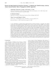
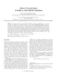
FIGURE 2: Dependence <strong>of</strong> the cytochrome b 561 reduction rate on<br />
ascorbate concentration. Chromaffin-vesicle membranes were<br />
mixed with ascorbic acid <strong>by</strong> a stopped-flow method, and the<br />
absorbance <strong>of</strong> cytochrome b 561 was recorded as described in<br />
Experimental Procedures. Each point is the average ((SD) <strong>of</strong> three<br />
separate determinations <strong>of</strong> the initial rate <strong>of</strong> reduction at pH 5.5<br />
(O) or 8.0 (b). The lines are least-squares fits to eq 1.<br />
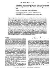
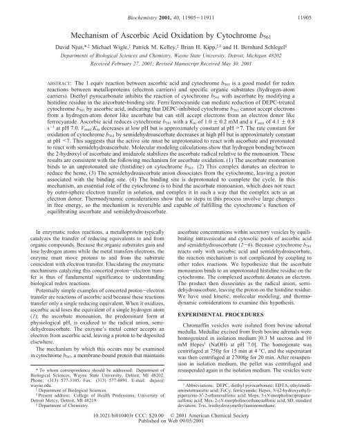
FIGURE 1: DEPC inhibits reduction <strong>of</strong> cytochrome b 561 <strong>by</strong> ascorbate<br />
but not <strong>by</strong> ferrocyanide. Chromaffin-vesicle membranes were<br />
divided; half were treated with DEPC (A and B), and the rest were<br />
kept as an untreated control (C and D). Reduction <strong>of</strong> cytochrome<br />
b 561 in 1 mL samples was monitored spectrophotometrically.<br />
Ascorbate (50 µL <strong>of</strong> a 100 mM solution) and ferricyanide (50 µL<br />
<strong>of</strong> a 100 mM solution) were added at the indicated times. Finally,<br />
a few grains <strong>of</strong> sodium dithionite were added to determine 100%<br />
cytochrome reduction.<br />
then purified <strong>by</strong> centrifugation through a layer <strong>of</strong> 1.6 M<br />
sucrose and 10 mM Hepes (NaOH) at pH 7.0 (7) and<br />
resuspended in storage medium [0.2 M Tris(PO 4 ) (pH 7.0)].<br />
One-eighth volume <strong>of</strong> 30% glycerol and 70% storage<br />
medium (v/v) was added to lyse the vesicles (8). After 20<br />
min at 4 °C, the membrane suspension was frozen and stored<br />
at -80 °C until it was needed.<br />
For chemical modification studies (Figure 1), chromaffinvesicle<br />
membranes were thawed and dialyzed for 24 h at 4<br />
°C against 0.1 M KCl and 5 mM potassium phosphate (pH<br />
7.0). Diethyl pyrocarbonate (13.8 mM final concentration)<br />
was added to half <strong>of</strong> the membrane suspension. After the<br />
solution had been kept for 20 min at room temperature, the<br />
DEPC reaction was terminated <strong>by</strong> adding histidine (18 mM<br />
final concentration) to both DEPC-treated and control<br />
samples. Each sample was then mixed with an equal volume<br />
<strong>of</strong> assay medium [0.2 M KCl, 10 mM methylamine, 50 mM<br />
potassium phosphate, and 200 µM EDTA (pH 7.0)]. <strong>Cytochrome</strong><br />
b 561 reduction was followed spectrophotometrically<br />
using an SLM-Aminco DW2000 spectrophotometer operated<br />
in the dual-wavelength mode (561 nm relative to the<br />
isosbestic point at 569 nm).<br />
For studies <strong>of</strong> reaction kinetics (Figure 2), chromaffinvesicle<br />
membranes were thawed and centrifuged at 27000g<br />
for 20 min at 4 °C. The pellet was resuspended in dialysis<br />
medium [0.15 M KCl, 100 µM EDTA, and 10 mM Mops<br />
(pH 7.0)] and dialyzed for 48 h at 4 °C against dialysis<br />
medium (4 × 100 volumes) to remove residual ascorbic acid<br />
and catecholamines. These chromaffin-vesicle ghosts were<br />
then purified on a Ficoll/sucrose density gradient (8).<br />
<strong>Cytochrome</strong> b 561 reduction was followed spectrophotometrically<br />
using an Aminco-Morrow stopped-flow apparatus<br />
attached to an SLM-Aminco DW2000 spectrophotometer<br />
operated in the dual-wavelength mode. <strong>Ascorbic</strong> acid was<br />
freshly prepared in 0.2 M KCl, 10 mM methylamine, 200<br />
µM EDTA, and either 10 mM Mes (pH 5.5, 6.0, or 6.5) or<br />
10 mM Hepes (pH 7.0, 7.5, or 8.0). This was mixed <strong>by</strong><br />
injection with ghosts diluted into the same medium, and the<br />
absorbance difference (561 nm - 569 nm) was recorded.<br />
The initial rate <strong>of</strong> cytochrome b 561 reduction was converted<br />
to units <strong>of</strong> inverse seconds using a molar extinction coefficient<br />
<strong>of</strong> 17 500 M -1 (9), the protein concentration, and a<br />
cytochrome-to-membrane protein ratio <strong>of</strong> 2.3 µmol/g (10).<br />
K m and V max were determined using a least-squares method<br />
to fit cytochrome reduction rates V measured at different<br />
ascorbate concentrations [AH - ] to the Michaelis-Menten<br />
equation:<br />
V ) (V max [AH - ])/([AH - ] + K m ) (1)<br />
For studies in D 2 O (Figure 6), cytochrome b 561 was<br />
solubilized in 0.1% NP40 after the membranes had been<br />
washed in 1% Tween 20 as described <strong>by</strong> Wakefield et al.<br />
(11). Solubilized cytochrome b 561 was then dialyzed against<br />
200 mM NaCl, 20 mM Hepes, and 1 mM EDTA (pH 6.8)<br />
in either D 2 OorH 2 O. The cytochrome was mixed <strong>by</strong> a<br />
stopped-flow method with ascorbic acid in the same medium,<br />
and the initial rate <strong>of</strong> reduction (0.67 µM cytochrome b 561 )<br />
was followed spectrophotometrically as described above.<br />
Molecular modeling calculations were carried out with the<br />
Gaussian series <strong>of</strong> programs (12). To simplify the computations,<br />
the side chain <strong>of</strong> ascorbate was removed, histidine was<br />
modeled <strong>by</strong> imidazole, and the geometry optimization was<br />
restricted to structures in which the ascorbate and imidazole<br />
rings were coplanar. Note that the actual geometry need not<br />
be coplanar, because rotation about the hydrogen bond to
<strong>Mechanism</strong> <strong>of</strong> <strong>Ascorbic</strong> <strong>Acid</strong> <strong>Oxidation</strong> <strong>by</strong> <strong>Cytochrome</strong> b 561 Biochemistry, Vol. 40, No. 39, 2001 11907<br />
any arbitrary angle is not expected to have a significant effect<br />
on energy. Geometries <strong>of</strong> the monomers and hydrogenbonded<br />
dimers were optimized at the HF/6-31G* level <strong>of</strong><br />
theory. Relative energies <strong>of</strong> ascorbate in different oxidation<br />
and protonation states were calculated at the PMP2/6-<br />
31+G** and B3LYP/6-31+G** levels <strong>of</strong> theory using the<br />
HF/6-31G*-optimized geometry. Test calculations on hydrogen<br />
bonding between acetic acid, acetate, and imidazole<br />
indicated that the HF/6-31G* interaction energies were within<br />
1-2 kcal/mol <strong>of</strong> the PMP2/6-31+G** and B3LYP/6-<br />
31+G** values. Hence, the hydrogen bond energies for<br />
ascorbate were calculated at the HF/6-31G* level <strong>of</strong> theory.<br />
Protein concentrations were determined using the BCA<br />
assay (13). DEPC, Hepes, Mes, Mops, and Tris were<br />
obtained from Sigma Chemical Co.<br />
RESULTS<br />
Although cytochrome b 561 reacts with ascorbate on both<br />
sides <strong>of</strong> the chromaffin-vesicle membrane, reaction is<br />
normally observed only at the external site because <strong>of</strong> spatial<br />
and kinetic factors. Externally added ascorbate does not<br />
permeate to the inside <strong>of</strong> resealed chromaffin-vesicle ghosts<br />
(14). Moreover, cytochrome b 561 is reduced 1 order <strong>of</strong><br />
magnitude faster from the outside than from the inside (15),<br />
so any ascorbate permeating into the ghosts would cause only<br />
a negligible fraction <strong>of</strong> the total observed reduction.<br />
<strong>Cytochrome</strong> b 561 has an essential histidine residue in the<br />
external ascorbate-binding site (16). Ethoxyformylation <strong>of</strong><br />
this histidine residue with diethyl pyrocarbonate prevents<br />
ascorbate from directly reducing cytochrome b 561 , but allows<br />
ferri/ferrocyanide to mediate the transfer <strong>of</strong> electrons from<br />
ascorbate to the cytochrome’s heme (Figure 1). Although<br />
ferricyanide addition oxidizes the cytochrome, the presence<br />
<strong>of</strong> ferricyanide accelerates the reduction <strong>of</strong> cytochrome b 561<br />
<strong>by</strong> ascorbate in DEPC-treated membranes (Figure 1A,B). In<br />
untreated membranes, ascorbate reduces cytochrome b 561<br />
quickly in the presence or absence <strong>of</strong> ferricyanide (Figure<br />
1C,D). This argues that the histidine residue is required for<br />
reaction with the hydrogen-atom donor ascorbate but is not<br />
needed for reaction with the electron donor ferrocyanide.<br />
To test the possibility that the histidine residue functions<br />
in proton transfer, the pH dependence <strong>of</strong> the reaction between<br />
ascorbic acid and cytochrome b 561 was investigated. The rate<br />
<strong>of</strong> cytochrome b 561 reduction follows Michaelis-Menten<br />
kinetics, saturating at high concentrations <strong>of</strong> external ascorbate<br />
(Figure 2). K m is much higher at pH 5.5 than at pH 8.0,<br />
although the V max values are comparable. At pH 7.0,<br />
ascorbate reduces cytochrome b 561 with a K m <strong>of</strong> 1.0 ( 0.2<br />
mM and a V max <strong>of</strong> 4.1 ( 0.8 s -1 . This compares to a K m <strong>of</strong><br />
0.34 mM reported <strong>by</strong> Flatmark and Terland (17).<br />
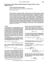
Over the physiological pH range, V max changes little<br />
(Figure 3A), while K m increases markedly at low pH (Figure<br />
3B). As a consequence, a plot <strong>of</strong> V max /K m is constant above<br />
and decreases below pH 6.5 (Figure 4). Because ascorbate<br />
does not have a pK near 7, the kinetics are consistent with<br />
ascorbate binding to an unprotonated histidine residue on<br />
the cytochrome.<br />
The reverse reaction, oxidation <strong>of</strong> cytochrome b 561 <strong>by</strong><br />
semidehydroascorbate, may also be examined. It is not<br />
practical to increase the ascorbate radical concentration to a<br />
saturating level, but we have been able to determine an<br />
FIGURE 3: pH dependence <strong>of</strong> kinetic parameters for cytochrome<br />
b 561 reduction <strong>by</strong> ascorbate. V max (A) and K m (B) were determined<br />
<strong>by</strong> experiments as described in the legend <strong>of</strong> Figure 1. Each point<br />
is the average ((SD) <strong>of</strong> at least three determinations each done<br />
using a different preparation <strong>of</strong> membranes. Lines were calculated<br />
using eq 2 for V max and eq 3 for K m with the following values:<br />
pK c ) 6.5, K Dm ) 0.7 mM, and k red ) 4.3 s -1 .<br />
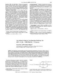
FIGURE 4: pH dependence <strong>of</strong> ascorbate reduction and oxidation.<br />
V max /K m values (b) were calculated from the data shown in Figure<br />
3. Values for the rate constant k for oxidation <strong>by</strong> semidehydroascorbate<br />
(O) are from Kelley et al. (18). Lines were calculated<br />
using eq 4 and k ) k ox /[K Dr (1 + K c /[H + ])] where k ox /K Dr ) 3.2 ×<br />
10 6 M -1 s -1 and other values as in the legend <strong>of</strong> Figure 3.<br />
apparent rate constant, an approximation to V max /K m (18).<br />
This rate constant diminishes at pH >6.5 (Figure 4),<br />
suggesting that the ascorbate radical reacts with the protonated<br />
histidine.<br />
Other potential sources <strong>of</strong> pH dependence may be excluded.<br />
The pKs <strong>of</strong> ascorbic acid (pK 1 ) 4.5, pK 2 ) 11.34)<br />
and its radical (pK r )-0.45) lie outside <strong>of</strong> the physiological<br />
range. The midpoint reduction potential <strong>of</strong> the heme is<br />
independent <strong>of</strong> pH (19).<br />
To see how a histidine residue might interact with ascorbic<br />
acid in the cytochrome’s binding site, we have used a
11908 Biochemistry, Vol. 40, No. 39, 2001 Njus et al.<br />
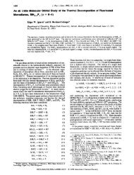
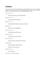
FIGURE 6: Dependence <strong>of</strong> the rate <strong>of</strong> cytochrome reduction on<br />
ascorbate concentration in H 2 O and D 2 O. <strong>Cytochrome</strong> b 561 in H 2 O<br />
(O) orD 2 O(b) was mixed with ascorbic acid <strong>by</strong> a stopped-flow<br />
method, and the absorbance was recorded as described in Experimental<br />
Procedures. Each point is the average ((SD) <strong>of</strong> at least<br />
three separate determinations <strong>of</strong> the initial rate <strong>of</strong> reduction.<br />
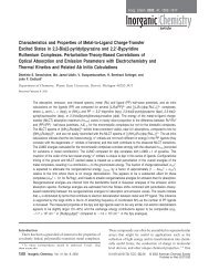
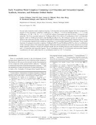
FIGURE 5: Hydrogen bonding energies <strong>of</strong> imidazole with oxygen<br />
atoms in ascorbate and semidehydroascorbate model compounds.<br />
Hydrogen bond energies (italics) between each oxygen atom on<br />
the ascorbate derivative and the appropriate nitrogen atom <strong>of</strong><br />
unprotonated imidazole were calculated at the HF/6-31G* level <strong>of</strong><br />
theory. Relative reaction energies were calculated at the PMP2/6-<br />
31+G** (bold) or B3LYP/6-31+G** (parentheses) level <strong>of</strong> theory.<br />
All energies are in kilocalories per mole.<br />
molecular modeling approach. The effect <strong>of</strong> hydrogen<br />
bonding between histidine and ascorbate on the stability <strong>of</strong><br />
the various ascorbate forms may be assessed <strong>by</strong> calculating<br />
hydrogen bonding energies. To simplify the calculations,<br />
unprotonated imidazole was used to represent histidine and<br />
an ascorbate analogue lacking the side chain was used to<br />
represent ascorbate (Figure 5). Molecular modeling is limited<br />
to simple systems and does not take into account effects <strong>of</strong><br />
solvent or neighboring groups. As a consequence, absolute<br />
values <strong>of</strong> energies are not significant, but relative energies<br />
can help define which structures are most likely.<br />
Molecular modeling calculations show several things.<br />
When imidazole and the dianion <strong>of</strong> the ascorbate analogue<br />
are allowed to interact, energy minimization results in the<br />
transfer <strong>of</strong> a proton from the imidazole to the dianion,<br />
yielding a complex between the monoanions <strong>of</strong> imidazole<br />
and the ascorbate analogue. This argues again that the<br />
ascorbate dianion cannot be stabilized <strong>by</strong> the active site <strong>of</strong><br />
the cytochrome and, therefore, the monoanion is more likely<br />
to be the electron donor to the heme.<br />
The monoanion can exist in two possible forms depending<br />
on which <strong>of</strong> the two hydroxyl groups ionizes. The more<br />
stable form in solution has the undissociated proton on the<br />
2-hydroxyl group. This structure permits resonance, distributing<br />
the negative charge between the oxygen atoms on C 1<br />
and C 3 . Molecular modeling confirms that this is the more<br />
stable form (Figure 5).<br />
The ascorbate monoanion could interact with imidazole<br />
through hydrogen bonds at any one <strong>of</strong> the three oxygen atoms<br />
on C 1 ,C 2 ,orC 3 . Hydrogen bonding through the 2-hydroxyl<br />
<strong>of</strong> the ascorbate analogue stabilizes the radical relative to<br />
the monoanion. This is not the case for hydrogen bonding<br />
through the oxygen atoms at the C 1 or C 3 position, however.<br />
Hydrogen bonding through the 2-hydroxyl group<br />
reduces the influence <strong>of</strong> that H and allows the unpaired<br />
electron to be distributed over three oxygen atoms. By<br />
contrast, hydrogen bonding at either <strong>of</strong> the other two<br />
positions leaves this H in place and introduces another H<br />
from the imidazole, thus further constraining the distribution<br />
<strong>of</strong> the unpaired electron.<br />
Finally, energy minimization shows that atomic coordinates<br />
do not change significantly when the ascorbate<br />
analogue, hydrogen bonded to imidazole through the 2-hydroxyl<br />
group, is oxidized to the free radical. This means that<br />
electron transfer from the bound ascorbate monoanion may<br />
occur without requiring molecular reorientation.<br />
Since the ascorbate monoanion loses a proton along with<br />
an electron when reducing cytochrome b 561 , the reaction<br />
might exhibit a deuterium isotope effect. Indeed, substituting<br />
D 2 O for H 2 O slows the rate at which ascorbate reduces<br />
cytochrome b 561 to between 14 and 40% <strong>of</strong> the rate in H 2 O<br />
(Figure 6). The effect is greatest at low ascorbate concentrations,<br />
indicating that K m increases slightly in D 2 O and V max<br />
is diminished considerably. By contrast, the rate at which<br />
ascorbate reduces cytochrome c is changed little in D 2 O (data<br />
not shown).<br />
DISCUSSION<br />
A synthesis <strong>of</strong> the data presented here along with other<br />
information leads to a hypothesis for the mechanism <strong>of</strong><br />
cytochrome b 561 reduction <strong>by</strong> ascorbic acid (Figure 7).<br />
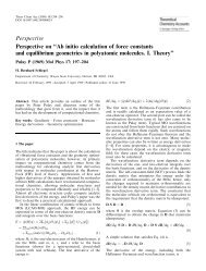
Central features <strong>of</strong> this model are as follows. (1) The<br />
ascorbate monoanion binds to an unprotonated site (histidine)<br />
on cytochrome b 561 . (2) This complex donates an electron<br />
to reduce the heme. (3) Semidehydroascorbate dissociates<br />
from the cytochrome as the radical anion, transferring a<br />
proton to the binding site. (4) The binding site is deprotonated<br />
to complete the cycle. Let us consider the evidence for this<br />
mechanism beginning with the ascorbate-binding site.<br />
Chemical modification studies (16, 20) using diethyl<br />
pyrocarbonate suggest that the ascorbate-binding site contains
<strong>Mechanism</strong> <strong>of</strong> <strong>Ascorbic</strong> <strong>Acid</strong> <strong>Oxidation</strong> <strong>by</strong> <strong>Cytochrome</strong> b 561 Biochemistry, Vol. 40, No. 39, 2001 11909<br />
FIGURE 7: Hypothesized mechanism for concerted proton-electron<br />
transfer from ascorbic acid to cytochrome b 561 . (1) Binding <strong>of</strong> the<br />
ascorbate monoanion, (2) Electron transfer. (3) Dissociation <strong>of</strong> the<br />
ascorbate radical anion. (4) Deprotonation <strong>of</strong> the binding site<br />
histidine. Two additional reactions, deprotonation and reduction <strong>of</strong><br />
the ascorbate free radical in solution, are also included to complete<br />
the cycle used to analyze thermodynamic equilibrium (eq 5).<br />
an essential histidine residue. As shown here (Figure 1),<br />
inactivation <strong>by</strong> DEPC prevents direct reduction <strong>of</strong> the heme<br />
<strong>by</strong> ascorbic acid, but permits reduction <strong>of</strong> the heme <strong>by</strong> other<br />
agents. To examine this quantitatively, DEPC-treated cytochrome<br />
b 561 is reduced <strong>by</strong> 5 mM ascorbate at a rate <strong>of</strong> 1.5<br />
× 10 -3 s -1 (Figure 1A). This indicates a rate constant <strong>of</strong><br />
0.3 M -1 s -1 , which is 1500 times slower than the uninhibited<br />
value (6). Following ferricyanide addition, DEPC-treated<br />
cytochrome b 561 is reduced at a rate <strong>of</strong> 1.1 × 10 -2 s -1 . This<br />
gives an apparent rate constant for reduction <strong>by</strong> ferrocyanide<br />
<strong>of</strong>2M -1 s -1 , which compares to the value <strong>of</strong> 13 M -1 s -1<br />
characteristic <strong>of</strong> unmodified cytochrome b 561 (6). Reduction<br />
<strong>of</strong> the cytochrome <strong>by</strong> ferrocyanide is clearly much less<br />
affected <strong>by</strong> DEPC than is reduction <strong>by</strong> ascorbate.<br />
Two factors may account for the small effect <strong>of</strong> DEPC<br />
on reduction <strong>by</strong> ferrocyanide. First, Takeuchi et al. (20) have<br />
shown that ethoxyformylation <strong>of</strong> Lys85 reduces the rate <strong>of</strong><br />
cytochrome b 561 reduction <strong>by</strong> ascorbate. Elimination <strong>of</strong> the<br />
positive charge on this lysine residue may reduce the affinity<br />
<strong>of</strong> the cytochrome for ascorbate. Removal <strong>of</strong> charge would<br />
be expected to have an even greater inhibitory effect on<br />
reaction with the ferrocyanide tetraanion. In addition, the<br />
rate at which ferrocyanide reduces cytochrome b 561 may be<br />
limited <strong>by</strong> the rate at which ferrocyanide is generated.<br />
Because the reaction between cytochrome b 561 and ferri/<br />
ferrocyanide greatly favors oxidation <strong>by</strong> ferricyanide, the<br />
ferricyanide concentration must be extremely low (
11910 Biochemistry, Vol. 40, No. 39, 2001 Njus et al.<br />
<strong>of</strong> initial rates limits the data to reduction <strong>of</strong> the first heme.<br />
Rapid electron transfer between two hemes would not affect<br />
the quantitation <strong>of</strong> reduction, because the hemes have<br />
indistinguishable absorption spectra (19).<br />
According to the mechanism described above, the bound<br />
ascorbate monoanion acts as an electron donor and forms<br />
the bound semidehydroascorbate radical as an intermediate.<br />
The monoanion in solution is a relatively poor electron donor<br />
having a reduction potential <strong>of</strong> 0.766 V. If it is to reduce<br />
cytochrome b 561 (E° ) 0.140 V), then the ascorbate-binding<br />
site must destabilize the monoanion and stabilize the free<br />
radical to facilitate the electron transfer reaction. Molecular<br />
modeling shows that hydrogen bonding with the imidazole<br />
may accomplish this if the unprotonated imidazole is<br />
hydrogen bonded to the 2-hydroxyl <strong>of</strong> the ascorbate monoanion.<br />
The hydrogen bond energy at this location is relatively<br />
stronger for the free radical than for the monoanion.<br />
Hydrogen bond energies at the other two oxygen atoms are<br />
stronger for the monoanion than for the free radical.<br />
Furthermore, minimized structures <strong>of</strong> imidazole-ascorbate<br />
complexes hydrogen bonded through C 2 show that the<br />
various forms <strong>of</strong> ascorbate (monoanion, free radical, and<br />
radical anion) all have similar atomic coordinates. Thus,<br />
reorientation <strong>of</strong> the ascorbate-histidine complex does not<br />
need to occur as the cytochrome proceeds through the<br />
reduction reaction.<br />
The bound ascorbate free radical must dissociate from the<br />
site and lose a proton to form the semidehydroascorbate<br />
radical anion. This could happen <strong>by</strong> (1) the bound radical<br />
dissociating and then deprotonating, (2) the bound radical<br />
losing a proton and then dissociating as the semidehydroascorbate<br />
radical anion, or (3) the bound radical transferring<br />
a proton to the binding site and then dissociating as<br />
semidehydroascorbate. The third case is consistent with the<br />
kinetics <strong>of</strong> oxidation <strong>by</strong> semidehydroascorbate (Figure 4).<br />
The pH dependence <strong>of</strong> the rate constant suggests that the<br />
semidehydroascorbate radical anion reacts with a protonated<br />
site having a pK <strong>of</strong> ∼6.5. The other two possibilities are<br />
energetically improbable. In the first case, stabilization <strong>of</strong><br />
the bound radical sufficient to achieve a reasonable reduction<br />
potential for electron donation (0.3 V) would require an<br />
extremely tight binding affinity (K D ) 10 -11 M). In the<br />
second case, either the semidehydroascorbate would have<br />
to bind with very high affinity or the pK <strong>of</strong> the bound radical<br />
would have to increase substantially. To achieve a dissociation<br />
constant <strong>of</strong> 1 µM with a reasonable reduction potential,<br />
the pK would have to increase to 5 from the value <strong>of</strong> -0.45<br />
observed in solution.<br />
The third case, <strong>by</strong> contrast, can be achieved with reasonable<br />
energetics. Thermodynamic equilibrium in this system<br />
(Figure 7) relates the parameters K Dm , E° c , K Dr ,pK r ,pK c , and<br />
E° f :<br />
E° c ) E° f + (RT/F log e)(log K Dr - log K Dm - pK c + pK r )<br />
(5)<br />
FIGURE 8: Dependence <strong>of</strong> the reduction potential <strong>of</strong> bound ascorbate<br />
on the dissociation constant for the semidehydroascorbate radical<br />
anion. Lines were calculated using eq 5 with the following values:<br />
K Dm ) 1 mM, E° f ) 0.766 V, pK r )-0.45, and T ) 295 K.<br />
Values used for pK c are 6.0 (top line), 6.5 (middle line), and 7.0<br />
(bottom line).<br />
Knowing E° f ) 0.766 V, K Dm ) 1 mM, and pK r )-0.45,<br />
we can determine E° c as a function <strong>of</strong> K Dr and pK c (Figure<br />
8). pK c , the pK <strong>of</strong> the active site residue, is ∼6 or∼7. K Dr ,<br />
the binding constant for semidehydroascorbate, is unknown,<br />
but in studies <strong>of</strong> cytochrome b 561 oxidation, we could not<br />
observe saturation <strong>by</strong> semidehydroascorbate up to concentrations<br />
<strong>of</strong> 1 µM (18). Consequently, K Dr is expected to be<br />
greater than 10 -6 M.<br />
Semidehydroascorbate oxidizes cytochrome b 561 10 3 times<br />
faster than ascorbate reduces it (18). This is consistent with<br />
the midpoint potential <strong>of</strong> the bound ascorbate or semidehydroascorbate<br />
being ∼300 mV, 160 mV more positive than<br />
the midpoint potential <strong>of</strong> the cytochrome (140 mV; 17, 19).<br />
A midpoint potential <strong>of</strong> this magnitude would be compatible<br />
with a dissociation constant for semidehydroascorbate (K Dr )<br />
<strong>of</strong> ∼100 µM (Figure 8). These values not only are reasonable<br />
but also show that the mechanism outlined in Figure 7 does<br />
not involve any steps requiring large changes in free energy.<br />
The reactions, therefore, are all readily reversible, a necessity<br />
for a cytochrome whose function is to equilibrate ascorbate<br />
and semidehydroascorbate.<br />
The mechanism proposed here hypothesizes that the<br />
reactivity <strong>of</strong> ascorbate is controlled <strong>by</strong> the ascorbate-binding<br />
site. The heme serves only as an acceptor in the electron<br />
transfer reaction. As a consequence, ascorbate bound to<br />
cytochrome b 561 is poised for electron donation to any<br />
suitable electron acceptor. If the heme is already reduced,<br />
then the bound ascorbate may react adventitiously with<br />
another oxidant. Of particular interest is molecular oxygen,<br />
which may be reduced to the superoxide radical anion,<br />
initiating the production <strong>of</strong> a variety <strong>of</strong> damaging oxygen<br />
•-<br />
radicals. The reduction potential <strong>of</strong> the O 2 /O 2 pair is ∼300<br />
mV under physiological conditions (1, 26). To avoid generation<br />
<strong>of</strong> additional superoxide, it is obviously advantageous<br />
for the bound ascorbate to have a midpoint potential that is<br />
g300 mV, implying a binding constant for semidehydroascorbate<br />
that is 100 µM or weaker (Figure 8). As discussed<br />
above, these are likely values.<br />
According to the Marcus theory for electron transfer<br />
reactions in solution (27), outer-sphere electron transfer<br />
reactions are spontaneous and occur at rates that depend<br />
solely on the intrinsic reactivity <strong>of</strong> the compounds involved<br />
and on the difference in their relative reduction potentials.<br />
Such reactions are obviously not well suited to biological<br />
systems in which reaction rates are carefully regulated and<br />
desired redox reactions <strong>of</strong>ten involve reactants with relatively
<strong>Mechanism</strong> <strong>of</strong> <strong>Ascorbic</strong> <strong>Acid</strong> <strong>Oxidation</strong> <strong>by</strong> <strong>Cytochrome</strong> b 561 Biochemistry, Vol. 40, No. 39, 2001 11911<br />
close reduction potentials as, for example, in respiratory and<br />
other redox chains. The mechanism presented here suggests<br />
that biological systems have brought redox reactions under<br />
control <strong>by</strong> selecting redox compounds that do not react <strong>by</strong><br />
outer-sphere electron transfer but can be made to behave as<br />
electron donors and acceptors when bound in an appropriate<br />
site. The function <strong>of</strong> the redox enzyme, therefore, is to<br />
provide both a site for unlocking the electron-transferring<br />
capability <strong>of</strong> the substrate and also a redox center with which<br />
the bound substrate can react, preempting spontaneous<br />
reactions with other undesired reactants. An attraction <strong>of</strong> this<br />
perspective is that, like the Marcus theory for uncatalyzed<br />
reactions, it allows enzymatic redox reactions to be analyzed,<br />
at least to a first approximation, in terms <strong>of</strong> the separate<br />
properties <strong>of</strong> the reactants: a complexed organic substrate<br />
and the enzyme’s metal center.<br />
REFERENCES<br />
1. Njus, D., and Kelley, P. M. (1991) FEBS Lett. 284, 147-<br />
151.<br />
2. Njus, D., Knoth, J., Cook, C., and Kelley, P. M. (1983) J.<br />
Biol. Chem. 258, 27-30.<br />
3. Srivastava, M., Duong, L. T., and Fleming, P. J. (1984) J.<br />
Biol. Chem. 259, 8072-8075.<br />
4. Wakefield, L. M., Cass, A. E. G., and Radda, G. K. (1986) J.<br />
Biol. Chem. 261, 9739-9745.<br />
5. Menniti, F. S., Knoth, J., and Diliberto, E. J., Jr. (1986) J.<br />
Biol. Chem. 261, 16901-16908.<br />
6. Njus, D., and Kelley, P. M. (1993) Biochim. Biophys. Acta<br />
1144, 235-248.<br />
7. Smith, A. D., and Winkler, H. (1967) Biochem. J. 103, 480-<br />
482.<br />
8. Njus, D., and Radda, G. K. (1979) Biochem. J. 180, 579-<br />
585.<br />
9. Silsand, T., and Flatmark, T. (1974) Biochim. Biophys. Acta<br />
395, 257-266.<br />
10. Duong, L. T., Fleming, P. J., and Russell, J. T. (1984) J. Biol.<br />
Chem. 259, 4885-4889.<br />
11. Wakefield, L. M., Cass, A. E. G., and Radda, G. K. (1984) J.<br />
Biochem. Biophys. Methods 9, 331-341.<br />
12. Frisch, M. J., Trucks, G. W., Schlegel, H. B., Gill, P. M. W.,<br />
Johnson, B. G., Robb, M. A., Cheeseman, J. R., Keith, T.,<br />
Petersson, G. A., Montgomery, J. A., Raghavachari, K., Al-<br />
Laham, M. A., Zakrzewski, V. G., Ortiz, J. V., Foresman, J.<br />
B., Cioslowski, J., Stefanov, B. B., Nanayakkara, A., Challacombe,<br />
M., Peng, C. Y., Ayala, P. Y., Chen, W., Wong, M.<br />
W., Andres, J. L., Replogle, E. S., Gomperts, R., Martin, R.<br />
L., Fox, D. J., Binkley, J. S., Defrees, D. J., Baker, J., Stewart,<br />
J. P., Head-Gordon, M., Gonzalez, C., and Pople, J. A. (1995)<br />
GAUSSIAN 94, Gaussian, Inc., Pittsburgh, PA.<br />
13. Smith, P. K., Krohn, R. I., Hermanson, G. T., Mallia, A. K.,<br />
Gartner, F. H., Provenzano, M. D., Fujimoto, E. K., Goeke,<br />
N. M., Olson, B. J., and Klenk, D. C. (1985) Anal. Biochem.<br />
150, 76-85.<br />
14. Tirrell, J. G., and Westhead, E. W. (1979) Neuroscience 4,<br />
181-186.<br />
15. Jalukar, V., Kelley, P. M., and Njus, D. (1991) J. Biol. Chem.<br />
266, 6878-6882.<br />
16. Kipp, B. H., Kelley, P. M., and Njus, D. (2001) Biochemistry<br />
40, 3931-3937.<br />
17. Flatmark, T., and Terland, O. (1971) Biochim. Biophys. Acta<br />
253, 487-491.<br />
18. Kelley, P. M., Jalukar, V., and Njus, D. (1990) J. Biol. Chem.<br />
265, 19409-19413.<br />
19. Apps, D. K., Boisclair, M. D., Gavine, F. S., and Pettigrew,<br />
G. W. (1984) Biochim. Biophys. Acta 764, 8-16.<br />
20. Takeuchi, F., Kobayashi, K., Tagawa, S., and Tsubaki, M.<br />
(2001) Biochemistry 40, 4067-4076.<br />
21. Tsubaki, M., Kobayashi, K., Ichise, T., Takeuchi, F., and<br />
Tagawa, S. (2000) Biochemistry 39, 3276-3284.<br />
22. Degli Esposti, M., Kamenskiy, Y. A., Arutjunjan, A. M., and<br />
Konstantinov, A. A. (1989) FEBS Lett. 254, 74-78.<br />
23. Burbaev, D. S., Moroz, I. A., Kamenskiy, Y. A., and<br />
Konstantinov, A. A. (1991) FEBS Lett. 283, 97-99.<br />
24. Tsubaki, M., Nakayama, M., Okuyama, E., Ichikawa, Y., and<br />
Hori, H. (1997) J. Biol. Chem. 272, 23206-23210.<br />
25. Kamensky, Y. A., and Palmer, G. (2001) FEBS Lett. 491,<br />
119-122.<br />
26. Chance, B., Sies, H., and Boveris, A. (1979) Physiol. ReV.<br />
59, 527-605.<br />
27. Marcus, R. A., and Sutin, N. (1985) Biochim. Biophys. Acta<br />
811, 265-322.<br />
BI010403R