Clinical study on 5000 patients - F care systems
Clinical study on 5000 patients - F care systems
Clinical study on 5000 patients - F care systems

You also want an ePaper? Increase the reach of your titles
YUMPU automatically turns print PDFs into web optimized ePapers that Google loves.
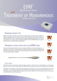
EFFICACY AND RESULTS OF THERMOCOAGULATION MODALITY IN THE<br />
TREATMENT OF RETICULAR AND TELENGIECTASIC VEINS: MIDTERM AND<br />
LONGTERM RESULTS OF <strong>5000</strong> CASES.<br />
F <strong>care</strong> <strong>systems</strong> nv www.f<strong>care</strong><strong>systems</strong>.com Tel: +32 3 451 51 45<br />
K<strong>on</strong>tichsesteenweg 54 info@f<strong>care</strong><strong>systems</strong>.com Fax: +32 3 451 51 39<br />
2630 Aartselaar (Belgium
ABSTRACT<br />
Background: Reticular and telengiectasic varicosities are <strong>on</strong>e of the most comm<strong>on</strong> problems<br />
in the general populati<strong>on</strong> effecting predominantly females. They have higher prevalence in<br />
females between 20-70 years with a rate of 55%. Although these varicosities cause pain and<br />
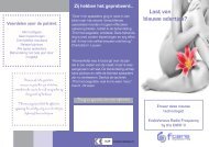
feeling of tiredness, they mostly cause cosmetic problems in <strong>patients</strong>. Schlerotherapy is a gold<br />
standard threapeutical opti<strong>on</strong> for the treatment of reticular and telengiectasies over 0.3 mm.<br />
But for the <strong>on</strong>es smaller than 0.3 mm, there are other opti<strong>on</strong>s such as thermocoagulati<strong>on</strong> with<br />
radiofrequency and laser energy. The aim of this <str<strong>on</strong>g>study</str<strong>on</strong>g> is to evaluate the efficacy of<br />
thermocoagulati<strong>on</strong> method for the treatment of reticular and telengiectasies.<br />
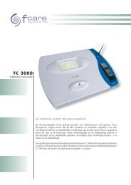
Patients and Methods: Between April 2004 and February 2008 TC 3000 thermocoagulati<strong>on</strong><br />
device had been used in <strong>5000</strong> <strong>patients</strong> for the treatment of reticular and telengiectasic<br />
varicosities. The lesi<strong>on</strong>s were divided into four groups in accordance with the classificati<strong>on</strong> of<br />
telengiectasies as linear, spider, arborized and papillary. Groups c<strong>on</strong>tained 7853, 5123, 4122<br />
and 2757 lesi<strong>on</strong>s respectively. 4753 of the <strong>patients</strong> were female and the mean age was 28.<br />
Mean follow-up time 24.2±11.9 m<strong>on</strong>ths. Improvement findings of the lesi<strong>on</strong>s were evaluated<br />
in five improvement levels.<br />
Results: Improvement levels in lineer telengiectasy group were 100%. But in more<br />
complicated spider, arborized telengiectasies and bluish-purple small reticular varicosities,<br />
this improvement ratio was approximately 75%. 40% of such cases needed sec<strong>on</strong>d and 15%<br />
needed a third applicati<strong>on</strong> of the thermocoagulati<strong>on</strong>. The mean improvement level in all<br />
lesi<strong>on</strong>s was 4.3 after all the treatments. When the lesi<strong>on</strong>s were evaluated according to their<br />
localizati<strong>on</strong>s, efficacy of the treatment was higher at the lesi<strong>on</strong>s that located below the knee,<br />
ankle and feet when compared to the lesi<strong>on</strong>s located over the knee and the external thigh.<br />
C<strong>on</strong>clusi<strong>on</strong>s: Thermocoagulati<strong>on</strong> is an efficient and hope-giving transcutaneous method for<br />
the treatment of small reticular and telengiectasix varicosities that can not be treated<br />
effectively with microsclerosis.<br />
Key words: Reticular veins, telengiectasies, thermocoagulati<strong>on</strong> method.<br />
F <strong>care</strong> <strong>systems</strong> nv www.f<strong>care</strong><strong>systems</strong>.com Tel: +32 3 451 51 45<br />
K<strong>on</strong>tichsesteenweg 54 info@f<strong>care</strong><strong>systems</strong>.com Fax: +32 3 451 51 39<br />
2630 Aartselaar (Belgium
Introducti<strong>on</strong><br />
Venous insufficiency is <strong>on</strong>e of the most comm<strong>on</strong> chr<strong>on</strong>ic health problems that effect <strong>patients</strong>’<br />
quality of life, health ec<strong>on</strong>omics and expenses. Recently CEAP (C: clinical, E: etiologic, A:<br />
Anatomic, P: Pathophysiologic) was adopted worldwide to facilitate meaningful<br />
communicati<strong>on</strong> about CVD and serve as a basis for more scientific analysis of management<br />
alternatives. This classificati<strong>on</strong>, based <strong>on</strong> correct diagnosis, was also expected to serve as a<br />
systematic guide in the daily clinical investigati<strong>on</strong> of <strong>patients</strong> as an orderly documentati<strong>on</strong><br />
system and basis for decisi<strong>on</strong>s regarding appropriate treatment. According to this<br />
classificati<strong>on</strong> small reticular and telengiectasic varicosities bel<strong>on</strong>g to the C1 class.(1) This<br />
comm<strong>on</strong> health problem effects 15% of adult populati<strong>on</strong> and 55% of females between the<br />
ages of 20-70 years.(2,3) When compared to trunk varicosities, treatment of small reticular<br />
and telengiectasic varicosities is more complicated for both <strong>patients</strong> and the doctors and takes<br />
time. Although these type of lesi<strong>on</strong>s cause leg discomfort, pain and feeling of heaviness and<br />
tiredness in the leg, they mostly appear as a cosmetic problem in young ladies. (4) Therefore<br />
it is sometimes very demanding to satisfy the expectati<strong>on</strong>s of these group <strong>patients</strong>.<br />
Due to these difficulties in the treatment of these lesi<strong>on</strong>s, lots of treatment opti<strong>on</strong>s have been<br />
used so far. Schlerotherapy as a gold standard treatment measure in the treatment of reticular<br />
veins has its place almost more than 150 years. Since its first explanati<strong>on</strong> by Chassaignac (5)<br />
in France in 1855, schlerotherapy maintained its popularity although the techniques and the<br />
drugs have been changed. Nowadays with the use of drugs with low adverse effect profile and<br />
32 Gauge fine needles, it is now possible to treat small reticular and some telengiectasies.<br />
However it is very difficult or impossible to treat red telengiectasies in <strong>patients</strong> with<br />
c<strong>on</strong>traindicati<strong>on</strong> to drugs (allergies) and patient with needle phobia. Therefore new treatments<br />
are always in search for these groups of <strong>patients</strong>.<br />
F <strong>care</strong> <strong>systems</strong> nv www.f<strong>care</strong><strong>systems</strong>.com Tel: +32 3 451 51 45<br />
K<strong>on</strong>tichsesteenweg 54 info@f<strong>care</strong><strong>systems</strong>.com Fax: +32 3 451 51 39<br />
2630 Aartselaar (Belgium
Thermocoagulati<strong>on</strong> (radiofrequency energy) method is <strong>on</strong>e of the alternative modalities that<br />
have been in use for the treatment of small reticular and telengiectasic varicosities that can not<br />
be treated by schlerotherapy for more than 8 years. Main principle in thermocoagulati<strong>on</strong><br />
method is a thermal damage that has been formed by a 4 MHz radiofrequency wave. When it<br />
is applied <strong>on</strong> a given lesi<strong>on</strong>, it causes a 70 °C temperature inside the treated vessel. As a result,<br />
plasma proteins are coagulated and parietal structures are destroyed. With this mechanism, the<br />
system is completely different from electrocoagulati<strong>on</strong>. Thermocoagulati<strong>on</strong> is a user-friendly,<br />
simple method and can be applied in large numbers according to the patient’s and doctor’s<br />
patience.<br />
The aim of this <str<strong>on</strong>g>study</str<strong>on</strong>g> is to evaluate the efficacy of thermocoagulati<strong>on</strong> method in small and<br />
telengiectasic varicosities.<br />
Patients and Methods<br />
Between April 2004 and February 2008, <strong>5000</strong> cases were treated by TC 3000 (Formes Et<br />
Performances, Bordeaux, France) (Picture 1) for small reticular and telengiectasic varicosities.<br />
4753 of the <strong>patients</strong> were female and mean age was 28.3±15.9 years. All <strong>patients</strong> were<br />
informed before the procedure and written c<strong>on</strong>sents were taken. Patients were advised to have<br />
epilati<strong>on</strong> of their legs prior to <str<strong>on</strong>g>study</str<strong>on</strong>g>. All of the lesi<strong>on</strong>s were mapped by taking picture before<br />
the treatment. Small reticuler veins (
Prilocain 25 mg, Emla 5%, AstraZeneca) was applied prior to treatment in patient with low<br />
pain threshold. A clear topical antiseptic (Dermal Wound Cleanser, Smith&Nephew Inc.,<br />
Largo, FL, USA) was used just before the treatment over the target areas. At the beginning of<br />
the series cold applicati<strong>on</strong> was used with ice packs to provide topical anesthesia before the<br />
treatment, but so<strong>on</strong> we quitted this procedure. The reas<strong>on</strong> for this was the disappearance of<br />
some lesi<strong>on</strong>s as a result of the cold induced vasoc<strong>on</strong>stricti<strong>on</strong>. Temperature and the lightning<br />
levels of the therapy room were kept optimum. The lesi<strong>on</strong>s were also vanished when the<br />
patient felt cold.<br />
Thermocoagulati<strong>on</strong> applicati<strong>on</strong> technique:<br />
TC 3000 (Formes Et Performances, Bordeaux, France) thermocogualati<strong>on</strong> device has three<br />
parts: <strong>on</strong>e c<strong>on</strong>sole for radiofrequency energy generator, <strong>on</strong>e isolated needle for applying<br />
energy to the lesi<strong>on</strong> and <strong>on</strong>e footswitch for synchr<strong>on</strong>izati<strong>on</strong> between generator and the needle.<br />
Applied energy and the durati<strong>on</strong> can be adjusted over the generator c<strong>on</strong>sole (Picture 2).<br />
Isolated needle has a very special design. Outer surface is totally isolated and <strong>on</strong>ly the tip part<br />
is c<strong>on</strong>ductive. With this design it gives the radiofrequency energy <strong>on</strong>ly the applied vessel and<br />
protects surrounding tissues (Picture 3).<br />
This needle is made of nickel. But there are also golden needles for the <strong>patients</strong> who are<br />
allergic to nickel. According to the lesi<strong>on</strong> diameter special different sized isolated needles<br />
from Ballet and FCare Systems brands were used ([Ballet brand: K3 0.075-0.080 mm, K5G<br />
(golden needle) 0.80-0.125 mm K6 0.150 mm], [FCare <strong>systems</strong>: R3i 0.08 mm, R6i 0.15mm]).<br />
After the selecti<strong>on</strong> of the needle type according to the lesi<strong>on</strong>, it is attached to the handle.<br />
Energy and the durati<strong>on</strong> are adjusted. For telengiectasies that were treated with K3-K5G, R3i<br />
needles 20-30% energy level and durati<strong>on</strong> of 0.2-0.3 sec<strong>on</strong>d were used. For lesi<strong>on</strong>s bigger<br />
than 0.1 mm that were treated with K6, R6i 40-50% energy level and 0.4-0.5 sec<strong>on</strong>d durati<strong>on</strong><br />
were used. For papillary lesi<strong>on</strong>s 60% energy level and 0.6 sec<strong>on</strong>d adjustments were used.<br />
F <strong>care</strong> <strong>systems</strong> nv www.f<strong>care</strong><strong>systems</strong>.com Tel: +32 3 451 51 45<br />
K<strong>on</strong>tichsesteenweg 54 info@f<strong>care</strong><strong>systems</strong>.com Fax: +32 3 451 51 39<br />
2630 Aartselaar (Belgium
Patients were positi<strong>on</strong>ed whether supine or pr<strong>on</strong>e according to the place of the treated lesi<strong>on</strong>.<br />
In order to have better visi<strong>on</strong> magnifying glasses were used. Needles were inserted<br />
perpendicularly over the lesi<strong>on</strong>. After the seeing some vanishing of the lesi<strong>on</strong>, energy is<br />
delivered by pressing <strong>on</strong> the footswitch (Picture 4). There should be 1-2 mm distances<br />
between each pulse of energy. Lesser or l<strong>on</strong>ger distances are not approved (Picture 5). Lesser<br />
distances may cause the prol<strong>on</strong>gati<strong>on</strong> of the recovery time, while l<strong>on</strong>ger distances may<br />
decrease the success rate. There is no need for compressi<strong>on</strong> stockings or bandages after the<br />
treatment.<br />
Post-procedural <strong>care</strong> and follow-up:<br />
Topical ice packs or cold shower were applied over the treated area. Patients were advised to<br />
c<strong>on</strong>tinue to their daily social activities. There was no limitati<strong>on</strong> for sun bath. Patients were<br />
seen <strong>on</strong> the sec<strong>on</strong>d day of treatment and then weekly until the complete recovery of crusts.<br />
During follow period sec<strong>on</strong>d or third treatments were performed for the residual lesi<strong>on</strong>s.<br />
Patients are followed yearly thereafter.<br />
RESULTS<br />
Lesi<strong>on</strong> become flushed and swollen immediately after the RF therapy. These new lesi<strong>on</strong>s are<br />
called as oreols (Picture 6). One day later these oreols vanished and left their places to crusts<br />
(Picture 7). After a mean period of four weeks (1 to 8 weeks) these crust are peeled<br />
sp<strong>on</strong>taneously and completely recovered (Picture 8).<br />
In <strong>patients</strong> with skin phototype I and II after peeling of the crusts, a pale, white, slightly raised<br />
scatris of the crusts remained under the crusts. These lesi<strong>on</strong>s lasted 1-3 m<strong>on</strong>ths and then<br />
completely resolved. Mean follow-up period was 24.2±11.9 m<strong>on</strong>ths.<br />
F <strong>care</strong> <strong>systems</strong> nv www.f<strong>care</strong><strong>systems</strong>.com Tel: +32 3 451 51 45<br />
K<strong>on</strong>tichsesteenweg 54 info@f<strong>care</strong><strong>systems</strong>.com Fax: +32 3 451 51 39<br />
2630 Aartselaar (Belgium
Patients were evaluated according to the 5 recovery levels:<br />
Level 1:
energy may cause treatment failure. Furthermore it is also of crucial importance to see the<br />
vanishing of the lesi<strong>on</strong> before applying the energy even in combinati<strong>on</strong> of right needle size<br />
and energy. By this c<strong>on</strong>firmati<strong>on</strong> method, paravascular applicati<strong>on</strong> of the energy is avoided.<br />
All this treatment optimizati<strong>on</strong> can be achieved by experience and meticulous applicati<strong>on</strong>.<br />
For lesi<strong>on</strong>s especially overpatellar and lateral thigh area, since these areas are pr<strong>on</strong>e to<br />
cellulitis and edema, target treatment success may not be reached. In order to increase success<br />
in these areas, sec<strong>on</strong>d and third applicati<strong>on</strong>s may be required.<br />
Electrocoagulati<strong>on</strong>, microsclerosis, laser energy and phototherapy are the other alternative<br />
treatment modalities to radiofrequency thermocoagulati<strong>on</strong> in the treatment of reticular and<br />
telengiectasic veins. Microsclerosis is very well-known modalities and applied with a high<br />
success rates in lesi<strong>on</strong>s ≥0.3 mm by experienced hands from very l<strong>on</strong>g time. Currently<br />
microsclerosis is the gold standard therapy. However there is always complicati<strong>on</strong> risk related<br />
with the sclerosing agent, c<strong>on</strong>centrati<strong>on</strong>, applied technique and lesi<strong>on</strong> diameter. The thinnest<br />
needle size is 32 G and for the lesi<strong>on</strong>s that this needle size can be introduced, schlerotherapy<br />
should be the treatment of choice. (10) For residual lesi<strong>on</strong>s and lesi<strong>on</strong> that can not be<br />
cannulated thermocoagulati<strong>on</strong> method is good and effective alternative. Inadvertent events<br />
may be seen due to weak subcutaneous tissue and thin skin especially at the lesi<strong>on</strong> located to<br />
ankle and the foot. Therefore it is better to use transdermal radiofrequency for these lesi<strong>on</strong>s.<br />
Laser energy is another popular opti<strong>on</strong> that has been in use in any of the medical discipline for<br />
several reas<strong>on</strong>s. It has been used in the treatment of varicose veins since 1980. The basic<br />
principle in the laser energy is absorpti<strong>on</strong> of the energy by chromophoric structures. Then heat<br />
is generated and c<strong>on</strong>tained in these tissue targets. The producti<strong>on</strong> of high temperatures within<br />
the targeted structure, combined with its rapid dissipati<strong>on</strong>, enable exquisitely selective injury<br />
in microscopic tissue targets. Heat is dissipated by c<strong>on</strong>ducti<strong>on</strong> and radiative transfer as so<strong>on</strong><br />
as it is created. Selective target heating is produced when the energy is deposited at a rate<br />
F <strong>care</strong> <strong>systems</strong> nv www.f<strong>care</strong><strong>systems</strong>.com Tel: +32 3 451 51 45<br />
K<strong>on</strong>tichsesteenweg 54 info@f<strong>care</strong><strong>systems</strong>.com Fax: +32 3 451 51 39<br />
2630 Aartselaar (Belgium
faster than the rate of cooling of the target structure. Different wavelengths (810 nm, 940 nm,<br />
980 nm) have been approved by FDA for the treatment of varicosities.<br />
In the 1980s, arg<strong>on</strong> lasers were used, but the 488-nm wavelength was str<strong>on</strong>gly absorbed by<br />
melanin. Also, arg<strong>on</strong> lasers were c<strong>on</strong>tinuous lasers that did not allow for selective heating of<br />
vessels, so scarring was comm<strong>on</strong>. In 1983, Anders<strong>on</strong> and Parrish's principles of selective<br />
photothermolysis guided the design of the pulsed dye laser (PDL), which successfully treated<br />
facial telangiectasias and port-wine stains. Although a 577-nm wavelength was originally<br />
chosen to match the yellow absorpti<strong>on</strong> peak of oxyhemoglobin, it was quickly realized that a<br />
585-nm wavelength resulted in more effective treatment of portwine stains.<br />
A sec<strong>on</strong>d generati<strong>on</strong> of PDLs with a l<strong>on</strong>ger wavelength (595 nm) and l<strong>on</strong>ger pulse durati<strong>on</strong><br />
(1.5 millisec<strong>on</strong>ds) as released in 1996. These PDLs penetrated deeper but were still <strong>on</strong>ly<br />
effective for vessels up to 1 mm in depth and 1 mm in width. Over the past 5 years, nearinfrared<br />
lasers targeting the broad absorpti<strong>on</strong> band from 750 to 1100 nm have been used to<br />
penetrate further. The l<strong>on</strong>ger wavelength alexandrite, diode, and Nd:YAG permit sufficient<br />
energy to heat deeper leg veins up to 3 mm wide. (11-14)<br />
There is no standard method or algorithm for reticular and telengiectasies. It is also<br />
impossible to cure all types of lesi<strong>on</strong>s with <strong>on</strong>e laser energy source. Its’ usage in this type<br />
lesi<strong>on</strong>s is limited. Also it has some disadvantages such as need for sun-protecti<strong>on</strong>, limited use<br />
in dark skin color etc.<br />
If we think that all lesi<strong>on</strong> types of CEAP class (C0-C6) can be found in a patient’s leg, in<br />
order to provide a complete treatment all treatment opti<strong>on</strong>s must be in our treatment spectrum.<br />
And all of them must be used when they are indicated.<br />
When compared with other treatment modalities RF energy is a user friendly opti<strong>on</strong> and has<br />
some advantages. It can be applied in all skin types, no need for sun protecti<strong>on</strong>, and does not<br />
F <strong>care</strong> <strong>systems</strong> nv www.f<strong>care</strong><strong>systems</strong>.com Tel: +32 3 451 51 45<br />
K<strong>on</strong>tichsesteenweg 54 info@f<strong>care</strong><strong>systems</strong>.com Fax: +32 3 451 51 39<br />
2630 Aartselaar (Belgium
cause skin necrosis or pigmentati<strong>on</strong> disorders. It can safely be used in the treatment of<br />
varicosities that can not be treated with microschlerotherapy.<br />
F <strong>care</strong> <strong>systems</strong> nv www.f<strong>care</strong><strong>systems</strong>.com Tel: +32 3 451 51 45<br />
K<strong>on</strong>tichsesteenweg 54 info@f<strong>care</strong><strong>systems</strong>.com Fax: +32 3 451 51 39<br />
2630 Aartselaar (Belgium
REFERENCES<br />
1. Eklöf B, Rutherford RB, Bergan JJ, Carpentier PH, Gloviczki P, Kistner RL, Meissner<br />
MH, M<strong>on</strong>eta GL, Myers K, Padberg FT, Perrin M, Ruckley CV, Smith PC, Wakefield<br />
TW; American Venous Forum Internati<strong>on</strong>al Ad Hoc Committee for Revisi<strong>on</strong> of the CEAP<br />
Classificati<strong>on</strong>. Revisi<strong>on</strong> of the CEAP classificati<strong>on</strong> for chr<strong>on</strong>ic venous disorders:<br />
c<strong>on</strong>sensus statement. J Vasc Surg. 2004 Dec;40(6):1248-52. Review.<br />
2. Callam MJ. Epidemiology of varicose veins. Br J Surg 1994; 81:173<br />
3. Bradbury A, Evans CJ, Allan P, Lee AJ, Ruckley CV, Fowkes FG. The relati<strong>on</strong>ship<br />
between lower limb symptoms and superficial and deep venous reflux <strong>on</strong> duplex<br />
ultras<strong>on</strong>ography:the Edinburgh Vein Study. J Vasc Surg 2000;32:921–931.<br />
4. Beale RJ, Gough MJ. Treatment opti<strong>on</strong>s for primary varicose veins--a review. Eur J Vasc<br />
Endovasc Surg. 2005 Jul;30(1):83-95. Review.<br />
5. Handbook of Patient Care in Vascular Surgery.3rd Editi<strong>on</strong>. In Edit: Hallet JW. Varicose<br />
veins. Pg:255-281. 1995<br />
6. Bergan JJ. Risk Factors, Manifestati<strong>on</strong>s,and <str<strong>on</strong>g>Clinical</str<strong>on</strong>g> Examinati<strong>on</strong> of the Patient with<br />
Primary Venous Insufficiency. In: Began JJ eds. The Vein Book. Burlint<strong>on</strong> MA,<br />
2007:119-125.<br />
7. Weiss RA, Weiss MA. Resoluti<strong>on</strong> of pain associated with varicose and telangiectatic leg<br />
veins after compressi<strong>on</strong> sclerotherapy, J Dermatol Surg Onc. 1990. 16: 333–336.<br />
8. Coughlin LB, Gandy R, Rosser S, de Cossart L. Factors associated with varicose veins in<br />
pregnant women, Phlebology. 2002. 16: 167–169.<br />
9. Browse NL, Burnand KG, Irvine AT, Wils<strong>on</strong> NM. Diseases of the veins. 2nd ed. L<strong>on</strong>d<strong>on</strong>,<br />
Arnold, 1999.<br />
F <strong>care</strong> <strong>systems</strong> nv www.f<strong>care</strong><strong>systems</strong>.com Tel: +32 3 451 51 45<br />
K<strong>on</strong>tichsesteenweg 54 info@f<strong>care</strong><strong>systems</strong>.com Fax: +32 3 451 51 39<br />
2630 Aartselaar (Belgium
10. Rabe E, Pannier-Fischer F, Gerlach H, Breu FX, Guggenbichler S, Zabel M; German<br />
Society of Phlebology. Guidelines for sclerotherapy of varicose veins (ICD 10: I83.0,<br />
I83.1, I83.2, and I83.9). Dermatol Surg. 2004 May;30(5):687-93<br />
11. Ricard JL. Un nouveau procede dans le traitement des varicosites et de la couperosa: la<br />
thermocoagulati<strong>on</strong>. Actualities d’Angeiologie. 2000; 233:89-92<br />
12. Chard<strong>on</strong>neau JM. Treatment of telangiectasies by the technique of thermocoagulati<strong>on</strong>.<br />
Study <strong>on</strong> 50 cases.<br />
13. Chard<strong>on</strong>neau JM. Thermocoagulati<strong>on</strong> A Practicle guide to the treatment of varicosities.<br />
14. Zimmet SE. Treatment of leg veins. Supplement to Endovascular Today<br />
November/December 2004: 23-27<br />
15. Kauvar A, Khrom T. Laser Treatment of Leg Veins. Semin Cutan Med Surg 24:184-192,<br />
2005<br />
16. Sadick NS. Laser and intense pulsed light therapy for the esthetic treatment of lower<br />
extremity veins. Am J Clin Dermatol. 2003;4(8):545-54. Review<br />
17. Kunishige JY, Goldberg LH, Friedman PM. Laser therapy for leg veins. Clin Dermatol.<br />
2007 Sep-Oct;25(5):454-61<br />
F <strong>care</strong> <strong>systems</strong> nv www.f<strong>care</strong><strong>systems</strong>.com Tel: +32 3 451 51 45<br />
K<strong>on</strong>tichsesteenweg 54 info@f<strong>care</strong><strong>systems</strong>.com Fax: +32 3 451 51 39<br />
2630 Aartselaar (Belgium
Figure 1: Recovery levels according to the lesi<strong>on</strong> types.<br />
100<br />
90<br />
80<br />
70<br />
60<br />
50<br />
40<br />
30<br />
20<br />
10<br />
0<br />
0 1 2 3 4<br />
Patient<br />
Percentage<br />
70<br />
60<br />
50<br />
40<br />
30<br />
20<br />
10<br />
0<br />
0 1 2 3 4<br />
Patient<br />
percentage<br />
Figure 1a: Recovery level of linear lesi<strong>on</strong>s<br />
Figür 1b: Recovery levels of spider lesi<strong>on</strong>s<br />
60<br />
50<br />
40<br />
30<br />
20<br />
10<br />
0<br />
0 1 2 3 4<br />
Patient<br />
percentage<br />
50<br />
45<br />
40<br />
35<br />
30<br />
25<br />
20<br />
15<br />
10<br />
5<br />
0<br />
0 1 2 3 4<br />
Patient<br />
percentage<br />
Figür 1c: Recovery levels of spider lesi<strong>on</strong>s arborized lesi<strong>on</strong>s<br />
Figür 1d: Recovery levels of papillary lesi<strong>on</strong>s<br />
F <strong>care</strong> <strong>systems</strong> nv www.f<strong>care</strong><strong>systems</strong>.com Tel: +32 3 451 51 45<br />
K<strong>on</strong>tichsesteenweg 54 info@f<strong>care</strong><strong>systems</strong>.com Fax: +32 3 451 51 39<br />
2630 Aartselaar (Belgium
Figure 2: Recovery percentages of the lesi<strong>on</strong>s according to their localizati<strong>on</strong>s after the first<br />
treatment.<br />
Foot<br />
Ankle<br />
Calf<br />
Medial knee<br />
Lateral knee<br />
Recovery<br />
percentage<br />
Medial tigh<br />
Lateral tigh<br />
Anterior tigh<br />
0 20 40 60 80 100<br />
F <strong>care</strong> <strong>systems</strong> nv www.f<strong>care</strong><strong>systems</strong>.com Tel: +32 3 451 51 45<br />
K<strong>on</strong>tichsesteenweg 54 info@f<strong>care</strong><strong>systems</strong>.com Fax: +32 3 451 51 39<br />
2630 Aartselaar (Belgium
Picture 1: TC 300 thermocoagulati<strong>on</strong> device<br />
Picture 2: Adjusting the RF energy level and durati<strong>on</strong><br />
Picture 3: Diagram showing special isolated kneedle and applicati<strong>on</strong><br />
F <strong>care</strong> <strong>systems</strong> nv www.f<strong>care</strong><strong>systems</strong>.com Tel: +32 3 451 51 45<br />
K<strong>on</strong>tichsesteenweg 54 info@f<strong>care</strong><strong>systems</strong>.com Fax: +32 3 451 51 39<br />
2630 Aartselaar (Belgium
Picture 4: Thermocoagulati<strong>on</strong> applicati<strong>on</strong> method<br />
Picture 5: Diagram showing the optimal spot interval<br />
a) Normal<br />
b) Very narrow spot<br />
Picture 6: Oreols<br />
c) Very wide spot<br />
F <strong>care</strong> <strong>systems</strong> nv www.f<strong>care</strong><strong>systems</strong>.com Tel: +32 3 451 51 45<br />
K<strong>on</strong>tichsesteenweg 54 info@f<strong>care</strong><strong>systems</strong>.com Fax: +32 3 451 51 39<br />
2630 Aartselaar (Belgium
Picture 7: Crusts following oreols<br />
Picture 8: Healed lesi<strong>on</strong><br />
F <strong>care</strong> <strong>systems</strong> nv www.f<strong>care</strong><strong>systems</strong>.com Tel: +32 3 451 51 45<br />
K<strong>on</strong>tichsesteenweg 54 info@f<strong>care</strong><strong>systems</strong>.com Fax: +32 3 451 51 39<br />
2630 Aartselaar (Belgium