Full text - Mycologia Balcanica
Full text - Mycologia Balcanica
Full text - Mycologia Balcanica

You also want an ePaper? Increase the reach of your titles
YUMPU automatically turns print PDFs into web optimized ePapers that Google loves.
MYCOLOGIA BALCANICA 7: 117–123 (2010)<br />
117<br />
New records of fungi, fungus-like organisms, and slime moulds<br />
from Europe and Asia: 20–27<br />
Compiled by Cvetomir M. Denchev<br />
Abstract. Synnemacrodictys stilboidea on Ailanthus altissima and Juniperus chinensis is recorded for the first time<br />
from Korea and Asia. Occurrence of Diplodia subtecta on Acer palmatum, Melanconis aucta on Alnus glutinosa, and<br />
Microbotryum stellariae on Stellaria graminea is reported from Bulgaria. Records of three larger basidiomycetes<br />
are given as new for Ukraine (Cantharellus amethysteus) and Bulgaria (Sarcodon joeides and Pluteus salicinus). A<br />
new Turkish record of a myxomycete, Physarum perfectum, is also presented.<br />
Key words: Acer palmatum, Ailanthus altissima, Alnus glutinosa, Bulgaria, Cantharellus amethysteus, Diplodia<br />
subtecta, Juniperus chinensis, Korea, Melanconis aucta, Microbotryum stellariae, myxomycetes, Physarum perfectum,<br />
Pluteus salicinus, Sarcodon joeides, Stellaria graminea, Synnemacrodictys stilboidea, Turkey, Ukraine<br />
20. Synnemacrodictys stilboidea (anamorphic fungi) in Korea<br />
In October 2004, a synnematous hyphomycete (Dematiaceae)<br />
was found on bark of Ailanthus altissima and dead twigs<br />
of Juniperus chinensis in Seoul. In spite of different substrates,<br />
the external appearance of the two collections was similar.<br />
Under a stereomicroscope, the conidiomata look like minute<br />
palm trees with spreading crowns. Further studies showed<br />
that the morphological characters were very close in both specimens.<br />
Robust synnemata with dictyo sporous conidia indicate<br />
that this fungus belongs to a group of hyphomycete<br />
genera comprising Dictyocatenulata, Kostermansinda, Kostermansindiopsis,<br />
Pantospora, Sclerographium, Tretopileus and<br />
Waihonghopesis, with special simi larity to Kostermansinda.<br />
However, the morphology of synnema, conidiogenous cells and<br />
conidia showed that the fungus from Korea fits well to recently<br />
described genus Synnemacrodictys W.A. Baker & Morgan-Jones<br />
(Gams et al. 2009). The type species, S. stilboidea, was found in<br />
Cuba on a dead branch of Zantoxylum sp. (Mercado & Mena<br />
1986), and it was also recorded from Mexico, on twigs of an<br />
unknown tree (Heredia et al. 2000). The description below is<br />
based on the specimens from Korea.<br />
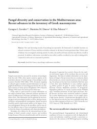
Synnemacrodictys stilboidea (J. Mena & Mercado) W.A. Baker<br />
& Morgan-Jones, Mycotaxon 110: 107, 2009. — Acrodictys<br />
stilboidea J. Mena & Mercado, in Mercado Sierra & Mena<br />
Portales, Acta Bot. Hung. 32: 190, 1986. Figs 1–2<br />
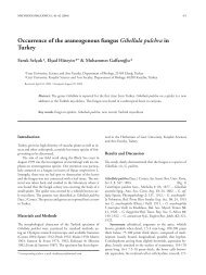
Colonies effuse, hairy, brown or dark brown. Synnemata<br />
soli tary, simple, more or less clavate, up to 180 μm long, up to<br />
Figs 1–2. Synnemacrodictys stilboidea.
118 New records of fungi, fungus-like organisms, and slime moulds from Europe and Asia: 20-27<br />
35 μm wide at the base, up to 20 μm in the middle and up to<br />
25 μm wide just beneath the apical part consisting of conidia.<br />
Conidiophores macronematous, straight or slightly flexuous,<br />
septate, olive-brown or brown. Conidiogenous cells integrated,<br />
monoblastic, determinate, cylindrical. Conidia solitary,<br />
dictyosporous, ellipsoid or turbinate, slightly constricted at<br />
the septa, 32–37 × 14–17 μm (on Ailanthus altissima) and<br />
31–39 (–40) × 14–16 μm (on Juniperus communis), smooth,<br />
olive-brown; basal cells of the conidia obconical, truncate at<br />
the base, paler than the conidial main body.<br />
Specimens examined. On bark of Ailanthus altissima (Mill.)<br />
Swingle. KOREA: Seoul, near Anam Dormitory, 18 Oct<br />
2004, V. Mel’nik (LE 226 202). On dead twigs of Juniperus<br />
chinensis L. KOREA: Seoul, near Nacheon Stadium, 18 Oct<br />
2004, V. Mel’nik (LE 226 199).<br />
Acknowledgements. The author thanks Dr. Keith Seifert for valuable<br />
comments.<br />
Gams, W., Seifert, K.A. & Morgan-Jones, G. 2009. New and validated<br />
hyphomycete taxa to resolve nomenclatural and taxonomic issues. —<br />
Mycotaxon 110: 89–108.<br />
Heredia, G., Arias, R.M. & Reyes Estebanez, M. 2000. Contribución al<br />
conocimiento de los hongos Hyphomycetes de México. — Acta Botánica<br />
Mexicana 51: 39–51.<br />
Mercado-Sierra, A. & Mena-Portales, J. 1986. Hifomicetes de Topes de Collan tes,<br />
Cuba I. Especies holoblásticas. — Acta Botanica Hungarica 32: 189–205.<br />
Vadim A. Mel’nik<br />
V.L. Komarov Botanical Institute of the Russian Academy of Sciences,<br />
2 Prof. Popov Str., St. Petersburg 197376, Russia (e-mail: vadim.melnik@mail.ru)<br />
21. Diplodia subtecta (anamorphic fungi) in Bulgaria<br />
In 2007, a new anamorphic fungus on branches of an<br />
ornamental tree of genus Acer was found. The sample is<br />
documented with microphotographs and concise description.<br />
Microscopic features in LM were observed in lactophenol.<br />
Measurements of the conidia are given in the form: min–<br />
max (mean ± standard deviation). The specimen is kept at<br />
the Mycological Collection of the Institute of Biodiversity<br />
and Ecosystem Research, Bulgarian Academy of Sciences<br />
(SOMF). Identification of the fungus is confirmed by the<br />
works of Grove (1937), Merezhko (1980), Farr et al. (1989),<br />
and Mułenko et al. (2008).<br />
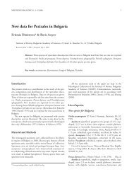
Diplodia subtecta Fr., Summa Veg. Scand., Section Post.<br />
(Stockholm): 417, 1849. Figs 3–4<br />
Pycnidia in linear rows, on branches, immersed, then<br />
erumpent, single or aggregated, globose, dark brown to black,<br />
thick-walled, 200–400 μm in diameter. Ostioles circular,<br />
papillate, 80–100 μm in diameter. Conidia at first hyaline,<br />
unicellular, cylindric with rounded apex, smooth, later<br />
becoming brown to dark brown, oblong ellipsoid, sometimes<br />
ovoid, bicellular, with distinct septum, slightly constricted<br />
at the septum, thick-walled, apex obtuse, base truncate or<br />
obtuse, 17–24 × 7.5–10 (20.0±1.9 × 8.7±1.0) μm (n = 50).<br />
Specimen examined: BULGARIA: Sofia region, Sofia,<br />
‘King Boris Garden’, on branches of Acer palmatum Thunb.,<br />
2 Oct 2007, leg. A. Pencheva (SOMF 27 939).<br />
Acknowledgements. The study was supported by a grant no. RD 56-1313/2006<br />
(Municipality of Sofia).<br />
Farr, D.F., Bills, G.F., Chamuris, G.P. & Rossman, A.Y. 1989. Fungi on plants<br />
and plant products in the United States. APS Press, Minnesota, USA.<br />
Grove, W.B. 1937. British stem- and leaf-fungi (Coelomycetes). Vol. 2.<br />
Cambridge University Press, London.<br />
Merezhko, T.A. 1980. [Flora Fungorum RSS Ucrainicae. Ordo Sphaeropsidales,<br />
familia Sphaeropsidaceae (Phaeodidymae)]. Naukova Dumka, Kiev. (In<br />
Russian)<br />
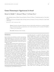
Figs 3–4. Diplodia subtecta. 3. Part of a pycnidium with young<br />
conidia. 4. Mature conidia. Scale bars = 10 μm
<strong>Mycologia</strong> <strong>Balcanica</strong> 7 (2010) 119<br />
Mułenko, W., Majewski, T & Ruszkiewicz-Michalska, M. [eds] 2008. A<br />
preliminary checklist of micromycetes in Poland. – In: Biodiversity of<br />
Poland. Vol. 9. W. Szafer Institute of Botany, Polish Academy of Sciences,<br />
Kraków.<br />
Ekaterina F. Sameva<br />
Institute of Biodiversity and Ecosystem Research,<br />
Bulg. Acad. Sci., 2 Gagarin St., 1113 Sofia, Bulgaria<br />
(e-mail: sameva@bio.bas.bg)<br />
22. Melanconis aucta (Melanconidaceae) in Bulgaria<br />
Specimens are kept at the Mycological Collection of the<br />
Institute of Biodiversity and Ecosystem Research, Bulgarian<br />
Academy of Sciences (SOMF). The asci and ascospores<br />
of the studied specimens are documented with color<br />
microphotographs from semipermanent slides made in water<br />
solution of Cotton Blue. The obtained data for the spores<br />
were examined using standard statistic methods and are<br />
presented in the brief description of the fungus in the form:<br />
(min–) mean ± standard deviation (–max), length/width ratio<br />
(min–max). The identification is confirmed by the work of<br />
Wehmeyer (1941).<br />
Melanconis aucta (Berk. & Broome) Wehm., Univ. Mich.<br />
Stud., Sci. Sér. 14: 58, 1941.<br />
Stromata 0.5–1 mm in diam, immersed, later projecting<br />
through the periderma, arising in the form of conic disc,<br />
pustulate. Ectostroma small, conic, grayish. Beaks 1–5,<br />
erumpent through the pustule, along the ruptures. Perithecia<br />
300–600 μm, globose or depressed, in groups, immersed<br />
in the tissues of the bark. Asci 85–95 × 30–35 μm, broadly<br />
ellipsoid, 8-spored. Ascospores (25.5–) 34.9±3.7 (–41) ×<br />
(10–) 11.8±1.0 (–14) μm, n = 100, l/w (2.5–3.5), hyaline, one<br />
sep tate, elongate-ellipsoid, constricted or non-constricted at<br />
the septum, with small short, hyaline spatuliform appendage<br />
at both ends; often 3-septate an d brown coloured when ripe.<br />
Specimens examined: BULGARIA: Forebalkan: Lovech<br />
distr., Golyama Zhelyazna village, near river Toplya, on bark<br />
and twigs of Alnus glutinosa (L.) Gaertn., 24 May 2009, D.Y.<br />
Stoykov (SOMF 27 278 & 27 285); Western Stara Planina<br />
Mts: along the village of Stargel, 26 Jun 2009, D.Y. Stoykov<br />
(SOMF 27 318).<br />
In Europe, Melanconis aucta is recorded on twigs of Alnus<br />
glutinosa and A. incana (L.) Moench (Betulaceae). It is similar<br />
in its morphology to M. glutinosae Z. Urb., but bears smaller<br />
ascospores and has longer and thinner asci. Wehmeyer (1941)<br />
gave smaller width of the asci (25–27 μm).<br />
Acnowledgements. Part of this study was supported by the European Social<br />
Fund, within the Operational Program “Development of the Human<br />
Resources”, Project BG051PO001/07/3.3-02/70/17.06.2008, through the<br />
Bulgarian Ministry of Education, Youth, and Science.<br />
Wehmeyer, L.E. 1941. A revison of Melanconis, Pseudovalsa, Prosthecium and<br />
Titania. — University of Michigan Studies, Scientific Series 14: 1–161.<br />
Dimitar Y. Stoykov<br />
Institute of Biodiversity and Ecosystem Research,<br />
Bulg. Acad. Sci., 2 Gagarin St., 1113 Sofia, Bulgaria<br />
(e-mail: stoikov @bio.bas.bg)<br />
23. Cantharellus amethysteus – a new for Ukraine chanterelle<br />
species<br />
The specimen is deposited in the Herbarium of the<br />
Botany Department of Biological faculty of Taras Shevchenko<br />
National University of Kyiv. The microscopic structures were<br />
inspected on dried material. Sections of hymenium and<br />
pileipellis were made at about ½ of the radius of the pileus<br />
and examined in 3 % KOH. The spores were studied in water<br />
and 3 % KOH separately.<br />
Cantharellus amethysteus (Quél.) Sacc., Syll. Fung. 5: 482,<br />
1887. — C. cibarius var. amethysteus Quél., Assoc. Fr. Avanc.<br />
Sci. Congr. Rochelle: 11: 397, 1883. Figs 5–6<br />
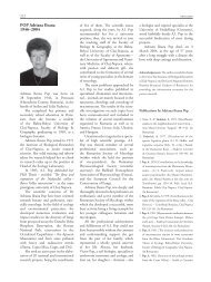
Basidiomata single or in groups of 3–5. Pileus 30–<br />
60 mm in diam., turbinate, laterally convex, applanate,<br />
then somewhat depressed, often slightly tuberous. Margin<br />
inflexed, more or less wavy, yellow, egg-yellow, pale orange<br />
to ochre, gaining a lilac or pale violaceous with vinaceous<br />
tinge pubescence. Hymenium wrinkled, venous to almost<br />
gilled, veins thick, almost gill-like, branched, decurrent,<br />
egg-yellow. Stipe 30–50 × 5–12 mm, tapering downwards,<br />
smooth, egg-yellow to pale orange, solid. Flesh white, then<br />
with yellowish tinge, with pleasant smell and taste. Spore<br />
print white with yellowish or pinkish tinge. Basidiospores<br />
smooth, ovoid, broadly ellipsoid, ellipsoid, with prominent<br />
apiculus, hyaline, (9–) 10.3±0.82 (–11.5) × (5.5–) 6.1±0.35<br />
(–6.5) μm (n = 30), ratio (1.47–) 1.71±0.19 (–2.18), filled<br />
with oil drops. Basidia elongate-clavate to elongate-cylindric,<br />
2–8-spored, mainly 4–6-spored, hyaline, up to 100 × 10<br />
μm (n = 10). Cheilo- and pleurocystidia absent. Pileipellis<br />
made up of compacted wavy hyphae up to 10.5 μm thick,<br />
flared up into a fairly loose layer of irregularly interwoven<br />
hyaline non-gelatinized hyphae up to 8.0 μm thick with<br />
clavate end cells. Stipitipellis of semi-parallel hyaline to<br />
pale stramineous hyphae, 8.0–11.0 μm thick, with some<br />
hymenial elements at stipe apex. Flesh of flexuous hyaline<br />
hyphae 2.5–5.0 μm thick. Clamp connections present in<br />
all tissues.<br />
Habitat – on soil in coniferous (Picea abies L.) forest.<br />
Specimen examined: UKRAINE: L’viv region, Stryi district,<br />
surroundings of the village Mokhnate, 48°53.608’ N,<br />
23°14.548’ E, alt. ca 672 m, 8 Sep 2009, O.O. Senchylo.
120 New records of fungi, fungus-like organisms, and slime moulds from Europe and Asia: 20-27<br />
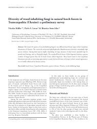
Fig. 5. Cantharellus amethysteus: a – fruitbodies; b – basidia;<br />
c – pileipellis; d – spores. Bars = 1 cm for fruitbodies and 10<br />
μm for microstructures<br />
European species. Similar fruitbodies were encountered by<br />
the first author in the Crimea in 2002 (Yalta Mountain<br />
Forest Nature Reserve, near Mount Koshka) but the<br />
specimens were not kept. Therefore, the Carpathian finding<br />
we describe in this paper represents the only known location<br />
of C. amethysteus in Ukraine. Its Crimean spread, although<br />
possible, still has to be confirmed. This is the first record of<br />
the species in Ukraine.<br />
The species is very close to Cantharellus cibarius and is<br />
treated by some authors as its variation. C. amethysteus is similar<br />
to C. cibarius in many aspects, except the lilac tinge pubescence<br />
on the cap. It is better developed in young specimens and<br />
may fade out with age. The specimens observed by one of the<br />
authors in the Crimea had the violaceous pubescence only<br />
in the middle of the pileus, while its margins remained paleorange<br />
like hymenium and stipe. The combination of yellow<br />
and lilac colours of the fruitbody is also a characteristic of<br />
C. melanoxeros Desm., but this species has pale violaceous<br />
hymenium, while its cap surface and stipe are yellow (Watling<br />
& Turnbull 1998).<br />
Interesting to note that the spores of our specimen are on<br />
average longer than those reported by other authors [8.0–10.0<br />
× 5.5–6.0 (–6.5) μm] (Watling & Turnball 1998). However,<br />
all the other features are so characteristic that leave no doubt<br />
of its identity.<br />
Acknowledgements. Authors are very grateful to O.O. Senchylo for kindly<br />
granted specimens.<br />
Watling, R. & Turnbull, E. 1998. British fungus flora. Agarics and boleti. Vol.<br />
8. Cantharellaceae, Gomphaceae and amyloid-spored and xeruloid members<br />
of Tricholomataceae (excl. Mycena). Alden Press LTD, Edinburgh.<br />
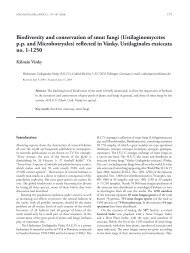
Fig. 6. The point of the record of Cantharellus amethysteus<br />
(Quél.) Sacc. in Ukraine<br />
The mushroom was found during an expedition in<br />
2009, in the Carpathian Mountains. An inspection of<br />
this specimen allowed identification of it as Cantharellus<br />
amethysteus (Cantharellales, Cantharellaceae) – a rather rare<br />
Mykola P. Prydiuk1 & Veronika V. Dzhagan2<br />
¹ Department of Mycology, M.G. Kholodny Institute of Botany,<br />
National Academy of Sciences of Ukraine,<br />
2 Tereshchenkivs’ka Street, 01001 Kyiv, Ukraine<br />
(corresponding author’s e-mail: prydiuk@gmail.com)<br />
² Botany Department, Faculty of Biology,<br />
Taras Shevchenko National University of Kyiv, 64,<br />
Volodymyrs’ka Street, 01601 Kyiv, Ukraine<br />
24. Sarcodon joeides (Bankeraceae) in Bulgaria<br />
Air dried specimens of the fungus are preserved in the<br />
Mycological Collection at the Institute of Biodiversity and<br />
Ecosystem Research, Bulgarian Academy of Sciences (SOMF).<br />
The samples are documented with colour photographs and<br />
concise description. Colour notations are in accordance with<br />
the British Fungus Flora Colour Chart (Anonymous 1969).<br />
Microscopic features are observed in water and 3 % KOH and<br />
measured in water. The measurement values for basidiospores<br />
and basidia are presented below in the following manner:<br />
(min–) mean±σ (–max); for the rest of the microscopic<br />
structures minimum and maximum values are noted. The<br />
identification is justified by Breitenbach & Kränzlin (1986).<br />
Line drawings were prepared by tracing digital photograph<br />
images on transparent paper. The SEM microphotograph is<br />
taken on JEOL JSM-6390.<br />
Sarcodon joeides (Pass.) Bataille, Bull. Soc. Mycol. Fr. 39(4):<br />
205, 1924. Figs 7–8<br />
Basidiomata usually simple or sometimes concrescent.<br />
Pileus up to 10 cm in diam, convex to flattened, sometimes<br />
slightly depressed, fawn, clay pink, vinaceous buff, pale<br />
chestnut, pale brown vinaceous, finely fibrilose or glabrous,<br />
sometimes finely cracked, without scales or exceptionally with<br />
scarse adpressed small scales. Stipe up to 5 × 1.5 cm, central<br />
or excentric, cylindric or tapering downwards, tomentose to
<strong>Mycologia</strong> <strong>Balcanica</strong> 7 (2010) 121<br />
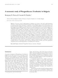
Fig. 8. SEM-micrograph of basidiospores of Sarcodon joeides<br />
Kongur chalets, under Castanea sativa Mill., 22 Sep 2009, leg.<br />
D. Stoykov, B. Assyov & I. Assyova (SOMF 27 940).<br />
Sarcodon joeides is a rarely encountered species (Hrouda<br />
2005a, b). It is easily distinguished in the field by the colour<br />
of the flesh and the habitat under broadleaf trees.<br />
Fig. 7. Microscopic features of Sarcodon joeides: a – basidiospores,<br />
b – basidia, c – con<strong>text</strong> hyphae, d – hyphae of spines.<br />
Scale bars = 10 μm<br />
fibrillose, more or less concolorous with the cap. Con<strong>text</strong><br />
vinaceous, livid vinaceous or lilac. Spines up to 4 mm long,<br />
decurrent, subulate, dirty whitish at first, then vinaceous<br />
buff to clay buff. Odour pleasant, somewhat fruity. Taste<br />
slightly bitter. Basidiospores tuberculate, (4–) 5.1±0.3 (–6)<br />
× (3.5–) 3.7±0.3 (–4.5) μm (including tubercules), ratio<br />
(1.1–) 1.4±0.1 (–1.5) (n = 30). Basidia clavate, 4-sterigmate,<br />
(32.5–) 41.7±6.6 (–57.5) × (5–) 5.9±1.2 (–7.5) μm (n = 30).<br />
Con<strong>text</strong> hyphae 10–17.5 μm wide, hyaline, thin-walled,<br />
septate, without clamp-connexions. Hyphae in spines 2.5–5<br />
μm wide, hyaline, thin-walled, sometimes branched, septate,<br />
with long segments.<br />
Specimen examined: BULGARIA: Belasitsa Mt., Petrich<br />
distr., Kongura Nature Reserve, between Belasitsa and<br />
Acknowledgements. The research of the authors on the fungal diversity<br />
of Bulgaria was sponsored by the European Social Fund, within the<br />
Operational Program “Development of the Human Resources” (project<br />
BG051PO001/07/3.3-02/70/17.06.2008), through the Bulgarian Ministry<br />
of Education, Youth and Science.<br />
Anonymous. 1969. Flora of British fungi. Colour Identification Chart. Her<br />
Majesty’s Stationery Office, Edinburgh.<br />
Breitenbach, J. & Kränzlin, F. 1986. Pilze der Schweiz. Bd. 2, Nichtblätterpilze.<br />
Verlag Mykologia, Luzern.<br />
Hrouda, P. 2005a. Bankeraceae in Central Europe. 1. — Czech Mycology<br />
57: 57–78.<br />
Hrouda, P. 2005b. Bankeraceae in Central Europe. 2. — Czech Mycology<br />
57: 279–297.<br />
Boris Assyov & Dimitar Y. Stoykov<br />
Institute of Biodiversity and Ecosystem Research,<br />
Bulg. Acad. Sci., 2 Gagarin St., 1113 Sofia, Bulgaria<br />
(corresponding author’s e-mail: bassyoff@hotmail.com)<br />
25. Pluteus salicinus (Pluteaceae) in Bulgaria<br />
Air dried specimens are preserved in the Mycological<br />
Collection at the Institute of Biodiversity and Ecosystem<br />
Research, Bulgarian Academy of Sciences (SOMF). The<br />
samples are documented with color photographs and concise<br />
description. Colour notations in the description below refer<br />
to the British Fungus Flora Colour Chart (Anonymous 1969).<br />
Microscopic features are observed and measured in water.<br />
Measurement values are presented below in the following<br />
manner: (min–) mean±σ (–max). Spore volume (Vm) is<br />
calculated according to the formula Vm=4/3π.(1/2Sw)².1/2Sl;<br />
Sl – spore length, Sw – spore width, and the result is estimated<br />
to an integer number (Breitenbach & Kränzlin 1991)<br />
Pluteus salicinus (Pers. : Fr.) P. Kumm., Führer Pilzk., p. 99,<br />
1871.<br />
Pileus up to 5 cm in diam, at first campanulate, then<br />
convex to flat, usually with a low umbo, finely radiately<br />
fibrilose, olivaceous buff, pale grey olivaceous to smoke grey,<br />
usually darker and with fine scales in the centre. Stipe up to 5
122 New records of fungi, fungus-like organisms, and slime moulds from Europe and Asia: 20-27<br />
× 0.5 cm, fibrilous, whitish, somewhat bluish green or greyish<br />
at the base. Flesh white to dirty white, more greyish below the<br />
pileipellis. Gills free, crowded, initially cream, then pale pink<br />
to clay pink at maturity. Spore print pink. Smell and taste<br />
indistinct. Basidiospores subglobose or ovoid, (6.5–) 7.3±0.4<br />
(–8) × (4.5–) 5.2±0.4 (–6) μm (n = 30), ratio (1.3–) 1.4±0.1<br />
(–1.7), spore volume (80–) 103±19 (–151) μm³. Basidia<br />
clavate, 4-spored, (22–) 25.7±2.5 (–31) × (6.5–) 7.4±0.5<br />
(–7.5) μm (n = 15). Cystidia abundant, with hooked apex,<br />
(55–) 67.2±7.1 (–82.5) × (13–) 18.0±2.9 (–24) μm (n = 20),<br />
with 2–4 hooks, 3–6 μm long. Pileipellis a cutis of septate<br />
hyphae.<br />
Specimens examined: BULGARIA: Western Stara<br />
Planina Mts, Vratsa distr., Vrachanski Balkan Nature Park,<br />
Mizhishnitsa locality, on a dead stump of a deciduous tree, 18<br />
Aug 2006, B. Assyov & D. Stoykov (SOMF 27 941); Sofia<br />
distr., Kostinbrod municipality, between Tsaritchina and<br />
Tchibaovtsi villages, on dead wood, 15 Jun 2008, B. Assyov<br />
(SOMF 27 942); Sofia city, Zapaden park, on a dead branch<br />
of a deciduous tree, 14 Oct 2008, B. Assyov (SOMF 27 943).<br />
The Bulgarian specimens correspond very well to the<br />
descriptions given by Orton (1986), Printz (1992), Citérin<br />
& Eyssartier (1998), and Heilmann-Clausen (2008). The<br />
peculiar colours of basidiomata together with the combination<br />
of hooked cystidia and cutis are diagnostic features of this<br />
species.<br />
The occurence of this species in Bulgaria is of special<br />
interest as it has been reported by Stijve & Bonnard (1986) to<br />
contain psilocybine.<br />
Acknowledgements. The research of the authors on the fungal diversity<br />
of Bulgaria was sponsored by the European Social Fund, within the<br />
Operational Program “Development of the Human Resources” (project<br />
BG051PO001/07/3.3-02/70/17.06.2008), through the Bulgarian Ministry<br />
of Education, Youth and Science.<br />
Anonymous. 1969. Flora of British fungi. Colour Identification Chart.<br />
Her Majesty’s Stationery Office, Edinburgh.<br />
Breitenbach, J. & Kränzlin, F. 1991. Pilze der Schweiz. Bd. 3/1,<br />
Röhrlinge und Blätterpilze. Verlag Mykologia, Luzern.<br />
Citérin, M. & Eyssartier, G. 1998. Clé analytique du genre Pluteus<br />
Fr. — Documents Mycologique 111: 47–67.<br />
Heilmann-Clausen, J. 2008. Pluteus Fr. — In: H. Knudsen & J.<br />
Vesterholt [eds]. Funga Nordica. Pp. 335–344. Nordsvamp,<br />
Kopenhagen.<br />
Orton, P.D. 1986. Pluteaceae: Pluteus & Volvariella. — In: D.M.<br />
Henderson, P.D. Orton & R. Watling [eds]. British fungus flora.<br />
Agarics and boleti. Vol. 4. Royal Botanic Garden Edinburgh,<br />
Edinburgh.<br />
Printz, P. 1992. Genus Pluteus Fr. — In: L. Hansen & H. Knudsen<br />
[eds]. Nordic Macromycetes. Vol. 2. Pp. 199–203. Nordsvamp,<br />
Kopenhagen.<br />
Stijve, T. & Bonnard, J. 1986. Psilocybine et ureé dans le genre Pluteus.<br />
— <strong>Mycologia</strong> Helvetica 2: 123–130.<br />
Boris Assyov & Dimitar Y. Stoykov<br />
Institute of Biodiversity and Ecosystem Research,<br />
Bulg. Acad. Sci., 2 Gagarin St., 1113 Sofia, Bulgaria<br />
(corresponding author’s e-mail: bassyoff@hotmail.com)<br />
26. Microbotryum stellariae (Microbotryaceae) in Bulgaria<br />
Microbotryum stellariae is reported as a new record for<br />
Bulgaria.<br />
For LM observations, the spores were mounted in<br />
lactophenol solution on glass slides, gently heated to boiling<br />
point and then cooled. The measurements of spores are<br />
given in the form: min–max (mean ± 1 standard deviation).<br />
For SEM, the spores were attached to specimen holders by<br />
double-sided adhesive tape and coated with gold. The surface<br />
structure of spores was observed at 15 kV and photographed<br />
with a JEOL SM-6390 scanning electron microscope.<br />
Microbotryum stellariae (Sowerby) G. Deml & Oberw.,<br />
Phytopath. Z. 104: 354, 1982. Fig. 9<br />
Sori in anthers. Spore mass powdery, dark purple. Spores<br />
globose, subglobose or broadly ellipsoidal, 5–7.5 × 4.5–<br />
7 (6.5±0.6 × 6.0±0.5) μm (n = 50); wall reticulate, meshes<br />
irregularly polygonal, 0.7–1.3 μm in diameter.<br />
Specimen examined: BULGARIA: Rila Mts, near the Rila<br />
Monastery, on Stellaria graminea L., July 2008, leg. C.M.<br />
Denchev (SOMF).<br />
Acknowledgements. The study was supported by grant no. DO 02-181/2008<br />
(Bulgarian National Science Fund).<br />
Fig. 9. Microbotryum stellariae on Stellaria graminea – a spore<br />
in SEM. Bar = 1 μm<br />
Cvetomir M. Denchev & Teodor T. Denchev<br />
Institute of Biodiversity and Ecosystem Research,<br />
Bulg. Acad. Sci., 2 Gagarin St., 1113 Sofia, Bulgaria<br />
(corresponding author’s e-mail: cmdenchev@yahoo.co.uk)
<strong>Mycologia</strong> <strong>Balcanica</strong> 7 (2010) 123<br />
27. Physarum perfectum (Physaraceae) – a new myxomycete<br />
record for the myxobiota of Turkey<br />
In August 2008, during routine field trips to different<br />
localities of Turkey, many samples of myxomycetes were<br />
collected. According to the checklists by Sesli & Denchev<br />
(2005), Dulger (2007) and Yagiz and Afyon (2007), Physarum<br />
perfectum was found to be a new record for Turkey. This<br />
taxon was identified with the aid of the work of Nannenga-<br />
Bremekamp (1991). The specimen cited is deposited in<br />
the Herbarium of Canakkale Onsekiz Mart University in<br />
Canakkale and in the first author’s personal collection.<br />
Physarum perfectum M. Peck, in Peck & Gilbert, Am. J. Bot.<br />
19: 134, 1932. Figs 10–11<br />
Sporocarps loosely gregarious, stipitate. Sporothecae<br />
grayish white, globose 0.6–0.8 mm in diam. Hypothallus very<br />
thin, colorless, widely effused. Stalk yellowish white, stout,<br />
calcareous, nearly smooth, slightly tapered upwards, 50–<br />
55 % of total height. Columella calcareous, well developed,<br />
white, conical, ca 30 % of the sporothecae. Peridium thin<br />
membrane, evenly granular with included lime and thickly<br />
sprinkled with rounded, mainly superficial, white lime scales.<br />
Capillitium moderately dense with numerous rounded or<br />
somewhat elongated, pale yellow, calcareous nodes. Spores in<br />
mass black, brown in transmitted light, minutely roughened,<br />
9–11 μm in diam.<br />
Specimen examined: TURKEY, Mersin, Camliyayla,<br />
37°14’49.21’’ N, 34°37’44.08’’ E, alt. 928 m, on dead twig of<br />
Pinus sp., 25 Aug 2008, B. Dulger (BD 659).<br />
According to Nannenga-Bremekamp (1991), this species<br />
is distinguished from Physarum murinum by the color of the<br />
stalk in transmitted light. This is ochraceous in P. perfectum<br />
and orange-brown in P. murinum. Also, in the color of the<br />
lime nodes, although the brown lime nodes of P. murinum<br />
are sometimes a very pale brown. P. globuliferum usually has<br />
a longer stalk which is often pinched and orange-brown at<br />
the base.<br />
Dulger, B. 2007. Checklist of the myxomycetes in Turkey. — <strong>Mycologia</strong><br />
<strong>Balcanica</strong> 4: 151–155.<br />
Nannenga-Bremekamp, N.E. 1991. A guide to temperate myxomycetes.<br />
Biopress Ltd., Bristol.<br />
Fig. 10. Stereomicroscopic image of the sporangia of Physarum<br />
perfectum. Scale bar = 0.5 mm<br />
Fig. 11. A view of capillitium and spores of Physarum perfectum.<br />
Scale bar = 50 μm<br />
Sesli, E. & Denchev, C.M. 2005. Checklists of the myxomycetes and<br />
macromycetes in Turkey. — <strong>Mycologia</strong> <strong>Balcanica</strong> 2: 119–160.<br />
Yagiz, D. & Afyon, A. 2007. The ecology and chorology of myxomycetes in<br />
Turkey. — Mycotaxon 101: 279–282.<br />
Basaran Dulger & Selvi Duman<br />
Department of Biology, Faculty of Science & Arts,<br />
Canakkale Onsekiz Mart University, 17020 Canakkale, Turkey<br />
(corresponding author’s e-mail: basarandulger@yahoo.com)