GREEN Form - AHP Diet Drug Settlement
GREEN Form - AHP Diet Drug Settlement
GREEN Form - AHP Diet Drug Settlement

You also want an ePaper? Increase the reach of your titles
YUMPU automatically turns print PDFs into web optimized ePapers that Google loves.
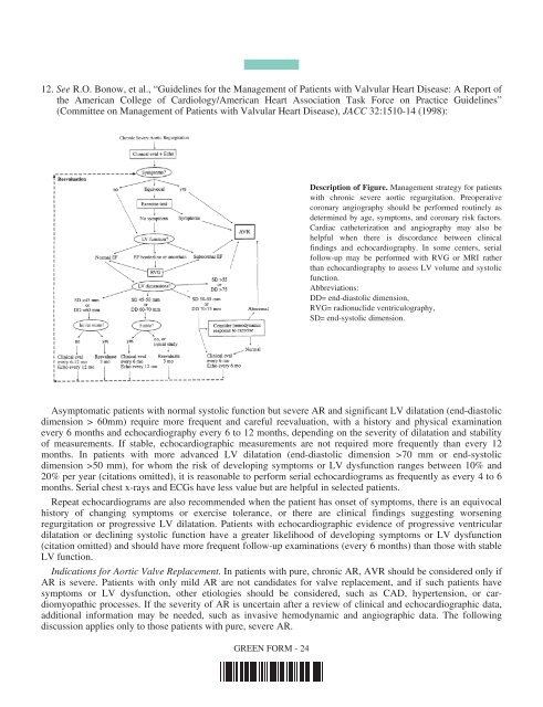
12. See R.O. Bonow, et al., “Guidelines for the Management of Patients with Valvular Heart Disease: A Report of<br />
the American College of Cardiology/American Heart Association Task Force on Practice Guidelines”<br />
(Committee on Management of Patients with Valvular Heart Disease), JACC 32:1510-14 (1998):<br />
Description of Figure. Management strategy for patients<br />
with chronic severe aortic regurgitation. Preoperative<br />
coronary angiography should be performed routinely as<br />
determined by age, symptoms, and coronary risk factors.<br />
Cardiac catheterization and angiography may also be<br />
helpful when there is discordance between clinical<br />
findings and echocardiography. In some centers, serial<br />
follow-up may be performed with RVG or MRI rather<br />
than echocardiography to assess LV volume and systolic<br />
function.<br />
Abbreviations:<br />
DD= end-diastolic dimension,<br />
RVG= radionuclide ventriculography,<br />
SD= end-systolic dimension.<br />
Asymptomatic patients with normal systolic function but severe AR and significant LV dilatation (end-diastolic<br />
dimension > 60mm) require more frequent and careful reevaluation, with a history and physical examination<br />
every 6 months and echocardiography every 6 to 12 months, depending on the severity of dilatation and stability<br />
of measurements. If stable, echocardiographic measurements are not required more frequently than every 12<br />
months. In patients with more advanced LV dilatation (end-diastolic dimension >70 mm or end-systolic<br />
dimension >50 mm), for whom the risk of developing symptoms or LV dysfunction ranges between 10% and<br />
20% per year (citations omitted), it is reasonable to perform serial echocardiograms as frequently as every 4 to 6<br />
months. Serial chest x-rays and ECGs have less value but are helpful in selected patients.<br />
Repeat echocardiograms are also recommended when the patient has onset of symptoms, there is an equivocal<br />
history of changing symptoms or exercise tolerance, or there are clinical findings suggesting worsening<br />
regurgitation or progressive LV dilatation. Patients with echocardiographic evidence of progressive ventricular<br />
dilatation or declining systolic function have a greater likelihood of developing symptoms or LV dysfunction<br />
(citation omitted) and should have more frequent follow-up examinations (every 6 months) than those with stable<br />
LV function.<br />
Indications for Aortic Valve Replacement. In patients with pure, chronic AR, AVR should be considered only if<br />
AR is severe. Patients with only mild AR are not candidates for valve replacement, and if such patients have<br />
symptoms or LV dysfunction, other etiologies should be considered, such as CAD, hypertension, or cardiomyopathic<br />
processes. If the severity of AR is uncertain after a review of clinical and echocardiographic data,<br />
additional information may be needed, such as invasive hemodynamic and angiographic data. The following<br />
discussion applies only to those patients with pure, severe AR.<br />
<strong>GREEN</strong> FORM - 24