GREEN Form - AHP Diet Drug Settlement
GREEN Form - AHP Diet Drug Settlement
GREEN Form - AHP Diet Drug Settlement
Create successful ePaper yourself
Turn your PDF publications into a flip-book with our unique Google optimized e-Paper software.
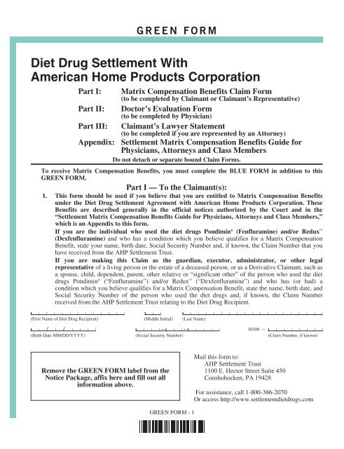
<strong>GREEN</strong> FORM<br />
<strong>Diet</strong> <strong>Drug</strong> <strong>Settlement</strong> With<br />
American Home Products Corporation<br />
Part I: Matrix Compensation Benefits Claim <strong>Form</strong><br />
(to be completed by Claimant or Claimant’s Representative)<br />
Part II: Doctor’s Evaluation <strong>Form</strong><br />
(to be completed by Physician)<br />
Part III: Claimant’s Lawyer Statement<br />
(to be completed if you are represented by an Attorney)<br />
Appendix: <strong>Settlement</strong> Matrix Compensation Benefits Guide for<br />
Physicians, Attorneys and Class Members<br />
Do not detach or separate bound Claim <strong>Form</strong>s.<br />
To receive Matrix Compensation Benefits, you must complete the BLUE FORM in addition to this<br />
<strong>GREEN</strong> FORM.<br />
Part I — To the Claimant(s):<br />
1. This form should be used if you believe that you are entitled to Matrix Compensation Benefits<br />
under the <strong>Diet</strong> <strong>Drug</strong> <strong>Settlement</strong> Agreement with American Home Products Corporation. These<br />
Benefits are described generally in the official notices authorized by the Court and in the<br />
“<strong>Settlement</strong> Matrix Compensation Benefits Guide for Physicians, Attorneys and Class Members,”<br />
which is an Appendix to this form.<br />
If you are the individual who used the diet drugs Pondimin ® (Fenfluramine) and/or Redux <br />
(Dexfenfluramine) and who has a condition which you believe qualifies for a Matrix Compensation<br />
Benefit, state your name, birth date, Social Security Number and, if known, the Claim Number that you<br />
have received from the <strong>AHP</strong> <strong>Settlement</strong> Trust.<br />
If you are making this Claim as the guardian, executor, administrator, or other legal<br />
representative of a living person or the estate of a deceased person, or as a Derivative Claimant, such as<br />
a spouse, child, dependent, parent, other relative or “significant other” of the person who used the diet<br />
drugs Pondimin ® (“Fenfluramine”) and/or Redux (“Dexfenfluramine”) and who has (or had) a<br />
condition which you believe qualifies for a Matrix Compensation Benefit, state the name, birth date, and<br />
Social Security Number of the person who used the diet drugs and, if known, the Claim Number<br />
received from the <strong>AHP</strong> <strong>Settlement</strong> Trust relating to the <strong>Diet</strong> <strong>Drug</strong> Recipient.<br />
❙ ❘ ❘ ❘ ❘ ❘ ❘ ❘ ❘ ❘ ❘ ❘ ❘ ❙ ❘ ❘ ❙ ❘ ❘ ❘ ❘ ❘ ❘ ❘ ❘ ❘ ❘ ❘ ❘ ❘ ❘ ❘ ❘ ❘ ❘<br />
(First Name of <strong>Diet</strong> <strong>Drug</strong> Recipient) (Middle Initial) (Last Name)<br />
❙ ❘ / ❘ / ❘ ❘ ❘ ❘ ❙ ❘ ❘ ❘ – ❙ ❘ ❘ – ❙ ❘ ❘ ❘ ❘ 18300 – ❙ ❘ ❘ ❘ ❘ ❘ ❘ ❘<br />
(Birth Date MM/DD/YYYY) (Social Security Number) (Claim Number, if known)<br />
Remove the <strong>GREEN</strong> FORM label from the<br />
Notice Package, affix here and fill out all<br />
information above.<br />
Mail this form to:<br />
<strong>AHP</strong> <strong>Settlement</strong> Trust<br />
1100 E. Hector Street Suite 450<br />
Conshohocken, PA 19428<br />
For assistance, call 1-800-386-2070<br />
Or access http://www.settlementdietdrugs.com<br />
<strong>GREEN</strong> FORM - 1
2. If you seek Matrix Compensation Benefits, you must complete this <strong>GREEN</strong> FORM if and when the<br />
<strong>Diet</strong> <strong>Drug</strong> Recipient has a Matrix-Level medical condition.<br />
If you have qualified for and have been paid a Matrix Compensation Benefit, then you preserved your right<br />
to receive incremental payments if the <strong>Diet</strong> <strong>Drug</strong> Recipient’s medical condition has worsened and the change<br />
places your Claim on a higher level of the payment Matrix. To seek additional payment based on a worsened<br />
medical condition, you must complete another <strong>GREEN</strong> FORM.<br />
Check the appropriate box below:<br />
❒ This is an original <strong>GREEN</strong> FORM ❒ This is a <strong>GREEN</strong> FORM seeking additional payment for a<br />
worsened medical condition.<br />
3. If you are submitting this form as the Representative of the estate of the <strong>Diet</strong> <strong>Drug</strong> Recipient, or on<br />
behalf of a <strong>Diet</strong> <strong>Drug</strong> Recipient who has become incapacitated, complete the information below:<br />
❙ ❘ ❘ ❘ ❘ ❘ ❘ ❘ ❘ ❘ ❘ ❘ ❘ ❙ ❘ ❘ ❙ ❘ ❘ ❘ ❘ ❘ ❘ ❘ ❘ ❘ ❘ ❘ ❘ ❘ ❘ ❘ ❘ ❘ ❘<br />
(First Name of Representative) (Middle Initial) (Last Name)<br />
❙ ❘ ❘ ❘ ❘ ❘ ❘ ❘ ❘ ❘ ❘ ❘ ❘ ❘ ❘ ❘ ❘ ❘ ❘ ❘ ❘ ❘ ❘ ❘ ❘ ❘ ❘ ❘ ❘ ❘ ❘ ❘ ❘ ❘ ❘ ❘ ❘<br />
(Street Address)<br />
❙ ❘ ❘ ❘ ❘ ❘ ❘ ❘ ❘ ❘ ❘ ❘ ❘ ❘ ❘ ❘ ❘ ❘ ❘ ❘ ❙ ❘ ❘ ❙ ❘ ❘ ❘ ❘ ❘ – ❙ ❘ ❘ ❘ ❘<br />
(City) (State) (Zip Code)<br />
( ❘ ❘ ) ❘ ❘ ❘ – ❘ ❘ ❘ ❘ ❘ ( ❘ ❘ ) ❘ ❘ ❘ – ❘ ❘ ❘ ❘ ❘<br />
(Daytime Area Code & Phone Number)<br />
(Evening Area Code & Phone Number)<br />
❙ ❘ ❘ ❘ ❘ ❘ ❘ ❘ ❘ ❘ ❘ ❘ ❘ ❘ ❘ ❘ ❘ ❘ ❘ ❘ ❘ ❘ ❘ ❘ ❘ ❘ ❘ ❘ ❘ ❘ ❘ ❘ ❘ ❘ ❘ ❘ ❘<br />
(E-mail Address, if any)<br />
❙ ❘ ❘ ❘ ❘ ❘ ❘ ❘ ❘ ❘ ❘ ❘ ❘ ❘ ❘ ❘ ❘ ❘ ❘ ❘ ❘ ❘ ❘ ❘ ❘ ❘ ❘ ❘ ❘ ❘ ❘ ❘ ❘ ❘ ❘ ❘ ❘<br />
(Legal Relationship to <strong>Diet</strong> <strong>Drug</strong> Recipient [trustee, power of attorney, etc.])<br />
NOTE—If you have not previously provided to the <strong>AHP</strong> <strong>Settlement</strong> Trust a copy of the court order or<br />
other document appointing you as the personal representative of the <strong>Diet</strong> <strong>Drug</strong> Recipient, you must<br />
attach or include a copy of your court approval or other authorization to represent the <strong>Diet</strong> <strong>Drug</strong> Recipient in<br />
this <strong>Settlement</strong> with your completed <strong>GREEN</strong> FORM. Check whichever box is applicable:<br />
❒ I have already provided the requested documentation previously or on another form and there is no<br />
change.<br />
❒ A copy of my court approval or other authorization to represent the <strong>Diet</strong> <strong>Drug</strong> Recipient is attached.<br />
<strong>GREEN</strong> FORM - 2
4. If you are submitting this form as a Derivative Claimant, (i.e., a spouse, parent, child, dependent,<br />
relative, or “significant other” of a <strong>Diet</strong> <strong>Drug</strong> Recipient), complete the information below:<br />
a. (NOTE—Current and correct information is required for all Derivative Claimants. If there is<br />
information for more than one Derivative Claimant, check here ❒ and then use a blank piece of<br />
paper or a photocopy of this question to provide the information for each applicable Derivative<br />
Claimant. Include that paper with this form. Be advised that a single benefit amount in<br />
accordance with Matrix A-2 or B-2 (See pages 17-18 of the Appendix) will be apportioned<br />
between all eligible Derivative Claimants.)<br />
❙ ❘ ❘ ❘ ❘ ❘ ❘ ❘ ❘ ❘ ❘ ❘ ❘ ❙ ❘ ❘ ❙ ❘ ❘ ❘ ❘ ❘ ❘ ❘ ❘ ❘ ❘ ❘ ❘ ❘ ❘ ❘ ❘ ❘ ❘<br />
(First Name) (Middle Initial) (Last Name)<br />
❙ ❘ ❘ ❘ ❘ ❘ ❘ ❘ ❘ ❘ ❘ ❘ ❘ ❘ ❘ ❘ ❘ ❘ ❘ ❘ ❘ ❘ ❘ ❘ ❘ ❘ ❘ ❘ ❘ ❘ ❘ ❘ ❘ ❘ ❘ ❘ ❘<br />
(Street Address)<br />
❙ ❘ ❘ ❘ ❘ ❘ ❘ ❘ ❘ ❘ ❘ ❘ ❘ ❘ ❘ ❘ ❘ ❘ ❘ ❘ ❙ ❘ ❘ ❙ ❘ ❘ ❘ ❘ ❘ – ❙ ❘ ❘ ❘ ❘<br />
(City) (State) (Zip Code)<br />
( ❘ ❘ ) ❘ ❘ ❘ – ❘ ❘ ❘ ❘ ❘ ( ❘ ❘ ) ❘ ❘ ❘ – ❘ ❘ ❘ ❘ ❘<br />
(Daytime Area Code & Phone Number)<br />
(Evening Area Code & Phone Number)<br />
❙ ❘ ❘ ❘ ❘ ❘ ❘ ❘ ❘ ❘ ❘ ❘ ❘ ❘ ❘ ❘ ❘ ❘ ❘ ❘ ❘ ❘ ❘ ❘ ❘ ❘ ❘ ❘ ❘ ❘ ❘ ❘ ❘ ❘ ❘ ❘ ❘<br />
(E-mail Address, if any)<br />
❙ ❘ / ❘ / ❘ ❘ ❘ ❘ ❙ ❘ ❘ ❘ – ❙ ❘ ❘ – ❙ ❘ ❘ ❘ ❘<br />
(Date of Birth MM/DD/YYYY)<br />
(Social Security Number)<br />
b. Specify the relationship of the Derivative Claimant to the <strong>Diet</strong> <strong>Drug</strong> Recipient.<br />
❒ Spouse<br />
❒ Parent<br />
❒ Child<br />
❒ Dependent, specify<br />
❒ Other relative, specify<br />
❒ Significant other, specify<br />
c. If you selected “Spouse” above, what is the current status of the relationship of the Derivative<br />
Claimant to the <strong>Diet</strong> <strong>Drug</strong> Recipient<br />
❒ Married ❒ Divorced ❒ Separated ❒ Widowed<br />
Date of the Marriage: ❙ ❘ / ❘ / ❘ ❘ ❘ ❘<br />
(MM/DD/YYYY)<br />
<strong>GREEN</strong> FORM - 3
d. If the Derivative Claimant is a Spouse who is currently estranged from the <strong>Diet</strong> <strong>Drug</strong> Recipient,<br />
state the date of separation and/or divorce.<br />
Date: ❙ ❘ / ❘ / ❘ ❘ ❘ ❘<br />
(MM/DD/YYYY)<br />
(Provide evidence of the date of separation or divorce, i.e., separation agreement or divorce decree.)<br />
e. Identify the basis on which the Derivative Claimant is claiming “derivative” benefits.<br />
❒ Loss of Consortium/Per Quod (e.g., loss of marital services and relationship)<br />
❒ Loss of Support<br />
❒ Loss of Service<br />
❒ Other, explain:<br />
NOTE: If you are completing this questionnaire as a Representative or Derivative Claimant, the following<br />
questions using the term “You” refer to the “<strong>Diet</strong> <strong>Drug</strong> Recipient.”<br />
5. Check which Matrix Level of Severity (see Appendix pages 18-21) you believe you currently qualify<br />
for:<br />
❒ Level I ❒ Level II ❒ Level III ❒ Level IV ❒ Level V<br />
6. Check which Matrix (see Appendix pages 16-17) you believe you qualify for:<br />
❒ Matrix A-1 (the full compensation Matrix) ❒ Matrix B-1 (the reduced compensation Matrix)<br />
7. State your age and the date on which you were diagnosed with the condition or experienced the event<br />
(e.g., date of surgery) which you believe qualifies you for payment at the Matrix Level set forth in the<br />
answer to Question #5:<br />
Date of diagnosis/event: ❙ ❘ / ❘ / ❘ ❘ ❘ ❘ Age at diagnosis/event:<br />
(MM/DD/YYYY)<br />
8. To the best of your knowledge, did you have the condition which you believe qualifies you for payment<br />
at the Matrix Level before you took Pondimin ® and/or Redux <br />
❒ Yes ❒ No ❒ Don’t Know<br />
9. Are you represented by any lawyer in connection with this Claim<br />
❒ Yes<br />
❒ No<br />
If you checked the box marked “Yes,” have your lawyer complete the Claimant’s Lawyer Statement (Part III,<br />
p. 15 of this <strong>GREEN</strong> FORM).<br />
10. To complete the submission of your Claim, you must provide all (a) hospital reports of the admitting<br />
history and physical examinations, (b) cardiac catheterization reports, (c) hospital discharge<br />
summaries, (d) operation or surgery reports, (e) pathology reports, and (f) the written report and a<br />
copy of the videotape or disk of the Echocardiogram results which relate to the condition for which<br />
you seek compensation.<br />
<strong>GREEN</strong> FORM - 4
In the space below, list the medical providers who have provided medical treatment related to your<br />
Claim.<br />
Name of Physician,<br />
Clinic or Hospital<br />
Address of Physician,<br />
Clinic or Hospital<br />
Date(s) of Treatment,<br />
Service or Admission<br />
❙ ❘ / ❘ / ❘ ❘ ❘ ❘<br />
(MM/DD/YYYY)<br />
❙ ❘ / ❘ / ❘ ❘ ❘ ❘<br />
(MM/DD/YYYY)<br />
❙ ❘ / ❘ / ❘ ❘ ❘ ❘<br />
(MM/DD/YYYY)<br />
If there are additional physicians, clinics or hospitals, check here ❒ and use an additional sheet to list them.<br />
Remember to include that sheet with this form.<br />
11. The undersigned hereby consent(s) to the disclosure of the information contained herein to the extent<br />
necessary to process this Claim for <strong>Settlement</strong> Benefits. Each person signing below agrees to cooperate<br />
with the <strong>AHP</strong> <strong>Settlement</strong> Trust and to provide any necessary medical record authorizations and<br />
releases for the <strong>AHP</strong> <strong>Settlement</strong> Trust to gather information needed to substantiate or audit the Claim.<br />
Each person signing below acknowledges and understands that this form is an official Court document<br />
sanctioned by the Court that presides over the <strong>Diet</strong> <strong>Drug</strong> <strong>Settlement</strong>, and submitting it to the <strong>AHP</strong><br />
<strong>Settlement</strong> Trust is equivalent to filing it with a Court. After reviewing the information which has been<br />
supplied on this form by a Board-Certified Physician (Part II) and, if applicable, by an attorney (Part<br />
III), each person declares under penalty of perjury that the information provided in this form is true<br />
and correct to the best of his/her knowledge, information and belief.<br />
(Signature of <strong>Diet</strong> <strong>Drug</strong> Recipient, if living)<br />
(Signature(s) of Legal Representative(s) of <strong>Diet</strong> <strong>Drug</strong> Recipient, if any)<br />
(Signature(s) of Derivative Claimant, i.e., Spouse, Parent, Child, Dependent, Other Relative, or<br />
“Significant Other,” if any)<br />
(Signature(s) of Derivative Claimant, i.e., Spouse, Parent, Child, Dependent, Other Relative, or<br />
“Significant Other,” if any)<br />
(Signature(s) of Derivative Claimant, i.e., Spouse, Parent, Child, Dependent, Other Relative, or<br />
“Significant Other,” if any)<br />
❙ ❘ / ❘ / ❘ ❘ ❘ ❘<br />
(MM/DD/YYYY)<br />
❙ ❘ / ❘ / ❘ ❘ ❘ ❘<br />
(MM/DD/YYYY)<br />
❙ ❘ / ❘ / ❘ ❘ ❘ ❘<br />
(MM/DD/YYYY)<br />
❙ ❘ / ❘ / ❘ ❘ ❘ ❘<br />
(MM/DD/YYYY)<br />
❙ ❘ / ❘ / ❘ ❘ ❘ ❘<br />
(MM/DD/YYYY)<br />
<strong>GREEN</strong> FORM - 5
[THIS PAGE INTENTIONALLY LEFT BLANK.]<br />
<strong>GREEN</strong> FORM - 6
Important Information to Claimants<br />
Regarding Part II of This <strong>Form</strong><br />
Part II of this form must be completed by a Board-Certified Cardiologist or a Board-Certified<br />
Cardiothoracic Surgeon. However, if the Claim is based upon the <strong>Diet</strong> <strong>Drug</strong> Recipient developing endocardial<br />
fibrosis, then you may, if you prefer, have a Board-Certified Pathologist complete Part II regarding the existence<br />
of the pathological criteria for endocardial fibrosis. If the Claim is based upon the determination of the functional<br />
outcome that a <strong>Diet</strong> <strong>Drug</strong> Recipient has or had six months after a stroke, then, if you prefer, a Board-Certified<br />
Neurologist or Board-Certified Neurosurgeon may also complete the questions in Part II of the form that concern<br />
that outcome.<br />
Part II—To the Board-Certified Physician<br />
Part I of this form identifies an individual who was prescribed and ingested the diet drugs Pondimin ®<br />
(“Fenfluramine”) and/or Redux (“Dexfenfluramine”) and who has a condition that may qualify the patient, his or<br />
her legal representatives and/or members of the family for payment as part of the Nationwide Class Action<br />
<strong>Settlement</strong> with American Home Products Corporation.<br />
A Board-Certified Cardiologist or Board-Certified Cardiothoracic Surgeon must complete Part II of this form.<br />
(The response to Question F.11 may be supplied by a Board-Certified Neurologist or Board-Certified<br />
Neurosurgeon, or based upon information supplied by such specialists. The response to Question L.6 may be<br />
supplied by a Board-Certified Pathologist, or based upon information supplied by such specialist.)<br />
In completing the form you may consider, rely upon and use the patient’s Echocardiograms, medical records and<br />
reports, hospital records or reports, the patient’s medical history or other sources of information you regularly and<br />
routinely use in your practice.<br />
Please certify below that the patient either has or does not have a given condition to a reasonable degree of<br />
medical certainty. The conditions that are relevant to the determination of this Claim are defined by reference to<br />
well-accepted, published criteria, which are excerpted in the <strong>Settlement</strong> Matrix Compensation Benefits Guide for<br />
Physicians, Attorneys and Class Members, which are set forth in the Appendix.<br />
A claimant who qualifies for a particular Matrix payment, by virtue of a properly interpreted Echocardiogram<br />
showing the required levels of regurgitation and/or complicating factors, after exposure to Pondimin ® and/or<br />
Redux , shall not be disqualified from receiving that Matrix payment if a subsequent Echocardiogram shows that<br />
the required levels of regurgitation and/or complicating factors are no longer present.<br />
A. Medical Background: What is your name, office address, and telephone number<br />
❙ ❘ ❘ ❘ ❘ ❘ ❘ ❘ ❘ ❘ ❘ ❘ ❘ ❙ ❘ ❘ ❙ ❘ ❘ ❘ ❘ ❘ ❘ ❘ ❘ ❘ ❘ ❘ ❘ ❘ ❘ ❘ ❘ ❘ ❘<br />
(First Name) (Middle Initial) (Last Name)<br />
❙ ❘ ❘ ❘ ❘ ❘ ❘ ❘ ❘ ❘ ❘ ❘ ❘ ❘ ❘ ❘ ❘ ❘ ❘ ❘ ❘ ❘ ❘ ❘ ❘ ❘ ❘ ❘ ❘ ❘ ❘ ❘ ❘ ❘ ❘ ❘ ❘<br />
(Office Address)<br />
❙ ❘ ❘ ❘ ❘ ❘ ❘ ❘ ❘ ❘ ❘ ❘ ❘ ❘ ❘ ❘ ❘ ❘ ❘ ❘ ❙ ❘ ❘ ❙ ❘ ❘ ❘ ❘ ❘ – ❙ ❘ ❘ ❘ ❘<br />
(City) (State) (Zip Code)<br />
( ❘ ❘ ) ❘ ❘ ❘ – ❘ ❘ ❘ ❘ ❘<br />
(Area Code & Telephone Number)<br />
Check whether you are:<br />
❒ A Board-Certified Cardiologist ❒ A Board-Certified Cardiothoracic Surgeon<br />
❒ Other (Must be Board-Certified)<br />
Check whether you have level 2 training in echocardiography as specified in the “Recommendations of the<br />
American Society of Echocardiography Committee on Physician Training in Echocardiography.” 1<br />
❒ Yes ❒ No<br />
1 A.S. Pearlman, et al., Guidelines for Optimal Physician Training in Echocardiography: Recommendations of the American Society of Echocardiography<br />
Committee for Physician Training in Echocardiography, 60 Am. J. Cardiol. 158-163 (1987).<br />
<strong>GREEN</strong> FORM - 7
B. Patient Information:<br />
State the name of the patient (<strong>Diet</strong> <strong>Drug</strong> Recipient) for whom you are providing the information contained in<br />
this form.<br />
❙ ❘ ❘ ❘ ❘ ❘ ❘ ❘ ❘ ❘ ❘ ❘ ❘ ❙ ❘ ❘ ❙ ❘ ❘ ❘ ❘ ❘ ❘ ❘ ❘ ❘ ❘ ❘ ❘ ❘ ❘ ❘ ❘ ❘ ❘<br />
(First Name of <strong>Diet</strong> <strong>Drug</strong> Recipient) (Middle Initial) (Last Name)<br />
C. 1. Did the above-named patient have an Echocardiogram which was conducted in accordance with the<br />
standards and criteria as outlined in Feigenbaum 2 (1994) or Weyman 3 (1994)<br />
❒ Yes ❒ No<br />
2. If the answer to Question C.1 is “Yes,” state the date when the Echocardiogram was performed.<br />
Date: ❙ ❘ / ❘ / ❘ ❘ ❘ ❘<br />
(MM/DD/YYYY)<br />
3. Based on your review of the Echocardiogram tape or disk, does the above-named <strong>Diet</strong> <strong>Drug</strong> Recipient<br />
have the following conditions as defined by Singh 4 (Check each that applies):<br />
a. For mitral regurgitation, the following determined in any apical view:<br />
❒ Mild mitral regurgitation, defined as (1) either the regurgitant jet area/left atrial area (“RJA/<br />
LAA”) ratio is more than 5% or the mitral regurgitant jet height is greater than 1 cm from the<br />
valve orifice, and (2) the RJA/LAA ratio is less than 20%.<br />
❒ Moderate mitral regurgitation, defined as regurgitant jet area in any apical view equal to or<br />
greater than 20% of the left atrial area but less than or equal to 40% (20%-40% RJA/LAA).<br />
❒ Severe mitral regurgitation, defined as > 40% RJA/LAA.<br />
❒ None of the above.<br />
b. For aortic regurgitation, the following determined in the parasternal long-axis view or in the apical<br />
long-axis view, if the parasternal long-axis view is unavailable:<br />
❒ Mild aortic regurgitation, defined as regurgitant jet diameter equal to or greater than 10% but<br />
less than 25% of the outflow tract height (10%-24% jet height (“JH”)/left ventricular outflow<br />
tract height (“LVOTH”)).<br />
❒ Moderate aortic regurgitation, defined as 25%-49% JH/LVOTH.<br />
❒ Severe aortic regurgitation, defined as > 49% JH/LVOTH.<br />
❒ None of the above.<br />
D. Based on your review of the Echocardiogram tape or disk (or the results of any cardiac catheterization<br />
or surgical examination), does the above-named <strong>Diet</strong> <strong>Drug</strong> Recipient have any of the following<br />
conditions:<br />
1. Congenital Aortic Valve Abnormalities: Unicuspid, Bicuspid or Quadricuspid aortic valve; ventricular<br />
septal defect associated with aortic regurgitation<br />
❒ Yes ❒ No<br />
2. Aortic dissection involving the aortic root and/or aortic valve<br />
❒ Yes ❒ No<br />
3. Aortic sclerosis at the time that the <strong>Diet</strong> <strong>Drug</strong> Recipient was first diagnosed with mild or greater aortic<br />
regurgitation if he or she was 60 or older at that time<br />
❒ Yes ❒ No<br />
2 H. Feigenbaum, Echocardiography 68-133 (5th ed. 1994).<br />
3 A. E. Weyman, Principles and Practice of Echocardiography 75-97 (2d ed. 1994).<br />
4 J. P. Singh, et al., Prevalence and Clinical Determinants of Mitral, Tricuspid and Aortic Regurgitation (The Framingham Heart Study), 83 Am. J. Cardiol.<br />
897-902 (1999).<br />
<strong>GREEN</strong> FORM - 8
4. Aortic root dilation >5.0 cm<br />
❒ Yes ❒ No<br />
5. Aortic stenosis with an aortic valve area 2 mm above the atrial-ventricular border during systole, and >5 mm leaflet thickening<br />
during diastole, as determined by a Board-Certified Cardiologist 5 <br />
❒ Yes ❒ No<br />
8. Chordae tendinae rupture or papillary muscle rupture, or acute myocardial infarction associated with<br />
acute mitral regurgitation<br />
❒ Yes ❒ No<br />
9. Mitral annular calcification<br />
❒ Yes ❒ No<br />
10. M-Mode and 2-D Echocardiographic evidence of rheumatic heart valves (doming of the anterior leaflet<br />
and/or anterior motion of the posterior leaflet and/or commissural fusion), except where a Board-<br />
Certified Pathologist has examined mitral valve tissue and determined that there was no evidence of<br />
rheumatic valve disease<br />
❒ Yes ❒ No<br />
E. To the best of your knowledge, has the above-named <strong>Diet</strong> <strong>Drug</strong> Recipient had the following:<br />
1. Heart valve surgery to repair or replace the mitral valve prior to Pondimin ® and/or Redux use<br />
❒ Yes ❒ No<br />
2 Heart valve surgery to repair or replace the aortic valve prior to Pondimin ® and/or Redux use<br />
❒ Yes ❒ No<br />
3. Bacterial endocarditis prior to Pondimin ® and/or Redux use<br />
❒ Yes ❒ No<br />
4. Mild or greater aortic regurgitation confirmed by echocardiography prior to Pondimin ® and/or Redux <br />
use<br />
❒ Yes ❒ No<br />
5. Moderate or greater mitral regurgitation confirmed by echocardiography prior to Pondimin ® and/or<br />
Redux use<br />
❒ Yes ❒ No<br />
6. Carcinoid tumor of a type associated with aortic and/or mitral valve lesions<br />
❒ Yes ❒ No<br />
7. History of daily use of methysergide or ergotamines for a continuous period of longer than 120 days<br />
❒ Yes ❒ No<br />
5 L.A. Freed, et al., Prevalence and Clinical Outcomes of Mitral Valve Prolapse, 341 New Eng. J. Med. 1, 2 (1999).<br />
<strong>GREEN</strong> FORM - 9
8. A diagnosis of Systemic Lupus Erythematosus and valvular regurgitation and/or abnormalities of a type<br />
associated with Systemic Lupus Erythematosus 6<br />
❒ Yes<br />
❒ No<br />
9. A diagnosis of rheumatoid arthritis and valvular regurgitation and/or abnormalities of a type associated<br />
with rheumatoid arthritis 7<br />
❒ Yes<br />
❒ No<br />
F. To the best of your knowledge, has the above-named <strong>Diet</strong> <strong>Drug</strong> Recipient developed the following<br />
conditions after the date on which the patient first used Pondimin ® and/or Redux :<br />
1. Bacterial endocarditis associated with either mild or greater aortic regurgitation and/or moderate or<br />
greater mitral regurgitation [If “Yes,” documentation supporting bacterial endocarditis must be<br />
provided.]<br />
❒ Yes ❒ No<br />
2. Pulmonary Hypertension secondary to severe aortic regurgitation with a peak systolic pulmonary<br />
pressure >40 mm Hg 8 measured by cardiac catheterization or with a peak systolic pulmonary artery<br />
pressure >45 mm Hg measured by Doppler Echocardiography, at rest, utilizing standard procedures 9,10<br />
assuming a right atrial pressure of 10 mm Hg<br />
❒ Yes ❒ No<br />
3. Pulmonary Hypertension secondary to moderate or greater mitral regurgitation with peak systolic<br />
pulmonary artery pressure >40 mm Hg measured by cardiac catheterization or with a peak systolic<br />
pulmonary artery pressure >45 mm Hg 11 measured by Doppler Echocardiography, at rest, utilizing<br />
standard procedures assuming a right atrial pressure of 10 mm Hg<br />
❒ Yes ❒ No<br />
4. Abnormal left ventricular end-systolic dimension >50 mm 12 by M-mode or 2-D echocardiography or<br />
abnormal left ventricular end-diastolic dimension >70 13 mm as measured by M-mode or 2-D<br />
echocardiography<br />
❒ Yes ❒ No<br />
5. Abnormal left atrial supero-inferior systolic dimension >5.3 cm 14 (apical four chamber view) or<br />
abnormal left atrial antero-posterior systolic dimension >4.0 cm (parasternal long-axis view) measured<br />
by 2-D directed M-mode or 2-D echocardiography with normal sinus rhythm using sites of<br />
measurement recommended by the American Society of Echocardiography 15<br />
❒ Yes<br />
❒ No<br />
6 Harrison’s Principles of Internal Medicine 1878 (14th ed. 1998).<br />
7 Id. at 1885.<br />
8 Braunwald, Heart Disease: Textbook of Cardiovascular Medicine 796-98 (1997).<br />
9 Feigenbaum, supra at 201-02.<br />
10 Chan, K-L., et al., Comparison of Three Doppler Ultrasound Methods in the Prediction of Pulmonary Artery Disease, 9 J. Am. Coll. Cardiol. 549-554<br />
(1987).<br />
11 Braunwald, supra.<br />
12 Bonow R.O., et al., Guidelines for the Management of Patients With Valvular Heart Disease: A Report of the American College of Cardiology/American<br />
Heart Association Task Force on Practice Guidelines (Committee on Management of Patients With Valvular Heart Disease), 32 J. Am. Coll. Cardiol.<br />
1510-14 (1998).<br />
13 Id.<br />
14 Weyman, supra at 1290-1292.<br />
15 Henry, W.L. et al., Report of the American Society of Echocardiography Committee on Nomenclature and Standards in Two-dimensional<br />
Echocardiography, 62 Circulation 212-17 (1980).<br />
<strong>GREEN</strong> FORM - 10
6. Abnormal left ventricular end-systolic dimension greater than or equal to 45 mm 16 by M-mode or 2-D<br />
Echocardiogram<br />
❒ Yes ❒ No<br />
7. Arrhythmias, defined as chronic atrial fibrillation/flutter that cannot be converted to normal sinus<br />
rhythm, or atrial fibrillation/flutter requiring ongoing medical therapy, either of which are associated<br />
with left atrial enlargement (Abnormal left atrial supero-inferior systolic dimension >5.3 cm 17 (apical<br />
four chamber view) or abnormal left atrial antero-posterior systolic dimension >4.0 cm (parasternal<br />
long-axis view) measured by 2-D directed M-mode or 2-D echocardiography.)<br />
❒ Yes ❒ No<br />
8. Ejection fractions as follows: 18<br />
50% – 60% ❒ Yes ❒ No 30% – 34% ❒ Yes ❒ No<br />
40% – 49% ❒ Yes ❒ No
12. A peripheral embolus due to bacterial endocarditis and/or as a consequence of atrial fibrillation with left<br />
atrial enlargement as defined above which resulted in:<br />
a. Severe impairment to the kidneys, defined as chronic severe renal failure requiring hemodialysis or<br />
Continuous Abdominal Peritoneal Dialysis for more than six months.<br />
❒ Yes ❒ No<br />
b. Severe impairment to the abdominal organs, defined as impairment requiring intra-abdominal<br />
surgery.<br />
❒ Yes ❒ No<br />
c. Severe impairment to the extremities, defined as impairment requiring amputation of a major limb.<br />
❒ Yes ❒ No<br />
G. Does the above-named <strong>Diet</strong> <strong>Drug</strong> Recipient have New York Heart Association Functional Class<br />
symptoms as follows:<br />
1. Class I ❒ Yes ❒ No 3. Class III ❒ Yes ❒ No<br />
2. Class II ❒ Yes ❒ No 4. Class IV ❒ Yes ❒ No<br />
If the individual has such symptoms, supply documentation of these symptoms as documented by the<br />
attending Board-Certified Cardiothoracic Surgeon or Board-Certified Cardiologist.<br />
H. Did the above-named <strong>Diet</strong> <strong>Drug</strong> Recipient have valvular repair or replacement surgery and have one<br />
or more of the following complications either during surgery, within 30 days after surgery, or during<br />
the same hospital stay as surgery:<br />
1. Renal failure, defined as chronic, severe renal failure requiring regular hemodialysis or Continuous<br />
Abdominal Peritoneal Dialysis (CAPD) for greater than six months following aortic and/or mitral valve<br />
replacement surgery<br />
❒ Yes ❒ No<br />
2. Peripheral embolus following surgery resulting in severe permanent impairment of the kidneys,<br />
abdominal organs, or extremities NOTE: Severe permanent impairment of the kidneys means chronic<br />
severe renal failure requiring hemodialysis or continuous abdominal peritoneal dialysis for more than<br />
six months. Severe impairment of the abdominal organs means impairment requiring intra-abdominal<br />
surgery. Severe impairment of the extremities means impairment requiring amputation of a major limb.<br />
❒ Yes ❒ No<br />
3. Quadriplegia or paraplegia resulting from cervical spine injury during valvular heart surgery<br />
❒ Yes ❒ No<br />
I. Did the above-named <strong>Diet</strong> <strong>Drug</strong> Recipient have valve repair or replacement surgery and have:<br />
1. Post-operative endocarditis, mediastinitis or sternal osteomyelitis, any of which required reopening of<br />
the median sternotomy for treatment<br />
❒ Yes ❒ No<br />
2. A post-operative serious infection defined as HIV or Hepatitis C within six months of surgery as a result<br />
of blood transfusion associated with the surgery<br />
❒ Yes ❒ No<br />
<strong>GREEN</strong> FORM - 12
J. Did the above-named <strong>Diet</strong> <strong>Drug</strong> Recipient have valvular repair or replacement surgery and require a<br />
second surgery through the sternum within 18 months of the initial surgery due to prosthetic valve<br />
malfunction, poor fit, or complications reasonably related to the initial surgery<br />
❒ Yes<br />
❒ No<br />
K. Did the above-named <strong>Diet</strong> <strong>Drug</strong> Recipient have valvular repair or replacement surgery and have a left<br />
ventricular ejection fraction of < 40% at any time six months or later after the valvular repair or<br />
replacement surgery<br />
❒ Yes<br />
❒ No<br />
If your answer to Question K was “Yes,” an Echocardiogram report and Echocardiogram tape or disk<br />
performed and interpreted in accordance with the standards and criteria outlined in Question C.1<br />
above must be furnished.<br />
L. Did the above-named <strong>Diet</strong> <strong>Drug</strong> Recipient have one or more of the following:<br />
1. A heart transplant<br />
❒ Yes<br />
❒ No<br />
2. Irreversible pulmonary hypertension secondary to valvular heart disease defined as peak-systolic<br />
pulmonary artery pressure >50 mm Hg 22 (by cardiac catheterization), at rest, following repair or<br />
replacement surgery of the aortic and/or mitral valve(s)<br />
❒ Yes<br />
❒ No<br />
3. A persistent non-cognitive state 23 caused by a complication of valvular heart disease (e.g., cardiac<br />
arrest) or valvular repair/replacement surgery<br />
❒ Yes<br />
❒ No<br />
If the individual has such a condition, supply a detailed statement of the attending Board-<br />
Certified Cardiologist or Board-Certified Cardiothoracic Surgeon supported by medical records<br />
setting forth the basis for your opinion that the persistent non-cognitive state was caused by a<br />
complication of valvular heart disease or valvular repair/replacement surgery.<br />
4. Death resulting from a condition caused by valvular heart disease or valvular repair/replacement<br />
surgery<br />
❒ Yes<br />
❒ No<br />
Supply a detailed statement of the attending Board-Certified Cardiologist or Board-Certified<br />
Cardiothoracic Surgeon supported by medical records setting forth your opinion that the<br />
patient’s death resulted from a condition caused by valvular heart disease and/or valvular repair/<br />
replacement surgery.<br />
5. Ventricular fibrillation or sustained ventricular tachycardia which results in hemodynamic compromise<br />
❒ Yes<br />
❒ No<br />
22 Braunwald, supra at 596-98.<br />
23 Adelman, G., Encyclopedia of Neuroscience 268 (1987).<br />
<strong>GREEN</strong> FORM - 13
6. Endocardial Fibrosis<br />
a. Diagnosed by<br />
1) Endomyocardial biopsy that demonstrates fibrosis and a cardiac catheterization that<br />
demonstrates restrictive cardiomyopathy or<br />
2) Autopsy that demonstrates endocardial fibrosis; AND<br />
b. Other causes of endocardial fibrosis have been excluded, such as: dilated cardiomyopathy,<br />
myocardial infarction, amyloid, Loeffler’s endocarditis, endomyocardial fibrosis as defined in<br />
Braunwald (involving one or both ventricles, commonly involving the chordae tendineae, with<br />
partial obliteration of either ventricle commonly present), 24 focal fibrosis secondary to valvular<br />
regurgitation, e.g. “jet lesions,” fibrosis secondary to catheter instrumentation, and hypertrophic<br />
cardiomyopathy with septal fibrosis<br />
❒ Yes<br />
❒ No<br />
This form is an official Court document sanctioned by the Court that presides over the <strong>Diet</strong> <strong>Drug</strong><br />
<strong>Settlement</strong> and submitting it to the <strong>AHP</strong> <strong>Settlement</strong> Trust is equivalent to filing it with a Court. I<br />
declare under penalty of perjury that the information provided in this form is correct to the best<br />
of my knowledge, information and belief.<br />
❙ ❘ / ❘ / ❘ ❘ ❘ ❘<br />
(Date: MM/DD/YYYY)<br />
(Signature of Board-Certified Physician)<br />
For Use With Written Statements<br />
24 Braunwald, supra at 1433-34.<br />
<strong>GREEN</strong> FORM - 14
Part III — Claimant’s Lawyer Statement<br />
If you checked the box marked “Yes” in Part I, Question #9, have your lawyer complete this statement and<br />
submit it with this <strong>GREEN</strong> FORM.<br />
1. Provide the following information about “Your Client”:<br />
❙ ❘ ❘ ❘ ❘ ❘ ❘ ❘ ❘ ❘ ❘ ❘ ❘ ❙ ❘ ❘ ❙ ❘ ❘ ❘ ❘ ❘ ❘ ❘ ❘ ❘ ❘ ❘ ❘ ❘ ❘ ❘ ❘ ❘ ❘<br />
(First Name of Your Client) (Middle Initial) (Last Name)<br />
2. Provide the following information about yourself:<br />
❙ ❘ ❘ ❘ ❘ ❘ ❘ ❘ ❘ ❘ ❘ ❘ ❘ ❘ ❘ ❘ ❘ ❘ ❘ ❘ ❘ ❘ ❘ ❘ ❘ ❘ ❘ ❘ ❘ ❘ ❘ ❘ ❘ ❘ ❘ ❘ ❘<br />
(Law Firn Name)<br />
❙ ❘ ❘ ❘ ❘ ❘ ❘ ❘ ❘ ❘ ❘ ❘ ❘ ❙ ❘ ❘ ❙ ❘ ❘ ❘ ❘ ❘ ❘ ❘ ❘ ❘ ❘ ❘ ❘ ❘ ❘ ❘ ❘ ❘ ❘<br />
(First Name of Attorney) (Middle Initial) (Last Name)<br />
❙ ❘ ❘ ❘ ❘ ❘ ❘ ❘ ❘ ❘ ❘ ❘ ❘ ❘ ❘ ❘ ❘ ❘ ❘ ❘ ❘ ❘ ❘ ❘ ❘ ❘ ❘ ❘ ❘ ❘ ❘ ❘ ❘ ❘ ❘ ❘ ❘<br />
(Street Address)<br />
❙ ❘ ❘ ❘ ❘ ❘ ❘ ❘ ❘ ❘ ❘ ❘ ❘ ❘ ❘ ❘ ❘ ❘ ❘ ❘ ❙ ❘ ❘ ❙ ❘ ❘ ❘ ❘ ❘ – ❙ ❘ ❘ ❘ ❘<br />
(City) (State) (Zip Code)<br />
( ❘ ❘ ) ❘ ❘ ❘ – ❘ ❘ ❘ ❘ ❘ ( ❘ ❘ ) ❘ ❘ ❘ – ❘ ❘ ❘ ❘ ❘<br />
(Daytime Area Code & Phone Number)<br />
(Fax Area Code & Number)<br />
❙ ❘ ❘ ❘ ❘ ❘ ❘ ❘ ❘ ❘ ❘ ❘ ❘ ❘ ❘ ❘ ❘ ❘ ❘ ❘ ❘ ❘ ❘ ❘ ❘ ❘ ❘ ❘ ❘ ❘ ❘ ❘ ❘ ❘ ❘ ❘ ❘<br />
(E-mail Address, if any)<br />
3. Include a copy of the contingency fee agreement between yourself and Your Client.<br />
4. State the amount of out-of-pocket costs incurred by you in your representation of Your Client for<br />
his/her diet drug claim. (Include a copy of your cost sheet with this form.) $<br />
5. Has a subrogation lien or claim been asserted with respect to Your Client’s right to receive benefits<br />
under the <strong>Diet</strong> <strong>Drug</strong> <strong>Settlement</strong> ❒ Yes ❒ No<br />
If your answer is “Yes,” identify by whom and the amount: $<br />
❙ ❘ ❘ ❘ ❘ ❘ ❘ ❘ ❘ ❘ ❘ ❘ ❘ ❘ ❘ ❘ ❘ ❘ ❘ ❘ ❘ ❘ ❘ ❘ ❘ ❘ ❘ ❘ ❘ ❘ ❘ ❘ ❘ ❘ ❘ ❘ ❘<br />
(Name of Subrogee)<br />
❙ ❘ ❘ ❘ ❘ ❘ ❘ ❘ ❘ ❘ ❘ ❘ ❘ ❘ ❘ ❘ ❘ ❘ ❘ ❘ ❘ ❘ ❘ ❘ ❘ ❘ ❘ ❘ ❘ ❘ ❘ ❘ ❘ ❘ ❘ ❘ ❘<br />
(Address)<br />
❙ ❘ ❘ ❘ ❘ ❘ ❘ ❘ ❘ ❘ ❘ ❘ ❘ ❘ ❘ ❘ ❘ ❘ ❘ ❘ ❙ ❘ ❘ ❙ ❘ ❘ ❘ ❘ ❘ – ❙ ❘ ❘ ❘ ❘<br />
(City) (State) (Zip Code)<br />
Does the Claimant contest the lien ❒ Yes ❒ No<br />
If yes, describe the lien and the basis for the contest on a separate sheet and include it with this form.<br />
This form is an official Court document sanctioned by the Court that presides over the <strong>Diet</strong> <strong>Drug</strong><br />
<strong>Settlement</strong>, and submitting it to the <strong>AHP</strong> <strong>Settlement</strong> Trust is equivalent to filing it with a Court. I<br />
declare under penalty of perjury that all of the information provided in this form is true and correct to<br />
the best of my knowledge, information and belief.<br />
(Attorney’s Signature)<br />
❙ ❘ / ❘ / ❘ ❘ ❘ ❘<br />
(Date MM/DD/YYYY)<br />
For assistance call 1-800-386-2070, or access the <strong>AHP</strong> <strong>Settlement</strong> Trust website at http://www.settlementdietdrugs.com.<br />
<strong>GREEN</strong> FORM - 15
Appendix to <strong>GREEN</strong> FORM<br />
<strong>Diet</strong> <strong>Drug</strong> <strong>Settlement</strong> With<br />
American Home Products Corporation<br />
<strong>Settlement</strong> Matrix Compensation Benefits Guide<br />
for Physicians, Attorneys and Class Members<br />
A. A Nationwide Class Action <strong>Settlement</strong> has been reached with American Home Products Corporation, which<br />
will resolve the claims of individuals who took the diet drugs Pondimin ® and/or Redux .<br />
B. Under the <strong>Settlement</strong>, patients who took the diet drugs Pondimin ® and/or Redux have a right to receive<br />
compensation if they have developed serious levels of valvular heart disease.<br />
C. The amounts which individuals are entitled to recover under this <strong>Settlement</strong> depend on the person’s age at<br />
diagnosis of valvular heart disease, the person’s “Level of Severity” and additional criteria as set forth<br />
below. Payments will be made according to these “Matrices”:<br />
Matrix A-1<br />
Age at diagnosis/event<br />
Severity ≤ 24 25-29 30-34 35-39 40-44 45-49 50-54 55-59 60-64 65-69 70-79<br />
I $123,750 $117,563 $111,685 $106,100 $100,795 $95,755 $90,967 $86,419 $82,098 $73,888 $36,944<br />
II $643,500 $611,325 $580,759 $551,721 $524,135 $497,928 $473,032 $449,381 $426,912 $384,221 $192,111<br />
III $940,500 $893,475 $848,801 $806,361 $766,043 $727,741 $691,354 $656,786 $623,947 $561,552 $280,776<br />
IV $1,336,500 $1,269,675 $1,206,191 $1,145,881 $1,088,587 $1,034,158 $982,450 $933,327 $886,661 $797,995 $398,998<br />
V $1,485,000 $1,410,750 $1,340,213 $1,273,202 $1,209,542 $1,149,065 $1,091,612 $1,037,031 $985,180 $886,662 $443,331<br />
Matrix B-1<br />
Age at diagnosis/event<br />
Severity ≤ 24 25-29 30-34 35-39 40-44 45-49 50-54 55-59 60-64 65-69 70-79<br />
I $24,750 $23,513 $22,337 $21,221 $20,159 $19,152 $18,194 $17,284 $16,420 $14,778 $7,389<br />
II $128,700 $122,265 $116,152 $110,344 $104,827 $99,586 $94,606 $89,876 $85,383 $76,844 $38,422<br />
III $188,100 $178,695 $169,760 $161,272 $153,208 $145,548 $138,270 $131,357 $124,790 $112,310 $56,155<br />
IV $267,300 $253,935 $241,238 $229,176 $217,717 $206,831 $196,489 $186,665 $177,332 $159,599 $79,800<br />
V $297,000 $282,150 $268,043 $254,641 $241,908 $229,813 $218,322 $207,406 $197,036 $177,332 $88,666<br />
D. The circumstances which determine whether “Matrix A-1” or “Matrix B-1” is applicable are as follows:<br />
1. For Matrix A-1: <strong>Diet</strong> <strong>Drug</strong> Recipients who ingested Pondimin ® and/or Redux for 61 or more days,<br />
who were diagnosed as FDA Positive, whose conditions are eligible for matrix payments but who do<br />
not have any condition or circumstance which makes Matrix B-1 applicable, receive payments on<br />
Matrix A-1.<br />
2. For Matrix B-1: <strong>Diet</strong> <strong>Drug</strong> Recipients who are eligible for matrix payments and to whom one or more<br />
of the following conditions apply, receive payments on Matrix B-1:<br />
Š For claims as to the mitral valve, <strong>Diet</strong> <strong>Drug</strong> Recipients who were diagnosed as having Mild Mitral<br />
Regurgitation (regardless of the duration of ingestion of Pondimin ® and/or Redux ).<br />
<strong>GREEN</strong> FORM - 16
Š <strong>Diet</strong> <strong>Drug</strong> Recipients who ingested Pondimin ® and/or Redux for 60 days or less, who were diagnosed<br />
as FDA Positive.<br />
Š <strong>Diet</strong> <strong>Drug</strong> Recipients who ingested Pondimin ® and/or Redux for 61 or more days, who were<br />
diagnosed as FDA Positive with any of the following conditions:<br />
With respect to an aortic valve claim:<br />
Š The following congenital aortic valve abnormalities: unicuspid, bicuspid or quadricuspid valves,<br />
ventricular septal defect associated with aortic regurgitation;<br />
Š Aortic dissection involving the aortic root and/or aortic valve;<br />
Š Aortic sclerosis in people who are ≥ 60 years old as of the time they are first diagnosed as FDA<br />
Positive;<br />
Š Aortic root dilatation >5.0 cm;<br />
Š Aortic stenosis with an aortic valve area 2mm above the<br />
atrial-ventricular border during systole, and >5mm leaflet thickening during diastole, as determined<br />
by a Board-Certified Cardiologist.<br />
Š Chordae tendineae rupture or papillary muscle rupture; or acute myocardial infarction associated with<br />
acute mitral regurgitation;<br />
Š Mitral annular calcification;<br />
Š M-Mode and 2-D Echocardiographic evidence of rheumatic mitral valves (doming of the anterior<br />
leaflet and/or anterior motion of the posterior leaflet and/or commissural fusion), except where there<br />
is no evidence of rheumatic valve disease upon pathological examination of mitral valve tissue.<br />
With respect to claims for the aortic and/or mitral valve(s):<br />
Š Heart valve surgery prior to Pondimin ® and/or Redux use on the valve that is the basis of claim;<br />
Š Bacterial endocarditis prior to Pondimin ® and/or Redux use;<br />
Š FDA Positive regurgitation (confirmed by Echocardiogram) prior to Pondimin ® and/or Redux use for<br />
the valve that is the basis of claim;<br />
Š Systemic Lupus Erythematosus or Rheumatoid Arthritis 1 and valvular regurgitation and/or valvular<br />
abnormalities of a type associated with those conditions 2 ;<br />
Š Carcinoid tumor of a type associated with aortic and/or mitral valve lesions;<br />
Š History of daily use of methysergide or ergotamines for a continuous period of longer than 120 days.<br />
E. <strong>Diet</strong> <strong>Drug</strong> Recipients’ spouses, children and “significant others” (“Derivative Claimants”) may also be<br />
eligible for Matrix Payments under the law, and if so, they will be paid an amount set forth in one of<br />
“Derivative Matrices”— Matrix A-2 or Matrix B-2. Derivative Claimants will be paid at the same “Level of<br />
Severity” and age at diagnosis as the <strong>Diet</strong> <strong>Drug</strong> Recipient. Matrix A-2 will be used where the <strong>Diet</strong> <strong>Drug</strong><br />
Recipient was eligible for Matrix A-1 payments and Matrix B-2 will be used where the <strong>Diet</strong> <strong>Drug</strong> Recipient<br />
was eligible for Matrix B-1 payments.<br />
<strong>GREEN</strong> FORM - 17
Matrix A-2<br />
Age at diagnosis/event<br />
Severity ≤ 24 25-29 30-34 35-39 40-44 45-49 50-54 55-59 60-64 65-69 70-79<br />
I $1,250 $1,187 $1,128 $1,072 $1,018 $967 $919 $873 $829 $739 $500<br />
II $6,500 $6,175 $5,866 $5,573 $5,294 $5,030 $4,778 $4,539 $4,312 $3,842 $1,921<br />
III $9,500 $9,025 $8,574 $8,145 $7,738 $7,351 $6,983 $6,634 $6,302 $5,616 $2,808<br />
IV $13,500 $12,825 $12,184 $11,575 $10,996 $10,446 $9,924 $9,428 $8,956 $7,980 $3,990<br />
V $15,000 $14,250 $13,537 $12,861 $12,218 $11,607 $11,026 $10,475 $9,951 $8,867 $4,433<br />
Matrix B-2<br />
Age at diagnosis/event<br />
Severity ≤ 24 25-29 30-34 35-39 40-44 45-49 50-54 55-59 60-64 65-69 70-79<br />
I $500 $500 $500 $500 $500 $500 $500 $500 $500 $500 $500<br />
II $1,300 $1,235 $1,173 $1,115 $1,059 $1,006 $956 $908 $862 $768 $500<br />
III $1,900 $1,805 $1,715 $1,629 $1,548 $1,470 $1,397 $1,327 $1,260 $1,123 $562<br />
IV $2,700 $2,565 $2,437 $2,315 $2,199 $2,089 $1,985 $1,885 $1,791 $1,596 $798<br />
V $3,000 $2,850 $2,707 $2,572 $2,444 $2,321 $2,205 $2,095 $1,990 $1,773 $886<br />
F. Under the matrices, the “Levels of Severity” which qualify <strong>Diet</strong> <strong>Drug</strong> Recipients for recovery on the<br />
<strong>Settlement</strong> matrices are as follows:<br />
(1) Matrix Level I is severe left sided valvular heart disease without complicating factors, and is defined as<br />
one of the following:<br />
(a) Severe aortic regurgitation (AR) > 49% jet height/left ventricular outflow tract height (JH/<br />
LVOTH) 3 and/or severe mitral regurgitation (MR) > 40% regurgitant jet area/left atrial area (RJA/<br />
LAA) 4,5 and no complicating factors as defined below;<br />
(b) FDA Positive valvular regurgitation 6 with bacterial endocarditis contracted after commencement of<br />
Pondimin ® and/or Redux use.<br />
(2) Matrix Level II is left sided valvular heart disease with complicating factors, and is defined as:<br />
(a) Moderate AR (25%–49% JH/LVOTH) 7 or Severe AR (> 49% JH/LVOTH) 8 with one or more of<br />
the following:<br />
i) Pulmonary hypertension secondary to severe aortic regurgitation with a peak systolic<br />
pulmonary artery pressure > 40 mm Hg measured by cardiac catheterization or with a peak<br />
systolic pulmonary artery pressure > 45 mm Hg 9 measured by Doppler Echocardiography, at<br />
rest, utilizing standard procedures 10,11 assuming a right atrial pressure of 10 mm Hg;<br />
ii) Abnormal left ventricular end-systolic dimension > 50 mm 12 by M-mode or 2-D<br />
Echocardiography or abnormal left ventricular end-diastolic dimension > 70 mm 13 as<br />
measured by M-mode or 2-D Echocardiography;<br />
iii) Ejection fraction of < 50% 14 ; and/or<br />
(b) Moderate MR (20%–40% RJA/LAA) 15 or Severe MR (> 40% RJA/LAA) 16 with one or more of the<br />
following:<br />
i) Pulmonary hypertension secondary to valvular heart disease with peak systolic pulmonary<br />
artery pressure > 40 mm Hg measured by cardiac catheterization or with a peak systolic<br />
pulmonary artery pressure > 45 mm Hg 17 measured by Doppler Echocardiography, at rest,<br />
utilizing the procedures described in Section F.2.(a)(i);<br />
ii)<br />
Abnormal left atrial supero-inferior systolic dimension > 5.3 cm 18 (apical four chamber view)<br />
or abnormal left atrial antero-posterior systolic dimension > 4.0 cm (parasternal long axis<br />
view) measured by 2-D directed M-mode or 2-D echocardiography with normal sinus rhythm<br />
using sites of measurement recommended by the American Society of Echocardiography 19 ;<br />
iii) Abnormal left ventricular end-systolic dimension ≥ 45 mm 20 by M-mode or 2-D<br />
Echocardiogram;<br />
<strong>GREEN</strong> FORM - 18
iv) Ejection fraction of ≤ 60% 21 .<br />
v) Arrhythmias, defined as chronic atrial fibrillation/flutter that cannot be converted to normal<br />
sinus rhythm, or atrial fibrillation/flutter requiring ongoing medical therapy, either of which<br />
are associated with left atrial enlargement; as defined in Section F.2.(b)(ii).<br />
(3) Matrix Level III is left sided valvular heart disease requiring surgery or conditions of equal severity,<br />
and is defined as:<br />
(a) Surgery to repair or replace the aortic and/or mitral valve(s) following the use of Pondimin ® and/or<br />
Redux ;or<br />
(b) Severe regurgitation and the presence of ACC/AHA Class I indications for surgery to repair or<br />
replace the aortic 22 and/or mitral 23 valve(s) and a statement from the attending Board Certified<br />
Cardiothoracic Surgeon or Board Certified Cardiologist supported by medical records regarding<br />
the recommendations made to the patient concerning valvular surgery, with the reason why the<br />
surgery is not being performed; or<br />
(c) Qualification for payment at Matrix Level I(b) (as described in Section F.1.b. above) or Matrix<br />
Level II and, in addition, a stroke due to bacterial endocarditis contracted after use of Pondimin ®<br />
and/or Redux or as a consequence of chronic atrial fibrillation with left atrial enlargement as<br />
defined in Section F.2.(b)(ii) which results in a permanent condition which meets the criteria of<br />
AHA Stroke Outcome Classification 24 Functional Level II, determined six months after the event.<br />
(4) Matrix Level IV is defined as follows:<br />
(a) Qualification for payment at Matrix Level I(b) (as described in Section F.1.b. above), II or III and,<br />
in addition, a stroke due to bacterial endocarditis contracted after use of Pondimin ® and/or Redux <br />
or as a consequence of chronic atrial fibrillation with left atrial enlargement as defined in Section<br />
F.2.(b)(ii) which results in a permanent condition which meets the criteria of AHA Stroke<br />
Outcome Classification 25 Functional Level III, determined six months after the event; or<br />
(b) Qualification for payment at Matrix Level I(b), II, or III and, in addition, a peripheral embolus due<br />
to Bacterial Endocarditis contracted after use of Pondimin ® and/or Redux or as a consequence of<br />
atrial fibrillation with left atrial enlargement as defined in Section F.2.(b)(ii) which results in<br />
severe permanent impairment to the kidneys, abdominal organs, or extremities, where severe<br />
permanent impairment means:<br />
i) for the kidneys, chronic severe renal failure requiring hemodialysis or Continuous Abdominal<br />
Peritoneal Dialysis for more than six months;<br />
ii) for the abdominal organs, impairment requiring intra-abdominal surgery;<br />
iii) for the extremities, impairment requiring amputation of a major limb; or<br />
(c) The individual has the following:<br />
i) Qualification for payment at Matrix Level III; and<br />
ii) New York Heart Association Functional Class I or Class II symptoms as documented by the<br />
attending Board Certified Cardiothoracic Surgeon or Board-Certified Cardiologist; and<br />
iii) Valvular repair and replacement surgery or ineligibility for surgery due to medical reasons as<br />
documented by the attending Board-Certified Cardiothoracic Surgeon or Board-Certified<br />
Cardiologist; and<br />
iv) Significant damage to the heart muscle, defined as: (a) a left ventricular ejection fraction<br />
< 30% with aortic regurgitation or a left ventricular ejection fraction < 35% with mitral<br />
regurgitation in patients who have not had surgery and meet the criteria of Section F.3.(b) or<br />
(b) a left ventricular ejection fraction < 40% six months after valvular repair or replacement<br />
surgery in patients who have had such surgery; or<br />
<strong>GREEN</strong> FORM - 19
(d)<br />
(e)<br />
(f)<br />
(g)<br />
The individual has had valvular repair or replacement surgery and has one or more of the following<br />
complications which occur either during surgery, within 30 days after surgery, or during the same<br />
hospital stay as the surgery:<br />
i) Renal failure, defined as chronic severe renal failure requiring regular hemodialysis or<br />
Continuous Abdominal Peritoneal Dialysis for greater than six months following aortic and/or<br />
mitral valve replacement surgery;<br />
ii) Peripheral embolus following surgery resulting in severe permanent impairment to the<br />
kidneys, abdominal organs, or extremities;<br />
iii) Quadriplegia or paraplegia resulting from cervical spine injury during valvular heart surgery;<br />
or<br />
A stroke caused by aortic and/or mitral valve surgery and the stroke has produced a permanent<br />
condition which meets the criteria of the AHA Stroke Outcome Functional Levels II or III<br />
determined six months after the event. 26<br />
The individual has had valvular repair or replacement surgery and suffers from post operative<br />
endocarditis, mediastinitis or sternal osteomyelitis, either of which requires reopening the median<br />
sternotomy for treatment, or a post-operative serious infection defined as HIV or Hepatitis C<br />
within six months of surgery as a result of blood transfusion associated with the heart valve<br />
surgery.<br />
The individual has had valvular repair or replacement surgery and requires a second surgery<br />
through the sternum within 18 months of the initial surgery due to prosthetic valve malfunction,<br />
poor fit, or complications reasonably related to the initial surgery.<br />
(5) Matrix Level V is defined as:<br />
(a)<br />
(b)<br />
Endocardial Fibrosis (A) diagnosed by (1) endomyocardial biopsy that demonstrates fibrosis and<br />
cardiac catheterization that demonstrates restrictive cardiomyopathy or (2) autopsy that<br />
demonstrates endocardial fibrosis and (B) other causes, including dilated cardiomyopathy,<br />
myocardial infarction, amyloid, Loeffler’s endocarditis, endomyocardial fibrosis as defined in<br />
Braunwald (involving one or both ventricles, located in the inflow tracts of the ventricles,<br />
commonly involving the chordae tendineae, with partial obliteration of either ventricle commonly<br />
present) 27 , focal fibrosis secondary to valvular regurgitation (e.g., “jet lesions”), focal fibrosis<br />
secondary to catheter instrumentation, and hypertrophic cardiomyopathy with septal fibrosis, have<br />
been excluded; or<br />
Left sided valvular heart disease with severe complications, defined as Matrix Levels I(b) (as<br />
described in Section F.1.b. above), III or IV above with one or more of the following:<br />
i) A severe stroke following aortic and/or mitral valve surgery or due to bacterial endocarditis<br />
contracted after use of Pondimin ® and/or Redux or as a consequence of chronic atrial<br />
fibrillation with left atrial enlargement as defined in Section F.2.b.(ii) and the severe stroke<br />
has resulted in a permanent condition which meets the criteria of AHA Stroke Outcome<br />
Classification 28 Functional Levels IV or V, determined six months after the event; or<br />
ii)<br />
The individual has the following:<br />
a) Qualification for payment at Matrix Levels III or IV; and<br />
b) New York Heart Association Functional Class III or Class IV symptoms as documented<br />
by the attending Board-Certified Cardiothoracic Surgeon or Board-Certified<br />
Cardiologist; and<br />
c) Valvular repair or replacement surgery or ineligibility for surgery due to medical reasons<br />
as documented by the attending Board-Certified Cardiothoracic Surgeon or Board-<br />
Certified Cardiologist; and<br />
d) Significant damage to the heart muscle, defined as: (i) a left ventricular ejection fraction<br />
< 30% with aortic regurgitation or a left ventricular ejection fraction < 35% with mitral<br />
<strong>GREEN</strong> FORM - 20
(c)<br />
(d)<br />
iii)<br />
iv)<br />
regurgitation, in patients who have not had surgery and meet the criteria of Section F.3.b.<br />
or (ii) a left ventricular ejection fraction < 40% six months after valvular repair or<br />
replacement surgery in patients who have had such surgery; or<br />
Heart transplant;<br />
Irreversible pulmonary hypertension (PH) secondary to valvular heart disease defined as<br />
peak-systolic pulmonary artery pressure > 50 mm Hg 29 (by cardiac catheterization) at rest<br />
following repair or replacement surgery of the aortic and/or mitral valve(s);<br />
v) Persistent non-cognitive state 30 caused by a complication of valvular heart disease (e.g.,<br />
cardiac arrest) or valvular repair/replacement surgery supported by a statement from the<br />
attending Board-Certified Cardiothoracic Surgeon or Board-Certified Cardiologist, supported<br />
by medical records; or<br />
Death resulting from a condition caused by valvular heart disease or valvular repair/replacement<br />
surgery which occurred post-Pondimin ® and/or Redux use supported by a statement from the<br />
attending Board Certified Cardiothoracic Surgeon or Board Certified Cardiologist, supported by<br />
medical records; or<br />
The individual otherwise qualifies for payment at Matrix Level II, III, or IV and suffers from<br />
ventricular fibrillation or sustained ventricular tachycardia which results in hemodynamic<br />
compromise.<br />
G. In defining the “Levels of Severity” which qualify Class Members for Matrix Compensation Benefits, the<br />
<strong>Settlement</strong> requires the application of a standardized methodology or protocol. Endnotes have been used in<br />
the description of levels of valvular heart disease to indicate reference to a standardized methodology or<br />
protocol. The referenced methodologies or protocols, together with the corresponding endnote, are as<br />
follows:<br />
ENDNOTES<br />
1. See Harrison’s Principles of Internal Medicine, 1878, 1885 (14th ed. 1998).<br />
2. See C. Otto, The Practice of Clinical Echocardiography, 589-91, 592-93 (1997):<br />
Mitral regurgitation can be associated with rheumatoid arthritis. The mitral valve may have the following<br />
echocardiographic features: rheumatoid nodules present-usually 50%<br />
Valvular regurgitation was assessed qualitatively using these semiquantitative categories as guidelines.<br />
JH= jet height; LAA= left atrial area; LVOH= left ventricular outflow height; RAA= right atrial area; RJA= regurgitant jet area;<br />
w/in= within.<br />
<strong>GREEN</strong> FORM - 21
Conventional pulsed Doppler echocardiography was performed routinely in apical 4- and 5-chamber views by<br />
selective placement of the sample volume on the color Doppler echocardiographic regurgitation signals when<br />
present. Valvular regurgitation was diagnosed using color-coded Doppler imaging proximal to the valve plane<br />
during its closure and extended into the chamber proximal to the valve. For color Doppler studies, gain settings<br />
were adjusted to eliminate background speckling and to maximize the extent of intracavity velocity coding. MR<br />
was sought from the parasternal long-axis, apical 4- and 2-chamber, apical long-axis, and subcostal views. AR<br />
was sought using the parasternal long-axis, parasternal short-axis, apical 5-chamber, and apical long-axis views.<br />
MR was considered to be present if blue, green, or mosaic signals were seen originating from the mitral valve<br />
and spreading into the left atrium during systole. AR was considered to be present if red, yellow, or mosaic<br />
signals (blue in the parasternal long axis) were seen originating from the aortic valve and spreading into the left<br />
ventricle during diastole. Valvular regurgitation was assessed qualitatively using semiquantitative guidelines and<br />
graded none, trace, mild, moderate, or severe (Table I).<br />
4. Id.<br />
5. Helmcke, F., Nanda, N.C., Hsiung, M.C., Soto, B., Adey, C.K., Goyal, R.G., Gatewood, R.P., Jr., “Color<br />
Doppler Assessment of Mitral Regurgitation with Orthogonal Planes,” Circulation, 75(1):175-83 (1987):<br />
Three two-dimensional echocardiographic planes (parasternal long and short axis, apical four-chamber view)<br />
were used to analyze variables of the mitral regurgitant jet signals in the left atrium. The best correlation with<br />
angiography was obtained when the regurgitant jet area (RJA) (maximum or average from three planes) expressed<br />
as a percentage of the left atrial area (LAA) obtained in the same plane as the maximum regurgitant area was<br />
considered. The maximum RJA/LAA was under 20% in 34 of 36 patients with angiographic grade I mitral<br />
regurgitation, between 20% and 40% in 17 of 18 patients with grade II mitral regurgitation, and over 40% in 26 of<br />
28 patients with severe mitral regurgitation.<br />
6. See Centers for Disease Control and Prevention, “Cardiac Valvulopathy Associated with Exposure to<br />
Fenfluramine or Dexfenfluramine: US Department of Health and Human Services Interim Public Health<br />
Recommendations,” MMWR Morb. And Mortal. Wkly Rep., 46:1061-66 (1997):<br />
Minimal degrees of regurgitation (i.e., trace or mild mitral regurgitation [MR] or trace aortic regurgitation<br />
[AR]) are relatively common in the general population and are not generally considered abnormal. Therefore, in<br />
this analysis, a case of fenfluramine- or dexfenfluramine-associated cardiac valvulopathy was defined as<br />
documented AR of mild or greater severity and/or MR of moderate or greater severity after exposure to these<br />
drugs.<br />
7. See Singh, supra, note 3.<br />
8. Id.<br />
9. E. Braunwald, Heart Disease. A Textbook of Cardiovascular Medicine 796-98 (1997):<br />
Although pulmonary hypertension is widely recognized as developing in patients with left atrial hypertension<br />
due to mitral stenosis, it can also occur in patients with pure mitral regurgitation. In one series, nearly half of a<br />
cohort of 41 patients with severe mitral regurgitation had pulmonary artery systolic pressures in excess of 50 mm<br />
Hg (citation omitted).<br />
Left ventricular diastolic failure may result from hypertension; aortic stenosis; ischemic heart disease;<br />
hypertrophic restrictive and congestive cardiomyopathies; and constrictive pericarditis. Because chronic increases<br />
in mean left ventricular filling pressure exceeding 25mm Hg are uncommon, the resulting pulmonary arterial<br />
hypertension is only moderate unless reactive pulmonary hypertension also occurs. In the absence of the latter, a<br />
normal pulmonary artery mean pressure of 15 mm Hg may arise to approximately 30 mm Hg as a result of left<br />
ventricular diastolic dysfunction. Because cardiac output is usually reduced in such patients, the mean pulmonary<br />
artery pressure would be considerably less than 30 mm Hg if pulmonary vascular resistance remains unchanged.<br />
However, many patients with left ventricular diastolic dysfunction exhibit increased pulmonary vascular<br />
resistance and moderately severe pulmonary hypertension.<br />
<strong>GREEN</strong> FORM - 22
10. H. Feigenbaum, Echocardiography 201-03 (5th ed. 1994):<br />
The principle technique for determining pulmonary artery pressure involves the use of the tricuspid regurgitant<br />
jet and the Bernoulli equation. By determining the right ventricular systolic pressure and ruling out the existence<br />
of any obstruction in the right ventricular outflow tract, one can determine the pulmonary artery systolic pressure.<br />
This technique is probably the most accurate for quantitating pulmonary artery pressure (citation omitted).<br />
11. K.L. Chan, et al., “Comparison of Three Doppler Ultrasound Methods in the Prediction of Pulmonary Artery<br />
Pressure,” JACC 9:549-54 (1987):<br />
Pulmonary artery pressure was noninvasively estimated by three Doppler echocardiographic methods in 50<br />
consecutive patients undergoing cardiac catheterization. First, a systolic transtricuspid gradient was calculated<br />
from Doppler-detected triscuspid regurgitation; clinical jugular venous pressure or a fixed value of 14 mm Hg<br />
was added to yield systolic pulmonary artery pressure. Second, acceleration time from pulmonary flow analysis<br />
was used in a regression equation to derive mean pulmonary artery pressure. Third, right ventricular isovolumic<br />
relaxation time was calculated from Doppler-determined pulmonary valve closure and tricuspid valve opening;<br />
systolic pulmonary artery pressure was then derived from a nomogram.<br />
In 48 patients (96%) at least one of the methods could be employed. A tricuspid pressure gradient, obtained in<br />
36 patients (72%), provided reliable prediction of systolic pulmonary artery pressure. The prediction was superior<br />
when 14 mm Hg rather than estimated jugular venous pressure was used to account for right atrial pressure. In 44<br />
patients (88%), pulmonary artery flow was analyzed. Prediction of mean pulmonary artery pressure was<br />
unsatisfactory (r= 0.65) but improved (r= 0.85) when only patients with a heart rate between 60 and 100 beats/min<br />
were considered. The effect of correcting pulmonary flow indexes for heart rate was examined by correlating<br />
different flow indexes before and after correction for heart rate. There was a good correlation between corrected<br />
acceleration time and either systolic (r= -0.85) or mean (r= -0.83) pulmonary artery pressure. Because of a high<br />
incidence of arrhythmia, right ventricular relaxation time could be determined in only 11 patients (22%).<br />
Noninvasive prediction of pulmonary artery pressure is feasible in most patients. Among the three methods,<br />
tricuspid gradient measurement seems to be the most useful and practical. Heart rate correction may improve the<br />
accuracy of using acceleration time in predicting pulmonary artery pressure; Doppler-determined right ventricular<br />
relaxation time seems to be of limited usefulness.<br />
Doppler recordings were obtained from apical, parasternal and subcostal positions. The tricuspid regurgitation<br />
signal moved away from the transducer and consisted of a relatively dense high velocity spectral representation.<br />
Systematic search for the Doppler signal of tricuspid regurgitation was performed to achieve optimal recording,<br />
which consisted of highest maximal velocity with a distinct envelope on the spectral display. No correction was<br />
used to compensate for any presumed angle between the ultrasound beam and the direction of maximal velocity<br />
flow. The modified Bernoulli equation was employed to derive a systolic transtricuspid gradient that equals 4 v 2 ,<br />
in which vis the maximal regurgitant velocity in meters per second.<br />
There is no systematic difference in systolic pulmonary artery pressure between the Doppler-derived and<br />
manometric measurements. In individual patients, considerable difference may occur. This may be related to the<br />
variability of the angle between the ultrasound beam and the blood flow. The SEE was similar to that reported in<br />
other series (citations omitted). With an estimated pressure of 50 mm Hg, the 95% limits were 34 and 66 mm Hg.<br />
Such an estimate is probably within the bounds of clinical usefulness, because pulmonary artery pressure is a<br />
dynamic measurement and can vary by more than 30% within a 24 hour period (citation omitted).<br />
<strong>GREEN</strong> FORM - 23
12. See R.O. Bonow, et al., “Guidelines for the Management of Patients with Valvular Heart Disease: A Report of<br />
the American College of Cardiology/American Heart Association Task Force on Practice Guidelines”<br />
(Committee on Management of Patients with Valvular Heart Disease), JACC 32:1510-14 (1998):<br />
Description of Figure. Management strategy for patients<br />
with chronic severe aortic regurgitation. Preoperative<br />
coronary angiography should be performed routinely as<br />
determined by age, symptoms, and coronary risk factors.<br />
Cardiac catheterization and angiography may also be<br />
helpful when there is discordance between clinical<br />
findings and echocardiography. In some centers, serial<br />
follow-up may be performed with RVG or MRI rather<br />
than echocardiography to assess LV volume and systolic<br />
function.<br />
Abbreviations:<br />
DD= end-diastolic dimension,<br />
RVG= radionuclide ventriculography,<br />
SD= end-systolic dimension.<br />
Asymptomatic patients with normal systolic function but severe AR and significant LV dilatation (end-diastolic<br />
dimension > 60mm) require more frequent and careful reevaluation, with a history and physical examination<br />
every 6 months and echocardiography every 6 to 12 months, depending on the severity of dilatation and stability<br />
of measurements. If stable, echocardiographic measurements are not required more frequently than every 12<br />
months. In patients with more advanced LV dilatation (end-diastolic dimension >70 mm or end-systolic<br />
dimension >50 mm), for whom the risk of developing symptoms or LV dysfunction ranges between 10% and<br />
20% per year (citations omitted), it is reasonable to perform serial echocardiograms as frequently as every 4 to 6<br />
months. Serial chest x-rays and ECGs have less value but are helpful in selected patients.<br />
Repeat echocardiograms are also recommended when the patient has onset of symptoms, there is an equivocal<br />
history of changing symptoms or exercise tolerance, or there are clinical findings suggesting worsening<br />
regurgitation or progressive LV dilatation. Patients with echocardiographic evidence of progressive ventricular<br />
dilatation or declining systolic function have a greater likelihood of developing symptoms or LV dysfunction<br />
(citation omitted) and should have more frequent follow-up examinations (every 6 months) than those with stable<br />
LV function.<br />
Indications for Aortic Valve Replacement. In patients with pure, chronic AR, AVR should be considered only if<br />
AR is severe. Patients with only mild AR are not candidates for valve replacement, and if such patients have<br />
symptoms or LV dysfunction, other etiologies should be considered, such as CAD, hypertension, or cardiomyopathic<br />
processes. If the severity of AR is uncertain after a review of clinical and echocardiographic data,<br />
additional information may be needed, such as invasive hemodynamic and angiographic data. The following<br />
discussion applies only to those patients with pure, severe AR.<br />
<strong>GREEN</strong> FORM - 24
(1) SYMPTOMATIC PATIENTS WITH NORMAL LV SYSTOLIC FUNCTION. AVR is indicated in patients<br />
with normal systolic function (defined as ejection fraction ≥0.50 at rest) who have NYHA functional Class<br />
III or IV symptoms.<br />
New onset of mild dyspnea has different implications in severe AR, especially in patients with increasing LV<br />
chamber size or evidence of declining LV systolic function into the low normal range.<br />
(2) SYMPTOMATIC PATIENTS WITH LV DYSFUNCTION. Patients with NYHA-functional Class II, III, or<br />
IV symptoms and with mild to moderate LV systolic dysfunction (ejection fraction 0.25 to 0.49) should<br />
undergo AVR. Patients with functional Class IV symptoms have worse postoperative survival rates and<br />
lower likelihood of recovery of systolic function compared with patients with less severe symptoms, but<br />
AVR will improve ventricular loading conditions and expedite subsequent management of LV dysfunction.<br />
Symptomatic patients with advanced LV dysfunction (ejection fraction 60mm) present difficult management issues. Some patients will manifest meaningful recovery of LV<br />
function after operation, but many will have developed irreversible myocardial changes. The mortality<br />
associated with valve replacement approaches 10%, and postoperative mortality over the subsequent few<br />
years is high. Valve replacement should be considered more strongly in patients with NYHA functional<br />
Class II and III symptoms, especially if (1) symptoms and evidence of LV dysfunction are of recent onset<br />
and (2) intensive short-term therapy with vasodilators, diuretics, and/or intravenous positive inotropic agents<br />
results in substantial improvement in hemodynamics or systolic function. However, even in patients with<br />
NYHA functional Class IV symptoms and ejection fraction 75mm or end-systolic dimension >55mm), even<br />
if ejection fraction is normal.<br />
Patients with severe AR in whom the degree of dilatation has not reached but is approaching these threshold<br />
values (for example, LV end-diastolic dimension of 70 to 75 mm or end-systolic dimension of 50 to 55 mm)<br />
should be followed carefully with frequent echocardiograms every 4 to 6 months. In addition, it is reasonable<br />
to recommend AVR in such patients if there is evidence of declining exercise tolerance or abnormal<br />
hemodynamic responses to exercise, for example, an increase in pulmonary artery wedge pressure ≥ 25 mm<br />
Hg with exercise.<br />
A decrease in ejection fraction during exercise should not be used as an indication for AVR in asymptomatic<br />
patients with normal systolic function at rest, because the exercise ejection fraction response is multifactorial<br />
and the strength of the evidence is limited. The ejection fraction response to exercise has not proved to have<br />
independent prognostic value in patients undergoing surgery (citation omitted).<br />
Valve replacement should also not be recommended in asymptomatic patients with normal systolic function<br />
merely because of evidence of LV dilation as long as the dilation is not severe (end-diastolic dimension
monitoring (citation omitted), but such patients often reach a new steady state and may do well for extended<br />
periods of time. Hence, valve replacement is not recommended until the threshold values noted above are<br />
reached or symptoms or LV systolic dysfunction develop.<br />
Recommendations for Aortic Valve Replacement<br />
in Chronic Severe Aortic Regurgitation<br />
INDICATION<br />
1. Patients with NYHA Functional Class III or IV symptoms and preserved LV systolic<br />
function, defined as normal ejection fraction at rest (ejection fraction ≥ 0.50).<br />
2. Patients with NYHA Functional Class II symptoms and preserved LV systolic function<br />
(ejection fraction ≥ 0.50 at rest) but with progressive LV dilatation or declining ejection<br />
fraction at rest on serial studies or declining effort tolerance on exercise testing.<br />
3. Patients with Canadian Heart Association Functional Class II or greater angina with or<br />
without CAD.<br />
4. Asymptomatic or symptomatic patients with mild to moderate LV dysfunction at rest<br />
(ejection fraction 0.25 to 0.49).<br />
5. Patients undergoing coronary artery bypass surgery or surgery on the aorta or other<br />
heart valves.<br />
CLASS<br />
I<br />
I<br />
I<br />
I<br />
I<br />
13. See Id.<br />
14. See Id.<br />
15. See Singh, supra note 3.<br />
16. See Id.<br />
17. See Braunwald, supra note 9.<br />
18. See A.E. Weyman, Principles and Practice of Echocardiography 1290-92 (1994).<br />
<strong>GREEN</strong> FORM - 26
19. See W.L. Henry et al., “Report of the American Society of Echocardiography Committee on Nomenclature<br />
and Standards in Two-dimensional Echocardiography,” Circulation, 62:212-17 (1980):<br />
Nomenclature for Transducer Location<br />
The Committee recommends that when the transducer is placed in the suprasternal notch that it be referred to as in<br />
the suprasternal location. When the transducer is located near the midline of the body and beneath the lowest ribs,<br />
the transducer should be referred to as in the subcostal location. When the transducer is located over the apex<br />
impulse, the Committee recommends that this be referred to as the apical location. If the term apical is used<br />
alone, it will be assumed that this refers to a left-sided apical position. The area bounded superiorly by the left<br />
clavicle, medially by the sternum and inferiorly by the apical region will be referred to as the parasternal<br />
location. If the term parasternal is used alone, it will be assumed to be the left parasternal location. In those<br />
unusual situations in which the apex impulse is palpated on the right chest, a transducer placed over the rightsided<br />
apex impulse will be referred to as in the right apical location. The region bounded superiorly by the right<br />
clavicle, medially by the sternum and inferiorly by the right apical region will be referred to as the right<br />
parasternal location.<br />
<strong>GREEN</strong> FORM - 27
Imaging Planes<br />
Three orthogonal planes will be used to describe the imaging planes used to visualize the heart with twodimensional<br />
echocardiography. The imaging plane that transects the heart perpendicular to the dorsal and ventral<br />
surfaces of the body and parallel to the long axis of the heart will be referred to as the long-axis plane. The plane<br />
that transects the heart approximately parallel to the dorsal and ventral surfaces of the body will be referred to as<br />
the four-chamber plane.<br />
Two Dimensional Echocardiographic Imaging Planes<br />
Identification of Two-dimensional Images<br />
The Committee recommends that two-dimensional images be identified by referring to the transducer location<br />
and the imaging plane. For example, if the transducer is placed in the parasternal location and oriented so that the<br />
imaging plane transects the heart parallel to the long-axis of the heart, the Committee recommends that the<br />
resulting image be referred to as a parasternal long-axis view. As another example, if the transducer is placed in<br />
the apical location and oriented so that the four-chamber imaging plane is used, the Committee recommends that<br />
the resultant image be referred to as an apical four-chamber view.<br />
<strong>GREEN</strong> FORM - 28
Chronic Severe Mitral Regurgitation<br />
<strong>GREEN</strong> FORM - 29
Timing of Surgery for Symptomatic Patients With Normal Left Ventricular Function. Patients with symptoms of<br />
congestive heart failure despite normal LV function on echocardiography (ejection fraction >0.60 and endsystolic<br />
dimension 0.60 and end-systolic dimension
21. See Id.<br />
22. See Id.<br />
23. See Id.<br />
24. See The American Heart Association Stroke Outcome Classification, approved by the American Heart<br />
Association Science Advisory and Coordinating Committee, Stroke 29: 1274-80 (1998):<br />
The AHA Stroke Outcome Classification (AHA.SOC) score classifies the severity and extent of neurological<br />
impairments that are the basis for disability. The classification also identifies the level of independence of stroke<br />
patients according to basic and more complex activities of daily living both at home and in the community. The<br />
classification score is meant to describe the limitations resulting from the current stroke. It is not an evaluation of<br />
disabilities caused by other neurological events. Furthermore, it is a summary score.<br />
AHA.SOC SCORE<br />
Stroke Outcome Classification<br />
(Number of Domains ) (Severity) (Function)<br />
Number of Neurological Domains Impaired<br />
Score<br />
0 0 domains impaired Neurological Domains<br />
1 1 domain impaired Motor, sensory, vision,<br />
2 2 domains impaired affect, cognition, language<br />
3 >2 domains impaired<br />
Severity of Impairment<br />
Level<br />
A No/minimal neurological deficit due to stroke in any domain<br />
B Mild/moderate deficit due to stroke in ≥1 domain(s)<br />
C Severe deficit due to stroke in ≥1 domain(s)<br />
Function<br />
Level<br />
I Independent in Basic Activities of Daily Living (BADL) and Instrumental Activities of Daily Living<br />
(IADL) activities and tasks required of roles patient had before the stroke. Patient is able to live alone,<br />
maintain a household, and access the community for leisure and/or productive activities such as shopping,<br />
employment, or volunteer work.<br />
II Independent in BADL but partially dependent in routine IADL. Patient is able to live alone but requires<br />
assistance/supervision to access the community for shopping and leisure activities. Patient may require<br />
occasional assistance with meal preparation, household tasks, and taking medications.<br />
III Partially dependent in BADL (
25. See Id.<br />
26. See Id.<br />
27. E. Braunwald, supra note 9 at 1433-34:<br />
Endomyocardial Fibrosis. EMF occurs most commonly in tropical and subtropical Africa, particularly<br />
Uganda and Nigeria. It is typified by fibrous endocardial lesions of the inflow portion of the right or left ventricle<br />
or both and often involves the AV valves, resulting in regurgitation (citation omitted).<br />
Pathology. A pericardial effusion, which may be quite large, may be present. The heart is normal in size or<br />
slightly enlarged, but massive cardiomegaly does not occur. The right atrium is often dilated, and in patients with<br />
severe right ventricular involvement there may be massive enlargement of this chamber. Indentation of the right<br />
border of the heart above the apex as a result of apical scarring may occur (citation omitted). Combined right and<br />
left ventricular disease occurs in about half the cases, with pure left ventricular involvement occurring in 40 per<br />
cent and pure right ventricular involvement in the remaining 10 per cent of patients who are examined post<br />
mortem (citation omitted).<br />
Left ventricular involvement is similar, with fibrosis extending from the apex up the inflow portion of the left<br />
ventricle to the posterior mitral valve leaflet. The anterior leaflet of the mitral valve and the outflow portion of the<br />
left ventricle are usually spared. Thrombi often overlie the endocardial lesions, and widely distributed endocardial<br />
calcific deposits may occur. The coronary arteries are uninvolved, as is the remainder of the body (citation<br />
omitted).<br />
Left Ventricular EMF. With predominant left-sided involvement, the endomyocardial fibrosis invades the<br />
apex of the ventricle and usually the chordae tendineae or the posterior mitral valve leaflet as well, leading to<br />
mitral valve regurgitation. The murmur may be confined to late systole, as is characteristic of the papillary muscle<br />
dysfunction type of murmur, or it may be pansystolic. Findings of pulmonary hypertension may be prominent. A<br />
protodiastolic gallop is commonly heard (citation omitted).<br />
28. See American Heart Association Stroke Outcome Classification, supra note 24.<br />
29. Braunwald supra note 9, at 796-98.<br />
30. See G. Adelman, Encyclopedia of Neuroscience, 268 (1987):<br />
The vegetative state is the condition wherein arousal (i.e., sleep-wake cycles) returns or remains but appropriate<br />
testing measures elicit no evidence of the person’s cognitive awareness of self or environment.<br />
<strong>GREEN</strong> FORM - 32


