IX71/IX81 - Olympus Microscopy Resource Center
IX71/IX81 - Olympus Microscopy Resource Center
IX71/IX81 - Olympus Microscopy Resource Center

You also want an ePaper? Increase the reach of your titles
YUMPU automatically turns print PDFs into web optimized ePapers that Google loves.
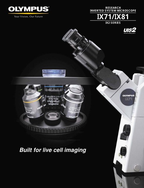
Built for live cell imaging<br />
RESEARCH<br />
INVERTED SYSTEM MICROSCOPE<br />
<strong>IX71</strong>/<strong>IX81</strong><br />
IX2 SERIES
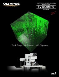
Motorized inverted system microscope<br />
<strong>IX81</strong>/<strong>IX81</strong>-ZDC<br />
Motorized System<br />
<strong>Olympus</strong> IX2 inverted microscopes<br />
combined with the new UIS2 optical system<br />
open a new world of live cell imaging.<br />
As new fluorochromes are developed and new methods of light excitation and manipulation become more popular<br />
for live cell experiments, more and more researchers will require the use of low phototoxicity near-IR wavelengths in<br />
addition to the conventional visible spectrum. <strong>Olympus</strong> has equipped its IX2 series microscopes with the new UIS2<br />
optical system precisely to meet those demands. With its high S/N ratio, its compensation for chromatic aberration<br />
over a much wider wavelength range and its flat, high transmittance , this new system sets a new world standard of<br />
fluorescence performance — efficiently detecting even faint fluorescence signals without damaging the cell, and<br />
optimizing multi-color observation. Delivering unprecedented image quality over a super wide light spectrum,<br />
the IX2 inverted system microscope will be your live cell instrument of choice now and in the future.<br />
1
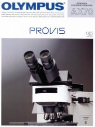
Research inverted system microscope<br />
<strong>IX71</strong><br />
Manual System<br />
2
UIS2 optics are designed to maximize S/N ratio and optical<br />
performance for live cell fluorescence imaging.<br />
Superior S/N ratio delivers imaging excellence<br />
The new UIS2 objective lenses have been designed to maximize<br />
signal to noise and outperform existing objectives by as much as<br />
50%. New objective characteristics include carefully selected low<br />
autofluorescence glass (with a significant reduction of fluorescence<br />
emitted by the antireflection coating and bonding material),<br />
combined with increased signal brightness thanks to improved<br />
numerical apertures (N.A.). With even faint fluorescence efficiently<br />
detected under weak excitation light, the UIS2 system sets new<br />
standards for fluorescence imaging of live cells.<br />
3<br />
Objective:<br />
UPLAPO100XO<br />
Mirror unit:<br />
U-MNIBA2<br />
Objective:<br />
UPLSAPO100XO<br />
Mirror unit:<br />
U-MNIBA3<br />
High N.A. objectives for fluorescence imaging<br />
Two new objectives for use with the UIS2 system are the<br />
PLAPON60XO, whose N.A. level of 1.42 is the best available for<br />
fluorescence imaging, and the UPLSAPO100XO, which is suitable<br />
for all applications. In addition to their high fluorescence S/N ratio,<br />
both these lenses are able to handle UV excitation light at parfocal<br />
45mm. The UPLSAPO100XO provides a transmittance of up to<br />
340nm.<br />
Objectives providing<br />
the best fluorescence<br />
S/N ratios.<br />
High performance mirror units<br />
optimized for fluorescence<br />
proteins.<br />
Stray light reducing function<br />
to absorb spurious reflections<br />
from dichromatic mirror.<br />
High transmittances over a wider<br />
wavelength range<br />
The IX2 series' built-in UIS2 objective achieves flat, high<br />
transmittance from visible to near-infrared light, thanks to its new UW<br />
multi-coating which effectively cuts reflection over the super wide<br />
band spectrum. In particular, transmission in the near infrared range<br />
is significantly enhanced. Overall, its performance all across the<br />
wavelength range makes it ideally suited for today's most demanding<br />
research applications.<br />
Transmittance(%)<br />
90<br />
80<br />
70<br />
60<br />
50<br />
40<br />
30<br />
20<br />
10<br />
High transmittance UPLSAPO100XO<br />
100 (new) UPLSAPO100XO<br />
(conventional) PLAPO100XO<br />
0 300 400 500 600 700 800
Effective compensation for<br />
chromatic aberration up to near-infrared<br />
The highest class UIS2 objectives are the UPLSAPO series, whose<br />
super apochromatic features effectively compensate for chromatic<br />
aberration from the visible spectrum all the way to 1000nm.<br />
This means that imaging from UV to IR is possible with just one<br />
objective. The series also offers outstanding image clarity without<br />
color shift for multi-color observations using fluorochromes covering<br />
a wide wavelength spectrum.<br />
Focus (µm)<br />
2<br />
1.5<br />
1<br />
0.5<br />
0<br />
UPLSAPO series chromatic aberration compensation<br />
Comparing chromatic aberration compensation levels:<br />
(The smaller the figure the better)<br />
(conventional) UPLAPO100XO<br />
(new) UPLSAPO100XO<br />
-0.5<br />
400 450 500 550 600 650 700 750 800 850 900<br />
Wavelength (nm)<br />
UPLSAPO100XO<br />
UPLAPO100XO<br />
Unique, optional ring slit<br />
illumination reduces<br />
objective autofluorescence.<br />
Cy7<br />
Alexa Fluor488<br />
UPLSAPO100XO<br />
Aspherical collector lens provide<br />
excellent excitation efficiency.<br />
UPLAPO100XO<br />
4
5<br />
Two-tier multi-port design ensures input/output flexibility.<br />
Two-tier optical design is also near-IR compatible<br />
The input/output of a parallel pencil of rays and the multiple port<br />
structure for gaining the primary image are designed internally in the<br />
form of tiers. To maximize the possible wavelength width, the<br />
optical path branching of each tier is also compatible with the nearinfrared<br />
spectrum. Even when more than one port is being used<br />
simultaneously, there is no change in the stage height; as a result,<br />
rigidity and illumination performance remain consistent.<br />
■ IX2 Two-tier optical path<br />
Upper back port<br />
Lower back port<br />
Tier-1 Tier-2 Bottom<br />
Optical path as seen from<br />
the left side of<br />
the microscope<br />
Objective<br />
Mirror unit<br />
Tier-1:<br />
Upper optical path<br />
selection<br />
Tube lens<br />
Tier-2:<br />
Lower optical path<br />
selection<br />
Bottom optical<br />
path selection<br />
Optical path as seen from<br />
the front of<br />
the microscope<br />
Right side port<br />
Left side port<br />
Upper Tier Lightpath Selection (optional)<br />
Located between objective and tube lenses so a parallel<br />
pencil of rays can be obtained or introduced. Primary<br />
image can be gained by adding a tube lens. Inserting<br />
optical components such as a dichromatic mirror does<br />
not produce a double image.<br />
(Choose either the right side port or the upper back port)<br />
Lower Tier Lightpath Selection (included)<br />
Located below the frame's tube lens, this tier allows<br />
primary image access to either the left side port or lower<br />
back port.<br />
Bottom Lightpath Selection<br />
A direct primary image can be obtained with no<br />
reflections. (Bottom port)<br />
■ Upper back port<br />
IR compatible<br />
■ Lower back port<br />
IR compatible<br />
■ Left side port<br />
IR compatible<br />
102mm from left side port mounting<br />
position to primary image.<br />
102mm<br />
■ Bottom port
Ring slit illumination unit to reduce noise / IX2-RFRS<br />
t Autofluorescence generated at the center of a lens causes noise, but this can<br />
be reduced by placing a ring slit at the reflected light illumination aperture<br />
diaphragm to allow only excitation<br />
light to pass through the objective<br />
perimeter (S/N ratio improvement:<br />
1.2 to 3 times). The unit can easily<br />
be attached to the IX2-RFAL, and<br />
is compatible with 40X, 60X, and<br />
100X oil immersion objectives<br />
simply by exchanging the ring slit<br />
units (F.N. 11). * Patent pending.<br />
● A multi-band dichromatic mirror is normally used to obtain multicolor<br />
images of multiple stained fluorescent samples by using filter<br />
wheels on the excitation and absorption sides. However, this kind<br />
of mirror encounters the problem that each fluorescence image<br />
gets darker as the number of color dyes increase, because the<br />
transmission spectrum becomes narrower and the transmittance<br />
falls to lower than 90% at best. <strong>Olympus</strong> has therefore developed<br />
the world’s first glass reflector that is not wavelength-dependent,<br />
offering a high transmittance of 94% across a wide wavelength<br />
range from 430nm to 700nm. Used in combination with the filter<br />
wheels on the excitation and emission sides, a wider variety of color<br />
dyes can be used and<br />
fluorescence images are captured<br />
more efficiently. *Special order basis product<br />
■ Glass reflector specifications<br />
26X38mm (t=1mm) glass substrate<br />
Transmittance 94% (at 430-700nm)<br />
26X38mm<br />
* Observations through eyepieces may have<br />
some restrictions<br />
Transmittance (%)<br />
100<br />
80<br />
60<br />
40<br />
20<br />
0<br />
Specimen<br />
Objective<br />
Excitation<br />
filter<br />
General observation<br />
Ring slit<br />
Excitation<br />
light<br />
Glass reflector captures fluorescence of multiple color dyes<br />
400 500<br />
Wavelength (nm)<br />
Hg lamp<br />
Xe lamp<br />
Annular illumination<br />
Normal illumination ➔ Ring slit illumination<br />
Illumination Normal Annular<br />
SIGNAL 408 479<br />
NOISE 36 18<br />
S/N 11.3 26.6<br />
600 700<br />
Autofluorescence<br />
Patent pending<br />
N<br />
IX2-RFRS and IX2-RS40/60/100<br />
S<br />
Glass reflector transmission properties<br />
Sapphire<br />
CFP<br />
YFP<br />
DsRed<br />
*Solid line: excitation, Dotted line: emission<br />
Excitation filter<br />
Emission filter<br />
Stray light reducing function equipped on all mirror units<br />
r One source of increased noise is very faint excitation light (stray<br />
light) transmitted without reflection by the dichromatic mirror. The<br />
IX2 series' fluorescent mirror unit incorporates a mechanism that<br />
absorbs more than 99% of this stray light.<br />
Light<br />
source<br />
Excitation light:<br />
Illumination light<br />
Excitation filter<br />
Dichromatic<br />
mirror<br />
Emission filter<br />
Specimen<br />
Fluorescence light<br />
for observation<br />
Objectives<br />
Stray light<br />
■ Usage examples of the glass reflector<br />
New FL system<br />
Stray light reducing function<br />
Sapphire-pm. CFP-CaM<br />
YFP-mt DsRed-nu<br />
Simultaneous imaging of Sapphire, CFP, YFP, and RFP. HeLa cells were imaged for<br />
Sapphire-pm, CFP-CaM, YFP-mt, and DsRed-nu. The images were obtained using either the<br />
glass plate in a normal cube.<br />
Optical components used for a 4-fluorophore imaging<br />
Dye ND Filter* 1 Excitation Light Path Reflector Emission Light Path<br />
Sapphire-pm 400DF15 535DF2<br />
CFP-CaM — 440DF20 Glass 480DF30<br />
YFP-mt 490DF20 535DF25<br />
DsRed-nu 546DF10 595RDF60<br />
* 1 ND filters in the holder of the illuminator.<br />
N<br />
8
A wide range of accessories to enable different kinds of<br />
fluorescence imaging.<br />
Bright excitation illumination for cell observation/manipulation<br />
The <strong>Olympus</strong> lineup for fluorescence illumination equipment meets a wide variety of needs<br />
including multi-stained fluorescence, ratio, photobleaching and uncaging observations.<br />
Low magnification performance is greatly improved and a metal halide system offers a precentered<br />
long bulb life option.<br />
9<br />
Fluorescence illumination light source Reflected light fluorescence illuminators<br />
Lamp housings<br />
Shape Model Aspherical* 1 Apochromatic *2 Average Lamp IR<br />
optics lens lamp life centering illumination<br />
100W mercury apo<br />
lamp housing/ √ √ 300h Required Good<br />
U-LH100HGAPO<br />
100W mercury<br />
lamp housing/ √ 300h Required Good<br />
U-LH100HG<br />
75W xenon apo<br />
lamp housing/ √ √ 200h Required Excellent<br />
U-LH75XEAPO* 3<br />
50W metal halide<br />
Not<br />
lamp housing/ √ 2000h required N/A<br />
U-LH50MH<br />
*1: Can collect light more efficiently than conventional aspherical optics.<br />
*2: Even illumination and no lamp focusing shift, even when changing excitation light wavelengths<br />
*3: Suitable for multi-color staining or ratio imaging because of flat light source spectrum.<br />
Configuration example<br />
[ L-shaped fluorescence illuminator/IX2-RFAL ]<br />
Offering twice the brightness of the<br />
preceding model, this L-Shaped illuminator<br />
offers removable aperture and field stop<br />
inserts with excellent access to lamphouse<br />
centration mechanism for maximum system<br />
flexibility.<br />
[ Fluorescence illuminator/IX2-RFA ]<br />
2.4 times brighter at low magnifications<br />
than our preceding model (comparison<br />
made with 10X objective). This illuminator is<br />
ideal for applications requiring bright<br />
excitation light, or low magnification<br />
fluorescence observation. The field stop<br />
(FS) is built in.<br />
[ Double lamphouse illuminator/IX2-RFAW ]<br />
Use two light sources simultaneously. Light<br />
stimulation can be performed during<br />
observation.<br />
[ Dual lamp housing attachment/U-DULHA ]<br />
This adapter unit allows users to attach<br />
different types of light sources<br />
simultaneously, and exchange them<br />
according to purpose.<br />
The IX2-RFA is used as a reflected light<br />
fluorescence<br />
illuminator.<br />
(Optical path:<br />
100/0, 0/100,<br />
F.N. 11)
[ Rectangular field stop/U-RFSS ]<br />
This unique field stop allows the user to control the area of<br />
fluorescence excitation anywhere inside the visual field. For<br />
example, photobleaching and phototoxicity can now be limited to<br />
only the area that is being imaged by the CCD improving overall<br />
brightness and cell viability over long term observations. The unit is<br />
attached at the field stop position of the fluorescence illuminator<br />
IX2-RFAL.<br />
Unnecessary exposure area<br />
caused by a round field stop<br />
[ Pinhole field stop/IX2-RFSPOT ]<br />
Providing spotlight illumination on the fluorescence specimen,<br />
this unit is useful in a variety of experiments. It is attached to the<br />
field stop position of the L-shape fluorescence illuminator IX2-RFAL.<br />
*Use commercially available pinhole plate<br />
Illumination modular units<br />
IR camera adapter<br />
New FL system<br />
[ Camera adapters/U-TV0.35XC-2, U-TV0.5XC-3, U-TV1X-2 ]<br />
These low-magnification camera adapters are attached to the left<br />
side port, and covers from visible light to near infrared red<br />
wavelength spectrum.<br />
Camera adapter Projection<br />
Projection area (F.N.)<br />
(Projection lens) magnifications<br />
2/3 inch CCD 1/2 inch CCD 1/3 inch CCD<br />
U-TV0.35XC-2 0.35X — 22 17.1<br />
U-TV0.5XC-3 0.5X 22 16 12<br />
U-TV1X-2 1X 11 8 6<br />
Practical field of view (mm) =<br />
Projection area<br />
U-TV0.35XC-2 U-TV0.5XC-3 U-TV1X-2 +<br />
U-CMAD3<br />
Field of view (F.N.)<br />
Projection area (Field Number)<br />
Objective magnifications<br />
2/3 inch CCD<br />
1/2 inch CCD<br />
10
11<br />
Nomarski DIC system offers the choice of optimal resolution or<br />
high contrast in live cell observation.<br />
DIC<br />
Live cells specimens vary in thickness from a nematode worm such<br />
as C. elegans to a monolayer of cultured cells. The requirements for<br />
DIC are also varied with thicker specimens having a lot of inherent<br />
contrast while thin cells are almost invisible. <strong>Olympus</strong> provides three<br />
DIC systems with varying amounts of shear. The greater the<br />
amount of shear a DIC prism imparts on the light passing through<br />
it, the greater the amount of contrast in the final image. Small shear,<br />
high resolution sets are excellent for thicker specimens. High<br />
contrast prism with twice the normal shear are excellent for very<br />
thin specimens.<br />
■ DIC sliders<br />
■ Selecting the optimum DIC prism optimum for specimen<br />
thickness and objective magnification<br />
Thin specimen<br />
(Big shearing volume)<br />
Thick specimen<br />
(Small shearing volume)<br />
High resolution DIC slider for transmitted<br />
light/U-DICTHR<br />
U-DICTHC<br />
for superior contrast with thin specimen<br />
observation<br />
U-DICT, U-DICTS<br />
for general observation<br />
U-DICTHR<br />
for superior resolution with thick specimen<br />
observation<br />
10X 40X 100X<br />
Differential Interference Contrast<br />
High contrast DIC slider for transmitted<br />
light/U-DICTHC<br />
Shift DIC sliders for transmitted light/<br />
U-DICTS<br />
DIC sliders for transmitted light/U-DICT<br />
■ Comparison of thick specimen (C. elegans), showing differences in shearing volume<br />
DIC observation using U-DICTHR<br />
DIC observation using general DIC slider<br />
■ Comparison of thin specimen, showing differences in shearing volume<br />
DIC observation using U-DICTHC DIC observation using general DIC slider<br />
Long working distance universal condenser/IX2-LWUCD<br />
Suitable for DIC observations from 10X to 100X magnification.<br />
Especially in the 20X to 40X observation range, high contrast or high<br />
resolution can be selected according to the thickness of the<br />
specimen. * Also compatible with 4X to 100X phase contrast observation by combining other<br />
optical components.<br />
• New DIC system gives a wider choice<br />
More DIC compatible objectives are<br />
available. Each condenser prism is<br />
compatible with more lenses making setup<br />
and configuration easier.<br />
■ HR/HC optical elements for IX2-LWUCD<br />
and applicable objectives<br />
DIC elements Applicable objectives<br />
IX2-DIC20HR UPLSAPO20X<br />
IX2-DIC20HC UPLFLN20X<br />
LUCPLFLN20X<br />
IX2-DIC40HR UPLSAPO40X<br />
IX2-DIC40HC UPLFLN40X<br />
UPLFLN40XO<br />
LUCPLFLN40X<br />
■ General type optical elements for<br />
IX2-LWUCD and applicable objectives<br />
DIC elements Applicable objectives<br />
IX2-DIC10 UPLSAPO10X<br />
UPLFLN10X<br />
IX2-DIC20 UPLSAPO20X<br />
UPLFLN20X<br />
LUCPLFLN20X<br />
IX2-DIC40 UPLSAPO40X<br />
UPLFLN40X<br />
UPLFLN40XO<br />
LUCPLFLN40X<br />
IX2-DIC60 PLAPON60XO<br />
UPLFLN60X<br />
UPLFLN60XOI<br />
LUCPLFLN60X<br />
IX2-DIC100 UPLSAPO100XO<br />
UPLFLN100XO<br />
UPLFLN100XOI
Water immersion DIC condenser/IX2-DICD<br />
High performance DIC condenser designed<br />
for excellent optical performance and<br />
specimen access in high magnification<br />
observations. Designed for specimen<br />
access, all controls are front mounted<br />
including prism exchange and aperture<br />
control. Three high numerical aperture front<br />
lenses are available including the water<br />
immersion IX2-TLW that offers 0.9 N.A.<br />
with 3.7mm of working distance and a 40°<br />
angle of approach for manipulators.<br />
■ Top lens combination<br />
Numerical Aperture Working Distance Immersion<br />
(N.A.) (W.D.)<br />
IX2-TLW 0.9 3.7mm Water<br />
U-TLD 0.9 1.5mm<br />
U-TLO 1.4 0.63mm Oil<br />
Focusable adapter/IX-ADUCD<br />
This condenser adapter allows the use of<br />
upright microscope condensers on the IX2,<br />
including the 8-position turret condenser<br />
(U-UCD8) for maximum system flexibility.<br />
The optical component exchange method<br />
for the universal condenser is the turret<br />
system, enabling smooth, easy exchange<br />
while using illumination with high N.A.<br />
The IX2 illumination pillar also offers a<br />
'condenser-only' tilt mechanism to<br />
quickly allow access to the<br />
specimen without tilting<br />
the entire illumination pillar.<br />
* IX2-TLW cannot be used for U-UCD8<br />
Gliding stage/IX2-GS<br />
The Gliding Stage was designed for quick<br />
rotation of the specimen using your<br />
fingertips. With 20mm of X-Y travel,<br />
360 degree rotation and completely flat<br />
surface, a specimen such as the nematode<br />
worm C. elegans can be quickly brought into<br />
the correct position and alignment for<br />
injection or micromanipulations.<br />
Water immersion DIC condenser IX2-DICD + water immersion<br />
top lens IX2-TLW<br />
IX2-GS<br />
40°<br />
New DIC system<br />
■ Simple principle of Nomarski DIC<br />
microscopy<br />
Nomarski DIC amplifies contrast by using the<br />
phase difference which occurs when light<br />
passes through material with different<br />
refraction values (e.g. a cell) in a particular<br />
medium (e.g. water). The wave direction of<br />
light from the microscope light source is<br />
unified in a polarizer (condenser side); and<br />
when it passes through the condenser side<br />
DIC prism, it separates into two phases which<br />
cross each other at right angles. The distance<br />
of separation is called the shearing amount.<br />
When two such separated lights pass through<br />
a medium with different refraction values (e.g.<br />
a cell), one of their phase is delayed; and<br />
when the two lights are re-composed by DIC<br />
slider (the observation side) and analyzer, the<br />
interference effect produces the contrast. This<br />
is the principle of Nomarski DIC.<br />
Polarizer<br />
DIC element<br />
Condenser<br />
Specimen<br />
Shearing<br />
volume<br />
Objective<br />
DIC slider<br />
Analyzer<br />
<strong>Olympus</strong> has developed the most suitable<br />
DIC prisms for different types of specimen,<br />
based on the shearing amount. When DIC<br />
contrast is low, the specimen is hard to<br />
observe, while high contrast also hinders<br />
observation because of excessive glare.<br />
<strong>Olympus</strong> has therefore developed three<br />
different types of DIC prisms to ensure clear<br />
observation for every kind of specimen.<br />
12
13<br />
Special equipment for<br />
relief contrast and phase contrast.<br />
RC<br />
The <strong>Olympus</strong> Relief Contrast system provides a high contrast,<br />
3-D image similar to DIC for specimens mounted in plastic vessels.<br />
Relief contrast is designed for cellular fertilization and making<br />
the nuclear envelope easier to see and penetrate.<br />
Relief contrast equipment<br />
* Unifies the shadow directions of each objective, improving<br />
operability at all magnifications.<br />
* Maintains a long working distance (45mm) for the condenser<br />
(IX2-MLWCD) so as not to hinder operation of the manipulator.<br />
Users can choose from two types of objectives for relief contrast<br />
work: cost-efficient Achromat models, or PlanSemiApochromat<br />
objectives with high resolution and excellent focusing right up to the<br />
image perimeters. Condenser (IX2-MLWCD) also supports DIC and<br />
phase contrast observations for maximum flexibility.<br />
Mouse embryo<br />
PH<br />
Phase contrast equipment<br />
Ultra long working distance condenser/IX-ULWCD<br />
This universal condenser for phase contrast and brightfield<br />
observations offers excellent workability, due to its long working<br />
distance (73mm) and compatibility with large containers: it should<br />
be used in combination with 4X -40X phase contrast objectives.<br />
Phase contrast observation is also possible with the IX2-LWUCD<br />
condenser, whose working distance is 27mm.<br />
Relief contrast equipment<br />
N.A. W.D.<br />
CPLN 10XRC *1 0.25 9.7mm<br />
LCACHN 20XRC *1 0.40 2.8mm<br />
LCACHN 40XRC *1 0.55 1.9mm<br />
CPLFLN 10XRC *1 0.30 9mm<br />
LUCPLFLN 20XRC *2 0.45 6.6 — 7.8mm<br />
LUCPLFLN 40XRC *2 ■Objectives for Relief Contrast observation<br />
Achromat for<br />
Relief<br />
Contrast<br />
Plan Fluorite<br />
for Relief<br />
Contrast<br />
0.60 3.0 — 4.2mm<br />
*1 Objective with compensation for 1mm plastic dish plus 0.5mm<br />
thick thermoplate<br />
*2 Objective with compensation ring for 0~2mm thick cover glass.<br />
The IX2-MLWCD comes equipped with optical component RC1 (for 10X objective), RC2 (for 20X<br />
objective), RC3 (for 40X objective) and a polarizer to adjust the contrast.<br />
■ Phase contrast optical elements for IX2-MLWCD<br />
and applicable objectives<br />
Optical elements for Applicable objectives<br />
IX2-MLWCD<br />
IX2-MPHL UPLFLN4XPH<br />
IX2-MPHC CPLFLN10XPH, CPLN10XPH, LCACHN20XPH<br />
IX2-MPH1 LUCPLFLN20XPH<br />
IX2-MPH2 LUCPLFLN40XPH, LCACHN40XPH, LUCPLFLN60XPH<br />
■ DIC optical elements for IX2-MLWCD and<br />
applicable objectives<br />
Optical elements Objectives<br />
IX2-MDIC20 UPLSAPO20X, UPLFLN20X, LUCPLFLN20X<br />
IX2-MDIC40 UPLSAPO40X, UPLFLN40X, UPLFLN40XO* ,<br />
LUCPLFLN40X<br />
* Use with shift DIC slider (U-DICTS).<br />
RC/PH System
A wide lineup of UIS2 objectives.<br />
UIS2 Objectives<br />
UIS2 objectives *<br />
Model N.A. W.D. F.N Cover glass Immersion Spring Correction Iris Water proof &<br />
(mm) thickness (mm) ring diaphragm oil proof function<br />
UPLSAPO UPLSAPO 4X 0.16 13 26.5 —<br />
UPLSAPO 10X 0.40 3.1 26.5 0.17<br />
UPLSAPO 20X 0.75 0.6 26.5 0.17 _<br />
UPLSAPO 40X 0.90 0.18 26.5 0.11-0.23 _ _<br />
UPLSAPO 60XW 1.20 0.28 26.5 0.15-0.2 Water _ _ _<br />
UPLSAPO 60XO 1.35 0.15 26.5 0.17 Oil _ _<br />
UPLSAPO 100XO 1.40 0.13 26.5 0.17 Oil _ _<br />
PLAPON PLAPON 60XO 1.42 0.15 26.5 0.17 Oil _ _<br />
PLAPON 60XOTIRFM 1.45 0.1 26.5 0.13-0.19 Oil _ _ _<br />
UPLFLN UPLFLN 4X 0.13 17 26.5 —<br />
UPLFLN 10X 0.30 10 26.5 —<br />
UPLFLN 20X 0.50 2.1 26.5 0.17 _<br />
UPLFLN 40X 0.75 0.51 26.5 0.17 _<br />
UPLFLN 40XO 1.30 0.2 26.5 0.17 Oil _ _<br />
UPLFLN 60X 0.90 0.2 26.5 0.11-0.23 _ _<br />
UPLFLN 60XOI 1.25-0.65 0.12 26.5 0.17 Oil _ _ _<br />
UPLFLN 100XO 1.30 0.2 26.5 0.17 Oil _ _<br />
UPLFLN 100XOI 1.3-0.6 0.2 26.5 0.17 Oil _ _ _<br />
LUCPLFLN LUCPLFLN 20X 0.45 6.6-7.8 22 0-2 _<br />
LUCPLFLN 40X 0.60 2.7-4 22 0-2 _<br />
LUCPLFLN 60X 0.70 1.5-2.2 22 0.1-1.3 _<br />
LUCPLFLN 20XPH 0.45 6.6-7.8 22 0-2 _<br />
LUCPLFLN 20XRC 0.45 6.6-7.8 22 0-2 _<br />
LUCPLFLN 40XPH 0.60 3.0-4.2 22 0-2 _<br />
LUCPLFLN 40XRC 0.60 3.0-4.2 22 0-2 _<br />
LUCPLFLN 60XPH 0.70 1.5-2.2 22 0.1-1.3 _<br />
UPLFLN-PH UPLFLN 4XPH 0.13 17 26.5 —<br />
UPLFLN 10XPH 0.30 10 26.5 —<br />
UPLFLN-PHP UPLFLN 4XPHP 0.13 16.4 22 —<br />
CPLFLN CPLFLN 10XPH 0.30 9.5 22 1<br />
CPLFLN 10XRC 0.30 9 22 1.5<br />
LCACHN LCACHN 20XPH 0.40 3.2 22 1<br />
LCACHN 20XPHP 0.40 3.2 22 1<br />
LCACHN 20XRC 0.40 2.8 22 1.5<br />
LCACHN 40XPH 0.55 2.2 22 1<br />
LCACHN 40XPHP 0.55 2.2 22 1<br />
LCACHN 40XRC 0.55 1.9 22 1.5<br />
CACHN & CPLN CACHN 10XPHP 0.25 8.8 22 1<br />
CPLN 10XPH 0.25 10 22 1<br />
CPLN 10XRC 0.25 9.7 22 1.5<br />
* All UIS2 objectives and WHN eyepieces: lead-free eco-glass<br />
UIS objectives<br />
Model N.A. W.D. F.N Cover glass Immersion Spring Correction Iris Water proof &<br />
(mm) thickness (mm) ring diaphragm oil proof cap<br />
UPLAPO UPLAPO 10XO3 0.40 0.24 26.5 0.17 Oil _ _<br />
UPLAPO 10XW3 0.40 0.43 26.5 0.17 Water _ _<br />
UPLAPO 20XO3 0.80 0.19 26.5 — Oil _ (_)<br />
UPLAPO 40XOI3 1.00-0.50 0.12 26.5 — Oil _ _ (_)<br />
PLAPO PLAPO 40X 0.95 0.13 26.5 0.11-0.23 _ _<br />
UAPO UAPO 20X3/340 0.75 0.55 22 0.17 _ _<br />
UAPO 40X3/340 0.90 0.2 22 0.11-0.23 _ _ _<br />
UAPO 40XOI3/340 1.35-0.65 0.1 22 0.17 Oil _ _ _<br />
UAPO 20XW3/340 0.70 0.4 22 0.17 Water _ _<br />
UAPO 40XW3/340 1.15 0.25 22 0.13-0.25 Water _ _ _<br />
APO APO 100XOHR 1.65 0.1 22 0.15 Oil _ _<br />
(_): Oil proof cap applicable.<br />
14
15<br />
High level basic performance makes a vital difference to<br />
experiment results.<br />
Thermally<br />
compensated<br />
relay lens optics<br />
Capture of high-clarity primary image<br />
The luminous flux can be captured or introduced from the upper<br />
back port or the right side port using a parallel pencil of rays.<br />
Because the UIS2 optical system is compensation free (i.e.<br />
compensation is performed only by the objective lens), a clear<br />
primary image* can be captured.<br />
* The "primary image" is the first image created by convergence of the luminous flux after passing<br />
through an objective. There is no loss in light quantity and no image deterioration.<br />
V-shaped optical path to reduce light loss<br />
In order to minimize light loss from reflection, a simple V-shaped<br />
optical system is employed. This restricts reflection inside the<br />
microscope to one-time-only, reducing light loss and allowing<br />
observation of even weak fluorescent signals.<br />
■ Thermally compensated relay lens optics<br />
Used for the observation optical path, thermally compensated relay<br />
lens optics involve combining lenses with different thermal<br />
characteristics to offset blurs caused by temperature change.<br />
Measures against thermal expansion to prevent blur<br />
■ External power supply<br />
Time-lapse observation over a long period of time will cause some<br />
heat strain to the microscope, from temperature changes in the<br />
environment and air blown from an air-conditioner. Because such<br />
changes can cause blurring, the IX2 series design team addressed<br />
the problem with great care. Countermeasures include locating the<br />
power supply for transmitted illumination on the outside of the<br />
microscope, thereby lessening heat strain on the inside and<br />
reducing blur to one-seventh of conventional models. Various<br />
accessories are provided to stabilize long-term time-lapses, such<br />
as incubator that reduces temperature changes in the environment<br />
and the effects of air conditioning.<br />
High body rigidity<br />
In order to maximize rigidity in every area, <strong>Olympus</strong> simplified or<br />
shortened structures from the focusing handle to the revolving<br />
nosepiece, thereby minimizing warp in the image traveling section.<br />
This in turn prevents blur when operating the objective correction<br />
ring or the Nomarski DIC slider.<br />
Revolving nosepiece<br />
guide structure<br />
The shorter the section in<br />
red, the less influence from<br />
heat and force — resulting<br />
in improved rigidity.
[ Frontal control ]<br />
Light path selection lever<br />
Two-stage selection<br />
between the observation<br />
tube and the port prevents<br />
operation errors in the dark<br />
room.<br />
TTL Pulse control switch<br />
ON/OFF switch for the<br />
motorized shutter (e.g.<br />
made by UNIBLITZ).<br />
Ease of use in a compact body<br />
[ Compact body ]<br />
Despite the compact body design, space has been allowed for a<br />
port on both sides and the bottom of the microscope as well as on<br />
the back, while the side areas are left open to ensure easy<br />
combination with a variety of peripherals. Using specialized tools,<br />
the microscope can be fixed to<br />
394<br />
an anti-vibration stand, thereby<br />
making it even more compact<br />
by removing the support<br />
extending out at the back of<br />
the unit. 290<br />
245<br />
135<br />
[ Tilting binocular tube/U-TBI90 ]<br />
A tilting observation tube with 35-85°<br />
elevation angle. When not in use, the<br />
tube can be flipped up and stowed<br />
away to keep it from projecting beyond<br />
the table edge. A tilting tube allows<br />
every user to find the most comfortable<br />
observation posture depending on<br />
physical build, and also enables<br />
observation while standing.<br />
Light adjustment dial<br />
Power ON/OFF switch<br />
Field stop (F.S.)<br />
Aperture stop (A.S.)<br />
Shutters for<br />
fluorescence excitation<br />
Focus knob<br />
[ Focus free collection ring ]<br />
The newly developed LUCPLFLN40X (N.A.<br />
0.6, W.D. 3.4mm*) and LUCPLFLN60X (N.A.<br />
0.70, W.D. 1.5-2.2mm) are compatible with<br />
any container thickness. Turning the<br />
correction ring does not blur the focus when<br />
correcting spherical aberration caused by<br />
different container thickness. A simple<br />
correction operation optimizes the<br />
observation image. * When using 1mm thickness container.<br />
■ LUCPLFLN 40X<br />
Operating the correction ring does<br />
not blur the focus.<br />
■ When using a conventional<br />
objective with correction ring<br />
Focus blurs when correction ring<br />
is operated.<br />
[ Oil immersion<br />
protection function ]<br />
Prevents immersion oil<br />
infiltrating the tip of the<br />
objective.<br />
Basic Characteristics<br />
[ Magnification changer ]<br />
Magnification between 1X<br />
and 1.6X can be changed<br />
without even touching the<br />
objective. 2X (IX2-CA2X) is<br />
optional.<br />
[ Glass stage insert<br />
plate/IX2-GCP ]<br />
The type and magnifications<br />
of objectives can be<br />
confirmed easily from stage<br />
surface. Objective<br />
magnifications are colorcoded<br />
for easier<br />
confirmation.<br />
* Not available in some areas<br />
[ Fluorescence indicator ]<br />
Self illuminated labels are<br />
used and easily visible in a<br />
dark room.<br />
[ Fluorescence turret<br />
confirmation window ]<br />
The fluorescent mirror unit<br />
in the optical path can be<br />
confirmed from the space<br />
between the left and right<br />
eyepieces of the<br />
observation tube.<br />
16
17<br />
Motorized system for live cell imaging.<br />
Controlling functions via PC, handset or operating buttons on the microscope body<br />
Functions of <strong>IX81</strong> control software /IX2-BSW<br />
Nearly every operating function on the <strong>IX81</strong> can be allocated to operation<br />
buttons on the PC, the hand switch and the microscope in any individual or<br />
multiple combinations by using IX2-BSW* control software. Some image<br />
analysis software can also be used to control microscope operation, image<br />
capturing and analysis; in this case, all operations are done from a single<br />
PC.<br />
* Included with the system controller IX2-UCB2<br />
Example : Switching from fluorescence observation to Nomarski DIC.<br />
q Close shutter for fluorescence<br />
illumination<br />
w Exchange FL mirror unit for<br />
DIC mirror unit<br />
e Open shutter for transmitted<br />
illumination<br />
With the <strong>IX81</strong>, this<br />
sequence of<br />
functions can be<br />
allocated to<br />
a single button<br />
■ Parfocal compensation function among objectives<br />
This function allows the focus point to be matched from<br />
low to high magnification objectives. Refocusing each time<br />
the magnification is changed is no longer necessary.<br />
■ Malfunction prevention<br />
Motorized units ensure that complicated operations are<br />
performed without error. Once the usage conditions are<br />
set, the setting screen can be hidden to avoid accidental<br />
change leading to faulty operation.<br />
■ Setting sensitivity of the fine focus movement for<br />
each objective magnification<br />
Users can set the amount of the fine focus movement per<br />
rotation of the focus adjustment knob.<br />
■ Save setting conditions for each operator<br />
Customized data can be stored in folders, and each folder<br />
labeled for different users or sets of conditions.<br />
Motorized universal condenser/<br />
IX2-LWUCDA2<br />
This condenser has six built-in optical<br />
components to enable brightfield, phase<br />
contrast and Nomarski DIC observations.<br />
Software allows switching optical<br />
components to be synchronized with the<br />
objective. (Manual AS included.)<br />
Handset/U-HSTR2<br />
A remote handset controls all motorized<br />
functions via a convenient and<br />
programmable interface.<br />
Focus handle/U-FH<br />
The focus handle allows remote control of<br />
the objective position relative to the<br />
specimen especially when using an<br />
incubator. Allows the user to be away from<br />
the microscope and still focus on the<br />
specimen.<br />
Course/Fine<br />
and<br />
transmitted<br />
light control<br />
are also<br />
included via<br />
buttons.<br />
Microscope front panel<br />
Easy to use buttons allow selection of light<br />
path, light intensity and lamp on/off control.<br />
Auxiliary buttons can be custom<br />
programmed. Includes LED lamp intensity<br />
meter.
Motorized bottom<br />
port unit with<br />
C-mount/IX2-TVRAC<br />
Motorized shutter/IX2-SHA<br />
Can be mounted in both transmitted and<br />
reflected light paths.<br />
Motorized filter wheel/<br />
U-FWR and U-FWO<br />
6 filter positions (32mm or 25mm diameter).<br />
Motorized sextuple<br />
revolving nosepiece<br />
Up to 6 objectives are mounted<br />
simultaneously, included with microscope<br />
frame.<br />
Motorized mirror unit turret/<br />
IX2-RFACA<br />
Six mirror units can be attached to the IX2-<br />
RFACA simultaneously, making it easy to<br />
switch between them during fluorescence<br />
observation of multi-stained specimens.<br />
(Manual shutter included)<br />
Internal motorized focus drive<br />
With minimum movement of 0.01µm,<br />
the user has precise focus control.<br />
Objective escape and<br />
zero-return buttons<br />
Moves objective to lower focus limit.<br />
Allows setting of default focus position.<br />
* Equipped on each side of microscope frame.<br />
System controller/IX2-UCB2<br />
All motorized units are powered by this<br />
external system controller. Included is an<br />
RS232C connection for PC commands and<br />
expansion slots for future system upgrades.<br />
Motorized Units<br />
U-FWR<br />
U-FWO<br />
18
19<br />
Maintaining long-term stability for live cell observation.<br />
Focus drift compensation function for<br />
time-lapse experiments.<br />
Motorized inverted research microscope with<br />
focus drift compensation/<strong>IX81</strong>-ZDC<br />
This landmark microscope model makes it easy to reproduce any<br />
preset focus position. For time lapse experiments, this capability<br />
reduces the need for Z stack images, which take into account the<br />
focus blur. Excitation light is not used for reproduction of the focus<br />
position, but only 785nm laser light, which is less phototoxic; there<br />
is therefore no need for concern about fading. In addition, the laser<br />
light is introduced through its own special light path, so all the IX2<br />
light path can be used for imaging.<br />
Vibration-free,<br />
high-sensitivity cooled CCD camera<br />
developed for live cell imaging.<br />
High sensitivity cooled CCD camera/DP30BW<br />
This camera is cooled (to 5°C) by the Peltier cooling system. Its<br />
built-in shutter and background subtraction function (reduction of<br />
dark current) enable high quality images to be captured even from<br />
weak fluorescence. Used with DP-BSW software, it is also<br />
compatible with still image and movie recording of live images.<br />
In addition, it can be used with imaging software designed for<br />
motorized units, making it possible to motorize many fluorescence<br />
imaging procedures from 2D to 6D, such as multi-color stained,<br />
deconvolution and time-lapse experiments.<br />
Live cell imaging system<br />
A.<br />
Closer<br />
than focus<br />
position<br />
B.<br />
Focusing<br />
position<br />
* Extended image<br />
C.<br />
Further<br />
than focus<br />
position<br />
Reflection<br />
plane<br />
Objective<br />
Light<br />
shielding<br />
plate<br />
Telan lens<br />
Laser light<br />
source<br />
(class 1)<br />
Split detector
Accessories to improve stability in long-duration observations<br />
[ CO2 incubators/MIU-IBC-I, MIU-IBC-IF ]<br />
Highly precise incubator control keeps the environment inside a<br />
laboratory dish completely stable, at just below 37°C temperature,<br />
90% moisture and 5% CO2 concentration (when using a CO2 5%<br />
concentration bomb); in this way, live cell activity can be maintained<br />
for around 2 days. In addition, a special structure device is<br />
employed to minimize drift on the stage caused by thermal<br />
expansion. As a result, this incubator is ideal for time-lapse<br />
experiments under a confocal laser<br />
scanning microscope. It is also possible to<br />
inject the cell in the laboratory dish by using<br />
the injection hole located on the top heater.<br />
* Built-in stage warming plate<br />
* Objective heater<br />
* 5% CO2 supply tube with ø4 outer diameter, ø2 inner diameter<br />
and 400mm length.<br />
* Not available in some areas<br />
MIU-IBC-I<br />
Basic configuration which allows control of<br />
heaters for top, bath, stage and objective.<br />
[ Thermoplate/MATS series ]<br />
This thermoplate maintains the cell<br />
specimen container temperature at<br />
37°C.<br />
* Tokai Hit Company products<br />
MIU-IBC-IF<br />
This configuration includes a built-in flowmeter<br />
for 5% CO2 and 95% air. Use the 5% CO2 and<br />
95% air bombs.<br />
[ Frame plate adapter/<br />
IX2-FP ]<br />
This is used to fix the<br />
microscope body<br />
to the antivibration<br />
stand.<br />
*M6 screws (available<br />
separately) are required for<br />
fixing.<br />
[ Nosepiece stage/IX2-NPS (patent pending) ]<br />
This newly-developed stage-and-pole configuration is fixed to the revolving nosepiece<br />
for long time observations. Its effect is to minimize the distance between the specimen<br />
and the objectives, which in turn minimizes the effect of temperature change and<br />
prevents blur during long observations. To use, attach one objective to the pole.<br />
[ Comparison of normal observation images ]<br />
Change of focus when environmental temperature (25°C )<br />
changes by ±5°C. *When the microscope is used without an incubator.<br />
With IX2-NPS<br />
Environmental temperature<br />
Normal observation images<br />
[ Incubators ]<br />
Incubators keep the microscope temperature stable, thus<br />
preventing blur caused by changes in the environmental<br />
temperature.<br />
[ Comparison of fluorescence images ]<br />
Time-lapse observation images.<br />
*When the microscope is used without an incubator.<br />
20°C 25°C 30°C Elapsed time<br />
0 30 60 120 min<br />
Normal observation images<br />
With IX2-NPS<br />
Live Cell Imaging System<br />
20
21<br />
Manipulator<br />
Manipulating cells.<br />
Micromanipulation system/ON3<br />
Low stage design combined with a lowered center of gravity offers<br />
superior stability for micromanipulation applications. A variety of<br />
screw holes are available to securely mount manipulators onto the<br />
microscope, allowing optimal choice of angles and positioning.<br />
Manipulator joysticks can be placed in the most comfortable,<br />
accessible position due to the compact frame design.<br />
ON3-99D ON3-99D with return mechanism (UT-R) Manual combination<br />
(ONM-2D+ONO-301D+UT-D)<br />
Human embryo<br />
Manual combination with return mechanism<br />
(ONM-2D+ONO-301D+UT-D+UT-R)
Laser Scanning Confocal Application System<br />
Simultaneous laser light stimulation and imaging.<br />
Confocal laser scanning microscope/<br />
FV1000 FLUOVIEW system<br />
The basic performance of this laser scanning microscope delivers<br />
significantly brighter, quicker and more precise imaging, obtaining<br />
accurate living organism information while minimizing the chance of<br />
damage to the specimen.<br />
Used with the (optional) SIM scanner, the FV1000 can synchronize<br />
the observation and light stimulation lasers so as to perform laser<br />
light stimulation and imaging simultaneously.<br />
TIRFM<br />
Ultra-sensitive fluorescence microscopy.<br />
TIRFM<br />
(Total Internal Reflection<br />
Fluorescence <strong>Microscopy</strong> System)<br />
Since 1997, <strong>Olympus</strong> has been a market leader in objective based<br />
Total Internal Reflection systems that allows evansecent wave<br />
illumination approximately 200nm into the specimen beyond the<br />
coverglass interface. <strong>Olympus</strong> extends that leadership role by<br />
offering three objectives for TIRFM including the world's highest NA<br />
objective, the 100X N.A. 1.65 objective.<br />
The incredibly thin optical section created be TIRFM allows an<br />
extremely high signal to noise image to be collected. Popular<br />
applications include vesicle tracking, cellular adhesions and single<br />
molecule events.<br />
• <strong>Olympus</strong>' original high N.A. objectives make it easy to produce an<br />
evanescent wave field. So little light is leaked that a high-contrast<br />
image can be obtained against a dark background.<br />
* N.A. 1.65, 100X objective (APO100XOHR).<br />
Incompatible resolution and brightness (use special cover glass and immersion oil)<br />
* N.A. 1.45 objectives (PLAPON60XOTIRFM, PLAPO100XOTIRFM)<br />
UPLSAPO100XO (N.A. 1.4)<br />
(Use normal cover glass and immersion oil)<br />
• After initial alignment of the single mode<br />
fiber to the laser head and TIRFM<br />
illuminator, a simple micrometer<br />
adjustment is all that is required to move<br />
the laser position between TIRFM and<br />
widefield illumination.<br />
Micrometer<br />
Total Internal Reflection Fluorescence<br />
observation with evanescent wave excitation<br />
Images of Kaede-expressed cells demonstrating<br />
the photoactivation acquired every 300msec and<br />
observed via 405 blue diode laser illumination with<br />
twin scanners.<br />
Widefield fluorescence observation with<br />
mercury arc lamp excitation<br />
22
23<br />
Spinning Disk Confocal<br />
Obtaining confocal images easily by use of an arc light source.<br />
Disk Scanning Confocal Microscope System<br />
The <strong>Olympus</strong> Disk Scanning Unit (DSU) offers confocal images<br />
using a white light, arc excitation source and CCD camera. The<br />
heart of the system is a unique, <strong>Olympus</strong> designed slit pattern disk<br />
that offers excellent light throughput and thinness of optical<br />
sectioning or "confocality'.<br />
• Compliance with various fluorochromes with different spectral<br />
characteristics.<br />
Since an arc light source is used, the unit can meet different<br />
fluorochrome requirements across a wide wavelength spectrum by<br />
simply exchanging a standard mirror unit.<br />
• Minimize excitation light damage to the specimen.<br />
The excitation light volume is reduced to around 5% as a result of<br />
passing through the disk. Even so, there is almost no fading of<br />
fluorescence emission from the surface of the focused sample.<br />
• Quick construction of 3D images.<br />
A single DSU image can be obtained very quickly, in 0.1~0.4<br />
seconds (exposure time with recommended camera). Overlapping<br />
successively photographed cross images enables speedy<br />
construction of a 3D image.<br />
• Low and high magnification objective support.<br />
Five DSU disks are available of varying slit spacing and width.<br />
Disks can be thus be matched to the magnification and N.A. of<br />
the preferred objective. The DSU supports 10X to 100X confocal<br />
imaging.<br />
• Easy exchange between confocal and reflected light<br />
fluorescence observation .<br />
IN/OUT of the confocal disk to the light path can be done by a<br />
hand switch or via software, so it is easy to exchange observation<br />
methods between DSU and reflected light fluorescence.<br />
Zebrafish 3-day embryo, ventral view, projection of 62 serial optical sections<br />
Adult brain of Drosophila, reflected light fluorescence image (left) and DSU image (right)<br />
* Not available in some areas
ARC EVA<br />
World’s first evanescent illumination system from an arc lamp source.<br />
TIRFM (Total Internal Reflection Fluorescence<br />
<strong>Microscopy</strong>) system with arc lamp source<br />
Featuring the <strong>Olympus</strong>-developed total internal reflection illumination system<br />
and slit mechanism to provide evanescent wave illumination from an arc<br />
lamp source. High signal to noise fluorescence observations with extremely<br />
thin optical sectioning can now be easily performed at the specimencoverslip<br />
interface.<br />
The arc lamp is focused on an off-center slit using a wedge prism.<br />
The light is then focused on the outer edge of the back focal plane<br />
of the objective thus causing the excitation light to exit the objective<br />
beyond the critical angle resulting in Total Internal Reflection. For<br />
normal fluorescence observation, the wedge prism and slit can be<br />
easily removed from the light path via a slider. Through the use of<br />
filters, this system<br />
enables a wider choice of<br />
excitation colors than is<br />
currently possible with<br />
wavelength-limited laser<br />
light sources.<br />
High-precision fluorescence turret /IX2-RFACEVA<br />
Turret includes three, highly precise,<br />
empty fluorescence filter cubes that<br />
permit dichromatic mirror switching<br />
while maintaining excitation light<br />
position on the back focal plane of<br />
the objective. This system makes<br />
multi-color observations easy and<br />
alleviates the usual adjustment of the<br />
excitation source when changing<br />
mirror units. Up to six mirror units<br />
can be installed.<br />
Exclusive<br />
objective<br />
High precisiton<br />
fluorescence turret<br />
Off-center slit slider<br />
Wedge prism slider<br />
Excitation filter slider<br />
Fluorescence lamp housing<br />
L shape fluorescence<br />
illuminator<br />
Main specifications<br />
Microscope Research inverted system<br />
microscope <strong>IX71</strong><br />
Fluorescence Arc illumination total internal reflection<br />
illuminator fluorescence unit IX2-ARCEVA<br />
(Slit slider, wedge prism slider and<br />
excitation filter slider)<br />
L-shape fluorescence illuminator<br />
IX2-RFAL<br />
Mirror unit High-precision fluorescence turret<br />
cassettes IX2-RFACEVA<br />
(choose from either (with centering mechanism and<br />
fluorescence turret) 3 vacant mirror units)<br />
Fluorescence turret<br />
IX2-RFAC<br />
Lamp light source 100W mercury lamp,<br />
75W Xenon lamp<br />
Objectives PLAPON60XOTIRFM<br />
N.A. 1.45, W.D. 0.1mm<br />
Used with normal cover glass and<br />
immersion oil<br />
Stage Left short handle stage IX-SVL2<br />
Total internal reflection<br />
illumination F.N.<br />
11<br />
Observation Recommend high sensitive camera<br />
IX2-ARCEVA<br />
SYSTEM DIAGRAM<br />
Conventional fluorescence observation<br />
TIRFM observation<br />
Microtubule of an NG108-15 cell labeled with Alexa488 through indirect fluorescence antibody test<br />
Kaede-Crk II protein expressed in a HeLa cell<br />
Application System<br />
* Not available in some areas<br />
24
25<br />
FRET<br />
Bright, simultaneous two-wavelength imaging using the primary image.<br />
FRET Split imaging system<br />
• Simultaneous two-color split imaging with one CCD camera.<br />
• Unique design splits the primary image for the highest efficiency<br />
and light transmission necessary for weak fluorescence signals<br />
such as CFP/YFP FRET experiments.<br />
•Compact and space-saving design takes advantage of the 70mm<br />
of free space between the microscope frame and the primary<br />
image plane found on all <strong>Olympus</strong> Research Upright and Inverted<br />
Microscopes.<br />
• Simple cassette mechanism makes it easy to switch between<br />
split and full frame imaging.<br />
• Unit is up to 10% brighter than similar relay lens based, image<br />
splitting systems.<br />
• When used with the rectangular field stop U-RFSS, excitation<br />
energy is limited to the camera's field of view, minimizing specimen<br />
photo-bleaching<br />
Cube cassette for<br />
split images<br />
Cube cassette for<br />
full image<br />
Split primary image<br />
camera port/U-SIP<br />
Rectangular field<br />
stop/U-RFSS<br />
Split primary image<br />
camera port<br />
U-SIP<br />
YFP CFP<br />
Split image<br />
Specimen<br />
Objective<br />
Fluorescence mirror unit<br />
Filter set such as<br />
XF88-2(OMEGA) or<br />
31044v2(CHROMA)<br />
Filter sliders<br />
(Emission, ND sliders)<br />
High resolution camera<br />
U-SIP main specifications<br />
YFP CFP<br />
HeLa cell, in which YC3.1 (cytoplasm) and YC3.1nu (with nuclear localization signal) are coexpressed.<br />
FRET changes are observed through histamine stimulation, and images are acquired at intervals of<br />
50msec.<br />
Prism<br />
Cube cassette for<br />
split image<br />
Mirrors<br />
Built-in separation<br />
dichromatic mirror<br />
(DM505)<br />
Rectangular field stop<br />
U-RFSS<br />
L shape fluorescence illuminator<br />
IX2-RFAL<br />
Excitation filter<br />
Telan lens<br />
Research inverted<br />
system microscope<br />
<strong>IX81</strong>/<strong>IX71</strong><br />
Microscope <strong>IX71</strong>/81<br />
Image separation Right and left 2-separation<br />
(can be adjusted independently)<br />
Built-in separation dichromatic mirror DM505 (special size)<br />
Filter slider Emission, ND filters' size ø25mm,<br />
total thickness:8mm<br />
Used together with commercially available filter<br />
set (XF88-2 OMEGA) or 31044v2(CHROMA)<br />
Field Number Split image: 8<br />
Full image: 11<br />
Magnifications 1X (primary image)<br />
Objectives 40X and higher<br />
Camera mounting C-mount<br />
Recommended camera Chip size 2/3 inch<br />
75W xenon apo<br />
lamp housing<br />
U-LH75XEAPO<br />
Power supply unit<br />
* Not available in some areas
Photoactivation Application System<br />
Photoactivation illuminator for inverted microscopy.<br />
Photoactivation Fluorescence<br />
Microscope system<br />
The photoactivation illuminator allows the exposure of UV light to<br />
specific regions of a cell for photoconversion, the uncaging of<br />
compounds and the photoactivation of specific fluorochromes.<br />
• A specified area of the cell can be exposed to UV light while<br />
observing the targeted cell by fluorescence or transmitted (DIC)<br />
method.<br />
• Compliance with FRAP or FLIP<br />
experiments (by special order).<br />
• Easy system upgrade by<br />
attaching double lamphouse<br />
illuminator<br />
IX2-RFAW to IX2 series inverted<br />
microscope.<br />
Double lamp<br />
house illuminator<br />
IX2-RFAW<br />
Excitation filter<br />
BP330-385<br />
Filter slider<br />
Pinhole slider<br />
Pinhole or slit<br />
Filter slider<br />
Double lamphouse illuminator IX2-RFAW<br />
Magnetic shutter<br />
Sample<br />
Field<br />
stop<br />
position *1<br />
Composed dichromatic<br />
mirror of right and left path<br />
Inverted microscope<br />
Filter wheel 1<br />
IX series<br />
such as Lambda10-2<br />
Excitation filter<br />
475AF20(XF1072 OMEGA) or<br />
HQ475/20x(CHROMA)<br />
Excitation filter *2<br />
550DF30(XF1021 OMEGA)<br />
Objective<br />
UPLFLN40XO<br />
Fluorescence mirror unit<br />
U-MF2+dichromatic mirror<br />
(DM400 on the illustration)<br />
Fluorescence filter *2<br />
575ALP (XF3089 OMEGA) or<br />
HQ575LP(CHROMA)<br />
Fluorescence filter<br />
530DF30(XF3107 OMEGA) or<br />
D530/30x(CHROMA)<br />
High resolution<br />
camera<br />
Filter wheel 2<br />
such as Lambda10-2<br />
*1 Field stop position is the same position as<br />
the focus point of the sample.<br />
*2 Use 550DF30 excitation filter in the filter<br />
wheel 1 and 575ALP fluorescence filter in<br />
the filter wheel 2 when observing red<br />
Kaede protein. Exchange of the<br />
fluorescence mirror unit is not required.<br />
The novel Kaede gene is useful in biology because it exhibits photoconversion. Normally, the Kaede<br />
gene shows green fluorescence but after exposure to UV light will exhibit red fluorescence. By<br />
exposing UV light to only a specific region within a labeled cell and then noting the movement of red<br />
beyond that region, observations of internal cellular dynamics can easily be made. The photo on the<br />
left shows a nerve cell (from a rat hippocampus) pre-labeled with green Kaede gene.<br />
The photo on the right side was taken after the right-most cell body was exposed to a 10µm diameter<br />
spot of UV light for 60 seconds, thus changing the Kaede gene from green to red. Note the<br />
translocation of the red shifted gene outside of the 10µm spot thus indicating intracellular transport<br />
mechanisms.<br />
IX2-RFAW Specifications<br />
Microscope <strong>IX81</strong>/71/51, IX70/50<br />
Pinhole slider 2-step exchange (pinhole or slit/vacant hole)<br />
Pinhole and slit are available on the market<br />
(ø16mm Melles Griot Inc. products)<br />
Exposed area on Pinhole diameter<br />
the specimen objective magnification<br />
Filter slider 3-step exchange (shutter/filter pocket/vacant hole)<br />
BP330-385 excitation filter equipped<br />
Excitation filter slider 5-step exchange (4-step filter pocket/vacant hole)<br />
Filter size Excitation filter: ø25mm,<br />
thickness: 6mm and below<br />
ND filter: ø32mm,<br />
thickness: 1mm and below<br />
Composed dichromatic mirror DM400 (standard)<br />
of right and left light path Slide IN/OUT type<br />
Power consumption 7.4A<br />
Dimensions Width: 710mm<br />
Depth: 740mm (from the front of tilting tube to<br />
the end of the illuminator)<br />
* Not available in some areas<br />
26
27<br />
<strong>IX71</strong> specifications<br />
Microscope body Revolving nosepiece Sextuple, simple waterproof mechanism incorporated<br />
Focus 9mm stroke (from stage surface, 7mm upward and 2mm downward), coaxial coarse and fine focusing knobs (minimum fine focus<br />
graduation: 1µm, full rotation of fine focusing knobs: 100µm), upper limit stopper, torque adjustment for coarse focusing<br />
Primary image port Lower port (standard left side port: S1F 100% or S8F 80%, or optional lower Back port selectable, 2-step light path selection),<br />
Upper port when built-in magnification changer 1X/1.6X is replaced<br />
(optional right side port or upper back port selectable, 2-step light path selection),<br />
Bottom port (option)<br />
Frontal operation Light path selector, Transmitted light intensity control and light ON/OFF switch, TTL Pulse control switch<br />
Transmitted light 100W transmitted light IX2-ILL100 Pillar tilt mechanism (30° inclination angle, with vibration reducing mechanism),<br />
illuminator illumination pillar Condenser holder (with 50mm stroke, swing-in/out mechanism),<br />
Field iris diaphragm adjustable, 4 filter holders (ø45mm, t=6mm or less)<br />
External power supply unit TH4-100/200 Two versions available (100V and 200V), Optional TH4-HS hand switch can be used, 2.2kg weight<br />
Observation tube Tilting binocular tube U-TBI90 35-85° continuous angle adjustable (eyepoint height range: 406mm-471mm),<br />
interpupillary distance adjustable between 50-76mm, diopter adjustment function, erect image, F.N. 22<br />
Binocular tube U-BI90CT Built-in focusing telescope, interpupillary distance adjustable 50-76mm, diopter adjustment function, F.N. 22<br />
U-BI90 Interpupillary distance adjustable 50-76mm, diopter adjustment function, F.N. 22<br />
Trinocular tube U-TR30H-2 3 step optical path selectable (observation : straight port = 100:0, 20:80, 0:100),<br />
interpupillary distance adjustable 50-76mm, diopter adjustment function, F.N. 22<br />
Stage Cross stage with flexible right handle IX2-SFR 50mm(X) X 50mm(Y) stroke, stage insert plate exchangeable (ø110mm)<br />
Cross stage with short left handle IX-SVL-2 50mm(X) X 43mm(Y) stroke, stage insert plate exchangeable (ø110mm)<br />
Plain stage IX2-SP 232mm(X) X 240mm(Y) stage size, stage insert plate exchangeable (ø110mm)<br />
IX-MVR Mechanical stage to be used with IX2-SP, 130mm(X) X 85mm(Y) stroke<br />
Narrow plain stage IX2-KSP 160mm(X) X 240mm(Y) stage size, stage insert plate exchangeable (ø110mm)<br />
CK40-MVR Mechanical stage to be used with IX2-KSP, 120mm(X) X 78mm(Y) stroke<br />
Gliding stage IX2-GS Upper circular stage 360° rotatable, 20mm(X/Y) travel<br />
Condenser Long working distance universal IX2-LWUCD 5 positions for optical devices (3 positions for ø30mm and 2 position for ø38mm),<br />
aperture iris diaphragm adjustable, N.A. 0.55 / W.D. 27mm<br />
Long working distance Relief Contrast IX2-MLWCD 4 positions for optical devices (for ø50mm, Relief Contrast optical devices rotatable),<br />
aperture iris diaphragm adjustable, N.A. 0.5 / W.D. 45mm<br />
Ultra long working distance IX-ULWCD 4 positions for optical devices (for ø29mm), aperture iris diaphragm adjustable, N.A. 0.3 / W.D. 73mm<br />
Water immersion DIC IX2-DICD + IX2-TLW Single position for optical device (include two optical device holders),<br />
40° injection pipette or electrode insertion angle, aperture iris diaphragm adjustable, N.A. 0.9 / W.D. 3.7mm<br />
Eyepiece WHN10X High eyepoint, F.N. 22<br />
WHN10X-H High eyepoint, diopter adjustment function, F.N. 22<br />
Reflected light Fluorescence illuminator IX2-RFAL L-shaped design with exchangeable F.S. and A.S. modules, two filter holder sliders (2 positions, ø32mm, t=6mm or less)<br />
fluorescence unit IX2-RFA Straight design with field iris diaphragm, filter holder slider (2 positions, ø32mm, t=6mm or less)<br />
Fluorescence cube turret IX2-RFAC 6 positions in a rotating turret, built-in shutter<br />
Light source 100W Hg lamp housing and transformer, or 75W Xe lamp housing and transformer<br />
<strong>IX81</strong> specifications<br />
Microscope body Revolving nosepiece Sextuple motorized with objective lens retraction in PC mode,<br />
simple waterproof mechanism incorporated<br />
Focus 9mm stroke (from stage surface, 7mm upward and 2mm downward), fine/coarse switchable focusing knobs (minimum graduation:<br />
0.01µm), objective lens escape/return buttons and return to memory position buttons (each side of microscope frame)<br />
Primary image port Lower port (standard left side port: S1F 100% or S8F 80%,<br />
or optional lower Back port selectable, 2-step light path selection),<br />
Upper port when built-in magnification changer 1X/1.6X is replaced<br />
(optional right side port or upper back port selectable, 2-step light path selection),<br />
Bottom port (option)<br />
Frontal operation Light path selector button, Transmitted light intensity control buttons and light ON/OFF switch button, Fine/Coarse focus selector button,<br />
TTL Pulse control switch (auxiliary) buttons<br />
Transmitted light 100W transmitted light illumination pillar IX2-ILL100 Pillar tilt mechanism (30° inclination angle, with vibration reducing mechanism),<br />
illuminator Condenser holder (with 50mm stroke, swing-in/out mechanism), Field iris diaphragm adjustable,<br />
4 filter holders (ø45mm, t=6mm or less)<br />
External power supply unit IX2-UCB2 Auto voltage selector (100V / 200V), RS232C interface for PC operation, IX2-BSW driver software<br />
Observation tube Tilting binocular tube U-TBI90 35-85° continuous angle adjustable (eyepoint height range: 406mm-471mm), interpupillary distance adjustable between 50-76mm,<br />
diopter adjustment function, erect image, F.N. 22<br />
Binocular tube U-BI90CT Built-in focusing telescope, interpupillary distance adjustable 50-76mm, diopter adjustment function, F.N. 22<br />
U-BI90 Interpupillary distance adjustable 50-76mm, diopter adjustment function, F.N. 22<br />
Trinocular tube U-TR30H-2+IX-ATU 3 step optical path selectable (observation: straight port = 100:0, 20:80, 0:100), interpupillary distance adjustable 50-76mm,<br />
diopter adjustment function, F.N. 22<br />
Stage Cross stage with flexible right handle IX2-SFR 50mm(X) X 50mm(Y) stroke, stage insert plate exchangeable (ø110mm)<br />
Cross stage with short left handle IX-SVL-2 50mm(X) X 43mm(Y) stroke, stage insert plate exchangeable (ø110mm)<br />
Plain stage IX2-SP 232mm(X) X 240mm(Y) stage size, stage insert plate exchangeable (ø110mm)<br />
IX-MVR Mechanical stage to be used with IX2-SP, 130mm(X) X 85mm(Y) stroke<br />
Narrow plain stage IX2-KSP 160mm(X) X 240mm(Y) stage size, stage insert plate exchangeable (ø110mm)<br />
CK40-MVR Mechanical stage to be used with IX2-KSP, 120mm(X) X 78mm(Y) stroke<br />
Gliding stage IX2-GS Upper circular stage 360° rotatable, 20mm(X/Y) travel<br />
Condenser Motorized long working IX2-LWUCDA2 Motorized turret with 6 position slots for optical devices (3 positions each for ø30mm and ø38mm), aperture iris diaphragm adjustable,<br />
distance universal N.A. 0.55 / W.D. 27mm<br />
Long working distance Relief Contrast IX2-MLWCD 4 positions for optical devices (for ø50mm, Relief Contrast optical devices rotatable), aperture iris diaphragm adjustable,<br />
N.A. 0.5 / W.D. 45mm<br />
Ultra long working distance IX-ULWCD 4 positions for optical devices (for ø29mm), aperture iris diaphragm adjustable, N.A. 0.3 / W.D. 73mm<br />
Water immersion DIC IX2-DICD+IX2-TLW Single position for optical device (include two optical device holders), 40 injection pipette or electrode insertion angle,<br />
aperture iris diaphragm adjustable, N.A. 0.9 / W.D. 3.7mm<br />
Eyepiece WHN10X High eyepoint, F.N. 22<br />
WHN10X-H High eyepoint, diopter adjustment function, F.N. 22<br />
Reflected light Fluorescence illuminator IX2-RFAL L-shaped design with exchangeable F.S. and A.S. modules, two filter holder sliders (2 positions, ø32mm, t=6mm or less)<br />
fluorescence unit IX2-RFA Straight design with field iris diaphragm, filter holder slider (2 positions, ø32mm, t=6mm or less)<br />
Fluorescence cube turret IX2-RFACA Motorized turret with 6 positions, built-in shutter<br />
Light source 100W Hg lamp housing and transformer, or 75W Xe lamp housing and transformer<br />
<strong>IX81</strong>-ZDC specifications<br />
Focusing position Dry objective Interface between air and cover glass<br />
Oil immersion objective Interface between sample (cultured liquid) and cover glass<br />
Offset method Controlled by software Compensation for shift of observation position toward the focusing plane is by Z-axis control (built into the <strong>IX81</strong>-ZDC)<br />
Observation methods Fluorescence /DIC: DIC cannot be used beside gray-sensitive colors.<br />
Dichromatic mirror IN/OUT method for AF laser introduction Manual exchange<br />
F.N. limitation Light volume is low at the image perimeter for F.N. 22 when using 2X, 4X, 10X objectives<br />
Focusing speed Within approx. 0.8 seconds (average) from near focusing position (not including offset time through software) Speed also varies according to the start position of<br />
auto focusing, and individual PC performance<br />
Focusing accuracy ±0.3µm (when environmental temperature change is within 5°C)<br />
Laser safety standard Class 1 (JISC6802, IEC825, CDRH)<br />
Laser safety function Front monitor method (Laser light volume by special PD) IEC60825<br />
Camera port Left side port Can only be combined with U-TV1X-2+U-CMAD3, U-DPCAD, U-SIP (primary image , 1X)<br />
Observation tube IX-ATU+U-TR30H-2+IX-TVAD+U-CMT<br />
IX-ATU+U-TR30-2+U-TV1X-2+U-CMAD3
<strong>IX71</strong> dimensions (unit: mm) <strong>IX81</strong> dimensions<br />
(unit: mm)<br />
<strong>IX71</strong>+fluorescence illuminator combination dimensions (unit: mm) <strong>IX71</strong>+L shape fluorescence illuminator dimensions<br />
*243~391<br />
*243~391<br />
<strong>IX81</strong>-ZDC dimensions<br />
135<br />
290<br />
*406~471<br />
Weight: 16kg Power consumption: 350VA<br />
* Lengths with an asterisk (*) vary according to interpupilllary distance.<br />
Weight: 20.2kg Power consumption: 480VA<br />
* Lengths with an asterisk (*) vary according to interpupilllary distance.<br />
431<br />
*243~391<br />
135 394<br />
290<br />
*406~471<br />
135 394<br />
290<br />
368<br />
4 5<br />
394<br />
584<br />
3<br />
584<br />
299<br />
667<br />
308<br />
308<br />
690.5<br />
667<br />
(unit: mm)<br />
TH4 dimensions<br />
135<br />
290<br />
IX2-UCB2 dimensions<br />
U-HSTR2 dimensions<br />
U-FH dimensions<br />
*406 ~471<br />
*243~391<br />
Weight: 16.9kg Power consumption: 350VA<br />
* Lengths with an asterisk (*) vary according to interpupilllary distance.<br />
Weight: 21.2kg Power consumption: 480VA<br />
* Lengths with an asterisk (*) vary according to interpupilllary distance.<br />
125<br />
216<br />
212<br />
IX-UBC<br />
120<br />
75 200<br />
14.5<br />
See manual<br />
125<br />
SW1<br />
ON OFF<br />
SW2<br />
RS232C<br />
RMT<br />
ERR<br />
NP<br />
MU<br />
RSHT<br />
AS<br />
FW1<br />
FW2<br />
FW3<br />
TL<br />
CDT<br />
Z/AF<br />
15<br />
180<br />
54.0 28.3<br />
82.3<br />
HS<br />
OB<br />
*406~471<br />
290<br />
135 394<br />
453<br />
549<br />
38.0<br />
48.6<br />
91.5<br />
64.3<br />
50.0<br />
146<br />
30°<br />
105<br />
394<br />
310<br />
332 (depth)<br />
7°<br />
35.5<br />
4.0 70.0<br />
75.5<br />
299<br />
151.4<br />
667<br />
(unit: mm)<br />
667<br />
308<br />
(unit: mm)<br />
28
Photos courtesy of:<br />
Teruhiko Wakayama, Ph.D., Laboratory for genomic Reprogramming, Head of Laboratory, Riken Kobe Institute, <strong>Center</strong> for Developmental Biology (CDB) (P15)<br />
Yuji Abe M.D.Ph.D., The 1st Department of Obstetrics & Gynecology School of Medicine, Toho University (P21)<br />
Ms. Ryoko Ando, Dr. Atsushi Miyawaki RIKEN Brain Science Institute Laboratory for Cell Function Dynamics (P8, P22 above, 25 and 26)<br />
Tohru Murakami, MD, PhD, Department of Neuromuscular and Developmental Anatomy, Gunma University Graduate School of Medicine (P23 above, zebra fish)<br />
Dr. Takeshi Awasaki and Dr. Kei Ito, Institute of Molecular and Cellular Biosciences, The University of Tokyo (P23 below, drosophia)<br />
Dr. Kazuo Kurokawa, Department of tumor virology, Research institute for microbial diseases, Osaka university (P24 below, Kaede-Crk II protein expressed in a HeLa cell)<br />
Specifications are subject to change without any obligation on the part of the manufacturer.<br />
ISO9001<br />
Certification<br />
Design and production<br />
adheres to ISO9001<br />
international quality standard.<br />
Certified ISO 14001 by<br />
UKAS<br />
ENVIRONMENTAL<br />
MANAGEMENT<br />
008<br />
ISO14001<br />
Certification<br />
Design and production at the OLYMPUS<br />
CORPORATION Ina Plant conforms with<br />
ISO14001 specifications for<br />
environmental management systems.<br />
This catalog is printed by enviromentally-friendly<br />
waterless printing system with soy ink.<br />
Printed in Japan M1539E-1004B