SCAMIT Newsletter Vol. 20 No. 5 2001 September
SCAMIT Newsletter Vol. 20 No. 5 2001 September
SCAMIT Newsletter Vol. 20 No. 5 2001 September

Create successful ePaper yourself
Turn your PDF publications into a flip-book with our unique Google optimized e-Paper software.
<strong>September</strong>, <strong>20</strong>01 <strong>SCAMIT</strong> <strong>Newsletter</strong> <strong>Vol</strong>. <strong>20</strong>, <strong>No</strong>. 5<br />
SUBJECT:<br />
<strong>No</strong> Meeting in December<br />
GUEST SPEAKER:<br />
DATE:<br />
TIME: 9:30 a.m. to 3:30 p. m.<br />
LOCATION:<br />
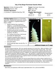
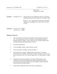
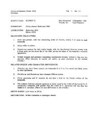
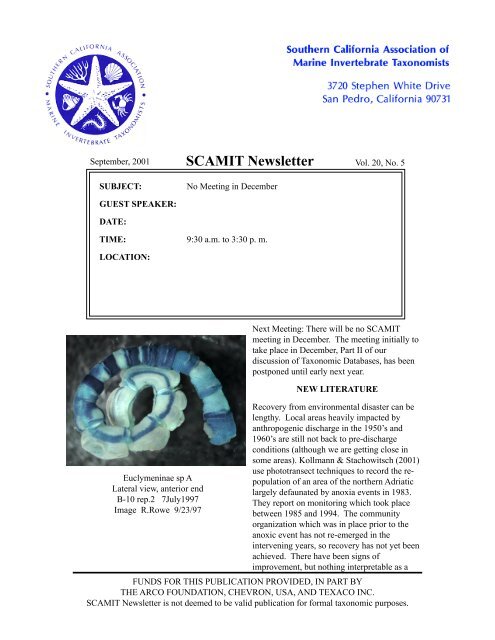
Euclymeninae sp A<br />
Lateral view, anterior end<br />
B-10 rep.2 7July1997<br />
Image R.Rowe 9/23/97<br />
Next Meeting: There will be no <strong>SCAMIT</strong><br />
meeting in December. The meeting initially to<br />
take place in December, Part II of our<br />
discussion of Taxonomic Databases, has been<br />
postponed until early next year.<br />
NEW LITERATURE<br />
Recovery from environmental disaster can be<br />
lengthy. Local areas heavily impacted by<br />
anthropogenic discharge in the 1950’s and<br />
1960’s are still not back to pre-discharge<br />
conditions (although we are getting close in<br />
some areas). Kollmann & Stachowitsch (<strong>20</strong>01)<br />
use phototransect techniques to record the repopulation<br />
of an area of the northern Adriatic<br />
largely defaunated by anoxia events in 1983.<br />
They report on monitoring which took place<br />
between 1985 and 1994. The community<br />
organization which was in place prior to the<br />
anoxic event has not re-emerged in the<br />
intervening years, so recovery has not yet been<br />
achieved. There have been signs of<br />
improvement, but nothing interpretable as a<br />
FUNDS FOR THIS PUBLICATION PROVIDED, IN PART BY<br />
THE ARCO FOUNDATION, CHEVRON, USA, AND TEXACO INC.<br />
<strong>SCAMIT</strong> <strong>Newsletter</strong> is not deemed to be valid publication for formal taxonomic purposes.
<strong>September</strong>, <strong>20</strong>01 <strong>SCAMIT</strong> <strong>Newsletter</strong><br />
<strong>Vol</strong>. <strong>20</strong>, <strong>No</strong>. 5<br />
restoration of pre-event conditions. Similar<br />
macroepifaunal data might be gathered<br />
offshore here in southern California using ROV<br />
video/still camera images.<br />
Consideration of monitoring program design<br />
always raises interesting questions. Paiva<br />
(<strong>20</strong>01) revisits territory covered by earlier<br />
authors with an experiment in shallow (10m)<br />
coastal Brazilian waters. He carefully used a<br />
nested design and repeated sampling at<br />
frequent (2 month) intervals to examine both<br />
spatial and temporal variability. His<br />
experiment was designed to mimic the scales<br />
of variation which might be found in a<br />
sampling program using remote sampling,<br />
although his samples were collected by divers.<br />
The result is not unexpected; significant spatial<br />
variability at all examined scales. One<br />
problem here is the extrapolation to monitoring<br />
design as a whole, and with the basic issue of<br />
sample size not being dealt with by the author.<br />
Given the constraints of many samples (180<br />
were incorporated into the overall design) a<br />
small sample size was chosen (0.008m2) this is<br />
not much larger than a Phleger corer. Samples<br />
of this size of marine macrobenthos are<br />
minimal at best, with each sample collecting<br />
only a relatively small fraction of the<br />
community at any given location. Personal<br />
experience in slightly deeper water with a<br />
slightly larger sampler (0.01m2) was that such<br />
small cores needed considerable replication to<br />
provide statistical power. Power analysis of<br />
the sampling referred to indicated that 17 cores<br />
would be required at each site to allow for<br />
detection of a 30% change in the mean<br />
between stations with 95% confidence. Since<br />
the shallower Brazilian stations are on fine<br />
sand bottom and do not support a particularly<br />
abundant fauna I suspect that even more of the<br />
smaller 0.008m2 cores would be required for<br />
adequate program sensitivity. This remains an<br />
interesting discussion, even if a power analysis<br />
would be likely to demonstrate that the<br />
significant variability witnessed was as much a<br />
result of inadequate sampling intensity as<br />
inherent spatial variability.<br />
2<br />
More and more investigations of wide-ranging<br />
species are adopting molecular techniques to<br />
augment morphology based taxonomy.<br />
Organisms such as scyphomedusae, which<br />
often offer few morphological details for<br />
consideration, represent particularly fertile<br />
ground for molecular examination. Dawson &<br />
Jacobs (<strong>20</strong>01) examined specimens of the<br />
jellyfish Aurelia from various parts of the<br />
world to test the idea that the two currently<br />
recognized taxa might hide additional diversity.<br />
They found that three named species were<br />
supportable on the DNA sequence evidence,<br />
and that among animals identified as Aurelia<br />
aurita, lay an additional 6 unrecognized sibling<br />
species in addition to the named form. On the<br />
west coast of <strong>No</strong>rth America the authors report<br />
two species; Aurelia labiata and Aurelia sp. 1.<br />
The authors indicate these species have<br />
morphological characters which differentiate<br />
them without recourse to collection of<br />
molecular data, but do not detail these<br />
characters in this paper.<br />
The accumulating molecular data for<br />
cnidarians has allowed a new phylogenetic<br />
analysis of the Anthozoa (Won, Rho, & Song<br />
<strong>20</strong>01). The authors performed a combined<br />
analysis using both molecular and<br />
morphological data. Both the morphological<br />
and combined analyses tell the same story, and<br />
both do not differ from previous<br />
morphologically based phylogenies. Within<br />
the anthozoans the major divisions shown in<br />
the analysis corresponded to Alcyonaria,<br />
Zoanthinaria, and Ceriantipatharia. Three<br />
hydrozoans were used as outgroups.<br />
Arthropod relationships are a bit more involved<br />
than those of the anthozoans. Giribet et al<br />
(<strong>20</strong>01) revisit this territory, ground well<br />
trampled by previous visitants. Using a total<br />
evidence approach they combined information<br />
gleaned from a series of molecular sources<br />
with morphological data, to yield a large body<br />
of evidence. The data considered was<br />
comprehensive, and a novel computational<br />
approach using 256 parallel processors was
<strong>September</strong>, <strong>20</strong>01 <strong>SCAMIT</strong> <strong>Newsletter</strong><br />
<strong>Vol</strong>. <strong>20</strong>, <strong>No</strong>. 5<br />
used to allow analysis of such a large amount<br />
of source data. The results strongly support<br />
Pancrustacea (Crustacea + Hexapoda),<br />
Myriapoda, and Chelicerata as monophyletic<br />
clades, with Pycnogonida as sister group.<br />
Onychophorans and tardigrades served as<br />
outgroups. More analyses using even larger<br />
samplings of the accumulating molecular data<br />
will probably follow.<br />
Most discussions of introduced or nonindigenous<br />
species deal with all potential taxa.<br />
Rodrigues & Suarez (<strong>20</strong>01) focus their<br />
attention specifically on decapod crustaceans.<br />
They consider the mechanisms of transport,<br />
and treat a number of introductions which<br />
predate the current upsurge in transfer of<br />
organisms by man. Decapods in particular,<br />
were more likely to move between ecosystems<br />
as adults in the day of the wooden boat. More<br />
modern steel hulled vessels, while making the<br />
voyages more rapidly, also provide much less<br />
opportunity for adult hitch-hiking on the vessel<br />
exterior. The authors do bring up several<br />
interesting reports of transfer on towed oil rigs.<br />
One such event happened here in California,<br />
where the Japanese crab Plagiusia dentata was<br />
introduced on a towed rig which spent over<br />
two months in transit from the western to the<br />
eastern Pacific. When introductions are<br />
discussed parochial approaches are<br />
contraindicated. The fact that the main focus<br />
of this paper is on introductions to Argentina is<br />
irrelevant. Most, if not all of the organisms<br />
discussed may show up on our doorstep at one<br />
time or another.<br />
SAMPLE SORTING TECHNIQUE<br />
While we try not to think of it more than we<br />
have to, the subject of sample sorting is critical<br />
to examinations of the benthos. We generally<br />
do a good job of it based on the results of the<br />
QC examinations of sample sorting undertaken<br />
as part of both the SCPBB program in 1994,<br />
and the recent B’98 Benthic program. It is<br />
time consuming, however, and attempts are<br />
constantly being made to reduce the effort<br />
required to bring sediments, debris, and<br />
organisms to a parting of the ways.<br />
In other areas, including both the Pacific<br />
<strong>No</strong>rthwest and the Western Atlantic, use is<br />
frequently made of vital staining with rose<br />
bengal. This is often touted as being a major<br />
labor saving method, and it may well be for the<br />
sorter or contract administrator. It is often,<br />
however, a nightmare for the taxonomist who<br />
finds many taxonomic clues changed or<br />
eliminated by the practice. Most, if not all<br />
monitoring groups in the Southern California<br />
Bight have avoided using vital staining for<br />
their routine monitoring. There are other<br />
options which can both improve the quality of<br />
the specimens procured by remote samplers,<br />
and simplify the removal of organisms from<br />
benthic sediments.<br />
Member Tom Parker forwarded the following<br />
hints from a posting on the Annelida Listserver.<br />
They originate with Roman Porras and<br />
obviously follow a pre-existing discussion<br />
thread. His comments are worth repeating, and<br />
we do so here:<br />
“RE: Macrofauna separation technologies<br />
As Mary Petersen, I usually use elutriation for<br />
separating macro and meiofauna from<br />
sediment. I put the sample or a fraction of it<br />
into a decantation funnel connected by its<br />
narrow opening to a tap. The water pass across<br />
the sample dragging the fauna, exits by the top<br />
of the funnel and it is conducted to a sieve in<br />
which the animals and debris are retained. If<br />
the sample is previously stained it is easy to see<br />
how the fauna leaves the sediment, after a few<br />
minutes, all animals and debris have been<br />
extracted while the sediment remains in the<br />
funnel. With the tap, you may regulate the flow<br />
at each moment in order to optimise the<br />
extraction.”<br />
3
<strong>September</strong>, <strong>20</strong>01 <strong>SCAMIT</strong> <strong>Newsletter</strong><br />
<strong>Vol</strong>. <strong>20</strong>, <strong>No</strong>. 5<br />
The problem arises when molluscs are present<br />
in the sample since they are difficult to separate<br />
from sediment and it is necessary to extract<br />
them by hand. However, Robinson and<br />
Chandler (1993) describe a technique to<br />
separate molluscs:<br />
“A method is presented which enables infaunal<br />
juvenile bivalves to be separated from<br />
associated sediment with a combination of<br />
elutriation and flotation techniques. Simple<br />
elutriation is used to remove less dense organic<br />
detritus and a relatively new, nontoxic<br />
compound (sodium polytungstate) is used to<br />
create a heavy liquid that sorts the bivalves<br />
from sediment by relative density. The<br />
technique was applicable to 12 species of<br />
molluscs of various sizes and was 98-100%<br />
effective in separating juvenile Mya arenaria,<br />
ranging from 0.5 to 24 mm in shell length,<br />
from surrounding sediment.”<br />
Gravimetric separation for mollusks is a<br />
technique with a long history. While working<br />
for Dr. Jim McLean at NHMLAC in the 60’s<br />
your editor often used it. In that application,<br />
however, mollusks were the only target.<br />
Samples were air-dried (too bad for all you<br />
worms and crustaceans!) then floated on<br />
carbon tetrachloride. While this offered the<br />
appropriate density for separating dried shelled<br />
mollusks, it was also highly toxic. The<br />
suggestion of using sodium polytungstate<br />
seems a great advance over the earlier<br />
technique. An even older technique was used<br />
by the 49ers’ of the California gold rush which<br />
relies not on heavy fluids but on plain water. It<br />
still is a good technique in the hands of an<br />
experienced worker. Using a dish or bowl of<br />
the appropriate cross-section and swirling it<br />
with the proper motion, mollusks can be<br />
mobilized and swished onto a screen, leaving<br />
nearly all the coarse sediment behind. The<br />
resulting blend of a little sand/gravel and shell<br />
debris + living shelled mollusks can then be<br />
sorted in very little time. If the procedure is<br />
repeated until no more mollusks are removed<br />
in a wash (5 or 6 swishes generally suffice) a<br />
4<br />
brief scan of the remaining sediments should<br />
verify that they are clear of animals. Using this<br />
technique on coarser mixed sediments from<br />
shallow high-energy environments can reduce<br />
processing time for a sample by over 80%,<br />
while providing good, repeatable removal of<br />
organisms. The technique can be used in<br />
conjunction with a previous elutriation, or the<br />
debris and floatable organisms (low density<br />
items like crustaceans and annelids) can be left<br />
in to be swish washed out with the mollusks.<br />
24 <strong>September</strong> Meeting Minutes<br />
The meeting was called to order by President<br />
Ron Velarde at 9:45. Vice-president Leslie<br />
Harris informed us about upcoming meetings.<br />
The October meeting is scheduled for two<br />
days, October 9 th and 10 th , at the Los Angeles<br />
County Museum of Natural History<br />
(LACMNH). On the first day, Don Cadien and<br />
Dean Pasko will make a presentation on<br />
Phoxocephalids. On the second day, John<br />
Chapman will make a presentation on<br />
Corophiids. Attendees are welcome to bring<br />
their unidentified amphipods. The <strong>No</strong>vember<br />
meeting will occur on the 5th , will also be<br />
held in Los Angeles and Angel Valdés will be<br />
the guest speaker with the topic being<br />
nudibranchs. There is no scheduled meeting<br />
for December. In January the meeting will be<br />
held at Dancing Coyote Ranch and the topic<br />
will be Edwarsiids. For the February meeting,<br />
we will exchange specimens of Pista for the<br />
round robin exercise that was discussed during<br />
last June’s meeting. The results of the<br />
specimen exchange will be discussed at the<br />
March meeting. Discussions of several smaller<br />
groups will be presented at the February<br />
meeting. Megan Lilly will discuss a new<br />
holothuroid, a new ascidian and possibly<br />
somethng about Octopus... Kelvin Barwick<br />
will follow up on entoprocts, and Don Cadien<br />
will revisit the cumacean genus Cyclaspis.
<strong>September</strong>, <strong>20</strong>01 <strong>SCAMIT</strong> <strong>Newsletter</strong><br />
<strong>Vol</strong>. <strong>20</strong>, <strong>No</strong>. 5<br />
A couple of ideas for future meetings were also<br />
mentioned. A meeting to further discuss<br />
taxonomic databases is pending. Phil Hoover,<br />
a new curatorial assistant at LACMNH, might<br />
be discussing an Amphipod paper at a future<br />
meeting.<br />
The Western Society of Naturalists annual<br />
meeting will be held <strong>No</strong>vember 9-12 in<br />
Ventura, California. For more information, go<br />
to their website at:<br />
www.wsn-online.org.<br />
The guest speaker for the day was Larry<br />
Lovell, and the topic was Euclymeninae<br />
reported from the Bight ‘98 program. Larry<br />
provided handouts to those attending. A copy<br />
of those handouts edited to reflect the results of<br />
the meeting are at the end of the newsletter.<br />
Larry discussed the results of his work on the<br />
Bight ‘98 Euclymeninae. Among the families<br />
(considering all taxa) recorded for the Bight<br />
‘98 program, the Maldanidae ranked 8 th in<br />
abundance. There were 4608 individuals.<br />
Euclymeninae accounted for 81.1% of the<br />
Maldanids (3770 individuals). Euclymeninae<br />
sp A was the most abundant euclymenid with<br />
1<strong>20</strong>1 individuals, and Petaloclymene pacifica<br />
was second with 996 individuals.<br />
We then reviewed Larry’s key to the<br />
subfamilies of Maldanidae from Southern<br />
California which follows the subfamily<br />
classification as presented in Fauchald (1977).<br />
We next looked at the key to the Euclymeninae<br />
of Southern California and reviewed it, couplet<br />
by couplet. Methyl green staining pattern is<br />
used as one of the main diagnostic characters<br />
in this key. Larry explained that in couplet 9,<br />
the phrase “better developed stain” means a<br />
more intense stain that is denser and broader<br />
(covering more surface area of the animal).<br />
The question was asked, “What tissue type<br />
uptakes methyl green stain” Larry answered<br />
that the stain is picked up by mucoid/glandular<br />
areas. He referred us to Arwidsson 1907 for<br />
more details.<br />
5<br />
Larry also brought an assortment of older<br />
illustrations (mostly of methyl green staining<br />
patterns), voucher sheets, and reference<br />
material for people to browse through and copy<br />
if they did not already have one. Included<br />
among these were identification sheets for<br />
Maldanidae sp 1, Maldanidae sp 2, and<br />
Petaloclymene pacifica. These sheets included<br />
illustrations of methyl green staining patterns<br />
for each species. Larry noted that Maldanidae<br />
sp 1 may turn out to be a described species,<br />
because all the specimens collected have been<br />
relatively small.<br />
There was a question about distinguishing the<br />
species in couplet 11. In “Clymenura”<br />
gracilis, the glandular area on setiger 8 forms a<br />
complete band, and lateral notches are absent<br />
on the prostomium. In C. columbiana,<br />
however, the glandular area on setiger 8 is a<br />
ventral, spade-shaped region, and lateral<br />
notches are present on the prostomium.<br />
Larry commented that he made identifications<br />
of all specimens based on the anterior portions.<br />
The posterior ends of Euclymenids were sorted<br />
into the fragment vials which Larry did not<br />
receive. Identifications can be problematic<br />
unless a minimum of eight or nine setigers<br />
were present. Larry said he can identify<br />
Clymenella species with a minimum of four<br />
segments.<br />
After a 10 minute break, we began to look at<br />
specimens. There was a question regarding a<br />
size limitation for methyl green stain.<br />
According to Larry, there is a less developed<br />
and less intense stain in juveniles. Within a<br />
size series, one can see the progression of stain<br />
patterns. Under the microscope first was a<br />
stained specimen of Axiothella. The specimen<br />
we looked at was collected from station 2491<br />
at a depth of 95 m. On setiger 8, the presetal<br />
area did not stain, and the postsetal area stained<br />
dark. On setigers 4 through 8, the pre-setal<br />
area stained light green, and the post-setal area<br />
stained dark green. This specimen was similar<br />
to A. rubrocincta, but Leslie pointed out that A.
<strong>September</strong>, <strong>20</strong>01 <strong>SCAMIT</strong> <strong>Newsletter</strong><br />
<strong>Vol</strong>. <strong>20</strong>, <strong>No</strong>. 5<br />
rubrocincta has a dark presetal stain on setigers<br />
4 through 8. We concluded that this is an<br />
undescribed species of Axiothella. Leslie said<br />
that she has notes on several undescribed<br />
species of Axiothella and that this is one of<br />
them. Therefore, all references to A.<br />
rubrocincta in Larry’s handouts should be<br />
changed to Axiothella sp. We examined a<br />
smaller specimen which had the same staining<br />
pattern. It was collected at station 2522 (Santa<br />
Cruz Island) at a depth of 80 m. Larry next<br />
looked at a small specimen of Axiothella<br />
rubrocincta collected from an International<br />
Treatment Plant station sampled by the City of<br />
San Diego. From a ventral view, there was<br />
dark solid stain on the anterior areas of setigers<br />
5 through 8. The posterior areas of setigers 5<br />
through 8 stained a darker but diffuse green.<br />
Most of the participants agreed they would<br />
mis-identify this specimen as A. rubrocincta. It<br />
was pointed out that the staining pattern<br />
illustrated in one of Leslie’s handouts had been<br />
copied very darkly for effect and unfortunately,<br />
did not represent the true staining pattern.<br />
Leslie added a cautionary note not to identify<br />
specimens based solely on staining pattern.<br />
Taxonomists should look at other characters,<br />
because she has noted that more than one<br />
species can have the same staining pattern.<br />
Checking for other characters can be<br />
particularly important when identifying<br />
juveniles of Axiothella because the rostrate<br />
setae in setigers 1 through 3 may not be fully<br />
formed.<br />
Next up was a specimen of Clymenella<br />
complanata. We located the lateral notch on<br />
the 4 th setiger which is a unique character for<br />
this species. It was suggested that you can slip<br />
the tip of your forcep under this notch to help<br />
in locating it. C. complanata had a deep<br />
transverse groove and nuchal organs on the<br />
cephalic plaque. This specimen was collected<br />
at station 2<strong>20</strong>9, off Orange County, at a depth<br />
of 65 m.<br />
The next specimen we examined was<br />
Clymenella sp A of Harris 1985. There was a<br />
handout for this species prepared by Leslie.<br />
Larry stated that Clymenella sp A was recorded<br />
in samples from Marine Ecological Consultants<br />
(MEC) only and was not recorded from other<br />
laboratories participating in Bight ‘98. We had<br />
a close-up view of the cephalic plaque. There<br />
were primary and secondary transverse<br />
grooves, and the nuchal organs were<br />
perpendicular to these grooves.<br />
We next examined a specimen of “Clymenura”<br />
gracilis. Larry put the genus in quotes,<br />
because it is suspected that this may not be the<br />
correct genus in that it does not follow the<br />
pattern of a ventral glandular area as do other<br />
species of Clymenura. In “Clymenura”<br />
gracilis, the glandular girdle extends all the<br />
way around the 8th setiger.<br />
The next worm we examined was Clymenura<br />
columbiana. All the specimens that Larry<br />
recorded were quite small, about .5 mm wide.<br />
On the ventrum of setiger 8, there was a spadeshaped,<br />
glandular area. This area stained green<br />
but the dark stain did not extend all the way<br />
around the setiger. On the dorsum of setiger 8,<br />
there was a thin band of light speckling. This<br />
worm was collected from station 2491, San<br />
Miguel Island, at a depth of 95 m. This station<br />
had course sediment.<br />
After lunch, we got back to work and looked at<br />
Euclymene campanula. Larry believes that<br />
specimens of E. campanula orient themselves<br />
anterior end down in the sediment resulting in<br />
the collection of mostly posterior ends. Larry<br />
pointed out that this is an example of a<br />
situation where it would be useful to count a<br />
posterior fragment as an individual. The<br />
posterior end of E. campanula was<br />
distinguished by 7 asetigerous segments, each<br />
with a pronounced ridge (or flanges) on the<br />
posterior part of the segment. The pygidium<br />
had a ring of short anal cirri. In this species,<br />
the number of anal cirri varies with size of the<br />
animal, but they are uniformly short in length.<br />
6
<strong>September</strong>, <strong>20</strong>01 <strong>SCAMIT</strong> <strong>Newsletter</strong><br />
<strong>Vol</strong>. <strong>20</strong>, <strong>No</strong>. 5<br />
Larry stained the specimen; the first 4 setigers<br />
stained green. The anterior portions of setigers<br />
5 through 8 stained green, giving a banded<br />
appearance. The ridges (or flanges) in the<br />
posterior end also stained green.<br />
A specimen of Isocirrus longiceps was up next.<br />
The posterior rim of the cephalic plaque came<br />
together to form a “V”. The cephalic plaque<br />
had multiple transverse grooves. There was a<br />
collar on the anterior end of setiger 4, .<br />
We then viewed a specimen of Petaloclymene<br />
pacifica. The methyl green stain on setiger 8<br />
extended both pre-tori and post-tori. There was<br />
a darker, green oval patch on the ventrum of<br />
setiger 8 on both sides of the tori. We located<br />
the dorsal pores on setigers 7-9 which is a<br />
character that distinguishes P. pacifica from<br />
other local fauna.<br />
Next, Larry brought out a specimen of<br />
Euclymene grossa newporti. We referred to<br />
Leslie’s older handout from 1985 with<br />
illustrations of the stain patterns. There was a<br />
dark green stain on presetal and postsetal areas<br />
on setiger 8. The staining pattern of this<br />
specimen did not exactly match the staining<br />
pattern of the illustration. There was a<br />
question about the identification, and Leslie<br />
suggested the type specimen should be<br />
examined and compared to this specimen.<br />
The next worm under the microscope was a<br />
specimen of Praxillella pacifica. The methyl<br />
green stain extends to the tori on setiger 8, but<br />
it does not extend beyond that. On segment 2,<br />
there was a lightly-stained, oval-shaped area.<br />
We referred to this as the “collar area”.<br />
Then we examined a specimen of Praxillella<br />
gracilis. There was a long, thin anterior<br />
palpode on the prostomium. The methyl green<br />
stain did not extend past the tori on setiger 8.<br />
A distinguishing feature on the next specimen,<br />
Maldanella robusta, was the absence of<br />
neurosetae on setiger one. The two nuchal<br />
organs on the cephalic plaque were V-shaped<br />
and anteriorly placed. The cephalic rim was<br />
fairly smooth. The pygidium of this worm had<br />
a scalloped edge. There was some discussion<br />
as to whether this specimen was M. harai, but<br />
we concurred the identification should remain<br />
as M. robusta.<br />
Larry then showed us a large specimen of<br />
Maldanidae sp 2 of Lovell and Phillips 1995<br />
provided by Tony Phillips for the meeting.<br />
Larry commented that all specimens he had<br />
previously seen had been much smaller. The<br />
m.g. staining pattern was like that of<br />
Euclymene campanula. Leslie confirmed the<br />
identification.<br />
The last species up for discussion that day was<br />
Maldanidae sp 1 of Lovell and Phillips 1995.<br />
Only small specimens of this species have been<br />
found. The cephalic rim was low and not well<br />
developed. There were single rostrate uncini<br />
on the first 3 setigers. Methyl green stain<br />
revealed a slight anterior band on the first<br />
setiger and well defined anterior bands on<br />
setigers 2 through 7. There was no stain after<br />
setiger 7 except around the uncini on setigers 8<br />
and 9. The pygidium had one long cirrus and<br />
about <strong>20</strong> short cirri. Leslie looked at the<br />
specimens and said she believed these are<br />
juvenile Euclymeninae sp A <strong>SCAMIT</strong> 1987<br />
commenting that the juveniles lack the double<br />
staining stripes that are seen in adults. She felt<br />
confident of the m.g. staining growth pattern<br />
she has seen from juvenile to adult.<br />
JOBS<br />
The San Francisco Estuary Institute is seeking<br />
a Program Manager for the Regional<br />
Monitoring Program. For more information,<br />
please see the attachment at the end of the<br />
newsletter (paper version) or visit their website<br />
at:<br />
www.sfei.org<br />
SIDE NOTE<br />
The email addresses for both Ron Velarde and<br />
Megan Lilly have changed. Please notice the<br />
new addresses in the informatin box. - M. Lilly<br />
7
<strong>September</strong>, <strong>20</strong>01 <strong>SCAMIT</strong> <strong>Newsletter</strong><br />
<strong>Vol</strong>. <strong>20</strong>, <strong>No</strong>. 5<br />
BIBLIOGRAPHY<br />
Arwiddson, I. (1907). Studien über die skandinavischen und arktischen Maldaniden nebst<br />
Zusammenstellung der übringen bisher bekannten Arten dieser Familie. Zoologisches<br />
Jahrbüch Supplement 9: 1-308.<br />
Dawson, Michael N., and David K. Jacobs. <strong>20</strong>01. Molecular evidence for cryptic species of<br />
Aurelia aurita (Cnidaria, Scyphozoa). Biological Bulletin <strong>20</strong>0:92-96.<br />
Day, J. H. (1976). A Monograph on the Polychaeta of Southern Africa. British Museum of<br />
Natural History Publication 656. Trustees of the British Museum (Natural History):<br />
London. 2 vols: Pt 1, Errantia pp. 1-458; Pt 2, Sedentaria pp. 459-878.<br />
Detinova, N.N. (1985). Polychaetous annelids of Reykjanes Ridge (<strong>No</strong>rth Atlantic). [Bottom<br />
Fauna from Mid-Ocean Rises in the <strong>No</strong>rth Atlantic]. Trudy Instituta Okeanologii 1<strong>20</strong>:<br />
96-136.<br />
Fauchald, K. (1977). The polychaete worms. Definitions and keys to the orders, families, and<br />
genera. Natural History Museum of Los Angeles County, Science Series 28: 1-188.<br />
Giribet, Gonzalo, Gregory D. Edgecombe, and Ward C. Wheeler. <strong>20</strong>01. Arthropod phylogeny<br />
based on eight molecular loci and morphology. Nature 413:157-161.<br />
Hartman, O. & Fauchald, K. (1971). Deep-water benthic polychaetous annelids off New<br />
England to Bermuda and other <strong>No</strong>rth Atlantic Areas. Part II. Allan Hancock<br />
Monographs in Marine Biology 6: 1-327.<br />
Imajima, I. & Shiraki, Y. (1982a). Maldanidae (Annelida:Polychaeta) from Japan (Part 1).<br />
Bulletin of the National Science Museum, Tokyo 8: 1-46.<br />
Imajima, I. & Shiraki, Y. (1982b). Maldanidae (Annelida:Polychaeta) from Japan (Part 2).<br />
Bulletin of the National Science Museum, Tokyo 8: 47-88.<br />
Kollmann, Herbert, and Michael Stachowitsch. <strong>20</strong>01. Long-term changes in the benthos of the<br />
northern Adriatic Sea: a phototransect approach. Marine Ecology 22(1-2):135-154.<br />
Paiva, P. C. <strong>20</strong>01. Spatial and temporal variation of a nearshore benthic community in southern<br />
Brazil: implications for the design of monitoring programs. Estuarine, Coastal and Shelf<br />
Science 52:423-433.<br />
Robinson, S. M. C., and R. A. Chandler. 1993. An effective and safe method for sorting small<br />
molluscs from sediment. Limnology and Oceanography 38(5):1088-1091<br />
Rodríguez, Gilberto, and Héctor Suárez. <strong>20</strong>01. Anthropogenic dispersal of decapod crustaceans<br />
in aquatic environments. Interciencia 26(1):282-288.<br />
Rouse, G.W. (<strong>20</strong>00). Family Maldanidae. Pp. 74-76 in Beesley, P. L., Ross, G.J.B. & Glasby, C.<br />
J.(eds) Polychaetes & Allies: The Southern Synthesis, Fauna of Australia. <strong>Vol</strong>. 4A<br />
Polychaeta, Myzostomida, Pogonophora, Echiura, Sipuncula. CSIRO Publishing :<br />
Melbourne xii 465 pp.<br />
Wolf, P.S. (1983). A revision of the Bogueidae Hartman and Fauchald 1971, and its reduction to<br />
Bogueinae, a subfamily of Maldanidae (Polychaeta). Proceedings of the Biological<br />
Society of Washington 96: 238-249.<br />
Won, J. H., B. J. Rho, and J. I. Song. <strong>20</strong>01. A phylogenetic study of the Anthozoa (Phylum<br />
Cnidaria) based on morphological and molecular characters. Coral Reefs <strong>20</strong>:39-50.<br />
8
<strong>September</strong>, <strong>20</strong>01 <strong>SCAMIT</strong> <strong>Newsletter</strong><br />
<strong>Vol</strong>. <strong>20</strong>, <strong>No</strong>. 5<br />
Please visit the <strong>SCAMIT</strong> Website at: http://www.scamit.org<br />
<strong>SCAMIT</strong> OFFICERS:<br />
If you need any other information concerning <strong>SCAMIT</strong> please feel free to contact any of the<br />
officers e-mail address<br />
President Ron Velarde (619)758-2331 rvelarde@sandiego.gov<br />
Vice-President Leslie Harris (213)763-3234 lhharris@bcf.usc.edu<br />
Secretary Megan Lilly (619)758-2336 mlilly@sandiego.gov<br />
Treasurer Ann Dalkey (310)648-5544 cam@san.ci.la.ca.us<br />
Back issues of the newsletter are available. Prices are as follows:<br />
<strong>Vol</strong>umes 1 - 4 (compilation)................................. $ 30.00<br />
<strong>Vol</strong>umes 5 - 7 (compilation)................................. $ 15.00<br />
<strong>Vol</strong>umes 8 - 15 ................................................ $ <strong>20</strong>.00/vol.<br />
Single back issues are also available at cost.
<strong>September</strong>, <strong>20</strong>01 <strong>SCAMIT</strong> <strong>Newsletter</strong><br />
<strong>Vol</strong>. <strong>20</strong>, <strong>No</strong>. 5<br />
Euclymeninae Reported from the<br />
Bight ’98 Program<br />
Lawrence L. Lovell<br />
The author identified all specimens of<br />
Euclymeninae from the Bight ‘98 project. This<br />
workshop presents the taxonomic characters<br />
and techniques used to identify those<br />
specimens. Dependable morphological<br />
characters and methyl green staining patterns<br />
were used to identify material to species level.<br />
Malmgren erected the family Maldanidae in<br />
1867. Arwidsson (1907) subsequently divided<br />
the family into five subfamilies; Euclymeninae,<br />
Lumbriclymeninae, Maldaninae,<br />
Nicomachinae, and Rhodininae. Three<br />
additional subfamilies have recently been<br />
proposed. Clymenurinae by Imajima and<br />
Shiraki (1982a), <strong>No</strong>toproctinae by Detinova<br />
(1985), and Boguinae by Wolf (1983) in<br />
moving the family Boguidae (Hartman and<br />
Fauchald 1971) into the Maldanidae.<br />
Important taxonomic publications on the<br />
family are by Arwidsson (1907), Day (1967),<br />
Fauchald (1977), Imajima and Shiraki (1982a,<br />
1982b), and Rouse (<strong>20</strong>00). For the purposes of<br />
this project the subfamilies presented in<br />
Fauchald (1977) are followed retaining the<br />
later proposed Clymenurinae within the<br />
subfamily Euclymeninae.<br />
The Euclymeninae are characterized by having<br />
both anterior and posterior ends with plaques<br />
and the anus terminally oriented. A cephalic<br />
rim, keel, and nuchal slits are present on the<br />
prostomium. The margin of the posterior<br />
plaque may be smooth, crenulate, or bordered<br />
by anal cirri and the anal cone may be<br />
projecting beyond the rim or low and not<br />
projecting beyond the rim. Some taxa have<br />
segmental collars or well-defined glandular<br />
areas in the thorax. <strong>No</strong>tosetae are capillary.<br />
Anterior neurosetae can be either acicular<br />
spines or rostrate uncini. It is thought that<br />
some species may drop their rostrate uncini and<br />
add acicular spines as they get older. These<br />
traditional characters have not been wholly<br />
adequate to identify specimens encountered in<br />
regional monitoring programs.<br />
The Euclymeninae are well-represented in<br />
southern California coastal shelf sediments.<br />
However, the tendency for them to fragment<br />
when collected has been a problem for<br />
taxonomists attaining species level<br />
identifications. Some particularly large<br />
specimens of certain taxa may have only their<br />
rear ends collected due to their large size and<br />
vertical head-down orientation in the<br />
sediments. When this occurs, programs that do<br />
not count these posterior fragments will miss<br />
the opportunity to add information on rarely or<br />
incompletely sampled taxa to their database.<br />
Attaining complete specimens for taxonomic<br />
analysis begins in the field. Gentle screening<br />
of sediment samples or use of a float table in<br />
the field will help keep fragmentation to a<br />
minimum. The amount of fragmentation is<br />
directly related to water pressure and rough<br />
handling. Use of a relaxant prior to fixation is<br />
recommended to further prevent fragmentation<br />
when the animals are exposed to Formalin.<br />
Subsequent handling by sorters and technicians<br />
performing biomass measurements are another<br />
possible cause of fragmentation. If the object<br />
is to have specimens that can be identified,<br />
then care must be taken prior to the taxonomy<br />
to provide material in good condition. As the<br />
old saying goes “Garbage in, garbage out!” or<br />
in this case “fragments in, no species IDs out”!<br />
Your database will be much cleaner and<br />
analyses more meaningful when a higher<br />
percentage of animals can be identified to<br />
species.<br />
Methyl green staining procedures follow those<br />
generally discussed by <strong>SCAMIT</strong> members at<br />
numerous meetings. A working solution dark<br />
enough to stain in a reasonable amount of time<br />
(10-15 minutes), but weak enough to see<br />
animals through the solution to pull them out,<br />
was used. As the solution becomes weaker
<strong>September</strong>, <strong>20</strong>01 <strong>SCAMIT</strong> <strong>Newsletter</strong><br />
<strong>Vol</strong>. <strong>20</strong>, <strong>No</strong>. 5<br />
through use and uptake by animals, additional<br />
stock solution of darkly mixed stain is added to<br />
bring the working solution back to working<br />
strength. There are no methyl green solution<br />
formulas suggested and each taxonomist must<br />
decide what works best for them in their<br />
particular working conditions to achieve<br />
workable staining of adequate strength. In any<br />
case, there is usually some destaining that will<br />
need to take place before some staining<br />
patterns will be discernable. Just as<br />
morphological character states develop and<br />
change with size and maturity, so do methyl<br />
green staining patterns. Juvenile patterns will<br />
look different or incomplete until placed in<br />
context with the overall development of the<br />
adult pattern.<br />
Axiothella rubrocincta (Johnson 1901)<br />
Axiothella sp.<br />
Clymenella complanata Hartman 1969<br />
Clymenella sp. A of Harris 1985<br />
Clymenura columbiana (E. Berkeley 1929)<br />
“Clymenura” gracilis Hartman 1969<br />
Euclymene campanula Hartman 1969<br />
Bight ’98 Euclymeninae<br />
By Lawrence L. Lovell<br />
Euclymene grossa newporti Berkeley and Berkeley 1941<br />
Euclymeninae sp. A <strong>SCAMIT</strong> 1987<br />
Isocirrus longiceps (Moore 1923)<br />
Maldanella robusta Moore 1906<br />
Petaloclymene pacifica Green 1997<br />
Praxillella gracilis (M. Sars 1861)<br />
Praxillella pacifica E. Berkeley 1929<br />
Axiothella sp., Clymenura columbiana, Euclymene grossa newporti, and Maldanella robusta have<br />
not been previously reported by POTW monitoring agencies and are not in Edition 3 of the Taxonomic<br />
Listing of Soft-Bottom Macro- and Megainvertebrates from Infaunal and Epibenthic Monitoring<br />
Programs in the Southern California Bight. Of the Euclymeninae taxa listed in Edition 3, only<br />
Euclymene delineata was not reported for the Bight ’98 project. Further discussion with the original<br />
POTW’s reporting E. delineata (LACSD and Hyperion) indicate that these were misidentified<br />
specimens from the 70’s and the labs no longer include that name in their species lists.
<strong>September</strong>, <strong>20</strong>01 <strong>SCAMIT</strong> <strong>Newsletter</strong><br />
<strong>Vol</strong>. <strong>20</strong>, <strong>No</strong>. 5<br />
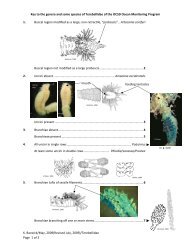
Key to the Subfamilies* of Maldanidae from Southern California<br />
By Lawrence L. Lovell<br />
1. Both cephalic and anal plaques absent ................................................ 2<br />
1. At least an anal plaque present ............................................................ 3<br />
2. Rostrate uncini in double rows, posterior segments with<br />
encircling collars .............................................................. Rhodininae<br />
2. Rostrate uncini in single rows, posterior segments<br />
not collared .......................................................... Lumbriclymeninae<br />
3. Cephalic plaque absent, anal plaque present .............. Nichomachinae<br />
3. Both cephalic and anal plaques present .............................................. 4<br />
4. Anus dorsal ..................................................................... Maldaninae<br />
4. Anus terminal ................................................................ Euclymeninae<br />
*This key follows the subfamily classification as presented in Fauchald<br />
(1977).
<strong>September</strong>, <strong>20</strong>01 <strong>SCAMIT</strong> <strong>Newsletter</strong><br />
<strong>Vol</strong>. <strong>20</strong>, <strong>No</strong>. 5<br />
Key to the Euclymeninae of Southern California<br />
By Lawrence L. Lovell<br />
1. Neurosetae absent on setiger one ............... ... ........Maldanella robusta<br />
1. Neurosetae present on setiger one ................. ......................................2<br />
2. Setiger four with deep encircling collar .............................................. 3<br />
2. Setiger four without collar ................................................................... 4<br />
3. Acicular spine count for setigers 1-3:1, 1, 1; 4-5 transverse folds on<br />
cephalic plaque; lateral edges meet in V-shape at rear of<br />
prostomium............................................................. Isocirrus longiceps<br />
3. Acicular spine count for setigers 1-3: 1, 1 / 2, 1 / 2; single transverse<br />
fold on cephalic plaque; lateral edges rounded at rear of prostomium<br />
...................................................................... .. Clymenella complanata<br />
3. Acicular spine count for setigers 1-3: 2, 2 / 3, 3 / 4; two transverse<br />
folds on cephalic plaque; lateral edges rounded at rear of<br />
prostomium...................................... Clymenella sp. A of Harris 1985<br />
4. Methyl green stain on setigers 4-7 is well developed on both pre and<br />
post setal areas .................................................................................... 5<br />
4. Methyl green stain on setigers 4-7 is well developed on the pre setal<br />
area only ............................................................................................ 8<br />
5. Methyl green stain on setiger 8 on both pre and postsetal areas ..........6<br />
5. Methyl green stain on setiger 8 on presetal area only ..........................7<br />
6. Neurosetae of setigers 1-3 with 4-8 neurosetae; dorsal pores absent,<br />
ventral pores on setigers 7-9........................................... Axiothella sp.<br />
6. Neurosetae of setigers 1-3 with single neurosetae; dorsal pores absent,<br />
ventral pores on setigers 6-9 ..................Euclymene grossa newporti<br />
6. Neurosetae of setigers 1-3 with 2-4 neurosetae, dorsal pores on setigers<br />
7-9, ventral pores on setigers 7-9 .....................Petaloclymene pacifica<br />
7. Prostomium with long thin anterior palpode ........ Praxillella gracilis<br />
7. Prostomium with short rounded anterior palpode ................. P. pacifica
<strong>September</strong>, <strong>20</strong>01 <strong>SCAMIT</strong> <strong>Newsletter</strong><br />
<strong>Vol</strong>. <strong>20</strong>, <strong>No</strong>. 5<br />
8. Methyl green stain after setiger 8 with racing stripes<br />
.................................................. Euclymeninae sp. A <strong>SCAMIT</strong> 1987<br />
8. Methyl green stain after setiger 8 without racing stripes ..................... 9<br />
9. Methyl green staining area better developed in early thoracic setigers,<br />
with lateral unstained line in segments 1-4, thickened presetal flanges<br />
develop in posterior segments ......................... Euclymene campanula<br />
9. Methyl green staining area better developed in later thoracic setigers,<br />
lateral unstained line absent, presetal flanges absent in posterior<br />
segments ............................................................................................ 10<br />
10. Glandular band on setiger 8 a complete band of similar size to<br />
previous segments, slight lateral notches present on prostomium<br />
.......................................................................... Axiothella rubrocincta<br />
10. Glandular area on setiger 8 a complete band, better developed<br />
ventrally than on previous segments; lateral notches absent on<br />
prostomium ............................................................ Clymenura gracilis<br />
10. Glandular area on setiger 8 ventral, spade-shaped; lateral notches<br />
present on prostomium ................................... Clymenura columbiana
<strong>September</strong>, <strong>20</strong>01 <strong>SCAMIT</strong> <strong>Newsletter</strong><br />
<strong>Vol</strong>. <strong>20</strong>, <strong>No</strong>. 5<br />
Bight ’98 Taxa Ranking by Family Levels<br />
1. Spionidae 29287 Polychaeta<br />
2. Sabellidae 9792 Polychaeta<br />
3. Capitellidae 9281 Polychaeta<br />
4. Amphiuridae 7841 Ophiuroidea<br />
5. Lumbrineridae 6572 Polychaeta<br />
6. Terebellidae 5783 Polychaeta<br />
7. Mytilidae 5353 Bivalvia<br />
8. Maldanidae 4608 Polychaeta<br />
(Euclymeninae 3770 81.8 % of Maldanidae)<br />
9. Cirratulidae 4593 Polychaeta<br />
10.Ampharetidae 4039 Polychaeta