Chapter 18: Cyclovertical Deviations
Chapter 18: Cyclovertical Deviations
Chapter 18: Cyclovertical Deviations

You also want an ePaper? Increase the reach of your titles
YUMPU automatically turns print PDFs into web optimized ePapers that Google loves.
CHAPTER<br />
<strong>18</strong><br />
<strong>Cyclovertical</strong> <strong>Deviations</strong><br />
The diagnosis and management of cyclovertical<br />
deviations are special challenges to the ophthalmologist.<br />
There are several disorders that on<br />
first glance appear similar clinically but differ<br />
widely in etiology and management. As in no<br />
other aspect of strabismology, correct diagnosis is<br />
of utmost importance, since an operation performed<br />
on the basis of an erroneous interpretation<br />
of the underlying problem may cause disastrous<br />
and permanent consequences with respect to the<br />
patient’s binocular function. Once the correct diagnosis<br />
has been made, medical and surgical management<br />
of such deviations does not present any<br />
special problems, and the therapeutic results can<br />
be the most gratifying in the field of strabismus.<br />
<strong>Cyclovertical</strong> deviations differ from horizontal<br />
deviations in several aspects. Sensorial adaptations<br />
in the form of amblyopia or anomalous retinal<br />
correspondence are noted far less frequently with<br />
this type of deviation than with horizontal strabismus.<br />
Comitance is rare, and the deviation is generally<br />
smaller in magnitude, yet the size of a cyclovertical<br />
deviation is not an indication of the extent<br />
of the problem caused for the patient. Although<br />
some patients with well-developed binocular functions<br />
often are able, by motor fusion, to overcome<br />
surprisingly large vertical deviations, the low fusional<br />
reserve in the vertical directions in most<br />
others precludes this compensatory mechanism.<br />
Consequently, a hyperdeviation of only 1 or 2 <br />
can cause diplopia or blurring of vision, especially<br />
during reading. Such small residual hyperdeviations<br />
following surgical alignment of horizontal<br />
strabismus are of special clinical significance, for<br />
they may present insurmountable obstacles to a<br />
functional cure.<br />
The prevalence of cyclovertical deviations in<br />
association with horizontal deviations or as isolated<br />
anomalies is high. White and Brown 128 observed<br />
that in patients with motility disorders,<br />
approximately half had isolated vertical anomalies<br />
and another third had combined horizontal and<br />
vertical muscle problems. Scobee 103 found a vertical<br />
component in 43% of 457 patients with esotropia.<br />
Bielschowsky 7 classified cyclovertical deviations<br />
into five groups: (1) purely comitant vertical<br />
deviations, (2) vertical deviations of paretic origin,<br />
(3) deviations with unilateral overaction of the<br />
inferior oblique muscles, (4) dissociated vertical<br />
deviations, and (5) vertical deviations combined<br />
with features of several of the other groups. This<br />
classification is still of some usefulness today even<br />
though we have learned since Bielschowsky that<br />
elevation in adduction is not exclusively caused<br />
by an overacting inferior oblique muscle.<br />
In this chapter nonparalytic and nonmechanical<br />
cyclodeviations are described. Paralytic deviations<br />
are discussed in <strong>Chapter</strong> 20, and hyperdeviations<br />
caused by mechanical factors (endocrine myopathy,<br />
congenital fibrosis, and orbital floor fractures)<br />
are described in <strong>Chapter</strong> 21.<br />
Comitant Hyperdeviations<br />
Etiology and Clinical Characteristics<br />
Truly comitant hyperdeviations occur infrequently.<br />
To find a patient with a significant vertical devia-<br />
377
378 Clinical Characteristics of Neuromuscular Anomalies of the Eye<br />
tion of the same magnitude in all positions of gaze<br />
with either eye fixating and with the head tilted to<br />
2, p.12<br />
either shoulder is indeed unusual. Anderson,<br />
in a survey of 600 patients with cyclovertical<br />
anomalies, was unable to find a single truly comitant<br />
deviation. Repeated measurements in the diagnostic<br />
positions of gaze in the majority of patients<br />
may reveal a paretic component or an<br />
apparently primary overaction of one or several<br />
cyclovertical muscles. The etiology of truly comitant<br />
deviations of a magnitude rarely exceeding a<br />
few prism diopters is not clear. At one point, some<br />
of the patients may have had a paretic deviation<br />
that became comitant with the passage of time<br />
(see <strong>Chapter</strong> 20). In others, an anomalous position<br />
of rest caused by anatomical or mechanical factors<br />
or abnormal innervation may be a causative mechanism.<br />
Therapy<br />
The very nature of comitant cyclovertical deviations<br />
means that prisms are ideally suited for relief<br />
of the patient. They should be distributed evenly<br />
before the two eyes (base-down before the hypertropic<br />
eye), and the prescription should be based<br />
on the minimal prismatic power that provides<br />
comfortable single binocular vision. When performing<br />
surgery to correct a coexisting horizontal<br />
deviation, comitant hyperdeviations can be eliminated<br />
by lowering the horizontal muscle insertions<br />
of the hypertropic eye or raising the insertions of<br />
the hypotropic eye (see <strong>Chapter</strong> 26).<br />
Dissociated Vertical<br />
<strong>Deviations</strong><br />
Dissociated vertical deviation (DVD) is among<br />
the most intriguing and least understood of all<br />
forms of strabismus. Even though the unique clinical<br />
features of this anomaly clearly distinguish it<br />
from other forms of vertical motor disturbance,<br />
the diagnosis may be difficult when associated<br />
with other forms of strabismus, especially with<br />
cylcovertical deviations. Although Bielschowsky<br />
8, p.34 credited Schweigger (<strong>18</strong>94), 102 Stevens<br />
(<strong>18</strong>95), and Duane (<strong>18</strong>96) for the first reports<br />
of this entity, it was he who provided the first<br />
comprehensive description and minute clinical<br />
analysis of DVD. 6<br />
Terminology<br />
The lack of precise etiologic information about<br />
DVD is reflected by the plethora of terms in<br />
use at one time or another: anatopia, alternating<br />
hyperphoria or hypertropia, double hypertropia,<br />
occlusion hypertropia, alternating sursumduction,<br />
dissociated double hypertropia, dissociated alternating<br />
hyperphoria, and dissociated vertical divergence.<br />
To speak in this context of alternating,<br />
dissociated, double, orocclusion hyperphoria or<br />
hypertropia, as many authors (including Bielschowsky<br />
6 ) have, is incorrect, because DVD is<br />
different from ordinary hyperdeviations. For instance,<br />
in a patient with right hypertropia, either<br />
the right eye is elevated when the left eye is<br />
fixating or the left eye is depressed when the right<br />
eye is fixating. On the other hand, in DVD, either<br />
eye elevates when the fellow eye is fixating. Alternating<br />
sursumduction, a term introduced by Lancaster<br />
65 and Swan 121 emphasizes the monocular<br />
nature of the movement (a duction and not a<br />
version or vergence), and its use has become<br />
rather widespread. Nevertheless, this description<br />
is not completely accurate because the movements<br />
are not limited exclusively to sursumduction but<br />
contain substantial elements of excycloduction<br />
and sometimes abduction. Moreover, the deviation<br />
does not always alternate but may be restricted to<br />
one eye. For these reasons, we prefer the generic<br />
term dissociated vertical deviation, which carries<br />
no implications with regard to the etiology of the<br />
condition and for which the abbreviation DVD<br />
has become widely accepted. Although no great<br />
friends of medical abbreviations, we will use DVD<br />
during the remainder of this discussion.<br />
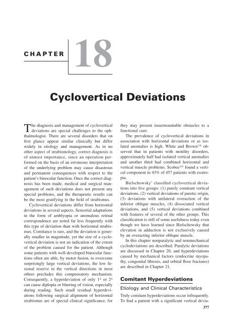
Clinical Characteristics<br />
DVD is characterized by the spontaneous drifting<br />
of either eye upward when the patient is fatigued<br />
or daydreaming or when fusion is artificially interrupted<br />
by covering one eye (Fig. <strong>18</strong>–1). When the<br />
elevated eye is covered, it may perform pendular,<br />
vertical movements. When the cover is removed,<br />
the elevated eye will move slowly downward and<br />
settle in the primary position.<br />
The amount of elevation when the eye is covered<br />
is variable, tending to increase after prolonged<br />
occlusion, and often differing between the<br />
two eyes. According to Bielschowsky 6 several<br />
other features are present in most patients with
<strong>Cyclovertical</strong> <strong>Deviations</strong> 379<br />
FIGURE <strong>18</strong>–1. Dissociated vertical deviation. A, Left eye<br />
fixating. B, Right eye fixating.<br />
DVD. These include excycloduction of the elevated<br />
eye and incycloduction of the fixating eye.<br />
As the elevated eye returns to the primary position<br />
it incycloducts. This torsional movement of the<br />
globe is often easily detected without magnification<br />
by the observer. Latent nystagmus, which<br />
often but not always has a cyclovertical component,<br />
may be associated with DVD. 6<br />
These additional symptoms justify consideration<br />
of DVDs as a syndrome. Observation of the<br />
iris pattern and the conjunctival vessels will nearly<br />
always reveal incycloduction as the elevated eye<br />
returns to the midline, indicating that it was excycloducted<br />
while in the dissociated position. The<br />
excycloduction of the elevating eye may be accompanied<br />
by a synchronous incycloduction of<br />
the fixating eye (cycloversion). 54 Occasionally, excycloduction<br />
of each eye under cover or spontaneously<br />
and latent nystagmus may be the only manifestation<br />
of a dissociated deviation. In such cases<br />
we speak of a dissociated torsional deviation<br />
(DTD). In other cases, the full syndrome with its<br />
vertical component may involve one eye only<br />
while it manifests itself as an isolated excycloduction<br />
in the other.<br />
Anderson 2 and Lyle and Bridgeman 69 drew attention<br />
to the association of a head tilt with DVD.<br />
The prevalence of anomalous head posture in this<br />
condition has been reported to range between<br />
23% 19 and 35%. 5 Most authors reported the head<br />
to be tilted away from the eye with the larger<br />
19, 101<br />
vertical deviation but the opposite has also<br />
been observed. 5, 23 Passive tilting of the head toward<br />
the opposite of the side of the habitual posture<br />
increases the vertical deviation, which has led<br />
to the conclusion that the anomalous head posture<br />
decreases the magnitude and thus improves the<br />
motor control of the type of alternatinng hyperphoria<br />
investigated by these authors. De Decker<br />
and Dannheim de-Decker 23 reported chin depression<br />
in patients with bilateral dissociated deviations.<br />
Surgical correction of DVD improves the<br />
23, 101<br />
head posture.<br />
DVD occurs in patients with and without overaction<br />
of the inferior oblique muscles and may<br />
also be associated with overaction of the superior<br />
oblique muscles and an A-pattern exodeviation in<br />
downward gaze 45, 71 (Fig. <strong>18</strong>–2). The vertical angle<br />
of dissociated deviations is usually somewhat less<br />
in abduction than in adduction; however, it may<br />
also be larger in abduction. 46 Latent nystagmus<br />
occurs in approximately half the patients with<br />
DVD and, in fact, is seldom encountered in the<br />
absence of this anomaly (see Anderson 2, p.16 ).<br />
If a photometric neutral filter wedge is placed<br />
before the fixating eye while the other eye is<br />
occluded and elevated, the eye behind the cover<br />
will make a gradual downward movement, and<br />
may even move below the primary position as the<br />
visual input to the fixating eye is progressively<br />
decreased by the filter wedge. When the wedge is<br />
moved from positions of greater to lesser filter<br />
FIGURE <strong>18</strong>–2. Dissociated vertical deviation with overaction of both superior oblique muscles. A,<br />
Left hypertropia with the right eye fixating and right hypertropia with the left eye fixating. B,<br />
Underaction of both inferior oblique and overaction of both superior oblique muscles.
380 Clinical Characteristics of Neuromuscular Anomalies of the Eye<br />
density, the eye behind the cover will elevate.<br />
This intriguing observation was first reported by<br />
Bielschowsky 6 and has become known as the<br />
Bielschowsky phenomenon. Bielschowsky explained<br />
the phenomenon that bears his name in<br />
the following manner: When the visual input to<br />
the fixating, say, the right eye is decreased by<br />
holding filters of increasing density before it, the<br />
effort to maintain fixation triggers an abnormal<br />
innervation to the elevators. The effort to maintain<br />
fixation with the right eye against this innervation<br />
elicits a compensatory innervation to the depressors.<br />
The left eye follows this innervation<br />
under cover and returns to the primary position or<br />
even below it.<br />
A DVD may occasionally occur as an isolated<br />
phenomenon in patients in whom binocular functions<br />
are apparently normal, but is found often in<br />
association with infantile esotropia and less often<br />
with accommodative acquired esotropia, exotropia,<br />
and heterotropia of sensory origin.<br />
64, 72, 109, 132<br />
An association with Duane’s syndrome has also<br />
been described. 17, 95 The high prevalence of DVD<br />
in essential infantile esotropia has been discussed<br />
in <strong>Chapter</strong> 16 and it is of interest that a similarly<br />
high rate of occurrence has been reported in infantile<br />
exotropia. 73 In spite of a careful search the<br />
condition is rarely diagnosed in infancy. In our<br />
experience the diagnosis is most commonly made<br />
between the ages of 2 and 5 years and often<br />
years after surgical alignment of the horizontal<br />
deviation. The age at surgical alignment could not<br />
be correlated with the manifestation of DVD. 47<br />
DVD is usually bilateral and asymmetrical. Of<br />
the 170 cases observed by us 80 in a group of 408<br />
children with essential infantile esotropia, only 24<br />
(14%) were unilateral and only 13 (9%) were<br />
symmetrical. However, unilateral occurrence is often<br />
observed in deeply amblyopic eyes 6 and in<br />
96, 109<br />
sensory heterotropia. The commonly occurring<br />
asymmetry between the two eyes is reversed<br />
in the supine position with the head tilted<br />
back: the eye with a larger deviation in the upright<br />
position has a smaller one with the patient supine<br />
and the head tilted back. 39 This observation suggests<br />
a possible effect of inputs from otolithic and<br />
possibly neck muscle sensors on the amplitude of<br />
a DVD.<br />
An active suppression mechanism usually will<br />
eliminate diplopia in patients with a spontaneous<br />
DVD. Exceptions to this rule are rare but do occur<br />
as shown in Case <strong>18</strong>–1.<br />
CASE <strong>18</strong>–1<br />
This 26-year-old man has had crossed eyes since<br />
infancy. Surgery was performed on the eye muscles<br />
when he was 3 years of age. For the past 13 years<br />
he has experienced intermittent double vision. Examination<br />
showed corrected visual acuity of 6/4.5<br />
OD and 6/6 OS. He wore prescription glasses to<br />
correct a mild compound myopic astigmatism. The<br />
patient had fairly pronounced latent nystagmus and<br />
an esotropia of 14 at near and distance fixation. In<br />
addition he had <strong>18</strong> right hypertropia with the OS<br />
fixating and 10 left hypertropia with the OD fixating.<br />
He exhibited characteristic incycloduction as each<br />
elevated eye took up fixation in primary position. A<br />
V pattern was absent, and there was no inhibitional<br />
palsy of the contralateral superior rectus when he<br />
fixated with the adducted elevated eye. As soon as<br />
the eyes were dissociated, the patient became<br />
aware of diplopia. The red-glass test established that<br />
the diplopia was in accord with the deviation; that<br />
is, the images were uncrossed and had a vertical<br />
component, the laterality of which depended on<br />
which eye was fixating. The diagnosis was residual<br />
esotropia with a DVD and absence of suppression.<br />
No treatment was advocated.<br />
Campos and coworkers 14 pointed out that suppression<br />
is not the only mechanism that accounts<br />
for absence of diplopia in this condition by showing<br />
that a binocular vertical perceptional adaptation<br />
may exist in these patients.<br />
Diplopia can be elicited in most patients with<br />
a dark-red glass, and the amount of separation<br />
between the images is used to measure the amplitude<br />
of elevation of each eye (see <strong>Chapter</strong> 12).<br />
The fact that the patient will localize the red image<br />
below the fixation light, regardless of whether the<br />
red glass is held before the right or left eye,<br />
clearly differentiates a DVD from other forms of<br />
cyclovertical anomalies in which the red image<br />
is localized below or above the fixation light,<br />
depending on which eye fixates.<br />
Measurement<br />
An accurate quantitative assessment of DVD may<br />
be obtained provided visual acuity in each eye is<br />
sufficient to visualize the fixation target, using a<br />
modification of the prism and cover test. As the<br />
patient focuses on the fixation target at 6 m distance,<br />
the occluder is quickly shifted to the fixating<br />
eye, allowing the previously dissociated and<br />
elevated eye to take up fixation. The cover is then<br />
returned to the nonfixating eye. As the alternate<br />
cover test is continued, increasing amounts of
<strong>Cyclovertical</strong> <strong>Deviations</strong> 381<br />
base-down prisms are held under the occluder in<br />
front of the nonfixating eye until the downward<br />
fixation movement of that eye is neutralized. The<br />
procedure is then repeated with the fellow eye<br />
fixating.<br />
Etiology<br />
Of the numerous theories advanced to explain the<br />
mechanism of this intriguing anomaly in the past,<br />
only a few will be mentioned in this chapter.<br />
Elastic preponderance of the elevator or the depressor<br />
muscles 102 ; paretic factors 25 especially bilateral<br />
paresis of the depressor muscles 104, p. <strong>18</strong>3 ;<br />
and imbalances between the amount of innervation<br />
originating from each vestibular organ 87 have been<br />
cited as causes in the older literature. For other<br />
explanations, see White, 127 Verhoeff, 125 Posner, 91<br />
Crone, 19 Helveston, 46 and Houtman and coworkers.<br />
53 It has even been reported that DVD may be<br />
caused by an abnormal visual pathway routing<br />
similar to that described in albinism 35 (see <strong>Chapter</strong><br />
9). However, as one may have expected, this find-<br />
3, 10, 62, 135<br />
ing could not be reproduced.<br />
11, 15, 41,<br />
The results of more recent investigations<br />
93, 94, 133<br />
are in basic agreement with what<br />
Bielschowsky 6 so lucidly described in 1931 and<br />
in his later publications 7, 8 : DVD is caused by a<br />
vertical vergence signal that elevates the occluded<br />
eye and would depress the fixating eye if it were<br />
not for a simultaneous supraversion impulse that<br />
cancels the innervation to depress the fixating eye<br />
while at the same time, according to Hering’s law,<br />
increasing the innervation flowing to the elevators<br />
of the occluded eye. Bielschowsky arrived at this<br />
explanation by meticulous clinical observation,<br />
sound reasoning, and without the benefit of modern<br />
search coil recording techniques that have<br />
essentially confirmed the validity of this innerva-<br />
41, 93, 133<br />
tional pattern and sequence<br />
The origin of the vertical vergence innervation<br />
is still a matter of dispute. Bielschowsky 6 suspected<br />
an alternating and intermittent excitation<br />
of both subcortical centers that govern vertically<br />
divergent eye movements. He cited as examples<br />
for such movements and support for the existence<br />
of such centers skew deviation and seesaw nystagmus<br />
and felt that the unilaterality of the condition<br />
in some cases is ‘‘based on the coincidence of<br />
the voluntary fixation impulse with the involuntary<br />
action of one of the vertical divergence centers.’’<br />
8, p. 35 The reason for this abnormal excitation<br />
of hypothetical vertical divergence centers remains<br />
unknown. There is no question, however, that the<br />
impulse for a DVD must originate in the fixating<br />
8, p. 36<br />
eye. Bielschowsky emphasized the need to<br />
differentiate between a hyperdeviation based on<br />
anatomical conditions, that is, an anomalous position<br />
of rest, and the dissociated deviations of innervational<br />
origin. Spielmann 116 convincingly confirmed<br />
this difference by showing that DVD does<br />
not occur when fixation is prevented by covering<br />
both eyes with translucent occluders (Fig. <strong>18</strong>–3).<br />
Spielmann 111–113 assumed that DVD is caused by<br />
an imbalance of binocular stimulation. Although<br />
this may explain the frequent occurrence of DVD<br />
in essential infantile esotropia and the occasional<br />
occurrence with sensory heterotropias, it does not<br />
account for DVD in patients with otherwise normal<br />
binocular functions.<br />
Several additional explanations were proposed<br />
in recent years. From the direction of the cyclorotation<br />
of the elevating eye (extorsion) and the<br />
31, 41, 93<br />
fixating eye (intorsion) several investigators<br />
have concluded that the vertical vergence movement<br />
must be predominantly mediated by the<br />
oblique muscles because the vertical rectus muscle<br />
would produce a cyclorotation in the opposite<br />
direction. Guyton 41 and Cheeseman and Guyton 15<br />
believe that this oblique muscle–generated cycloversion<br />
is a purposeful eye movement that damps<br />
latent cylovertical nystagmus to improve visual<br />
acuity. The accompanying elevation of the nonfixating<br />
eye, the DVD, is seen as an unavoidable<br />
FIGURE <strong>18</strong>–3. Combined vertical and horizontal dissociated<br />
deviation in the right eye (A) and predominantly<br />
vertical dissociated deviation in the left eye (B). C, Absence<br />
of dissociated vertical deviation in the fixation-free<br />
position when both eyes are covered with translucent<br />
Spielmann occluders. For details, see text.
382 Clinical Characteristics of Neuromuscular Anomalies of the Eye<br />
and undesirable byproduct of this nystagmus<br />
damping mechanism (see <strong>Chapter</strong> 23). There are<br />
a number of observations that are difficult to reconcile<br />
with this theory, 82 which is based on the<br />
concept of the inferior oblique muscle being the<br />
primary elevator in vertical vergences. In DVD<br />
the dissociated eye elevates not only in adduction<br />
but also in primary position and abduction (Fig.<br />
<strong>18</strong>–4). In fact, in some cases the elevation in<br />
abduction is greater than in adduction. Clearly,<br />
these are gaze positions in which the inferior<br />
oblique muscle has little or no elevating power<br />
and the superior rectus muscle must be the principal<br />
elevator. While it is indisputable that excycloduction<br />
of an elevating eye can only be caused by<br />
the inferior oblique muscle, it does not inescapably<br />
follow that this muscle is also the predominant<br />
elevator. One must also consider the possibility<br />
that both the superior rectus and inferior<br />
oblique muscles co-contract during elevation but<br />
that the stronger excyclotorsional effect of the<br />
inferior oblique overrides the weaker incyclotorsional<br />
effect of the superior rectus muscle. Moreover,<br />
in our experience and that of others 45 a<br />
DVD continues unabated after a myectomy of<br />
the ipsilateral inferior oblique muscle. Also, a<br />
nystagmus dampening purpose of the vertical<br />
FIGURE <strong>18</strong>–4. Dissociated vertical deviation in different<br />
gaze positions. A, In the case of a dissociated vertical<br />
deviation of the left eye, elevation occurs in adduction,<br />
primary position, and, to a lesser degree, in abduction. B,<br />
When the elevated adducted left eye takes up fixation,<br />
the covered right eye will be elevated to an equal degree.<br />
(From Noorden GK von: Atlas of Strabismus, ed 4. St<br />
Louis, Mosby–Year Book, 1983.)<br />
vergence is difficult to accept in view of the fact<br />
that a latent cyclovertical nystagmus is not a consistent<br />
feature of DVD. 6, 46, 56, 93 Finally, if DVD<br />
were elicited to dampen a latent nystagmus we<br />
would expect the nonfixating eye of a patient with<br />
DVD to elevate each time a patient with latent<br />
nystagmus reads his or her threshold acuity line<br />
on the office chart. Not only is this not the case<br />
but, on the contrary, DVD manifests itself typically<br />
when patients are daydreaming and uninvolved<br />
in active visual activities.<br />
Van Rijn and coworkers 94 felt that DVD represents<br />
a form of asymmetrical vertical heterophoria<br />
and could be considered as enhancement of a<br />
phenomenon that is present in normal subjects as<br />
well. They showed in patients with alternating<br />
hyperphoria, who have a right hyperphoria with<br />
the left eye fixating and a left hyperphoria with<br />
the right eye fixating, asymmetries of the vertical<br />
heterophoria angles, depending on which eye was<br />
fixating. It is true that alternating hyperphoria,<br />
which, incidentally, is an extremely rare clinical<br />
finding, bears a superficial resemblance to DVD.<br />
However, it is debatable whether these conditions<br />
are as closely related as assumed by these authors.<br />
A vertical heterophoria becomes manifest as soon<br />
as fusion is disrupted and the eye drifts into its<br />
anomalous position of rest. In DVD, the vertical<br />
movement is caused by an active vergence innervation<br />
and fusion is not a factor in controlling<br />
the deviation because it may occur in patients<br />
without the ability to fuse.<br />
Some authors have speculated that DVD is a<br />
manifestation of atavistic oculomotor reflexes that<br />
are present in birds and fish. 11, 14, 20, 41, 46, 94 Crone 20<br />
considered that DVD may represent a phylogenetic<br />
residuum of monocular vertical movements<br />
present in birds and inhibited in normal humans.<br />
Brodsky 11 suggested that DVD is a primitive dorsal<br />
light reflex in which asymmetrical visual input<br />
to the eyes evokes a vertical divergence movement.<br />
In lateral-eyed animals this reflex serves<br />
as a primitive visual-vestibular righting response.<br />
Suppressed in normal humans, it is thought to<br />
manifest itself when early-onset strabismus precludes<br />
normal binocular development. Can the<br />
stimulus for the dorsal reflex in fish be compared<br />
to the stimulus situation in a human strabismic<br />
infant A fish illuminated from one side depresses<br />
the eye ipsilateral to the light source and elevates<br />
the contralateral eye. The stimulus for this reflex<br />
eye movement, which does indeed resemble a<br />
vertical vergence response, is a difference in illu-
<strong>Cyclovertical</strong> <strong>Deviations</strong> 383<br />
mination between the two eyes. However, such<br />
differences do not occur in strabismus, as each<br />
eye receives the same amount of light. In strabismus,<br />
the asymmetry of visual input that disrupts<br />
binocularity is caused instead by the incongruity<br />
of the retinal images formed in each eye.<br />
To summarize, it is fair to state that despite<br />
numerous attempts to clarify it, the etiology of<br />
DVD is still obscure. Bielschowsky’s original explanation<br />
of this form of strabismus, confirmed by<br />
modern eye movement recording techniques, has<br />
established indisputably that DVD is a vertical<br />
vergence eye movement. However, the stimulus<br />
for this movement and its relationship to various<br />
forms of strabismus, especially to essential infantile<br />
esotropia, have yet to be convincingly identified.<br />
Differential Diagnosis<br />
Even though the pattern of the deviation and the<br />
results of the red-glass test are clearly different in<br />
DVD from those in other cyclovertical anomalies,<br />
clinicians sometimes confuse this condition with<br />
upshoot in adduction caused by overaction of the<br />
inferior oblique muscles (see p. 386). To be sure,<br />
overaction of the inferior oblique muscle may<br />
occur in patients with DVD, but such overaction<br />
cannot be held responsible for this anomaly. Several<br />
clinical findings clearly distinguish DVD from<br />
overaction of the inferior oblique muscle.<br />
First, in DVD the covered eye becomes elevated<br />
in abduction, primary position, and adduction<br />
(Fig. <strong>18</strong>–4). Conversely, with overaction of<br />
the inferior oblique muscles, each eye becomes<br />
elevated primarily in adduction and never in abduction<br />
unless there is coexisting contracture of<br />
the ipsilateral superior rectus muscle. Unlike<br />
DVD, overaction of the inferior obliques is commonly<br />
associated with a V-pattern esotropia. The<br />
reason for elevation in adduction in a DVD is that<br />
the adducted eye becomes occluded by the nasal<br />
bridge and fusion is suspended. In children under<br />
the age of 2 to 3 years the nasal bridge has not<br />
yet fully developed and the upshoot in adduction<br />
is rarely seen.<br />
Second, DVD is found also in patients in whom<br />
there is no noticeable overaction of the inferior<br />
oblique muscles and actually occurs frequently<br />
in those with underacting inferior oblique and<br />
overacting superior oblique muscles 45, 71 (see Fig.<br />
<strong>18</strong>–1).<br />
Third, when a patient with an overacting inferior<br />
oblique muscle fixates with the involved eye<br />
in the field of action of that muscle (elevation<br />
and adduction), the contralateral superior rectus<br />
muscle will underact (see Fig. 20–1). This apparent<br />
paresis of the superior rectus muscle has been<br />
discussed under inhibitional palsy (<strong>Chapter</strong> 20).<br />
Conversely, in patients with DVD who are tested<br />
in the same manner, underaction of the contralateral<br />
yoke muscle does not occur (Fig. <strong>18</strong>–4, B).<br />
Fourth, in patients with inferior oblique overaction,<br />
the speed of the refixation movement of the<br />
eye after covering the fellow eye is rapid (200 to<br />
400/s) compared with the much slower infraduction<br />
movements in patients with DVD, which are<br />
usually between 10 and 200/s. 46<br />
Fifth, the characteristic slow, tonic incycloduction<br />
of the eye as it returns from the dissociated<br />
to the primary position cannot be observed with<br />
equal facility when overaction of the inferior<br />
oblique is present. In that case refixation after<br />
covering the fixating eye is also accompanied by<br />
incylcoduction but this movement is so fast that it<br />
often escapes observation.<br />
The differential diagnosis between these two<br />
conditions is summarized in Table <strong>18</strong>–1 in which<br />
additional distinguishing findings are listed. Clear<br />
distinction between them is clinically important;<br />
for example, a recession or myectomy of the inferior<br />
oblique will have no effect on upshoot in<br />
adduction if the patient actually has a DVD.<br />
When associated with comitant or paretic<br />
cyclovertical anomalies the diagnosis of DVD is<br />
more difficult. When evaluating such patients, one<br />
must take into account the starting position of<br />
each eye before the cover is applied. For instance,<br />
if a right hypertropia is associated with a DVD,<br />
the right eye will become further elevated under<br />
the cover, and the fellow left hypotropic eye when<br />
covered will move upward the same amount but<br />
may only reach the midline, since it began its<br />
movement from a depressed position. Careful observation<br />
of each eye before, during, and after the<br />
cover has been applied is essential to detecting a<br />
dissociated vertical component in a patient with a<br />
comitant or paretic cyclovertical deviation.<br />
Therapy<br />
During the first half of the twentieth century clinicians<br />
took a rather passive attitude toward treatment<br />
of DVD. This conservatism probably finds<br />
its roots in Bielschowsky’s 6 teachings that this
384 Clinical Characteristics of Neuromuscular Anomalies of the Eye<br />
TABLE <strong>18</strong>–1. Differential Diagnosis: Dissociated Vertical Deviation vs. Inferior Oblique Overaction<br />
Dissociated Vertical<br />
Deviation<br />
Inferior Oblique<br />
Overaction<br />
Elevation From primary position, adduction Maximal in adduction, never<br />
and abduction<br />
in abduction<br />
Superior oblique action May overact Usually underaction<br />
V pattern Absent Often present<br />
Pseudoparesis of contralateral superior rectus Absent Present<br />
Incycloduction on refixation Present Absent<br />
Saccadic velocity of refixation movement 10–200/s 200–400/s<br />
Latent nystagmus Often present Absent<br />
Bielschowsky phenomenon Often present Absent<br />
condition does not lend itself to optical or operative<br />
therapy as do comitant or paretic deviations.<br />
Bielschowsky cited cases of torsional diplopia<br />
produced by surgery on the superior rectus muscles,<br />
and stated that therapy, if contemplated at<br />
all, should be directed toward strengthening the<br />
fusional mechanism.<br />
Patients with DVD usually are asymptomatic,<br />
and complaints of diplopia have been an infrequent<br />
problem in our experience; however, when<br />
the patient is daydreaming or tired, the elevated<br />
position of either eye may become conspicuous<br />
and a source of embarassment to the patient. In<br />
that case surgery may be considered. We have not<br />
observed DVD in adults as often as we have in<br />
children, and were, perhaps erroneously, under the<br />
impression that this disorder tends to improve<br />
with time. However, Harcourt and coworkers 44<br />
followed 100 patients with DVD for as long as<br />
7.3 years and found no significant decrease in the<br />
deviation during this period of observation.<br />
Surgical procedures preferred by various authors<br />
are (1) recession of the superior combined<br />
with resection of the inferior rectus muscles, 59 (2)<br />
resection of the inferior recti, 90 (3) retroequatorial<br />
myopexy (posterior fixation) of the superior recti<br />
combined with 27, 46, 58, 117 or without 68, 76, 77 a recession<br />
of these muscles, (4) unconventionally large<br />
32, 68,<br />
recessions (7 to 10 mm) of the superior recti,<br />
70, 124<br />
and (5) anterior displacement of the inferior<br />
oblique insertion, which may be combined with<br />
9, 13, 61, 97, 108, 119, 123<br />
superior rectus recession.<br />
Our initial enthusiasm for using a conventional<br />
(4 to 5 mm) recession of the superior recti combined<br />
with a retroequatorial myopexy 12 to 15<br />
mm behind the original insertion 27 has waned because<br />
of many recurrences occurring as late as<br />
several years after an initial satisfactory result.<br />
Currently, we prefer 7- to 9-mm recessions of the<br />
superior recti and vary the amount of surgery in<br />
the two eyes when the deviation is asymmetrical.<br />
This approach has yielded a cure or significant<br />
improvement (defined as a cosmetically insignificant<br />
residual angle) in 23 (72%) of 32 patients<br />
after a follow-up of at least 3 years. 32 Contrary to<br />
our earlier concern that such an extensive weakening<br />
procedure would produce a paresis of the<br />
superior rectus, we have not observed this complication.<br />
The effectiveness of superior rectus muscle<br />
recession in terms of correction of a deviation in<br />
prism diopters per millimeter of recession in other<br />
forms of vertical strabismus and the lesser effect<br />
of this procedure in a DVD of the same magnitude<br />
emphasize the unique position of this anomaly<br />
among other forms of strabismus.<br />
Several authors have in recent years reported<br />
good results with anterior displacement of the<br />
inferior oblique muscle 9, 13, 61, 97, 108, 119, 123 and this<br />
treatment has become the procedure of choice for<br />
some. However, we prefer recession of the superior<br />
rectus muscle(s), which is less likely to cause<br />
some of the complications reported after surgery<br />
on the inferior oblique insertion (see <strong>Chapter</strong> 26).<br />
The question has been debated whether surgery<br />
should always be performed in both eyes even<br />
though the deviation may be present preoperatively<br />
only in one eye. It is not uncommon and<br />
quite disappointing to have a patient return after<br />
surgery in one eye with a DVD in the fellow eye.<br />
However, since in our experience this does not<br />
happen in every instance of asymmetrical occurrence<br />
we operate on both eyes only when a deviation<br />
can be diagnosed preoperatively in both eyes.<br />
Recurrences are not uncommon even after unconventionally<br />
large recessions of the superior<br />
recti and require additional surgery consisting of<br />
a 4- to 5-mm resection of the inferior rectus muscle.<br />
Full correction or improvement, as defined<br />
above, can in our experience be achieved in 92%<br />
of the patients after this operation. 33
<strong>Cyclovertical</strong> <strong>Deviations</strong> 385<br />
Although in most patients with a conspicuous<br />
DVD surgery is the recommended treatment, the<br />
possible effectiveness of a conservative approach<br />
should not be ignored. This is especially true in<br />
patients with asymmetrical involvement or those<br />
accustomed to wearing glasses. For example, a<br />
patient without binocular vision (after horizontal<br />
surgical alignment in infantile esotropia) may exhibit<br />
a significant DVD of the left eye when fixating<br />
with the right eye, but only an insignificant<br />
deviation of the right eye may occur with the<br />
left eye fixating. A slight optical blur induced by<br />
increasing the power of the lens over the right eye<br />
(2.00 sph usually is sufficient) or a contact<br />
lens (see Case 24–1, p 538) will switch fixation<br />
preference to the left eye, and the DVD is no<br />
longer a cosmetic problem. Simon and coworkers<br />
110 have confirmed the efficacy of this approach<br />
in selected cases but have used atropine rather<br />
than optical penalization.<br />
Dissociated Horizontal<br />
<strong>Deviations</strong><br />
It has only recently been recognized that DVD<br />
may also have a horizontal component. Raab 92<br />
mentioned in 1970 that the vertical movement in<br />
DVD may be accompanied by abduction. Little<br />
attention was paid to this observation until the<br />
term dissociated horizontal deviations (DHDs) became<br />
established in the literature.<br />
30, 113, 130, 134<br />
The condition is characterized by intermittent,<br />
asymmetrical abduction and elevation of the dissociated<br />
eye (see Fig. <strong>18</strong>–2A). Occasionally, DHD<br />
occurs in an isolated form, not accompanied by a<br />
vertical deviation. As with DVD, latent nystagmus<br />
and excyclotropia of the deviated eye are frequently<br />
associated findings. Interestingly, and perhaps<br />
significantly, Wilson and coworkers 132 found<br />
that only two of six patients with a prominent<br />
DHD had a history of essential infantile esotropia.<br />
The remainder had accommodative esotropia, a<br />
condition that is only infrequently associated with<br />
DVD. These authors also reported that when DHD<br />
is associated with esotropia the patient may become<br />
exotropic during periods of visual inattention.<br />
In an earlier report Wilson and McClatchey 130<br />
had pointed out that unlike in ordinary intermittent<br />
exotropia, the alternate cover test reveals less exodeviation<br />
than when the eye abducts spontaneously<br />
or under cover, that the fixating eye may<br />
adduct during attempts to neutralize the horizontal<br />
deviation with base-in prisms, and that the exodeviation<br />
may be strictly unilateral. Moreover, they<br />
described what is similar to the Bielschowsky<br />
phenomenon in DVD: when neutral density filters<br />
are placed before the fixating eye the abducted<br />
dissociated eye returns to the primary position and<br />
may even adduct. Since we became aware of this<br />
condition we have observed several patients with<br />
a pure dissociated horizontal deviation in one eye<br />
and a pure DVD in the fellow eye.<br />
For dissociated exodeviations a 5- to 7-mm<br />
recession of the lateral rectus muscle of the involved<br />
eye is recommended when the size of the<br />
deviation is such that the patient or his or her<br />
parents desire correction. Satisfactory results have<br />
been reported using this approach. 131<br />
This may be combined with recession of the<br />
superior rectus when associated with a vertical<br />
component. In patients whose dissociated deviation<br />
is predominantly vertical with only a small<br />
horizontal component, we have found a large recession<br />
of the superior rectus is usually sufficient<br />
to correct both problems.<br />
Spielmann 115 described dissociated esodeviations<br />
that occur when either eye is covered with<br />
the semiopaque occluder. When both eyes are occluded<br />
(fixation-free position) the eyes remain<br />
aligned, which distinguishes this condition from<br />
esophoria. In our experience dissociated esodeviations<br />
occur much less frequently than dissociated<br />
exodeviations. If the deviation becomes intermittent<br />
and a cosmetic consideration, a posterior fixation<br />
of the medial rectus muscle of the involved<br />
eye 14 mm behind its insertion is effective. Observation<br />
rather than surgery has been advocated<br />
when the esotropia changes to exotropia during<br />
visual inattention. 132<br />
In view of the great clinical similarity of dissociated<br />
vertical, torsional, and horizontal deviations,<br />
we agree with those who consider these<br />
strabismus forms as variations on the same theme<br />
114, 134<br />
rather than as different entities sui generis.<br />
Elevation in Adduction<br />
(Strabismus<br />
Sursoadductorius)<br />
Clinical Characteristics<br />
When examining the versions, one may find elevation<br />
of an eye as it moves toward adduction (Fig.
386 Clinical Characteristics of Neuromuscular Anomalies of the Eye<br />
<strong>18</strong>–5). Once in a position of maximal elevation in<br />
adduction, the eye will be elevated further than a<br />
normal eye. This anomaly may be unilateral or<br />
bilateral and has been termed strabismus sursoadductorius<br />
or, if the opposite situation occurs, that<br />
is, the eye shows depression in adduction, strabismus<br />
deorsoadductorius. These Latin terms have<br />
never become popular in the English strabismologic<br />
literature where they are used in their translated<br />
forms, elevation (or upshoot) in adduction<br />
and depression (or downshoot) in adduction. Upshoot<br />
in adduction in its bilateral form is characterized<br />
by left hypertropia in dextroversion and<br />
right hypertropia in levoversion (double or alternating<br />
hypertropia), but a vertical deviation is<br />
present infrequently in primary position. Upshoot<br />
in adduction is an isolated phenomenon or occurs<br />
with esotropia or exotropia, often associated with<br />
a V pattern (see <strong>Chapter</strong> 19). It is frequently<br />
observed in infantile esotropia. It is often automatically<br />
and erroneously assumed that elevation in<br />
adduction is caused by inferior oblique overaction,<br />
that is, by excessive innervation flowing to<br />
that muscle. While this is often the case, we shall<br />
see that there are other causes for this condition.<br />
Etiology<br />
OVERACTION OF THE INFERIOR OBLIQUE<br />
MUSCLE. It has been customary to distinguish<br />
between primary and secondary overactions of this<br />
muscle. Primary overaction of the inferior oblique<br />
muscle in which there is no evidence for a past or<br />
present ipsilateral superior oblique paralysis or<br />
paresis is difficult to explain. A V-pattern type of<br />
strabismus is often present in such patients and,<br />
typically, the Bielschowsky head tilt test is negative.<br />
This condition occurs frequently in essential<br />
infantile esotropia.<br />
The explanations given for apparent primary<br />
overaction in the older literature are vague, to say<br />
the least. Duane, 25 for instance, suggested that<br />
there is normally an upshoot of the adducted eye<br />
because of the greater mechanical advantage of<br />
the inferior oblique muscle of the adducted eye<br />
over the superior rectus muscle of the abducted<br />
eye. It is of interest in this connection that Lisch<br />
and Simonsz 66 have reported in normal subjects<br />
up- and downshoot in adduction after prolonged<br />
monocular patching. This may suggest that there<br />
is a natural tendency for elevation and depression<br />
in adduction to occur but that under normal conditions<br />
such eye movements are controlled by fusion.<br />
66 Scobee 104, p.378 agreed that overaction of the<br />
inferior oblique muscle is normal because of the<br />
increased impulse required by the mechanically<br />
disadvantaged superior rectus muscle. This strong<br />
impulse is communicated to its yoke muscle, the<br />
inferior oblique of the adducted eye hidden behind<br />
the nose. He also stated that the elevating action<br />
of the inferior oblique muscle in adduction is<br />
greater than the depressing action of the superior<br />
oblique muscle. Therefore there would be an imbalance<br />
if the eyes should be dissociated by the<br />
nose, and the result would be an upshoot of the<br />
adducted eye. Lancaster 65 agreed with this view.<br />
Guibor 40 suggested that inferior oblique overactions<br />
could be caused by a synkinesis of that<br />
muscle with the ipsilateral medial rectus muscle<br />
owing to an impulse spread within the central<br />
nervous system.<br />
None of these older explanations are convincing<br />
and it remains quite doubtful whether a true<br />
primary overaction of an oblique muscle on an<br />
innervational basis exists at all. The discovery of<br />
muscle pulleys (see below) has directed our attention<br />
to other etiologic possibilities for this apparent<br />
overaction. We are in agreement with Clark<br />
and coworkers 16 who lamented the use of diagnosis-laden<br />
terms for ocular motility disorders except<br />
in cases where the etiology is clear. For this reason<br />
and because there are several causes for elevation<br />
in adduction that are unrelated to excessive inferior<br />
oblique muscle contraction, we recommend<br />
that the terms primary overaction of the inferior<br />
oblique (or, for that matter, of the superior oblique<br />
muscle) should be abandoned in favor of the more<br />
generic elevation (or upshoot) in adduction or<br />
depression (or downshoot) in adduction.<br />
Secondary overaction of the inferior oblique<br />
FIGURE <strong>18</strong>–5. Elevation in adduction. Marked left hypertropia in dextroversion and right hypertropia<br />
in levoversion. No vertical deviation in primary position.
<strong>Cyclovertical</strong> <strong>Deviations</strong> 387<br />
muscle is easier to understand and is caused by<br />
paresis or paralysis of the ipsilateral superior<br />
oblique muscle or by paresis or paralysis of the<br />
contralateral superior rectus muscle when the patient<br />
fixates with the paretic eye. In the latter<br />
situation the upshoot in adduction is actually<br />
caused by increased innervation flowing to the<br />
inferior oblique muscle, according to Hering’s law.<br />
However, in the former condition the upshoot in<br />
adduction is not caused by excessive innervation<br />
of the inferior oblique muscle but by a lack of<br />
tonus of its paralyzed antagonist. In this situation<br />
a normal innervational impulse will suffice to<br />
cause the eye to overshoot in the field of action<br />
of the inferior oblique.<br />
A similar situation exists when the balance<br />
of forces between superior and inferior oblique<br />
muscles is offset by anatomical rather than innervational<br />
causes, as in plagiocephaly (see <strong>Chapter</strong><br />
19). Here, the recessed trochlea has changed the<br />
plane of the superior oblique tendon, which places<br />
the inferior oblique at a functional advantage over<br />
the superior oblique, which causes an upshoot in<br />
adduction.<br />
In the following discussion we shall see that<br />
elevation in adduction may be caused by a number<br />
of other factors.<br />
HETEROTOPIA OF RECTUS MUSCLE PULLEYS.<br />
Recent work by Clark and coworkers 16 has provided<br />
evidence that what appears as primary overaction<br />
of an oblique muscle may actually be a<br />
secondary overaction of both elevators caused by<br />
heterotopia of muscle pulleys (see <strong>Chapter</strong> 3).<br />
Because of the significance of these findings with<br />
respect to the etiology of A and V patterns they<br />
will be discussed in <strong>Chapter</strong> 19<br />
OCULAR AND ORBITAL TORSION. For a discussion<br />
of the roles of ocular and orbital torsion<br />
in producing upshoot in adduction, the reader is<br />
directed to <strong>Chapter</strong> 19, where this mechanism is<br />
discussed in connection with the etiology of A-<br />
and V-pattern strabismus. It will suffice to say<br />
here that excyclotropia of an eye may cause elevation<br />
in adduction and depression in abduction in<br />
the absence of increased innervation of the oblique<br />
muscles (Fig. <strong>18</strong>–6). When the eye is excyclotorted<br />
the medial rectus muscle will no longer act<br />
as a pure adductor but will gain elevating action<br />
as well. Thus, medial rectus contraction will not<br />
only adduct but will also elevate the eye (upshoot)<br />
under these circumstances. Likewise, the lateral<br />
rectus muscle will no longer be a pure abductor<br />
but abduct and depress the eye (downshoot). The<br />
elevation in adduction is usually more prominent<br />
than the depression in abduction, which may even<br />
be absent. This may be due to structural differences<br />
between the medial and lateral aspects of<br />
the orbit. It is of historical interest in this connection<br />
that Bielschowsky 8, p.169 documented a case of<br />
an exotropic patient in whom a right hypertropia<br />
caused by an apparently overacting inferior<br />
oblique muscle disappeared after merely advancing<br />
and lowering the insertion of the medial rectus<br />
muscle.<br />
DUANE SYNDROME. Another cause of elevation<br />
in adduction, unrelated to inferior oblique overaction,<br />
is the result of co-contraction of the horizontal<br />
rectus muscle in Duane’s syndrome (see<br />
<strong>Chapter</strong> 21).<br />
DISSOCIATED VERTICAL DEVIATION. Elevation<br />
in adduction caused by DVD when fusion is interrupted<br />
by the nasal bridge has been mentioned<br />
above.<br />
Therapy<br />
When elevation in adduction is caused by an overacting<br />
inferior oblique muscle, treatment, when<br />
indicated, is surgical and should consist of a weakening<br />
procedure on that muscle. In view of the<br />
different etiologies for upshoot in adduction discussed<br />
above, Spielmann 115 warned against the<br />
indiscriminate use of inferior oblique weakening<br />
procedures for this condition. It seldom presents a<br />
cosmetic problem, considering that the eyes rarely<br />
move from primary position more than 15 to<br />
either side under casual conditions of seeing. Surgery<br />
is done mostly for functional reasons, that is,<br />
when the hypertropia in adduction presents an<br />
obstacle to fusion in lateral gaze or a V pattern<br />
exists that disrupts fusion in upward (V exotropia)<br />
or downward (V esotropia) gaze.<br />
In apparently unilateral overaction of the inferior<br />
oblique muscle, a careful search should always<br />
be made in the fellow eye. After myectomy<br />
or recession of an overacting inferior oblique muscle,<br />
it is not unusual for overaction in the fellow<br />
eye to become manifest. 129<br />
Depression in Adduction<br />
(Strabismus<br />
Deorsoadductorius)<br />
As mentioned in the preceding paragraph in connection<br />
with elevation, a depression in adduction
388 Clinical Characteristics of Neuromuscular Anomalies of the Eye<br />
FIGURE <strong>18</strong>–6. A, Elevation of the adducted and depression of the abducted and right eye in a<br />
patient who was orthotropic in primary position and had otherwise normal ductions and versions. B,<br />
Fundus photographs show a large right excyclotropia of the right eye.<br />
(Fig. <strong>18</strong>–7) may have more than one cause. Again,<br />
we must distinguish between primary and secondary<br />
forms. Secondary overaction is well understood<br />
and may occur on the basis of paresis or<br />
paralysis of the ipsilateral inferior oblique muscle<br />
or of the contralateral inferior rectus muscle. Another<br />
cause of secondary overaction is contracture<br />
of the contralateral superior rectus muscle, which<br />
is occasionally seen in conjunction with long-<br />
standing paralysis of the contralateral superior<br />
oblique muscle (see p. 435). In view of the relative<br />
frequency of superior oblique paralyses when<br />
compared with paralysis of the inferior oblique<br />
and inferior rectus muscles, it is not surprising<br />
that depression in adduction occurs less frequently<br />
than elevation.<br />
As in the case of so-called primary overaction<br />
of the inferior oblique muscle the etiology of<br />
FIGURE <strong>18</strong>–7. Left, Depression of either eye in adduction. Center, Fusion in primary position. Right,<br />
Apparent overaction of both superior oblique muscles with marked exodeviation (A pattern) in<br />
downward gaze. (From Noorden GK von: Atlas of Strabismus, ed 4. St Louis, Mosby–Year Book,<br />
1983.)
<strong>Cyclovertical</strong> <strong>Deviations</strong> 389<br />
apparently primary overaction of the superior<br />
oblique is obscure and, analogously to the former,<br />
we prefer the more generic term depression in<br />
adduction for this condition. The recent findings<br />
of heterotopic muscle pulleys to explain a downshoot<br />
on adduction on a mechanical basis has<br />
been mentioned (see p. 387). Ocular or orbital<br />
incyclotorsion may also cause depression in adduction,<br />
similar to the elevation produced by excyclotorsion.<br />
Duane’s syndrome with co-contraction<br />
of the horizontal rectus muscles and Brown’s<br />
syndrome are other causes.<br />
Cyclodeviations<br />
In cyclotropia the eyes are misaligned around the<br />
anteroposterior axis either as an isolated disturbance<br />
of ocular motility or, more frequently, in<br />
association with any other form of strabismus. In<br />
most instances, cyclodeviations are caused by an<br />
imbalance between the muscle pair affecting intorsion<br />
(superior oblique and superior rectus muscles)<br />
and the muscle pair producing extorsion of<br />
the globe (inferior oblique and inferior rectus muscles).<br />
Consequently, such deviations are associated<br />
almost invariably with paretic or paralytic cyclovertical<br />
muscle problems, particularly those<br />
caused by dysfunction of the oblique muscles.<br />
On the other hand, cyclodeviations also occur in<br />
association with DVD, in the A and V patterns of<br />
strabismus without an obvious paretic component,<br />
in endocrine ophthalmopathy, myasthenia gravis,<br />
122 plagiocephaly, 55 after surgery for retinal detachment,<br />
72 and in heterotopia of the macula, secondary<br />
to retinal traction. 105<br />
In recent years iatrogenic cyclodeviations have<br />
been produced surgically as a consequence of<br />
macular rotation for age-related macular degeneration.<br />
Diagnosis<br />
The diagnosis of cyclodeviations is discussed in<br />
<strong>Chapter</strong> 12.<br />
Clinical Characteristics<br />
No studies are available that reflect the prevalence<br />
of cyclodeviations. Most ophthalmologists do not<br />
routinely test for such anomalies unless the patient<br />
specifically complains about torsional diplopia. In<br />
the absence of a cyclovertical muscle imbalance<br />
such complaints are easily misinterpreted, as<br />
pointed out by Kushner. 63 The results of fundus<br />
photography (see Fig. <strong>18</strong>–6), the Maddox double<br />
rod test, and scotometry 67 show that cyclodeviations<br />
occur with great regularity and frequency<br />
with any disturbance of the oblique and, to a<br />
somewhat lesser degree, vertical rectus muscles.<br />
Curiously, however, with the exception of paretic<br />
conditions of recent onset, 42 particularly traumatic<br />
unilateral or bilateral superior oblique paralysis,<br />
symptoms related to cyclotropia—such as torsional<br />
diplopia, dizziness, and difficulties in negotiating<br />
stairways, steps, and street curbs—are seldom<br />
encountered in clinical practice.<br />
There are several reasons why patients with<br />
43, 79<br />
cyclodeviations are commonly asymptomatic.<br />
First, we must consider that cyclodeviations remain<br />
compensated for by cyclofusion through<br />
cyclovergences. 21, 52, 57, 86 In such patients the Maddox<br />
rods (see <strong>Chapter</strong> 12) will show various degrees<br />
of cyclotropia. However, when tested with<br />
Bagolini lenses and the coexisting vertical or horizontal<br />
deviations are prismatically corrected,<br />
cyclotropia will be absent. 99 This discrepancy in<br />
testing results is explained by the fact that Maddox<br />
rods disrupt fusion, whereas Bagolini lenses do<br />
not. Ruttum and von Noorden 99 pointed out that<br />
whereas the Maddox test is of value in substantiating<br />
and measuring cyclotropia, it addresses the<br />
position of the eye only under dissociated viewing<br />
conditions. The Bagolini test result, on the other<br />
hand, predicts how a patient will handle a cyclotropia<br />
by cyclofusion when coexisting vertical and<br />
horizontal deviations are surgically eliminated.<br />
The question whether cyclofusion occurs<br />
purely on a sensory basis or has a motor component<br />
has been discussed in <strong>Chapter</strong> 4. It has been<br />
claimed in the literature that in cyclophoria the<br />
involved eye realigns itself around its anteroposterior<br />
axis under the influence of cyclofusion.<br />
1, 26<br />
However, we have been unable to ascertain the<br />
presence of such a corrective cycloduction by direct<br />
observation (see also Jampel and coworkers 57 )<br />
or by comparison of fundus photographs taken<br />
under monocular and binocular viewing conditions.<br />
42, 78, 79 Likewise, Locke 67 found no change in<br />
the position of the vertically displaced blind spot<br />
of cyclotropic patients when perimetry was performed<br />
under monocular and binocular conditions.<br />
On the other hand, Herzau and Joos 48 noted variations<br />
in position of the blind spot during monocular<br />
and binocular perimetry on the phase difference<br />
haploscope in patients with cyclovertical
390 Clinical Characteristics of Neuromuscular Anomalies of the Eye<br />
strabismus and concluded that cyclofusional<br />
movements must exist after all (see also Kolling<br />
60 ). However, the velocity of such movements<br />
is slow and their amplitudes are small so that they<br />
may easily escape detection with the naked eye. 49<br />
SENSORIAL ADAPTATIONS. Many patients are<br />
unaware of image tilting because of suppression,<br />
anomalous retinal correspondence, 4 or, in rare instances,<br />
a compensatory anomalous head posture.<br />
85 However, these mechanisms do not explain<br />
the common and puzzling finding that a patient<br />
whose eye is found to be rotated around the anteroposterior<br />
axis on ophthalmoscopy or fundus<br />
photography (see <strong>Chapter</strong> 12) fails to see a tilted<br />
visual environment when the nonparalyzed eye is<br />
occluded. The reason for this frequent finding,<br />
for instance, in patients with congenital superior<br />
oblique palsy, is that adaptations have developed<br />
that are quite unique to cyclodeviations.<br />
The older literature contains references to the<br />
fact that the spatial response of retinal elements<br />
can be reordered along new vertical and horizontal<br />
meridians, 50, 51 and the famous case of Sachs and<br />
Meller 100 is cited often in this connection. Ruttum<br />
and von Noorden 98 and Olivier and von Noorden 89<br />
reinvestigated this phenomenon and confirmed<br />
that a spatial reorientation of the horizontal and<br />
vertical retinal meridians occurs in certain patients<br />
with congenital or early acquired cyclodeviations.<br />
This spatial adaptation compensates for the image<br />
tilt that would otherwise be perceived (Fig. <strong>18</strong>–8).<br />
It explains why patients with objective cyclotorsion<br />
of one eye continue to see a vertical line as<br />
vertical and a horizontal line as horizontal in the<br />
absence of all other visual clues. 98 This adaptation<br />
is not irreversible. Patients with a congenital<br />
cyclotropia may temporarily note a tilting of the<br />
environment in the opposite direction after surgical<br />
correction of the cyclotropia before normal,<br />
innate spatial orientation of the retinal meridians<br />
78, 83<br />
reestablishes itself. The practical implication<br />
of this finding in connection with postoperative<br />
adjustment of a Harada-Ito procedure is mentioned<br />
at the end of this chapter.<br />
Even in normal subjects there exists a certain<br />
degree of spatial adaptability of the vertical and<br />
horizontal retinal meridians and their central connection<br />
as demonstrated in the famous ‘‘tilt aftereffect’’<br />
experiment of Vernon 126 and Gibson and<br />
Radner. 37 This experiment may be easily repeated<br />
by the interested reader: monocular observation<br />
for a few minutes of a fixation mark bisecting a<br />
FIGURE <strong>18</strong>–8. Reorientation of the retinal meridians in a<br />
left eye with excyclotropia (view from behind the globe).<br />
The image of a cross will no longer be imaged on the<br />
normal vertical (V 1 –V 2 ) and horizontal (H 1 –H 2 ) retinal meridians<br />
but on the new vertical (Va 1 –Va 2 ) and horizontal<br />
(Ha 1 –Ha 2 ) meridians. Despite the objective excyclotorsion,<br />
a cross imaged on these new retinal meridians will<br />
not be seen as tilted by the patient.<br />
line inclined 45 will cause a subsequently viewed<br />
vertical line to appear inclined in the opposite<br />
direction. Adaptation to a tilted environment probably<br />
has a neurophysiologic basis in terms of a<br />
change in orientation tuning of the striate cortical<br />
neurons 12 and suggests a certain degree of cortical<br />
plasticity even in visually mature adults.<br />
PSYCHOLOGICAL ADAPTATION. Some cyclotropic<br />
patients may be unaware of a tilted environment<br />
because of empirical spatial clues. Experience<br />
has taught us that familiar objects such as<br />
doors, windows, houses, and trees have a consistent<br />
vertical or horizontal orientation in physical<br />
space. Such spatial clues from an orderly visual<br />
environment are used to correct for image tilting. 50<br />
As soon as this normal frame of visual reference<br />
is no longer available, for instance, in complete<br />
darkness, these patients become aware of cyclotropia.<br />
Ruttum and von Noorden 98 confirmed this<br />
by measuring the so-called subjective horizontal<br />
in cyclotropic subjects. The subjective horizontal<br />
is defined as a subject’s perception of a horizontal<br />
plane as opposed to its actual position in physical<br />
space. Its determination was once a popular diagnostic<br />
procedure in the diagnosis of cyclovertical
<strong>Cyclovertical</strong> <strong>Deviations</strong> 391<br />
strabismus. Asymptomatic patients with cyclotropia<br />
as diagnosed by fundus photography and<br />
Maddox rods may perceive a faintly illuminated<br />
horizontal line as tilted when no other visual clues<br />
are available. 98<br />
There is no uniformity in the utilization of<br />
these physiologic and psychological mechanisms<br />
that enable a cyclotropic patient to live in relative<br />
visual comfort despite a potentially disabling<br />
anomaly of ocular motility. 48, 60, 98, 1<strong>18</strong>, 120 The various<br />
adaptations to cyclodeviations are employed<br />
either exclusively or in combination with each<br />
other. This explains the often confusing variety of<br />
results obtained with different tests and the objective<br />
finding of cyclotropia in the absence of symptoms.<br />
A spectrum of effectiveness of these adaptations<br />
exists that includes incomplete adaptation<br />
with constant or intermittent torsional diplopia,<br />
complete but easily dissociated adaptations in<br />
those with acquired cyclotropia, and deep-seated<br />
adaptations that are effective under monocular and<br />
binocular conditions in patients with congenital<br />
cyclotropia.<br />
Therapy<br />
The use of cylindrical lenses with their axis placed<br />
so as to offset the cyclotropia has been advocated,<br />
but the value of this therapy is highly questionable.<br />
The treatment of symptomatic cyclotropia is<br />
surgical. When the action of the oblique muscles<br />
is abnormal, cyclotropia usually occurs in association<br />
with a clinically significant hyperdeviation;<br />
thus the choice of muscles on which to operate<br />
presents no difficulties, since elimination of the<br />
hyperdeviation also will correct the cyclodeviation.<br />
For instance, in a patient with paralysis of<br />
the homolateral superior oblique muscle, excyclotropia<br />
caused by unopposed action of the inferior<br />
oblique muscle can be eliminated by a weakening<br />
procedure on the inferior oblique muscle that will<br />
correct both the hyertropia and the cyclodeviation.<br />
Likewise, if the inferior oblique muscles are not<br />
overacting and the vertical deviation occurs only<br />
in the field of action of the paretic muscle, tucking<br />
of the tendon of the paretic superior oblique muscle<br />
is similarly effective in eliminating the hypertropia<br />
and the excyclotropia.<br />
Management of isolated cyclodeviations in patients<br />
without a significant associated vertical deviation<br />
presents a special problem. The most frequent<br />
cause for an isolated symptomatic<br />
cyclotropia is a residual excylotropia after traumatic<br />
trochlear paralysis. A conventional weakening<br />
or strengthening procedure on offending<br />
cyclovertical muscles may correct the cyclodeviation<br />
in such cases, but it will also produce an<br />
undesired vertical effect. A procedure is required<br />
that affects the cyclodeviation exclusively. This<br />
requirement is met by several operations. The advancement<br />
and lateralization of the superior<br />
oblique tendon for excyclotropia according to Harada<br />
and Ito 43 (see <strong>Chapter</strong> 26) and its many variations<br />
has become firmly established in our surgical<br />
armamentarium. However, this procedure cannot<br />
be performed when the superior oblique tendon is<br />
congenitally absent or has been previously tenotomized.<br />
In that case nasal transposition of the<br />
inferior rectus muscle 74, 84 is an effective surgical<br />
alternative to correct excyclotropia in downward<br />
gaze. When excyclotropia is also present in primary<br />
position we add a temporal transposition of<br />
the superior rectus muscle. 84 For incyclotropia,<br />
which occurs much less frequently than excyclotropia,<br />
the inferior rectus is shifted templeward<br />
and the superior rectus nasalward. 84 In our hands,<br />
the average effect of these operations in terms of<br />
rotating the eye around the anteroposterior axis is<br />
10, ranging from 8 to 12. Ohmi and coworkers 88<br />
reported similar results. Other procedures to correct<br />
cyclotropia without producing vertical or horizontal<br />
strabismus include slanting of the insertion<br />
of all rectus muscles, 34, 113 vertical transposition of<br />
the horizontal rectus muscles, 22 and transpositions<br />
of the anterior aspects of the inferior and superior<br />
oblique tendons. <strong>18</strong> The surgical technique for these<br />
procedures, as well as surgical induction of cyclotropia<br />
to counteract a compensatory head tilt in<br />
patients with congenital nystagmus, 81 is discussed<br />
in <strong>Chapter</strong>s 23 and 26.<br />
An overcorrection after surgery for cyclotropia<br />
(e.g., excyclotropia changing to incyclotropia)<br />
usually is only temporary and can be explained<br />
by the persistence of sensory adaptation to image<br />
tilting. 42, 78 The surgeon should keep this in mind<br />
and not be too hasty in planning a reoperation or<br />
adjusting the sutures on the first postoperative day<br />
if adjustable sutures are used for the Harada-Ito<br />
procedure. In fact, a slight overcorrection after the<br />
Harada-Ito procedure is desirable since the effect<br />
of surgery tends to decrease with time.<br />
A special challenge exists in patients who had<br />
vertical macular translocation for improvement of<br />
visual acuity of an eye with age-related macular<br />
degeneration. This operation invariably causes<br />
horizontal and vertical strabismus in addition to
392 Clinical Characteristics of Neuromuscular Anomalies of the Eye<br />
cyclotropia. The magnitude of this iatrogenic incyclotropia<br />
is formidable indeed if compared to<br />
what is usually encountered in cyclovertical strabismus<br />
and may range from as much as 33 degrees<br />
28, 29, 36 to 45 degrees. 106 It is all the more<br />
surprising that spontaneous adaptations have been<br />
reported in such patients 106, 107 but most will complain<br />
about an intolerable shift of the visual environment.<br />
Conventional surgery consisting of an<br />
advancement and transposition of the inferior<br />
oblique and a recession of the superior oblique<br />
muscle, as advocated by Conrad and de Decker <strong>18</strong><br />
and as employed by others, 36 or horizontal transposition<br />
as used by us, does not suffice to correct<br />
a cyclodeviation of this magnitude. Eckardt and<br />
Eckardt, 28, 29 recently recommended adding to the<br />
surgery on the oblique muscles (according to Conrad<br />
and de Decker <strong>18</strong> ) a transposition of strips from<br />
two or from all four rectus muscles to the insertion<br />
of the adjoining rectus muscles, similar to a Hummelsheim<br />
procedure. This nearly doubled the<br />
cyclorotational effect of the operation to a total<br />
of 30 to 40, which about compensated for the<br />
cyclotropia produced by macular translocation.<br />
Freedman and coworkers 36 advocated combined<br />
superior oblique muscle recession and inferior<br />
oblique advancement for incyclotropia following<br />
macular translocation. A rotation of only a retinal<br />
flap rather than the entire retina apparently causes<br />
less of a postoperative image tilt, since patients<br />
operated on in this fashion adjusted to the tilt<br />
without muscle surgery. 24, 75 Perhaps this modification,<br />
if proved to be as effective as total retinal<br />
rotation, will eventually eliminate the need for<br />
muscle surgery in conjunction with or following<br />
macular translocation.<br />
REFERENCES<br />
1. Alpern M: Muscular mechanisms. In Davson H, ed: The<br />
Eye. New York, Academic Press, 1962, p 177.<br />
2. Anderson JR: Ocular Vertical <strong>Deviations</strong> and the Treatment<br />
of Nystagmus, ed 2. London, British Medical Association,<br />
1959.<br />
3. Bach M, Kommerell G: Albino-type misrouting of the<br />
optic nerve fibers not found in dissociated vertical deviation.<br />
Graefes Arch Clin Exp Ophthalmol 230:158, 1992.<br />
4. Bagolini B, Campos E, Chiesi C: Plagiocephaly causing<br />
superior oblique deficiency and ocular torticollis. Arch<br />
Ophthalmol 100:1093, 1982.<br />
5. Bechtel RT, Kushner BJ, Morton GV: The relationship<br />
between dissociated vertical divergence (DVD) and head<br />
tilts. J Pediatr Ophthalmol Strabismus 33:303, 1996.<br />
6. Bielschowsky A: Die einseitigen und gegensinnigen<br />
(‘‘dissoziierten’’) Vertikalbewegungen der Augen.<br />
Graefes Arch Clin Exp Ophthalmol 125:493, 1931.<br />
7. Bielschowsky A: Disturbances of the vertical motor muscles<br />
of the eyes. Arch Ophthalmol 20:175, 1938.<br />
8. Bielschowsky A: Lectures on Motor Anomalies. Hanover,<br />
NH, Dartmouth Publishing Co, 1943/1956.<br />
9. Black BC: Results of anterior transposition of the inferior<br />
oblique muscle in incomitant dissociated vertical deviation.<br />
J Am Assoc Pediatr Opthalmol Strabismus 1:83,<br />
1997.<br />
10. Boylan C, Clement RA, Howrie A: Normal visual pathway<br />
routing in dissociated vertical deviation. Invest Ophthalmol<br />
Vis Sci 29:1165, 1988.<br />
11. Brodsky MC: Dissociated vertical divergence, a righting<br />
reflex gone wrong. Arch Ophthalmol 117:1216, 1996.<br />
12. Bruce CJ, Isley MR, Shinkman PG: Visual experience<br />
and development of interocular orientation disparity in<br />
visual cortex. J Neurophysiol 46:215, 1981.<br />
13. Burke JP, Scott WE, Kutschke PJ: Anterior transposition<br />
of the inferior oblique muscle for dissociated vertical<br />
deviation. Ophthalmology 100:245, 1993.<br />
14. Campos E, Schiavi C, Bolzani R, Cipolli C: Binocular<br />
vertical perceptual adaptation in essential infantile esotropia.<br />
Perceptual Mot skills 87:1211, 1998.<br />
15. Cheeseman EW, Guyton DL: Vertical fusional vergence.<br />
The key to dissociated vertical deviation. Arch Ophthalmol<br />
117: 1<strong>18</strong>8, 1999.<br />
16. Clark RA, Miller JM, Rosenbaum AL, Demer JL: Heterotopic<br />
muscle pulleys or oblique muscle dysfunction J<br />
Am Assoc Pediatr Opthalmol Strabismus 2:17, 1998.<br />
17. Clarke M, Johnson FM, Harcourt B: Left Duane’s retraction<br />
syndrome associated with dissociated vertical<br />
divergence—a case report. Br Orthopt J 45:78, 1988.<br />
<strong>18</strong>. Conrad HG, Decker W de: ‘‘Rotatorischer Kestenbaum.’’<br />
Umlagerungschirurgie bei Kopfzwangshaltungen zur<br />
Schulter. Klin Monatsbl Augenheilk 173:681, 1978.<br />
19. Crone RA: Alternating hyperphoria. Br J Ophthalmol<br />
38:592, 1954.<br />
20. Crone RA: Diplopia. Amsterdam, Excerpta Medica,<br />
1973, p 75.<br />
21. Crone RA, Everhard-Halm Y: Optically induced eye torsion.<br />
I. Fusional cyclovergence. Graefes Arch Clin Exp<br />
Ophthalmol 195:231, 1975.<br />
22. de Decker W: Rotatorischer Kestenbaum an den geraden<br />
Augenmuskeln. Z Prakt Augenheilkd 11:11, 1990.<br />
23. de Decker W, Dannheim-de Decker E: Unbeachtete<br />
Zwangshaltungen beim frühkindlichen Schielsyndrom.<br />
Klin Monatsbl Augenheilkd 215:95, 1999.<br />
24. De Juan E, Loewenstein A, Bressler NM, Alexander A:<br />
Limited retinal translocation of the retina for management<br />
of subfoveal choroidal neovascularization. II. A preliminary<br />
report in humans. Am J Ophthalmol 125:635, 1998.<br />
25. Duane A: Binocular movements. Arch Ophthalmol<br />
9:579, 1933.<br />
26. Duke-Elder S, Wybar K: System of Ophthalmology, vol<br />
6: Ocular Motility and Strabismus. St Louis, Mosby–Year<br />
Book, 1973, p 252.<br />
27. Duncan L, Noorden GK von: Surgical results in dissociated<br />
vertical deviations. J Pediatr Ophthalmol Strabismus<br />
21:25, 1984.<br />
28. Eckardt U, Eckardt C: Macular rotation with and without<br />
counter rotation of the globe in patients with age related<br />
macular degeneration. Graefes Arch Clin Exp Ophthalmol<br />
237:313, 1999.<br />
29. Eckardt U, Eckardt C: Orthoptische Probleme nach Makularotation<br />
mit und ohne Muskelchirurgie. Klin Monatsbl<br />
Augenheilkd; 212:212, 1998.<br />
30. Enke ES, Stewart SA, Scott WE, Wheeler DT: The prevalence<br />
of dissociated horizontal deviations in congenital<br />
esotropia. Am Orthopt J 44:109, 1994.<br />
31. Enright JT: Unexpected role of the oblique muscles in<br />
the human vertical fusional reflex. J Physiol (Lond)<br />
451:279, 1992.<br />
32. Esswein MB, Noorden GK von, Coburn A: Comparison
<strong>Cyclovertical</strong> <strong>Deviations</strong> 393<br />
of surgical methods in the treatment of dissociated vertical<br />
deviation. Am J Ophthalmol 113:287, 1992.<br />
33. Esswein Kapp MB, Noorden GK von: Treatment of residual<br />
dissociated vertical deviation with inferior rectus resection.<br />
J Pediatr Ophthalmol Strabismus 31:262, 1994.<br />
34. Fink WH: Surgery of the Vertical Muscles of the Eye,<br />
ed 2. Springfield, Il, Charles C Thomas, 1962, p 369.<br />
35. Fitzgerald BA, Billson FA: Dissociated vertical deviation:<br />
Evidence of abnormal visual pathway projection. Br J<br />
Ophthalmol 68:801, 1984.<br />
36. Freedman SF, Seaber JH, Buckley EG, et al: Combined<br />
superior oblique muscle recession and inferior oblique<br />
advancement and transposition for cyclotorsion associated<br />
with macula translocation surgery. J Am Assoc Pediatr<br />
Ophthalmol Strabismus 4:75 2000.<br />
37. Gibson JJ, Radner M: Adaptation, aftereffect and contrast<br />
in the perception of titled lines. I. Quantitative studies. J<br />
Exp Psychol 20:453, 1937.<br />
38. Gobin, M.H.: Sagittalization of the oblique muscles as a<br />
possible cause for the ‘‘A,’’ ‘‘V,’’ and ‘‘X’’ phenomena.<br />
Br J Ophthalmol 52:13, 1968.<br />
39. Goltz HC, Irving EL, Hill JA, et al: Dissociated vertical<br />
deviation: Head and body orientation affect the amplitude<br />
and velocity of the vertical drift. J Pediatr Ophthalmol<br />
Strabismus 33:307, 1996.<br />
40. Guibor GP: Synkinetic overaction of the inferior oblique<br />
muscle. Am J Ophthalmol 32:100, 1949.<br />
41. Guyton D: Dissociated vertical deviation: An exaggerated<br />
normal eye movement used to damp cyclovertica nystagmus.<br />
Trans Am Ophthalmol Soc 96:389, 1998.<br />
42. Guyton DL, Noorden GK von: Sensory adaptations to<br />
cyclodeviations. In Reinecke RD, ed: Strabismus. New<br />
York, Grune & Stratton, 1978.<br />
43. Harada M, Ito Y: Surgical correction of cyclotropia. Jpn<br />
J Ophthalmol 8:88, 1964.<br />
44. Harcourt B, Mein J, Johnson F: Natural history and<br />
associations of dissociated vertical divergence. Trans<br />
Ophthalmol Soc UK 100:495, 1980.<br />
45. Helveston EM: A-exotropia, alternating sursumduction<br />
and superior oblique overaction. Am J Ophthalmol<br />
67:377, 1969.<br />
46. Helveston EM: Dissociated vertical deviation: A clinical<br />
and laboratory study. Trans Am Ophthalmol Soc<br />
78:734, 1980.<br />
47. Helveston EM, Neely DF, Stidham DB, et al: Results of<br />
early alignment of congenital esotropia. Ophthalmology<br />
106:1716, 1999.<br />
48. Herzau V, Joos E: Untersuchungen von Bewegungen und<br />
Stellungsfehlern der Augen um ihre sagittale Achse Z<br />
Prakt Augenheilkd 4:270, 1983.<br />
49. Herzau V, Joos-Kratsch E: Objective and subjective evaluation<br />
of cyclovergence and cyclofusion. Doc Ophthalmol<br />
58:85, 1984.<br />
50. Herzau W: Bestimmung der Medianabweichung des<br />
Auges. Klin Monatsbl Augenheilkd 86:406, 1931.<br />
51. Hofmann FB, Bielschowsky A: Über die Einstellung der<br />
scheinbaren Horizontalen und Vertikalen bei Betrachtung<br />
eines von schrägen Konturen erfüllten Gesichtsfeldes.<br />
Arch Ges Physiol 126:453, 1909.<br />
52. Hooten K, Myers E, Worall R, Stark L: Cyclovergence:<br />
The motor response to cyclodisparity. Graefes Arch Clin<br />
Exp Ophthalmol 210:65, 1979.<br />
53. Houtman WA, Roze JH, De Vries B, Letsch MC: Alternating<br />
hyperphoria. Dissociated vertical deviation (DVD)<br />
occlusion hyperphoria. Doc Ophthalmol 78:135, 1991.<br />
54. Inoue M, Kita Y: Eye movements in dissociated vertical<br />
deviation. Acta Soc Ophthalmol Jpn 97:1312, 1993.<br />
55. Ioannakis K, Herzau V: Cyclodeviation in plagiocephaly.<br />
In Kaufmann H, ed: Transactions of the 16th Meeting<br />
of the European Strabismological Association, Giessen,<br />
Germany, 1987, p 297.<br />
56. Irving EL, Goltz HC, Steinbach MJ, Kraft SP: Vertical<br />
latent nystagmus component and vertical saccadic asymmetries<br />
in subjects with dissociated vertical deviation. J<br />
Am Assoc Pediatr Ophthalmol Strabismus 2:344, 1998.<br />
57. Jampel RS, Sterns AB, Bugola J: Cyclophoria or cyclovergence:<br />
Illusion or reality. In Moore S, Mein J, Stockbridge<br />
L, eds: Orthoptics: Past, Present, Future: Transactions<br />
of the Third International Orthoptic Congress. New<br />
York, Stratton Intercontinental, 1976, p 403.<br />
58. Kii T, Ogasawara K, Ohba M, et al: The effectiveness of<br />
the Faden operation on the superior rectus muscle combined<br />
with recession of the muscle for the treatment of<br />
dissociated vertical deviation. Acta Soc Ophthalmol Jpn<br />
98:98, 1994.<br />
59. Knapp P: Dissociated vertical deviations. In Fells P, ed:<br />
Second Congress of the International Strabismological<br />
Association. Marseilles, Diffusion Générale de Librairie,<br />
1976, p <strong>18</strong>5.<br />
60. Kolling G: Möglichkeiten und Grenzen der Messung von<br />
Zyklodeviationen unter besonderer Berücksichtigung des<br />
Deviometers nach Cüppers. Klin Monatsbl Augenheilkd<br />
178:89, 1981.<br />
61. Kratz RE, Rogers GL, Bremer DL, Leguire LE: Anterior<br />
tendon displacement of the inferior oblique for DVD. J<br />
Pediatr Ophthalmol Strabismus 26:212, 1989.<br />
62. Kriss A, Timms C, Elston J, et al: Visual evoked potentials<br />
in dissociated vertical deviation: A reappraisal. Br J<br />
Ophthalmol 73:265, 1989.<br />
63. Kushner BJ: Unexpected cyclcotropia simulating disruption<br />
of fusion. Arch Ophthalmol 110:1415, 1992.<br />
64. Kutluk S, Avilla CW, Noorden GK von: The prevalence<br />
of dissociated vertical deviation in patients with sensory<br />
heterotropia. Am J Ophthalmol 119:744, 1995.<br />
65. Lancaster WB: Factors underestimated, features overemphasized,<br />
and comments on classification. In Allen JE,<br />
ed: Strabismus Ophthalmic Symposium, II. St Louis,<br />
Mosby–Year Book, 1958, p 430.<br />
66. Lisch A, Simonsz HJ: Up- and downshoot in adduction<br />
after monocular patching in normal volunteers. Strabismus<br />
1:25, 1993.<br />
67. Locke JC: Heterotopia of the blind spot in ocular vertical<br />
muscle imbalance. Am J Ophthalmol 65:362, 1968.<br />
68. Lorenz B, Raab I, Boergen KP: Dissociated vertical deviation:<br />
What is the most effective surgical approach J<br />
Pediatr Ophthalmol Strabismus 29:21, 1992.<br />
69. Lyle TK, Bridgeman GJO: Worth and Chavasse’s Squint.<br />
The Biocular Reflexes and the Treatment of Strabmismus.<br />
London, Baillière, Tindall & Cox, 1959, p 350.<br />
70. Magoon E, Cruciger M, Jampolsky A: Dissociated vertical<br />
deviation: An asymmetric condition treated with large<br />
bilateral superior rectus recession. J Pediatr Ophthalmol<br />
Strabismus 19:152, 1982.<br />
71. McCall LC, Rosenbaum AL: Incomitant dissociated vertical<br />
deviation and superior oblique overaction. Ophthalmology<br />
98:911, 1991.<br />
72. Metz HS, Norris H: Cyclotorsional diplopia following<br />
retinal detachment surgery. J Pediatr Ophthalmol Strabismus<br />
24:287, 1987.<br />
73. Moore S, Cohen RT: Congenital exotropia. Am Orthopt<br />
J 35:68, 1985.<br />
74. Mumma JV: Surgical procedure for congenital absence<br />
of the superior oblique. Arch Ophthalmol 92:221, 1974.<br />
75. Ninomiya Y, Lewis JM, Hasegawa T, Tano Y: Retinotomy<br />
and foveal translocation for surgical management<br />
of subfoveal choroidal neovascular membranes. Am J<br />
Ophthalmol 122:613–621, 1996.<br />
76. Noorden GK von: Indications of the posterior fixation<br />
operation in strabismus. Ophthalmology 85:512, 1978.<br />
77. Noorden GK von: Posterior fixation suture in strabismus<br />
surgery. In Symposium on Strabismus: Transactions of
394 Clinical Characteristics of Neuromuscular Anomalies of the Eye<br />
the New Orleans Academy of Ophthalmology. St Louis,<br />
Mosby–Year Book, 1978, p 307.<br />
78. Noorden GK von: Clinical observations in cyclodeviations.<br />
Ophthalmology 86:1451, 1979.<br />
79. Noorden GK von: Cyclotropia: Theoretical and practical<br />
aspects (Costenbader lecture). J Pediatr Ophthalmol Strabismus<br />
21:126, 1984.<br />
80. Noorden GK von: Current concepts of infantile esotropia<br />
(Bowman Lecture). Eye 2:343, 1988.<br />
81. Noorden GK von: Horizontal transposition of the vertical<br />
rectus muscles for treatment of ocular torticollis. J Pediatr<br />
Ophthalmol Strabismus 30:8, 1993.<br />
82. Noorden GK von: Discussion of paper by Guyton D:<br />
Dissociated vertical deviation: An exaggerated normal<br />
eye movement used to damp cyclovertica nystagmus.<br />
Trans Am Ophthalmol Soc 46:389, 1998.<br />
83. Noorden GK von, Brown DJ, Parks M: Clinical observations<br />
in cyclotropia. Presented at the American Orthoptic<br />
Council—American Association of Certified Orthoptists<br />
Symposium at the American Academy of Ophthalmology<br />
and Otolaryngology, Dallas, Sept 16, 1973.<br />
84. Noorden GK von, Chu MW: Surgical treatment options<br />
in cyclotropia. J Pediatr Ophthalmol Strabismus 27:291,<br />
1990.<br />
85. Noorden GK von, Ruttum M: Torticollis in paralysis of<br />
the trochlear nerve. Am Orthopt J 33:16, 1983.<br />
86. Ogle KN, Ellerbrock VJ: Cyclofusional movements. Arch<br />
Ophthalmol 36:700, 1946.<br />
87. Ohm J: Schrägschielen. Arch Augenheilkd 39:619, 1928.<br />
88. Ohmi G, Fujukado T, Ohji M, et al: Horizontal transposition<br />
of verical rectus muscles for treatment of excyclotropia.<br />
Graefes Arch Clin Exp Ophthalmol 235:1, 1997.<br />
89. Olivier P, Noorden GK von: Excyclotropia of the nonparetic<br />
eye in unilateral superior oblique muscle paralysis.<br />
Am J Ophthalmol 93:30, 1982.<br />
90. Parks MM: Personal communication, Oct 5, 1973.<br />
91. Posner A: Noncomitant hyperphorias considered as aberrations<br />
of the postural tonus of the muscular apparatus.<br />
Am J Ophthalmol 27:1275, 1944.<br />
92. Raab E: Dissociative vertical deviation. J Pediatr Ophthalmol<br />
Strabismus 7:146, 1970.<br />
93. Rijn LJ van, Collewijn H: Eye torsion associated with<br />
disparity-induced vertical vergences in humans. Vision<br />
Res 17:2307, 1994.<br />
94. Rijn LJ van, ten Tusscher MPM, Jong I de, Hendrikse<br />
F: Asymmetrical vertical phorias indicating dissociated<br />
vertical deviation in subjects with normal binocular vision.<br />
Vision Res 38:2973, 1998.<br />
95. Rimmer S, Katz B: Dissociated vertical deviation in a<br />
patient with Duane’s retraction syndrome. J Clin Neuro<br />
Ophthalmol 10:38, 1990.<br />
96. Romero-Apis D: Comportamiento clínico de los estrabísmos<br />
secundarios. Anal Soc Mex Oftalmol 54:153,<br />
1980.<br />
97. Ruttum MS: Anterior transposition of the inferior oblique<br />
muscles.Am Orthop J 47:1<strong>18</strong>, 1997.<br />
98. Ruttum M, Noorden GK von: Adaptation to tilting of<br />
the visual environment in cyclotropia. Am J Ophthalmol<br />
96:229, 1983.<br />
99. Ruttum M, Noorden GK von: The Bagolini striated glass<br />
test for cyclotropia. Doc Ophthalmol 58:131, 1984.<br />
100. Sachs M, Meller J: Über einige eigentümliche Lokalisationsphänomene<br />
in einem Falle von hochgradiger Netzhautinkongruenz.<br />
Graefes Arch Clin Exp Ophthalmol<br />
57:1, 1904.<br />
101. Santiago AP, Rosenbaum AL: Dissociated vertical deviation<br />
and head tilt. Am Assoc Pediatr Ophthalmol Strabismus<br />
2:5, 1998.<br />
102. Schweigger C: Die Erfolge der Schieloperation. Arch<br />
Augenheilkd 30:165, <strong>18</strong>95.<br />
103. Scobee RG: Esotropia: Incidence, etiology and results of<br />
therapy. Am J Ophthalmol 34:817, 1951.<br />
104. Scobee RG: The Oculorotary Muscles, ed 2. St Louis,<br />
Mosby–Year Book, 1952.<br />
105. Schroeder B, Krzizok T, Kaufmann H, Kroll P: Störung<br />
des Binokularsehens bei Makulaheterotopie. Klin Monatsbl<br />
Augenheilkd 215:135, 1999.<br />
106. Seaber JH, Machemer R: Adaptation to monocular torsion<br />
after macular translocation. Graefes Arch Clin Exp<br />
Ophthalmolol 235:76, 1997.<br />
107. Seaber JH, Freedman SF, Toth C: Adaptation to torsion<br />
after macular translocation surgery. In Orthoptics in Focus-Vision<br />
for the New Millennium. Transactions of the<br />
Ninth International Orthoptic Congress, Stockholm,<br />
1999, p 25.<br />
108. Seawright AA, Gole GA: Results of anterior transposition<br />
of the inferior oblique. Aust N Z J Ophthalmol 24:339,<br />
1996.<br />
109. Sidikaro Y, Noorden GK von: Observations in sensory<br />
heterotropia. J Pediatr Ophthalmol Strabismus 19:12,<br />
1982.<br />
110. Simon JW, Bayramlar H, Kamath S: Atropine penalization<br />
therapy of disscociated vertical deviations. Binocular<br />
Vision Strabismus 11:263, 1996.<br />
111. Spielmann A: A translucent occluder for studying eye<br />
position under unilateral or bilateral cover test. Am Orthopt<br />
J 36:65, 1986.<br />
112. Spielmann A: Les divergences verticales dissociées: Excès<br />
de sursumversion lié à la fixation. Ophtalmologie<br />
1:457, 1987.<br />
113. Spielmann A: The oblique Kestenbaum procedure revisited<br />
(sloped recession of the recti). In Lenk-Schäfer M,<br />
ed: Orthoptic Horizons. Transactions of the Sixth International<br />
Orthoptic Congress, Harrogate, England, 1987, p<br />
433.<br />
114. Spielmann A: Déséquilibres verticaux et torsionels dans<br />
le strabisme précoce. Bull Soc Ophtalmol Fr 90:<br />
373,381, 1990.<br />
115. Spielmann A: Les strabismes. De l’analyse clinique à la<br />
synthèse chirurgicale, ed 2. Paris, Masson, 1991, p 204.<br />
116. Spielmann A: Les élévation en adduction: Contre une<br />
chirurgie systématique du muscle petit oblique. Ophtalmologie<br />
5:427, 1991.<br />
117. Sprague JB, Moore S, Eggers H, Knapp P: Dissociated<br />
vertical deviation: Treatment with the fadenoperation of<br />
Cüppers. Arch Ophthalmol 98:465, 1980.<br />
1<strong>18</strong>. Sradj H: Rollungsschielen. Physiologie. Instrumentarium.<br />
Therapie. Giessen, Germany, Köhler, 1979.<br />
119. Stager DR, Weakley DR Jr, Stager D: Anterior transposition<br />
of the inferior oblique. Anatomic assessment of the<br />
neurovascular bundle. Arch Ophthalmol 110:360, 1992.<br />
120. Sullivan MJ, Kertesz A: Peripheral stimulation and human<br />
cyclofusional response. Invest Ophthalmol Vis Sci<br />
<strong>18</strong>:1287, 1979.<br />
121. Swan K, cited by Lancaster WB: Factors underestimated,<br />
features overemphasized, and comments on classification.<br />
In Allen, JE, ed: Strabismus Ophthalmic Symposium, II.<br />
St Louis, Mosby–Year Book, 1958.<br />
122. Trobe JD: Cyclodeviations in acquired vertical strabismus.<br />
Arch Ophthalmol 102:717, 1984.<br />
123. Varn MM, Saunders RA, Wilson ME: Combined bilateral<br />
superior rectus muscle recession and inferior oblique<br />
muscle weakening for dissociated vertical deviation. J<br />
Am Assoc Pediatr Ophthalmol Strabismus 1:134, 1996.<br />
124. Velez G: Dissociated vertical deviation. Graefes Arch<br />
Clin Exp Ophthalmol 226:117, 1988.<br />
125. Verhoeff FH: Occlusion hypertropia. Arch Ophthalmol<br />
25:780, 1941.<br />
126. Vernon MD: The perception of inclined lines. Br J Psychol<br />
25:<strong>18</strong>6, 1934–1935.
<strong>Cyclovertical</strong> <strong>Deviations</strong> 395<br />
127. White JW: Hyperphoria. Diagnosis and treatment. Arch<br />
Ophthalmol 7:739, 1932.<br />
128. White JW, Brown HW: Occurrence of vertical anomalies<br />
associated with convergent and divergent anomalies.<br />
Arch Ophthalmol 21:999, 1939.<br />
129. Wieser D: Pseudoeinseitiger Strabismus sursoadductorius.<br />
Ophthalmologica 170:283, 1975.<br />
130. Wilson ME, McClatchey SK: Dissociated horizontal<br />
deviation. J Pediatr Ophthalmol Strabismus 28:90,<br />
1991.<br />
131. Wilson ME, Hutchinson AK, Saunders R: Outcomes<br />
from surgical therapy for dissociated horizontal deviations.<br />
J Am Assoc Pediatr Ophthalmol Strabismus 4:94,<br />
2000.<br />
132. Wilson ME, Saunders RA, Berland JE: Dissociated horizontal<br />
deviation and accommodative esotropia: Treatment<br />
options when an eso- or exodeviation co-exists. J Pediatr<br />
Ophthalmol Strabismus 32:228, 1995.<br />
133. Zubcov AA, Goldstein HP, Reinecke RD: Dissociated<br />
vertical deviation (DVD). The saccadic eye movements.<br />
Strabismus 2:1, 1994.<br />
134. Zubcov A, Reinecke RD, Calhoun JH: Asymmetric horizontal<br />
tropias, DVD, and manifest latent nystagmus: An<br />
explanation of dissociated horizontal deviation. J Pediatr<br />
Ophthalmol Strabismus 27:59, 1990.<br />
135. Zubcov AA, Fendick MG, Gottlob I, et al: Visual-evoked<br />
cortical potentials in dissociated vertical deviation. Am J<br />
Ophthalmol 112:714, 1991.