Methanol poisoning - While Science Sleeps
Methanol poisoning - While Science Sleeps
Methanol poisoning - While Science Sleeps

You also want an ePaper? Increase the reach of your titles
YUMPU automatically turns print PDFs into web optimized ePapers that Google loves.
Intensive Care Med (1992) 18:391-397<br />
Intensive Care<br />
Medicine<br />
9 Springer-Verlag 1992<br />
Review article<br />
<strong>Methanol</strong> <strong>poisoning</strong><br />
J.A. Kruse<br />
Division of Critical Care Medicine, Wayne State University School of Medicine, Medical Intensive Care Unit, Detroit Receiving Hospital,<br />
Detroit, Michigan, USA<br />
Received: 4 November 1991; accepted: 14 July 1992<br />
Abstract. <strong>Methanol</strong> ingestion is an uncommon form of<br />
<strong>poisoning</strong> that can cause severe metabolic disturbances,<br />
blindness, permanent neurologic dysfunction and death.<br />
<strong>While</strong> methanol itself may be harmless, it is converted in<br />
vivo to the highly toxic formic acid. The diagnosis is<br />
sometimes elusive and requires a high index of suspicion.<br />
Because antidotal treatment is available it is important to<br />
recognize methanol <strong>poisoning</strong> promptly. The presence of<br />
metabolic acidosis associated with an increased anion<br />
gap and increased osmol gap are important laboratory<br />
findings. Specific therapeutic measures include correction<br />
of the metabolic acidosis with sodium bicarbonate<br />
and administration of enteral or parenteral ethanol to<br />
competitively inhibit the metabolic breakdown of methanol<br />
to formic acid. Hemodialysis accelerates the elimination<br />
of both methanol and formic acid and also assists in<br />
correction of the metabolic acidosis. Experimental data<br />
suggests that administration of folic acid may be of benefit<br />
by hastening the metabolism of formic acid to carbon<br />
dioxide. Prompt institution of specific therapy can probably<br />
decrease the morbidity and mortality associated<br />
with this form of <strong>poisoning</strong>.<br />
Key words: Acidosis - Alcohol, methyl - Formic acids<br />
- Osmolar concentration - Poisoning - Poisons<br />
Toxicology<br />
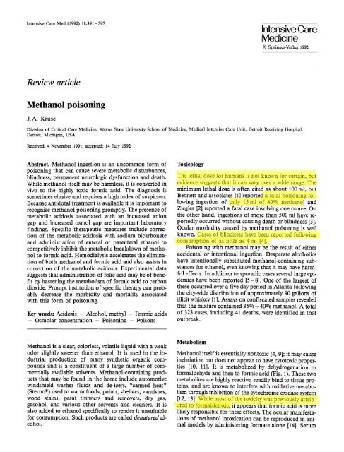
The lethal dose for humans is not known for certain, but<br />
evidence suggests that it can vary over a wide range. The<br />
minimum lethal dose is often cited as about 100 ml, but<br />
Bennett and associates [1] reported a fatal <strong>poisoning</strong> following<br />
ingestion of only 15 ml of 40% methanol and<br />
Ziegler [2] reported a fatal case involving one ounce. On<br />
the other hand, ingestions of more than 500 ml have reportedly<br />
occurred without causing death or blindness [3].<br />
Ocular morbidity caused by methanol <strong>poisoning</strong> is well<br />
known. Cases of blindness have been reported following<br />
consumption of as little as 4 ml [4].<br />
Poisoning with methanol may be the result of either<br />
accidental or intentional ingestion. Desperate alcoholics<br />
have intentionally substituted methanol-containing substances<br />
for ethanol, even knowing that it may have harmful<br />
effects. In addition to sporadic cases several large epidemics<br />
have been reported [5-8]. One of the largest of<br />
these occurred over a five day period in Atlanta following<br />
the city-wide distribution of approximately 90 gallons of<br />
illicit whiskey [1]. Assays on confiscated samples revealed<br />
that the mixture contained 35%- 40% methanol. A total<br />
of 323 cases, including 41 deaths, were identified in that<br />
outbreak.<br />
<strong>Methanol</strong> is a clear, colorless, volatile liquid with a weak<br />
odor slightly sweeter than ethanol. It is used in the industrial<br />
production of many synthetic organic com-j<br />
pounds and is a constituent of a large number of commercially<br />
available solvents. <strong>Methanol</strong>-containing products<br />
that may be found in the home include automotive<br />
windshield washer fluids and de-icers, "canned heat"<br />
(Sterno | used to warm foods, paints, shellacs, varnishes,<br />
wood stains, paint thinners and removers, dry gas,<br />
gasohol, and various other solvents and cleaners. It is<br />
also added to ethanol specifically to render it unsuitable<br />
for consumption. Such products are called denatured alcohol.<br />
Metabolism<br />
<strong>Methanol</strong> itself is essentially nontoxic [4, 9]; it may cause<br />
inebriation but does not appear to have cytotoxic properties<br />
[10, 11]. It is metabolized by dehydrogenation to<br />
formaldehyde and then to formic acid (Fig. 1). These two<br />
metabolites are highly reactive, readily bind to tissue proteins,<br />
and are known to interfere with oxidative metabolism<br />
through inhibition of the cytochrome oxidase system<br />
[12, 13]. <strong>While</strong> most of the toxicity was previously attributed<br />
to formaldehyde, it appears that formic acid is more<br />
likely responsible for these effects. The ocular manifestations<br />
of methanol intoxication can be reproduced in animal<br />
models by administering formate alone [14]. Serum
392<br />
formate concentrations have been shown to correlate better<br />
with clinical findings compared to methanol levels<br />
[15].<br />
The severe acidosis frequently observed in human<br />
cases of methanol <strong>poisoning</strong> can not be induced in rodents.<br />
<strong>Methanol</strong> can, however, induce severe metabolic<br />
acidosis, coma, and death in pigtail and rhesus monkeys<br />
[16-19]. Both primates and rodents metabolize methanol<br />
to formic acid; however, rodents are capabIe of rapidly<br />
converting formate into carbon dioxide. Formate therefore<br />
does not accumulate in rodents and they are spared<br />
both the acidosis and other toxic manifestations observed<br />
in primates. The metabolism of formate is an enzymemediated<br />
process that requires the presence of folate as a<br />
cofactor (Fig. 1). Akhough methanol does not cause acidosis<br />
in normal rats, it can induce formic acidosis in<br />
folate-deficient rats [11, 20]. <strong>While</strong> primates are capable<br />
of this same folate-dependent metabolism of formic acid,<br />
the rate of this pathway is much slower compared to that<br />
in rodents.<br />
Folic acid<br />
CH3-OH<br />
<strong>Methanol</strong><br />
Alcohol ~IL NAD +<br />
dehydrogenase NADH + H §<br />
o<br />
H-C-H<br />
Formaldehyde<br />
y<br />
Aldehyde |<br />
NAD<br />
NADPH ,~ I dehydr~ [~JL<br />
+ H* I Folate ~ NADH+ H +<br />
,A reductase 9 - 2H20<br />
H-C-OH ' ~ ~ C02<br />
Dihydrofolate Formic acid Peroxidase ()<br />
NADPH ~ I M ~ 2+<br />
+ H § ~] u IhATP<br />
Dihydrofelate| f"-Formvl-rHF~r<br />
I<br />
Excretion<br />
tf<br />
Lungs Kidne~ ,s<br />
Clinical manifestations<br />
Following ingestion, there is typically a lag period of<br />
about 12-24h before toxic manifestations occur [3,<br />
9-11, 15-19]. This interval can be quite variable, however,<br />
and ranges from less than one hour to over 72 h [1].<br />
Lack of symptoms should therefore not be interpreted as<br />
indicating insignificant intoxication, particularly if the<br />
patient presents promptly following ingestion. The lag<br />
period is due to the slow conversion of methanol to formaldehyde.<br />
Visual disturbances are common and range from dimming<br />
or blurring of vision, scintillations, photophobia,<br />
visual field defects, or "seeing a snowstorm", to a total<br />
loss of light perception [I, 7, 8, 21]. In one large epidemic<br />
all patients with at least mild acidosis and over half of<br />
those patients without acidosis had some type of visual<br />
symptoms [11. On fundoscopic examination an enlarged<br />
blind spot, hyperemia of the optic disc or frank papilledema<br />
may be observed [1, 7, 21]. Abnormal pupillary light<br />
reflexes have been described and range from a diminished<br />
reaction to fixed and dilated pupils [1]. Interference with<br />
neural axoplasmatic transport by formaldehyde and/or<br />
formate probably accounts for the ocular manifestations<br />
[11,121.<br />
Nausea, vomiting, and abdominal discomfort are<br />
common but are not universally seen. Epigastric pain<br />
may be severe and accompanied by abdominal rigidity.<br />
Like ethanol, methanol is a direct gastric irritant and can<br />
cause hemorrhagic gastritis. Pancreatitis has also been<br />
implicated as a cause of the abdominal pain, since high<br />
levels of serum amylase activity have been detected in<br />
many cases and pancreatitis has been confirmed in autopsy<br />
studies [1, 5]. In cases with significant acidosis,<br />
Kussmaul respirations may be observed. Headache, dizziness,<br />
malaise, agitation, generalized weakness, parathesias<br />
and sensorial depression may also occur [1, 10], Severe<br />
degrees of <strong>poisoning</strong> are associated with cerebral<br />
edema, coma, and/or seizures. The characteristic finding<br />
of bilateral cerebral infarction selectively involving the<br />
putamen and adjacent areas may be demonstrated by<br />
computed tomography or at postmortem examination<br />
[10].<br />
T2;ai"~.~oftlate<br />
10_.,orrn~y~l ;~ ~ 5,10_methenyi.TH F<br />
~ L ~ Methene-THF<br />
cyclohydrolyase p NADPH + H +<br />
5,10-methylene-THF |<br />
CO 2 dehydrogenase ~ = NADP +<br />
Serine transhydroxymethylase<br />
T<br />
-~. 5,10-methylene-THF<br />
Serine Glycine ~ NADH+ H §<br />
bt~e,2~ 5-methyI-THF<br />
Methionine Homocysteine<br />
Fig. 1. Metabolic pathways involved in methanol metabolism and relationship<br />
with relate metabolism. THF = tetrahydrofolate<br />
!<br />
1<br />
Laboratory findings<br />
The severity of the metabolic acidosis is variable and may<br />
not correlate well with the amount of methanol ingested,<br />
but it can be extremely severe [1, 8, 11, 12]. The decrease<br />
in plasma bicarbonate closely parallels the increase in<br />
plasma formate concentration in both animal models as<br />
well as in human methanol <strong>poisoning</strong>, indicating that<br />
most or all of the acidosis is accounted for by formic acid<br />
production [11, 15, 18]. A concomitant element of lactic<br />
acidosis is present in some cases. This may be due to the<br />
increased redox state of body tissues (i.e., increased ratio<br />
of NADH to NAD +) secondary to the oxidation of<br />
methanol and formaldehyde (Fig. 1). The increased redox<br />
state forces conversion of pyruvate to lactate. In addition,<br />
formic acid can interfere with intracellular respiration
393<br />
[13], thereby promoting anaerobic metabolism and lactate<br />
formation. However, in many cases flank circulatory<br />
shock and/or seizures are likely the predominant causes<br />
of increased lactate production [161.<br />
In their simplest form, serum electrolyte profiles performed<br />
by clinical chemistry laboratories include sodium,<br />
chloride, and carbon dioxide content determinations. The<br />
unmeasured serum anion concentration, or anion gap,<br />
can be defined as:<br />
Anion gap = [sodium]- [chloride]- [CO 2 content]<br />
with the traditional normal range varying from 8 to<br />
16mmol/1. Metabolic acidoses associated with an increased<br />
anion gap include lactic acidosis, ketoacidosis,<br />
the acidosis associated with renal failure, and several<br />
types of <strong>poisoning</strong> including salicylate, methanol, ethylene<br />
glycol, toluene and paraidehyde. In the case of methanol<br />
intoxication, the increase in the unmeasured anion<br />
fraction, as well as the acidosis, is predominantly due to<br />
accumulation of formate and, less consistently and to a<br />
smaller degree, to lactate [11, 12, 15, 18]. Due to its simplicity<br />
and availability, the anion gap is an important diagnostic<br />
indicator of possible methanol intoxication.<br />
Another important laboratory indicator of methanol<br />
<strong>poisoning</strong> is derived from comparison of measured and<br />
estimated serum osmolality. Serum osmolality may be estimated<br />
by [22]:<br />
Estimated osmolality =<br />
2 • sodium + BUN/2.8 + glucose/18<br />
The osmol gap is the difference between measured and<br />
calculated osmolality:<br />
Osmol gap =<br />
measured osmolality-estimated osmolality<br />
The normal osmol gap is approximately 10 mosm/kg water.<br />
This difference accounts for other normally occurring<br />
osmotically active constituents of serum. Any exogenous<br />
solute present in serum will raise the osmol gap. The osmotic<br />
effect of an ingested toxin is related to its molecular<br />
weight and the molar concentration of the toxin in serum.<br />
Since most drugs and toxins are either of high molecular<br />
weight or exert their toxicity at relatively low molar<br />
concentrations, they do not measurably affect the<br />
osmol gap. <strong>Methanol</strong>, on the other hand, has a low molecular<br />
weight and clinical intoxication is frequently associated<br />
with relatively high molar concentrations of the alcohol,<br />
such that the osmol gap may be appreciably increased.<br />
However, the most commonly encountered cause<br />
of an increased osmol gap is ethanol intoxication [23].<br />
Thus, serum ethanol concentration should be routinely<br />
assessed when evaluating the osmol gap. If ethanol is present<br />
in the blood, the gap may be significantly increased<br />
and it is then necessary to determine the quantitative contribution<br />
of ethanol to the osmol gap. This can be accomplished<br />
by including a term for ethanol in the formula for<br />
calculating osmolality:<br />
Estimated osmolality =<br />
2 x sodium + BUN/2.8 + glucose/18 + ethanol/4.6<br />
Using this formula to estimate serum osmolality, the<br />
clinician can determine whether a patient's serum ethanol<br />
level fully accounts for the osmol gap, or whether<br />
there is an otherwise unexplained increase in the gap due<br />
to methanol or some other substance. A few other ingested<br />
toxins, such as ethylene glycol and isopropanol, can<br />
similarly lead to significant increases in the osmol gap.<br />
Diagnosis<br />
In cases where the patient supplies an accurate history of<br />
the ingestion, the diagnosis is usually straightforward.<br />
However, in situations where the patient is unable or unwilling<br />
to supply such information, the diagnosis may<br />
prove elusive. Knowledge of the characteristic clinical and<br />
laboratory findings (Table 1) and a high index of suspician<br />
are crucial in these cases.<br />
Symptoms and physical signs are for the most part<br />
non-specific. Although methanol has a characteristic<br />
odor, it is not strong and may or may not be noted on the<br />
patient's breath. The odor of formalin has reportedly<br />
been detectable on the breath or urine of methanol-poisoned<br />
patients [24]. Abdominal pain is not infrequent but<br />
is also non-specific. Ocular findings are the most specific<br />
physical findings and are therefore most important diagnostically.<br />
Even more helpful are laboratory tests indicating<br />
the presence of metabolic acidosis associated with a<br />
high anion gap and high osmol gap. In the appropriate<br />
setting, these laboratory findings by themselves are of<br />
sufficient specificity to justify a presumptive diagnosis<br />
and institution of treatment.<br />
<strong>Methanol</strong> is frequently included as part of major toxicology<br />
screening batteries employed by clinical chemistry<br />
laboratories, often utilizing gas-liquid chromatography.<br />
However, it would be imprudent to await quantitative toxicologic<br />
assay results before making a presumptive diagnosis<br />
and initiating therapy, since it is likely that instituting<br />
specific treatment early in the course will lessen the<br />
morbidity and mortality [10]. An exception to this recommendation<br />
would be the situation where the clinician is<br />
certain that the institution's toxicology laboratory can<br />
routinely return such results within a matter of minutes.<br />
In cases where this is not possible, the clinician must<br />
make a presumptive diagnosis based on the presenting<br />
history, physical findings, and routine laboratory tests.<br />
Table 1. Characteristic clinical and laboratory findings in methanol intoxication<br />
9 Physical findings<br />
9 Kussmaul respirations<br />
9 Faint odor of methanol on breath<br />
9 Visual disturbances<br />
9 Nausea, vomiting, abdominal pain<br />
9 Altered sensorium<br />
9 Laboratory findings<br />
9 Elevated anion gap<br />
9 Metabolic acidosis<br />
9 Elevated osmol gap<br />
9 Positive serum methanol assay
394<br />
The constellation of ocular signs or symptoms, metabolic<br />
acidosis, and elevated anion and osmol gaps is indicative<br />
of methanol <strong>poisoning</strong> until proven otherwise.<br />
Treatment<br />
As with the initial management of all poisoned patients,<br />
general measures should be carried out to assure a patent<br />
airway, adequate ventilation, and adequate systemic perfusion.<br />
Gastric lavage is conventionally advocated to<br />
remove any residual poison, but is useful only soon after<br />
ingestion since methanol is rapidly absorbed from the<br />
gastrointestinal tract [8, 16]. The use of syrup of ipecac<br />
to induce emesis is contraindicated if the patient has an<br />
abnormal level of consciousness, since vomiting may result<br />
in aspiration of gastric contents. Its use may be hazardous<br />
as well in patients that present early with a nearnormal<br />
sensorium but who may subsequently progress to<br />
obtundation after ipecac is administered. Activated charcoal<br />
has also been recommended by some, although others<br />
have pointed out that its efficacy has not been substantiated<br />
[25]. Studies in normal volunteers indicate that<br />
enteral absorption of ethanol does not appear to be influenced<br />
by activated charcoal [26, 27], suggesting that<br />
methanol absorption might be similarly unaffected.<br />
Intravenously administered sodium bicarbonate<br />
should be given to methanol-poisoned patients with significant<br />
metabolic acidosis [5, 28, 29]. Reports suggest<br />
that such therapy can potentially ameliorate ocular manifestations,<br />
improve the sensorium, and perhaps reduce<br />
mortality.<br />
The mainstay of treatment for methanol intoxication<br />
is administration of ethanol [30, 31]. The enzymes responsible<br />
for converting methanol to formaldehyde and<br />
formic acid are also involved in the metabolism of ethanol<br />
to acetaldehyde and acetate. The two alcohols therefore<br />
act as competitive substrates. However, the rate of<br />
metabolism of methanol by these enzymes is only a fraction<br />
of that of ethanol [25, 32-34]. Thus, conversion of<br />
methanol into its toxic byproducts is slowed in the presence<br />
of ethanol. Typical criteria for initiating ethanol<br />
treatment include all patients with peak serum methanol<br />
concentrations greater than 20mg/dl and all patients<br />
with acidosis ascribed to methanol intoxication, regardless<br />
of whether or not symptoms are present [25]. Because<br />
methanol assay results may not be immediately<br />
available, it is important to initiate ethanol therapy in patients<br />
with a clear history of methanol ingestion and in<br />
patients strongly suspected of methanol <strong>poisoning</strong>.<br />
The clinical goal of ethanol therapy is to achieve a<br />
therapeutic serum ethanol level of between 100 and approximately<br />
150mg/dl [16, 25, 31, 35, 36]. This concentration<br />
is necessary to saturate alcohol dehydrogenase<br />
[36, 37]. Some reviewers have recommended up to<br />
200 mg/dl as the upper therapeutic limit [4, 24, 35, 36].<br />
To rapidly accomplish this goal it is necessary to administer<br />
a loading dose. Recommendations for calculating<br />
loading doses are shown in Table 2 [25, 31, 35, 36]. The<br />
patient's serum ethanol level should be assessed prior to<br />
administering the loading dose. For patients with baseline<br />
Table 2. Standard therapeutic ethanol dosing to achieve serum ethanol<br />
concentration of 100 mg/dl. Assumes typical volume of distribution<br />
and elimination kinetics; actual dosing should be titrated using frequent<br />
serum ethanol assays. (Based on data of H.G. McCoy [31])<br />
Loading dose<br />
Amount of absolute ethanol a<br />
Volume of 43070 oral solution b<br />
Volume of 90070 oral solntion c<br />
Volume of 10070 parenteral solution d<br />
Maintenance dose (not on dialysis)<br />
Amount of absolute ethanol a<br />
Volume of 4307o oral solution b<br />
Volume of 90070 oral solution c<br />
Volume of 10% parenteral solution d<br />
Maintenance dose during dialysis<br />
Amount of absolute ethanol a<br />
Volume of 43070 oral solution b<br />
Volume of 90% oral solution c<br />
Volume of 10% parenteral solution a<br />
Nondrinker Chronic drinker<br />
600mg/kg<br />
1.8ml/kg<br />
0.86ml/kg<br />
7.6mi/kg<br />
600mg/kg<br />
1.8ml/kg<br />
0.86mt/kg<br />
7.6ml/kg<br />
66mg/kg/h 154mg/kg/h<br />
0.20ml/kg/h 0.46ml/kg/h<br />
0.10ml/kg/h 0;21ml/kg/h<br />
0.83ml/kg/h 1.96rrd/kg/h<br />
169mg/kg/h 257 mg/kg/h<br />
0.50 ml/kg/h 0.77 ml/kg/h<br />
0.24 ml/kg/h 0.37 ml/kg/h<br />
2.13 ml/kg/h 3.26 ml/kg/h<br />
a Specific gravity = 0.79<br />
b Ethanol content = 34 g/dl (equivalent to 86 proof undiluted liquor)<br />
c Ethanol content = 7.1 g/dl<br />
Ethanol content = 7.9 g/dl<br />
ethanol levels in excess of 100 mg/dl the loading dose will<br />
be unnecessary, while those with lesser degrees of preexisting<br />
ethanol intoxication will require appropriate modification<br />
of their loading dose. When using the oral route,<br />
ethanol should be diluted to a final concentration of less<br />
than 400/0 and preferably less than 20% ethanol, since patients<br />
unaccustomed to drinking hard liquor frequently<br />
will not tolerate stronger solutions and vomiting is common.<br />
Relatively large fluid volumes are required for intravenous<br />
loading; eg, over a liter of 5% ethanol in water is<br />
necessary to load a 70 kg individual. The advantages and<br />
disadvantages of enteral versus parenteral ethanol dosing<br />
are summarized in Table 3. Following the loading dose, a<br />
maintenance regimen of ethanol can be accomplished using<br />
a 10% sterile solution of ethanol in water given by<br />
continuous intravenous infusion, or using hourly doses<br />
of commercial liquor given orally or by nasogastric tube<br />
(see Table 2) [31, 35, 36]. A higher maintenance infusion<br />
rate is required in chronic drinkers due to their higher rate<br />
of ethanol metabolism [31, 35 - 37]. It should be stressed<br />
that serial serum ethanol levels are necessary to insure<br />
that the target therapeutic level of 100 to 150mg/dl is<br />
reached and maintained. This is often difficult to achieve,<br />
even when hourly levels are obtained, and it is particularly<br />
difficult in chronic alcoholics and during the initiation<br />
of hemodialysis [15, 25, 38, 39].<br />
Forced diuresis has little effect on methanol elimination<br />
[3, 25, 31, 40]. Dialysis, on the other hand, effectively<br />
removes methanol and formate from the circulation<br />
[11, 30, 41-43]. In one report the half-life of methanol<br />
was reduced from 8 h to 2.5 h following institution of dialysis<br />
[31]. Hemodialysis is preferred to peritoneal dialysis<br />
because it offers more rapid clearance [8, 30, 44].<br />
Hemoperfusion should not be used since hemoperfusion<br />
columns may quickly become saturated with methanol
395<br />
Table 3. Advantages and disadvantages of enteral versus parenterai<br />
ethanol<br />
Advantages<br />
Disadvantages<br />
Enteral administration<br />
Ethanol treatment<br />
Parenterai administration<br />
Simplified preparation No risk of gastric irritaand<br />
administration of tion or aspiration<br />
loading dose by mouth or<br />
Simple titration of<br />
by nasogastric tube<br />
maintenance infusions by<br />
Can use relatively concen- adjusting infusion rate<br />
trated ethanol solutions,<br />
Can be used in patients<br />
thus minimizing the risk<br />
who are vomiting or have<br />
of fluid overload<br />
gastric bleeding<br />
Risk of gastritis and<br />
emesis; risk of aspiration,<br />
especially since sensorium<br />
is likely to be depressed<br />
Hourly maintenance doses<br />
require increased nursing<br />
time, possibly increasing<br />
risk of missed doses<br />
Central venous catheter<br />
required due to<br />
hyperosmolality of solution<br />
(10% ethanol in 5%<br />
dextrose is approximately<br />
2000 mosm/kg water<br />
Potential for fluid<br />
overload in susceptible<br />
patients, especially when<br />
using 5% ethanol solutions<br />
A<br />
--I<br />
m<br />
O<br />
E<br />
E<br />
I.U<br />
=Z<br />
n-<br />
O<br />
IJ.<br />
o<br />
O<br />
_1<br />
m<br />
8<br />
6<br />
4<br />
2<br />
I<br />
/<br />
p"...o.-"~<br />
/ -- <strong>Methanol</strong><br />
0<br />
0 10 20 30 40<br />
HOURS<br />
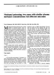
Fig. 2. Blood formate concentrations in two monkeys poisoned with<br />
methanol at time zero. One monkey was subsequently treated with<br />
5-formyl-tetrahydrofolate (5-formyl-THF; doses indicated by vertical<br />
arrows) demonstrating favorable effect on blood formate concentration<br />
compared to control animal. (Redrawn from Noker PEet al [17], with<br />
permission)<br />
!<br />
5O<br />
and rendered ineffective [4, 45]. In addition, hemoperfusion<br />
can not facilitate correction of the metabolic acidosis<br />
or fluid and electrolyte disturbances that are frequently<br />
present. Criteria for employing dialysis have varied [15,<br />
16, 46]. In general, dialysis should be employed in all<br />
cases developing ocular manifestations and in all cases<br />
with renal impairment, regardless of symptoms. A peak<br />
methanol level of greater than 50 mg/dl has frequently<br />
been cited as an indication for dialysis [4, 15, 25, 30, 31,<br />
42, 43, 47, 48]. However, lower levels may be misleading<br />
if the intoxication has advanced to the point where most<br />
of the methanol has been metabolized but toxic metabolites<br />
are still present. The presence of metabolic acidosis<br />
and a high anion gap in the face of a low methanol levels<br />
suggests this situation [10]. Since dialysis removes ethanol<br />
as well as methanol, patients undergoing dialysis will<br />
also require higher maintenance doses (Table 2) [30, 31,<br />
35, 36, 42, 43].<br />
The previously described relationship between formic<br />
acid metabolism and folic acid-dependent enzyme systems<br />
suggests that folic acid may play a role as a therapeutic<br />
adjunct in methanol <strong>poisoning</strong>. In primate models<br />
folate-deflciency increases the sensitivity to methanol<br />
<strong>poisoning</strong> [49] and folate administration increases the<br />
rate of formate metabolism during methanol intoxication<br />
[17, 19]. Folate has also been shown to reverse methanol<br />
toxicity even when the vitamin is administered 10 h after<br />
methanol dosing (Fig. 2) [17]. <strong>While</strong> its therapeutic efficacy<br />
in humans has not been examined, the animal data<br />
is convincing and administration of the vitamin is probably<br />
innocuous. Folic or folinic acid should therefore be<br />
administered to all patients with known or suspected<br />
methanol intoxication. Extrapolating from experimental<br />
data, large and repeated doses are probably necessary. In-<br />
travenous doses of 50 to 100 mg of folate every 4 h have<br />
been recommended [4, 15, 16, 25].<br />
Pyrazole has long been known to be a potent competitive<br />
inhibitor of alcohol dehydrogenase [50, 51]. Its use<br />
as a potential therapeutic agent for treatment of methanol<br />
<strong>poisoning</strong> has been tempered by its toxic effects on<br />
the liver and other tissues. 4-Methyl pyrazole, on the other<br />
hand, is an even more specific inhibitor of alcohol dehydrogenase<br />
and appears to be much less toxic [38,<br />
50-54]. The compound has been shown to dramatically<br />
inhibit production of formic acid from methanol in experimental<br />
models. Monkeys given usually lethal doses of<br />
methanol survived when rescued with 4-methyl pyrazole,<br />
even when the drug was administered after the methanol<br />
[52]. The drug is not currently available for clinical use<br />
in the U.S., but has been used investigationally.<br />
Prognosis<br />
Information from one large epidemic demonstrated that<br />
the severity of the acidosis correlates with outcome. In<br />
this group there was a 19% mortality rate among the 115<br />
patients that had serum carbon dioxide contents below<br />
20 mmol/1, compared to a 50% mortality rate for patients<br />
with carbon dioxide contents less than 10 mmol/l<br />
[1]. In a review of 725 cases by McNally, there were 335<br />
survivors, of which 90 suffered total blindness and 85 had<br />
some degree of visual disturbance during the acute intoxication<br />
[35]. However, recovery from visual impairment is<br />
common among survivors. In the epidemic reported by<br />
Chew and associates, there were 26 survivors, all of whom<br />
were acidotic to some degree and 15 of whom had visual<br />
impairment during the acute phase, but only two suffered<br />
permanent visual loss [29]. Other persistent neurologic<br />
deficits include tremor, spasticity, and a syndrome similar
396<br />
to Parkinsonism [56-58]. Prior or concomitant ethanol<br />
ingestion may mitigate the degree of toxicity for a given<br />
dose of methanol. Underlying folate deficiency may also<br />
be important. These factors, in addition to the ingested<br />
dose of methanol, may partially explain the wide variation<br />
that has been reported for the minimum toxic dose<br />
in humans.<br />
References<br />
1. Bennett IL Jr, Cary FH, Mitchell GL et al (1953) Acute methyl alcohol<br />
<strong>poisoning</strong>: a review based on experiences in an outbreak of 323<br />
cases. Medicine 32:431-463<br />
2. Ziegler SL (1921) The ocular menace of wood alcohol. Br J Opthalmol<br />
5:365-373, 411-417<br />
3. Tong TG (1982) The alcohols. Crit Care Q 4:75-85<br />
4. Bryson PD (1989) Comprehensive review in toxicology, 2nd edn.<br />
Aspen, Rockville, MD, pp 284-302<br />
5. Naraqi S, Dethlefs RF, Slobodniuk RA et al (1979) An outbreak of<br />
acute methyl alcohol intoxication. Aust NZ J Med 9:65-68<br />
6. Kane RL, Talbert W, Harlan Jet al (1968) A methanol <strong>poisoning</strong><br />
outbreak in Kentucky. Arch Environ Health 17:119-129<br />
7. Benton CD Jr, Calhoun FP Jr (1953) The ocular effects of methyl<br />
alcohol <strong>poisoning</strong>: report of a catastrophe involving 320 persons.<br />
Am J Ophthalmol 36:1677-1685<br />
8. Swartz RD, Millman RP, Billi JE et al (1981) Epidemic methanol<br />
<strong>poisoning</strong>: clinical and biochemical analysis of a recent episode.<br />
Medicine 60:373-382<br />
9. Riley LJ, Ilson BE, Narins RG (1987) Acute metabolic acid-base<br />
disorders. Crit Care Clin 5:699-724<br />
10. Suit PF, Estes ML (1990) <strong>Methanol</strong> intoxication: clinical features<br />
and differential diagnosis. Cleve Clin J Med 57:464-471<br />
11. McMartin KE, Ambre J J, Tephly TR (1980) <strong>Methanol</strong> <strong>poisoning</strong> in<br />
human subjects. Role for formic acid accumulation in the metabolic<br />
acidosis. Am J Med 68:414-418<br />
12. Shahangian S, Ash KO (1986) Formic and lactic acidosis in a fatal<br />
case of methanol intoxication. Clin Chem 32:395-397<br />
13. Nicholls P (1976) The effect of formate on cytochrome aa and on<br />
electron transport in the intact respiratory chain. Biochim Biophys<br />
Acta 430:13-29<br />
14. Martin-Amat G, McMartin KE, Hayreh SS et al (1978) <strong>Methanol</strong><br />
<strong>poisoning</strong>. Ocular toxicity produced by formate. Toxicol Appl<br />
Pharmacol 45:201-208<br />
15. Osterloh JD, Pond SM, Grady Set al (1986) Serum formate concentrations<br />
in methanol intoxication as a criteria for hemodialysis.<br />
Ann Intern Med 104:200-203<br />
16. Jacobsen D, McMartin KE (1986) <strong>Methanol</strong> and ethylene glycol<br />
<strong>poisoning</strong>s: mechanism of toxicity, clinical course, diagnosis and<br />
treatment. Med Toxicol 1:309-334<br />
17. Noker PE, EeUs JT, Tephly TR (1980) <strong>Methanol</strong> toxicity: treatment<br />
with folic acid and 5-formyl tetrahydrofolic acid. Alcoholism Clin<br />
Exp Res 4:378-383<br />
18. Clay KL, Murphy RC, Watkins WD (1975) Experimental methanol<br />
toxicity in the primate: analysis of metabolic acidosis. Toxicol Appl<br />
Pharmacol 34:49-61<br />
19. National Institute of Health (1979) Use of folate analogue in treatmerit<br />
of methyl alcohol toxic reactions is studied. JAMA<br />
242:1961 - 1962<br />
20. Makar AB, Tephly TR (1976) <strong>Methanol</strong> <strong>poisoning</strong> in the folate-deficient<br />
rat. Nature 261:715-716<br />
21. Hayreh MS, Hayreh SS, Baumbach GL et al (1977) Methyl alcohol<br />
<strong>poisoning</strong> III. Ocular toxicity. Arch Ophthalmol 95:1851-1858<br />
22. Dorwart WV, Chalmers L (1975) Comparison of methods for calculating<br />
serum osmolality from chemical concentrations, and the<br />
prognostic value Of such calculations. Clin Chem 21:190-194<br />
23. Robinson AG, Loeb JN (1971) Ethanol ingestion - commonest<br />
cause of elevated plasma osmolality N Engl J Med 284:1253 - 1255<br />
24. Becker CE (1983) <strong>Methanol</strong> <strong>poisoning</strong>. J Emerg Med 1:51-58<br />
25. Kulig K, Duffy JP, Linden CH et al (1984) Toxic effects of methanoI,<br />
ethylene glycol, and isopropyt alcokol. Topics Emerg Med<br />
6:14-28<br />
26. Hult6n B-A, Heath A, Mellstrand T et al (1985) Does alcohol absorb<br />
to activated charcoal Hum Toxicol 5:211-212<br />
27. Neuvonen PJ, Olkkola KT, Alanen T (1984) Effect of ethanol and<br />
pH on the adsorption of drugs to activated charcoal: studies in<br />
vitro and in man. Acta Pharmacol Toxicol 54:i-7<br />
28. Branch A, Tonning DJ (1945) Acute methyl alcohol <strong>poisoning</strong>; observations<br />
in some thirty cases. Can J Public Health 36:147-151<br />
29. Chew WB, Berger EH, Brines OA et al (1946) Alkali treatment of<br />
methyl alcohol <strong>poisoning</strong>. JAMA 130:61-64<br />
30. Keyvan-Larijarni H, Tannenberg AM (i974) <strong>Methanol</strong> intoxication.<br />
Comparison of peritoneal dialysis and hemodialysis treatment.<br />
Arch Intern Med 134:293-296<br />
31. McCoy HG, Cipolle RJ, Ehlers SM et al (1979) Severe methanol<br />
<strong>poisoning</strong>. Application of a pharmacokinetic model for ethanol<br />
therapy and hemodialysis. Am J Med 67:804-807<br />
32. Bartlett GR (1950) Inhibition of methanol oxidation by ethanol in<br />
the rat. Am J Physiol 163:619-621<br />
33. Zatman LJ (1946) The effect of ethanol on the metabolism of<br />
methanol in man. Biochem J 40:67-68<br />
34. Blair AH, Vallee BL (1966) Some catalytic properties of human liver<br />
alcohol dehydrogenase. Biochem 5:2026-2034<br />
35. Peterson CD (1981) Oral ethanol doses in patients with methanol<br />
<strong>poisoning</strong>. Am J Hosp Pharm 38:1024-1027<br />
36. Peterson CD, Collins A J, Himes JM et al (1981) Pharmacokinetics<br />
during therapy with ethanol and hemodialysis. N Engl J Med<br />
304:21-23<br />
37. Vestal RE, McGuire EA, Tobin JD et al (1977) Aging and ethanol<br />
metabolism. Clin Pharmacol Ther 21:343-354<br />
38. Weintraub M, Standish R (1988) 4-Methylpyrazole: an antidote for<br />
ethylene glycol and methanol intoxication. Hosp Formul<br />
23:960- 969<br />
39. Litovitz T (1986) The alcohols ethanol, methanol, isopropanol, ethylene<br />
glycol. Pediatr Clin North Am 33:311-323<br />
40. Chinard FP, Frisell WR (1976) <strong>Methanol</strong> intoxication: biochemical<br />
and clinical aspects. J Med Soc NJ 73:712-719<br />
41. Erlanson P, Fritz H, Hagstam K-E et al (1965) Severe methanol intoxication.<br />
Acta Med Scand 177:393-408<br />
42. Gonda A, Gault H, Churchill D et al (1978) Hemodialysis for methanol<br />
intoxication. Am J Med 64:749-758<br />
43. Tobin M, Llanos E (1979) Hemodialysis for methanol intoxication.<br />
J Dial 3:97-106<br />
44. Setter JG, Singh R, Brackett NC Jr (1967) Studies on the dialysis<br />
of methanol. Trans Am Soc Artif Intern Organs 13:178-182<br />
45. Whalen JE, Richards CJ, Ambre J (1979) Inadequate removal of<br />
methanol and formate using the sorbent based regeneration hemodialysis<br />
delivery system. Clin Nephrol 11:318 - 321<br />
46. Goldfrank LR, Flomenbaum NE, Lewin NA et al (1986) <strong>Methanol</strong><br />
and ethylene glycol. In: Goldfrank LR, Flomenbaum NE, Lewin<br />
NA et al (eds) Toxicologic emergencies, 3rd edn. Appleton-Century-<br />
Crofts, Norwalk, Conn, pp 452-467<br />
47. Smith SS (1983) Solvent toxicity: isopropanol, methanol, and ethylene<br />
glycol. ENT J 62:126-135<br />
48. Cowen DL (1964) Extracorporeal dialysis in methanol <strong>poisoning</strong>.<br />
Ann Intern Med 61:134-135<br />
49. McMartin KE, Martin-Amat G, Makar ABet al (1977) <strong>Methanol</strong><br />
<strong>poisoning</strong>. V. Role of formate metabolism in the monkey. J Pharmacol<br />
Exp Ther 201:564- 572<br />
50. Blomstrand R, Ostling-Wintzell H, Lof Aet al (1979) Pyrazoles as<br />
inhibitors of alcohol oxidation and as important tools in alcohol research:<br />
an approach to therapy against methanol <strong>poisoning</strong>; Proc<br />
Natl Acad Sci 76:3499- 3503<br />
51. Wilson WL, Bottiglieri NG (1962) Phase I studies with pyrazole.<br />
Cancer Chemother Res 21:137-141<br />
52. McMartin KE, Hedstrom K-G, Tolf B-R et al (1980) Studies on the<br />
metabolic interactions between 4-methylpyrazole and methanol using<br />
the monkey as an animal model. Arch Biochem Biophys<br />
199:606- 614
397<br />
53. Jacobsen D, Sebastian S, Blomstrand R et al (1988) 4-Methylpyrazole:<br />
a controlled study of safety in healthy human subjects after<br />
single, ascending doses. Alcoholism 12:516-522<br />
54. McMartin ME, Jacobsen D, Sebastian Set al (1987) Safety and metabolism<br />
of 4-methylpyrazole in human subjects. Vet Hum Toxicol<br />
29:471<br />
55. McNally WD (1939) Medical jurisprudence and toxicology. Sannders,<br />
Philadelphia<br />
56. McLean DR, Jacobs H, Mielke BW (1980) <strong>Methanol</strong> <strong>poisoning</strong>: a<br />
clinical and pathological study. Ann Neurol 8:161-167<br />
57. Ley CO, Gali FG (1983) Parkinsonian syndrome after methanol intoxication.<br />
Eur Neurol 22:405-409<br />
58. Guggenheim MA, Couch JR, Weinberg W (1971) Motor dysfunction<br />
as a permanent complication of methanol ingestion. Arch<br />
Neurol 24:550-554<br />
L A. Kruse, MD<br />
Detroit Receiving Hospital<br />
Room 55-10<br />
4201 St. Antoine Boulevard<br />
Detroit, Michigan 48201<br />
USA