PDF - Turkish Neurosurgery
PDF - Turkish Neurosurgery
PDF - Turkish Neurosurgery

You also want an ePaper? Increase the reach of your titles
YUMPU automatically turns print PDFs into web optimized ePapers that Google loves.
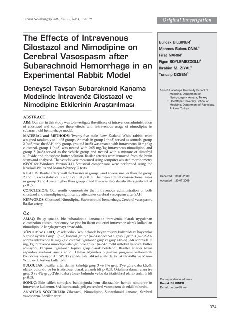
<strong>Turkish</strong> <strong>Neurosurgery</strong> 2009, Vol: 19, No: 4, 374-379<br />
Original Investigation<br />
The Effects of Intravenous<br />
Cilostazol and Nimodipine on<br />
Cerebral Vasospasm after<br />
Subarachnoid Hemorrhage in an<br />
Experimental Rabbit Model<br />
Burcak BILGINER 1<br />
Mehmet Bulent ONAL 2<br />
Firat NARIN 3<br />
Figen SOYLEMEZOGLU 4<br />
Ibrahim M. ZIYAL 5<br />
Tuncalp OZGEN 6<br />
Deneysel Tavflan Subaraknoid Kanama<br />
Modelinde ‹ntravenöz Cilostazol ve<br />
Nimodipine Etkilerinin Araflt›r›lmas›<br />
ABSTRACT<br />
AIM: Our aim in this study was to investigate the efficacy of intravenous administration<br />
of cilostazol and compare these effects with intravenous usage of nimodipine in<br />
subarachnoid hemorrhage model.<br />
MATERIAL and METHODS: Twenty-five male New Zealand White rabbits were<br />
assigned randomly to 1 of 5 groups. Animals in group 1 (n=5) served as controls, group<br />
2 (n=5) was the SAH-only group, group 3 (n=5) was treated with intravenous 10 mg/kg<br />
cilostazol, group 4 (n=5) was treated with 0.05 mg/kg intravenous nimodipine, and<br />
group 5 (n=5) served as the vehicle group and treated with a mixture of dimethyl<br />
sulfoxide and phosphate buffer solution. Basilar arteries were removed from the brain<br />
stems and analyzed. The vessels were measured using computer-assisted morphometry<br />
(SPOT for Windows Version 4.1). Statistical comparisons were performed using the<br />
Kruskall-Wallis and Mann-Whitney U tests.<br />
RESULTS: Basilar artery wall thicknesses in group 3 and 4 were smaller than the group<br />
2 and this was statistically significant at p
<strong>Turkish</strong> <strong>Neurosurgery</strong> 2009, Vol: 19, No: 4, 374-379<br />
Bilginer el al: The Effects of Intravenous Cilostazol and Nimodipine<br />
INTRODUCTION<br />
Despite its clinical significance, cerebral vasospasm<br />
is still an important clinical entity and its exact<br />
pathogenesis is unknown. Morbidity and mortality of<br />
cerebral vasospasm after subarachnoid hemorrhage is<br />
still high (1,6,13,21). The amount of subarachnoid blood<br />
is associated with the development of vasospasm and it<br />
leads to an inflammatory response during the first 48<br />
hour (7). The treatment modalities for cerebral<br />
vasospasm are aimed to improve cerebral blood flow<br />
by dilatation of effected vessels. Studies on prevention<br />
and reversal of cerebral vasospasm are focused on<br />
various therapeutic agents.<br />
Cilostazol is a potent inhibitor of phosphodiesterase<br />
3. It relaxes vascular smooth muscles with this effect<br />
and causes vasodilatation. It is also used for the<br />
treatment of ischemic neurological deficits and for the<br />
prevention from the recurrent cerebral infarction (12).<br />
Our aim in this study was to investigate the effects<br />
of intravenous usage of selective phosphodiesterase 3<br />
inhibitor cilostazol on cerebral vasospasm after SAH<br />
and compare these effects with the intravenous<br />
administration of nimodipine.<br />
MATERIALS and METHOD<br />
Animal Model<br />
The experimental protocol was approved by the<br />
Hacettepe University Animal Research Committee.<br />
Twenty-five male New Zealand White rabbits weighing<br />
2500-3000 were assigned randomly to 1 of 5 groups. The<br />
intravenous (iv) form of cilostazol (Pletal, Abdi Ibrahim,<br />
Turkey) was prepared by using dimethyl sulfoxide and<br />
phosphate buffer solution. Animals in group 1 (n=5)<br />
served as controls, group 2 (n=5) was the SAH only<br />
group, group 3 (n=5) was treated with an intravenous<br />
30 mg/kg cilostazol (1) 3 times at 12 hours, 24 hours<br />
and 36 hours after the SAH induction, group 4 (n=5)<br />
was treated with an 0.05 mg/kg intravenous<br />
nimodipine (8) (Nimotop, Bayer, Germany) for 6 times<br />
at 6,12,18, 24, 30 and 36 hours, and group 5 (n=5) served<br />
as a vehicle group and treated with a mixture of<br />
dimethyl sulfoxide and phosphate buffer solution after<br />
SAH induction. All procedures were performed by 2<br />
investigators who were not blinded to the treatment<br />
group during surgery and euthanasia. Vascular<br />
measurements were performed in a blinded fashion.<br />
Induction of Experimental SAH<br />
All animals subjected to experimental SAH were<br />
anesthetized by intramuscular injection of a mixture of<br />
ketamine 50 mg/kg (Ketalar, Parke-Davis, Eczacibasi,<br />
Istanbul, Turkey) and xylazine 10 mg/kg (Rompun,<br />
Bayer, Istanbul, Turkey), and all animals breathed<br />
spontaneously throughout the procedures. A 23-gauge<br />
butterfly needle was inserted into the cisterna magna<br />
after exposing of the atlanto-occipital membrane with a<br />
small incision at the occipitocervical junction. After<br />
withdrawal of 1.0 mL of CSF, 1 mL volume of<br />
nonheparinized autologous blood from the central ear<br />
artery was injected into the cisterna magna over 2<br />
minutes. The animals were then placed in a head-down<br />
position at 30° for 30 minutes to hold the blood in the<br />
basal cisterns. Arterial blood gases were analyzed<br />
during the surgical procedure and maintained within<br />
the physiological range. After recovering from<br />
anesthesia, the rabbits were observed for possible<br />
neurological deficits and then returned to the vivarium.<br />
Perfusion – Fixation<br />
All animals subjected to experimental SAH were<br />
euthanized by perfusion-fixation 48 hours after SAH<br />
induction. The animals were anesthetized as described<br />
above. The ear artery was catheterized for monitoring<br />
blood pressure and for blood gas analysis. When<br />
satisfactory respiratory parameters were obtained, a<br />
thoracotomy was performed, the left ventricle<br />
cannulated, the right atrium opened widely, and the<br />
abdominal aorta clamped. After perfusion of a flushing<br />
solution (Hanks' balanced salt solution [Sigma<br />
Chemical Co], pH 7.4. at 37°C, 300 mL), a fixative was<br />
perfused (2% paraformaldehyde. 2,5% glutaraldehyde<br />
in 0.1 M phosphate buffer, pH 7.4, at 37°C, 200 mL).<br />
Perfusion was performed at a standard height of 100 cm<br />
from the chest. Animals in the control group were killed<br />
using the same procedure. The brains were then<br />
removed and stored in formaldehyde fixation solution<br />
at 4°C overnight.<br />
Morphometric Analysis of the Basilar Artery<br />
Basilar arteries were removed from the brain stems<br />
and arterial segments from the proximal third of the<br />
artery were dissected for analysis. The vessels were<br />
embedded in epoxy resin, and cross-sections were cut at<br />
a thickness of 0.5 μm. The sections were mounted onto<br />
glass slides and stained with hematoxylin eosin for light<br />
microscopic analysis. The vessels were measured using<br />
computer-assisted morphometry (SPOT for Windows<br />
Version 4.1). Automated measurements of the crosssectional<br />
area of the arterial sections and arterial wall<br />
thickness were taken by an investigator who was<br />
blinded to the identity of the group to which the<br />
375
<strong>Turkish</strong> <strong>Neurosurgery</strong> 2009, Vol: 19, No: 4, 374-379<br />
Bilginer el al: The Effects of Intravenous Cilostazol and Nimodipine<br />
animals belonged. Five cross-sections of each vessel<br />
were selected randomly for measurement, and the<br />
average of these measurements were calculated.<br />
Statistical Analysis<br />
Data are expressed as mean ± SD. Statistical<br />
comparisons were performed using a Kruskall-Wallis<br />
and Mann-Whitney U tests. Statistical significance was<br />
accepted at p 0.05.<br />
RESULTS<br />
The value of the basilar artery wall thickness was<br />
48.4 ± 2.70 μm in the control group (group 1) and 73.0 ±<br />
7.41 μm in the SAH group (group 2). The two treatment<br />
groups after SAH induction, SAH+cilostazol (group 3)<br />
and SAH+nimodipine (group 4), had average values of<br />
49.0 ± 11.4 μm and 49.6 ± 9.98 μm respectively. For the<br />
vehicle group (group 5), the average value was 71.0 ±<br />
1.87 μm (Figure 1).<br />
The mean cross-sectional areas were 115823.8 ±<br />
18048.14 μm2 in group 1, 10491.00 ± 3652.47 μm2 in<br />
group 2, 38487.00 ± 14437.02 μm2 in group 3, 36735.00 ±<br />
9973.57 μm2 in group 4 and 10467.6± 2726.73 μm2 in the<br />
vehicle group (Figure 2).<br />
Mean basilar artery cross-sectional areas (CSA) and<br />
arterial wall thickness (AWT) values are provided in<br />
(Table I).<br />
Basilar artery wall thicknesses in group 3 and 4 were<br />
smaller than for group 2 and this was statistically<br />
significant at p
<strong>Turkish</strong> <strong>Neurosurgery</strong> 2009, Vol: 19, No: 4, 374-379<br />
Bilginer el al: The Effects of Intravenous Cilostazol and Nimodipine<br />
Figure 3: Basilar artery cross-sectional areas and arterial wall<br />
thicknesses are shown in different groups. A: Control group;<br />
B: SAH; C: SAH + iv cilostazol; D: SAH + iv nimodipine (at<br />
20x magnification)<br />
DISCUSSION<br />
Cerebral vasospasm is an important clinical<br />
problem and its pathophysiology is still not fully<br />
understood. The number of patients who develop<br />
symptomatic vasospasm is between 20-30% while<br />
radiographic vasospasm can be seen in 70% of patients<br />
without any clinical consequences (13). The major<br />
problem produced by cerebral vasospasm is ischemic<br />
neurological deficit and the treatment strategies are<br />
focused on these parameters.<br />
The pathogenesis of cerebral vasospasm is complex,<br />
multi-factorial and not completely elucidated. One of<br />
the major possible mechanisms of cerebral vasospasm<br />
depends on nitric oxide (NO) metabolism. NO is a<br />
potent vasodilator and has an important role in the<br />
Table II: Summary of physiological parameters of<br />
groups<br />
Group MABP pH pO2 pCO2<br />
1 99±1.02 7.41±0.02 110±5.55 39.8±1.03<br />
2 102±1.38 7.42±0.01 115±3.05 40.0±1.10<br />
3 102±1.17 7.40±0.05 113±2.09 41.4±1.04<br />
4 100±1.12 7.42±0.07 110±4.37 41.9±1.09<br />
5 100±1.04 7.41±0.03 112±3.95 41.1±1.01<br />
Results are expressed as mean ± SD<br />
MABP: Mean Arterial Blood Pressure<br />
development of cerebral vasospasm. Its main effect is<br />
the relaxation of vascular smooth muscle cells. The<br />
activation of soluble guanylyl cyclase by nitric oxide<br />
results in dephosphorylation of myosin light chains,<br />
activation of potassium channels and closure of voltagedependent<br />
calcium channels. This reaction produces<br />
smooth muscle relaxation (21,23). Another factor that<br />
plays an important role on pathogenesis of cerebral<br />
vasospasm is bilirubin oxidation products. These<br />
products occur after free radical oxidation of bilirubin<br />
and produce BOXes. The effects of BOXes are on<br />
vascular smooth muscle cells and produce<br />
vasoconstriction (6,22). Clark et al. have shown in their<br />
studies that the concentration of BOXes in cerebrospinal<br />
fluid correlates with the clinical vasospasm in patients<br />
with SAH (6). Another potent vasoconstrictor is<br />
Endothelin-1. It has two receptor subtypes ETA and<br />
ETB. ETA receptors are found on smooth muscle cells<br />
and mediate vasoconstriction. ETB receptors are located<br />
on both endothelial cells and venous smooth muscle<br />
cells. They mediate the release of relaxing factors by<br />
acting on endothelial cells and mediate vasoconstriction<br />
by acting on venous smooth muscle cells (5,19).<br />
Phosphodiesterase 3 is strongly expressed in<br />
platelets and vascular smooth muscle cells. This<br />
compound is responsible for the degradation of cyclic<br />
AMP (cAMP) and cyclic GMP (cGMP). These cyclic<br />
nucleotides has an important role on regulation of<br />
vascular tonus. The phosphodiesterase 3 inhibitor<br />
cilostazol increases the intracellular cAMP by blocking<br />
its hydrolysis (15). Cilostazol also lowers the<br />
intraplatelet Ca2+ and shows an antiaggregation effect<br />
on platelets and vasodilator effect on blood vessels.<br />
Tanaka et al. (25) has been reported in their<br />
experimental study that cilostazol dilates the pial<br />
arteries in cats. Birk et al. (2) reported that cilostazol<br />
dilates cerebral arteries in vitro and in another study<br />
they showed that cilostazol dilates large cerebral<br />
arteries in humans (3).<br />
Selective phosphodiesterase (PDE) 3 inhibitor<br />
cilostazol is known as an antiplatelet, vasodilator agent<br />
and its antiplatelet effect is potentiated by prostaglandin<br />
E1 (20). Tamai et al. reported cilostazol did not prolong<br />
bleeding time besides its antiplatelet effect (24).<br />
Cilostazol also inhibits adenosine uptake. Its inhibition<br />
effect on both PDE and adenosine uptake may play a<br />
role on vascular smooth muscle relaxation and<br />
vasodilatation (17).<br />
Kim et al. showed another inhibition mechanism of<br />
cilostazol on apoptotic death in human umbilical vein<br />
377
<strong>Turkish</strong> <strong>Neurosurgery</strong> 2009, Vol: 19, No: 4, 374-379<br />
Bilginer el al: The Effects of Intravenous Cilostazol and Nimodipine<br />
endothelial cells (14). In their experimental study on<br />
porcine carotid artery, Kohda et al. emphasize the<br />
prevention effect of cilostazol on carotid artery<br />
thrombosis after endothelial injury (16).<br />
Hashimoto et.al. mention that cilostazol induces<br />
nitric oxide production (10) and another effect of<br />
cilostazol about suppression of platelet/leukocyte<br />
aggregation in humans was reported by Ito et al. (11).<br />
Choi et al. reported in their studies that cilostazol has a<br />
neuroprotective effect against focal cerebral ischemia<br />
(4).<br />
Nimodipine is a lipid soluble 1,4-dihydropyridine–<br />
derivative Ca2+ channel blocker. Its main effect is to<br />
inhibit Ca2+ influx through voltage-sensitive L-type<br />
Ca2+ channels and inhibits contractions of vascular<br />
smooth muscle (26). There are many studies on the<br />
effects of nimodipine on cerebral vasospasm (8,9,18).<br />
Our aim in this study was to investigate the effects of<br />
intravenous usage of the selective phosphodiesterase 3<br />
inhibitor cilostazol on cerebral vasospasm after SAH<br />
and compare these effects with the intravenous<br />
administration of the calcium channel blocker<br />
nimodipine.<br />
Our results demonstrate that intravenous<br />
administration of both cilostazol and nimodipine<br />
significantly attenuates cerebral vasospasm after SAH.<br />
When we compared the effects of these compounds, we<br />
did not find a statistically important superiority to one<br />
another. We suggest that the beneficial effects of<br />
cilostazol depend on the combination of its<br />
vasodilatory, antiapoptotic, antiinflammatory and<br />
neuroprotective effects which have previously been<br />
demonstrated. We therefore propose cilostazol as a<br />
candidate for clinical trials in the treatment of delayed<br />
cerebral vasospasm and related ischemic neurologic<br />
deficit.<br />
REFERENCES<br />
1. Bilginer B, Onal B, Yigitkanli K, Soylemezoglu F, Bavbek M, Ziyal<br />
I.M, Ozgen T: Treatment of cerebral vasospasm with cilostazol in<br />
subarachnoid haemorrhage model. Acta Neurochir Suppl 104:<br />
291-296,2008<br />
2. Birk S, Edvinsson L, Olesen J, Kruuse C: Analysis of the effects of<br />
phosphodiesterase type 3 and 4 inhibitors in cerebral arteries. Eur<br />
J Pharmacol 489(1-2):93-100,2004<br />
3. Birk S, Kruuse C, Petersen KA, Jonassen O, Tfelt-Hansen P, Olesen<br />
J: The phosphodiesterase 3 inhibitor cilostazol dilates large<br />
cerebral arteries in humans without affecting regional cerebral<br />
blood flow. J Cereb Blood Flow Metab 24(12):1352-1358,2004<br />
4. Choi JM, Shin HK, Kim KY, Lee JH, Hong KW: Neuroprotective<br />
effect of cilostazol against focal cerebral ischemia via<br />
antiapoptotic action in rats. J Pharmacol Exp Ther 300(3):787-<br />
793,2002<br />
5. Chow M, Dumont AS, Kassell NF: Endothelin receptor<br />
antagonists and cerebral vasospasm: an update. <strong>Neurosurgery</strong><br />
51(6):1333-1341,2002<br />
6. Clark J, Sharp F: Bilirubin oxidation products (BOXes) and their<br />
role in cerebral vasospasm after subarachnoid hemorrhage. J<br />
Cereb Blood Flow Metab 26:1223-1233,2006<br />
7. Fassbender K, Hodapp B, Rossol S, Bertsch T, Schmeck J, Schütt S,<br />
Fritzinger M, Horn P, Vajkoczy P, Kreisel S, Brunner J, Schmiedek<br />
P, Hennerici M: Inflammatory cytokines in subarachnoid<br />
haemorrhage: association with abnormal blood flow velocities in<br />
basal cerebral arteries. J Neurol Neurosurg Psychiatry 70(4):534-<br />
537,2001<br />
8. Firat MM, Gelebek V, Orer HS, Belen D, Firat AK, Balkanci F:<br />
Selective intraarterial nimodipine treatment in an experimental<br />
subarachnoid hemorrhage model. AJNR Am J Neuroradiol<br />
26(6):1357-1362,2005<br />
9. Hänggi D, Turowski B, Beseoglu K, Yong M, Steiger HJ: Intraarterial<br />
nimodipine for severe cerebral vasospasm after<br />
aneurysmal subarachnoid hemorrhage: Influence on clinical<br />
course and cerebral perfusion. AJNR Am J Neuroradiol<br />
29(6):1053-1060,2008<br />
10. Hashimoto A, Miyakoda G, Hirose Y, Mori T: Activation of<br />
endothelial nitric oxide synthase by cilostazol via a cAMP/protein<br />
kinase A- and phosphatidylinositol 3-kinase/Akt-dependent<br />
mechanism. Atherosclerosis 189(2):350-357,2006<br />
11. Ito H, Miyakoda G, Mori T: Cilostazol inhibits platelet-leukocyte<br />
interaction by suppression of platelet activation. Platelets<br />
15(5):293-301,2004<br />
12. Kambayashi J, Liu Y, Sun B, Shakur Y, Yoshitake M, Czerwiec F:<br />
Cilostazol as a unic antithrombotic agent. Curr Pharm Des 9: 2289-<br />
2302,2003<br />
13. Kassell NF, Sasaki T, Colahan AR, Nazar G: Cerebral vasospasm<br />
following aneurysmal subarachnoid hemorrhage. Stroke<br />
16(4):562-572,1985<br />
14. Kim KY, Shin HK, Choi JM, Hong KW: Inhibition of<br />
lipopolysaccharide-induced apoptosis by cilostazol in human<br />
umbilical vein endothelial cells. J Pharmacol Exp Ther 300(2):709-<br />
715,2002<br />
15. Kimura Y, Tani T, Kanbe T, Watanabe K: Effect of cilostazol on<br />
platelet aggregation and experimental thrombosis. Arzneim<br />
Forsch 35:1144-1149,1985<br />
16. Kohda N, Tani T, Nakayama S, Adachi T, Marukawa K, Ito R,<br />
Ishida K, Matsumoto Y, Kimura Y: Effect of cilostazol, a<br />
phosphodiesterase III inhibitor, on experimental thrombosis in the<br />
porcine carotid artery. Thromb Res 96(4):261-8,1999<br />
17. Liu Y, Fong M, Cone J, Wang S, Yoshitake M, Kambayashi J:<br />
Inhibition of adenosine uptake and augmentation of ischemiainduced<br />
increase of interstitial adenosine by cilostazol, an agent to<br />
treat intermittent claudication. J Cardiovasc Pharmacol 36(3):351-<br />
360, 2000<br />
18. Marbacher S, Neuschmelting V, Graupner T, Jakob SM, Fandino J:<br />
Prevention of delayed cerebral vasospasm by continuous<br />
intrathecal infusion of glyceroltrinitrate and nimodipine in the<br />
rabbit model in vivo. Intensive Care Med 34(5):932-938,2008<br />
19. Masaki T: The endothelin family: An overview. J Cardiovasc<br />
Pharmacol 35(4 Suppl. 2):3-5,2000<br />
20. Minami N, Suzuki Y, Yamamoto M, Kihira H, Imai E, Wada H,<br />
Kimura Y, Ikeda Y, Shiku H, Nishikawa M: Inhibition of shear<br />
stress-induced platelet aggregation by cilostazol, a specific<br />
inhibitor of cGMP-inhibited phosphodiesterase, in vitro and ex<br />
vivo. Life Sci 61(25):383-389,1997<br />
378
<strong>Turkish</strong> <strong>Neurosurgery</strong> 2009, Vol: 19, No: 4, 374-379<br />
Bilginer el al: The Effects of Intravenous Cilostazol and Nimodipine<br />
21. Pluta RM: Delayed cerebral vasospasm and nitric oxide: Review,<br />
new hypothesis, and proposed treatment. Pharmacol Ther<br />
101(1):23-56,2005<br />
22. Pyne-Geithman GJ, Morgan CJ, Wagner K, Dulaney EM,<br />
Carrozzella J, Kanter DS, Zuccarello M, Clark JF: Bilirubin<br />
production and oxidation in CSF of patients with cerebral<br />
vasospasm after subarachnoid hemorrhage. J Cereb Blood Flow<br />
Metab 25(8):1070-1077,2005<br />
23. Sehba FA, Schwartz AY, Chereshnev I, Bederson JB: Acute<br />
decrease in cerebral nitric oxide levels after subarachnoid<br />
hemorrhage. J Cereb Blood Flow Metab 20(3):604-611,2000<br />
24. Tamai Y, Takami H, Nakahata R, Ono F, Munakata A: Comparison<br />
of the effects of acetylsalicylic acid, ticlopidine and cilostazol on<br />
primary hemostasis using a quantitative bleeding time test<br />
apparatus. Haemostasis 29(5):269-276,1999<br />
25. Tanaka K, Gotoh F, Fukuuchi Y, Amano T, Uematsu D, Kawamura<br />
J, Yamawaki T, Itoh N, Obara K, Muramatsu K: Effects of a<br />
selective inhibitor of cyclic AMP phosphodiesterase on the pial<br />
microcirculation in feline cerebral ischemia. Stroke 20(5):668-<br />
673,1989<br />
26. Tomassoni D, Lanari A, Silvestrelli G, Traini E, Amenta F:<br />
Nimodipine and its use in cerebrovascular disease: evidence from<br />
recent preclinical and controlled clinical studies. Clin Exp<br />
Hypertens 30(8):744-766,2008<br />
379