Regulation of Pathogenesis-Related Gene ... - The Plant Cell
Regulation of Pathogenesis-Related Gene ... - The Plant Cell
Regulation of Pathogenesis-Related Gene ... - The Plant Cell

Create successful ePaper yourself
Turn your PDF publications into a flip-book with our unique Google optimized e-Paper software.
<strong>The</strong> <strong>Plant</strong> <strong>Cell</strong>, Vol. 5, 159-169, February 1993 O 1993 American Society <strong>of</strong> <strong>Plant</strong> Physiologists<br />
<strong>Regulation</strong> <strong>of</strong> <strong>Pathogenesis</strong>-<strong>Related</strong><br />
<strong>Gene</strong> Expression in Tobacco<br />
Scott Uknes, Sandra Dincher, Leslie Friedrich, David Negrotto, Shericca Williams,<br />
Hope Thompson-Taylor, Sharon Potter, Eric Ward, and John Ryals‘<br />
Agricultura1 Biotechnology Research Unit, CIBA-GEIGY Corporation, 3054 Cornwallis Road, Research Triangle Park,<br />
North Carolina 27709<br />
<strong>Pathogenesis</strong>-related protein-la (PR-la) is a protein <strong>of</strong> unknown function that is strongly induced during the onset <strong>of</strong><br />
systemic acquired resistance (SAR) in tobacco. <strong>The</strong> expression <strong>of</strong> PR-la is under complex regulation that is controlled<br />
at least partially by the rate <strong>of</strong> transcription. In this study, we demonstrated that 661 bp <strong>of</strong> 5’flanking DNA was sufficient<br />
to impart tobacco mosaic virus and salicylic acid inducibility to a reporter gene. <strong>The</strong> PR-la promoter did not respond<br />
significantly to treatments with either auxin or cytokinin. Experiments with the protein synthesis inhibitor cycloheximide<br />
indicated that protein synthesis is required for salicylate-dependent mRNA accumulation. At flowering, the PR-la gene<br />
was expressed primarily in the mesophyll and epidermal tissues <strong>of</strong> the leaf blade and the sepals <strong>of</strong> the flower. Several<br />
artifacts, most importantly ectopic expression in pollen, were associated with the use <strong>of</strong> the P-glucuronidase reporter gene.<br />
INTRODUCTION<br />
Higher plants can be induced to become resistant against a<br />
variety <strong>of</strong> pathogens either by infection with a pathogen that<br />
forms necrotic lesions or by treatment with resistance-inducing<br />
compounds such as salicylic acid (SA) or 2,6-dichloroiso-<br />
nicotinic acid (Ross, 1961; Kuc, 1982; Métraux et al., 1991).<br />
Tobacco mosaic virus (TMV) infection <strong>of</strong> tobacco results in the<br />
formation <strong>of</strong> small necrotic lesions, thereby preventing further<br />
spread <strong>of</strong> the virus. After lesion formation, the plant becomes<br />
resistant to subsequent infection by TMV, or a variety <strong>of</strong> bac-<br />
teria1 and funga1 pathogens (Ross, 1961; Kuc, 1982). This<br />
response <strong>of</strong> tobacco to TMV was first studied in detail by<br />
Ross (Ross, 1961, 1966) and was termed systemic acquired<br />
resistance (SAR). However, the induction <strong>of</strong> general disease<br />
resistance is characteristic <strong>of</strong> similar responses in other plants<br />
that have been described for many years and collectively re-<br />
ferred to as plant immunity (Chester, 1933).<br />
Some <strong>of</strong> the biochemical changes occurring during the on-<br />
set <strong>of</strong> SAR in tobacco have been characterized at the leve1<br />
<strong>of</strong> protein accumulation (Gianinazzi et al., 1970; Van Loon and<br />
Van Kammen, 1970) and gene expression (Ward et al., 1991).<br />
Nine classes <strong>of</strong> SAR mRNAs have been shown to be coor-<br />
dinately induced from low levels in healthy tissue to levels as<br />
high as 1% <strong>of</strong> the total mRNA in tissues expressing resistance<br />
(Ward et al., 1991). Several <strong>of</strong> the mRNAs encode proteins with<br />
known in vitro antifungal activities such as glucanases (i.e.,<br />
pathogenesis-related protein-2 [PR-2]), chitinases (i.e., PR-3<br />
To whom correspondence should be addressed.<br />
and class M), and permatins (i.e., PR-5) (Mauch et al., 1988;<br />
Roberts and Selitrennik<strong>of</strong>f, 1988, 1990; Vigers et al., 1991).<br />
A particularly interesting SAR gene family encodes PR-la,<br />
PR-lb, and PR-lc. <strong>The</strong>se highly homologous 14-kD proteins<br />
are acid soluble, resistant to protease degradation, and<br />
secreted extracellularly. Recently, transgenic tobacco plants<br />
expressing high levels <strong>of</strong> PR-la have been shown to be signif-<br />
icantly resistant to downy mildew (D. Alexander, R. Goodman,<br />
M. Gut-Rella, C. Glascock, K. Weymann, L. Friedrich, D.<br />
Maddox, P Ahl-Goy, T. Luntz, E. Ward, and J. Ryals, un-<br />
published data). Based on these observations, it has been<br />
suggested that SAR proteins are at least partially responsible<br />
for maintaining the disease-resistant state (Lawton et al., 1993;<br />
Uknes et al., 1993).<br />
Biochemical analysis <strong>of</strong> phloem exudates isolated from<br />
plants induced to resistance has identified SA as a possible<br />
endogenous signal molecule for SAR in cucumber and tobacco<br />
(Malamy et al., 1990; Métraux et al., 1990; Yalpani et al., 1991).<br />
Exogenously applied SA induces the same set <strong>of</strong> SAR genes<br />
as biological induction, providing further evidence for SA as<br />
a signal (Ward et al., 1991). However, little is known about the<br />
mechanism <strong>of</strong> SAR gene induction. Presumably, SA is bound<br />
by a receptor and the binding triggers a signal transduction<br />
cascade that has an ultimate effect on transcription factors<br />
that regulate SAR gene expression. Recently, a SA binding<br />
activity was found in tobacco (Chen and Klessig, 1991). In<br />
studies to identify DNA sequences involved in regulating SAR<br />
gene expression, two conflicting reports on the location <strong>of</strong> the<br />
SA- and TMV-responsive elements within the PR-la promoter
160 <strong>The</strong> <strong>Plant</strong> <strong>Cell</strong><br />
have been published (Ohshima et a1.,1990; Van de Rhee et<br />
a1.,1990). In addition, Ohshima et al. (1990) concluded that the<br />
PR-1 promoter is wound inducible, whereas Van de Rhee et<br />
al. (1990) do not report a wound effect. Thus, the extent <strong>of</strong> 5'<br />
flanking DNA sufficient for pathogen and salicylate induction<br />
or regulation by wounding is still unclear.<br />
To address the questions raised by these studies, we have<br />
performed deletion analysis on the PR-la promoter linked to<br />
a reporter gene encoding P-glucuronidase (GUS)(Jefferson et<br />
aL1987). Transformed plants were assayed for the response<br />
<strong>of</strong> both the transgene and the endogenous PR-la gene to var-<br />
ious biological and chemical treatments. We found that 661<br />
bases <strong>of</strong> the 5'flanking region <strong>of</strong> the PR-la promoter were suf-<br />
ficient for both SA and TMV induction, supporting the findings<br />
by Van de Rhee et al. (1990). In addition, severa1 artifacts as-<br />
sociated with the use <strong>of</strong> GUS as a reporter gene were identified.<br />
<strong>The</strong> most important <strong>of</strong> these is ectopic expression in pollen.<br />
Finally, in studies on PR-la gene expression in the presence<br />
<strong>of</strong> the protein synthesis inhibitor cycloheximide (CHX), we show<br />
that low concentrations induce the gene whereas high con-<br />
centrations inhibit expression <strong>of</strong> PR-la in response to SA<br />
treatment. <strong>The</strong>se results indicate that ongoing protein synthesis<br />
is required for SAR gene induction and suggest that the plant<br />
may be able to sense a minor suppression <strong>of</strong> protein synthe-<br />
sis and react by triggering the expression <strong>of</strong> SAR genes.<br />
RES U LTS<br />
Basal Expression from the PR-1 Promoter<br />
To determine the amount <strong>of</strong> 5'flanking sequence from the PR-la<br />
gene sufficient to confer TMV and SA inducibility, a set <strong>of</strong> de-<br />
letions was constructed and fused to the GUS reporter gene.<br />
<strong>The</strong> constructs were transformed into tobacco with Agrobac-<br />
terium (Horsch et al., 1985) and -15 independent transformants<br />
<strong>of</strong> each deletion were selected. Each plantlet was vegetatively<br />
split when the plants had reached the five-leaf stage and five<br />
identical plants were propagated independently. One <strong>of</strong> these<br />
was allowed to set seed, and the remaining four were used<br />
for the experimental treatments: water, SA, Carborundum, and<br />
TMV. <strong>The</strong> water treatment was taken as an indication <strong>of</strong> the<br />
basal level expression from the promoter. All <strong>of</strong> the experiments<br />
were performed on whole plants to minimize artifacts as-<br />
sociated with leaf discs.<br />
<strong>The</strong> results <strong>of</strong> water treatment are shown in Figure 1. <strong>Plant</strong>s<br />
containing deletions <strong>of</strong> the PR-la promoter from -903 to -318<br />
showed very similar distributions <strong>of</strong> GUS expression among<br />
the independent transformants. GUS levels varied from m0.3<br />
to 300 units (1 unit = 1 pmol <strong>of</strong> methylumbelliferone per mg<br />
per min), with averages <strong>of</strong> 48, 27, 10, 12, 30, and 13 units for<br />
the -903, -825, -700, -661, -600, and -318 deletions,<br />
respectively. <strong>The</strong> average level <strong>of</strong> expression dropped to 2 units<br />
in the -222 deletion with a range from 0.2 to 10 units and<br />
dropped further in the -153 deletion to 0.5 units with a range<br />
<strong>of</strong> 0.1 to 1.5 units. Further deletion to -73 raised the basal ex-<br />
pression to an average <strong>of</strong> 3 units, with a range <strong>of</strong> values from<br />
0.1 to 20 units. <strong>The</strong> increased activity in the -73 deletion rela-<br />
tive to the -150 deletion suggests the presence <strong>of</strong> a negative<br />
regulatory element that affects basal activity in the -150 region<br />
<strong>of</strong> the promoter. <strong>The</strong> variation seen for all the lines in Figure<br />
1 is consistent with the variation observed for other promoters<br />
in transgenic tobacco and probably results from position ef-<br />
fects (Peach and Velten, 1991).<br />
Effects <strong>of</strong> Carborundum, TMV, and SA on<br />
GUS Expression<br />
Transgenic tobacco treated with TMV showed strong induc-<br />
tion <strong>of</strong> GUS activity (1% 10-, 28-, and 12-fold) compared to<br />
Carborundum treatment for deletion lines -903, -825, -700,<br />
and -661, respectively, as shown in Figure 2. <strong>The</strong> deletion<br />
lines -600, -318, -222, -150, and -73 had an induction<br />
<strong>of</strong> three-, one-, 15, two-, and threefold, respectively. <strong>The</strong>se<br />
results indicate that 661 bases <strong>of</strong> the PR-la promoter is suffi-<br />
cient for high-leve1 induction by TMV. Constructs with less than<br />
600 bases<strong>of</strong> the 5'flanking sequence did not confer substan-<br />
tia1 inducibility to the reporter gene.<br />
Carborundum treatment alone resulted in some induction<br />
<strong>of</strong> GUS activity compared to the basal level in T1 plants (Fig-<br />
ure 2A). This could be interpreted as wound induction <strong>of</strong> the<br />
PR-1 promoter. However, in previous experiments, we never<br />
detected an induction <strong>of</strong> PR-1 protein by Carborundum treat-<br />
ment in nontransgenic tobacco (data not shown). To investigate<br />
this effect further, the expression <strong>of</strong> endogenous PR-la mRNA<br />
was analyzed in two <strong>of</strong> the transgenic lines by primer exten-<br />
sion. Figure 3A shows PR-la mRNA accumulation in twotypical<br />
-903 primary transformants. In these T1 plants, endogenous<br />
PR-la was induced either four- or eightfold by Carborundum<br />
1ooO<br />
p 100<br />
8<br />
4<br />
J 10<br />
d<br />
B<br />
$ 1.0<br />
-I<br />
7j! 4 0.1<br />
a 425 -7W gB1 600 -318 -2Z2 -150 -73<br />
._.__...I<br />
.............
B<br />
iooo<br />
1.0<br />
•SCO -625 -700 -661 -600 -318 -222 -150 -73<br />
T1 Transform ant<br />
-903 -825 -700 -fl61 -600 -318 -222 -150 -73<br />
T1 Transformant<br />
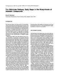
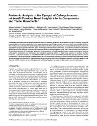
Figure 2. Induction <strong>of</strong> GUS Activity in Carborundum- or TMV-Treated<br />
Leaves <strong>of</strong> Transformed <strong>Plant</strong>s.<br />
(A) Carborundum-treated leaves (7 days after treatment) compared<br />
to the basal level.<br />
(B) TMV-treated leaves (7 days after inoculation) compared to the Carborundum<br />
treatment.<br />
Each point represents the level <strong>of</strong> GUS activity in a particular T, transformant<br />
after the experimental treatment, divided by the GUS activity<br />
<strong>of</strong> the same clone after treatment under the appropriate control conditions.<br />
GUS activity was determined fluorometrically (see Methods).<br />
and either 20- or 40-fold by TMV. However, in homozygous T3<br />
lines derived from these primary transformants, Carborundum<br />
no longer induced expression <strong>of</strong> the endogenous PR-1a gene,<br />
but TMV still induced expression more than 50-fold compared<br />
to the water-treated control (Figure 3B). As indicated in Figure<br />
3 and Table 1, this wound induction by Carborundum <strong>of</strong> both<br />
the endogenous PR-1a gene and the transgene was observed<br />
only in the primary transformants, suggesting that the result<br />
is an experimental artifact, probably due to regeneration <strong>of</strong><br />
the plants. Interestingly, this effect maps to approximately the<br />
same promoter region that is responsive to TMV infection<br />
(Figure 2).<br />
Because SA has been implicated as a possible inducer <strong>of</strong><br />
SAR, we investigated the effect <strong>of</strong> SA on the PR-1a/GUS deletions.<br />
<strong>The</strong> data presented in Figure 4 indicate that SA treatment<br />
produced an average induction <strong>of</strong> greater than fivefold for<br />
deletions from -903 to -661. <strong>The</strong> -600-bp deletion line<br />
showed an average SA induction <strong>of</strong> 2.3-fold, whereas deletion<br />
A<br />
.<br />
<strong>Regulation</strong> <strong>of</strong> the PR-1 a Promoter 161<br />
lines -318 to -73 were induced 1.3-fold or less. <strong>The</strong>refore,<br />
promoter fragments as short as 661 bases are sufficient to<br />
confer both SA and TMV inducibility to the reporter gene.<br />
Effect <strong>of</strong> CHX on PR-1a Expression<br />
To determine if a newly synthesized protein is required for SA<br />
induction <strong>of</strong> the PR-1a promoter, we measured the effects <strong>of</strong><br />
the protein synthesis inhibitor CHX on gene expression. <strong>The</strong><br />
data in Figure 5A show that a low concentration <strong>of</strong> CHX (0.01<br />
mg/mL), which inhibited protein synthesis by less than 20%<br />
(data not shown), caused an accumulation <strong>of</strong> PR-1 mRNA.<br />
However, no accumulation <strong>of</strong> mRNA resulted from higher concentrations<br />
<strong>of</strong> CHX (0.1 and 1 mg/mL), which blocked 95 and<br />
99% <strong>of</strong> protein synthesis (data not shown), respectively. PR-1a<br />
mRNA did not accumulate following treatment with both SA<br />
and high levels <strong>of</strong> CHX, which suggests that ongoing protein<br />
synthesis is required for induction by SA.<br />
A Tl<br />
147—<br />
113—<br />
B<br />
T3<br />
147—<br />
113— ^p<br />
-903-B -903-C<br />
M H20 CAR TMV HjO CAR TMV<br />
I 1<br />
-903-B -903-C<br />
"I<br />
Hfl CAR TMV HjO CAR TMV<br />
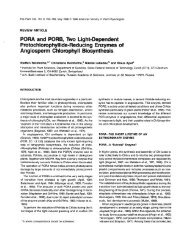
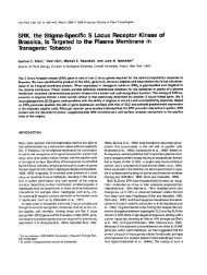
Figure 3. Primer Extension Analysis <strong>of</strong> PR-1a mRNA in T, and T3<br />
<strong>Plant</strong>s.<br />
RNA was isolated from two individual transformed lines 7 days after<br />
treatment.<br />
(A) RNA from -903-B and -903-C in the T, generation.<br />
(B) RNA from -903-B and -903-C in the T3 generation.<br />
<strong>The</strong> full-length primer extension product is 139 bp. Treatments were<br />
with water (H2O), Carborundum (CAR), or tobacco mosaic virus<br />
(TMV). M, molecular size standard; size <strong>of</strong> the markers is given in base<br />
pairs.
162 <strong>The</strong> <strong>Plant</strong> <strong>Cell</strong><br />
Table 1. Average Fold lnduction by TMV Infection, Chemical<br />
Treatment, and Wounding (T3 Lines)<br />
region. We have not determined whether CHX induction is a<br />
transcriptional or post-transcriptional event.<br />
No. <strong>of</strong><br />
Construct Carba TMV Kinetin BAPb NAAC Lines Kinetin, B-Benzylaminopurine, and<br />
-903 1.6 56.5 1.0 0.9 1.3 9 1-Naphthaleneacetic Acid Effects on PR-la<br />
-825 2.0 90.8 1.8 1.2 0.8 7 Promoter Activity<br />
-318 2.4 2.1 1.0 0.9 0.8 5<br />
a Carborundum.<br />
Several groups have reported that the PR proteins could be<br />
6-Benzylaminopurine<br />
induced by treatment with certain plant hormones (Antoniw<br />
1-Naphthaleneacetic acid. et al., 1980; Shinshi et al., 1987; Memelinket al., 1990). To map<br />
the potential hormone-responsive regions <strong>of</strong> the PR-la promoter,<br />
T3 plants from deletion lines -903, -825, and -318<br />
were treated with kinetin, 6-benzylaminopurine (BAP), l-naphthaleneacetic<br />
acid (NAA), Carborundum, or TMV, and the<br />
induction <strong>of</strong> GUS activity relative to water treatment was determined.<br />
As shown in Table 1, no significant induction (greater<br />
than threefold) was seen for kinetin, BAP, NAA, or Carborundum<br />
treatment. However, TMV showed strong induction<br />
(greater than 50-fold) in deletion lines -903 and -825, but<br />
not the -318 line as expected (Figure 2). Figure 6 shows the<br />
results when PR-la mRNA was measured in nontransgenic<br />
Xanthi-nc tobacco following BAP, kinetin, NAA, and SA treatment.<br />
Only SA treatment resulted in a significant increase in<br />
the level <strong>of</strong> RNA accumulation.<br />
<strong>The</strong> effect <strong>of</strong> CHX was also determined for the T3 lines <strong>of</strong><br />
the -903 PR-la/GUS fusions by primer extension (data not<br />
shown) and RNA gel blot analysis (Figure 56). GUS mRNA<br />
abundance was induced by low concentrations <strong>of</strong> CHX (0.01<br />
mglmL, less than 20% protein synthesis inhibition). However,<br />
in contrast to the endogenous gene, high concentrations <strong>of</strong><br />
CHX caused accumulation <strong>of</strong> GUS mRNA. This result was<br />
repeated in six -903 T3 lines, all <strong>of</strong> which yielded similar<br />
results (data not shown).<br />
Several possibilities could explain these results. First, because<br />
these lines were transgenic for the PR-la promoter, a<br />
trans effect due to an increased copy number <strong>of</strong> the promoter<br />
may have caused the difference in expression. If this were indeed<br />
the case, the endogenous gene should respond in a<br />
manner similar to the transgene. <strong>The</strong>refore, accumulation <strong>of</strong><br />
PR-la mRNA was evaluated in each <strong>of</strong> the -903 lines. As seen<br />
in Figure 5C, the endogenous PR-la mRNA accumulation in<br />
the transgenic plants was essentially the same as in the nontransformed<br />
plants, eliminating a frans effect as an explanation.<br />
Second, the anomalous GUS results could also be explained<br />
by the presence <strong>of</strong> CHX-responsive elements contained in the<br />
-903 promoter, which are normally silenced in the endogenous<br />
gene by the action <strong>of</strong> a cis-acting sequence upstream<br />
<strong>of</strong> -903. If this were the case, it should be possible to map<br />
the CHX-responsive element by analyzing CHX induction in<br />
each <strong>of</strong> the deletion lines. Six independent lines <strong>of</strong> each deletion<br />
were evaluated for CHX induction (data not shown) GUS<br />
mRNA accumulated to high levels following treatment <strong>of</strong> all<br />
the lines, suggesting that if there was a CHX-responsive element<br />
in the promoter, it had to be located between -73 and<br />
the transcriptional start site.<br />
Another possible explanation was that CHX affected the GUS<br />
coding sequence, rather than the PR-la promoter. To test this<br />
explanation, we analyzed two independent transgenic plants<br />
containing the -903 PR-la promoter fused to a Bacillus thuringiensis<br />
cry /A(b) gene for CHX induction. In controls treated<br />
with SA, B. thuringiensis toxin mRNAaccumulated to high levels<br />
(data not shown; Williams et al., 1992). However, CHX treatment<br />
(1 mglmL) resulted in no mRNA accumulation (data not<br />
shown). <strong>The</strong>refore, the simplest explanation is that CHX induction<br />
<strong>of</strong> the PR-VGUS gene is an artifact <strong>of</strong> the GUS coding<br />
Tissue-Specific Expression from the -903<br />
PR-la-GUS Transgene<br />
Nine T3 lines were examined for tissue-specific expression <strong>of</strong><br />
GUS under various conditions. Three patterns <strong>of</strong> expression<br />
were identified: consensus, constitutive, or undetectable. Five<br />
<strong>of</strong> the lines (-903-6, -903-F, -903-G, -903-H, and -903-1)<br />
I ............... I ............... I ............... I ............... I .............. 1 .............. 1 .............. 1 .............. I .............. 1<br />
#imo<br />
8<br />
f 100<br />
6<br />
lj 10<br />
E w 9 1.0<br />
................................<br />
o. 1<br />
1 .I ........... .....<br />
T1 Tramfonnant<br />
................................... I ....<br />
Figure 4. lnduction <strong>of</strong> GUS Activity in SATreated Leaves Compared<br />
to the Basal Levei.<br />
Each point represents the level <strong>of</strong> GUS activity in a particular TI trans-<br />
formant after SA treatment divided by the GUS activity <strong>of</strong> the same<br />
clone after treatment with water under the appropriate control condi-<br />
tions. GUS activity was determined fluorometrically (see Methods).
A<br />
SA +<br />
CHXmg/mL CHXmg/mL<br />
NT H20 0.01 0.1 1 SA 0.1 1<br />
D SA +<br />
t> CHXmg/mL CHXmg/mL<br />
NT HjO 0.01 0.1 1 SA 0.1 1<br />
CHX mg/mL<br />
NT H20 0.01 0.1 1 SA 0.1<br />
SA+<br />
CHX mg/mL<br />
PR-1<br />
GUS<br />
PR-1<br />
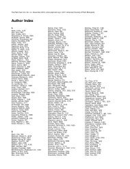
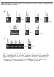
Figure 5. Effect <strong>of</strong> CHX on PR-1a and GUS mRNA Accumulation in<br />
Nontransformed and Transformed <strong>Plant</strong>s.<br />
RNA was isolated from leaves 24 hr after the treatment indicated. RNA<br />
gel blots were probed with either labeled PR-1a cDNA or labeled GUSspecific<br />
DMA. NT, nontreated tobacco; H2O, water treated; SA, treated<br />
with 1 mM salicylic acid.<br />
(A) PR-1a mRNA in nontransformed tobacco.<br />
(B) GUS mRNA in transformant -903-A.<br />
(C) Endogenous PR-1a mRNA in transformant -903-A.<br />
showed a consensus pattern <strong>of</strong> expression summarized in Table<br />
2. A sixth line (-903-A) mimicked this pattern but at higher<br />
levels <strong>of</strong> expression. As shown in Figure 7, the consensus pattern<br />
included high-level expression in the mesophyll layer and<br />
a moderate level <strong>of</strong> expression in the epidermal layer <strong>of</strong> the<br />
leaves in response to both SA (Figure 7A) and TMV (Figures<br />
7B and 7C), but no induction in response to water or Carborundum<br />
(Figure 7D). <strong>The</strong>re was also no GUS activity in the<br />
roots <strong>of</strong> transgenic plants following TMV infection <strong>of</strong> the leaves<br />
(Figure 7E).<br />
<strong>The</strong> TMV induction <strong>of</strong> GUS activity appeared greatest close<br />
to the lesion (Figures 7B and 7C), similar to what has been<br />
shown for the PR-1a protein (Antoniw and White, 1986) and<br />
consistent with the work <strong>of</strong> others (Ohshimaetal., 1990). However,<br />
when six independent plants from each <strong>of</strong> the deletion<br />
lines (-600 to -150) were tested for TMV and SA induction,<br />
no GUS staining was detectable (Figures 7F and 7G; data not<br />
shown). This is in contrast to the results <strong>of</strong> Ohshima et al. (1990),<br />
who found TMV-inducible GUS activity even in a 300-bp PR-1a<br />
promoter/GUS transgenic plant. It is possible that some other<br />
factor, perhaps the orientation <strong>of</strong> the PR-1a promoter relative<br />
<strong>Regulation</strong> <strong>of</strong> the PR-1 a Promoter 163<br />
to the cauliflower mosaic virus 35S promoter, is responsible<br />
for the observed difference in histochemical staining.<br />
Stem sections from the consensus lines showed no or low<br />
expression in response to inducers. Petiole sections showed<br />
low levels <strong>of</strong> expression in the cortex in response to TMV and<br />
SA; however, this GUS staining was detectable only after long<br />
incubation times with the chromogenic substrate (data not<br />
shown). <strong>The</strong> -903-A line also demonstrated GUS activity in<br />
the cells surrounding the vascular bundle <strong>of</strong> the petiole (i.e.,<br />
phloem) in response to SA after long incubation times (data<br />
not shown).<br />
Two lines (-903-C and -903-E) constitutively expressed<br />
GUS activity. Regardless <strong>of</strong> the presence <strong>of</strong> inducer, GUS activity<br />
was seen in leaves, petioles, and stems (data not shown).<br />
A single line (-903-D) had low GUS activity that was inducible<br />
and detectable by fluorometric assay, but not by in situ<br />
GUS staining (data not shown).<br />
PR-1a-GUS Expression in Flowering <strong>Plant</strong>s<br />
Several reports have indicated that PR proteins accumulate<br />
in leaf tissue during flowering (Fraser, 1981; Lotan et al., 1989).<br />
To determine if the -903/GUS plants were responding appropriately<br />
to this developmental signal, we assayed tobacco<br />
plants from the consensus set <strong>of</strong> T3 -903 PR-1a-GUS lines<br />
for GUS expression in certain tissues <strong>of</strong> untreated flowering<br />
plants. As can be seen in Figure 7H and summarized in Table<br />
2, the leaves <strong>of</strong> untreated flowering plants containing the<br />
-903/GUS gene showed GUS activity, whereas untreated nonflowering<br />
plant leaves showed no GUS activity (data not shown).<br />
In flower tissue, GUS activity was observed at the base <strong>of</strong> the<br />
sepals and in mature pollen grains (Figures 71, 7J, and 7K).<br />
Other tissues tested, including roots, petals, and the female<br />
structures <strong>of</strong> the flower, exhibited no reproducible GUS activity<br />
in response to flowering (Figure 7; data not shown).<br />
Nontransformed tobacco showed no GUS activity in any tissue,<br />
including pollen (Figure 7L; data not shown). In addition,<br />
H20 BAP KIN NAA SA<br />
Figure 6. Effect <strong>of</strong> Phytohormones on PR-1a Expression.<br />
Tobacco leaves were painted with the compound indicated at 1 mg/mL;<br />
kinetin (KIN), 6-benzylaminopurine (BAP), 1-naphthaleneacetic acid<br />
(NAA), and salicylic acid (SA). RNA was isolated 48 hr after treatment<br />
and probed with PR-1a cDNA on an RNA gel blot.
164 <strong>The</strong> <strong>Plant</strong> <strong>Cell</strong><br />
~~<br />
Table 2. Tissue Specificity <strong>of</strong> GUS Activity in -903 PR-ia-GUS<br />
Transgenic <strong>Plant</strong>s<br />
Tissue Water SA TMV Flowering<br />
Leaf<br />
Petiole<br />
-a<br />
-<br />
++b.C<br />
-<br />
+++C.d<br />
+e<br />
++++c<br />
+e<br />
Stem - - +I-8 ++e<br />
Root - - -<br />
Pollen ntf nt nt +++<br />
Sepal nt nt nt ++<br />
a - , No GUS activity detectable.<br />
+ to + + + +, Varying degrees <strong>of</strong> GUS staining.<br />
C Expressed predominantly in mesophyll cells.<br />
Expression strongest near lesions.<br />
e Expressed predominantly in the cortex.<br />
nt, Not tested.<br />
immature pollen grains <strong>of</strong> transgenic tobacco showed low or<br />
no GUS activity detectable by in situ staining (data not shown).<br />
It is difficult to determine the level <strong>of</strong> GUS activity in the an-<br />
ther tissues surrounding the pollen because <strong>of</strong> the possibility<br />
<strong>of</strong> contaminating GUS activity from the pollen (Figure 7L). Al-<br />
though PR-1 protein has been detected in sepals, perhaps<br />
associated with nectaries, it has not been detected in the pol-<br />
len (Lotan et al., 1989).<br />
To determine whether the pollen-specific expression was a<br />
reflection <strong>of</strong> the activity <strong>of</strong> the endogenous promoter, we ana-<br />
lyzed transgenic plants for the expression <strong>of</strong> both the native<br />
PR-la gene and the PR-1alGUS transgene in anthers/pollen.<br />
As shown in Figure 8, expression <strong>of</strong> the endogenous gene was<br />
not detected in antherslpollen, but the GUS gene was detect-<br />
able in antherslpollen at levels approaching that <strong>of</strong> induced<br />
leaves. RNA was also analyzed from transgenic tobacco con-<br />
taining the same PR-la promoter fused to a B. thuringiensis<br />
cry IA@) gene. No B. fhuringiensis toxin mRNA was detected<br />
in the pollen from two independent transformants. However,<br />
normal SA- and TMV-inducible expression was observed in<br />
leaf tissue (data not shown; Williams et al., 1992). <strong>The</strong> sim-<br />
plest explanation for this result is that the GUS coding region<br />
is influencing the expression <strong>of</strong> the transgene (either transcrip-<br />
tionally or post-transcriptionally), causing accumulation <strong>of</strong> GUS<br />
RNA in the pollen, a tissue in which the PR-la promoter is<br />
not normally active. <strong>The</strong>refore, caution should be used when<br />
interpreting GUS histochemical staining when the tissue spec-<br />
ificity <strong>of</strong> the endogenous gene is unknown.<br />
DlSCUSSlON<br />
TMV- and SA-Responsive Elements<br />
<strong>The</strong> expression <strong>of</strong> PR-la by vira1 infection and SA treatment<br />
has been studied for more than 10 years, yet an understanding<br />
-<br />
<strong>of</strong> the induction mechanism is not clear. In experiments de-<br />
signed to identify the cis-acting sequences <strong>of</strong> the PR-la<br />
promoter responsible for induction, Ohshima et al. (1990) con-<br />
cluded that as little as 300 bp <strong>of</strong> 5‘ flanking sequence was<br />
sufficient to appropriately express a GUS reporter gene. How-<br />
ever, Van de Rhee et al. (1990), using the same PR-la gene<br />
from tobacco and similar reporter gene constructs, concluded<br />
that induction required at least 685 bp.<br />
To resolve this issue, it is important to carefully study the<br />
data and the experimental procedures from both laboratories.<br />
Ohshima et al. (1990) reported induction <strong>of</strong> approximately eight-<br />
fold above H20 treatment by SA with their -900 construction<br />
and 1.5-fold with a -300 construction. <strong>The</strong>y further reported<br />
that the -300 and the -900 lines were equally induced by<br />
TMV infection, but the only data supporting this claim was a<br />
histochemical analysis, which is a difficult method to interpret<br />
quantitatively. On the other hand, Van de Rhee et al. (1990)<br />
reported induction <strong>of</strong> approximately fourfold by SA and -10-fold<br />
by TMV in a 685-bp construct and no induction in constructs<br />
containing 645 bp or less. In fact, with less than 645 bp, there<br />
was no measurable GUS activity. However, in contrast to the<br />
work by Ohshima et al. (1990), Van de Rhee et al. (1990) ana-<br />
lyzed at least 10 independent transformants from each deletion,<br />
lending more weight to their analysis.<br />
<strong>The</strong> observations in these studies are not mutually exclu-<br />
sive. <strong>The</strong> dispute lies in what each group considers to be<br />
“induction.” In the study <strong>of</strong> Ohshima et al. (1990), an increase<br />
<strong>of</strong> 50% in the level <strong>of</strong> GUS activity was scored as induced,<br />
whereas in the Van de Rhee et al. (1990) study, the induction<br />
had to be at least fivefold to be scored as induced. Using the<br />
same induction criteria, the two groups would have reached<br />
similar conclusions. In support <strong>of</strong> this interpretation, using a<br />
large number <strong>of</strong> primary transformants and the sensitive fluoro-<br />
metric assay for GUS, we have found that 661 bp <strong>of</strong> 5’flanking<br />
sequence is sufficient for fivefold salicylate and 13-fold TMV<br />
induction. We have extended these observations with data sug-<br />
gesting the presence <strong>of</strong> sequences responsible for high-leve1<br />
uninduced expression that lie approximately at position -300<br />
and a possible negative regulatory element that contributes<br />
to uninduced expression near position -150.<br />
However, it is worth noting that the endogenous PR-la gene<br />
is typically induced 4000- to 10,000-fold in TMV-infected or<br />
SA-treated leaves, increasing from very low levels to -1% <strong>of</strong><br />
the mRNA in induced tissue (Ward et al., 1991). Thus, other<br />
factors could play an important role in the regulation <strong>of</strong> PR-la<br />
expression, which might include as yet unidentified cis ele-<br />
ments in the more distant 5‘ or 3’ regions <strong>of</strong> the gene as well<br />
as the location <strong>of</strong> the gene in the genome.<br />
Artifacts Associated with GUS as a Reporter <strong>Gene</strong><br />
Severa1 studies have documented GUS artifacts associated<br />
with the histochemical GUS activity assay (Plegt and Bino,<br />
1989; Mascarenhas and Hamilton, 1992; Tor et al., 1992). In
Figure 7. In Situ GUS Staining <strong>of</strong> PR-1a-GUS <strong>Plant</strong>s.<br />
<strong>Regulation</strong> <strong>of</strong> the PR-1 a Promoter 165<br />
(A) SA-treated -903-B leaf cross-section stained 3 days after inoculation.<br />
(B) TMV lesion from an infected -903-F leaf stained 7 days after inoculation.<br />
(C) TMV lesion from an infected -903-A leaf cross-section stained 7 days after inoculation. <strong>The</strong> area between the veins is a TMV lesion.<br />
(D) Buffer-treated -903-A leaf cross-section stained 7 days after inoculation.<br />
(E) Secondary root from -903-A in which several leaves were infected with TMV, stained 7 days after treatment.<br />
(F) TMV lesion on -600-E, stained 7 days after infection.<br />
(G) SA-treated -600-A leaf disc showing lack <strong>of</strong> blue staining.<br />
(H) Petiole from flowering -903-A.<br />
(I) Immature flower cross-section <strong>of</strong> -903-A.<br />
(J) Anther cross-section from -903-A.<br />
(K) -903-B pollen.<br />
(L) Nontransformed tobacco pollen stained for GUS activity.
166 <strong>The</strong> <strong>Plant</strong> <strong>Cell</strong><br />
PR-1<br />
GUS<br />
£ NT SA £ NT SA NT SA<br />
fit<br />
-903-A -903-H -903-1<br />
Figure 8. Pollen-Specific Expression <strong>of</strong> the -903-GUS <strong>Gene</strong>.<br />
RNA was isolated from the -903 T3 lines indicated. RNA gel blots<br />
were probed with PR-1a (top) or GUS (bottom). Pollen, anthers and<br />
pollen from mature and immature flowers; NT, nontreated leaf tissue;<br />
SA, leaf tissue treated with 1 mM salicylic acid for 24 hr.<br />
this report, we have documented two artifacts associated with<br />
the use <strong>of</strong> GUS as a reporter gene in transgenic plants that<br />
are related to actual GUS gene expression and not with the<br />
GUS activity assay. We have shown that the treatment <strong>of</strong> tissue<br />
with high levels <strong>of</strong> CHX significantly increases the<br />
accumulation <strong>of</strong> GUS mRNA, but not mRNA from the endogenous<br />
PR-1a gene or from other reporter genes under the<br />
control <strong>of</strong> the PR-1a promoter. <strong>The</strong> cause <strong>of</strong> this accumulation<br />
is not clear. One explanation for the aberrant RNA accumulation<br />
could be that the GUS coding sequence contains a<br />
promoter element that responds to CHX treatment. Another<br />
possibility is that a low level <strong>of</strong> GUS transcription always occurs,<br />
but the mRNA becomes stabilized following CHX<br />
treatment. In either case, the reporter is clearly not responding<br />
in a manner consistent with expression <strong>of</strong> the endogenous<br />
gene or other genes under the control <strong>of</strong> the PR-1a promoter.<br />
<strong>The</strong> second artifact was in the ectopic expression <strong>of</strong> GUS<br />
activity in the anther and pollen <strong>of</strong> untreated flowering plants.<br />
This expression appears to be due to the GUS coding region<br />
because other reporter genes linked to the same promoter do<br />
not show this effect. In many similar studies using GUS as a<br />
reporter gene, investigators have concluded that the promoter<br />
under study directed pollen- or anther-specific expression<br />
(Guerrero et al., 1990; DeWitt et al., 1991; McCormick, 1991;<br />
Samac and Shah, 1991; Takahashi et al., 1992). Our data indicate<br />
that these results must be reevaluated and future<br />
experiments concerning pollen expression should be carried<br />
out either directly on the endogenous gene or with several<br />
different reporter genes to minimize artifacts. Moreover, further<br />
problems not yet documented may arise from the use <strong>of</strong><br />
GUS as a reporter gene. Thus, if GUS is not reliable as a<br />
reporter in the two cases documented here, when is it? It is<br />
clear that the GUS reporter gene should be used only when<br />
there is sufficient prior knowledge concerning the expression<br />
characteristics <strong>of</strong> the endogenous gene under study.<br />
Effects <strong>of</strong> Protein Synthesis Inhibitors<br />
<strong>The</strong> finding that low concentrations <strong>of</strong> CHX induced the endogenous<br />
PR-1a promoter, whereas high concentrations <strong>of</strong><br />
CHX prevented induction <strong>of</strong> the endogenous gene by SA, suggests<br />
an intriguing model for PR-1a induction. Incomplete<br />
inhibition <strong>of</strong> protein synthesis may be one signal for PR-1a gene<br />
induction. When a plant cell is infected, the pathogen may<br />
partially inhibit host protein synthesis. Some mammalian pathogens,<br />
such as polio virus, are known to exert such an effect<br />
on their hosts (Hershey, 1991). Conceivably, the plant could<br />
have evolved a mechanism to induce defense genes when<br />
protein synthesis is partially inhibited. However, because complete<br />
inhibition <strong>of</strong> protein synthesis abolished PR-1a induction,<br />
a newly synthesized protein is apparently required for the induction<br />
<strong>of</strong> the endogenous PR-1 a gene.<br />
METHODS<br />
5' Deletion Constructs<br />
An 1150-bp Xhol-Pstl fragment <strong>of</strong> a pathogenesis-related protein-la<br />
(PR-1a) genomic clone was subcloned into M13mp18, as described<br />
by Payne et al. (1988). Site-directed mutagenesis (Kunkel et al., 1987)<br />
<strong>of</strong> the resulting clone was performed at the initiation codon <strong>of</strong> the PR-1a<br />
protein, changing the sequence from TC ATG G to CC ATG G, thus<br />
creating an Ncol site. <strong>The</strong> resulting clone, designated PR-1aAXhoNco,<br />
was used as the source <strong>of</strong> PR-1a promoter DMA for the deletion<br />
constructs.<br />
A vector called pBSGusl.2 was created by a three-way ligation <strong>of</strong><br />
a 391-bp Sall-SnaBI fragment from pRAJ265 (Jefferson et al., 1987),<br />
containing the 5' end <strong>of</strong> the p-glucuronidase (GUS) coding sequence,<br />
with a 1707-bp SnaBI-EcoRI fragment from pBI221 (Clonetech, Palo<br />
Alto, CA), containing the 3' end <strong>of</strong> the GUS coding sequence and the<br />
nopaline synthase terminator, and pBS+ (Stratagene) digested with<br />
Sail and EcoRI. An Ncol site was created at the initiation codon <strong>of</strong><br />
the GUS gene using site-directed mutagenesis (Kunkel et al., 1987).<br />
PR-1aAXhoNco was digestedwith Ncol and other enzymes located<br />
within the PR-1a promoter sequence at varying distances upstream<br />
from the start <strong>of</strong> transcription: Xhol (-903), Alul (-825), Sspl (-318),<br />
Oral (-222), BstEII (-150), and Rsal (-73). <strong>The</strong>se promoter fragments<br />
were isolated; adapters were ligated at their 5' ends and cloned into<br />
pBSGus1.2, previously digested with Xhol and Ncol.<br />
Other 5' deletion constructs were made using the polymerase chain<br />
reaction (Perkin-Elmer, Norwalk, CT). Primers were designed to correspond<br />
to the -700, -661, and -600 sequences <strong>of</strong> the PR-1a promoter<br />
with Xhol restriction sites on the 5' end. <strong>The</strong> second primer for each<br />
reaction was located in the GUS coding region. <strong>The</strong> -903 construct<br />
described above was used for template DNA. Polymerase chain reaction<br />
was performed according to manufacturer's recommendations<br />
(Perkin-Elmer).<br />
<strong>The</strong> numbering <strong>of</strong> the constructs is according to the method <strong>of</strong> Payne<br />
et al. (1988), with the transcriptional start site designated as +1 and<br />
the translational start at +29. A putative TATAA box is located at -34.<br />
<strong>The</strong> junction <strong>of</strong> the PR-1a promoter fragments with the GUS gene is<br />
at the PR-1a translational start site. All constructs were sequenced to<br />
confirm their identity.
Each <strong>of</strong> the deletion constructs was digested with Kpnl-BamHI and<br />
the promoter-GUS fragment was cloned into Kpnl-BamHI-digested<br />
pClB200, a binary vector containing the neomycin phosphotransfer-<br />
ase II gene that confers kanamycin resistance. All <strong>of</strong> the plasmids were<br />
constructed such that the PR-la promoter was adjacent to the left bor-<br />
der <strong>of</strong> the T-DNA, with transcription from both the PR-la promoter and<br />
the neomycin phosphotransferase II gene in the same direction. <strong>The</strong><br />
resulting constructs were transformed into Agrobacterium tumefaciens<br />
(218542 according to the procedure <strong>of</strong> Holsters et al. (1978). ClB542<br />
is strain EHAlOl (Hood et al., 1986), in which the kanamycin marker<br />
<strong>of</strong> the plasmid has been replaced by the spectinomycin/streptomycin<br />
portion <strong>of</strong> TnZ<br />
Transformation <strong>of</strong> Tobacco<br />
Leaf discs <strong>of</strong> Nic<strong>of</strong>iana tabacum cv Xanthi-nc were transformed with<br />
Agrobacterium cultures, containing the deletion constructs, by the leaf<br />
disc cocultivation method (Horsch et al., 1985). Transgenic plant fines<br />
were named for the deletion used (for example, a -903 PR-Ia-GUS<br />
transformant was called -903-A). Approximately 15 independent trans-<br />
formants <strong>of</strong> each deletion were selected. <strong>Plant</strong>s were rooted on medium<br />
containing no hormones and 100 pglmL kanamycin. At approximately<br />
the five-leaf stage, each plantlet was vegetatively split and five identi-<br />
cal plants were individually propagated. One <strong>of</strong> these was allowed to<br />
set seed and the remaining four were used for GUS assays following<br />
treatment with either water, SA, Carborundum, or tobacco mosaic vi-<br />
rus (TMV). <strong>The</strong> water treatment was taken as an indication <strong>of</strong> the basal<br />
level expression from the promoter. Some TI plants were selfed, and<br />
the seeds were plated on kanamycin for germination. <strong>The</strong> resulting<br />
plants were selfed again and seed lines homozygous for kanamycin<br />
resistance were used for experiments involving T3 plants.<br />
<strong>Plant</strong> Treatments for Fluorometric GUS Assays<br />
When transgenic plants were ~ 4cm 0 high, three leaves per plant were<br />
treated with the U1 strain <strong>of</strong> TMV, or mock inoculated with Carborundum<br />
and buffer (10 mM NaPO., pH 7), as previously described (Payne<br />
et al., 1990). lnoculated leaves were harvested 7 days later. For experiments<br />
with phytohormones, water, 50 mM salicylate, 1 mg/mL<br />
kinetin, 1 mglmL 6-benzylaminopurine (BAP), and 1 mg/mL l-naphthaleneacetic<br />
acid (NAA) (all from Sigma) were all applied to three<br />
leaves per plant using a paint brush. <strong>The</strong>se leaves were harvested<br />
2 days following treatment.<br />
GUS Assays<br />
Fluorometric GUS assays were performed essentially according to the<br />
method <strong>of</strong> Jefferson et al. (1987). Reactions were carried out in the<br />
presence and absence <strong>of</strong> 2 mM 4-methylumbelliferyl-~-D-glucuronide<br />
(MUG) using 20 pL <strong>of</strong> supernatant from each sample (total reaction<br />
volume = 75 pL). Reactions were terminated after 60 min by the addi-<br />
tion <strong>of</strong> 225 pL <strong>of</strong> 0.2 M Na2C03. Duplicate fluorescence values for<br />
each sample were averaged, and the background fluorescence (reac-<br />
tion without MUG) was subtracted to obtain a concentration <strong>of</strong><br />
methylumbelliferone for each sample.<br />
BCA protein assays were performed on each sample according to<br />
the manufacturer‘s instructions (Pierce, Rockford, IL). Specific activi-<br />
ties were calculated as nanomoles per milligram <strong>of</strong> protein per minute<br />
<strong>Regulation</strong> <strong>of</strong> the PR-la Promoter 167<br />
and converted to activity units (picomoles <strong>of</strong> methylumbelliferone per<br />
milligram <strong>of</strong> protein per minute). A value <strong>of</strong> 0.1 activity unit represents<br />
the lower detection limit <strong>of</strong> the assay indicating that samples with this<br />
level <strong>of</strong> activity were just detectable in an 18-hr assay.<br />
For the in situ assays, tissue sections were cut by hand and placed<br />
overnight in O5 mglmL X-gluc (5-bromc-4chloro-3indolyl glucuronide),<br />
100 mM NaPO., pH 7, at 37% (Jefferson et al., 1987). Some sections<br />
were cleared in 1000/0 ethanol at room temperature.<br />
Primer Extension Mapping<br />
Primer extension reactions were carried out on total RNA, as described<br />
by Métraux et al. (1989), using a PR-Ia-specific oligonucleotide with<br />
the following sequence: 5’-ATAGTCTTGTTGAGAGTT-3’. Amounts <strong>of</strong><br />
transcript generated for each sample were quantitated by detecting<br />
P-decay <strong>of</strong> phosphorus-32 with a Betascope 603 blot analyzer (Beta-<br />
gen, Waltham, MA).<br />
RNA Extraction and Gel Blot Hybridization<br />
RNA was purified from frozen tissue samples by phenollchlor<strong>of</strong>orm<br />
extraction followed by lithium chloride precipitation (Lagrimini et al.,<br />
1987). Ten-microgram samples <strong>of</strong> total RNA were separated by elec-<br />
trophoresis through formaldehyde agarose gels and blotted to nylon<br />
membrane (<strong>Gene</strong>Screen Plus; New England Nuclear, Boston, MA),<br />
as previously described (Ausubel et al., 1987). Ethidium bromide was<br />
included in the sample loading buffer at 40 mglmL, which allowed<br />
photography under UV light after electrophoresis to confirm equal sam-<br />
ple loading. Probes were made by the random-priming method<br />
(Feinberg and Vogelstein, 1983) with a PrimeTime C kit (International<br />
Biotechnologies, Inc., New Haven, CT). Hybridizations and washing<br />
were performed according to the method <strong>of</strong> Church and Gilbert (1984).<br />
Relative amounts <strong>of</strong> each transcript were quantitated with a Betascope,<br />
as described above.<br />
Cycloheximide Treatments<br />
CHX (Sigma), SA, H20, or SA plus CHX was injected by pricking a<br />
fully expanded tobacco leaf with a needle and forcing a small amount<br />
<strong>of</strong> liquid into the leaf with a 10” syringe. <strong>The</strong> water soaked area was<br />
marked and the plants were kept in the laboratory for 24 hr before har-<br />
vesting the injected area. Leaves treated with SA plus CHX were<br />
pretreated with CHX alone for 30 min prior to the SA plus CHX treatment.<br />
Protein synthesis was determined by %-methionine incorporation into<br />
trichloroacetic acid-precipitable material. <strong>The</strong> leaf tissue was injected<br />
with %-methionine (Amersham, Arlington Heights, IL; 100 pCilmL in<br />
distilled water) and after 1 hr the tissue was frozen in liquid nitrogen.<br />
<strong>The</strong> frozen tissue was ground in 2 x SDS sample buffer (Laemmli,<br />
1970), and trichloroacetic acid precipitable counts were determined<br />
as described (Promega manual, “Translation in Vitro”; Promega,<br />
Madison, WI).<br />
ACKNOWLEDGMENTS<br />
We gratefully acknowledge Ben Miflin and Helmut Kessmann for valu-<br />
able discussions; Alice Montoya, Susan Gordon, Greg Howe, and
168 <strong>The</strong> <strong>Plant</strong> <strong>Cell</strong><br />
Wayne Middlesteadt for technical assistance; Greg Crawford and Janie<br />
Schlotzhauer for care <strong>of</strong> the plants; Judy Watkins for preparing me-<br />
dia; and Mary-Dell Chilton, Bruce Lee, and Bernard Vernooij for critically<br />
reading the manuscript.<br />
Received November 6, 1992; accepted December 23, 1992.<br />
REFERENCES<br />
Antoniw, J.F., Kueh, J., Walkey, D.G.A., and White, R.F. (1980). <strong>The</strong><br />
presence <strong>of</strong> pathogenesis-related proteins in callus <strong>of</strong> Xanthi-nc<br />
tobacco. Phytopathol. Z 101, 179-184.<br />
Antoniw, J.F., and White, R.F. (1986). Changes with time in the distribution<br />
<strong>of</strong> virus and PR protein around single local lesions <strong>of</strong> TMV<br />
infected tobacco. <strong>Plant</strong> MOI. Biol. 6, 145-149.<br />
Ausubel, F.M., Brent, R., Kingston, R.E., Moore, D.D., Seidman,<br />
J.G., Smlth, J.A., and Struhl, K. (1987). Current Protocols in Molecular<br />
Biology, Vol. 1 (New York: J. Wiley and Sons).<br />
Chen, i!., and Klessig, D. (1991). ldentification <strong>of</strong> a soluble salicylic<br />
acid-binding protein that may function in signal transduction in the<br />
plant disease-resistance response. Proc. Natl. Acad. Sci. USA 88,<br />
8179-8183.<br />
Chester, K.S. (1933). <strong>The</strong> problem <strong>of</strong> acquired physiological immunity<br />
in plants. Q. Rev. Biol. 8, 275-324.<br />
Church, G.M., and Gllbert, W. (1984). Genomic sequencing. Proc.<br />
Natl. Acad. Sci. USA 81, 1991-1995.<br />
DeWitt, N.D., Harper, J.F., and Sussman, M.R. (1991). Evidence for<br />
a plasma membrane proton pump in phloem cells <strong>of</strong> higher plants.<br />
<strong>Plant</strong> J. 1, 121-128.<br />
Feinberg, A.P., and Volgelstein, B. (1983). A technique for radiolabeling<br />
DNA restriction endonuclease fragments to high specific activity.<br />
Anal. Biochem. 152, 232-238.<br />
Fraser, R.S.S. (1981). Evidence for the Occurrence <strong>of</strong> the "pathogenesisrelated<br />
proteins in the leaves <strong>of</strong> healthy tobacco plants during flowering.<br />
Physiol. <strong>Plant</strong> Pathol. 19, 69-76.<br />
Glaninazzi, S., Martin, C., and Vallle, J.C. (1970). Hypersensibilité<br />
aux virus, temperature et proteines solubles chez le Nicotiana Xanthi<br />
n.c. Apparition de nouvelles macromolecules lors de Ia repression<br />
de Ia synthbse virale. C. R. Acad. Sci. Ser. D 270, 2382-2386.<br />
Guerrem, F.D., Crossland, L., Smutzer, G.S., Hamilton, D.A., and<br />
Mascarenhas, J.P. (1990). Promoter sequences from a maize pollenspecific<br />
gene direct tissue-specific transcription in tobacco. MOI.<br />
Gen. <strong>Gene</strong>t. 224, 161-168.<br />
Hershey, J. (1991). Translational control in mammalian cells. Annu.<br />
Rev. Biochem. 60, 717-755.<br />
Holsters, M., de Waele, D., Depicker, A., Messens, E., van Montagu,<br />
M., and Schell, J. (1978). Transfection and transformation <strong>of</strong> Mrobac-<br />
terium tumefaciens. MOI. Gen. <strong>Gene</strong>t. 163, 181-187.<br />
Hood, E.E., Helmer, G.L., Fraley, R.T., and Chilton, M.-D. (1986).<br />
<strong>The</strong> hypervirulence <strong>of</strong> Agrobacterium tumefaciens A281 is encoded<br />
in a region <strong>of</strong> pTiBc542 outside <strong>of</strong> T-DNA. J. Bacteriol. 168,1291-1301.<br />
Horsch, R.B., Fry, J.E., H<strong>of</strong>fmann, N.L., Eichholtz, D., Rogers, S.G.,<br />
and Fraley, R.T. (1985). A simple and general method for transfer-<br />
ring genes into plants. Science 227, 1229-1231.<br />
Jefferson, R.A., Kavanagh, T.A., and Bevan, M.W. (1987). GUS fusions:<br />
P-Glucuronidase as a sensitive and versatile gene fusion<br />
marker in higher plants. EMBO J. 6, 3901-3907.<br />
Kuc, J. (1982). lnduced immunity to plant disease. BioScience 32,<br />
854-860.<br />
Kunkel, T.A., Roberts, J.D., and Zakour, R.A. (1987). Rapid and efficient<br />
site-specific mutagenesis without phenotypic selection.<br />
Methods Enzymol. 154, 367-369.<br />
Laemmli, U. (1970). Cleavage <strong>of</strong> structural proteins during the assembly<br />
<strong>of</strong> the head <strong>of</strong> bacteriophage T4. Nature 227, 680-685.<br />
Lagrimini, L.M., Burkhart, W., Moyer, M., and Rothstein, S. (1987).<br />
Molecular cloning <strong>of</strong> complementary DNA encoding the ligninforming<br />
peroxidase from tobacco: Molecular analysis and tissuespecific<br />
expression. Proc. Natl. Acad. Sci. USA 84, 7542-7546.<br />
Lawton, K., Uknes, S., Friedrich, L., Gaffney, T., Alexander, D.,<br />
Goodman, R., Mltraux, J.-P., Kessmann, H., Ahl Goy, P., Gut<br />
Rella, M., Ward, E., and Ryals, J. (1993). <strong>The</strong> molecular biology<br />
<strong>of</strong> systemic acquired resistance. In Mechanisms <strong>of</strong> Defense Responses<br />
in <strong>Plant</strong>s, B. Fritig and M. Legrand, eds (Dordrecht: Kluwer<br />
Academic), pp. 410-420.<br />
Lotan, T., Ori, N., and Fluhr, R. (1989). <strong>Pathogenesis</strong>-related proteins<br />
are developmentally regulated in tobacco flowers. <strong>Plant</strong> <strong>Cell</strong> 1,<br />
881-887.<br />
Malamy, J., Carr, J.P., Klessig, D.F., and Raskin, I. (1990). Salicylic<br />
acid: A likely endogenous signal in the resistance response <strong>of</strong><br />
tobacco to vira1 infection. Science 250, 1002-1004.<br />
Mascarenhas, J.P., and Hamilton, D.A. (1992). Artifacts in the localization<br />
<strong>of</strong> GUS activity in anthers <strong>of</strong> petunia transformed with a CaMV<br />
35s-GUS construct. <strong>Plant</strong> J. 2, 405-408.<br />
Mauch, F., Mauch-Mani, E., and Boller, T. (1988). Antifungal hydrolases<br />
in pea tissue. II. lnhibition <strong>of</strong> funga1 growth by combinations<br />
<strong>of</strong> chitinase and P-1,3-glucanase. <strong>Plant</strong> Physiol. 88, 936-942.<br />
McCormick, S. (1991). Molecular analysis <strong>of</strong> male gametogenesis in<br />
plants. Trends <strong>Gene</strong>t. 7, 298-303.<br />
Memelink, J., Linthorst, H.J.M., Schilpemort, R.A., and Hoge, J.H.C.<br />
(1990). Tobacco genes encoding acidic and basic is<strong>of</strong>orms <strong>of</strong><br />
pathogenesis-related proteins display different expression patterns.<br />
<strong>Plant</strong> MOI. Biol. 14, 119-126.<br />
Mltraux, J.-P., Burkhart, W., Moyer, M., Dincher, S., Middlesteadt,<br />
W., Williams, S., Payne, G., Carnes, M., and Ryals, J. (1989). Isolation<br />
<strong>of</strong> a complementary DNA encoding a chitinase with structural<br />
homology to a bifunctional lysozyme/chitinase. Proc. Natl. Acad. Sci.<br />
USA 86, 896-900.<br />
Mltraux, J.-P., Signer, H., Ryals, J., Ward, E., Wyss-Benz, M.,<br />
Gaudin, J., Raschdorf, K., Schmid, E., Blum, W., and Inverardi,<br />
B. (1990). lncrease in salicylic acid at the onset <strong>of</strong> systemic acquired<br />
resistance in cucumber. Science 250, 1004-1006.<br />
Mltraux, J.-P., Ahl Goy, P., Staub, T., Speich, J., Steinemann, A.,<br />
Ryals, J., and Ward, E. (1991). lnduced resistance in cucumber in<br />
response to 2,6-dichloroisonicotinic acid and pathogens. In Advances<br />
in Molecular <strong>Gene</strong>tics <strong>of</strong> <strong>Plant</strong>-Microbe Interactions, Vol. 1, H.<br />
Hennecke and D.P.S. Verma, eds (Dordrecht: Kluwer), pp. 432-439.<br />
Ohshlma, M., Itoh, H., Matsuoka, M., Murakami, T., and Ohashi,<br />
Y. (1990). Analysis <strong>of</strong> stress-induced or salicylic acid-induced expression<br />
<strong>of</strong> the pathogenesis-related la protein gene in transgenic<br />
tobacco. <strong>Plant</strong> <strong>Cell</strong> 2, 95-106.<br />
Payne, G., Parks, T.D., Burkhart, W., Dlncher, S., Ahl, P., Mltraux,<br />
J.-P., and Ryals, J. (1988). lsolation <strong>of</strong> the genomic clone for
pathogenesis-related protein la from Nicotiana tabacum cv. Xanthi-<br />
nc. <strong>Plant</strong> MOI. Biol. 11, 89-94.<br />
Payne, O., Ahl, P., Moyer, M., Harper, A., Beck, J., Meins, F., Jr.,<br />
and Ryals, J. (1990). lsolation <strong>of</strong> complementary DNA clones<br />
encoding pathogenesis-related proteins P and Q, two acidic<br />
chitinases from tobacco. Proc. Natl. Acad. Sci. USA 87, 98-102.<br />
Peach, C., and Velten, J. (1991). Transgene expression variability (po-<br />
sition effect) <strong>of</strong> CAT and GUS reporter genes driven by linked<br />
divergent T-DNA promoters. <strong>Plant</strong> MOI. Biol. 17, 49-60.<br />
Plegt, L., and Bino, R.J. (1989). pGlucuronidase activity during de-<br />
velopment <strong>of</strong> the male gametophyte from transgenic and<br />
non-transgenic plants. MOI. Gen. <strong>Gene</strong>t. 216, 321-327.<br />
Roberts, W.K., and Selitrennik<strong>of</strong>f, C.P. (1988). <strong>Plant</strong> and bacterial<br />
chitinases differ in antifungal activity. J. Gen. Microbiol. 134, 169-176.<br />
Roberts, W.K., and Selitrennik<strong>of</strong>f, CP. (1990). Zeamatin, an antifungal<br />
protein from maize with membrane-permeabilizing activity. J. Gen.<br />
Microbiol. 136, 1771-1778.<br />
Ross, A.F. (1961). Systemic acquired resistance induced by localized<br />
virus infections in plants. Virology 14, 340-358.<br />
Ross, A.F. (1966). Systemic effects <strong>of</strong> local lesion formation. In Viruses<br />
<strong>of</strong> <strong>Plant</strong>s, A.B.R. Beemster and J. Dijkstra, eds (Amsterdam: North-<br />
Holland), pp. 127-150.<br />
Samac, D.A., and Shah, D.M. (1991). Developmental and pathogen-<br />
induced activation <strong>of</strong> the Arabidopsis acidic chitinase promoter. <strong>Plant</strong><br />
<strong>Cell</strong> 3, 1063-1072.<br />
Shinshi, H., Mohnen, D., and Meins, F.J. (1987). <strong>Regulation</strong> <strong>of</strong> a plant<br />
pathogenesis-related enzyme: lnhibition <strong>of</strong> chitinase and chitinase<br />
mRNA accumulation in cultured tobacco tissues by auxin and cytoki-<br />
nin. Proc. Natl. Acad. Sci. USA 84, 89-93.<br />
Takahashi, T., Naito, S., and Komeda, Y. (1992). <strong>The</strong> Arabidopsis<br />
HSP18.2 promoter/GUS gene fusion in transgenic Arabidopsis plants:<br />
<strong>Regulation</strong> <strong>of</strong> the PR-la Promoter 169<br />
A powerful tool for the isolation <strong>of</strong> regulatory mutants <strong>of</strong> the heat-<br />
shock response. <strong>Plant</strong> J. 2, 751-761.<br />
Tor, M., Mantell, S.H., and Ainsworth, C. (1992). Endophytic bacte-<br />
ria expressing P-glucuronidase cause false positives in transformation<br />
<strong>of</strong> Dioscorea species. <strong>Plant</strong> <strong>Cell</strong> Rep. 11, 452-456.<br />
Uknes, S., Lawton, K., Ward, E., Gaffney, T., Friedrich, L.,<br />
Alexander, D., Goodman, R., MBtraux, J.-P., Kessmann, H., Ahl<br />
Goy, P., Gut Rella, M., and Ryals, J. (1993). <strong>The</strong> molecular biology<br />
<strong>of</strong> systemic acquired resistance. In <strong>Plant</strong> Responses to the Envi-<br />
ronment, Vol. II, i? Gressh<strong>of</strong>f, ed (Boca Raton: CRC Press), in press.<br />
Van de Rhee, M.D., Van Kan, J.A.L., GonzAlezJabn, M.T., and BOI,<br />
J.F. (1990). Analysis <strong>of</strong> regulatory elements involved in the induc-<br />
tion <strong>of</strong> two tobacco genes by salicylate treatment and virus infection.<br />
<strong>Plant</strong> <strong>Cell</strong> 2, 357-366.<br />
Van Loon, L.C., and Van Kammen, A. (1970). Polyacrylamide disc<br />
electrophoresis <strong>of</strong> the soluble proteins from Nicotiana tabacum var.<br />
“Samsun” and “Samsun NN” II. Changes in protein constitution af-<br />
ter infection with tobacco mosaic virus. Virology 40, 199-211.<br />
Vigers, A.J., Roberts, W.K., and Selitrennik<strong>of</strong>f, C.P. (1991). A new<br />
family <strong>of</strong> antifungal proteins. MOI. <strong>Plant</strong>-Microbe Interact. 4,315-323.<br />
Ward, E.R., Uknes, S.J., Williams, S.C., Dincher, S.S., Wiederhold,<br />
D.L., Alexander, D.C, Ahl-Goy, P., MQraux, J.-P., and Ryals, J.A.<br />
(1991). Coordinate gene activity in response to agents that induce<br />
systemic acquired resistance. <strong>Plant</strong> <strong>Cell</strong> 3, 1085-1094.<br />
Williams, S., Friedrich, L., Dincher, S., Caroui, N., Kessmann,<br />
H., Ward, E., and Ryals, J. (1992). Chemical regulation <strong>of</strong> Bacillus<br />
thuringiensis delta-endotoxin expression in transgenic plants. Bio/<br />
Technology 10, 540-543.<br />
Yalpani, N., Silverman, P., Wllson, T.M.A., Kleier, D.A., and Raskin,<br />
1. (1991). Salicylic acid is a systemic signal and an inducer <strong>of</strong><br />
pathogenesis-related proteins in virus-infected tobacco. <strong>Plant</strong> <strong>Cell</strong><br />
3, 809-818.
<strong>Regulation</strong> <strong>of</strong> pathogenesis-related protein-1a gene expression in tobacco.<br />
S Uknes, S Dincher, L Friedrich, D Negrotto, S Williams, H Thompson-Taylor, S Potter, E Ward and<br />
J Ryals<br />
<strong>Plant</strong> <strong>Cell</strong> 1993;5;159-169<br />
DOI 10.1105/tpc.5.2.159<br />
Permissions<br />
eTOCs<br />
CiteTrack Alerts<br />
Subscription Information<br />
This information is current as <strong>of</strong> November 10, 2012<br />
https://www.copyright.com/ccc/openurl.do?sid=pd_hw1532298X&issn=1532298X&WT.mc_id=pd_hw153229<br />
8X<br />
Sign up for eTOCs at:<br />
http://www.plantcell.org/cgi/alerts/ctmain<br />
Sign up for CiteTrack Alerts at:<br />
http://www.plantcell.org/cgi/alerts/ctmain<br />
Subscription Information for <strong>The</strong> <strong>Plant</strong> <strong>Cell</strong> and <strong>Plant</strong> Physiology is available at:<br />
http://www.aspb.org/publications/subscriptions.cfm<br />
© American Society <strong>of</strong> <strong>Plant</strong> Biologists<br />
ADVANCING THE SCIENCE OF PLANT BIOLOGY