Surgical repair of root and tooth perforations - Wiley Online Library
Surgical repair of root and tooth perforations - Wiley Online Library
Surgical repair of root and tooth perforations - Wiley Online Library

You also want an ePaper? Increase the reach of your titles
YUMPU automatically turns print PDFs into web optimized ePapers that Google loves.
<strong>Surgical</strong> <strong>repair</strong> <strong>of</strong> <strong>root</strong> <strong>and</strong> <strong>tooth</strong> <strong>perforations</strong><br />
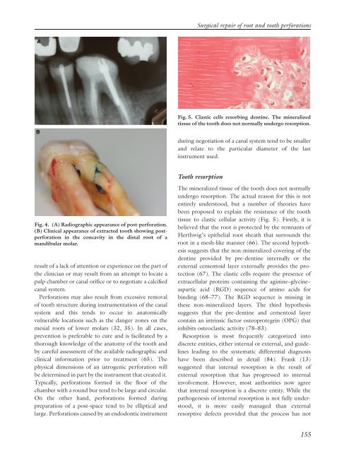
Fig. 5. Clastic cells resorbing dentine. The mineralized<br />
tissue <strong>of</strong> the <strong>tooth</strong> does not normally undergo resorption.<br />
during negotiation <strong>of</strong> a canal system tend to be smaller<br />
<strong>and</strong> relate to the particular diameter <strong>of</strong> the last<br />
instrument used.<br />
Tooth resorption<br />
Fig. 4. (A) Radiographic appearance <strong>of</strong> post-perforation.<br />
(B) Clinical appearance <strong>of</strong> extracted <strong>tooth</strong> showing postperforation<br />
in the concavity in the distal <strong>root</strong> <strong>of</strong> a<br />
m<strong>and</strong>ibular molar.<br />
result <strong>of</strong> a lack <strong>of</strong> attention or experience on the part <strong>of</strong><br />
the clinician or may result from an attempt to locate a<br />
pulp chamber or canal orifice or to negotiate a calcified<br />
canal system.<br />
Perforations may also result from excessive removal<br />
<strong>of</strong> <strong>tooth</strong> structure during instrumentation <strong>of</strong> the canal<br />
system <strong>and</strong> this tends to occur in anatomically<br />
vulnerable locations such as the danger zones on the<br />
mesial <strong>root</strong>s <strong>of</strong> lower molars (32, 35). In all cases,<br />
prevention is preferable to cure <strong>and</strong> is facilitated by a<br />
thorough knowledge <strong>of</strong> the anatomy <strong>of</strong> the <strong>tooth</strong> <strong>and</strong><br />
by careful assessment <strong>of</strong> the available radiographic <strong>and</strong><br />
clinical information prior to treatment (65). The<br />
physical dimensions <strong>of</strong> an iatrogenic perforation will<br />
be determined in part by the instrument that created it.<br />
Typically, <strong>perforations</strong> formed in the floor <strong>of</strong> the<br />
chamber with a round bur tend to be large <strong>and</strong> circular.<br />
On the other h<strong>and</strong>, <strong>perforations</strong> formed during<br />
preparation <strong>of</strong> a post-space tend to be elliptical <strong>and</strong><br />
large. Perforations caused by an endodontic instrument<br />
The mineralized tissue <strong>of</strong> the <strong>tooth</strong> does not normally<br />
undergo resorption. The actual reason for this is not<br />
entirely understood, but a number <strong>of</strong> theories have<br />
been proposed to explain the resistance <strong>of</strong> the <strong>tooth</strong><br />
tissue to clastic cellular activity (Fig. 5). Firstly, it is<br />
believed that the <strong>root</strong> is protected by the remnants <strong>of</strong><br />
Herthwig’s epithelial <strong>root</strong> sheath that surrounds the<br />
<strong>root</strong> in a mesh-like manner (66). The second hypothesis<br />
suggests that the non-mineralized covering <strong>of</strong> the<br />
dentine provided by pre-dentine internally or the<br />
external cementoid layer externally provides the protection<br />
(67). The clastic cells require the presence <strong>of</strong><br />
extracellular proteins containing the aginine–glycine–<br />
aspartic acid (RGD) sequence <strong>of</strong> amino acids for<br />
binding (68–77). The RGD sequence is missing in<br />
these non-mineralized layers. The third hypothesis<br />
suggests that the pre-dentine <strong>and</strong> cementoid layer<br />
contain an intrinsic factor osteoprotegrin (OPG) that<br />
inhibits osteoclastic activity (78–83).<br />
Resorption is most frequently categorized into<br />
discrete entities, either internal or external, <strong>and</strong> guidelines<br />
leading to the systematic differential diagnosis<br />
have been described in detail (84). Frank (13)<br />
suggested that internal resorption is the result <strong>of</strong><br />
external resorption that has progressed to internal<br />
involvement. However, most authorities now agree<br />
that internal resorption is a discrete entity. While the<br />
pathogenesis <strong>of</strong> internal resorption is not fully understood,<br />
it is more easily managed than external<br />
resorptive defects provided that the process has not<br />
155