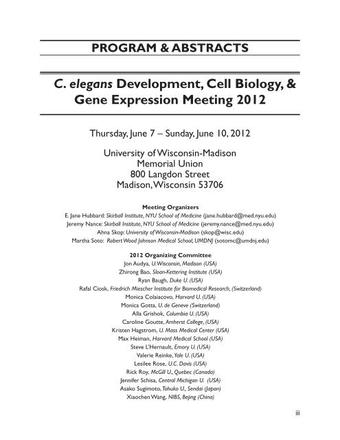
C. elegans Development, Cell Biology, & Gene Expression Meeting ...
C. elegans Development, Cell Biology, & Gene Expression Meeting ...
C. elegans Development, Cell Biology, & Gene Expression Meeting ...

You also want an ePaper? Increase the reach of your titles
YUMPU automatically turns print PDFs into web optimized ePapers that Google loves.
PROGRAM & ABSTRACTS<br />
C. <strong>elegans</strong> <strong>Development</strong>, <strong>Cell</strong> <strong>Biology</strong>, &<br />
<strong>Gene</strong> <strong>Expression</strong> <strong>Meeting</strong> 2012<br />
Thursday, June 7 – Sunday, June 10, 2012<br />
University of Wisconsin-Madison<br />
Memorial Union<br />
800 Langdon Street<br />
Madison, Wisconsin 53706<br />
<strong>Meeting</strong> Organizers<br />
E. Jane Hubbard: Skirball Institute, NYU School of Medicine (jane.hubbard@med.nyu.edu)<br />
Jeremy Nance: Skirball Institute, NYU School of Medicine (jeremy.nance@med.nyu.edu)<br />
Ahna Skop: University of Wisconsin-Madison (skop@wisc.edu)<br />
Martha Soto: Robert Wood Johnson Medical School, UMDNJ (sotomc@umdnj.edu)<br />
2012 Organizing Committee<br />
Jon Audya, U. Wisconsin, Madison (USA)<br />
Zhirong Bao, Sloan-Kettering Institute (USA)<br />
Ryan Baugh, Duke U. (USA)<br />
Rafal Ciosk, Friedrich Miescher Institute for Biomedical Research, (Switzerland)<br />
Monica Colaiacovo, Harvard U. (USA)<br />
Monica Gotta, U. de <strong>Gene</strong>ve (Switzerland)<br />
Alla Grishok, Columbia U. (USA)<br />
Caroline Goutte, Amherst College, (USA)<br />
Kristen Hagstrom, U. Mass Medical Center (USA)<br />
Max Heiman, Harvard Medical School (USA)<br />
Steve L’Hernault, Emory U. (USA)<br />
Valerie Reinke, Yale U. (USA)<br />
Lesilee Rose, U.C. Davis (USA)<br />
Rick Roy, McGill U., Quebec (Canada)<br />
Jennifer Schisa, Central Michigan U. (USA)<br />
Asako Sugimoto, Tohuko U., Sendai (Japan)<br />
Xiaochen Wang, NIBS, Bejing (China)<br />
iii
iv<br />
www.workshops.biologists.com<br />
www.neb.com<br />
www.landesbioscience.com<br />
SPONSORS<br />
ACKNOWLEDGEMENTS<br />
www.nsf.org<br />
www.kramerscientific.com<br />
www.powersscientific.com<br />
www.prairie-technologies.com www.unionbio.com<br />
All Sponsoring Companies<br />
University of Wisconsin Memorial Union Conference Services<br />
91 High Street ● Amesbury, Massachusetts 01913 USA ● Tel +1 978-388-7159 ● Fax: +1 978-388-7854<br />
sales@kramerscientific.com ● www.kramerscientific.com
C. <strong>elegans</strong> <strong>Development</strong>, <strong>Cell</strong> <strong>Biology</strong>, & <strong>Gene</strong><br />
<strong>Expression</strong> <strong>Meeting</strong><br />
Thursday, June 7 – Sunday, June 10, 2012<br />
Conference Program<br />
Thursday, June 07, 2012<br />
12 noon–7:30 pm Registration Check-In Annex Room<br />
12 noon Poster Set up Great Hall, Reception Room, and Main Lounge<br />
5:00–7:00 pm Opening Reception Tripp Commons<br />
7:00 Opening Remarks<br />
7:00–9:00 pm Platform Session #1 Union Theater<br />
Morphogenesis I and Polarity<br />
Chairs: Lesilee Rose and Asako Sugimoto<br />
7:15 Keynote: Ken Kemphues<br />
Three pathways to polarity maintenance<br />
7:45 Jessica L Feldman (Lab: Priess)<br />
A role for the centrosome and PAR-3 in the hand-off of<br />
microtubule organizing center function during epithelial<br />
polarization<br />
8:00 Yelena Y Bernadskaya (Lab: Soto)<br />
Three Axonal Guidance Pathways Help Polarize the Actin<br />
Cytoskeleton During Embryonic Epidermal <strong>Cell</strong> Migration<br />
8:15 Jessica Shivas (Lab: Skop)<br />
Arp2/3 mediates early endosome dynamics that participate in<br />
the maintenance of polarity in C. <strong>elegans</strong><br />
8:30 Hongjie Zhang (Lab: Gobel)<br />
Clathrin/AP-1 cooperate with sphingolipids to regulate apical<br />
polarity and lumen formation during C. <strong>elegans</strong> tubulogenesis<br />
8:45 Vijaykumar S Meli (Lab: Frand)<br />
The Fibrillin-like fbn-1 <strong>Gene</strong> Regulates Epithelial Stem <strong>Cell</strong> and<br />
ECM Dynamics in Molts<br />
v
9:00–11:00 pm Poster Session #1 & Refreshments Great Hall, Reception Room,<br />
and Main Lounge<br />
(ODD number posters present)<br />
vi<br />
Great Hall & Reception Room (4th floor)<br />
<strong>Cell</strong> <strong>Biology</strong> 51 83<br />
<strong>Cell</strong> cycle and cytokinesis 84 92<br />
<strong>Cell</strong> Death 93 103<br />
<strong>Cell</strong> Fate 104 121<br />
<strong>Gene</strong> Regulation 122 145<br />
Germline 146 184<br />
Main Lounge (2nd floor)<br />
Morphogenesis 185 211<br />
New Technologies 212 220<br />
Polarity 221 231<br />
Sex Determination 232 234<br />
Friday, June 08, 2012<br />
7:00 am Registration continues Annex Room<br />
7:30–9:00 am Breakfast Buffet Inn Wisconsin<br />
9:00–10:45 am Platform Session #2 Union Theater<br />
Morphogenesis II & <strong>Cell</strong> Death<br />
Chairs: Max Heiman and Caroline Goutte<br />
9:00 Keynote: Shai Shaham (Lab: Shaham)<br />
A New C. <strong>elegans</strong> <strong>Cell</strong> Death Program: Implications for<br />
Neurodegeneration and Cancer<br />
9:45 Yan Zhang (Lab: Wang)<br />
C. <strong>elegans</strong> NRF-5 Regulates <strong>Cell</strong> Corpse Engulfment By<br />
Mediating PS Appearance On Phagocytes<br />
10:00 Sasha De HeBoldnau (Lab: Braeckman)<br />
Globin 12 of Caenorhabditis <strong>elegans</strong> Regulates the p38 and JNK<br />
MAPK Pathways through Redox Signaling to Control Germline<br />
Apoptosis<br />
10:15 Michael Hurwitz (Lab: Hurwitz)<br />
sli-1 Cbl Inhibits the Engulfment of Apoptotic <strong>Cell</strong>s<br />
10:30 Matthias K Morf (Lab: Hajnal)<br />
MADD-2 Negatively Regulates Anchor <strong>Cell</strong> Invasion<br />
10:45 Vida Praitis (Lab: Praitis)<br />
The C. <strong>elegans</strong> Hailey-Hailey Disease Homolog pmr-1 is Essential<br />
for <strong>Cell</strong> Migration During Gastrulation<br />
11:00–11:15 am Refreshment Break Union Theater Lobby
11:15 am–1:00 pm Platform Session #3 Union Theater<br />
Germline I and Gametogenesis<br />
Chairs: Steve L’Hernault and Rafal Ciosk<br />
11:15 Keynote: David Greenstein (Lab: Greenstein)<br />
Control of Oocyte Meiotic Maturation: Links to Germ <strong>Cell</strong><br />
Proliferation and Global Control of the Oogenic Program<br />
12:00 Kari Messina (Lab: Shakes)<br />
Regulators of MSP Assembly and Dynamics in C. <strong>elegans</strong><br />
Spermatocytes<br />
12:15 Gunasekaran Singaravelu (Lab: Singson)<br />
The sperm surface localization of the TRP-3/SPE-41<br />
Ca2+ permeable channel depends on SPE-38 function in<br />
Caenorhabditis <strong>elegans</strong><br />
12:30 Jun Takayama (Lab: Onami)<br />
Timely <strong>Gene</strong>ration of the Fertilization Calcium Wave by a<br />
Sperm TRP Channel<br />
12 :45 Simona Rosu (Lab: Villeneuve)<br />
Regulation of Meiotic DSB Formation in C. <strong>elegans</strong><br />
1:00–2:30 pm Luncheon Buffet Inn Wisconsin<br />
2:30–5:30 pm Platform Session #4 Union Theater<br />
<strong>Cell</strong> Cycle and <strong>Cell</strong> <strong>Biology</strong><br />
Chairs: Jon Audhya and Richard Roy<br />
2:30 Keynote: Karen Oegema (Lab: Oegema)<br />
Title: TBD<br />
3:15 Marie Delattre (Lab: Delattre)<br />
Evolution of spindle shape and motion in one-cell stage<br />
nematode embryos<br />
3:30 Jill M Schumacher (Lab: Schumacher)<br />
The Tousled-like Kinase TLK-1 is a Component of the Outer<br />
Kinetochore and Potentiates Mitotic Spindle Dynamics in the<br />
Early C. <strong>elegans</strong> Embryo<br />
3:45 Asako Sugimoto (Lab: Sugimoto)<br />
Identification of unconventional components of the γ-tubulin<br />
complex in C. <strong>elegans</strong><br />
4:00 Elsa Kress (Lab: Gotta)<br />
The Cdc48/p97 cofactor UBXN-2 and its orthologues p47/p37<br />
control centrosome maturation in prophase via Aurora A<br />
4:15–4:30 pm Refreshment Break Union Theater Lobby<br />
4:30 Anjon Audhya (Lab: Audhya)<br />
Regulation of COPII subunit recruitment to ER exit sites<br />
4:45 Joshua N Bembenek (Lab: Chan)<br />
Condensin I: A New Component of the Abscission Checkpoint<br />
vii
5:00 Matyas Gorjanacz (Lab: Mattaj)<br />
LEM-4 Coordinates Mitotic Signaling on BAF to Enable its<br />
Essential Function in Nuclear Envelope Formation<br />
5:15 Ismar Kovacevic (Lab: Cram)<br />
Filamin is Required to Initiate Calcium Signaling and Maintain<br />
F-actin Organization in the Spermatheca<br />
5:30–7:00 pm Dinner Buffet Inn Wisconsin<br />
7:00–9:00 pm Poster Session #2 & Refreshments Great Hall, Reception Room,<br />
and Main Lounge<br />
(EVEN number posters present)<br />
viii<br />
Great Hall & Reception Room (4th floor)<br />
<strong>Cell</strong> <strong>Biology</strong> 51 83<br />
<strong>Cell</strong> cycle and cytokinesis 84 92<br />
<strong>Cell</strong> Death 93 103<br />
<strong>Cell</strong> Fate 104 121<br />
<strong>Gene</strong> Regulation 122 145<br />
Germline 146 184<br />
Main Lounge (2nd floor)<br />
Morphogenesis 185 211<br />
New Technologies 212 220<br />
Polarity 221 231<br />
Sex Determination 232 234<br />
9:00–11:30 pm Late Night Poster Session Great Hall, Reception Room,<br />
and Main Lounge<br />
Open Viewing (All numbered posters present)<br />
Saturday, June 09, 2012<br />
7:00 am Registration Continues Annex Room<br />
7:30–9:00 am Breakfast Buffet Inn Wisconsin<br />
9:00 am–12:00 noon Platform Session #5 Union Theater<br />
Germline II, Meiosis, and Sex Determination/Dimorphism<br />
Chairs: Monica Colaiácovo and Jennifer Schisa<br />
9:00 Keynote: Monica Colaiácovo (Lab: Colaiácovo)<br />
Germline maintenance and meiosis: mechanistic insights from C.<br />
<strong>elegans</strong><br />
9:30 Aaron Kershner (Lab: Kimble)<br />
Identification of Direct GLP-1/Notch Targets that Regulate<br />
Germline Stem <strong>Cell</strong>s<br />
9:45 Rafal Ciosk (Lab: Ciosk)<br />
Genome-wide Analysis of GLD-1 Mediated mRNA Regulation<br />
Uncovers a Role in mRNA Storage
10:00 E. Jane Albert Hubbard (Lab: Hubbard)<br />
In the C. <strong>elegans</strong> Germ Line, S6K promotes <strong>Cell</strong> Cycle<br />
Progression and the Proliferative Fate and mediates the Effects<br />
of Diet<br />
10:50–10:30 am Refreshment Break Union Theater Lobby<br />
10:30 Mara Schvarzstein (Lab: Villeneuve)<br />
Chromosome and centrosome inheritance in meiosis<br />
10:45 Daniel Cortes Estrada (Lab: McNalley)<br />
Non-random Segregation of Unpaired X Chromosomes in C.<br />
<strong>elegans</strong> Female Meiosis (asbt. # 152)<br />
11:00 Anna K Allen (Lab: Golden)<br />
Role of the Inhibitory Kinase WEE-1.3 in Regulating the Meiotic<br />
<strong>Cell</strong> Cycle and Fertility in C. Elegans<br />
11:15 Michael J. White VanGompel (Lab: Rose)<br />
The Torsin Homolog OOC-5 is Required for Normal<br />
Nucleoporin Localization<br />
11:30 Matthew Berkseth (Lab: Zarkower)<br />
Identification of Direct Targets of the Caenorhabditis<br />
<strong>elegans</strong> Global Sexual Regulator TRA-1 by Chromatin<br />
Immunoprecipitation<br />
11:45 Te-Wen Lo (Lab: Meyer)<br />
Evolution of Caenorhabditis Dosage Compensation<br />
12:00–2:00 pm Luncheon Buffet (posters down by 2:00 pm) Inn Wisconsin<br />
2:30–4:00 pm Workshops Union Theater<br />
4:00–4:30 pm Refreshment Break Union Theater Lobby<br />
4:30–6:30 pm Platform Session #6 Union Theater<br />
<strong>Gene</strong> Regulation<br />
Chairs: Valerie Reinke and Ryan Baugh<br />
Introduction: Alla Grishok<br />
4:30 Keynote: Craig Mello (Lab: Mello)<br />
RNAi and Immortality: Recognition of Self/non-Self RNA in the<br />
C. <strong>elegans</strong> Germline<br />
5:15 Gyorgyi Csankovszki (Lab: Csankovszki)<br />
The onset of dosage compensation is linked to the loss of<br />
developmental plasticity<br />
5:30 David J Katz (Lab: Katz)<br />
The Histone Demethylase SPR-5 and the Histone<br />
Methyltransferase MET-2 Comprise a Novel Epigenetic<br />
Reprogramming Switch<br />
5:45 Shouhong Guang (Lab: Guang)<br />
Nuclear RNAi mediates silencing of repetitive sequences in C.<br />
<strong>elegans</strong><br />
ix
6:00 Xiao-Dong Yang (Lab: Lin)<br />
Dimerization of γCatenin/WRM-1 Allows Intermolecular<br />
Autophosphorylation of LIT-1 in the Activation Loop<br />
6:15 Morris Maduro (Lab: Maduro)<br />
Organ defects in adults resulting from threshold blastomere<br />
specification<br />
7:00–9:30 pm Banquet Union South<br />
9:30–Midnight Dance Union South<br />
x<br />
Sunday, June 10, 2012<br />
9:00–12:30 pm Platform Session #7 Union Theater<br />
<strong>Cell</strong> Fate and Emerging Technologies<br />
Chairs: Monica Gotta and Zhirong Bao<br />
9:00 am Keynote: Julie Ahringer (Lab: Ahringer)<br />
Title: TBD<br />
9:45 Hillel Kugler (Lab: Kugler)<br />
Modeling germline population dynamics<br />
10:00 Julia L Moore (Lab: Bao)<br />
Dev-scape: An intuitive tool for automated phenotyping with<br />
single cell resolution<br />
10:15 Abigail Cabunoc (Lab: Stein)<br />
WormBase 2012: Website Redesign<br />
11:00 Scott Robertson (Lab: Lin)<br />
DSL-2 Mediates a Notch Signal From EMS Descendant(s) to<br />
ABp Descendants<br />
11:15 Jennifer A Schumacher Tucker (Lab: Chuang)<br />
Intercellular Calcium Signaling in a Gap Junction <strong>Cell</strong> Network<br />
Establishes Left-Right Asymmetric Neuronal Fates<br />
11:30 Colin Maxwell (Lab: Baugh)<br />
Nutritional control of mRNA isoform expression during<br />
developmental arrest and recovery in C. <strong>elegans</strong><br />
11:45 David J Reiner (Lab: Reiner)<br />
Ras and its Effector RalGEF Both Perform Dual, Antagonistic<br />
Functions during C. <strong>elegans</strong> Vulval Patterning<br />
12:00 Allison L Abbott (Lab: Abbott)<br />
The microRNA miR-786 is Required for Rhythmic Calcium<br />
Wave Initiation in the C. <strong>elegans</strong> Intestine<br />
12:30–2:00 pm Luncheon Buffet Inn Wisconsin
TABLE OF CONTENTS<br />
Thursday, June 07, 2012 - 7:00–9:00 pm<br />
Platform Session #1 - Union Theater<br />
Morphogenesis I and Polarity<br />
Abstracts 1 - 6<br />
Chairs: Lesilee Rose and Asako Sugimoto<br />
1 Keynote: Three pathways to polarity maintenance<br />
Ken Kemphues<br />
2 A role for the centrosome and PAR-3 in the hand-off of microtubule<br />
organizing center function during epithelial polarization<br />
Jessica Feldman, James Priess<br />
3 Three Axonal Guidance Pathways Help Polarize the Actin Cytoskeleton<br />
During Embryonic Epidermal <strong>Cell</strong> Migration<br />
Yelena Bernadskaya, Andre Wallace, Jillian Nguyen, William Mohler, Martha Soto<br />
4 Arp2/3 mediates early endosome dynamics that participate in the<br />
maintenance of polarity in C. <strong>elegans</strong><br />
Jessica Shivas, Ahna Skop<br />
5 Clathrin/AP-1 cooperate with sphingolipids to regulate apical polarity<br />
and lumen formation during C. <strong>elegans</strong> tubulogenesis<br />
Hongjie Zhang, Ahlee Kim, Nessy Abraham, Liakot Khan, David Hall, John Fleming, Verena<br />
Gobel<br />
6 The Fibrillin-like fbn-1 <strong>Gene</strong> Regulates Epithelial Stem <strong>Cell</strong> and ECM<br />
Dynamics in Molts<br />
Vijaykumar Meli, Alison Frand<br />
Friday, June 08, 2012 - 9:00–10:45 am<br />
Platform Session #2 - Union Theater<br />
Morphogenesis II & <strong>Cell</strong> Death<br />
Abstracts 7 - 12<br />
Chairs: Max Heiman and Caroline Goutte<br />
7 Keynote: A New C. <strong>elegans</strong> <strong>Cell</strong> Death Program: Implications for<br />
Neurodegeneration and Cancer<br />
Shai Shaham<br />
8 C. <strong>elegans</strong> NRF-5 Regulates <strong>Cell</strong> Corpse Engulfment By Mediating PS<br />
Appearance On Phagocytes<br />
Yan Zhang, Haibin Wang, Xiaochen Wang<br />
xi
9 Globin 12 of Caenorhabditis <strong>elegans</strong> Regulates the p38 and JNK MAPK<br />
Pathways through Redox Signaling to Control Germline Apoptosis<br />
Sasha De Henau, Lesley Tilleman, Francesca Germani, Caroline Vlaeminck, Jacques<br />
Vanfleteren, Luc Moens, Sylvia Dewilde, Bart Braeckman<br />
10 sli-1 Cbl Inhibits the Engulfment of Apoptotic <strong>Cell</strong>s<br />
Courtney Anderson, Shan Zhou, Emma Sawin, Bob Horvitz, Michael Hurwitz<br />
11 MADD-2 Negatively Regulates Anchor <strong>Cell</strong> Invasion<br />
Matthias Morf, Ivo Rimann, Mariam Alexander, Peter Roy, Alex Hajnal<br />
12 The C. <strong>elegans</strong> Hailey-Hailey Disease Homolog pmr-1 is Essential for<br />
<strong>Cell</strong> Migration During Gastrulation<br />
Vida Praitis, Rebecca Mandt, Leah Imlay, Charlotte Feddersen, Alexander Sullivan-Wilson,<br />
Tyson Stock, Walter Liszewski, Adityarup Chakravorty, Dae Gon Ha, Angela Schacht,<br />
Michael Miller, Lensa Yohannes, Juliet Mushi, Zelealem Yilma, Sarah Kniss, Jeff Simske<br />
xii<br />
Friday, June 08, 2012 - 11:15 am–1:00 pm<br />
Platform Session #3 - Union Theater<br />
Germline I and Gametogenesis<br />
Abstracts 13 - 17<br />
Chairs: Steve L’Hernault and Rafal Ciosk<br />
13 Keynote: Control of Oocyte Meiotic Maturation: Links to Germ <strong>Cell</strong><br />
Proliferation and Global Control of the Oogenic Program<br />
David Greenstein<br />
14 Regulators of MSP Assembly and Dynamics in C. <strong>elegans</strong> Spermatocytes<br />
Kari Messina, Marc Presler, Leah Towarnicky, Diane Shakes<br />
15 The sperm surface localization of the TRP-3/SPE-41 Ca2+ permeable<br />
channel depends on SPE-38 function in Caenorhabditis <strong>elegans</strong><br />
Gunasekaran Singaravelu, Indrani Chatterjee, Sina Rahimi, Marina Druzhinina, Lijun<br />
Kang, Shawn Xu, Andrew Singson<br />
16 Timely <strong>Gene</strong>ration of the Fertilization Calcium Wave by a Sperm TRP<br />
Channel<br />
Jun Takayama, Shuichi Onami<br />
17 Regulation of Meiotic DSB Formation in C. <strong>elegans</strong><br />
Simona Rosu, Anne Villeneuve
18 Keynote: Title: TBD<br />
Karen Oegema<br />
Friday, June 08, 2012 - 2:30–5:30 pm<br />
Platform Session #4 - Union Theater<br />
<strong>Cell</strong> Cycle and <strong>Cell</strong> <strong>Biology</strong><br />
Abstracts 18 - 26<br />
Chairs: Jon Audhya and Richard Roy<br />
19 Evolution of spindle shape and motion in one-cell stage nematode<br />
embryos<br />
Aurore-Cecile Valfort, Soizic Riche, Reza Farhadifair, Daniel Needleman, Marie Delattre<br />
20 The Tousled-like Kinase TLK-1 is a Component of the Outer<br />
Kinetochore and Potentiates Mitotic Spindle Dynamics in the Early C.<br />
<strong>elegans</strong> Embryo<br />
Jessica De Orbeta, Jason Ford, Gary Deyter, Tokiko Furuta, Jill Schumacher<br />
21 Identification of unconventional components of the γ-tubulin complex<br />
in C.<strong>elegans</strong><br />
Nami Haruta, Eisuke Sumiyoshi, Yu Honda, Masahiro Terasawa, Mika Toya, Asako<br />
Sugimoto<br />
22 The Cdc48/p97 cofactor UBXN-2 and its orthologues p47/p37 control<br />
centrosome maturation in prophase via Aurora A<br />
Elsa Kress, Francoise Schwager, Rene Holtackers, Esther Zanin, Francois Prodon, Jonas<br />
Seiler, Annika Eiteneuer, Asako Sugimoto, Hemmo Meyer, Patrick Meraldi, Monica Gotta<br />
23 Regulation of COPII subunit recruitment to ER exit sites<br />
Kristen Witte, Amber Schuh, Jan Hegermann, Ali Sarkeshik, Jonathan Mayers, Katrin<br />
Schwarze, John Yates III, Stefan Eimer, Anjon Audhya<br />
24 Condensin I: A New Component of the Abscission Checkpoint<br />
Joshua Bembenek, Koen Verbrugghe, Gyorgyi Csankovszki, Raymond Chan<br />
25 LEM-4 Coordinates Mitotic Signaling on BAF to Enable its Essential<br />
Function in Nuclear Envelope Formation<br />
Matyas Gorjanacz, Claudio Asencio, Iain Davidson, Rachel Santarella-Mellwig, Geraldine<br />
Seydoux , Iain Mattaj<br />
26 Filamin is Required to Initiate Calcium Signaling and Maintain F-actin<br />
Organization in the Spermatheca<br />
Ismar Kovacevic, Erin Cram<br />
xiii
xiv<br />
Saturday, June 09, 2012 - 9:00 am–12:00 noon<br />
Platform Session #5 - Union Theater<br />
Germline II, Meiosis, and Sex Determination/Dimorphism<br />
Abstracts 27 - 35<br />
Chairs: Monica Colaiácovo and Jennifer Schisa<br />
27 Keynote: Germline maintenance and meiosis: mechanistic insights from<br />
C. <strong>elegans</strong><br />
Monica Colaiácovo<br />
28 Identification of Direct GLP-1/Notch Targets that Regulate Germline<br />
Stem <strong>Cell</strong>s<br />
Aaron Kershner, Heaji Shin, Judith Kimble<br />
29 Genome-wide Analysis of GLD-1 Mediated mRNA Regulation Uncovers<br />
a Role in mRNA Storage<br />
Claudia Scheckel, Dimos Gaidatzis, Jane Wright, Rafal Ciosk<br />
30 In the C. <strong>elegans</strong> Germ Line, S6K promotes <strong>Cell</strong> Cycle Progression and<br />
the Proliferative Fate and mediates the Effects of Diet<br />
Dorota Korta, Debasmita Roy, Simon Tuck, E. Jane Albert Hubbard<br />
31 Chromosome and centrosome inheritance in meiosis<br />
Mara Schvarzstein, Anne Villeneuve<br />
32 Role of the Inhibitory Kinase WEE-1.3 in Regulating the Meiotic <strong>Cell</strong><br />
Cycle and Fertility in C. Elegans<br />
Anna Allen, Jessica Nesmith, Andy Golden<br />
33 The Torsin Homolog OOC-5 is Required for Normal Nucleoporin<br />
Localization<br />
Michael White VanGompel, Lesilee Rose<br />
34 Identification of Direct Targets of the Caenorhabditis <strong>elegans</strong> Global<br />
Sexual Regulator TRA-1 by Chromatin Immunoprecipitation<br />
Matthew Berkseth, Kohta Ikegami, Jason Lieb, David Zarkower<br />
35 Evolution of Caenorhabditis Dosage Compensation<br />
Te-Wen Lo, Caitlin Schartner, Catherine Pickle, Barbara Meyer
Saturday, June 09, 2012 - 4:30–6:30 pm<br />
Platform Session #6 - Union Theater<br />
<strong>Gene</strong> Regulation<br />
Abstracts 36 - 41<br />
Chairs: Valerie Reinke and Ryan Baugh<br />
36 Keynote: RNAi and Immortality: Recognition of Self/non-Self RNA in<br />
the C. <strong>elegans</strong> Germline<br />
Craig Mello<br />
37 The onset of dosage compensation is linked to the loss of<br />
developmental plasticity<br />
Laura Custer, Gyorgyi Csankovszki<br />
38 The Histone Demethylase SPR-5 and the Histone Methyltransferase<br />
MET-2 Comprise a Novel Epigenetic Reprogramming Switch<br />
Shana Kerr, Chelsey Chandler, Joshua Francis, Erica Mills, David Katz<br />
39 Nuclear RNAi mediates silencing of repetitive sequences in C. <strong>elegans</strong><br />
Fei Xu, Xufei Zhou, Hui Mao, Jiaojiao Ji, Shouhong Guang<br />
40 Dimerization of γCatenin/WRM-1 Allows Intermolecular<br />
Autophosphorylation of LIT-1 in the Activation Loop<br />
Xiao-Dong Yang, Scott Robertson , Rueyling Lin<br />
41 Organ defects in adults resulting from threshold blastomere<br />
specification<br />
Morris Maduro, Gina Broitman-Maduro, Leila Magistrado, Shruthi Satish<br />
Sunday, June 10, 2012 - 9:00–12:30 pm<br />
Platform Session #7 - Union Theater<br />
<strong>Cell</strong> Fate and Emerging Technologies<br />
Abstracts 42 - 50<br />
Chairs: Monica Gotta and Zhirong Bao<br />
42 Keynote: Title: TBD<br />
Julie Ahringer<br />
43 Modeling germline population dynamics<br />
Hillel Kugler, E. Jane Albert Hubbard<br />
44 Dev-scape: An intuitive tool for automated phenotyping with single cell<br />
resolution<br />
Julia Moore, Zhuo Du, Anthony Santella, Christian Pohl, Zhirong Bao<br />
xv
45 WormBase 2012: Website Redesign<br />
Abigail Cabunoc, Norie de la Cruz, Adrian Duong, Maher Kassim, Xiaoqi Shi, Todd Harris,<br />
Lincoln Stein<br />
46 DSL-2 Mediates a Notch Signal From EMS Descendant(s) to ABp<br />
Descendants<br />
Scott Robertson, Jessica Medina, Rueyling Lin<br />
47 Intercellular Calcium Signaling in a Gap Junction <strong>Cell</strong> Network<br />
Establishes Left-Right Asymmetric Neuronal Fates<br />
Jennifer Schumacher Tucker, Chieh Chang, Chiou-Fen Chuang<br />
48 Nutritional control of mRNA isoform expression during developmental<br />
arrest and recovery in C. <strong>elegans</strong><br />
Colin Maxwell, Igor Antoshechkin, Nicole Kurhanewicz, Jason Belsky, L. Ryan Baugh<br />
49 Ras and its Effector RalGEF Both Perform Dual, Antagonistic Functions<br />
during C. <strong>elegans</strong> Vulval Patterning<br />
Kimberly Monahan, Rebecca Whitehurst, Tanya Zand, Channing Der, David Reiner<br />
50 The microRNA miR-786 is Required for Rhythmic Calcium Wave<br />
Initiation in the C. <strong>elegans</strong> Intestine<br />
Benedict Kemp, Erik Allman, Lois Immerman, Megan Mohnen, Maureen Peters, Keith<br />
Nehrke, Allison Abbott<br />
xvi<br />
Poster Topic<br />
<strong>Cell</strong> <strong>Biology</strong><br />
Abstracts 51 - 83<br />
51 GLO-2 is a BLOC-1 Subunit that Functions in Gut Granule Biogenesis<br />
Alec Barrett, Olivia Foster, Annalise Vine, Greg Hermann<br />
52 The Conventional Kinesin-1/UNC-116 Acts in PHB Phasmid Neurons<br />
to Mediate Proper <strong>Cell</strong> Body Position<br />
Ben Barsi-Rhyne, Kristine Miller, Chris Vargas, Miri VanHoven<br />
53 <strong>Gene</strong>tic Interaction and Structure/Function Studies of MEL-28, a<br />
Protein Required for Nuclear Envelope Function and Chromosome<br />
Segregation<br />
Anita Fernandez, Carly Bock, Allison Lai, Emily Mis, Fabio Piano<br />
54 Oocyte Meiotic Spindle Assembly in C. <strong>elegans</strong><br />
Amy Connolly, Sara Christensen, Valerie Osterberg, Josh Lowry, John Yochem, Bruce<br />
Bowerman<br />
55 Identifying Proteins that Interact with the Serine/Threonine Kinase<br />
UNC-82 in Muscle <strong>Cell</strong>s<br />
Christopher Duchesneau, April Reedy, Hiroshi Qadota, Guy Benian, Pamela Hoppe
56 A LET-23 localization and expression screen identifies a novel<br />
mechanism of EGFR regulation through Ezrin/Radixin/Moesin proteins<br />
Juan Escobar Restrepo, Peter Gutierrez, Andrea Haag, Alessandra Buhler, Christina<br />
Herrmann, Maeva Langouet, David Kradolfer, Erika Frohli, Attila Stetak, Alex Hajnal<br />
57 Growth of Muscle Adhesion Complexes During Postembryonic<br />
<strong>Development</strong><br />
Brandon Fields, Nate Szewczyk, Lewis Jacobson<br />
58 CDK-1 inhibits meiotic spindle shortening and dynein-dependent<br />
spindle rotation in C. <strong>elegans</strong><br />
Jonathan Flynn, Marina Ellefson, Francis McNally<br />
59 The C. <strong>elegans</strong> Uterine Seam <strong>Cell</strong>: a Model for Studying Nuclear<br />
Migration and <strong>Cell</strong> Outgrowth<br />
Srimoyee Ghosh, Paul Sternberg<br />
60 Cadherin FMI-1 Maintains the Structure of the PVD Mechanosensory<br />
Neurons<br />
Julie Grimm, Benjamin Podbilewicz<br />
61 Two Functional Domains in C. <strong>elegans</strong> Glypican LON-2 Can<br />
Independently Inhibit DBL-1 Growth Factor Signaling but Require<br />
Accessory Moieties<br />
Suparna Bageshwar, Tina Gumienny<br />
62 Mutational Analysis of Residues Required for Activation the UNC-82<br />
Serine-Threonine Kinase<br />
Jason Kintzele, Pamela Hoppe<br />
63 <strong>Gene</strong>tic Analysis of Calcium Regulation in the C. <strong>elegans</strong> Intestine<br />
Jocelyn Laboy, Kenneth Norman<br />
64 The Tubulin Deglutamylase CCPP-6 Functions Exclusively in Ciliated<br />
Dopaminergic Neurons in C. <strong>elegans</strong><br />
Ethan Landes, Brendan O’Flaherty, Elizabeth De Stasio, Peter Swoboda, Brian Piasecki<br />
65 Protein Sequences Within the UNC-82 S/T Kinase that Affect<br />
Subcellular Localization in Pharyngeal Muscle<br />
Latrisha Lane, Chiyen Wong, Caitlyn Carter, Pamela Hoppe<br />
66 Characterization of vh45, a Candidate Regulator of Early to Late<br />
Endosomal Maturation<br />
Fiona Law, Shang Xiang, Christian Rocheleau<br />
67 cil-5 Mediates Ciliary Receptor Localization and Sensory Function in C.<br />
<strong>elegans</strong><br />
Kara Braunreiter, Greg Fischer, Casey Gabrhel, Jamie Lyman Gingerich<br />
xvii
68 Neuroligin has <strong>Cell</strong>-autonomous and Non-autonomous Functions in C.<br />
<strong>elegans</strong><br />
Jacob Manjarrez, Greg Mullen, Ellie Mathews, Jerrod Hunter, Jim Rand<br />
69 <strong>Gene</strong>tic and Molecular Dissection of Novel Pathways Required for<br />
Nuclear Migration in the Model System C. <strong>elegans</strong>.<br />
Yu-Tai Chang, Shaun Murphy, Jonathan Kuhn, Minh Ngo, Daniel Starr<br />
70 FLN-1/filamin is required for spermathecal contractility<br />
Jose Orozco, Ismar Kovacevic, Erin Cram<br />
71 Isolation of Mutations that alter Nile Red Staining in C. <strong>elegans</strong><br />
Stephanie Burge, Anthony Otsuka<br />
72 Epithelial Dynamics During the G1-to-G2 Pore <strong>Cell</strong> Swap in the<br />
Excretory System<br />
Jean Parry, Amanda Zacharias, Hasreet Gill, John Murray, Meera Sundaram<br />
73 The Arp2/3 activator WAVE/SCAR Promotes Clathrin Mediated<br />
Endocytosis in the Polarized C. <strong>elegans</strong> Intestinal Epithelia<br />
Falshruti Patel, Martha Soto<br />
74 Visualizing Dynamics of Meiotic Prophase Chromosome Structures<br />
Divya Pattabiraman, Marc Presler, Grace Chen, Anne Villeneuve<br />
75 CRL2/LRR-1 E3-Ligase Prevents Progression Through Meiotic Prophase<br />
in the Adult C. <strong>elegans</strong> Germline<br />
Julien Burger, Jorge Merlet, Nicolas Tavernier , Benedicte Richaudeau, Asja Moerkamp,<br />
Rafal Ciosk, Bruce Bowerman, Lionel Pintard<br />
76 Regulated Nucleocytoplasmic Shuttling of SPAT-1/BORA Coordinates<br />
CDK-1 and PLK-1 Activation For Proper Mitotic Entry in the Early C.<br />
<strong>elegans</strong> Embryo<br />
Nicolas Tavernier , Anna Noatynska, Julien Burger, Costanza Panbianco, Jorge Merlet,<br />
Benedicte Richaudeau, Emmanuelle Courtois, Thibaud Leger, Monica Gotta, Lionel Pintard<br />
77 PPFR-1 Phosphatase 4 subunit is a regulator of MEI-1/Katanin activity<br />
during meiosis that is rapidly targeted for degradation by CRL-3/MEL-<br />
26 E3-ligase in the transition to mitosis in C. <strong>elegans</strong><br />
Jose-Eduardo Gomes, Benedicte Richaudeau, Etienne Formstecher, Paul Mains, Lionel<br />
Pintard<br />
78 A <strong>Gene</strong>tic Analysis of the Axon Guidance of the C. <strong>elegans</strong> Pharyngeal<br />
Neuron M1<br />
Osama Refai, Evvi Rollins, Patrcia Rhos, Jeb Gaudet<br />
79 Using C. <strong>elegans</strong> to Explore the Role of Presenilin in Calcium Signaling<br />
Shaarika Sarasija, Kenneth Norman<br />
xviii
80 Novel Roles For A <strong>Cell</strong> Adhesion Protein DYF-7 In C. <strong>elegans</strong> Body Size<br />
Determination<br />
Robbie Schultz, Tina Gumienny<br />
81 DAF-16 Promotes <strong>Development</strong>al Growth in Response to Persistent<br />
Somatic DNA Damage<br />
Michael Muller, Maria Ermolaeva, Laia Castells-Roca, Peter Frommolt, Sebastian Greiss,<br />
Jennifer Schneider, Bjorn Schumacher<br />
82 Purification and Characterization of Glyceraldehyde-3-Phosphate<br />
Dehydrogenase from Caenorhabditis <strong>elegans</strong><br />
Valeria S. Valbuena, Megan Gautier, Justin Spengler, M. Banks Greenberg, M. Leigh<br />
Cowart, Katherine Walstrom<br />
83 Three axonal guidance pathways differentially signal to the regulators<br />
of the actin cytoskeleton during axonal migration<br />
Andre Wallace, Yelena Bernadskaya, Martha Soto<br />
Poster Topic<br />
<strong>Cell</strong> cycle and cytokinesis<br />
Abstracts 84 - 92<br />
84 Microtubules and Fertilization: The MEI-1/Katanin mediated<br />
cytoskeletal transition from meiosis to mitosis in the developing<br />
embryo<br />
Sarah Beard, Paul Mains<br />
85 Understanding Proteasomal Regulation of SZY-20 in the Centrosome<br />
Assembly Pathway<br />
Michael Bobian, Mi Hye Song<br />
86 Mitotic spindle proteomics reveals conserved Caenorhabditis <strong>elegans</strong><br />
proteins potentially necessary for cytokinesis<br />
Mary Kate Bonner, Daniel Poole, Tao Xu, Ali Sarkeshik, John Yates III, Ahna Skop<br />
87 Non-random Segregation of Unpaired X Chromosomes in C. <strong>elegans</strong><br />
Female Meiosis<br />
Daniel Cortes Estrada, Francis McNally<br />
88 Parallel mechanisms promote RhoA activation during polarization and<br />
cytokinesis in the early C. <strong>elegans</strong> embryo<br />
Yu Chung Tse, Michael Werner, Katrina Longhini, Jean-Claude Labbe, Bob Goldstein,<br />
Michael Glotzer<br />
89 ATX-2, the C. <strong>elegans</strong> ortholog of ataxin 2, is necessary for cytokinesis.<br />
Megan Gnazzo, Ahna Skop<br />
xix
90 Identification and Characterization of mel-15 as a New Paternal-effect<br />
Lethal Mutant in C. <strong>elegans</strong><br />
Aimee Jaramillo-Lambert, Kathryn Stein, Andy Golden<br />
91 RNA-binding Proteins ATX-2/PAB-1 Regulate Centrosome Assembly<br />
and Size<br />
Sarah Mets, Kelly Haynes, Eric Vertin, Dongyan Zhang, Mi Hye Song<br />
92 ubc-25 encodes a conserved ubiquitin-conjugating enzyme that is<br />
required for developmentally controlled cell cycle quiescence<br />
David Tobin, Sarah Roy, Mako Saito<br />
xx<br />
Poster Topic<br />
<strong>Cell</strong> Death<br />
Abstracts 93 - 103<br />
93 NAD salvage biosynthesis and programmed cell death; a new model for<br />
investigating cell death mechanisms<br />
Matt Crook, Wendy Hanna-Rose<br />
94 The Possible Role of Autophagic <strong>Cell</strong> Death in the Regulation of<br />
Excitotoxicity in C. <strong>elegans</strong><br />
John Del Rosario, Itzhak Mano<br />
95 <strong>Gene</strong>s Required for <strong>Cell</strong> Shedding, a Caspase-Independent Mechanism<br />
of Programmed <strong>Cell</strong> Elimination<br />
Dan Denning, Bob Horvitz<br />
96 Investigating the pro-apoptotic function of ced-9<br />
Kaitlin Driscoll, Peter Reddien, Brad Hersh, Bob Horvitz<br />
97 SPTF-3 SP1 and PIG-1 MELK Function in Distinct Pathways to<br />
Promote M4 Neuron <strong>Cell</strong>-Type Specific Programmed <strong>Cell</strong> Death<br />
Takashi Hirose , Bob Horvitz<br />
98 Using HITS-CLIP to study mRNA targets of RNA-binding proteins<br />
involved in germ cell apoptosis in C. <strong>elegans</strong><br />
Martin Keller, Deni Subasic, Kishore Shivendra, Michaela Zavolan, Micheal Hengartner<br />
99 Utilization of Alternative mRNAs for CED-4/Apaf-1 During Germ <strong>Cell</strong><br />
Apoptosis<br />
J. Kaitlin Morrison, Brett Keiper<br />
100 A Small-Molecule Screen Identifies a Linker <strong>Cell</strong> Death Inhibitor<br />
Andrew Schwendeman, Shai Shaham<br />
101 Wave Regulatory Complex <strong>Gene</strong>s Are Involved in the Engulfment of<br />
Apoptotic <strong>Cell</strong>s<br />
Elena Simionato, Michael Hurwitz
102 In Search of <strong>Gene</strong>s that Regulate Germ <strong>Cell</strong> Apoptosis in C. <strong>elegans</strong><br />
Angel Villanueva-Chimal , Carlos Silva-Garcia , Laura Lascarez-Lagunas, Rosa Navarro<br />
103 let-70, an E2 Ubiquitin-Conjugating Enzyme, Promotes the Non-<br />
Apoptotic Death of the Linker <strong>Cell</strong><br />
Jennifer Zuckerman<br />
Poster Topic<br />
<strong>Cell</strong> Fate<br />
Abstracts 104 - 121<br />
104 Elucidating the let-7 Independent Role of lin-28<br />
Jennifer Alaimo, Bhaskar Vadla, Kevin Kemper, Eric Moss<br />
105 Regulation and function of SYS-1/beta-catenin during hypodermal<br />
stem cell divisions<br />
Austin Baldwin, Bryan Phillips<br />
106 Germline Expressed GLP-1 Regulates Embryonic Endoderm<br />
Specification<br />
Ahmed Elewa, Takao Ishidate, Sandra Vergara, Tae-Ho Shin, Masaki Shirayama, Craig<br />
Mello<br />
107 Investigating the Role of SEM-4/SALL in <strong>Development</strong> of the<br />
Postembryonic Mesoderm<br />
Vikas Ghai, Chenxi Tian, Jun Liu<br />
108 A Screening To Find Suppressors Of The Wnt Pathway<br />
Eva Gomez-Orte, Begona Ezcurra, Beatriz Saenz-Narciso, Juan Cabello<br />
109 MEX-5 regulates mRNA stability during germ cell development and<br />
asymmetric cell division<br />
Manoel Prouteau, Gilles Udin, Monica Gotta<br />
110 A Screen for Mislocalization and Misexpression of LET-23 EGF<br />
Receptor during Vulval <strong>Development</strong><br />
Andrea Haag, Juan Escobar Restrepo, Alex Hajnal<br />
111 A Role of the LIN-12/Notch Signaling Pathway in Diversifying the Non-<br />
Striated Egg-Laying Muscles in C. <strong>elegans</strong><br />
Jared Hale, Carolyn George, Nirav Amin, Zachary Via, Leila Toulabi, Jun Liu<br />
112 UNC-62/Meis and CEH-20/Pbx proteins work together to control<br />
asymmetric cell divisions during C. <strong>elegans</strong> development by regulating<br />
WRM-1/γ-catenin localisation<br />
Samantha Hughes, Charles Brabin, Alison Woollard<br />
xxi
113 The Ras-ERK/MAPK Regulatory Network Controls Dedifferentiation In<br />
Caenorhabditis <strong>elegans</strong> Germline<br />
Dong Seok Cha, Udaya Sree Datla, Sarah Hollis, Judith Kimble, Myon-Hee Lee<br />
114 A sma-9 Suppressor Screen to Identify New Players in the BMP-like<br />
Sma/Mab Pathway in C. <strong>elegans</strong><br />
Lindsey Szymczak, Katharine Constas, Arielle Schaeffer, Sinthu Ranjan, Saad Kubba,<br />
Emad Alam, Dennis Liu, Chenxi Tian, Herong Shi, Jun Liu<br />
115 Further evidence for the importance of the MED-1 and -2 GATA<br />
factors in endoderm specification<br />
Morris Maduro, Gina Broitman-Maduro, Shruthi Satish<br />
116 Regulation and function of nhr-67/tailless in uterus development<br />
George McClung, Lauren Pioppo, Jenny Hall, Rachel Dordal, Catherine Ezzio, Evan<br />
Fletcher, Amanda Gavin, Sheila Clever, Bruce Wightman<br />
117 Does lin-46 Tip the Balance of hbl-1 Activity in the Succession of<br />
Hypodermal Blast Fates?<br />
Eric Moss, Kevin Kemper, Bhaskar Vadla<br />
118 Post-transcriptional Regulation of Maternally-supplied Wnt Ligand<br />
During Early Embryogenesis<br />
Marieke Oldenbroek, Scott Robertson, Tugba Guven-Ozkan, Rueyling Lin<br />
119 Abstract withdrawn<br />
120 Regulation of LET-23 EGFR signaling and trafficking by a putative Arf1-<br />
GEF<br />
Olga Skorobogata, Christian Rocheleau<br />
121 Examining the Fate of Centrosomally Uncoupled SYS-1/Beta-catenin<br />
to Explore Spindle-Independent Roles of the Centrosome during<br />
Asymmetric <strong>Cell</strong> Divisions<br />
Setu Vora, Bryan Phillips<br />
xxii<br />
Poster Topic<br />
<strong>Gene</strong> Regulation<br />
Abstracts 122 - 145<br />
122 Function and evolution of the diverged NR2E nuclear receptors nhr-111<br />
and nhr-239<br />
Emily Bayer, G. Michael Baer, Christopher Alvaro, Katherine Weber, Ramzy Burns, Michael<br />
Lilly, Anvi Patel, Benjamin Perlman, Sheila Clever, Bruce Wightman<br />
123 Redefining POP-1 Binding Sites in C. <strong>elegans</strong><br />
Chandan Bhambhani, Ken Cadigan
124 In vivo Regulation of the Alternative Splicing of the Pro- and Anti-<br />
Apoptotic <strong>Gene</strong> ced-4<br />
Anna Corrionero, Bob Horvitz<br />
125 Identifying HLH-8/Twist Homodimer Target <strong>Gene</strong>s<br />
Nirupama Singh, Peng Wang, Ann Corsi<br />
126 Understanding the Role of Overlapping MicroRNA Networks During<br />
Nematode <strong>Development</strong><br />
Jeanyoung Jo, Kimberly Breving, Kenya Madric, Aurora Esquela-Kerscher<br />
127 Intracellular Trafficking and Endocytic Regulation of the DBL-1/BMPlike<br />
pathway in C. <strong>elegans</strong><br />
Ryan Gleason, Adenrele Akintobi, Ying Li, Barth Grant, Richard Padgett<br />
128 Identification and characterization of targets of the REF-1 family<br />
member, HLH-25<br />
Raymarie Gomez, Han-ting Chou, Casonya Johnson<br />
129 The Mediator Subunit CDK-8 Negatively Regulates EGFR-Ras-MAPK<br />
in Vulva <strong>Development</strong><br />
Jennifer Grants, Stefan Taubert<br />
130 A Lipid-Binding Protein that Modifies cGMP Signaling is Required for<br />
Host Odor Sensing and Body Morphology in Pristionchus pacificus<br />
Ray Hong, Jessica Cinkornpumin, Dona Roonalika Wisidagama, Veronika Rapoport<br />
131 Elucidating The Role of <strong>Gene</strong>tic Redundancy In The Wnt Signaling<br />
Pathway In Regulating Q Neuroblast Migration<br />
Ni Ji, Teije Middelkoop, Hendrik Korswagen, Alexander van Oudenaarden<br />
132 Can the Rate of Transcription be Quantitatively Determined in<br />
Relation to Transcription Factor Binding Affinity?<br />
Brett Lancaster, James McGhee<br />
133 Regulated Splicing of the Cholinergic <strong>Gene</strong> Locus<br />
Ellie Mathews, Greg Mullen, Jim Rand<br />
134 Short Capped RNAs and Nuclear Run-On Reveal Pol II Pausing and<br />
Backtracking in C. <strong>elegans</strong><br />
Colin Maxwell, William Kruesi, Nicole Kurhanewicz, Leighton Core, Colin Waters, Igor<br />
Antoshechkin, John Lis, Barbara Meyer, L. Ryan Baugh<br />
135 The mRNA Splicing Regulator SPK-1 Is Required for <strong>Cell</strong> Polarity in<br />
One-<strong>Cell</strong> C. <strong>elegans</strong> Embryos<br />
Martin Mikl, Carrie Cowan<br />
xxiii
136 The Transcriptional Repressor Protein CTBP-1 Regulates the<br />
Differentiation of DA Motor Neurons<br />
Hannah Nicholas, Duygu Yucel, Estelle Llamosas, Anna Reid, Aaron Lun, Sashi Kant,<br />
Merlin Crossley<br />
137 The Role of C. <strong>elegans</strong> bHLH-29 Transcription Factor in Stress<br />
Response<br />
Thanh Quach, Casonya Johnson<br />
138 Loss of the ubiquitin-specific protease usp-48 allows for direct<br />
conversion of a somatic tissue into neurons in Caenorhabditis <strong>elegans</strong><br />
Dylan Rahe, Tulsi Patel, Oliver Hobert<br />
139 Chromatin Structure and Genome Stability in C. <strong>elegans</strong><br />
Valerie Robert, Cedric Rakotomalala, Cecile Bedet, Florence Couteau, Monique Zetka,<br />
Francesca Palladino<br />
140 A New Attempt to Elicit an RNAi Phenotype with the LIMhomeodomain<br />
Transcription Factor LIM-7<br />
Laura Vallier, John Coppola<br />
141 The Histone Demethylase UTX-1 Is Essential for Normal<br />
<strong>Development</strong>, Independently of Its Enzymatic Activity<br />
Julien Vandamme, Lisa Salcini<br />
142 A Conserved SBP-1/Phosphatidylcholine Feedback Circuit Regulates<br />
Lipogenesis in Metazoans<br />
Amy Walker, Rene Jacobs, Jenny Watts, Veerle Rottiers, Lorissa Niebergall, Anders Naar<br />
143 HLH-29, REF-1 family protein functions in the spermatheca<br />
Ana White, Casonya Johnson<br />
144 Promoter analysis of the GATA type transcription factor ELT-2<br />
Tobias Wiesenfahrt, Jannette Berg, James McGhee<br />
145 <strong>Gene</strong>tic Screen for Novel Repair <strong>Gene</strong>s Implicated in UV-induced DNA<br />
Damage Response<br />
Stefanie Wolters, Bjoern Schumacher<br />
xxiv<br />
Poster Topic<br />
Germline<br />
Abstracts 146 - 184<br />
146 The eIF4E-binding protein IFET-1 is a broad-scale translational<br />
repressor and is required for normal P granule ultrastructure<br />
Madhu Sengupta, Lloyd Low, Joseph Patterson, Traude Beilharz, Jennifer Schisa, Peter<br />
Boag
147 Spindle assembly checkpoint proteins monitor synapsis during meiosis<br />
in C. <strong>elegans</strong><br />
Tisha Bohr, Piero Lamelza, Needhi Bhalla<br />
148 A global genomic survey of genes that mediate LKB1/PAR-4dependent<br />
germline stem cell quiescence in C. <strong>elegans</strong><br />
Rita Chaouni, Richard Roy<br />
149 VPR-1, a VAPB homolog required for germ line proliferation and<br />
differentiation<br />
Pauline Cottee, Jack Vibbert, Sung Min Han, Michael Miller<br />
150 Paternal Mitochondria Elimination From the Germline in C. <strong>elegans</strong><br />
Embryos<br />
Dominika Bienkowska, Sylvain Bertho, Carrie Cowan<br />
151 CACN-1 is required for gonad and germline development<br />
Hiba Tannoury, Erin Cram<br />
152 HIS-35, a histone H2A variant that differs from canonical H2A by one<br />
amino acid, functions in fertility<br />
Francisco Guerrero, Rodrigo Estrada, Meghann Shorrock, Margaret Jow, Diana Chu<br />
153 SNF-10, an SLC6 transporter required for sperm activation by C.<br />
<strong>elegans</strong> males<br />
Kristin Fenker, Angela Hansen, Conrad Chong, Molly Jud, Gillian Stanfield<br />
154 Putative protamines, SPCH-1/2/3, localize to mature sperm chromatin<br />
and may play a role in fertility<br />
Jennifer Gilbert, Dana Byrd, Diana Chu<br />
155 Sperm Vs Sperm: Determining the <strong>Cell</strong>ular Basis of Sperm<br />
Competition<br />
Jody Hansen, Daniela Chavez, Gillian Stanfield<br />
156 Evaluating the Role of the V-ATPase B Subunit Utilizing C.<strong>elegans</strong><br />
Sperm<br />
Melissa Henderson, Elizabeth Gleason, Ying Long, Taylor Walsh, Emily Wang, Steven<br />
L’Hernault<br />
157 The RNA binding protein TIA-1.2 is essential for fertility in C. <strong>elegans</strong><br />
Gabriela Huelgas Morales, Carlos Silva Garcia, Rosa Navarro Gonzalez<br />
158 Germline Hexosamine Pathway Synthesis of UDP-GlcNAc is Regulated<br />
by SUP-46<br />
Wendy Johnston, Aldis Krizus, Arun Ramani, Andrew Fraser, James Dennis<br />
159 Role of Notch re-localization in establishing germline stem cell<br />
quiescence in C. <strong>elegans</strong> dauer larvae<br />
Pratik Kadekar, Nathan Navidzadeh, Patrick Narbonne, Emily Wendland, Richard Roy<br />
xxv
160 Protein synthesis regulation in the germline: eIF4 factors promote<br />
selective mRNA translation for meiosis, differentiation, maturation or<br />
apoptosis.<br />
Melissa Henderson, Jacob Subash, Vince Contreras, Anren Song, Sara Labella, Andrew<br />
Friday, Monique Zetka, Robert Rhoads, Brett Keiper<br />
161 P-TEFb—Independent Phosphorylation of RNA Polymerase II CTD-<br />
Ser2 in the C. <strong>elegans</strong> Germline<br />
Elizabeth Bowman, Bill Kelly<br />
162 sacy-1 Links Somatic Control of Oocyte Meiotic Maturation, Germline<br />
Sex Determination, and Gamete Maintenance<br />
Seongseop Kim, J. Amaranath Govindan, Zheng Jin Tu, David Greenstein<br />
163 Investigating the Role of SMC-5/6 in Preventing Germline Genomic<br />
Rearrangement<br />
Killeen Kirkconnell, Dane Session, Raymond Chan<br />
164 The let-479 <strong>Gene</strong> Encodes a Homolog of SPE-42 and is Required for C.<br />
<strong>elegans</strong> Fertilization<br />
Tim Kroft, Luke Wilson, Lindsey Magnuson, Gabe Fall<br />
165 Spindle Assembly Checkpoint Plays a Role In DNA-damage-induced<br />
<strong>Cell</strong> Cycle Arrest In C. <strong>elegans</strong> Male Germ Line<br />
Katherine Lawrence, JoAnne Engebrecht<br />
166 Investigating the Role of Membrane Trafficking in Temperature-<br />
Sensitive Lethal Mutants with Defects in both Gonad <strong>Development</strong> and<br />
Embryonic Eggshell Production<br />
Josh Lowry, Amy Connolly, John Yochem, Bruce Bowerman<br />
167 Genome destabilization and checkpoint activation during cell cycle<br />
reentry of the primordial germ cells Z2 and Z3<br />
Ash Williams, Brendan Kramer, Matthew Michael<br />
168 Sensory Regulation of the C. <strong>elegans</strong> Germ Line through TGF-γ-<br />
Dependent Signaling in the Niche<br />
Diana Dalfo, David Michaelson, E Albert Hubbard<br />
169 In Vitro Analysis of C. <strong>elegans</strong> H2A Variants<br />
Ahmad Nabhan, Geeta Narlikar, Diana Chu<br />
170 ZHP-3 Regulates Meiotic Chromosome Dynamics<br />
Christian Nelson, Cate Paschal, Needhi Bhalla<br />
171 Distinct roles for FBF-1 and FBF-2 in silencing meiotic mRNAs<br />
Alexandre Paix, Ekaterina Voronina, Geraldine Seydoux<br />
xxvi
172 Natural Variants of C. <strong>elegans</strong> Demonstrate Defects in Both Sperm<br />
Function and Oogenesis at Elevated Temperatures<br />
Lisa Petrella, Susan Strome<br />
173 Exploring Novel Features of Gametogenesis in a Non-C. <strong>elegans</strong> Clade<br />
Kathryn Rehain, Zechariah Dillingham, Ethan Winter, Diane Shakes<br />
174 Nutritional Control of Germline Stem <strong>Cell</strong>s<br />
Hannah Seidel, Judith Kimble<br />
175 Characterization of SYGL-1, A Novel Regulator of Germline Stem<br />
<strong>Cell</strong>s<br />
Heaji Shin, Aaron Kershner, Judith Kimble<br />
176 Uncovering the Role of Condensin I during C. <strong>elegans</strong> Meiosis<br />
Margarita Sifuentes, Joshua Bembenek, Karishma Collette, Gyorgyi Csankovszki<br />
177 The metazoan gene akirin is required for synaptonemal complex<br />
disassembly and bivalent structure during Caenorhabditis <strong>elegans</strong><br />
meiosis<br />
Amy Clemons, Heather Brockway, Yizhi Yin, Yaron Butterfield, Steven Jones, Monica<br />
Colaiacovo, Sarit Smolikove<br />
178 Chromatin Regulation in the Meiotic Germ Line<br />
Matthew Snyder, Xia Xu, Eleanor Maine<br />
179 Global Control of the Oogenic Program by Components of OMA-1<br />
Ribonucleoprotein Particles<br />
Caroline Spike, Donna Coetzee, David Greenstein<br />
180 Early and Late Roles for Gonadal Innexins: Germ <strong>Cell</strong> Proliferation and<br />
Meiotic Maturation<br />
Todd Starich, David Hall, David Greenstein<br />
181 Oocyte-to-embryo Transition: a Screen for mbk-2 Suppressors<br />
Yuemeng Wang, Harold Smith, Kevin O’Connell, Geraldine Seydoux<br />
182 A Functional RNAi Screen Identifies Regulators of RNP Granule<br />
Assembly in Aging Oocytes<br />
Megan Wood, Kevin Gorman, Joseph Patterson, Jennifer Schisa<br />
183 A Novel Function of MRE-11 in Caenorhabditis <strong>elegans</strong><br />
Yizhi Yin, Sarit Smolikove<br />
184 Illuminating the Formation and Regulation of Meiotic Crossovers with<br />
GFP:COSA-1<br />
Karl Zawadzki, Rayka Yokoo, Anne Villeneuve<br />
xxvii
xxviii<br />
Poster Topic<br />
Morphogenesis<br />
Abstracts 185 - 211<br />
185 exc-2 and Maintenance of Tube Structure of the Excretory Canals<br />
Hikmat Al-Hashimi, Matthew Buechner<br />
186 C. <strong>elegans</strong> nuclear hormone receptor, nhr-25 regulates vulval terminal<br />
cell properties and migrations during development<br />
Nagagireesh Bojanala, Marek Jindra, Masako Asahina<br />
187 Characterizing regulators of the C. <strong>elegans</strong> cytoskeleton<br />
Benjamin Chan, Simon Rocheleau, Paul Mains<br />
188 The Morphological and Functional Alterations of the Anal Depressor<br />
Muscle in Male C.<strong>elegans</strong><br />
Xin Chen, L. Rene Garcia<br />
189 TMD-1 / Tropomodulin Regulates Intestinal and Excretory <strong>Cell</strong><br />
<strong>Development</strong><br />
Rachel Walker, Corey Hoffman, Elisabeth Cox-Paulson<br />
190 Roles Of Heparan Sulfate Proteoglycans In Embryonic Morphogenesis<br />
Katsufumi Dejima, Suk-Ryool Kang , Andrew Chisholm<br />
191 C. <strong>elegans</strong> body size is regulated by TGF-γ signalling in multiple tissues.<br />
Aidan Dineen, Jeb Gaudet<br />
192 Functional Dissection of SAX-7, a Homologue of Human L1CAM in C.<br />
<strong>elegans</strong> Dendritic Branch Formation<br />
Xintong Dong, Oliver Liu, Kang Shen<br />
193 ani-1 is required for morphogenesis of C. <strong>elegans</strong> embryos and functions<br />
in parallel to the rho-1 pathway.<br />
Nellie Fotopoulos, Yun Chen, Alisa Piekny<br />
194 A Genome-Wide RNAi Screen to Identify New Components of a<br />
Muscle-To-Epidermis Mechanotransduction Pathway Essential for<br />
Embryonic Elongation<br />
Christelle Gally, Agnes Aubry, Michel Labouesse<br />
195 The EXC-1 RAS-Domain Protein Mediates Vesicle Movement in the<br />
Excretory Canals<br />
Kelly Grussendorf, Brendan Mattingly, Alex Salem, Matthew Buechner<br />
196 A Screen For <strong>Gene</strong>s Controlling Vulval Morphogenesis<br />
Qiutan Yang, Matthias Morf, Sarfarazhussain Farooqui , Juan Escobar, Alex Hajnal
197 LEP-2/Makorin Promotes let-7 microRNA-mediated Terminal<br />
Differentiation in Male Tail Tip Morphogenesis<br />
R Antonio Herrera, Karin Kiontke, Samuel Ahn, David Fitch<br />
198 pix-1 <strong>Gene</strong>rates a Gradient of Contraction Forces in Hypodermal <strong>Cell</strong>s<br />
of Elongating Embryos in Caenorhabditis <strong>elegans</strong><br />
Sharon Harel, Emmanuel Martin, Bernard Nkengfac, Karim Hamiche, Mathieu Neault,<br />
Sarah Jenna<br />
199 Analysis of the Role of ENU-3 in Axon Outgrowth and Guidance in C.<br />
<strong>elegans</strong><br />
Callista Yee, Karmen Lam, Anna Bosanac, Marie Killeen<br />
200 Identifying Regulators of Gonadal <strong>Development</strong> in C. <strong>elegans</strong> by <strong>Cell</strong>specific<br />
Transcriptional Profiling<br />
Mary Kroetz, David Zarkower<br />
201 Caenorhabditis <strong>elegans</strong> DNA-2 Helicase/Endonuclease Plays A Vital<br />
Role In Maintaining Genome Stability, Morphogenesis, And Life Span<br />
Myon-Hee Lee, Sarah Hollis, Bum Ho Yoo, Keith Nykamp<br />
202 The Role of LIN-3 During Morphogenesis of the Dorsal Lumen in the<br />
Vulva<br />
Louisa Mueller, Matthias Morf, Alex Hajnal<br />
203 Somatic gonad precursor migration in C. <strong>elegans</strong><br />
Monica Rohrschneider, Jeremy Nance<br />
204 VAB-9 and Vertebrate Orthologue TM4SF10 Cooperate with Adherens<br />
Junction Proteins and Actomyosin to Regulate Epithelial Polarity and<br />
Morphogenesis<br />
Jeff Simske<br />
205 The C. <strong>elegans</strong> DM domain genes dmd-3 and mab-3 function during the<br />
late stages of male gonad development<br />
Michele Smith, Alyssa Herrmann, Emily Kivlehan, Lauren Whipple, Douglas Portman, D.<br />
Adam Mason<br />
206 Analysis of Non-Muscle Myosin II During Dorsal Intercalation in<br />
Caenorhabditis <strong>elegans</strong><br />
Elise Walck-Shannon, Jeff Hardin<br />
207 Establishing Caenorhabditis <strong>elegans</strong> as a Model for Neural Tube Defects<br />
Bridget Waller, Kassi Crocker, Timothy Walston<br />
208 Anillin is required for Epidermal Morphogenesis during C. <strong>elegans</strong><br />
Embryogenesis<br />
Denise Wernike, Alisa Piekny<br />
xxix
209 What Causes Partial Penetrance of a <strong>Development</strong>al Phenotype?<br />
Claire Williams, Maxwell Heiman<br />
210 MIG-10 interacts with ABI-1 to induce asymmetric outgrowthpromoting<br />
activity in response to guidance cues<br />
Yan Xu, Christopher Quinn<br />
211 Molecular characterization of maternally malformed 3 (mal-3)<br />
Yemima Budirahardja, Thang Doan, Ronen Zaidel Bar<br />
xxx<br />
Poster Topic<br />
New Technologies<br />
Abstracts 212 - 220<br />
212 A Semi-Automated Pipeline for the Identification of Novel Mutants<br />
with <strong>Cell</strong> Number Defects<br />
Peter Appleford, Alison Woollard<br />
213 A Novel Fluorescence-Based Method to Visualize Protein-Protein<br />
Interactions in Living Caenorhabditis <strong>elegans</strong><br />
Han Ting Chou, Casonya Johnson<br />
214 Spectrum: Building Pathways to Biomedical Research Careers for Girls<br />
and Women of Color<br />
Diana Chu, Rebecca Garcia, Kimberly Tanner<br />
215 Establishing and using a modified NGM (ENGM) to culture an<br />
manipulate the entomopathogenic nematode, Heterorhabditis<br />
bacteriophora<br />
Zsofia Csanadi, Abate Birhan Addise, Anita Alexa, Barnabas Jenes, Zsofia Banfalvi, Andrea<br />
Mathe-Fodor, Katalin Belafi-Bako, Andras Fodor<br />
216 A MultiSite Gateway®-Compatible Three-Fragment Vector<br />
Construction Kit Using Galactose Selection<br />
Iskra Katic, Wolfgang Maier<br />
217 Screening for C. <strong>elegans</strong> Mutants with Subtle Phenotypes with<br />
Microfluidics and Computer Vision<br />
Adriana San-Miguel, Matthew Crane, Peri Kurshan, Kang Shen, Hang Lu<br />
218 Two Novel Staining Protocols Resolve Caenorhabditis <strong>elegans</strong> Cuticular<br />
Structures For Live Imaging And Transmission Electron Microscopy<br />
Robbie Schultz, E. Ann Ellis, Tina Gumienny<br />
219 Improving the Sensitivity and Selectivity of Mutation Identification by<br />
Next-<strong>Gene</strong>ration Sequencing<br />
Sijung Yun, Michael Krause, Harold Smith
220 Worm Proteins Overtake Biochemistry Lab to Inspire Inquiry<br />
Katherine Walstrom<br />
Poster Topic<br />
Polarity<br />
Abstracts 221 - 231<br />
221 Understanding temporal and spatial features of polarity establishment<br />
Simon Blanchoud, Felix Naef, Pierre Gonczy<br />
222 PAR proteins regulate the localization of LET-99 during asymmetric<br />
division<br />
Eugenel Espiritu, Jui-Ching Wu, Lesilee Rose<br />
223 On the Role of RGA-3/4 in Foci Formation of NMY-2 in C. <strong>elegans</strong><br />
Masashi Fujita, Shuichi Onami<br />
224 Isolation, Identification, and Characterization of Free-Living<br />
Nematodes<br />
Lauren Leister, Alan Massouh, Alexis Plaga, Ramon Carreno, Danielle Hamill<br />
225 A Dominant Mutation in a C. <strong>elegans</strong> Splicing Factor Results in<br />
Reversed AP Polarity in the Early Embryo<br />
Reza Keikhaee, Bruce Nash, John Yochem, Bruce Bowerman<br />
226 Identifying Mechanisms of Contact-Mediated <strong>Cell</strong> Polarization<br />
Diana Klompstra, Dorian Anderson, Jeremy Nance<br />
227 ER Compartmentalisation and the Regulation of Polarity in the C.<br />
<strong>elegans</strong> Embryos<br />
Zuo Yen Lee, Monica Gotta, Yves Barral<br />
228 A Cullin-5-RING Ubiquitin Ligase Regulates Asymmetric <strong>Cell</strong> Division<br />
in Early C.<strong>elegans</strong> Embryos<br />
Anne Pacquelet, Emeline Daniel, Gregoire Michaux<br />
229 Evolution of GPR Regulation in the Control of Spindle Positioning for<br />
Two Cænorhabditis Species Embryos<br />
Soizic Riche, Francoise Argoul, Melissa Zouak, Alain Arneodo, Jacques Pecreaux, Marie<br />
Delattre<br />
230 Coupling Centrosome Position And Cortical Polarity<br />
Sabina Sanegre, Carrie Cowan<br />
231 GLD-3(S) Contributes to PIE-1 Asymmetry in Zygotes<br />
Jarrett Smith, Geraldine Seydoux<br />
xxxi
xxxii<br />
Poster Topic<br />
Sex Determination<br />
Abstracts 232 - 234<br />
232 Phosphorylation State of a Tob/BTG Protein, FOG-3, Regulates<br />
Initiation and Maintenance of the Caenorhabditis <strong>elegans</strong> Sperm Fate<br />
Program<br />
Myon-Hee Lee, Kyung Won Kim, Clinton Morgan, Dyan Morgan, Judith Kimble<br />
233 Molecular Analyses of FOG-1 and FOG-3, Terminal Regulators of the<br />
Sperm/Oocyte <strong>Cell</strong> Fate Decision<br />
Daniel Noble, Scott Aoki, Marco Ortiz Sanchez, Kyung Won Kim, Judith Kimble<br />
234 RNA-Seq Analysis of Germline Sex Reprogramming<br />
Elena Sorokin, Judith Kimble
Three pathways to polarity maintenance<br />
Ken Kemphues<br />
Cornell University<br />
Contact: kjk1@cornell.edu<br />
Lab: Kemphues<br />
Keynote 1<br />
1
A role for the centrosome and PAR-3 in the hand-off of microtubule<br />
organizing center function during epithelial polarization<br />
Jessica Feldman, James Priess<br />
Fred Hutchinson Cancer Research Center, Seattle, WA, USA<br />
The centrosome is the major microtubule organizing center (MTOC) in dividing cells<br />
and in many post-mitotic, differentiated cells. In other cell types, however, MTOC function<br />
is reassigned from the centrosome to non-centrosomal sites. Here, we analyze how MTOC<br />
function is reassigned to the apical membrane of C. <strong>elegans</strong> intestinal cells. After the terminal<br />
intestinal cell division, the centrosomes and nuclei move near the future apical membranes,<br />
and the postmitotic centrosomes lose all, or nearly all, of their associated microtubules. We<br />
show that microtubule-nucleating proteins such as γ-tubulin and CeGrip-1 that are centrosome<br />
components in dividing cells become localized to the apical membrane, which becomes<br />
highly enriched in microtubules. Our results suggest that centrosomes are critical to specify<br />
the apical membrane as the new MTOC. First, γ-tubulin appears to redistribute directly from<br />
the migrating centrosome onto the lateral, then apical membrane. Second, γ-tubulin fails to<br />
accumulate apically in wild-type cells following laser ablation of the centrosome. We show<br />
that centrosomes localize apically by first moving toward lateral foci of the conserved polarity<br />
proteins PAR-3 and PAR-6, and then move together with these foci toward the future apical<br />
surface. Embryos lacking PAR-3 fail to localize their centrosomes apically, and have aberrant<br />
localization of γ-tubulin and CeGrip-1. These data suggest that PAR proteins contribute to<br />
apical polarity in part by determining centrosome position and that the reassignment of MTOC<br />
function from centrosomes to the apical membrane is associated with a physical hand-off of<br />
nucleators of microtubule assembly.<br />
Contact: jlfeldma@fhcrc.org<br />
Lab: Priess<br />
2<br />
Platform Session #1 - Morphogenesis I and Polarity
Three Axonal Guidance Pathways Help Polarize the Actin<br />
Cytoskeleton During Embryonic Epidermal <strong>Cell</strong> Migration<br />
Yelena Bernadskaya1 , Andre Wallace1 , Jillian Nguyen1 , William Mohler2 , Martha<br />
Soto1 1 2 UMDNJ/RWJMS, Piscataway, NJ, USA, University of Connecticut,<br />
Farmington, CT, USA<br />
Migrating cells must integrate multiple guidance cues to direct their movements during<br />
embryonic development. Some of the best-studied regulators of cell migration and growth are<br />
the UNC-6/netrin, SLT-1/slit and VAB-2/Ephrin guidance cues and their receptors, UNC-40/<br />
DCC, SAX-3/Robo and VAB-1/Eph. However, the mechanisms that interpret these signals<br />
downstream of the receptors and reorganize the actin cytoskeleton accordingly are not well<br />
understood. Using live imaging of F-actin in developing embryos we show three guidance<br />
receptors, UNC-40/DCC, SAX-3/Robo and VAB-1/Eph, differentially regulate the subcellular<br />
polarization and abundance of F-actin in migrating epidermal cells. Interestingly, our data<br />
suggests that high levels of F-actin are not essential for directed migration but that correct<br />
polarization of remaining F-actin is. Using genetic and molecular techniques we find that the<br />
three guidance receptors affect the localization of the WAVE/SCAR complex and its activator<br />
CED-10/Rac1, thus regulating formation of branched actin networks in the embryonic epidermis.<br />
Loss of any of these receptors results in defects in epidermal morphogenesis similar to those<br />
observed in the ced-10 and wve-1 mutants. Our results suggest that proper membrane<br />
recruitment and activation of CED-10/Rac1 and of WAVE/SCAR result in polarized F-actin that<br />
permits polarized movements and suggest how multiple guidance cues can result in distinct<br />
changes in actin nucleation during morphogenesis.<br />
Contact: bernadye@umdnj.edu<br />
Lab: Soto<br />
Platform Session #1 - Morphogenesis I and Polarity<br />
3
Arp2/3 mediates early endosome dynamics that participate in the<br />
maintenance of polarity in C. <strong>elegans</strong><br />
Jessica Shivas, Ahna Skop<br />
University of Wisconsin-Madison<br />
The widely conserved Arp2/3 complex is crucial for the formation of branched actin networks.<br />
These networks play important roles in a variety of cellular processes, including endocytosis.<br />
In C. <strong>elegans</strong>, the actin cytoskeleton has been characterized in its role in the establishment of<br />
PAR asymmetry and cytokinesis. However, the contributions of actin to maintaining polarity,<br />
prior to the onset of mitosis, remain unclear. Endocytic recycling has been reported to function<br />
as an important mechanism in the dynamic stabilization of cellular polarity. We previously<br />
reported a role for the C. <strong>elegans</strong> ortholog of dynamin, DYN-1, in the stabilization of PAR<br />
asymmetry during maintenance phase through its participation in spatially and temporally<br />
regulated endocytosis. We now provide evidence that depletion of the Arp2 subunit of the Arp2/3<br />
complex, ARX-2, disrupts the cortical formation and localization of short actin filaments and<br />
foci that are normally present during polarity maintenance phase. We also observe defects in<br />
the organization and dynamics of endocytic regulators and polarity proteins during this time.<br />
We detect actin in association with the early endosome and endosomes are significantly larger<br />
upon disruption of ARX-2 levels. Finally, we detect aberrant accumulations of cytoplasmic<br />
PAR-6 in association with the enlarged early endosomes for prolonged periods of time when<br />
ARX-2 levels are reduced. This is observed when PAR-6 occupies a slightly smaller cortical<br />
area, suggesting a disruption in the endocytic dynamics associated with PAR-6. We propose<br />
a mechanism in which Arp2/3 regulates actin dynamics at the early endosome that promote<br />
rapid recycling of internalized polarity cues during polarity maintenance phase.<br />
Contact: shivas@wisc.edu<br />
Lab: Skop<br />
4<br />
Platform Session #1 - Morphogenesis I and Polarity
Clathrin/AP-1 cooperate with sphingolipids to regulate apical polarity<br />
and lumen formation during C. <strong>elegans</strong> tubulogenesis<br />
Hongjie Zhang 1 , Ahlee Kim 1 , Nessy Abraham 1 , Liakot Khan 1 , David Hall 2 , John<br />
Fleming 1 , Verena Gobel 1<br />
1 Massachusetts <strong>Gene</strong>ral Hospital/Harvard Medical School, Boston, MA,<br />
USA, 2 Albert Einstein College of Medicine, Bronx, NY, USA<br />
Biological tubes are composed of polarized epithelial cells with apical membranes building<br />
the central lumen and basolateral membranes contacting adjacent cells and the extracellular<br />
matrix. We carried out a genome-wide morphological RNAi screen that examined the requirement<br />
of lethal genes for tube/lumen formation, using animals engineered with ERM-1::GFP-labeled<br />
apical/lumenal membranes. This screen identified a distinctive intestinal phenotype where<br />
the contiguous central lumen was transformed into multiple ectopic laterals lumens. Further<br />
analysis revealed that multiple-lumen formation was caused by a conversion of apicobasal<br />
polarity, with displacement of apical membrane components to the lateral membrane and/or<br />
cytoplasm and de novo formation of microvilli at lateral membranes. This polarity conversion<br />
appeared to occur independent of prior junction assembly defects, compatible with a trafficking<br />
defect disrupting the directional targeting of membrane components or polarity regulators.<br />
Among other molecules, the loss of several unrelated fatty-acid- and sphingolipid(SL)biosynthetic<br />
enzymes was found to cause this phenotype. Follow-up biosynthetic pathway<br />
screens identified membrane glycosphingolipids (GSLs) as the underlying lipid compound,<br />
mediating the function of these enzymes. GSLs are presumed raft components that reside<br />
on vesicle membranes and on lumenal plasma membranes. They have a documented<br />
apical sorting function in mammalian cell lines, although have not yet been shown to define<br />
membrane domain identities in vivo. The loss of CHC-1, the clathrin heavy chain, and of<br />
several subunits of the clathrin AP-1 adaptor also caused a polarity/ectopic lumen phenotype,<br />
supporting the notion of an underlying trafficking defect. Clathrin, however, has a well-defined<br />
role in endocytosis, but its regulation of plasma-membrane-directed transport is thought to be<br />
limited to the basolateral membrane. Here, we demonstrate that CHC-1/AP-1 cooperate with<br />
SL-biosynthetic enzymes in apical sorting. We show that GFP::CHC-1 and BODIPY-ceramide<br />
vesicles associate perinuclearly and assemble asymmetrically at polarized plasma membrane<br />
domains, in a codependent and AP-1-dependent manner. Based on these findings, we propose<br />
a trafficking pathway for apical membrane polarity in tubulogenesis that implies: (1) a clathrin/<br />
AP-1 function on an apically-directed transport route; and (2) the convergence of this route<br />
with a sphingolipid-dependent apical trafficking path.<br />
Contact: hzhang14@partners.org<br />
Lab: Gobel<br />
Platform Session #1 - Morphogenesis I and Polarity<br />
5
The Fibrillin-like fbn-1 <strong>Gene</strong> Regulates Epithelial Stem <strong>Cell</strong> and ECM<br />
Dynamics in Molts<br />
Vijaykumar Meli, Alison Frand<br />
University of California Los Angeles, Los Angeles, California, USA<br />
The molting cycle involves the periodic removal and deposition of extracellular matrices<br />
(ECM). The stem cell-like lateral seam cells contribute to the production of new matrices<br />
during the molts, but undergo asymmetric divisions early in every larval stage. In addition,<br />
successive transitions between seam cell temporal fates coincide with the molts. However, the<br />
molecular mechanisms that coordinate ECM and stem cell dynamics during the molts are not<br />
yet understood. Here, we describe FBN-1, a protein that is similar to human fibrillins, which<br />
are the major components of ECM fibers defective in Marfan Syndrome and other inherited<br />
disorders of skin and connective tissue. The fbn-1 gene emerged from a full-genome, RNAibased<br />
screen for larvae unable to fully shed cuticles; fbn-1(tm290) mutants also exhibit molting<br />
defects. A transcriptional fbn-1::gfp-pest fusion gene is transiently but reiteratively expressed<br />
in the hypoderm during every molt. In addition, the expression of multiple splice variants of<br />
fbn-1 suggests substantial post-transcriptional gene regulation. To better define the function<br />
of fbn-1, we characterized the status of the cuticle in fbn-1 mutants, using cell and molecular<br />
biological approaches including TEM. A functional COL 19::GFP fusion protein was improperly<br />
deposited and disorganized in cuticles of fbn-1(lf) adults, and structural cuticle abnormalities<br />
were detected by TEM. Consistent with these findings, rearrangements in the actin cytoskeleton<br />
of the hypodermis were not obvious in fbn-1 mutants undergoing the fourth molt, but were<br />
readily detected in wild-type animals stained with Rh-phalloidin. Further, the lateral seam<br />
cells were detected using standard markers for the cell nuclei and margins. At the L4-to-adult<br />
transition, some seam cells failed to fuse or exit the cell cycle in approximately 35% of fbn-1(-)<br />
animals. The axis of seam cell division was also abnormal in fbn-1(RNAi) animals, suggesting<br />
de-regulation of the Wnt signaling pathway. Indeed, genetic analyses confirmed that mutations<br />
that affect the Wnt or other conserved cell-ECM signaling pathways modify the phenotypes of<br />
fbn-1(-) larvae. Taken together, our findings indicate that FBN-1 polymers likely serve as both<br />
structural and instructive components of matrices remodeled during the molts. We propose<br />
that the certain activities of FBN-1 macromolecules orchestrate stem cell and ECM dynamics<br />
in larval development.<br />
Contact: vmeli@mednet.ucla.edu<br />
Lab: Frand<br />
6<br />
Platform Session #1 - Morphogenesis I and Polarity
A New C. <strong>elegans</strong> <strong>Cell</strong> Death Program: Implications for<br />
Neurodegeneration and Cancer<br />
Shai Shaham<br />
Rockefeller (USA)<br />
Death is a vital developmental cell fate required to sculpt organs, eliminate harmful cells,<br />
and counter cell division. Apoptosis, an extensively studied cell death process, requires<br />
caspase protease activation, and is accompanied by chromatin compaction and cytoplasmic<br />
shrinkage. Mice lacking apoptotic effectors survive to adulthood, a surprising result given<br />
the prevalence of cell death during murine development. Thus, non-apoptotic cell death may<br />
play key roles in animal development. <strong>Gene</strong>s dedicated to non-apoptotic developmental cell<br />
death have not been previously described. We study the programmed death of the linker cell,<br />
which leads gonadal elongation in Caenorhabditis <strong>elegans</strong> males. Strikingly, the linker cell dies<br />
independently of caspases and other apoptotic effectors. Moreover, dying linker cells display<br />
non-apoptotic ultrastructural features including nuclear envelope crenellation, uncondensed<br />
chromatin, and organelle swelling. We uncovered a novel program, unleashed within the<br />
linker cell to promote its demise. One component, PQN-41- a polyglutamine-repeat protein,<br />
promotes and is expressed at the onset of death. Regulators and co-expressed effectors have<br />
also been identified. Dying linker cells bear ultrastructural similarities to dying cells in normal<br />
vertebrate development and to degenerating cells in polyglutamine-induced diseases. Our<br />
results may, therefore, provide in-roads to understanding non-apoptotic cell death in metazoan<br />
development and disease.<br />
Contact: Shai.Shaham@rockefeller.edu<br />
Lab: Shaham<br />
Keynote 2<br />
7
C. <strong>elegans</strong> NRF-5 Regulates <strong>Cell</strong> Corpse Engulfment By Mediating PS<br />
Appearance On Phagocytes<br />
Yan Zhang, Haibin Wang, Xiaochen Wang<br />
National Institute of Biological Sciences, Beijing, China<br />
Phagocytosis of apoptotic cells is crucial for tissue remodeling, suppression of inflammation,<br />
and regulation of immune responses. Phosphatidylserine (PS), which is confined to the inner<br />
leaflet of plasma membrane in living cells, is exposed on the surface of apoptotic cells, thus<br />
serving as an “eat me” signal for engulfment. How PS is externalized and recognized is<br />
not well understood. We recently identified C. <strong>elegans</strong> TTR-52 as an extracellular bridging<br />
molecule which links PS on apoptotic cells with the CED-1 receptor on phagocytes. However,<br />
whether additional extracellular proteins are involved in recognizing apoptotic cells remains<br />
to be determined.<br />
In this study, we identified NRF-5, a secreted lipid transfer/LPS-binding family protein, as<br />
a novel regulator of cell corpse engulfment. The NRF-5 protein is expressed in and secreted<br />
from body wall muscle cells and clusters around apoptotic cells. We found that recognition of<br />
cell corpses by NRF-5 is disrupted in ced-7(lf) mutants but not altered in tat-1(lf) mutants which<br />
cause ectopic exposure of PS on living cell surfaces. As loss of tat-1 results in appearance<br />
of TTR-52 on the surface of both dying and living cells, NRF-5 and TTR-52 may recognize<br />
apoptotic cells in different manners. We observed that PS, which is externalized to the outer<br />
leaflet of plasma membranes in apoptotic cells, is also detected on the surface of engulfing<br />
cells. Loss of NRF-5 function completely blocks PS appearance on engulfing cells, a phenotype<br />
observed in both ced-7(lf) and ttr-52(lf) mutants. Our data suggest that NRF-5 may function<br />
together with CED-7 and TTR-52 to mediate PS appearance on phagocytes, and thus promotes<br />
cell corpse engulfment.<br />
Contact: yanzhang@nibs.ac.cn<br />
Lab: Wang<br />
8<br />
Platform Session #2 Morphogenesis II and <strong>Cell</strong> Death
Globin 12 of Caenorhabditis <strong>elegans</strong> Regulates the p38 and JNK<br />
MAPK Pathways through Redox Signaling to Control Germline<br />
Apoptosis<br />
Sasha De Henau1 , Lesley Tilleman2 , Francesca Germani2 , Caroline Vlaeminck1 ,<br />
Jacques Vanfleteren1 , Luc Moens2 , Sylvia Dewilde2 , Bart Braeckman1 1 2 Ghent University, Ghent, Belgium, University of Antwerp, Antwerp,<br />
Belgium<br />
Redox signaling is present in a wide range of cell biological processes, including cell<br />
proliferation, cell differentiation, cell migration, and apoptosis. This form of signaling is tightly<br />
controlled, compartmentalized and tissue-specific. Herein, we show that a globin of C. <strong>elegans</strong>,<br />
globin-12 (GLB-12), functions through redox signaling and that it regulates multiple aspects of<br />
oogenesis, including germline apoptosis.<br />
The broad role of GLB-12 in oogenesis is demonstrated by the effects of glb-12 RNAi; it<br />
causes severely reduced fecundity, smaller gonads, increased levels of germline apoptosis,<br />
and several defects during oocyte development. A translational reporter shows that GLB-12 is<br />
membrane-bound and present in the distal gonadal sheath cells, the spermatheca-uterine valve<br />
and the uterus. By focusing on the increase in germline apoptosis, we found that GLB-12 signals<br />
through the JNK and p38 MAPK pathways; when one or both of these pathways is eliminated,<br />
the increase in germline apoptosis following glb-12 RNAi is no longer present. This finding is<br />
further supported by Western blot data, which shows that GLB-12 negatively regulates the p38<br />
MAPK pathway. Furthermore, we demonstrate that the p38 MAPK pathway is specifically active<br />
in the distal part of the germline, overlapping the same region where GLB-12 is present.<br />
Our biochemical analysis of GLB-12 made clear that this globin functions in redox signaling.<br />
Unlike most other globins, GLB-12 cannot bind oxygen; instead, it will actively convert oxygen<br />
to superoxide by electron transfer. The relatively unstable superoxide can in vivo be converted<br />
into hydrogen peroxide by superoxide dismutases (SODs). This hydrogen peroxide can then<br />
act as a biological messenger in redox signaling. When we applied glb-12 RNAi in mutants for<br />
the five C. <strong>elegans</strong> sod genes, we found that fecundity is further reduced in the intracellular<br />
SOD-1 mutant and restored to almost normal levels in the extracellular SOD-4 mutant. This<br />
suggests that these two SODs modulate the redox signaling pathway that is used by GLB-12.<br />
Translational reporters for SOD-1 and SOD-4 confirm their presence in, or around, the gonadal<br />
sheath. Furthermore, we also show that GLB-12, SOD-1 and SOD-4 work together to regulate<br />
p38 MAPK activity and germline apoptosis levels.<br />
Based on our results, we present a model in which GLB-12 is part of a redox signaling pathway<br />
that is modulated by an intracellular and extracellular SOD. This pathway regulates p38 and JNK<br />
MAPK activity, and, consequently, germline apoptosis levels.<br />
Contact: sasha.dehenau@ugent.be<br />
Lab: Braeckman<br />
Platform Session #2 Morphogenesis II and <strong>Cell</strong> Death<br />
9
sli-1 Cbl Inhibits the Engulfment of Apoptotic <strong>Cell</strong>s<br />
Courtney Anderson1 , Shan Zhou1 , Emma Sawin1 , Bob Horvitz2 , Michael Hurwitz1 1 2 Yale University School of Medicine, New Haven, CT, USA, MIT, Cambridge,<br />
MA, USA<br />
The engulfment of apoptotic cells is required for normal metazoan development and<br />
tissue remodeling. In C. <strong>elegans</strong>, two parallel and partially redundant conserved pathways<br />
act in cell-corpse engulfment. One pathway includes the adaptor protein CED-2 CrkII and the<br />
small GTPase CED-10 Rac, and acts to rearrange the cytoskeleton of the engulfing cell. The<br />
other pathway includes the receptor tyrosine kinase CED-1 and might recruit membranes to<br />
extend the surface of the engulfing cell. Loss-of-function (lf) mutations in these genes cause<br />
the persistence of unengulfed cell corpses.<br />
Cbl, the mammalian homolog of the C. <strong>elegans</strong> signaling protein SLI-1, interacts with CrkL<br />
and Rac and modulates the actin cytoskeleton. Mutation of Cbl contributes to a wide range of<br />
human cancers. SLI-1 contains three domains, an N-terminal tyrosine kinase binding domain,<br />
a RING finger domain and a C-terminal proline-rich domain. SLI-1 inhibits LET-23 EGFR/LET-<br />
60 Ras signaling by ubiquitinating LET-23 via its RING finger domain.<br />
We found that sli-1(lf) suppresses the engulfment defects of ced-1 pathway null mutants<br />
but not of ced-10 Rac pathway mutants, suggesting that sli-1 acts either upstream of the<br />
ced-10 Rac pathway, in parallel to both pathways or downstream of the ced-1 pathway. The<br />
ced-10 Rac pathway is also required for proper migration of the distal tip cells (DTCs) during<br />
the development of the C. <strong>elegans</strong> gonad. sli-1(lf) partially restores normal DTC migration<br />
in ced-10 Rac pathway null mutants. Thus, SLI-1 does not act by inhibiting the CED-10 Rac<br />
pathway and is unlikely to be downstream of the CED-1 pathway since the CED-1 pathway is<br />
not involved in DTC migration. Mutation of another inhibitor of engulfment, the tyrosine kinase<br />
gene abl-1, in combination with sli-1(lf) enhanced the effect on both engulfment and DTC<br />
migration, demonstrating that the two genes act independently of each other.<br />
Experiments using sli-1 transgene constructs lacking specific domains of sli-1 show that<br />
only constructs lacking the N-terminus fail to rescue the sli-1 DTC migration defect completely,<br />
suggesting that the role of SLI-1 in these processes requires the tyrosine kinase binding domain<br />
but is at least partially ubiquitin ligase-independent. Consistent with this finding, modulation<br />
of LET-60 Ras signaling had no effect on engulfment. We propose that SLI-1 opposes the<br />
engulfment of apoptotic cells via a pathway that is distinct from the two known engulfment<br />
pathways using a mechanism not yet identified for SLI-1 in worms.<br />
Contact: Michael.hurwitz@yale.edu<br />
Lab: Hurwitz<br />
10<br />
Platform Session #2 Morphogenesis II and <strong>Cell</strong> Death
MADD-2 Negatively Regulates Anchor <strong>Cell</strong> Invasion<br />
Matthias Morf1,2 , Ivo Rimann1 , Mariam Alexander3 , Peter Roy3 , Alex Hajnal1 1Institute of Molecular Life Sciences, University of Zurich, Zurich,<br />
Switzerland, 2Molecular Life Sciences PhD program, Uni ETH Zurich,<br />
Switzerland, 3Department of Molecular <strong>Gene</strong>tics, The Terrence Connelly<br />
Centre for <strong>Cell</strong>ular and Biomolecular Research, University of Toronto,<br />
Toronto, Canada<br />
<strong>Cell</strong> invasion is a tightly regulated process, during which cells cross tissue borders.<br />
Uncontrolled invasion can lead to metastatic cancer growth. During C. <strong>elegans</strong> larval<br />
development, a specialized cell in the somatic gonad called anchor cell (AC) breaks two<br />
basal laminae and then invades the adjacent vulval tissue to form a connection between the<br />
developing vulva and uterus. Multiple signals from the vulval cells and the ventral nerve cord<br />
regulate AC invasion. How the AC can integrate these signals is largely unknown.<br />
We have identified MADD-2, a conserved RING finger and E3 ubiquitin ligase protein, as a<br />
regulator of AC invasion. madd-2 has independently been identified by both the P. Roy lab as a<br />
gene acting downstream of the UNC-40 Netrin receptor and controlling muscle arm extensions<br />
and by the Bargmann & Tessier-Lavigne groups that showed a similar role of madd-2 in axon<br />
branching. The human madd-2 homologue, Mid1, is mutated in most cases of Opitz syndrome,<br />
a disease characterized by ventral midline defects.<br />
Our analysis of basal laminae breaching in madd-2 mutants demonstrates that AC invasion<br />
is delayed, but to a lesser extend than in unc-6 mutants. Surprisingly, madd-2(lf) partially<br />
rescues the unc-6(lf) AC invasion defects, suggesting that during AC invasion madd-2 is<br />
epistatic to unc-6. Additional analyses of AC shape, polarity and dynamics demonstrate a loss<br />
of proper AC orientation and the formation of highly dynamic ectopic protrusions in madd-2(lf)<br />
mutants, indicating a loss of directed invasion. We thus tested whether madd-2(lf) could allow<br />
AC invasion in the absence of any guidance cues. To eliminate all guidance cues for the AC,<br />
we ablated the VPCs in madd-2(lf); unc-6(lf) double mutants and checked for signs of basal<br />
laminae breaching. While the AC detached from the basal laminae that remained intact in VPC<br />
ablated unc-6(lf) single mutants, we observed AC attachment and initial signs of basal lamina<br />
breaching but no invasion in VPC ablated madd-2(lf); unc-6(lf) double mutants.<br />
We propose that madd-2 prevents undirected AC invasion, thus allowing the AC to respond<br />
to guidance cues from the ventral nerve cord and the vulval cells. In summary, madd-2 is the<br />
first negative regulator of AC invasion identified.<br />
Contact: matthias.morf@imls.uzh.ch<br />
Lab: Hajnal<br />
Platform Session #2 Morphogenesis II and <strong>Cell</strong> Death<br />
11
The C. <strong>elegans</strong> Hailey-Hailey Disease Homolog pmr-1 is Essential for<br />
<strong>Cell</strong> Migration During Gastrulation<br />
Vida Praitis 1 , Rebecca Mandt 1 , Leah Imlay 1 , Charlotte Feddersen 1 , Alexander<br />
Sullivan-Wilson 1 , Tyson Stock 1 , Walter Liszewski 1 , Adityarup Chakravorty 1 , Dae<br />
Gon Ha 1 , Angela Schacht 1 , Michael Miller 1 , Lensa Yohannes 1 , Juliet Mushi 1 ,<br />
Zelealem Yilma 1 , Sarah Kniss 2 , Jeff Simske 3<br />
1 Grinnell College, Grinnell, IA, USA, 2 U. of Chicago, Chicago, IL USA,<br />
3 Rammelkamp Ctr, Cleveland, OH, USA<br />
Hailey-Hailey disease or benign familial pemphigus (MIM# 169600) is a semi-dominant<br />
human disease marked by severe skin lesions and blistering, thought to be the result of<br />
altered cell adhesion in keratinocytes. The disease is caused by mutations in ATP2C1/<br />
SPCA1, a Ca2+/Mn2+ ATPase that localizes to the golgi where it acts in protein processing,<br />
metal homeostasis, and Ca2+ signaling. Our laboratory has been characterizing the role of<br />
PMR-1, the C. <strong>elegans</strong> homolog of ATP2C1. We first identified alleles of pmr-1 in two genetic<br />
screens designed to identify conditional alleles of genes required during morphogenesis.<br />
Subsequent mapping, complementation, and sequencing analysis confirmed that the identified<br />
strains carried mutations in pmr-1. All four alleles of pmr-1 are temperature-sensitive, showing<br />
complete embryonic lethality at 25C, but with increased viability at lower temperatures. Embryos<br />
homozygous for pmr-1 loss-of-function alleles die with variable terminal phenotypes, including<br />
enclosure failures, head ruptures, body morphogenesis defects and pharynx unattached<br />
phenotypes, defects that look superficially similar to the human cell adhesion phenotypes.<br />
Analysis using GFP expression constructs, as well as antisera staining experiments with a<br />
variety of cell fate markers, indicate cell fates are normal in Pmr-1 embryos. Similarly, cell<br />
lineaging analysis using StarryNite and Acetree software indicates cell division timing is normal.<br />
However, using these same tools, and taking advantage of temperature-shift analysis, we<br />
were able to identify specific cells that become mis-positioned in Pmr-1 embryos during midgastrulation.<br />
Our analysis indicates that while ingression is normal, cells that migrate along<br />
the surface of the embryo, including ventral neuroblasts, C-derived blastomeres, and anterior<br />
cells that give rise to hypodermis and ring ganglia, exhibit significantly reduced rates of cell<br />
migration compared to controls. It is these failed migrations that cause the later enclosure and<br />
morphogenesis defects. To better understand the molecular pathways responsible for the cell<br />
migration defects we have begun gene interaction experiments. Our analysis indicates that the<br />
embryonic lethality caused by pmr-1 loss-of-function alleles can be partially suppressed by a<br />
gain-of-function allele of the inositol-sensitive Ca++ channel gene itr-1. The most parsimonious<br />
explanation for these data is that pmr-1 and itr-1 act in the same Ca++ signaling pathway<br />
during cell migration.<br />
Contact: praitis@grinnell.edu<br />
Lab: Praitis<br />
12<br />
Platform Session #2 Morphogenesis II and <strong>Cell</strong> Death
Control of Oocyte Meiotic Maturation: Links to Germ <strong>Cell</strong> Proliferation<br />
and Global Control of the Oogenic Program<br />
David Greenstein<br />
University of Minnesota, Minneapolis, MN, USA<br />
In sexually reproducing animals, oocytes arrest at diplotene or diakinesis and resume<br />
meiosis (meiotic maturation) in response to hormones. Chromosome segregation errors in<br />
female meiosis I are the leading cause of human birth defects; age-related changes in the<br />
hormonal environment of the ovary are a suggested cause. C. <strong>elegans</strong> serves as an incisive<br />
genetic model for studying the control of oocyte meiotic maturation by hormonal signaling and<br />
soma-germline interactions. The meiotic maturation processes in C. <strong>elegans</strong> and mammals<br />
share a number of molecular and biological similarities. Major sperm protein (MSP) and<br />
luteinizing hormone (LH), though unrelated in sequence, both trigger meiotic resumption<br />
using somatic Gαs-adenylate cyclase-PKA pathways and soma-to-germline gap-junctional<br />
communication. At a molecular level, the oocyte responses apparently involve the control of<br />
conserved protein kinase pathways and post-transcriptional gene regulation in the oocyte. At<br />
a cellular level, the responses include cortical cytoskeletal rearrangement, nuclear envelope<br />
breakdown, assembly of the acentriolar meiotic spindle, chromosome segregation, and likely<br />
changes important for fertilization and the oocyte-to-embryo transition. In my talk I will discuss<br />
our efforts to define oocyte meiotic maturation control mechanisms from signal reception<br />
to oocyte response. I will focus on recent results that establish links between the control of<br />
meiotic maturation and mechanisms needed for germ cell proliferation and the global control<br />
of the oogenic program.<br />
Contact: green959@umn.edu<br />
Lab: Greenstein<br />
Keynote 3<br />
13
Regulators of MSP Assembly and Dynamics in C. <strong>elegans</strong><br />
Spermatocytes<br />
Kari Messina, Marc Presler, Leah Towarnicky, Diane Shakes<br />
College of William and Mary, Williamsburg, VA, USA<br />
In crawling spermatozoa, the major sperm protein (MSP) plays two key roles: as a<br />
cytoskeletal protein, its polymerization/depolymerization dynamics drive pseudopod motility,<br />
and as an extracellular signaling molecule, it triggers both oocyte maturation and ovulation.<br />
However during spermatogenesis, all transcription and most translation of this highly abundant<br />
protein (40% of the soluble protein in haploid sperm) occurs prior to the meiotic divisions. Thus<br />
in developing spermatocytes, MSP is sequestered into transient organelles called fibrous bodies<br />
(FBs) which both prevent MSP from interfering with chromosome segregation and cytokinesis<br />
and subsequently facilitate its segregation away from residual bodies and into spermatids. While<br />
biochemical studies have illuminated key details regarding the polymerization of MSP within<br />
pseudopods, the only factor previously known to be essential for the initial assembly of MSP<br />
into FBs was SPE-6, a spermatogenesis-specific member of the casein kinase I superfamily<br />
(Varkey et al., 1993; Muhlrad and Ward, 2002).<br />
Our laboratory has now identified SPE-7 as a key player in FB assembly. spe-7(mn252)<br />
spermatocytes fail to assemble FBs and exhibit subsequent cell cycle and cytokinesis defects.<br />
An anti-SPE-7 antibody reveals that wildtype SPE-7 dynamically localizes to FBs, segregates<br />
to budding spermatids, and then becomes undetectable in mature, haploid spermatids. We are<br />
currently analyzing SPE-7 dynamics in a variety of spermatogenesis-defective (Spe) mutants to<br />
understand its role in FB assembly and disassembly. Intriguingly, SPE-7 assembles into FB-like<br />
structures in the absence of spe-6, suggesting a model in which SPE-7 functions to nucleate<br />
FB assembly with kinase activity by SPE-6 required for MSP addition. On the other hand, the<br />
disappearance of SPE-7 does not coincide with FB disassembly; in gsp-3/4 mutant spermatids,<br />
MSP remains persistently locked in FB-like structures yet the timely disappearance of SPE-7<br />
remains unaffected. Notably, SPE-7 is stabilized in mutants in which individual spermatids fail<br />
to separate from the central residual body, suggesting a link to the segregation and separation<br />
events of the budding division and perhaps to the subcellular relocalization of SPE-6, which<br />
also occurs at this time.<br />
Contact: klprice@email.wm.edu<br />
Lab: Shakes<br />
14<br />
Platform Session #3 - Germline I and Gametogenesis
The sperm surface localization of the TRP-3/SPE-41 Ca2+ permeable<br />
channel depends on SPE-38 function in Caenorhabditis <strong>elegans</strong><br />
Gunasekaran Singaravelu1 , Indrani Chatterjee1 , Sina Rahimi1 , Marina<br />
Druzhinina1 , Lijun Kang2 , Shawn Xu2 , Andrew Singson1 1 2 Waksman Institute, Piscataway, NJ, USA, University of Michigan, Ann<br />
Arbor, MI, USA<br />
Despite undergoing normal development and acquiring normal morphology and motility,<br />
mutations in spe-38 or trp-3/spe-41 cause identical phenotypes in Caenorhabditis <strong>elegans</strong><br />
– mutant sperm fail to fertilize oocytesdespite direct contact. SPE-38 is a novel, four-pass<br />
transmembrane protein and TRP-3/SPE-41 is a Ca2+ permeable channel. Localization of<br />
both of these proteins is confined to the membranous organelles (MOs) in undifferentiated<br />
spermatids. In mature spermatozoa, SPE-38 is localized to the pseudopod and TRP-3/SPE-41<br />
is localized to the whole plasma membrane. Here we show that the dynamic redistribution of<br />
TRP-3/SPE-41 from MOs to the plasma membrane is dependent on SPE-38. In spe-38 mutant<br />
spermatozoa, TRP-3/SPE-41 is trapped within the MOs and fails to reach cell surface despite<br />
MO fusion with the plasma membrane. Split-ubiquitin yeast-two-hybrid analyses revealed that<br />
the cell surface localization of TRP-3/SPE-41 is likely regulated by SPE-38 through a direct<br />
protein-protein interaction mechanism. We have identified sequences that influence the physical<br />
interaction between SPE-38 and TRP-3/SPE-41, and show that these sequences in SPE-38<br />
are required for fertility in transgenic animals. We identified additional proteins that can bind<br />
with either SPE-38 or TRP-3/SPE-41 and a novel protein that can bind with both SPE-38 and<br />
TRP-3. Despite the mislocalization of TRP-3/SPE-41 in spe-38 mutant spermatozoa, ionomycin<br />
or thapsigargin induced influx of Ca2+ remains unperturbed. This work reveals a new paradigm<br />
for the regulated surface localization of a Ca2+ channel.<br />
Contact: guna@waksman.rutgers.edu<br />
Lab: Singson<br />
Platform Session #3 - Germline I and Gametogenesis<br />
15
Timely <strong>Gene</strong>ration of the Fertilization Calcium Wave by a Sperm TRP<br />
Channel<br />
Jun Takayama, Shuichi Onami<br />
RIKEN Quantitative <strong>Biology</strong> Center, Kobe, Japan<br />
Fertilization calcium wave is a universal trigger for the egg activation that converts the<br />
egg to the embryo. Although egg activation occurs in a timely coordinated fashion among<br />
other processes, the mechanism that ensures the on-time generation of the calcium wave is<br />
unknown. Here we show that the timely generation of the calcium wave is controlled by TRP-<br />
3, a sperm-specific calcium-permeable channel in C. <strong>elegans</strong>.<br />
First, we found that fertilization generates a biphasic calcium wave in the oocyte: the fast<br />
local wave and the slow global wave. The fast local wave emerged and disappeared near the<br />
sperm entry point, whereas the slow global wave traveled from that point to the opposite pole.<br />
The calcium response started at the moment of sperm entry, which was visualized either by a<br />
sudden bulge of the oocyte cytoplasm or by a dark “patch” appeared in the oocyte membrane<br />
labeled by GFP-PH.<br />
Next, we examined the calcium response of mutants defective in sperm or oocyte function.<br />
spe-9 mutants, whose sperm cannot enter into the oocyte, showed no calcium response. Egg<br />
activation factor mutant spe-11, whose sperm cannot activate the egg but can enter into the<br />
oocyte, showed a wildtype-like biphasic wave. Although most of the mutants of trp-3 cannot<br />
enter into the oocyte, we found that trp-3(sy693) escapers that entered into the oocyte showed<br />
no local wave but a wildtype-like global wave with delayed onset. Local calcium elevation could<br />
trigger the simultaneous generation of the global wave in computer simulation that assumes<br />
that the oocyte has calcium-induced calcium release (CICR) machinery. Approximately half of<br />
the embryos fertilized by trp-3 escapers did not hatch. On the other hand, egg factor mutant<br />
egg-3(tm1191), whose oocyte fails to become activated, showed a reduced amplitude in the<br />
global wave.<br />
These results suggest that (i) triggering factor(s) for the calcium wave and SPE-11 are<br />
independently transmitted from sperm and (ii) TRP-3 is required both for the generation of the<br />
local wave and the timely generation of the global wave. The global wave may be generated<br />
either spontaneously with delayed onset or in response to the local calcium elevation, as is the<br />
case with IP3 receptors that are stimulated by IP3. Therefore the local calcium wave mediated<br />
by sperm TRP-3 may guarantee the on-time generation of the calcium wave probably to<br />
coordinate with other processes during the oocyte-to-embryo transition.<br />
Contact: jtakayama@riken.jp<br />
Lab: Onami<br />
16<br />
Platform Session #3 - Germline I and Gametogenesis
Regulation of Meiotic DSB Formation in C. <strong>elegans</strong><br />
Simona Rosu, Anne Villeneuve<br />
Stanford University, Stanford, CA<br />
Crossovers (COs) between the DNA molecules of homologous chromosomes provide the<br />
basis of physical links (chiasmata) that ensure proper segregation of homologs at the meiosis<br />
I division. Crossover recombination is initiated by formation of double strand DNA breaks<br />
(DSBs) by the SPO-11 protein.<br />
We identified a new gene (F26H11.6, defined by the me96 mutation) involved in promoting<br />
DSB formation during C. <strong>elegans</strong> meiosis. me96 mutant worms exhibit a defect in chiasma<br />
formation that becomes progressively more severe with age. Immunofluorescence experiments<br />
suggest the defect is a result of reduced DSBs; furthermore, providing exogenous breaks<br />
by irradiation rescues chiasma formation. The F26H11.6 protein localizes to chromatin in<br />
transition zone and early pachytene nuclei, corresponding to the presumed timing of DSB<br />
formation during meiotic prophase. This localization suggests that F26H11.6 may help create<br />
a chromatin environment that is competent for SPO-11 dependent DSB formation. F26H11.6<br />
localization is dependent on CHK-2, a kinase required for early meiotic events including pairing,<br />
chromosome mobilization, and DSB formation. Furthermore, F26H11.6 localization is prolonged<br />
in mutants lacking synaptonemal complex (SYP) proteins (which exhibit persistent chromosome<br />
mobilization and elevated levels of DSB-dependent recombination intermediates) and in some<br />
mutants impaired in downstream steps of CO formation. These and other data suggest that<br />
F26H11.6 localization serves as a marker for DSB competence, providing a visual readout for<br />
the operation of feedback mechanisms that coordinate competence for DSB formation with<br />
the organizational state of the chromosomes and that couple cessation of DSB formation with<br />
the formation of CO-competent recombination intermediates.<br />
Contact: srosu@stanford.edu<br />
Lab: Villeneuve<br />
Platform Session #3 - Germline I and Gametogenesis<br />
17
Title: TBD<br />
Karen Oegema<br />
University of California - San Diego<br />
Contact: koegema@ucsd.edu<br />
Lab: Oegema<br />
18<br />
Keynote 4
Evolution of spindle shape and motion in one-cell stage nematode<br />
embryos<br />
Aurore-Cecile Valfort1 , Soizic Riche1 , Reza Farhadifar2 , Daniel Needleman2 ,<br />
Marie Delattre1 1 2 LBMC, CNRS-University of Lyon1, Lyon, France, Harvard University,<br />
SEAS, MCB, Cambridge, MA, USA<br />
Although genomic, molecular, development and phenotypic evolution have been largely<br />
explored; there is little understanding of how cell biological processes evolve. Due to their<br />
fundamental role at the basis of developmental processes, cellular functions are highly<br />
constrained and are often thought to rely on very conserved underlying mechanisms. However,<br />
constraints on the final phenotype could hide a diversity of underlying mechanisms that remain<br />
cryptic, yet contributing to the flexibility of cellular processes. Most nematode embryos undergo<br />
a first asymmetric division that gives rise to a small posterior cell containing germ cell fate.<br />
We used this unique feature to ask 1) whether this strong phenotypic conservation reflects<br />
different underlying mechanisms of asymmetric spindle positioning, 2) which steps are the<br />
most constrained, 3) which steps can vary and allow the diversification of solutions retained<br />
in the course of evolution.<br />
To address these questions we recorded the first embryonic division of ~60 different<br />
nematode species (3 different strains per species, 15 embryos per strain) by DIC microscopy<br />
and found a large diversity of intermediate phenotypes such as nuclei centration, spindle<br />
position, centrosome size, spindle motion, spindle elongation, etc. We developed an automated<br />
DIC image analysis, enabling us to quantity the differences in thousands of embryos from<br />
hundreds of strains. Importantly, differences are also found on a microevolutionnary time<br />
scale, between species of the Caenorahbditis genus. Parameter measurement is underway<br />
and will be mapped on the well-known phylogeny to deduct the evolutionary trend of spindle<br />
shape and motion changes.<br />
In a subset of interesting species, in depth analysis will be done to identify parameters<br />
changes. We will present our detailed comparison between C. <strong>elegans</strong> and C. briggsae<br />
embryos. Through a combination of molecular and biophysical approaches we uncovered<br />
a new conserved mechanism in the control of anaphase spindle oscillations and identified<br />
interspecific changes in the control of the G-protein regulator GPR.<br />
Contact: marie.delattre@ens-lyon.fr<br />
Lab: Delattre<br />
Platform Session #4 - <strong>Cell</strong> Cycle and <strong>Cell</strong> <strong>Biology</strong><br />
19
The Tousled-like Kinase TLK-1 is a Component of the Outer<br />
Kinetochore and Potentiates Mitotic Spindle Dynamics in the Early C.<br />
<strong>elegans</strong> Embryo<br />
Jessica De Orbeta, Jason Ford, Gary Deyter, Tokiko Furuta, Jill Schumacher<br />
University of Texas MD Anderson Cancer Center<br />
Tousled-like kinases are highly conserved and have been implicated in chromatin<br />
remodeling, transcription, and DNA replication and repair. We discovered that C. <strong>elegans</strong><br />
TLK-1 also has an essential role in mitosis as a substrate and activator of the Aurora B<br />
kinase AIR-2. AIR-2 phosphorylates TLK-1 at S634 and while TLK-1 is highly expressed in<br />
interphase nuclei, a phospho-specific antibody revealed that pTLK-1(S634) is localized to<br />
kinetochores, as is a GFP-TLK-1 transgenic protein. To discern the functional role of TLK-1 at<br />
kinetochores, we interrogated the placement of TLK-1 in the kinetochore assembly hierarchy.<br />
This analysis revealed that TLK-1 is localized to the outer kinetochore downstream of BUB-1,<br />
and is independent of the NDC-80 or RZZ kinetochore complexes. Examination of BUB-1dependent<br />
kinetochore proteins revealed that TLK-1 kinetochore localization is disrupted in<br />
hcp-1/cenp-f(RNAi) embryos but is not affected by loss of the highly related HCP-2 protein, or<br />
the CENP-F-dependent proteins LIS-1 and CLS-2/CLASP. Instead, our results revealed that<br />
TLK-1 is required for the kinetochore localization of both LIS-1 and CLS-2. While neither CLS-2<br />
nor TLK-1-depletion results in severe mitotic defects in early embryos, embryos co-depleted<br />
for TLK-1 and CLS-2 display gross errors in chromosome congression and segregation,<br />
suggesting that these proteins are collaborating to enable functional kinetochore-microtubule<br />
attachments. We recently identified a second role for TLK-1 in mitotic spindle dynamics in<br />
the early C. <strong>elegans</strong> embryo. Live imaging of GFP-labeled embryos treated with control or<br />
tlk-1(RNAi) revealed that spindle rotation is significantly delayed in TLK-1-depleted one-cell<br />
embryos. While nuclear envelope breakdown (NEB), and chromosome condensation and<br />
congression occurred with similar kinetics, the nuclear/centrosome complex did not rotate<br />
until after NEB and congression in tlk-1(RNAi) embryos, resulting in metaphase spindles that<br />
were perpendicular to the anterior-posterior axis. Despite this delay, the spindles of the TLK-<br />
1-deficient embryos completed rotation by mid-anaphase, essentially undergoing a very fast,<br />
albeit late rotation. Additional live imaging revealed that significantly fewer microtubules reach<br />
the cell cortex in one-cell tlk-1(RNAi) embryos, suggesting that TLK-1 potentiates microtubule<br />
dynamics in the early C. <strong>elegans</strong> embryo.<br />
Contact: jschumac@mdanderson.org<br />
Lab: Schumacher<br />
20<br />
Platform Session #4 - <strong>Cell</strong> Cycle and <strong>Cell</strong> <strong>Biology</strong>
Identification of unconventional components of the γ-tubulin complex<br />
in C. <strong>elegans</strong><br />
Nami Haruta1 , Eisuke Sumiyoshi1 , Yu Honda1 , Masahiro Terasawa2 , Mika Toya2 ,<br />
Asako Sugimoto1,2 1Graduate School of Life Sciences, Tohoku University, Sendai, Japan,<br />
2Laboratory for <strong>Development</strong>al Genomics, RIKEN Center for <strong>Development</strong>al<br />
<strong>Biology</strong>,Kobe, Japan<br />
The γ-tubulin complexes (γTuC) play an important role in microtubule nucleation. Many<br />
organisms have two types of γTuCs,the γ-tubulin small complex (γTuSC) and the γ-tubulin<br />
ring complex (γTuRC).The γTuSC consists of γ-tubulin and two other components (GCP2and<br />
GCP3 in mammals), and γTuSCs and several additional components(such as GCP4~6)<br />
form γTuRCs that have higher polymerization activity than γTuSCs. In C.<strong>elegans</strong>, while the<br />
γTuSC components are known (γ-tubulin/TBG-1,GCP2/GIP-1, GCP3/GIP-2), γTuRC-specific<br />
components have not been identified in the genome.<br />
Here, we report the identification of two candidate proteins for γTuC components. These<br />
two proteins, tentatively named GTAP-1 and GTAP-2, were co-immunoprecipitated with<br />
FLAG-tagged γ-tubulin from C. <strong>elegans</strong> embryonic extracts. Although both proteins did not<br />
have detectable homologies with γTuRC-specific components of other organisms, yeast<br />
two-hybrid analysis showed the physical interaction between GTAP-2 and one of the γTuSC<br />
components, GIP-2, and sucrose gradient centrifugation showed that both GTAP-1 and-2 were<br />
cofractionated with γ-tubulin. Live imaging of GFP-tagged GTAP-1 and -2 in C. <strong>elegans</strong> embryo<br />
revealed that both proteins colocalized with γ-tubulin at centrosomes throughout cell cycle,<br />
and this centrosomal localization is dependent on γ-tubulin. On the other hand, RNAi depletion<br />
of GTAP-1 and/or -2 caused ~70% decrease of γ-tubulin at centrosomes, but surprisingly the<br />
amount of microtubules was unaffected and the RNAi-treated embryos were viable. Under the<br />
condition in which the amount of centrosomal γ-tubulin was reduced, depletion of GTAP-1/2<br />
decreased centrosomal microtubules. Taken together, we propose that GTAP-1 and GTAP-2<br />
are novel components of the γTuC, and that they contribute to the recruitment of γ-tubulin to<br />
centrosomes. Additionally, our results imply that in normal conditions only a minor fraction of<br />
the γTuC at centrosomes is used for microtubule nucleation.<br />
Contact: asugimoto@m.tohoku.ac.jp<br />
Lab: Sugimoto<br />
Platform Session #4 - <strong>Cell</strong> Cycle and <strong>Cell</strong> <strong>Biology</strong><br />
21
The Cdc48/p97 cofactor UBXN-2 and its orthologues p47/p37 control<br />
centrosome maturation in prophase via Aurora A<br />
Elsa Kress1 , Francoise Schwager1 , Rene Holtackers2 , Esther Zanin3 , Francois<br />
Prodon1 , Jonas Seiler4 , Annika Eiteneuer4 , Asako Sugimoto5 , Hemmo Meyer4 ,<br />
Patrick Meraldi2 , Monica Gotta1 1Faculty of Medicine, University of <strong>Gene</strong>va, <strong>Gene</strong>va, Switzerland,<br />
2Eidgenossische Technische Hochschule (ETH), Zurich, Switzerland,<br />
3Ludwig Institute for Cancer Research, University of California, San Diego,<br />
USA, 4Faculty of <strong>Biology</strong>, Universitat Duisburg-Essen, Essen, Germany,<br />
5Graduate School of Life Sciences, Tohoku University, Sendai, Japan.<br />
Tight temporal regulation of mitotic events and the spatial coordination of the cleavage<br />
plane with the axis of chromosome segregation are essential prerequisites for a successful<br />
cell division. During asymmetric cell divisions, the axis of chromosome segregation must also<br />
be aligned with the axis of polarity to allow proper segregation of cell fate determinants. This is<br />
achieved by orienting the mitotic spindle along the axis of polarity. Here we show that premature<br />
centrosome maturation results in defects in spindle orientation and aberrant asymmetric cell<br />
division.<br />
We find that depletion of UBXN-2, a substrate adapter of the AAA ATPase cdc48/<br />
p97, prevents alignment of the mitotic spindle with the polarity axis in C.<strong>elegans</strong>. Our data<br />
indicate that UBXN-2 limits Aurora A accumulation at centrosomes during early prophase. In<br />
ubxn-2(RNAi) embryos, Aurora A is recruited earlier than in wild type, centrosomes display<br />
enhanced accumulation of microtubule dynamics regulators and an increased growth rate of<br />
astral microtubules. Furthermore UBXN-2 and AIR-1 co-immunoprecipitate from embryonic<br />
extracts suggesting a close association of these molecules. The spindle defects observed in<br />
ubxn-2(RNAi) embryos are partially rescued by co-depletion of AIR-1. Therefore, we postulate<br />
that UBXN-2 regulates AIR-1 levels at centrosomes to couple centrosome maturation timing<br />
with mitotic progression. This pathway is conserved as we further find in human cells, that<br />
the two orthologues of UBXN-2, p37 and p47 regulate the timing of centrosome separation in<br />
prophase, by limiting the centrosomal recruitment of Aurora A.<br />
Contact: elsa.kress@unige.ch<br />
Lab: Gotta<br />
22<br />
Platform Session #4 - <strong>Cell</strong> Cycle and <strong>Cell</strong> <strong>Biology</strong>
Regulation of COPII subunit recruitment to ER exit sites<br />
Kristen Witte1 , Amber Schuh1 , Jan Hegermann2 , Ali Sarkeshik3 , Jonathan<br />
Mayers1 , Katrin Schwarze2 , John Yates III3 , Stefan Eimer2 , Anjon Audhya1 1 2 University of Wisconsin-Madison, Madison, WI, USA, European<br />
Neuroscience Institute and Center for Molecular Physiology of the Brain<br />
(CMPB), 37077 Goettingen, Germany, 3The Scripps Research Institute, La<br />
Jolla, California 92037, USA<br />
Export of proteins from the endoplasmic reticulum (ER) in COPII-coated vesicles occurs at<br />
defined sites, which contain the scaffolding protein SEC-16. Although SEC-16 has been shown<br />
to interact with multiple COPII subunits to mediate vesicle biogenesis at the ER, mechanisms<br />
by which COPII recruitment is regulated remain poorly defined. Using both functional genomics<br />
and biochemical approaches, we identify a new, conserved SEC-16-interacting protein named<br />
TFG-1 that is required for COPII subunit accumulation at ER exit sites. Consistent with this<br />
finding, depletion of TFG-1 inhibits secretion of multiple cargoes from the ER. Furthermore,<br />
using immuno-gold EM techniques, we demonstrate that TFG-1 localizes to a matrix between<br />
ER exit sites and the Golgi. We hypothesize that a TFG-1 matrix may serve as a molecular<br />
sink, which helps to retain COPII components locally and facilitate efficient vesicle egress<br />
from the ER.<br />
Contact: audhya@wisc.du<br />
Lab: Audhya<br />
Platform Session #4 - <strong>Cell</strong> Cycle and <strong>Cell</strong> <strong>Biology</strong><br />
23
Condensin I: A New Component of the Abscission Checkpoint<br />
Joshua Bembenek, Koen Verbrugghe, Gyorgyi Csankovszki, Raymond Chan<br />
University of Michigan, Ann Arbor, MI, USA<br />
During cell division, chromosomes must clear the path of the cleavage furrow before the<br />
onset of cytokinesis. If chromatin obstructs the furrow, it can be damaged and furrow regression<br />
can occur causing mutations and aneuploidy, defects common in cancer. The abscission<br />
checkpoint stabilizes the cleavage furrow in the presence of chromatin obstructions, thus<br />
preventing the cleavage furrow from regressing and allowing more time for resolving chromatin<br />
obstructions (Steigemann et al., <strong>Cell</strong>, 2009). Whether cells with chromatin obstructions in a<br />
developing embryo can suppress cytokinesis failure has not been determined. This is especially<br />
important because conventional checkpoints that minimize chromosome segregation defects<br />
are sometimes attenuated during early embryonic divisions. To investigate this, we inactivated<br />
several genes essential for segregation in C. <strong>elegans</strong> embryos. We found that the P0 blastomere<br />
robustly suppresses furrow regression following depletion of condensin, cohesin, HCP-3<br />
(CENP-A) and TOP-2 (topoisomerase II). The robustness of this suppression is reduced in AB<br />
and P1 blastomeres and when condensin is depleted. This response correlates with activation<br />
of AIR-2 (Aurora B) at the spindle midzone, which is needed for the abscission checkpoint in<br />
other systems. Condensin I, but not condensin II, localizes to the spindle midzone in anaphase<br />
and to the midbody during cytokinesis. Interestingly, condensin I shows prominent accumulation<br />
in the spindle midzone and midbody region in the presence of chromatin bridges, in a SPD-1<br />
and AIR-2 dependent manner. We postulate that condensin I is required for either sensing or<br />
responding to chromatin obstructions to prevent cleavage furrow regression.<br />
Contact: bembenek@umich.edu<br />
Lab: Chan<br />
24<br />
Platform Session #4 - <strong>Cell</strong> Cycle and <strong>Cell</strong> <strong>Biology</strong>
LEM-4 Coordinates Mitotic Signaling on BAF to Enable its Essential<br />
Function in Nuclear Envelope Formation<br />
Matyas Gorjanacz1 , Claudio Asencio1 , Iain Davidson1 , Rachel Santarella-<br />
Mellwig1 , Geraldine Seydoux 2 , Iain Mattaj1 1 2 European Molecular <strong>Biology</strong> Laboratory, Heidelberg, Germany, Howard<br />
Hughes Medical Institute, Johns Hopkins University School of Medicine,<br />
Baltimore, USA<br />
The nucleus is the most complex eukaryotic organelle. Its structure is defined by the<br />
nuclear envelope (NE); composed of the NE membranes, nuclear pore complexes and in<br />
metazoa the nuclear lamina. In higher eukaryotes NE disassembles and reassembles during<br />
every cell division in order to allow faithful segregation of condensed sister chromatids. These<br />
mitotic events are driven by spatiotemporally controlled reversible phosphorylation of key<br />
molecules. While multiple phosphorylation events have been already described to drive NE<br />
disassembly, it is almost entirely unknown how dephosphorylation is regulated to enable its<br />
reassembly. By screening Caenorhabditis <strong>elegans</strong> strains harboring temperature sensitive<br />
embryonic lethal mutations we have identified lem-4 as a mitotic regulator that is required for<br />
the function of both a mitotic kinase and a phosphatase that act on BAF, an essential factor of<br />
nuclear assembly. We found that during mitotic exit LEM-4 is required for dephosphorylation<br />
of BAF by simultaneously inhibiting BAF’s mitotic kinase, VRK-1, and by stimulating a protein<br />
phosphatase 2A (PP2A) complex that can dephosphorylate BAF. By coordinating VRK-1 and<br />
PP2A mediated signaling on BAF, LEM-4 enables postmitotic NE reformation in a function that<br />
is conserved from worm to humans.<br />
Contact: gorjanac@embl.de<br />
Lab: Mattaj<br />
Platform Session #4 - <strong>Cell</strong> Cycle and <strong>Cell</strong> <strong>Biology</strong><br />
25
Filamin is Required to Initiate Calcium Signaling and Maintain F-actin<br />
Organization in the Spermatheca<br />
Ismar Kovacevic, Erin Cram<br />
Northeastern University, Boston, MA, USA<br />
Mechanosensation at the cellular and tissue levels is critical for normal development and<br />
organ function. We are using the C. <strong>elegans</strong> spermatheca as a model system to study how cells<br />
sense mechanical forces in vivo. The spermatheca is a simple myoepithelial tube that naturally<br />
experiences cycles of stretching and constriction. Ovulated oocytes stretch the spermatheca,<br />
and trigger directional constriction to propel embryos into the uterus. We identified the C.<br />
<strong>elegans</strong> filamin ortholog FLN-1 as being required for normal spermathecal transit. Filamin is a<br />
stretch-sensitive structural and signaling scaffold that binds F-actin, transmembrane receptors,<br />
and a variety of intracellular signaling proteins. FLN-1 is expressed in spermathecal and<br />
uterine cells, colocalizes with F-actin, and is required to maintain the actin cytoskeleton in the<br />
spermatheca and uterus. Filamin-deficient animals accumulate embryos in the spermatheca,<br />
and consequently lay damaged eggs and exhibit reduced brood sizes. PLC-1/phospholipase<br />
C-ε is also required for the exit of embryos from the spermatheca, and analysis of double<br />
mutant animals suggests that PLC-1 and FLN-1 act in the same pathway. Because PLC-1<br />
is thought to be upstream of intracellular calcium release, we used GCaMP—a genetically<br />
encoded calcium indicator—to image calcium during ovulation and spermathecal transit.<br />
Entry of an oocyte into the spermatheca initiates a distinctive series of calcium oscillations.<br />
The calcium transients originate at the distal end of the spermatheca and travel towards<br />
the uterus, in the same direction as ovulated oocytes. This suggests the calcium waves are<br />
controlling the directional constriction of the spermatheca. Loss of FLN-1 results in delayed<br />
onset of calcium signaling, followed by abnormal calcium oscillations. As expected, loss of<br />
PLC-1 entirely eliminates calcium signaling in the spermatheca. Gain-of-function mutations in<br />
the ITR-1/IP3 receptor enhance calcium release in the spermatheca, and partially rescue the<br />
brood size defect of filamin-deficient animals. We hypothesize that filamin is required in the<br />
spermatheca to maintain the cytoskeleton, respond to increased tension, and initiate calcium<br />
oscillations via the phosphatidylinositol pathway. Current work is focused on understanding<br />
the dual roles of filamin as a signaling and structural scaffold, as well as uncovering other<br />
components of the pathway.<br />
Contact: ismar.k@husky.neu.edu<br />
Lab: Cram<br />
26<br />
Platform Session #4 - <strong>Cell</strong> Cycle and <strong>Cell</strong> <strong>Biology</strong>
Germline maintenance and meiosis: mechanistic insights from C.<br />
<strong>elegans</strong><br />
Monica Colaiácovo<br />
Harvard Medical School<br />
Contact: mcolaiacovo@genetics.med.harvard.edu<br />
Lab: Colaiácovo<br />
Keynote 5<br />
27
Identification of Direct GLP-1/Notch Targets that Regulate Germline<br />
Stem <strong>Cell</strong>s<br />
Aaron Kershner1 , Heaji Shin1 , Judith Kimble1,2 1 2 University of Wisconsin-Madison, Madison, WI, USA, Howard Hughes<br />
Medical Institute, University of Wisconsin-Madison, Madison, WI, USA<br />
GLP-1/Notch signaling maintains germline stem cells (GSCs): its loss drives germ cells<br />
from mitosis to meiosis at any stage of development and in either sex. Downstream of GLP-<br />
1/Notch, two PUF proteins, FBF-1 and FBF-2 (collectively FBF), are also required for GSC<br />
maintenance, but only in L4s and adults. Thus, GLP-1/Notch must act through other factors<br />
in addition to FBF. We reasoned that such other factors might be targets of both GLP-1/Notch<br />
and FBF, and therefore investigated 14 genes common to lists of likely GLP-1/Notch (1) and<br />
FBF targets (2). Interestingly, simultaneous loss of two of these genes had the glp-1 null<br />
phenotype: single lst-1 (lateral signaling target) or sygl-1 (synthetic Glp) mutants had virtually<br />
normal germlines, but lst-1 sygl-1 double mutants made only 4-8 germ cells that differentiated<br />
into sperm (also see Shin et al abstract). Moreover, lst-1 and sygl-1 were also required for<br />
GSC maintenance in males and adults. Thus, lst-1 and sygl-1 are redundantly required for<br />
GSC maintenance throughout development, and their deletion mimics glp-1 loss. LST-1 protein<br />
harbors one Nanos-like zinc finger motif, suggesting a role in RNA regulation, but SYGL-1 has<br />
no recognizable motifs. To gain clues to function, we placed lst-1 sygl-1 in the genetic pathway<br />
of GSC control and examined their expression. Epistasis placed lst-1 and sygl-1 downstream<br />
or parallel to GLP-1/Notch and upstream of the GLD meiotic entry regulators. In situs revealed<br />
abundant lst-1 and sygl-1 mRNAs in the distal germline where GSCs reside, but not more<br />
proximally until oogenesis. Consistent with the idea that they are GLP-1/Notch targets, lst-1 and<br />
sygl-1 mRNA distal expression depended on active GLP-1/Notch signaling. Moreover, removal<br />
of LAG-1 binding sites from the sygl-1 promoter abolished distal but not proximal expression.<br />
Therefore, lst-1 and sygl-1 are likely bona fide targets ofGLP-1/Notch signaling in the distal<br />
germline. Epitope-tagged LST-1 protein localized to the cytoplasm. Based on its cytoplasmic<br />
location and its putative zinc finger, we suggest that LST-1, and possibly SYGL-1, have a key<br />
role in RNA regulation. Most importantly, this work forges an essential link between GLP-1/<br />
Notch signaling and its direct targets in the GSC self-renewal pathway.<br />
(1) Yoo and Greenwald (2004), Science 303:637-8; (2) Kershner and Kimble (2010), PNAS 107:3936-41.<br />
Contact: akershner@wisc.edu<br />
Lab: Kimble<br />
28<br />
Platform Session #5 - Germline II, Meiosis and Sex Determination/Dimorphism
Genome-wide Analysis of GLD-1 Mediated mRNA Regulation<br />
Uncovers a Role in mRNA Storage<br />
Claudia Scheckel, Dimos Gaidatzis, Jane Wright, Rafal Ciosk<br />
Friedrich Miescher Institute for Biomedical Research, Basel, Switzerland<br />
Translational repression is often accompanied by mRNA degradation. In contrast, many<br />
mRNAs in germ cells and neurons are ‘stored’ in the cytoplasm in a repressed but stable<br />
form. Unlike repression, the stabilization of these mRNAs is little understood. A key player<br />
in C. <strong>elegans</strong> germ cell development is the STAR domain protein GLD-1. By genome-wide<br />
analysis of mRNA regulation in the germ line, we observed that GLD-1 has a widespread role in<br />
repressing translation but, importantly, also in stabilizing a sub-population of its mRNA targets.<br />
Additionally, these mRNAs appear to be stabilized by the DDX6-like RNA helicase CGH-1, which<br />
is a conserved component of germ granules and processing bodies. Because many GLD-1<br />
and CGH-1 stabilized mRNAs encode factors important for the oocyte-to-embryo transition<br />
(OET), our findings suggest that the regulation by GLD-1 and CGH-1 serves two purposes.<br />
Firstly, GLD-1 dependent repression prevents precocious translation of OET-promoting mRNAs.<br />
Secondly, GLD-1 and CGH-1 dependent stabilization ensures that these mRNAs are sufficiently<br />
abundant for robust translation when activated during OET. In the absence of this protective<br />
mechanism, the accumulation of OET-promoting mRNAs, and consequently the oocyte-toembryo<br />
transition, might be compromised.<br />
Contact: rafal.ciosk@fmi.ch<br />
Lab: Ciosk<br />
Platform Session #5 - Germline II, Meiosis and Sex Determination/Dimorphism<br />
29
In the C. <strong>elegans</strong> Germ Line, S6K promotes <strong>Cell</strong> Cycle Progression<br />
and the Proliferative Fate and mediates the Effects of Diet<br />
Dorota Korta1 , Debasmita Roy1 , Simon Tuck2 , E. Jane Albert Hubbard1 1 2 New York University, New York (NY), USA, Umea University, Umea,<br />
Sweden<br />
Proper coordination of cell cycle progression with the balance between proliferative and<br />
differentiated fates is crucial for normal development in all organisms. In addition, an organism’s<br />
nutritional environment can also influence development. However, mechanisms underlying<br />
this coordination remain poorly understood. We use C. <strong>elegans</strong> germline development as a<br />
model to understand the molecular basis for this coordination. Our recent results implicate<br />
several highly conserved signaling pathways in this regulation, including the Insulin (Michaelson<br />
et al., 2010), Target of Rapamycin (TOR) (Korta et al. 2012), and TGFβ (Dalfó et al., 2012)<br />
pathways, together with the previously known role of the Notch pathway (see Hansen and<br />
Schedl, 2006; Kimble and Crittenden, 2007 for reviews). Recently, we found that rsks-1/<br />
p70S6-Kinase (S6K), a direct substrate of the TOR/RAPTOR complex, is required for the<br />
proper accumulation of proliferative germ cells (“progenitors”) during larval development.<br />
This accumulation is important for establishing an optimal adult germline progenitor pool.<br />
This role for rsks-1 is germline-autonomous and requires a conserved TOR phosphorylation<br />
site. We found a similar but more severe defect upon germline-depletion of let-363/TOR or<br />
daf-15/RAPTOR. In other organisms, the TOR/RAPTOR complex also positively regulates<br />
eIF4E. Consistent with a conserved pathway in worms, we found that ife-1/eIF4E is also<br />
required for optimal expansion of the larval progenitor pool and that loss of both rsks-1 and<br />
ife-1 together reduces the germline progenitor pool more severely than either single mutant,<br />
similar to the depletion of TOR. Further, we found that rsks-1/S6K both promotes overall cell<br />
cycle progression and inhibits larval germline progenitor differentiation, and its germline role<br />
is genetically distinct from its influence on lifespan. Finally, we showed that rsks-1 activity is<br />
an important mediator of the effects of diet, especially amino acids, on the expansion of the<br />
larval germline progenitor pool . Our current work explores differences and similarities between<br />
the roles of TOR/ RAPTOR, ife-1 and rsks-1, and the mechanisms by which these pathways<br />
regulate cell cycle, differentiation and response to diet.<br />
Contact: jane.hubbard@med.nyu.edu<br />
Lab: Hubbard<br />
30<br />
Platform Session #5 - Germline II, Meiosis and Sex Determination/Dimorphism
Chromosome and centrosome inheritance in meiosis<br />
Mara Schvarzstein, Anne Villeneuve<br />
Stanford University<br />
Successful embryonic development requires that at fertilization each gamete provide<br />
complementary components to the zygote. In C. <strong>elegans</strong>, the sperm contribute both a haploid<br />
genome and a pair of engaged centrioles. Correct centriole organization during male meiosis is<br />
critical to ensure normal bipolar mitotic spindle in the zygote. We identify a new role for meiosis<br />
specific HORMA domain proteins in regulating centriole dynamics during spermatocyte meiosis.<br />
During male meiosis centrioles normally undergo two rounds of duplication, resulting in haploid<br />
sperm each containing a single tightly engaged centriole pair. In horma mutants, we observe<br />
inappropriate separation of centrioles during meiosis II, resulting in separated centrioles in<br />
sperm. Further, an extra pair of centrosomes is detected in a subset of zygotes, presumably<br />
reflecting a single additional round of centriole duplication that was enabled by precocious<br />
centriole separation. We showed previously that HORMAs HTP-1/2 prevent premature loss of<br />
sister chromatid cohesion in the meiotic divisions by preventing removal of cohesin complexes<br />
containing the meiosis-specific subunit REC-8. We find that rec-8 spermatocytes have similar<br />
inappropriate centriole separation phenotypes to those observed in htp-1/2 mutants. Our<br />
mutant analysis also implicates separase and shugoshin in centriole separation. Our findings<br />
are consistent with HORMA proteins preventing centriole disengagement by antagonizing<br />
separase-dependent cohesin removal. These findings suggest that the same specialized<br />
meiotic mechanisms that evolved to prevent premature release of sister chromatid cohesion<br />
also function to inhibit centriole separation, thereby ensuring that the zygote inherits the<br />
appropriate complement of chromosomes and centrioles.<br />
Although premature separation of centrioles in the horma spermatocyte results in a subset<br />
of one-cell embryos having multipolar spindles, centriole separation is not sufficient to trigger<br />
multipolar spindle formation in the dividing mutant spermatocytes. Instead we found that<br />
chromosome structure is important for normal spindle organization in spermatocyte meiosis.<br />
Our analysis of meiotic mutant spermatocytes, uncovered that a subset of these mutants<br />
exhibited multipolar spindles. Presence or absence of aberrant spindles correlated with<br />
ability of chromosomes to biorient at meiosis I. Our analyses imply that the presence of a few<br />
chromosomes proficient for biorientation at meiosis I ensure the formation of bipolar spindles.<br />
Contact: maras1@stanford.edu<br />
Lab: Villeneuve<br />
Platform Session #5 - Germline II, Meiosis and Sex Determination/Dimorphism<br />
31
Role of the Inhibitory Kinase WEE-1.3 in Regulating the Meiotic <strong>Cell</strong><br />
Cycle and Fertility in C. Elegans<br />
Anna Allen, Jessica Nesmith, Andy Golden<br />
National Institutes of Health, Bethesda, MD, USA<br />
Meiosis is a specialized cell cycle by which the haploid gametes, oocytes and sperm, are<br />
produced. It is of crucial importance for sexual reproduction and human health, as defects<br />
during the meiotic divisions have serious deleterious outcomes such as infertility, spontaneous<br />
miscarriages, birth defects, and tumorigenesis. Meiosis is controlled via dueling regulatory<br />
phosphorylation events on the cyclin-dependent kinase (Cdk1) component of maturation<br />
promoting factor (MPF). The Wee1/Myt1 family of kinases provides inhibitory phosphorylations<br />
that keep MPF inactive, halting the meiotic cell cycle until it is stimulated to resume and<br />
coordinate oocyte maturation with fertilization. We have previously shown in C. <strong>elegans</strong> that<br />
depletion of the Myt1 ortholog WEE-1.3 causes precocious oocyte maturation and a very<br />
penetrant infertility phenotype. To further investigate the function of WEE-1.3 during oocyte<br />
maturation, we generated WEE-1.3 fluorescently tagged transgenic fusion lines and found<br />
that the WEE-1.3 protein exhibited a perinuclear expression pattern throughout the germline<br />
and developing embryo. By quantitative PCR we demonstrated that WEE-1.3-depleted<br />
germlines, containing precocious oocytes, have begun to transcribe embryonic genes and<br />
exhibit inappropriate expression of proteins normally limited to fertilized eggs. In addition, we<br />
performed an RNAi suppressor screen of the infertile phenotype exhibited upon WEE-1.3<br />
depletion to identify novel factors that when co-depleted with WEE-1.3 restore fertility to the<br />
animals. We screened ~1900 essential genes and identified 150 that are suppressors of the<br />
WEE-1.3 depletion phenotype. Currently we are investigating the mechanism of how one<br />
suppressor ETR-1, an RNA-binding protein with human homologs implicated in disease, is<br />
functioning. Notably, our data supports a novel role for ETR-1 in germline development and/<br />
or function. Many of the genes identified in this screen have the potential to be important,<br />
previously unknown, players in both the meiotic and mitotic cell cycles due to their interaction<br />
with a known cell cycle inhibitor. These studies are providing valuable input not only into meiotic<br />
maturation, but also into how the cell cycle is appropriately regulated and potential ways to<br />
bring an abnormal cell cycle back under control.<br />
Contact: allenanna@mail.nih.gov<br />
Lab: Golden<br />
32<br />
Platform Session #5 - Germline II, Meiosis and Sex Determination/Dimorphism
The Torsin Homolog OOC-5 is Required for Normal Nucleoporin<br />
Localization<br />
Michael White VanGompel, Lesilee Rose<br />
UC Davis, Davis, CA, USA<br />
The AAA+ ATPase OOC-5 is required for the re-establishment of polarity and spindle<br />
rotation in the P1 cell of 2-cell embryos. OOC-5 localizes to the endoplasmic reticulum (ER)<br />
and contiguous nuclear envelope, and is a homolog of the human DYT1 gene encoding the<br />
TorsinA protein. Mutations in DYT1 lead to early onset dystonia, a neuromuscular disease<br />
that usually presents during adolescence. Recent work has identified nuclear envelope (NE)<br />
proteins as interacting partners of TorsinA, including the outer nuclear envelope KASH protein<br />
Nesprin-3. What the precise role of TorsinA at the NE is remains unclear. Interestingly, it has<br />
been shown that in C. <strong>elegans</strong> the depletion of certain nucleoporins, components of nuclear<br />
pore complexes (NPCs), leads to an ooc-5 like phenotype. This suggests a link between<br />
Torsins and nuclear pores in the worm. Using antibody staining and GFP reporter strains<br />
we found defects in nucleoporin localization in ooc-5 worms, though lamin appears normal.<br />
Nucleoporin localization is apparently normal in the mitotic zone of mutant gonads. Clustering of<br />
nucleoporins occurs in the transition zone, where germ cells enter meiosis, and persists through<br />
all stages of meiosis. Interestingly, mislocalization of the outer nuclear envelope KASH protein<br />
ZYG-12 is not present until pachytene, after the transition zone, and is less severe than NPC<br />
defects. We are currently investigating whether NPC defects precede ZYG-12 defects in ooc-5<br />
worms, and whether nuclear exclusion is affected. Nucleoporin clustering is also apparent in<br />
ooc-5 intestinal cells, indicating that ooc-5-dependent localization of NPC components is not<br />
germ-cell specific. Furthermore, EM analysis of ooc-5 oocytes shows blebbing of the nuclear<br />
envelope similar to that reported in torsinA mutant mice. Our results suggest that OOC-5 plays<br />
an important role in nuclear pore biology, a function which is likely to be broadly conserved.<br />
Contact: mjwvangompel@ucdavis.edu<br />
Lab: Rose<br />
Platform Session #5 - Germline II, Meiosis and Sex Determination/Dimorphism<br />
33
Identification of Direct Targets of the Caenorhabditis <strong>elegans</strong> Global<br />
Sexual Regulator TRA-1 by Chromatin Immunoprecipitation<br />
Matthew Berkseth1 , Kohta Ikegami2 , Jason Lieb2 , David Zarkower1 1 2 University of Minnesota, Minneapolis, MN, USA, University of North<br />
Carolina, Chapel Hill, NC, USA<br />
The nematode Caenorhabditis <strong>elegans</strong> naturally occurs as two highly dimorphic sexes, the<br />
XX hermaphrodite and the XO male. Sex is determined by a genetic pathway culminating in<br />
the transcription factor TRA-1, the worm homologue of vertebrate GLI proteins. Null mutations<br />
in tra-1 result in hermaphrodite-to-male sex reversal, indicating that TRA-1 and its downstream<br />
targets are responsible for generating all sexual dimorphism in the worm. However only a few<br />
direct TRA-1 targets have been described, and additional biologically important targets likely<br />
remain to be identified.<br />
To identify TRA-1 target genes throughout the C. <strong>elegans</strong> genome, we have performed<br />
chromatin immunoprecipitations using an affinity-purified rabbit polyclonal TRA-1 antibody<br />
followed by deep sequencing (ChIP-seq). We have identified ~400 TRA-1 binding sites in C.<br />
<strong>elegans</strong> with this approach. This list includes most of the previously identified TRA-1 binding<br />
sites and is significantly enriched for close matches to the TRA-1 consensus binding sequence.<br />
We have performed ChIP-seq experiments in L2, L3, and young adult animals, and found that<br />
TRA-1 binding at some sites varies across developmental time. We have also performed TRA-1<br />
ChIP-seq in mutant animals lacking a germline and identified ~40 peaks significantly reduced<br />
in germline-less animals, suggesting they may be bound by TRA-1 only in the germline. We<br />
have also performed ChIP-seq on L3 stage C. briggsae, and identified ~50 TRA-1 binding sites<br />
in this species, several of which have been conserved in C. <strong>elegans</strong>, suggesting that that the<br />
regulation of nearby genes by TRA-1 is likely to be functionally important.<br />
To examine what role putative TRA-1 targets may play in sexual development, we have<br />
generated reporters for many genes adjacent to TRA-1 binding sites, and identified several<br />
that are expressed in a largely male-specific manner. We are in the process of ablating TRA-1<br />
binding sites in these reporters to determine whether their male-specific expression patterns<br />
are controlled directly by TRA-1. Surprisingly, several targets are genes known to function<br />
upstream of TRA-1 in the global sex determination pathway, including xol-1, fem-3, and sup-<br />
26, suggesting that one function of TRA-1 is to feed back onto the sex determination pathway<br />
to reinforce the sex determination decision.<br />
Contact: berk0136@umn.edu<br />
Lab: Zarkower<br />
34<br />
Platform Session #5 - Germline II, Meiosis and Sex Determination/Dimorphism
Evolution of Caenorhabditis Dosage Compensation<br />
Te-Wen Lo, Caitlin Schartner, Catherine Pickle, Barbara Meyer<br />
HHMI/UC Berkeley, Berkeley, CA, USA<br />
Comparative studies have shown remarkable divergence in the conservation of developmental<br />
mechanisms. Strategies to determine sexual fate and to compensate X-chromosome dosage<br />
between sexes have evolved particularly rapidly: mammals, flies, and worms use fundamentally<br />
different methods. Understanding such rapidly changing processes requires comparisons<br />
over shorter evolutionary time-scales, such as between C. briggsae and C.<strong>elegans</strong> (15-30<br />
Myr). Comparison of sex determination and dosage compensation across nematode species<br />
using heritable, targeted mutagenesis protocols we developed has shown that key features<br />
of the dosage compensation complex (DCC) and the genetic pathway that coordinates sex<br />
determination and dosage compensation are conserved. Despite conservation of the DCC<br />
and its regulatory hierarchy, the mechanisms for targeting the DCC to X chromosomes have<br />
diverged. The cis-acting DNA recruitment elements on X (rex) and their motifs that attract the<br />
DCC are distinct. C. <strong>elegans</strong> rex sites ported to C. briggsae fail to bind the C. briggsae DCC.<br />
The reciprocal also holds: C.briggsae rex sites ported into C.<strong>elegans</strong> fail to bind the C. <strong>elegans</strong><br />
DCC. Also, C. briggsae rex sites lack the X-enriched C. <strong>elegans</strong> DNA motifs pivotal for DCC<br />
recruitment. The divergence of DCC binding sites between C. <strong>elegans</strong> and C. briggsae prompted<br />
us to explore X targeting in C. sp.9, which is closer to C. briggsae than to C. <strong>elegans</strong>. C. sp.<br />
9 proteins homologous to DCC subunits of both C. briggsae and C. <strong>elegans</strong> co-localize on X<br />
chromosomes of C. sp. 9 hermaphrodites and C. briggsae/C. sp. 9 hybrid hermaphrodites.<br />
On-going ChIP-seq experiments will reveal the level of divergence in X targeting mechanisms.<br />
Dosage compensation provides a unique opportunity to study the co-evolution of regulator<br />
proteins and their binding sites. The evolution of DCC binding sites followed a different pattern<br />
from that of binding sites for conserved regulatory proteins that control many unrelated cellular<br />
processes. For multi-functional proteins few significant changes have occurred in their DNA<br />
binding domains and cognate DNA binding motifs. In contrast, DCC complexes, which lack<br />
the constraints of multiple functions, exhibit robust divergence in binding sites.<br />
To facilitate our evolutionary studies, we are optimizing site-directed mutagenesis for other<br />
nematode species and devising protocols for integration of homologous DNA. The most recent<br />
success has been P. pacificus.<br />
Contact: te-wen.lo@berkeley.edu<br />
Lab: Meyer<br />
Platform Session #5 - Germline II, Meiosis and Sex Determination/Dimorphism<br />
35
RNAi and Immortality: Recognition of Self/non-Self RNA in the C.<br />
<strong>elegans</strong> Germline<br />
Craig Mello<br />
University of Massachusetts Medical School, HHMI<br />
Contact: craig.mello@umassmed.edu<br />
Lab: Mello<br />
36<br />
Keynote 6
The onset of dosage compensation is linked to the loss of<br />
developmental plasticity<br />
Laura Custer, Gyorgyi Csankovszki<br />
University of Michigan, Ann Arbor, MI, USA<br />
Dosage compensation is a specialized gene regulatory process to equalize X chromosome<br />
gene expression between sexes. The dosage compensation complex (DCC) localizes to<br />
both X chromosomes of hermaphrodites, which leads to a two-fold downregulation of gene<br />
expression. In adult somatic tissues this is accompanied by DCC-dependent depletion of<br />
histone H4 lysine 16 acetylation (H4K16ac), a mark of active transcription, and enrichment of<br />
H4K20 monomethylation (H4K20me1), a repressive mark, on the X chromosomes. The DCC<br />
first accumulates on the X chromosomes at the 30-cell stage, coinciding with the developmental<br />
transition from plasticity to differentiation. We found that dosage compensated X chromosomes<br />
acquire their distinguishing chromatin marks with different kinetics. H4K20me1 becomes<br />
X-enriched at the comma stage, several cell cycles after DCC accumulation, suggesting that<br />
it is a late mark in the dosage compensation process. By contrast, H4K16ac is depleted on<br />
the X chromosomes even before the onset of dosage compensation. As opposed to later<br />
in development, this early depletion of acetylation does not depend on the presence of the<br />
DCC. Instead, depletion requires the activities of MES-2 (a subunit of a complex similar to<br />
the Polycomb Repressive Complex), as well as MES-4, and is observed in both males and<br />
hermaphrodites, perhaps as a consequence of germline silencing of the X chromosomes.<br />
The MES proteins are also required for the timely loss of developmental plasticity, and mes<br />
mutant embryos exhibit a delay in this transition. Consistent with the hypothesis that the<br />
onset of dosage compensation is linked to differentiation, DCC localization and H4K20me1<br />
accumulation on the X chromosomes are delayed in mes mutant hermaphrodite embryos. We<br />
propose that as embryonic blastomeres lose their developmental plasticity, the X chromosomes<br />
in hermaphrodites transition from a MES protein-regulated state to DCC-mediated repression.<br />
Contact: gyorgyi@umich.edu<br />
Lab: Csankovszki<br />
Platform Session #6 - <strong>Gene</strong> Regulation<br />
37
The Histone Demethylase SPR-5 and the Histone Methyltransferase<br />
MET-2 Comprise a Novel Epigenetic Reprogramming Switch<br />
Shana Kerr, Chelsey Chandler, Joshua Francis, Erica Mills, David Katz<br />
Emory University, Emory University<br />
Extensive epigenetic reprogramming is required at fertilization to re-establish a developmental<br />
ground state, but the mechanism of this reprogramming is poorly understood. We previously<br />
demonstrated that mutation of spr-5, the C. <strong>elegans</strong> ortholog of the histone demethylase<br />
LSD1/KDM1, results in progressive sterility over generations due to the transgenerational<br />
accumulation of the histone modification H3K4me2. Thus H3K4me2 can serve as a stable<br />
epigenetic transcriptional memory, and erasure of H3K4me2 by SPR-5 in the germline prevents<br />
the inappropriate transgenerational transmission of this memory. Intriguingly, mutation of met-<br />
2, which encodes an H3K9 methyltransferase similar to SETDB1, also results in progressive<br />
sterility over generations. As H3K4me2 and K3K9me are thought to have opposing effects<br />
on transcription, these similarities are consistent with a role for MET-2 in reinforcing SPR-<br />
5 mediated erasure of H3K4me2 through the addition of H3K9me. In order to investigate<br />
this possibility, we created spr-5;met-2 double mutant worms. spr-5;met-2 mutants exhibit a<br />
synthetic maternal effect sterile phenotype with germline defects that are reminiscent of late<br />
generation spr-5 worms. In addition, spr-5;met-2 mutants have an accumulation of H3K4me2<br />
at SPR-5 germline targets that is well beyond that of the single mutants alone and this increase<br />
in H3K4me2 correlates with huge increases in the expression of these genes. These data<br />
suggest that erasure of the active histone modification (H3K4me2) is coupled to the acquisition<br />
of a repressive histone modification (H3K9me2) during reprogramming at fertilization and that<br />
this novel epigenetic reprogramming mechanism is critical to undergo the cell fate transition<br />
from gametes to the embryo.<br />
Contact: djkatz@emory.edu<br />
Lab: Katz<br />
38<br />
Platform Session #6 - <strong>Gene</strong> Regulation
Nuclear RNAi mediates silencing of repetitive sequences in C. <strong>elegans</strong><br />
Fei Xu, Xufei Zhou, Hui Mao, Jiaojiao Ji, Shouhong Guang<br />
Univ. of Sci. & Tech. of China<br />
Nuclear RNAi (Nrde) pathway has been shown to inhibit transcription elongation and mediate<br />
heritable gene silencing in C.<strong>elegans</strong>. The endogenous function of Nrde pathway remains<br />
unclear. We investigated the genetic requirements of RNAi-induced off-target gene silencing<br />
and surprisingly identified that the nrde mutants are resistant to off-target gene silencing.<br />
>dpy-13 is a collagen gene, which belongs to a large gene family that contains more than<br />
150 members with high sequence similarity. dpy-13(e458) mutant lacks most of the coding<br />
region, likely being a null mutation. dpy-13(e458) animals exhibit a dumpy phenotype, with<br />
a length roughly half of wild type N2 animals. eri(-) and ergo-1(-) animals exhibit enhanced<br />
sensitivity to RNAi. Feeding eri(-) and ergo-1(-) animals targeting the dpy-13 sequence elicits<br />
a phenotype which is extremely more severe (worm-ball-like) than dpy-13(e458). This finding<br />
indicates that dsRNA targeting the dpy-13 gene is able to trigger an off-target silencing effect.<br />
We examined the genetic requirements for this off-target effect. Interestingly, nrde-3, but<br />
not its secondary Argonaute paralogues in C. <strong>elegans</strong>, is critical for this process,suggesting<br />
that the Nrde pathway preferentially silences repetitive sequence elements. Consistent<br />
with this, nrde genes are required to silence transgenes which form repetitive arrays. We<br />
examined the endogenous small RNAs bound small RNAs bound to NRDE-3. Unlike the other<br />
two secondary Argonaute proteins CSR-1 and WAGO-1, most of the NRDE-3 targets share<br />
extensive sequence similarity.<br />
The functionality of Nrde pathway in silencing repetitive sequences is discussed.<br />
Contact: sguang@ustc.edu.cn<br />
Lab: Guang<br />
Platform Session #6 - <strong>Gene</strong> Regulation<br />
39
Dimerization of βCatenin/WRM-1 Allows Intermolecular<br />
Autophosphorylation of LIT-1 in the Activation Loop<br />
Xiao-Dong Yang, Scott Robertson , Rueyling Lin<br />
UT Southwestern Medical Center, Dallas, TX, USA<br />
Activation of Wnt target genes in C. <strong>elegans</strong> embryos requires the TCF protein, POP-1,<br />
and a coactivating β-catenin, SYS-1. In Wnt responsive cells, nuclear POP-1 is lowered as a<br />
result of nuclear export, a process requiring phosphorylation of POP-1 by the conserved MAP<br />
kinase LIT-1. We have shown previously that the diverged β-catenin /WRM-1 binds to both<br />
POP-1 and LIT-1, functioning as a substrate-binding subunit for the LIT-1 kinase. In addition,<br />
WRM-1 is required for LIT-1 kinase activity, independent of its substrate-binding capability.<br />
The molecular mechanism by which WRM-1 activates LIT-1 remains unknown. Despite being<br />
highly conserved, the mammalian homolog NLK differs from LIT-1 in that it can be activated<br />
when expressed by itself. It has been shown that NLK undergoes homodimerization, which<br />
is essential for intermolecular autophosphorylation of T286 in the activation loop and kinase<br />
activity. We show that, unlike NLK, LIT-1 does not oligomerize effectively, nor does it undergo<br />
intermolecular autophosphorylation when expressed by itself. Coexpression with WRM-1<br />
resulted in LIT-1 self association and phosphorylation at T220, which corresponds to T286 in<br />
NLK. We identify the domain required for both LIT-1 self association and activation to be the<br />
C-terminal 150 amino acids of WRM-1, a domain distinct from that responsible for binding to<br />
LIT-1 (aa’s 1-150) or POP-1 (ARM repeats 3-5). The C-terminal domain of WRM-1 is predicted<br />
to contain two coiled-coil motifs, motifs that often mediate protein-protein interactions. Using<br />
artificial coiled-coil motifs that can oligomerize, we show that substituting the C-terminal domain<br />
of WRM-1 with a dimmerization motif restores the ability of WRM-1 to activate LIT-1. More<br />
importantly, fusing an oligomerization motif to full-length LIT-1 results in LIT-1 activation in the<br />
absence of WRM-1. Together, our results demonstrate a molecular mechanism by which WRM-<br />
1 activates LIT-1. WRM-1 dimerizes via its C-terminal coiled-coil motifs, resulting in multiple<br />
molecules of LIT-1, bound to the N-terminal domain of WRM-1, to be juxtaposed, allowing<br />
intermolecular phosphorylation. The resultant phosphorylation of LIT-1 in the activation loop<br />
leads to kinase activity and POP-1 phosphorylation.<br />
Contact: xiaodong.yang@utsouthwestern.edu<br />
Lab: Lin<br />
40<br />
Platform Session #6 - <strong>Gene</strong> Regulation
Organ defects in adults resulting from threshold blastomere<br />
specification<br />
Morris Maduro, Gina Broitman-Maduro, Leila Magistrado, Shruthi Satish<br />
University of California, Riverside, Riverside, CA, USA<br />
The embryonic E cell generates the C. <strong>elegans</strong> gut. E specification results from the<br />
transient expression of end-1 and end-3 in the early E lineage, which results in activation of<br />
elt-2 and elt-7, which is maintained by positive autoregulation. A recent work (Raj et al., 2010)<br />
examined the correlation of expression of end-1 and end-3 with activation of elt-2 in a skn-1<br />
mutant background, in which specification of endoderm occurs in approximately 20-30% of<br />
embryos. The results suggested that activation of elt-2 occurs when a threshold of end-1 and<br />
end-3 expression has been reached. What is not known is whether successful activation of<br />
elt-2 by a threshold amount of the ends is sufficient for normal endoderm development, and<br />
whether or not animals that have ‘just barely specified E’ develop otherwise normally due to<br />
the positive autoregulation of elt-2, and the ability of embryos to buffer early deficits in end-1<br />
and/or end-3 expression. These questions have been difficult to address experimentally, as<br />
knockdown of the upstream regulators that affect activation of the ends (skn-1, pop-1, med-<br />
1/2) results in arrested embryos due to their essential roles in other lineages; the existing<br />
null mutants in end-1 and end-3 have only subtle defects individually (95-100% of embryos<br />
make gut), and double end-1 end-3 mutants completely fail to make any endoderm at all; and<br />
overexpression of elt-2 is known to be sufficient to make endoderm, but this does not reflect<br />
threshold activation by the ends. By inserting single-copy transgenes in which the MED-1,2<br />
binding sites have been mutated, we have generated strains in which activation of only end-1<br />
and/or end-3 are compromised, resulting in a spectrum of endoderm specification phenotypes<br />
arising from otherwise isogenic animals. Many embryos contain no gut, some have a few or<br />
several gut cells, and others make a relatively normal gut of 20 or more gut cells. Adults derived<br />
from the latter have phenotypes that can be attributed to abnormalities in intestine differentiation,<br />
such as accumulation of significantly more lipids. We have performed RNA-Seq of intact L4s<br />
and dissected adult intestines in these strains and identified differences with wild-type. Our<br />
results suggest that threshold activation of elt-2 by itself is not sufficient for normal endoderm<br />
differentiation, even when an apparently normal intestine is made, and that the adult intestine<br />
can carry a memory of incomplete specification by the ends.<br />
Contact: mmaduro@ucr.edu<br />
Lab: Maduro<br />
Platform Session #6 - <strong>Gene</strong> Regulation<br />
41
Title: TBD<br />
Julie Ahringer<br />
Gurdon Institute, University of Cambridge<br />
Contact: j.ahringer@gurdon.cam.ac.uk<br />
Lab: Ahringer<br />
42<br />
Keynote 7
Modeling germline population dynamics<br />
Hillel Kugler1 , E. Jane Albert Hubbard2 1 2 Microsoft Research Cambridge, New York University School of Medicine,<br />
Skirball Institute, Kimmel Stem <strong>Cell</strong> Center<br />
In recently published work [1] we constructed a dynamic computer model of the C. <strong>elegans</strong><br />
germ cell population and used it to analyze the interplay between Notch signaling, cell cycle<br />
control, and anatomical constraints. Our analyses of model simulations provided predictions<br />
that were validated by laboratory studies. For example, we showed that germ cell proliferation<br />
rate during larval stages can influence the differentiation pattern in the adult.<br />
In more recent work, we have extended the previous model and have overcome certain<br />
limitations. For example we can now capture the three dimensional structure of the gonad<br />
and represent cells “pushing’’ each other during division. We have developed new general<br />
web-based tools to make the model accessible and amenable to extension and in-silico<br />
experimentation. The development of these tools is based on the premise that while a modelbuilding/model-testing<br />
cycle is part and parcel of experimental biology that could be facilitated<br />
by dynamic computer-based models, many existing computational methods and tools are<br />
not accessible to experimental biologists (e.g., large sets of differential equations or complex<br />
software code). We are building these tools with the hope that they will be amenable to modeling<br />
many aspects of C. <strong>elegans</strong> development.<br />
[1] Y. Setty, D. Dalfo, D.Z. Korta, E.J. A. Hubbard, and H. Kugler (2012) A model of stem cell population<br />
dynamics: in-silico analysis and in-vivo validation. <strong>Development</strong> 139: 47-56.<br />
Contact: hkugler@microsoft.com<br />
Lab: Kugler<br />
Platform Session #7 - <strong>Cell</strong> fate and New Technologies<br />
43
Dev-scape: An intuitive tool for automated phenotyping with single<br />
cell resolution<br />
Julia Moore1 , Zhuo Du1 , Anthony Santella1 , Christian Pohl2 , Zhirong Bao1 1 2 Sloan-Kettering Institute, New York, NY, USA, Frankfurt Institute for<br />
Molecular Life Sciences, Frankfurt, Germany<br />
Advances in imaging technology have provided an experimental platform in which dynamic<br />
biological processes can be observed at high spatial and temporal resolution. These advances<br />
have facilitated studies directly observing the progression of C. <strong>elegans</strong> embryogenesis at<br />
the single cell level. New computational tools are needed to efficiently identify subtle but<br />
statistically significant abnormalities in this high resolution data. We addressed this problem<br />
by constructing an automated phenotyping pipeline to format, analyze and display the data in<br />
intuitive and biologically meaningful forms.<br />
Dev-scape takes advantage of C. <strong>elegans</strong>’ invariant lineage to carefully characterize each<br />
cell’s wild type behavior. 50 wild type embryos were used to define statistical distributions of<br />
each cell’s proliferation, differentiation and morphogenesis. Mutant and RNAi treated embryos<br />
are measured and compared to the wild type distributions to quantify the statistical significance<br />
of any abnormalities. The raw data and corresponding significance values are displayed in<br />
multiple ways to elucidate complex phenotypes. Dev-scape provides 1) unprecedented insights<br />
into the variability in normal development and 2) the ability to detect aberrant behavior in<br />
single cells. By pairing Dev-scape with whole genome RNAi libraries, we can investigate the<br />
landscape of possible perturbations of embryogenesis.<br />
Contact: moorej@mskcc.org<br />
Lab: Bao<br />
44<br />
Platform Session #7 - <strong>Cell</strong> fate and New Technologies
WormBase 2012: Website Redesign<br />
Abigail Cabunoc, Norie de la Cruz, Adrian Duong, Maher Kassim, Xiaoqi Shi,<br />
Todd Harris, Lincoln Stein<br />
Ontario Institute for Cancer Research, Toronto, Canada<br />
WormBase (www.wormbase.org) has served the ever growing needs of the nematode<br />
research community since 2000. Initially created as a resource for the C.<strong>elegans</strong> genome and<br />
its biology, WormBase now includes a wider variety of data from 15 related species. This growth<br />
in data is mirrored by increased website use. In response to the constantly growing data and<br />
user traffic, the WormBase web team has redesigned both the underlying architecture and<br />
user interface of the website. In this interactive presentation, we will review the new WormBase<br />
website, walk through basic and advanced tasks using the new interface, and examine several<br />
features which take advantage of the nematode research community.<br />
Contact: abigail.cabunoc@oicr.on.ca<br />
Lab: Stein<br />
Platform Session #7 - <strong>Cell</strong> fate and New Technologies<br />
45
DSL-2 Mediates a Notch Signal From EMS Descendant(s) to ABp<br />
Descendants<br />
Scott Robertson, Jessica Medina, Rueyling Lin<br />
UT Southwestern Medical Center, Dallas, Texas, USA<br />
Specification of pharyngeal precursors during embryogenesis requires multiple Notchmediated<br />
cell-cell interactions. In the 12-cell embryo, the MS blastomere contacts two of the<br />
four ABa descendants and induces them to produce pharyngeal muscle later. At this stage,<br />
all ABa- and ABp-derived blastomeres express the Notch receptor, GLP-1, and some ABp<br />
descendants contact MS. However, no pharyngeal muscle is made from ABp-derived cells.<br />
It has been shown that a Notch interaction occurring in the 4-cell stage between P2 and ABp<br />
renders ABp descendants unresponsive to the subsequent Notch signal from MS. The Notch<br />
ligand responsible for the interaction in 4-cell is APX-1, but that in MS is not known. apx-1<br />
mutant embryos produce induced pharynx from both ABa and ABp descendants. We generated<br />
a transcriptional GFP reporter that expresses in all Notch responsive early blastomeres, in<br />
a manner dependent on GLP-1. We noted a high level of GFP expressed in two of the four<br />
ABp descendants, ABprp and ABplp, in 12-cell embryos. At this stage, ABprp and ABplp, but<br />
not their siblings are in close contact with EMS descendants. We show that the high level of<br />
GFP in ABprp and ABplp is dependent on the EMS-specific transcription factor, SKN-1. Via<br />
microarray analyses, we identified two SKN-1-dependent Notch ligands, DSL-1 and DSL-2,<br />
expressing in the 12-cell embryo. dsl-2, but not dsl-1, depletion reduces the GFP in ABprp<br />
and ABplp to a level similar to other ABp descendants. Furthermore, depletion of dsl-2 in<br />
apx-1(-) mutant embryos resulted in a decrease of pharyngeal tissue. We showed that the<br />
pharyngeal marker, PHA-4 is only abolished in ABp-derived, but not ABa-derived cells in apx-<br />
1;dsl-2(RNAi) embryos. In summary, we have uncovered a Notch/DSL-2-mediated signal from<br />
the EMS descendant(s) to ABp descendants at the 12-cell stage that is distinct from the one<br />
inducing the anterior pharynx. Our results show that the P2-to-ABp Notch signal does not<br />
render ABp unresponsive to subsequent Notch signaling. Instead, it alters the way in which<br />
ABp descendants respond to subsequent Notch signaling.<br />
Contact: scott.robertson@utsouthwestern.edu<br />
Lab: Lin<br />
46<br />
Platform Session #7 - <strong>Cell</strong> fate and New Technologies
Intercellular Calcium Signaling in a Gap Junction <strong>Cell</strong> Network<br />
Establishes Left-Right Asymmetric Neuronal Fates<br />
Jennifer Schumacher Tucker, Chieh Chang, Chiou-Fen Chuang<br />
Cincinnati Children’s Hospital Research Foundation, Cincinnati, OH, USA<br />
Many highly specialized cells must derive from a limited number of progenitors during<br />
nervous system development. An example of neuronal diversification is establishment of<br />
asymmetric gene expression across the left-right (L-R) axis, which occurs in the C. <strong>elegans</strong><br />
AWC chemosensory neuron pair. <strong>Expression</strong> of the odorant receptor str-2 is random: either the<br />
right or left AWC expresses str-2 to become AWCON , and the contralateral cell becomes AWCOFF .<br />
Intercellular communication between the AWCs and 34 other neurons through the NSY-5/innexin<br />
gap junction network is required for asymmetric str-2 expression, but the mechanism by which<br />
NSY-5 mediates communication is unclear. Ca2+ is a good candidate signal because it moves<br />
through gap junctions in other contexts, and Ca2+ signaling cascade components are required<br />
autonomously in AWC to promote AWCOFF . However, the role of Ca2+ flux in non-AWCs has<br />
not been investigated. To test the hypothesis that Ca2+ mediates intercellular communication<br />
by traveling through NSY-5 gap junctions, we expressed genetically encoded calcium buffer<br />
proteins and tissue-specific nsy-5 RNAi under the control of various promoters in the NSY-5<br />
network. Using genetic mosaic analysis to modify intracellular Ca2+ or NSY-5 levels in specific<br />
cells, we show that Ca2+ within non-AWCs requires NSY-5 to influence str-2 expression,<br />
suggesting that Ca2+ may be the signal that passes through NSY-5 gap junctions. Our results<br />
reveal that the AWC neuron pair relies on a balance of autonomous and non-autonomous Ca2+ inputs to diversify cell fates.<br />
Contact: jennifer.tucker@cchmc.org<br />
Lab: Chuang<br />
Platform Session #7 - <strong>Cell</strong> fate and New Technologies<br />
47
Nutritional control of mRNA isoform expression during developmental<br />
arrest and recovery in C. <strong>elegans</strong><br />
Colin Maxwell 1 , Igor Antoshechkin 2 , Nicole Kurhanewicz 3 , Jason Belsky 1 , L. Ryan<br />
Baugh 1<br />
1 Duke University, Durham (NC), USA, 2 California Institute of Technology,<br />
Pasadena (CA), USA, 3 University of North Carolina, Chapel Hill (NC), USA<br />
Nutrient availability profoundly influences gene expression. Many animal genes encode<br />
multiple transcript isoforms, yet the effect of nutrient availability on transcript isoform expression<br />
has not been studied in genome-wide fashion. When C. <strong>elegans</strong> larvae hatch without food they<br />
arrest development in the first larval stage (L1 arrest). Starved larvae can survive L1 arrest<br />
for weeks, but growth and post-embryonic development are rapidly initiated in response to<br />
feeding. We used RNA-seq to characterize the transcriptome during L1 arrest and over time<br />
after feeding. 27% of detectable protein coding genes were differentially expressed during<br />
recovery from L1 arrest, with the majority of changes initiating within the first hour, demonstrating<br />
widespread, acute effects of nutrient availability on gene expression. We used two independent<br />
approaches to track expression of individual exons and mRNA isoforms, and we connected<br />
changes in expression to functional consequences by mining a variety of databases. These<br />
two approaches identified an overlapping set of genes with alternative isoform expression, and<br />
they converged on common functional patterns. <strong>Gene</strong>s affecting mRNA splicing and translation<br />
are regulated by alternative isoform expression, revealing post-transcriptional consequences<br />
of nutrient availability on gene regulation. We also found that phosphorylation sites are often<br />
alternatively expressed, revealing a common mode by which alternative isoform expression<br />
modifies protein function and signal transduction. Our results detail rich changes in C. <strong>elegans</strong><br />
gene expression as larvae initiate growth and post-embryonic development, and they provide<br />
an excellent resource for ongoing investigation of transcriptional regulation and developmental<br />
physiology.<br />
Contact: cs.maxwell@gmail.com<br />
Lab: Baugh<br />
48<br />
Platform Session #7 - <strong>Cell</strong> fate and New Technologies
Ras and its Effector RalGEF Both Perform Dual, Antagonistic<br />
Functions during C. <strong>elegans</strong> Vulval Patterning<br />
Kimberly Monahan, Rebecca Whitehurst, Tanya Zand, Channing Der, David<br />
Reiner<br />
University of North Carolina, Chapel Hill<br />
The Ras small GTPase oncoprotein interacts with a spectrum of functionally diverse effectors<br />
to promote cancer growth. How effector utilization is dynamically regulated to achieve different<br />
cellular consequences remains poorly understood. This is a critical question since many anti-<br />
Ras drug discovery efforts focus on inhibition of a single effector, Raf, and clinical results<br />
suggest that multidrug therapies will be frequently required to avoid drug resistance or increase<br />
treatment efficacy. <strong>Development</strong>al fate patterning of the C. <strong>elegans</strong> vulva and Drosophila R7<br />
photoreceptor are classic model systems for studying Ras signaling. EGF induces the six<br />
C. <strong>elegans</strong> epithelial vulval precursor cells (VPCs) to form a highly reproducible 3°-3°-2°-<br />
1°-2°-3° pattern of fates; 1° and 2° cells pursue specialized vulval development programs<br />
while 3° cells remain unspecialized epithelial cells. EGFR activates Ras and the canonical<br />
Raf-MEK-ERK effector pathway to regulate transcription-dependent induction of a single 1<br />
cell, which subsequently produces DSL ligands that laterally induce Notch-dependent 2 fate<br />
in the two neighboring VPCs. Multiple signaling mechanisms are thought to mediate mutual<br />
antagonism between 1° and 2° fates. We described recently that Ras switches effectors during<br />
vulval patterning by, in presumptive 2° cells, engaging the noncanonical effector RalGEF. Ras<br />
thereby mediates an EGF pro-2° signal in support of the Notch pro-2° signal, and provides the<br />
molecular mechanism for interpretation of an EGF patterning gradient. RalGEF activates the<br />
Ral small GTPase to promote 2° fate through an unknown downstream pathway. We find that<br />
RalGEF has an additional, Ral-independent function that antagonizes its Ral-dependent pro-<br />
2° function. Previous mammalian cell studies suggest that RalGEF can scaffold PDK and Akt<br />
as a component of the PI3K phospholipid-signaling cascade. We find that PI3K antagonizes<br />
2° fate in a RalGEF-dependent manner, consistent with RalGEF functioning as a PDK-Akt<br />
scaffold. Furthermore, the PI3K cascade inhibits FoxO transcription factor activation, and our<br />
results suggest that FoxO promotes 2° fate. We therefore hypothesize that RalGEF toggles<br />
between activating Ral to drive 2° fate and scaffolding the PI3K cascade to inhibit 2° fate, and<br />
that these two RalGEF pathways compete to control FoxO pro-2° activity. Perhaps Ras and<br />
RalGEF dual signaling reinforces initial cell fate patterning and increases developmental fidelity.<br />
Contact: dreiner@med.unc.edu<br />
Lab: Reiner<br />
Platform Session #7 - <strong>Cell</strong> fate and New Technologies<br />
49
The microRNA miR-786 is Required for Rhythmic Calcium Wave<br />
Initiation in the C. <strong>elegans</strong> Intestine<br />
Benedict Kemp 1 , Erik Allman 2 , Lois Immerman 3 , Megan Mohnen 1 , Maureen<br />
Peters 3 , Keith Nehrke 2 , Allison Abbott 1<br />
1 Marquette University, Milwaukee, (WI), USA, 2 University of Rochester<br />
School of Medicine and Dentistry, Rochester, (NY), USA, 3 Oberlin College,<br />
Oberlin, (OH), USA<br />
Rhythmic behaviors are ubiquitous phenomena in animals. The best studied rhythmic<br />
behavior in C. <strong>elegans</strong> is defecation, which involves three coordinated muscle contractions<br />
every ~50 sec. The execution of the defecation motor program depends on intercellular<br />
calcium waves that initiate in the posterior intestine. Thus, the posterior intestine functions as<br />
the pacemaker for this rhythmic behavior. However, the molecular mechanism for pacemaker<br />
activity is not well understood. We found that the microRNA mir-786 is necessary for the<br />
supremacy of the posterior cell in the rhythmic initiation of calcium waves. Loss of mir-786<br />
results in long arrhythmic defecation cycles (Miska et al., 2007) with calcium wave initiation<br />
often observed in the non-posterior regions of the intestine. These calcium waves often fail to<br />
trigger a full defecation motor program. <strong>Gene</strong>tic data indicates that mir-786 functions upstream<br />
of IP 3R dependent calcium release. mir-786 is expressed in the posterior-most ring of intestinal<br />
cells, int9. We identify elo-2, as a likely direct target for miR-786 in the posterior intestine. elo-2<br />
encodes a fatty acid elongase previously demonstrated to regulate defecation cycling (Kniazeva<br />
et al., 2003). We propose that miR-786 regulates lipid composition in the int9 posterior cells<br />
thereby functioning to establish or maintain pacemaker activity for this rhythmic behavior.<br />
Kniazeva, M.,Sieber, M., Mccauley, S., Zhang, K., Watts, J. L., and Han, M. (2003). <strong>Gene</strong>tics 163, 159-<br />
169.<br />
Miska, E. A., Alvarez-Saavedra, E., Abbott, A. L., Lau, N.C., Hellman, A. B., McGonagle, S. M., Bartel, D.<br />
P., Ambros, V. R., and Horvitz, H. R. (2007). PLoS <strong>Gene</strong>t 3,e215.<br />
Contact: allison.abbott@marquette.edu<br />
Lab: Abbott<br />
50<br />
Platform Session #7 - <strong>Cell</strong> fate and New Technologies
GLO-2 is a BLOC-1 Subunit that Functions in Gut Granule Biogenesis<br />
Alec Barrett, Olivia Foster, Annalise Vine, Greg Hermann<br />
Lewis and Clark College, Portland, OR, USA<br />
Caenorhabditis <strong>elegans</strong> intestinal cells are characterized by the presence of gut granules,<br />
lysosome-related storage organelles that contain autofluorescent and birefringent material. Gut<br />
granule formation requires the activity of evolutionarily conserved genes that when disrupted<br />
result in the loss and/or mislocalization of birefringent material into the embryonic intestinal<br />
lumen (the glo phenotype). Here we present our phenotypic and molecular analysis of glo-2,<br />
which encodes a small cytoplasmically localized protein that is orthologous to mammalian<br />
Pallidin. Pallidin functions as part of the 8-subunit containing BLOC-1 complex in trafficking<br />
to lysosome related organelles and defects in BLOC-1 activity result in the human disease<br />
Hermansky-Pudlak Syndrome. Seven BLOC-1 subunit homologues exist in C. <strong>elegans</strong> and we<br />
show that they likely form a complex and function together in gut granule biogenesis. Our studies<br />
of intracellular trafficking are consistent with BLOC-1 acting in parallel to the AP-3 mediated<br />
pathway to gut granules. Notably, loss of BLOC-1 activity has only subtle effects on trafficking<br />
to conventional lysosomes. We find that glo-2(-) is partially suppressed by overexpression<br />
of RAB-7, suggesting overlap in trafficking pathways to lysosome-related and conventional<br />
lysosomal organelles, which co-exist in C. <strong>elegans</strong> intestinal cells. Phenotypic analysis and<br />
genetic interactions point to a role of RAB-7 in facilitating the movement of gut granule cargo<br />
from the conventional endolysosomal trafficking pathway to gut granules. We present results<br />
of a genetic enhancer screen to identify factors that function with, and parallel to, BLOC-1.<br />
Contact: apbarrett@lclark.edu<br />
Lab: Hermann<br />
Poster Topic: <strong>Cell</strong> <strong>Biology</strong><br />
51
The Conventional Kinesin-1/UNC-116 Acts in PHB Phasmid Neurons<br />
to Mediate Proper <strong>Cell</strong> Body Position<br />
Ben Barsi-Rhyne, Kristine Miller, Chris Vargas, Miri VanHoven<br />
San Jose State University, San Jose,(CA), USA<br />
<strong>Cell</strong> migration in the nervous system is vital for developing proper nervous tissue structure<br />
and defects in this process have been implicated in mental retardation. <strong>Cell</strong> migration is an<br />
active process that begins with a migration promoting signal that leads to cell polarization<br />
and extension of membrane protrusions at the leading edge. Many pathways have been<br />
found to play a role in this process, but the downstream molecular mechanisms have yet<br />
to be thoroughly characterized. UNC-116/Kinesin-1 has been previously been shown to be<br />
required for many processes including trafficking of synaptic vesicle components to the active<br />
zone and growth cone migration. We have found that UNC-116/Kinesin-1 also plays a role in<br />
maintaining neuronal cell body position. To understand the role of UNC-116/Kinesin-1 and its<br />
potential pathway members in this process, we study the PHB phasmid neurons in the lumbar<br />
ganglion of Caenorhabditis <strong>elegans</strong>. Interestingly this class of neurons is born in approximately<br />
its final position. However, using a combination of dye filling and cell-specific expression of<br />
the mCherry fluorophore, we have found that UNC-116/Kinesin-1 is required to maintain the<br />
anterior-posterior position of the PHB cell body throughout development. In unc-116/kinesin-1<br />
mutant animals, at least one PHB cell body is frequently found anterior of the anal valve.<br />
In addition, the number of animals with the mutant phenotype increases progressively with<br />
developmental stage from approximately 15% in L1s to 65% in adult animals. Furthermore, cell<br />
specific rescue experiments indicate that UNC-116/Kinesin-1 functions cell autonomously to<br />
mediate this process. Our preliminary results suggest that this process occurs partially through<br />
an UNC-6/Netrin attractive signal. In addition, we have found that KLC-2, one of two kinesinlight-chains,<br />
also plays a role in this process. Interestingly, our preliminary data indicates that<br />
this defect may be specific for the UNC-116/Kinesin-1, as OSM-3/ Kinesin-2 and several other<br />
kinesin-like proteins including KLP-11, ZEN-4, VAB-8, and UNC-104 do not have severe defects<br />
in this process. We believe that these results suggest a novel role for UNC-116/Kinesin-1 in<br />
maintenance of PHB cell body position. To further elucidate the function of UNC-116/kinesin-1<br />
in this process, we will continue to test additional potential pathway members.<br />
Contact: ben.barsirhyne@gmail.com<br />
Lab: VanHoven<br />
52<br />
Poster Topic: <strong>Cell</strong> <strong>Biology</strong>
<strong>Gene</strong>tic Interaction and Structure/Function Studies of MEL-28, a<br />
Protein Required for Nuclear Envelope Function and Chromosome<br />
Segregation<br />
Anita Fernandez2 , Carly Bock2 , Allison Lai2 , Emily Mis3 , Fabio Piano1 1 2 New York University, New York, (NY), USA, Fairfield University, Fairfield,<br />
(CT), USA, 3Yale University, New Haven. (CT), USA<br />
We have been studying mel-28/elys, a gene required both for the structural integrity of the<br />
nuclear envelope during interphase and the proper behavior of the chromatin during mitosis.<br />
MEL-28/ELYS is a large conserved protein that shuttles between the nuclear envelope and<br />
the kinetochore during the cell cycle. We performed a mel-28 genetic interaction screen and<br />
identified ~50 genes that cause novel phenotypes when RNAi-depleted in a mel-28 mutant<br />
background. We found genes that encode proteins required for translation, components of<br />
dynein and its regulators, and nucleoporins. To determine which domains of MEL-28 are<br />
required for its function we are executing a structure/function approach. We have found that<br />
the N-terminal AT-hook domains are required for MEL-28 function, but that the conserved<br />
coiled-coil domain is not.<br />
Contact: carlybock14@gmail.edu<br />
Lab: Piano<br />
Poster Topic: <strong>Cell</strong> <strong>Biology</strong><br />
53
Oocyte Meiotic Spindle Assembly in C. <strong>elegans</strong><br />
Amy Connolly, Sara Christensen, Valerie Osterberg, Josh Lowry, John Yochem,<br />
Bruce Bowerman<br />
Institue of Molecular <strong>Biology</strong>, University of Oregon<br />
In contrast to the centrosome-based assembly of mitotic spindles, how acentrosomal<br />
oocytes assemble bipolar meiotic spindles in the absence of centrosomes remains poorly<br />
understood. With its powerful genetics and transparent anatomy, Caenorhabditis <strong>elegans</strong> is a<br />
powerful model system for investigating this fundamental process. We can analyze the dynamics<br />
of oocyte meiotic spindle assembly with live cell imaging using GFP and mCherry fusions<br />
to meiotic spindle proteins. Using these tools, we are investigating how the meiotic spindle<br />
assembly genes mei-1, klp-18 and aspm-1 interact to produce a bipolar meiotic spindle. We are<br />
also investigating two new temperature-sensitive mutants with meiotic spindle defects, called<br />
or1092ts and or1292ts. Mutant one-cell or1092ts zygotes have multiple maternal pronuclei<br />
after the completion of the oocyte meiotic cell divisions, but mitotic cell divisions appear<br />
roughly normal. Complementation tests show or1092ts is not an allele of mei-1, mei-2, klp-18,<br />
or aspm-1. Based on live cell imaging, the or1092ts meiotic spindleis a disorganized array of<br />
microtubules with unorganized chromosomes and fails to extrude a polar body. The or1292ts<br />
mutant exhibits defects in both meiotic and mitotic spindle function. The meiotic spindle often<br />
fails to segregate chromosomes properly, as evident by the anaphase bridges detected during<br />
meiosis and the presence of multiple maternal pronuclei during the one cell stage. During the<br />
one-cell stage in or1292 mutant zygotes, pronuclear migration is abnormal and while the mitotic<br />
spindle aligns along the long axis it fails to move toward the posterior pole. In addition, we have<br />
observed intriguing defects in the regulation of microtubule dynamics in or1292ts mutants.<br />
Dense arrays of microtubules appear throughout the cortex during meiosis, and cytoplasmic<br />
microtubules appear throughout the anterior end of the embryo during pronuclear migration. We<br />
are currently using whole genome sequencing methods to determine the mutation site in both<br />
or1092ts and or1292ts. We have so far learned that or1092ts maps approximately between<br />
10.5 and 12 Mb on Linkage group III; or1292ts maps roughly between 2 and 4 Mb on Linkage<br />
group III. Once we identify the causal mutations, we will further investigate the requirements<br />
for the affected genes during meiotic spindle assembly, and the role of the or1292ts locus in<br />
regulating microtubule dynamics.<br />
Contact: amyc@uoregon.edu<br />
Lab: Bowerman<br />
54<br />
Poster Topic: <strong>Cell</strong> <strong>Biology</strong>
Identifying Proteins that Interact with the Serine/Threonine Kinase<br />
UNC-82 in Muscle <strong>Cell</strong>s<br />
Christopher Duchesneau2 , April Reedy2 , Hiroshi Qadota1 , Guy Benian1 , Pamela<br />
Hoppe2 1 2 Emory University, Atlanta, Georgia, U.S.A., Western Michigan University,<br />
Kalamazoo, Michigan, U.S.A.<br />
We are interested in the mechanisms underlying myosin filament organization in striated<br />
muscle. UNC-82 is a serine/threonine kinase orthologous to human ARK5 (NUAK1) and<br />
SNARK (NUAK2). Mutations in the unc-82 gene cause defects in thick filament organization in<br />
striated muscle cells (Waterston et al. 1980). A full-length UNC-82::GFP fusion protein rescues<br />
this phenotype and localizes at or near the M-line, which is the thick filament attachment<br />
structure. Defects in the localization of the thick filament proteins myosin and paramyosin as<br />
well as the M-line component UNC-89/obscurin are observed in all unc-82(e1323) null mutant<br />
worms by the three-fold stage of embryogenesis (Hoppe et al. 2010). To better understand<br />
the role of unc-82, we have analyzed the distribution of components representing different<br />
“complexes” at the M-line, which contains many structural and probably signaling components.<br />
We found that membrane-proximal proteins such as UNC-112/kindlin were unaffected while<br />
membrane-distal proteins such as UNC-98, a thick filament component, showed large abnormal<br />
accumulations similar to those observed with the M-line component UNC-89/obscurin. These<br />
results indicate that UNC-82 activity is required for the proper organization of membrane-distal<br />
M-line components and suggest that UNC-82 targets an M-line or thick filament protein. To<br />
test which proteins may interact with UNC-82 directly, adults with ectopically localized myosin,<br />
paramyosin, and UNC-82::GFP were examined using antibody staining to determine which other<br />
proteins were recruited to the ectopic accumulations of these three proteins. Colocalization has<br />
been observed between UNC-82::GFP, myosin, paramyosin, and UNC-98, which is a potential<br />
chaperone required for the incorporation of paramyosin into thick filaments (Miller et al. 2008).<br />
A full-length paramyosin::GFP fusion expressed through heat shock in adults localizes to the<br />
thick filaments in wild-type adults, but fails to completely incorporate into filaments in unc-82<br />
null mutants. Instead, much of the paramyosin is localized in numerous aberrant accumulations<br />
within the cytoplasm. Taken together, these results suggest that UNC-82 physically interacts,<br />
either directly or indirectly, with one or more of these three proteins: myosin, paramyosin,<br />
and UNC-98. These closely associated proteins may be targets of UNC-82 kinase activity,<br />
regulators of UNC-82, or possibly effectors of UNC-82 function.<br />
Contact: christopher.d.duchesneau@wmich.edu<br />
Lab: Hoppe<br />
Poster Topic: <strong>Cell</strong> <strong>Biology</strong><br />
55
A LET-23 localization and expression screen identifies a novel<br />
mechanism of EGFR regulation through Ezrin/Radixin/Moesin<br />
proteins<br />
Juan Escobar Restrepo1 , Peter Gutierrez1 , Andrea Haag1 , Alessandra Buhler1 ,<br />
Christina Herrmann1 , Maeva Langouet1 , David Kradolfer2,1 , Erika Frohli1 , Attila<br />
Stetak3,1 , Alex Hajnal1 1Institute of Molecular Life Sciences. University of Zurich, Zurich,<br />
Switzerland, 2Swedish University of Agricultural Sciences. Uppsala<br />
BioCenter. Sweden, 3University of Basel, Basel, Switzerland<br />
C. <strong>elegans</strong> let-23 encodes the sole member of the ErbB family of receptors and is required<br />
for the formation of the hermaphrodite vulva. LET-23 is expressed at the plasma membrane of<br />
the six epithelial Vulval Precursor <strong>Cell</strong>s (VPC) where is retained at the baso-lateral membrane<br />
by interacting with its C-terminal PDZ binding motif to a ternary complex formed by the PDZ<br />
proteins LIN-2, LIN-7 and LIN-10. Baso-lateral localization of LET-23 in the VPCs is required<br />
for efficient interaction with LIN-3 EGF secreted by the anchor cell (AC) in the somatic gonad.<br />
We have screened an RNAi library of all genes exhibiting a Protruding Vulva (Pvl) phenotype<br />
for defects in receptor localization and/or expression using a functional LET-23::GFP reporter<br />
(poster A.Haag et. al). We have identified ERM-1, the homologue of mammalian Ezrin, Radixin<br />
and Moesin proteins that link plasma membrane proteins to the actin cytoskeleton, as a negative<br />
regulator of the EGFR/RAS/MAPK pathway possibly by sequestering and stabilizing LET-23<br />
in an inactive compartment at or near to the baso-lateral plasma membrane of the VPCs. The<br />
following lines of evidence support our model: (1) erm-1 (lf) or RNAi treatment against erm-1<br />
causes a reduction in the baso-lateral LET-23::GFP signal. (2) erm-1(lf) suppresses the Vulvaless<br />
phenotype in reduction-of-function mutations in the LET-23/LET-60/MPK-1 pathway and enhances<br />
the Multivulva phenotype in a gain-of-function mutation in let-60 ras. (3) An ERM-1::mCherry<br />
translational reporter co-localizes with LET-23::GFP at the baso-lateral plasma membrane of the<br />
VPCs. (4) Recombinant ERM-1::GST interacts with LET-23 from worm extracts. The interaction<br />
is independent of LIN-7 or the PDZ binding motif of LET-23, suggesting that ERM-1 interacts<br />
with LET-23 through a complex distinct from the LIN-2/LIN-7/LIN-10 complex. (5) Fluorescence<br />
recovery after photobleaching with erm-1(lf) mutants showed a significantly faster recovery of<br />
basal LET-23::GFP compared to the wild-type. Taken together, our results indicate that ERM-1<br />
inhibits the internalization of LET-23::GFP from the baso-lateral plasma membrane and/or the<br />
lateral diffusion within the plasma membrane. We propose that ERM-1 retains a fraction of LET-<br />
23 in an inactive compartment, thereby competing with the activating LET-23/LIN-2/LIN-7/LIN-10<br />
complex. ERM-1 may act as a buffer to prevent the immediate activation of the entire pool of<br />
baso-lateral LET-23 by LIN-3 and thus allow a prolonged LET-23 signal<br />
Contact: juan.escobar@imls.uzh.ch<br />
Lab: Hajnal<br />
56<br />
Poster Topic: <strong>Cell</strong> <strong>Biology</strong>
Growth of Muscle Adhesion Complexes During Postembryonic<br />
<strong>Development</strong><br />
Brandon Fields1 , Nate Szewczyk2 , Lewis Jacobson1 1Department of Biological Sciences, University of Pittsburgh, Pittsburgh,<br />
PA 15260 USA, 2MRC/Arthritis Research UK Centre for Musculoskeletal<br />
Ageing Research, University of Nottingham, Derby, DE22 3DT UK<br />
The integrin-containing transmembrane attachment complexes of C. <strong>elegans</strong> are highly<br />
homologous to human focal adhesion complexes. In C. <strong>elegans</strong>, these form muscle-muscle<br />
attachments and anchor muscle contractile fibers to the hypodermis (dense bodies). Knockdown<br />
via mutation (reduces protein function) of either of two genes (unc-112 or unc-52) or RNAi<br />
(reduces amount of normal protein) of any one of fourteen genes encoding members of this<br />
complex provokes protein degradation in muscle cytosol and a variety of structural defects.<br />
In an unc-112ts mutant, paralysis occurs within 24h after shift of adults to nonpermissive<br />
temperature. Worms heterozygous for the unc-112ts mutation move at rates comparable to wild<br />
type, showing that the mutant protein does not “poison” adhesion complexes. Furthermore,<br />
acute RNAi treatment of adults causes sarcomere disruption and soluble protein degradation<br />
in 24h. Taken together, these observations imply a continuing requirement for new dense<br />
body proteins to maintain structural integrity. Does this reflect addition of new dense bodies,<br />
or protein accretion to existing dense bodies? Confocal microscopy of an unc-95::gfp strain<br />
and morphometric analysis were used to show that the number of dense bodies per muscle<br />
cell remains constant as the worm nearly doubles in length from L4 to mid-adulthood, while<br />
the mean size of each dense body increases. This implies that dense bodies are dynamic<br />
structures to which new proteins are added during postembryonic development and growth.<br />
Contact: fields.bdf@gmail.com<br />
Lab: Jacobson<br />
Poster Topic: <strong>Cell</strong> <strong>Biology</strong><br />
57
CDK-1 inhibits meiotic spindle shortening and dynein-dependent<br />
spindle rotation in C. <strong>elegans</strong><br />
Jonathan Flynn, Marina Ellefson, Francis McNally<br />
University of California, Davis<br />
In animals, the female meiotic spindle is positioned at the egg cortex in a perpendicular<br />
orientation to facilitate the disposal of half of the chromosomes into a polar body. In<br />
Caenorhabditis <strong>elegans</strong>, the metaphase spindle lies parallel to the cortex, dynein is dispersed<br />
on the spindle, and the dynein activators ASPM-1 and LIN-5 are concentrated at spindle poles.<br />
Anaphase-promoting complex (APC) activation results in dynein accumulation at spindle poles<br />
and dynein-dependent rotation of one spindle pole to the cortex, resulting in perpendicular<br />
orientation. To test whether the APC initiates spindle rotation through cyclin B–CDK-1<br />
inactivation, separase activation, or degradation of an unknown dynein inhibitor, CDK-1 was<br />
inhibited with purvalanol A in metaphase-I–arrested, APC-depleted embryos. CDK-1 inhibition<br />
resulted in the accumulation of dynein at spindle poles and dynein-dependent spindle rotation<br />
without chromosome separation. These results suggest that CDK-1 blocks rotation by inhibiting<br />
dynein association with microtubules and with LIN-5–ASPM-1 at meiotic spindle poles and<br />
that the APC promotes spindle rotation by inhibiting CDK-1.<br />
Contact: flynn.jonathan@gmail.com<br />
Lab: McNally<br />
58<br />
Poster Topic: <strong>Cell</strong> <strong>Biology</strong>
The C. <strong>elegans</strong> Uterine Seam <strong>Cell</strong>: a Model for Studying Nuclear<br />
Migration and <strong>Cell</strong> Outgrowth<br />
Srimoyee Ghosh, Paul Sternberg<br />
California Institute of Technology, Pasadena, CA, USA<br />
Defects in nuclear migration and cellular outgrowth are hallmarks of degenerative disease.<br />
Determining the molecular mechanisms that govern proper nuclear migration and cellular<br />
outgrowth may shed light on the pathologies of these diseases. We hope to identify new<br />
complexes that are involved in these processes by studying a cell where both nuclear migration<br />
and cellular outgrowth are occurring -- the uterine seam cell. The uterine seam cell (UTSE)<br />
connects the uterus to the body wall. It is a syncytium composed of nine nuclei that move<br />
outward in a bidirectional manner. The UTSE cell body stretches outward faster than its nuclei<br />
move, indicating that these two processes are distinct from one another. We are using a twoprong<br />
approach to identify the molecular mechanisms necessary for proper nuclear migration<br />
and cell outgrowth in UTSE development. Initially, we looked for cues from other parts of the<br />
uterus that influence UTSE behavior. Using ablation experiments we saw that two types of<br />
epithelial cells that line the uterine lumen, uterine toroid 1 and uterine toroid 2, are necessary<br />
for proper UTSE development. We also used a candidate screen to identify genes involved in<br />
UTSE nuclear migration and cell outgrowth. Not surprisingly, the KASH protein UNC-83, the<br />
SUN protein UNC-84, and its corresponding nuclear anchoring protein, ANC-1, are necessary<br />
for proper UTSE nuclear migration. <strong>Gene</strong>s involved in extracellular matrix formation, such as<br />
the alpha integrin INA-1, and the laminin EPI-1 also play a role. Based upon expression studies<br />
we tested astacins, a class of zinc metalloproteases with no known function in migration or<br />
outgrowth. Two of these astacins, NAS-22 and NAS-21, had effects on UTSE development.<br />
We are currently determining the mechanism by which these genes ensure proper UTSE<br />
nuclear migration and cell outgrowth.<br />
Contact: sghosh@caltech.edu<br />
Lab: Sternberg<br />
Poster Topic: <strong>Cell</strong> <strong>Biology</strong><br />
59
Cadherin FMI-1 Maintains the Structure of the PVD Mechanosensory<br />
Neurons<br />
Julie Grimm, Benjamin Podbilewicz<br />
Technion Institute of Technology, Haifa, Israel<br />
C. <strong>elegans</strong> arborized sensory neuron--the PVD--is complex but highly stereotyped. It<br />
must innervate a large area without leaving gaps that could endanger the survival of the<br />
nematode. On the other hand, the dendrites should not be overly elaborate or hindered by<br />
unnecessary connections. The process of growth, retraction and overall maintenance during<br />
the development and maturation of the PVD may provide insight into how more complex<br />
neuronal circuits function. Recently, the seven transmembrane cadherin FMI-1 was implicated<br />
in PVD morphology (Oren-Suissa, M. , PhD thesis, 2011). FMI-1 is known to be important for<br />
follower axon navigation in C. <strong>elegans</strong> (Steimel et. al, 2010) as well as synapse morphology<br />
(Najarro et. al, 2012). It was found that mutants lacking the extracellular domain of FMI-1<br />
showed defects in follower axon navigation. However, the intracellular domain was crucial for<br />
pioneer axon migration, which suggests dual or even multiple functions for the cadherin. FMI-1’s<br />
Drosophila ortholog, flamingo (fmi-1), was also found to be crucial for neuronal development<br />
(Kimura et. al, 2006). Kimura et. al found that fmi-1 null mutants exhibited extensive dendritic<br />
branching--but only in their sensory neurons. Our findings suggest that FMI-1 may have<br />
the opposite effect in the PVD of C. <strong>elegans</strong>. Work on the null allele tm306 has shown that<br />
without FMI-1 the PVD is severely retarded in growth. It appears that this phenotype becomes<br />
more pronounced with age and may even result from excessive retraction or degeneration of<br />
branches; however this aspect needs further investigation. Our aim is to understand the role<br />
of FMI-1 in PVD development and maintenance, and also determine if and how it interacts<br />
with other known PVD modifying proteins. Live imaging of PVD development during different<br />
life stages and recovery from dendritic injury will help elucidate the importance of FMI-1 in<br />
neuronal maintenance. Furthermore, using the various alleles now available to us, as well as<br />
fmi-1 constructs containing the full gene as well as engineered constructs missing various<br />
domains, we can understand not only if fmi-1 is required, but also which aspects are required<br />
for what steps of dendritic maintenance. Finally, and to merge this work with previous studies<br />
of the lab, we plant to evaluate which pathway FMI-1 functions in: in the same pathway as<br />
dendritic sculptors identified in our lab EFF-1 or NHR-25, or in parallel?<br />
Contact: julie@tx.technion.ac.il<br />
Lab: Podbilewicz<br />
60<br />
Poster Topic: <strong>Cell</strong> <strong>Biology</strong>
Two Functional Domains in C. <strong>elegans</strong> Glypican LON-2 Can<br />
Independently Inhibit DBL-1 Growth Factor Signaling but Require<br />
Accessory Moieties<br />
Suparna Bageshwar, Tina Gumienny<br />
Texas A&M Health Science Center College of Medicine, College Station<br />
(TX), USA<br />
Glypicans are GPI-linked proteoglycans with regulatory roles in several intercellular signaling<br />
pathways. How their structural complexity specifies function, including regulating Transforming<br />
Growth Factor-β (TGF-β) signaling, is a significant, but unresolved question relevant to both<br />
developmental and disease biology. We have previously established that C. <strong>elegans</strong> glypican<br />
LON-2 negatively regulates body size signaling by DBL-1, a TGF-β superfamily member,<br />
and binds TGF-β members (Gumienny et al., Current <strong>Biology</strong> (2007) 17(2): 159-164). We<br />
examined the functional requirements for glypican regulation of body length by DBL-1, a C.<br />
<strong>elegans</strong> TGF-β superfamily member.<br />
We provide evidence that two parts of C. <strong>elegans</strong> glypican LON-2 can independently<br />
inhibit DBL-1 signaling in vivo: the N-terminal furin protease product and the heparan-sulfated<br />
C-terminal region. While these two parts are each sufficient for LON-2 activity, furin cleavage of<br />
LON-2 into two parts is not required for LON-2 to inhibit DBL-1 signaling. While the C-terminal<br />
protease product is dispensable for LON-2 minimal core protein activity, it does affect the<br />
localization of LON-2. The glycosyl-phosphatidylinositol (GPI) membrane anchor is also not<br />
absolutely required for LON-2 core protein activity, but is required for the heparan-sulfated<br />
C-terminus to function.<br />
Furthermore, we show that an RGD protein-protein interaction motif in the LON-2 N-terminal<br />
domain is necessary for LON-2 core protein activity. Our work supports a model that LON-2<br />
inhibits TGF-β signaling by acting as a scaffold for growth factor and an RGD-binding protein.<br />
In the context of the native LON-2 glypican, the N-terminal protein core and heparan sulfate<br />
side chains may together specify growth factor regulatory activity, facilitated by the GPI anchor<br />
and the RGD protein-protein interaction accessory moieties.<br />
Contact: gumienny@medicine.tamhsc.edu<br />
Lab: Gumienny<br />
Poster Topic: <strong>Cell</strong> <strong>Biology</strong><br />
61
Mutational Analysis of Residues Required for Activation the UNC-82<br />
Serine-Threonine Kinase<br />
Jason Kintzele, Pamela Hoppe<br />
Western Michigan University, Kalamazoo, Michigan, United States<br />
C. <strong>elegans</strong> UNC-82 is a member of the AMPK related kinase family (AMPK-RK), which<br />
contains at least 12 genes related to AMPK (reviewed by Bright, 2009). Although AMPK was<br />
originally identified as a kinase that monitored cellular energy levels, other members of the<br />
AMPK-RK family have several other roles in the cell, including establishment of cell polarity,<br />
cytoskeletal organization, and neuronal pathfinding (Kemphues et al. 1988, Kim et al. 2010,<br />
Chartier et al. 2011). The C. <strong>elegans</strong> gene unc-82 is required for myosin organization in the<br />
striated body wall muscles (Hoppe et al. 2010). The vertebrate orthologs of C. <strong>elegans</strong> UNC-82<br />
are the AMPK-RK family members ARK-5/NUAK1 and SNARK/NUAK2, which in human cell<br />
lines have been implicated in cancer cell survival, cellular stress responses, cellular motility<br />
and metabolic disorders (Suzuki et al. 2004, Yamamoto et al. 2008, Legembre et al. 2004,<br />
Tsuchihara et al. 2008). The role of these proteins in normal development and physiology<br />
is unclear. In cell lines, both have been found to localize to the nucleus and to regulate the<br />
cytoskeleton (Kuga et al. 2008, Zagórska et al. 2010). Biochemical studies of the human<br />
enzymes (Lizcano et al 2004) suggest that most AMPK-RKs are activated by the upstream<br />
kinase LKB1 (the ortholog of C. <strong>elegans</strong> PAR-4). However, data from other cell lines suggest that<br />
an NDR kinase and or the AKT/PKB kinase may also be involved (Suzuki et al. 2005, Lizcano<br />
et al 2004). We are using C. <strong>elegans</strong> as a model system to determine the mechanism(s) of<br />
activation of kinase catalytic activity and to identify possible downstream targets of the UNC-<br />
82/ARK-5/SNARK proteins. In current experiments, we have targeted conserved residues<br />
that have been implicated in regulation of kinase activity in vertebrate systems and are testing<br />
their requirement in proper patterning of myosin in body-wall muscle. The easily scoreable<br />
myosin disorganization phenotype of the unc-82 gene will also allow us to screen for possible<br />
upstream kinases required for UNC-82 activation.<br />
Contact: jason.a.kintzele@wmich.edu<br />
Lab: Hoppe<br />
62<br />
Poster Topic: <strong>Cell</strong> <strong>Biology</strong>
<strong>Gene</strong>tic Analysis of Calcium Regulation in the C. <strong>elegans</strong> Intestine<br />
Jocelyn Laboy, Kenneth Norman<br />
Albany Medical College, Albany NY USA<br />
Defecation in the nematode Caenorhabditis <strong>elegans</strong> is a readily observable ultradian<br />
behavioral rhythm that occurs once every 40-50 s and is mediated by rhythmic calcium<br />
oscillations in the intestinal epithelium. For this behavior, calcium release from the endoplasmic<br />
reticulum into the cell cytosol is dependent on the inositol-1,4,5-triphosphate receptor (IP3R).<br />
One proposed mechanism of action through which this occurs in the C. <strong>elegans</strong> intestine<br />
involves the PLC-gamma (PLC-3) mediated cleavage of phosphatidylinositol-4,5-bisphosphate<br />
(PIP2) into two products, diacylglycerol and IP3. Cleavage of PIP2 activates TRPM-family<br />
calcium channel, GON-2, and allows calcium ions into the cell (1). This calcium signal and the<br />
other cleavage product of PIP2, IP3, trigger the opening of IP3Rs to create a robust calcium<br />
transient. To further understand the mechanisms underlying intestinal calcium oscillations, we<br />
are investigating the role of two other mutants in this behavior, vav-1 and kqt-3. vav-1 encodes<br />
a conserved Rho/Rac-family guanine nucleotide exchange factor. VAV-1 is expressed in the<br />
C. <strong>elegans</strong> intestine, and the null mutant exhibits calcium related behavioral defects, such as<br />
lengthened defecation cycle period similar to plc-3 and gon-2 mutants. kqt-3 encodes a KCNQfamily<br />
potassium channel that is also expressed in the intestine and the null mutation results<br />
in an altered defecation cycle similar to plc-3 and gon-2. Using genetic and cell biological<br />
analyses, we are examining the hypothesis that kqt-3, vav-1, plc-3 and gon-2 act in a common<br />
signaling pathway to mediate IP3R calcium transients. Thus far, our preliminary analysis<br />
indicates that plc-3 and gon-2 act in a common pathway, which is consistent with a previous<br />
study (1); however, kqt-3 appears to act in a parallel pathway to regulate calcium oscillations<br />
in the intestinal epithelium. Since signaling pathways are well conserved, these studies should<br />
provide insight into the mechanisms underlying IP3R mediated calcium oscillations.<br />
1. Xing J, Strange K., Am J Physiol <strong>Cell</strong> Physiol. 2010 Feb;298(2):C274-82.<br />
Contact: laboyj@mail.amc.edu<br />
Lab: Norman<br />
Poster Topic: <strong>Cell</strong> <strong>Biology</strong><br />
63
The Tubulin Deglutamylase CCPP-6 Functions Exclusively in Ciliated<br />
Dopaminergic Neurons in C. <strong>elegans</strong><br />
Ethan Landes1 , Brendan O’Flaherty1 , Elizabeth De Stasio1 , Peter Swoboda2 ,<br />
Brian Piasecki1 1 2 Lawrence University, Appleton, (WI), USA, Karolinska Institute, Huddinge,<br />
Sweden<br />
Cilia are microtubule-based organelles that protrude from the cell surfaces of most<br />
eukaryotic cells. These complex organelles are utilized in a variety of sensory and motilitybased<br />
processes, including olfaction, light perception, and fluid propulsion. In Caenorhabditis<br />
<strong>elegans</strong>, cilia are found exclusively at the terminal ends of the dendritic processes of 60<br />
neurons in an adult hermaphrodite. These ciliated sensory neurons (CSNs) are completely<br />
non-motile and are utilized in a variety of behavioral processes including chemosensation,<br />
mechanosensation, and thermosensation. Recently, the tubulin modifying protein CCPP-1 has<br />
been implicated in the ciliogenic pathway of C. <strong>elegans</strong> (Curr Biol. 21: 1685-1694. 2011). CCPP-<br />
1 and its paralog, CCPP-6, both function in the deglutamylation of a-tubulin, a posttranslational<br />
modification that affects the velocity of kinesin-II along ciliary microtubules. We are currently<br />
characterizing the ccpp-6 gene in C. <strong>elegans</strong>. A ccpp-6 gene to GFP translational fusion<br />
construct is expressed exclusively in cephalic neurons (CEMs), a class of CSNs that have<br />
been implicated in dopaminergic signaling in C. <strong>elegans</strong>.CCPP-6::GFP localizes to the cilium,<br />
dendrite, axon, and neuronal cell body but is excluded from the nucleus of these cells. We<br />
intend to identify how this gene affects the behavior of nematodes and the role that CCPP-6<br />
plays in dopaminergic signaling.<br />
Contact: ethan.e.landes@lawrence.edu<br />
Lab: Piasecki<br />
64<br />
Poster Topic: <strong>Cell</strong> <strong>Biology</strong>
Protein Sequences Within the UNC-82 S/T Kinase that Affect<br />
Subcellular Localization in Pharyngeal Muscle<br />
Latrisha Lane, Chiyen Wong, Caitlyn Carter, Pamela Hoppe<br />
Western Michigan University, Kalamazoo, Michigan, USA<br />
We are interested in the mechanisms underlying the establishment and maintenance of<br />
the contractile apparatus in muscle cells. In previous work we identified the UNC-82 serine/<br />
threonine kinase, which is required for the organization of the myosin filaments and some<br />
M-line components of body-wall muscle. The human orthologs of UNC-82 are NUAK1/ARK5<br />
and NUAK2/SNARK. In body-wall muscle, which is similar to skeletal muscle in vertebrates,<br />
UNC-82::GFP is located at or near the M-line, which is the site where myosin filaments attach<br />
(Hoppe et al., 2010). We are currently investigating the function of the UNC-82 kinase in<br />
pharyngeal muscle cells, which resemble cardiac muscle. In the pharynx muscle of an otherwise<br />
wild-type strain, UNC-82::GFP is detectable only near the apical plasma membrane, which<br />
is adjacent to the cuticle-lined pharyngeal lumen. Since these cells are single-sarcomere<br />
muscles that contain radially-arranged actin and myosin filaments, the UNC-82::GFP is distant<br />
from the myosin filaments, which lie in the central region of the cell. To investigate the protein<br />
sequences required for UNC-82 localization in pharyngeal muscle, we generated GFP fusion<br />
constructs driven by the pharynx-muscle-specific myo-2 promoter. A GFP fusion that contained<br />
only the N-terminal region including the kinase domain showed diffuse cytoplasmic localization.<br />
In contrast, a fusion containing the remaining ~1300 C-terminal amino acids localized to the<br />
nucleus. Point mutation of conserved catalytic residues within the kinase domain of a full-length<br />
construct resulted in UNC-82::GFP appearing in radially-arranged filamentous structures. These<br />
data suggest that a full-length, catalytically active protein is required for protein localization<br />
in pharyngeal muscle cells. The regulatory pathway of the UNC-82 ortholog NUAK1 in some<br />
human cell lines involves both phosphorylation in the kinase domain as well as phosphorylation<br />
of a threonine residue well outside the kinase domain in a putative Akt site. In C. <strong>elegans</strong>, the<br />
putative Akt site is located in an alternatively spliced exon that is included in transcripts made<br />
in pharyngeal muscle. We hypothesize that the regulatory pathway of NUAK 1 is conserved<br />
in UNC-82, and that activation of UNC-82 kinase activity requires phosphorylation at this site.<br />
To test this, we are currently performing site-directed mutagenesis of the threonine within the<br />
phosphorylation motif to test if mutant constructs have altered subcellular localization similar<br />
to that observed with kinase-dead mutant constructs.<br />
Contact: latrisha.s.lane@wmich.edu<br />
Lab: Hoppe<br />
Poster Topic: <strong>Cell</strong> <strong>Biology</strong><br />
65
Characterization of vh45, a Candidate Regulator of Early to Late<br />
Endosomal Maturation<br />
Fiona Law, Shang Xiang, Christian Rocheleau<br />
McGill University, Montreal, Canada<br />
Diseases such as cancer, metabolic disorders and neuronal degeneration can originate from<br />
endocytic trafficking defects whereby improper management of cargoes, such as transmembrane<br />
receptors, result in prolonged activation of signaling pathways or toxic accumulation of<br />
aggregates. To understand how these defects arise, functions of the components controlling<br />
endocytic trafficking need to be characterized and clarified.<br />
As internalized cargo progresses along the endocytic pathway, Rab GTPase proteins<br />
associate with the enclosing vesicular membrane. These proteins alternate between inactivated<br />
GDP and activated GTP bound forms through the actions of Guanine nucleotide Exchange<br />
Factors (GEFs) and GTPase Activating Proteins (GAPs). Rab GTPases provide directionality<br />
to endocytic traffic and recruit effectors for mediating downstream processes. A vesicle<br />
carrying cargo destined for degradation requires Rab5 to be exchanged for Rab7 GTPase as<br />
it matures from an early to late endosome. This Rab conversion involves a multistep process<br />
where Rab5-GTP first recruits Rab7 GEF to activate Rab7, and Rab7-GTP then recruits a<br />
GAP to inactivate Rab5. Yet how this event is regulated and the mechanisms necessary for<br />
this event to occur are not fully understood. Using C. <strong>elegans</strong>, our lab has identified TBC-2<br />
as a regulator of RAB-5/RAB-7 conversion. From biochemical and genetic studies, we found<br />
that TBC-2 is a RAB-5 GAP and that it requires RAB-7 to localize onto endocytic membranes.<br />
Results suggest that RAB-7 recruits TBC-2 to inactivate RAB-5 and therefore facilitates the<br />
RAB-5/RAB-7 conversion. Loss of tbc-2 function or expression of constitutively active RAB-5<br />
result in the formation of large RAB-7 positive endosomes in intestinal cells.<br />
To find regulators of TBC-2 function, a forward genetic screen was conducted to isolate<br />
mutations that exhibit a tbc-2-like endosomal phenotype. One mutant, named vh45, displays<br />
large GFP::RAB-7 positive vesicles in intestinal cells. Data from complementation tests indicate<br />
that vh45 is not a mutation in tbc-2, but represents a new candidate regulator of endosomal<br />
maturation. I aim to determine which gene is affected by the vh45 mutation using a combination<br />
of whole-genome-sequencing and SNP mapping. I will further characterize the vh45 large<br />
vesicular phenotype through genetic and cell biological approaches to determine how vh45<br />
disrupts endosomal maturation and whether it regulates TBC-2 localization or function.<br />
Contact: fiona.law@mail.mcgill.ca<br />
Lab: Rocheleau<br />
66<br />
Poster Topic: <strong>Cell</strong> <strong>Biology</strong>
cil-5 Mediates Ciliary Receptor Localization and Sensory Function in<br />
C. <strong>elegans</strong><br />
Kara Braunreiter 1 , Greg Fischer 2 , Casey Gabrhel 1 , Jamie Lyman Gingerich 1<br />
1 University of Wisconsin-Eau Claire, Eau Claire, WI, U.S.A., 2 University of<br />
Wisconsin-Madison, Madison, WI<br />
Primary (non-motile) cilia act as sensory antennae enabling cells to perceive the extracellular<br />
environment and respond appropriately. We are investigating the role of cil-5, a gene originally<br />
identified by its role in ciliary localization of the male-specific PKD-2 receptor. In addition to<br />
the PKD-2::GFP mislocalization observed in males, both male and hermaphrodite cil-5(my13)<br />
mutant C. <strong>elegans</strong> exhibit defective uptake of lipophilic dyes in head and tail neurons. These<br />
phenotypes suggest that cilium structure and function may be affected. Both sensitivity to<br />
chemicals and regulation of fat storage have previously been shown to be dependent on<br />
functional cilia. In chemotaxis assays, cil-5 mutants show hypersensitivity to some, but not all,<br />
volatile chemicals. In addition, analysis of intestinal fat droplets suggests that cil-5 mutants do<br />
not properly regulate fat storage. We are currently examining the integrity of the sheath cells<br />
which support ciliated neurons, using a whole genome sequencing approach to clone cil-5, and<br />
employing RNAinterference to identify additional factors involved in ciliary receptor localization.<br />
Contact: lymangjs@uwec.edu<br />
Lab: Lyman Gingerich<br />
Poster Topic: <strong>Cell</strong> <strong>Biology</strong><br />
67
Neuroligin has <strong>Cell</strong>-autonomous and Non-autonomous Functions in<br />
C. <strong>elegans</strong><br />
Jacob Manjarrez, Greg Mullen, Ellie Mathews, Jerrod Hunter, Jim Rand<br />
Oklahoma Medical Research Foundation, Oklahoma City, OK<br />
Neuroligins are postsynaptic adhesion proteins originally identified by their binding to<br />
presynaptic neurexins. Studies suggest that neuroligins function primarily in the maturation<br />
and/or maintenance of synapses. There are four neuroligin genes in humans, and mutations<br />
in the genes encoding neuroligin-3 and neuroligin-4 are associated with autism spectrum<br />
disorders (ASDs) in some families. We had previously examined the expression, localization and<br />
biological functions of neuroligin in Caenorhabditis <strong>elegans</strong>. C. <strong>elegans</strong> has a single neuroligin<br />
gene (nlg-1), and we had shown that nlg-1 null mutants are viable and are not deficient in<br />
any major motor functions. However, they are defective in a subset of sensory behaviors and<br />
sensory processing. nlg-1 mutants are also hypersensitive to oxidative stress (i.e., exposure<br />
to paraquat); this is an unexpected phenotype for a synaptic mutant. Like many other stresssensitive<br />
mutants, nlg-1 mutants also have a reduced lifespan and an increased level of<br />
oxidative protein damage (Hunter et al., 2010). All of these mutant phenotypes are rescued<br />
by transgenic expression of mammalian neuroligin (human neuroligin-4 or rat neuroligin-1).<br />
The C. <strong>elegans</strong> and mammalian neuroligins, therefore, appear to be functionally equivalent<br />
(including having the ability to prevent or counteract oxidative stress).<br />
NLG-1 is normally expressed in ~20% of C. <strong>elegans</strong> neurons, including the pair of AIY<br />
interneurons. AIY cells receive direct synaptic input from different types of sensory neurons<br />
(e.g., chemosensory, thermosensory, nociceptive), and have been shown to play an important<br />
role in integration of sensory information. We find that expressing NLG-1 only in the AIY<br />
interneurons is sufficient to rescue all of the sensory deficits as well as the elevated oxidative<br />
stress present in nlg-1 mutants. However, we find that expressing NLG-1 ectopically in the<br />
AWA or AFD sensory neurons or the RIA or RIM interneurons (neurons which do not normally<br />
express this protein) can also rescue some mutant phenotypes. It is both noteworthy and<br />
surprising that expression of NLG-1 in only the two AIY neurons is sufficient to rescue all of<br />
the mutant phenotypes we examined. Equally noteworthy and surprising is the phenotypic<br />
rescue observed when the only NLG-1 in the animal is expressed ectopically in AWA or RIM<br />
cells that normally do not express NLG-1 - clearly a cell-non-autonomous effect.<br />
Supported by the Simons Foundation and Autism Speaks.<br />
Contact: Jacob-Manjarrez@omrf.org<br />
Lab: Rand<br />
68<br />
Poster Topic: <strong>Cell</strong> <strong>Biology</strong>
<strong>Gene</strong>tic and Molecular Dissection of Novel Pathways Required for<br />
Nuclear Migration in the Model System C. <strong>elegans</strong>.<br />
Yu-Tai Chang, Shaun Murphy, Jonathan Kuhn, Minh Ngo, Daniel Starr<br />
University of California, Davis, University of California, Davis<br />
Moving the nucleus to an intracellular location facilitates many cell and developmental<br />
processes including mitotic and meiotic cell divisions, fertilization, cell migration, differentiation,<br />
and establishment of cellular polarity. The (Linker ofNucleoskeleton and Cy to skeleton)<br />
complexof SUN (UNC-84) and KASH (UNC-83) nuclear envelope proteins are involved in<br />
conserved mechanisms of nuclear migration. However, many nuclear migration events rely on<br />
independent mechanisms. To investigate novel mechanisms of nuclear migration, we utilize the<br />
behavior of larval P-cell nuclei in C. <strong>elegans</strong>. Failure in nuclear migration leads to P-cell death<br />
resulting in uncoordinated (Unc) and egg-laying defective(Egl) animals missing P-cell derived<br />
lineages. Null mutations in unc-83 or unc-84 inhibit nuclear migration by disrupting interactions<br />
between the nucleoskeleton and the cytoskeleton at 25°C, but at 15°C, P-cell nuclear migration<br />
occurs similar to wild-type. We therefore hypothesize that additional pathway(s) function<br />
synthetically to the unc-83/unc-84pathway to move P-cell nuclei at 15°C. To test our hypothesis,<br />
we isolated eightemu (enhancer of the nuclear migration defect of unc-83 or unc-84) alleles.<br />
Compared to unc-84null animals, emu; unc-84 double mutants had significantly fewer UNC-<br />
47::GFP-positive GABA neurons (that were derived from P-cell lineages) at all temperatures.<br />
Using whole-genome sequencing, we have determined that the yc20 allele is a lesion in toca-1.<br />
We also found that fln-2 is an emu gene. toca-1(RNAi) and toca-1(tm2056) phenocopied yc20.<br />
Moreover, the P-cell-specific rescue by the p hlh-3::toca-1::gfptransgene suggested that TOCA-1<br />
functions to move nuclei in a cell-autonomous manner. TOCA-1 and FLN-2 have both been<br />
shown to be involved in actindynamics. Thus, we are currently examining actin organization in<br />
toca-1 and fln-2 mutant P-cells. In summary, TOCA-1 and FLN-2 function in a novel pathway<br />
for nuclearmigration.<br />
Contact: spmurphy@ucdavis.edu<br />
Lab: Starr<br />
Poster Topic: <strong>Cell</strong> <strong>Biology</strong><br />
69
FLN-1/filamin is required for spermathecal contractility<br />
Jose Orozco, Ismar Kovacevic, Erin Cram<br />
Northeastern University, Boston, MA, USA<br />
The ability of tissues to sense and respond to mechanical forces is critical in development and<br />
normal physiology. We use the C. <strong>elegans</strong> spermatheca as a model to study mechanosensation<br />
in vivo. The spermatheca is a simple contractile tube that stretches to accommodate oocytes<br />
following ovulation. Following the entry of an oocyte, the spermatheca constricts in the distal<br />
to proximal direction to propel the fertilized oocyte into the uterus. The spermathecal cells are<br />
characterized by circumferential F-actin filaments, which play a critical role during constriction<br />
of the spermatheca. Previous work by our group has shown that FLN-1/filamin is required for<br />
maintenance of the F-actin cytoskeleton and for normal spermathecal constriction. Loss of<br />
FLN-1 results in a progressive disorganization of the F-actin cytoskeleton, suggesting that<br />
FLN-1 is required to reinforce the F-actin cytoskeleton. FLN-1 is required to initiate calcium<br />
signaling in the spermatheca and may play a structural role during spermathecal constriction.<br />
Calcium release in contractile cells stimulates myosin contractility. NMY-1/non-muscle myosin<br />
appears to be the only myosin expressed in the spermatheca, suggesting that NMY-1 is the main<br />
force generator. Indeed, nmy-1 depletion via RNAi, like fln-1 depletion, results in spermathecal<br />
constriction defects, and an abnormal spermatheca-uterine valve. NMY-1 contractility is<br />
controlled by the phosphorylation state of MLC-4/myosin light chain. MLC-4 activity is negatively<br />
regulated by MEL-11/myosin light chain phosphatase. mel-11(RNAi) results in a robust and<br />
striking spermathecal rupture phenotype due to hyperconstriction of the spermatheca around<br />
the embryo. The mel-11(RNAi) rupture phenotype is suppressed by loss of FLN-1. Ongoing<br />
work is focused on understanding how calcium signaling impinges on the myosin regulatory<br />
network, and whether filamin serves a structural and/or signaling role in the spermatheca.<br />
Contact: orozco.jos@husky.neu.edu<br />
Lab: Cram<br />
70<br />
Poster Topic: <strong>Cell</strong> <strong>Biology</strong>
Isolation of Mutations that alter Nile Red Staining in C. <strong>elegans</strong><br />
Stephanie Burge1 , Anthony Otsuka2,1 1 2 Illinois State University, Normal, IL, USA, University of Hawaii at Hilo, Hilo,<br />
HI, USA<br />
There has been considerable interest in changes in gene activity that alter Nile red staining<br />
(Asrafi et al. Nature, 421:268-271, 2003). Some of these genes are involved in insulin regulation<br />
and lipid accumulation. We employed standard ethylmethane sulfonate mutagenesis to identify<br />
mutants that alter Nile red staining. Snip-SNP mapping was used to position several mutations<br />
on the genetic and physical maps. Further characterization was conducted based on the Nile<br />
red phenotype, light scattering phenotype, confocal microscopy, and thin layer chromatographic<br />
analysis of lipids. Optical sectioning by confocal microscopy revealed different sizes and<br />
patterns of lipid droplets in the mutants. Thin layer chromatography of lipids from the mutants<br />
demonstrated altered lipid profiles. In studies on longevity, one mutant showed a small, but<br />
significant, increase in life span. Because of the power of C. <strong>elegans</strong> genetics and the available<br />
molecular tools, this system is well suited to the study of lipid accumulation.<br />
Contact: ajotsuka@hawaii.edu<br />
Lab: Otssuka<br />
Poster Topic: <strong>Cell</strong> <strong>Biology</strong><br />
71
Epithelial Dynamics During the G1-to-G2 Pore <strong>Cell</strong> Swap in the<br />
Excretory System<br />
Jean Parry, Amanda Zacharias, Hasreet Gill, John Murray, Meera Sundaram<br />
University of Pennsylvania, Philadelphia, PA, US<br />
Epithelial cells from the epidermis, rectum, and excretory system have all been shown<br />
to dedifferentiate and migrate away to form neurons during the course of normal C. <strong>elegans</strong><br />
development (Jarriault et al., 2008, Sulston and Horvitz, 1977). These events resemble classic<br />
epithelial-to-mesenchymal transition (EMT) and provide simple models for investigating the<br />
genetic control of epithelial junction dynamics and cell fate plasticity. The excretory system<br />
is comprised of three unicellular epithelial tubes connected in tandem, the canal cell, duct<br />
cell, and pore cell. The excretory pore cell is initially formed by the G1 cell during embryonic<br />
development. During mid L1, the G1cell will lose its epithelial characteristics and migrate<br />
towards the head, eventually dividing to produce two neuronal daughters. As G1 withdraws,<br />
it is replaced by the neighboring G2 cell. This process involves loss of the G1 autocellular<br />
and intercellular junctions, and remodeling of duct cell junctions to connect to a new partner.<br />
Incredibly, this transition occurs several hours after the excretory system has begun functioning.<br />
Newly discovered markers for the excretory system allowed us to observe this programmed<br />
EMT-like event in vivo. By fluorescently tagging the duct and pore cytoplasm (dct-5p::mCherry),<br />
junctions (AJM-1::GFP), and canal and duct lumen (VHA-5::GFP), we can perform live imaging<br />
of the G1/G2 swap. This imaging has revealed a highly stereotyped sequence of events that<br />
occur with sharp temporal precision over the course of a single hour in development. A forward<br />
mutagenesis screen in our lab was recently performed to identify prospective mutants in which<br />
the G1 cell does not withdraw from the excretory system or in which other aspects of the pore<br />
swap go awry, giving us a molecular entryway into this dynamic process.<br />
Contact: jparry@mail.med.upenn.edu<br />
Lab: Sundaram<br />
72<br />
Poster Topic: <strong>Cell</strong> <strong>Biology</strong>
The Arp2/3 activator WAVE/SCAR Promotes Clathrin Mediated<br />
Endocytosis in the Polarized C. <strong>elegans</strong> Intestinal Epithelia<br />
Falshruti Patel, Martha Soto<br />
UMDNJ-RWJMS<br />
<strong>Cell</strong>s must internalize proteins and other molecules from their surfaces to survive. Studies<br />
in single-celled yeasts demonstrate the essential role of the branched actin nucleator, Arp2/3,<br />
and its activating nucleation promoting factors (NPFs) in the process of invagination from<br />
the cell surface through Clathrin-Mediated Endocytosis (CME). However, some mammalian<br />
studies have disputed the role of F-actin and Arp2/3 in CME in multicellular organisms. We<br />
investigated the role of Arp2/3 during endocytosis in C. <strong>elegans</strong>, a multicellular organism with<br />
polarized epithelia. The Arp2/3 activator, WAVE/SCAR, is essential for C. <strong>elegans</strong> embryonic<br />
morphogenesis, which was attributed to its ability to promote cellular protrusions. However,<br />
depletion of WAVE/SCAR alters junctional maturation, suggesting processes beyond protrusion<br />
formation are disrupted. We have shown that loss of the WAVE complex components lead<br />
to progressive defects in intestinal lumen morphogenesis and altered distribution of Apical<br />
Junction proteins, which suggested a role for WAVE in maintenance of polarity. We show<br />
here that loss of WAVE complex components severely disrupts the distribution of Transferrin<br />
Receptor, a protein that is internalized via CME. We find that the WAVE complex components<br />
and proteins involved in CME are mutually dependent for proper enrichment at the apical region<br />
of the C. <strong>elegans</strong> intestine. Consistent with these observations of WAVE/SCAR’s role in CME,<br />
one of the components of WAVE complex, GEX-3, interacts with dynamin in yeast-two hybrid<br />
studies. Further, the TOCA/F-BAR endocytosis proteins biochemically interact with the WAVE/<br />
SCAR complex in mammals and C. <strong>elegans</strong>. We propose that WAVE-Arp2/3 dependent actin<br />
nucleation promotes CME at the apical intestinal epithelium and that altered CME contributes<br />
to the apical morphogenesis defects of WAVE mutants.<br />
Contact: patelfb@umdnj.edu<br />
Lab: Soto<br />
Poster Topic: <strong>Cell</strong> <strong>Biology</strong><br />
73
Visualizing Dynamics of Meiotic Prophase Chromosome Structures<br />
Divya Pattabiraman1 , Marc Presler2 , Grace Chen1 , Anne Villeneuve1 1 2 Stanford University, Stanford, California, USA, Harvard University,<br />
Cambridge, Massachusetts, USA<br />
The synaptonemal complex (SC) is a highly-ordered proteinaceous structure that assembles<br />
at the interface between aligned homologous chromosome pairs during meiotic prophase.<br />
Although EM images of SCs give the impression of a rigid, scaffold-like structure, recent<br />
studies suggest that the SC may be much more dynamic than previously appreciated. We<br />
are investigating the dynamics of the SC structure using FRAP (fluorescence recovery after<br />
photobleaching) to visualize exchange of SC components within assembled SCs during meiotic<br />
prophase. The C. <strong>elegans</strong> system is particularly well-suited for this analysis, as the dispersal of<br />
pachytene chromosomes around the periphery of the nucleus, surrounding a centrally located<br />
nucleolus, makes it possible to bleach a portion of the SCs within a given nucleus while leaving<br />
the remainder of the SCs unbleached, providing both internal controls and imaging landmarks.<br />
Further, several different nuclei can be bleached and monitored in a single experiment. We<br />
have developed a protocol for FRAP analysis in pachytene nuclei of intact worms, using a<br />
strain expressing a functional GFP-tagged version of SYP-3, a component of the SC central<br />
region. Using this approach, we have revealed a previously hidden dynamics of the SC<br />
structure. We detect significant recovery of GFP::SYP-3 within 10 minutes after photobleaching,<br />
and recovery approaches a maximal value by 1- 1.5 h. In many experiments, recovery in<br />
the bleached portion of a partially bleached nucleus occurs concomitant with diminishing of<br />
signal in the unbleached portion of the same nucleus (relative to adjacent unbleached control<br />
nuclei), implying exchange of subunits between SCs. The observed time scale of recovery is<br />
slower than that seen for an oocyte nucleoplasmic protein (on the order of a few seconds) or<br />
for microtubules in the first mitotic spindle (on the order of a few minutes). However, it is much<br />
faster than that observed for components of the nuclear pore scaffold, which do not turnover<br />
in differentiated post-mitotic cells. Moreover, the observed rate of subunit exchange raises the<br />
possibility that the SCs may undergo complete turnover of their subunits during the duration of<br />
the pachytene stage. Thus, this structure has the potential to undergo substantial remodeling<br />
and reorganizing in response to different ongoing events of meiotic prophase.<br />
Contact: divyapr@stanford.edu<br />
Lab: Villeneuve<br />
74<br />
Poster Topic: <strong>Cell</strong> <strong>Biology</strong>
CRL2/LRR-1 E3-Ligase Prevents Progression Through Meiotic<br />
Prophase in the Adult C. <strong>elegans</strong> Germline<br />
Julien Burger 1 , Jorge Merlet 1 , Nicolas Tavernier 1 , Benedicte Richaudeau 1 , Asja<br />
Moerkamp 2 , Rafal Ciosk 2 , Bruce Bowerman 3 , Lionel Pintard 1<br />
1 Institut Jacques Monod, CNRS, Paris, France, 2 Friedrich Miesher Institute<br />
For Biochemical Research, Basel, Switzerland, 3 Institut of Molecular<br />
<strong>Biology</strong>, University of oregon, Eugene, US<br />
Precise control of the transition from self-renewal to terminal differentiation in stem cells<br />
is critical to maintain a balance between cell populations: an excess of stem cell self-renewal<br />
can lead to tumourigenesis, whereas an excess of differentiation can deplete the stem-cell<br />
pool. In the adult Caenorhabditis <strong>elegans</strong> germline, Notch signals emanate from the somatic<br />
distal tip cell to maintain germline stem cells (GSCs) in a proliferative state by repressing the<br />
expression of meiotic promoting factors. In this study, we show that the ubiquitin-proteolytic<br />
system act synergistically with the Notch pathway to prevent meiotic entry. Using a novel<br />
temperature-sensitive allele of the cul-2 gene, we found that the CUL-2 RING E3 ubiquitin<br />
ligase in combination with the Leucine Rich Repeat 1 substrate recognition subunit (CRL2/<br />
LRR-1) negatively regulates the transition from the mitotic zone of the germline to the meiotic<br />
programme of chromosome pairing, synapsis, and recombination.<br />
Contact: pintard.lionel@ijm.univ-paris-diderot.fr<br />
Lab: Pintard<br />
Poster Topic: <strong>Cell</strong> <strong>Biology</strong><br />
75
Regulated Nucleocytoplasmic Shuttling of SPAT-1/BORA Coordinates<br />
CDK-1 and PLK-1 Activation For Proper Mitotic Entry in the Early C.<br />
<strong>elegans</strong> Embryo<br />
Nicolas Tavernier 1 , Anna Noatynska 2 , Julien Burger 1 , Costanza Panbianco 2 ,<br />
Jorge Merlet 1 , Benedicte Richaudeau 1 , Emmanuelle Courtois 1 , Thibaud Leger 1 ,<br />
Monica Gotta 2 , Lionel Pintard 1<br />
1 Institut Jacques Monod CNRS, Paris , 2 CMU, University of <strong>Gene</strong>va<br />
Acquisition of lineage-specific cell cycle duration is critical for metazoan development. In<br />
early C. <strong>elegans</strong> two-cell stage embryos, the anterior AB blastomere divides systematically<br />
before the posterior P1 blastomere and this asynchrony of cell division appears critical for<br />
proper embryonic development. Previous work established that asymmetric localization of<br />
the polo-like kinase PLK-1 promotes precocious mitotic entry in AB but it remains unclear<br />
how PLK-1 is regulated. Here we identify a positive feedback loop that coordinates PLK-1<br />
and CDK-1 that involves tight regulation of the PLK-1 activator SPAT-1/Bora. We show that<br />
SPAT-1 is a nucleocytoplasmic shuttling protein containing functional nuclear localization<br />
(NLS) and nuclear export (NES) sequences. CDK-1 phosphorylates SPAT-1 presumably in<br />
the nucleus on multiple phosphorylation sites including a polo-docking site (S251), which is<br />
adjacent to the nuclear localization signal (NLS). Phosphorylation of S251 residue has two<br />
functions: first it orients the shuttling of SPAT-1 towards the cytoplasm by inhibiting the NLS<br />
activity and second, it contributes to PLK-1 activation by promoting the interaction between<br />
SPAT-1 and PLK-1. Once activated in the cytoplasm, PLK-1 reinforces CDK-1 activation. In<br />
addition, PLK-1 phosphorylates and targets SPAT-1 for degradation, possibly to terminate the<br />
positive feedback loop and to recycle PLK-1. We propose that multisite phosphorylation of<br />
SPAT-1 might set the threshold of mitotic entry, and contribute to the robustness of cell cycle<br />
timing regulation in the early embryo.<br />
Contact: pintard.lionel@ijm.univ-paris-diderot.fr<br />
Lab: PINTARD<br />
76<br />
Poster Topic: <strong>Cell</strong> <strong>Biology</strong>
PPFR-1 Phosphatase 4 subunit is a regulator of MEI-1/Katanin activity<br />
during meiosis that is rapidly targeted for degradation by CRL-3/MEL-<br />
26 E3-ligase in the transition to mitosis in C. <strong>elegans</strong><br />
Jose-Eduardo Gomes 1 , Benedicte Richaudeau 1 , Etienne Formstecher 2 , Paul<br />
Mains 3 , Lionel Pintard 1<br />
1 Institut Jacques Monod, CNRS, Paris, France, 2 HYBRIGENICS SA, Paris,<br />
France, 3 Departments of Biochemistry & Molecular <strong>Biology</strong>/Medical<br />
<strong>Gene</strong>tics University of Calgary, Canada<br />
Protein phosphorylation by kinases is one of the most widespread forms of post-translational<br />
modification in eukaryotes. Owing to the action of protein phosphatases, phosphorylation<br />
can be reversed. Whereas protein Kinases and their phosphorylation targets have received<br />
much attention, comparatively much less is known about the role and regulation of protein<br />
phosphatases. Here we present a thorough analysis of the function and regulation of PPFR-1,<br />
a regulatory subunit of a trimeric Protein Phosphatase 4 (PP4) complex during the meiosis to<br />
mitosis transition in C. <strong>elegans</strong>. We show that PPFR-1 positively regulates the microtubulesevering<br />
activity of the MEI-1/MEI-2 Katanin complex during meiosis. PPFR-1 dephosphorylates<br />
MEI-1 and thereby activates the complex, which facilitates the disassembly of the meiotic<br />
spindle during anaphase and proper extrusion of polar bodies. Importantly, like its target MEI-<br />
1, PPFR-1 is degraded by the ubiquitin-proteolytic system after meiosis. PPFR-1 specifically<br />
interacts with MEL-26, the substrate recognition subunit of the CRL-3/MEL-26 E3-ligase and<br />
like MEI-1, accumulates at centrosomes during mitosis in mel-26(-) embryos. We conclude<br />
that CRL3/MEL-26 degrades both MEI-1 and its activating PPase presumably to ensure spatial<br />
regulation of the microtubule-severing activity of the katanin complex and its rapid inactivation<br />
during the meiosis-to-mitosis transition.<br />
Contact: pintard.lionel@ijm.univ-paris-diderot.fr<br />
Lab: Pintard<br />
Poster Topic: <strong>Cell</strong> <strong>Biology</strong><br />
77
A <strong>Gene</strong>tic Analysis of the Axon Guidance of the C. <strong>elegans</strong><br />
Pharyngeal Neuron M1<br />
Osama Refai, Evvi Rollins, Patrcia Rhos, Jeb Gaudet<br />
University of Calgary, Calgary, Alberta, Canada<br />
The guidance of axons to their correct targets within an organ is a critical step in<br />
development. During the pharyngeal development, the M1 motorneuron establishes an axon<br />
that spans the whole organ and encounter its different tissues. Thus, M1 is most likely receiving<br />
guidance and interacting with its neighbour cells e.g. muscles and glands. Electron micrographs<br />
show that the M1 axon bundles with the g1P gland projection through the anterior half of the<br />
pharynx (i.e. the procorpus). Ablation of glands results in defects of the M1 trajectory at the<br />
procorpus, suggesting that g1P is necessary for M1 guidance. Growth cone defective mutants<br />
e.g. unc-51, unc-119, unc-115 and unc-34 showed defects similar to these that was observed<br />
after killing the gland. Whereas, genes of the major guidance pathways e.g. unc-6, sax-3,<br />
vab-1, spm-1 and smp-2 appear to play a minor role in guidance of the M1 axon. To identify<br />
novel mutations that may affect M1 axon migration, we screened 5000 hapliod genomes in a<br />
forward genetic screen. We isolated 12 mutants with abnormal morphology of the M1 neuron<br />
including alleles for known genes such as unc-51 and rpm-1. Interestingly, we didn’t observe<br />
any guidance defect at the M1 trajectory within the isthmus, suggesting that this part of the<br />
axon is established in a non-guidance manner. Taken together, our results present a model<br />
where the M1 axon outgrowth involves two phases: a growth cone-independent phase in the<br />
isthmus, which is followed by a growth cone-dependent phase through the procorpus.<br />
Contact: omrefai@ucalgary.ca<br />
Lab: Gaudet<br />
78<br />
Poster Topic: <strong>Cell</strong> <strong>Biology</strong>
Using C. <strong>elegans</strong> to Explore the Role of Presenilin in Calcium<br />
Signaling<br />
Shaarika Sarasija, Kenneth Norman<br />
Albany Medical College, Albany, NY, USA<br />
Mutations in the genes encoding Presenilin-1 (PS1) and Presenilin-2 (PS2) occur in<br />
most early onset Familial Alzheimer’s Disease (FAD), a rare form AD. Despite the fact that<br />
altered presenilin activity has been known to have a role in Alzheimer’s disease pathology,<br />
the functional consequences of mutations in presenilins are controversial and hence not fully<br />
understood. In fact, mutations in presenilins have been implicated in such diverse functions<br />
as altered processing of beta-amyloid precursor protein, Notch signaling, calcium entry and<br />
calcium removal from the cytoplasm. Thus, the role of presenilins in Alzheimer’s disease has<br />
remained elusive. The classic hallmarks of Alzheimer’s disease pathology are the formation of<br />
amyloid plaques and neurofibrillary tangles. However, it is thought that altered cellular events,<br />
like unbalanced calcium signaling precedes the formation of these pathological markers.<br />
Importantly, the dysregulation of intracellular calcium signaling can lead to excitotoxicity and<br />
cell degeneration. To identify mechanisms that regulate intracellular calcium signaling, we are<br />
utilizingC.<strong>elegans</strong> to understand the role presenilins play in calcium regulation. The C. <strong>elegans</strong><br />
genes sel-12 and hop-1 encode transmembrane domain proteins orthologous to human<br />
presenilins. We are interested in investigating whether mutations in sel-12 and/or hop-1 can<br />
alter calcium homeostasis in C. <strong>elegans</strong>. Thus far we have found that the sel-12 null mutant,<br />
ty11, is hypersensitive to the muscle cell acetylcholine receptor agonist, levamisole, and the<br />
acetylcholine esterase inhibitor, aldicarb, suggesting that the muscle of the sel-12 mutant is<br />
hyper-excitable. Additionally, we have found that the mitochondria in the muscle of sel-12<br />
mutants are structurally disrupted. Mitochondria act as a significant cytosolic calcium buffer<br />
in cells and mitochondrial calcium overload can lead to their disruption. Furthermore, our<br />
preliminary data points to the rescue of the hypersensitivity of the ty11 mutant to levamisole<br />
when it is introduced into a ryanodine null mutant, unc-68 background, which further supports<br />
a role of SEL-12/presenilin in calcium regulation. To directly investigate calcium signaling in<br />
the muscle of sel-12 mutants, we will employ optogenetic tools to measure calcium transients<br />
upon muscle stimulation. Since signaling mechanisms are well conserved, using C. <strong>elegans</strong><br />
as a model system to unravel the role presenilins have in calcium signaling should provide<br />
insight into the pathological conditions that arise in AD.<br />
Contact: sarasis@mail.amc.edu<br />
Lab: Norman<br />
Poster Topic: <strong>Cell</strong> <strong>Biology</strong><br />
79
Novel Roles For A <strong>Cell</strong> Adhesion Protein DYF-7 In C. <strong>elegans</strong> Body<br />
Size Determination<br />
Robbie Schultz, Tina Gumienny<br />
Molecular and <strong>Cell</strong>ular Medicine, Texas A&M Health Science Center, College<br />
Station, TX, USA<br />
<strong>Cell</strong> adhesion is critical for all multicellular organisms. While gross disruption of cell contacts<br />
is lethal, mild or tissue-specific cell adhesion defects can lead to developmental disorders<br />
and contribute to diseases, including deafness and schizophrenia. One class of cell adhesion<br />
proteins is the zona pellucida (ZP)-domain proteins, a family of extracellular molecules defined<br />
by their ZP domain, a protein polymerization motif. My project’s goal is to establish C. <strong>elegans</strong><br />
ZP-domain family member DYF-7 as a model to understand the molecular and cellular action<br />
of ZP-domain proteins during postnatal development. DYF-7 is a critical anchoring protein for<br />
adhering dendrites of sensory neurons during embryonic development [1]. Loss of dyf-7 function<br />
is non-lethal and leads not only to sensory perception defects, as the affected neuronal cells do<br />
not contact the environment properly, but also body size defects [2]. Body size development is<br />
strictly regulated in C. <strong>elegans</strong> through genetic, structural, and environmental components. The<br />
best studied of these genetic regulators is the TGF-β DBL-1 pathway. While previous studies<br />
have shown that DYF-7 is involved in neural tip anchoring, explaining its sensing defect, DYF-<br />
7’s role in body size development remains unexplored.<br />
We found dyf-7 acts post-embryonically to regulate body size. dyf-7 is expressed in larvae<br />
and adults. To determine the mode of dyf-7’s regulation of body size, we performed epistasis<br />
analyses between dyf-7 and dbl-1 pathway members. Double mutants are significantly smaller<br />
than either single mutant, indicating dyf-7 regulates body length at least partially independent<br />
from the dbl-1 TGF-β pathway. However, we also showed that expression of a DBL-1 pathway<br />
reporter was altered in animals lacking endogenous dyf-7, suggesting that dyf-7 regulates dbl-1<br />
pathway signaling. In addition to a role in DBL-1 signaling, we found that loss of dyf-7 affects<br />
the organization of the cuticle, a defect that could be caused by mild cell adhesion defects<br />
in the cells secreting the cuticle and could also contribute to the small body size phenotype.<br />
These results indicate that the cell adhesion protein DYF-7 regulates body size development<br />
post-embryonically through integration of independent and dependent mechanisms, including<br />
TGF-β DBL-1 pathway signaling, cuticle organization, and environmental sensation.<br />
1. Heiman, M. and Shaham, S. (2009). <strong>Cell</strong> 137, 344-355.<br />
2. Starich, T., et al. (1995). <strong>Gene</strong>tics 139, 171-188.<br />
Contact: Schultz@medicine.tamhsc.edu<br />
Lab: Gumienny<br />
80<br />
Poster Topic: <strong>Cell</strong> <strong>Biology</strong>
DAF-16 Promotes <strong>Development</strong>al Growth in Response to Persistent<br />
Somatic DNA Damage<br />
Michael Muller, Maria Ermolaeva, Laia Castells-Roca, Peter Frommolt, Sebastian<br />
Greiss, Jennifer Schneider, Bjorn Schumacher<br />
Cologne Excellence Cluster for <strong>Cell</strong>ular Stress Responses in Aging-<br />
Associated Diseases (CECAD), Institute for <strong>Gene</strong>tics, University of<br />
Cologne, Germany<br />
Congenital defects in genome maintenance systems cause complex disease phenotypes<br />
characterized by developmental failure, cancer susceptibility and premature aging. In contrast to<br />
well-characterized cellular DNA damage checkpoint mechanisms, it remains poorly understood<br />
how DNA damage responses affect organismal development and maintain functionality of<br />
tissues when DNA damage gradually accumulates with aging. Here we show that transcriptioncoupled<br />
repair defects that in human Cockayne syndrome patients lead to developmental<br />
growth defects and progeria, specifically impair somatic development upon UV damage in C.<br />
<strong>elegans</strong>. DNA repair proficient animals, in contrast, transiently arrest development. Employing<br />
comprehensive gene expression analysis we identified a network of insulin-like growth factor<br />
signalling (IIS) genes that responds to DNA damage during C. <strong>elegans</strong> development. We<br />
show that the FoxO transcription factor DAF-16 is activated in response to DNA damage<br />
during development while the DNA damage responsiveness of DAF-16 declines with aging.<br />
We demonstrate that DAF-16 alleviates DNA damage induced developmental arrest through<br />
differential activation of downstream target genes that contrasts its established role in the<br />
starvation response, and even in the absence of DNA repair promotes developmental growth<br />
and enhances somatic tissue functionality. We propose that IIS mediates developmental DNA<br />
damage responses and that DAF-16 activity enables developmental progression amid persistent<br />
DNA lesions and promotes tissue maintenance through enhanced tolerance of DNA damage<br />
that accumulates with aging.<br />
Contact: bjoern.schumacher@uni-koeln.de<br />
Lab: Schumacher<br />
Poster Topic: <strong>Cell</strong> <strong>Biology</strong><br />
81
Purification and Characterization of Glyceraldehyde-3-Phosphate<br />
Dehydrogenase from Caenorhabditis <strong>elegans</strong><br />
Valeria S. Valbuena, Megan Gautier, Justin Spengler, M. Banks Greenberg, M.<br />
Leigh Cowart, Katherine Walstrom<br />
New College of Florida, Sarasota, FL<br />
Glyceraldehyde-3-Phosphate Dehydrogenase (GPD) is the glycolytic enzyme that adds<br />
inorganic phosphate to its substrate so that net ATP production is possible later in the glycolysis<br />
pathway. C. <strong>elegans</strong> has four gpd genes. The genes gpd-1 and gpd-4 are nearly identical<br />
and mainly expressed in embryos, while the homologous gpd-2 and gpd-3 are expressed<br />
in postembryonic worms (Huang et al., 1989, JMB 206, 411). The postembryonic genes are<br />
involved in protection from anoxia (Mendenhall et al., 2006, <strong>Gene</strong>tics 174, 1173) and are<br />
upregulated in dauers and long-lived daf-2 mutants (McElwee et al. 2006, Mech. Age. Dev. 127,<br />
922). In this project, high yields of worm extracts were achieved by large-scale worm production<br />
in egg plates. GPD was purified from mixed populations of C. <strong>elegans</strong> using a new protocol<br />
that included gel filtration and Blue Sepharose CL-6B affinity chromatography. In comparison<br />
to the previous methods described by Yarbrough and Hetch (JBC 259, 14711, 1984), our<br />
purification resulted in a higher yield of enzyme. Based on the Yarbrough and Hetch results,<br />
we expect that our GPD sample consists mainly of the adult GPD-2 and GPD-3 enzymes.<br />
SDS-PAGE results showed that the affinity column fractions contained several bands, but<br />
control enzyme assays did not indicate the presence of contaminating activity. When stored at<br />
2 °C, the partially purified enzyme retained its activity for over a week. The reaction conditions<br />
were optimized, and a pH near 8.5 was a critical condition for maximum GPD activity with the<br />
glyceraldehyde-3-phosphate (G3P) substrate. Kinetic assays with varying concentrations of<br />
G3P and NAD+ were performed, and the Km values were 0.3 mM and ~1 mM, respectively.<br />
* This project was funded by grants from the NCF Council of Academic Affairs and<br />
the NCF Dubois-Felsmann Student Research and Travel Endowment.<br />
Contact: valeria.valbuena@ncf.edu<br />
Lab: Walstrom<br />
82<br />
Poster Topic: <strong>Cell</strong> <strong>Biology</strong>
Three axonal guidance pathways differentially signal to the regulators<br />
of the actin cytoskeleton during axonal migration<br />
Andre Wallace, Yelena Bernadskaya, Martha Soto<br />
RWJMS-UMDNJ<br />
Neuronal development is controlled by multiple guidance cues which are responsible for<br />
orchestrating the directed growth and migration of axons and growth cones. In C. <strong>elegans</strong>,<br />
it is known that UNC-40/DCC, SAX-3/Robo and VAB-1/Eph are three of the main receptors<br />
governing this process but it is still not clear how they regulate the actin cytoskeleton. Recent<br />
work from our lab has shown that during embryonic morphogenesis, these axonal guidance<br />
receptors function to modulate the actin cytoskeleton through effects on the WAVE/SCAR<br />
complex. Subsequently, we hypothesize that during neuronal development, directed growth<br />
and migration of axons and growth cones are dependent on proper reorganization of the<br />
actin cytoskeleton by the WAVE/SCAR complex. To test this hypothesis, we employed gainof-function<br />
mutations of these three axonal guidance receptors which are commonly used to<br />
identify downstream targets of these pathways. We examined how mutations of the WAVE/<br />
SCAR complex affect axonal migration and cell body morphology of the AVM mechanosensory<br />
neuron. Depletion of WAVE proteins led to suppressed AVM defects in the SAX-3 gain-offunction<br />
mutant. On the other hand, AVM ventral migration defects in the myr::unc-40 gainof-function<br />
were enhanced with the depletion of WAVE/SCAR proteins. Finally, depletion of<br />
WAVE/SCAR components had no effect on AVM defects in the VAB-1 gain-of-function mutation.<br />
These results suggest that SAX-3 signals through the WAVE/SCAR complex during neuronal<br />
development. In addition, our results propose that UNC-40 may be functioning in other WAVE/<br />
SCAR-independent pathways to regulate neuronal development. Overall, these results provide<br />
a platform on which we can study how axonal guidance cues function in reorganization of the<br />
actin cytoskeleton during neuronal development.<br />
Contact: wallacag@umdnj.edu<br />
Lab: Soto<br />
Poster Topic: <strong>Cell</strong> <strong>Biology</strong><br />
83
Microtubules and Fertilization: The MEI-1/Katanin mediated<br />
cytoskeletal transition from meiosis to mitosis in the developing<br />
embryo<br />
Sarah Beard, Paul Mains<br />
The University of Calgary, Calgary. AB, Canada<br />
During embryonic development, dramatic changes of the C. <strong>elegans</strong> cytoskeleton occur<br />
in the transition from meiosis to mitosis requiring precise regulation of molecules specific to<br />
each type of spindle. Defects in microtubule organization during development can result in<br />
tissue pathologies, aneuploidy or even cancer. The microtubule severing complex, MEI-1, is<br />
required in meiosis to keep the spindle small but is inactivated prior to mitosis. This inhibition of<br />
MEI-1 during mitosis is dependent on the MEL-26/CUL-3 E3 ubiquitin ligase complex targeting<br />
MEI-1 for degradation prior to mitosis. The first aim of the project is to measure anti-MEI-1<br />
staining levels in several mutant strains to determine how known genes function relative to<br />
one another. We aim to establish a standardized method to measure antibody staining levels<br />
from images of the embryos. Another pathway, involving the anaphase promoting complex<br />
(APC) and the MBK-2/DYRK kinase, has been found to promote mitotic MEI-1 degradation<br />
in parallel to MEL-26 mediated degradation of MEI-1. We wish to decipher whether APC and<br />
MBK-2 act in parallel or sequentially relative to one other in this process. We are also interested<br />
in deciphering the exact role of CUL-2, another E3 ubiquitin ligase, that is previously known<br />
to prevent MEL-26 from accumulating during meiosis. Making double mutants should resolve<br />
whether CUL-2 is also the missing ligase for MBK-2 mediated MEI-1 degradation functioning<br />
in parallel to the MEL-26/CUL-3 pathway, or if CUL-2 acts sequentially as an upstream<br />
activator of MEL-26/CUL-3. The second aim of the project is to continue investigating potential<br />
regulators of the cytoskeleton in the transition from meiosis to mitosis. We will conduct targeted<br />
RNAi screens for missing components of the pathway such as kinases, ubiquitin ligases<br />
and substrate adaptors. For example, FEM-1, a substrate adaptor for CUL-2 E3 ubiquitin<br />
ligase involved in sex determination, could be potential candidate for MEI-1 regulation during<br />
embryonic development. This project will assist in decoding the key regulatory molecules of<br />
the developmental remodeling of the cytoskeleton and progressively work our way back to the<br />
initial triggers of the pathway at fertilization.<br />
Contact: sarahmbeard@gmail.com<br />
Lab: Mains<br />
84<br />
Poster Topic: <strong>Cell</strong> cycle and cytokinesis
Understanding Proteasomal Regulation of SZY-20 in the Centrosome<br />
Assembly Pathway<br />
Michael Bobian, Mi Hye Song<br />
Michigan Technological University, Houghton, MI, USA<br />
The centrosome is a critical mediator in the animal cell cycle and serves as the primary<br />
microtubule-organizing center. During the cell cycle, the centrosome orchestrates microtubule<br />
dynamics, and forms mitotic bipolar spindles that are critical for accurate chromosome<br />
segregation. In C. <strong>elegans</strong>, SZY-20 is a suppressor of ZYG-1, a functional homolog to the<br />
human kinase Plk4. SZY-20 plays a critical role in regulating centrosome size and duplication.<br />
This protein localizes both at the centrosome and in the cytoplasm in a cell cycle dependent<br />
manner. Centrosomal levels of SZY-20 are highest during prometaphase and metaphase. Loss<br />
of SZY-20 leads to increased centrosome size, abnormal cell divisions, a failure of polar body<br />
extrusion, cytokinesis failure, shortened metaphase spindles, and detached centrosomes from<br />
the nuclear envelope (Song et al., 2008). To further elucidate the role of SZY-20 in regulating<br />
centrosome assembly and cell cycle, we utilized proteomics to identify proteins complexed<br />
with SZY-20. Among them, we have identified proteasome components that are reproducibly<br />
pulled-down with SZY-20. Interestingly, the proteasome has been shown to function at the<br />
centrosome to regulate the cell cycle (Didier et al., 2007). Here, we seek to understand how<br />
the proteasome functions in association with SZY-20 to regulate centrosome assembly. We<br />
hypothesize that SZY-20 is negatively regulated by the proteasome. Since centrosomal<br />
levels of SZY-20 are antiphasic to those of proteasomal component levels during the cell<br />
cycle progression, the proteasome might influence SZY-20 levels to control centrosome size<br />
and assembly. We have begun to characterize the genetic interaction between SZY-20 and<br />
proteasomal components by RNAi-knockdown and confocal microscopy.<br />
Contact: mrbobian@mtu.edu<br />
Lab: Song<br />
Poster Topic: <strong>Cell</strong> cycle and cytokinesis<br />
85
Mitotic spindle proteomics reveals conserved Caenorhabditis <strong>elegans</strong><br />
proteins potentially necessary for cytokinesis<br />
Mary Kate Bonner1 , Daniel Poole1 , Tao Xu2 , Ali Sarkeshik2 , John Yates III2 , Ahna<br />
Skop1 1 2 University of Wisconsin-Madison, Madison, WI, USA, Scripps Research<br />
Institute, La Jolla, CA, USA<br />
Cytokinesis is an important and fundamental process in the development of all organisms.<br />
The factors that establish the cleavage furrow have remained mysterious and have eluded<br />
many for over 130 years. In order to identify factors required for early steps in cytokinesis,<br />
mitotic spindles from synchronized Chinese Hamster Ovary (CHO) cells were isolated. Proteins<br />
enriched from isolated metaphase-enriched spindles were identified by multidimensional protein<br />
identification technology (MudPIT) in collaboration with the Yates Lab at Scripps. We identified<br />
1155 proteins associated with the mitotic spindle at one or more peptide hit (Bonner et al.,<br />
2011). Comparison of our data to the previously published CHO midbody proteome (Skop<br />
et al., 2004) revealed 314 proteins in common and 841 proteins unique to the CHO spindle<br />
associated proteins. <strong>Gene</strong> Ontology (GO) analysis revealed that 27% of the spindle proteins<br />
were associated with membrane, microtubules, actin cytoskeleton or unknown classes. To<br />
identify factors necessary for the membrane-cytoskeleton remodeling during cytokinesis, we<br />
are currently screening the orthologs in Caenorhabditis <strong>elegans</strong> using feeding RNAi. We have<br />
identified cognate C. <strong>elegans</strong> genes that correspond to 71% of the CHO candidate proteins.<br />
Of these candidates, 30% were not assigned a phenotype in previous C. <strong>elegans</strong> cell division<br />
screens. Of the C. <strong>elegans</strong> candidate genes that had been identified in previous screens, 65%<br />
of these proteins had been attributed with an EMB phenotype. We are currently screening<br />
candidate genes by feeding RNAi. To do this, we are assaying for embryonic lethality and<br />
multi-nucleate phenotypes in early embryos. We will present the results from these screens.<br />
Contact: mbonner@wisc.edu<br />
Lab: Skop<br />
86<br />
Poster Topic: <strong>Cell</strong> cycle and cytokinesis
Non-random Segregation of Unpaired X Chromosomes in C. <strong>elegans</strong><br />
Female Meiosis<br />
Daniel Cortes Estrada, Francis McNally<br />
UC Davis, Davis CA<br />
Attachment between homologous chromosomes during meiosis I is essential for accurate<br />
segregation of chromosomes. Surprisingly, humans with three X chromosomes (triploX) have<br />
normal fertility and give birth predominantly to children with a normal chromosome complement.<br />
If the unpaired X segregated randomly at meiosis I, triploX mothers would produce 50% triploX<br />
daughters. The low frequency of triploX offspring observed, around 11-14% of daughters,<br />
suggests that female meiosis possesses a mechanism that prevents the inheritance of the<br />
unpaired X chromosome. In the C. <strong>elegans</strong> him-8 mutant, which possesses two unpaired X<br />
chromosomes at meiosis I, a similar situation occurs. If the two univalent X chromosomes<br />
segregated randomly at meiosis I, him-8 mutants should produce 25% XO male, 50% XX<br />
hermaphrodite and 25% XXX progeny. Instead Hodgkin et al., reported 38% XO male, 56%<br />
XX hermaphrodite and 8% XXX progeny, suggesting that female meiosis in C.<strong>elegans</strong> also<br />
possesses a mechanism of selective removal of unpaired X chromosomes. We are currently<br />
testing the hypothesis that univalent X chromosomes are deposited into the first polar body with<br />
high frequency. Using live imaging and fixed immunofluorescence, we found that 95% of him-8<br />
metaphase I spindles have 7 chromosomes whereas 100% of wild-type metaphase I spindles<br />
had 6 chromosomes; demonstrating that in him-8 worms both univalent X chromosomes<br />
are still present by metaphase I. At metaphase II, 100% of embryos had 6 chromosomes<br />
whereas 40% of him-8 metaphase II spindles had 5 chromosomes, 55% had 6 chromosomes<br />
and 5% had 7 chromosomes. These numbers suggest that univalent chromosomes are lost<br />
between metaphase I and metaphase II. 88% of him-8 anaphase I spindles had 1 or 2 lagging<br />
chromosomes whereas only 2% of wild-type anaphase I spindles had lagging chromosomes.<br />
Quantifying the fates of lagging chromosomes reveals that 65% of these are expelled into<br />
the polar bodyand 35% are retained in the embryo. Our results are consistent with a model in<br />
which univalent X chromosomes biorient at metaphase I but lag at anaphase I because cohesin<br />
between sister chromatids is not cleaved. The delayed, poleward movement of these bioriented<br />
univalents is biased toward the polar body end of the spindle. Preliminary data suggests that<br />
the contractile ring may be involved in resolution of lagging chromosomes through its normal<br />
polar body formation activity. We are currently testing the involvement of the contractile ring<br />
in biased resolution of lagging chromosomes.<br />
Contact: dbcortes@ucdavis.edu<br />
Lab: McNally<br />
Poster Topic: <strong>Cell</strong> cycle and cytokinesis<br />
87
Parallel mechanisms promote RhoA activation during polarization and<br />
cytokinesis in the early C. <strong>elegans</strong> embryo<br />
Yu Chung Tse 1 , Michael Werner 1 , Katrina Longhini 1 , Jean-Claude Labbe 2 , Bob<br />
Goldstein 3 , Michael Glotzer 1<br />
1 University of Chicago, Chicago, IL, USA, 2 Universite de Montreal, Montreal,<br />
Canada, 3 University of North Carolina at Chapel Hill, Chapel Hill, NC, USA<br />
The GTPase RhoA is a central regulator of cellular contractility in a wide variety of<br />
biological processes. During these events, RhoA is activated by guanine nucleotide exchange<br />
factors (GEFs). These molecules are highly regulated to ensure that RhoA activation occurs<br />
at the proper time and place. During cytokinesis, RhoA is activated by the RhoGEF ECT-2.<br />
In human cells, ECT-2 activity requires its association with CYK-4, which is a component of<br />
the centralspindlin complex. In contrast, in C. <strong>elegans</strong> embryos, not all ECT-2 dependent<br />
functions during cytokinesis require CYK-4. Here, we identify a novel protein, NOP-1, that<br />
functions in parallel to CYK-4 to promote RhoA activation. We use mutations in nop-1 and<br />
cyk-4 to dissect cytokinesis and cell polarization. NOP-1 makes a significant, albeit largely<br />
redundant, contribution to cytokinesis. In contrast, NOP-1 is required for RhoA activation during<br />
the establishment phase of polarization.<br />
Contact: mglotzer@uchicago.edu<br />
Lab: Glotzer<br />
88<br />
Poster Topic: <strong>Cell</strong> cycle and cytokinesis
ATX-2, the C. <strong>elegans</strong> ortholog of ataxin 2, is necessary for<br />
cytokinesis.<br />
Megan Gnazzo, Ahna Skop<br />
University of Wisconsin-Madison<br />
Mutations in ataxin-2 give rise to the devastating neurodegenerative disease spinocerebellar<br />
ataxia type 2 (SCA2). In SCA2 an increased expansion of a CAG repeat encoding a polyglutamine<br />
tract in ataxin-2 is observed. The human gene ataxin-2 has also been implicated in an increased<br />
risk for amyotrophic lateral sclerosis (ALS). The reason by which the mutations in ataxin-2 lead<br />
to neurodegeneration are unknown, and the cellular functions of ataxin-2 remain unclear. Our<br />
lab identified the ataxin-2 gene from isolated mammalian midbodies and the corresponding<br />
C. <strong>elegans</strong> ortholog, ATX-2, displayed defects in cytokinesis (Skop et al, 2004). To determine<br />
why ATX-2 leads to cytokinesis defects, we are characterizing its role in the early C. <strong>elegans</strong><br />
embryo. Bioinformatic analysis revealed that ATX-2 is very highly conserved. ATX-2 contains<br />
several RNA binding motifs suggesting a role for ATX-2 in the control of mRNA translation.<br />
Local control of mRNA translation has been proposed as a mechanism for regulating synapse<br />
plasticity. We hypothesize that ATX-2 may play a role in mediating the local translation of RNAs<br />
found in the midbody during cytokinesis. We have identified four ATX-2 isoforms and would<br />
like to know how these isoforms function throughout embryonic development. We are currently<br />
constructing GFP constructs to two of the identified isoforms (full-length and C-terminal) to<br />
determine the localization of these constructs in the early embryo. Live imaging analysis has<br />
revealed defects in both meiotic and mitotic cytokinesis. Here, the second polar body often<br />
fails to be extruded and during the first division late failures in cytokinesis are observed. We<br />
will present our current analysis of ATX-2 function in cytokinesis.<br />
Contact: gnazzo@wisc.edu<br />
Lab: Skop<br />
Poster Topic: <strong>Cell</strong> cycle and cytokinesis<br />
89
Identification and Characterization of mel-15 as a New Paternal-effect<br />
Lethal Mutant in C. <strong>elegans</strong><br />
Aimee Jaramillo-Lambert, Kathryn Stein, Andy Golden<br />
LBG/NIDDK/NIH<br />
During fertilization the oocyte and the sperm fuse, restoring the somatic chromosome<br />
number and initiating zygote development. Oocytes and sperm are both products of meiosis,<br />
however, they are highly differentiated with distinct characteristics unique to their roles in<br />
fertilization and early embryogenesis. Oocytes are large, sedentary cells that provide a haploid<br />
genome and large stockpiles of RNA and proteins necessary for early embryonic cell divisions<br />
until zygotic transcription is initiated. Sperm are small, motile cells streamlined for fertilization.<br />
Sperm are generally thought of as only providing a haploid genome, but sperm also supply<br />
centrosomes and the signal for the initiation of the embryonic program. There is evidence that<br />
sperm contain other factors required for embryogenesis. Absence of these paternally provided<br />
components results in embryonic lethality (paternal-effect embryonic-lethal). In C. <strong>elegans</strong><br />
only a single paternal-effect lethal (PEL) gene has been characterized, spe-11. SPE-11 is a<br />
novel cytoplasmic protein supplied by the sperm. The absence of functional SPE-11 results<br />
in embryonic failure at early stages. Although important, the molecular details of the sperm’s<br />
contribution to early embryogenesis remain largely unknown. We are interested in identifying<br />
and characterizing other potential paternal-effect candidates. Previous genetic screens identified<br />
embryonic lethal mutants that could be rescued by wild-type males indicating a sperm defect.<br />
Currently, we are characterizing one of these mutants, mel-15. Initial analysis confirms that<br />
sperm from mel-15 mutant males produce dead embryos, even when fertilizing wild-type<br />
oocytes. In addition, preliminary characterization indicates that mel-15 male sperm lack DNA<br />
and may have aberrant tail development. We are currently in the process of determining the<br />
molecular identity of mel-15.<br />
Contact: jaramillolamban@mail.nih.gov<br />
Lab: Golden<br />
90<br />
Poster Topic: <strong>Cell</strong> cycle and cytokinesis
RNA-binding Proteins ATX-2/PAB-1 Regulate Centrosome Assembly<br />
and Size<br />
Sarah Mets, Kelly Haynes, Eric Vertin, Dongyan Zhang, Mi Hye Song<br />
Michigan Technological University, Houghton, MI, USA<br />
Centrosomes are critical sites for controlling microtubule dynamics, and exhibit dynamic<br />
changes in size, promptly responding to changing cellular demands during the cell cycle.<br />
As cells progress to mitosis, centrosomes recruit more PCM (maturation) and nucleate<br />
more microtubules to form bipolar spindles. The szy-20 gene encodes a novel centrosomeassociated<br />
RNA-binding protein that negatively regulates ZYG-1. szy-20 mutants possess<br />
enlarged centrosomes which lead to abnormal microtubule processes and embryonic lethality.<br />
Thus, SZY-20 limits centrosome size by negatively regulating the recruitment of centrosome<br />
components. SZY-20 contains putative RNA-binding domains; mutating these domains perturbs<br />
RNA-binding by SZY-20 in vitro and its capacity to regulate centrosome size in vivo. It has been<br />
shown that a number of RNA-binding proteins associate with centrosomes and microtubules,<br />
and that they function to assemble mitotic spindles.<br />
To further understand the roles of SZY-20 and RNA-binding proteins in the regulation of<br />
centrosome assembly and size, We used proteomics to identify factors complexed with SZY-20,<br />
and identified other known RNA-binding proteins. By RNAi knockdown, we found that many<br />
SZY-20 interacting factors affect cell cycle and centrosome behavior. Some of these factors<br />
also exhibit strong genetic interactions with szy-20 and/or zyg-1. We are currently characterizing<br />
RNA-binding proteins (ATX-2 and PAB-1) using the range of genetic, cell biological, biochemical<br />
and optical approaches, to understand how this RNA-binding protein complex coordinates with<br />
SZY-20 and ZYG-1 to achieve proper centrosome assembly and size.<br />
Contact: mhsong@mtu.edu<br />
Lab: Song<br />
Poster Topic: <strong>Cell</strong> cycle and cytokinesis<br />
91
ubc-25 encodes a conserved ubiquitin-conjugating enzyme that is<br />
required for developmentally controlled cell cycle quiescence<br />
David Tobin 1 , Sarah Roy 1 , Mako Saito 1,2<br />
1 Dartmouth Medical School, Hanover, NH, USA, 2 Norris Cotton Cancer<br />
Center, Lebanon, NH, USA<br />
Temporal control of cell cycle regulation is crucial in developing multi-cellular organisms,<br />
however the mechanisms that coordinate this process are largely unknown. Forward and<br />
reverse genetic screens in our lab identified genes necessary for the quiescence of vulva<br />
precursor cells (VPCs) in C. <strong>elegans</strong>, which temporarily exit the cell cycle at their generation<br />
in L1 until resuming divisions in L3. Notably, this screen identified the cdc-14 phosphatase as<br />
a novel regulator of quiescence in several cell lineages, contrary to its requirement for mitosis<br />
in yeast. Over 100 genes were identified our screens, most of which have not been previously<br />
implicated in regulating cell cycle quiescence.<br />
The screen identified ubc-25, a gene encoding a conserved ubiquitin-conjugating enzyme,<br />
as a positive regulator of VPC quiescence. ubc-25(ok1732) mutants display hyper-proliferation<br />
of intestinal nuclei, indicating its requirement for quiescence of intestinal nuclei. Hyperproliferation<br />
of ubc-25(ok1732) intestinal nuclei is enhanced by cdc-14(he141) or fzr-1(ku298)<br />
mutant alleles, or by lin-35 RNAi, suggesting that ubc-25 acts in a distinct genetic pathway<br />
from these known negative regulators of G1/S. In contrast, RNAi of the SCF component cul-1<br />
produces excess intestinal nuclei, which is not enhanced by the ubc-25(ok1732) mutation,<br />
suggesting that ubc-25 and cul-1 act in a linear genetic pathway. Loss of ubc-25 activity partially<br />
restores intestinal nuclear divisions in cyd-1(he112) mutant animals, and is further rescued when<br />
combined with RNAi of either cdc-14 or lin-35. These data are consistent with the hypothesis<br />
that cyd-1 promotes cell cycle entry through inhibiting parallel negative regulators of G1/S<br />
such as lin-35, cdc-14, and ubc-25.<br />
Our genetic evidence places ubc-25 in the SCF mediated pathway to control G1/S. We<br />
propose that ubc-25 is involved in degrading CYE-1. Consistent with this hypothesis, RNAi<br />
of ubc-25 restores intestinal nuclear divisions in cye-1(eh10) null mutants, which may occur<br />
through stabilization of maternally contributed CYE-1 protein. We currently are using western<br />
blot analysis to directly test the role of ubc-25 in CYE-1 stability, and generating UBC-25::GFP<br />
expressing strains to determine its localization during development and cell division.<br />
Contact: david.v.tobin@gmail.com<br />
Lab: Saito<br />
92<br />
Poster Topic: <strong>Cell</strong> cycle and cytokinesis
NAD salvage biosynthesis and programmed cell death; a new model<br />
for investigating cell death mechanisms<br />
Matt Crook, Wendy Hanna-Rose<br />
The Pennsylvania State University, University Park, (PA), USA<br />
We have developed a new model for studying programmed cell deaths in response to<br />
insult in C. <strong>elegans</strong>. PNC-1 converts nicotinamide (NAM) to nicotinic acid (NA) and is the first<br />
enzyme in the salvage pathway for biosynthesis of NAD+. In pnc-1 mutants two cell types, uv1<br />
and OLQ, die in response to distinct insults and using mechanisms distinct from each other<br />
and from mec-4-induced necrosis. The four uv1 cells, at the uterine-vulval junction, die with a<br />
typical necrotic morphology in response to excess NAM, a by-product of NAD+ consumption<br />
that is cleared by PNC-1 activity. In stark contrast to mec-4d induced touch cell necrosis, uv1<br />
death is unaffected by gross manipulation of cytoplasmic Ca2+ concentration or knockdown<br />
of genes involved in autophagy and nutrient sensing. uv1 cell death is, however, rescued by<br />
overexpression of LIN-3 or overactivation of its receptor LET-23, which are also responsible<br />
for uv1 specification. let-60/ Ras signaling is necessary but not sufficient for this rescue and<br />
IP3 production is neither required nor sufficient for rescue. This suggests that let-23 signals<br />
via another, as yet unidentified, pathway to promote survival of the uv1 cells, an avenue<br />
under further investigation. In contrast to the uv1 cell death model, NAM accumulation is not<br />
a sufficient insult to cause death of the OLQ cells, which are mechanosensory cells involved<br />
in head withdrawal and foraging. OLQ cell death is also not rescued by NA, suggesting that<br />
both PNC-1 product depletion (and the subsequent effects on NAD production) and substrate<br />
accumulation contribute to the insult that kills the OLQ cells. OLQ cell death increases when<br />
animals are grown on dead food, an undefined yet nutritionally poor growth medium that has<br />
previously been shown to exacerbate phenotypes caused by loss of NAD biosynthesis. Our<br />
data suggests a role for Ca2+ signaling, autophagy and nutrient sensing in OLQ cell death, but<br />
in a very different way to their roles in mec-4d induced touch cell necrosis. Thus, it is becoming<br />
clearer that not only is necrosis very much a controlled form of cell death, but that there are<br />
a wide range of insults that triggers the necrotic cell death program and variety of underlying<br />
mechanisms by which it is carried out. We believe that our pnc-1 model presents a powerful<br />
and complimentary system to further increase our understanding of genetically programmed<br />
cell death, its causes and its execution.<br />
Contact: mxc83@psu.edu<br />
Lab: Hanna-Rose<br />
Poster Topic: <strong>Cell</strong> Death<br />
93
The Possible Role of Autophagic <strong>Cell</strong> Death in the Regulation of<br />
Excitotoxicity in C. <strong>elegans</strong><br />
John Del Rosario, Itzhak Mano<br />
Physiology, Pharmacology & Neuroscience, Sophie Davis Biomedical<br />
School, City College, City University of New York, New York, NY, USA<br />
Stroke is a leading cause of death in the United States. One of the main causes of stroke is a<br />
blockage of blood supply to the brain. The ensuing lack of oxygen triggers a neurodegenerative<br />
cascade in a process called brain ischemia. The excitatory neurotransmitter L-Glutamate (Glu)<br />
is normally expelled out from the synaptic cleft by the Glu Transporters (GluTs). The malfunction<br />
of GluTs due to a shortage in energy causes Glu to accumulate in the synapses to abnormal<br />
levels and over stimulates the Glu receptors (GluRs) on the post-synaptic cell, leading to the<br />
degeneration of the post-synaptic neuron in a process called excitotoxicity. We use a model<br />
of excitotoxicity in the nematode Caenorhabditis <strong>elegans</strong> by knocking-out the GluT gene glt-3<br />
in a sensitive background. Recent reports suggest that autophagy, an evolutionary conserved<br />
cell death process, is involved in related forms of neurodegeneration. We now examine the<br />
possible role of autophagy in excitotoxic neurodegeneration and elaborate on its mechanism.<br />
We have two main focuses: 1) we are testing if autophagy takes place by monitoring it<br />
through an autophagy fluorescent flag; 2) we are determining the probable role of autophagy<br />
in excitotoxic neurodegeneration by using mutations that block autophagy such as bec-1 and<br />
unc-51(master-regulatory genes for autophagy).We are following the effect of autophagy-related<br />
mutation on the extent of neurodegeneration in our model of excitotoxicity. Understanding the<br />
molecular cascade of excitotoxicity and the potential involvement of autophagy in this process<br />
in nematodes might help us suggest protective strategies to reduce brain damage caused by<br />
brain ischemia.<br />
Contact: jdelros00@ccny.cuny.edu<br />
Lab: Mano<br />
94<br />
Poster Topic: <strong>Cell</strong> Death
<strong>Gene</strong>s Required for <strong>Cell</strong> Shedding, a Caspase-Independent<br />
Mechanism of Programmed <strong>Cell</strong> Elimination<br />
Dan Denning, Bob Horvitz<br />
HHMI, Dept. <strong>Biology</strong>, MIT, Cambridge, MA 02139 USA<br />
Programmed cell death plays critical roles in metazoan development and in the removal of<br />
damaged, infected or cancerous cells. Although most developmental cell deaths in C. <strong>elegans</strong><br />
require the CED-3 caspase, some cells die in mutants completely lacking ced-3 function. We<br />
have determined the identities of eight cells that can be eliminated via extrusion (or shedding)<br />
from ced-3 mutant embryos. In wild-type embryos, these cells undergo ced-3-mediated<br />
apoptosis followed by engulfment. Thus, the canonical programmed cell death pathway and<br />
cell shedding function redundantly to ensure the elimination of a subset of cells fated to die.<br />
One of the cells that can be shed from ced-3 embryos is ABplpappap, the sister cell of which<br />
produces the RMEV neuron and the excretory cell. We predicted that ABplpappap might survive<br />
and adopt the fate of its sister cell in animals defective in both canonical programmed cell death<br />
and cell shedding. To identify factors required for cell shedding, we screened mutagenized<br />
ced-3 animals for ectopic excretory cells, using the transgenic reporter Ppgp-12::gfp to facilitate<br />
visualization of excretory-like cells. Three of our isolates with ectopic excretory cells contain<br />
mutations in pig-1, which encodes an AMPK-related serine-threonine kinase. A null mutation<br />
of pig-1 reduces the number of shed cells in ced-3 embryos by 75%, indicating that pig-1 is<br />
required generally for the generation of shed cells. Most mammalian AMPK-related kinases<br />
are activated via phosphorylation by the LKB1:STRAD:MO25 tumor suppressor complex.<br />
Inactivation of par-4/LKB1, strd-1/STRAD or mop-25.1 and mop-25.2 (paralogs of MO25) also<br />
blocks cell shedding in ced-3 animals. Additionally, the conserved T-Loop threonine (T169)<br />
of PIG-1, the predicted phosphorylation target of PAR-4/LKB1, is required for PIG-1 function,<br />
suggesting that the PAR-4 complex directly activates PIG-1 to regulate cell shedding.<br />
We are currently using SNP mapping and whole-genome DNA sequence determination<br />
to identify the genes mutated in other mutant strains with ectopic excretory cells. Through<br />
biochemical, genetic and cell biological experiments, we will determine how these genes<br />
cooperate with pig-1. Our goal is a mechanistic understanding of the cell shedding process.<br />
Contact: denningd@mit.edu<br />
Lab: Horvitz<br />
Poster Topic: <strong>Cell</strong> Death<br />
95
Investigating the pro-apoptotic function of ced-9<br />
Kaitlin Driscoll, Peter Reddien, Brad Hersh, Bob Horvitz<br />
Massachusetts Institute of Technology, Cambridge, MA<br />
Programmed cell death is a fundamental process that is required for proper development<br />
and tissue homeostasis in many organisms. <strong>Gene</strong>tic analyses of C. <strong>elegans</strong> led to the discovery<br />
of the core components of the apoptosis pathway. One component, ced-9, is known to have<br />
an anti-apoptotic function, as ced-9(null) animals are maternal-effect lethal due to massive<br />
amounts of cell death and gain-of-function mutations in ced-9 prevent normal programmed<br />
cell deaths from occurring. However, ced-9 also has a pro-apoptotic function, which has not<br />
been well characterized. This function was discovered because weak ced-3 loss-of-function<br />
animals have more extra cells in a ced-9(null) background, indicating that decreasing ced-9<br />
function can decrease cell death.<br />
In a genetic screen for mutations that enhance a defect in programmed cell death of weak<br />
ced-3(n2472) mutants, we recovered a ced-9(n3377) allele that not only enhances the ced-3<br />
cell-death defect but also has a recessive cell-death defect on its own. Evidence suggests<br />
that ced-9(n3377) has a loss of ced-9 killing function rather than a gain of protective function.<br />
First, ced-9(n3377) confers a recessive increase in cell survival, which is different from the<br />
dominant gain of protective function ced-9(n1950) allele. Second, ced-9(n3377) acts different<br />
from the gain-of-function allele in relation to CED-4 localization. CED-9 normally localizes<br />
to mitochondria, where it binds CED-4 and prevents CED-4 from activating CED-3. Upon<br />
EGL-1 binding to CED-9, CED-4 is released and localizes to the perinuclear membrane. In<br />
ced-9 gain-of-function animals CED-4 is localized to mitochondria, even when EGL-1 is overexpressed.<br />
By contrast, in ced-9(n3377); ced-3(n2427) animals CED-4 is localized to the<br />
perinuclear membrane, as it is in ced-9(null);ced-3(n2427) animals. This finding is consistent<br />
with the increase in cell survival in ced-9(n3377) animals being caused by a loss of ced-9<br />
killing function rather than a slight gain of protective function.<br />
Currently we are performing genetic screens to obtain additional loss of ced-9 killing function<br />
alleles and to identify suppressors of the ced-9(n3377) cell-death defect. These screens might<br />
identify genes that regulate the ced-9 killing function as well as unknown components of the<br />
cell-death pathway. We are also using molecular and biochemical techniques with ced-9(n3377)<br />
to evaluate if the ced-9 pro-apoptotic function is mediated through the core apoptotic pathway<br />
components and/or its regulation of mitochondrial fusion and fission. Ideally, these experiments<br />
will lead to a better understanding of the apoptosis pathway and possibly novel therapeutic<br />
targets for diseases caused by misregulation of programmed cell death.<br />
Contact: kbd@mit.edu<br />
Lab: Horvitz<br />
96<br />
Poster Topic: <strong>Cell</strong> Death
SPTF-3 SP1 and PIG-1 MELK Function in Distinct Pathways to<br />
Promote M4 Neuron <strong>Cell</strong>-Type Specific Programmed <strong>Cell</strong> Death<br />
Takashi Hirose , Bob Horvitz<br />
Dept. <strong>Biology</strong>, MIT, Cambridge, MA 02139 USA<br />
In C. <strong>elegans</strong>, 131 somatic cells undergo programmed cell death during wild-type<br />
hermaphrodite development. While genes that cause programmed cell death have been well<br />
studied, less is known about how a particular cell is specified to survive or to die by programmed<br />
cell death. To identify pathways involved in cell-type specific programmed cell death, we<br />
screened for mutations that cause a defect in the death of the sister of the pharyngeal M4<br />
motor neuron. The M4 neuron is generated during embryonic development and survives to<br />
regulate muscle contraction in the pharynx, while the M4 sister dies by programmed cell death.<br />
Using genetic screens, we identified seven genes required for M4 sister cell death: ceh-32,<br />
ceh-34, eya-1, sptf-3, pig-1, gcn-1 and abcf-3. Here we describe our studies of the SP1 family<br />
transcription factor SPTF-3 and the AMPA-related protein kinase PIG-1. Reduction of sptf-3<br />
function decreases expression of the pro-apoptotic BH3-only gene egl-1 in the M4 sister and<br />
does not enhance a defect in M4 sister cell death in ced-9 null mutants. By contrast, a loss<br />
of pig-1 function does not affect egl-1 expression in the M4 sister and enhances a defect in<br />
M4 sister cell death in ced-9 null mutants. Also, sptf-3; pig-1 double mutants have a stronger<br />
defect in M4 sister cell death than do either of the single mutants. These results indicate that<br />
sptf-3 acts through the canonical cell-death execution pathway, while pig-1 acts in a distinct<br />
pathway in the regulation of M4 sister cell death.<br />
We previously reported that the C. <strong>elegans</strong> Six family homeodomain protein CEH-34 and<br />
the Eyes absent homolog EYA-1 promote the death of the M4 sister through the transcriptional<br />
activation of egl-1 (Hirose et al., PNAS 107, 15479-15484, 2010). An sptf-3 deletion does not<br />
affect ceh-34 or eya-1 expression in the M4 sister. This result suggests that sptf-3 acts in a<br />
distinct pathway from that of ceh-34 and eya-1 to promote egl-1 expression in the M4 sister.<br />
Our findings indicate that M4 sister cell death is regulated by at least three different<br />
pathways, in which 1) ceh-34 and eya-1 promote egl-1 expression, 2) sptf-3 promotes egl-<br />
1 expression via a pathway distinct from that of ceh-34 and eya-1, and 3) pig-1 functions<br />
independently of the canonical cell-death execution pathway.<br />
Contact: thirose@mit.edu<br />
Lab: Horvitz<br />
Poster Topic: <strong>Cell</strong> Death<br />
97
Using HITS-CLIP to study mRNA targets of RNA-binding proteins<br />
involved in germ cell apoptosis in C. <strong>elegans</strong><br />
Martin Keller 1,3 , Deni Subasic 1,3 , Kishore Shivendra 2 , Michaela Zavolan 2 , Micheal<br />
Hengartner 1<br />
1 Institute of Molecular Life Science, University of Zurich, Switzerland,<br />
2 Biozentrum, University of Basel, Switzerland, 3 Molecular Life Science PhD<br />
Programm, Life Science Zurich Graduate School, ETH/University of Zurich,<br />
Switzerland<br />
Post-transcriptional control of mRNAs by RNA-binding proteins (RBPs) has a prominent<br />
role in the regulation of gene expression. RBPs interact with mRNAs to control their biogenesis,<br />
splicing, transport, localization, translation and stability. Defects in such regulation can lead to<br />
a wide range of human diseases from neurological disorders to cancer. Many RBPs are known<br />
to regulate apoptosis in the adult C. <strong>elegans</strong> germline. How these RBPs control apoptosis<br />
is however largely unknown. To address this question, we set out to establish a method to<br />
identify the mRNA targets apoptosis-associated RBPs. In a proof of principle experiment, we<br />
selected GLD-1, as translational regulator already has many known targets, including CEP-1,<br />
the C. <strong>elegans</strong> homologue p53 tumor suppressor. Using HITS-CLIP (cross-linking and RBP<br />
immunoprecipitation coupled with high-throughput sequencing) we could identify many of<br />
these known targets and the known binding motif of GLD-1 demonstrating the value of our<br />
method. Interestingly, we found that GLD-1 strongly binds to several sites in the 3’UTR of its<br />
own mRNA, suggesting that GLD-1 undergo auto regulation. Moreover, mRNAs for many<br />
of the other germline apoptosis RBPs (CGH-1, CPB-3, DAZ-1 and GLA-3) were also bound<br />
by GLD-1, hinting at the possible existence of an RBP regulon that orchestrates germ cell<br />
apoptosis. We now plan to apply our method on the complete set of RBPs that regulate germ<br />
cell apoptosis in order to identify the pathway that links germline apoptosis RBPs to the core<br />
apoptosis machinery.<br />
Contact: martin.keller@imls.uzh.ch<br />
Lab: Hengartner<br />
98<br />
Poster Topic: <strong>Cell</strong> Death
Utilization of Alternative mRNAs for CED-4/Apaf-1 During Germ <strong>Cell</strong><br />
Apoptosis<br />
J. Kaitlin Morrison, Brett Keiper<br />
Brody School of Medicine at East Carolina University<br />
Germ cell apoptosis is the process by which superfluous oocyte progenetor cells are<br />
eliminated by committing themselves to die via signaling through the cell death (ced) signaling<br />
pathway. Nearly half of all germ cells in the C. <strong>elegans</strong> gonad are fated for death before reaching<br />
maturity. These cells are believed to act as “nurse cells” providing cytoplasmic components<br />
needed by their sibling cells. During apoptosis changes in protein synthesis occur upon<br />
activation of caspases that cleave the translation initiation factor, eIF4G, which is involved in<br />
the cap dependent recruitment of mRNA to the ribosome. Our study focuses on the relative<br />
contribution of the C. <strong>elegans</strong> eIF4G (IFG-1) cap dependent and independent isoforms (p170<br />
and p130) to shifts in the protein synthesis mechanism and the selection of germ cells to die.<br />
Specifically, we are assessing the effect of such mechanisms on the translation of ced-4 mRNA<br />
variants. In addition to previously known splice variants of ced-4, ced-4L and ced-4S that have<br />
opposing apoptotic activities we have identified several alternative ced-4 message variants by<br />
RT-PCR and RNase Protection mapping. The distribution of the message variants and their<br />
translational efficiency was assayed in wildtype worms and worms depleted of either IFG-1<br />
p170, CED-9, CED-3 or the germ line protein GLA-3. Our findings suggest a physiological<br />
link between translational control by IFG-1 and the expression of CED-4 to induce germ cell<br />
apoptosis.<br />
Contact: morrisonju09@students.ecu.edu<br />
Lab: Keiper<br />
Poster Topic: <strong>Cell</strong> Death<br />
99
A Small-Molecule Screen Identifies a Linker <strong>Cell</strong> Death Inhibitor<br />
Andrew Schwendeman, Shai Shaham<br />
Rockefeller University, New York, NY, USA<br />
Programmed cell death plays a central role in animal development and disease. Although<br />
apoptosis is the best characterized cell death mechanism, it does not seem to account for all<br />
vertebrate programmed cell death. Our lab has described molecular and morphological features<br />
of the death of the C. <strong>elegans</strong> male-specific linker cell at the L4-adult transition (Abraham et<br />
al., 2007; Blum et al., 2012). These studies show that linker cell death is independent of all<br />
known apoptotic genes, and is accompanied by ultrastructural characteristics distinct from<br />
those of apoptotic cells. This novel morphological signature, including uncondensed chromatin,<br />
nuclear envelope crenellation, and swollen mitochondria and endoplasmic reticulum, have<br />
been described in dying cells of the developing vertebrate nervous system and in pathologies<br />
such as Huntington’s disease. To isolate additional regulators of linker cell death, and to test<br />
conservation with vertebrate cell-death processes, we developed a high-throughput smallmolecule<br />
screen to identify inhibitors of linker cell death. Animals carrying a mig-24::GFP<br />
reporter transgene, expressed specifically in the linker cell, are grown on E. coli OP50-seeded<br />
plates until the L4 stage, washed, and incubated in S Basal buffer with candidate compounds in<br />
384-well plates. After animals have become adults, a fluorescence cytometer is used to count<br />
surviving linker cells in each well. From a pilot screen of 1269 compounds, we identified a<br />
single small molecule that inhibits linker cell death without obvious pleiotropies. The compound<br />
has an IC50 in the nanomolar range, and its characteristics and method of action are under<br />
investigation. We aim to screen larger libraries to identify additional inhibitors.<br />
Contact: aschwendem@rockefeller.edu<br />
Lab: Shaham<br />
100<br />
Poster Topic: <strong>Cell</strong> Death
Wave Regulatory Complex <strong>Gene</strong>s Are Involved in the Engulfment of<br />
Apoptotic <strong>Cell</strong>s<br />
Elena Simionato, Michael Hurwitz<br />
Yale University, School of Medicine, New Haven, CT, USA<br />
The engulfment of apoptotic cells is a conserved process involving the cytoskeletal<br />
rearrangement of one cell to surround another cell. This process is mediated by two parallel<br />
and partially redundant signalling pathways (CED-1, CED-6, CED-7, DYN-1 and CED-2,<br />
CED-5, CED-10, CED-12 and ABI-1). CED-1, 6 and 7 activate the GTPase DYN-1 Dynamin, a<br />
regulator of membrane dynamics. CED-2, 5 and 12 activate the small GTPase CED-10 Rac, a<br />
regulator of the actin cytoskeleton. ABI-1 is also a cytoskeletal regulator found in several protein<br />
complexes in mammals. One such complex is the Wave regulatory complex (WRC), which<br />
mediates actin polymerization in response to activation by Rac signaling. The WRC genes in<br />
worms are abi-1, wve-1, gex-2, gex-3 and nuo-3. Prior work has suggested that these genes<br />
are also involved in engulfment (Soto et al., 2002; Patel et al., 2008). Because all WRC genes<br />
are essential to the development of the animal, we tested if wve-1 and gex-2 are involved<br />
in engulfment using partial knockdown by feeding RNAi. We analyzed the effects of wve-1<br />
and gex-2 RNAi in strains that were already partially defective in dyn-1 and ced-10 pathway<br />
genes to look for engulfment defects. The partial knockdown of wve-1 and gex-2 enhance<br />
the engulfment defects of all genes tested. To further analyze the role of wve-1 and gex-2,<br />
we tested their effects on distal tip cell (DTC) migration during gonad development, a process<br />
regulated by the ced-10 pathway. wve-1 and gex-2 RNAi enhanced the DTC migration defect<br />
of all ced-10 Rac pathway genes, suggesting that wve-1 and gex-2 act downstream of or in<br />
parallel to CED-10 Rac pathway in this process. We also tested the effect of wve-1 and gex-2<br />
RNAi on actin dynamics during engulfment in strains defective in dyn-1 and ced-10 pathway.<br />
wve-1 and gex-2 RNAi led to a decrease in the number of cell corpses surrounded by actin,<br />
consistent with their known effects on the actin cytoskeletal rearrangement. Since RNAi of all<br />
WRC genes tested enhanced the engulfment defects of both known engulfment pathways,<br />
the WRC might act in parallel to both pathways. Alternatively, the WRC could be downstream<br />
of CED-10 but also activated by other pathways in parallel to the CED-10 pathway. In either<br />
model, our data imply that proteins independent of CED-10 activate the WRC. To date no<br />
activators of the WRC have been found independent of Rac proteins. We are now working to<br />
identify new CED-10 Rac-independent regulators of the WRC.<br />
Contact: elena.simionato@yale.edu<br />
Lab: Hurwitz<br />
Poster Topic: <strong>Cell</strong> Death<br />
101
In Search of <strong>Gene</strong>s that Regulate Germ <strong>Cell</strong> Apoptosis in C. <strong>elegans</strong><br />
Angel Villanueva-Chimal , Carlos Silva-Garcia , Laura Lascarez-Lagunas, Rosa<br />
Navarro<br />
Departamento de Biologia Celular y Desarrollo, Instituto de Fisiologia<br />
Celular, Universidad Nacional Autonoma de Mexico. Mexico.<br />
Apoptosis is a common feature of metazoan germ line. In C. <strong>elegans</strong>, fifty percent of germ<br />
cells are eliminated by apoptosis during oogenesis (physiological apoptosis). Higher levels of<br />
germ line apoptosis can be triggered by stress conditions such as DNA damage, starvation,<br />
heat shock, oxidative and osmotic stress. DNA damaged-induced apoptosis requires the p53<br />
protein and the DNA damage machinery repair. On the other hand, heat shock, oxidative<br />
and osmotic stresses increase germ cell apoptosis in C. <strong>elegans</strong> by the MAPKK pathway,<br />
and through an EGL-1 and CEP-1 independent mechanism. The mechanisms that regulate<br />
physiological apoptosis and starvation-induced germ cell apoptosis are still unknown. We<br />
are currently searching genes that control physiological apoptosis and/or starvation-induced<br />
germ cell apoptosis. We tested six DNA or RNA binding protein coding genes that change<br />
their expression levels when animals are starved for 6 h. We found that under normal growing<br />
conditions high levels of germ line apoptosis are detected when C54G4.6, C28H8.9, T23F6.4,<br />
M03C11.8 and C27A12.2 are silenced. Additionally, we found a regular response to starvationinduced<br />
germ cell apoptosis when F56D2.6, C54G4.6, T23F6.4, M03C11.8 were silenced. On<br />
the other hand, C28H8.9(RNAi) and C27A12.2(RNAi) animals did not respond to starvationinduced<br />
germ cell apoptosis. C28H8.9(RNAi) animals showed less oocytes per gonad than<br />
control animals and C27A12.2(RNAi) animals showed early embryonic and growing defects,<br />
oocyte accumulation, alterations in the transition of mitosis to meiosis and 40% less offspring<br />
than control animals.<br />
Contact: leunammevill@gmail.com<br />
Lab: Navarro<br />
102<br />
Poster Topic: <strong>Cell</strong> Death
let-70, an E2 Ubiquitin-Conjugating Enzyme, Promotes the Non-<br />
Apoptotic Death of the Linker <strong>Cell</strong><br />
Jennifer Zuckerman<br />
The Rockefeller University, New York, NY, USA<br />
<strong>Cell</strong> death is essential for animal development.Though apoptosis occurs during vertebrate<br />
development,evidence suggests it may not be the only operative cell-death process.The<br />
C.<strong>elegans</strong> linker cell is a male-specific cell that guides gonad formation.It lives for two days and<br />
dies to allow fusion of the vas deferens to the cloaca.Our studies showed that the linker cell<br />
dies independently of all known C.<strong>elegans</strong> cell-death genes.Further,dying linker cells exhibit<br />
non-apoptotic features including nuclear-membrane invagination, decondensed chromatin<br />
and swelling of organelles- features characteristic of non-apoptotic developmental cell death<br />
in the vertebrate nervous system and of neurodegeneration promoted by polyQ expansions,<br />
as in Huntington’s disease.<br />
An RNAi screen for genes promoting linker cell death uncovered the ubiquitin-conjugating<br />
enzyme let-70.Wild-type males fed E.coli expressing let-70 dsRNA show a strong block in linker<br />
cell death.The final stage of linker cell migration is blocked, but is genetically separable from<br />
death.Linker cell-only RNAi using rde-1 rescue blocks death but not migration, supporting the<br />
idea that these processes are independent and that let-70 functions cell-autonomously to kill.<br />
let-70::GFP fusions are expressed in the linker cell as the cell dies.Similar expression is<br />
seen with PQN-41,a polyQ protein important for linker cell death(Blum et al., 2012),suggesting<br />
that a transcriptional program regulates its timing. pqn-41 expression is controlled by the SEK-<br />
1 MAPKK.Epistasis studies show that let-70 RNAi in concert with a sek-1 mutation does not<br />
result in additive survival.Also, let-70::GFP is not expressed in sek-1(ag1)mutants,suggesting<br />
that sek-1 operates upstream of let-70.Similar results were obtained with RNAi against the<br />
MAPK scaffold tir-1,and the MLL complex genes swd-2.2 and set-16.By contrast,let-70::GFP<br />
expression remains high after RNAi of the nuclear hormone receptor nhr-67,and joint RNAi of<br />
nhr-67/let-70 produces additive survival,suggesting these genes act in parallel.<br />
To identify LET-70 targets,we screened for E3s required for linker cell death. We found that<br />
RNAi against the seven-in-absentia homolog siah-1 or the ring-box rbx-1 blocked linker cell<br />
death in 10% of animals.siah-1(tm1968);rbx-1(RNAi)animals show a 30% survival,indicating<br />
that these genes play partially-redundant roles.Furthermore,linker cell-only RNAi of proteasome<br />
subunits blocks linker cell death.Our studies suggest an important role for protein degradation<br />
pathways in linker cell death.<br />
Contact: jzuckerman@rockefeller.edu<br />
Lab: Shaham<br />
Poster Topic: <strong>Cell</strong> Death<br />
103
Elucidating the let-7 Independent Role of lin-28<br />
Jennifer Alaimo, Bhaskar Vadla, Kevin Kemper, Eric Moss<br />
UMDNJ-Graduate School of Biomedical Sciences, Stratford, New Jersey,<br />
United States of America<br />
lin-28 is a conserved regulator of cell fate succession in animals. In Caenorhabditis <strong>elegans</strong>,<br />
it is a component of the heterochronic gene pathway that governs larval developmental timing,<br />
while its vertebrate homologs promote pluripotency and control differentiation in diverse tissues.<br />
LIN-28 is an RNA binding protein that can directly inhibit let-7 microRNA processing by a<br />
novel mechanism that is conserved from worms to humans. We found that C. <strong>elegans</strong> LIN-28<br />
protein can interact with four distinct let-7 family pre-microRNAs, but in vivo, can inhibit the<br />
premature accumulation of only let-7. Surprisingly, however, lin-28 does not require let-7 or<br />
its relatives for its characteristic promotion of second larval stage cell fates. We have shown<br />
that lin-28 acts in two steps: first, the let-7-independent positive regulation of hbl-1 through its<br />
3’UTR to control L2 stage-specific cell fates; and second, a let-7-dependent step that controls<br />
subsequent fates via repression of lin-41. The transcription factor encoded by hbl-1 is known<br />
to be regulated by at least three let-7 family members, and is thought to be the most proximal<br />
regulator of the succession of L2 to L3 cell fates. However, in addition to the eight potential<br />
let-7 family binding sites, there are also two potential lin-4 sites in hbl-1’s 3’UTR. The role of<br />
these sites in hbl-1 regulation is currently unknown. Present work seeks to elucidate if lin-<br />
28’s positive regulation of hbl-1 is direct through binding of the hbl-1 mRNA or indirect via a<br />
mechanism involving the lin-4 family of microRNAs.<br />
Contact: tsialije@umdnj.edu<br />
Lab: Moss<br />
104<br />
Poster Topic: <strong>Cell</strong> Fate
Regulation and function of SYS-1/beta-catenin during hypodermal<br />
stem cell divisions<br />
Austin Baldwin, Bryan Phillips<br />
University of Iowa<br />
The stem cell-like asymmetric divisions of the epithelial seam cells are controlled by the<br />
Wnt/beta-catenin Asymmetry (WBA) signaling pathway. The activity of this pathway results<br />
in the anterior daughter fusing to hyp7 (WBA independent fate) and the posterior daughter<br />
retaining the seam cell fate (WBA dependent fate). POP-1/TCF is exported from the posterior<br />
nucleus by a known mechanism involving WRM-1/beta-catenin and LIT-1/nemo-like kinase.<br />
SYS-1/beta-catenin is regulated postranslationally by the WBA pathway in other tissues, but<br />
little is known of SYS-1 expression or regulation in the seam stem cells. To investigate the<br />
mechanism of seam cell specification, we analyzed SYS-1 localization in wild type larvae<br />
and manipulated SYS-1 levels during seam cell division. We show that SYS-1 displays<br />
dynamic localization during seam cell division. SYS-1 localizes in rapid sequence to the cell<br />
cortex, midbody, and both centrosomes in the dividing mother cell. After cytokinesis, SYS-1 is<br />
undetectable in the unsignaled hypodermal daughter, but accumulates in the nucleus of the<br />
signaled seam cell daughter. The SYS-1 localization pattern is under the control of upstream<br />
WBA pathway members. The resultant reciprocally asymmetric pattern of low SYS-1/high<br />
POP-1 in the anterior daughter nucleus and high SYS-1/low POP-1 in the posterior daughter<br />
nucleus is consistent with a role in Wnt target gene transactivation. We also find that elevating<br />
SYS-1 levels in both daughters at the time of seam cell division induces duplication of the<br />
stem cell fate at the expense of the hypodermal fate. These data position SYS-1 as a target<br />
of Wnt regulation during seam stem cell specification and suggest that SYS-1, like canonical<br />
beta-catenin, plays a conserved role in maintaining stem cell populations.<br />
Contact: austin-t-baldwin@uiowa.edu<br />
Lab: Phillips<br />
Poster Topic: <strong>Cell</strong> Fate<br />
105
Germline Expressed GLP-1 Regulates Embryonic Endoderm<br />
Specification<br />
Ahmed Elewa1 , Takao Ishidate1 , Sandra Vergara1 , Tae-Ho Shin2 , Masaki<br />
Shirayama1 , Craig Mello1 1 2 UMass Medical School, Worcester, MA, USA, Baylor College of Medicine,<br />
Huston, TX, USA<br />
Mutations in the CCCH zinc finger gene pos-1 and RNAi of the KH domain gene gld-1<br />
result in identical maternal-effect embryonic lethal phenotypes involving the misspecification<br />
of several embryonic cell fates: including transformation of ABp to ABa, failure to specify<br />
endoderm and failure to specify germ-line cell fates. POS-1 and GLD-1 have been shown to<br />
function together to suppress translation of the GLP-1 mRNA in posterior sister cells at the<br />
4-cell stage of embryogenesis. However, GLP-1, a transmembrane receptor related to Notch,<br />
is not required for endoderm specification, and its mis-expression in early pos-1 embryos has<br />
not been linked to the loss of endoderm fate. We were therefore surprised to find the mutation<br />
glp-1( as a temperature sensitive suppressor of the pos-1 endoderm defect.<br />
glp-1( exhibits an amino-acid substitution (G1031D) in the 4th (of 7) ankyrin repeats.<br />
Several other glp-1ts alleles exhibit amino acid substitutions in the same ankyrin repeat region<br />
(including e2141, bn18 and q231). We found that all of these apparent loss of function ts alleles<br />
also suppress the endoderm defect of pos-1(zu148).Interestingly, the temperature sensitivity<br />
point for endoderm restoration occurs in the gonad hours prior to fertilization suggesting that<br />
glp-1 activity in the distal gonad can interfere with endoderm specification in pos-1 mutants<br />
much later in the early embryo.<br />
This temporal displacement of suppression is reminiscent of that described between efl-1<br />
and pos-1 (Page B et al Mol. <strong>Cell</strong> March 2001 and Tenlen J et al. <strong>Gene</strong>tics December 2006).<br />
EFL-1 is a homologue of vertebrate E2F transcription factor and mediates the expression of<br />
a host of targets in the C. <strong>elegans</strong> germline (Chi W & Reinke V <strong>Development</strong> 2006). Coupled<br />
with our previous finding that translation regulator gld-3 suppresses the endoderm defect of<br />
pos-1 and gld-1, we are exploring a model where glp-1 and efl-1 activity in the distal gonad<br />
promotes the expression of a gut antagonizer dependent on GLD-3 for its translation and<br />
repressed by GLD-1 and POS-1 in wild-type embryos to permit gut development.<br />
Contact: ahmed.elewa@umassmed.edu<br />
Lab: Mello<br />
106<br />
Poster Topic: <strong>Cell</strong> Fate
Investigating the Role of SEM-4/SALL in <strong>Development</strong> of the<br />
Postembryonic Mesoderm<br />
Vikas Ghai, Chenxi Tian, Jun Liu<br />
Cornell University, Ithaca, NY, USA<br />
The C. <strong>elegans</strong> postembryonic mesoderm lineage, the M lineage, is a unique system<br />
that provides a high degree of temporal and cellular resolution for studying development of<br />
mesodermal tissues. The M lineage is derived from a single pluripotent precursor cell, the<br />
M mesoblast, which undergoes two waves of proliferation and differentiation to produce 32<br />
cells. These cells include 14 bodywall muscles, two coelomocytes (dorsally-derived), and two<br />
multi-potent sex myoblasts (SMs; ventrally-derived) that will give-rise to 16 vulval and uterine<br />
muscles. Using this system, we have elucidated the mechanisms of several highly conserved<br />
transcription factors and signaling pathways in mesoderm development. These include a<br />
rolefor LET-381/FoxF and CEH-34/Six2 in coelomocyte specification (Amin et al.,2010), a<br />
role for SEM-2/SoxC in promoting the proliferative SM fate (Tian etal., 2011), and input from<br />
BMP, Notch, and Wnt pathways (Foehr et al., 2006; Greenwald et al.,1983; Foehr and Liu,<br />
2008; Amin et al. 2009).<br />
Interestingly, SEM-4, the sole C. <strong>elegans</strong> member of the Spalt/SALL family of C2H2 zinc<br />
finger transcription factors is required for both dorsal and ventral cell fates in the M-lineage,<br />
as both coelomocytes and SMs are transformed to BWM cells in sem-4 mutants (Basson and<br />
Horvitz, 1996). Here we investigate the role of SEM-4 during the development of the M-lineage.<br />
sem-4 is expressed in a pattern in the M-lineage overlapping with both LET-381 and SEM-2.<br />
The M-lineage expression of sem-2 does not change in sem-4(n1378)mutants, nor does the<br />
expression of sem-4 in sem-2(n1343) mutants. This suggests that while sem-2 and sem-4 do<br />
not regulate each other’s expression, they probably either directly interact with each other or<br />
regulate the same set of target genes. We are currently testing these possibilities. We are also<br />
performing experiments between sem-4 and let-381,as well as other genes in the M-lineage<br />
to determine their regulatory relationships.<br />
Contact: vghai@cornell.edu<br />
Lab: Liu<br />
Poster Topic: <strong>Cell</strong> Fate<br />
107
A Screening To Find Suppressors Of The Wnt Pathway<br />
Eva Gomez-Orte2 , Begona Ezcurra1 , Beatriz Saenz-Narciso1 , Juan Cabello1 1 2 CIBIR, FUNDACION RIOJA SALUD, Center for Biomedica Research of La<br />
Rioja (CIBIR), Logrono, Spain<br />
The Wnt pathway typically has been defined as a pathway involved in fate specification.<br />
However, in addition to this function, the Wnt pathway is involved in other processes such as<br />
cell migration, engulfment of apoptotic corpses or proper mitotic spindle orientation. Our lab<br />
works in understanding how this pathway is regulated and how different signals are integrated<br />
to produce a coordinated response.<br />
We have started a screening to find genetic suppressors of mutants in different components<br />
of the Wnt pathway. Towards this aim, we have generated a strain lit-1 (t1512); wIs84 (pJM66<br />
elt-2::GFP::LacZ, pRF4 rol-6(su1006dm)) that carries an integrated intestinal GFP marker and<br />
a Temperature Sensitive mutation in lit-1. LIT-1 is a Nemo-like kinase that phosphorilates POP-<br />
1, the transcription factor of the Wnt pathway. In the absence of LIT-1, the unphosphorilated<br />
POP-1 remains in the nucleus and avoid the expression of the endoderm specific genes. Thus,<br />
at 15 C, lit-1 (t1512); wIs84 is viable and has a fluorescent intestine; whereas at 25 C produces<br />
only dead embryos without intestine and hence without any fluorescence.<br />
We have performed a pilot EMS screening to find lit-1(t1512) TS worms that at 25 C, were<br />
able to produce embryos with intestine (GFP positive). After mutagenize 100000 haploid<br />
genomes, we have found two independent lit-1 (t1512) strains that develop intestine. These<br />
strains are currently being outcrossed. The nature of the suppressor mutation will be determined<br />
by deep sequencing. We expect to find regulators downstream or in parallel to LIT-1.<br />
Contact: egogmj@yahoo.es<br />
Lab: Cabello<br />
108<br />
Poster Topic: <strong>Cell</strong> Fate
MEX-5 regulates mRNA stability during germ cell development and<br />
asymmetric cell division<br />
Manoel Prouteau, Gilles Udin, Monica Gotta<br />
CMU, University of <strong>Gene</strong>va, <strong>Gene</strong>va, Switzerland<br />
Establishment and maintenance of cell polarity are essential for many biological processes<br />
such as asymmetric cell division, proliferation, differentiation, and morphogenesis. In the C.<br />
<strong>elegans</strong> embryo the conserved PAR proteins regulate the cytoplasmic asymmetric localization<br />
of CCCH tandem zinc finger proteins (CCCH-proteins), which specify somatic and germ cell<br />
fates. Two redundant CCCH-proteins, MEX-5 and MEX-6 (referred to as MEX-5/6), accumulate<br />
in the anterior half of the one-cell embryo and are important to regulate polarity and cell fate<br />
specification. In yeast and mammalian cells, CCCH proteins bind specific mRNAs and promote<br />
their degradation. This activity depends on their ability to shuttle in and out of the nucleus.<br />
Interestingly, in mex-6;mex-5 mutant embryos, the enrichment of certain mRNAs (called<br />
class II mRNAs) in the germline precursors is lost and these mRNAs are found in all cells.<br />
We therefore investigated whether MEX-5/6 regulate polarity processes also by controlling<br />
class II mRNA stability. We find that MEX-5 is in a complex with proteins of the mRNA decay<br />
machinery in the embryo. Consistent with a role in mRNA decay, the total levels of class II<br />
mRNAs are increased in mex-5 mutants. In addition, we show that MEX-5, as the yeast and<br />
mammalian homologues, is shuttling in the nuclei of somatic cells. In the germline lineage,<br />
where MEX-5 must be kept inactive to avoid mRNA degradation, nuclear shuttling is inhibited by<br />
PAR-1 phosphorylation. Taken together our data suggest a model in which MEX-5 contributes<br />
to germline development by controlling the segregation of mRNAs in the P lineage via their<br />
degradation in somatic blastomeres.<br />
Contact: monica.gotta@unige.ch<br />
Lab: Gotta<br />
Poster Topic: <strong>Cell</strong> Fate<br />
109
A Screen for Mislocalization and Misexpression of LET-23 EGF<br />
Receptor during Vulval <strong>Development</strong><br />
Andrea Haag, Juan Escobar Restrepo, Alex Hajnal<br />
University of Zurich, Zurich, Switzerland<br />
In polarized epithelial cells, the apical and basolateral membranes are composed of distinct<br />
proteins and lipids that provide specific functions. The mammalian epidermal growth factor<br />
receptor (EGFR), a member of the ErbB family of receptors, is mainly localized to the basolateral<br />
cell membrane (Kuwada et al., 1998). Mislocalization of mammalian ErB family members to<br />
the apical surface can de-regulate signaling by the receptor and result in disease (Du et al.,<br />
1995). Similarly, the C. <strong>elegans</strong> EGFR homolog LET-23 is targeted to the basolateral plasma<br />
membrane in vulval precursor cells. Vulval development is impaired if LET-23 is mislocalized.<br />
Previously, a ternary complex consisting of LIN-7, LIN-2 and LIN-10 has been shown to play<br />
an important role in the retention of LET-23 on the basolateral surface (Kaech et al., 1998).<br />
Nonetheless, the exact mechanism of LET-23 localization and the control of the receptor<br />
dynamics remain poorly understood. To identify new regulators of LET-23 localization, we<br />
performed an RNAi feeding screen using a functional LET-23::GFP translational reporter. We<br />
analyzed over 700 RNAi clones that are known to cause a protruding vulva (Pvl) phenotype.<br />
By evaluating LET-23::GFP expression at different developmental stages, we were able to<br />
identify several genes regulating LET-23 localization or expression in the VPCs and their<br />
descendants. To investigate if receptor mislocalization alters LET-23 signaling, we performed<br />
RNAi against selected candidates in a sensitized let-60 ras(gf) background. By this approach,<br />
we have so far identified three genes that negatively regulate RAS/MAPK signaling and control<br />
LET-23 localization. To confirm the RNAi results, we are currently analyzing LET-23::GFP<br />
localization in the corresponding mutant strains. Further experiments aim at investigating how<br />
these candidate genes control LET-23 localization and/or expression.<br />
Kuwada SK, Lund KA, Li XF, Cliften P, Amsler K, Opresko LK, Wiley HS (1998) Differential<br />
signaling and regulation of apical vs. basolateral EGFR in polarized epithelial cells. Am J<br />
Physiol. 275; C1419-28<br />
Du J, Wilson PD (1995) Abnormal polarization of EGF receptors and autocrine stimulation<br />
of cyst epithelial growth in human ADPKD. Am J Physiol. 269; C487-95<br />
Kaech SM, Whitfield CW, Kim SK (1998) The LIN-2/LIN-7/LIN-10 complex mediates<br />
basolateral membrane localization of the C. <strong>elegans</strong> EGF receptor LET-23 in vulval epithelial<br />
cells. <strong>Cell</strong>. 94; 761-71<br />
Contact: andrea.haag@imls.uzh.ch<br />
Lab: Hajnal<br />
110<br />
Poster Topic: <strong>Cell</strong> Fate
A Role of the LIN-12/Notch Signaling Pathway in Diversifying the Non-<br />
Striated Egg-Laying Muscles in C. <strong>elegans</strong><br />
Jared Hale, Carolyn George, Nirav Amin, Zachary Via, Leila Toulabi, Jun Liu<br />
Department of Molecular <strong>Biology</strong> and <strong>Gene</strong>tics, Cornell University, Ithaca,<br />
NY, 14853<br />
Our lab aims to understand the mechanisms underlying the diversification of the<br />
postembryonic mesoderm lineage, which arises during embryogenesis from a pluripotent cell<br />
known as the M mesoblast. During postembryonic development in C. <strong>elegans</strong>, the M mesoblast<br />
divides to generate fourteen striated body-wall muscles, two coelomocytes, and two sex<br />
myoblasts (SMs). The SMs further divide and differentiate into sixteen non-striated egg-laying<br />
muscles: four each of type I and type II vulval muscles and uterine muscles, respectively. While<br />
the MyoD family of transcription factors has been shown to play an evolutionarily conserved<br />
role in specifying the striated/skeletal muscles, relatively little is known about how different<br />
types of non-striated/smooth muscles are specified. In an RNAi screen for transcription factors<br />
important for muscle development, we found that RNAi knockdown of lag-1, which encodes a<br />
component of the LIN-12/Notch pathway, led to the production of extra type I vulval muscles.<br />
Similar phenotypes were also observed in animals with reduced functions of the Notch receptor<br />
LIN-12 and its ligand LAG-2. The extra type I vulval muscles in animals with reduced LIN-12/<br />
Notch signaling did not appear to be a result of extra SMs or extra SM proliferation, rather a<br />
fate transformation from type II vulval muscles to type I vulval muscles. Consistent with this,<br />
we observed nuclear localization of the functional LIN-12::GFP in the undifferentiated type<br />
II vulval muscles. Thus LIN-12/Notch signaling is used repeatedly in the M lineage, first for<br />
promoting the ventrally-derived SM fate (Greenwald et al., 1983; Foehr and Liu, 2008), and<br />
second for specifying the type II vulval muscles in diversifying the SM lineage.<br />
Contact: jjh278@cornell.edu<br />
Lab: Liu<br />
Poster Topic: <strong>Cell</strong> Fate<br />
111
UNC-62/Meis and CEH-20/Pbx proteins work together to control<br />
asymmetric cell divisions during C. <strong>elegans</strong> development by<br />
regulating WRM-1/β-catenin localisation<br />
Samantha Hughes, Charles Brabin, Alison Woollard<br />
Oxford University, Oxford<br />
During larval development, stem cell-like seam cells undergo asymmetric divisions producing<br />
an anterior daughter cell that differentiates by fusing with the hypodermal syncytium and a<br />
posterior daughter that retains the seam fate and has the ability to undergo further proliferation.<br />
As a result of a genome wide RNAi screen, we identified two transcription factor genes, ceh-20/<br />
Pbx and unc-62/Meis, that are required for these asymmetric cell fate decisions. Animals that<br />
lack ceh-20 and/or unc-62 display severe seam cell hyperplasia that is absolutely dependent<br />
upon the function of rnt-1 and bro-1. Intriguingly the hyperplasia is largely restricted to the<br />
anterior H lineages of the seam. Lineage analysis reveals that the hyperplasia is the result of<br />
complete symmetrisation of asymmetric divisions towards the posterior proliferative fate. We<br />
observed the dynamic nature of CEH-20 localisation whose nuclear distribution is regulated<br />
by UNC-62. In addition, we found that the distribution of WRM-1/β-catenin is perturbed in the<br />
absence of CEH-20 suggesting a molecular mechanism by which ceh-20/unc-62 may control<br />
the establishment of seam division asymmetry.<br />
Contact: samantha.hughes@bioch.ox.ac.uk<br />
Lab: Woollard<br />
112<br />
Poster Topic: <strong>Cell</strong> Fate
The Ras-ERK/MAPK Regulatory Network Controls Dedifferentiation In<br />
Caenorhabditis <strong>elegans</strong> Germline<br />
Dong Seok Cha1 , Udaya Sree Datla1 , Sarah Hollis1 , Judith Kimble2,3 , Myon-Hee<br />
Lee1,4 1Brody School of Medicine at East Carolina University, Greenville, NC, USA,<br />
2 3 4 HHMI, University of Wisconsin-Madison, Madison, WI, USA, Lineberger<br />
Comprehensive Cancer Center, University of North Carolina-Chapel Hill,<br />
Chapel Hill, NC, USA.<br />
How a committed cell can be reverted to an undifferentiated state is a central question in<br />
stem cell biology. This biological event, called dedifferentiation, is capable of replacing stem cells<br />
as they get aged or damaged. Tremendous progress has been made, but the mechanisms are<br />
poorly understood. Here we demonstrate that activation of Ras-ERK/MAPK signaling promotes<br />
cellular dedifferentiation in the Caenorhabiditis <strong>elegans</strong> germline. To activate signaling, we<br />
removed two negative regulators, the PUF-8 RNA-binding protein and the LIP-1 dual specificity<br />
phosphatase. Removal of these two regulators caused spermatocytes to dedifferentiate and<br />
begin mitotic divisions. Interestingly, reduction of Ras-ERK/MAPK signaling, either by mutation<br />
or chemical inhibition, blocked the initiation of dedifferentiation. By RNAi screening, we identified<br />
RSKN-1/P90RSK as a downstream effector of MPK-1/ERK and as critical for dedifferentiation:<br />
rskn-1 RNAi suppressed dedifferentiation of spermatocytes and induced meiotic cell divisions.<br />
Because these regulators are all broadly conserved, we suggest that similarly molecular circuitry<br />
may control cellular dedifferentiation in other organisms, including humans.<br />
Contact: leemy@ecu.edu<br />
Lab: Lee<br />
Poster Topic: <strong>Cell</strong> Fate<br />
113
A sma-9 Suppressor Screen to Identify New Players in the BMP-like<br />
Sma/Mab Pathway in C. <strong>elegans</strong><br />
Lindsey Szymczak, Katharine Constas, Arielle Schaeffer, Sinthu Ranjan, Saad<br />
Kubba, Emad Alam, Dennis Liu, Chenxi Tian, Herong Shi, Jun Liu<br />
Cornell University, Ithaca, USA<br />
The bone morphogenetic protein (BMP) pathway plays essential roles in multiple<br />
developmental processes during metazoan development. In C. <strong>elegans</strong>, the BMP-like Sma/Mab<br />
pathway regulates body size and male tail patterning. We have previously shown that the Sma/<br />
Mab pathway also regulates mesoderm development. In particular, mutations in the zinc finger<br />
containing protein SMA-9 cause a dorsal to ventral fate transformation in the postembryonic<br />
mesodermal M lineage. This M lineage phenotype of sma-9 mutants can be suppressed by<br />
mutations in the Sma/Mab pathway, suggesting that SMA-9 antagonizes the function of Sma/<br />
Mab signaling in patterning the M lineage (Foehr et al., 2006). The suppression of the sma-<br />
9 M lineage phenotype by Sma/Mab pathway mutants appears specific, as mutations in the<br />
TGFbeta-like dauer pathway or mutations affecting body size without affecting the Sma/Mab<br />
pathway do not suppress the sma-9 M lineage defect. Through the suppressor screen, we<br />
have identified a positive modulator of the Sma/Mab pathway named DRAG-1, which is a<br />
membrane-associated protein that belongs to the RGM (repulsive guidance molecule) family<br />
(Tian et al., 2010).<br />
Motivated by our findings described above, we carried out a large-scale sma-9 suppressor<br />
screen for additional players in the Sma/Mab pathway. By clonally screening through 5300<br />
haploid genomes, we identified thirty seven single-locus, recessive sma-9 suppressor mutations.<br />
Complementation tests showed that these include two alleles of sma-2, three alleles of sma-3,<br />
one allele each of sma-4, sma-6, daf-4, dbl-1 and lon-1. The remaining mutations define at least<br />
eleven complementation groups. Through whole genome sequencing, RNAi and transgenic<br />
rescue experiments, we have identified the corresponding gene for one of the complementation<br />
groups. Molecular genetic studies of this gene suggest that it encodes a trans-membrane<br />
protein that functions as a positive modulator of Sma/Mab signaling. Current research aims to<br />
decipher the molecular mechanism on how this gene product modulates Sma/Mab signaling.<br />
Contact: JL53@cornell.edu<br />
Lab: Liu<br />
114<br />
Poster Topic: <strong>Cell</strong> Fate
Further evidence for the importance of the MED-1 and -2 GATA factors<br />
in endoderm specification<br />
Morris Maduro, Gina Broitman-Maduro, Shruthi Satish<br />
University of California, Riverside, Riverside, CA, USA<br />
The E cell clonally generates the C. <strong>elegans</strong> intestine. A feed-forward transcription factor<br />
cascade involving SKN-1, MED-1,2 and END-1,3 drives specification of E in the pre-gastrulation<br />
embryo. We have previously reported that RNAi of med-1,2 results in some 50% of embryos<br />
lacking endoderm. A strain that is homozygous for putative null alleles of med-1 and med-2<br />
also results in ~50% of gut(-) embryos when the mothers carry irDp1, a modified version of<br />
sDp3 that carries unc-32(+), unc-119::YFP and med-1(+). Curiously, when mothers of med-<br />
1,2(-) embryos are heterozygous for one (or both) of the med genes, the resultant med-1,2(-)<br />
embryos fail to make gut only ~15% of the time. We have shown using in situ hybridization<br />
that there are maternal med transcripts in the germlines of wild-type hermaphrodites, but not<br />
irDp1(+) hermaphrodites, suggesting that a maternal contribution of the meds explains the<br />
difference in penetrance of the gut defect. Others have suggested that the unc-32 transgene,<br />
and not abrogation of a maternal contribution, is responsible for the difference, and that the<br />
higher RNAi phenotype results from non-specific knockdown of other genes. Using transgene<br />
reporters for gut fate, we find that unc-32 sequences in irDp1 have no detectable effect on<br />
whether or not gut cells contain gut granules in med-1,2(-) embryos. We further assessed<br />
the contribution of the MEDs to endoderm specification by removing the GTATACYYY MED<br />
binding sites from the end-1 and end-3 genes. We inserted single copies of wild-type and<br />
MEDsite-less end-1 and end-3 genes into other genomic contexts by MosSCI and microparticle<br />
bombardment. We introduced these into a double mutant end-1,3 background or a strain<br />
carrying the deficiency itDf2, which deletes end-1 and end-3 (as well as many other genes).<br />
We find that when the MED sites are mutated in end-1 and end-3, 30%-50% embryos fail<br />
to make intestine. This is significantly higher than the 15% that is seen due to zygotic loss<br />
of the meds alone, and consistent with our prior reports using med-1; med-2; irDp1 mothers<br />
and RNAi. We also find that adults derived from embryos in which the end genes lack MED<br />
sites have defects associated with gut differentiation (see other abstract by Maduro et al.).<br />
Our results add to our prior findings that the MEDs play a significant role in the specification<br />
of endoderm in C. <strong>elegans</strong>.<br />
Contact: mmaduro@ucr.edu<br />
Lab: Maduro<br />
Poster Topic: <strong>Cell</strong> Fate<br />
115
Regulation and function of nhr-67/tailless in uterus development<br />
George McClung, Lauren Pioppo, Jenny Hall, Rachel Dordal, Catherine Ezzio,<br />
Evan Fletcher, Amanda Gavin, Sheila Clever, Bruce Wightman<br />
Muhlenberg College, Allentown, PA USA<br />
The tailless family of nuclear receptors is highly conserved among animals. The C. <strong>elegans</strong><br />
tailless ortholog, nhr-67, is expressed in a dynamic pattern in pre-uterine cells. nhr-67 is<br />
initially expressed in the 4 pre-VU cells during the L2 stage, and subsequently upregulated in<br />
the anchor cell (AC), in response to the lin-12/lag-2 reciprocal signaling system. During the<br />
L3 stage, nhr-67 expression is maintained at high levels in the AC and at low levels in the six<br />
π cells whose twelve progeny form cells of the adult ventral uterus. nhr-67 is also expressed<br />
in the male LC and in neurons and functions in these cells have been defined by the Hobert<br />
and Sternberg labs.<br />
Homozygous deletions of nhr-67 generally cause developmental arrest in embryogenesis<br />
or after hatching at L1, with tail morphology defects. Mutants homozygous for hypomorphic<br />
nhr-67 promoter mutations that were identified by Bernard Lakowski’s laboratory, are viable,<br />
but defective for the development of the π cells and the AC. nhr-67(lf) mutations are epistatic to<br />
lin12(gf) mutations in the π cells, indicating that lin-12 activity depends on nhr-67. <strong>Expression</strong><br />
of lin-12::gfp in all four pre-VU cells and later in VU cells depends on nhr-67, indicating that<br />
nhr-67 is an important regulator of lin-12 and therefore VU response to lag-2 signal. nhr-67 is<br />
also required for expression of zmp-1 and lag-2 in the AC. Taken together, these data define<br />
an important regulatory role for nhr-67 in both AC and VU development (Verghese et al., 2011,<br />
Dev. Biol. 356:516).<br />
Phenotypic similarities suggest that egl-43 and hlh-2 might function upstream or in parallel<br />
with nhr-67 to regulate AC development. The nhr-67 promoter mutations define a 276bp region<br />
that is important for nhr-67 function in uterine development. Deletion of this region results in a<br />
loss of nhr-67 expression in pre-VU, AC, and VU cells. We have collaborated with the Walhout<br />
Lab to identify potential upstream transcription factors that may bind to six evolutionarilyconserved<br />
candidate cis-acting elements in the 276bp region. Two of the six elements are<br />
perfect matches to E boxes that are predicted to bind HLH-2/HLH-4 and/or HLH-2/HLH-10<br />
heterodimers. We are evaluating several candidates using one-hybrid and EMSA approaches.<br />
This work was supported by a grant from the NSF.<br />
Contact: GM237053@gws2.muhlenberg.edu<br />
Lab: Wightman<br />
116<br />
Poster Topic: <strong>Cell</strong> Fate
Does lin-46 Tip the Balance of hbl-1 Activity in the Succession of<br />
Hypodermal Blast Fates?<br />
Eric Moss, Kevin Kemper, Bhaskar Vadla<br />
UMDNJ-GSBS, Stratford, NJ, USA<br />
In the regulation of larval hypodermal cell fates, the heterochronic genes lin-28 and lin-46<br />
appear to act oppositely (positively and negatively, respectively) on hbl-1. HBL-1 is a zincfinger<br />
transcription factor of the Ikaros family and seems to be the most direct effector of the<br />
L2 seam cell lineage pattern, which is characterized by a symmetric division followed by an<br />
asymmetric one that produces a hyp7 syncytial nucleus and a blast cell.<br />
lin-28 is well known to block let-7 accumulation, but let-7 and its target lin-41 are not involved<br />
in the regulation of seam cell fates in the L2 (see abstract by J. Alaimo). Through lin-46 we<br />
expect to learn more about how lin-28 and hbl-1 work.<br />
lin-46 null alleles completely suppress lin-28 null alleles restoring the normal cell lineage<br />
succession throughout the animal. LIN-46 protein is similar to proteins of bacteria and<br />
eukaryotes involved in the biosynthesis of an enzyme cofactor, but it seems to have been<br />
exapted for a role in the heterochronic pathway. Although LIN-46 retains the overall folding<br />
and structure of its homologs, the certain critical residues and surface charges have changed.<br />
Somewhere on LIN-46 is a surface that binds specifically to a important pair of C-terminal<br />
zinc fingers of HBL-1. In all other Ikaros family members, these zinc fingers are for homo- and<br />
hetero-dimerization. HBL-1 is unique among Ikaros proteins for not homodimerizing.<br />
So we have several questions to answer: Is the sole purpose of HBL-1’s C-terminal zinc<br />
fingers to permit negative regulation by LIN-46? Do these zinc fingers bind another Ikaros<br />
family member or some other protein necessary for HBL-1 activity? Does LIN-46 block this<br />
binding or does it simply drag HBL-1 off to the trash? So far we have evidence that HBL-1<br />
uses these zinc fingers for another purpose.<br />
Finally, lin-46 shows oscillating expression at both the protein and RNA levels slightly out<br />
of phase with the major oscillating gene in the heterochronic pathway, lin-42. lin-46 expression<br />
peaks just prior to the molt, suggesting a burst of HBL-1 inhibitory activity a few hours before<br />
cell fates are executed. If lin-46 is missing, hbl-1 appears to resist down-regulation for at least<br />
one more stage. Therefore, the role of lin-46 in the pathway may be to ensure repression of<br />
hbl-1 activity at a critical time and the “ticking over” of cell fates in proper succession.<br />
Contact: mosseg@umdnj.edu<br />
Lab: Moss<br />
Poster Topic: <strong>Cell</strong> Fate<br />
117
Post-transcriptional Regulation of Maternally-supplied Wnt Ligand<br />
During Early Embryogenesis<br />
Marieke Oldenbroek1 , Scott Robertson1 , Tugba Guven-Ozkan2 , Rueyling Lin1 1 2 UTsouthwestern, Dallas, Texas, USA, Scripps Research Institute, Jupiter,<br />
Florida, USA<br />
During C. <strong>elegans</strong> embryogenesis, blastomere fate specification requires critical cell-cell<br />
interactions that are under precise spatiotemporal regulation. The mRNA for most components<br />
of signaling pathways in embryos are provided maternally and distributed uniformly. Temporal<br />
and spatial expression patterns of most maternally supplied proteins are regulated through the<br />
3’ UTR of their mRNA. One Wnt signaling pathway in early embryos specifies the endoderm<br />
precursor and orientates division axes. While the signaling cells for these Wnt-mediated<br />
interactions have been demonstrated by genetic analyses and chimeric experiments, the<br />
expression pattern of the Wnt ligand, MOM-2, has not been determined. We show here that<br />
a reporter containing GFP fused to H2B and the mom-2 3’UTR is expressed precisely in<br />
known Wnt signaling cells. The expression is first detected in the germline blastomere, P2,<br />
in 4-cell embryos, and continues in descendants of P2. Through in vitro RNA binding assays<br />
and genetic analysis, we identified proteins that bind to the mom-2 3’ UTR and regulate its<br />
expression. Repression of mom-2 in 1- and 2-cell embryos is dependent on two RNA binding<br />
proteins, MEX-3 and SPN-4. In 4-cell embryos, MEX-3 becomes localized primarily in somatic<br />
blastomeres where it continues to repress mom-2. In the germline blastomere P2, SPN-4 is<br />
present at a high level but does not repress mom-2 expression. <strong>Expression</strong> of MOM-2 in P2<br />
requires two germline-blastomere-specific RNA-binding proteins, PIE-1 and MEX-1, which both<br />
outcompete SPN-4 for binding to the mom-2 3’ UTR, thereby alleviating its repressive effect in<br />
P2. Repression of mom-2 in oocytes depends on two other RNA binding proteins, OMA-1 and<br />
OMA-2. After fertilization, OMA-1 and OMA-2 are degraded, partly due to their phosphorylation<br />
by GSK-3. Failure to degrade OMA proteins results in a phenotype similar to mom-2 mutant<br />
embryos. GSK-3 is well established as a negative regulator in the canonical Wnt pathway in<br />
flies and vertebrates. However, previous genetic studies reveal a positive role for GSK-3 in C.<br />
<strong>elegans</strong> embryos. Our results show this discrepancy to be due to a negative feedback loop,<br />
in which reduced GSK-3 activity leads to OMA protein persistence and ectopic repression of<br />
MOM-2 expression. Overall, our findings show the importance of translational regulation in<br />
restricting the expression of the C. <strong>elegans</strong> Wnt ligand MOM-2.<br />
Contact: marieke.oldenbroek@utsouthwestern.edu<br />
Lab: Lin<br />
118<br />
Poster Topic: <strong>Cell</strong> Fate
Abstract withdrawn.<br />
Contact: riddle@lifesci.ucsb.edu<br />
Lab: Rothman<br />
Poster Topic: <strong>Cell</strong> Fate<br />
119
Regulation of LET-23 EGFR signaling and trafficking by a putative<br />
Arf1-GEF<br />
Olga Skorobogata, Christian Rocheleau<br />
McGill University, Montreal, QC, Canada<br />
Epidermal Growth Factor Receptor (EGFR)/Ras/Mitogen Activated Protein Kinase (MAPK)<br />
signaling regulates cell proliferation, migration and apoptosis and misactivation can lead to<br />
cancer. An important mechanism of signal downregulation involves EGFR endocytosis and<br />
trafficking to the lysosome.<br />
In C. <strong>elegans</strong> a highly conserved LET-23 EGFR signaling pathway is required for vulval cell<br />
fate specification.The LIN-2 CASK/ LIN-7 Veli/ LIN-10 Mint11 complex is required for basolateral<br />
localization of LET-23 in the Vulval Precursor <strong>Cell</strong>s (VPCs). Mutations in lin-2 result in a strong<br />
Vulvaless (Vul) phenotype due to the apical mislocalization of LET-23.<br />
To identify new regulators of LET-23 signaling and trafficking, we conducted a forward<br />
genetic screen for essential suppressors of lin-2(e1309). One of the strong suppressor mutants<br />
identified, vh4, is partial embryonic lethal, has secretory defects in several tissues and enlarged<br />
lysosomal compartments. We used SNP mapping with whole-genome sequencing, as well as<br />
RNAi and genetic complementation, to identify vh4 as a hypomorphic allele of agef-1. AGEF-1<br />
is homologous to the human BIG1/2Arf Guanine nucleotide exchange factors that localize to<br />
the trans-Golgi and endosomes. The vh4 mutation leads to substitution of a conserved Glu to<br />
Lys within a conserved domain of unknown function. Consistent with AGEF-1 being a Class<br />
I and II Arf GEF, a deletion mutant of Class I Arf, arf-1.2(ok796), was able to suppress the<br />
lin-2 Vul phenotype, moreover RNAi-mediated depletion of ARF-3, a Class II Arf, in arf-1.2;<br />
lin-2 animals resulted in even stronger suppression. <strong>Gene</strong>tic epistasis experiments suggest<br />
that agef-1 might function at the level of LET-23 EGFR. Thus AGEF-1, ARF-1.2, and ARF-3<br />
might promote the degradation or antagonize signaling of LET-23 EGFR in the VPCs. We<br />
hypothesize that AGEF-1 might either regulate the secretion of a negative regulator of LET-23<br />
EGFR or trafficking of lysosomal enzymes required for efficient degradation of LET-23 EGFR.<br />
<<br />
Contact: christian.rocheleau@mcgill.ca<br />
Lab: Rocheleau<br />
120<br />
Poster Topic: <strong>Cell</strong> Fate
Examining the Fate of Centrosomally Uncoupled SYS-1/Beta-catenin<br />
to Explore Spindle-Independent Roles of the Centrosome during<br />
Asymmetric <strong>Cell</strong> Divisions<br />
Setu Vora, Bryan Phillips<br />
University of Iowa<br />
A specialized Wnt/β-catenin signaling pathway is responsible for carrying out reiterated<br />
asymmetric cell divisions, allowing cells of the developing embryo and larva to navigate<br />
through a multitude of lineages. Asymmetric divisions regulated by this pathway give rise to<br />
daughter cells that exhibit differential activity of Wnt signaling components, allowing them<br />
to take on distinct transcriptional profiles. SYS-1/β-catenin is a major transcriptional effector<br />
of this pathway and is expressed at higher levels in the signaled daughter cell after a given<br />
Wnt-regulated asymmetric division. SYS-1 is subject to strict post-translational regulation and<br />
shows dynamic patterns of subcellular localization during the cell cycle. We are studying how<br />
subcellular localization of SYS-1 contributes to its proper regulation. In a number of different<br />
systems, β-catenin localizes to the centrosomes and has previously been implicated to function<br />
in microtubule outgrowth and centriole splitting. Similarly, SYS-1 localizes to the pericentriolar<br />
material (PCM) of the centrosomes during C. <strong>elegans</strong> cell divisions but is dispensable for proper<br />
spindle formation and orientation. Thus, the functional or regulatory significance of SYS-1<br />
centrosomal localization is not known. We have used a two-step screening process to identify<br />
SYS-1 centrosomal regulators. First, a split-ubiquitin based yeast two hybrid screen with a SYS-<br />
1 bait was used to identify SYS-1 interactors. Second, RNAi knockdown of interactors identified<br />
specific factors required for SYS-1 centrosomal localization. As these treatments uncouple<br />
SYS-1 from the centrosomes, we can now evaluate the regulatory status of centrosomally<br />
localized SYS-1 and examine the resulting effects on SYS-1 expression and target cell fates.<br />
Because the centrosome has been implicated in protein processing and degradation during<br />
mitosis, we are using this experimental system to explore a potential spindle-independent role<br />
of the centrosome in housing and distributing regulatory molecules such as β-catenin during<br />
asymmetric cell divisions.<br />
Contact: setu-vora@uiowa.edu<br />
Lab: Phillips<br />
Poster Topic: <strong>Cell</strong> Fate<br />
121
Function and evolution of the diverged NR2E nuclear receptors nhr-<br />
111 and nhr-239<br />
Emily Bayer, G. Michael Baer, Christopher Alvaro, Katherine Weber, Ramzy<br />
Burns, Michael Lilly, Anvi Patel, Benjamin Perlman, Sheila Clever, Bruce<br />
Wightman<br />
Muhlenberg College, Allentown, PA, USA<br />
The NR2E subclass of nuclear receptors is conserved from cnidarians to vertebrates.<br />
Phylogenetic analysis identifies at least three major clades: the NR2E1/2 clade (nhr-67 and<br />
tailless), the NR2E3/5 clade (fax-1 and PNR), and a new clade that includes nhr-239 of<br />
Caenorhabditis, the HR83 nuclear receptors of insects, and an ortholog in non-vertebrate<br />
deuterostomes. Therefore, the nhr-239 clade appears to have an ancient origin, but has been<br />
lost from vertebrates. An additional NR2E gene, nhr-111, has a fax-1-like ligand-binding domain<br />
(LBD), but so far appears only in C. <strong>elegans</strong> and C. brenneri genomes, indicating that it is a<br />
relatively recent evolutionary elaboration within Caenorhabditis. We performed swaps among<br />
the LBDs of NR2E genes, demonstrating that NHR-111 and NHR-67 could function in place<br />
of the FAX-1 LBD. This prompted us to test a simple deletion of the FAX-1 LBD; it too could<br />
provide robust function. These experiments suggest that the LBD is not required for at least<br />
some functions of an evolutionarily-conserved C. <strong>elegans</strong> NR—and by extension that fax-1<br />
activity is not necessarily ligand-dependent.<br />
The ok2526 deletion removes 5’ flanking DNA and the first exon of nhr-239, plus the last<br />
exon of the neighboring feh-1 gene. We have found that this deletion generates a fusion<br />
transcript between feh-1 and nhr-239, which probably produces no functional NHR-239<br />
product. ok2526 homozygotes display a behavioral phenotype that appears to be related to<br />
that of npr-1; they avoid lawns of OP50 bacteria often congregating a short distance outside<br />
of the lawn, creating a “halo.” This phenotype is not observed on UV-killed lawns, suggesting<br />
that it is not a simple physical response to bacteria. nhr-239 is expressed at low levels in two<br />
to three pairs of head neurons, at least one of which appears to be of a sensory type and is<br />
located in the vicinity of URX.<br />
The ok519 deletion of the nhr-111 gene produces no detectable nhr-111 transcript, but<br />
does not cause an obvious morphological or behavioral phenotype. nhr-111 is a major node in<br />
high-throughput one-hybrid analysis of neuronal and metabolic promoters, an observation that<br />
seems surprising for a relatively recent evolutionary elaboration. We have found that nhr-111<br />
is expressed broadly in most nematode tissues. We are currently testing possible functions<br />
for nhr-111 in regulation of genes that function in metabolism.<br />
This work is supported by a grant from the NSF.<br />
Contact: EB246457@gws1.muhlenberg.edu<br />
Lab: Wightman<br />
122<br />
Poster Topic: <strong>Gene</strong> Regulation
Redefining POP-1 Binding Sites in C. <strong>elegans</strong><br />
Chandan Bhambhani, Ken Cadigan<br />
University of Michigan, Ann Arbor, MI, USA<br />
We are deciphering the rules for DNA binding used by POP-1, a member of the T-cell<br />
factor (TCF) family of transcription factors and a major mediator of Wnt/β-catenin signaling in<br />
C. <strong>elegans</strong>. While all TCFs contain a High Mobility Group (HMG) domain that can bind DNA<br />
specifically, most invertebrate and some vertebrate TCFs also possess another DNA binding<br />
domain termed the C-Clamp. In Drosophila, the C-Clamp is known to bind motifs known as<br />
Helper sites, which are found in the vicinity of functional HMG binding sites and are essential<br />
components of the cis-regulatory Wnt Response Elements (WREs). POP-1 contains both the<br />
HMG and C-clamp domains, and so to better understand the architectural make-up of worm<br />
WREs, we are exploring the role of Helper sites in this organism. Potential Helper sites were<br />
found near the functional HMG sites in the WREs of Wnt targets ceh-22, psa-3 and end-1. We<br />
find that Helper sites are essential for Wnt induced expression of ceh-22 in the distal tip cells of<br />
the somatic gonad. In vitro, Helper sites are required for binding of POP-1 to all three WREs,<br />
and their importance is being tested for psa-3 and end-1 in vivo. Our data thus far indicate a<br />
bias in the spacing and orientation of functional HMG and Helper sites, which has enabled<br />
us to identify additional putative WREs in a computational search. Our study highlights the<br />
importance of Helper sites in defining functional POP-1 binding sites, and a better understanding<br />
of how POP-1 locates Wnt targets should facilitate further in silico identification of new WREs<br />
in the genome.<br />
Contact: bchandan@umich.edu<br />
Lab: Cadigan<br />
Poster Topic: <strong>Gene</strong> Regulation<br />
123
In vivo Regulation of the Alternative Splicing of the Pro- and Anti-<br />
Apoptotic <strong>Gene</strong> ced-4<br />
Anna Corrionero, Bob Horvitz<br />
HHMI, Dept. <strong>Biology</strong>, MIT, Cambridge, MA 02139 USA<br />
The processing of pre-mRNAs by alternative splicing provides a cell with the ability to<br />
generate multiple mRNAs from a single gene. Despite many efforts to study this process,<br />
the regulation of alternative splicing in vivo and in a tissue- or developmental stage-specific<br />
manner as well as the functional implications of alternative splicing are not well understood.<br />
The C. <strong>elegans</strong> CED-4 protein promotes the activation of the caspase CED-3 and is essential<br />
for canonical programmed cell death (PCD). However, the ced-4 transcript is alternatively<br />
spliced, giving rise to two different isoforms with antagonistic functions generated by use of<br />
alternative 3’ splice sites (ss) in exon 4. The main isoform, CED-4S, is pro-apoptotic, while<br />
CED-4L is anti-apoptotic. ced-4 is the only apoptotic gene known to be alternatively spliced in<br />
C. <strong>elegans</strong>. How ced-4 alternative splicing is regulated is largely unknown.<br />
To study the regulation of ced-4 alternative splicing in vivo we have generated fluorescent<br />
reporters so that expression of CED-4L will give rise to GFP, while expression of CED-4S will<br />
give rise to RFP. These reporters showed a higher levels of expression of the ced-4S isoform,<br />
consistent with the alternative splicing levels observed for the endogenous gene. A deletion<br />
analysis of the reporters indicated the presence of two sequences important for the regulation<br />
of ced-4 splicing. One, located upstream of the exon 4L 3’ss, contains possible binding sites<br />
for the Fox-1 family members FOX-1 and ASD-1 and for the muscle-specific splicing factor<br />
SUP-12. These proteins might prevent the recognition of exon 4L 3’ss. Deletion of the FOX-<br />
1/ASD-1 but not of the SUP-12 binding site weakly increased CED-4L expression. However,<br />
single mutants of fox-1, asd-1 or sup-12 did not modify the ced-4 isoform ratio and did not<br />
exhibit PCD defects in the anterior pharynx or ventral cord.<br />
We are now testing the effects of double mutants of these genes as well as of other genes<br />
involved in alternative splicing, such those that encode the Serine/Arginine-rich proteins<br />
implicated in cell survival. We are also trying to identify factor/s that bind upstream of exon<br />
4 using biochemistry. In addition, we are performing a genetic screen for mutants with an<br />
increased CED-4L signal.<br />
We hope that by studying ced-4 alternative splicing using our fluorescent reporter system<br />
we will identify not only factors involved in alternative splicing but also modulators of the<br />
apoptotic pathway itself.<br />
Contact: annacs@mit.edu<br />
Lab: Horvitz<br />
124<br />
Poster Topic: <strong>Gene</strong> Regulation
Identifying HLH-8/Twist Homodimer Target <strong>Gene</strong>s<br />
Nirupama Singh, Peng Wang, Ann Corsi<br />
The Catholic University of America, Washington, DC, USA<br />
The basic helix-loop-helix (bHLH) transcription factor, Twist, plays an important role in<br />
mesoderm development. The bHLH factors influence transcription by binding to a consensus<br />
DNA sequence called an E box as either a homodimer or a heterodimer. In C. <strong>elegans</strong>, the<br />
Twist homolog HLH-8 is required for a subset of mesodermal development, including M lineage<br />
patterning and differentiation of vulval and enteric muscles. The only known heterodimeric<br />
partner for HLH-8 is the more broadly expressed bHLH factor HLH-2/Daughterless protein.<br />
Previous work in our laboratory has shown that distinct HLH-8-containing dimers function in C.<br />
<strong>elegans</strong> development. For example, HLH-8 homodimers play a major role in the undifferentiated<br />
M lineage cells whereas HLH-8/HLH-2 heterodimers function in differentiated cells derived<br />
from the M lineage, the vulval muscles. In order to understand the unique functions of these<br />
dimers in development, we wanted to identify a collection of target genes that are regulated<br />
by each dimer. To identify target genes of HLH-8 homodimers, we overexpressed the protein<br />
and looked for genes that were upregulated using Affymetrix Oligonucleotide microarrays.<br />
We screened though the most promising candidates by making gfp reporters and looking for<br />
mesodermal expression. This approach led to 5 genes whose expression was coincident with<br />
HLH-8. These reporters were also examined in hlh-8 mutants to validate that their expression<br />
depended on HLH-8 and examined in hlh-2 mutants since HLH-8 homodimer candidates<br />
should not depend on HLH-2 for expression. Based on these criteria, we identified one gene<br />
that is expressed in vulval muscles and depends on HLH-8 but not HLH-2 for expression. The<br />
promoter of this gene was examined by making 5’ deletion constructs and by site-directed<br />
mutagenesis. These approaches led to the identification of a single palindromic E box that is<br />
responsible for vulval muscle expression and makes the prediction that this site may be found<br />
in other HLH-8 homodimer target genes.<br />
Contact: corsi@cua.edu<br />
Lab: Corsi<br />
Poster Topic: <strong>Gene</strong> Regulation<br />
125
Understanding the Role of Overlapping MicroRNA Networks During<br />
Nematode <strong>Development</strong><br />
Jeanyoung Jo, Kimberly Breving, Kenya Madric, Aurora Esquela-Kerscher<br />
Eastern Virginia Medical School, Norfolk, Virginia, USA<br />
<strong>Development</strong>al genetics has aided our understanding of the intricate regulatory programs<br />
that rapidly turn a fertilized egg into a fully-grown organism. MicroRNAs (miRNAs) constitute<br />
a major class of conserved ~22 nucleotide non-coding RNAs that play pivotal roles in<br />
developmental gene regulation. They are found to control a wide-range of processes in the<br />
embryo such as cellular differentiation, proliferation, and apoptosis. However, few physiologically<br />
relevant and developmentally important miRNA targets have been identified and in silico<br />
miRNA target predictions are often error-prone and unreliable. Work with the simple but<br />
genetically powerful Caenorhabditis <strong>elegans</strong> (C. <strong>elegans</strong>) nematode worm allows us to screen<br />
for biologically relevant miRNA-target interactions in vivo that direct essential events during<br />
development and are potentially conserved in mammalian systems. We have focused on the<br />
role of the lin-4 and let-7 miRNA families during the formation of the gonad and egg-laying<br />
structures (vulva) in worms. This study sought to determine if members within a miRNA family<br />
are biologically distinct due to their unique temporal and spatial expression patterns or if<br />
they are really functionally different despite possessing identical “miRNA seeds”. Our results<br />
indicated that 1) closely related miRNAs members of the lin-4 family could not functionally<br />
compensate for one another using an in vivo lin-4 loss-of-function rescue assay while 2) certain<br />
members of the let-7 family are functionally redundant with non-homologous lin-4 miRNAs.<br />
Specifically, our deletion studies revealed that the poorly characterized lin-4 homologue, miR-<br />
237, and the let-7 family members, miR-48 and miR-84 function in an overlapping network<br />
likely with chromatin remodeling genes to direct cell cycle progression in the germline as well<br />
as vulva morphogenesis. We are currently using unbiased RNAi suppressor screens to identify<br />
physiologically relevant miRNA targets and investigate if these miRNA-target interactions are<br />
conserved in mammalian cells. This dual system approach will provide novel insights into the<br />
biological roles of miRNAs during human development and disease.<br />
Funding: This work is supported by EVMS start-up funds and a grant from the Thomas F.<br />
and Kate Miller Jeffress Memorial Trust (to A.E-K.).<br />
Contact: kerschae@evms.edu<br />
Lab: Esquela-Kerscher<br />
126<br />
Poster Topic: <strong>Gene</strong> Regulation
Intracellular Trafficking and Endocytic Regulation of the DBL-1/BMPlike<br />
pathway in C. <strong>elegans</strong><br />
Ryan Gleason 1 , Adenrele Akintobi 2 , Ying Li 1 , Barth Grant 2 , Richard Padgett 1<br />
1 Waksman Institute, Rutgers University, 2 Rutgers University<br />
Endocytosis has long been known as simply a way to internalize and traffic nutrients and<br />
membrane associated molecules that cannot pass the plasma membrane of the cell. Recent<br />
evidence has led to an emerging model where endocytosis orchestrates the topological<br />
landscape of signal transduction pathways throughout the cell by regulating the availability<br />
and trafficking (degradation versus recycling) of transmembrane signaling proteins. Using C.<br />
<strong>elegans</strong> as a model to study the endocytic trafficking of two TGFβ transmembrane receptors,<br />
DAF-4 and SMA-6, we have identified various endocytic pathways that regulate receptor<br />
internalization, availability, and recycling. Interestingly, these two receptors function in the<br />
same signal transduction pathway, but are recycled disparately. This work was prompted by<br />
the identification of sma-10, which was discovered through a genetic screen in the Padgett<br />
Lab, and which we show also affects the trafficking of the type I and type II receptors, SMA-6<br />
and DAF-4. Experiments were done to distinguish between a role in receptor secretion and/<br />
or in internalization of the receptors. We show that sma-10 does not act in secretion of the<br />
receptors but acts after receptor internalization. Further data will be presented to show how<br />
TGFβ receptors traffic in C. <strong>elegans</strong> and what role sma-10 plays in this process.<br />
Contact: rygleason@me.com<br />
Lab: Padgett<br />
Poster Topic: <strong>Gene</strong> Regulation<br />
127
Identification and characterization of targets of the REF-1 family<br />
member, HLH-25<br />
Raymarie Gomez, Han-ting Chou, Casonya Johnson<br />
Georgia State University<br />
Proteins in the REF-1 family are distinguished by the presence of two basic helix-loop helix<br />
domains. HLH-25 is one of six members of this family, and though the gene encoding hlh-25<br />
is known to be expressed during embryonic development in response to Notch signaling,<br />
there is no genetic or molecular data about the role of this family member in embryonic or<br />
post-embryonic development. Recently, our laboratory completed a microarray analysis of<br />
gene expression changes in hlh-25 mutants compared to wild-type animals. This analysis<br />
has uncovered putative roles for HLH-25 in regulating genes required for cell division and cell<br />
cycle progression. The objective of the research presented here is to further characterize the<br />
role of HLH-25 in these processes. My aims are first to validate twenty HLH-25 target genes<br />
by monitoring changes in their expression in wild-type and hlh-25 mutant animals. Here, I<br />
report on my efforts to generate transcriptional fusions for four of the targets and to isolate<br />
and characterize transgenic lines for each reporter construct.<br />
Contact: rgomezvazquez1@student.gsu.edu<br />
Lab: Johnson<br />
128<br />
Poster Topic: <strong>Gene</strong> Regulation
The Mediator Subunit CDK-8 Negatively Regulates EGFR-Ras-MAPK<br />
in Vulva <strong>Development</strong><br />
Jennifer Grants, Stefan Taubert<br />
University of British Columbia, Vancouver, Canada<br />
The Mediator complex is a conserved coregulator of eukaryotic transcription. Certain<br />
Mediator subunits are required for transcription of all protein coding genes, whereas others<br />
regulate specific gene programs. Cyclin dependent kinase 8 (CDK-8) is one Mediator subunit<br />
that exerts gene-specific regulation. To identify genes and programs regulated by CDK-8, we<br />
performed microarray analysis comparing cdk-8 null mutants and wild-type worms. Intriguingly,<br />
CDK-8 regulates gene targets in common with LIN-35/pRB, a regulator of C. <strong>elegans</strong> vulva<br />
development that negatively affects signaling through an EGFR-Ras-MAPK pathway. We found<br />
that, like LIN-35, CDK-8 also regulates vulva development, as cdk-8 null mutants display a<br />
low-penetrance multivulva (Muv) phenotype. Furthermore, null mutation of a negative regulator<br />
of the EGFR-Ras-MAPK pathway enhances the Muv phenotype of cdk-8 mutants. Thus, CDK-<br />
8 controls vulva development via negative regulation of an EGFR-Ras-MAPK pathway. As<br />
another Mediator subunit, SUR-2, is a critical positive regulator of transcription downstream<br />
of the EGFR-Ras-MAPK pathway, I will explore the possibility that antagonism between the<br />
SUR-2 and CDK-8 Mediator subunits may fine-tune the output of an important developmental<br />
signaling cascade.<br />
Contact: jgrants@cmmt.ubc.ca<br />
Lab: Taubert<br />
Poster Topic: <strong>Gene</strong> Regulation<br />
129
A Lipid-Binding Protein that Modifies cGMP Signaling is Required for<br />
Host Odor Sensing and Body Morphology in Pristionchus pacificus<br />
Ray Hong, Jessica Cinkornpumin, Dona Roonalika Wisidagama, Veronika<br />
Rapoport<br />
California State University Northridge, Northridge (CA)<br />
In Pristionchus pacificus, the cGMP dependent protein kinase Ppa-EGL-4 is an important<br />
genetic modifier of insect pheromone reception. Upregulation of Ppa-egl-4 transcripts depends<br />
on intracellular cGMP levels and is highly variable among wild isolates from diverse host<br />
ranges. In particular, chemoattraction to the Oriental beetle sex pheromone is very strong in<br />
the Washington isolate (PS1843) but is elicited in the reference isolate California (PS312) only<br />
after a brief exogenous cGMP treatment. To obtain additional cGMP signaling factors involved in<br />
insect pheromone attraction, we performed forward genetic screens in P. pacificus for mutants<br />
that do not show attraction toward the oriental beetle pheromone (Z-7-tetradecen-2-one).<br />
Out of the four Oriental Beetle pheromone Insensitive mutants (obi’s) that we have isolated,<br />
the obi-1 allele showed the strongest chemosensory phenotype specific for lack of attraction<br />
to ZTDO. In addition to chemosensation, obi-1 acts upstream and parallel to Ppa-egl-4 in<br />
regulating pharyngeal pumping and locomotion. We identified the molecular lesion of obi-1 and<br />
found a nonsense mutation in an uncharacterized ORF encoding a protein with a lipid-binding<br />
motif. The transcriptional reporter shows obi-1p::gfp expression in specialized hypodermal cells,<br />
including the seam and putative duct cells. More importantly, obi-1p::gfp is also expressed in<br />
the amphid sheath cells, the glial cells responsible for forming enclosed compartments around<br />
the chemosensory amphid neurons. We further analyzed the obi-1 ortholog expression in C.<br />
<strong>elegans</strong> and found overlapping expression patterns with obi-1 in P. pacificus, but lacking in<br />
amphid sheath expression. We hypothesize that OBI-1 proteins are secreted and interacting<br />
with chemosensory neurons in the fluid-filled amphid compartments to modulate cGMP<br />
signaling during chemosensation. OBI-1 belongs to a small, mostly uncharacterized, but highly<br />
conserved group of lipid-binding proteins found in both C. <strong>elegans</strong> and P. pacificus as well as<br />
most metazoans.<br />
Contact: ray.hong@csun.edu<br />
Lab: Hong<br />
130<br />
Poster Topic: <strong>Gene</strong> Regulation
Elucidating The Role of <strong>Gene</strong>tic Redundancy In The Wnt Signaling<br />
Pathway In Regulating Q Neuroblast Migration<br />
Ni Ji1 , Teije Middelkoop2 , Hendrik Korswagen2 , Alexander van Oudenaarden1 1 2 Massachusetts Institute of Technology, Cambridge (MA), USA, Hubrecht<br />
Institute, Utrecht (Utrecht), The Netherlands<br />
The intriguing observation that functionally redundant genes are highly conserved despite<br />
little selective pressure has puzzled biologists for a long time. To explore the emergent function<br />
of genetic redundancy, we used the C. <strong>elegans</strong> Qneuroblasts to examine how multiple Wnt<br />
receptors function together to regulate mab-5/Hox, a key Wnt target gene necessary and<br />
sufficient to drive posterior migration. To identify the contribution of each receptor to mab-<br />
5activation, we used single molecule Fluorescent In Situ Hybridization(smFISH) to profile the<br />
expression of Wnt receptor and target genes in a series of wild-type and Wnt signaling mutant<br />
strains. Specifically, we found three Frizzled-type receptors (mig-1/Fz, lin-17/Fz, and mom-5/<br />
Fz) to be specifically expressed in QL, two of which (mig-1 and lin-17) exhibited strong but<br />
opposite correlation with mab-5. In single and compound Frizzledmutants, we found mab-5<br />
expression to be progressively reduced and increasingly variable. The variability in mab-5<br />
expression correlated strongly with the partially penetrant migration phenotype, consistent with<br />
our recent report (Raj et al., 2010). Surprisingly, we found that mutations in either the Frizzled<br />
receptors, mab-5 or the Wnt ligand egl-20, can induce changes in the expression of the (nonmutated)<br />
receptors. Applying a mathematical inference algorithm to our gene expression data,<br />
we arrived at amost-probable network topology that features two negative and one positive<br />
egl-20-dependent feedback loops targeting, respectively, mig-1, mom-5 and lin-17. Network<br />
modeling predicts a division of labor among the three receptors between the initialization<br />
and maintenance phase of mab-5 activation. In an ongoing effort, we express Q cell specific<br />
Frizzled transgenes to assess downstream consequences when feedback regulation on the<br />
receptors is perturbed.<br />
Reference: Raj A etal. (2010) Variability in gene expression underlies incomplete penetrance.<br />
Nature 463,913: 913-918.<br />
Contact: seaurchinjinni@gmail.com<br />
Lab: van Oudenaarden<br />
Poster Topic: <strong>Gene</strong> Regulation<br />
131
Can the Rate of Transcription be Quantitatively Determined in Relation<br />
to Transcription Factor Binding Affinity?<br />
Brett Lancaster, James McGhee<br />
University of Calgary, Calgary, Alberta, Canada<br />
Transcription factors (TFs) are proteins that bind to DNA, usually at a sequence motif
Regulated Splicing of the Cholinergic <strong>Gene</strong> Locus<br />
Ellie Mathews, Greg Mullen, Jim Rand<br />
Oklahoma Medical Research Foundation, Oklahoma City, OK, USA<br />
Acetylcholine (ACh) is a major neurotransmitter in both vertebrate and invertebrate nervous<br />
systems. A single conserved locus encodes both the ACh biosynthetic enzyme (ChAT; cha-<br />
1) and the vesicular ACh transporter (VAChT; unc-17) proteins. The VAChT coding region is<br />
contained within the first intron of the cha-1 gene, and alternative splicing gives rise to separate<br />
ChAT and VAChT transcripts. We identified two sets of inverted repeat sequences (designated<br />
R1 and R2) in the non-coding sequences flanking the unc-17 coding region, which form the<br />
basis for a model of “hairpin-mediated” alternative splicing of unc-17 and cha-1 transcripts. To<br />
test the role(s) of these inverted repeat sequences, we engineered a dual reporter in which<br />
the unc-17 coding region was replaced with green fluorescent protein (GFP) and the cha-1<br />
coding region with a red florescent protein (“wCherry”). The resulting construct contains all of<br />
the conserved non-coding sequences in the regions flanking unc-17 and cha-1, and transgenic<br />
animals containing this construct correctly express both GFP and wCherry in the appropriate<br />
neurons. We have shown that both R1 and R2 repeats are necessary for normal expression of<br />
wCherry (cha-1). We found that the R1 hairpin structure, but not the precise R1 sequence, was<br />
important for downstream splicing. In contrast, both the R2 sequence and the hairpin structure<br />
were important for splicing. Analysis of unc-17 deletions (p1156 and md1447) identified a cryptic<br />
R1-pairing sequence, designated R’. We believe that R1/R1’ pairing is sufficient for adequate<br />
splicing of cha-1 mRNA, and we are currently testing this assumption. We have also analyzed<br />
the cha-1 - unc-17 genomic regions from five Caenorhabditis species. There is considerable<br />
species-to-species variation in the sequences, yet in each case, the R1 sequences from each<br />
species are much better matches to each other than they are to any other species. We also<br />
found structural counterparts of the R2 sequences in the Drosophila genome, and counterparts<br />
of the R1 sequences in the mouse and rat genomes. We therefore believe that such “hairpinmediated”<br />
alternative splicing of VAChT and ChAT transcripts may be a general feature of<br />
cholinergic regulation. Supported by NIH grant R21 NS072923.<br />
Contact: mathewse@omrf.org<br />
Lab: Rand<br />
Poster Topic: <strong>Gene</strong> Regulation<br />
133
Short Capped RNAs and Nuclear Run-On Reveal Pol II Pausing and<br />
Backtracking in C. <strong>elegans</strong><br />
Colin Maxwell 1 , William Kruesi 2 , Nicole Kurhanewicz 5 , Leighton Core 3 , Colin<br />
Waters 3 , Igor Antoshechkin 4 , John Lis 3 , Barbara Meyer 2 , L. Ryan Baugh 1<br />
1 Duke University, Durham (NC), USA, 2 University of California at Berkeley,<br />
Berkeley (CA), USA, 3 Cornell University, Ithaca (NY), USA, 4 California<br />
Institute of Technology, Pasadena (CA), USA, 5 University of North Carolina,<br />
Chapel Hill (NC), USA<br />
When C. <strong>elegans</strong> larvae hatch in the absence of food they enter a state of development<br />
arrest (L1 arrest). Starved larvae respond rapidly to feeding, quickly initiating growth and postembryonic<br />
development. We used ChIP-seq to show that Pol II is poised at the 5’ ends of growth<br />
and development genes during L1 arrest (Baugh et al, Science 2009). Pol II binding patterns<br />
suggest that elongation is under nutritional control, with the polymerase increasing elongation<br />
at many genes during immediate recovery from arrest. mRNA abundance also increases<br />
disproportionately for these genes. However, from these experiments it is impossible to know<br />
the post-recruitment point of regulation; ie, pre-initiation or elongation complex. We therefore<br />
used an RNA-seq protocol specific for short, capped RNAs to determine if we could detect the<br />
5’ end of nascent transcripts as hallmarks of a paused elongation complex. We conducted a<br />
series of controls to confirm the specificity of our protocol, and we analyzed Drosophila S2 cells<br />
to compare with published results. We complemented this approach with global nuclear run-on<br />
(GRO-seq). Our analysis also benefits from mRNA-seq data for mature transcripts during L1<br />
arrest and recovery as well as published Pol II ChIP-seq. We detect short, capped RNAs from<br />
the 5’ end of approximately 2,000 protein coding genes in L1 larvae. Pol II accumulation is also<br />
observed in the promoter-proximal region of these genes, and GRO-seq confirms that Pol II is<br />
paused on these genes. 3’ sequencing reveals a short, capped RNA size distribution similar<br />
to what has been reported for Drosophila and mammals. Analysis of a TFIIS mutant alters the<br />
size distribution, consistent with a model that involves TFIIS- facilitated cleavage of the 3’ end<br />
of the nascent transcript and backtracking to relieve pausing. Our results clearly demonstrate<br />
that Pol II pausing occurs in C. <strong>elegans</strong> larvae and that it is mechanistically related to pausing<br />
in other systems. This is particularly noteworthy in light of the absence of NELF homologs and<br />
the presence of 5’ trans-splicing in the worm.<br />
Contact: cs.maxwell@gmail.com<br />
Lab: Baugh<br />
134<br />
Poster Topic: <strong>Gene</strong> Regulation
The mRNA Splicing Regulator SPK-1 Is Required for <strong>Cell</strong> Polarity in<br />
One-<strong>Cell</strong> C. <strong>elegans</strong> Embryos<br />
Martin Mikl, Carrie Cowan<br />
IMP Vienna, Austria<br />
<strong>Cell</strong> polarity is a prerequisite for asymmetric division, which gives rise to daughter cells with<br />
different developmental fates. In one-cell C. <strong>elegans</strong> embryos, cell polarity comprises distinct<br />
cortical domains of actomyosin contractility and PAR proteins. Polarity establishment is initiated<br />
by a centrosome-dependent cue. In an RNAi screen for polarity establishment defects we<br />
identified one gene, spk-1, that appeared to have normal centrosome assembly and only mildly<br />
reduced cortical activity but nonetheless failed to establish correct PAR protein localization.<br />
Consistent with defects in PAR polarity, spk-1(RNAi) embryos divided symmetrically to give<br />
rise to equivalent daughter cells.<br />
SPK-1 is a kinase targeting the SR protein family of mRNA splicing factors and thereby<br />
potentially influences splice site selection. A transcriptome-wide analysis of RNA from WT<br />
and spk-1(RNAi) worms by Illumina sequencing did not show a general splicing defect,<br />
but misregulation of transcript levels in a small number of genes. By analyzing splice form<br />
abundance of candidate polarity mediators in SPK-1 depleted worms, we identified a splicing<br />
change in the par-5 3’UTR that leads to a reduction in PAR-5 protein levels in the embryo.<br />
par-5(RNAi) embryos showed similar polarity establishment defects to spk-1(RNAi) embryos,<br />
namely a reduced and instable PAR-2 domain. Thus SPK-1 may facilitate polarity establishment<br />
by regulating PAR-5, which in turn controls PAR-2 availability and maintenance at the cortex.<br />
Contact: martin.mikl@imp.ac.at<br />
Lab: Cowan<br />
Poster Topic: <strong>Gene</strong> Regulation<br />
135
The Transcriptional Repressor Protein CTBP-1 Regulates the<br />
Differentiation of DA Motor Neurons<br />
Hannah Nicholas1 , Duygu Yucel1 , Estelle Llamosas1 , Anna Reid1 , Aaron Lun1 ,<br />
Sashi Kant1 , Merlin Crossley2 1 2 University of Sydney, University of New South Wales, Sydney, NSW,<br />
Australia<br />
The C-terminal binding proteins are a group of transcriptional co-repressor proteins that<br />
are conserved throughout the animal kingdom. Nematodes have a single CtBP gene, called<br />
CTBP-1, which we have found to be predominantly expressed in the nervous system of C.<br />
<strong>elegans</strong>. In ctbp-1 mutants, motor neurons of the DA class fail to express the important marker of<br />
terminal differentiation unc-4. This is of interest since mutation of zag-1, which encodes a critical<br />
neuronal transcription factor, leads to a similar defect in DA motor neurons. We discovered<br />
through yeast two-hybrid screens that ZAG-1 physically interacts with CTBP-1. These results<br />
suggest that ZAG-1 regulates neuronal gene expression at least in part by recruiting CTBP-1<br />
as an essential co-repressor in vivo.<br />
Mammalian members of the CtBP family are recruited to promoters through interactions<br />
with DNA-bound transcription factors that contain amino acid motifs of the form PXDLS, and<br />
CTBP-1 is similarly able to interact with PXDLS-containing transcription factors, such as ZAG-<br />
1. Interestingly, we have found that the C. <strong>elegans</strong> CTBP-1 protein also contains intrinsic DNA<br />
binding capacity the form of a THAP domain. Following in vitro site-selection experiments, we<br />
have used the CisOrtho program1 to identify promoters that contain putative CTBP-1-THAP<br />
binding sites, representing candidate CTBP-1 target genes. With reference to both our own<br />
and published2 microarray datasets comparing transcripts from wild type animals with those<br />
from ctbp-1 mutants, and to expression pattern data, we have defined a sub-set of these as<br />
likely in vivo targets of CTBP-1-mediated repression.<br />
Given the reported role of CTBP-1 in the regulation of lifespan and stress resistance2 , our<br />
identification of CTBP-1 target genes will make an important contribution to understanding the<br />
function of this transcriptional regulator in a range of contexts.<br />
1. Bigelow HR, Wenick AS, Wong A, Hobert O. 2004. BMC Bioinformatics 5: 27<br />
2. Chen S, Whetstine JR, Ghosh S, Hanover JA, Gali RR, et al. 2009. Proc Natl Acad Sci<br />
U S A 106: 1496-501<br />
Contact: hannah.nicholas@sydney.edu.au<br />
Lab: Nicholas<br />
136<br />
Poster Topic: <strong>Gene</strong> Regulation
The Role of C. <strong>elegans</strong> bHLH-29 Transcription Factor in Stress<br />
Response<br />
Thanh Quach, Casonya Johnson<br />
Georgia State University, Atlanta, GA, USA<br />
Iron is an essential element that involves in many biological processes such as oxygen<br />
transport, electron transport, mitochondrial energy production, DNA synthesis, and heme<br />
synthesis. In excess, iron can become toxic due to free radicals generation when reacting with<br />
oxygen through Fenton reaction. Thus, ferritin plays an important role in iron homeostasis in<br />
both prokaryotes and eukaryotes. The basic helix-loop-helix transcription factor, HLH-29, a<br />
member of the REF-1 family, is found to affect the expression of the ferritin genes ftn-1 and ftn-<br />
2. Our focus is to determine the role of HLH-29 transcription factor in the iron homeostasis and<br />
stress response through the regulation of ftn-1. We are also interested in how ftn-1 is regulated<br />
by HLH-29. We have confirmed with RT-qPCR that the ftn-1 is affected by hlh-29 knockout. To<br />
determine if excess iron would cause toxicity and thereby shortens the C. <strong>elegans</strong> life span,<br />
we exposed the worms to ferric ammonium citrate. However, no change in the life span was<br />
observed. We have also tried to depleted the iron with a metal chelator, 2,2’-dipyridyl, and<br />
observed if the development was affected. Our preliminary result was inconclusive.<br />
Contact: tquach1@student.gsu.edu<br />
Lab: Johnson<br />
Poster Topic: <strong>Gene</strong> Regulation<br />
137
Loss of the ubiquitin-specific protease usp-48 allows for direct<br />
conversion of a somatic tissue into neurons in Caenorhabditis<br />
<strong>elegans</strong><br />
Dylan Rahe, Tulsi Patel, Oliver Hobert<br />
Columbia University<br />
Multicellular organisms begin as a single cell, executing an intricate choreography of<br />
genetic and epigenetic regulation to eventually become a complete organism. As pluripotent<br />
cells divide and specialize, their potential to become different cell types becomes increasingly<br />
restricted. The conversion of one cell fate to another, either in a normal context (development)<br />
or in an artificial environment (cellular reprogramming), is dependent on cellular context and<br />
involves both genetic and epigenetic changes. Fate-specifying transcription factors are not<br />
only regulated by proper spatiotemporal expression, but also negative cues which are thought<br />
to be mediated at the chromatin level. This has been evidenced by the limited and contextdependent<br />
ability for key fate-specifying transcription factors such as MyoD to induce specific<br />
cell fates in other cell types. To understand this context dependency and approach a mechanistic<br />
understanding of these negative cues during development, we use an assay in which a single<br />
terminal-fate-specifying transcription factor, CHE-1, is overexpressed by heatshock in late-<br />
or post-developmental stages. In wild-type animals, very few cells adopt CHE-1-dependent<br />
fates upon ectopic induction, consistent with previous data; however, we have recently shown<br />
that the absence of lin-53, a component of many chromatin-remodeling complexes, results<br />
in efficient and direct conversion of mitotic germ cells to a CHE-1-dependent fate. To extend<br />
these studies, we undertook a forward genetic screen for mutants in which direct conversion of<br />
tissues is observed upon ubiquitous CHE-1 induction. In one such mutant, ot674, we observe<br />
the expression of CHE-1-dependent fate markers in hypodermal tissue of all developmental<br />
stages. ot674 is an early stop in the gene usp-48. usp-48 encodes a ubiquitin-specific protease,<br />
which shares homology with the human gene USP48 and the yeast UBP15, all of which have<br />
no known function. Here we present initial characterization of the usp-48 mutant and the<br />
reprogramming phenotype it exhibits.<br />
Contact: dpr2113@columbia.edu<br />
Lab: Hobert<br />
138<br />
Poster Topic: <strong>Gene</strong> Regulation
Chromatin Structure and Genome Stability in C. <strong>elegans</strong><br />
Valerie Robert1 , Cedric Rakotomalala1 , Cecile Bedet1 , Florence Couteau2 ,<br />
Monique Zetka2 , Francesca Palladino1 1 2 CNRS UMR5239, ENS-Lyon, Lyon, France, Departement of <strong>Biology</strong>, McGill<br />
University, Montreal, Canada<br />
Methylation of histone H3 lysine 4 (H3K4me), a mark associated with gene activation, is<br />
mediated by SET1 and the related mixed lineage leukemia (MLL) histone methyltransferases<br />
(HMTs) across species. Caenorhabditis <strong>elegans</strong> contains one SET1 protein, SET-2, and one<br />
MLL-like protein, SET-16.<br />
In a previous study we demonstrated that SET-2 is required for both di-and tri-methyation<br />
of H3K4 in the germline. Loss of SET-2 results in progressive sterility over several generations,<br />
suggesting an important function for H3K4 methylation in the maintenance of a functional<br />
germ line.<br />
In this poster, we will present data from genetics and irradiation assays showing that SET-2<br />
is essential for genome stability and might be involved in double-strand break repair (DSBR).<br />
To further investigate a putative link between chromatin structure and DSBR in the C.<br />
<strong>elegans</strong> germline, we examined the IR sensitivity of strains affected for two additional chromatin<br />
marks, H3K9 or H3K36 methylation. Interestingly, preliminary results indicate that the H3K36<br />
methyltransferase MET-1 may be required for DSBR.<br />
Contact: valerie.robert@ens-lyon.fr<br />
Lab: Palladino<br />
Poster Topic: <strong>Gene</strong> Regulation<br />
139
A New Attempt to Elicit an RNAi Phenotype with the LIMhomeodomain<br />
Transcription Factor LIM-7<br />
Laura Vallier, John Coppola<br />
Hofstra University<br />
The LIM-homeodomain family of transcription factors are required for a multitude of<br />
developmental processes and family members have been noted to contribute in T-cell leukemia<br />
and breast cancer. Proteins within this family are expressed in C. <strong>elegans</strong> in neuronal tissues<br />
and in the gonadal sheath and other somatic non-neuronal tissue types. The LIM-7 LIMhomeodomain<br />
protein is an Islet ortholog and is expressed in numerous neurons, the gonadal<br />
sheath, as well as other cells that are probably muscular in origin. An in-frame deletion of the<br />
lim-7 gene results in L1 larval lethality; prior to death mutant animals exhibit pleiotropy: they are<br />
uncoordinated and ~50% have unattached pharynges as the two most prevalent phenotypes.<br />
Rescue of the deletion mutation using extragenic arrays results in sterile hermaphrodites with<br />
a few non-viable embryos and loose sperm in the body cavity. Despite a lethal phenotype,<br />
numerous attempts to elicit a double-stranded RNA (dsRNA) interference phenotype have<br />
not resulted in robust differences from controls. Recently, a new sensitized background was<br />
developed to assist in the transport of dsRNA into neuronal tissues, which are notoriously<br />
resistant to the effect of dsRNA interference. In this system, the SID-1 transmembrane protein,<br />
which is essential for systemic RNAi (Winston et al 2002), is placed under the pan-neuronal<br />
promoter unc-119 (Punc-119sid-1) to allow increased transport of dsRNA into the neurons (Calixto<br />
et al 2010). Since lim-7 is expressed in many neurons we hypothesized that the lack of RNAi<br />
phenotype may lie in failure to allow dsRNA to enter neuronal cells efficiently. Using the sid-1<br />
technology we are testing various aspects of the lim-7 phenotype via dsRNAi to ascertain if<br />
lack of a phenotype using dsRNAi in previous attempts was due to the expression of LIM-7<br />
within neurons. Early results indicate a slower growth in Punc-119sid-1 animals treated with lim-7<br />
dsRNAi than in the controls.<br />
Contact: biolgv@hofstra.edu<br />
Lab: Vallier<br />
140<br />
Poster Topic: <strong>Gene</strong> Regulation
The Histone Demethylase UTX-1 Is Essential for Normal <strong>Development</strong>,<br />
Independently of Its Enzymatic Activity<br />
Julien Vandamme, Lisa Salcini<br />
BRIC - University of Copenhagen, Denmark<br />
Eukaryotic DNA is packaged within the nucleus through its association with histone proteins<br />
to form chromatin. Histones are subject to a wide variety of post-translational modifications,<br />
these epigenetic modifications influence gene expression and provide a unique mechanism<br />
for fine-tuning cellular differentiation and development in multicellular organisms. In particular,<br />
histone lysine methylation (mono-, di- and tri- states) is a dynamic epigenetic mark, playing<br />
fundamental roles in chromatin organization and function. Proteins of the Jumonji family are<br />
able to demethylate lysines via their JmjC domain and are conserved from yeast to humans.<br />
Here we report on the biological functions of the JmjC-containing protein UTX-1, the C. <strong>elegans</strong><br />
homologue of mammalian UTX, a histone demethylase specific for di- and tri-methylated lysine<br />
27 of histone H3 (H3K27me2/3). We demonstrate that utx-1 is an essential gene that is required<br />
for correct embryonic and postembryonic development. Consistent with its homology to UTX,<br />
UTX-1 regulates global levels of H3K27me2/3. Surprisingly, we found that the catalytic activity<br />
is not required for the developmental function of this protein. Biochemical analysis identified<br />
UTX-1 as a component of a complex that includes: SET-16 (a histone methyltransferase for<br />
H3K4), PIS-1, F21H12.1, ASH-2 and WDR-5. This complex is identical to the one described<br />
in mammals. <strong>Gene</strong>tic analysis indicates that the defects associated with loss of UTX-1 are<br />
likely mediated by compromised SET-16/UTX-1 complex activity. Taken together, these<br />
results demonstrate that UTX-1 is required for many aspects of nematode development but,<br />
unexpectedly, this function is independent of its enzymatic activity.<br />
Contact: julien.vandamme@bric.ku.dk<br />
Lab: Salcini<br />
Poster Topic: <strong>Gene</strong> Regulation<br />
141
A Conserved SBP-1/Phosphatidylcholine Feedback Circuit Regulates<br />
Lipogenesis in Metazoans<br />
Amy Walker3 , Rene Jacobs4 , Jenny Watts1 , Veerle Rottiers2 , Lorissa Niebergall3 ,<br />
Anders Naar2 1 2 Washington State University, Pullman, WA, Harvard Medical School,<br />
Boston, 3UMASS Medical School Worcester, MA USA, 4University of Alberta,<br />
Edmonton, Canada<br />
Transcriptional regulation may be affected by developmental cues, signal transduction or<br />
stress response pathways. A growing body of evidence also suggests that some transcription<br />
factors are affected by metabolic cues to coordinate gene expression with nutrient availability.<br />
The SREBP family of transcription factors regulate genes for generating fatty acids and<br />
phospholipid in metazoans and cholesterol synthesis in vertebrates. These factors are inhibited<br />
by cholesterol in vertebrates, however the regulatory feedback mechanisms for lipogenic<br />
genes have been less clear. Using C. <strong>elegans</strong> and mammalian models, we have found that<br />
SBP-1 and mammalian SREBP-1 control the expression of genes in the 1-carbon cycle, which<br />
produces methyl groups necessary for protein and phospholipid methylation. In addition, lack<br />
of phospholipid methylation initiates a regulatory cascade that results in increased SBP-1/<br />
SREBP-1 activity. Up-regulation of SBP-1/SREBP-1 activity when methylation capacity or<br />
phospholipid synthesis is diminished results in increased lipogenesis and may be relevant to<br />
the development of fatty liver in mammals. Finally, our discovery that SREBP transcription<br />
factors are linked to metabolic pathways controlling methylation opens additional avenues in<br />
understanding how gene regulation is linked to metabolic control.<br />
Contact: amy.walker@umassmed.edu<br />
Lab: Walker<br />
142<br />
Poster Topic: <strong>Gene</strong> Regulation
HLH-29, REF-1 family protein functions in the spermatheca<br />
Ana White, Casonya Johnson<br />
GSU<br />
HLH-29, a member of the REF-1 family of transcription factors (TFs), is a basic helix-loophelix<br />
(bHLH) protein that contains two bHLH domains, domain A and domain B. HLH-29 is<br />
expressed throughout development and life of the animal. Loss of hlh-29 and its paralog hlh-28<br />
produces various phenotypes which includes sever ovulation defects. <strong>Gene</strong>tic data indicates<br />
that hlh-29 functions in the distal spermatheca valve and in the spermatheca-uterine valve in<br />
the IP3 signaling pathway to regulate ovulation.<br />
bHLH TFs are known to form homodimers and heterodimers. In order to find binding<br />
partners for HLH-29 our lab has performed a yeast-two-hybrid screening using the C. <strong>elegans</strong><br />
transcription factor library. Results identified FKH-6 as a protein that interacts with HLH-29<br />
domain A. Far-western and pull-down assays confirmed HLH-29/FKH-6 interactions.<br />
Comparing expression profiles of each reporter gene, HLH-29::gfp was found to be<br />
expressed in the developing spermatheca of L4 stage animals and in the adult animals.<br />
Whereas FKH-6 is expressed in precursors of the somatic gonad, the Z1/Z4 cells during the<br />
L1 stage, and then expressed again in the spermatheca of early L3 to adult stage animals.<br />
These profiles suggest that HLH-29 interacts with FKH-6 in the L4 or in the adult animals. We<br />
propose that they regulate genes required in the L4 stage for spermatheca development or at<br />
a later stage for proper ovulation and fertilization.<br />
To further establish molecular interactions in vitro and in vivo our goals are 1) identify<br />
downstream targets of HLH-29/FKH-6 using electrophoresis mobility shift assays (EMSA) and<br />
RT-qPCR; 2) to correlate morphological changes in single and double mutant animals. One<br />
candidate gene is a nuclear hormone receptor NR4A human homolog of C.<strong>elegans</strong> nhr-6 which<br />
causes similar loss of function phenotypes as hlh-29 mutants, including ovulation defects,<br />
abnormal egg morphology, oocyte fragmentation, emo (endomitoticoocytes), and spermathecal<br />
exit and entrance defects. Our previous data also suggest that nhr-6 transcriptional activity is<br />
affected by the loss of HLH-29. More recently we identified regulatory binding sequences for<br />
FKH-6 and HLH-29 in both the promoter region and the regulatory sequences within intron 5.<br />
Additionally, our preliminary EMSA show that FKH-6 can bind to the regulatory region of nhr-6,<br />
and that HLH-29 can supershift this complex. Finally, our morphological studies suggest that,<br />
unlike FKH-6, HLH-29 does not affect morphology of the somatic gonad.<br />
Contact: ana.white@att.net<br />
Lab: Johnson<br />
Poster Topic: <strong>Gene</strong> Regulation<br />
143
Promoter analysis of the GATA type transcription factor ELT-2<br />
Tobias Wiesenfahrt, Jannette Berg, James McGhee<br />
University of Calgary, University of Calgary<br />
It has been suggested that the GATA type transcription factor ELT-2 is the major regulator<br />
of transcription in the C. <strong>elegans</strong> intestine after endoderm specification, both embryonically and<br />
post embryonically. Rothman and Maduro have shown that the redundant GATA factors END-1<br />
and END-3 are necessary for endoderm specification. Ectopic expression of END-1 or END-3<br />
can initiate ectopic expression of ELT-2, suggesting that END-1 and END-3 can activate elt-2<br />
expression in the earliest endoderm lineage. Previous experiments also showed that ELT-2 can<br />
bind to its own promoter in vivo. To understand the molecular details of how elt-2 transcription<br />
is initiated during embryonic development and is maintained thereafter, we are analyzing the<br />
promoter region of elt-2 in C. <strong>elegans</strong>. Comparison of 5 kb upstream sequences of the elt-2<br />
gene from 4 different Caenorhabditis species revealed three conserved regions (CRI-CRIII).<br />
Deletion series as well as analysis of reporter constructs containing different combinations of<br />
the CRs suggested that CRI contains the basal promoter and CRIII contains the main enhancer<br />
of elt-2. The function of CRII is not yet clear. To find potential binding sites for END-1, END-3<br />
and ELT-2, we searched for GATA sites within the CRs of the 5kb upstream region of elt-2.<br />
We identified 3, 3 and 4 conserved GATA sites within CRI, CRII and CRIII respectively. Band<br />
shift assays showed that END-1 and ELT-2 can bind to at least one and all four GATA sites<br />
within CRIII in vitro respectively. This suggests that END-1 (END-3 has not yet been tested)<br />
can activate elt-2 expression directly. Mutating the GATA sites within CRIII individually and<br />
in different combinations, suggested that every CRIII GATA site contributes positively to elt-2<br />
expression. Reporter expression was absent after mutating all GATA sites within CRIII and CRI,<br />
suggesting that elt-2 regulation is exclusively dependent on GATA factors. To test if ELT-2 can<br />
drive intestinal specification and differentiation in the absence of END-1/END-3, we expressed<br />
elt-2 under control of the end-1 and end-3 promoters in the end-1/end-3 double mutant (kindly<br />
provided by Morris Maduro). Indeed, the end-1p::elt-2 construct is able to rescue the end-1/<br />
end-3 double mutant with reasonable penetrance, showing that the endoderm differentiation<br />
factor ELT-2 can drive endoderm specification and further supporting the hypothesis that ELT-2<br />
is involved in the regulation of every gene expressed in the intestine.<br />
Contact: tobias2278@gmx.de<br />
Lab: McGhee<br />
144<br />
Poster Topic: <strong>Gene</strong> Regulation
<strong>Gene</strong>tic Screen for Novel Repair <strong>Gene</strong>s Implicated in UV-induced DNA<br />
Damage Response<br />
Stefanie Wolters, Bjoern Schumacher<br />
CECAD at the institut of genetics, Cologne, Germany<br />
Ultraviolet radiation of the sunlight represents an environmental carcinogen, which can lead<br />
to helix-distorting lesions in the genome and therefore cause cancer. To cope with this damaging<br />
influence cells developed a multistep “cut and patch” mechanism to repair the impaired DNA.<br />
The Nucleotide Excision Repair (NER) removes helix distorting lesions and is divided into two<br />
branches, which differ in damage recognition: while initiation factors of Global-Genome-NER<br />
(GG-NER) scan the whole genome for damage, Transcription Coupled Repair (TCR) initiates<br />
repair when RNA polymerase II encounters a lesion during transcription.<br />
The NER pathway is highly conserved between mammals and C. <strong>elegans</strong>. We identified<br />
UV hypersensitivity phenotypes of known C. <strong>elegans</strong> NER mutants distinct for GG-NER und<br />
TCR. While mutations in TCR lead to developmental arrest at L1 larval stage, defects in GG-<br />
NER lead to germ line arrest and sterility upon UV radiation of L1 larvae. We took advantage<br />
of these phenotypes to design a random mutagenesis based screening strategy.<br />
By performing this screening method we identified five different mutants that are<br />
hypersensitive to UV light. The respective mutations were mapped to the C. <strong>elegans</strong> genome<br />
via SNP mapping technique and whole genome sequencing was performed in order to identify<br />
the impaired genes. Non-complementation analysis revealed that mutations in structural<br />
maintenance of chromosomes-5 (smc-5) lead to the UV-sensitivity phenotype in two of the five<br />
mutants. Previous studies, which were performed mainly in yeast, showed that smc-5 plays a<br />
role in homologous recombination repair and might be necessary for DNA structure as well. In<br />
this work we analyze the role of smc-5 in repair of UV-B induced DNA damage and its interplay<br />
with NER by genetic analysis, biochemistry and immunohistochemistry.<br />
Contact: wolterss@uni-koeln.de<br />
Lab: Schumacher<br />
Poster Topic: <strong>Gene</strong> Regulation<br />
145
The eIF4E-binding protein IFET-1 is a broad-scale translational<br />
repressor and is required for normal P granule ultrastructure<br />
Madhu Sengupta1 , Lloyd Low1 , Joseph Patterson2 , Traude Beilharz1 , Jennifer<br />
Schisa2 , Peter Boag1 1 2 Monash University, Melbourne, Victoria, Australia, Central Michigan<br />
University, Mount Pleasant, MI<br />
P granules are large cytoplasmic ribonucleoprotein complexes that associate with the<br />
nuclear pore complexes on developing germ cells. We have identified that the C. <strong>elegans</strong><br />
homologue of the eIF4E-transporter, IFET-1, is required for normal P granule formation and<br />
translational regulation of many germ cell mRNAs. Ultrastructural analysis using Transmission<br />
electron microscopy (TEM) of ifet-1 null animals indicates that the electron dense “crest” and<br />
“base” of P granules are abnormally formed, suggesting that RNA may not be concentrated<br />
in the granule normally. In addition, although P granules are similar in size to wild-type, there<br />
is a significant reduction in the number of nuclear pores associated with P granules in ifet-1<br />
null animals. In the absence of IFET-1, the P granule components CGH-1 and CAR-1 fail to<br />
localise normally and instead are diffusely spread throughout the cytoplasm. When grown at<br />
25oC, ~25% ifet-1 null animals have a masculinised gonad containing only sperm, and this<br />
phenotype is enhanced when the general translational inhibitors CGH-1, CAR-1 or PATR-1<br />
are knocked down by RNAi. We also found that IFET-1 is required for translational repression<br />
in the distal gonad for 7 out 7 GFP-tagged germline reporter constructs tested. Orthologues of<br />
IFET-1, CGH-1, CAR-1 and PATR-1, play a crucial role in decapping-mediated mRNA decay in<br />
yeast and mammalian cells, where they repress translation as a first committed step towards<br />
mRNA decay. We propose that IFET-1, CGH-1, CAR-1 and PATR-1 function collectively as<br />
broad-scale translational inhibitors that repress some maternal mRNAs by forming a repressive<br />
complex on mRNA in P granules.<br />
Contact: peter.boag@monash.edu<br />
Lab: Boag<br />
146<br />
Poster Topic: Germline
Spindle assembly checkpoint proteins monitor synapsis during<br />
meiosis in C. <strong>elegans</strong><br />
Tisha Bohr, Piero Lamelza, Needhi Bhalla<br />
University California Santa Cruz, Santa Cruz (CA), USA<br />
In order to achieve proper meiotic chromosome segregation, homologous chromosomes<br />
must pair and synapse in prophase I to promote crossover recombination. Improper chromosome<br />
segregation can lead to aneuploidy, which is associated with miscarriages, birth defects and<br />
tumorigenesis. <strong>Cell</strong> cycle checkpoints ensure accurate chromosome segregation by monitoring<br />
key events during cell division. In C. <strong>elegans</strong>, the synapsis checkpoint monitors synapsis of<br />
homologous chromosomes and triggers cell death in the event of asynapsis. The synapsis<br />
checkpoint requires cis-acting sites near the end of each chromosome, termed Pairing Centers<br />
(PCs), for activation. PCs promote pairing and synapsis by establishing transient connections<br />
with the cytoplasmic microtubule network via attachment to the nuclear envelope, but how<br />
they activate the synapsis checkpoint is currently unknown. The mitotic spindle assembly<br />
checkpoint also uses cis-acting sites, centromeres, as platforms for checkpoint activation to<br />
monitor microtubule attachments and tension at kinetochores. I will show that components<br />
of the spindle assembly checkpoint are also required for the synapsis checkpoint during C.<br />
<strong>elegans</strong> meiosis. Moreover, two components, Mad2 and Zwilch, localize to prophase I nuclei<br />
of the germline. These data support a model in which spindle assembly checkpoint proteins<br />
localize to pairing centers to monitor tension between homologues and/or attachment to<br />
microtubules in order to satisfy the synapsis checkpoint. These and future experiments will<br />
not only dissect how the synapsis checkpoint works in C. <strong>elegans</strong> but will also provide insight<br />
into conserved mechanisms that monitor chromosome behavior during cell division to maintain<br />
genomic integrity.<br />
Contact: tbohr@ucsc.edu<br />
Lab: Bhalla<br />
Poster Topic: Germline<br />
147
A global genomic survey of genes that mediate LKB1/PAR-4dependent<br />
germline stem cell quiescence in C. <strong>elegans</strong><br />
Rita Chaouni, Richard Roy<br />
Department of <strong>Biology</strong>, DBRI, McGill University, Montreal, Quebec, Canada<br />
Upon encountering harsh environmental conditions, Caenorhabditis <strong>elegans</strong> larvae are able<br />
to alter their developmental program and enter the dauer diapause, an alternative developmental<br />
stage that enables larvae to endure long periods of starvation and stress. During this arrested<br />
state, the germline stem cells, which normally divide during reproductive development, halt<br />
their proliferation and are consequently rendered quiescent. Previous work has revealed that<br />
LKB1/par-4 and AMPK/aak-2 cooperate under such nutrient-deficient conditions in order to<br />
mediate this germline stem cell quiescence. The knockdown of either one of these genes<br />
causes aberrant germline hyperplasia in dauer larvae, while the inactivation of LKB1/par-4 in<br />
AMPK/aak-2 null mutants causes an enhanced hyperplasia phenotype. Thus, although LKB1/<br />
par-4 is known to regulate AMPK/aak-2 in C. <strong>elegans</strong>, our genetic analyses suggest that it is<br />
unlikely that AMPK/aak-2 is the sole mediator of germline stem cell quiescence downstream<br />
of LKB1/par-4.<br />
LKB1/par-4 is a tumor suppressor protein kinase that is implicated in the rare, autosomal<br />
dominant disease Peutz-Jeghers syndrome (PJS). In order to better understand its function in<br />
tumorigenesis, we characterized its role in regulating cellular quiescence in developmentally<br />
arrested larvae using a genome-wide RNA interference-based screen to identify suppressors<br />
of PAR-4-mediated germline hyperplasia. We identified several genes whose loss-of-function<br />
was found to rescue the germline hyperplasia observed in par-4 dauer larvae, suggesting that<br />
their expression is misregulated in the absence of LKB1/par-4. Future endeavors include the<br />
characterization of key candidates, many of which impinge on the actin cytoskeleton and its<br />
regulation. Further understanding of the function of these genes will provide additional insight<br />
as to how LKB1/PAR-4 blocks tumorous growth by regulating cell cycle quiescence.<br />
Contact: rita.chaouni@mail.mcgill.ca<br />
Lab: Roy<br />
148<br />
Poster Topic: Germline
VPR-1, a VAPB homolog required for germ line proliferation and<br />
differentiation<br />
Pauline Cottee, Jack Vibbert, Sung Min Han, Michael Miller<br />
University of Alabama at Birmingham, Birmingham, Alabama, USA<br />
The Major Sperm Protein (MSP) is an important protein for sperm motility and oocyte<br />
maturation. Secreted from motile sperm, MSP binds to the Eph receptor VAB-1 and other<br />
unknown receptors expressed on oocytes and sheath cells to induce oocyte maturation<br />
and ovulation. The MSP domain is an evolutionary conserved motif and is linked to the<br />
neurodegenerative diseases amyotrophic lateral sclerosis (ALS) and late-onset spinal muscular<br />
atrophy (Nishimura et al., 2004). A point mutation (P56S) within the MSP domain of VAPB/<br />
ALS8 gene causes reduced VAPB expression in humans and ALS mouse models, indicating<br />
VAPB plays a role in the pathogenesis of this disease. Previous studies have shown that the<br />
VAPB-MSP domain is cleaved and secreted, and acts as a ligand for Eph receptors. Further,<br />
the P56S point mutation causes VAPB proteins to aggregate and is failed to be secreted (Tsuda<br />
et al., 2008; Han et al., 2012). VPR-1 is the worm VAPB homologue. Loss of this protein in the<br />
worm recapitulates many of the pathologies observed in ALS patients, including mitochondrial<br />
dysfunction and lipid metabolism defects. Further, VPR-1 null mutant animals are sterile due<br />
to germ cells failing to proliferate and differentiate. Germ line proliferation and differentiation<br />
defects in VPR-1 null mutant animals were rescued with extrachromosomal arrays of fosmid<br />
DNA containing the complete vpr-1 genomic locus, indicating that VPR-1 has a role in germ<br />
line development. Examination of the distal tip cells of VPR-1 null mutant animals has shown<br />
that these cells were enlarged and have shorter processes in comparison those of wild-type<br />
animals. Additionally, visualization of the sheath cells in VPR-1 mutant hermaphrodites showed<br />
severe defects in these cells. Using a sheath cell driven CED-1::GFP transgene, we have<br />
also observed an increase in apoptosis in the germ line of vrp-1 mutant animals. Silencing of<br />
selected genes involved in the physiological and DNA-damage induced apoptosis pathways<br />
indicate that the increase in apoptosis in VPR-1 mutant animals is of a physiological basis<br />
rather than from DNA damage. My current goal is to use mosaic analysis to understand where<br />
VPR-1 functions in regulating germ line and somatic gonad development.<br />
Contact: Pcottee@uab.edu<br />
Lab: Miller<br />
Poster Topic: Germline<br />
149
Paternal Mitochondria Elimination From the Germline in C. <strong>elegans</strong><br />
Embryos<br />
Dominika Bienkowska, Sylvain Bertho, Carrie Cowan<br />
I.M.P., Vienna, Austria<br />
An almost universal feature of sexual reproduction is the strict uniparental inheritance of<br />
mitochondria in the zygote. While several theories have been postulated to account for why<br />
mitochondria should be inherited from only one parent, there is sparse evidence in support of<br />
these ideas. In C. <strong>elegans</strong>, paternal mitochondria are delivered to the oocyte during fertilization<br />
but are gradually degraded in the embryo through autophagy. Using long-term time-lapse<br />
imaging of paternal mitochondria during embryogenesis, we find that the germline lineage<br />
preferentially eliminates paternal mitochondria compared to the soma. Already in the first<br />
asymmetric cell division of the embryo P0, PAR polarity promotes segregation of paternal<br />
mitochondria away from the germline. Germline elimination of paternal mitochondria appears<br />
to be completed by division of P3. In many embryos, some paternal mitochondria are present<br />
in the soma as late as the 100-cell stage, suggesting paternal mitochondria may not be harmful<br />
to the organism per se but rather specifically prevented from entering the germline. We are now<br />
using genetics to identify the mechanisms downstream of PAR polarity that control germlinespecific<br />
elimination of paternal mitochondria and to identify the effects of aberrant paternal<br />
mitochondria inheritance on germline development.<br />
Contact: cowan@imp.ac.at<br />
Lab: Cowan<br />
150<br />
Poster Topic: Germline
CACN-1 is required for gonad and germline development<br />
Hiba Tannoury, Erin Cram<br />
Northeastern University, Boston, MA, USA<br />
CACN-1 is a well conserved protein of unknown molecular function, required in C. <strong>elegans</strong><br />
for larval development including DTC migration, somatic gonad and germline development<br />
and fertility. CACN-1 is expressed throughout the hermaphrodite reproductive system, and<br />
observations in somatic gonad marker strains treated with cacn-1 RNAi show that cacn-1 is<br />
required for the development of the somatic gonad. Observations in rrf-1 mutant animals,<br />
in which the somatic (but not germline) RNAi response is defective, suggest that cacn-1 is<br />
required in the germline for fertility. C. <strong>elegans</strong>, a self-fertile hermaphrodite, produces sperm<br />
late in larval development before switching to oocyte production in adulthood. This switch is<br />
regulated by a set of RNA binding proteins and splicing factors downstream of the germline<br />
sex determination gene fem-3. In cacn-1 RNAi treated animals, copious sperm, but few, if<br />
any, oocytes are produced. <strong>Gene</strong>tic interaction studies indicate that cacn-1 normally functions<br />
upstream of the sperm-to-oocyte differentiation decision pathway by repressing the fem-3,<br />
fog-1, and fog-3 male-fate promoting genes. Transcriptome sequencing data analysis shows<br />
upregulation of terminal male-fate differentiation factors, fog-1 and fog-3, in cacn-1 depleted<br />
animals. Continued expression of FOG-1 and FOG-3 likely disrupts the germ cell decision to<br />
switch to production of oocytes. Therefore, CACN-1 functions similarly to the known spermto-oocyte<br />
regulatory RNA binding proteins/splicing factors to negatively regulate the male fate<br />
promoting genes of the fem-3 pathway.<br />
Contact: e.cram@neu.edu<br />
Lab: Cram<br />
Poster Topic: Germline<br />
151
HIS-35, a histone H2A variant that differs from canonical H2A by one<br />
amino acid, functions in fertility<br />
Francisco Guerrero, Rodrigo Estrada, Meghann Shorrock, Margaret Jow, Diana<br />
Chu<br />
San Francisco State University, San Francisco, CA, U.S.<br />
Histone variants are incorporated during germ cell development to execute transcriptional<br />
programs that specify cell fate. HIS-35, a histone H2A variant that we have found via proteomic<br />
analysis, is enriched on sperm chromatin in comparison to embryo chromatin. Because HIS-35<br />
differs by only 1 amino acid from canonical S-phase histone H2A, it is unclear when or why<br />
HIS-35 is incorporated during spermatogenesis. We hypothesize that HIS-35 incorporation<br />
during sperm formation regulates germ cell differentiation. Consistent with this, we found<br />
his-35(tm1328) deletion mutant hermaphrodites only produce 49% of the total progeny of N2<br />
control hermaphrodites and lay few unfertilized oocytes or dead embryos. They also exhibit slow<br />
growth and small size. Cytological analysis of his-35(tm1328) mutant male and hermaphrodite<br />
germlines reveal incompletely penetrant defects in germ cell formation, including reduced<br />
numbers of developing germ cells and occasional defects in chromosome segregation. This<br />
suggests HIS-35 incorporation is important for optimal germ cell formation. Because of the high<br />
degree of similarity to H2A, HIS-35 is resistant to traditional immunochemistry techniques, thus<br />
we are creating transgenes to investigate HIS-35 incorporation during germ cell development.<br />
We have constructed GFP::HIS-35 to follow HIS-35 incorporation during specific stages<br />
of spermatogenesis. We are also constructing HIS-35 fused to DNA methyltransferase to<br />
help us track HIS-35 incorporation throughout the genome. Defining HIS-35 expression and<br />
localization, as well as the genes it interacts with, will elucidate mechanisms for how HIS-35<br />
functions in fertility.<br />
Contact: rodrigo_estrada@sbcglobal.net<br />
Lab: Chu<br />
152<br />
Poster Topic: Germline
SNF-10, an SLC6 transporter required for sperm activation by C.<br />
<strong>elegans</strong> males<br />
Kristin Fenker, Angela Hansen, Conrad Chong, Molly Jud, Gillian Stanfield<br />
University of Utah, Salt Lake City, UT, USA<br />
A key step of spermatogenesis is the acquisition of cellular motility. In nematodes, sperm<br />
move by crawling, and they become motile during a regulated process termed sperm activation<br />
in which subcellular rearrangements lead to formation of a pseudopod and relocalization of<br />
proteins required for fertilization to the cell surface. In C. <strong>elegans</strong>, sperm activation is regulated<br />
differentially in males and hermaphrodites to promote the two sexes’ distinct requirements for<br />
reproductive success. Male sperm activation is regulated by a serine protease, TRY-5, which<br />
is transferred during mating in seminal fluid to couple the onset of male sperm motility to their<br />
entrance into the female reproductive tract. The coupling of these events is important, as early<br />
sperm activation leads to male infertility, while a delay in achieving motility could cause sperm<br />
to be displaced from the hermaphrodite’s reproductive tract. Protease treatment of sperm in<br />
vitro induces their activation, suggesting TRY-5 may act directly on targets on the sperm plasma<br />
membrane. To search for such targets, we performed a genetic screen for factors required for<br />
sperm activation by males and obtained several alleles of the solute carrier 6 (SLC6) family<br />
plasma membrane transporter snf-10. Like try-5, snf-10 is not required for fertility and acts in<br />
parallel to the spe-8 group hermaphrodite sperm activation genes. However, unlike try-5, snf-<br />
10 is not required for males to transfer activating factor, and its function is required in sperm.<br />
snf-10 mutant sperm fail to activate in response to protease in vitro or TRY-5 in vivo, but they<br />
can activate upon treatment with another known activator, the weak base triethanolamine.<br />
We currently are analyzing the localization of SNF-10 in sperm cells, and preliminary results<br />
suggest that it is present on the plasma membrane. Taken together, this plasma membrane<br />
localization and the fact that it functions downstream of try-5 make SNF-10 a strong candidate<br />
for the target of TRY-5 cleavage.<br />
Contact: kristin.fenker@utah.edu<br />
Lab: Stanfield<br />
Poster Topic: Germline<br />
153
Putative protamines, SPCH-1/2/3, localize to mature sperm chromatin<br />
and may play a role in fertility<br />
Jennifer Gilbert, Dana Byrd, Diana Chu<br />
San Francisco State University, San Francisco, (CA), USA<br />
During spermatogenesis, chromatin becomes highly compacted to ensure the efficient<br />
delivery of DNA to the oocyte. Compaction of sperm chromatin in most animals is facilitated by<br />
deposition of small nuclear basic proteins (SNBPs) called protamines, which bind in the major<br />
groove of DNA to allow bending of the DNA. While protamine incorporation to compact sperm<br />
chromatin is well conserved, the high variability of protamine gene and protein sequences across<br />
phyla has complicated the identification of these proteins across species. We hypothesize that<br />
C. <strong>elegans</strong> have SNBPs that share similar molecular features and functions as protamines.<br />
As such, we expect them to be small, highly basic proteins enriched in sperm chromatin, and<br />
localize to DNA during late stages of spermatogenesis. To identify such proteins, a proteomic<br />
approach was taken. We identified three nearly-identical proteins, SPCH-1/2/3 that were<br />
abundant in sperm chromatin samples and not found in embryo chromatin. SPCH-1/2/3 are<br />
only 22kD and they have a predicted isoelectric point of ~13.7. Also consistent with the amino<br />
acid content of SNBPs, SPCH-1/2/3 consist of a high percent of arginine (28%) and serine<br />
(29%) residues. Immunostaining using an antibody that recognizes all three SPCH proteins<br />
shows that SPCH-1/2/3 localize to DNA in late stages of spermatogenesis and around adult<br />
sperm. Immediately after fertilization, SPCH-1/2/3 mark the paternal pronuclei and then are<br />
removed as the sperm pronucleus decondenses. Using proteomic analysis of acid solubilized<br />
sperm chromatin, we find that SPCH-1/2/3 are highly phosphorylated. Interestingly, major<br />
sites of phosphorylation are found on amino acids that differentiate SPCH-1 from SPCH-2 and<br />
SPCH-3. Due to the importance of protamines in sperm DNA compaction, we anticipate that<br />
loss of SPCH function could lead to fertility defects. In fact, our preliminary progeny counts of<br />
single SPCH mutants suggest elimination of SPCH function reduces fertility. Thus, SPCH-1/2/3<br />
appear to function as protamines and may play an important role in male fertility.<br />
Contact: jenji@mail.sfsu.edu<br />
Lab: Chu<br />
154<br />
Poster Topic: Germline
Sperm Vs Sperm: Determining the <strong>Cell</strong>ular Basis of Sperm<br />
Competition<br />
Jody Hansen, Daniela Chavez, Gillian Stanfield<br />
University of Utah<br />
In C. <strong>elegans</strong>, male sperm must compete with hermaphrodite self sperm to fertilize<br />
oocytes. Male sperm precedence, the differential fertilization success of male sperm over<br />
hermaphrodite self sperm, is nearly absolute and relies on intrinsic differences between male<br />
and hermaphrodite sperm. Sperm motility, but not fertilization competence, is required for<br />
male precedence. Thus, C. <strong>elegans</strong> sperm competition provides a robust system to study<br />
cell competition in the context of migrating cells. Male spermatid size is significantly larger<br />
than that of hermaphrodites, and this larger size is correlated with faster crawling speeds and<br />
preferential residence in the spermathecae, where fertilization occurs. We seek to understand<br />
the cellular and molecular mechanisms contributing to male sperm precedence. By identifying<br />
sperm-specific gene products required for this process, we will improve our understanding of<br />
sperm migration and how it relates to cell competition. We have identified a mutant, me69, that<br />
displays reduced male sperm precedence. In addition, me69 mutant male sperm accumulate<br />
slowly in the spermathecae as compared to those of the wild type, suggesting a defect in some<br />
aspect of motility or directional migration. However, me69 mutant hermaphrodites have normal<br />
brood sizes, providing evidence that mutant sperm retain functional motility. Notably, me69<br />
mutant spermatids are the same size as wild-type spermatids, suggesting that mechanisms<br />
in addition to size contribute to male sperm precedence. Our current goal is to identify the<br />
me69 gene and to determine whether the me69 defect is due to changes in adhesion, cell<br />
signaling, or other factors affecting cell migration. Ultimately, identifying the cellular basis of<br />
the me69 defect also will differentiate among models of sperm competition. Currently, we have<br />
preliminary data identifying a candidate gene for me69, and we are building strains to test for<br />
rescue and determine protein expression. We also are characterizing the me69 migration defect<br />
by tracking wild-type and mutant sperm as they travel in the hermaphrodite reproductive tract.<br />
Contact: jody.hansen@utah.edu<br />
Lab: Stanfield<br />
Poster Topic: Germline<br />
155
Evaluating the Role of the V-ATPase B Subunit Utilizing C.<strong>elegans</strong><br />
Sperm<br />
Melissa Henderson, Elizabeth Gleason, Ying Long, Taylor Walsh, Emily Wang,<br />
Steven L’Hernault<br />
Emory University, Atlanta, GA, USA<br />
Secretory vesicles are used during spermatogenesis to deliver proteins to the cell surface<br />
prior to sperm-egg fusion. Many of these proteins are essential for fertilization to take place.<br />
In C.<strong>elegans</strong> the membranous organelles (MOs) fulfill the role of these important secretory<br />
vesicles. As MOs mature, they undergo acidification, which is similar to what occurs in the<br />
acrosome of mammalian sperm. Our recent publication demonstrated that the acidification on<br />
MOs results from the presence of the V-ATPase complex and this process can be disrupted<br />
with the drug bafilomycin. C. <strong>elegans</strong> encodes two V-ATPase subunits, vha-12 and spe-5.<br />
vha-12 is located on the X chromosome and transcriptionally silent during spermatogenesis<br />
leaving spe-5, which is located on chromosome I, as the only B subunit utilized in sperm. The<br />
B subunit has two distinct roles in the V-ATPase complex, the hydrolysis of ATP and the binding<br />
of actin, which occurs at a highly conserved actin-binding site. We are evaluating these roles<br />
by further analysis of several separation of function spe-5 mutants, including a transgenic<br />
MosSci line in which actin binding has been abolished. vha-12 expression is required in many<br />
somatic cells, so the existence of spe-5 allows analysis the sperm V-ATPase mutants in sterile,<br />
but otherwise viable, animals.<br />
Contact: melissa.henderson@emory.edu<br />
Lab: L’Hernault<br />
156<br />
Poster Topic: Germline
The RNA binding protein TIA-1.2 is essential for fertility in C. <strong>elegans</strong><br />
Gabriela Huelgas Morales, Carlos Silva Garcia, Rosa Navarro Gonzalez<br />
<strong>Cell</strong>ular Physiology Institute UNAM, Mexico City, Mexico<br />
RNA binding proteins, such as TIA-1 and TIAR, regulate RNA at different levels in a variety<br />
of organisms. In the nucleus, these proteins participate in alternative splicing while in the<br />
cytoplasm they regulate mRNA stability and/or translation rate. Along with these functions,<br />
under stress conditions, TIA-1/TIAR aggregate to form stress granules in a reversible manner<br />
to inhibit translation and protect mRNAs in harmful conditions. In C. <strong>elegans</strong>, the lack of one<br />
of the three homologs of TIA-1, C18A3.5, leads to several germ line defects, including sterility.<br />
This fact is interesting since Tia-1(-/-) and Tiar(-/-) knockout mice show a high percentage of<br />
sterility and embryonic lethality as well, by a mechanism that remains unknown.<br />
Our aim is to study the phenotype of the tia-1.2 mutant focusing in its germ line defects, and<br />
specifically to understand TIA-1.2’s role in oogenesis, ovulation and fertility. tia-1.2 mutants are<br />
temperature sensitive, showing p-vulva and sterility at 25°C. By Nomarski and fluorescence<br />
microscopy we observed that tia-1.2 (tm361) have a smaller distal region along with shorter<br />
mitosis and larger pachytene regions, suggesting that there could be a misregulation on this<br />
mechanism. Oocytes seem to go through all maturation steps, but later on, mutant worms<br />
show an EMO phenotype and oocytes accumulate in the uterus. Based on this phenotype, we<br />
are currently testing several candidate genes that could be regulated by TIA-1.2.<br />
Contact: ghuelgas@email.ifc.unam.mx<br />
Lab: Navarro Gonzalez<br />
Poster Topic: Germline<br />
157
Germline Hexosamine Pathway Synthesis of UDP-GlcNAc is<br />
Regulated by SUP-46<br />
Wendy Johnston1 , Aldis Krizus1 , Arun Ramani2 , Andrew Fraser2 , James Dennis1 1Samuel Lunenfeld Research Institute, Mount Sinai Hospital, Toronto,<br />
Canada, 2Department of Molecular <strong>Gene</strong>tics, University of Toronto, Toronto,<br />
Canada<br />
Eukaryotic cells are encased in a sugar coating, in the form of secreted and membraneassociated<br />
glycoconjugates. Extracellular glycoconjugates are essential for cell-cell interactions,<br />
and play critical roles in modulating cell signaling. The hexosamine pathway synthesizes UDP-<br />
GlcNAc, an essential building block for N- and O-glycan-modified glycoproteins, proteoglycans<br />
and sugar polymers including chitin. Chitin and chitin-binding glycoproteins in the eggshell<br />
and extraembryonic matrix (EEM) are required for multiple events in the C. <strong>elegans</strong> oocyteto-embryo<br />
transition, including generation of a polyspermy barrier at fertilization, high fidelity<br />
meiotic chromosome segregation, polar body extrusion, normal MBK-2 localization, and A-P<br />
polarization (reviewed in Johnston and Dennis, 2012, <strong>Gene</strong>sis). Therefore, the C. <strong>elegans</strong><br />
zygote and surrounding eggshell/EEM provide an excellent model system for investigating the<br />
role of extracellular matrix glycoconjugates in early developmental events.<br />
gna-2 and gna-1 encode a key enzyme in the hexosamine pathway. In a gna-2(qa705)<br />
suppressor screen, we isolated 4 alleles of sup-46. Suppression of gna-2(qa705) maternal effect<br />
embryonic lethality depends on GNA-1 for production of UDP-GlcNAc. SUP-46 is required for<br />
normal hatchling number, particularly at high temperature (26oC) where sup-46 mutant brood<br />
sizes are ~25% of control. Genome-wide profiling of transcripts in sup-46(qa710) by RNA-Seq<br />
demonstrates increased abundance of gna-1. Moreover, sup-46(qa710, 708) have elevated<br />
levels of GNA-1::GFP in the germline. Together, these results identify SUP-46 as a regulator of<br />
GNA-1 in the hexosamine pathway synthesis of UDP-GlcNAc. Current studies are underway<br />
to determine the mechanism by which SUP-46 controls gna-1 transcript level, and to examine<br />
the role of SUP-46 and GNA-1 in stress resistance.<br />
Supported by a CIHR grant.<br />
Contact: johnston@lunenfeld.ca<br />
Lab: Dennis<br />
158<br />
Poster Topic: Germline
Role of Notch re-localization in establishing germline stem cell<br />
quiescence in C. <strong>elegans</strong> dauer larvae<br />
Pratik Kadekar, Nathan Navidzadeh, Patrick Narbonne, Emily Wendland, Richard<br />
Roy<br />
Department of <strong>Biology</strong>, DBRI, McGill University, Montreal, Quebec, Canada<br />
Stem cells are regulated and maintained by signals that emanate from their specific<br />
environmental niche. Similarly in C. <strong>elegans</strong> germline, the somatic distal tip cells (DTCs)<br />
located at the extremities of the gonad, form a niche for the germline stem cells (GSCs) and<br />
regulate their proliferation. GSCs express the Notch receptor/GLP-1 on their membrane and<br />
is activated by the Notch ligand/LAG-2 expressed in the DTCs. This activation instructs the<br />
GSCs to undergo mitosis while inhibiting them from executing the meiotic pathway. Interestingly,<br />
the Notch ligand/LAG-2, is expressed and is active in the quiescent C. <strong>elegans</strong> dauer germ<br />
line. This suggests that in dauers, mitotic quiescence is regulated at some point downstream<br />
of receptor activation. We have found that over the course of dauer diapause, the Notch<br />
receptor/GLP-1 undergoes subcellular re-localization from the membrane to the rachis. In the<br />
hyperplasic dauer germline of an AMPK or PAR-4 mutant, GLP-1 is still present around the<br />
membrane of the GSCs and fails to re-localize to the rachis. This re-localization of the Notch<br />
receptor may therefore contribute to the appropriate establishment of mitotic arrest in the<br />
GSCs. If AMPK or PAR-4 are compromised, the Notch receptor/GLP-1 does not appropriately<br />
re-localize and may therefore be associated with the observed germline hyperplasia in these<br />
mutants. We are currently trying to unravel the link between LKB1/AMPK signaling and this<br />
novel Notch response.<br />
Contact: pratik.kadekar@mail.mcgill.ca<br />
Lab: Roy<br />
Poster Topic: Germline<br />
159
Protein synthesis regulation in the germline: eIF4 factors promote<br />
selective mRNA translation for meiosis, differentiation, maturation or<br />
apoptosis.<br />
Melissa Henderson1 , Jacob Subash1 , Vince Contreras1 , Anren Song2 , Sara<br />
Labella3 , Andrew Friday1 , Monique Zetka3 , Robert Rhoads2 , Brett Keiper1 1Brody School of Medicine at East Carolina University, Greenville, NC<br />
27834, 2LSU Health Sciences Center, Shreveport, LA 71130, 3McGill University, Montreal, Quebec, Canada<br />
Translational control of mRNAs represents the most important mode of gene regulation in<br />
animal germ cells, and many specific examples have been described. Only recently has the role<br />
of translation initiation factors (eIFs) in such translational control been appreciated. Our labs<br />
are uncovering mRNAs specifically regulated by individual isoforms of eIF4E and eIF4G, two<br />
subunits of the cap-binding initiation complex, in C. <strong>elegans</strong> germ cells. Deficiency in individual<br />
isoforms of eIF4E (e.g. IFE-1, IFE-2, IFE-3) or eIF4G (IFG-1) changes the fate of oocyte and/or<br />
sperm differentiation, resulting in blocked maturation steps, inefficient meiotic recombination,<br />
gamete fate switching, or induced germ cell apoptosis. Each reduces fertility, but in surprisingly<br />
unique ways depending upon the translation factor type. Altering the balance of IFG-1 p170/<br />
p130 isoforms triggers the physiological apoptotic cascade by inducing cap-independent<br />
synthesis of CED-4, the worm Apaf-1 homolog. For two IFE isoforms we have identified mRNAs<br />
that uniquely require that isoform for efficient translation. These mRNAs encode proteins of<br />
critical function in oocyte and/or spermatocyte differentiation. IFE-1 is required for late stage<br />
oocytes to efficiently translate pos-1, pal-1, mex-1, and oma-1 mRNAs. Spermatocytes lacking<br />
IFE-1 fail in the final budding/cytokinesis step, accumulating as multinucleated secondary<br />
spermatocytes. Translation of msh-4 and msh-5 mRNAs, on the other hand, requires IFE-2<br />
to synthesize proteins for meiotic crossover, allowing those cells to complete proper meiotic<br />
chromosome segregation. Neither eIF4E deficiency results in loss of global protein synthetic<br />
activity nor general growth capacity of the gonad. Thus, germ cell translation initiation factors<br />
appear to drive mRNA selection for specific developmental functions. The evidence supports<br />
a positive regulatory network of eIF4E-eIF4G-mediated translational control directing gamete<br />
differentiation/survival and cell death.<br />
Contact: keiperb@ecu.edu<br />
Lab: Keiper<br />
160<br />
Poster Topic: Germline
P-TEFb—Independent Phosphorylation of RNA Polymerase II CTD-<br />
Ser2 in the C. <strong>elegans</strong> Germline<br />
Elizabeth Bowman, Bill Kelly<br />
Emory University, Atlanta, GA, USA<br />
Multiple phosphorylation events targeting the C terminal domain (CTD) of the catalytic<br />
subunit accompany the progression of RNA Polymerase II (Pol II) through different stages of<br />
transcription. For example, the elongation stage is associated with phosphorylation of Serine 2<br />
(Ser2P) of the Pol II CTD. This phospho-epitope has long been considered to be the product of<br />
the P TEFb (CDK 9/Cyclin T) complex in metazoans. Surprisingly, we have found that Ser2P in<br />
the C. <strong>elegans</strong> germ line occurs independently of the P TEFb complex. Instead, the appearance<br />
of Ser2P is fully dependent on another CTD Ser2 kinase/cyclin complex, CDK 12/Cyclin K.<br />
Whereas CDK 9/Cyclin T knockdown results in complete loss of Ser2-P in somatic lineages,<br />
substantial Ser2P is still present in the germ line at all stages. In contrast, CDK 12 and/or Cyclin<br />
K knock down results in only a partial Ser2P decrease in somatic nuclei, but complete loss of<br />
Ser2P in germ cells at all developmental stages. In addition, we find that CDK 12/Cyclin K,<br />
rather than P TEFb, appears to be the Pol II Ser2 kinase complex regulated by the maternal<br />
transcriptional repressor, PIE 1, in the nascent embryonic germ line. Transgenic analyses<br />
suggest that although CDK-9, CDK-12, and Cyclin K are ubiquitously expressed, Cyclin T (the<br />
partner of CDK-9) appears to have reduced expression in the germ line. Interestingly, the Pol<br />
II elongation regulator and known target of CDK-9, DSIF, is also expressed in all lineages.<br />
The striking prominence of P TEFb-independent Pol II CTD phosphorylation in the germline<br />
suggests that there may be basic, separable differences between transcriptional processes<br />
operating in the germline cycle, and in those engaged in differentiating somatic lineages.<br />
Contact: bkelly@emory.edu<br />
Lab: Kelly<br />
Poster Topic: Germline<br />
161
sacy-1 Links Somatic Control of Oocyte Meiotic Maturation, Germline<br />
Sex Determination, and Gamete Maintenance<br />
Seongseop Kim, J. Amaranath Govindan, Zheng Jin Tu, David Greenstein<br />
University of Minnesota, Minneapolis, MN, USA<br />
All described MSP-dependent meiotic maturation events in the germline require the<br />
activity of the Gas-adenylate cyclase-protein kinase A pathway in the gonadal sheath cells. We<br />
conducted genetic screens for mutations that suppress the sterility of acy-4 adenylate cyclase<br />
null mutations. We identified 66 suppressor of adenylate cyclase (sacy) mutations that define<br />
at least 17 genes. Using a combination of whole-genome sequencing and positional cloning,<br />
we identified the molecular identities of eight sacy genes, which include sacy-1, twk-1, uev-<br />
1, tom-1, pde-6, spr-2, spr-4, and spr-5. Our screen appears to have recovered regulators<br />
of meiotic maturation that function in both the germline (sacy-1 and spr-5) and the gonadal<br />
sheath cells (twk-1). sacy-1, spr-5, and twk-1 function downstream of kin-1/pka and appear<br />
to affect fertility cumulatively.<br />
Here we introduce sacy-1 which, in addition to negatively regulating meiotic maturation,<br />
functions in the hermaphrodite sperm-oocyte switch, and is required for gamete maintenance.<br />
sacy-1 encodes a highly conserved DEAD-box helicase orthologous to the Drosophila<br />
translational regulator Abstrakt. A rescuing GFP::SACY-1 fusion is expressed in most or<br />
all cells, and is localized to the nucleus and cytoplasm. We isolated three viable and fertile<br />
hypomorphic missense alleles of sacy-1 as acy-4 suppressors. Surprisingly, we found that<br />
these sacy-1 mutant alleles could suppress germline feminization and self-sterility caused<br />
by null mutations in fog-2. A maternal wild-type copy of sacy-1 antagonizes the suppression<br />
of fog-2 feminization. sacy-1 functions upstream of fem-3 in the germline sex-determination<br />
pathway, possibly at the level of tra-2. A sacy-1 null allele is sterile in both sexes and exhibits<br />
a gamete degeneration phenotype–sperm and oocytes appear to undergo necrotic cell death.<br />
The sacy-1 null gamete degeneration phenotype is independent of ced-3 and ced-4, but partially<br />
suppressed by a mutation in UNC-68 ryanodine receptor. Interestingly, germline feminization<br />
markedly delays oocyte degeneration in sacy-1 null mutants. sacy-1 null females ovulate<br />
large numbers of unfertilized oocytes, indicating that sacy-1 is a strong negative regulator of<br />
meiotic maturation. <strong>Gene</strong>tic epistasis analysis indicates that sacy-1 likely functions upstream<br />
of oma-1 and oma-2, which are redundantly required for meiotic maturation. SACY-1 might<br />
function widely in translational regulation in the germline.<br />
Contact: kimx1285@umn.edu<br />
Lab: Greenstein<br />
162<br />
Poster Topic: Germline
Investigating the Role of SMC-5/6 in Preventing Germline Genomic<br />
Rearrangement<br />
Killeen Kirkconnell, Dane Session, Raymond Chan<br />
University of Michigan, Ann Arbor, MI, USA<br />
De novo germline mutations can lead to spontaneous abortion and genetic defects in<br />
humans. A frequently observed class of germline mutation is genomic rearrangement, which<br />
includes deletions and duplications (Lupski, 1997). Study of recurrent genomic rearrangements<br />
in human sperm found evidence suggesting rearrangements can originate from aberrant<br />
inter-homolog and intra-chromatid homologous recombination (Turner, 2008). Surprisingly,<br />
rearrangements from inter-sister recombination are estimated to be extremely rare, which<br />
may indicate that inter-sister recombination normally acts to suppress germline genomic<br />
rearrangement. We have previously shown that the C. <strong>elegans</strong> smc-5 and smc-6 mutants had<br />
defects in meiotic homologous recombination repair, likely in sister-chromatid recombination<br />
(Bickel, 2010). Homozygous smc-5 and smc-6 mutants also exhibit a mortal germline phenotype<br />
and become sterile over several generations, which suggests an accumulation of germline<br />
mutations.<br />
This study addresses whether the smc-5 and smc-6 mutants accumulate a higher<br />
frequency of germline mutations, and determines the types of mutations occurring. We<br />
adapted a phenotypic assay to identify spontaneous germline mutations at three genetic<br />
loci in the smc-5 smc-6 mutant background. The unc-93(e1500) mutation produces a toxic<br />
protein which confers uncoordinated movement. An unc-93(e1500) worm can revert to<br />
wildtype movement with mutations that disrupt the expression of the mutant UNC-93 protein<br />
or two ancillary proteins, SUP-9 and SUP-10 (Greenwald,1980). We found that the smc-5<br />
smc-6 mutant has an approximately 93-fold higher reversion rate than wildtype worms. The<br />
reversion phenotype is heritable, so these reversion events are the result of germline mutations.<br />
<strong>Gene</strong>tic complementation tests and PCR analysis are performed to determine which of the<br />
three genes contains the mutation responsible for the reversion. Our preliminary results<br />
confirm that the majority of reversions are due to disruption of unc-93, sup-9, or sup-10. More<br />
importantly, PCR analysis of the mutated genes indicates a bias towards deletion events, with<br />
many removing an entire gene. These data support the prediction that smc-5 and smc-6 are<br />
important for preventing chromosome structural variations. We are mapping and sequencing<br />
the rearrangement breakpoints, and we plan to conduct a genome-wide analysis of de novo<br />
rearrangements in the smc-5 smc-6 mutant.<br />
Contact: killeenk@umich.edu<br />
Lab: Chan<br />
Poster Topic: Germline<br />
163
The let-479 <strong>Gene</strong> Encodes a Homolog of SPE-42 and is Required for<br />
C. <strong>elegans</strong> Fertilization<br />
Tim Kroft, Luke Wilson, Lindsey Magnuson, Gabe Fall<br />
University of Minnesota Duluth, Duluth, MN, USA<br />
We analyzed our collection of spermatogenesis defective (spe) mutants for late acting spe-9<br />
class mutants that are likely to be involved in sperm-egg recognition, binding, or fusion during<br />
fertilization. Both males and hermaphrodites homozygous for eb137 mutations produce sperm<br />
that appear normal but fail to fertilize oocytes. Sperm persist in the spermathecae of eb137<br />
mutant hermaphrodites and the hermaphrodites produce large numbers of oocytes, suggesting<br />
that sperm activation and major sperm protein signaling to the developing oocytes and somatic<br />
gonad are not compromised. Following mating, eb137 male-derived sperm migrate to the<br />
hermaphrodite spermathecae and compete with hermaphrodite sperm despite their inability<br />
to fertilize oocytes. Complementation testing with known LG V spe genes demonstrated that<br />
eb137 is an allele of let-479. Positional cloning, transgenic rescue, and sequence analysis<br />
revealed that let-479 encodes a homolog of the SPE-42 protein, which we previously showed<br />
is also required for C. <strong>elegans</strong> fertilization. LET-479 is predicted to be a 6-pass sperm plasma<br />
membrane protein with membrane topology identical to that of SPE-42. Like SPE-42, LET-479<br />
contains 3 domains of interest: a large extracellular hydrophilic domain between transmembrane<br />
spans 3 and 4 containing 6 conserved cysteine residues, a DC-STAMP domain that includes<br />
transmembrane spans 5 and 6, and a C-terminal cytoplasmic RING domain. The presence of<br />
two proteins that are so similar, function in the same process, yet are completely non-redundant<br />
raises intriguing questions regarding the roles LET-479 and SPE-42 play during fertilization. The<br />
presence of SPE-42 and LET-479 homologs in all species that use bona fide sperm and eggs<br />
for fertilization suggests what we learn in the C. <strong>elegans</strong> system will be useful in understanding<br />
the mechanism of fertilization in other species, including humans.<br />
Contact: tkroft@gmail.com<br />
Lab: Kroft<br />
164<br />
Poster Topic: Germline
Spindle Assembly Checkpoint Plays a Role In DNA-damage-induced<br />
<strong>Cell</strong> Cycle Arrest In C. <strong>elegans</strong> Male Germ Line<br />
Katherine Lawrence, JoAnne Engebrecht<br />
University of California Davis, Davis, CA, USA<br />
Persistent DNA damage in germline stem cells leads to embryonic lethality, progeny<br />
inviability or germline tumors. Consequently, cells closely monitor genomic integrity and can<br />
delay their progress through the cell cycle so that repair precedes division. In C. <strong>elegans</strong>,<br />
genotoxic perturbations to proliferative cells in the distal tip of the gonad activate checkpoints<br />
that initiate a cell cycle arrest. When this arrest is in response to stalled replication forks induced<br />
by hydroxyurea (HU), it is characterized by enlarged nuclei and can be visualized cytologically.<br />
HU damage is sensed by the C. <strong>elegans</strong> homolog of ATR, a PI3-related protein kinase,<br />
and launches a signaling cascade that results in a G1/S phase arrest. The signal transducers<br />
and downstream effectors of this DNA-damage-response (DDR) pathway have been studied<br />
extensively in hermaphrodites, but have not been investigated fully in males. While RNAi<br />
knockdown of several of these genes disrupts checkpoint output in hermaphrodites, the same<br />
treatment does not prevent HU-induced arrest in males. Our preliminary results strongly<br />
suggest that not all components of the DDR are essential for male cell-cycle arrest in response<br />
to stalled replication forks.<br />
We next investigated functional redundancy between the DDR and the spindle assembly<br />
checkpoint (SAC), which is most often associated with regulating kinetochore attachment<br />
to spindles during prometaphase/metaphase of mitosis and meiosis. We found that RNAi<br />
knockdown of several SAC components alone did not affect HU-induced cell-cycle arrest in<br />
males; however, knockdown of both ATR and SAC resulted in a failure to arrest in the presence<br />
of HU. This result suggests that, in males, the DDR and SAC work together to elicit arrest in<br />
the presence of stalled forks. To analyze this differentially regulated HU-induced arrest, we<br />
identified markers that characterize the stages of the cell cycle. Preliminary data suggests<br />
that the SAC, like the DDR, mediates an S phase arrest not predictive of its expected role as<br />
an inhibitor of cdc20 at metaphase. Future work aims to understand this novel role for SAC<br />
components and investigate the mechanisms used by the SAC to induce an S phase arrest.<br />
Contact: kslawrence@ucdavis.edu<br />
Lab: Engebrecht<br />
Poster Topic: Germline<br />
165
Investigating the Role of Membrane Trafficking in Temperature-<br />
Sensitive Lethal Mutants with Defects in both Gonad <strong>Development</strong><br />
and Embryonic Eggshell Production<br />
Josh Lowry, Amy Connolly, John Yochem, Bruce Bowerman<br />
Institute of Molecular <strong>Biology</strong>, University of Oregon, Eugene OR, USA<br />
Membrane trafficking plays a major role in gonad development and embryogenesis in C.<br />
<strong>elegans</strong>. For example, signaling between the Distal Tip cells and early germline cells relies on<br />
both secretory and endocytic functions. Eggshell components are delivered to the surface of<br />
fertilized zygotes through secretory pathways as well. In the course of a recent mutant screen,<br />
we have identified temperature-sensitive, embryonic lethal mutants with eggshell defects that,<br />
when shifted to 26?C at the L1 stage, also have gonad development defects as adults. Those<br />
with >50% adult sterility make up the Osm/Ste class of mutants, comprised of 39 strains. We<br />
hypothesize that the two phenotypes reflect different gene requirements at different stages of<br />
development, and that the causal mutations affect genes involved in membrane trafficking.<br />
To determine if these mutants do have defects in membrane trafficking, we have taken a<br />
high-throughput genomics approach, employing the method of simultaneous whole-genome<br />
sequencing and SNP mapping1, to identify the affected genes in these mutants. To do this,<br />
we outcross our mutant strains to the Hawaiian polymorphic strain CB4856. We then isolate<br />
~50 F2 progeny homozygous for the mutation and allow them to reproduce. These populations<br />
are then pooled, using approximately equivalent numbers of individuals from each population,<br />
and genomic DNA is extracted and prepared for sequencing on an Illumina HiSeq 2000. The<br />
short-read alignment program MAQgene is used to both align the resultant data to the C.<br />
<strong>elegans</strong> reference genome and identify SNPs from each of the parent strains. Plotting the ratio<br />
of Hawaiian SNP reads to total read depth allows us to map the mutations to intervals ranging<br />
from ~1-3Mbp in length. We can then search through the sequencing pileup in the mapped<br />
interval to identify candidate mutations.<br />
We have performed a pilot study and obtained high resolution SNP mapping data for<br />
8 Osm/Ste mutants and are now compiling a list of candidate mutations for each strain. In<br />
addition, we are also preparing the other 31 strains for sequencing and analysis. This work<br />
will provide us with a toolkit we can use to understand the molecular basis for the phenotypes<br />
we have observed.<br />
References: Doitsidou, M. et al. C. <strong>elegans</strong> mutant identification with a one-step wholegenome-sequencing<br />
and SNP mapping strategy. PLoS One 5(11): e15435 (2010).<br />
Contact: jlowry@uoregon.edu<br />
Lab: Bowerman<br />
166<br />
Poster Topic: Germline
Genome destabilization and checkpoint activation during cell cycle<br />
reentry of the primordial germ cells Z2 and Z3<br />
Ash Williams, Brendan Kramer, Matthew Michael<br />
University of Southern California, Los Angeles, CA, USA<br />
The primordial germ cells Z2/Z3 are born during early embryogenesis. After completing<br />
S phase, Z2/Z3 arrest their cell cycles at prophase and remain arrested for the remainder<br />
of embryogenesis. During this period of prophase arrest, RNA pol II transcription is largely<br />
quiescent. Upon L1 hatching, and only if nutrients are present, Z2/Z3 activate RNA pol II<br />
transcription and reenter the cell cycle. We have been studying the events that occur within<br />
Z2/Z3 during cell cycle reentry. We find that the genome is transiently destabilized soon after<br />
feeding, at the same time that RNA pol II transcription is activated, and that this destabilization<br />
event activates the cell cycle checkpoint protein CHK-1. After repair of DNA damage, CHK-1 is<br />
deactivated, and the cells proceed into mitosis. Importantly, this cycle of genome damage and<br />
repair does not occur in somatic nuclei. We have also observed that RNAi-mediated depletion<br />
of CHK-1 causes Z2/Z3 to enter mitosis precociously, about one hour ahead of schedule, and<br />
they do so with unrepaired DNA damage. Taken together, our results identify a germ line-specific<br />
genome destabilization event that is monitored by CHK-1 and that is part of the normal cell<br />
cycle reentry process for Z2/Z3. The results of more recent experiments designed to uncover<br />
the molecular basis for the genome destabilization event will also be reported at the meeting.<br />
Contact: mattm@usc.edu<br />
Lab: Michael<br />
Poster Topic: Germline<br />
167
Sensory Regulation of the C. <strong>elegans</strong> Germ Line through TGF-β-<br />
Dependent Signaling in the Niche<br />
Diana Dalfo, David Michaelson, E. Jane Albert Hubbard<br />
New York University School of Medicine, New York, NY, USA<br />
Germline progenitors accumulate during larval development to form an adult pool from<br />
which gametes are produced. We found that, independent of its roles in the dauer decision<br />
and lifespan, TGF-β modulates the balance of proliferation versus differentiation in the larval C.<br />
<strong>elegans</strong> germ line in response to sensory cues that report population density (dauer pheromone)<br />
and food abundance. TGF-β ligand-producing ASI sensory neurons are required for TGFβ-mediated<br />
germ cell accumulation, and the TGF-β receptor and downstream transcription<br />
complex act in the distal tip cell, the germline stem cell niche. Our results implicate TGF-β<br />
neuroendocrine signaling as a mediator between environmental cues, sensory neurons and<br />
the stem cell niche to influence the balance of proliferation and differentiation of the germline<br />
stem/progenitor pool.<br />
Contact: mole333@gmail.com<br />
Lab: Hubbard<br />
168<br />
Poster Topic: Germline
In Vitro Analysis of C. <strong>elegans</strong> H2A Variants<br />
Ahmad Nabhan 1 , Geeta Narlikar 2 , Diana Chu 1<br />
1 San Francisco State University, San Francisco, CA, USA, 2 University of<br />
California, San Francisco, San Francisco, CA USA<br />
The introduction of histone variants enables eukaryotic cells to regulate access to DNA.<br />
How histone variants act to alter chromatin structure and perform their function remains a<br />
mystery. Studies have implicated variants in a plethora of activities, for example, HTZ-1 plays<br />
a role in gene regulation and repair. Also in some organisms, including C. <strong>elegans</strong>, sperm<br />
specific histone variants are incorporated during global transcriptional repression. In C. <strong>elegans</strong>,<br />
HTZ-1 and HTAS-1 are the only H2A variants showing significant variation from H2A (59%<br />
and 51% identical, respectively). HTZ-1 is enriched in the embryo while HTAS-1 is only found<br />
in sperm. The disparity these two proteins exhibit in localization is reflected structurally: HTZ-<br />
1 has an extended C-terminus while HTAS-1 has an extended N terminus. This leads to an<br />
interesting question: what type of chromatin structure do histone variants enriched in vastly<br />
different environments create? To understand this, we will assess how HTZ-1 and HTAS-1<br />
alter chromatin structure to modulate accessibility to genetic material. We hypothesize the two<br />
variants alter chromatin structure to meet the transcriptional needs of the cell. Therefore we<br />
expect HTZ-1 increase accessibility to genetic material and HTAS-1 to decreases accessibility.<br />
To this end, we have expressed recombinant HTZ-1 and HTAS-1 along with C. <strong>elegans</strong><br />
H2A H2B, H3 and H4 in E. coli. Proteins were purified using size exclusion and ion exchange<br />
chromatography. They were then combined in equa-molar amounts to create canonical and<br />
variant nucleosome core particles (NCPs). The stability of these core particles, which is<br />
inversely proportional to accessibility, will be measured as a function of ionic concentration<br />
using electrophoresis. We have also labeled H2A and its variants with Cy5, which were refolded<br />
with Cy3 labeled DNA, which will be used in our FRET experiments to gain a quantitative<br />
understanding of the influence variants have on stability of the nucleosome. Another factor<br />
affecting chromatin accessibility, reversible nucleosome unwrapping, will also be monitored<br />
using restriction enzyme activity (REA) (Widom 1991). In vitro analysis of C. <strong>elegans</strong> H2A<br />
subtypes will reveal the intrinsic properties responsible for their funcion in vivo.<br />
Contact: A nab<br />
Lab: Chu<br />
Poster Topic: Germline<br />
169
ZHP-3 Regulates Meiotic Chromosome Dynamics<br />
Christian Nelson, Cate Paschal, Needhi Bhalla<br />
University of California Santa Cruz, Santa Cruz, CA, USA<br />
During meiosis, homologs must pair with their unique partner and undergo crossover<br />
recombination, forming physical linkages that hold chromosomes together until the first meiotic<br />
division. Once homologs have recombined, chromosomes must be restructured to promote<br />
attachment of homologs to opposite spindle poles and to ensure proper segregation. In C.<br />
<strong>elegans</strong>, ZHP-3 is required for both genetic exchange as well as the accompanying largescale<br />
changes in chromosome structure during meiotic prophase. ZHP-3 contains a RING<br />
finger domain and purified ZHP-3 possesses auto-ubiquitination activity in vitro, suggesting<br />
that ZHP-3 is a ubiquitin ligase and that it may ubiquitinate target substrates to accomplish<br />
its tasks. Moreover, the localization of ZHP-3 during meiotic prophase is highly dynamic,<br />
suggesting it is heavily regulated. My project aims to identify meiotic substrates of ZHP-3<br />
taking both a candidate biochemical approach as well as an unbiased genetic approach.<br />
Additionally, purified ZHP-3 will be analyzed via mass spectrometry to identify co-purifying<br />
proteins, potential regulators, and post-translational modifications. Preliminary data shows that<br />
ZHP-3 is a MAP kinase target in vitro and that MAP kinase is required for changes in ZHP-3<br />
localization, suggesting that phosphorylation may act to temporally target ZHP-3 to unique<br />
substrates as meiotic prophase progresses. Combined, these data will help us to elucidate how<br />
ZHP-3 coordinates genetic recombination with large-scale chromosome structure changes to<br />
ensure proper meiotic chromosome segregation.<br />
Contact: chrinels@ucsc.edu<br />
Lab: Bhalla<br />
170<br />
Poster Topic: Germline
Distinct roles for FBF-1 and FBF-2 in silencing meiotic mRNAs<br />
Alexandre Paix, Ekaterina Voronina, Geraldine Seydoux<br />
Johns Hopkins University School of Medicine, Baltimore, MD, USA<br />
Many genes in the C. <strong>elegans</strong> germline are regulated by post-transcriptional mechanisms<br />
acting through 3’ UTR sequences. We are interested in how this regulation manifests itself at<br />
the level of RNA stability, transport, translation and/or localization. To address this question,<br />
we have examined the sub-cellular localization of mRNAs in the germline, particularly meiotic<br />
mRNAs that are silenced in the distal (mitotic) region by FBF-1 and FBF-2 (gld-1, him-3, htp-<br />
1/2). We have found that meiotic RNAs are distributed in a low distal/high proximal gradient in<br />
the mitotic zone, as reported previously for gld-1 (Jones et al., 1996). This pattern is unaffected<br />
in fbf-2 mutants. In contrast, in fbf-1 mutants, meiotic RNAs accumulate with FBF-2 in large<br />
aggregates in the rachis of the mitotic zone. In fbf-1 fbf-2 double mutants, meiotic mRNAs<br />
are uniformly distributed throughout the mitotic zone and translated. These findings suggest<br />
that FBF-1 and FBF-2 silence meiotic mRNAs by different mechanisms, and support a role<br />
for FBF-1 in RNA transport or degradation.<br />
Contact: apaix1@jhmi.edu<br />
Lab: Seydoux<br />
Poster Topic: Germline<br />
171
Natural Variants of C. <strong>elegans</strong> Demonstrate Defects in Both Sperm<br />
Function and Oogenesis at Elevated Temperatures<br />
Lisa Petrella, Susan Strome<br />
UC Santa Cruz, Santa Cruz<br />
The temperature sensitivity of fertility is conserved from nematodes through mammals<br />
and is generally correlated with a loss of sperm function. In order to better understand the<br />
mechanisms that underlie high temperature sterility in worms, we are analyzing fertility and<br />
germline organization at elevated temperature in 15 different wild-type isolates of C. <strong>elegans</strong><br />
from around the world. Previous studies in C. briggsae and Drosophila showed that strains<br />
originating from temperate latitudes lose fertility at a lower temperature than strains originating<br />
from tropical latitudes. We determined the fertility of temperate and tropical strains of C. <strong>elegans</strong><br />
and observed no correlation between latitude of strain origin and fertility at high temperature. We<br />
observed a wide distribution of population fertility among wild-type isolates at 27°C, ranging from<br />
7% to 56%. In order to better understand the causes of high temperature sterility, temperature<br />
shift experiments were performed. Males up-shifted to high temperature as L4/young adults<br />
maintain fertility, while males raised at high temperature lose fertility. Sterile animals contain<br />
a wild-type-appearing germ line with mature spermatids. Down-shifting males raised at<br />
high temperature does not restore fertility. These male results differ from those observed in<br />
Drosophila and suggest that in C. <strong>elegans</strong> spermatogenesis is irreversibly impaired in males<br />
that develop at high temperature. Mating and down-shift experiments with hermaphrodites<br />
were performed to investigate the relative contributions of spermatogenic and oogenic defects<br />
to high temperature loss of fertility. We identified isolates that demonstrate predominantly<br />
spermatogenic defects, strains that show a mixture of spermatogenic and oogenic defects,<br />
and one strain that shows predominantly oogenic defects. Interestingly, among strains, the<br />
ability to maintain hermaphrodite sperm function at hightemperature does not correlate with<br />
the ability to maintain male sperm functionat high temperature. Our studies have uncovered<br />
unexpectedly high variation in both the loss of fertility and underlying problems with sperm<br />
function in natural variants of C. <strong>elegans</strong> at high temperature. One variant provides a novel<br />
example of oogenic defects underlying loss of fertility. These variants can now be used to<br />
investigate the molecular mechanisms that underlie the buffering of fertility in the face of<br />
environmental change.<br />
Contact: lpetrell@ucsc.edu<br />
Lab: Strome<br />
172<br />
Poster Topic: Germline
Exploring Novel Features of Gametogenesis in a Non-C. <strong>elegans</strong><br />
Clade<br />
Kathryn Rehain, Zechariah Dillingham, Ethan Winter, Diane Shakes<br />
College of William and Mary, Williamsburg, VA, USA<br />
Several features of the C. <strong>elegans</strong> germline make it an excellent and somewhat simplified<br />
system for studying gametogenesis: 1) the germline can be observed directly through the<br />
transparent body wall, 2) the entire linear timeline of gametogenesis can be observed within<br />
individual gonads, and 3) mitotically dividing germ cells have the capacity to develop into<br />
either oocytes or spermatocytes. However as other nematode species are equally well suited<br />
for comparative studies of gametogenesis, we have begun to characterize gametogenesis<br />
in a Rhabditid clade that phylogenetically includes both Rhabditella axei and Rhabditis. sp.<br />
SB347, species with diverse reproductive modes, and all of which can be easily cultivated in<br />
the lab (Kiontke and Fitch, 2005; Felix, 2004; Shakes et al. 2011). In comparison to C. <strong>elegans</strong>,<br />
nematodes within this clade have gonads with many fewer cells. More specifically, both the<br />
distal mitotic region and the pachytene regions are not only organized differently than in C.<br />
<strong>elegans</strong>, but are, in comparison, highly reduced in both size and cell number. In the proximal<br />
gonad, the developing oocytes have extremely unusual chromatin morphology which proves<br />
to correlate with the presence of extremely large nucleoli. One interesting feature of this clade<br />
is that the males exhibit a wide range of sperm sizes and their spermatocytes exhibit diverse<br />
patterns of meiotic and partitioning divisions which, in some cases, lead to the differential<br />
survival of their X and non-X-bearing sperm (Shakes et al., 2011; this study). In summary,<br />
analysis of gametogenesis within this non C. <strong>elegans</strong> clade is revealing “natural variations on<br />
a theme” which have important implications not only for both our general understanding of<br />
nematode gametogenesis but also for modifications in the program of spermatogenesis that<br />
may underlie the evolution of diverse reproductive modes.<br />
Contact: knrehain@email.wm.edu<br />
Lab: Shakes<br />
Poster Topic: Germline<br />
173
Nutritional Control of Germline Stem <strong>Cell</strong>s<br />
Hannah Seidel, Judith Kimble<br />
Howard Hughes Medical Institute, University of Wisconsin-Madison,<br />
Madison, WI, USA<br />
The germline of C. <strong>elegans</strong> represents a tractable model for studying how nutritional<br />
cues regulate stem cell behavior. Previous work showed that food availability influences the<br />
proliferation of undifferentiated germ cells in the larval germline; this response is mediated in<br />
part by insulin/IGF-like signaling (Dev. 2010 137:671-80) and TGF-β signaling (Curr. Biol. 2012<br />
Epub Apr 5). Likewise, in oogenic hermaphrodites, nutrient deprivation causes germlines to<br />
shrink, and this shrinkage is reversible upon re-feeding (Science 2009 326:954-8, PLoS One<br />
2011 6: e28074). Here we show that proliferation of adult germline stem cells (GSCs) requires<br />
the presence of food: Under fed conditions, GSCs divide continuously, only ceasing division<br />
as they enter the meiotic cell cycle; upon food removal, however, GSCs stop dividing and<br />
become quiescent. This quiescence can last for several days, with cell division only resuming<br />
upon the reintroduction of food. In addition, we find that food availability may also influence<br />
the effect of GLP-1/Notch signaling. Under fed conditions, GLP-1 signaling from the niche<br />
maintains GSCs in an undifferentiated state, and loss of this signal causes all germ cells to<br />
differentiate. By contrast, under some conditions, GSCs do not enter the meiotic cell cycle<br />
when starved glp-1(q224ts) mutants are shifted to restrictive temperature. One interpretation<br />
of this result is that GLP-1 is dispensable for maintenance of the quiescent GSCs typical of<br />
starved animals; however other interpretations exist. We are currently investigating possible<br />
regulators of starvation-induced quiescence and stem cell maintenance.<br />
Contact: hsseidel@wisc.edu<br />
Lab: Kimble<br />
174<br />
Poster Topic: Germline
Characterization of SYGL-1, A Novel Regulator of Germline Stem <strong>Cell</strong>s<br />
Heaji Shin1 , Aaron Kershner1 , Judith Kimble1,2 1Department of Biochemistry, University of Wisconsin-Madison, Madison,<br />
WI, USA, 2Howard Hughes Medical Institute, University of Wisconsin-<br />
Madison, Madison, WI, USA<br />
C. <strong>elegans</strong> germline stem cells (GSCs) are maintained by GLP-1/Notch signaling from the<br />
stem cell niche. We have identified two putative Notch target genes, lst-1 (lateral signaling<br />
target) and sygl-1 (synthetic germline proliferation defective), that act redundantly to maintain<br />
GSCs throughout development and in both sexes (see Kershner, Shin and Kimble abstract).<br />
Here we focus on characterization of sygl-1, which had not been studied previously. The sygl-<br />
1 locus encodes a single transcript (T27F6.4) that is predicted to generate a protein of 206<br />
amino acids length. Standard programs (NCBI conserved domain search, Pfam 26.0, Phyre2)<br />
revealed no predicted folded domains or motifs in the SYGL-1 amino acid sequence. The<br />
sygl-1 locus is found in closely-related nematodes (C. japonica, C. remanei, C. briggsae, and<br />
C. brenneri), but is not broadly conserved. A sygl-1 deletion mutant (tm5040) removes 720bp,<br />
including the first exon and part of the first intron. The sygl-1 deletion mutant is homozygous<br />
viable, but possesses a smaller than normal mitotic region, consistent with the idea that sygl-1<br />
is required to maintain the normal number of germ cells within the mitotic region. To understand<br />
the molecular function of SYGL-1, we are in the process of making an epitope-tagged SYGL-1<br />
transgene and screening for SYGL-1 partner proteins.<br />
Contact: hshin38@wisc.edu<br />
Lab: Kimble<br />
Poster Topic: Germline<br />
175
Uncovering the Role of Condensin I during C. <strong>elegans</strong> Meiosis<br />
Margarita Sifuentes, Joshua Bembenek, Karishma Collette, Gyorgyi Csankovszki<br />
University of Michigan, Ann Arbor, Michigan<br />
Condensin complexes are key determinants of higher-order chromatin structure during<br />
meiosis and mitosis in eukaryotes. However the different roles of condensins I and II in<br />
meiosis are poorly understood and have yet to be elucidated in eukaryotes. Interestingly,<br />
analysis of condensin I in C. <strong>elegans</strong>, demonstrates it localizes to a ring shaped domain<br />
between chromosomes at the midbivalent during metaphase and to the meiotic spindle<br />
between separating chromosomes during anaphase. Other proteins such as the Chromosomal<br />
Passenger Complex, BUB-1, KLP-19, HCP-1/2, and CLS-2 also localize to the midbivalent,<br />
suggesting that condensin I may function with proteins at the ring shaped domain to promote<br />
chromosome orientation, alignment, and separation.<br />
Depletion of condensin I by RNAi interference leads to gross meiotic spindle defects and<br />
abnormal chromosome organization. Our data suggests that condensin I supports orientation<br />
and movement of chromosomes throughout meiosis. Additionally, condensin I depletion<br />
prevents successful chromosome resolution at anaphase.<br />
Future studies will determine how condensin I functions together with other proteins at the<br />
midbivalent to influence chromosomal orientation during meiotic congression and segregation<br />
in C. <strong>elegans</strong>.<br />
Contact: marqidez@umich.edu<br />
Lab: Csankovszki<br />
176<br />
Poster Topic: Germline
The metazoan gene akirin is required for synaptonemal complex<br />
disassembly and bivalent structure during Caenorhabditis <strong>elegans</strong><br />
meiosis<br />
Amy Clemons1 , Heather Brockway1 , Yizhi Yin1 , Yaron Butterfield2 , Steven Jones2 ,<br />
Monica Colaiacovo3 , Sarit Smolikove1 1 2 3 University of Iowa, British Columbia Cancer Research Centre, Harvard<br />
Medical School<br />
During meiotic prophase I homologous chromosomes associate via the synaptonemal<br />
complex (SC). This structure is essential for proper crossover formation and therefore<br />
required for accurate chromosome segregation during meiosis. At the late stages of meiotic<br />
prophase I, following the formation of crossovers, the SC disassemble concurrently with the<br />
remodeling of the newly formed bivalent. It is expected that both events are crucial for proper<br />
meiotic chromosome segregation. However, little direct evidence supports this claim and the<br />
mechanisms controlling SC disassembly remain unclear. Here we identify akir-1 as a novel<br />
gene involved in these key events of meiotic prophase I in Caenorhabditis <strong>elegans</strong>. AKIR-1<br />
is a conserved protein in metazoans that lacks any known function in meiosis. We show that<br />
akir-1 mutants exhibit aberrant meiotic division accompanied by severe meiotic defects in late<br />
prophase I. These defects include improper disassembly of the SC and aberrant restructuring<br />
of the bivalent. Although initial steps of SC disassembly (asymmetric disassembly) progress<br />
normally, resolution of chromosome arms is severely impaired. This includes defects specific<br />
to central region proteins of the SC, that are still capable of bridging homologs in the absence<br />
of crossovers (spo-11 mutant background). Earlier meiotic events, such as homologous<br />
pairing, RAD-51 loading and crossover formation, are not affected in the akir-1 mutants. Our<br />
studies places AKIR-1 downstream from any other protein known to affect SC disassembly.<br />
Furthermore, defects in arm resolution lead to structural abnormalities of the bivalent. These<br />
are accompanied by failure to compact the bivalent, which is independent of the condensin<br />
complex. Overall our data indicates that akir-1 is involved in promoting proper bivalent formation<br />
by the timely disassembly of the SC and its proper restructuring, leading to sharpening of our<br />
understanding of these fundamental meiotic events.<br />
Contact: sarit-smolikove@uiowa.edu<br />
Lab: Smolikove<br />
Poster Topic: Germline<br />
177
Chromatin Regulation in the Meiotic Germ Line<br />
Matthew Snyder, Xia Xu, Eleanor Maine<br />
Syracuse University, Syracuse, NY, USA<br />
Meiotic silencing is a conserved phenomenon targeting unpaired chromosomes and<br />
chromosomal regions during prophase of meiosis I. Meiotic silencing in animals typically occurs<br />
at the chromatin level and involves accumulation of histone modifications thought to promote<br />
a closed chromatin configuration. This chromatin structure may contribute to transcriptional<br />
repression and meiotic chromosomal events such as chromosome disjunction (Bean et al<br />
2004, Jaramillo-Lambert and Engebrecht 2010). During meiosis in C. <strong>elegans</strong>, non-synapsed<br />
chromosomes are enriched for H3K9me2 relative to synapsed chromosomes (Kelly et al.<br />
2002; Bean et al. 2004). Such non-synapsed chromosomes include the male X, homologous<br />
chromosomes that fail to synapse due to mutation, and chromosomal translocations/<br />
duplications. The pattern of H3K9me2 accumulation during meiosis depends on activity of a<br />
small RNA pathway (Maine et al. 2005; She et al. 2009), which may direct activity of the histone<br />
methyltransferase, MET-2, responsible for germline H3K9me2 (Bessler et al. 2010). Taking<br />
a combined biochemical/genetic approach, we are identifying additional factors important for<br />
regulating H3K9me2 distribution during meiosis. We will describe our latest findings.<br />
Contact: mpsnyd01@syr.edu<br />
Lab: Maine<br />
178<br />
Poster Topic: Germline
Global Control of the Oogenic Program by Components of OMA-1<br />
Ribonucleoprotein Particles<br />
Caroline Spike, Donna Coetzee, David Greenstein<br />
University of Minnesota, Minneapolis, MN, USA<br />
The oocytes of most sexually reproducing animals arrest at diplotene or diakinesis and<br />
resume meiosis (meiotic maturation) in response to hormones. The defining feature of meiotic<br />
maturation is M-phase entry triggered by CDK/cyclin B. In wild-type C. <strong>elegans</strong>, M-phase entry is<br />
spatially restricted to the –1 oocyte by mechanisms that remain to be defined. Meiotic maturation<br />
requires the presence of sperm, Gas-adenylate cyclase-PKA signaling in the gonadal sheath<br />
cells, and germline function of two Tis11-like CCCH zinc-finger proteins, OMA-1 and OMA-<br />
2. To elucidate the mechanisms by which the OMA proteins promote meiotic maturation, we<br />
purified OMA ribonucleoprotein particles (OMA RNPs) from oocytes and identified associated<br />
proteins and mRNAs using mass spectrometry and RIP-Chip, respectively. To distinguish core<br />
OMA RNP components from those tethered by RNA, we also purified OMA complexes after<br />
treatment with RNase A. Many protein and mRNA components of OMA RNPs are critically<br />
important regulators of the oogenic program. OMA RNPs contain a large number of germlineexpressed<br />
RNA-binding proteins including translational activators and repressors. Core OMA<br />
RNP components include multiple subunits of the GLD-2 poly(A) polymerase and CCR4/NOT1<br />
deadenylase complexes and many RNA-binding proteins, including, MEX-3, MEX-1, and LIN-<br />
41. OMA RNP components that appear to be tethered primarily via RNA associations include<br />
the P-body proteins CGH-1/p54 and CAR-1/Rap55.<br />
To examine the role of OMA RNP components in translational regulation, we generated<br />
3’UTR-reporter constructs for several mRNA components of OMA RNPs. OMA-1/2 repress the<br />
translation of most tested targets in oocytes. Depletion of core OMA RNP components identified<br />
the NHL-family protein LIN-41 as a regulator of at least two OMA targets. Strikingly lin-41 is<br />
required for normal oogenesis and meiotic progression. In lin-41 null mutants, pachytenestage<br />
female germ cells fail to progress to diplotene, and instead enter M-phase. By contrast,<br />
oma-1; oma-2 mutant oocytes never enter M-phase. Interestingly, the OMA RNP components<br />
GLD-2 and LIN-41 exhibit epistasis and mutual suppression: double mutant oocytes arrest<br />
with a chromosome configuration resembling pachytene. We hypothesize that the OMA RNP<br />
complex promotes meiotic maturation in the –1 oocyte by balancing and spatially restricting<br />
the repression and activation of critical mRNA targets.<br />
Contact: spike001@umn.edu<br />
Lab: Greenstein<br />
Poster Topic: Germline<br />
179
Early and Late Roles for Gonadal Innexins: Germ <strong>Cell</strong> Proliferation<br />
and Meiotic Maturation<br />
Todd Starich1 , David Hall2 , David Greenstein1 1Department of <strong>Gene</strong>tics, <strong>Cell</strong> <strong>Biology</strong> and <strong>Development</strong>, University of<br />
Minnesota, Minneapolis, MN 55455, 2Albert Einstein College of Medicine,<br />
Yeshiva University, Bronx, NY 10461<br />
The germline gap junction innexins inx-14 and inx-22 are negative regulators of meiotic<br />
maturation that act downstream of Gαs. In addition, inx-14(0) animals are sterile, producing<br />
few germ cells. We have further investigated germline innexins and determined functional<br />
relationships between inx-8 and inx-9 in the somatic sheath and DTC, and inx-14, inx-21 and<br />
inx-22 in the germline; we have found that innexins have an early role in germ cell proliferation,<br />
as well as later roles including meiotic maturation.<br />
Through use of INX-8, -14, -21 and -22 specific antibodies, and INX-8, -9 and -14 GFP<br />
fusions, we have established a complex co-dependency for localization of innexin proteins to<br />
gap junction-like puncta. Within the germline, INX-14 requires either INX-21 or INX-22 to localize<br />
to puncta; INX-21 and INX-22 reciprocally require INX-14 to localize. A reciprocal dependency<br />
also exists between germline innexins and INX-8 and -9 for localization. inx-14(0) animals<br />
produce few germ cells; this phenotype is mimicked in inx-22(0) inx-21(RNAi-injected) animals,<br />
supporting dependence of INX-14 on INX-21 or INX-22 for function. This phenotype is also<br />
mimicked in inx-8 inx-9 double mutants (see below), which avg.
Oocyte-to-embryo Transition: a Screen for mbk-2 Suppressors<br />
Yuemeng Wang1 , Harold Smith2 , Kevin O’Connell2 , Geraldine Seydoux1 1Johns Hopkins University School of Medicine, Baltimore, MD, USA,<br />
2National Institutes of Health, Bethesda, MD, USA<br />
MBK-2 is a kinase required for the oocyte-to-embryo transition. MBK-2 is activated during<br />
the transition and phosphorylates oocyte proteins whose stability or activity must be modified<br />
in zygotes. To identify new factors that function with MBK-2 during the transition, we have<br />
conducted a screen for suppressors of dd5, a temperature sensitive mutation in the kinase<br />
domain of MBK-2 (Quintin et al, 2003). We isolated 21 suppressors in a screen of more than<br />
one million genomes. We are using whole genome sequencing after outcrossing to a Hawaiian<br />
strain to map and identify the suppressors (Doitsidou et al, 2010). So far we have found that<br />
four suppressors are intragenic, and four map to genes implicated in cell cycle regulation<br />
(emb-30, fzy-1, plk-1, such-1). We will present our progress at the meeting.<br />
Contact: ywang125@jhmi.edu<br />
Lab: Seydoux<br />
Poster Topic: Germline<br />
181
A Functional RNAi Screen Identifies Regulators of RNP Granule<br />
Assembly in Aging Oocytes<br />
Megan Wood, Kevin Gorman, Joseph Patterson, Jennifer Schisa<br />
Central Michigan University, Mount Pleasant, MI, US<br />
In many animal species, oocytes arrest in meiosis until they are fertilized. It is well<br />
established that fertility diminishes as oocytes age, and several cytological studies have<br />
reported changes in the cytoplasm of aging, arrested oocytes. In this study our goal is to<br />
better understand the regulation and function of large ribonucleoprotein (RNP) granules that<br />
assemble in the germlines of Caenorhabditis nematodes that are either stressed, or in which<br />
ovulation is arrested due to old age or an absence of sperm. The large RNP granules are<br />
hypothesized to regulate mRNA stability or translation in arrested or stressed oocytes when<br />
fertilization is delayed (Jud et al., 2008).<br />
We have performed a targeted, functional RNAi screen to identify genes that are required<br />
for the assembly of RNP granules in meiotically-arrested oocytes, and we have identified over<br />
100 genes that are necessary for the RNA-binding protein MEX-3 to assemble efficiently into<br />
large granules. Preliminary analyses suggest the largest gene classes of the screen positives<br />
are: RNA-binding proteins that localize to the RNP granules including cgh-1, car-1, puf-5, and<br />
pie-1; MSP signaling pathway members including inx-14, ceh-18, and goa-1; RNAi regulators<br />
including dcr-1 and csr-1; nuclear pore complex components including npp-9, npp-10, and ran-4;<br />
and microtubule-related proteins including tbb-1, tbb-2, and dhc-1. Interestingly, in yeast and<br />
mammalian cells, disruption of microtubules stimulates the formation of P bodies (processing<br />
bodies), while such disruption prevents the assembly of stress granules. With the discovery<br />
of these novel regulators of RNP granule assembly, we are now testing our hypothesis for<br />
their function. In several cases, when the normal assembly of RNP granules is prevented,<br />
we observe that fertility is decreased, supporting the hypothesis that RNP granules may help<br />
maintain the integrity of oocytes when fertilization is delayed. We are currently determining if<br />
RNA stability is diminished or translation of maternal mRNAs is de-repressed in oocytes when<br />
RNP granule assembly is defective.<br />
Contact: meganpwood@yahoo.com<br />
Lab: Schisa<br />
182<br />
Poster Topic: Germline
A Novel Function of MRE-11 in Caenorhabditis <strong>elegans</strong><br />
Yizhi Yin, Sarit Smolikove<br />
Univ.of Iowa,Iowa,IA, USA<br />
Accurate chromosome segregation in meiosis requires interhomolog crossovers (COs)<br />
generated by repair of programmed meiotic double-strand DNA breaks (DSBs) via homologous<br />
recombination (HR) pathway. Mre11 is one member of the MRX (Mre11, Rad50, Xrs2/Nbs1)<br />
complex that is essential for the formation of interhomolog COs. Studies in yeast indicate the<br />
MRX complex plays dual roles in COs formation, the formation of DSBs and the resection<br />
of DSBs. However, in Caenorhabditis <strong>elegans</strong> the evidence for MRX complex’s role in DSBs<br />
resection is limited. The currently known mutants of genes in MRX complex are null, showing<br />
absence of chiasma, indicating lack of meiotic DSBs. Here we report the first separation of<br />
function allele of mre-11 in C. <strong>elegans</strong>, mre-11(iow1). This allele shows defects specific to<br />
resection. mre-11 mutants exhibit a phenotype of chromosomal fragmentation and aggregation<br />
at the diakinesis stage of meiotic prophase I. This is accompanied by 100% of embryonic<br />
lethality and high level of germline apoptosis. As expected, mre-11(iow1) is epistatic to mre-<br />
11 null and msh-5 mutants, and acts downstream of spo-11. RPA-1 and RAD-51 are the<br />
single stranded DNA (ssDNA) binding proteins in C. <strong>elegans</strong>, which sequentially associate to<br />
ssDNA following resection. In mre-11(iow1) mutants, the loading of RPA-1 onto the meiotic<br />
chromosomes is severely decreased , while RAD-51 does not load to meiotic chromosomes.<br />
These results suggest failure to resect meiotic DSBs to form ssDNA required for formation of<br />
COs via HR pathway in mre-11(iow1) mutants. However, since the HR pathway is blocked in<br />
mre-11(iow1) mutants, meiotic DSBs can only be repaired by other DNA repair pathways that are<br />
not usually used in meiosis. One of these pathways is the non-homologous end joining (NHEJ)<br />
pathway. Blocking the NHEJ pathway by the addition of the cKu-80 (a gene essential for NHEJ<br />
pathway) null mutation to the mre-11(iow1) genetic background, partially rescues mre-11(iow1)<br />
phenotypes (measured by RAD-51 loading, chromosomal aggregation and fragmentation,<br />
embryonic lethality and germline apoptosis). These results suggest NHEJ partially attributes<br />
to the mutant phenotypes observed in mre-11(iow1) and plays a role in repairing the meiotic<br />
DSBs in mre-11(iow1) mutants. Compared to wild type worms, mre-11(iow1) mutants do not<br />
load RAD-51 on DSBs induced by γ-irradiation from onset of prophase I to early-mid pachytene<br />
and show a reduced RAD-51 localization in other stages. This indicates that the resection<br />
function of MRE-11 is mainly required in early to mid meiotic prophase and is replaced in the<br />
other stages by an alternative nuclease. Overall our analysis of our unique mutant establish a<br />
role or MRE-11 in resection of DSBs, restricted to a subset of meiotic stages, and in blocking<br />
the NHEJ pathway in these nuclei. MRE-11 therefore plays a central role in controlling modes<br />
of DSB rapier in meiosis through is function in DSB resection.<br />
Contact: yizhi-yin@uiowa.edu<br />
Lab: Smolikove<br />
Poster Topic: Germline<br />
183
Illuminating the Formation and Regulation of Meiotic Crossovers with<br />
GFP:COSA-1<br />
Karl Zawadzki, Rayka Yokoo, Anne Villeneuve<br />
Stanford University, Stanford, USA<br />
Faithful chromosome segregation during meiosis I requires crossover (CO) recombination<br />
events that form the basis of temporary links between homologous chromosomes. Rather than<br />
creating many COs, most organisms create only a small number of widely spaced COs while<br />
ensuring that each pair of homologs receives at least one. This regulation of CO number and<br />
placement is collectively termed “crossover control” but the underlying mechanisms are poorly<br />
understood. In C. <strong>elegans</strong> CO control is particularly robust, with each homolog pair receiving<br />
one CO. We are investigating the formation and regulation of meiotic COs, building on our<br />
discovery of COSA-1 (Crossover Site Associated), a conserved CO protein that is required<br />
for the formation of COs and localizes to CO sites during the late pachytene and diplotene<br />
stages of meiotic prophase. A functional GFP::COSA-1 fusion protein serves as a robust in<br />
vivo reporter for the CO control system, as GFP::COSA-1 reliably localizes to six foci per<br />
nucleus (i.e. one focus for each homolog pair), even in the context of a large excess of DSBs.<br />
These and other data suggest that GFP::COSA-1 foci represent a more reliable surrogate<br />
than COs per se for the events that are being distributed by the interference mechanism. Thus<br />
to investigate the basis of CO control, we are conducting a genetic screen for mutations that<br />
alter the number of GFP::COSA-1 foci. We have isolated over a dozen mutants with defects in<br />
different meiotic processes and are further characterizing those mutants that may be relevant<br />
to understanding CO control.<br />
Contact: zawadzki@stanford.edu<br />
Lab: Villeneuve<br />
184<br />
Poster Topic: Germline
exc-2 and Maintenance of Tube Structure of the Excretory Canals<br />
Hikmat Al-Hashimi, Matthew Buechner<br />
University of Kansas, Lawrence, (KS), USA<br />
Long, narrow, single-celled tubes are found in many tissues in our body, such as capillaries<br />
and the Schwann cells that wrap neurons with a myelin sheath. The excretory canal cell provides<br />
a model for investigating the formation and maintenance of narrow tubes. We study nine exc<br />
genes, mutations of which allow the excretory canals to swell into fluid-filled cysts. The position<br />
of all these genes have been narrowed, but a few remain to be cloned. I am currently cloning<br />
the exc-2 gene. This locus was shown previously to be located on the X chromosome between<br />
mec-2 and dpy-8, a region of ~425 Kb that contains ~170 genes. We have microinjected 12<br />
fosmids that cover the majority of this region; though the mutant has not been rescued yet,<br />
we have narrowed the locus to ~145KB.<br />
Additionally, in order to identify new genes that affect the maintenance of the tube structure<br />
of the excretory canal, we plan to carry out an RNAi screening via feeding. exc mutations are<br />
viable, but other mutations that affect structure of the canal lumen (such as erm-1 and cdc-<br />
42) have lethal effects in other tissues. In order to overcome the possible extra-canal lethality<br />
of candidate genes, we have constructed transgenic worms that should be RNAi-sensitive<br />
within the excretory cell and not in other tissues, by adapting the method used by the Chalfie<br />
lab (Calixto et al., Nature Methods ‘10) to knock down genes in the touch neurons. We have<br />
integrated a construct that drives expression of sid-1 to high levels in the canal through the<br />
use of the vha-1 promoter, in a sid-1 (RNAi-deficient) mutant. Preliminary tests show loss of<br />
RNAi effects for a hypodermally expressed gene (dpy-8), and we are currently confirming the<br />
effects of knockdowns of exc and other genes on both canal and extra-canal phenotypes.<br />
Contact: hikmat@ku.edu<br />
Lab: Buechner<br />
Poster Topic: Morphogenesis<br />
185
C. <strong>elegans</strong> nuclear hormone receptor, nhr-25 regulates vulval terminal<br />
cell properties and migrations during development<br />
Nagagireesh Bojanala, Marek Jindra, Masako Asahina<br />
<strong>Biology</strong> Centre, ASCR/Univ. South Bohemia, Ceske Budejovice, Czech<br />
Republic<br />
C. <strong>elegans</strong> vulva has been intensively studied for cell fate specifications and morphogenesis<br />
during development and a number of signaling pathways are involved in this process. The<br />
mature vulva is composed of 22 cells formed from the daughters of 3 vulva precursor cells<br />
[VPCs, P(5-7).p] that undergo three rounds of cell divisions to differentiate into seven distinct<br />
cell types, Vul A to F and the proper migration of these cells is essential to form functional vulva.<br />
Previous studies have shown the role of nhr-25 (ftz-f1/sf-1 homolog) in vulval cell inductions<br />
and morphogenesis, yet its role in cell migration has not been fully explored. We followed the<br />
terminal differentiation properties of Vul A-F and their migrations in nhr-25(lf) animals. L2-nhr-<br />
25(RNAi) was used in this study to bypass the initial VPC induction defects. Defects during<br />
the short range migrations of Pn.pxx cells were seen in our nhr-25(RNAi) animals; P6.pxx<br />
(1°) and P5/7.pxx (2°) cells failed to initiate their dorsal and lateral migrations respectively.<br />
At the time of vulva fate execution process, the presumptive Vul E and Vul C cells changed<br />
their division axis from transverse (T) to longitudinal (L). In addition, the outer most cell types<br />
Vul A, B1 and B2 often failed to reach the vulva midline. Similar defects have been reported<br />
when semaphorin and Rac/Rho pathway gene functions were compromised. Interestingly,<br />
SMP-1::GFP expression was affected in nhr-25 (RNAi) animals and the genetic interaction<br />
between nhr-25 and smp-1(ev715) or plx-1(ev724) revealed that nhr-25 works in parallel to the<br />
semaphorin pathway. We also observed abnormal toroidal fusions within Vul E/F, A/B1/B2 and<br />
B1/B2 in nhr-25(RNAi) animals. Altered expressions of terminal vulval differentiation markers<br />
indicate that non-homologous cells may adopt homologous fate in nhr-25 (lf) background and<br />
later fused with each other. In summary, NHR-25 regulates vulva morphogenesis at multiple<br />
levels; at the initial induction, at the terminal differentiation of Vul A-F cell types and at the<br />
time of migration of vulval cells where NHR-25 co-operates with semaphorin pathway. Since<br />
we observed the genetic interaction between nhr-25 and semaphorin pathway also in the<br />
epidermal seam cells, the co-operation of these pathways may be universal. Supported by<br />
204/09/H058 and Z60220518.<br />
Contact: gireesh@paru.cas.cz<br />
Lab: Asahina<br />
186<br />
Poster Topic: Morphogenesis
Characterizing regulators of the C. <strong>elegans</strong> cytoskeleton<br />
Benjamin Chan, Simon Rocheleau, Paul Mains<br />
<strong>Gene</strong>s and <strong>Development</strong> Research Group, University of Calgary, Calgary,<br />
AB, Canada<br />
All eukaryotic organisms begin as a roughly spherical ball of cells, but the final shape of<br />
a species is a precise and tightly regulated process. In Caenorhabditis <strong>elegans</strong>, a smooth<br />
muscle-like contraction of an actin/myosin network mediates the elongation of a worm embryo<br />
from a ball of cells into a long, thin worm. Here we will continue to characterize a gene known<br />
to regulate this process, and discuss a novel gene which may be also involved. Previous work<br />
has shown that non-muscle myosin is able to generate contractile force through two redundant<br />
pathways in C. <strong>elegans</strong>. Phosphorylation of myosin light chain (MLC-4) activates non-muscle<br />
myosins NMY-1/2 which drives contraction. In contrast, dephosphorylation of MLC-4 is regulated<br />
by MEL-11/myosin phosphatase. In one pathway, the small GTPase RHO-1 activates LET-502/<br />
Rho-binding kinase which inhibits MEL-11, halting contraction. In a second parallel pathway,<br />
FEM-2/protein phosphatase 2c is involved in regulating contraction. In a suppressor screen<br />
of mel-11, an allele of a Rho GEF (guanine exchange factor) rhgf-2 was identified. Previous<br />
work has shown that rhgf-2 genetically acts upstream of let-502 and in parallel to fem-2. In<br />
addition, RHGF-2 is believed to act as a GEF for RHO-1. However, the cellular localization<br />
of RHGF-2 has yet to be determined, and our current work will use a RHGF-2 antibody to<br />
address this question. In our previous suppressor screen of mel-11, an allele of a novel gene,<br />
sb89, was also isolated. We will continue to characterize this gene through traditional mapping<br />
experiments and whole genome sequencing.<br />
Contact: bgchan@ucalgary.ca<br />
Lab: Mains<br />
Poster Topic: Morphogenesis<br />
187
The Morphological and Functional Alterations of the Anal Depressor<br />
Muscle in Male C.<strong>elegans</strong><br />
Xin Chen 1 , L. Rene Garcia 1,2<br />
1 Department of <strong>Biology</strong> Texas A&M University College Station TX , 2 Howard<br />
Hughes Medical Institute<br />
The anal depressor is a sexually dimorphic muscle in C. <strong>elegans</strong>. It is an H-shaped cell<br />
in hermaphrodites and larvae males and alters its shape and function significantly during<br />
L4 male development. In adult males, the anal depressor functions as an auxiliary spicule<br />
protractor muscle whereas in hermaphrodites and larvae males it participates in defecation.<br />
We are interested in exploring the mechanism through which the anal depressor alters its<br />
morphology and function.<br />
First, we observed the morphological change of the anal depressor throughout the larval and<br />
adult stages using fluorescent microscopy. We found that from L1 to L3 stage the myofilaments<br />
contained within the anal depressor run dorsal-ventrally and from L2 stage relative to the<br />
hermaphrodite, the male anal depressor does not increase in size. At early L4, a ventral slit<br />
forms at the anal depressor, demarcating the ventral attachment into anterior and posterior<br />
regions. During mid L4 stage the anterior region elongates dorsal-anteriorly to attach to the<br />
developing dorsal protractor muscle, whereas the posterior region remains attached to the<br />
rectum. The myofilaments contained within the anterior attachment begin to reorient to run<br />
anterior-posteriorly. From late L4 until adulthood, the myofilaments contained within the posterior<br />
region atrophies making the central region of the anal depressor extremely thin.<br />
Questions arise as to whether the morphogenesis responds to intrinsic or extrinsic signals,<br />
or a combination of both. Extrinsic signals might come from the cells that are adjacent to the<br />
anal depressor. To test this hypothesis, we laser-ablated muscle, neuronal and epithelial<br />
precursor cells and monitored anal depressor development. We found that the M cell ablation,<br />
which eliminated all the sex muscles, resulted in anterior movement defects and abnormal<br />
morphology of the anal depressor in the adult male.<br />
We performed EMS mutagenesis and identified three mutant lines where the males had<br />
a normal larval, but abnormal adult anal depressor.Two mutant lines have severe defects in<br />
spicule morphogenesis whereas the third line has a subtle alteration in the position of the<br />
spicules. In the third mutant line, the adult anal depressor took the late L4 morphology with<br />
the posterior myofilaments failed to reorganize anterior-posteriorly.<br />
Contact: xchen@bio.tamu.edu<br />
Lab: Garcia<br />
188<br />
Poster Topic: Morphogenesis
TMD-1 / Tropomodulin Regulates Intestinal and Excretory <strong>Cell</strong><br />
<strong>Development</strong><br />
Rachel Walker, Corey Hoffman, Elisabeth Cox-Paulson<br />
SUNY College at <strong>Gene</strong>seo, <strong>Gene</strong>seo, NY<br />
Tropomodulins are actin-binding proteins that regulate the stability of the slow-growing<br />
ends of actin filaments. C. <strong>elegans</strong> has a tropomodulin homolog, TMD-1/UNC-94 that encodes<br />
two isoforms with functional and sequence similarity to vertebrate tropomodulins. TMD-1 is<br />
involved in body wall muscle development (1,2), regulation of junctional actin in hypodermal<br />
cells (3), and we have also found that it is needed for proper morphology of the intestine and<br />
excretory cell. In the intestine, TMD-1 localizes to the terminal web, which is an actin and<br />
intermediate filament rich structure that underlies the apical, luminal membrane. Loss of tmd-<br />
1 function results in flattened morphology of the intestinal lumen and a reduction in luminal<br />
volume. In worms homozygous for the strong loss-of-function allele, tmd-1(tm724), the terminal<br />
web is thinner, the concentration of F-actin is reduced, and the intermediate filament protein,<br />
IFB-2, shows a slightly abnormal distribution. This points to a role for TMD-1 in regulating the<br />
ultrastructure of the terminal web. Additionally, tmd-1(tm724) mutants exhibit an overabundance<br />
of gut granules, indicating a potential defect in vesicle trafficking. In addition to intestinal<br />
abnormalities, tmd-1(tm724) mutants have excretory cell defects; specifically the canals fail<br />
to extend anteriorly and posteriorly during development. Improper structure of the intestine<br />
and excretory cell may account for the larval lethality, accumulation of fluid, and slow growth<br />
exhibited by tmd-1(tm724) mutants. Together, the data supports a new role for a tropomodulin<br />
in the development of tubular epithelial tissues.<br />
1. Yamashiro et al. (2008) J. <strong>Cell</strong> Sci. 121: 3867-77.<br />
2. Stevensen et al. (2007) J. Mol. Bio. 374: 936-50.<br />
3. Cox-Paulson et al. (2012) in revision at Current <strong>Biology</strong>.<br />
Contact: coxe@geneseo.edu<br />
Lab: Cox-Paulson<br />
Poster Topic: Morphogenesis<br />
189
Roles Of Heparan Sulfate Proteoglycans In Embryonic Morphogenesis<br />
Katsufumi Dejima, Suk-Ryool Kang , Andrew Chisholm<br />
University of California, San Diego, La Jolla, CA, USA<br />
Heparan sulfate proteoglycans (HSPGs) are cell surface or extracellular matrix molecules<br />
that are comprised of a core protein modified with heparan sulfate (HS), a negatively charged<br />
linear polysaccharide. HSPGs have been known to be associated with various biological<br />
processes such as growth factor signaling and cell adhesion. In C. <strong>elegans</strong>, mutations in genes<br />
involved in HSPG synthesis lead to embryonic lethality with morphogenetic defects, including<br />
defective ventral enclosure (Refs. 1,2). However, the cellular roles of specific HSPGs during<br />
early embryogenesis prior to ventral enclosure are still unclear. To define the specific roles of<br />
HSPGs in early development we are taking several approaches. First, we are examining the<br />
expression pattern of HSPGs in early embryonic development. Immunostaining of embryos<br />
with the HSPG side chain antibody 3G10 reveals widespread cell surface expression. We<br />
are testing whether this expression pattern reflects early embryonic expression of the known<br />
core proteins such as syndecan/SDN-1 or glypican/GPN-1. Second, we are characterizing<br />
the phenotypes of mutants with severe defects in HS synthesis (rib-1, rib-2, hst-1) using<br />
semi-automated cell lineage analysis (ref. 3). While such HS synthesis mutants display fully<br />
penetrant early embryonic lethality, animals lacking multiple known HSPG core proteins are<br />
semi-viable, suggesting the existence of additional as-yet uncharacterized essential HSPGs in<br />
the embryo. We are biochemically purifying total embryonic HSPGs in the hopes of identifying<br />
the complete complement of C. <strong>elegans</strong> HSPGs.<br />
1. Hudson ML et al. 2006. Dev Biol 294: 352-65.<br />
2. Kitagawa H et al. 2007. J Biol Chem 282: 8533-44.<br />
3. Giurumescu CA et al. 2011. Worm Breeder’s Gazette 18 No. 4 p5<br />
Contact: kdejima@ucsd.edu<br />
Lab: Chisholm<br />
190<br />
Poster Topic: Morphogenesis
C. <strong>elegans</strong> body size is regulated by TGF-β signalling in multiple<br />
tissues.<br />
Aidan Dineen, Jeb Gaudet<br />
University of Calgary, Calgary, (Alberta), Canada<br />
The coordination of organ size and body size to mediate proportional growth is an interesting<br />
biological problem. In C. <strong>elegans</strong> , body size is partially regulated by a TGF-β signaling pathway<br />
that also functions in male tail development and is therefore termed the Sma/Mab pathway<br />
(for Small and Male abnormal). Loss of function mutations in pathway genes such as the<br />
ligand dbl-1 or downstream receptor regulated Smad (R-Smad) sma-3 result in decreased<br />
post-embryonic growth, with adult sma mutants only achieving ~60-70% the body size of wild<br />
type animals. This small phenotype is due to a decrease in cell size, rather than cell number.<br />
Previous work has demonstrated that the Sma/Mab pathway can function cell-autonomously<br />
in the hypodermis to positively regulate body size. However, many of the components of this<br />
signalling pathway are expressed in additional organs such as the intestine and pharynx, raising<br />
the question of a possible role for this pathway in these organs. We compared the pharynx<br />
size of sma-3 mutants to those of dpy mutants, a class of mutants that have small body sizes<br />
due to cuticle defects, not the Sma/Mab pathway. We found that while sma and dpy mutants<br />
have a similar reduction in body lengths, the pharynx size of sma mutants was significantly<br />
reduced compared to dpy mutants. This result indicates that Sma/Mab pathway signalling is<br />
required for normal growth of the pharynx in addition to its previously described role in body size<br />
regulation in the hypodermis. We further show that contrary to previous models, the Sma/Mab<br />
pathway functions in multiple tissues to control body size. In particular, we find that pharyngeal<br />
expression of the R-Smad protein SMA-3 is sufficient to partially rescue both pharynx size<br />
and body size defects of sma-3 mutants. These results suggest that the Sma/Mab signalling<br />
pathway can function non cell-autonomously to regulate cell size and therefore body size. In<br />
addition, we find that rescue of sma-3 mutants improves as the number of sma-3 expressing<br />
tissues is increased (e.g. expression of sma-3 in pharyngeal muscles, marginal cells and the<br />
hypodermis provides better rescue than expression in any tissue alone). Overall, our results<br />
suggest a model where TGF-β signalling in multiple tissues activates one or more downstream<br />
secreted signals that act non cell-autonomously to regulate body size in C. <strong>elegans</strong>.<br />
Contact: aedineen@ucalgary.ca<br />
Lab: Gaudet<br />
Poster Topic: Morphogenesis<br />
191
Functional Dissection of SAX-7, a Homologue of Human L1CAM in C.<br />
<strong>elegans</strong> Dendritic Branch Formation<br />
Xintong Dong, Oliver Liu, Kang Shen<br />
Stanford University, Stanford (CA), US<br />
Dendritic morphology is critical for neuronal information processing. The location, shape and<br />
size of dendritic arbors determine what signals a neuron receives and how they are integrated<br />
and further transmitted. However, the mechanisms by which neurons acquire elaborate typespecific<br />
dendritic arbors have not been completely elucidated. In particular, the interaction<br />
between dendritic branches and its growth substrate is poorly understood. Unlike most neurons<br />
in the nematode Caenorhabditis <strong>elegans</strong> that are simple in morphology, the mechanosensory<br />
neuron PVD forms elaborate branches that envelope the body of the worm. The architecture<br />
of these branches is stereotypical and beautifully organized, following general principles of<br />
dendritic development such as tiling and self-avoidance, thus providing a strong tool to study<br />
genetic regulation of dendritic branching. Immunoglobin superfamily cell adhesion molecules<br />
(IgCAMs) have been implicated in many important aspects of neurogenesis including axon<br />
guidance, cell migration and synapse formation. This study focused on SAX-7, a homologue<br />
of human L1CAM. We have shown that loss of SAX-7 results in truncated 1° branches,<br />
disorganized 2° branches and complete loss of 3° or 4° branches in PVD. SAX-7 functions in<br />
hypodermal cells and subcellularly localizes around hypodermal-SEAM cell junctions close to<br />
1° branches and sublateral regions where 3° branches are formed. Forward genetic screen has<br />
revealed a putative receptor for SAX-7 which functions autonomously in PVD. These results<br />
suggest a role of interaction between cell surface molecules and pre-patterned extracellular<br />
cues in directing dendritic branch development.<br />
Contact: xdong1@stanford.edu<br />
Lab: Shen<br />
192<br />
Poster Topic: Morphogenesis
ani-1 is required for morphogenesis of C. <strong>elegans</strong> embryos and<br />
functions in parallel to the rho-1 pathway.<br />
Nellie Fotopoulos, Yun Chen, Alisa Piekny<br />
Concordia University, Montreal (Quebec), Canada<br />
Morphogenesis describes the changes in cell shape and movement that give rise to<br />
tissues and is crucial for the development of all metazoans. These changes are driven by<br />
the actomyosin cytoskeleton. Cytokinesis, the final stage of mitosis, also requires the cell to<br />
undergo extensive cell shape changes to form two daughter cells. This is controlled by RhoA,<br />
which regulates the polymerization of actin and activation of myosin to build and ingress the<br />
contractile ring. Anillin is a component of the contractile ring, which maintains stable furrow<br />
ingression by scaffolding RhoA, myosin and actin filaments. Some of the core regulators of<br />
cytokinesis also regulate epidermal cell shape changes during C. <strong>elegans</strong> morphogenesis,<br />
but roles for anillin in morphogenesis have not yet been described. Of the three C. <strong>elegans</strong><br />
anillin homologues, ANI-1 has the highest overall homology to human anillin. ani-1 regulates<br />
myosin localization and asymmetric furrow ingression during cytokinesis and polar body<br />
extrusion during meiosis. We hypothesize that ani-1 may also regulate the cytoskeletal-driven<br />
cell shape changes and movements for morphogenetic events. We found that ani-1 depletion<br />
leads to a range of phenotypes, including ventrally-ruptured embryos and lumpy dumpy larva<br />
that could indicate problems in ventral enclosure or elongation. This requirement for ani-1 is<br />
not strictly maternal since rde-1(ani-1 RNAi) worms outcrossed to N2 also displayed lumpy<br />
dumpy larva. Time-lapse imaging AJM-1::GFP (ani-1 RNAi) embryos revealed both ventral<br />
enclosure failure due to failed migration and fusion of ventral epidermal cells, and elongation<br />
phenotypes due to failed seam cell shape changes. ani-1 RNAi enhanced alleles of genes<br />
known to regulate elongation (rho-1, mlc-4, mel-11, let-502, rhgf-2and nmy-1), suggesting that<br />
ani-1 functions in parallel to the rho-1-mediated actin-myosin seam cell shape changes that<br />
drive elongation. Furthermore, time-lapse imaging of the enhanced nmy-1(sb113); ani-1 RNAi<br />
phenotype revealed ventral enclosure defects. Collectively, these findings support roles for<br />
ani-1 during ventral enclosure and we are currently investigating genetic interactions between<br />
ani-1 and known regulators of ventral enclosure.<br />
Contact: nelliefotopo@gmail.com<br />
Lab: Piekny<br />
Poster Topic: Morphogenesis<br />
193
A Genome-Wide RNAi Screen to Identify New Components of a<br />
Muscle-To-Epidermis Mechanotransduction Pathway Essential for<br />
Embryonic Elongation<br />
Christelle Gally, Agnes Aubry, Michel Labouesse<br />
IGBMC, Strasbourg, France<br />
C. <strong>elegans</strong> embryonic elongation is controlled by myosin II in the epidermis and muscle<br />
contractions. Indeed, loss of muscle activity results in elongation arrest at the 2-fold stage<br />
(Pat phenotype) for a reason that has long remained unclear. We recently unraveled a<br />
mechanotransduction pathway between muscles and epidermis that accounts to a large extent<br />
for the Pat phenotype (Nature, 471, 99-103, 2011). Specifically, during the second phase of<br />
embryonic elongation, muscle contractions trigger a remodeling of hemidesmosomes (HDs)<br />
in the epidermis through the activation of the serine/threonine p21-activated kinase PAK-1.<br />
HDs connect the apical and basal sides of the epidermis through intermediate filaments. They<br />
ensure epidermis integrity and anchor it to the extracellular matrix. We showed that PAK-1 is<br />
a HD component whose kinase activity controls intermediate filament phosphorylation and<br />
their anchoring to HDs. PAK-1 seems to relay activity of the adaptor protein GIT-1, with which<br />
it forms a highly conserved complex together with the RacGEF protein PIX-1 at the HDs.<br />
GIT-1 appears to be the most upstream target of the mechanotransduction pathway since its<br />
localization to the HDs is maintained by muscle contraction. However, since git-1(tm1962)<br />
mutants do not induce a Pat-like embryonic arrest, a prediction is that muscle contraction<br />
also triggers a second parallel pathway, probably to activate myosin II. In order to identify this<br />
putative parallel pathway, we have recently undertaken a genome-wide RNAi screen in the<br />
synthetic git-1(tm1962) mutant background. We are looking for enhancers that might lead to<br />
a Pat-like elongation arrest. As an approach, we adapted the protocol developed by the lab<br />
of Andrew Fraser (Nat Protoc, 1, 1617-20, 2006), where we use 96-well worm liquid cultures<br />
looking for late embryonic arrests. We will present our preliminary findings.<br />
To better understand the link between HDs biogenesis and embryonic elongation, we are<br />
also developing new tools to visualize different components of the myosin II pathway and of<br />
the HDs. One of them is VAB-10A, the homolog of human plectin. vab-10 is a 27kb gene that<br />
encodes several isoforms. VAB-10A is localized to the HDs. We developed a genomic fusion<br />
with the GFP to follow dynamics of VAB-10A localization during muscle contraction. We think<br />
that this new fluorescent tool will help us to complete our view of the mechanotransduction<br />
pathway that controls embryonic elongation.<br />
Contact: gally@igbmc.fr<br />
Lab: Labouesse<br />
194<br />
Poster Topic: Morphogenesis
The EXC-1 RAS-Domain Protein Mediates Vesicle Movement in the<br />
Excretory Canals<br />
Kelly Grussendorf, Brendan Mattingly, Alex Salem, Matthew Buechner<br />
University of kansas, Lawrence, (KS), USA<br />
Tubulogenesis involves formation of tubule shape and diameter along both the apical<br />
(lumenal) and basal sides. Once formed, the lumen diameter must be maintained as the<br />
animal moves and grows. The single-cell excretory canal cell provides a simple model to study<br />
these processes. The cell is located near the terminal bulb of the pharynx, and extends two<br />
hollow processes to the left and right lateral side of the worm, where they bifurcate and extend<br />
anteriorly and posteriorly to form an H-shaped structure. A set of nine exc genes maintain the<br />
narrow diameter of the canal apical surface. Mutations in these genes allow formation of fluidfilled<br />
cysts in the lumen of the canal.<br />
The Exc-1 loss-of-function (lof) phenotype shows cysts in the canals that are often located<br />
at the ends of the canal. These cysts vary in size and number, from cysts not much wider<br />
than normal lumen to cysts expanded to the entire diameter of the worm. We have cloned the<br />
exc-1 gene, which encodes a homologue of the RAS GTPase family, specifically the family<br />
of Interferon-Inducible GTPases (IIGP). This protein is expressed in the canals, and also in<br />
the amphid sheath, a glial structure that surrounds the amphid neuron sensory endings. exc-<br />
1 (lof) mutants show accumulation of recycling endosome marker EEA-1, and concomitant<br />
attenuation of recycling endosome marker RME-1 within the excretory canals, a phenomenon<br />
also seen for exc-5 mutants.<br />
Overexpression of exc-1 forms a tubule with a normal apical surface but defective in the<br />
formation of the basal surface. Epistasis experiments suggest that EXC-1 acts downstream<br />
of the EXC-9 LIM domain protein, and upstream of the EXC-5 guanine exchange factor. In<br />
addition, EXC-1 and EXC-9 bind directly to each other, as indicated via yeast two-hybrid assay.<br />
Our results suggest that these proteins function together to allow efficient movement from early<br />
endosomes to recycling endosomes. We are conducting further assays to assess binding of<br />
EXC-1 to other possible target proteins.<br />
Contact: grusseke@ku.edu<br />
Lab: Buechner<br />
Poster Topic: Morphogenesis<br />
195
A Screen For <strong>Gene</strong>s Controlling Vulval Morphogenesis<br />
Qiutan Yang, Matthias Morf, Sarfarazhussain Farooqui , Juan Escobar, Alex<br />
Hajnal<br />
Institute of molecule life science, University of Zurich, Zurich, Switzerland<br />
The C. <strong>elegans</strong> egg-laying organ, the vulva, is an outstanding system to investigate the<br />
principles of organogenesis. Studies of vulval induction have led to a detailed molecular model<br />
of vulval fate specification, which is based on the concerted action of the conserved EGFR/<br />
RAS/MAPK, NOTCH and WNT signaling pathways. However, the molecular mechanisms<br />
governing vulval morphogenesis are largely unknown.<br />
After the last of three rounds of cell divisions, the 22 vulval cells undergo morphogenesis,<br />
which involves several distinct aspects: (1) The formation of a lumen through invagination of<br />
the vulval cells, (2) the circumferential extension of the cells towards the vulval midline, (3) the<br />
formation of homotypic contacts and fusion between contralateral partner cells, which results<br />
in the formation of seven syncytial toroids, (4) the contraction of the ventral lumen, (5) the<br />
expansion of the dorsal lumen through invasion of the anchor cell (AC) invasion, and (6) the<br />
eversion of the vulval tissue.<br />
In order to systematically identify genes required for vulval morphogenesis, we are<br />
performing a RNA interference (RNAi) screen of all genes, which have been reported to exhibit a<br />
protruding vulva (Pvl) phenotype when mutated or upon RNAi treatment. Since a Pvl phenotype<br />
can be caused by defects at any stage of vulval development, we are using the AJM-1::GFP<br />
reporter to label the apical junctions, which allows us to examine the number and shape of<br />
the toroids, the size of the vulval lumen and to observe the cell fusions. In our screen, we are<br />
scoring the morphology of the vulva at the “Christmas tree” stage in mid L4 larvae, after the<br />
toroids have been formed but before vulval eversion begins. Since most of the genes affecting<br />
cell fate specification have been previously identified, we are concentrating on candidate genes<br />
that do not alter the cell fates or vulval lineage but act at a later stage during fate execution.<br />
We will present our classification of the different morphogenesis phenotypes observed so far<br />
and further explore the roles of promising candidates genes during vulval morphogenesis.<br />
Contact: alex.hajnal@imls.uzh.ch<br />
Lab: Hajnal<br />
196<br />
Poster Topic: Morphogenesis
LEP-2/Makorin Promotes let-7 microRNA-mediated Terminal<br />
Differentiation in Male Tail Tip Morphogenesis<br />
R Antonio Herrera, Karin Kiontke, Samuel Ahn, David Fitch<br />
New York University, New York, (NY), US<br />
In C. <strong>elegans</strong>, heterochronic genes regulate when stage-specific events occur during larval<br />
development. They interact in a pathway to ultimately schedule terminal differentiation at the<br />
last larval stage (L4). In males, heterochronic genes control when a sex-specific terminal<br />
differentiation program, tail tip morphogenesis (TTM), occurs. During TTM the tail tip cells<br />
(hyp8-11) change their larval cone-shape by cell fusion, retraction, and migration to produce<br />
the rounded dome found in adults. Heterochronic genes that specify L4 fates (lin-41 and let-7)<br />
schedule TTM to start at middle L4. When lost, lin-41 and let-7 cause TTM to occur earlier (in<br />
L3) or later (in adults), resulting in adult tail tip phenotypes that are over-retracted or unretracted<br />
and leptoderan-like (Lep), respectively. We found a new heterochronic gene, lep-2, which<br />
is required for TTM to occur during L4. With comparative genomic hybridization on a lep-2<br />
deletion mutant, we identified lep-2 as the sole C. <strong>elegans</strong> Makorin (Mkrn). Mkrns are ancient<br />
eukaryotic genes which have conserved motifs; a RING domain flanked by four C3H-zinc<br />
fingers. However, the functional role of Mkrns during development is not well known. We found<br />
that lep-2 mutant males retain the larval tail tip into adulthood and shift the expression of the<br />
TTM master regulator, dmd-3, later than expected. lep-2 animals exhibit other developmentaldelay<br />
phenotypes: they fail to exit the larval molting cycle or produce an “adult” cuticle and, in<br />
males, the Lep phenotype is suppressed after passage through the dauer larvae stage—all<br />
characteristics of a mutation in a heterochronic gene. Through epistasis analysis, we have<br />
determined that lep-2 resides in the heterochronic pathway downstream of lin-14 to promote<br />
let-7. In lep-2 mutants we observe elevated levels of heterochronic gene reporters that are<br />
downregulated prior to and during TTM (lin-28 & lin-41). Our genetic data suggest that the<br />
function of lep-2 is to negatively regulate lin-28, the let-7 repressor. Also, LIN-28 protein levels<br />
are elevated in lep-2 mutants. LEP-2/Mkrn, LIN-28 and let-7 are highly conserved genes that<br />
regulate differentiation in mammals in a manner consistent with C. <strong>elegans</strong> heterochronic<br />
development. This suggests that an ancient Mkrn function may be to promote let-7 during<br />
differentiation across eukaryotes.<br />
Contact: antonio.herrera@nyu.edu<br />
Lab: Fitch<br />
Poster Topic: Morphogenesis<br />
197
pix-1 <strong>Gene</strong>rates a Gradient of Contraction Forces in Hypodermal <strong>Cell</strong>s<br />
of Elongating Embryos in Caenorhabditis <strong>elegans</strong><br />
Sharon Harel, Emmanuel Martin, Bernard Nkengfac, Karim Hamiche, Mathieu<br />
Neault, Sarah Jenna<br />
UQAM, Montreal, Quebec, Canada<br />
Early stage of elongation is driven by the contraction of circumferential actin filaments (CAFs)<br />
in lateral hypodermal cells where myosin-light chains (MLC-4 and MLC-5) are phosphorylated<br />
by the Rho GTPases effectors LET-502, MRCK-1 and PAK-1. These kinases are antagonized<br />
by the MLC phosphatase MEL-11 which is active in ventral and dorsal hypodermal cells and<br />
inactive in the lateral cells were most of the contraction force occurs. The regulators of MLC<br />
phosphorylation are organized in two parallel pathways the let-502/mel-11/mrck-1 and the<br />
pak-1 pathways. We identified the Rac- and Cdc-42-GEF, pix-1, as a new component of the<br />
pak-1 pathway. We showed that pix-1controls early and late stages of elongation in parallel of<br />
let-502 and mel-11 and in parallel or upstream of the GTPases rac-2 and cdc-42. We also show<br />
that pix-1 activity during early elongation establishes a gradient of contraction forces along the<br />
anterior-posterior and the dorsal-ventral axes of the embryo. These contraction gradients are<br />
required to insure the appropriate morphology and elongation of the larvae.<br />
Contact: jenna.sarah@uqam.ca<br />
Lab: Jenna<br />
198<br />
Poster Topic: Morphogenesis
Analysis of the Role of ENU-3 in Axon Outgrowth and Guidance in C.<br />
<strong>elegans</strong><br />
Callista Yee1 , Karmen Lam2 , Anna Bosanac1 , Marie Killeen1 1 2 Ryerson University, Toronto, York University, 4700 Keele St., Toronto,<br />
Ontario<br />
During development, many cells including neurons migrate from their places of birth to their<br />
final destinations along defined and usually invariant pathways. <strong>Development</strong> of a properly<br />
patterned and functional nervous system relies on many guidance cues including Netrin/UNC-<br />
6, Slit/SLT-1 and the Wnts that guide migrating axons to their final correct destinations and<br />
allow them to synapse with the correct targets. There are receptors for each of these cues<br />
expressed on the growth cones at the tips of the migrating axons in chordates and in many<br />
metazoa. C. <strong>elegans</strong> has proven to be a good model for analysis of these proteins due to its<br />
simple nervous system consisting of 302 neurons in the hermaphrodite that can be visualized<br />
in vivo using appropriate markers.<br />
We conducted a genetic enhancer screen in an unc-5(e53)background to find mutations that<br />
enhanced the axon guidance defects of the DA and DB classes of motor neurons and found<br />
five independent mutants. Mutations in enu-3 had very weak motor axon guidance defects<br />
and enhanced the short range migration defects of the motor neurons in the hypomorphic<br />
strain unc-5(e152). The mutations enhanced the motor axon outgrowth defects in worm strains<br />
lacking either functional Netrin/UNC-6 or its receptor UNC-5 (Yee et al., 2011). Strains lacking<br />
functional UNC-40 were not significantly affected in motor neuron outgrowth or guidance by<br />
mutations in enu-3. It is likely that the motor neuron axon outgrowth defects observed in the<br />
absence of UNC-5 are due to the presence of functional UNC-40. The involvement of UNC-<br />
40 and ENU-3 in motor axon outgrowth the absence of UNC-5 has been further investigated.<br />
ENU-3 (H04D03.1) is a novel putative trans-membrane protein of unknown function with<br />
four close homologues in the C. <strong>elegans</strong> genome, all larger than ENU-3. All five proteins have<br />
putative signal peptides and are predicted to be trans-membrane proteins. ENU-3::GFP was<br />
expressed throughout the nervous system, particularly along the ventral cord. We found that<br />
enu-3(tm4519) had no significant defects in the migrations of the touch receptor neurons but<br />
enhanced the defects of an unc-40 mutant strain in an UNC-6 dependent manner. The defects<br />
observed suggest that the axons may not be properly adherent to the surfaces over which they<br />
migrate. Our current research is directed towards understanding the nature of the interactions<br />
between ENU-3 and UNC-40.<br />
Contact: mkilleen@ryerson.ca<br />
Lab: Killeen<br />
Poster Topic: Morphogenesis<br />
199
Identifying Regulators of Gonadal <strong>Development</strong> in C. <strong>elegans</strong> by <strong>Cell</strong>specific<br />
Transcriptional Profiling<br />
Mary Kroetz, David Zarkower<br />
University of Minnesota, Minneapolis, MN, USA<br />
The gonad of C. <strong>elegans</strong> originates in the embryo as a four-cell primordium composed<br />
of two somatic precursor cells (Z1 and Z4) that flank two germ line precursor cells (Z2 and<br />
Z3). The gonad primordium is morphologically identical in the two sexes, but soon after the<br />
animal completes embryogenesis it begins to develop via one of two distinct sex-specific<br />
programs of organogenesis. Despite the extensive sexual dimorphism and previously defined<br />
cell lineages of the gonad, the genetic pathways that direct the development of this organ,<br />
including the sex-specific development, remain largely unknown. The overall aim of this work<br />
is to define the genetic networks that regulate gonadal development in both sexes. To identify<br />
early gonadal regulators, we used cell-specific transcriptional profiling of Z1/Z4 during mid-L1<br />
larval development, just prior to the first division of Z1/Z4 when gonadogenesis begins and<br />
the first sex-specific differences of the gonad arise. We used a Z1/Z4-specific gfp reporter<br />
to isolate these cells in hermaphrodites by FACS and profiled transcripts by RNA-seq. Of<br />
the eight transcripts that are known to be enriched in Z1/Z4 during L1, all of them showed<br />
Z1/Z4-enrichment. Among the 200 most enriched transcripts in Z1/Z4, we identified several<br />
unannotated transcripts that are highly specific to Z1/Z4. A number of the Z1/Z4-enriched<br />
transcripts have subsequently been validated by reporter analysis, confirming the effectiveness<br />
of this approach. We are determining loss of function phenotypes by RNAi depletion and mutant<br />
analysis. Work is currently underway to identify male-specific Z1/Z4-enriched transcripts from<br />
fully masculinized XX-pseudomales. Comparisons of male vs hermaphrodite Z1/Z4-enriched<br />
transcripts will help identify transcripts important for sex-specific gonadal development.<br />
Contact: kroet006@umn.edu<br />
Lab: Zarkower<br />
200<br />
Poster Topic: Morphogenesis
Caenorhabditis <strong>elegans</strong> DNA-2 Helicase/Endonuclease Plays A Vital<br />
Role In Maintaining Genome Stability, Morphogenesis, And Life Span<br />
Myon-Hee Lee1,2 , Sarah Hollis1 , Bum Ho Yoo3 , Keith Nykamp4 1Brody School of Medicine at East Carolina University, Greenville, NC, USA,<br />
2Lineberger Comprehensive Cancer Center, University of North Carolina-<br />
Chapel Hill, Chapel Hill, NC, USA, 3Department of Biochemistry, Yonsei<br />
University, South Korea, 4Center for Molecular Diagnostics and BioBanking,<br />
Prevention <strong>Gene</strong>tics, Marshfield, WI, USA<br />
In eukaryotes, highly conserved Dna2 helicase/endonuclease proteins are involved in DNA<br />
replication, DNA double-strand break repair, telomere regulation, and mitochondrial function.<br />
The Dna2 protein assists Fen1 (Flap structure-specific endonuclease 1) protein in the maturation<br />
of Okazaki fragments. In yeast, Dna2 is absolutely essential for viability, whereas Fen1 is not. In<br />
C. <strong>elegans</strong>, however, CRN-1 (a Fen1 homolog) is essential, but Dna2 is not. Here we explored<br />
the biological function of C. <strong>elegans</strong> Dna2 (Cedna-2) in multiple developmental processes.<br />
We find that Cedna-2 contributes to embryonic viability, the morphogenesis of both late-stage<br />
embryos and male sensory rays, and normal life span. Our results support a model whereby<br />
CeDNA-2 minimizes genetic defects and maintains genome integrity during cell division and<br />
DNA replication. These finding may provide insight into the role of Dna2 in other multi-cellular<br />
organisms, including humans, and could have important implications for development and<br />
treatment of human conditions linked to the accumulation of genetic defects, such as cancer<br />
or aging.<br />
References:<br />
Lee et al. (2011) Caenorhabditis <strong>elegans</strong> DNA-2 helicase/endonuclease plays a vital role in maintaining<br />
genome stability, morphogenesis, and life span. Biochem Biophys Res Commun., 407(3): 495-500.<br />
Lee et al.(2003) Caenorhabditis <strong>elegans</strong> dna-2 is involved in DNA repair and is essential for germ-line<br />
development. FEBS Lett., 555(2): 250-256.<br />
Lee et al.(2003) Dna2 requirementfor normal reproduction of Caenorhabditis <strong>elegans</strong> is temperaturedependent.<br />
Mol <strong>Cell</strong>s,15(1): 81-86.<br />
Contact: leemy@ecu.edu<br />
Lab: Lee<br />
Poster Topic: Morphogenesis<br />
201
The Role of LIN-3 During Morphogenesis of the Dorsal Lumen in the<br />
Vulva<br />
Louisa Mueller, Matthias Morf, Alex Hajnal<br />
Institute of Molecular Life Science, Zurich, Switzerland<br />
The hermaphrodite vulva is an excellent organ to identify and study the molecular<br />
mechanisms controlling tissue morphogenesis during development. Vulval development is<br />
initiated by the anchor cell (AC) in the somatic gonad, which secretes LIN-3 EGF and induces<br />
the vulval cell fates in three of the six adjacent vulval precursor cells. After vulval induction,<br />
the AC breaches two basal laminae and invades in-between the innermost 1°-fated VPC<br />
descendants (the VulF cells). AC invasion is important for proper morphogenesis of the dorsal<br />
lumen formed by the VulF toroids [1] and to establish the uterine-vulval connection. During<br />
vulval morphogenesis, LIN-3 is secreted from the VulF cells to specify the uv1 fate [2]. Here, we<br />
investigated another function of LIN-3 produced by VulF during dorsal lumen morphogenesis.<br />
Vulva-specific lin-3 RNAi using an rde-1(lf) mutant expressing rde-1(wt) in the Pn.p cells<br />
prevented the expansion of the dorsal lumen by the AC. A similar defect in dorsal lumen<br />
morphogenesis was observed in egl-38(lf) mutants that do not express LIN-3 in VulF cells<br />
[1,3]. Moreover, egl-38(lf) mutants displayed defects in AC polarization. Based on these results,<br />
we propose that LIN-3 expressed by the VulF cells controls dorsal lumen morphogenesis by<br />
polarizing the AC and thus enabling it to migrate in between the VulF cells and expand the<br />
dorsal lumen.<br />
[1] Estes, K. A. and Hanna-Rose, W. (2009). Dev Biol 328, 297-304.<br />
[2] Chang, C., Newman, A. P. and Sternberg, P.W. (1999). Curr Biol 9, 237-46.<br />
[3] Rajakumar, V. and Chamberlin, H. M. (2007). Dev Biol 301, 240-53.<br />
Contact: louisa.mueller@imls.uzh.ch<br />
Lab: Hajnal<br />
202<br />
Poster Topic: Morphogenesis
Somatic gonad precursor migration in C. <strong>elegans</strong><br />
Monica Rohrschneider, Jeremy Nance<br />
New York University School of Medicine, New York, NY, USA<br />
In many organisms, the somatic gonad precursor cells (SGPs) and the primordial germ<br />
cells (PGCs) are born at a distance from one another, and must migrate in order to coalesce<br />
and form the primordial gonad. In C. <strong>elegans</strong>, the SGPs migrate nearly half the length of the<br />
embryo to reach the PGCs. This migration is critical, as the SGPs are required for survival and<br />
proliferation of the germ cells. However, little is known about what drives the SGPs to migrate,<br />
and what triggers them to stop.<br />
As a first step in addressing these questions, we constructed fluorescent transgenic strains<br />
to characterize the migration of the SGPs and their interactions with neighboring cells. We<br />
observed three distinct phases of SGP migration—first the SGPs migrated posteriorly along<br />
the edge of the endoderm. When they reached the PGCs, the SGPs extended a single long<br />
projection around the posterior of the PGCs. And finally the SGPs wrapped completely around<br />
the PGCs.<br />
We used genetic transformation of the PGCs and of endoderm cells to test the hypothesis<br />
that these cells are required for the three phases of SGP migration. Surprisingly, SGP posterior<br />
migration was grossly normal in mes-1 mutants which lack PGCs, and in end-1 end-3 mutants,<br />
which lack endoderm, suggesting that PGCs and endoderm do not provide a long-range<br />
attractive cue to the SGPs. However, SGPs extended longer and more disorganized projections<br />
in mes-1 mutants, and continued to extend projections long after wild-type SGPs had wrapped<br />
around the PGCs, suggesting that PGCs direct SGP wrapping. In end-1 end-3 mutants, SGP<br />
projections and wrapping were partially disrupted, suggesting that endoderm development or<br />
morphogenesis may be required for normal SGP wrapping of PGCs.<br />
Because the SGPs migrate posteriorly between the endoderm and mesoderm cell layers,<br />
and it had previously been reported that basement membrane forms between the germ layers,<br />
we depleted laminin to investigate the role of the basement membrane in SGP migration. In<br />
lam-1(RNAi) embryos, the SGPs migrate farther posteriorly, while still extending relatively<br />
normal projections and eventually wrapping around the PGCs. Therefore, basement membrane<br />
may be required for the SGPs to stop migrating when they reach the PGCs. Our analysis of<br />
SGP migration provides a foundation for identifying the molecular mechanisms that promote<br />
each step of primordial gonad assembly.<br />
Contact: Monica.Rohrschneider@med.nyu.edu<br />
Lab: Nance<br />
Poster Topic: Morphogenesis<br />
203
VAB-9 and Vertebrate Orthologue TM4SF10 Cooperate with Adherens<br />
Junction Proteins and Actomyosin to Regulate Epithelial Polarity and<br />
Morphogenesis<br />
Jeff Simske<br />
Rammelkamp Center, Cleveland (OH), USA<br />
Regulation of morphogensis and cell polarity requires the coordinated interaction between<br />
the actomyosin contractile apparatus and cellular junctions. VAB-9 is an adherens junction<br />
protein belonging to the claudin/PMP22/EMP family of tetraspan integral membrane proteins.<br />
vab-9 mutants have variable defects in elongation. Elongation requires coordinated contraction<br />
of actomyosin cables in the embryonic epidermis following enclosure. Non-muscle myosin<br />
activity is regulated in part by phosphorylation and activation of the regulatory light chain by Rho<br />
kinase LET-502 and de-phosphorylation and inactivation by the phosphatase MEL-11. Prior to<br />
elongation, MEL-11 is present in the cytoplasm of epidermal cells, but becomes localized to cell<br />
junctions as elongation proceeds, suggesting that preventing MEL-11 inhibition of actomyosin<br />
contractility requires junctional re-localization (Piekney and Mains, 2003). VAB-9, MEL-11 and<br />
activated myosin light chain proteins all co-localize at the cell junctions of enclosing epidermal<br />
cells and remain at seam cell junctions during elongation. Co-localization between MEL-11<br />
and activated myosin light chain at junctions suggests regulation of myosin activity by MEL-<br />
11 is complex and highly dynamic. In vab-9 mutants, MEL-11 fails to localize to cell junctions,<br />
indicating that VAB-9 promotes elongation through MEL-11 sequestration at junctions. In<br />
vab-9 animals activated myosin light chain levels appear lower in the cytosol, possibly due to<br />
inactivation by mislocalized MEL-11. vab-9 mutations suppress reduction-of-function mel-11<br />
enclosure defects and sterility; similarly, mel-11 activity is required for VAB-9 localization, and<br />
altering actomyosin activity modifies vab-9 phenotypes. The VAB-9 vertebrate orthologue<br />
TM4SF10 was examined in MDCK cells to determine whether function is conserved and to<br />
characterize TM4SF10 protein complexes. Since vab-9 can be functionally replaced by a GFPtagged<br />
version of the vertebrate protein TM4SF10, it is likely that at least some functions are<br />
conserved. In MDCK cells, TM4SF10-GFP co-localizes with and co-IPs with adherens junction<br />
proteins. Overexpression of TM4SF10-GFP results in delay in reformation of cell junctions<br />
following calcium switch, reduced apical surface area, altered cell polarity, disorganized cell<br />
junctions and altered F-actin organization. SiRNA inactivation appears to generally result in<br />
the opposite phenotypes. Direct interaction between VAB-9/TM4SF10 and actomyosin proteins<br />
has not been demonstrated in either system.<br />
Contact: jsimske@metrohealth.org<br />
Lab: Simske<br />
204<br />
Poster Topic: Morphogenesis
The C. <strong>elegans</strong> DM domain genes dmd-3 and mab-3 function during<br />
the late stages of male gonad development<br />
Michele Smith1 , Alyssa Herrmann1 , Emily Kivlehan1 , Lauren Whipple1 , Douglas<br />
Portman2 , D. Adam Mason1,2 1 2 Siena College, Loudonville, (NY), 12211, USA, University of Rochester,<br />
Rochester, (NY) USA<br />
The development of an organism from a fertilized egg into a fully formed adult is a spectacularly<br />
complex process. Complexity is further compounded by the fact that, in most animal species, the<br />
sex-determination pathway must modify developmental pathways in order to generate two sexually<br />
dimorphic body forms. Previous studies have demonstrated that DM-domain transcription factors<br />
control sex-specific development in diverse animal phyla, suggesting that these genes were part of<br />
a core sexual differentiation pathway in the common ancestor of all eumetozoans. We have been<br />
examining the function of two C. <strong>elegans</strong> DM-domain transcription factors, DMD-3 and MAB-3, in<br />
directing the development of male-specific structures. We have previously observed that, DMD-3<br />
and MAB-3 play a central role in guiding the male-specific remodeling of the L4 tail that generates<br />
the sexually dimorphic blunt-ended adult male tail.<br />
The expression patterns of dmd-3 and mab-3 reporter transgenes suggested that these genes<br />
also play a role in directing male gonad development. Beginning in mid-L3 males, both dmd-3 and<br />
mab-3 are expressed in the male-specific linker cell, a cell that functions to lead the male gonad<br />
from the mid-body down to the hindgut. In addition, in early L4 males dmd-3 expression commences<br />
in a subset of vas deferens cells that lie directly behind the migrating linker cell. Finally, dmd-3 is<br />
expressed male-specifically in the hindgut cells that eventually engulf the linker cell in mid-L4 males.<br />
Consistent with this expression pattern, mab-3 ; dmd-3double mutant males show distinct defects<br />
in linker cell migration. We observe that the linker cell fails complete its migration down to the hindgut<br />
in some mab-3; dmd-3 double mutant L4 males. In addition, double mutant linker cells exhibit an<br />
aberrant morphology during migration. Specifically, we observe that a high percentage of double<br />
mutant linker cells project out long cellular processes in both the anterior and posterior direction<br />
during the final leg of their migration in early L4 males. This occasionally culminates in pieces of the<br />
linker cell becoming detached from the main cell body. dmd-3single mutant males exhibit a similar,<br />
but less severe defect in linker cell migration, while linker cell migration in mab-3 mutants appears<br />
indistinguishable from wild type. Earlier stages of linker cell migration appear normal in the single and<br />
double mutant males. In addition, we find that linker cells that do complete migration fail to be engulfed<br />
properly by the hindgut cells in mab-3 ; dmd-3 late-L4 males, resulting in the persistence of the linker<br />
cell into adulthood. The failure of the linker cell to reach and/or be engulfed by the hindgut should<br />
result in a gonad that does not properly connect to the cloaca. Together, these results demonstrate<br />
that DMD-3 and MAB-3 function together to direct the late stages of male gonad development.<br />
Contact: me21smit@siena.edu<br />
Lab: Mason<br />
Poster Topic: Morphogenesis<br />
205
Analysis of Non-Muscle Myosin II During Dorsal Intercalation in<br />
Caenorhabditis <strong>elegans</strong><br />
Elise Walck-Shannon, Jeff Hardin<br />
University of Wisconsin-Madison<br />
<strong>Cell</strong> intercalation is a morphogenetic movement that is used throughout animal development<br />
to shorten a tissue along one axis and extend it along the orthogonal axis. Dorsal intercalation<br />
within the C. <strong>elegans</strong> embryonic epidermis is a simple model to study cell intercalation. During<br />
dorsal intercalation, two rows of ten epidermal cells converge into one row of twenty cells.<br />
Non-muscle myosin II is required for cell intercalation in multiple systems; however, its role<br />
during these processes is not well understood. In C.<strong>elegans</strong> there are at least two non-muscle<br />
myosin II isoforms expressed during embryonic development that differ in their heavy chains,<br />
which are encoded by nmy-1 and nmy-2. I find that non-muscle myosin II has isoform-specific<br />
requirements immediately after dorsal cell division that are essential for dorsal intercalation<br />
to complete in C. <strong>elegans</strong>. Loss of function for nmy-2 alone yields misshapen cells that still<br />
increase in length, while loss of both nmy-1 and nmy-2 function yields misshapen cells that do<br />
not elongate. As dorsal cells acquire their normal polarized morphology shortly after division,<br />
NMY-2::GFP filaments accumulate inthe apical cortex and show periodic changes in intensity<br />
that are inversely correlated to cell area. Together, this suggests that non-muscle myosin II has<br />
a role early in intercalation. In future studies, I plan to study both the regulation of contractile<br />
activity by the phosphorylation of myosin regulatory light chain and the regulation of non-muscle<br />
myosin II localization. Determining the normal regulation and function of non-muscle myosin II<br />
during cell intercalation could have broad implications for both its misregulation during disease<br />
and its role during normal development.<br />
Contact: walck@wisc.edu<br />
Lab: Hardin<br />
206<br />
Poster Topic: Morphogenesis
Establishing Caenorhabditis <strong>elegans</strong> as a Model for Neural Tube<br />
Defects<br />
Bridget Waller, Kassi Crocker, Timothy Walston<br />
Truman State University<br />
The exact causes of most neural tube defects (NTDs) remain unknown. The disabling birth<br />
defect spina bifida, however, may result from a combination of genetic and environmental risk<br />
factors, including alcohol consumption early in pregnancy. Morphogenesis in Caenorhabditis<br />
<strong>elegans</strong> involves similar cell movements to what is seen in the vertebrate neural tube and<br />
many similar molecular contributions. The ease of embryonic study makes C. <strong>elegans</strong> a<br />
tractable model to understand the mechanisms that affect cell migration. Therefore, the goal<br />
of this project is to establish C. <strong>elegans</strong> as a model for NTDs through studying the embryonic<br />
defects that result from alcohol exposure. In this experiment, C. <strong>elegans</strong> embryos exposed in<br />
utero to 300 mM alcohol, equivalent to a BAC of 0.075, experienced a lethality rate of 47.25%<br />
(n=3200). Current work is classifying the stage of failed embryogenesis and the morphogenetic<br />
movements that are sensitive to exposure to alcohol.<br />
Contact: tdwalston@truman.edu<br />
Lab: Walston<br />
Poster Topic: Morphogenesis<br />
207
Anillin is required for Epidermal Morphogenesis during C. <strong>elegans</strong><br />
Embryogenesis<br />
Denise Wernike, Alisa Piekny<br />
Concordia University, Montreal<br />
In the early embryo, anillin (ANI-1) serves as a scaffold for actin, myosin and membranebinding<br />
septins during cytokinesis. Cytokinesis is the process when one mother cell gives<br />
rise to two genetically identical daughter cells. It requires the rearrangement of cytoskeletal<br />
components to form an actin-myosin contractile ring, which ingresses to pinch in the cytoplasm<br />
of the cell. Anillin crosslinks and controls the dynamics of contractile ring components to ensure<br />
that the ring closure occurs with high fidelity. Cytokinesis can be interpreted as a form of cell<br />
shape change comparable to morphogenetic events that take place during tissue formation in<br />
C. <strong>elegans</strong> embryogenesis. Epidermal cells that undergo these events must rearrange their<br />
cytoskeleton, using actin-myosin filaments for cell shape changes and migrations to enclose<br />
the embryo and to drive its shape change. However, it is not known if anillin fulfills functions<br />
outside of cell division, particularly during C. <strong>elegans</strong> embryonic morphogenesis. We recently<br />
determined that anillin is maternally and zygotically required throughout embryogenesis, and<br />
RNAi-depleted embryos display phenotypes consistent with roles for ani-1 in ventral enclosure<br />
and elongation. Live-imaging of ani-1 RNAi embryos during ventral enclosure revealed that<br />
epidermal cells failed to meet at the ventral midline. In order to further explore a role for ani-1<br />
in ventral enclosure, genetic epistasis experiments were performed with known regulators of<br />
cell adhesion. Adherens junctions are believed to play a pivotal role during C. <strong>elegans</strong> ventral<br />
enclosure, where they tightly seal the migrating ventral cells along the ventral midline. Adherens<br />
junctions consist of an extracellular domain, termed cadherin, that connects adjacent epithelial<br />
cells to each other, and an intracellular region that directly links to the actin cytoskeleton through<br />
its interaction with α-catenin and β-catenin. We found that hypomorphic alleles of cadherin and<br />
catenin enhanced anillin loss-of-function phenotypes. In addition, overexpression of α-catenin<br />
significantly alleviated anillin phenotypes, suggesting that anillin and α-catenin are in the same<br />
pathway. Further experiments are being performed to elucidate the relationship between anillin<br />
and components of the cell adhesion machinery.<br />
Contact: denise.wernike@gmail.com<br />
Lab: Piekny<br />
208<br />
Poster Topic: Morphogenesis
What Causes Partial Penetrance of a <strong>Development</strong>al Phenotype?<br />
Claire Williams1,2 , Maxwell Heiman1,2 1 2 Children’s Hospital, Boston, MA, USA, Harvard Medical School, Boston,<br />
MA, USA<br />
Partial penetrance is a poorly understood phenomenon that hinders the accurate prediction<br />
of phenotype from genotype. From first principles, partial penetrance implies a variable activity<br />
and a thresholding mechanism that partitions that activity into an “all or none” phenotype. To<br />
address the question of which activities may be variable and subject to a threshold, we have<br />
turned to an example in C. <strong>elegans</strong> neurodevelopment. Amphid sensory neuron dendrites are<br />
normally anchored at the nose tip by DYF-7, an extracellular matrix protein with a zona pellucida<br />
domain. Individual animals bearing a partially penetrant dyf-7 allele, dyf-7(ns117), exhibit one<br />
of two phenotypes, either short or full-length dendrites, even within isogenic populations raised<br />
in a shared environment. This partial penetrance suggests that, in these mutants, a limiting<br />
activity in dendrite development is subject to stochastic variability. To determine if this limiting<br />
activity is that of dyf-7(ns117) itself, we increased the dosage of dyf-7(ns117) in an otherwise<br />
dyf-7-null genetic background. Indeed, higher dosages of dyf-7(ns117) led to a decrease in<br />
penetrance of the abnormal phenotype, implying that some aspect of dyf-7(ns117) expression is<br />
the limiting, variable activity. This variability could arise at the level of transcription, translation,<br />
or protein processing and trafficking: DYF-7 normally undergoes extracellular proteolytic<br />
cleavage to assemble a “cap” at the dendrite tip. To visualize DYF-7 processing and trafficking<br />
in vivo, we tagged DYF-7 with an extracellular GFP and a cytoplasmic mCherry and observed<br />
differential localization of the two tags, consistent with proteolytic cleavage. The extracellular<br />
tag assembled in caps at dendrite tips while the cytoplasmic tag spread diffusely throughout<br />
cell membranes. In contrast, the extracellular and cytoplasmic tags on DYF-7(ns117) showed<br />
extensive overlap and were both present throughout cell membranes, suggesting that defective<br />
protein processing underlies the partial penetrance of this phenotype. Previous examples of<br />
partial penetrance have involved transcriptional variability and a thresholding mechanism<br />
based on positive feedback loops in gene regulatory networks. Thus, dyf-7(ns117) presents a<br />
counterexample in which variability in protein processing is somehow thresholded to produce<br />
an “all or none” developmental phenotype.<br />
Contact: cwilliams@genetics.med.harvard.edu<br />
Lab: Heiman<br />
Poster Topic: Morphogenesis<br />
209
MIG-10 interacts with ABI-1 to induce asymmetric outgrowthpromoting<br />
activity in response to guidance cues<br />
Yan Xu, Christopher Quinn<br />
University of Wisconsin Milwaukee<br />
Actin regulatory proteins have been implicated in the control of growth cone morphology.<br />
However, little is known about how these proteins are spatially organized to orchestrate the<br />
directional response to axon guidance cues. A key to understanding this process may be<br />
provided by the asymmetric accumulation of the MIG-10 (lamellipodin) cytoplasmic scaffolding<br />
protein in response to guidance cues. However, the mechanism that links MIG-10 to actin<br />
polymerization is not known. Using an RNAi screen in C. <strong>elegans</strong>, we identified the actin<br />
regulatory protein ABI-1 (Abi1) as a partner for MIG-10. We find that MIG-10 binds to the SH3<br />
domain of ABI-1 and dosage sensitive genetic interactions indicate that MIG-10, ABI-1 and<br />
WVE-1 function together to mediate axon guidance. Analysis of double mutants shows that<br />
these proteins function in both the attractive response to UNC-6 (netrin) and the repulsive<br />
response to SLT-1 (slit). Epistasis analysis reveals that ABI-1 and WVE-1 function downstream<br />
of MIG-10 to mediate its outgrowth-promoting activity. Moreover, experiments with cultured<br />
mammalian cells suggest that the interaction between MIG-10 and ABI-1 mediates a conserved<br />
mechanism that promotes formation of lamellipodia. Together, these observations indicate that<br />
ABI-1 and WVE-1 mediate the outgrowth-promoting activity of MIG-10 to spatially direct axon<br />
growth in response to the UNC-6 and SLT-1 guidance cues.<br />
Contact: xu2@uwm.edu<br />
Lab: Quinn<br />
210<br />
Poster Topic: Morphogenesis
Molecular characterization of maternally malformed 3 (mal-3)<br />
Yemima Budirahardja1 , Thang Doan1 , Ronen Zaidel Bar1,2 1 2 Mechanobiology Institute Singapore, Singapore, Department of<br />
Bioengineering, National University of Singapore, Singapore<br />
During C. <strong>elegans</strong> morphogenesis, the ovoid-shaped embryo elongates four fold into a<br />
worm-shaped larva before hatching. Defects in elongation result in larval/adult body shape<br />
defects or developmental arrest. <strong>Gene</strong>tic screens for maternal effect morphologically abnormal<br />
worms are likely to uncover genes required during embryonic morphogenesis. We are using<br />
whole genome sequencing to identify the affected genes in several mutant strains isolated by<br />
Hekimi et. al. in 1995 in such a screen [1]. One of the strains, MQ466 (mal-3), shows specific<br />
defects in elongation. It is temperature sensitive with close to 5% embryonic lethality (Emb)<br />
at 15°C, over 50% Emb at 20°C, and approximately 90% Emb at 25°C. Time-lapse Nomarski<br />
imaging at 20°C showed the majority of Emb is due to elongation arrest, concomitant with<br />
the formation of vacuoles within the embryo. Visualizing the junctional protein alpha-catenin/<br />
HMP-1 in mutant embryos revealed improper cell alignment during ventral enclosure and what<br />
appears like uneven pulling forces perpendicular to the seam cells during elongation. Whole<br />
genome sequencing suggests mal-3 most likely results from a single missense mutation in<br />
T09B4.1, a mannosyltransferase involved in the synthesis of glycosylphosphatidylinositol<br />
(GPI). Attachment of a GPI-anchor is a post-translational modification leading to anchoring of a<br />
protein to the outer leaflet of the plasma membrane. Recently, GPI synthesis was shown to be<br />
essential for germline development in the worm [2], but its role in morphogenesis is currently<br />
unknown. Among the candidate GPI-anchored proteins in C. <strong>elegans</strong> are several cell adhesion<br />
and signaling proteins. Ongoing work is aimed at validating the molecular nature of mal-3 and<br />
at identifying the presumed GPI-anchored proteins whose function is critical for elongation.<br />
References:<br />
[1] Hekimi, S., Boutis, P., and Lakowski, B. (1995). Viable maternal-effect mutations that affect the<br />
development of the nematode Caenorhabditis <strong>elegans</strong>. <strong>Gene</strong>tics 141, 1351-1364.<br />
[2] Murata, D., Nomura, K.H., Dejima, K., Mizuguchi, S., Kawasaki, N., Matsuishi-Nakajima, Y., Ito, S.,<br />
Gengyo-Ando, K., Kage-Nakadai, E., Mitani, S., et al. (2012). GPI-anchor synthesis is indispensable for<br />
the germline development of the nematode Caenorhabditis <strong>elegans</strong>. Mol Biol <strong>Cell</strong> 23, 982-995.<br />
Contact: biezbr@nus.edu.sg<br />
Lab: Zaidel-Bar<br />
Poster Topic: Morphogenesis<br />
211
A Semi-Automated Pipeline for the Identification of Novel Mutants<br />
with <strong>Cell</strong> Number Defects<br />
Peter Appleford, Alison Woollard<br />
University of Oxford, Oxford, UK<br />
C. <strong>elegans</strong> RNAi screens provide a rapid and convenient way to identify novel genes<br />
involved in cell and developmental processes. However, not all tissues are amenable to<br />
silencing by RNAi and the knock down effect itself is both transient and variable. With the<br />
advent of cheaper whole-genome resequencing, traditional forward genetic screens are once<br />
again becoming a more attractive proposition.<br />
Although the mapping of new alleles by Whole Genome Sequencing (WGS) has been<br />
demonstrated to work by at least two similar methodologies, the initial screening for interesting<br />
phenotypes still represents a significant bottleneck in terms of the labour and time required.<br />
Our lab is interested in the mechanisms controlling the balance between proliferation and<br />
differentiation in the stem-like seam cell lineage. We have used the Biosorter (essentially, a<br />
large particle flow cytometer) developed by Union Biometrica to test whether such a platform is<br />
suitable for counting seam cells in the worm, utilising expression of the seam-specific scm::gfp<br />
reporter. As proof of principle, we initially showed that the Biosorter is capable of scoring the<br />
expected 16 peaks corresponding to seam nuclei present in late L4 hermaphrodites and that<br />
worms with aberrantly high numbers of seam nuclei are also correctly identified.<br />
We have now conducted three pilot screens with the Biosorter and have isolated at least 12<br />
mutant strains which exhibit a range of defects in seam cell development, including seam cell<br />
hyperplasia and seam cell spacing abnormalities. We are currently preparing mutant strains<br />
for sequence analysis and we will present our progress at the meeting.<br />
Contact: peter.appleford@bioch.ox.ac.uk<br />
Lab: Woollard<br />
212<br />
Poster Topic: New Technologies
A Novel Fluorescence-Based Method to Visualize Protein-Protein<br />
Interactions in Living Caenorhabditis <strong>elegans</strong><br />
Han Ting Chou, Casonya Johnson<br />
GSU, Atlanta, Georgia, USA<br />
Molecular and genetic studies have shown that proteins work in multimeric complexes to<br />
direct cellular events, and that transcription factors, in particular, can use selective interactions<br />
to differentially regulate gene expression. Existing methods for detecting these interactions are<br />
limited in the information that they provide, usually because the method of detection is most<br />
appropriate for studies done in cell culture or in single-celled organisms. Here we developed<br />
a transformative method to detect transient interactions among transcription factors within<br />
the nuclei of the cells of living animals. The transparent nematode Caenorhabditis <strong>elegans</strong><br />
was used as the model organism and fluorescence imaging was used to detect interaction<br />
between a transcription factor tethered to the nuclear membrane and a fluorescently-labeled<br />
partner. Experiments were carried to study interactions of the REF-1 family proteins of the basic<br />
helix-loop-helix (bHLH) transcription factor superfamily involved in a wide range of biological<br />
processes. Interactions between HLH-29 and its dimerization partners were monitored during<br />
the normal life-cycle of wild-type hermaphrodites. This novel technique of capturing dynamic<br />
protein-protein interactions complements genome-wide studies of transcriptional networks<br />
that have been critical for the mechanistic and systems-wide studies being performed today.<br />
Contact: hanting@yahoo.com<br />
Lab: Johnson<br />
Poster Topic: New Technologies<br />
213
Spectrum: Building Pathways to Biomedical Research Careers for<br />
Girls and Women of Color<br />
Diana Chu, Rebecca Garcia, Kimberly Tanner<br />
San Francisco State University, San Francisco, CA, USA<br />
While progress has been made in encouraging girls in science, women of color are still<br />
largely absent from the biomedical research community and few materials or models currently<br />
exist that are designed specifically to attract girls of color tothese careers. The Science<br />
Education Partnership and Assessment Laboratory (SEPAL) in the Department of <strong>Biology</strong><br />
at San Francisco State University (SFSU) has developed the Spectrum effort to address the<br />
dearth of women of color in biology. Through Spectrum, biomedical scientists who are women<br />
of color – including SFSU undergraduate students, Masters students, alumni in local doctoral<br />
and biotechnology positions, and faculty – and middle and high school students and teachers<br />
collaborate to: 1) co-sponsor after-school science clubs targeted at girls of color in high needs<br />
public schools, 2) develop a mentoring community of women of color trainees in biomedical<br />
research, 3) develop a series of video biographies that highlight the research programs of<br />
women of color biomedical researchers and scientific trainees, and 4) partner with the local<br />
and national Expanding Your Horizons organizations to disseminate Spectrum activities. During<br />
its initial three years, Spectrum engaged 279 middle and high school girls (46% Latina, 12%<br />
African American, 21% Asian, 13% Unknown, 8%White) across five club sites providing ~20<br />
hours of academic enrichment inbiomedical science for each girl, including two field trips to<br />
the laboratories of SFSU women of color biologists. Evaluation data shows increases in the<br />
percentage of participating girls who agree with the following statements: 1) I have heard a<br />
woman scientist talk about how she became a scientist (pre: 50%, post: 92%), 2) I have heard<br />
a woman scientist talk about why she likes science (pre:61%, post: 95%), and 3) I have met a<br />
woman scientist who is like me (pre: 33%, post: 62%). Spectrum is supported by the National<br />
Center for Research Resources and the Division of Program Coordination, Planning, and<br />
Strategic Initiatives of the National Institutes of Health through R25RR024307, Supplement<br />
R25RR024307-05S, and Supplement R25RR024307-03S1.<br />
Contact: chud@sfsu.edu<br />
Lab: Chu<br />
214<br />
Poster Topic: New Technologies
Establishing and using a modified NGM (ENGM) to culture an<br />
manipulate the entomopathogenic nematode, Heterorhabditis<br />
bacteriophora<br />
Zsofia Csanadi1 , Abate Birhan Addise2 , Anita Alexa3 , Barnabas Jenes4 , Zsofia<br />
Banfalvi4 , Andrea Mathe-Fodor5 , Katalin Belafi-Bako1 , Andras Fodor2 1Research Institute of Bioengineering, Membrane Technology and<br />
Energetics H-8200, Veszprem, (Egyetem), Hungary, 2Institute of Animal<br />
Science & Breeding, University of Pannonia, Keszthely, Deak F., Hugary,<br />
3Department of Biochemistry, Eotvos Lorand University, Budapest,<br />
Pazmany, Hugary, 4Biotechnology Research Center, Godollo, Szent-<br />
Gyorgyi, Hungary, 5Molecular and <strong>Cell</strong>ular Imaging Center, Ohio State<br />
University, Wooster, (OH), United States<br />
Entomopathogenic nematode (EPN) species belonging to Heterorhabditis and Steinernema<br />
genera similarly to C. <strong>elegans</strong> are feeding on bacteria. But they can grow only on their own<br />
symbionts, Xenorhabdus and Photorhabdus bacteria, respectively. NGM is not an appropriate<br />
medium for neither of the symbiotic partners.<br />
In order to make reliable genetics on EPN species we developed a new solid media called<br />
ENGM (Entomopathogenic Nematode Growth Media) on which both H. bacteriophora and C.<br />
<strong>elegans</strong> develop normally. They are visible on the plate under stereomicroscope throughout<br />
their life cycles. We compromised the advantages of NGM and Woots agar media, and tested<br />
different ingredients both qualitatively and quantitatively.<br />
A proof of the usefulness of the new (ENGM) media is that we managed produce RNAi<br />
phenocopies from both C. <strong>elegans</strong> and H. bacteriophora by feeding them with the appropriate<br />
construction. In case of H. bacteriophora it was a Photorhabdus strain transformed with C.<br />
<strong>elegans</strong> dpy-3 feeding construction.<br />
We have started a project to produce and breed transgenic H. bacteriophora expressing<br />
tps1 gene from the yeast. We found that the <strong>Gene</strong> Booster technique is amenable for producing<br />
transgenic Heterorhabditis expressing govern by heat-inducible (hsp2) promoter governed<br />
yeast tps-1 cloned into the appropriate Fire vector. The phenotype is high osmotic tolerance.<br />
Contact: csanadi@almos.uni-pannon.hu<br />
Lab: Belafi-Bako<br />
Poster Topic: New Technologies<br />
215
A MultiSite Gateway®-Compatible Three-Fragment Vector<br />
Construction Kit Using Galactose Selection<br />
Iskra Katic, Wolfgang Maier<br />
Friedrich Miescher Institute for Biomedical Research, Basel, Switzerland<br />
Invitrogen’s Gateway system, which uses recombinational cloning through phage lambda<br />
integrase, offers speed and reliability in creating molecular constructs. MultiSite Gateway<br />
enables modular design of expression vectors containing promoters, genes, and 3’UTRs in a<br />
sequence- and orientation-specific manner. The efficiency of Gateway cloning relies strongly<br />
on a negative selection against the ccdB death gene, which kills cells transformed with nonrecombined<br />
educt vector.<br />
We and others have observed occasional mutations in the ccdB counter-selection cassette<br />
causing many false positive clones in subsequent Gateway reactions. We have therefore<br />
replaced the ccdB cassette with a galactokinase (galK) cassette, which, in principle, allows for<br />
both positive and negative selection. This system offers several advantages over conventional<br />
Gateway cloning using ccdB selection, including easy maintenance of donor and destination<br />
vectors. We have created a MultiSite Gateway-compatible vector construction kit, which also<br />
contains MosSCI and MosSCI-biotic destination vectors.<br />
Contact: iskra.katic@fmi.ch<br />
Lab: N/A<br />
216<br />
Poster Topic: New Technologies
Screening for C. <strong>elegans</strong> Mutants with Subtle Phenotypes with<br />
Microfluidics and Computer Vision<br />
Adriana San-Miguel1 , Matthew Crane1 , Peri Kurshan2 , Kang Shen2 , Hang Lu1 1 2 Georgia Institute of Technology, Atlanta, (GA), USA, Stanford University,<br />
Stanford, (CA), USA<br />
<strong>Gene</strong>tic screens in C. <strong>elegans</strong> have led to understanding the function of many relevant<br />
genes by detecting animals with interesting phenotypes. The identification of mutants with<br />
significantly altered phenotypes, easily identifiable by simple visual inspection, has reached a<br />
saturation point. Aside from the difficulty of typical screens that require manual handling and<br />
inspection of a very large number of animals, the current challenge lies on the identification<br />
of mutants with very subtle phenotypes difficult to identify by eye. Performing screens based<br />
on fluorescent reporters of very small features, such as synapses, present an exceptionally<br />
difficult scenario.<br />
The challenges include extracting quantitative information regarding synapse size, intensity<br />
and distribution from images where lipid droplets and gutauto-florescence is present. While<br />
human eyes can distinguish between some of these, they are not sensitive to slight differences<br />
in size, intensity or size distribution of such small features. Here, we use computer vision<br />
algorithms to objectively quantify relevant information from a mutagenized animal population<br />
and thus, identify mutants with subtle phenotypes.<br />
Additionally, we utilize microfluidic devices as a platform for automated worm imaging,<br />
handling and sorting. The developed microfluidic device has several advantages over devices<br />
previously used for worm imaging: it is a robust single layer device which can be easily fabricated<br />
without the need of alignment or incorporation of valves in a second layer. Moreover, the device<br />
has been designed to orient the worms in a dorsal-down position with the purpose of imaging<br />
the posterior dorsal area in all worms, where synapses are located. Computer algorithms allow<br />
the identification of worms which are in the right orientation and are suitable for imaging, as<br />
well as those which have a slightly altered phenotype and are sorted as mutants.<br />
Integrating microfluidics and computer vision we have generated a platform for automated<br />
high-throughput imaging and sorting of thousands of worms based on quantitative synapserelated<br />
features. This method enables imaging, phenotyping and sorting about 100 times<br />
faster than manual handling. Not only does this method allow performing genetic screens in<br />
a simple, automated and fast manner, but it also provides a platform for discovery of mutants<br />
which would otherwise be overlooked in a typical manual screen.<br />
Contact: asanmiguel@gatech.edu<br />
Lab: Lu<br />
Poster Topic: New Technologies<br />
217
Two Novel Staining Protocols Resolve Caenorhabditis <strong>elegans</strong><br />
Cuticular Structures For Live Imaging And Transmission Electron<br />
Microscopy<br />
Robbie Schultz 1 , E. Ann Ellis 2 , Tina Gumienny 1<br />
1 Molecular and <strong>Cell</strong>ular Medicine, Texas A&M Health Science Center,<br />
College Station, TX, USA, 2 Microscopy and Imaging Center, Texas A&M<br />
University, College Station, TX, USA<br />
The C. <strong>elegans</strong> cuticle is a transparent exoskeleton that surrounds the animal, protecting<br />
the organism from the environment and facilitating locomotion. The cuticle is composed of<br />
several layers, including multiple inner collagen layers and an outer lipid layer. Ultrastructure<br />
patterns the cuticle, where alae form longitudinal ridges that run the length of the animal and<br />
annuli form circumferential ridges around the animal. While this ultrastructure is of interest<br />
to many researchers using C. <strong>elegans</strong>, it is not easily distinguished using standard methods.<br />
We have developed two techniques to visualize C. <strong>elegans</strong> cuticle structures: staining living<br />
organisms using a vital lipophilic dye, DiI (1,1’-dioctadecyl-3,3,3’,3’-tetramethylindocarbocyanine<br />
perchlorate), for compound or confocal microscopy studies, and staining and fixing tissues<br />
with malachite green for transmission electron microscopy (TEM) studies.<br />
For live animal imaging, DiI fluorescently stains the outermost lipid layer. This stain resolves<br />
both annuli and alae (when present) [1]. Also, finer structures of the adult male tail, including<br />
rays and the fan, are highlighted upon staining with DiI [1].<br />
For TEM studies of adult C. <strong>elegans</strong>, malachite green binds to the lipid layer that surrounds<br />
the animal, including the outer layer of the alae. Malachite green also preserves and differentially<br />
stains the inner layers of cuticle, distinguishing the cortical, medial, and basal layers.<br />
We demonstrate DiI and malachite green dyes are useful tools to resolve the structure of<br />
the C. <strong>elegans</strong> cuticle for correlative studies using compound/confocal and TEM microscopy,<br />
respectively.<br />
Reference<br />
[1] R. D. Schultz, T. L. Gumienny, JoVE (2012) 59: e3362.<br />
Contact: Schultz@medicine.tamhsc.edu<br />
Lab: Gumienny<br />
218<br />
Poster Topic: New Technologies
Improving the Sensitivity and Selectivity of Mutation Identification by<br />
Next-<strong>Gene</strong>ration Sequencing<br />
Sijung Yun, Michael Krause, Harold Smith<br />
NIDDK/NIH, Bethesda, MD<br />
Next-generation sequencing provides a rapid and powerful means for identifying<br />
mutations on a genome-wide scale. Advances in sequencing technology have sparked similar<br />
improvements in software for sequence analysis. We have developed a modular analysis<br />
pipeline for mutation identification that uses a combination of publicly available tools: BFAST<br />
(for alignment), SAMTools (for mutation calling), and ANNOVAR (for annotation). We compared<br />
our results to those obtained using MAQ<strong>Gene</strong>, the standard analysis pipeline for C. <strong>elegans</strong><br />
mutation identification. We observed a large degree of overlap between the sets of singlenucleotide<br />
polymorphisms (SNPs) identified by the two pipelines. For SNPs common to both<br />
pipelines, validation by Sanger sequencing indicated a high degree of accuracy. Similar<br />
analysis of the SNPs unique to each pipeline revealed a high false-positive rate for both, as<br />
well as a modest false-negative rate for MAQ<strong>Gene</strong>. We defined additional criteria that allow<br />
us to discriminate false-positive from true-positive SNP calls. We also validated a number of<br />
small insertions and deletions (indels) that were not detected using MAQ<strong>Gene</strong>. Our pipeline<br />
provides advantages for mutation identification in terms of sensitivity (by recovering SNPs<br />
and small indels that were previously missed) as well as selectivity (by limiting the number of<br />
false-positive SNP calls).<br />
Contact: smithhe2@niddk.nih.gov<br />
Lab: Krause<br />
Poster Topic: New Technologies<br />
219
Worm Proteins Overtake Biochemistry Lab to Inspire Inquiry<br />
Katherine Walstrom<br />
New College of Florida, Sarasota, FL<br />
During Spring 2012, I changed my Biochemistry Lab course from a mostly “cookbook” lab<br />
course to an inquiry-based course. I chose lactate dehydrogenase (LDH-1) and four C. <strong>elegans</strong><br />
predicted alcohol dehydrogenases as enzymes the students could study. Each group chose<br />
an enzyme, and they subcloned the cDNA into a protein expression vector and purified the<br />
enzyme during the first half of the course. During the rest of the course, the students proposed<br />
research projects to perform with their enzyme (if it had detectable activity) or with a similar,<br />
purchased enzyme. All seven groups proposed very different research projects, and some<br />
were more sophisticated than others. Most of the projects could eventually be developed<br />
into an undergraduate thesis project, which is required for all students at our institution. The<br />
projects addressed two main topics. One group of projects involved site-directed mutagenesis<br />
to change the substrate specificity of LDH-1. The other projects involved removing the zinc<br />
ion(s) in the alcohol dehydrogenases and replacing them with other metal ions. Enzyme kinetics<br />
experiments will be performed with the original and modified enzymes. The experiments we<br />
performed could easily be adapted to other lab courses or for other undergraduate research<br />
projects. I am also taking suggestions for enzymes to study next year! The most successful<br />
projects will involve enzymes with fewer than ~500 amino acids and with a predicted enzyme<br />
activity that can be detected using UV-VIS spectroscopy.<br />
Contact: kwalstrom@gmail.com<br />
Lab: Walstrom<br />
220<br />
Poster Topic: New Technologies
Understanding temporal and spatial features of polarity establishment<br />
Simon Blanchoud1 , Felix Naef2 , Pierre Gonczy1 1 2 EPFL SV ISREC, Lausanne, Switzerland, EPFL SV IBI, Lausanne,<br />
Switzerland<br />
Even though polarity establishment in the one-cell stage C. <strong>elegans</strong> embryo has been<br />
studied qualitatively using forward genetic and RNAi-based functional genomics, how polarity<br />
components interact in space and time remains poorly understood. This is due in part to<br />
the lack of automated methods to gather quantitative information with subcellular precision.<br />
Consequently, data quantification is often performed manually, which is repetitive, inefficient<br />
and prevents the precise and consistent analysis of large datasets.<br />
To circumvent this limitation, we developed a multi-channel image analysis software<br />
coupled with a reference coordinate system that we termed ASSET (for Algorithm for the<br />
Segmentation and the Standardization of C. <strong>elegans</strong> Time-lapse recordings). By automatizing<br />
the segmentation, our algorithm enables us to combine the great spatial and temporal resolution<br />
achieved in live recordings with an efficient computational pipeline, permitting the fast and<br />
coherent processing of a large number of recordings. Consequently, ASSET provides an<br />
adequate platform for image-based automated quantifications of dynamical processes.<br />
We now use ASSET to precisely measure fluorescence intensities from time-lapse recordings<br />
of PAR fusion proteins, starting with the posterior GFP-PAR-2 fusion protein. Combined with<br />
an effective mathematical model [Goehring et al., Science, 2011], these recordings allow<br />
us to quantify precisely key spatio-temporal features of polarity establishment. Of particular<br />
importance, we can derive in this manner values for the parameters governing the mutual<br />
inhibition of the anterior and posterior polarity complexes, yielding important insights on the<br />
underlying molecular mechanisms.<br />
Contact: simon.blanchoud@epfl.ch<br />
Lab: Gönczy<br />
Poster Topic: Polarity<br />
221
PAR proteins regulate the localization of LET-99 during asymmetric<br />
division<br />
Eugenel Espiritu, Jui-Ching Wu, Lesilee Rose<br />
University of California, Davis, CA, USA<br />
Mitotic spindle positioning is essential for asymmetric divisions, where the spindle must be<br />
aligned with the axis of cell polarity. In many systems, PAR polarity proteins establish polarization<br />
of the cell and regulate spindle movements via a complex including Gα, GPR and LIN-5. We<br />
previously showed that LET-99 is a key regulator of GPR asymmetry in C. <strong>elegans</strong> one-cell<br />
embryos. LET-99 is asymmetrically localized at the cortex in a lateral-posterior band pattern,<br />
where it inhibits GPR localization. Analysis of LET-99 localization in mutant backgrounds<br />
showed that PAR-3 inhibits cortical LET-99 at the anterior cortex, while a gradient of PAR-1<br />
inhibits LET-99 at the posterior-most cortex. In addition, PAR-1, a Ser/Thr kinase, was found to<br />
associate with LET-99 in vitro. To gain further insight in the mechanism of LET-99 localization,<br />
we tested LET-99 for association with the C. <strong>elegans</strong> 14-3-3 protein, PAR-5. In other systems,<br />
phosphorylation of targets by PAR-1 and the PAR-3 associated kinase PKC-3 generates<br />
binding sites for 14-3-3 proteins, which alters the targets’ localization. We found that PAR-5<br />
bound to His-LET-99 in wild-type embryo extracts, but PAR-5 binding was greatly diminished<br />
in extracts from par-1(RNAi) embryos. Computer predictions for 14-3-3 binding sites followed<br />
by yeast-two hybrid (Y2H) assays identified two LET-99 serine residues essential for PAR-5<br />
binding. To determine the in vivo relevance of these sites, we introduced S-to-A mutations<br />
into an otherwise full-length rescuing LET-99 transgene (LET-99-AA). When transferred into a<br />
let-99 deletion mutant background, the transgene-encoded LET-99-AA protein mislocalized to<br />
the entire posterior cortex of the one-cell embryo, similar to what was observed for LET-99 in<br />
par-1 mutant embryos. These and other results support the model that PAR-5 binds to LET-99<br />
to prevent association with the posterior-most cortex, and that this interaction is regulated by<br />
phosphorylation of LET-99 by PAR-1. To begin to determine how LET-99 localization is restricted<br />
from the anterior cortex, we analyzed LET-99 after depletion of anterior PAR components.<br />
We found that PAR-3 is not sufficient for normal LET-99 localization, but rather the PAR-3<br />
associating proteins, PAR-6 and PKC-3, restrict LET-99 localization from the anterior. In the<br />
future, we will test the hypothesis that PAR-1 and PKC-3 directly phosphorylate LET-99 using<br />
in vitro kinase assays.<br />
Contact: eugespiritu@ucdavis.edu<br />
Lab: Rose<br />
222<br />
Poster Topic: Polarity
On the Role of RGA-3/4 in Foci Formation of NMY-2 in C. <strong>elegans</strong><br />
Masashi Fujita, Shuichi Onami<br />
RIKEN Quantitative <strong>Biology</strong> Center, Kobe, Japan<br />
Cortical actomyosin network often forms dense foci in animal embryos. It is unknown<br />
what mechanism underlies these non-uniform distributions. In vitro studies have reported that<br />
reconstituted actomyosin network can form aggregates without help of additional biochemical<br />
regulation. However, it is unclear whether the same mechanism governs foci formation in<br />
embryos. During the polarity establishment phase of one-cell C. <strong>elegans</strong> embryos, nonmuscle<br />
myosin NMY-2 forms many foci at the cell cortex, and its contractility is regulated by small<br />
GTPase RHO-1, RhoGAP RGA-3/4, and RhoGEF ECT-2.<br />
Here we propose a hypothesis that de novo formation of NMY-2 foci is controlled by<br />
RhoGAP-mediated lateral inhibition. We observed that mCherry::RGA-3 has foci-like distribution<br />
at the cell cortex and colocalize with NMY-2::GFP during the polarity establishment phase.<br />
Based on this observation, we constructed a mathematical model, in which Rho diffuse laterally<br />
on the membrane surface. Foci were modeled to have RhoGAP activity, which would make Rho<br />
predominantly the GDP-bound inactive form in the neighborhood of pre-existing foci. Computer<br />
simulation of this model successfully generated alternate patterns of active Rho and RhoGAP.<br />
We will perform knockdown experiments and compare the results with theoretical predictions.<br />
Contact: m-fujita@riken.jp<br />
Lab: Onami<br />
Poster Topic: Polarity<br />
223
Isolation, Identification, and Characterization of Free-Living<br />
Nematodes<br />
Lauren Leister, Alan Massouh, Alexis Plaga, Ramon Carreno, Danielle Hamill<br />
Ohio Wesleyan University<br />
Our lab is interested in cell division, polarity establishment, and other early developmental<br />
processes. Nematodes, which include the rhabditids, are among the most widespread phyla<br />
of animals, and Caenorhabditis <strong>elegans</strong> is the best studied of the rhabditids. We seek to<br />
understand the similarities and differences between C. <strong>elegans</strong> and other rhabditids. In this<br />
study, we traveled to southern Florida to collect rhabditid nematodes for further characterization.<br />
Free-living rhabditids are often found in the soil, and many of these are known to spend part of<br />
their life cycle in association with millipedes; for this reason, both soil samples and millipedes<br />
were collected. We were able to culture 27 isolates using reagents and techniques commonly<br />
applied to C. <strong>elegans</strong>. We are using a combination of phenotypic and molecular techniques to<br />
characterize and identify these worms. We have photographed and measured the worms, taken<br />
time-lapse videos of their embryos, and used immunofluorescence microscopy to visualize<br />
sub-cellular components including microtubules and DNA. We have observed differences in the<br />
overall size of the worms, in tail morphologies, in the gonads, and in embryonic development.<br />
Furthermore we have isolated genomic DNA from each strain, amplified the 18S rRNA gene<br />
using PCR, and sequenced the products. These sequences were compared to each other<br />
and to sequences from public databases. We believe that the worms we collected represent<br />
five species, three of which are from the genus Oscheius, and some of which may represent<br />
previously undescribed species. Additional phenotypic and DNA sequence analysis will be<br />
needed to confirm this. With respect to early embryonic development, the rhabditids we isolated<br />
share some similarities with C. <strong>elegans</strong>, but there are intriguing differences as well. There are<br />
double-nuclei in the blastomeres of one group of worms. We have also noticed differences<br />
in polarity, represented by variability in the meeting point of the sperm and egg pronuclei, the<br />
relative sizes of the cells at the two-cell stage, and the timing and orientation of cell division at<br />
the two-cell stage. Essential elements of early polarity in C. <strong>elegans</strong> are not observed in some<br />
of these rhabditid nematodes. We believe that comparative studies like this will not only help<br />
us better understand an important phylum of animals, but they will also help us to understand<br />
cell division and developmental patterns more generally.<br />
Contact: drhamill@owu.edu<br />
Lab: Hamill<br />
224<br />
Poster Topic: Polarity
A Dominant Mutation in a C. <strong>elegans</strong> Splicing Factor Results in<br />
Reversed AP Polarity in the Early Embryo<br />
Reza Keikhaee, Bruce Nash, John Yochem, Bruce Bowerman<br />
Institute of Molecular <strong>Biology</strong>, University of Oregon, Eugene, Oregon, USA<br />
<strong>Cell</strong> polarity is a fundamental property of most cells and is critical to generate cell diversity<br />
during development. While many of the molecules required for anterior-posterior (AP) cell<br />
polarity are conserved across animals, the mechanisms that establish it remain unclear.<br />
We have identified a semi-dominant, temperature-sensitive (ts), embryonic-lethal allele of<br />
the C. <strong>elegans</strong> ortholog of human SF3a66 called or430ts. SF3a66 is one of three subunits<br />
composing SF3a (splicing factor 3a) that is involved in the processing of pre-mRNA, but recent<br />
studies have shown that SF3a66 may also act as a microtubule binding and bundling protein<br />
independent of its RNA splicing function. About one half of the embryos produced by or430ts/<br />
or430ts worms exhibit a remarkable reversal of AP cell polarity at the one-cell stage when<br />
raised at the restrictive temperature. Using a combination of visible marker mapping and whole<br />
genome sequencing, we found that or430ts maps to LG IV near the C. <strong>elegans</strong> ortholog of<br />
SF3a66 (which we have named repo-1 for reversed polarity), and we identified a mis-sense<br />
mutation in this gene. Reducing repo-1 function with RNAi results in significant reduction in<br />
the penetrance of polarity reversal; hence we concluded that the repo-1 mis-sense mutation<br />
is responsible for this phenotype. In addition to the polarity reversal, we also have observed<br />
several microtubule related defects in or430ts one-cell zygotes, consistent with a possible<br />
role for a REPO-1 microtubule binding activity. As a partial reversal of polarity has previously<br />
been observed in mutants that arrest in Meiosis I, we examined meiotic spindle assembly but<br />
did not observe defects in spindle structure or cell cycle progression. Nevertheless, we have<br />
been able to partially eliminate the or430ts polarity reversal by reducing the function of unc-<br />
116 and lin5 using RNAi, which was shown previously to result in a failure of the oocyte meotic<br />
spindle to move to the cortex. We then hypothesized that a reduction in PKC-3 activity, due<br />
to splicing defects in or430ts mutants,might make these mutant embryos sensitive to polarity<br />
reversal by a normal oocyte meiotic spindle. Consistent with this hypothesis, we have found<br />
that reducing the gene dosage of pkc-3 in or430ts mutant results in an increased frequency<br />
of polarity reversal. We conclude that one important role for anteriorly enriched PKC-3 is to<br />
prevent oocyte meiotic spindle microtubules from establishing a reversed posterior pole.<br />
Contact: keikhaee@uoregon.edu<br />
Lab: Bowerman<br />
Poster Topic: Polarity<br />
225
Identifying Mechanisms of Contact-Mediated <strong>Cell</strong> Polarization<br />
Diana Klompstra, Dorian Anderson, Jeremy Nance<br />
Skirball Institute, NYU School of Medicine , New York, NY<br />
During gastrulation, cells move to a position in the embryo that is appropriate for the type<br />
of tissue or organ that they will form. The directional movements of gastrulation are facilitated<br />
by a polarity that early embryonic cells acquire that allows them to asymmetrically localize<br />
cytoskeletal components. The polarity of early embryonic cells is determined by cell-cell<br />
contacts, which restrict the PAR polarity proteins PAR-3, PAR-6, and PKC-3 to contact-free<br />
surfaces. The goal of my project is to determine how cell contacts induce the PAR protein<br />
asymmetries that polarize early embryonic cells, preparing them for gastrulation.<br />
We previously identified the RhoGAP protein PAC-1 as an upstream regulator that is<br />
required to exclude PAR proteins from contacted surfaces of early embryonic cells. PAC-1<br />
itself is recruited specifically to sites of cell contact, and directs PAR protein asymmetries by<br />
inhibiting the Rho GTPase CDC-42. How PAC-1is able to sense where contacts are located<br />
and localize specifically to these sites is unknown. We identified an N-terminal fragment of<br />
PAC-1 that is sufficient for localization to cell contacts, and showed that localization of this<br />
fragment depends on HMR-1/E-cadherin. We show that HMR-1recruitment to cell contacts<br />
depends on the presence of HMR-1 in the adjacent cell, suggesting that HMR-1 homotypic<br />
interactions recruit or stabilize the protein at contact sites. Finally, we show catenins that<br />
interact with the HMR-1 cytoplasmic tail function redundantly to recruit the PAC-1 N-terminal<br />
domain. In contrast to the PAC-1 N-terminus, full-length PAC-1 can localize to cell contacts<br />
when HMR-1/E-cadherin is removed, indicating that a redundant signal functions with HMR-<br />
1/E-cadherin to recruit PAC-1 to contacts. These findings provide insights into how a polarity<br />
regulator is spatially segregated to a subdomain of the cortex to polarize cells.<br />
Contact: diana.klompstra@med.nyu.edu<br />
Lab: Nance<br />
226<br />
Poster Topic: Polarity
ER Compartmentalisation and the Regulation of Polarity in the C.<br />
<strong>elegans</strong> Embryos<br />
Zuo Yen Lee 1 , Monica Gotta 2 , Yves Barral 1<br />
1 Institut fur Biochemie, ETH Zurich, Zurich, Switzerland, 2 Centre Medical<br />
Universitaire, University of <strong>Gene</strong>va, <strong>Gene</strong>va, Switzerland<br />
Diffusion barriers are used to compartmentalise different domains in polarised and<br />
specialised cells, such as budding yeast, primary cilium, spermatozoa and neurons. They are<br />
important in maintaining the structure and the functions of the cells. In budding yeast, lateral<br />
diffusion barriers have been identified at the bud neck in the plasma, the nuclear envelope and<br />
the endoplasmic reticulum (ER) membranes. As an example, the lateral diffusion barrier in the<br />
nuclear envelope promotes asymmetric distribution of aging factors by retaining them in the<br />
mother cells while creating young buds. The C. <strong>elegans</strong> embryos are highly polarised entities,<br />
particularly in their first division where polarity markers and fate determinants are asymmetrically<br />
distributed. During the establishment of polarity, the cytoplasmic proteins MEX-5 and PIE-1<br />
are enriched exclusively in the anterior and the posterior of the embryo, respectively. The<br />
mechanisms of MEX-5 maintenance in the anterior domain are not well-understood. In wild<br />
type embryos, it was found that the ER was reorganised and redistributed asymmetrically after<br />
the first division with anterior enrichment. Additionally, the dynamics of MEX-5 was found to be<br />
decreased in the anterior domain. Therefore, we hypothesise that MEX-5 could be retained<br />
by an organelle in the cell and our interest is to investigate if the ER barrier is conserved in<br />
the C. <strong>elegans</strong> embryo, the molecular nature of such barrier and subsequently how it may<br />
contribute to the regulation of polarity in the embryos. By using photobleaching technique, the<br />
data showed restricted diffusion of the membrane protein SP12::GFP between the anterior<br />
and posterior ER domains. In contrast, the luminal protein KDEL::GFP diffused rapidly. The<br />
results showed that the ER in the C. <strong>elegans</strong> embryo is compartmentalised and potentially<br />
contribute as a mechanism to the polarity maintenance in the embryo.<br />
Contact: zuoyen.lee@bc.biol.ethz.ch<br />
Lab: Barral<br />
Poster Topic: Polarity<br />
227
A Cullin-5-RING Ubiquitin Ligase Regulates Asymmetric <strong>Cell</strong> Division<br />
in Early C.<strong>elegans</strong> Embryos<br />
Anne Pacquelet, Emeline Daniel, Gregoire Michaux<br />
Institut de <strong>Gene</strong>tique et de Developpement de Rennes, Rennes, France<br />
The early C. <strong>elegans</strong> embryo undergoes several rounds of asymmetric divisions in the P<br />
lineage, which are essential to generate different embryonic cell types, including germline cells.<br />
These asymmetric divisions depend on the establishment of a polarity axis - defined by the<br />
asymmetric localization of the PAR proteins – prior to mitosis. They also require proper spindle<br />
positioning along this polarity axis as well as the asymmetric inheritance of cell fate determinants<br />
during mitosis. We are interested in understanding the role of protein degradation and in<br />
particular of Cullin-RING ubiquitin ligases (CRLs) in these processes. CRLs are multisubunit<br />
complexes containing a cullin family protein, a RING domain containing protein, RBX-1 or<br />
RBX-2, as well as a substrate recognition module. We are currently investigating the role of<br />
Cullin-5-RING ubiquitin ligase complexes (CRL5) whose functions are so far poorly understood.<br />
We found that cul-5 mutants strongly enhance the embryonic lethality due to partial par-2<br />
and par-4 loss of function. Notably, par-2;cul-5 embryos divide similarly to par-2 embryos at<br />
the one-cell stage but show enhanced defects in P1 spindle orientation at the two-cell stage,<br />
suggesting that CUL-5 may have a specific role in regulating polarity in P1 cells. Surprisingly,<br />
while par-4 embryos divide asymmetrically, cul-5par-4 mutants divide symmetrically at the onecell<br />
stage, giving rise to two equally sized cells. However, these embryos do not have strong<br />
defects in PAR protein polarity. Ongoing experiments will help us understanding whether the<br />
symmetric division observed in cul-5par-4 embryos is due to improper mitotic spindle and/or<br />
cytokinesis furrow positioning. Importantly, we found that loss of the RING domain containing<br />
protein RBX-2 gives rise to similar phenotypes as loss of CUL-5. Moreover, we are currently<br />
investigating which substrate specific adaptor is working together with CUL-5 in the regulation<br />
of asymmetric divisions.<br />
Altogether, our results indicate that a Cullin-5-RING ubiquitin ligase contributes to the<br />
asymmetric divisions of the P lineage, by regulating both the size asymmetry of the first<br />
embryonic division and the orientation of the mitotic spindle during P1 division. These results<br />
thereby uncover new mechanisms involved in regulating asymmetric divisions as well as so<br />
far unknown functions of Cullin-5-RING ubiquitin ligases.<br />
Contact: anne.pacquelet@univ-rennes1.fr<br />
Lab: Michaux<br />
228<br />
Poster Topic: Polarity
Evolution of GPR Regulation in the Control of Spindle Positioning for<br />
Two Cænorhabditis Species Embryos<br />
Soizic Riche1 , Francoise Argoul2 , Melissa Zouak1 , Alain Arneodo2 , Jacques<br />
Pecreaux3 , Marie Delattre1 1 2 Laboratory of Molecular <strong>Biology</strong> of the <strong>Cell</strong>, Lyon, France, Physic<br />
Laboratory, Lyon France, 3Institute of <strong>Gene</strong>tics and <strong>Development</strong>al biology<br />
of Rennes, Rennes, France<br />
Asymmetric cell division is a fundamental mechanism relying on proper mitotic spindle<br />
positioning. In C. <strong>elegans</strong> one-cell embryos, it gives rise to two daughter cells of unequal size<br />
and fate. After fertilization and pronuclei meeting at the posterior side of the cell, the nucleuscentrosome<br />
complex (NCC complex) migrates back to the cell center. This centrally located<br />
spindle is then displaced toward the posterior pole during anaphase. While it is displaced, the<br />
spindle undergoes transverse oscillations that are more pronounced for the posterior than the<br />
anterior pole. These movements are known to be controlled by pulling forces acting on astral<br />
microtubules and the number and the molecular nature of motors have been characterized: a<br />
complex made of Gα proteins, linked to GPR (a GoLoco containing protein), LIN-5 (the Numa<br />
homolog) and Dynein is thought to be anchored at the cortex and activated at the onset of<br />
mitosis to pull on the spindle during anaphase.<br />
Our comparative analysis between C. <strong>elegans</strong> and C. briggsae embryos shows that a same<br />
division is achieved through different movements of pronuclei and spindle. We found that the<br />
pronuclei migrate further towards the anterior cell side in a Gα/GPR/LIN-5 dependent manner.<br />
Furthermore, anaphase spindle oscillations are delayed, lower in amplitude, and shorter in<br />
duration in C. briggsae. Through a combination of microtubule laser destruction, mutant analysis<br />
and mathematical modelling, we revealed the existence of a conserved positional switch for<br />
oscillations superimposed on the time control for spindle positioning. This switch is linked to<br />
the localisation of GPR crescent at the posterior cell side, which is conserved between species.<br />
However, we uncovered a differential localisation of GPR at the anterior cortex of embryos<br />
between species, suggesting evolutionary changes in GPR regulation. Importantly, GPR<br />
proteins share only 67% of similarity between C. <strong>elegans</strong> and C. briggsae. To gain insights<br />
into GPR regulation, we performed gene replacement experiment of gpr between species.<br />
Our preliminary data suggest that the differential localisation of GPR could be explained by<br />
differences in the protein sequences. Experiments are underway to narrow down the important<br />
residues responsible for conserved and divergent GPR functions.<br />
Contact: soizic.riche@ens-lyon.fr<br />
Lab: Delattre<br />
Poster Topic: Polarity<br />
229
Coupling Centrosome Position And Cortical Polarity<br />
Sabina Sanegre, Carrie Cowan<br />
Institute of Molecular Pathology, Vienna, Austria<br />
In one-cell C. <strong>elegans</strong> embryos, centrosomes play a key role in polarity establishment.<br />
Coincident with the initiation of polarity, paternally contributed centrioles begin to recruit<br />
pericentriolar material (PCM). Delays in PCM assembly delay polarization, and depletion of core<br />
PCM components, such as the structural protein SPD-5, prevent polarization. The molecular<br />
mechanism by which centrosomes control polarity establishment, however, is still unknown. The<br />
centrosomal kinase AIR-1 has been shown to be required for correct polarization of one-cell<br />
embryos. We find that AIR-1 has two functions in polarity establishment: it inhibits spontaneous<br />
polarization, and it coordinates the site of polarity establishment with the position of the<br />
centrosomes. In embryos depleted of AIR-1, spontaneous polarization generated a functional<br />
anterior-posterior polarity axis, including proper segregation of cortical and cytoplasmic fate<br />
determinants, suggesting that AIR-1 may not be required for polarity itself but rather for<br />
positioning the polarity axis in response to centrosome position. Despite AIR-1’s established<br />
role in centrosome maturation, AIR-1 depletion had no effect on the initial recruitment of PCM,<br />
further supporting that AIR-1 acts downstream of PCM assembly to control polarity. AIR-1’s<br />
centrosomal localization appears to be mediated by direct interaction with SPD-5. Thus SPD-5dependent<br />
recruitment of AIR-1 to centrosomes integrates the temporal and spatial information<br />
provided by PCM assembly with downstream signals that establish polarity.<br />
Contact: sabina.sanegre@imp.ac.at<br />
Lab: Cowan<br />
230<br />
Poster Topic: Polarity
GLD-3(S) Contributes to PIE-1 Asymmetry in Zygotes<br />
Jarrett Smith, Geraldine Seydoux<br />
Johns Hopkins School of Medicine<br />
PIE-1is a germ cell fate determinant that is asymmetrically segregated to the posterior<br />
of the zygote before the first division. In a screen for temperature-sensitive lethal mutations,<br />
Yingsong Hao identified three alleles of gld-3 that delayPIE-1 asymmetry in the zygote (Hao,<br />
Y.,2005 - Ph. D. Thesis, Johns Hopkins U.). gld-3 codes for two isoforms GLD-3S and GLD-<br />
3L with different carboxy-termini (Eckmann et al.,2004). GLD-3S and GLD-3L share five KH<br />
domains and a region that interacts with the poly-A polymerase GLD-2. GLD-3L also contains<br />
an FBF-binding site (Eckmannet al., 2004). We sequenced the allele ax202 and found thatthis<br />
mutation maps to the carboxy-terminus of GLD-3S. gld-3S(RNAi),but not gld-3L(RNAi), lead<br />
to embryonic lethality. Yeast two-hybrid experiments showed that PIE-1 binds preferentially to<br />
GLD-3S, and that this interaction is attenuated by ax202. These results suggest that GLD-3S<br />
could play a direct role in promoting PIE-1 asymmetry in zygotes.<br />
Contact: jsmit314@jhmi.edu<br />
Lab: Seydoux<br />
Poster Topic: Polarity<br />
231
Phosphorylation State of a Tob/BTG Protein, FOG-3, Regulates<br />
Initiation and Maintenance of the Caenorhabditis <strong>elegans</strong> Sperm Fate<br />
Program<br />
Myon-Hee Lee1,2 , Kyung Won Kim2 , Clinton Morgan2 , Dyan Morgan2 , Judith<br />
Kimble2 1Brody School of Medicine at East Carolina University, Greenville, NC, USA,<br />
2University of Wisconsin-Madison, Madison, WI, USA<br />
FOG-3, the single Caenorhabditis <strong>elegans</strong> Tob/BTG protein, directs germ cells to adopt<br />
the sperm fate at the expense of oogenesis. Importantly, FOG-3 activity must be maintained<br />
for the continued production of sperm that is typical of the male sex. Vertebrate Tob protein<br />
shave antiproliferative activity and ERK phosphorylation of Tob proteins has been proposed to<br />
abrogate “antiproliferative” activity. Here we investigate FOG-3 phosphorylation and its effect<br />
on sperm fate specification. We found both phosphorylated and unphosphorylated forms of<br />
FOG-3 in nematodes. We then interrogated the role of FOG-3 phosphorylation in sperm fate<br />
specification. Specifically, we assayed FOG-3 transgenes for rescue of a fog-3 null mutant.<br />
Wild-type FOG-3 rescued both initiation and maintenance of sperm fate specification. A FOG-<br />
3 mutant with its four consensus ERK phosphorylation sites substituted to alanines, called<br />
FOG-3(4A), rescued partially: sperm were made transiently but not continuously in both<br />
sexes. A different FOG-3 mutant with its sites substituted to glutamates, called FOG-3(4E),<br />
had no rescuing activity on its own, but together with FOG-3(4A) rescue was complete. Thus,<br />
when FOG-3(4A) and FOG-3(4E) were both introduced into the same animals, sperm fate<br />
specification was not only initiated but also maintained, resulting incontinuous spermatogenesis<br />
in males. Our findings suggest that unphosphorylated FOG-3 initiates the sperm fate program<br />
and that phosphorylated FOG-3 maintains that program for continued sperm production typical<br />
of males. We discuss implications of our results for Tob/BTG proteins in vertebrates.<br />
Contact: leemy@ecu.edu<br />
Lab: Kimble<br />
232<br />
Poster Topic: Sex Determination
Molecular Analyses of FOG-1 and FOG-3, Terminal Regulators of the<br />
Sperm/Oocyte <strong>Cell</strong> Fate Decision<br />
Daniel Noble1 , Scott Aoki2 , Marco Ortiz Sanchez3,1 , Kyung Won Kim1 , Judith<br />
Kimble1,2 1 2 University of Wisconsin-Madison, Madison, WI, USA, Howard Hughes<br />
Medical Institute, University of Wisconsin-Madison, Madison, WI, USA,<br />
3Universidad Nacional Autonoma de Mexico, Cuernavaca, Mexico<br />
Signaling from somatic cells regulates germline sex determination in all animals tested<br />
(worms, flies, vertebrates), but the germ cell response is best understood in nematodes. C.<br />
<strong>elegans</strong> uses a divergent hedgehog signaling pathway to control sexual differentiation in both<br />
somatic and germ cells (1). Within germ cells, the fog-1 and fog-3 genes are essential for<br />
sperm fate specification in both sexes and are terminal regulators of the sperm/oocyte fate<br />
decision (2). Both FOG-1 and FOG-3 proteins are implicated in mRNA regulation: FOG-1 is an<br />
RNA-binding protein of the cytoplasmic polyadenylation element binding (CPEB) class, while<br />
FOG-3 is a putative Tob/BTG protein (2),which functions in vertebrates as an adapter within<br />
an RNA regulatory complex. We previously reported the generation of a functional epitopetagged<br />
fog-3::FLAG transgene (3). We have now also generated a functional epitope-tagged<br />
FLAG::fog-1 transgene. Both fog-3::FLAG and FLAG::fog-1 are maintained as rescuing<br />
transgenes in strains that lack the corresponding endogenous gene. Our current work focuses<br />
on identification of interacting proteins as well as associated mRNAs for both FOG proteins.<br />
Our preliminary results suggest that FOG-1 and FOG-3 may interact with each other and<br />
control an overlapping set of mRNAs.<br />
(1) Zarkower, D. (2006) Somatic sex determination, WormBook; (2) Ellis, R. and Schedl, T. (2007) Sex<br />
determination in the germ line, WormBook; (3) Lee, M.-H. et al (2011) PNAS 108, 9125-9130.<br />
Contact: dcnoble@wisc.edu<br />
Lab: Kimble<br />
Poster Topic: Sex Determination<br />
233
RNA-Seq Analysis of Germline Sex Reprogramming<br />
Elena Sorokin1 , Judith Kimble1,2 1 2 University of Wisconsin-Madison, Madison, WI, USA, Howard Hughes<br />
Medical Institute, University of Wisconsin-Madison, Madison, WI, USA<br />
The molecular basis of germ cell fate specification as sperm or oocyte remains poorly<br />
understood. Whereas cell fates in somatic tissues are typically regulated transcriptionally,<br />
only post-transcriptional regulators have emerged as key for sperm/oocyte specification (e.g.<br />
FOG-1, FOG-3: see abstract by Noble et al). We hypothesized that, if transcriptional factors<br />
are terminal regulators of the sperm/oocyte decision, they must not be not tractable genetically,<br />
either due to redundancy or pleiotropy. We therefore took a completely different approach,<br />
which takes advantage of our recent discovery that U0126, a MEK kinase inhibitor, transforms<br />
a puf-8; lip-1 masculinized germline to produce functional oocytes instead of sperm (Morgan et<br />
al., 2010). More recently we have learned that MAPK activity is lowered within 15 minutes of<br />
U0126 treatment, and reprogramming occurs within hours of drug treatment (C. Morgan and<br />
J. Kimble, unpublished). Here we sequenced mRNAs isolated from whole animals during an<br />
18-hr time course of chemical treatment. Data were obtained from U0126-treated or DMSO<br />
vehicle-treated animals of three distinct genotypes: puf-8; lip-1, which begin spermatogenic<br />
but reprogram their germlines to oogenesis after U0126 treatment; N2, which are oogenic<br />
with and without drug; and puf-8; fbf-1, which are spermatogenic with and without drug. We<br />
are analyzing our data for changes in mRNA abundance and changes in mRNA isoforms. Our<br />
preliminary results reveal a strong drug response in all three strains, but few changes specific<br />
to reprogramming. We are now testing the few genes changed for effect on germline sex<br />
determination in sensitized mutant backgrounds. Our progress will be reported at the meeting.<br />
Morgan, C.T., Lee, M.-H., Kimble, J., 2010. Chemical reprogramming of Caenorhabditis<br />
<strong>elegans</strong> germ cell fate. Nat Chem Biol 6, 102-4.<br />
Contact: sorokin@wisc.edu<br />
Lab: Kimble<br />
234<br />
Poster Topic: Sex Determination
A<br />
Abbott, Allison L .............................. 50<br />
Abraham, Nessy ............................... 5<br />
Addise, Abate Birhan .................... 215<br />
Ahn, Samuel ................................. 197<br />
Ahringer, Julie ................................. 42<br />
Akintobi, Adenrele M..................... 127<br />
Alaimo, Jennifer ............................ 104<br />
Alam, Emad .................................. 114<br />
Alexa, Anita................................... 215<br />
Alexander, Mariam.......................... 11<br />
Al-Hashimi, Hikmat ....................... 185<br />
Allen, Anna K .................................. 32<br />
Allman, Erik .................................... 50<br />
Alvaro, Christopher ....................... 122<br />
Amin, Nirav M ................................111<br />
Anderson, Courtney........................ 10<br />
Anderson, Dorian.......................... 226<br />
Antoshechkin, Igor .................. 48, 134<br />
Aoki, Scott .................................... 233<br />
Appleford, Peter J ......................... 212<br />
Argoul, Francoise ......................... 229<br />
Arneodo, Alain .............................. 229<br />
Asahina, Masako .......................... 186<br />
Asencio, Claudio............................. 25<br />
Aubry, Agnes ................................ 194<br />
Audhya, Anjon ................................ 23<br />
B<br />
Baer, G. Michael ........................... 122<br />
Bageshwar, Suparna ...................... 61<br />
Baldwin, Austin T .......................... 105<br />
Banfalvi, Zsofia ............................. 215<br />
Bao, Zhirong ................................... 44<br />
Barral, Yves .................................. 227<br />
Barrett, Alec .................................... 51<br />
Barsi-Rhyne, Ben ........................... 52<br />
Baugh, L. Ryan ....................... 48, 134<br />
AUTHOR INDEX<br />
Bayer, Emily.................................. 122<br />
Beard, Sarah M .............................. 84<br />
Bedet, Cecile ................................ 139<br />
Beilharz, Traude H ........................ 146<br />
Belafi-Bako, Katalin ...................... 215<br />
Belsky, Jason A ............................... 48<br />
Bembenek, Joshua N ............. 24, 176<br />
Benian, Guy M ................................ 55<br />
Berg, Jannette .............................. 144<br />
Berkseth, Matthew .......................... 34<br />
Bernadskaya, Yelena Y .............. 3, 83<br />
Bertho, Sylvain ............................. 150<br />
Bhalla, Needhi ...................... 147, 170<br />
Bhambhani, Chandan ................... 123<br />
Bienkowska, Dominika ................. 150<br />
Blanchoud, Simon ........................ 221<br />
Boag, Peter R ............................... 146<br />
Bobian, Michael R .......................... 85<br />
Bock, Carly ..................................... 53<br />
Bohr, Tisha.................................... 147<br />
Bojanala, Nagagireesh ................. 186<br />
Bonner, Mary Kate .......................... 86<br />
Bosanac, Anna ............................. 199<br />
Bowerman, Bruce ..... 54, 75, 166, 225<br />
Bowman, Elizabeth ....................... 161<br />
Brabin, Charles ............................. 112<br />
Braeckman, Bart P ........................... 9<br />
Braunreiter, Kara ............................ 67<br />
Breving, Kimberly ......................... 126<br />
Brockway, Heather ....................... 177<br />
Broitman-Maduro, Gina .......... 41, 115<br />
Budirahardja, Yemima .................. 211<br />
Buechner, Matthew ............... 185, 195<br />
Buhler, Alessandra.......................... 56<br />
Burge, Stephanie A ......................... 71<br />
Burger, Julien............................ 75, 76<br />
Burns, Ramzy ............................... 122<br />
Butterfield, Yaron .......................... 177<br />
Byrd, Dana.................................... 154<br />
235
C<br />
Cabello, Juan................................ 108<br />
Cabunoc, Abigail............................. 45<br />
Cadigan, Ken ................................ 123<br />
Carreno, Ramon A ........................ 224<br />
Carter, Caitlyn ................................. 65<br />
Castells-Roca, Laia ........................ 81<br />
Cha, Dong Seok ........................... 113<br />
Chakravorty, Adityarup ................... 12<br />
Chan, Benjamin G ........................ 187<br />
Chan, Raymond C .................. 24, 163<br />
Chandler, Chelsey N....................... 38<br />
Chang, Chieh.................................. 47<br />
Chang, Yu-Tai ................................. 69<br />
Chaouni, Rita ................................ 148<br />
Chatterjee, Indrani .......................... 15<br />
Chavez, Daniela ........................... 155<br />
Chen, Grace ................................... 74<br />
Chen, Xin ...................................... 188<br />
Chen, Yun ..................................... 193<br />
Chisholm, Andrew D ..................... 190<br />
Chong, Conrad ............................. 153<br />
Chou, Han Ting............................. 213<br />
Chou, Han-ting ............................. 128<br />
Christensen, Sara ........................... 54<br />
Chu, Diana............ 152, 154, 169, 214<br />
Chuang, Chiou-Fen ........................ 47<br />
Cinkornpumin, Jessica K .............. 130<br />
Ciosk, Rafal .............................. 29, 75<br />
Clemons, Amy .............................. 177<br />
Clever, Sheila ....................... 116, 122<br />
Coetzee, Donna............................ 179<br />
Colaiacovo, Monica ................ 27, 177<br />
Collette, Karishma ........................ 176<br />
Connolly, Amy ......................... 54, 166<br />
Constas, Katharine ....................... 114<br />
Contreras, Vince ........................... 160<br />
Coppola, John .............................. 140<br />
Core, Leighton J ........................... 134<br />
Corrionero, Anna .......................... 124<br />
Corsi, Ann K.................................. 125<br />
236<br />
Cortes Estrada, Daniel B ................ 87<br />
Cottee, Pauline A .......................... 149<br />
Courtois, Emmanuelle .................... 76<br />
Couteau, Florence ........................ 139<br />
Cowan, Carrie R ........... 135, 150, 230<br />
Cowart, M. Leigh ............................ 82<br />
Cox-Paulson, Elisabeth A ............. 189<br />
Cram, Erin J ...................... 26, 70, 151<br />
Crane, Matthew M ........................ 217<br />
Crocker, Kassi .............................. 207<br />
Crook, Matt ..................................... 93<br />
Crossley, Merlin ............................ 136<br />
Csanadi, Zsofia............................. 215<br />
Csankovszki, Gyorgyi ....... 24, 37, 176<br />
Custer, Laura M .............................. 37<br />
D<br />
Dalfo, Diana .................................. 168<br />
Daniel, Emeline ............................ 228<br />
Datla, Udaya Sree ........................ 113<br />
Davidson, Iain F.............................. 25<br />
De Henau, Sasha ............................. 9<br />
de la Cruz, Norie............................. 45<br />
De Orbeta, Jessica ......................... 20<br />
De Stasio, Elizabeth ....................... 64<br />
Degema, Karen .............................. 18<br />
Dejima, Katsufumi ........................ 190<br />
Del Rosario, John S........................ 94<br />
Delattre, Marie ........................ 19, 229<br />
Denning, Dan.................................. 95<br />
Dennis, James W ......................... 158<br />
Der, Channing J .............................. 49<br />
Dewilde, Sylvia ................................. 9<br />
Deyter, Gary M. .............................. 20<br />
Dillingham, Zechariah ................... 173<br />
Dineen, Aidan ............................... 191<br />
Doan, Thang ................................. 211<br />
Dong, Xintong ............................... 192<br />
Dordal, Rachel .............................. 116<br />
Driscoll, Kaitlin ................................ 96<br />
Druzhinina, Marina ......................... 15
Du, Zhuo ......................................... 44<br />
Duchesneau, Christopher D ........... 55<br />
Duong, Adrian ................................. 45<br />
E<br />
Eimer, Stefan .................................. 23<br />
Eiteneuer, Annika............................ 22<br />
Elewa, Ahmed............................... 106<br />
Ellefson, Marina L ........................... 58<br />
Ellis, E. Ann .................................. 218<br />
Engebrecht, JoAnne ..................... 165<br />
Ermolaeva, Maria ........................... 81<br />
Escobar Restrepo, Juan M ..... 56, 110<br />
Escobar, Juan MI .......................... 196<br />
Espiritu, Eugenel B ....................... 222<br />
Esquela-Kerscher, Aurora............. 126<br />
Estrada, Rodrigo........................... 152<br />
Ezcurra, Begona ........................... 108<br />
Ezzio, Catherine P ........................ 116<br />
F<br />
Fall, Gabe T .................................. 164<br />
Farhadifar, Reza ............................. 19<br />
Farooqui, Sarfarazhussain ........... 196<br />
Feddersen, Charlotte ...................... 12<br />
Feldman, Jessica L ........................... 2<br />
Fenker, Kristin............................... 153<br />
Fernandez, Anita G......................... 53<br />
Fields, Brandon .............................. 57<br />
Fischer, Greg .................................. 67<br />
Fitch, David................................... 197<br />
Fleming, John ................................... 5<br />
Fletcher, Evan............................... 116<br />
Flynn, Jonathan R .......................... 58<br />
Fodor, Andras ............................... 215<br />
Ford, Jason R ................................. 20<br />
Formstecher, Etienne ..................... 77<br />
Foster, Olivia................................... 51<br />
Fotopoulos, Nellie ......................... 193<br />
Francis, Joshua W .......................... 38<br />
Frand, Alison R ................................. 6<br />
Fraser, Andrew G.......................... 158<br />
Friday, Andrew J ........................... 160<br />
Frohli, Erika .................................... 56<br />
Frommolt, Peter .............................. 81<br />
Fujita, Masashi ............................. 223<br />
Furuta, Tokiko ................................. 20<br />
G<br />
Gabrhel, Casey............................... 67<br />
Gaidatzis, Dimos ............................ 29<br />
Gally, Christelle ............................. 194<br />
Garcia, L. Rene ............................ 188<br />
Garcia, Rebecca E. ...................... 214<br />
Gaudet, Jeb ............................ 78, 191<br />
Gautier, Megan K............................ 82<br />
Gavin, Amanda ............................. 116<br />
George, Carolyn ............................111<br />
Germani, Francesca ......................... 9<br />
Ghai, Vikas ................................... 107<br />
Ghosh, Srimoyee ............................ 59<br />
Gilbert, Jennifer M ........................ 154<br />
Gill, Hasreet .................................... 72<br />
Gleason, Elizabeth J..................... 156<br />
Gleason, Ryan J ........................... 127<br />
Glotzer, Michael .............................. 88<br />
Gnazzo, Megan M .......................... 89<br />
Gobel, Verena................................... 5<br />
Golden, Andy ............................ 32, 90<br />
Goldstein, Bob ................................ 88<br />
Gomes, Jose-Eduardo.................... 77<br />
Gomez, Raymarie......................... 128<br />
Gomez-Orte, Eva.......................... 108<br />
Gonczy, Pierre .............................. 221<br />
Gorjanacz, Matyas.......................... 25<br />
Gorman, Kevin.............................. 182<br />
Gotta, Monica ........... 22, 76, 109, 227<br />
Govindan, J. Amaranath ............... 162<br />
Grant, Barth .................................. 127<br />
Grants, Jennifer M ........................ 129<br />
Greenberg, M. Banks ..................... 82<br />
Greenstein, David ........... 13, 162, 179<br />
237
Greenstein, David ......................... 180<br />
Greiss, Sebastian ........................... 81<br />
Grimm, Julie ................................... 60<br />
Grussendorf, Kelly A ..................... 195<br />
Guang, Shouhong .......................... 39<br />
Guerrero, Francisco...................... 152<br />
Gumienny, Tina L .............. 61, 80, 218<br />
Gutierrez, Peter .............................. 56<br />
Guven-Ozkan, Tugba ................... 118<br />
H<br />
Ha, Dae Gon................................... 12<br />
Haag, Andrea.......................... 56, 110<br />
Hajnal, Alex........11, 56, 110, 196, 202<br />
Hale, Jared J .................................111<br />
Hall, David ................................ 5, 180<br />
Hall, Jenny .................................... 116<br />
Hamiche, Karim ............................ 198<br />
Hamill, Danielle R ......................... 224<br />
Han, Sung Min .............................. 149<br />
Hanna-Rose, Wendy ...................... 93<br />
Hansen, Angela ............................ 153<br />
Hansen, Jody M............................ 155<br />
Hardin, Jeff ................................... 206<br />
Harel, Sharon ............................... 198<br />
Harris, Todd W ................................ 45<br />
Haruta, Nami .................................. 21<br />
Haynes, Kelly.................................. 91<br />
Hegermann, Jan ............................. 23<br />
Heiman, Maxwell G ...................... 209<br />
Henderson, Melissa A ........... 156, 160<br />
Hengartner, Micheal O.................... 98<br />
Hermann, Greg ............................... 51<br />
Herrera, R Antonio ........................ 197<br />
Herrmann, Alyssa ......................... 205<br />
Herrmann, Christina ....................... 56<br />
Hersh, Brad .................................... 96<br />
Hirose, Takashi ............................... 97<br />
Hobert, Oliver ............................... 138<br />
Hoffman, Corey ............................ 189<br />
Hollis, Sarah E ...................... 113, 201<br />
238<br />
Holtackers, Rene ............................ 22<br />
Honda, Yu ....................................... 21<br />
Hong, Ray L .................................. 130<br />
Hoppe, Pamela E ............... 55, 62, 65<br />
Horvitz, Bob .......... 10, 95, 96, 97, 124<br />
Hubbard, E. Jane Albert ... 30, 43, 168<br />
Huelgas Morales, Gabriela ........... 157<br />
Hughes, Samantha ....................... 112<br />
Hunter, Jerrod ................................. 68<br />
Hurwitz, Michael ..................... 10, 101<br />
I<br />
Ikegami, Kohta................................ 34<br />
Imlay, Leah ..................................... 12<br />
Immerman, Lois .............................. 50<br />
Ishidate, Takao ............................. 106<br />
J<br />
Jacobs, Rene L ............................. 142<br />
Jacobson, Lewis ............................. 57<br />
Jaramillo-Lambert, Aimee ............... 90<br />
Jenes, Barnabas........................... 215<br />
Jenna, Sarah ................................ 198<br />
Ji, Jiaojiao ....................................... 39<br />
Ji, Ni .............................................. 131<br />
Jindra, Marek ................................ 186<br />
Jo, Jeanyoung .............................. 126<br />
Johnson, Casonya M ... 128, 137, 143,<br />
213<br />
Johnston, Wendy L ....................... 158<br />
Jones, Steven ............................... 177<br />
Jow, Margaret ............................... 152<br />
Jud, Molly ..................................... 153<br />
K<br />
Kadekar, Pratik ............................. 159<br />
Kang, Alan SR .............................. 190<br />
Kang, Lijun...................................... 15<br />
Kant, Sashi ................................... 136<br />
Kassim, Maher................................ 45<br />
Katic, Iskra .................................... 216
Katz, David J .................................. 38<br />
Keikhaee, Reza ............................ 225<br />
Keiper, Brett D ........................ 99, 160<br />
Keller, Martin................................... 98<br />
Kelly, Bill ....................................... 161<br />
Kemp, Benedict J ........................... 50<br />
Kemper, Kevin ...................... 104, 117<br />
Kemphues, Ken ................................ 1<br />
Kerr, Shana C ................................. 38<br />
Kershner, Aaron ...................... 28, 175<br />
Khan, Liakot...................................... 5<br />
Killeen, Marie T. ............................ 199<br />
Kim, Ahlee ........................................ 5<br />
Kim, Kyung Won ................... 232, 233<br />
Kim, Seongseop ........................... 162<br />
Kimble, Judith 28, 113, 174, 175, 232,<br />
233, 234<br />
Kintzele, Jason ............................... 62<br />
Kiontke, Karin ............................... 197<br />
Kirkconnell, Killeen S .................... 163<br />
Kivlehan, Emily ............................. 205<br />
Klompstra, Diana .......................... 226<br />
Kniss, Sarah ................................... 12<br />
Korswagen, Hendrik ..................... 131<br />
Korta, Dorota Z ............................... 30<br />
Kovacevic, Ismar ...................... 26, 70<br />
Kradolfer, David .............................. 56<br />
Kramer, Brendan .......................... 167<br />
Krause, Michael ............................ 219<br />
Kress, Elsa ..................................... 22<br />
Krizus, Aldis .................................. 158<br />
Kroetz, Mary B .............................. 200<br />
Kroft, Tim L ................................... 164<br />
Kruesi, William S .......................... 134<br />
Kubba, Saad ................................. 114<br />
Kugler, Hillel.................................... 43<br />
Kuhn, Jonathan A ........................... 69<br />
Kurhanewicz, Nicole ............... 48, 134<br />
Kurshan, Peri ................................ 217<br />
L<br />
Labbe, Jean-Claude ....................... 88<br />
Labella, Sara ................................ 160<br />
Labouesse, Michel........................ 194<br />
Laboy, Jocelyn T ............................. 63<br />
Lai, Allison ...................................... 53<br />
Lam, Karmen ................................ 199<br />
Lamelza, Piero.............................. 147<br />
Lancaster, Brett ............................ 132<br />
Landes, Ethan ................................ 64<br />
Lane, Latrisha S ............................. 65<br />
Langouet, Maeva ............................ 56<br />
Lascarez-Lagunas, Laura I ........... 102<br />
Law, Fiona ...................................... 66<br />
Lawrence, Katherine S ................. 165<br />
Lee, Myon-Hee ............. 113, 201, 232<br />
Lee, Zuo Yen ................................ 227<br />
Leger, Thibaud ................................ 76<br />
Leister, Lauren W ......................... 224<br />
L’Hernault, Steven W .................... 156<br />
Li, Ying .......................................... 127<br />
Lieb, Jason ..................................... 34<br />
Lilly, Michael ................................. 122<br />
Lin, Rueyling ..................... 40, 46, 118<br />
Lis, John T .................................... 134<br />
Liszewski, Walter ............................ 12<br />
Liu, Dennis.................................... 114<br />
Liu, Jun ..........................107, 111, 114<br />
Liu, Oliver ..................................... 192<br />
Llamosas, Estelle ......................... 136<br />
Lo, Te-Wen ..................................... 35<br />
Long, Ying..................................... 156<br />
Longhini, Katrina M......................... 88<br />
Low, Lloyd..................................... 146<br />
Lowry, Josh ............................. 54, 166<br />
Lu, Hang ....................................... 217<br />
Lun, Aaron .................................... 136<br />
Lyman Gingerich, Jamie ................. 67<br />
M<br />
Madric, Kenya............................... 126<br />
239
Maduro, Morris ....................... 41, 115<br />
Magistrado, Leila ............................ 41<br />
Magnuson, Lindsey ...................... 164<br />
Maier, Wolfgang ............................ 216<br />
Maine, Eleanor ............................. 178<br />
Mains, Paul E ................... 77, 84, 187<br />
Mandt, Rebecca ............................. 12<br />
Manjarrez, Jacob ............................ 68<br />
Mano, Itzhak ................................... 94<br />
Mao, Hui ......................................... 39<br />
Martin, Emmanuel ........................ 198<br />
Mason, D. Adam ........................... 205<br />
Massouh, Alan R .......................... 224<br />
Mathe-Fodor, Andrea .................... 215<br />
Mathews, Ellie ........................ 68, 133<br />
Mattaj, Iain W.................................. 25<br />
Mattingly, Brendan C .................... 195<br />
Maxwell, Colin ........................ 48, 134<br />
Mayers, Jonathan ........................... 23<br />
McClung, George ......................... 116<br />
McGhee, James ................... 132, 144<br />
McNally, Francis J .................... 58, 87<br />
Medina, Jessica .............................. 46<br />
Meli, Vijaykumar S ............................ 6<br />
Mello, Craig ............................ 36, 106<br />
Meraldi, Patrick ............................... 22<br />
Merlet, Jorge............................. 75, 76<br />
Messina, Kari .................................. 14<br />
Mets, Sarah .................................... 91<br />
Meyer, Barbara J .................... 35, 134<br />
Meyer, Hemmo ............................... 22<br />
Michael, Matthew.......................... 167<br />
Michaelson, David ........................ 168<br />
Michaux, Gregoire ........................ 228<br />
Middelkoop, Teije .......................... 131<br />
Mikl, Martin ................................... 135<br />
Miller, Kristine ................................. 52<br />
Miller, Michael ......................... 12, 149<br />
Mills, Erica S ................................... 38<br />
Mis, Emily K .................................... 53<br />
Moens, Luc ....................................... 9<br />
240<br />
Moerkamp, Asja .............................. 75<br />
Mohler, William A .............................. 3<br />
Mohnen, Megan.............................. 50<br />
Monahan, Kimberly B ..................... 49<br />
Moore, Julia L ................................. 44<br />
Morf, Matthias K ............................. 11<br />
Morf, Matthias ....................... 196, 202<br />
Morgan, Clinton T ......................... 232<br />
Morgan, Dyan E............................ 232<br />
Morrison, J. Kaitlin .......................... 99<br />
Moss, Eric G ......................... 104, 117<br />
Mueller, Louisa ............................. 202<br />
Mullen, Greg ........................... 68, 133<br />
Muller, Michael................................ 81<br />
Murphy, Shaun P ............................ 69<br />
Murray, John I ................................. 72<br />
Mushi, Juliet.................................... 12<br />
N<br />
Naar, Anders M ............................. 142<br />
Nabhan, Ahmad ............................ 169<br />
Naef, Felix .................................... 221<br />
Nance, Jeremy ..................... 203, 226<br />
Narbonne, Patrick ......................... 159<br />
Narlikar, Geeta.............................. 169<br />
Nash, Bruce .................................. 225<br />
Navarro Gonzalez, Rosa E ........... 157<br />
Navarro, Rosa E ........................... 102<br />
Navidzadeh, Nathan ..................... 159<br />
Neault, Mathieu ............................ 198<br />
Needleman, Daniel ......................... 19<br />
Nehrke, Keith .................................. 50<br />
Nelson, Christian R....................... 170<br />
Nesmith, Jessica ............................ 32<br />
Ngo, Minh ....................................... 69<br />
Nguyen, Jillian .................................. 3<br />
Nicholas, Hannah R...................... 136<br />
Niebergall, Lorissa J ..................... 142<br />
Nkengfac, Bernard........................ 198<br />
Noatynska, Anna ............................ 76<br />
Noble, Daniel ................................ 233
Norman, Kenneth ..................... 63, 79<br />
Nykamp, Keith .............................. 201<br />
O<br />
O’Connell, Kevin F........................ 181<br />
O’Flaherty, Brendan ........................ 64<br />
Oldenbroek, Marieke .................... 118<br />
Onami, Shuichi ....................... 16, 223<br />
Orozco, Jose M .............................. 70<br />
Ortiz Sanchez, Marco ................... 233<br />
Osterberg, Valerie ........................... 54<br />
Otsuka, Anthony J .......................... 71<br />
P<br />
Pacquelet, Anne ........................... 228<br />
Padgett, Richard W ...................... 127<br />
Paix, Alexandre............................. 171<br />
Palladino, Francesca .................... 139<br />
Panbianco, Costanza ..................... 76<br />
Parry, Jean M ................................. 72<br />
Paschal, Cate R............................ 170<br />
Patel, Anvi..................................... 122<br />
Patel, Falshruti B ............................ 73<br />
Patel, Tulsi .................................... 138<br />
Pattabiraman, Divya ....................... 74<br />
Patterson, Joseph R ............. 146, 182<br />
Pecreaux, Jacques ....................... 229<br />
Perlman, Benjamin ....................... 122<br />
Peters, Maureen A .......................... 50<br />
Petrella, Lisa N ............................. 172<br />
Phillips, Bryan T .................... 105, 121<br />
Piano, Fabio ................................... 53<br />
Piasecki, Brian ................................ 64<br />
Pickle, Catherine S ......................... 35<br />
Piekny, Alisa J ....................... 193, 208<br />
Pintard, Lionel..................... 75, 76, 77<br />
Pioppo, Lauren ............................. 116<br />
Plaga, Alexis R ............................. 224<br />
Podbilewicz, Benjamin.................... 60<br />
Pohl, Christian ................................ 44<br />
Poole, Daniel S ............................... 86<br />
Portman, Douglas ......................... 205<br />
Praitis, Vida .................................... 12<br />
Presler, Marc ............................ 14, 74<br />
Priess, James R ............................... 2<br />
Prodon, Francois ............................ 22<br />
Prouteau, Manoel ......................... 109<br />
Q<br />
Qadota, Hiroshi............................... 55<br />
Quach, Thanh K ........................... 137<br />
Quinn, Christopher C .................... 210<br />
R<br />
Rahe, Dylan P .............................. 138<br />
Rahimi, Sina ................................... 15<br />
Rakotomalala, Cedric ................... 139<br />
Ramani, Arun K ............................ 158<br />
Rand, Jim ............................... 68, 133<br />
Ranjan, Sinthu .............................. 114<br />
Rapoport, Veronika ....................... 130<br />
Reddien, Peter................................ 96<br />
Reedy, April R ................................. 55<br />
Refai, Osama M.............................. 78<br />
Rehain, Kathryn ............................ 173<br />
Reid, Anna .................................... 136<br />
Reiner, David J ............................... 49<br />
Rhoads, Robert E ......................... 160<br />
Rhos, Patrcia .................................. 78<br />
Richaudeau, Benedicte ...... 75, 76, 77<br />
Riche, Soizic ........................... 19, 229<br />
Rimann, Ivo .................................... 11<br />
Robert, Valerie J ........................... 139<br />
Robertson, Scott M ....................... 118<br />
Robertson, Scott ....................... 40, 46<br />
Rocheleau, Christian .............. 66, 120<br />
Rocheleau, Simon K ..................... 187<br />
Rohrschneider, Monica R ............. 203<br />
Rollins, Evvi .................................... 78<br />
Rose, Lesilee S ...................... 33, 222<br />
Rosu, Simona ................................. 17<br />
Rottiers, Veerle ............................. 142<br />
241
Roy, Debasmita .............................. 30<br />
Roy, Peter ....................................... 11<br />
Roy, Richard ......................... 148, 159<br />
Roy, Sarah H .................................. 92<br />
S<br />
Saenz-Narciso, Beatriz ................. 108<br />
Saito, Mako..................................... 92<br />
Salcini, Lisa .................................. 141<br />
Salem, Alex................................... 195<br />
Sanegre, Sabina ........................... 230<br />
San-Miguel, Adriana ..................... 217<br />
Santarella-Mellwig, Rachel ............. 25<br />
Santella, Anthony............................ 44<br />
Sarasija, Shaarika .......................... 79<br />
Sarkeshik, Ali ............................ 23, 86<br />
Satish, Shruthi ........................ 41, 115<br />
Sawin, Emma ................................. 10<br />
Schacht, Angela.............................. 12<br />
Schaeffer, Arielle ........................... 114<br />
Schartner, Caitlin M ........................ 35<br />
Scheckel, Claudia ........................... 29<br />
Schisa, Jennifer A ................. 146, 182<br />
Schneider, Jennifer ......................... 81<br />
Schuh, Amber ................................. 23<br />
Schultz, Robbie D ................... 80, 218<br />
Schumacher Tucker, Jennifer A ...... 47<br />
Schumacher, Bjoern ..................... 145<br />
Schumacher, Bjorn ......................... 81<br />
Schumacher, Jill M ......................... 20<br />
Schvarzstein, Mara ......................... 31<br />
Schwager, Francoise ...................... 22<br />
Schwarze, Katrin ............................ 23<br />
Schwendeman, Andrew R ............ 100<br />
Seidel, Hannah S.......................... 174<br />
Seiler, Jonas ................................... 22<br />
Sengupta, Madhu ......................... 146<br />
Session, Dane .............................. 163<br />
Seydoux, Geraldine 25, 171, 181, 231<br />
Shaham, Shai ........................... 7, 100<br />
Shakes, Diane C........................... 173<br />
242<br />
Shakes, Diane ................................ 14<br />
Shen, Kang ........................... 192, 217<br />
Shi, Herong................................... 114<br />
Shi, Xiaoqi ...................................... 45<br />
Shin, Heaji .............................. 28, 175<br />
Shin, Tae-Ho ................................. 106<br />
Shirayama, Masaki ....................... 106<br />
Shivas, Jessica ................................. 4<br />
Shivendra, Kishore ......................... 98<br />
Shorrock, Meghann ...................... 152<br />
Sifuentes, Margarita H. ................. 176<br />
Silva-Garcia, Carlos G .......... 102, 157<br />
Simionato, Elena .......................... 101<br />
Simske, Jeff ............................ 12, 204<br />
Singaravelu, Gunasekaran ............. 15<br />
Singh, Nirupama ........................... 125<br />
Singson, Andrew............................. 15<br />
Skop, Ahna ........................... 4, 86, 89<br />
Skorobogata, Olga........................ 120<br />
Smith, Harold E .................... 181, 219<br />
Smith, Jarrett ................................ 231<br />
Smith, Michele .............................. 205<br />
Smolikove, Sarit.................... 177, 183<br />
Snyder, Matthew P ....................... 178<br />
Song, Anren .................................. 160<br />
Song, Mi Hye ............................ 85, 91<br />
Sorokin, Elena P ........................... 234<br />
Soto, Martha C ..................... 3, 73, 83<br />
Spengler, Justin W.......................... 82<br />
Spike, Caroline ............................. 179<br />
Stanfield, Gillian.................... 153, 155<br />
Starich, Todd ................................. 180<br />
Starr, Daniel A ................................. 69<br />
Stein, Kathryn K.............................. 90<br />
Stein, Lincoln D .............................. 45<br />
Sternberg, Paul W .......................... 59<br />
Stetak, Attila.................................... 56<br />
Stock, Tyson ................................... 12<br />
Strome, Susan .............................. 172<br />
Subash, Jacob J ........................... 160<br />
Subasic, Deni ................................. 98
Sugimoto, Asako....................... 21, 22<br />
Sullivan-Wilson, Alexander ............. 12<br />
Sumiyoshi, Eisuke .......................... 21<br />
Sundaram, Meera V ....................... 72<br />
Swoboda, Peter .............................. 64<br />
Szewczyk, Nate .............................. 57<br />
Szymczak, Lindsey ....................... 114<br />
T<br />
Takayama, Jun ............................... 16<br />
Tanner, Kimberly D ....................... 214<br />
Tannoury, Hiba .............................. 151<br />
Taubert, Stefan ............................. 129<br />
Tavernier, Nicolas ..................... 75, 76<br />
Terasawa, Masahiro ....................... 21<br />
Tian, Chenxi ......................... 107, 114<br />
Tilleman, Lesley ................................ 9<br />
Tobin, David V ................................ 92<br />
Toulabi, Leila..................................111<br />
Towarnicky, Leah ............................ 14<br />
Toya, Mika ...................................... 21<br />
Tse, Yu Chung ................................ 88<br />
Tu, Zheng Jin ................................ 162<br />
Tuck, Simon .................................... 30<br />
U<br />
Udin, Gilles ................................... 109<br />
V<br />
Vadla, Bhaskar ..................... 104, 117<br />
Valbuena, Valeria S. M ................... 82<br />
Valfort, Aurore-Cecile ...................... 19<br />
Vallier, Laura G ............................. 140<br />
van Oudenaarden, Alexander ....... 131<br />
Vandamme, Julien ........................ 141<br />
Vanfleteren, Jacques R .................... 9<br />
VanGompel, Michael White ............ 33<br />
VanHoven, Miri ............................... 52<br />
Vargas, Chris .................................. 52<br />
Verbrugghe, Koen JC ..................... 24<br />
Vergara, Sandra ........................... 106<br />
Vertin, Eric ...................................... 91<br />
Via, Zachary ..................................111<br />
Vibbert, Jack ................................. 149<br />
Villanueva-Chimal, Angel E .......... 102<br />
Villeneuve, Anne ......... 17, 31, 74, 184<br />
Vine, Annalise ................................. 51<br />
Vlaeminck, Caroline.......................... 9<br />
Vora, Setu ..................................... 121<br />
Voronina, Ekaterina ...................... 171<br />
W<br />
Walck-Shannon, Elise M .............. 206<br />
Walker, Amy K .............................. 142<br />
Walker, Rachel ............................. 189<br />
Wallace, Andre ........................... 3, 83<br />
Waller, Bridget .............................. 207<br />
Walsh, Taylor A ............................. 156<br />
Walston, Timothy .......................... 207<br />
Walstrom, Katherine M ........... 82, 220<br />
Wang, Emily ................................. 156<br />
Wang, Haibin .................................... 8<br />
Wang, Peng .................................. 125<br />
Wang, Xiaochen ............................... 8<br />
Wang, Yuemeng ........................... 181<br />
Waters, Colin T ............................. 134<br />
Watts, Jenny ................................. 142<br />
Weber, Katherine P ...................... 122<br />
Wendland, Emily ........................... 159<br />
Werner, Michael .............................. 88<br />
Wernike, Denise ........................... 208<br />
Whipple, Lauren ........................... 205<br />
White, Ana .................................... 143<br />
Whitehurst, Rebecca E ................... 49<br />
Wiesenfahrt, Tobias ...................... 144<br />
Wightman, Bruce .................. 116, 122<br />
Williams, Ash ................................ 167<br />
Williams, Claire R ......................... 209<br />
Wilson, Luke D ............................. 164<br />
Winter, Ethan ................................ 173<br />
Wisidagama, Dona Roonalika ...... 130<br />
243
Witte, Kristen .................................. 23<br />
Wolters, Stefanie .......................... 145<br />
Wong, Chiyen ................................. 65<br />
Wood, Megan P ............................ 182<br />
Woollard, Alison .................... 112, 212<br />
Wright, Jane E ................................ 29<br />
Wu, Jui-Ching ............................... 222<br />
X<br />
Xiang, Shang .................................. 66<br />
Xu, Fei ............................................ 39<br />
Xu, Shawn ...................................... 15<br />
Xu, Tao ........................................... 86<br />
Xu, Xia .......................................... 178<br />
Xu, Yan ......................................... 210<br />
Y<br />
Yang, Qiutan ................................. 196<br />
Yang, Xiao-Dong ............................ 40<br />
Yates III, John ........................... 23, 86<br />
Yee, Callista .................................. 199<br />
Yilma, Zelealem .............................. 12<br />
Yin, Yizhi ............................... 177, 183<br />
Yochem, John ................. 54, 166, 225<br />
Yohannes, Lensa ............................ 12<br />
Yokoo, Rayka ............................... 184<br />
Yoo, Bum Ho ................................ 201<br />
Yucel, Duygu ................................ 136<br />
Yun, Sijung ................................... 219<br />
Z<br />
Zacharias, Amanda L ...................... 72<br />
Zaidel Bar, Ronen ......................... 211<br />
Zand, Tanya P ................................ 49<br />
Zanin, Esther .................................. 22<br />
Zarkower, David...................... 34, 200<br />
Zavolan, Michaela .......................... 98<br />
Zawadzki, Karl A ........................... 184<br />
Zetka, Monique ..................... 139, 160<br />
Zhang, Dongyan ............................. 91<br />
Zhang, Hongjie ................................. 5<br />
244<br />
Zhang, Yan ....................................... 8<br />
Zhou, Shan ..................................... 10<br />
Zhou, Xufei ..................................... 39<br />
Zouak, Melissa ............................. 229<br />
Zuckerman, Jennifer A .................. 103
VE.<br />
DR.<br />
NIVERSITY<br />
ELM<br />
D R.<br />
UNIVERSITY BUILDINGS, ACCOMMODATIONS, PARKING and POINTS OF INTEREST<br />
Conference Site Location Accommodation Location Parking Lot Location Point of Interest Location<br />
1. Memorial Union C1 A. Lowell Hall F2 Lot 6 C1 University Bookstore D2<br />
2 Union South & Hotel B2 B. Chadbourne Hall C2 Lot 29 C3 Walgreens D2<br />
C. University Inn D2 Lot 46 D2 Walgreens Pharmacy D2<br />
D. Doubletree Hotel E3 Lot 83 D2 Monona Terrace G2<br />
E Dahlmann Campus Inn D2 Overture Center F2<br />
1<br />
2<br />
3<br />
2012<br />
CAMPUS<br />
AVE.<br />
BREESE<br />
TER.<br />
DR.<br />
C. <strong>elegans</strong> <strong>Development</strong>, <strong>Cell</strong> <strong>Biology</strong>, & <strong>Gene</strong> <strong>Expression</strong> <strong>Meeting</strong><br />
Camp Randall<br />
Stadium<br />
Thursday, June 7 – Sunday, June 10, 2012<br />
A B C D E<br />
F<br />
ENGINEERING<br />
Union South<br />
and Hotel<br />
2<br />
Chadbourne<br />
Hall<br />
B<br />
Memorial Union<br />
P6<br />
1<br />
Fountain<br />
Walgreens<br />
Pharmacy<br />
P29<br />
University<br />
Bookstore<br />
Lowell Hall<br />
Walgreens<br />
Lake St.<br />
Parking Ramp<br />
P83<br />
P46<br />
A<br />
E<br />
Dahlmann<br />
Campus Inn<br />
C<br />
University<br />
Inn<br />
D<br />
Doubletree<br />
Hotel<br />
A B C D E<br />
F<br />
Overture<br />
Center<br />
State<br />
Capitol<br />
G<br />
Walkway<br />
Monona<br />
Terrace<br />
LAKE<br />
MONONA<br />
G<br />
1<br />
2<br />
3
N<br />
W<br />
Studio B<br />
Studio A<br />
Workshops<br />
w Union Theater (1st floor)<br />
Slide Preview Room<br />
{ Rosewood (3rd Floor West)<br />
Meal/Buffet Rooms<br />
x Inn Wisconsin (2nd floor East)<br />
Phone<br />
Opening • Reception<br />
y Tripp Commons (2nd floor)<br />
Banquet/Dance<br />
Union South-See Campus Map<br />
�<br />
Entrance<br />
�<br />
�<br />
Play Circle<br />
Theater<br />
Wisconsin Union<br />
Theater<br />
Park Street<br />
Entrance<br />
Stairs<br />
Stairs to studiosW<br />
Rosewood<br />
Room<br />
CE <strong>Development</strong> Locations<br />
Registration<br />
{ Annex Room (2nd floor)<br />
Oral Sessions<br />
w Union Theater (1st floor)<br />
Poster Sessions<br />
u Great Hall (4th floor)<br />
v Reception Room (4th floor)<br />
z Main Lounge (2nd floor)<br />
E<br />
S<br />
Stairs to<br />
2nd floor<br />
Great Hall<br />
Phone<br />
•<br />
M<br />
Langdon<br />
Room<br />
3rd Floor West<br />
NOTE: Studios are not handicapped<br />
accessible. Accessible from West stairs only.<br />
W<br />
Stairs<br />
To Union Theater<br />
Box<br />
Office<br />
Stairs<br />
Hotel Rooms<br />
�<br />
Class of '24<br />
Recept. Room<br />
Elevator<br />
Browsing<br />
Library<br />
Stairs<br />
Stairs<br />
Stairs<br />
To Park Street<br />
4th Floor<br />
Elevator<br />
Capitol View<br />
�<br />
Main Lounge<br />
Outside Terrace Area<br />
Stairs<br />
Rathskeller<br />
MEMORIAL UNION<br />
Board<br />
Room<br />
Beefeaters<br />
Room<br />
To Terrace<br />
W/M<br />
Stairs<br />
Stairs<br />
Tripp Deck<br />
Annex Room<br />
Conference Headquarters rs<br />
To MU Games Room (downstairs)<br />
Paul Bunyan Room<br />
Art Gallery<br />
Elevator<br />
Main Entrance<br />
W<br />
Round<br />
Table<br />
Room<br />
Elevator<br />
Old Madison Room<br />
M<br />
�<br />
Tripp Commons<br />
Elevator<br />
Stairs<br />
M W<br />
Stairs<br />
Phone•<br />
Stairs<br />
Lakefront on Langdon<br />
Elevator<br />
�<br />
Central Reservations<br />
and Conference Services<br />
MU Floor Legend<br />
Floor Room in Use<br />
Fourth<br />
Third<br />
Second<br />
First<br />
3rd Floor East<br />
NOTE: Accessible from East elevator/<br />
East end of building only.<br />
2nd Floor<br />
Profile Room<br />
Inn Wisconsin Room<br />
M<br />
W<br />
Inn WI Deck<br />
1st Floor<br />
Stairs Elevator Information Desk<br />
Tyme<br />
Phones<br />
Stairs<br />
Daily Scoop<br />
Deli<br />
Front<br />
Entrance