Anatomy of the Pea Plant Background Information - SLC Home Page
Anatomy of the Pea Plant Background Information - SLC Home Page
Anatomy of the Pea Plant Background Information - SLC Home Page

Create successful ePaper yourself
Turn your PDF publications into a flip-book with our unique Google optimized e-Paper software.
<strong>Anatomy</strong> <strong>of</strong> <strong>the</strong> <strong>Pea</strong> <strong>Plant</strong><br />
Modified from a lab developed by Margaret E. McCully, Pr<strong>of</strong>essor <strong>of</strong> Biology, Carleton University.<br />
<strong>Background</strong> <strong>Information</strong><br />
You will be introduced to plant<br />
anatomy by becoming familiar with <strong>the</strong><br />
anatomy <strong>of</strong> just one plant, <strong>the</strong> pea (Pisum<br />
sativum). Compared to <strong>the</strong> anatomy <strong>of</strong> most<br />
animals, <strong>the</strong> anatomy <strong>of</strong> vascular plants is<br />
relatively simple; <strong>the</strong>re are only four major<br />
organs (leaves, stems, roots and flowers) and<br />
four major types <strong>of</strong> tissues (epidermis,<br />
parenchyma, xylem, and phloem). Although<br />
<strong>the</strong>re is considerable variation in <strong>the</strong><br />
arrangement, size, and number <strong>of</strong> <strong>the</strong>se organs<br />
and tissues, higher plants all have a similar<br />
internal structure.<br />
Different types <strong>of</strong> plant cells are<br />
distinguished mainly by differences in <strong>the</strong><br />
arrangement and/or chemical composition <strong>of</strong><br />
<strong>the</strong>ir extracellular matrices. When a section <strong>of</strong><br />
<strong>the</strong> stem <strong>of</strong> a living pea plant is stained with<br />
toluidine blue a number <strong>of</strong> cell types can be<br />
distinguished because <strong>the</strong>ir walls stain<br />
different colors.<br />
Epidermis<br />
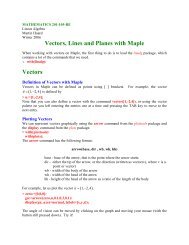
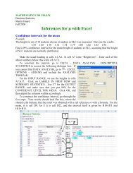
Epidermal tissue covers <strong>the</strong> surface <strong>of</strong><br />
most plant organs. The cells are frequently long<br />
and narrow in surface view (Fig 1.). The<br />
margins <strong>of</strong> <strong>the</strong>se cells vary in outline and may<br />
be straight or curvy, depending on <strong>the</strong> species.<br />
Figure 1. Surface view <strong>of</strong> epidermal cells. (Bracegindle<br />
and Miles. 1977)<br />
One <strong>of</strong> <strong>the</strong> earliest adaptations <strong>of</strong> plants<br />
was <strong>the</strong> production <strong>of</strong> a waxy cuticle by<br />
epidermal cells <strong>of</strong> stems and leaves. Although<br />
<strong>the</strong> cuticle provides protection from<br />
desiccation, it also blocks gas exchange, thus a<br />
complementary early adaptation was <strong>the</strong><br />
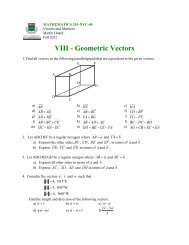
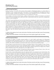
production <strong>of</strong> guard cells. These cells are<br />
produced when two immature epidermal cells<br />
with adjoining end walls divide asymmetrically<br />
to produce two small, adjacent, frequently<br />
kidney-shaped cells. These are guard cells<br />
(Fig. 2.). Early in <strong>the</strong>ir development <strong>the</strong> part <strong>of</strong><br />
<strong>the</strong> wall between adjacent guard cells comes<br />
apart and a pore or stoma (pl. stomata) is<br />
formed which opens directly into <strong>the</strong> interior<br />
<strong>of</strong> <strong>the</strong> leaf. Guard cells control <strong>the</strong> size <strong>of</strong> <strong>the</strong><br />
stomata openings and hence <strong>the</strong> exchange <strong>of</strong><br />
CO 2 , oxygen and water vapor across leaf and<br />
stem surfaces.<br />
<strong>Anatomy</strong> <strong>of</strong> <strong>the</strong> <strong>Pea</strong> <strong>Plant</strong> 1
guard cell<br />
stomatal pore<br />
cytoplasm<br />
nuclei<br />
vacuole<br />
chloroplasts<br />
nucleus<br />
chloroplasts<br />
Figure 2. Stomata and <strong>the</strong>ir guard cells. (Bracegindle<br />
and Miles. 1977)<br />
Parenchyma Tissue<br />
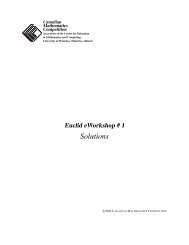
Most <strong>of</strong> <strong>the</strong> plant cells you will see in<br />
your thin sections will be parenchyma tissue.<br />
These cells always contain one or more large<br />
vacuoles and <strong>the</strong>ir cytoplasm is limited to a<br />
narrow peripheral layer and thin cross strands<br />
(Fig 3.). The walls <strong>of</strong> <strong>the</strong>se cells are thin and<br />
are composed mainly <strong>of</strong> cellulose and pectin.<br />
Check <strong>the</strong> staining key (Table 2, Hand Cut<br />
Sections) to guess what color <strong>the</strong>y stain with<br />
toluidine blue.<br />
Most <strong>of</strong> <strong>the</strong> parenchyma cells in <strong>the</strong><br />
above-ground parts <strong>of</strong> plants contain<br />
chloroplasts and are thus sites <strong>of</strong><br />
photosyn<strong>the</strong>sis. Both green and non-green<br />
parenchyma cells store metabolites, notably<br />
starch.<br />
The vascular tissues: xylem and phloem<br />
Ano<strong>the</strong>r early plant adaptation to land<br />
was <strong>the</strong> development <strong>of</strong> vascular tissue which<br />
allows <strong>the</strong> transport <strong>of</strong> nutrients and water, as<br />
well as provides support.<br />
Figure 3. Parenchyma cells. (Bracegindle and Miles. 1977)<br />
Xylem tissue is unique in that when it<br />
is mature it contains mainly dead cells. In<br />
general, xylem is composed <strong>of</strong> long, narrow,<br />
tubular cells. When <strong>the</strong> cells are young (and<br />
still alive) <strong>the</strong>ir walls (called primary wall) are<br />
quite thin and stain pink with toluidine blue<br />
When <strong>the</strong>se cells mature (i.e. <strong>the</strong>y are no<br />
longer enlarging), <strong>the</strong>y produce a secondary<br />
wall which is <strong>of</strong>ten laid down in a spiral<br />
pattern. Eventually, lignin appears in xylem.<br />
This hard, decay-resistant polymer provides<br />
strength to plant organs. The ultimate<br />
development <strong>of</strong> xylem occurs in trees where<br />
most <strong>of</strong> <strong>the</strong> wood is composed <strong>of</strong> dead xylem<br />
tubes. Lignified walls stain green with<br />
toluidine blue.<br />
Phloem is a complex tissue composed<br />
<strong>of</strong> a number <strong>of</strong> cell types, <strong>the</strong> most important<br />
being sieve tubes and companion cells. In<br />
some plants <strong>the</strong> phloem contains long heavywalled<br />
phloem fibers. Phloem is found<br />
throughout a plant in association with xylem.<br />
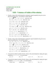
Sieve tubes are narrow, elongated,<br />
roughly cylindrical cells which remain alive at<br />
<strong>Anatomy</strong> <strong>of</strong> <strong>the</strong> <strong>Pea</strong> <strong>Plant</strong> 2
maturity. They are however, unique cells in<br />
several respects. At maturity <strong>the</strong>y lose <strong>the</strong>ir<br />
nuclei (living, functioning enucleated cells are<br />
rare in ei<strong>the</strong>r plants or animals). Maturing<br />
sieve tubes produce enzymes which dissolve<br />
portions <strong>of</strong> <strong>the</strong>ir end walls to produce a sievelike<br />
plate (sieve plate) between adjacent cells<br />
in each file <strong>of</strong> sieve tubes (Fig. 4.).<br />
Figure 4. Phloem tissue. (Bracegindle and Miles. 1977)<br />
The side walls <strong>of</strong> sieve tubes are thicker<br />
than those <strong>of</strong> parenchyma cells but unlike<br />
those <strong>of</strong> xylem cells <strong>the</strong>y are <strong>of</strong> uniform<br />
thickness and do not become lignified. The<br />
sieve tube walls are rich in cellulose but<br />
contain ra<strong>the</strong>r little pectin so are relatively<br />
unstained by toluidine blue.<br />
In most plants (angiosperms) each sieve<br />
tube has beside it a companion cell. These are<br />
smaller in diameter and shorter than <strong>the</strong> sieve<br />
tube and intimately connected with it by<br />
numerous plasmodesmata (holes in <strong>the</strong> cell<br />
wall which connect adjacent cells). The<br />
companion cell is alive at maturity and<br />
contains a nucleus, plastids (usually<br />
chloroplasts), mitochondria, abundant<br />
ribosomes and <strong>of</strong>ten many small vacuoles.<br />
Phloem fibers are narrow, highly<br />
elongated cells with tapered ends which are<br />
dead at maturity. Before death <strong>the</strong>se cells<br />
secrete a uniformly thick secondary wall which<br />
becomes lignified. The lignin <strong>of</strong> phloem fibers<br />
appears to be different chemically from that <strong>of</strong><br />
xylem walls since it stains a bright, light blue<br />
color with toluidine blue.<br />
See your textbook for more photos and<br />
information on plant anatomy.<br />
Note:<br />
• tissues are composed <strong>of</strong> more than one<br />
type <strong>of</strong> cell<br />
• organs are composed <strong>of</strong> more than one type<br />
<strong>of</strong> tissue<br />
References<br />
Bracegindle and Miles. 1977. An Atlas <strong>of</strong> <strong>Plant</strong> Structure Vol.<br />
1. London: Heinemann Educational Books Ltd.<br />
<strong>Anatomy</strong> <strong>of</strong> <strong>the</strong> <strong>Pea</strong> <strong>Plant</strong> 3