Systematic revision of the Family Micrococcidae (Homoptera ...
Systematic revision of the Family Micrococcidae (Homoptera ...
Systematic revision of the Family Micrococcidae (Homoptera ...

Create successful ePaper yourself
Turn your PDF publications into a flip-book with our unique Google optimized e-Paper software.
- 202 <br />
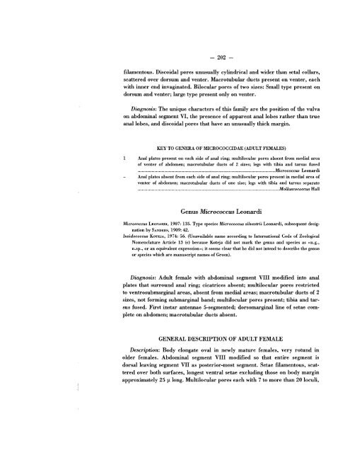
filamentous. Discoidal pores unusually cylindrical and wider than setal collars,<br />
scattered over dorsum and venter. Macrotubular ducts present on venter, each<br />
with inner end invaginated. Bilocular pores <strong>of</strong> two sizes: Small type present on<br />
dorsum and venter; large type present only on venter.<br />
Diagnosis: The unique characters <strong>of</strong> this family are <strong>the</strong> position <strong>of</strong> <strong>the</strong> vulva<br />
on abdominal segment VI, <strong>the</strong> presence <strong>of</strong> apparent anal lobes ra<strong>the</strong>r than true<br />
anal lobes, and discoidal pores that have an unusually thick margin.<br />
KEY TO GENERA OF MICROCOCCIDAE (ADULT FEMALES)<br />
1<br />
Anal plates present on each side <strong>of</strong> anal ring; multilocular pores absent from medial area<br />
<strong>of</strong> venter <strong>of</strong> abdomen; macrotubular ducts <strong>of</strong> 2 sizes; legs with tibia and tarsus fused<br />
.........................•..•....................................................................Micrococcus Leonardi<br />
Anal plates absent from each side <strong>of</strong> anal ring; multilocular pores present in medial area <strong>of</strong><br />
venter <strong>of</strong> abdomen; macrotubular ducts <strong>of</strong> one size; legs with tibia and tarsus separate<br />
...................................................................................•................Molluscococcus Hall<br />
Genus Micrococcus Leonardi<br />
Micrococcus LEONARDI, 1907: 135. Type species Micrococcus silvestrii Leonardi, subsequent designation<br />
by SANDERS, 1909: 42.<br />
Ixeidococcus KOTEJA, 1974: 56. (Unavailable name according to International Code <strong>of</strong> Zoological<br />
Nomenclature Article 13 (c) because Koteja did not mark <strong>the</strong> genus and species as «n.g.,<br />
n.sp., or an equivalent expression l>; it seems clear that he did not intend to describe <strong>the</strong> genus<br />
or species which are manuscript names <strong>of</strong> Green).<br />
Diagnosis: Adult female with abdominal segment VIII modified into anal<br />
plates that surround anal ring; cicatrices absent; multilocular pores restricted<br />
to ventrosubmarginal areas, absent from medial areas; macrotubular ducts <strong>of</strong> 2<br />
sizes, not forming submarginal band; multilocular pores present; tibia and tarsus<br />
fused. First instar antennae 5-segmented; dorsomarginal line <strong>of</strong> setae complete<br />
on abdomen; macrotubular ducts absent.<br />
GENERAL DESCRIPTION OF ADULT FEMALE<br />
Description: Body elongate oval in newly mature females, very rotund III<br />
older females. Abdominal segment VIII modified so that entire segment is<br />
dorsal leaving segment VII as posterior-most segment. Setae filamentous, scattered<br />
over both surfaces, longest ventral setae excluding those on body margin<br />
approximately 25 fA. long. Multilocular pores each with 7 to more than 20 loculi,