Systematic revision of the Family Micrococcidae (Homoptera ...
Systematic revision of the Family Micrococcidae (Homoptera ...
Systematic revision of the Family Micrococcidae (Homoptera ...

You also want an ePaper? Increase the reach of your titles
YUMPU automatically turns print PDFs into web optimized ePapers that Google loves.
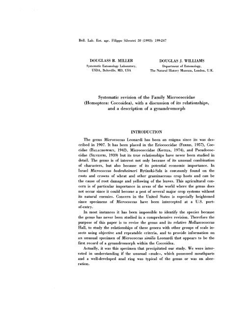
Boll. Lab. Ent. agr. Filippo Silvestri 50 (1993): 199-247<br />
DOUGLASS R. MILLER<br />
<strong>Systematic</strong> Entomology Laboratory,<br />
USDA, Beltsville, MD, USA<br />
DOUGLAS J. WILLIAMS<br />
Department <strong>of</strong> Entomology,<br />
The Natural History Museum, London, U.K.<br />
<strong>Systematic</strong> <strong>revision</strong> <strong>of</strong> <strong>the</strong> <strong>Family</strong> <strong>Micrococcidae</strong><br />
(<strong>Homoptera</strong>: Coccoidea), with a discussion <strong>of</strong> its relationships,<br />
and a description <strong>of</strong> a gynandromorph<br />
INTRODUCTION<br />
The genus Micrococcus Leonardi has been an enigma since its was described<br />
in 1907. It has been placed in <strong>the</strong> Eriococcidae (FERRIS, 1957), Coccidae<br />
(BALACHOWSKY, 1942), <strong>Micrococcidae</strong> (KOTEIA, 1974), and Pseudococcidae<br />
(SILVESTRI, 1939) but its true relationships have never been studied in<br />
detail. The genus is <strong>of</strong> interest not only because <strong>of</strong> its unusual combination<br />
<strong>of</strong> characters, but also because <strong>of</strong> its potential economic importance. In<br />
Israel Micrococcus bodenheimeri Bytinski-Salz is com.monly found on <strong>the</strong><br />
roots and crowns <strong>of</strong> wheat and o<strong>the</strong>r graminaceous crop hosts and can be<br />
<strong>the</strong> cause <strong>of</strong> root damage and yellowing <strong>of</strong> <strong>the</strong> leaves. This agricultural concern<br />
is <strong>of</strong> particular importance in areas <strong>of</strong> <strong>the</strong> world where <strong>the</strong> genus does<br />
not occur since it could become a pest <strong>of</strong> several major crop systems without<br />
its natural enemies. Concern in <strong>the</strong> United States is especially heightened<br />
since specimens <strong>of</strong> Micrococcus have been intercepted at a U.S. port<strong>of</strong>-entry.<br />
In most instances it has been impossible to identify <strong>the</strong> species because<br />
<strong>the</strong> genus has never been studied in a comprehensive <strong>revision</strong>. Therefore <strong>the</strong><br />
purpose <strong>of</strong> this paper is to revise <strong>the</strong> genus and its relative Molluscococcus<br />
Hall, to study <strong>the</strong> relationships <strong>of</strong> <strong>the</strong>se genera with o<strong>the</strong>r groups <strong>of</strong> scale insects<br />
using objective and repeatable criteria, and to provide information on<br />
an unusual specimen <strong>of</strong> Micrococcus similis Leonardi that appears to be <strong>the</strong><br />
first record <strong>of</strong> a gynandromorph within <strong>the</strong> Coccoidea.<br />
Actually, it was this specimen that precipitated our study. We were interested<br />
in understanding if <strong>the</strong> unusual «male», which possessed mouthparts<br />
and a well-developed anal ring was typical <strong>of</strong> <strong>the</strong> genus or was an aberration.
- 200<br />
METHODS AND TERMINOLOGY<br />
Terminology follows that <strong>of</strong> WILLIAMS & GRANARA DE WILLINK (1992) and McKENZIE (1967)<br />
for adult females, AFlFl (1968) and GlLlOMEE (1967) for adult males, and MILLER (1991) for first,<br />
second, and third instal's. Specialized teI'minology for <strong>the</strong> <strong>Micrococcidae</strong> includes <strong>the</strong> anal plates,<br />
multilocular pores, intrastigmatic pores, parastigmatic pores, cicatrices, and apparent anal lobes.<br />
The anal plates develop from <strong>the</strong> anal lobes in <strong>the</strong> first instar. In <strong>the</strong> immatures and young adult<br />
females <strong>of</strong> Micrococcus <strong>the</strong>y appear as two dorsally projecting structures that are heavily sclerotized<br />
and are located laterad <strong>of</strong> <strong>the</strong> anal ring. In old adult females, <strong>the</strong> anal plates become fused<br />
and completely surround <strong>the</strong> anal ring. Multilocular pores have many loculi that surround a<br />
central hub <strong>of</strong> loculi giving <strong>the</strong> appearance <strong>of</strong> a sieve. These pores are derived from pores in <strong>the</strong><br />
earlier instal's, that have a solid central hub and no central loculi. Intrastigmatic pores are<br />
multilocular pores that occur in <strong>the</strong> atrium <strong>of</strong> <strong>the</strong> spiracle or on <strong>the</strong> sclerotized plate surrounding<br />
<strong>the</strong> spiracle. Parastigmatic pores are multilocular pores that occur on <strong>the</strong> derm surrounding <strong>the</strong><br />
spiracles and form a band near <strong>the</strong> body margin; this band may extend from <strong>the</strong> head, past <strong>the</strong><br />
spiracles, to <strong>the</strong> abdomen, or may be more restricted. Cicatrices are two large circular structures<br />
on <strong>the</strong> dorsal abdomen <strong>of</strong> Molluscococcus. It is unclear if <strong>the</strong>se structures are homologous with <strong>the</strong><br />
anal plates found in Micrococcus; perhaps examination <strong>of</strong> second and third instal's will clarify <strong>the</strong><br />
derivation <strong>of</strong> <strong>the</strong>se structures. The apparent anal lobes are slightly protruding structures located<br />
at <strong>the</strong> posterior apex <strong>of</strong> <strong>the</strong> abdomen that contain several long setae. They are part <strong>of</strong> abdominal<br />
segment 7, ra<strong>the</strong>r than segment 8, and are not homologus to <strong>the</strong> anal lobes <strong>of</strong> o<strong>the</strong>r scale insects.<br />
Discoidal pores in all species are usually wider than <strong>the</strong> setal collars, heavily sclerotized and<br />
cylindrical. According to MAROTIA et al. (1995) <strong>the</strong>y have a sieve like structure when being<br />
studied with <strong>the</strong> scanning electron microscope.<br />
Measurements and numbers are from 10 specimens when available and are given as a mean<br />
with ranges in paren<strong>the</strong>ses. Phylogenetic analyses were performed using Hennig 86 (FARRIS,<br />
1988). The functions mhennig «mh*», branch swapping «bb», and ultimately implicit enumeration<br />
- 201 <br />
femaLe, and first instar specimens were examined and were supplemented by information in<br />
MILLER & LAMBDIN (1978); Lecanodiaspis acaciae (Maskell) (Lecanodiaspididae) - female and first<br />
instar specimens were examined and were supplemented with information in HOWELL &<br />
KOSZTARAB (1972), AFlFI & KOSZTARAB (1969), and WILLIAMS & KOSZTARAB (1970); Micrococcus<br />
bodenheimeri Bytinski-Salz (<strong>Micrococcidae</strong>) - female, male, and first instar specimens were examined;<br />
Molluscococcus fibrillae Hall (<strong>Micrococcidae</strong>) - female and first instar specimens were<br />
examined; Pollinia pollini (Costa) (Asterolecaniidae) - female, male, and first instar specimens<br />
were examined; Nanokermes pubescens (Bogue) (Kermesidae) - female, male, and first instar<br />
specimens were examined and were supplemented with information in BULLINGTON & KOSZTARAB<br />
(1985) and BAER & KOSZTARAB (1985); Pulvinaria acericola (Walsh and Riley) (Coccidae) <br />
female, male, and first instar specimens were examined and were supplemented with information<br />
in GILIOMEE (1967); Puto kosztarabi Miller et Miller (Pseudococcidae) - female, male, and first instar<br />
specimens were examined and were supplemented with information in MILLER & MILLER<br />
(1993b); Sphaerolecanium prunastri (Fonscolombe) (Coccidae) - female, male, and first instar<br />
specimens were examined and were supplemented with information in GILIOMEE (1967); Tachardiella<br />
larreae Chamberlin (Tachardiidae) - female and first instar specimens were examined along<br />
with adult males <strong>of</strong> Tachardina aurantiaca Cockerell, which were supplemented with information<br />
in CHAMBERLIN (1923).<br />
Abbreviations <strong>of</strong> depositories are as follows: The Natural History Museum, London, United<br />
Kingdom (BMNH); The Coccoidea Collection, Department <strong>of</strong> Entomology, The Volcani Center,<br />
Bet Dagan, Israel (ICV); Museum National d'Histoire Naturelle, Paris, France (MNHN); University<br />
<strong>of</strong> California, Bohart Collection, Davis, California (UCD); United States National Museum <strong>of</strong><br />
Natural History, Beltsville, Maryland (USNM).<br />
<strong>Family</strong> <strong>Micrococcidae</strong> Silvestri<br />
<strong>Family</strong> <strong>Micrococcidae</strong> Silvestri.<br />
Type genus Micrococcus LEONARDI, 1907: 135.<br />
Micrococci SILVESTRI, 1939: 702.<br />
Micrococcinae Silvestri; BALACHOWSKY, 1942: 43.<br />
Micrococcini Silvestri; BALACHOWSKY, 1948: 255.<br />
<strong>Micrococcidae</strong> Silvestri; KOTEJA, 1974: 56.<br />
Description: Body <strong>of</strong> adult female elongate oval to broadly oval. Antennae<br />
with fewer than 4 segments, basal segment widest, third segment elongate, bluntly<br />
conical. Labium I-segmented, rectangular in shape, with 5 pairs <strong>of</strong> setae on<br />
each side. Legs well developed, without translucent pores, with sensoria on<br />
trochanter arranged diagonally towards proximal end (Fig. 1); tarsus without<br />
basal sensillum; tarsal and claw digitules conspicuous and capitate. Anal ring<br />
dorsal with 10-28 setae and numerous pores. Apparent anal lobes each with<br />
marginal setae longer than o<strong>the</strong>r marginal setae. Vulva situated on segment VI.<br />
Spiracles well developed with numerous intraspiracular multilocular pores and<br />
with or without parastigmatic multilocular pores, <strong>the</strong>se sometimes extending<br />
almost around entire margin or associated only with spiracles. Setae normally
- 202 <br />
filamentous. Discoidal pores unusually cylindrical and wider than setal collars,<br />
scattered over dorsum and venter. Macrotubular ducts present on venter, each<br />
with inner end invaginated. Bilocular pores <strong>of</strong> two sizes: Small type present on<br />
dorsum and venter; large type present only on venter.<br />
Diagnosis: The unique characters <strong>of</strong> this family are <strong>the</strong> position <strong>of</strong> <strong>the</strong> vulva<br />
on abdominal segment VI, <strong>the</strong> presence <strong>of</strong> apparent anal lobes ra<strong>the</strong>r than true<br />
anal lobes, and discoidal pores that have an unusually thick margin.<br />
KEY TO GENERA OF MICROCOCCIDAE (ADULT FEMALES)<br />
1<br />
Anal plates present on each side <strong>of</strong> anal ring; multilocular pores absent from medial area<br />
<strong>of</strong> venter <strong>of</strong> abdomen; macrotubular ducts <strong>of</strong> 2 sizes; legs with tibia and tarsus fused<br />
.........................•..•....................................................................Micrococcus Leonardi<br />
Anal plates absent from each side <strong>of</strong> anal ring; multilocular pores present in medial area <strong>of</strong><br />
venter <strong>of</strong> abdomen; macrotubular ducts <strong>of</strong> one size; legs with tibia and tarsus separate<br />
...................................................................................•................Molluscococcus Hall<br />
Genus Micrococcus Leonardi<br />
Micrococcus LEONARDI, 1907: 135. Type species Micrococcus silvestrii Leonardi, subsequent designation<br />
by SANDERS, 1909: 42.<br />
Ixeidococcus KOTEJA, 1974: 56. (Unavailable name according to International Code <strong>of</strong> Zoological<br />
Nomenclature Article 13 (c) because Koteja did not mark <strong>the</strong> genus and species as «n.g.,<br />
n.sp., or an equivalent expression l>; it seems clear that he did not intend to describe <strong>the</strong> genus<br />
or species which are manuscript names <strong>of</strong> Green).<br />
Diagnosis: Adult female with abdominal segment VIII modified into anal<br />
plates that surround anal ring; cicatrices absent; multilocular pores restricted<br />
to ventrosubmarginal areas, absent from medial areas; macrotubular ducts <strong>of</strong> 2<br />
sizes, not forming submarginal band; multilocular pores present; tibia and tarsus<br />
fused. First instar antennae 5-segmented; dorsomarginal line <strong>of</strong> setae complete<br />
on abdomen; macrotubular ducts absent.<br />
GENERAL DESCRIPTION OF ADULT FEMALE<br />
Description: Body elongate oval in newly mature females, very rotund III<br />
older females. Abdominal segment VIII modified so that entire segment is<br />
dorsal leaving segment VII as posterior-most segment. Setae filamentous, scattered<br />
over both surfaces, longest ventral setae excluding those on body margin<br />
approximately 25 fA. long. Multilocular pores each with 7 to more than 20 loculi,
203 <br />
with single central loculus or more than 1 central loculus. Parastigmatic<br />
pores, when present, comprising multilocular pores, occurring outside <strong>of</strong><br />
stigmatic plate. Intrastigmatic pores comprising multilocular pores, restricted<br />
to sclerotized plates surrounding spiracular atria. Invaginated tubular ducts<br />
<strong>of</strong> 2 sizes: broader ducts present on medial and submarginal areas <strong>of</strong> abdomen<br />
and metathorax; narrower ducts on medial and submarginal areas <strong>of</strong><br />
abdomen and head. Anal lobes modified into anal plates that surround anal<br />
ring. Anal lobe-like structures developed on segment VII with several long<br />
setae giving appearance <strong>of</strong> normal lobes. Legs with tibia and tarsus fused.<br />
GENERAL DESCRIPTION OF THIRD INSTAR FEMALE<br />
Description: Same as adult female except as follows. Body elongate oval.<br />
Longest ventral setae excluding those on body margin less than 25 (J. long.<br />
Multilocular pores each with 5 to more than 15 loculi. Parastigmatic pores<br />
usually absent. Invaginated tubular ducts absent. Vulva absent. Anal ring<br />
with more than 4 setae on each side; anal-ring pores in 2 rows.<br />
GENERAL DESCRIPTION OF SECOND INSTAR FEMALE<br />
Description: Same as third instar female except as follows. Multilocular<br />
pores each with 5 to more than 15 loculi. Parastigmatic pores present. Anal<br />
ring with fewer than 5 setae on each side; anal-ring pores absent or in 1<br />
row.<br />
GENERAL DESCRIPTION OF FIRST INSTAR<br />
Description: Body elongate. Abdominal segment VIII modified into sclerotized<br />
lobes which transform into plates in later instars; each lobe with a<br />
sclerotized bar and long apical seta. Antennae 5-segmented. Setae filamentous,<br />
with 2 pairs <strong>of</strong> longitudinal lines on each side <strong>of</strong> dorsum, with 3 pairs<br />
<strong>of</strong> lines on each side <strong>of</strong> venter. Bilocular pores absent. Multilocular pores<br />
with 5 to 9 loculi, without central loculus. Parastigmatic and intrastigmatic<br />
pores present. Discoidal pores present on both surfaces. Invaginated tubular<br />
ducts absent. Microtubular ducts present on submarginal area <strong>of</strong> venter,<br />
with conspicuous dermal orifice. Legs without sensillum on tarsus; tibia and<br />
tarsus separate; tibia divided by tarsus about 1.6; tarsal digitules staggered,<br />
not arranged side-by-side, apices acute or slightly enlarged; tarsal digitules<br />
on front legs each with 1 small filamentous seta and 1 enlarged seta; claw<br />
digitules each with 1 large seta, and 1 conspicuously smaller seta.
- 204 <br />
GENERAL DESCRIPTION OF ADULT MALE OF MICROCOCCUS<br />
Description: Without wings and sclerotization on head and thorax. Head<br />
with 1 pair <strong>of</strong> eyes. Body elongate. Abdominal segment VIII with sclerotized<br />
lobes. Labium absent, clypeolabral shield present. Antennae 3-segmented.<br />
Setae filamentous, with 3 indefinite, longitudinal lines on each side <strong>of</strong> dorsum.<br />
Bilocular pores <strong>of</strong> 2 sizes, smaller size associated with setal clusters on dorsum,<br />
and on lateral areas <strong>of</strong> venter; larger size scattered over venter. Multilocular<br />
pores absent. Parastigmatic and intrastigmatic pores absent. Discoidal pores<br />
present on both surfaces. Invaginated tubular ducts absent. Microtubular ducts<br />
absent. Penial sheath forming complete capsule, partially divided dorsally.<br />
Aedeagus tubular, with blunt apex. Legs with tibia and tarsus fused; tarsus<br />
without basal sensillum; tarsal and claw digitules conspicuous and capitate; tarsal<br />
digitules with 1 digitule conspicuously smaller than o<strong>the</strong>r.<br />
KEY TO INSTARS<br />
1 Antennae 2- or 3-segmented; tibia and tarsus at least partially fused 2<br />
Antennae 5- or 6-segmented; tibia and tarsus separate<br />
first instar<br />
2 (1) Apex <strong>of</strong> abdomen without aedeagus and penial sheath 3<br />
Apex <strong>of</strong> abdomen with aedeagus and penial sheath<br />
adult male<br />
3 (2) Anal ring with more than 4 setae on each side 4<br />
Anal ring with fewer than 5 setae on each side<br />
second instar female<br />
4 (3) Vulva and tubular ducts present adult female<br />
Vulva and tubular ducts absent<br />
third instar female<br />
KEY TO ADULT FEMALES<br />
1 Parastigmatic pores absent or restricted to thorax 2<br />
Parastigmatic pores present on thorax and abdomen 5<br />
2 (1) Parastigmatic pores absent; with fewer than 10 setae on each anal plate 3<br />
Parastigmatic pores near spiracular plates; with more than 10 setae on each anal plate .<br />
.........................................................................................................similis Leonardi<br />
3 (2) Some marginal discoidals oval, narrow area <strong>of</strong> oval sclerotized in shape <strong>of</strong> an eye (we have<br />
not seen adults <strong>of</strong> this species but assume that this statement will be true on adults as well<br />
as immatures)<br />
rungsi Balachowsky<br />
Marginal discoidals round, or if oval without sclerotization in shape <strong>of</strong> an eye .4<br />
4 (3) Antennae 3-segmented; longest seta on apparent anal lobes 237 (172·306) fllong ..<br />
. .bodenheimeri Bytinski-Salz<br />
Antennae 2-segmented (segments 3 and 2 partially or completely fused); longest seta on apparent<br />
anal lobes 90 (83-96) fl<br />
dumonti Balachowsky<br />
5 (1) Longest dorsal seta on metathorax longer than 100 fl; more than 8 setae on each hind<br />
femur<br />
.longispinus Miller et Williams<br />
Longest dorsal seta on metathorax shorter than 100 fl; 8 setae or fewer on each hind<br />
femur 6<br />
6 (5) Tibia+tarsus more than 300 fllong; with more than 5 parastigmatic pores near each lateral
- 205 <br />
margin <strong>of</strong> abdominal segment IV; longest seta on anal plates longer than 50 fl. .<br />
......................................................................................................silvestrii Leonardi<br />
Tibia +tarsus less than 300 fI. long; with fewer than 5 sieve pores near each lateral margin<br />
<strong>of</strong> abdominal segment IV; longest seta on anal plates less than 50 fl. ..<br />
. ..confusus Miller et Williams<br />
KEY TO SECOND INSTAR FEMALES<br />
1<br />
2 (1)<br />
With pores in anal ring<br />
Without pores in anal ring<br />
Parastigmatic pores present near anterior spiracles<br />
Parastigmatic pores absent near anterior spiraclcs<br />
2<br />
silvestrii Leonardi<br />
rungsi Balachowsky<br />
.longispinus Miller et Williams<br />
KEY TO THIRD INSTAR FEMALES<br />
1 Longest dorsal setae less than 100 fl.; dorsomedial setae about same length as remaining<br />
setae 2<br />
Longest dorsal setae more than 100 fl.; dorsomedial setae noticeably longer than remaining<br />
setae<br />
.longispinus Miller et Williams<br />
2 (1) Antenna more than 120 fI. long silvestrii Leonardi and similis Leonardi<br />
Antenna less than 120 fI. long<br />
bodenheimeri Bytinski-Salz<br />
KEY TO FIRST INSTARS<br />
Fewer than 15 parastigmatic pores on each side <strong>of</strong> body; usually without a pore or cluster<br />
<strong>of</strong> pores near body margin between clusters associated with anterior and posterior<br />
spiracles<br />
bodenheimeri Bytinski-Salz<br />
15 or more parastigmatic pores on each side <strong>of</strong> body; with 1 or more pores near body<br />
margin between clusters associated with anterior and posterior spiracles ..<br />
.................................dumonti Balachowsky, silvestrii Leonardi, and rungsi Balachowsky<br />
TREATMENT OF SPECIES<br />
Micrococcus bodenheimeri Bytinski-Salz<br />
Micrococcus bodenheimeri BYTlNSKI-SALZ, 1961: 90-96.<br />
lxeidococcus graminis KOTEJA 1974: 56. (Unavailable name according to <strong>the</strong> International Code <strong>of</strong><br />
Zoological Nomenclature Article 13 (c) because Koteja did not mark <strong>the</strong> genus and species as<br />
- 206<br />
Adult female (Fig. I)<br />
Recognition characters: Mounted, 1.7 (1.5-2.1) mm long, 0.9 (0.5-1.2) mm<br />
wide.<br />
Dorsum with filamentous setae scattered over surface; longest seta on dorsomedial<br />
area <strong>of</strong> metathorax 19 (15-26) fL long, all setae in area about same<br />
length. Marginal setae usually slightly longer than setae on rest <strong>of</strong> dorsum,<br />
some specimens with long marginal setae on any <strong>of</strong> abdominal segments, one<br />
specimen with long marginal setae around entire body. Small bilocular pores<br />
abundant over surface except on posterior one or two segments where "rare or<br />
absent. Discoidal pores oval to circular, scattered over surface, most abundant<br />
in marginal and submarginal areas.<br />
Anal plates each with 5 (4-6) setae; longest seta 38 (29-44) fL long. Anal ring<br />
with 6 (5-6) setae and 36 (29-43) pores on each half. Apparent anal lobes each<br />
with 7 (3-12) setae; longest seta 237 (172-306) fL long.<br />
Venter with small setae arranged in segmental rows, longest seta on abdomen,<br />
excluding marginal ones, 23 (20-26) fL long. Macrotubular ducts <strong>of</strong> two<br />
sizes: Broader size on abdomen and metathorax, absent from marginal areas;<br />
narrower size on head and pro- and mesothorax, absent from marginal areas.<br />
Small bilocular pores scattered over marginal and submedial areas. Large<br />
bilocular pores usually on head, thorax, and anterior abdominal segments,<br />
some specimens with <strong>the</strong>se pores on head and thorax only. Discoidal pores oval<br />
to circular, scattered over surface. Intrastig,iYlatic pores with 7-28 loculi, 71 (62<br />
79) pores associated with each anterior spiracle. Parastigmatic pores absent.<br />
Legs with hind femora each with 6 (5-6) setae; tibia +tarsus with 6 (5-7)<br />
setae; tarsal and claw digitules with conspicuous, clubbed apices, all about<br />
same size; femur 181 (170-189) fL long; tibia +tarsus 230 (210-250) fL long;<br />
femur length divided by tibia +tarsus length 0.8 (0.7-0.8). Antennae 3-segmented,<br />
153 (143-160) fL long; segment III 75 (64-90) fL long, with 11 (10-11)<br />
setae; segment II 31 (26-35) fL long, 67 (58-79) fL wide.<br />
Notes - The description is based on 180 specimens from 21 localities.<br />
The adult female <strong>of</strong> this species differs from most o<strong>the</strong>r species <strong>of</strong> Micrococcus<br />
by lacking parastigmatic pores and by having short appendages. Micrococcus<br />
dumonti also lacks parastigmatic pores and has appendages that are slightly<br />
longer than on M. bodenheimeri, but is distinguished by having 2-segmented<br />
antennae, apparent anal-lobe setae that are about 90 fL long, and <strong>the</strong> longest<br />
anal plate setae that are about 15 fL long. Micrococcus bodenheimeri has 3-segmented<br />
antennae, apparent anal-lobe setae that are about 237 fL long, and <strong>the</strong><br />
longest anal plate setae that are about 38 fL long.
" /0<br />
/:<br />
, //~<br />
,<br />
, j.j<br />
'.': ' ./ .J<br />
~.y!-<br />
".! .<br />
-, ,<br />
• -1 e.<br />
/<br />
/<br />
/.,<br />
...:.<br />
/<br />
/<br />
/<br />
/<br />
/<br />
/<br />
/<br />
/<br />
/<br />
/<br />
.. J• .. :; ....<br />
., "<br />
.. -.<br />
...'.<br />
.' .<br />
.J J ..<br />
J '<br />
, ~,<br />
, /<br />
_. ,/" ..<br />
.. .. .. ." ..<br />
'j<br />
/<br />
--_/<br />
/<br />
/<br />
.! •<br />
J<br />
. '.<br />
,<br />
"-'''0<br />
--- ~<br />
, -<br />
- We:
- 208 <br />
Third instar female (Fig. II)<br />
Recognition characters: Mounted, 1.6 (1.5-1.8) mm long, 0.8 (0.7-0.9) mm<br />
wide.<br />
Dorsum with filamentous setae arranged in 3 pairs <strong>of</strong> longitudinal lines on<br />
each side <strong>of</strong> abdomen, medial, mediolateral, and marginal; longest seta on dorsomedial<br />
area <strong>of</strong> metathorax 10 (9-11) fl long, all setae in area about same<br />
length. Marginal setae <strong>of</strong>ten slightly longer than setae on rest <strong>of</strong> dorsum. Small<br />
bilocular pores scattered over surface except on posterior one or two segments<br />
where rare or absent. Discoidal pores oval or circular, scattered over surface,<br />
most abundant in marginal and submarginal areas.<br />
Anal plates each with 5 setae; longest seta 23 (14-32) fllong. Anal ring with 5<br />
(5-6) setae and 9(6-10) pores on each half. Apparent anal lobes each with 7 (4<br />
10) setae; longest seta 86 (81-91) fllong.<br />
Venter with small setae arranged in segmental rows, longest seta on abdomen,<br />
excluding marginal ones, 17 (15-20) fllong.<br />
Small bilocular pores scattered over surface, absent from posterior 2 or 3<br />
abdominal segments or scarce. Large bilocular pores usually on head, thorax,<br />
and anterior 1 to 3 abdominal segments, some specimens with <strong>the</strong>se pores on<br />
head and thorax only. Discoidal pores oval to circular, scattered over surface,<br />
except absent from medial area <strong>of</strong> posterior 2 or 3 abdominal segments. Intrastigmatic<br />
pores each with 6-10 loculi, 7 (5-8) pores associated with each<br />
anterior spiracle. Parastigmatic pores absent.<br />
Legs with hind femora each with 6 (5-7) setae; tibia +tarsus with 6 (4-7)<br />
setae; tarsal and claw digitules with conspicuous, clubbed apices, all about<br />
same size; femur 113 (111-119) fl long; tibia+tarsus 146 (141-153) fl long;<br />
femur length divided by tibia +tarsus length 0.7 (0.7-0.8). Antennae 3-segmented,<br />
91 (82-96) fl long; segment III 49 (47-52) fl long, with 9 (8-10) setae;<br />
segment II 16 (12-18) fllong, 56 (52-62) fl wide.<br />
Notes - The description is based on 14 specimens from 2 localities.<br />
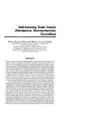
First instar (Sex not determined) (Fig. III)<br />
Recognition characters: Mounted, 0.7 (0.6-0.9) mm long, 0.2 (0.2-0.3) mm<br />
wide.<br />
Dorsum with filamentous setae arranged in 2 longitudinal lines on each side<br />
<strong>of</strong> body, submedial line restricted to segment I, thorax, and head, marginal line<br />
present around perimeter <strong>of</strong> body; longest seta on dorsomedial area <strong>of</strong><br />
metathorax 11 (10-14) fl long, all setae in area about same length. Marginal
,"<br />
"<br />
l!<br />
I<br />
"l<br />
-- I',<br />
,<br />
.\. .\ ,<br />
\ \.. ..<br />
, ,<br />
-\---- ~---I "<br />
,<br />
, , '<br />
~ ~ .. i<br />
,<br />
,<br />
., :,------ c5/<br />
,.<br />
.. '..<br />
. "<br />
@) - - -- - -- ~. ;!~ -'- - -{- <br />
:~ '< . \.'[:<br />
..:: ~<br />
~ .-'. . .<br />
: ,'/ .<br />
, <br />
~- - .<br />
- ~.<br />
.~....<br />
'.,<br />
'.<br />
- 60Z
- 210 <br />
~<br />
0"<br />
~'<br />
.<br />
,.<br />
e,e *<br />
fJ<br />
1------<br />
i' '.<br />
--------',.<br />
/<br />
,/<br />
"""'~<br />
------v===v-<br />
OJ/<br />
Fig. III - Micrococcus bodenheimeri Bytinski-Salz. - First instar.<br />
d<br />
~<br />
setae usually about same length as rest <strong>of</strong> dorsum. Discoidal pores arranged in<br />
2 pairs <strong>of</strong> longitudinal lines on each side <strong>of</strong> body, mediolateral line variable,<br />
comprising 1 to 4 pores on any <strong>of</strong> abdominal segments II to VII, marginal line<br />
present around perimeter <strong>of</strong> body.
- 211 <br />
Anal lobes (plates) each with 3 (3 or 4) setae; longest seta 352 (260-424) fL<br />
long. Anal ring with 3 (8-11) pores on each half.<br />
Venter with small setae and 9 setae arranged in segmental lines, longest seta<br />
on abdomen, excluding marginal ones, 18 (14-23) fL long. Microtubular ducts<br />
present in 1 submarginal line on each side <strong>of</strong> body, with 2 ducts present on<br />
each segment. Discoidal pores in mediolateral and marginal areas <strong>of</strong> body,<br />
forming 2 pairs <strong>of</strong> indistinct longitudinal lines on each side <strong>of</strong> body.<br />
Intrastigmatic pores without central loculi, with 5-7 loculi, with 1 pore in<br />
each spiracle. Parastigmatic pores near each spiracle and near hind pair <strong>of</strong><br />
legs, without central loculi, with 5-9 loculi, with 9 (7-11) pores on each side <strong>of</strong><br />
body. Two specimens with 1 pore on 1 side <strong>of</strong> body between clusters near<br />
anterior and posterior spiracular clusters.<br />
Legs with hind femora each with 3 setae; tibiae with 3; tarsi with 4; tarsal<br />
digitule on hind 2 pairs <strong>of</strong> legs with acute or slightly swollen apices, front pair<br />
with 1 digitule small and filamentous and 1 digitule larger and slightly<br />
enlarged; claw digitules unequal in size, 1 with conspicuously swollen base and<br />
clubbed apex, 1 filamentous with acute or slightly swollen apex; femur 96 (84<br />
116) fL long; tibia 95 (79-126) fL long; tarsus 60 (52-69) fL long; femur length<br />
divided by tibia +tarsus length 0.6 (0.5-0.7). Antennae 130 (119-140) fL long;<br />
segments III and Veach with 1 conspicuously long seta, longest seta on segment<br />
V 210 (195-227) fL long.<br />
Notes - The description is based on 30 specimens from 3 localities.<br />
This material includes 6 specimens that are conspicuously larger than <strong>the</strong><br />
rest <strong>of</strong> <strong>the</strong> series. They have legs and antennae that are approximately <strong>the</strong> same<br />
size as those described above, and we suspect that <strong>the</strong>y are <strong>the</strong> form that aestivates<br />
during <strong>the</strong> dry season. These specimens are approximately 2 mID long and<br />
1 mm wide and differ from those described above by having <strong>the</strong> orifices <strong>of</strong> <strong>the</strong><br />
microtubular ducts, <strong>the</strong> parastigmatic pores, and <strong>the</strong> anal lobes much more<br />
heavily sclerotized.<br />
Adult male (Fig. IV)<br />
Recognition characters: Mounted, 1.9 (1.6-2.3) mm long, 0.7 (0.6-0.7) mm<br />
wide.<br />
Dorsum with filamentous setae arranged in 3 longitudinal lines on each side<br />
<strong>of</strong> abdomen, medial, mediolateral, and marginal; with 6 (6-8) setae on<br />
metathorax; longest seta on dorsomedial area <strong>of</strong> metathorax 17 (14-23) fL long,<br />
all setae in area about same length. Marginal setae usually about same length as<br />
rest <strong>of</strong> dorsum. Small bilocular pores uncommon, most prevalent near body
- 212 <br />
0-_<br />
~<br />
... ',.<br />
~"': ..<br />
. .<br />
"<br />
'." " "<br />
'1· "<br />
" ~"....,.<br />
"<br />
._-:[)\<br />
-..:' 1<br />
: ::> .....<br />
t.<br />
1:;-0<br />
:...<br />
/". '<br />
··/':F<br />
/<br />
f •<br />
.",,:.<br />
~.<br />
" .."".<br />
" ".l ' _ ..<br />
.." ."...<br />
~<br />
_~O<br />
d50,<br />
.: :'f " 'i.<br />
--@j<br />
11l<br />
j/I1l/'<br />
.~ i •<br />
.. ll1l<br />
~-------<br />
".'<br />
..<br />
I":;::'.<br />
/;<br />
.. ~.: .. • i<br />
1/'.<br />
.:.. \,<br />
/' .<br />
~-<br />
.. "<br />
~ ....<br />
- I'-'~~<br />
.,,11l<br />
i'<br />
.. l1li .<br />
"<br />
~<br />
4D.<br />
w-~<br />
11l/ ..<br />
.-------i<br />
; i<br />
4D.<br />
---0:<br />
~<br />
0<br />
///<br />
111<br />
/<br />
Fig. IV - Micrococcus bodenheimeri Bytinski-Salz, - Adult male,<br />
~<br />
""0
- 213 <br />
marginodorsal setae. Discoidal pores oval to circular, scattered over surface,<br />
most abundant near body margin.<br />
Anal lobes sclerotized, joined by sternite on segment VIII, developed into<br />
oval process, each with 2 (1-3) setae; longest seta about 22 (21-27) fLlong. Anal<br />
ring without setae or pores. Penial sheath partially divided into 2 segments on<br />
ventral surface, 320 (193-398) fL long. Aedeagus apically blunt, tubular, 130<br />
(92-161) fLlong.<br />
Venter with setae arranged in segmental rows, longest seta on abdomen, excluding<br />
marginal ones, 22 (18-25) fL long. Small biloculars in small numbers<br />
near body marginal. Large bilocular pores on head, thorax, and abdomen, most<br />
abundant on thorax. Discoidal pores oval to circular, scattered over surface.<br />
Intrastigmatic and parastigmatic pores absent. Abdominal sternites noticeably<br />
sclerotized towards posterior edges.<br />
Legs with hind femora each with 19 (18-20) setae; tibia +tarsus with 25 (17<br />
28) setae; tarsal and claw digitules with clubbed apices, all about same size;<br />
femur 214 (175-262) fL long; tibia +tarsus 218 (184-265) fL long; femur length<br />
divided by tibia +tarsus length 1.0 (0.9-1.0). Antennae 3-segmented, 167 (131<br />
200) fLlong; segment III 94 (78-104) fLlong, with 26 (21-32) setae; segment II 32<br />
(28-39) fLlong, 86 (81-94) fL wide.<br />
Notes - The description is based on 4 specimens from 1 locality.<br />
One specimen is quite different from <strong>the</strong> o<strong>the</strong>rs by having unsually small appendages.<br />
Type material: The holotype is <strong>the</strong> left specimen <strong>of</strong> two mounted on a slide that is labelled as<br />
follows: Left label «C:2807,/1, Micrococcus/bodenheimerilByS./holotypus ad leftlParatypus ad»;<br />
right label«on grass roots/lSRAEL,trivon 6.1I.56/AgByS.» (lCV). There also is an allotype with<br />
<strong>the</strong> same data.<br />
Specimens examined: Cyprus, Kyrenia, 1-16-1932, II-4-1932, I11-24-1932, IV-1l-1932, IV-13<br />
1932, IV-24-1932, on roots <strong>of</strong> grass, E.E. Green, 45 adult females, 7 third instars, and 6 first instars<br />
on 25 slides (BMNH, UCD); Famagusta, 111-9-1932, IV--1932, on roots <strong>of</strong> grass, T.<br />
Shiakides, 6 adult females, 16 first instars on 5 slides (BMNH); Lefkonico Pass, 11-22-1968, on<br />
roots <strong>of</strong> wheat, G.P. Georghiou, 8 adult females on 2 slides (BMNH); Nicosia, --1984, on wheat<br />
roots, 2 adult females on 2 slides (BMNH); Limasol, 11-28-1932, on roots <strong>of</strong> grasses, E.E. Green,<br />
adult female (BMNH).<br />
Israel, in quarantine at St. Louis, Missouri, USA, X-I0-1992, on Triticum sp., G. Olson, 6 first<br />
instars on 2 slides (USNM); Bet-Alpha, on Triticum sp. roots, 3 adult females on 1 slide (lCV); Bet<br />
Dagan, 111-3-1987, on Cynodon dactylon, S. Amitai, 7 adult females on 5 slides (ICV); Galon,<br />
11-12-1954, on Phalaris sp. roots, attended by Tapinoma phaenicissus, H. Bytinski-Salz, 4 first instars<br />
on 4 slides (ICV); Jerusalem, 1--1929, 1I--1929, 111-18-1932, on grass, F.S. Bodenheimer,<br />
20 adult females on 16 slides (BMNH, ICV); Jerusalem, 1-25-43, 11-6-1943, on grass in Tapinoma<br />
sp. nest, F.S. Bodenheimer,S adult females on 2 slides (lCV); Migda, 111-17-1980, on Stipa sp., M.<br />
Berlinger, 2 adult females on slide (lCV); Nebi Rubin, date, on roots <strong>of</strong> grasses in ants nest, 12<br />
second instars and 36 adult females on 15 slides (ICV); Netanya, 111-23-1962, on Gramineae roots,
- 214 <br />
10 adult females on 6 slides (ICV); Raanana, 11-10-1941, H. Bytinski-Salz, 3 adult females on 3<br />
slides (ICV); Rehovoth, 111-28-1942, on Avena sterilis, 4 adult females on 1 slide (ICV); Rishon<br />
Lezziyon, IV-1l-1952, 3 adult females on 1 slide (ICV); Shivta in Negev, 111-20-1969, on grass<br />
roots, Y. Ben-Dov, 2 second instar females and 12 first instars on 5 slides (ICV); Tel Aviv, 1-25<br />
1982, on Triticum vulgarae (damaging roots <strong>of</strong> plant and causing yellowing), 7 adult female on 3<br />
slides (ICV); Tivon, 111-25-1954, V-6-1954, 11-6-1956 on grass roots, in Tapinoma sp. nest, H.<br />
Bytinski-Salz, 3 adult females, 3 adult males 4 slides (ICV, BMNH); Tivon, 11I--1957, on grass<br />
roots, H. Bytinski-Salz, 1 adult female (lCV); Tivon, 111-3-1958, on grass, M. Sternlicht, 5 adult<br />
females on 4 slides (BMNH, ICV).<br />
Micrococcus confusus Miller et Williams, sp. nov.<br />
Adult female (Fig. V)<br />
Recognition characters: Holotype mounted, 2.0 mm long (paratypes 2.6<br />
(2.0-5.1) mm), 0.9 mm wide (paratypes 1.9 (1.1-4.7) mm).<br />
Dorsum with filamentous setae scattered over surface; longest seta on dorsomedial<br />
area <strong>of</strong> metathorax 19 (L long (paratypes 18 (15-22) (L), all setae in area<br />
about same length. Marginal setae about same length as rest <strong>of</strong> dorsum. Small<br />
bilocular pores abundant over surface. Discoidal pores circular, scattered over<br />
surface, most abundant in marginal and submarginal areas.<br />
Anal plates each with 5 setae (paratypes 5 (4-7)); longest seta 27 (L long (paratypes<br />
26 (17-35) (L). Anal ring with 7 setae (paratypes 6 (5-7» and 54 pores<br />
(paratypes 43 (35-61)) on each half. Apparent anal lobes each with 3 setae (paratypes<br />
3 (2-5)); longest seta 291 (L long (paratypes 361 (285-415) (L).<br />
Venter with small setae arranged in segmental lines, longest seta on abdomen,<br />
excluding marginal ones, 23 (L long (paratypes 20 (16-25) (L).<br />
Macrotubular ducts <strong>of</strong> two sizes: Broader size on abdomen and sometimes on<br />
metathorax, absent from marginal areas; narrower size on head and thorax,<br />
absent from marginal areas. Small bilocular pores scattered over surface, least<br />
abundant on medial and submedial areas. Large bilocular pores abundant on<br />
head, thorax, and anterior segments <strong>of</strong> abdomen. Discoidal pores circular,<br />
numerous, scattered over surface. Intrastigmatic pores each with 14-18 loculi,<br />
with 96 pores (paratypes with 74 (44-117)) pores associated with each anterior<br />
spiracle. Parastigmatic pores with 5-16 loculi, with 151 pores (paratypes with<br />
150 (113-207» pores on each side <strong>of</strong> body, present from head to abdominal segment<br />
II (paratypes from head to abdominal segments II, III or IV, 1 specimen<br />
with pores on segments V and VI, but with only a single pore on each segment).<br />
Legs with hind femora each with 6 or 8 setae (paratypes 6 (4-7»; tibia+tarsus<br />
with 6 setae (paratypes 6 (4-6»; tarsal and claw digitules with conspicuous,<br />
clubbed apices, all about same size; femur 200 (L long (paratypes 209 (185-247)<br />
(L); tibia +tarsus 251 (L long (paratypes 269 (224-289) (L); femur length divided
- 215 <br />
iiiit .<br />
. ....<br />
. . .. ,~ .<br />
0""<br />
"..<br />
. "<br />
. .<br />
..<br />
."..<br />
4b ... . ·0: ..<br />
(xj. - ..<br />
. .- ....<br />
... . ~ ..<br />
'.<br />
'. .' .<br />
. ' /'<br />
"" .'<br />
...' .:..' ....<br />
• l • • I.<br />
·' .<br />
..<br />
r<br />
......<br />
. .. . ...<br />
~.<br />
1-- ':-------, ~'" ,;", '\:',":'-"\<br />
4bw/<br />
• . •• • '.' • l1.1 ';:" • :" '~
- 216 <br />
Notes - The description is based on 21 adult females from 5 localitites.<br />
This species is similar to Micrococcus silvestrii but differs by having: <strong>the</strong><br />
hind tibia+tarsus about 265 fL long; <strong>the</strong> femur about 210 fL long; <strong>the</strong> antennae<br />
about 185 fL long; antennal segment III about 90 fL long; longest seta on anal<br />
plates about 25 fL; and multilocular pores generally limited to abdominal segments<br />
II and III, when present on IV, V, or VI with only 1 or 2 such pores on<br />
each segment. Micrococcus silvestrii, on <strong>the</strong> o<strong>the</strong>r hand, has: <strong>the</strong> hind<br />
tibia+tarsus about 330 fL long; <strong>the</strong> femur about 250 fL long; antennal segment<br />
III about 120 fL long; longest seta on anal plates about 85 fL; and multilocular<br />
pores abundant on abdominal segment IV, usually with more than 5 pores on<br />
each side <strong>of</strong> segment and usually with several pores on segment V.<br />
Type material: The holotype is one <strong>of</strong> three adult females on a slide that is labelled as follows:<br />
«Nom: Micrococcus similis! Leon.! Hote: Poa (racines)! Localite: [Morocco] DAIET bRAII! Date:<br />
20.1I1.1949! Collect.: Rungs! Determ.: Rungs! No 1917!5335-Jn A second label is attached to <strong>the</strong><br />
back <strong>of</strong> <strong>the</strong> slide «Micrococcus! confusus! Miller and! Williams! HOLOTYPE &! Paratypes» and a<br />
map is given <strong>of</strong> <strong>the</strong> position <strong>of</strong> <strong>the</strong> holotype. In addition <strong>the</strong>re are nine o<strong>the</strong>r paratypes; <strong>the</strong> type<br />
series is deposited in Museum national d'Histoire naturelie, Paris, France. O<strong>the</strong>r specimens considered<br />
to be conspecific with this species are listed in <strong>the</strong> specimens examined section but are not<br />
considered to be paratypes because <strong>of</strong> <strong>the</strong>ir poor condition.<br />
Specimens examined: Morocco, Dayet Ifrah (33 34 N 4 55W), 111-20-49, on Poa sp., Rungs, 12<br />
adult females on 3 slides (MNHN); Ifrane, IV-12-52, on , Theodorides, 3 adult females on 1 slide<br />
(MNHN); Sidi Ahmed el Berroussi (), X-1l-41, on grass, 1 adult female (MNHN); specific locality<br />
unknown, 11-20-38, on, Bouhelier, 1 adult female (MNHN).<br />
Algeria, Eulma () Constantine, V--32, on barley, Delassus, 4 adult females on 4 slides<br />
(MNHN).<br />
Micrococcus dumonti Balachowsky<br />
Synonymy: Micrococcus dumonti BALACIIOWSKY, 1936: 164.<br />
Adult female (Fig. VI)<br />
Recognition characters: Mounted, 3.9 (2.6-6.6) mm long, 3.2 (1.8-6.0) mm<br />
wide.<br />
Dorsum with filamentous setae scattered over surface; longest seta on dorsomedial<br />
area <strong>of</strong> metathorax 13 (11-15) fL long, all setae in area about same<br />
length. Marginal setae about same length as rest <strong>of</strong> dorsum. Small bilocular<br />
pores in small numbers over surface except on head where rare or absent. Discoidal<br />
pores round, evenly distributed over surface.
/<br />
/<br />
/<br />
/<br />
/<br />
I<br />
/<br />
. !<br />
/<br />
/<br />
",<br />
.! •<br />
. .'<br />
'.'<br />
, ,<br />
/<br />
.-<br />
• _ i. /~i.<br />
.J<br />
----0<br />
~<br />
- LIZ
- 218 <br />
Anal plates each with 5 (4-5) setae; longest seta 15 (8-21) fL long. Anal ring<br />
with 8 (6-10) setae and 39 (35-43) pores on each half. Apparent anal lobes each<br />
with 8 (6-11) setae; longest seta 90 (83-96) fLlong.<br />
Venter with setae arranged in segmental rows, longest seta on abdomen, excluding<br />
marginal ones, about 14 (13-14) fL long. Macrotubular ducts <strong>of</strong> two<br />
sizes: Broader size on abdomen and sometimes metathorax, absent from<br />
marginal areas; narrower size on head and pro- and mesothorax, absent from<br />
marginal areas. Small bilocular pores scattered, most abundant near legs, rare<br />
or absent on head. Large bilocular pores on head and thorax. Discoidal pores<br />
round, most abundant near legs and on anterior abdominal segments. Intrastigmatic<br />
pores with 8-18 loculi, 94 (70-115) pores associated with each anterior<br />
spiracle. Parastigmatic pores absent.<br />
Legs with hind femora each with 7 setae; tibia+tarsus with 5 setae; tarsal<br />
and claw digitules broken on available specimens; femur 210 (202-218) fL long;<br />
tibia +tarsus about 260 fLlong; femur length divided by tibia +tarsus length 0.8.<br />
Antennae 2-segmented (sometimes with signs <strong>of</strong> division on second segment),<br />
161 (143-177) fL long; apical segment from campaniform sensilla to apex 71 (58<br />
83) fL long, with 11 (10-12) setae; segment II (from base <strong>of</strong> segment to<br />
campaniform sensilla) 32 (32-34) fL long, 67 (62-71) fL wide.<br />
Notes - The description is based on 8 specimens from 1 locality. For a comparison<br />
<strong>of</strong> this species with Micrococcus bodenheimeri see <strong>the</strong> notes section <strong>of</strong><br />
<strong>the</strong> latter species.<br />
Type material: We have studied <strong>the</strong> syntype material <strong>of</strong> this species and here designate as<br />
lectotype a specimen on a single slide labelled «DumontilBalachowskylRacines d'Aristida pungens<br />
/ Tunisie / Bordj-bou-Hedma / (Tunisie) / Dumont coll. xii.1928 / MNHN 5386-3». In addition<br />
<strong>the</strong>re are seven o<strong>the</strong>r paralectotype slides each with single adult specimens with <strong>the</strong> same<br />
data (Museum National d'Histoire naturelle, Paris).<br />
Specimens examined: Tunisia, Bordj-bou-Hedma (), XII--1928, on Aristida pungens (roots),<br />
Dumont, 8 adult females on 8 slides (MNHN).<br />
Micrococcus longispinus Miller et Williams, sp. nov.<br />
Adult female (Fig. VII)<br />
Recognition characters: Holotype mounted, 1.9 mm long (paratypes 1.8<br />
(1.6-2.1) mm), 1.2 mm wide (paratypes 1.1 (1.0-1.2) mm).<br />
Dorsum with filamentous setae scattered over surface; longest seta on dor
- 219 <br />
1Ir"<br />
@<br />
4t><br />
(f},<br />
. "<br />
I ••••<br />
.;~,:::-;':'''='., ~.v<br />
j::':' ":~ :;....::<br />
e... -··0 . ..p.'-.~.<br />
.",. '.,~ ,.<br />
, .<br />
..... . .<br />
.-... . ~.<br />
':~'::'" '>':.;.<br />
. -0.<br />
o ,.: • ) ••<br />
... . . ..<br />
~ .".. ------=-:--:-.,.'<br />
/<br />
»--<br />
, '.<br />
'.'<br />
:::'".' ..... "<br />
[.~ :: ',: '. '. '-:;: :<br />
.......<br />
,<br />
" '<br />
'i"<br />
I'<br />
...,&<br />
I<br />
•<br />
4t><br />
~/<br />
-'<br />
"<br />
,<br />
,<br />
, ,<br />
, ,<br />
1Ir<br />
D.J.W.<br />
1if' " 4t><br />
@<br />
@<br />
Fig. VII - Micrococcus longispinus Miller et Williams sp. nov. - Adult female.<br />
''0
- 220 <br />
somedial area <strong>of</strong> metathorax 234 fL long (paratypes 191 (160-222) fL), dorsal<br />
setae vary from short to long. Marginal setae short (one paratype with long<br />
marginal setae on segment VI). Small bilocular pores abundant over surface.<br />
Discoidal pores circular, scattered over surface, most abundant in marginal<br />
and submarginal areas.<br />
Anal plates each with 4 setae (paratypes 5 (4-6»; longest seta 20 fL long (paratypes<br />
30 (27-32) fL). Anal ring with 8 setae (paratypes 13 (9-18» and 38 pores<br />
(paratypes 37 (30-45» on each half. Apparent anal lobes each with 7 setae (paratypes<br />
7 (4-12»; longest seta 518 fL long (paratypes 444 (420-469) fL).<br />
Venter with small setae arranged in segmental lines, longest seta on abdomen,<br />
excluding marginal ones, 25 fL long (paratypes 20 (15-22) fL).<br />
Macrotubular ducts <strong>of</strong> two sizes: Broader size on abdomen and metathorax,<br />
absent from marginal areas; narrower size on head and thorax, absent from<br />
marginal areas. Small bilocular pores scattered over surface, least abundant on<br />
medial and submedial areas. Large bilocular pores on head, thorax, and anterior<br />
abdominal segments. Discoidal pores circular, scattered over surface. Intrastigmatic<br />
pores cannot be counted on available specimens because <strong>of</strong> orientation<br />
<strong>of</strong> spiracle. Parastigmatic pores with single central loculus (some paratypes<br />
with sieve pores), with 9-14 loculi, 277 pores on each side <strong>of</strong> body (paratypes<br />
299 (279-312» from head to abdominal segment VIII.<br />
Legs with hind femora each with 12 setae (paratypes 11 (9-11»; tibia+tarsus<br />
with 8 setae (paratypes 8 (7-8»; tarsal and claw digitules with conspicuous,<br />
clubbed apices, all about same size; femur 264 fL long (paratypes 331 (321-338)<br />
fL); tibia+tarsus 333 fL long (paratypes 352 (345-365) fL); femur length divided<br />
by tibia+tarsus length 0.8 (paratypes 0.9 (0.9-1.0». Antennae 3-segmented,<br />
256 fL long (paratypes 284 (259-296) fL); segment III 141 fL long (paratypes 136<br />
(131-146) fL) with 12 setae; segment II 54 fL long (paratypes 63 (62-66) fL), 71 fL<br />
wide (paratypes 71 (71-72) fL).<br />
Notes - The description is based on 3 specimens from 1 locality.<br />
Micrococcus longispinus can be separated from all o<strong>the</strong>r species <strong>of</strong><br />
Micrococcus by <strong>the</strong> dorsomedial setae that are conspicuously longer than all<br />
o<strong>the</strong>r dorsal setae and by <strong>the</strong> large number <strong>of</strong> parastigmatic pores (about 300<br />
on each side <strong>of</strong> body).<br />
Third instar female (Fig. VIII)<br />
Recognition characters: Mounted, 1.6 mm long, 0.4 mm wide.<br />
Dorsum with filamentous setae arranged in 3 pairs <strong>of</strong> longitudinal lines on<br />
each side <strong>of</strong> abdomen, medial, mediolateral, and marginal; not abundant, with<br />
about 20 setae on metathorax; longest seta on dorsomedial area <strong>of</strong> metathorax
lIT,<br />
~/;: \:1<br />
~ /@<br />
• 0; ,,'<br />
\0 '~ /<br />
'.<br />
- 221 <br />
" ,<br />
.~ .<br />
/<br />
,/ ,/<br />
"<br />
." .:::~ '.<br />
, .<br />
/@<br />
1IJ<br />
~<br />
~-------<br />
i'<br />
//<br />
-' ,<br />
..<br />
, i<br />
..<br />
" I •<br />
.~l. f ,<br />
r<br />
" .<br />
•.~t--".<br />
::.'~: ~....<br />
.";....<br />
r'.<br />
'-,'<br />
• •....1<br />
-.....<br />
'\;\. .<br />
" /.~, - - !'~'-,-<br />
/,<br />
.::-:"<br />
.~. - - ---@)<br />
/. "<br />
-- ii<br />
'\ :'-\<br />
i i.<br />
....<br />
"I'<br />
.-l-_<br />
(lb,<br />
: .~ .~<br />
'.'<br />
.'<br />
• t:''''<br />
•<br />
• i ..<br />
i' ~: '\ •<br />
4t>:<br />
CD~ ~<br />
.',<br />
fl. ": .<br />
,~.' ,'. ':. '~I: 'i' .';'<br />
:~~<br />
.,<br />
~/ :'"<br />
:;\'-<br />
. ,<br />
,.<br />
j ,<br />
.•• 1<br />
" ~<br />
'~<br />
//<br />
1If~'<br />
'1IT<br />
~<br />
~<br />
\0<br />
\0<br />
Fig, VIII - Micrococcus longispinus Miller et Williams sp, nov, - Third instar female,
- 222 <br />
197 fL long, medial setae conspicuously longer than o<strong>the</strong>r setae on segment.<br />
Marginal setae usually about same length as on rest <strong>of</strong> dorsum. Small bilocular<br />
pores scattered over surface. Discoidal pores circular, scattered over surface,<br />
most abundant in marginal and submarginal areas.<br />
Anal plates each with 4 or 5 setae; longest seta 27 fL long. Anal ring with 5 or<br />
6 setae and 13 and 14 pores on each half. Apparent anal lobes each with 8 and<br />
9 setae; longest seta 347 fL long.<br />
Venter with small setae arranged in segmental rows, longest seta on abdomen,<br />
excluding marginal ones, 17 fL long. Small bilocular pores scattered<br />
over surface. Large bilocular pores usually on head, thorax, and anterior abdominal<br />
segements. Discoidal pores circular, scattered over surface. Intrastigmatic<br />
pores with 12-16 loculi, with 6 and 8 pores associated with each anterior<br />
spiracle. Parastigmatic pores absent.<br />
Legs with hind femora each with 6 and 8 setae; tibia +tarsus with 6 and 8<br />
setae; tarsal and claw digitules with clubbed apices all about same size; femur<br />
174 fL long; tibia+tarsus 234 and 240 fL long; femur length divided by<br />
tibia+tarsus length 0.7. Antennae 3-segmented, 165 and 156 fL long; segment<br />
III 75 fL long, with 11 and 13 setae; segment II 37 and 40 fL long, 67 and 64 fL<br />
wide.<br />
Notes - The description is based on 1 specimen from 1 locality.<br />
Second instar female (Fig. IX)<br />
Recognition characters: Mounted, 1.2 mm long, 0.6 mm wide.<br />
Dorsum with filamentous setae arranged in 3 pairs <strong>of</strong> longitudinal lines on<br />
each side <strong>of</strong> abdomen, medial, mediolateral, and marginal; unusually abundant,<br />
with 78 setae on metathorax; longest seta on dorsomedial area <strong>of</strong><br />
metathorax 32 fL long, all setae in area about same length. Marginal setae usually<br />
about same length as on rest <strong>of</strong> dorsum. Small bilocular pores sparsely<br />
scattered over surface. Discoidal pores circular, scattered over surface, most<br />
abundant in marginal and submarginal areas.<br />
Anal plates each with 4 or 5 setae; longest seta 8 fL long. Anal ring with 5<br />
setae and 9 and 6 pores on each half. Apparent anal lobes each with 4 or 5<br />
setae, broken, but at least 60 fL long.<br />
Venter with small setae arranged in segmental rows, longest seta on abdomen,<br />
excluding marginal ones, 16 fL long. Small bilocular pores scattered<br />
over surface, except absent or rare on posterior portion <strong>of</strong> abdomen. Large<br />
bilocular pores on head and thorax. Discoidal pores circular, scattered over<br />
surface, except absent or rare on posterior portion <strong>of</strong> abdomen. Intrastigmatic<br />
pores so distorted that unable to determine number <strong>of</strong> loculi in spiracular
..,-, -j ;.<br />
- 223 <br />
~;LilJ:', -- -- ~-~,<br />
0' \. '.".~ ~! j" ~<br />
,<br />
, •./e,_ • u, oJ<br />
~;::.;e ::/_' ~<br />
"<br />
..........<br />
,',,, ....<br />
~'<br />
o ~<br />
-." .<br />
-'t'~~';~ 41.<br />
\ ".<br />
,-,<br />
.... e ':
- 224<br />
atrium; also unable to locate pores in anterior spiracles, present in posterior<br />
spiracles. Parastigmatic pores represented by 1 pore on 1 side <strong>of</strong> available<br />
specimen.<br />
Legs with hind femora each with 7 and 11 setae; tibia+tarsus with 8 and 10<br />
setae; tarsal and claw digitules with clubbed apices all about same size; femur<br />
128 fL long; tibia +tasus 143 and 148 fL long; femur length divided by tibia +tarsus<br />
length 0.9. Antennae 3-segmented, 110 fL long; segment III bluntly conical,<br />
47 fL long, with 9 setae; segment II 31 fL long, 42 fL wide.<br />
Notes - The description is based on 1 specimen from 1 locality.<br />
Type material: The holotype adult female is labelled as follows: «Tunisia, ii-1944, W. Pickles»<br />
on same slide as second instar female deposited in The Natural History Museum, London. In additional<br />
<strong>the</strong>re are two paratypes deposited in The Natural History Museum, London and one paratype<br />
deposited in <strong>the</strong> U.S. National Collection in Beltsville, Maryland.<br />
Specimens examined: Tunisia, specific locality unknown, 11-1944, W. Pickles, 3 adult females,<br />
1 third instar female, 1 second instar female () on 3 slides (BMNH, USNM).<br />
Micrococcus rungsi Balachowsky<br />
Synonymy: MicrococclLS rungsi BALACHOWSKY, 1936: 160.<br />
MicrococclLS bernardi RUNGS, 1943: 103 NEW SYNONYMY.<br />
Adult female<br />
Unknown. The original description was based on second instars.<br />
Second instar female (Fig. X)<br />
Recognition characters: Mounted, 2.0 (1.8-2.2) mm long, 0.9 (0.9-1.0) mm<br />
wide.<br />
Dorsum with filamentous setae arranged in 3 longitudinal lines on each side<br />
<strong>of</strong> abdomen, medial, mediolateral, and marginal; with 10 (7-13) setae on<br />
metathorax; longest seta on dorsomedial area <strong>of</strong> metathorax 15 (11-19) fL long,<br />
all setae in area about same length. Marginal setae same length as on rest <strong>of</strong><br />
dorsum. Small bilocular pores sparse, scattered over surface, most abundant<br />
near body margin. Discoidal pores circular and oval, scattered over surface,<br />
most abundant in marginal and submarginal areas. Oval pores with sclerotization<br />
near long portion <strong>of</strong> oval giving appearance <strong>of</strong> an eye, situated mostly on<br />
thorax and anterior segments <strong>of</strong> abdomen.
a,<br />
,<br />
-t!!!!J,<br />
a,<br />
/85<br />
I<br />
• - i<br />
.,.<br />
.<br />
.. .<br />
·,<br />
,<br />
,<br />
• !<br />
I •<br />
.:.. . \<br />
\..., ! • • J. 1 ! ! • - /<br />
""~' '. ~<br />
• \ • " ~--I--,=~ •<br />
• ! -... • J f· !<br />
• .! • \<br />
· '\<br />
~.-<br />
• I.<br />
/<br />
/<br />
_/---~<br />
• \ .' ,. J •<br />
. ...<br />
,/<br />
t·<br />
-_.<br />
""\. -:.<br />
':" -"<br />
! t<br />
! • ~<br />
j ....<br />
I • !<br />
•• .J<br />
...<br />
.,<br />
•<br />
---10:<br />
i<br />
~<br />
i<br />
~ , ,<br />
, ,<br />
-t:J<br />
.. /'<br />
. ,,".<br />
../<br />
_/ .<br />
- SZZ
- 226<br />
Anal plates each with 5 (4-5) setae; longest seta 23 (20-25) (J. long. Anal ring<br />
with 4 setae and 2 (0-8) pores. Apparent anal lobes each with 4 (2-5) setae; longest<br />
seta 34 (20-49) (J. long.<br />
Venter with small setae arranged in segmental rows, longest seta on abdomen,<br />
excluding marginal ones, 17 (12-20) (J. long. Small bilocular pores present<br />
in marginal areas. Large bilocular pores usually present in submarginal<br />
areas <strong>of</strong> head, thorax, and abdomen, some specimens with large clusters <strong>of</strong><br />
pores on marginal areas <strong>of</strong> each abdominal segment, o<strong>the</strong>r specimens with only<br />
1 or 2 pores in <strong>the</strong>se areas. Discoidal pores circular and oval, oval pores usually<br />
present near legs, scattered over surface. Intrastigmatic pores with<br />
central loculus or with several loculi in center, with 5-10 loculi, 8 (4-14) pores<br />
associated with each anterior spiracle. Parastigmatic pores with central loculus<br />
or with several loculi in center, with 7-10 loculi, 4 (0-12) pores near each anterior<br />
spiracle, 1 (0-2) pores near each posterior spiracle.<br />
Legs with hind femora each with 3 (2-3) setae; tibia +tarsus with 6 (5-7)<br />
setae; tarsal and claw digitules with conspicuous, clubbed apices, all about<br />
same size; femur 103 (79-119) (J. long; tibia+tarsus 130 (111-153) (J. long; femur<br />
length divided by tibia +tarsus length 0.8 (0.7-0.9). Antennae 3-segmented, 83<br />
(72-99) (J. long; segment III 39 (32-49) (J. long, with 11 (9-13) setae; segment II 19<br />
(14-24) (J.long, 52 (40-62) (J. wide.<br />
Notes - The description is based on 31 specimens from 2 localities.<br />
First instar (Sex not determined)<br />
Recognition characters: Mounted, 1.7 (1.5-1.9) mm long, 1.0 (0.9-1.1) mm<br />
wide.<br />
Dorsum with filamentous setae arranged in 1 longitudinal line on each side<br />
<strong>of</strong> body, submedial line not observed because <strong>of</strong> poor condition <strong>of</strong> specimens,<br />
marginal line present around perimeter <strong>of</strong> body; longest seta on dorsomedial<br />
area <strong>of</strong> metathorax 11 (J. long, all setae in area about same length. Discoidal<br />
pores arranged in 1 pair <strong>of</strong> longitudinal lines on each side <strong>of</strong> body, present<br />
around perimeter <strong>of</strong> body.<br />
Anal lobes (plates) with setae broken; longest seta 103 (J. long. Anal ring with<br />
3 setae and 10 (9-11) pores on each half.<br />
Venter with small setae arranged in segmental lines, longest seta on abdomen,<br />
excluding marginal ones, 20 (J. long. Microtubular ducts present in 1<br />
submarginal line on each side <strong>of</strong> body, with 2 ducts present on each segment,<br />
surrounded by area <strong>of</strong> sclerotization. Discoidal pores present on surface, but<br />
exact position not determined. Intrastigmatic pores without central loculi, with<br />
5-7 loculi, with 1 pore in each spiracle (1 specimen with 2 pores in 1 spiracle).
- 227 -<br />
Parastigmatic pores near each spiracle, near hind pair <strong>of</strong> legs, and with 1 pore<br />
in lateral area between spiracular clusters, without central loculi, with 5-9<br />
loculi, with 14 (12-16) pores on each side <strong>of</strong> body.<br />
Legs with hind femora each with 3 setae; tibiae with 3; tarsi with 4; tarsal<br />
and claw digitules broken; femur 100 (99-101) fJ. long; tibia 90 (89-91) fJ. long;<br />
tarsus 62 (59-64) fJ. long; femur length divided by tibia +tarsus length 0.6 (0.6<br />
0.7). Antennae 119 (114-124) fJ. long; setae broken except 1 long seta on segment<br />
V 143 fJ. long.<br />
Notes - The description is based on 2 large (aestivating) specimens from 1 locality.<br />
Type material: We have studied <strong>the</strong> syntype series <strong>of</strong> this species and here designate as lectotype<br />
1 <strong>of</strong> 3 second instar females on a slide that is labelled as follows: "Nom: Micrococcus Rungsi /<br />
Balachowsky / Hote: Lygaeum spartum / Localite: Goulmina / Date: 9-VI-34/ Collect.: Rungs /<br />
Determ.: / TYPE 849» A second label is attached to <strong>the</strong> back <strong>of</strong> <strong>the</strong> slide "Micrococcus / rungsi /<br />
Balachowsky / LECTOTYPE & / Paralectotypes» and a map is given <strong>of</strong> <strong>the</strong> position <strong>of</strong> <strong>the</strong> lectotype.<br />
In addition <strong>the</strong>re are-twelve o<strong>the</strong>r paralectotypes; <strong>the</strong> type series is deposited in Museum<br />
National d'Histoire naturelle, Paris, France.<br />
We also have studied <strong>the</strong> syntype series <strong>of</strong> Micrococcus bernardi and here designate as lectotype<br />
1 <strong>of</strong> 2 second instar females on a slide that is labelled as follows: "Nom: Micrococcus /<br />
Bernardi / Rgs / Hote: Nids de Tapinoma / Simrothi Kr. / Localite: Ifrane / Date: 8-X-42 /<br />
Collect.: F Bernard / Determ.: Ch. Rungs / types No 1476» A second label is attached to <strong>the</strong> back<br />
<strong>of</strong> <strong>the</strong> slide "Micrococcus / bernardi / Rungs / LECTOTYPE & / Paralectotypes» and a map is<br />
given <strong>of</strong> <strong>the</strong> position <strong>of</strong> <strong>the</strong> lectotype; <strong>the</strong> slide also contains a pupa. A third label reads" MNHN /<br />
type 53253 ». In addition <strong>the</strong>re are sixteen o<strong>the</strong>r paralectotypes; <strong>the</strong> type series is deposited in<br />
Museum National d'Histoire Naturelle, Paris, France.<br />
Specimens examined: Morocco, Goulmima, VI-9-34, on Lygaeum spartum, C. Rungs, 13<br />
second instar females and 2 fIrst instars on 6 slides (MNHN); Ifrane, Moyen Atlas, X-8-42, associated<br />
with Tapinoma simrothi, M.F. Bernard, 18 second instar females and 1 fourth instar male on<br />
7 slides (MNHN).<br />
Micrococcus silvestrii Leonardi<br />
Because this species was described in detail in <strong>the</strong> previous paper (MAROTrA<br />
et al., 1995) we are providing a diagnosis only. For more detailed information<br />
see MAROTrA et al (l.c.).<br />
Adult female<br />
Diagnosis: Dorsum with longest seta on dorsomedial area <strong>of</strong> metathorax 22<br />
(17-25) fJ. long. Anal plates each with 4 (3-6) setae; longest seta 85 (52-118) fJ.<br />
long. Anal ring with 7 (7-9) setae and 48 (44-56) pores on each half. Apparent
- 228<br />
anal lobes each with 4 (3-5) setae; longest seta 260 (222-285) fL long. Venter with<br />
broadest size tubular ducts on abdomen and metathorax. With 192 (149-236)<br />
parastigmatic pores on each side <strong>of</strong> body from prothorax or mesothorax to abdominal<br />
segments IV or V. Legs with hind femora each with 6 (5-6) setae; hind<br />
femur 250 (248-253) fL long; tibia +tarsus 333 (328-338) fL long. Antennae 236<br />
(210-260) fL long; segment III 121 (93-146) fL long.<br />
Notes - This species is most similar to Micrococcus confusus. For a comparison<br />
<strong>of</strong> <strong>the</strong>se species see <strong>the</strong> « Notes» section <strong>of</strong> M. confusus.<br />
Micrococcus similis Leonardi<br />
Synonymy: Micrococcus similis LEONARDI, 1907: 143.<br />
Adult female (Fig. XI)<br />
Recognition characters: Mounted, 3.3 (1.4-5.2) mm long, 2.1 (0.6-3.5) rom<br />
wide.<br />
Dorsum with filamentous setae scattered over surface; longest seta on dorsomedial<br />
area <strong>of</strong> metathorax 23 (20-24) fL long, most setae strongly curved and<br />
about same length. Marginal setae about same length as rest <strong>of</strong> dorsum. Small<br />
bilocular pores abundant over surface. Discoidal pores circular, scattered over<br />
surface.<br />
Anal plates each with 13 (11-14) setae; longest seta 21 (17-25) fL long. Anal<br />
ring with 5 (4-5) setae and 34 (26-44) pores on each half. Apparent anal lobes<br />
each with 4 (3-7) setae; longest seta 349 (284-384) fL long.<br />
Venter with small setae arranged in segmental rows, longest seta on abdomen,<br />
excluding marginal ones, 31 (25-37) fL long. Macrotubular ducts <strong>of</strong> two<br />
sizes: Broader size restricted to sublateral and medial areas <strong>of</strong> abdominal segments<br />
V or VI and VII, absent from marginal areas; narrower size on head,<br />
prothorax, mesothorax, and sometimes metathorax, absent from marginal<br />
areas. Small bilocular pores scattered over surface, least abundant on medial<br />
and submedial areas <strong>of</strong> abdomen. Large bilocular pores on head, thorax, and<br />
anterior abdominal segments to segment IV or V. Discoidal pores circular, scattered<br />
over surface. Intrastigmatic pores each with 14-18 loculi, 74 (55-95) pores<br />
associated with each anterior spiracle. Parastigmatic pores usually with single<br />
central loculus and about 12 loculi, 50 (29-72) pores on each side <strong>of</strong> body, restricted<br />
to areas around spiracular plates.<br />
Legs with hind femora each with 10 (9-12) setae; tibia + tarsus with 6 (5-8)<br />
setae; tarsal and claw digitules with conspicuous, clubbed apices, all about<br />
same size; femur 213 (198-227) fL long; tibia+tarsus 234 (202-259) fL long;
" "td'o<br />
,<br />
---~<br />
W<br />
eJ""<br />
~---->""<br />
~ .",<br />
~-- -~-"--<br />
'0W<br />
• -\'1.<br />
I:<br />
•<br />
I I<br />
.'..<br />
\ '/<br />
/<br />
. ,'l<br />
, /<br />
I<br />
,<br />
,<br />
.~.<br />
J'<br />
" !<br />
'1<br />
,,~ ........<br />
:- "<br />
'\<br />
\ .<br />
- 6ZZ
- 230 <br />
femur length divided by tibia +tarsus length 0.9 (0.9-1.0). Antennae 3-segmented,<br />
169 (148-192) fJ. long; segment III 81 (71-91) fJ. long, with 11 (10-13)<br />
setae; segment II 34 (30-44) fJ. long, 58 (42-52) fJ. wide.<br />
Notes - This description is based on 8 specimens from 2 localities.<br />
Micrococcus similis is unique within Micrococcus by having <strong>the</strong> parastigmatic<br />
pores restricted to <strong>the</strong> areas surrounding <strong>the</strong> spiracles, by having <strong>the</strong><br />
tubular ducts on <strong>the</strong> abdomen restricted to <strong>the</strong> posterior segments, and by having<br />
more than 10 setae on each anal plate.<br />
The original description <strong>of</strong> this species includes an illustration that has 11<br />
setae on each side <strong>of</strong> <strong>the</strong> anal ring. Although this is outside <strong>of</strong> <strong>the</strong> range <strong>of</strong> variation<br />
that we have recorded, in o<strong>the</strong>r species similar variation has been<br />
observed. For example, in Micrococcus dumonti most specimens have 6 setae<br />
on each side <strong>of</strong> <strong>the</strong> anal ring, but 1 specimen has 10 on 1 side and 9 on <strong>the</strong><br />
o<strong>the</strong>r. Based on this evidence, we conclude that <strong>the</strong> specimens described here<br />
are within <strong>the</strong> expected range <strong>of</strong> variation for Micrococcus similis.<br />
Third instar female (Fig. XII)<br />
Recognition characters: Mounted, 1.6 (1.4-1.9) mm long, 0.8 (0.6-0.8) mm<br />
wide.<br />
Dorsum with filamentous setae arranged in 3 pairs <strong>of</strong> longitudinal lines on<br />
each side <strong>of</strong> abdomen, medial, mediolateral, and marginal; longest seta on dorsomedial<br />
area <strong>of</strong> metathorax 19 (18-20) fJ. long, all setae in area about same<br />
length. Marginal setae usually about same length as rest <strong>of</strong> dorsum. Small<br />
bilocular pores scattered over surface. Discoidal pores circular, scattered over<br />
surface, most abundant in marginal and submarginal areas.<br />
Anal plates each with 5 (4-6) setae; longest seta 24 (17-32) fJ. long. Anal ring<br />
with 6 (5-6) setae and 13 (11-16) pores on each half. Apparent anal lobes each<br />
with 7 (4-11) setae; longest seta 274 (252-301) fJ. long.<br />
Venter with small setae arranged in segmental rows, longest seta on abdomen,<br />
excluding marginal ones, 17 (15-20) fJ. long. Small bilocular pores scattered<br />
over surface. Large bilocular pores usually on head, thorax, and anterior<br />
abdominal segments. Discoidal pores circular, scattered over surface. Intrastigmatic<br />
pores with 8-16 loculi, 10 (6-18) pores associated with each anterior<br />
spiracle. Parastigmatic pores absent.<br />
Legs with hind femora each with 9 (7-10) setae; tibia +tarsus with 6 (5-7)<br />
setae; tarsal and claw digitules with conspicuous, clubbed apices, all about<br />
same size; femur 169 (163-183) fJ. long; tibia +tarsus 217 (200-225) fJ. long;<br />
femur length divided by tibia +tarsus length 0.8 (0.7-0.8). Antennae 3-segmented,<br />
165 (150-177) fJ. long; segment III bluntly conical, 80 (74-89) fJ. long,<br />
with 10 (8-12) setae; segment II 38 (35-42) fJ. long, 65 (59-74) fJ. wide.
.'<br />
j .•j<br />
../..<br />
••<br />
/" '.'<br />
.~<br />
.'<br />
, ,<br />
1•• j •<br />
, ,<br />
.. ';/' :.,: .<br />
.. ~<br />
• j<br />
•<br />
.,<br />
./ .<br />
" ,<br />
, ,<br />
f· 1·/ •• : ; •••<br />
· ...<br />
• j.<br />
.,.....'.,<br />
:';>0 _<br />
:-~<br />
,<br />
... '.<br />
! .<br />
. .<br />
.. ~ .<br />
, .<br />
·...<br />
.'<br />
.. ~!.: ..<br />
'.. ..<br />
· .<br />
..<br />
'...<br />
..,...:,<br />
.. '<br />
• ,:-
- 232 <br />
Notes - The description is based on 7 specimens from 1 locality.<br />
Gynandromorph (Fig. XIII)<br />
Recognition characters: Mounted, 1.5 mm long, 0.7 mm wide.<br />
Dorsum with filamentous setae scattered over surface; longest seta on dorsomedial<br />
area <strong>of</strong> metathorax 32 fL long, all setae in area about same length.<br />
Marginal setae about same length as rest <strong>of</strong> dorsum. Small bilocular pores<br />
abundant over surface. Discoidal pores circular, scattered over surface, most<br />
abundant in marginal and submarginal areas.<br />
Anal plates each with 9 and 12 setae; longest seta 37 fL long. Anal ring with 4<br />
or 5 setae and 12 and 18 pores on each half. Apparent anal lobes each with 4<br />
setae; longest seta 291 fL long. Penial sheath undivided, 126 fL long. Aedeagus<br />
apically blunt, heavily sclerotized, 49 fL long.<br />
Venter with small setae arranged in segmental lines, longest seta on abdomen,<br />
excluding marginal ones, 42 fL long. One size <strong>of</strong> macrotubular duct similar<br />
in size to small type <strong>of</strong> adult female, present between front pair <strong>of</strong> legs.<br />
Small bilocular pores scattered over surface, least abundant on medial and submedial<br />
areas <strong>of</strong> abdomen. Large bilocular pores on head, thorax, and anterior<br />
abdominal segments. Discoidal pores circular, scattered over surface. Intrastigmatic<br />
pores each with about 17 loculi, 21 pores associated with each anterior<br />
spiracle. Parastigmatic pores usually with single central loculus, with 5 or 7<br />
loculi, 11 pores on each side <strong>of</strong> body, restricted to separate areas <strong>of</strong> spiracular<br />
plates.<br />
Legs with hind femora each with 11 and 13 setae; tibia + tarsus with 6 or 7<br />
setae; tarsal and claw digitules with conspicuous, clubbed apices, all about<br />
same size; femur 175 and 182 fL long; tibia+tarsus 198 and 203 fL long; femur<br />
length divided by tibia+tarsus length 0.9. Antennae 3-segmented, 128 and<br />
141 fL long; segment III 64 and 72 fL long, with 13 setae; segment II 20 and 30 fL<br />
long, 44 and 47 fL wide.<br />
Notes - The description is based on a single specimen.<br />
Type material: Type material <strong>of</strong> this species has not been examined. Syntypes should be located<br />
in Tempio Pausania, Sardinia, Italy, where Leonardi did his research, but Salvatore Marotta<br />
indicates that he was unable to locate any scale material when he visited <strong>the</strong>re in 1989. The types<br />
were collected in Sardinia, in <strong>the</strong> Province <strong>of</strong> Sassari, Italy on roots <strong>of</strong> a grain.<br />
Specimens examined: Tunisia, 11-1944, W. Pickles, 7 adult females, 7 third instar females, 1<br />
gynandromorph, on 14 slides (BMNH). Algeria, III--1943, associated with Tapinoma ;p., 1 adult<br />
female on 1 slide (BMNH).
A<br />
u,<br />
b<br />
~<br />
'~- -<br />
~--<br />
~ I<br />
-';' ..<br />
." .',<br />
, i<br />
, "<br />
, /<br />
, "<br />
I'<br />
/' i i I<br />
- 233 <br />
.,--.. ~ ..<br />
"<br />
•Z" •<br />
" "<br />
.', '\ ",<br />
,. .- \<br />
. \ , ", ..<br />
'<br />
-@2)<br />
.. ' • I .1 - ~,<br />
• i<br />
'\ .- \<br />
i<br />
\ '\ .. i. _ "1<br />
i i, .. i •<br />
,.,<br />
.,. ,..<br />
.<br />
./ ../<br />
r . ~<br />
,~<br />
,/<br />
"<br />
~ ~ ,.;',' 1<br />
: 10 i I" ..<br />
--------<br />
'/<br />
.i ..~.i<br />
, ~ . I I<br />
I<br />
i<br />
I .. I I ..<br />
i<br />
.l.l:'_ =.~~<br />
. 1.<br />
" "/<br />
.~ /./<br />
l. 'I" .. 1..<br />
. ,. '.:--.<br />
. .. ",'<br />
..<br />
;.t' :<br />
., .<br />
I'/~•. ' ~ :'\ \~<br />
-------~<br />
..li ~:<br />
f: I. i<br />
'... .. ·'·7~ / fIr<br />
. i r ;, ·<br />
. " i I" i <br />
------~<br />
'\ .' '," '\ ..... I: t . ..<br />
"t F .. i<br />
.. j"r<br />
'\ ,1 '\ ~ "<br />
. \<br />
,.<br />
J 0;,<br />
'~O<br />
_.~ij ~"".- -<br />
.... ,~.<br />
('11;,<br />
4h.<br />
w--<br />
1::f~<br />
b<br />
c. J _<br />
---------I~ .I<br />
••• r\<br />
r r" f r.\,<br />
r f"<br />
...... \<br />
.' :i \ ~ .---,~<br />
.~,..<br />
' ..<br />
~<br />
.', .<br />
i '\ i<br />
'\ '\ .<br />
. i i 'i"'<br />
.i i·i T••·'<br />
,. I<br />
~~~-1:f<br />
i_~ rj"<br />
Fig. XIII - Micrococcus similis Leonardi. - Gynandromorph.<br />
I<br />
~:'--'<br />
..:<br />
;,<br />
~ 1<br />
4h:<br />
"CO<br />
b
- 234 -<br />
Molluscococcus Hall<br />
Molluscococcus HALL, 1941: 236. Type species Molluscococcus fibrillae Hall, by original designation<br />
and monotypy.<br />
Diagnosis: Adult female without anal plates that surround anal ring;<br />
cicatrices present; multilocular pores present in medial and mediolateral areas<br />
<strong>of</strong> ventral abdomen; macrotubular ducts <strong>of</strong> 1 size, forming submarginal band;<br />
multilocular pores absent; tibia and tarsus separate. First instar with antennae<br />
6-segmented; dorsomarginal setae absent from segments IV to VII;<br />
macrotubular ducts present.<br />
Molluscococcus fibrillae Hall<br />
Synonymy: Molluscococcusfibrillae HALL, 1941: 236.<br />
Adult female (Fig. XIV)<br />
Recognition characters: Mounted, 2.9 (2.4-3.2) mm long, 2.9 (2.3-3.4) mm<br />
wide.<br />
Dorsum with filamentous setae scattered over surface; longest seta on dorsomedial<br />
area <strong>of</strong> thorax variable from specimen to specimen, some specimens<br />
from South Marendellas with conspicuously long setae, 839 (812-928) f.L long,<br />
also with a few short setae on dorsum, o<strong>the</strong>r specimens from South Marendellas<br />
and all specimens from Inkomo with short setae, 51 (44-61) f.L long, all dorsal<br />
setae about same length. Smooth margin bilocular pores abundant over surface.<br />
Discoidal pores circular, evenly distributed over surface. Each side <strong>of</strong> abdomen<br />
with conspicuous large cicatrix.<br />
Anal plates absent. Anal ring surrounded by heavily sclerotized crescentic<br />
area. Anal ring with 9 (6-14) setae and 28 (18-35) pores on each half. Margins <strong>of</strong><br />
apparent anal lobes with a few setae about 70 f.L long. Anal cleft present from<br />
body margin to anal ring, with multilocular pores present in cleft.<br />
Venter with small setae arranged in segmental rows, longest seta on abdomen,<br />
excluding marginal ones, 65 (44-128) f.L long. Macrotubular ducts<br />
arranged in broad band on mediolateral area <strong>of</strong> venter, absent elsewhere; ducts<br />
about same size as small ducts in Micrococcus. Smooth margin bilocular pores<br />
present in areas laterad <strong>of</strong> macrotubular duct band, absent elsewhere. Irregular<br />
margin bilocular pores on head, thorax, and abdominal segment II. Discoidal<br />
pores circular, scattered over surface, except rare or absent on thorax<br />
near irregular margin bilocular pores, uncommon near multilocular pores.<br />
Multilocular pores in medial areas <strong>of</strong> segments II-VII. Intrastigmatic pores <strong>of</strong><br />
multilocular type, with 5-10 loculi, with or without central loculus, 24 (18-33)
- 235 <br />
,<br />
,<br />
'<br />
w<br />
)©<br />
,<br />
,<br />
'" .-."<br />
- " \ .; .<br />
,,~<br />
\ I<br />
,<br />
I<br />
\ I<br />
\ ,<br />
~<br />
~<br />
D.j.W.<br />
, ,<br />
',,', I<br />
'\<br />
Fig. XIV - Molluscococcus fibrillae Hall. - Adult female.
- 236 <br />
pores associated with anterior spiracle. Parastigmatic pores <strong>of</strong> multilocular<br />
type, with 6-15 loculi, with or without central loculus, 40 (24-59) pores on each<br />
side <strong>of</strong> body, restricted to areas around spiracular plates, occasionally near<br />
base <strong>of</strong> antennae.<br />
Legs with hind femora each with 10 (8-11) setae; tibia with 5 (3-7) setae; tarsus<br />
with 4 (2-6) setae; tarsus without modified digitules; claw digitules with inconspicuous,<br />
clubbed apices, not reaching apex <strong>of</strong> claw; femur 221 (207-237) 1.1..<br />
long; tibia 194 (178-210) 1.1.. long; tarsus 89 (71-101) 1.1.. long; femur length divided<br />
by tibia +tarsus length 0.8. Antennae 3-segmented, 174 (153-183) 1.1.. long; segment<br />
III 83 (77-96) 1.1.. long, with 12 (10-13) setae; segment II 40 (30-49) 1.1.. long,<br />
53 (49-59) 1.1.. wide.<br />
Notes - This description is based on 21 specimens from 2 localities.<br />
This species is quite unusual since it folds itself around <strong>the</strong> rootlets <strong>of</strong> <strong>the</strong><br />
host and appears like a bivalve mollusc. A deep groove is formed inside <strong>of</strong> <strong>the</strong><br />
macrotubular duct band. It also is interesting that <strong>the</strong>re are 2 distinct types <strong>of</strong><br />
females, 1 with very long dorsal setae (all collected in Marendellas), and <strong>the</strong><br />
o<strong>the</strong>r with short dorsal setae occurring both in Marendellas and Inkomo. We<br />
could fmd no o<strong>the</strong>r evidence to suggest that <strong>the</strong>se differences characterize two<br />
different species.<br />
First instar (Sex not determined) (Fig. XV)<br />
Recognition characters: Mounted, 0.7 (0.7-0.8) mm long, 0.3 (0.3-0.4) mm<br />
wide.<br />
Dorsum with filamentous setae arranged in 2 longitudinal rows on each side<br />
<strong>of</strong> body, submedial line restricted to segment I, thorax, and head, marginal line<br />
present around perimeter <strong>of</strong> body, except on segments IV-VIII; longest seta on<br />
dorsomedial area <strong>of</strong> metathorax 11 (10-12) 1.1.. long, all setae in area about same<br />
length. Marginal setae usually about same length as rest <strong>of</strong> dorsum. Discoidal<br />
pores arranged in 1 longitudinal line on each side <strong>of</strong> body.<br />
Anal lobes (plates) each with 3 (3 or 4) setae; longest seta 224 (190-247) 1.1..<br />
long. Anal ring with 3 setae and 7 (6-9) pores on each half.<br />
Venter with small setae arranged in segmental rows, longest seta on abdomen,<br />
excluding marginal ones, 32 (23-40) 1.1.. long. Microtubular ducts present<br />
in 1 submarginal line on each side <strong>of</strong> body, with 1 duct present on each segment.<br />
Macrotubular ducts present around perimeter <strong>of</strong> body, with 3 ducts on<br />
each lateral margin <strong>of</strong> each abdominal segment. Discoidal pores absent. Intrastigmatic<br />
pores absent. With 1 parastigmatic pore near each spiracle, absent<br />
elsewhere, without central loculi, with 5 loculi.<br />
Legs with hind femora each with 3 setae; tibiae with 3; tarsi with 4; tarsal
@,<br />
- 237 <br />
i<br />
i<br />
1<br />
./<br />
i<br />
. '\<br />
"<br />
~<br />
.-----<br />
/<br />
" (Q)<br />
-------11_<br />
, , , ,,<br />
~-@<br />
, ,,<br />
@<br />
Fig. XV - Molluscococcusfibrillae Hall. - First instar.<br />
'~<br />
digitules on hind 2 pairs <strong>of</strong> legs with swollen apices, front pair with 1 digitule<br />
small and filamentous and 1 digitule larger and clubbed; claw digitules equal in<br />
size, with clubbed apices; femur 90 (89-91) fL long; tibia 79 (74-81) fL long; tarsus<br />
56 (54-59) fL long; femur length divided by tibia +tarsus length 0.7 (0.6-0.7).
- 238 <br />
Antennae 6-segmented, 142 (133-148) fL long; segments III and V without conspicuously<br />
long seta, longest seta on segment VI 76 (59-94) fL long.<br />
Notes - The description is based on 9 specimens from 1 locality.<br />
Type material: We have examined <strong>the</strong> holotype <strong>of</strong> this species which has <strong>the</strong> following engraved<br />
on <strong>the</strong> left side <strong>of</strong> <strong>the</strong> slide «Molluscococcus/ fibrillae Hall/ Hyparrhenia/ filipendula/<br />
(rootlets)/ South/ Marendellas» right side with label «BM 1943-53/834/ GENOTYPE/ TYPE/ W.J.<br />
HALL! 15.111.40». We also have examined 21 paratypes deposited in <strong>the</strong> Natural History<br />
Museum, London and 11 in <strong>the</strong> U. S. National Collection at Beltsville, Maryland.<br />
Specimens examined: Zimbabwe [Sou<strong>the</strong>rn Rhodesia], South Marendellas, II-23-36, on Hyparrhenia<br />
jilipendula, 12 adult females on 6 slides (BMNH); Marendellas, III-9-28, on H. jilipendula,<br />
4 adult females on 2 slides (BMNH); Inkomo, 111-26-28, on H. jilipendula, 8 adult females<br />
and 9 first instars on 6 slides (BMNH, USNM).<br />
PHYLOGENETIC ANALYSIS<br />
The relationship <strong>of</strong> Micrococcus and Molluscococcus with o<strong>the</strong>r scale taxa<br />
has been controversial since <strong>the</strong> first species was discovered. FERRIS (1921,<br />
1957) suggested that Micrococcus belonged in what is now called <strong>the</strong> family<br />
Eriococcidae; SILVESTRI (1939) suggested that it should be associated with <strong>the</strong><br />
mealybugs; and KOTEJA (1974) indicated that it should be a separate family related<br />
to <strong>the</strong> Coccidae.<br />
To test <strong>the</strong>se hypo<strong>the</strong>ses, a phylogentic analysis was implemented including<br />
representatives <strong>of</strong> each <strong>of</strong> <strong>the</strong> groups suggested as close relatives. Using<br />
characteristics <strong>of</strong> adult females, adult males, and first instars, we anticipated<br />
that we would be able to provide definitive evidence about <strong>the</strong> placement <strong>of</strong><br />
this peculiar group <strong>of</strong> scale insects.<br />
Characters. Character distributions for 20 taxa and 39 characters are presented<br />
in <strong>the</strong> matrix in table 1. Unknown characters were coded as missing data.<br />
Adultfemale<br />
1. Asterolecanium type 8-shaped pores 0 = absent, 1 = present<br />
2. Small dark-rimmed 8-shaped pores 0 = absent, 1 = present<br />
3. Translucent pores on hind legs 0 = present, 1 = absent<br />
4. Tubular ducts 0 = not invaginated, a = invaginated, b = eriococcid microtubulars<br />
5. Arch plate o= absent, l=present<br />
6. Trilocular pores 0 = present, 1 =absent<br />
7. Quinquelocular pores 0 = absent, 1 = present<br />
8. Cribriform pores 0 =absent, 1 =present<br />
9. Spiracular setae 0 =absent, 1 =present
- 239 <br />
10. Spiracular furrow O=without defined pore row, 1 = with defined pore row<br />
ll. Pores in spiracular plate surrounding atrium, 0 =absent, 1 = present<br />
lla.Vulva position 0 = between segments 7 and 8, 1= on segment 6<br />
First instar<br />
12. Ostioles 0 = present, 1= absent<br />
13. Outer distal tibial seta 0 = present, 1= absent<br />
14. Outer medial tibial seta 0 = present, 1= absent<br />
15. Tibial seta O=more than 5,1=4 or 5,2=3,3=2 to 0<br />
16. Campaniform sensilla on tarsus 0= present, 1= absent<br />
17. Tarsal digitules on hind leg 0 = acute, 1 = clubbed<br />
18. Tarsal digitules O=side by side, 1= noticeably staggered<br />
19. Antennal segments 0=7-segmented, 1=6-segmented, 2 = fewer than 6<br />
20. Front tarsal digitules 0 = equal in size, 1= 1 smaller with acute apices<br />
21. Trochanter sensoria 0 = closest to femur, 1=transverse or closest to coxa<br />
22. Labium 0 = 3-segmented, 1= 1- or 2-segmented<br />
23. Number <strong>of</strong> setae on labium 0=9 or more, 1=6, 7, or 8, 2=4 or 5, 3=3 or<br />
fewer<br />
24. Lines <strong>of</strong> setae on each side <strong>of</strong> dorsum o= more than 2,1 =2,2= 1<br />
25. Anal lobes 0 =uumodified, 1 = modified and sclerotized<br />
26. Sclerotized area outside <strong>of</strong> anal ring 0 = absent, 1 = present<br />
Adult male<br />
27. Aedeagus 0 = acute or rounded, 1= blunt<br />
28. Ungual digitules 0 = simple, 1 = clubbed<br />
29. Scutum 0 = solid or partially clear, 1 =clear<br />
30. Penial sheath 0 = simple, 1= at least partially divided<br />
31. Lateral pore clusters, O=with pores and setae, 1 = with setae only<br />
32. Postoccipital ridge, 0 = simple laterally, 1 = divided<br />
33. Sclerotized lobe on segment 8, o= absent, 1 = present<br />
34. Medial ridge <strong>of</strong> mesosternite, 0 = absent, 1= present<br />
Life history data<br />
35. Number <strong>of</strong> female instars, 0 =4, 1 = 3<br />
Characters in any instar<br />
36. Enlarged setae, 0 = absent, 1 = present<br />
37. Cruciform (bilocular) pores, 0 = absent, 1 = present<br />
38. Aclerdid type microtubular ducts, o= absent, 1 = present
- 240<br />
RESULTS AND DISCUSSION<br />
Two equally parsimonious trees were found (length=88, CI=53, RI=73).<br />
The difference in <strong>the</strong> two trees stemmed around <strong>the</strong> position <strong>of</strong> Coccus hesperidum<br />
relative to <strong>the</strong> o<strong>the</strong>r two s<strong>of</strong>t scale species, i.e., ei<strong>the</strong>r C. hesperidum is<br />
<strong>the</strong> sister <strong>of</strong> <strong>the</strong> group comprising Pulvinaria acericola and Sphaerolecanium<br />
prunastri or all three species form a trichotomy. We have chosen <strong>the</strong> former<br />
(Fig. XVI) since it is <strong>the</strong> tree that is produced using successive weighting and is<br />
<strong>the</strong> only tree produced when using <strong>the</strong> mh* function in combination with<br />
branch swapping. Differences in <strong>the</strong> two trees do not effect <strong>the</strong> conclusions <strong>of</strong><br />
this paper.<br />
CHARACTERS THAT JUSTIFY THE MONOPHYLY OF THE CLADES ARE<br />
AS FOLLOWS.<br />
Stem 1. Adult female with microtubular ducts (4.b); trilocular pores absent<br />
(6.1); quinquelocular pores present (7.1). First instar with ostioles absent<br />
(12.1); tibiae with four or five setae (I5.1); tarsal digitules clubbed (17.1);<br />
antennae 6-segmented (19.1); trochanter sensoria transverse or nearest coxa<br />
(21.1); with eight or fewer setae on labium (23.1); with 2 or fewer lines <strong>of</strong> setae<br />
on each side <strong>of</strong> dorsum (24.1). Adult male with ungual digitules clubbed (28.1);<br />
TABLE 1. - The character matrix used in <strong>the</strong> phylogentic analysis. A «-» is given when a character<br />
state is unknown.<br />
Character Number<br />
00000000011l 1111l1l12222222 22233333 3 333<br />
123456789011 234567890123456 78901234 5 678<br />
a<br />
kosztarabi 000000000000 000000000000000 00000000 o 000<br />
bodenheimeri 01l101l0001l 110211121112210 ll-I--I o OIl<br />
sallei 001201100000 111301010112201 01000100 1 100<br />
droserae 000211100000 101l01010101l00 01000100 1 no<br />
viridis 00120110000 100101010112100 01000100 1 100<br />
conica 000-01l00000 1012011IOIOllOO 01000100 1 100<br />
fagisuga 000201100010 111301020101101 ------- 1 100<br />
arundinariae 001l01l00010 110211111112210 11211001 o 101<br />
larreae 001l01l0001 111301111112210 01200100 1 000<br />
adentocorymbus IlllOllOOIOO 111301011113200 01201001 1 000<br />
proteae IllllllOOIOO 111301011113200 01201101 1 000<br />
acaciae IlllllllllOO 101301010112210 01201100 1 010<br />
andinus 101l1l1l0100 111301011101200 01201100 1 010<br />
pollini 101-1l100000 111301011111200 01201100 1 000<br />
gilletti 001101100000 101101010101200 01200100 1 110<br />
pubescens 00110110000 101l01011l01200 01200100 no<br />
hesperidum 01l101l01l00 110311111112210 01200011 o 000<br />
acericola 011101101100 110211111112210 01200011 o 000<br />
prunastri 01l-01l01l00 110211111112210 01200011 o 000<br />
fibrillae 011101100011 110211111112210 ------- o OIl
"N ..,<br />
"o ..,<br />
241 <br />
~ c 41 ..... II)<br />
"<br />
U C .a.J .-t IroI<br />
o ..... IroI<br />
'C ~ ... 0 .. C U """ Q "<br />
'C<br />
" .... " ~ U "<br />
.., .., .., ..,<br />
... Jl .., .... 'C ~<br />
o " '"<br />
- -- ... - -<br />
~<br />
'. '. <br />
- ~<br />
..... 'C ~<br />
\l.I<br />
.a "<br />
~ " " '"<br />
... ~<br />
..... 'w _<br />
.., ..<br />
C<br />
... ~ ~<br />
U ...<br />
1<br />
4<br />
f2<br />
1 3 JS<br />
6<br />
1<br />
7<br />
1<br />
11 f2<br />
flO<br />
19<br />
fa<br />
~ 15<br />
f14<br />
13<br />
T<br />
17<br />
Fig. XVI w<br />
Phylogenetic tree showing relationship <strong>of</strong> <strong>the</strong> <strong>Micrococcidae</strong>. Character<br />
changes for clades. Stem 1: 4.b; 6.1; 7.1; 12.1; 15.1; 17.1; 19.1; 21.1; 23.1;<br />
24.1; 28.1; 32.1; 35.1; 36.1 - Stem 2: 14.1 - Stem 3: 37.1 - Stem 4: 3.1; 4.b;<br />
24.2; 29.1 - Stem 5: 15.2 - Stem 6: 13.1; 15.3 - Stem 7: 3.1; 22.1; 23.2; 24.2;<br />
25.1 - Stem 8: 4.b; 20.1; 29.1; 36.0 - Stem 9: 1.1; 5.1; 31.1 - Stem 10: 2.1;<br />
10.1 - Stem 11: 8.1; 37.1- Stem 12: 23.3; 34,1 - Stem 13: 11.1; 18.1; 25.1<br />
Stem 14: 2.1; 14.0; 15.2; 16.1; 32.0; 33.1; 34.1; 35.0 - Stem 15: 27.1; 30.1;<br />
38.1 - Stem 16: 11a.l; 37.1 - Stem 17: 9.1; 10.1; 11.0.<br />
postoccipital ridge at least partially divided (32.1). Life history with three<br />
female instars (reversal at stem 14) (35.1). Any instar with enlarged setae present<br />
(convergent with Aclerda arundinariae, reversal at stem 8) (36.1).<br />
Stem 2. First instar with outer medial tibial seta absent (<strong>the</strong>re is a reversal<br />
to seta present in stem 14) (14.1).<br />
Stem 3. Any instar with cruciform (bilocular) pores (convergent with stem<br />
25 and 20) (37.1).<br />
Stem 4. Adult female without translucent pores on hind legs (convergent<br />
with Carphochloroides viridis and stem 7) (3.1); with invaginated tubular ducts<br />
and without eriococcid-type microtubular ducts present (4.b). First instar with<br />
one line <strong>of</strong> setae on each side <strong>of</strong> dorsum (convergent with stem 7) (24.2). Adult<br />
male with scutum clear (convergent with stem 8) (29.1).<br />
Stem 5. First instar with 3 setae on hind tibia (15.2).<br />
Stem 6. First instar with outer distal tibial seta absent (reversal III
- 242 -<br />
Lecanodiaspis acaciae) (13.1); two or fewer setae on hind tibia (convergent<br />
with Coccus hesperidum, reversal at stem 14) (15.3).<br />
Stem 7. Adult female with translucent pores on hind legs absent (convergent<br />
with Carphochloroides viridis and stem 4) (3.1). First instar with labium 1- or<br />
2-segmented (convergent with C. viridis, reversal in Cerococcus andinus)<br />
(22.1); four or five setae on labium (reversal in Pollinia pollini and Cerococcus<br />
andinus) (23.2); one line <strong>of</strong> setae on each side <strong>of</strong> dorsum (convergent with stem<br />
4) (24.2); anal lobes modified and sclerotized (convergent with Lecanodiaspis<br />
acaciae) (25.1).<br />
Stem 8. Adult female with invaginated tubular ducts and without eriococcidtype<br />
microtubular ducts (convergent with stem 4) (4.b). First instar with tarsal<br />
digitules on front legs with one digitule that is smaller and with acute apex (convergent<br />
with Pollinia pollini, reversal in Lecanodiaspis acaciae) (20.1). Adult<br />
male with scutum clear (convergent with stem 4) (29.1). Any instar without<br />
enlarged setae (reversal) (36.0).<br />
Stem 9. Adult female with Asterolecanium-type 8-shaped pores (1.1); arch<br />
plate present (convergent with Acanthococcus droserae, reversal in Grammococcus<br />
adentocorymbus) (5.1). Adult male with lateral pore clusters with<br />
setae only (convergent with Aclerda arundinariae) (31.1).<br />
Stem 10. Adult female with small, dark-rimmed pores present (convergent<br />
with stem 14, reversal in Aclerda arundinariae and Lecanodiaspis andinus)<br />
(2.1); spiracular furrow with well defined row <strong>of</strong> pores (convergent with stem<br />
17) (10.1).<br />
Stem 11. Adult female with cribriform pores (8.1). Any instar with<br />
cruciform (bilocular) pores present (convergent with stem 3 and 16) (37.1).<br />
Stem 12. First instar with three or fewer setae on labium (23.3). Adult male<br />
with medial ridge <strong>of</strong> mesosternite present (convergent with stem 14) (34.1).<br />
Stem 13. Adult female with pores in spiracular plate surrounding atrium<br />
present (convergent with Cryptococcus fagisuga; reversal at stem 17) (11.1).<br />
First instar with tarsal digitules noticeably staggered (convergent with<br />
Apiomorpha conica) (18.1); anal lobes modified and sclerotized (convergent<br />
with Lecanodiaspis acaciae) (25.1).<br />
Stem 14. Adult female with small, dark-rimmed pores present (convergent<br />
with stem 10, reversal in Aclerda arundinariae and Cerococcus andinus) (2.1).<br />
First instar with outer medial tibial seta absent (reversal) (14.0); three tibial<br />
setae (reversal) (15.2); campaniform sensilla on tarsus absent (16.1). Adult male<br />
with postoccipital ridge undivided laterally (reversal) (32.0); sclerotized lobe<br />
present on segment eight (reversal in Aclerda arundinariae) (33.1); medial<br />
ridge <strong>of</strong> mesosternite present (convergent with stem 12) (34.1). Life history with<br />
four female instars (reversal) (35.0).<br />
Stem 15. Adult male with aedeagus blunt (27.1); penial sheath at least partially<br />
divided (30.1). Any instar with aclerdid type microtubular ducts (38.1).
- 243<br />
Stem 16. Adult female with vulva on segment 6 (11a.l). Any instar with<br />
cruciform (bilocular) pores present (convergent with stems 3 and 11) (37.1).<br />
Stem 17. Adult female with spiracular setae present (convergent with<br />
Lecanodiaspis acaciae) (9.1); spiracular furrow with well defined row <strong>of</strong> pores<br />
(convergent with stem 10) (10.1); pores in spiracular plate surrounding atrium<br />
absent (reversal) (11.0).<br />
CONCLUSIONS<br />
The primary thrust <strong>of</strong> <strong>the</strong> analysis was to revise this unusual group <strong>of</strong> scale<br />
insects so that specimens collected in quarantine and elsewhere could be identified.<br />
We recognize two genera and eight species within <strong>the</strong> family <strong>Micrococcidae</strong>.<br />
Illustrations, descriptions and keys are presented for each species except<br />
for Micrococcus silvestrii which is described in detail in <strong>the</strong> paper preceeding<br />
this one (MAROITA et al. 1995).<br />
Ano<strong>the</strong>r thrust <strong>of</strong> this paper is to determine <strong>the</strong> relationships <strong>of</strong> Micrococcus<br />
and Molluscococcus with o<strong>the</strong>r scale taxa. Our results place <strong>the</strong> genera in a<br />
well-defined group <strong>of</strong> families that includes <strong>the</strong> Coccidae (Coccus hesperidum,<br />
Pulvinaria acericola, Sphaerolecanium prunastri), Aclerdidae (Aclerda arundinariae),<br />
and Tachardiidae (Tachardiella larreae). Numerous synapomophies<br />
associate Micrococcus and Molluscococcus with this group ra<strong>the</strong>r than with <strong>the</strong><br />
Eriococcidae or Pseudococcidae. The precise position <strong>of</strong> Micrococcus and<br />
Molluscococcus with <strong>the</strong> Aclerdidae and Coccidae is not as clear.<br />
Our analysis suggests that <strong>the</strong> genera are most closely related to <strong>the</strong><br />
Aclerdidae based on <strong>the</strong> structure <strong>of</strong> <strong>the</strong> penial sheath, aedeagus, and <strong>the</strong> unusual<br />
microtubular ducts found on <strong>the</strong> first instar. Unfortunately, <strong>the</strong> genital<br />
characters appear to be relatively simple, and <strong>the</strong> similarity <strong>of</strong> <strong>the</strong><br />
microtubular ducts may be artificial. Based on <strong>the</strong> fact that both <strong>the</strong><br />
Aclerdidae and <strong>the</strong> group comprising Micrococcus and Molluscococcus feed on<br />
monocotyledonous hosts, it seems logical that <strong>the</strong>se groups could be sister taxa.<br />
However, to provide more definitive information on <strong>the</strong> subject it is important<br />
to undertake a critical analysis <strong>of</strong> details <strong>of</strong> <strong>the</strong> various wax glands and pores.<br />
This analysis will require use <strong>of</strong> <strong>the</strong> scanning and transmission electron microscopes.<br />
It currently is'difficult or impossible to make informed homology decisions<br />
about <strong>the</strong>se important glandular structures using a light microscope. The<br />
Scanning Electron Microscope observations presented by MAROITA et al. (1995)<br />
are helpful in this regard, but more needs to be done with <strong>the</strong> ultrastructure <strong>of</strong><br />
<strong>the</strong> wax glands.<br />
It is our current position to consider Micrococcus and Mollu.scococcus to be<br />
a separate family, <strong>the</strong> <strong>Micrococcidae</strong>, ra<strong>the</strong>r than to incorporate <strong>the</strong>m in <strong>the</strong><br />
Aclerdidae. The latter approach would significantly modify <strong>the</strong> definition <strong>of</strong> <strong>the</strong><br />
Aclerdidae and would not take into account <strong>the</strong> possible superficial nature <strong>of</strong>
- 244<br />
<strong>the</strong> characteristics used to define <strong>the</strong> group including Micrococcus,<br />
Molluscococcus, and <strong>the</strong> Aclerdidae.<br />
Although <strong>the</strong> focus <strong>of</strong> this analysis is on <strong>the</strong> relationships <strong>of</strong> Micrococcus and<br />
Molluscococcus with o<strong>the</strong>r higher scale taxa, some o<strong>the</strong>r results merit mention.<br />
The asterolecanoid taxa form a well-defined group <strong>of</strong> families including <strong>the</strong><br />
Cerococcidae (Cerococcus andinus), Lecanodiaspididae (Lecanodiaspis<br />
acaciae), Asterolecaniidae (Grammococcus adentocorymbus and<br />
Asterolecanium proteae), and Pollinia pollini. The placement <strong>of</strong> Pollinia<br />
pollini is problematic since it is <strong>the</strong> sister group to all <strong>of</strong> <strong>the</strong> rest <strong>of</strong> <strong>the</strong><br />
asterolecanoid taxa. It currently is placed in <strong>the</strong> Asterolecaniidae, but should<br />
be studied in more detail to ascertain its correct position. The arrangement <strong>of</strong><br />
<strong>the</strong> remaining taxa is problematic. Only Nanokermes pubescens and<br />
Eriokermes gilletti, which currently are placed in <strong>the</strong> family Kermesidae, form<br />
a group that conforms to <strong>the</strong> current classification. The o<strong>the</strong>r taxa, which<br />
currently are placed in <strong>the</strong> Eriococcidae, are paraphyletic. Unlike previous<br />
analyses where typical eriococcid taxa were chosen for evaluation (MILLER &<br />
MILLER 1993a, 1993b), we purposefully chose unusual eriococcid taxa for this<br />
analysis in case Micrococcus and Molluscococcus would demonstrate affinities<br />
with <strong>the</strong>m. Our results suggest that <strong>the</strong> definition <strong>of</strong> <strong>the</strong> family Eriococcidae requires<br />
more careful scrutiny and analysis and that some <strong>of</strong> <strong>the</strong> more unusual<br />
genera may need to be assigned to o<strong>the</strong>r families.<br />
Several items require fur<strong>the</strong>r study. The number <strong>of</strong> female instars is not<br />
absolutely certain at this time. Based on <strong>the</strong> specimens that we have examined<br />
it appears that <strong>the</strong>re are four female and five male instars, but this should be<br />
clarified with a detailed life history study. It also seems likely that we have described<br />
only a small portion <strong>of</strong> <strong>the</strong> total <strong>Micrococcidae</strong> fauna. We are aware <strong>of</strong><br />
one additional species from North Africa, but <strong>the</strong> specimens are in such poor<br />
condition that we decided not to include it in this publication. We suspect that<br />
many <strong>of</strong> <strong>the</strong> larger Mediterranean islands will have endemic species and that<br />
many Mediterranean countries such as Egypt, France, Greece, Lebanon, Libya,<br />
Syria and Turkey also will have micrococcid components to <strong>the</strong>ir scale fauna.<br />
More intensive collecting on grasses in Africa may also lead to <strong>the</strong> discovery <strong>of</strong><br />
additional species <strong>of</strong> Molluscococcus or related new genera. The unusual<br />
cicatrices that occur on Molluscococcus deserve special analysis in second and<br />
third instars as a possible mechanism to understand <strong>the</strong>ir homology with structures<br />
in Micrococcus.<br />
SUMMARY<br />
The family <strong>Micrococcidae</strong> is revised comprising eight species and two genera. Keys, illustrations,<br />
and descriptions are included for all species; first instars, second instar females, third instar<br />
females, and adult males are treated when specimens are available. A phylogenetic analysis is<br />
presented that demonstrates that <strong>the</strong> <strong>Micrococcidae</strong> is most closely related to <strong>the</strong> Ac!erdidae. A
- 245 <br />
gynandromorph <strong>of</strong> Micrococcus similis is described and illustrated; this is <strong>the</strong> first record <strong>of</strong> a<br />
gynandromorph within <strong>the</strong> Coccoidea.<br />
RIASSUNTO<br />
Revisione sistematica della famiglia <strong>Micrococcidae</strong> (<strong>Homoptera</strong>: Coccoidea), con discussione<br />
delle interrelazioni filogenetiche e descrizione di un ginandromorfo.<br />
E stata fatta la <strong>revision</strong>e della famiglia <strong>Micrococcidae</strong> includendo otto specie e due generi.<br />
Sono fornite chiavi, illustrazioni e descrizioni per tutte Ie specie; vengono descritti, quando disponibili,<br />
Ie neanidi di prima, seconda e terza eta femminili e gli adulti dei maschi. E fornita una<br />
analisi filogenetica che dimostra come i <strong>Micrococcidae</strong> siano strettamente correlati con gli<br />
Aelerdidae. Edescritto ed illustrato un ginandromorfo di Micrococcus similis che risulta essere la<br />
prima segnalazione di un ginandromorfo all'interno dei Coccoidea.<br />
Acknowledgements - We thank Daniele Matile-Ferrero for kindly allowing us to study <strong>the</strong><br />
entire collection <strong>of</strong> Micrococcus in <strong>the</strong> Laboratoire d'Entomologie, Museum National d'Histoire<br />
Naturelle, Paris. Yair Ben-Dov has given access to <strong>the</strong> specimens <strong>of</strong> Micrococcus in <strong>the</strong> Department<br />
<strong>of</strong> Entomology, The Volcani Center, Bet-Dagan, Israel, for which we <strong>of</strong>fer our sincere<br />
thanks. We also thank Salvatore Marotta and Antonio Tranfaglia, Dipartimento di Biologia, Difesa<br />
e Biotecnologie Agro-Forestali, Potenza, Italy, Yair Ben-Dov, Bet Dagan, Israel, Natalia<br />
Vandenberg and Michael Schauff, <strong>Systematic</strong> Entomology, Laboratory, PSI, ARS, USDA, Washington,<br />
D. C. for <strong>the</strong>ir comments and constructive criticisms <strong>of</strong> <strong>the</strong> various drafts <strong>of</strong> this<br />
manuscript.<br />
REFERENCES<br />
AFIFI, S. A. - 1968 - Morphology and taxonomy <strong>of</strong> <strong>the</strong> adult males <strong>of</strong> <strong>the</strong> families Pseudococcidae<br />
and Eriococcidae. - Bull. Brit. Mus. (Nat. Hist.) Entomol. Suppl. 13: 1-210.<br />
AFIFI, S. A. & M. KOSZTARAB - 1969 - Morphological and systematic studies on <strong>the</strong> adult males <strong>of</strong><br />
Lecanodiaspis. - Va. Polytech. Inst. St. Univ., Res. Div. Bull. 36: 3-23.<br />
BAER, R. G. & M. KOSZTARAB - 1985 - Studies on <strong>the</strong> morphology and systematics <strong>of</strong> scale insects. <br />
No. 12. A Morphological and systematic study <strong>of</strong> <strong>the</strong> first and second instars <strong>of</strong> <strong>the</strong> family<br />
Kermesidae in <strong>the</strong> Nearctic Region. - Va. Polytech. Inst. St. Univ., Va. Agric. Expt. Sta. Bull.<br />
85-11: 119-261.<br />
BALACHOWSKY, A. S. - 1936 - Contribution a I'etude des coccides du Nord Africain (l5me note).<br />
Sur deux remarquables Pseudococcinae decouverts dans Ie Sahara Marocain par Ch. Rungs. <br />
Bull. Soc. Entomol. France 41: 157-165.<br />
BALACHOWSHY, A. S. - 1942 - Essai sur la classification des cochenilles. - Ann. l'Ecole Nat.<br />
d'Agric. Grignon 3: 34-48.<br />
BALACHOWSKY, A. S. - 1948 - Les cochenilles de France, du Nord de l'Afrique, et du Bassin Mediterraneen.<br />
IV. Monographie des Coccoidea; Classification Diaspidinae (Premiere partie). <br />
Actual. Sci. Indus. 1054: 243-394.<br />
BULLINGTON, S. W. & M. KOSZTARAB - 1985 - Studies on <strong>the</strong> morphology and systematics <strong>of</strong> scale<br />
insects - No. 12. Revision <strong>of</strong> <strong>the</strong> family Kermesidae based on adult and third instar females. <br />
Va. Polytech. Inst. St. Univ., Va. Agric. Expt. Sta. Bull. 85-11: 1-118.<br />
BYTINSKI-SALz, H. - 1961 - Micrococcus bodenheimeri nov. spec. - Bull. Res. Council Isr., Sec. B,<br />
Zool. 10: 90-96.
- 246 <br />
CHAMBERLIN, J. C. - 1923 - A systematic monograph <strong>of</strong> <strong>the</strong> Tachardinae or lac insects. - Bull.<br />
Entomol. Res. 14: 147-212.<br />
FARRIS, J. S. - 1988 - Hennig 86 reference. Version 1.5, published by author.<br />
FERRIS, G. F. - 1957 - Notes on some little known genera <strong>of</strong> <strong>the</strong> Coccoidea. - Microentomology 22:<br />
59-79.<br />
GILIOMEE, J. H. - 1967 - Morphology and taxonomy <strong>of</strong> <strong>the</strong> adult males <strong>of</strong> <strong>the</strong> family Coccidae. <br />
Bull. Brit. Mus. (Nat. Hist.) Entomol. Suppl. 7: 1-168.<br />
GILIOMEE, J. H. & J. MUNTING - 1968 - A new species <strong>of</strong> Asterolecanium Targ. from South Africa. <br />
J. Entomol. Soc. S. Afr. 31: 221-229.<br />
GULLAN, P. J. - 1984 - A <strong>revision</strong> <strong>of</strong> <strong>the</strong> gall-forming coccoid genus Apiomorpha Riibsaamen. <br />
Austral. J. Zool., Suppl. Ser. 97: 203 pp.<br />
HALL, W. J. - 1941 - On some new species and two new genera <strong>of</strong> Coccidae from Sou<strong>the</strong>rn<br />
Rhodesia. - J. Entomol. Soc. S. Afr. 4: 221-239.<br />
HAMON, A. B. & M. KOSZTARAB - 1979 - Studies on <strong>the</strong> morphology and systematics <strong>of</strong> scale insects<br />
- No. 11. Morphology and systematics <strong>of</strong> <strong>the</strong> first instars <strong>of</strong> <strong>the</strong> genus Cerococcus. - Va.<br />
Polytech. mst. St. Univ., Res. Div. Bull. 146: 122 pp.<br />
HOWELL, J. A. & M. KOSZTARAB - 1972 - Studies on <strong>the</strong> morphology and systematics <strong>of</strong> scale insects<br />
- No.4. Morphology and systematics <strong>of</strong> <strong>the</strong> adult females <strong>of</strong> <strong>the</strong> genus Lecanodiaspis. <br />
Va. Polytech. mst. St. Univ., Res. Div. Bull. 70: 248 pp.<br />
KOTEJA, J. - 1974 - Comparative studies on <strong>the</strong> labium in <strong>the</strong> Coccinea. - Zeszyty Nauk Akad.<br />
Rolniczej Krak. 27: 1-162.<br />
LAMBDIN, P. L. & M. KOSZTARAB - 1977 - Studies on <strong>the</strong> morphology and systematics <strong>of</strong> scale insects<br />
- No. 10. Morphology and systematics <strong>of</strong><strong>the</strong> adult females <strong>of</strong> <strong>the</strong> genus Cerococcus. - Va.<br />
Polytech. mst. St. Univ., Res. Div. Bull. 128: 252 pp.<br />
LEONARDI, G. - 1907 - Contribuzione alla conoscenza delle cocciniglie Italiane. - Boll. Lab. Zool.<br />
Generale Agr. 1: 135-169.<br />
LEONARDI, G. - 1920 - Monografia delle cocciniglie Italiane. - Della Torre, Portici: 555 pp.<br />
MAROTTA, S., SPICCIARELLI, R. & A. TRANFAGLIA - 1995 - Diagnosis <strong>of</strong> Micrococcus Leonardi, redescription<br />
<strong>of</strong> its type species with discussion <strong>of</strong> <strong>the</strong> status <strong>of</strong> <strong>the</strong> family <strong>Micrococcidae</strong><br />
(<strong>Homoptera</strong> Coccoidea). - Boll. Lab. Ent. agr. Filippo Silvestri 50 (1993): 175-198.<br />
MCCONNELL, H. S. - 1953 - The classification <strong>of</strong> <strong>the</strong> coccid family Aclerdidae. - Univer. Md.<br />
Agric. Expt. Sta., Tech. Bull. A-75: 121 pp.<br />
MCKENZIE, H. L. - 1967 - Mealybugs <strong>of</strong> California with taxonomy, biology, and control <strong>of</strong> North<br />
American species. - University <strong>of</strong> California Press, Berkeley: 526 pp.<br />
MILLER, D. R. & P. L. LAMBDIN - 1978 - A new genus and two new species <strong>of</strong> asterolecaniid scale<br />
insects on palm from Columbia and Trinidad. - Proc. Entomol. Soc. Wash. 80: 240-263.<br />
MILLER, D. R. - 1991 - Superfamily Coccoidea. pp. 90-106, in Stehr, F. W. Immature Insects.<br />
Volume 2. KendalllHunt Publishing Company, Dubugue, Iowa: 975 pp.<br />
MILLER, D. R., T-X Lro, & J. O. HOWELL - 1992 - A new species <strong>of</strong> Acanthococcus from sundew<br />
(Drosera) with a key to <strong>the</strong> instars <strong>of</strong> Acanthococcus. - Proc. Entomol. Soc. Wash. 94: 512<br />
523.<br />
MILLER, D. R. & G. L. MILLER - 1993a - Eriococcidae <strong>of</strong> <strong>the</strong> eastern United States. - Contr. Amer.<br />
Entomol. Inst. 27: 91 pp.<br />
MILLER, D. R. & G. L. MILLER - 1993b - A new species <strong>of</strong> Puto and a preliminary analysis <strong>of</strong> <strong>the</strong><br />
phylogenetic position <strong>of</strong> <strong>the</strong> Puto group within <strong>the</strong> Coccoidea. - Jeffersoniana 4: 35 pp.<br />
MILLER, D. R. & G. L. MILLER - 1993c - Description <strong>of</strong> a new genus <strong>of</strong> scale insect with a discussion<br />
<strong>of</strong> relationships among families related to <strong>the</strong> Kermesidae. - System. Entomol. 18: 237<br />
251.<br />
RUNGS, C. - 1943 - Un nouveau Micrococcus du Maroc: Micrococcus Bernardi, nov. sp. - Bull.<br />
Soc. d'Hist. Nat.PAfr. Nord 34: 102-107.
- 247 <br />
SANDERS, J. G. - 1909 - Catalogue <strong>of</strong> recently described Coccidae- -II. - U.S. Dept. Agric. Bur.<br />
Entomol. Tech. Ser. 16: 33-60<br />
SILVESTRI, F. - 1939 - Fam. Coccidae. Compendio di Entomologia Applicata. Parte Speciale. <br />
Tipografia Bellavista, Portici, 1: 618-860.<br />
THERON, J. G. - 1968 - Studies on <strong>the</strong> morphology and relationships <strong>of</strong> male Apiomorpha and Opisthoscelis.<br />
- Austral. J. Zool. 16: 87-99.<br />
WILLIAMS, D. J. & M. C. GRANARA DE WILLINK - 1992 - Mealybugs <strong>of</strong> Central and South America. <br />
CAB International, Wallingford, United Kingdom: 635 pp.<br />
WILLIAMS, M. L. & M. KOSZTARAB - 1970 - Morphology and systematics <strong>of</strong> scale insects - No.2. A<br />
morphological and systematic study on <strong>the</strong> first instar nymphs <strong>of</strong> <strong>the</strong> genus Lecanodiaspis. <br />
Va. Polytech. Inst. St. Univ., Res. Div. Bull. 52: 96 pp.<br />
Author for correspondence: D. R. Miller, <strong>Systematic</strong> Entomology Laboratory, USDA, ARS,<br />
PSI, Building 046, BARC-W, Beltsville, MD 20705, USA.