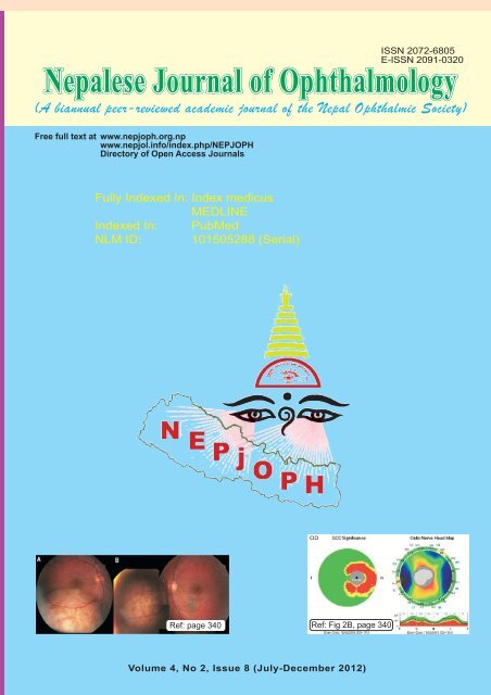
nepjoph 2012.pmd - Nepalese Journal of Ophthalmology
nepjoph 2012.pmd - Nepalese Journal of Ophthalmology
nepjoph 2012.pmd - Nepalese Journal of Ophthalmology

You also want an ePaper? Increase the reach of your titles
YUMPU automatically turns print PDFs into web optimized ePapers that Google loves.
<strong>Nepalese</strong> <strong>Journal</strong> <strong>of</strong> <strong>Ophthalmology</strong><br />
Free full text at www.<strong>nepjoph</strong>.org.np<br />
www.nepjol.info/index.php/NEPJOPH<br />
Directory <strong>of</strong> Open Access <strong>Journal</strong>s<br />
Fully Indexed In: Index medicus<br />
MEDLINE<br />
Indexed In: PubMed<br />
NLM ID: 101505288 (Serial)<br />
Ref: page 340 Ref: Fig 2B, page 340<br />
Volume 4, No 2, Issue 8 (July-December 2012)
NEPALESE JOURNAL OF OPHTHALMOLOGY<br />
(A biannual peer-reviewed academic journal <strong>of</strong> the Nepal Ophthalmic Society)<br />
Vol 4, No 2, Issue 8 (July - December 2012)<br />
Founder Patron<br />
S Ruit, MD<br />
Pr<strong>of</strong>essor & Director<br />
Tilganga Institute <strong>of</strong> <strong>Ophthalmology</strong><br />
Kathmandu, Nepal<br />
Editorial Board<br />
Patron<br />
D N Shah, MD<br />
Pr<strong>of</strong>essor & Director<br />
B P Koirala Lion's Center for Ophthalmic Studies<br />
Kathmandu<br />
Editor-in-Chief<br />
Badri P Badhu, MD<br />
Pr<strong>of</strong>essor & Head<br />
Department <strong>of</strong> <strong>Ophthalmology</strong><br />
B P Koirala Institute <strong>of</strong> Health Sciences<br />
Dharan, Nepal<br />
Chairperson<br />
J K Shrestha, FRCS, FRCOph<br />
Pr<strong>of</strong>essor & Head<br />
Department <strong>of</strong> <strong>Ophthalmology</strong><br />
Institute <strong>of</strong> Medicine<br />
Kathmandu<br />
Members<br />
Ananda Kumar Sharma, MD<br />
Associate Pr<strong>of</strong>essor<br />
B P Koirala Lion's Center for Ophthalmic Studies<br />
Kathmandu<br />
Sanjay Kumar Singh, MD<br />
Associate Pr<strong>of</strong>essor and Director<br />
Biratnagar Eye Hospital<br />
Nepal<br />
Sudesh Subedi, MD<br />
Pr<strong>of</strong>essor<br />
National Academy <strong>of</strong> Medical Sciences<br />
Kathmandu<br />
Managing Editor<br />
Akshaya P Gautam, MHPE<br />
Assistant Pr<strong>of</strong>essor<br />
Department <strong>of</strong> Health Pr<strong>of</strong>essions Education<br />
B P Koirala Institute <strong>of</strong> Health Sciences<br />
Dharan, Nepal<br />
International Editors<br />
Abhaya Vasavada, India<br />
A K Grover, India<br />
David F Chang, USA<br />
Jeevan S Titiyal, India<br />
L Vijaya, India<br />
Takeshi Naito, Japan<br />
Alan Crandall, USA<br />
Chandra Shekhar, India<br />
Jagat Ram, India<br />
Jyotirmay Biswas, India<br />
Raj Vardhan Azad, India
NEPALESE JOURNAL OF OPHTHALMOLOGY<br />
(A biannual peer-reviewed academic journal <strong>of</strong> the Nepal Ophthalmic Society)<br />
Advisory Board<br />
Canada<br />
India<br />
Martin Spencer, MD<br />
S K Arya, MD<br />
Sandeep Kumar, MS<br />
Yogesh Gupta, MS<br />
The Netherlands<br />
USA<br />
Folkert Tegelberg, MD<br />
Gerard Smith, MD<br />
J S Stilma, MD<br />
Chundak Tenzing, MD<br />
Steve Waller, MD<br />
Nepal<br />
D B Karki, MD<br />
M P Upadhyay, FRCS<br />
O K Malla, FRCS<br />
S P Shrestha, MS, DOMS<br />
S Ruit, MD<br />
Linguistic Advisor<br />
Dr Tulsi Kadel, MD, MSc<br />
Pr<strong>of</strong>essor <strong>of</strong> Forensic Medicine<br />
Institute <strong>of</strong> Medicine, Tribhuvan University<br />
Kathmandu, Nepal<br />
Editorial Office<br />
Department <strong>of</strong> <strong>Ophthalmology</strong><br />
B P Koirala Institute <strong>of</strong> Health Sciences<br />
Ghopa, Dharan-18, Sunsari, Nepal<br />
Email:<br />
<strong>nepjoph</strong>@bpkihsacademics.edu.np<br />
editor@<strong>nepjoph</strong>.org.np<br />
Tel: 00977-25-521017, ext: 5320, 2036<br />
Tel/Fax:00977-25- 520251, 530119<br />
Additional Advisor<br />
Pr<strong>of</strong> Rajendra Raj Wagle, MD, MPH, PhD<br />
Clinical Epidemiologist<br />
Pr<strong>of</strong>essor<br />
Institute <strong>of</strong> Medicine, Tribhuvan University<br />
Kathmandu, Nepal<br />
Computer Advisor<br />
Dr Damodar Sharma<br />
Department <strong>of</strong> Clinical Pharmacology<br />
B P Koirala Institute <strong>of</strong> Health Sciences<br />
Dharan, Nepal<br />
ISSN 2072-6805<br />
E-ISSN 2091-0320<br />
©Copyright: Nepal Ophthalmic Society. The expressions and opinions in the articles are solely <strong>of</strong> the authors and<br />
do not represent those <strong>of</strong> the editorial board or <strong>of</strong> the Nepal Ophthalmic Society. Advertisements or products<br />
mentioned in the journal cannot be considered as endorsement by the editorial board or the Nepal Ophthalmic<br />
Society. Websites: www.<strong>nepjoph</strong>.org.np, www.nepjol.info/index.php/<strong>nepjoph</strong>/index<br />
Printed at: Jeevan Printers Pvt. Ltd., Sorahkhutte, Kathmandu, Nepal, Tel.: 00977-1-4360345, Fax: 4358155, Email: jpsp@wlink.com.np
NEPALESE JOURNAL OF OPHTHALMOLOGY<br />
Volume 4, No 2, Issue 8 (July - December 2012)<br />
Contents<br />
Page No<br />
Guest editorial<br />
Infectious uveitis: Recent advances in diagnosis and treatment<br />
Pathanapitoon K, Stilma J S 215-216<br />
Original articles<br />
1. Outcome <strong>of</strong> phacoemulsification in eyes with cataract and corneal<br />
opacity partially obscuring the pupillary area<br />
Panda A, Krishna SN, Dada T 217-223<br />
2. Trypan blue staining <strong>of</strong> filtering bleb in eyes with operated trabeculectomy<br />
Dada T, Bali SJ, Mohan S, Bhartiya S, Sobti A, Panda A 224-229<br />
3. Efficacy <strong>of</strong> sutureless and glue free limbal conjunctival autograft<br />
for primary pterygium surgery<br />
Malik KPS, Goel R, Gupta A, Gupta SK, Kamal S, Malik VK, Singh S 230-235<br />
4. Ganglion cell complex scan in the early prediction <strong>of</strong> glaucoma<br />
Ganekal S 236-241<br />
5. Outcome <strong>of</strong> conjunctival autograft transplantation in pterygium surgery in<br />
a community based hospital in Nepal<br />
Sharma R, Marasini S, Nepal BP 242-247<br />
6. Phacoemulsification surgery by a nationally-trained cataract surgeon <strong>of</strong> Nepal<br />
Bajimaya S, Sharma BR, Shrestha JB, Maharjan IM, Matsushima H, Akura J 248-255<br />
7. Trends in idiopathic retinal vasculitis in a tertiary eye care centre <strong>of</strong> Nepal<br />
over a ten-year period<br />
Sitaula (Kharel) R, Aryal S, Joshi SN, Shrestha JK 256-262<br />
8. Visual outcome in open globe injuries<br />
Thevi T , Mimiwati Z , Reddy SC 263-270<br />
9. Expression <strong>of</strong> oxidative stress in metastatic retinoblastoma-a comparative study<br />
Mukhopadhyay S, Dutta J, Raut R, Datta H, Bhattacharyay AK 271-276<br />
10. Gender equity in eye health <strong>of</strong> Nepal: A hospital-based study<br />
Shrestha MK, Chan H, Gurung R 277-281<br />
11. Prevalence <strong>of</strong> blindness and visual impairment and its causes among people<br />
aged 50 years and above in Karnali Zone, Nepal<br />
Dulal S, Sapkota YD 282-287<br />
12. Hemodynamic effects <strong>of</strong> intraocular epinephrine during cataract surgery:<br />
a double blinded placebo controlled randomized clinical trial<br />
Seyed Ali Mohammad Miratashi, Shekoufeh Behdad, Vida Ayatollahi, Afsar Ahmadi 288-294
Review articles<br />
Artificial drainage devices for glaucoma surgery: an overview<br />
Chaudhary M, Grover S, Baisakhiya S, Bajaj A, Bhatia MS 295-302<br />
Brief communication<br />
Vision science literature <strong>of</strong> Nepal in the database “Web <strong>of</strong> Science”<br />
Risal S, Prasad HN 303-308<br />
Case reports<br />
1. Repositioning <strong>of</strong> Ahmed glaucoma valve tube in the anterior chamber<br />
with prolene sutures to manage tube-endothelial touch<br />
Dada T, Gupta R, Tinwala SI, Sobti A, Panda A 309-311<br />
2. Prosthetic rehabilitation <strong>of</strong> a patient with enucleated eye - a case report<br />
Garg P, Garg S, Bansal D, Suresh S 312-314<br />
3. Ocular myocysticercosis: Favorable outcomes with early diagnosis<br />
and appropriate therapy<br />
Chopra R, Kapoor H, Chopra A 315-318<br />
4. Accommodative spasm with bilateral vision loss due to untreated<br />
intermittent exotropia in an adult<br />
Shanker V, Ganesh S, Sethi S 319-322<br />
5. Melanocytoma <strong>of</strong> the optic disc – a case report<br />
Sharma H, Puri L R 323-325<br />
6. Homocystinuria masquerading as vitamin B12 deficiency<br />
326-328<br />
7. Malignant transformation <strong>of</strong> kissing nevus- a rare entity<br />
Kharel (Sitaula) R, Bhatta S, Shrestha GB, Shrestha JK 329-332<br />
8. An unusual case <strong>of</strong> transient cortical blindness with sagittal sinus thrombosis<br />
in a case <strong>of</strong> Henoch-Schonlein purpura<br />
Samanta SK, Mahapatra N, Aich B, Sarkar N, Chatterjee A 333-335<br />
9. Weill- Marchesani Syndrome: a rare case report<br />
Puri L R, Sharma H, Aryal S 336-338<br />
10. Choroidal metastases as the sole initial presentation <strong>of</strong> metastatic lung cancer:<br />
Case report and review <strong>of</strong> literature<br />
Salah S, Khader J, Yousef Y, Salem A, Al-Hussaini M, Al-Asady R 339-342<br />
Letters to editor<br />
1. Surgically induced astigmatism <strong>of</strong> small incision cataract surgery<br />
Ale JB 343<br />
2. Trench, lollipop, lift and chop technique for mild to moderate cataracts<br />
Vivekanand U, Kulkarni C 344-345<br />
3. Patient reported evaluation <strong>of</strong> functional symptoms (PREFS):<br />
a simple method <strong>of</strong> ascertaining patient satisfaction post cataract surgery<br />
Waqar S , Holdstock C, Evans N 346-347<br />
4. How should the ophthalmologists treat the methanol-induced toxic optic neuropathy<br />
Sanaei-Zadeh H, MD 348-350<br />
Acknowledgement 351
Pathanapitoon et al<br />
Infectious uveitis<br />
Nepal J Ophthalmol 2012; 4 (8): 215-216<br />
Guest editorial<br />
Infectious uveitis: Recent advances in diagnosis and treatment<br />
Pathanapitoon K 1 , Stilma J S 2<br />
1<br />
Associate Pr<strong>of</strong>essor, Department <strong>of</strong> <strong>Ophthalmology</strong>, Chiang Mai University,<br />
110 Intawaroros Road, Chiang Mai 50200, Thailand<br />
Tel. (+66)-53-945512 E-mail: kpathana@med.cmu.ac.th<br />
2<br />
Pr<strong>of</strong>essor Emeritus <strong>of</strong> <strong>Ophthalmology</strong>, University Medical Centre, Utrecht, The Netherlands and<br />
Honorary consultant Sierra Leone<br />
Hooivaartsweg 4, 8459 ET LUINJEBERD, The Netherlands<br />
Tel. (+31)-0513-528 707 E-mail: j.stilma@hetnet.nl<br />
Uveitis can be caused by a number <strong>of</strong> different infectious agents, requiring a robust clinical experience and<br />
appropriate diagnostic tools for accurate diagnosis and treatment. A definitive diagnosis <strong>of</strong> infectious<br />
uveitis can be determined with laboratory investigations including polymerase chain reaction (PCR) analysis<br />
<strong>of</strong> intraocular fluids and the Goldmann-Witmer coefficient for local antibody production. However, in<br />
many eye care centers, these laboratory analyses are not available due to prohibitively high costs and<br />
accessibility. In these situations, a literature review <strong>of</strong> proven causes is essential to the ophthalmologist in<br />
order to make a diagnosis and devise the most effective treatment strategy for each case.<br />
Over the past few decades, there has been growing awareness <strong>of</strong> cytomegalovirus (CMV)-associated<br />
anterior uveitis (AU), which represents a distinct clinical entity that occurs in immunocompetent patients<br />
and has primarily been reported in Asia. In a recent series from Northern Thailand, 10 out <strong>of</strong> 30 (33%)<br />
immunocompetent patients with a negative initial screening for AU were confirmed to have CMV by PCR<br />
(Kongyai et al, 2012). CMV-associated AU commonly presents as a recurrent unilateral hypertensive AU<br />
that is associated with typical keratic precipitates (KPs) (white, medium sized, nodular deposits), iris<br />
atrophy, and the absence <strong>of</strong> posterior synechiae. Some cases may present with corneal endotheliitis associated<br />
with KPs. The KPs are usually seen in the inferior half <strong>of</strong> the cornea and may be diffuse, linear, ringshaped,<br />
or appear as a coin-like lesion. Additionally, they may be associated with immune ring formation.<br />
Fuch’s heterchromic iridocyclitis and Posner-Schlossman syndrome have also been associated with CMV.<br />
Fuch’s heterochromic iridocyclitis is a type <strong>of</strong> chronic uveitis associated with fine stellate KPs, iris atrophy,<br />
and cataract in the absence <strong>of</strong> synechiae. Posner-Schlossman syndrome is characterized by recurrent<br />
episodes <strong>of</strong> unilateral low-grade AU with acute onset <strong>of</strong> highly elevated intraocular pressure above 40 mm<br />
Hg without previous therapy. Currently, the treatment for CVM-associated AU includes systemic ganciclovir,<br />
intravitreal ganciclovir injections or topical ganciclovir gel for a minimum <strong>of</strong> three months. However, the<br />
overall recurrence rate is about 75.0% following antiviral therapy and do not greatly differ from eyes that<br />
did not receive antiviral treatment. Topical ganciclovir gel has a lower response rate, a lower relapse rate<br />
(25-50%) and fewer adverse effects (Jap & Chee, 2011).<br />
Another common cause <strong>of</strong> infectious uveitis is ocular toxoplasmosis (OT). OT is characterized by necrotizing<br />
retinitis that is frequently located near a retinochoroidal scar and is associated with vitreous and anterior<br />
chamber inflammation. However, in primary OT, a chorioretinal scar may be absent. Atypical clinical<br />
features <strong>of</strong> OT that have been reported include isolated anterior uveitis, intraocular inflammatory reactions<br />
without focal necrotizing retinochoroiditis, serous retinal detachment, choroiditis without retinitis, occlusive<br />
215
Pathanapitoon et al<br />
Infectious uveitis<br />
Nepal J Ophthalmol 2012; 4 (8): 215-216<br />
retinal vasculitis, neuroretinitis and papillitis.<br />
Laboratory diagnosis <strong>of</strong> OT is determined by direct<br />
detection <strong>of</strong> T gondii DNA with PCR and/or indirect<br />
detection <strong>of</strong> infection by detection <strong>of</strong> T gondiispecific<br />
antibodies using the Goldmann-Witmer<br />
coefficient or serology for anti-T gondii IgG or IgM.<br />
The addition <strong>of</strong> an anti-T gondii IgA assay or IgG<br />
avidity increases the sensitivity <strong>of</strong> identification in<br />
cases <strong>of</strong> recently acquired toxoplasmosis. A<br />
thorough assessment requires multiple laboratory<br />
tests at different time points.<br />
Treatment regimens include the following<br />
combinations:<br />
1. pyrimethamine, sulfadiazine and leucovorin;<br />
2. trimethoprim–sulfamethoxazole;<br />
3. pyrimethamine, sulfadiazine, and clindamycin;<br />
4. sulfadiazine and clindamycin;<br />
5. pyrimethamine and clindamycin;<br />
6. azithromycin alone or combined with either<br />
pyrimethamine or trimethoprim–<br />
sulfamethoxazole; and<br />
7. intravitreal clindamycin, with a dosage ranging<br />
from 1.0 mg/0.1 mL to 1.5 mg/0.1 mL, given<br />
once or 3–4 times.<br />
The effect <strong>of</strong> intravitreal clindamycin alone for ocular<br />
toxoplasmosis has not yet been evaluated.<br />
Finally, human immunodeficiency virus (HIV) has<br />
been identified as another infectious agent associated<br />
with uveitis. Ocular manifestations in HIV-infected<br />
patients occur as a result <strong>of</strong> progressive immune<br />
dysfunction and are caused by opportunistic<br />
infections and malignancies. Since the introduction<br />
<strong>of</strong> highly active anti-retroviral therapy (HAART),<br />
the prevalence <strong>of</strong> immune recovery uveitis (IRU)<br />
has increased. However, HIV itself can also be a<br />
cause <strong>of</strong> intraocular inflammation, as several cases<br />
<strong>of</strong> HIV-associated uveitis have been recently<br />
reported. A HIV- infected patient with anterior<br />
uveitis had an intraocular HIV-1 RNA load largely<br />
exceeding that <strong>of</strong> the plasma, with no evidence <strong>of</strong><br />
other intraocular infectious agents causing uveitis<br />
other than HIV itself. Positive intraocular HIV-1<br />
RNA loads are associated with high HIV-1 RNA<br />
plasma loads and found in patients who are not<br />
undergoing HAART. Clinical features include<br />
anterior uveitis with KPs and/or vitritis without retinal<br />
lesions or scars that does not respond to topical<br />
corticosteroid therapy. Intraocular inflammation<br />
subsides after decreasing intraocular and plasma<br />
HIV loads following HAART (Pathanapitoon et al,<br />
2011).<br />
In conclusion, though many challenges remain, there<br />
has been marked progress in the proper diagnosis<br />
and treatment <strong>of</strong> infectious uveitis in recent years.<br />
Institutions with advanced laboratory facilities<br />
should pursue research endeavors that can help<br />
improve the quality <strong>of</strong> care provided to patients in<br />
ophthalmic clinics around the world.<br />
References<br />
Jap A, Chee SP (2011). Cytomegalovirusassociated<br />
anterior segment infection. Expert Rev.<br />
Ophthalmol; 6: 517–528.<br />
Kongyai N, Pathanapitoon K, Sirirungsi W, et<br />
al (2012). Viral causes <strong>of</strong> unexplained anterior<br />
uveitis in Thailand. Eye 2012 Jan 13. doi: 10.1038/<br />
eye.2011.363. [Epub ahead <strong>of</strong> print].<br />
Pathanapitoon K, Riemens A, Kongyai N, et<br />
al (2011). Intraocular and plasma HIV-1 RNA loads<br />
and HIV uveitis. AIDS; 25: 81-86.<br />
Source <strong>of</strong> support: nil. Conflict <strong>of</strong> interest: none<br />
216
Panda A et al<br />
Phacoemulsification in eyes with corneal opacity<br />
Nepal J Ophthalmol 2012; 4 (8):217-223<br />
Original article<br />
Outcome <strong>of</strong> phacoemulsification in eyes with cataract and corneal<br />
opacity partially obscuring the pupillary area<br />
Panda A, Krishna SN, Dada T<br />
Cornea, Refractive & Glaucoma Services<br />
Dr Rajendra Prasad Centre for Ophthalmic sciences, AIIMS, New Delhi, India<br />
Abstract<br />
Objective: To evaluate the intra-operative difficulties and postoperative visual outcome<br />
following phacoemulisification and intraocular lens (IOL) implantation in eyes with cataract<br />
and a coexisting corneal opacity partially obscuring the pupillary area.<br />
Materials and methods: The study included 205 eyes <strong>of</strong> 205 patients with cataract, an<br />
extensive corneal opacity partially obscuring the pupillary area and a corrected distance visual<br />
acuity (CDVA) <strong>of</strong> less than 40/200 who had undergone phacoemulisification with IOL<br />
implantation by a single surgeon. The patients were followed up on day 1, day 7, 1 month and<br />
3 months postoperatively. Intra-operative and post operative course and CDVA were<br />
evaluated.<br />
Results: Seventy nine percent <strong>of</strong> the patients underwent phaco-emulsification via superior<br />
clear corneal approach while the rest were operated via temporal clear corneal approach.<br />
Trypan blue (0.06%) dye assisted capsulorrhexis was successfully completed in all eyes with<br />
additional maneuvers including posterior synechiolysis and sphincterotomy. Nucleotomy with<br />
primary chop technique and phacoemulsification were performed uneventfully in all but one<br />
eye, which was converted to an extra capsular cataract extraction (ECCE). A foldable<br />
intraocular lens was implanted in 76 eyes, rigid IOL in 128 eyes and 1 eye was left aphakic.<br />
The pre-operative CDVA <strong>of</strong> less than 40/200 improved to 20/60 at the end <strong>of</strong> 3 months<br />
follow up.<br />
Conclusions: Phacoemulsification and intraocular lens implantation provides ambulatory and<br />
useful vision in eyes with coexisting cataract and corneal opacity.<br />
Key words: corneal opacity, cataract, phacoemulsification, iris hook, sphincterotomy<br />
Introduction<br />
The triple procedure with Penetrating Keratoplasty<br />
(PK) (Gong X ,1990; Panda A, 1999; Rao SK,<br />
1999; Shimmura S, 2003; Baykara M, 2008) or<br />
Lamellar Keratoplasy (LK) (Senoo T, 2001;<br />
Received on: 18.10.2011 Accepted on: 27.05.2012<br />
Address for correspondence: Pr<strong>of</strong> Anita Panda<br />
Dr R P Centre for Ophthalmic Sciences,<br />
AIIMS, New Delhi<br />
Phone: +91-11-26593177<br />
E-mail: anita49@yahoo.com<br />
Muraine MC, 2002) is the conventional mean for<br />
visual rehabilitation <strong>of</strong> an eye with corneal opacity<br />
and a coexistent cataract. Because <strong>of</strong> the risk <strong>of</strong><br />
graft rejection and infections, these patients require<br />
a meticulous post-operative follow up. However,<br />
this may not be possible for patients who come from<br />
rural background and live in remote hilly areas. In<br />
developing countries, the availability <strong>of</strong> a good quality<br />
217
Panda A et al<br />
Phacoemulsification in eyes with corneal opacity<br />
Nepal J Ophthalmol 2012; 4 (8):217-223<br />
donor cornea is one <strong>of</strong> the greatest limitations.<br />
Ironically, the corneal problems usually require<br />
keratoplasty. Often, eyes with corneal opacities<br />
are associated with a co-existing cataract. Though<br />
a simple cataract surgery is not an alternative in these<br />
cases, it may be a good modality to provide<br />
ambulatory vision in one eyed patients, elderly<br />
patients with poor dexterity and those who are less<br />
likely to comply with a post-operative, meticulous<br />
follow up (Gupta AK, 1992; Panda A, 2007). With<br />
this in mind, we analyzed the results <strong>of</strong> our patients<br />
who were subjected to phacoemulsificaton and IOL<br />
implantation in eyes with cataracts and extensive<br />
corneal opacities partially sparing the pupil during<br />
the period from February 2000 - January 2010.<br />
Materials and methods<br />
A retrospective chart review was performed to<br />
evaluate the data <strong>of</strong> 205 consecutive eyes <strong>of</strong> 205<br />
patients with cataracts and extensive corneal<br />
opacities partly obscuring the pupillary area and who<br />
were subjected to phacoemulsification with either<br />
foldable or rigid intraocular lens implantation by a<br />
single anterior segment surgeon (AP).<br />
Only those cases in which the anterior capsule and<br />
the pupillary margin were visible with slit lamp biomicroscopy<br />
and the CDVA was less than 40/200<br />
were enrolled.<br />
A complete medical and ocular history was taken<br />
in all patients. A through ocular examination under<br />
a slit lamp was performed to obtain the details <strong>of</strong><br />
the cornea and anterior chamber details. Intraocular<br />
pressure was recorded by applanation tonometer<br />
and fundus evaluation performed with an indirect<br />
ophthalmoscope. Laser inferometry, A- and B-scan<br />
ultrasonography for posterior segment evaluation<br />
and electrophysiological evaluations were carried<br />
out whenever required. The preoperative factors<br />
evaluated included etiology <strong>of</strong> the corneal<br />
opacification, the type <strong>of</strong> cataract and the preoperative<br />
CDVA.<br />
All the surgeries were performed under peribulbar<br />
lidocaine (Xylocaine 2%, Astra-IDL) and<br />
bupivacaine (Sensoricaine 0.5%, Astra- IDL) (1:1)<br />
218<br />
anaesthesia. Pre-operative preparation <strong>of</strong> the<br />
surgery included use <strong>of</strong> tropicamide 1% three times<br />
every ten minutes for maximum dilation. Depending<br />
upon the availability <strong>of</strong> maximum clear cornea either<br />
superior or temporal site was selected for the<br />
incision. Synechiolysis with viscoelastics or with<br />
fine iris repositor, and multiple sphincterotomies<br />
were performed whenever indicated. Trypan Blue<br />
(Vision Blue, Dorc Int., Netherlands) 0.1mL <strong>of</strong><br />
0.06% was injected into the anterior chamber (AC),<br />
under air bubble to stain the anterior capsule for 10<br />
seconds and then washed with balanced salt solution<br />
(BSS). High-viscosity viscoelastic (Sodium<br />
hyaluronate 14 mg/ml) was injected into the AC.<br />
Capsulorrhexis was initiated with a bent 26 G needle<br />
(Fig1a, 1b) but completed with Utrata forceps. The<br />
anterior capsule was grasped with Utrata forceps<br />
at the junction adjacent to the corneal opacity<br />
margin to ensure an adequate flap and firm hold<br />
(Fig.1c & 1d) The tearing <strong>of</strong> the capsule was<br />
continued in a single motion until it was visible at<br />
the other end <strong>of</strong> the opacity margin (Fig1e), then<br />
the rhexis was completed (1f). The viscoelastic was<br />
thoroughly washed from the AC followed by<br />
multiple quadrant hydrodissection in all until the edge<br />
<strong>of</strong> the nucleus tented in a clear area and was<br />
protruded outside the anterior capsule. Viscoelastic<br />
was injected behind and in front <strong>of</strong> the lens. A<br />
primary chop nucleotomy was performed (Fig 2a,<br />
2b) followed by standard phacoemulsification. Care<br />
was taken to complete the phacoemulsification in<br />
the visible pupillary area by rotating the nucleus<br />
pieces to the maximum clear area (Fig 2c, 2d).<br />
Automated irrigation and aspiration <strong>of</strong> cortical<br />
materials was performed under retro-illumination at<br />
the visible area aided by posterior capsular trypan<br />
blue staining to complete the remaining cortex<br />
aspiration (3a).<br />
In suspected cases <strong>of</strong> posterior capsule rupture<br />
either viscoelastic or 0.1 cc -4mg triamcinolone<br />
acetonide was injected into the AC to confirm the<br />
presence <strong>of</strong> any vitreous. An in-the-bag, foldable,<br />
single/three piece acrylic lens (Acrys<strong>of</strong>; Alcon Labs,<br />
fort Worth, TX, USA)/ Acrifold (APPA, India) or
Panda A et al<br />
Phacoemulsification in eyes with corneal opacity<br />
Nepal J Ophthalmol 2012; 4 (8):217-223<br />
rigid PMMA lens (APPA, India), was chosen for<br />
implantation as dictated by the patient’s economic<br />
constraints (3b-3c). Remaining cortical matter, if<br />
any, was aspirated after dialing the lens in the bag.<br />
Viscoelastic was aspirated using automated<br />
irrigation and aspiration and the AC was formed<br />
with an air bubble. (3d-3e)<br />
Post-operatively, the patients received<br />
Betametasone sodium phosphate eye drops<br />
(Betnesol, Glaxo) 0.1% 2 hourly and cipr<strong>of</strong>loxacin<br />
eye drops (Cifran, Ranbaxy) 0.3% four times a day.<br />
Cycloplegics were given to all and Timolol maleate<br />
(Iotim, FDC) 0.5% eye drops were added<br />
whenever required. The patients were followed up<br />
postoperatively on day1, week 1, month 1 and<br />
month 6.<br />
Main outcome measures<br />
The parameters assessed during surgery included<br />
the completion <strong>of</strong> capsulorrhexis, phacoemulsification,<br />
irrigation and aspiration, intraocular<br />
lens implantation and the presence <strong>of</strong> complications,<br />
if any. Postoperative assessment included the<br />
record <strong>of</strong> corneal clarity, position <strong>of</strong> intraocular lens,<br />
CDVA and other complications, if any.<br />
Results<br />
The mean age <strong>of</strong> the patients was 52.85 ± 8.45years<br />
(range 42-67 years, males- 112, females- 93). Most<br />
patients (138) had corneal opacity due to healed<br />
keratitis (Table 1).<br />
Table 1<br />
Causes <strong>of</strong> corneal opacities<br />
Causes<br />
Frequency<br />
Post-inflammatory 138<br />
Post-traumatic 47<br />
Post-surgery 13<br />
Trachoma sequelae 7<br />
The main incision used was the superior approach<br />
in 162 eyes. The temporal approach was used in<br />
43 eyes. Additional maneuvers to enlarge the<br />
pupillary area included posterior synechiolysis (159<br />
eyes) and additional sphincterotomy (63 eyes). The<br />
capsulorrhexis was successfully completed in all<br />
cases. No extension <strong>of</strong> the capsulorhexis occurred<br />
in any <strong>of</strong> the cases. A successful primary chop was<br />
possible in all cases. PC rent was suspected in<br />
fourteen eyes, which, however, were ruled out in<br />
five after injecting visco-elastic and five after injecting<br />
triamcinolone acetonide into the anterior chamber<br />
(AC) (Fig 4). In one eye, there was vitreous strand<br />
in AC, identified with triamcinolone acetonide and<br />
there was strong suspicion <strong>of</strong> nucleus instability in<br />
the bag. In this case, phacoemulsification technique<br />
was aborted and was converted to conventional<br />
extra capsular surgery (ECCE). The nucleus<br />
delivery was completed with wire vectis and the<br />
anterior vitrectomy was performed to clean the<br />
vitreous from the AC and wound, and the eye was<br />
left aphakic. In the remaining three cases, there was<br />
a small capsular tear. Rigid IOL was implanted<br />
successfully into the sulcus in those cases. The<br />
foldable intraocular lens (IOL) was implanted in 79,<br />
rigid IOL in 128 and one eye was left aphakic.<br />
On postoperative day 1, there was residual cortical<br />
matter in the anterior chamber requiring re-aspiration<br />
in 5 eyes (Fig 5). Eleven eyes revealed grade II-III<br />
AC reaction which was controlled with intensive<br />
topical and systemic corticosteroid use (Fig 5). Pre<br />
op CDVA <strong>of</strong>
Panda A et al<br />
Phacoemulsification in eyes with corneal opacity<br />
Nepal J Ophthalmol 2012; 4 (8):217-223<br />
Figure 1c, 1d: Re-grasp <strong>of</strong> the anterior capsule<br />
flap with Utrata forceps at the junction adjacent<br />
to the corneal opacity margin was done to ensure<br />
an adequate flap and firm hold<br />
Figure 3a: Irrigation & aspiration<br />
Fig 3b & 3c: Foldable IOL implantation in the bag<br />
Fig:<br />
Fig: 1d<br />
Figure 1e & 1f: The tearing <strong>of</strong> the capsule was<br />
continued at a single motion till it is visible at the<br />
other end <strong>of</strong> the opacity margin. 1e- diagrammatic<br />
representation & 1f intra-operative photograph<br />
Figure 3d: Aspiration <strong>of</strong> the viscoelastic<br />
Figure 2a, 2b: Primary chop was performed in<br />
visible area<br />
Figure 3e: Anterior chamber formation with the<br />
air bubble<br />
Figure 2c & 2d: Phaco-emulsification is done in<br />
the visible pupillary area by rotating the nucleus<br />
pieces to the maximum clear area<br />
Figure 4: Diagram showing frequency <strong>of</strong> posterior<br />
capsular tear and management<br />
220
Panda A et al<br />
Phacoemulsification in eyes with corneal opacity<br />
Nepal J Ophthalmol 2012; 4 (8):217-223<br />
Figure 5: Diagram showing frequency <strong>of</strong> postoperative<br />
complications and management<br />
Figure 6: Pie chart showing postoperative visual<br />
acuity<br />
Discussion<br />
While modern cataract surgery is safe, effective and<br />
provides excellent visual outcome, this outcome may<br />
not necessarily be achieved in eyes with associated<br />
ocular problems or following complicated cataract<br />
surgery. In these cases, there is an increased risk <strong>of</strong><br />
surgical complications and reduced visual outcome<br />
(Salem et al, 1987).<br />
Meredith and Maumenee (1979) classified cataracts<br />
into 3 grades as determined by the surgical outcome.<br />
According to this classification, Salem and Ismail<br />
found 46.5% <strong>of</strong> patients belonged to group II and<br />
III in their study <strong>of</strong> which 41% had co-existent<br />
corneal lesions. Our experience is supported by<br />
their comments, which note a higher incidence <strong>of</strong><br />
coexisting cataract and corneal opacity in developing<br />
countries. Thus, the triple procedure is required to<br />
rehabilitate these patients. Though the triple<br />
procedure (Gong X ,1990; Panda A, 1999; Rao<br />
SK, 1999; Shimmura S, 2003; Baykara M, 2008)<br />
is the method <strong>of</strong> choice to manage corneal lesions<br />
with a coexistent cataract, factors such as scarcity<br />
<strong>of</strong> good donor tissue, post keratoplasty graft<br />
rejection, glaucoma, and infection may impede the<br />
chances <strong>of</strong> successful penetrating keratoplasty<br />
(PK). This may be especially true in patients from<br />
rural areas in developing countries due to irregular<br />
follow up and poor ocular hygiene. Moreover, the<br />
patients with extensive corneal opacity are poor<br />
candidates for PK due to association <strong>of</strong><br />
vascularisation and poor ocular surface as well as a<br />
high chance <strong>of</strong> developing post-operative suture<br />
related problems, which jeopardize the graft status.<br />
Thus, a simple technique is needed. Gupta et al<br />
(1992) and Panda et al (2007) advocate<br />
conventional ECCE to provide ambulatory vision<br />
in such eyes.<br />
In modern era, phacoemulsification is the preferred<br />
technique <strong>of</strong> cataract surgery as it allows for early<br />
visual rehabilitation. However, a successful<br />
phacoemulsification may be difficult in eyes with<br />
corneal opacities because <strong>of</strong> poor visualization <strong>of</strong><br />
intraocular morphology, and altered dynamics<br />
((Farjo AA, 2003; Czumbel N, 2009; Chang YS,<br />
2008). Surgical modifications, such as multiple<br />
sphincterotomy and iris hook application have been<br />
used to enlarge the visible papillary area and to<br />
overcome visual limitations. Use <strong>of</strong> dye, endoscope<br />
and Chandler illumination aided phacoemulsification<br />
also enhances surgical ease (Gregory ME, 2007;<br />
Sinha R, 2004; Uka J, 2005; Bhartiya P, 2002;<br />
Singh D, 1996). In the present study, we desired to<br />
make these patients ambulatory with cataract<br />
surgery alone by modifying the vital steps <strong>of</strong> surgery.<br />
A fully dilated pupil allows visualizing the anterior<br />
capsule and capsular bag anatomy, so efforts are<br />
made to achieve this by breaking the posterior<br />
synechiae with additional sphincterotomies. This<br />
technique not only increases the visibility, but also<br />
helps improve post-operative visual outcome. The<br />
quality <strong>of</strong> peripheral vision may be expected to be<br />
sub-optimal when compared to central vision.<br />
However, Drew’s and Drew’s (1964) advocated<br />
optical iridectomy in eyes with obscured pupillary<br />
221
Panda A et al<br />
Phacoemulsification in eyes with corneal opacity<br />
Nepal J Ophthalmol 2012; 4 (8):217-223<br />
zone. They argued that though the peripheral part<br />
<strong>of</strong> the human optical system does not form as sharp<br />
an image as the central part, the optical iridectomy<br />
improves visual prognosis because <strong>of</strong> the addition<br />
<strong>of</strong> peripheral bundle <strong>of</strong> rays. These peripheral rays<br />
help form a relatively clear image superimposed on<br />
the blurred image <strong>of</strong> the central part. However, in<br />
our study, we preferred multiple sphincterotomies<br />
instead <strong>of</strong> a complete iridectomy in order to<br />
preserve some functional anatomy <strong>of</strong> the iris.<br />
Although visual outcome in these eyes was<br />
suboptimal, all patients achieved ambulatory vision,<br />
enabling them to carry out daily activities. Anterior<br />
capsular staining was performed in all to demarcate<br />
the obscured capsular anatomy, to achieve a<br />
complete capsulorrhexis successfully and posterior<br />
capsular staining to enchance the safety <strong>of</strong> cortical<br />
aspiration in the presence <strong>of</strong> corneal opacities.<br />
Precision <strong>of</strong> capsulorrhexis by our technique<br />
enabled us to complete a successful circular<br />
capsulorrhexis. Making the nuclear border partially<br />
prolapsed from the bag by multiple site hydrodissection,<br />
helped us to perform a successful primary<br />
chop in all. Either viscoelastic or triamcinolone<br />
acetonide injection into the anterior chamber (AC)<br />
aided the identification <strong>of</strong> vitreous in AC which was<br />
a boon for the decision <strong>of</strong> IOL insertion.<br />
Conclusion<br />
Phacoemulsification in selected cases <strong>of</strong> corneal<br />
opacities with cataract is safe and feasible both as<br />
a primary therapeutic option in cases where PK is<br />
risky because <strong>of</strong> various limiting factors.<br />
Acknowledgement<br />
The authors would like to extend a word <strong>of</strong> thanks<br />
to Arvind Kumar, Anand Agarwal, Lalit Tejwani,<br />
Shikha and Sapna.<br />
References<br />
AI Salem M, Ismail L (1987). Factors<br />
influencing visual outcome after cataract extraction<br />
among Arabs in Kuwait. Br J Ophthalmol; 71:458-<br />
61<br />
Baykara M, Ucan G (2008). Modifying the<br />
position <strong>of</strong> cataract incisions in triple procedure. Eur<br />
J Ophthalmol; 18:891-4.<br />
Bhartiya P, Sharma N, Ray M, Sinha R,<br />
Vajpayee RB (2002). Trypan blue assisted<br />
phacoemulsification in corneal opacities. Br J<br />
Ophthalmol; 86:857-9.<br />
Chang YS, Hsiao JH, Tseng SH, Kuo PH,<br />
Chen FK (2008). Indocyanine green-assisted<br />
phacoemulsification in cases <strong>of</strong> complicated or<br />
simple advanced cataracts. J Formos Med Assoc;<br />
107:710-9.<br />
Czumbel N, Albert A (2009). Cataract surgery<br />
(phacoemulsification) in difficult cases. Oftalmologia;<br />
53:41-4.<br />
Drews LC, Drews RC (1964). Optical<br />
Iridectomy. Am J Ophthalmol; 58:789-96.<br />
Farjo AA, Meyer RF, Farjo QA (2003).<br />
Phacoemulsification in eyes with corneal<br />
opacification. J Cataract Refract Surg; 29:242-5.<br />
Gong X, Du N, Chen J, Zhang J, Feng C,<br />
Chen L (1990). Penetrating keratoplasty combined<br />
with cataract extraction. Yan Ke Xue Bao;6:7-10.<br />
Gregory ME, Bibby K (2007). Dye-assisted<br />
small incision cataract surgery in an eye with cataract<br />
and coexisting corneal scarring and epithelial<br />
disease. Eye; 21:299-300.<br />
Gupta AK, Grover AK, Gurha N<br />
(1992).Traumatic cataract surgery with intraocular<br />
lens implantation in children. J Pediatr Ophthalmol<br />
Strabismus; 29:73-8.<br />
Meredith TA and Maumee AE (1979). A<br />
review <strong>of</strong> one thousand cases <strong>of</strong> intracapsular<br />
cataract extraction: I. complications. Ophthalmic<br />
Surgery; 10:32-41.<br />
Muraine MC, Collet A, Brasseur G (2002).<br />
Deep lamellar keratoplasty combined with cataract<br />
surgery. Arch Ophthalmol; 120:812-5.<br />
Panda A, Kumar TS (1991). Keratoplasty and<br />
cataract extraction. Indian J Ophthalmol; 39:102-<br />
4.<br />
Panda A, Kumar S, Das H, Badhu BP (2007).<br />
Striving for the perfect surgery in traumatic cataract<br />
following penetrating trauma in a tertiary care<br />
222
Panda A et al<br />
Phacoemulsification in eyes with corneal opacity<br />
Nepal J Ophthalmol 2012; 4 (8):217-223<br />
hospital in eastern Nepal. JNMA J Nepal Med<br />
Assoc; 46:119-25.<br />
Rao SK, Padmanabhan P (1999). Combined<br />
phacoemulsification and penetrating keratoplasty.<br />
Ophthalmic Surg Lasers; 30:488-91.<br />
Senoo T (2001). Combined surgery with deep<br />
lamellar keratoplasty. Semin Ophthalmol; 16:126-<br />
36.<br />
Shimmura S, Ohashi Y, Shiroma H, Shimazaki<br />
J, Tsubota K (2003). Corneal opacity and cataract:<br />
triple procedure versus secondary approach.<br />
Cornea; 22:234-8.<br />
Singh D (1996). Localized bullous separation<br />
<strong>of</strong> Descemet’s membrane after previous cataract<br />
surgery. J Cataract Refract Surg; 22:147-9.<br />
Sinha R, Sharma N, Vajpayee RB (2004).<br />
Visual outcome <strong>of</strong> cataract surgery with pupillary<br />
sphincterotomy in eyes with coexisting corneal<br />
opacity. BMC Med; 2:10.<br />
Uka J, Minamoto A, Hirayama T, Adachi T,<br />
Yamane K, Mishima HK (2005). Endoscope-aided<br />
cataract surgery in corneal opacity associated with<br />
aniridia. J Cataract Refract Surg; 31:1455-6.<br />
Source <strong>of</strong> support: nil. Conflict <strong>of</strong> interest: none<br />
223
Dada T et al<br />
Trypan blue staining <strong>of</strong> filtering bleb<br />
Nepal J Ophthalmol 2012; 4(8):224-229<br />
Original article<br />
Received on: 02.11.2011 Accepted on: 05.04.2012<br />
Address for correspondence: Dr. Tanuj Dada, Additional<br />
Pr<strong>of</strong>essor<br />
Dr. Rajendra Prasad Centre for Ophthalmic Sciences<br />
All India Institute <strong>of</strong> Medical Sciences,<br />
New Delhi 110029, India.<br />
Fax 91-1126588919<br />
E mail: tanujdada@gmail.com<br />
224<br />
Trypan blue staining <strong>of</strong> filtering bleb in eyes with operated<br />
trabeculectomy<br />
Dada T, Bali SJ, Mohan S, Bhartiya S, Sobti A, Panda A<br />
Glaucoma Facility<br />
Dr Rajendra Prasad Centre for Ophthalmic Sciences<br />
All India Institute <strong>of</strong> Medical Sciences<br />
New Delhi 110029, India<br />
Abstract<br />
Objective: To report the use <strong>of</strong> trypan blue staining <strong>of</strong> the filtering bleb to assess its functional<br />
status in eyes undergoing phacoemulsification after trabeculectomy.<br />
Subjects and methods: This retrospective study was conducted at a tertiary eye care centre<br />
in North India and studied 33 eyes <strong>of</strong> 33 patients ( with previously operated trabeculectomy),<br />
who underwent phacoemulsification. Trypan blue dye (0.06%) was used to stain the anterior<br />
capsule. After completion <strong>of</strong> phacoemulsification, the staining <strong>of</strong> the trabeculectomy bleb was<br />
noted as diffuse, patchy, minimal or no staining.<br />
Results: Of the 33 eyes, 13 had diffuse staining (39.4%, mean IOP = 9.3 ± 2.2 mm Hg), 7<br />
(21.2%, mean IOP= 15.5 ± 1.8 mm Hg) had patchy staining, 4 had minimal staining (12.1%,<br />
mean IOP= 17.5 ± 0.5mm Hg) and nine (27.3%, mean IOP= 19.3 ± 1.6 mm Hg) had no<br />
staining. These staining patterns were labeled as groups 1 - 4 respectively. Statistical analysis<br />
showed that the difference between the IOPs in Group 1 - 2 and between Group 2 - 3 was<br />
not significant statistically (p=0.682 and 0.665 respectively). However the differences between<br />
the IOPs between Groups 1 - 3, 1 - 4, 2 - 4, and 3 - 4 were found to be highly significant<br />
statistically (p
Dada T et al<br />
Trypan blue staining <strong>of</strong> filtering bleb<br />
Nepal J Ophthalmol 2012; 4(8):224-229<br />
subconjunctival fibrosis, prevent bleb failure, and<br />
achieve better intraocular pressure control (Parrish<br />
et al 2001, Chen et al 1990).<br />
Knowing whether the bleb is patent and functional<br />
has important implications in the management <strong>of</strong><br />
glaucoma cases. The aqueous drainage through the<br />
filtration bleb cannot be accurately quantified. Many<br />
authors (Pitch et al 1998, Clarke et al 2003) have<br />
investigated morphological criteria <strong>of</strong> these blebs<br />
to correlate clinical and functional aspects. Although<br />
some authors have described the evaluation <strong>of</strong><br />
filtering bleb by thermography (Kawasaki et al<br />
2009) and in vivo confocal microscopy (Messmer<br />
et al 2006, Labbe et al 2005), there is still a lack <strong>of</strong><br />
clinical tests that can objectively delineate the<br />
functional status <strong>of</strong> the bleb.<br />
The diffusion <strong>of</strong> trypan blue dye into the filtration<br />
bleb following its instillation into the anterior chamber<br />
has already been documented (Agrawal et al 2005).<br />
However to the best <strong>of</strong> our knowledge, no study in<br />
literature has classified the diffusion patterns <strong>of</strong> the<br />
dye during phacoemulsification surgery and<br />
correlated it with bleb function.<br />
In this clinical study, patients with previously<br />
operated trabeculectomy undergoing cataract<br />
surgery were evaluated. The staining patterns <strong>of</strong> the<br />
blebs obtained after trypan blue injection into the<br />
anterior chamber were analyzed and correlated to<br />
the mean IOP recorded before the cataract surgery<br />
in order to evaluate the functioning <strong>of</strong> the bleb.<br />
Subjects and methods<br />
This retrospective charts review was carried out<br />
on 33 eyes <strong>of</strong> 33 patients recruited from the followup<br />
cases attending the glaucoma clinic at our centre<br />
between October 2007 and December 2009.<br />
Patients with a previously operated trabeculectomy<br />
with mitomycin C, having a visually significant<br />
cataract with a best corrected visual acuity <strong>of</strong> 20/<br />
40 or less, and presenting at least 6 months or more<br />
after trabeculectomy were included in the study.<br />
Patients with corneal opacities, severe dry eye,<br />
uveitis, and previous ocular surgery other than<br />
trabeculectomy were excluded.<br />
The pre-operative evaluation included a detailed<br />
ophthalmic history and clinical examination <strong>of</strong> all<br />
eyes. This included near and distant best corrected<br />
visual acuity, Goldmann applanation tonometry, slit<br />
lamp evaluation <strong>of</strong> the bleb, gonioscopy, optic nerve<br />
head evaluation with a + 90 diopter lens, and ocular<br />
biometry. The study conformed to the Declaration<br />
<strong>of</strong> Helsinki, and informed written consent was<br />
obtained from all patients. All patients were assigned<br />
to undergo phacoemulsification with intraocular lens<br />
implantation by a single surgeon (TD). Maximum<br />
mydriasis was obtained by a combination <strong>of</strong><br />
tropicamide 0.5% and phenylepherine 0.5% applied<br />
three times at ten minute intervals preoperatively.<br />
All surgeries were performed under either topical<br />
anaesthesia using sterile 0.5% proparacaine drops<br />
(Paracain, Sunways, Mumbai, India).<br />
All patients underwent a clear corneal<br />
phacoemulsification via a temporal incision <strong>of</strong> size<br />
2.75 mm along with a superior side port incision.<br />
The anterior chamber was reformed with air and<br />
0.1 ml <strong>of</strong> trypan blue dye (0.06%, Visiblue, Shah<br />
and Shah, India) was injected under the air bubble<br />
to stain the anterior capsule using a 27 G cannula.<br />
Capsulorrhexis was performed under viscoelastics<br />
cover (1 % hydroxypropylmethylcellulose).<br />
Phacoemulsification was completed by the stop and<br />
chop technique followed by implantation <strong>of</strong> a<br />
hydrophobic acrylic foldable intraocular lens in the<br />
bag. The filtering bleb was observed for trypan<br />
blue staining and graded. The grading for the staining<br />
pattern <strong>of</strong> the bleb was as follows: Group 1 no<br />
staining, Group 2 minimal staining, Group 3 patchy<br />
staining, and Group 4 diffuse staining (Fig. 1-4).<br />
Post operatively the patient received gatifloxacin<br />
0.3%, prednisolone acetate 1% four times a day<br />
which were tapered over four weeks. If the patient<br />
was receiving ocular hypotensive medications in the<br />
pre-operative period, these were continued<br />
postoperatively.<br />
Statistics: Statistical analyses were performed<br />
using SPSS (version 12.0, SPSS Inc, Chicago, IL).<br />
Fischer’s Analysis <strong>of</strong> Variation (one way ANOVA),<br />
225
Dada T et al<br />
Trypan blue staining <strong>of</strong> filtering bleb<br />
Nepal J Ophthalmol 2012; 4(8):224-229<br />
post hoc analysis using Bonferronis test were used<br />
to determine the significance <strong>of</strong> the difference in the<br />
IOPs <strong>of</strong> the four groups.<br />
Results<br />
The study included 33 eyes <strong>of</strong> 33 patients. There<br />
were 17 males and 16 females. The mean age was<br />
58.6 +12.4 years. The time interval between the<br />
trabeculectomy and cataract surgery varied from 7<br />
to 23 months. All patients had undergone a<br />
trabeculectomy using a limbus based conjunctival<br />
flap. Mitomycin C (0.2 mg/ml for 3 minutes,<br />
subconjunctival) was used during trabeculectomy<br />
in all eyes.<br />
Of the thirty three eyes, 13 eyes had diffuse staining<br />
(39.4%), 7 (21.2%) had patchy staining, 4 had<br />
minimal staining (12.1%) and 9 eyes (27.3%) had<br />
no staining (Figure 1-4). On correlating the staining<br />
with the IOP, it was found that eyes with diffuse<br />
staining had previously undergone trabeculectomy<br />
with Mitomycin C application and had the least IOP<br />
before cataract surgery (6-12 mm Hg) with a mean<br />
IOP <strong>of</strong> 9.3 ± 2.2 mm Hg. Morphologically, these<br />
blebs were elevated, polycystic and avascular. Eyes<br />
with patchy staining had a mean pre-cataract surgery<br />
IOP <strong>of</strong> 15.5 ± 1.8 mm <strong>of</strong> Hg (Range: 14 -18 mm<br />
<strong>of</strong> Hg) while eyes with minimal staining had a mean<br />
pre-cataract surgery IOP <strong>of</strong> 17.5 ± 0.5mm <strong>of</strong> Hg<br />
(Range: 17-18mm <strong>of</strong> Hg). Eyes with a failed filtering<br />
bleb showed no staining with the IOP in the range<br />
<strong>of</strong> 18-22 mm Hg, and a mean <strong>of</strong> 19.3 ± 1.6 mm <strong>of</strong><br />
Hg on topical anti-glaucoma medications (Fig 5).<br />
Figure 1: Diffuse staining <strong>of</strong> the bleb<br />
Figure 2: Patchy staining <strong>of</strong> the bleb<br />
Figure 3: Minimal staining <strong>of</strong> the bleb<br />
Figure 4: No staining <strong>of</strong> the bleb<br />
Figure 5: Box plot showing mean intraocular<br />
pressure in patients with different grades <strong>of</strong><br />
staining<br />
226
Dada T et al<br />
Trypan blue staining <strong>of</strong> filtering bleb<br />
Nepal J Ophthalmol 2012; 4(8):224-229<br />
One-way analysis <strong>of</strong> variation (ANOVA), and Post<br />
Hoc Bonferroni’s test revealed that this difference<br />
in IOP was highly statistically significant (p
Dada T et al<br />
Trypan blue staining <strong>of</strong> filtering bleb<br />
Nepal J Ophthalmol 2012; 4(8):224-229<br />
with more diffuse staining corroborating with better<br />
IOP control.<br />
Conclusion<br />
During phacoemulsification in patients with a<br />
previously operated filtering bleb, vital staining <strong>of</strong><br />
the sub-epithelial tissue by trypan blue dye delineates<br />
the loose sub-conjunctival spaces which assist<br />
filtration. The greater the staining, the greater is the<br />
bleb function, which correlates well with the posttrabeculectomy<br />
intraocular pressure. Noting the<br />
staining pattern <strong>of</strong> the bleb during<br />
phacoemulsification can help quantify the functional<br />
status <strong>of</strong> the bleb, and help the surgeon plan for<br />
bleb resuscitation measures.<br />
References<br />
Addicks EM, Quigley HA, Green WR, Robin<br />
AL (1983). Histologic characteristics <strong>of</strong> filtering<br />
blebs in glaucomatous eyes. Arch Ophthalmol;<br />
101:795-798.<br />
Agrawal S, Agrawal J, Agrawal TP (2005).<br />
Use <strong>of</strong> trypan blue to confirm the patency <strong>of</strong> filtering<br />
surgery. J Cataract Refract Surg; 31:235-237<br />
Cairns J (1968). Trabeculectomy: Preliminary<br />
report <strong>of</strong> a new method. Am J Ophthalmol; 66:673–<br />
679.<br />
Carassa RG, Bettin P, Fiori M, Brancato R<br />
(2003). Viscocanalostomy versus trabeculectomy<br />
in white adults affected by open-angle glaucoma: a<br />
2-year randomized, controlled trial. <strong>Ophthalmology</strong><br />
;110:882–887.<br />
Chen CW, Huang HT, Blair JS (1990).<br />
Trabeculectomy with simultaneous topical<br />
application <strong>of</strong> mitomycin-C in refractory glaucoma.<br />
J Ocular Pharmacol; 6:175–182.<br />
Clarke JC, Wells AP, Sangermani CD (2003).<br />
A system for grading filtration blebs following<br />
trabeculectomy. Invest Ophthalmol Vis Sci; 44:E-<br />
Abstract, 1201.<br />
Coleman AL, Hill R, Wilson MR, et al (1995).<br />
Initial clinical experience with the Ahmed glaucoma<br />
Valve implant. Am J Ophthalmol; 120:23–31<br />
Feron EJ, Veckeneer M, Parys-Van<br />
Ginderdeuren R, Van Lommel A, Melles GR,<br />
Stalmans P (2002). Trypan blue staining <strong>of</strong> epiretinal<br />
membranes in proliferative vitreoretinopathy. Arch<br />
Ophthalmol; 120:141–144.<br />
Healey PR, Crowston JG (2005). Trypan blue<br />
identifies antimetabolite treatment area in<br />
trabeculectomy. Br J Ophthalmol; 89:1152–1156.<br />
Hoskins HD Jr, Iwach AG, Vassiliadis A,<br />
Drake MV, Hennings DR (1991). Subconjunctival<br />
THC:YAG laser thermal sclerostomy.<br />
<strong>Ophthalmology</strong>; 98:1394–1399.<br />
Kawasaki S, Mizoue S, Yamaguchi M et al<br />
(2009). Evaluation <strong>of</strong> filtering bleb function by<br />
thermography. Br J Ophthalmol; 93:1331-1336.<br />
Labbe A, Dupas B, Hamard P, Baudouin C<br />
(2005). In vivo confocal microscopy study <strong>of</strong> blebs<br />
after filtering surgery. <strong>Ophthalmology</strong>; 112:1979-<br />
1986.<br />
Lichter PR, Musch DC, Gillespie BW, Guire<br />
KE, Janz NK, Wren PA, Mills RP (2001). CIGTS<br />
Study Group. Interim clinical outcome in the<br />
collaborative initial glaucoma treatment study<br />
comparing initial treatment randomized to<br />
medications or surgery. <strong>Ophthalmology</strong>; 108:1943-<br />
1953.<br />
Lloyd MA, Baerveldt G, Heuer DK, Minckler<br />
DS, Martone JF (1994). Initial clinical experience<br />
with the Baerveldt implant in complicated<br />
glaucomas. <strong>Ophthalmology</strong>; 101:640–650.<br />
Melles GR, de Waard PW, Pameyer JH,<br />
Houdijn Beekhuis W (1999). Trypan blue capsule<br />
staining to visualize the capsulorhexis in cataract<br />
surgery. J Cataract Refract Surg; 25:7–9.<br />
Messmer EM, Zapp DM, Mackert MJ, Thiel<br />
M, Kampik A (2006). In vivo confocal microscopy<br />
228
Dada T et al<br />
Trypan blue staining <strong>of</strong> filtering bleb<br />
Nepal J Ophthalmol 2012; 4(8):224-229<br />
<strong>of</strong> filtering blebs after trabeculectomy. Arch<br />
Ophthalmol; 124: 1095-1103.<br />
Molteno AC (1969). New implant for drainage in<br />
glaucoma: Clinical trial. Br J Ophthalmol; 53:606–<br />
615.<br />
Norn MS (1973). Pachometric study on the<br />
influence <strong>of</strong> corneal endothelial vital staining. Corneal<br />
thickness after cataract extraction studied by vital<br />
staining with trypan blue. Acta Ophthalmol<br />
(Copenh); 51:679–686.<br />
Parrish RK 2nd, Schiffman JC, Feuer WJ,<br />
Heuer DK (2001). Fluorouracil Filtering Surgery<br />
Study Group. Prognosis and risk factors for early<br />
postoperative wound leaks after trabeculectomy<br />
with and without 5-fluorouracil. Am J<br />
Ophthalmol;132:633–640.<br />
Pitch G, Grehn F (1998). Classification <strong>of</strong><br />
filtering blebs in trabeculectomy: biomicroscopy and<br />
functionality. Curr Opin Ophthalmol; 2:2-8.<br />
Sanchez E, Schnyder CC, Sickenberg M,<br />
Chiou AG, Hédiguer SE, Mermoud A (1996–97).<br />
Deep sclerectomy: results with and without collagen<br />
implant. Int Ophthalmol; 20:157–162.<br />
Stegmann R, Pienar A, Miller D (1999).<br />
Viscocanalostomy for open-angle glaucoma in black<br />
African patients. J Cataract Refract Surg; 25:316–<br />
322.<br />
Wise JB, Witter SL (1979). Argon laser<br />
therapy for open-angle glaucoma. A pilot study.<br />
Arch Ophthalmol; 97:319–322.<br />
Federov SN, I<strong>of</strong>fe DI, Ronkina TE (1982).<br />
Glaucoma surgery—deep sclerectomy. Vestn<br />
Oftalmol; 4:6–10.<br />
Yamamoto T, Sakuma T, Kitazawa Y (1995).<br />
An ultrasound biomiscroscopic study <strong>of</strong> filtering<br />
blebs after mitomycin C trabeculectomy.<br />
Opthalmology; 102:1770-1776.<br />
Source <strong>of</strong> support: nil. Conflict <strong>of</strong> interest: none<br />
229
Malik KPS et al<br />
Conjunctival autograft for pterygium surgery<br />
Nepal J Ophthalmol 2012; 4(8):230-235<br />
Original article<br />
Received on: 26.01.2012 Accepted on: 19.05.2012<br />
Address for correspondence: Dr Ruchi Goel,<br />
513/514, First Floor, Double Storey, New Rajinder Nagar,<br />
New Delhi-110060.<br />
Tel: 919811305645, 91-011-42411839.<br />
Email: gruchi1@rediffmail.com<br />
230<br />
Efficacy <strong>of</strong> sutureless and glue free limbal conjunctival autograft<br />
for primary pterygium surgery<br />
Malik KPS 1 , Goel R 2 , Gupta A 1 , Gupta SK 1 , Kamal S, 2 Malik VK 1 , Singh S 2<br />
1<br />
Subharti Medical College, Meerut, Uttar Pradesh, India<br />
2<br />
Guru Nanak Eye Centre, Maulana Azad Medical College, New Delhi, India<br />
Abstract<br />
Introduction: There are numerous adjunctive measures described to reduce the recurrence<br />
rates after pterygium excision.<br />
Objective: To study the efficacy and complications <strong>of</strong> sutureless and glue free limbal conjunctival<br />
autograft for the management <strong>of</strong> primary pterygium over a period <strong>of</strong> one year.<br />
Materials and methods: A prospective interventional case series was carried out in 40<br />
consecutive eyes with primary nasal pterygium requiring surgical excision. Pterygium excision<br />
with limbal conjunctival autografting without using glue or sutures was performed in all the<br />
patients followed by bandaging for 48 hours. The patients were followed up post operatively<br />
on 2 nd day, 1 week, 6 weeks, 6 months and 12 months. They were examined for haemorrhage,<br />
wound gape, graft shrinkage, chemosis, graft dehiscence, recurrence or any other complication.<br />
Results: The mean age <strong>of</strong> the patients was 42.8 years (range 23-61), 75% <strong>of</strong> which were<br />
males. Total graft dehiscence occurred in 2 eyes (5%), graft retraction in 3 eyes (7.5%) and<br />
recurrence was seen in 1 eye (2.5%). At 6 weeks postoperatively, the gain in uncorrected<br />
visual acuity ranged from 0.18 to 0.5 log MAR in 7 eyes. No other complication was noted.<br />
Conclusions: Sutureless and glue free limbal conjunctival autografting following pterygium<br />
excision is a safe, effective and economical option for the management <strong>of</strong> primary pterygium.<br />
Keywords: Pterygium, conjunctiva, limbal-conjunctival autograft, fibrin glue, pterygium surgery<br />
Introduction<br />
Pterygium is a common disorder in many parts <strong>of</strong><br />
the world, with reported prevalence rates ranging<br />
from 0.3 to 29% (Moran & Hollows, 1988; Taylor<br />
et al, 1984). In general, conservative therapy for<br />
pterygium is warranted as recurrences after pterygia<br />
excision are frequent and aggressive. Numerous<br />
adjunctive measures have been described to reduce<br />
the recurrence rates after its excision. These may<br />
be broadly classified as medical methods, betairradiation<br />
and surgical methods (Ang et al, 2007).<br />
Limbal -conjunctival autograft is currently the most<br />
popular surgical procedure as it has been suggested<br />
that including the limbal stem cells act as a barrier<br />
to the conjunctival cells migrating onto the corneal<br />
surface.<br />
The most common method <strong>of</strong> autograft fixation is<br />
suturing, with drawbacks <strong>of</strong> prolonged operating<br />
time, postoperative discomfort, suture abcesses,<br />
buttonholes, and granuloma formation which usually<br />
requires a second operation for removal (Starck et<br />
al, 1991).
Malik KPS et al<br />
Conjunctival autograft for pterygium surgery<br />
Nepal J Ophthalmol 2012; 4(8):230-235<br />
Replacing sutures with tissue adhesives may shorten<br />
the operating time, improve postoperative comfort,<br />
and avoid suture related complications. However,<br />
the major concern <strong>of</strong> the commercial fibrin glue is<br />
the cost and the potential risk <strong>of</strong> transmitted<br />
infection. Autologous fibrin glue has been used as<br />
an alternative method for graft fixation by some<br />
authors (Cohen & Donald, 1993; Foroutan et al,<br />
2011). A recent cross-sectional study also describes<br />
successful outcome with sutureless and glue-free<br />
conjunctival autograft (Wit et al, 2010). We<br />
conducted this prospective interventional study in a<br />
larger number <strong>of</strong> patients to determine the outcome<br />
<strong>of</strong> sutureless glue-free limbal conjunctival autograft<br />
for primary pterygium surgery.<br />
Materials and methods<br />
This prospective interventional case series included<br />
consecutive 40 eyes with primary nasal pterygium<br />
requiring surgical excision from July 2010 to<br />
December 2010. The indication for surgical<br />
intervention was one or more <strong>of</strong> the following:<br />
diminution <strong>of</strong> vision either because <strong>of</strong> induced<br />
astigmatism or encroachment onto the pupillary<br />
area, marked cosmetic deformity, marked<br />
discomfort and irritation unrelieved by medical<br />
management, limitation <strong>of</strong> ocular motility secondary<br />
to restriction or documented progressive growth<br />
towards the visual axis so that ultimate visual loss<br />
could reasonably be assumed. An informed consent<br />
was taken from each patient.<br />
The study was approved by the Institutional<br />
Research Ethics Committee at Subharti Medical<br />
College, Meerut, Uttar Pradesh, India.<br />
The primary outcome measures included graft<br />
dislocation and pterygium recurrence. Graft success<br />
was defined as an intact graft by the end <strong>of</strong> 6 weeks<br />
after operation without need for sutures. Recurrence<br />
was defined as any growth <strong>of</strong> conjunctiva exceeding<br />
1mm onto the cornea.<br />
A detailed medical and ophthalmic history, including<br />
gender, age and previous eye surgery was taken.<br />
Exclusion criteria included recurrent pterygium,<br />
glaucoma, retinal pathology requiring surgical<br />
intervention, history <strong>of</strong> previous ocular surgery or<br />
trauma. Preoperative ophthalmic evaluation<br />
comprised <strong>of</strong> uncorrected and best corrected visual<br />
acuity (BCVA), digital anterior segment<br />
photography, slit lamp examination and funduscopy.<br />
Surgical technique<br />
All surgical procedures were performed by the same<br />
surgeon (KPSM) to ensure consistency. Operations<br />
were performed under peribulbar anaesthesia using<br />
2% Xylocaine injection. The body <strong>of</strong> the pterygium<br />
was dissected 4 mm from the limbus, down to the<br />
bare sclera. Blunt and sharp dissection by Wescott<br />
scissors (Geuder, Germany) was done for<br />
separating the fibrovascular tissue from the<br />
surrounding conjunctiva. The pterygium was<br />
removed from the cornea (superficial keratectomy)<br />
using a crescent knife. Only the thickened portions<br />
<strong>of</strong> conjunctiva and the immediate adjacent and<br />
subjacent Tenon‘s capsule showing tortuous<br />
vasculature were excised. Where possible,<br />
haemostasis was allowed to occur spontaneously<br />
without the use <strong>of</strong> cautery. The size <strong>of</strong> the defect<br />
was measured with Castoveijo callipers (Bausch<br />
& Lomb Storz; Storz instruments, St Louis, MO,<br />
USA).<br />
For harvesting the donor limbal conjunctival<br />
autograft, 0.5ml <strong>of</strong> Xylocaine was injected using<br />
30 G needle subconjunctivally to allow dissection<br />
between the conjunctiva and tenon‘s layer in the<br />
superior bulbar conjunctiva. An oversized graft with<br />
an additional 2.0mm <strong>of</strong> length and width relative to<br />
the dimensions <strong>of</strong> the bare sclera was dissected<br />
including the superior limbal stem cells.<br />
The graft was placed on the bare sclera in such a<br />
way so as to maintain the original orientation <strong>of</strong> the<br />
juxtalimbal border towards the cornea. The scleral<br />
bed was viewed through the transparent conjunctiva<br />
to ensure that residual bleeding does not lift the graft.<br />
Small central haemorrhages were tamponaded with<br />
direct compression. The free graft was held in<br />
position for 10 minutes by application <strong>of</strong> gentle<br />
pressure over it with a lens spatula. The stabilisation<br />
231
Malik KPS et al<br />
Conjunctival autograft for pterygium surgery<br />
Nepal J Ophthalmol 2012; 4(8):230-235<br />
<strong>of</strong> graft was tested with a Merocel spear centrally<br />
and on each free edge to ensure firm adherence to<br />
the sclera. The eye was bandaged for 48 hours.<br />
Postoperative regimen<br />
After removal <strong>of</strong> the patch, the patient was advised<br />
not to rub the eye and topical Loteprednol eye drops<br />
were administered four times a day which was<br />
tapered over 6 weeks. Chloramphenicol eye drops<br />
were instilled four times a day for 2 weeks.<br />
The patients were followed up post operatively on<br />
2 nd day, 1 week, 6 weeks, 6 months and 12 months<br />
(Figure 1a and b). Refraction was performed at 6<br />
weeks. The patients were examined for<br />
haemorrhage, wound gape, graft shrinkage,<br />
chemosis, graft dehiscence, recurrence or any other<br />
complication.<br />
Results<br />
The mean age <strong>of</strong> the patients was 42.8 years (range<br />
23-61), 75% <strong>of</strong> which were males. All the patients<br />
were followed up for one year after surgery and<br />
there were no drop outs. Table 1 summarizes the<br />
patient pr<strong>of</strong>ile and outcomes.<br />
Total graft dehiscence occurred in 2 eyes (5%). In<br />
one patient, it developed following injury with a<br />
finger on the 4 th postoperative day. In the other there<br />
was lack <strong>of</strong> adhesion due to accidental inclusion <strong>of</strong><br />
Tenon‘s in the free limbal conjunctival graft.<br />
Figure 1(a). Preoperative photograph <strong>of</strong> pterygium<br />
causing cosmetic deformity. (b) Post operative<br />
photograph at 1 week showing satisfactory graft<br />
take up.<br />
Figure 2. Post operative photograph on day 3<br />
showing a thickened and congested graft where<br />
Tenon’s was accidentally included.<br />
The removal <strong>of</strong> Tenon‘s also caused delayed healing<br />
<strong>of</strong> the donor site. The graft appeared thickened and<br />
congested on the 3 rd postoperative day (Figure 2)<br />
and the dehiscence was noticed on the 7 th day. Both<br />
the patients were managed by securing the same<br />
graft using 8,0 vicryl suture.<br />
Graft retraction occurred in 3 eyes (7.5%) on the<br />
conjunctival side. There was mild chemosis in all<br />
these patients. All the three patients were managed<br />
conservatively by bandaging for 48 hours. The<br />
chemosis disappeared by the end <strong>of</strong> 7 th<br />
postoperative day.<br />
At 6 weeks postoperatively, the gain in uncorrected<br />
visual acuity (UCVA) ranged from 0.18 to 0.5 log<br />
MAR in 7 eyes. There was no change in UCVA in<br />
rest <strong>of</strong> the patients. The BCVA showed no change<br />
following surgery.<br />
Recurrence was seen in 1 eye (2.5%) at 6 months.<br />
None <strong>of</strong> the patients developed button hole <strong>of</strong><br />
conjunctival graft, excessive bleeding, perforation<br />
<strong>of</strong> the globe with suture needle, injury to medial<br />
rectus, dellen, pyogenic granuloma, symblepharon<br />
formation or scleral necrosis.<br />
232
Malik KPS et al<br />
Conjunctival autograft for pterygium surgery<br />
Nepal J Ophthalmol 2012; 4(8):230-235<br />
Table 1<br />
Patient characteristics, outcomes and complications <strong>of</strong> sutureless and glue free limbal conjunctival<br />
autograft for primary pterygium surgery<br />
S.no<br />
Sex<br />
Age<br />
(years)<br />
Total graft<br />
dehiscence<br />
Graft<br />
retraction<br />
Recurrence<br />
Improvement in<br />
uncorrected<br />
visual acuity(log MAR)<br />
1 M 23 No No No 0<br />
2 M 58 No No No 0<br />
3 M 34 No No No 0<br />
4 M 56 No No No 0<br />
5 F 39 No No No 0.18<br />
6 M 46 No No No 0<br />
7 M 45 No No No 0.3<br />
8 F 35 No No No 0<br />
9 M 46 No No No 0<br />
10 F 34 No Yes No 0<br />
11 M 31 No No No 0<br />
12 M 50 No No No 0.5<br />
13 M 60 No No No 0<br />
14 F 49 No No No 0<br />
15 M 45 No No No 0<br />
16 M 35 No No No 0<br />
17 M 39 No No No 0<br />
18 M 35 No Yes No 0<br />
19 M 29 No No No 0<br />
20 M 42 No No No 0.18<br />
21 M 32 No No No 0<br />
22 M 43 No No No 0<br />
23 F 32 No No No 0<br />
24 M 54 No No No 0.18<br />
25 F 34 Yes No No 0.18<br />
26 M 51 No No No 0<br />
27 M 55 No No No 0<br />
28 F 43 No No No 0<br />
29 M 27 No No Yes 0<br />
30 M 51 No No No 0<br />
31 M 32 No No No 0<br />
32 M 41 No No No 0<br />
33 F 61 No No No 0<br />
34 F 36 No Yes No 0<br />
35 M 42 No No No 0.3<br />
36 M 29 Yes No No 0<br />
37 F 57 No No No 0<br />
38 M 41 No No No 0<br />
39 M 59 No No No 0<br />
40 M 60 No No No 0<br />
233
Malik KPS et al<br />
Conjunctival autograft for pterygium surgery<br />
Nepal J Ophthalmol 2012; 4(8):230-235<br />
Discussion<br />
Recurrence after a successful excision continues to<br />
remain a challenge in pterygium surgery. Various<br />
adjunctive therapies like radiotherapy, antimetabolite<br />
or antineoplastic drugs, conjunctival flap, amniotic<br />
membrane, lamellar keratoplasty, conjunctival and<br />
limbal conjunctival grafts have been proposed to<br />
prevent recurrence. Ex-vivo expanded conjunctival<br />
epithelial sheet on an amniotic membrane substrate<br />
has been shown to achieve immediate<br />
epithelialisation <strong>of</strong> ocular surface, reduced<br />
postoperative inflammation and faster ocular<br />
rehabilitation. The procedure is especially useful for<br />
closing large surgical defects following excision <strong>of</strong><br />
extensive pterygium (Ang et al., 2003).<br />
Generally, the pterygium recurrences occur during<br />
the first 6 months after surgery Adamis et al., 1990).<br />
Conjunctival autografts are associated with<br />
recurrence rates <strong>of</strong> 2-39% that are comparable to<br />
that <strong>of</strong> Mitomycin-C and beta-irradiation, without<br />
the attendant risk <strong>of</strong> sight-threatening complications<br />
(Ang et al., 2003). The limbal conjunctival autograft<br />
has a recurrence rate ranging from 0-15% (Du et<br />
al., 2002; Al-Fayez, 2002). Though it has been<br />
suggested that limbal conjunctival grafts are more<br />
effective than conjunctival autografts, it is technically<br />
more demanding and there is added risk <strong>of</strong> limbal<br />
damage.<br />
Fibrin glue has been used as an alternative to sutures<br />
for securing the conjunctival grafts. Koranyi et al<br />
(2004) demonstrated a recurrence rate <strong>of</strong> 5.3%<br />
with glue versus 13.5% with sutures and suggested<br />
that immediate adherence <strong>of</strong> the graft and lack <strong>of</strong><br />
postoperative inflammation may inhibit fibroblast<br />
ingrowth and reduce the recurrence.<br />
The main issue in using commercial fibrin glue,<br />
despite viral inactivation techniques, is transmission<br />
<strong>of</strong> infectious agents like Human Infection <strong>of</strong><br />
parvovirus B19 (HPV B19) and prions (Foroutan<br />
et al, 2011). In addition, three cases <strong>of</strong> anaphylactic<br />
reaction have been reported after use <strong>of</strong> TISSEEL<br />
fibrin sealant which was possibly due to bovine<br />
protein aprotinin, used as an antifibrinolytic agent<br />
(Oswald et al, 2003). Foroutan et al (2011)<br />
prepared autologous fibrin glue and used tranexamic<br />
234<br />
acid as a antifibrinolytic agent to tide over the<br />
problem <strong>of</strong> disease transmission and anaphylaxis<br />
respectively. Autologous fibrin, though much safer,<br />
is yet to be used widely because <strong>of</strong> the time taken<br />
to procure the fibrin and lack <strong>of</strong> laboratory facilities<br />
at all centers. Fibrinogen compounds may be<br />
susceptible to inactivation by iodine preparations<br />
such as those used for conjunctival disinfection<br />
before pterygium surgery (Wit et al, 2010).<br />
In our series only one eye (2.5%) had a recurrence.<br />
Foroutan et al (2011) had a recurrence rate <strong>of</strong><br />
13.33% (2 eyes out <strong>of</strong> 15) in three year follow up<br />
with autologous fibrin. Using similar procedure as<br />
ours, Wit et al (2010) had no recurrence in 15 eyes<br />
with a mean follow up <strong>of</strong> 9.2 months. The authors<br />
suggested that apposition <strong>of</strong> the lids to the bulbar<br />
conjunctiva provides a natural biological dressing<br />
and confers a unique wound healing environment.<br />
The lids provide compression, a smooth frictionless<br />
surface, and a vascular bed with immune capability<br />
in close proximity to the injury site.<br />
Graft retraction, was seen in 3 eyes (7.5%) in our<br />
series which disappeared once the chemosis was<br />
controlled. It did not affect the final position <strong>of</strong> the<br />
graft. Graft retraction occurred in 20% cases in<br />
Foroutan et al (2011) series. Tan (1999) advocated<br />
that risk <strong>of</strong> graft retraction could be minimised with<br />
meticulous dissection <strong>of</strong> subepithelial graft tissue.<br />
Wit et al (2010) postulated that sutureless and glue<br />
free graft resulted in an even tension across the<br />
whole <strong>of</strong> the graft interface and no direct tension on<br />
the free graft edges resulting in reduced stimulus for<br />
the formation <strong>of</strong> subconjunctival scar.<br />
Graft dehiscence is a recognized complication <strong>of</strong><br />
using tissue glue (Uy et al, 2005; Srinivasan &<br />
Slomovic, 2007). With autologous fibrin,<br />
dehiscence occurred in 13.33% cases and was<br />
attributed to a low concentration <strong>of</strong> thrombin and<br />
fibrinogen in the autologous glue as compared to<br />
commercial preparation.<br />
Graft dehiscence occurred in two <strong>of</strong> our eyes <strong>of</strong><br />
which one resulted following trauma and the other<br />
was the result <strong>of</strong> accidental inclusion <strong>of</strong> Tenon’s<br />
tissue in the free graft. The importance <strong>of</strong> a thin
Malik KPS et al<br />
Conjunctival autograft for pterygium surgery<br />
Nepal J Ophthalmol 2012; 4(8):230-235<br />
graft with careful dissection from the Tenon’s capsule<br />
is mandatory for a successful graft take up.<br />
The preference for the site <strong>of</strong> donor graft in our<br />
series was superior as this area gets covered by the<br />
upper lid enabling better cosmesis and healing. Some<br />
authors prefer inferior bulbar conjunctiva,<br />
considering that the superior conjunctiva may be<br />
required for a future filtration surgery (Broadway<br />
et al, 1998).<br />
None <strong>of</strong> our patients developed corneal ulcer, scleral<br />
melting, conjunctivitis, dellen, symblepheron<br />
formation, excessive bleeding, injury to medial<br />
rectus muscle, secondary glaucoma, iritis, corneal<br />
perforation or corneal ulcer.<br />
Conclusion<br />
Sutureless and glue free limbal conjunctival<br />
autografting following pterygium excision is a safe,<br />
effective and economical option for the management<br />
<strong>of</strong> primary pterygium requiring surgical intervention.<br />
References<br />
Adamis AP, Starck T, Kenyon KR (1990).<br />
The management <strong>of</strong> pterygium. Ophthalmol Clin<br />
North Am 3: 611-23.<br />
Al-Fayez M-F (2002). Limbal versus<br />
conjunctival autograft transplantation for advanced<br />
and recurrent pterygium. <strong>Ophthalmology</strong> 109: 1752-<br />
55.<br />
Ang LP, Chua JL, Tan DT (2007). Curr Opin<br />
Ophthalmol 18: 308-3.<br />
Broadway DC, Grierson I., Hitchings RA<br />
(1998). Local effects <strong>of</strong> previous conjunctival<br />
incisional surgery and the subsequent outcome <strong>of</strong><br />
filtration surgery. Am J Ophthalmol 125: 805-18.<br />
Cohen RA, Mc Donald MB (1993). Fixation<br />
<strong>of</strong> conjunctival autografts with an organic tissue<br />
adhesive. Arch Ophthalmol 111: 1167-8.<br />
Du Z, Jiang D, Nie A (2002). Limbal epithelial<br />
autograft transplantation in treatment <strong>of</strong> pterygium.<br />
Chin J Ophth 38: 351-54.<br />
Source <strong>of</strong> support: nil. Conflict <strong>of</strong> interest: none<br />
Foroutan A, Beigzadeh F, Ghaempanah MJ,<br />
Eshghi P, Amirizadeh N, Sianati H, Foroutan P<br />
(2011). Efficacy <strong>of</strong> autologous fibrin glue for<br />
primary pterygium surgery with conjunctival<br />
autograft. Iranian <strong>Journal</strong> <strong>of</strong> ophthalmology 23: 39-<br />
47.<br />
Koranyi G, Seregard S, Kopp ED (2004). Cut<br />
and paste: a no suture, small incision approach to<br />
pterygium surgery. Br J Ophthalmol 88: 911-14.<br />
Moran DJ, Hollows FC (1984). Pterygium and<br />
ultraviolet radiation: a positive correlation. Br J<br />
Ophthalmol 68: 346-3.<br />
Oswald AM, Joly LM, Gury C, Disdet M,<br />
Leduc V, Kanny G (2003). Fatal intraoperative<br />
anaphylaxis related to aprotinin after local<br />
application <strong>of</strong> fibrin glue. Anesthesiology 99: 762-<br />
63.<br />
Srinivasan S, Slomovic AR (2007). Eye<br />
rubbing causing conjunctival graft dehiscence<br />
following pterygium surgery with fibrin glue. Eye 21:<br />
865-67.<br />
Starck T, Kenyon KR, Serrano F (1991).<br />
Conjunctival autograft for primary and recurrent<br />
pterygia: surgical technique and problem<br />
management. Cornea 10: 196-202.<br />
Tan D (1999). Conjunctival grafting for ocular<br />
surface disease. Curr Opin Ophthalmol 10: 277-<br />
81.<br />
Taylor H.R., West S, Munoz B, Rosenthal FS,<br />
Bressler SB, Bressler NM (1992). The long term<br />
effects <strong>of</strong> visible light on the eye. Arch Ophthalmol<br />
110: 99-104.<br />
Uy H.S., Reyes J.M., Flores J.D., Lim-Bon-<br />
Siong R. (2005). Comparison <strong>of</strong> fibrin glue and<br />
sutures for attaching conjunctival autografts after<br />
pterygium excision. <strong>Ophthalmology</strong> 112: 667-71.<br />
Wit D, Athanasiadis I, Sharma A, Moore J<br />
(2010). Sutureless and glue free conjunctival<br />
autograft in pterygium surgery: a case series. Eye<br />
24: 1474-77.<br />
235
Ganekal S<br />
Ganglion cell complex scan<br />
Nepal J Ophthalmol 2012;4(8):236-241<br />
Original article<br />
Ganglion cell complex scan in the early prediction <strong>of</strong> glaucoma<br />
Ganekal S<br />
Nayana Super Specialty Eye Hospital and Research Center, Davangere, Karnataka, India<br />
Abstract<br />
Objective: To compare the macular ganglion cell complex (GCC) with peripapillary retinal<br />
fiber layer (RNFL) thickness map in glaucoma suspects and patients.<br />
Subjects and methods: Forty participants (20 glaucoma suspects and 20 glaucoma patients)<br />
were enrolled. Macular GCC and RNFL thickness maps were performed in both eyes <strong>of</strong><br />
each participant in the same visit. The sensitivity and specificity <strong>of</strong> a color code less than 5%<br />
(red or yellow) for glaucoma diagnosis were calculated. Standard Automated Perimetry was<br />
performed with the Octopus 3.1.1 Dynamic 24-2 program. Statistics: The statistical analysis<br />
was performed with the SPSS 10.1 (SPSS Inc. Chicago, IL, EUA). Results were expressed<br />
as mean ± standard deviation and a p value <strong>of</strong> 0.05 or less was considered significant.<br />
Results: Provide absolute numbers <strong>of</strong> these findings with their units <strong>of</strong> measurement. There<br />
was a statistically significant difference in average RNFL thickness (p=0.004), superior RNFL<br />
thickness (p=0.006), inferior RNFL thickness (p=0.0005) and average GCC (p=0.03)<br />
between the suspects and glaucoma patients. There was no difference in optic disc area<br />
(p=0.35) and vertical cup/disc ratio (p=0.234) in both groups. While 38% eyes had an<br />
abnormal GCC and 13% had an abnormal RNFL thickness in the glaucoma suspect group,<br />
98% had an abnormal GCC and 90% had an abnormal RNFL thickness in the glaucoma<br />
group.<br />
Conclusion: The ability to diagnose glaucoma with macular GCC thickness is comparable to<br />
that with peripapillary RNFL thickness . Macular GCC thickness measurements may be a<br />
good alternative or a complementary measurement to RNFL thickness assessment in the<br />
clinical evaluation <strong>of</strong> glaucoma.<br />
Key-words: FD- OCT, ganglion cell complex, retinal nerve fiber layer thickness<br />
Introduction<br />
Optical coherence tomography (OCT) has allowed<br />
in vivo quantitative analysis <strong>of</strong> the peripapillary<br />
retinal nerve fiber layer (RNFL), and measuring the<br />
RNFL has been useful for diagnosing glaucoma<br />
(Huang ML and Chen HY, 2005: Parikh RS et al<br />
Received on: 09.11.2011 Accepted on: 05.05.2012<br />
Address for correspondence: Dr Sunil Ganekal, FRCS<br />
Department <strong>of</strong> <strong>Ophthalmology</strong>, Nayana Super specialty Eye<br />
Hospital and Research Center, Davangere, Karnataka, India,<br />
577004<br />
Tel: 91(8192) 220088 Fax: 91(8192) 220088<br />
E-mail: drgsunil@yahoo.com<br />
2007). However, the normal variation <strong>of</strong> the<br />
peripapillary RNFL and pathological peripapillary<br />
changes make the diagnosis <strong>of</strong> glaucoma difficult<br />
when interpreting OCT peripapillary RNFL<br />
measurements by comparing them with the<br />
normative database.<br />
Retinal ganglion cells encompass three layers in the<br />
retina, 1) the retinal nerve fiber layer (RNFL) which<br />
is made up <strong>of</strong> the ganglion cell axons, 2) the ganglion<br />
236
Ganekal S<br />
Ganglion cell complex scan<br />
Nepal J Ophthalmol 2012;4(8):236-241<br />
cell layer (GCL) which is made up <strong>of</strong> the ganglion<br />
cell bodies, and 3) the inner-plexiform layer (IPL)<br />
which is made up <strong>of</strong> the ganglion cell dendrites. All<br />
three layers are collectively known as the ganglion<br />
cell complex (GCC) (Fig 1). Fourier Domain OCT<br />
can measure the thickness <strong>of</strong> the macular GCC,<br />
which extends from the internal limiting membrane<br />
to the inner nuclear layer including the ganglion cell<br />
layer and provides a unique analysis <strong>of</strong> the percent<br />
loss <strong>of</strong> these layers compared to an extensive<br />
normative database.<br />
Glaucoma is characterized by the selective loss <strong>of</strong><br />
retinal ganglion cells (RGC) (Garway-Heath DF et<br />
al 2000: Harwerth RS et al 1999: Zeimer R et al<br />
1998). Because the macular region contains more<br />
than 50% <strong>of</strong> all the RGCs, assessing ganglion cell<br />
changes in the macular region may be more useful<br />
in diagnosing glaucoma than measuring peripapillary<br />
RNFL thickness (Ishikawa H et al 2005, Tan O et<br />
al 2008: Van Buren JM 1963). RTVue-100<br />
(Optovue, Fremont, California) is a commercially<br />
available OCT device with Fourier-domain (FD)<br />
technology.<br />
Although previous studies have shown the utility <strong>of</strong><br />
peripapillary RNFL measurements in glaucoma<br />
patients, little is known about the comparison<br />
between RNFL thickness and macular GCC or the<br />
diagnostic ability <strong>of</strong> GCC using FD-OCT. In this<br />
study, we used FD-OCT to compare macular GCC<br />
and peripapillary RNFL thickness to aid in the early<br />
diagnosis <strong>of</strong> glaucoma.<br />
Materials and methods<br />
Forty participants [n=20 glaucoma suspects (GS;<br />
normal SAP, C/D ratio <strong>of</strong> more than 0.5 or<br />
asymmetry <strong>of</strong> more than 0.2 and/or ocular<br />
hypertension) and n=20 glaucoma patients (MD <strong>of</strong><br />
less -12 dB, glaucomatous optic neuropathy)] were<br />
enrolled. The study was approved by our institutional<br />
review board (IRB) and complied with the tenets<br />
<strong>of</strong> the Declaration <strong>of</strong> Helsinki. Informed consent<br />
was obtained from all participants.<br />
All eyes underwent applanation tonometry, dark<br />
room gonioscopy, stereoscopic optic disc<br />
photography, red-free RNFL photography and<br />
RTVue FD-OCT after pupillary dilation to a<br />
minimum diameter <strong>of</strong> 5 mm on the same day.<br />
Peripapillary RNFL and perifoveal GCC thickness<br />
measurements were obtained using RTVue-100 by<br />
the same operator in the same visit. Standard<br />
Automated Perimetry was performed with the<br />
Octopus 3.1.1 Dynamic 24-2 program.<br />
OCT measurements<br />
The average thickness <strong>of</strong> the GCC and RNFL was<br />
measured using RTVue-100 (s<strong>of</strong>tware version:<br />
4.0.5.39), which acquires 26,000 A scans per<br />
second and has a 5 ìm depth resolution in tissue.<br />
The RNFL thickness was determined by the nerve<br />
head map 4 mm diameter (NHM4) mode, which<br />
measures RNFL thickness by recalculating data<br />
along a 3.45 mm diameter circle around the optic<br />
disc using a map created from en face imaging<br />
utilizing six circular scans ranging from 2.5 to 4.0 mm<br />
in diameter (587 or 775 A scans each) and 12 linear<br />
data inputs (3.4 mm length, 452 A scans each). Disc<br />
area measurements were also obtained using the<br />
NHM4 mode.<br />
GCC parameters were obtained by the MM7<br />
protocols, centered 1 mm temporal to the fovea.<br />
This protocol uses one horizontal line with a 7 mm<br />
scan length (934 A scans) followed by 15 vertical<br />
lines with a 7 mm scan length and 0.5 mm interval<br />
(800 A scans)(Fig 2A). The GCC thickness was<br />
measured from the internal limiting membrane to the<br />
inner plexiform layer boundary. The focal loss<br />
volume (FLV) as the integral <strong>of</strong> deviation in areas<br />
<strong>of</strong> significant focal GCC loss and global loss volume<br />
(GLV) as the sum <strong>of</strong> negative fractional deviation in<br />
the entire area were also computed. Images with a<br />
Signal Strength Index less than 35 with overt<br />
misalignment <strong>of</strong> the surface detection algorithm or<br />
with overt decentration <strong>of</strong> the measurement circle<br />
location were excluded.<br />
RNFL and GCC thicknesses in the normal range<br />
were represented by green backgrounds, those that<br />
were abnormal at the 5% level were represented<br />
by yellow backgrounds, and those that were<br />
237
Ganekal S<br />
Ganglion cell complex scan<br />
Nepal J Ophthalmol 2012;4(8):236-241<br />
abnormal at the 1% level were represented by red<br />
backgrounds.<br />
The statistical analysis was performed with the SPSS<br />
10.1 (SPSS Inc. Chicago, IL, EUA). Results were<br />
expressed as mean ± standard deviation and a p<br />
value <strong>of</strong> 0.05 or less was considered significant.<br />
Results<br />
Patients were categorized into two groups: the<br />
glaucoma suspects (n=20) and glaucoma (n=20)<br />
groups. The mean age <strong>of</strong> the participants was<br />
50.69±15.90 years (range 22–77 in the glaucoma<br />
suspect group; 22–78 in the glaucoma group).<br />
There was no difference in optic disc area (p=0.35)<br />
and vertical cup/disc ratio (p=0.234) comparing<br />
both groups (Table 1). However, there was a<br />
statistically significant difference in average RNFL<br />
thickness (p=0.004), superior RNFL thickness<br />
(p=0.006), inferior RNFL thickness (p=0.0005)<br />
and average GCC (p=0.03) between the suspects<br />
and glaucoma patients (Table 2). The GCC<br />
thickness showed strong correlations with RNFL<br />
thickness (correlation coefficient = 0.763,<br />
p
Ganekal S<br />
Ganglion cell complex scan<br />
Nepal J Ophthalmol 2012;4(8):236-241<br />
Table 2<br />
GCC vs RNFL thickness<br />
Group RNFL thickness Superior Inferior GCC thickness<br />
Glaucoma suspects 112.41 ± 10.92 110.42 ± 9.91 114.38 ± 13.61 95.40 ± 8.11<br />
Glaucoma patients 98.57 ± 13.68 100.45 ± 16.35 98.49 ± 15.79 86.06 ± 12.43<br />
Discussion<br />
Although glaucoma is clinically defined as optic disc<br />
cupping with corresponding visual field defects, the<br />
underlying disease process in glaucoma is the loss<br />
<strong>of</strong> RGC (Quigley HA et al1989: Quigley HA et<br />
al1980: Sommer A et al 1977). Approximately onethird<br />
<strong>of</strong> the RGC population resides within the<br />
posterior pole. In the macula, the RGC layer is more<br />
than one cell layer thick with the RGC body diameter<br />
being 10 to 20 times larger when compared to their<br />
axons. In addition, the central retina has less<br />
variability in cell density when compared to the<br />
peripheral retina (Glovinsky Y et al 1993). Thus, in<br />
some cases detecting RGC loss in the macula may<br />
allow for earlier detection <strong>of</strong> glaucoma.<br />
The higher resolution RTVue system allows for more<br />
specific segmentation by allowing only the retinal<br />
layers associated with the ganglion cells to be<br />
analyzed. This method <strong>of</strong> segmenting out the<br />
ganglion cell complex targets the layers directly<br />
associated with the ganglion cells, whereas the<br />
stratus method can only analyze the entire retinal<br />
thickness.<br />
In the past, most investigators have focused on<br />
comparing the measurements <strong>of</strong> the macula and the<br />
optic disc. At the time, most commercial imaging<br />
instruments yielded measurements <strong>of</strong> only one or<br />
the other. Now, many techniques are available for<br />
obtaining both measurements in one session. The<br />
perifoveal region yields information on the ganglion<br />
cells and their axons located at the centre <strong>of</strong> the<br />
macula, which are represented in perimetry by only<br />
a few points at the centre <strong>of</strong> the visual field, whereas<br />
the peripapillary region reflects the entire retina. The<br />
time course <strong>of</strong> the disease and treatment decisions<br />
may differ between eyes with a well-preserved<br />
central macula and damaged peripheral retina, and<br />
one with damage to both areas. By including both<br />
regions, it may be possible to gain new knowledge<br />
on the process <strong>of</strong> glaucomatous damage through<br />
an additional role for measuring GCC in glaucoma<br />
assessment.<br />
Ishikawa H et al (2000) developed a s<strong>of</strong>tware<br />
algorithm to perform automatic retinal layer<br />
segmentation in the macula for the commercially<br />
available Stratus TD-OCT and reported that<br />
macular inner retinal layer thickness measurements<br />
could indeed be used to discriminate normal from<br />
glaucomatous eyes. They found that the outer retinal<br />
layers were not affected in glaucoma. However, one<br />
<strong>of</strong> the limitations <strong>of</strong> the study was variable scan<br />
quality. Over one-third <strong>of</strong> the scans on glaucomatous<br />
eyes had to be excluded from segmentation analysis<br />
due to poor quality scans related to speckle noise<br />
and uneven tissue reflectivity. The authors suggested<br />
that higher resolution and improved signal quality<br />
(higher signal-to-noise ratio), as provided by FD-<br />
OCT, may be needed for better quality image<br />
acquisition to allow accurate retinal layer<br />
segmentation.<br />
Greenfield et al (2003) reported that OCT-derived<br />
macular thickness was well correlated with changes<br />
in visual function and RNFL structure in moderately<br />
advanced glaucoma. They reported a strong<br />
correlation between mean macular thickness and<br />
visual field mean deviation (R 2 =0.47, p
Ganekal S<br />
Ganglion cell complex scan<br />
Nepal J Ophthalmol 2012;4(8):236-241<br />
outer retina improved the diagnostic power <strong>of</strong> the<br />
macular measurement. This could be explained by<br />
the fact that the outer retina, which is not much<br />
affected by glaucoma, takes up 65% to 70% <strong>of</strong><br />
total retinal thickness and, therefore, could contribute<br />
variation in thickness that decreases discriminant<br />
power. The diagnostic power <strong>of</strong> GCC average was<br />
also higher than that <strong>of</strong> MR in the discrimination<br />
between pre-perimetric group (PPG) and normal<br />
eyes, but the advantage was not statistically<br />
significant.<br />
Macular GCC measurement by OCT may detect<br />
pre-perimetric glaucoma earlier in those cases where<br />
the ganglion cell loss is more predominantly macular<br />
rather than peripheral (Tan O et al 2009). The<br />
addition <strong>of</strong> GCC data to NFL increased glaucoma<br />
detection rate from 78% to 87% in the perimetric<br />
group and from 45% to 56% in the pre-perimetric<br />
group (Tan O et al 2009).<br />
Tan O et al (2009) in their study showed GCC<br />
detected an additional 9% <strong>of</strong> perimetric glaucoma<br />
cases and 11% <strong>of</strong> pre-perimetric glaucoma cases<br />
that were not detected by NFL. These results are<br />
consistent with our results, 38% eyes had an<br />
abnormal GCC and 13% had an abnormal RNFL<br />
thickness in the glaucoma suspect group, 98% had<br />
an abnormal GCC and 90% had an abnormal<br />
RNFL thickness in the glaucoma group. The<br />
reliability <strong>of</strong> the GCC increases in the glaucoma<br />
grooup than in the suspects or pre-perimetric<br />
group.Even though our results show that GCC<br />
imaging can detect glaucoma cases in spite <strong>of</strong> normal<br />
RNFL thickness (Fig 2B&2C),further prospective<br />
studies are needed before such a definitive<br />
conclusion is made due to a small sample size and<br />
also we did not compare the two groups (Glaucoma<br />
and Glaucoma suspects) with the normal group.<br />
Conclusion<br />
The ability to diagnose glaucoma with macular GCC<br />
thickness is comparable to peripapillary RNFL<br />
thickness. Macular GCC thickness measurements<br />
may be a good alternative or a complementary<br />
measurement to RNFL thickness and visual field<br />
test in the clinical evaluation and management <strong>of</strong><br />
glaucoma.<br />
References<br />
Huang ML, Chen HY (2005). Development<br />
and comparison <strong>of</strong> automated classifiers for<br />
glaucoma diagnosis using stratus optical coherence<br />
tomography. Invest Ophthalmol Vis Sci; 46:4121–<br />
9.<br />
Parikh RS, Parikh S, Sekhar GC, Kumar RS,<br />
Prabakaran S, Babu JG, Thomas R (2007).<br />
Diagnostic capability <strong>of</strong> optical coherence<br />
tomography (Stratus OCT 3) in early glaucoma.<br />
<strong>Ophthalmology</strong> 2007; 114:2238–43.<br />
Garway-Heath DF, Caprioli J, Fitzke FW,<br />
Hitchings RA (2000).Scaling the hill <strong>of</strong> vision: the<br />
physiological relationship between light sensitivity<br />
and ganglion cell numbers. Invest Ophthalmol Vis<br />
Sci; 41:1774–82.<br />
Harwerth RS, Carter-Dawson L, Shen F,<br />
Smith EL 3rd, Crawford ML (1999).Ganglion cell<br />
losses underlying visual field defects from<br />
experimental glaucoma. Invest Ophthalmol Vis Sci;<br />
40:2242–50.<br />
Zeimer R, Asrani S, Zou S, Quigley H, Jampel<br />
H (1998). Quantitative detection <strong>of</strong> glaucomatous<br />
damage at the posterior pole by retinal thickness<br />
mapping. A pilot study. <strong>Ophthalmology</strong>; 105:224–<br />
31.<br />
Ishikawa H, Stein DM, Wollstein G, Beaton<br />
S, Fujimoto JG, Schuman JS (2005).Macular<br />
segmentation with optical coherence tomography.<br />
Invest Ophthalmol Vis Sci; 46:2012–17.<br />
Tan O, Li G, Lu AT,Varma R, Huang D (2008).<br />
Mapping <strong>of</strong> macular substructures with optical<br />
coherence tomography for glaucoma diagnosis.<br />
<strong>Ophthalmology</strong>; 115:949–56.<br />
Van Buren JM (1963). The retinal ganglion cell<br />
layer: a physiological-anatomical correlation in man<br />
and primates <strong>of</strong> the normal topographical anatomy<br />
240
Ganekal S<br />
Ganglion cell complex scan<br />
Nepal J Ophthalmol 2012;4(8):236-241<br />
<strong>of</strong> the retinal ganglion cell layer and its alterations<br />
with lesions <strong>of</strong> the visual pathways. Springfield, IL:<br />
Charles C Thomas. Details required<br />
Quigley HA, Dunkelberger GR, Green WR<br />
(1989). Retinal ganglion cell atrophy correlated with<br />
automated perimetry in human eyes with glaucoma.<br />
Am J Ophthalmol; 107:453–464.<br />
Quigley HA, Miller NR, George T (1980).<br />
Clinical evaluation <strong>of</strong> nerve fiber layer atrophy as<br />
an indicator <strong>of</strong> glaucomatous optic nerve damage.<br />
Arch Ophthalmol.; 98:1564–1571.<br />
Sommer A, Miller NR, Pollack I, Maumenee<br />
AE, George T (1977).The nerve fiber layer in the<br />
diagnosis <strong>of</strong> glaucoma. Arch Ophthalmol; 95:2149–<br />
2156.<br />
Glovinsky Y, Quigley H, Pease ME (1993).<br />
Foveal ganglion cell loss is size dependent in<br />
experimental glaucoma. Invest Ophthalmol Vis Sci;<br />
34:395–400.<br />
Greenfield DS, Bagga H, Knighton RW<br />
(2003). Macular thickness changes in glaucomatous<br />
optic neuropathy detected using optical coherence<br />
tomography. Arch Ophthalmol; 121:41–46.<br />
Tan O, Chopra V, Lu AT, Schuman JS,<br />
Ishikawa H, Wollstein G, Varma R, Huang D (2009).<br />
Detection <strong>of</strong> macular ganglion cell loss in glaucoma<br />
by Fourier-domain optical coherence tomography.<br />
<strong>Ophthalmology</strong>; 116(12):2305-14.<br />
Source <strong>of</strong> support: nil. Conflict <strong>of</strong> interest: none<br />
241
Sharma R et al<br />
Conjunctival autograft transplantation in pterygium<br />
Nepal J Ophthalmol 2012; 4(8):242-247<br />
Original article<br />
Outcome <strong>of</strong> conjunctival autograft transplantation in pterygium<br />
surgery in a community based hospital in Nepal<br />
Sharma R, Marasini S, Nepal BP<br />
Department <strong>of</strong> <strong>Ophthalmology</strong>, Kathmandu University Hospital, Dhulikhel, Nepal<br />
Abstract<br />
Introduction: The mainstay <strong>of</strong> treatment for pterygium is surgical excision with or without a<br />
graft. The most common problem with this intervention is recurrence, for which a multitude <strong>of</strong><br />
factors have been described.<br />
Objective: To evaluate the recurrence rate <strong>of</strong> pterygium in conjunctival autologous graft<br />
transplantation after its excision in people living in a hilly region <strong>of</strong> Nepal<br />
Materials and methods: A prospective interventional study was conducted in patients<br />
undergoing pterygium excision with conjunctival autologous graft transplantation. The patients<br />
were followed up for 20 months. Any recurrence <strong>of</strong> pterygium was noted during this period.<br />
Results: Thirty four patients with an age range <strong>of</strong> 29 to 65 years (mean 43.88±9.19 years)<br />
were included. Female predominated (n=21, 61.8%) in the study. Service holders formed a<br />
majority <strong>of</strong> the cases (n=14, 41.2%) followed by farmers (n=10, 29.4%, OR 0.019, 95% CI<br />
= 0.002 to 0.209). Ocular discomfort was the main presenting complaint. Dry eye was seen<br />
in 30 patients (88.23%). Most <strong>of</strong> the patients (26, 76.5%) had a grade II pterygium. Graft<br />
displacement was the main complication in two (5.88%) patients and recurrence <strong>of</strong> pterygium<br />
was found in three (8.82%).<br />
Conclusion: Conjunctival autologous transplantation is a safe and effective method for the<br />
treatment <strong>of</strong> pterygium with a minimal recurrence rate.<br />
Key-words: Autologous graft, conjunctiva, pterygium, recurrence<br />
Introduction<br />
A pterygium is a fibrovascular, wing-shaped<br />
encroachment <strong>of</strong> conjunctiva onto the cornea. A<br />
multitude <strong>of</strong> factors are associated with the<br />
development <strong>of</strong> pterygium; the most common cause<br />
being UV type B radiation (Moran et al, 1984;<br />
Taylor et al, 1992). Recent studies have suggested<br />
that p53 genes, human papillomavirus, localized<br />
Received on: 12.01.2012<br />
Accepted on:03.05.2012<br />
Address for correspondence: Dr Ranjana Sharma, MD<br />
(Ophth)<br />
Department <strong>of</strong> <strong>Ophthalmology</strong>, Kathmandu University<br />
Hospital, Dhulikhel, Nepal<br />
Tel: (977)- 9841501308<br />
Email: ranjana_sharma66@yahoo.com<br />
limbal stem cell deficiency and uncontrolled cell<br />
proliferation may be associated with the<br />
development <strong>of</strong> pterygium (Di Girolamo et al, 2004;<br />
Gallagher et al, 2001; Reisman et al, 2004; Tan et<br />
al, 2000). The currently accepted pathogenesis is<br />
the Ultraviolet light-induced damage to the limbal<br />
stem cell which leads to the subsequent<br />
conjunctivalisation <strong>of</strong> the cornea (Dushku et al,<br />
1994).<br />
The mainstay <strong>of</strong> pterygium treatment is surgical<br />
excision <strong>of</strong> the head, neck and body <strong>of</strong> the<br />
242
Sharma R et al<br />
Conjunctival autograft transplantation in pterygium<br />
Nepal J Ophthalmol 2012; 4(8):242-247<br />
pterygium. Indications for surgery include visual<br />
impairment, cosmetic disfigurement, ocular motility<br />
restriction, recurrent inflammation, interference with<br />
contact lens wear and, rarely, changes suggestive<br />
<strong>of</strong> neoplasia. The main histopathologic changes in<br />
primary pterygium is elastotic degeneration <strong>of</strong> the<br />
conjunctival collagen (Spencer, 1985). Meticulous<br />
surgical intervention is <strong>of</strong>ten combined with<br />
adjunctive measures to prevent recurrence <strong>of</strong> the<br />
disease (Leonard et al, 2007). Adjunctive measures<br />
include postoperative beta-irradiation, thiotepa<br />
drops, intraoperative and postoperative mitomycin-<br />
C, various techniques <strong>of</strong> conjunctival grafting and<br />
amniotic membrane transplantation (Singh et al,<br />
1990; Kenyon et al, 1985; Sangwan et al, 2007).<br />
Postoperative recurrence <strong>of</strong> pterygium is a universal<br />
problem, which is evident by the fact that operative<br />
techniques are constantly changing. In spite <strong>of</strong> great<br />
advances in the field <strong>of</strong> ophthalmic surgery,<br />
pterygium still presents a challenge to the ophthalmic<br />
surgeon. The success rate <strong>of</strong> pterygium surgery is<br />
marred by its high rate <strong>of</strong> recurrence. Though the<br />
pterygium has been incised, removed, split, excised,<br />
transplanted, coagulated and irradiated, there still<br />
is no single operation which permanently resolves it<br />
(Singh & Rana, 1982). The reported success rates<br />
<strong>of</strong> these techniques vary widely, from 5% for<br />
pterygium excision with conjunctival autografting to<br />
89% for simple excision (Sebban et al, 1991).<br />
Pterygium excision with a conjunctival autologous<br />
graft has gained worldwide acceptance as the most<br />
favorable technique because it has proven to be<br />
both safe and effective in reducing pterygium<br />
recurrence. Although studies have reported<br />
encouraging results and fewer side effects using lowdose<br />
intraoperative application <strong>of</strong> mitomycin-C, the<br />
optimal concentration and duration <strong>of</strong> application<br />
are still being refined (Helal et al, 1996).<br />
We report our technique and results <strong>of</strong> pterygium<br />
excision with conjunctival-limbal auto grafting in the<br />
management <strong>of</strong> advanced primary pterygium<br />
occurring in a hilly region <strong>of</strong> Nepal<br />
(Kavrepalanchowk and surrounding districts).<br />
Materials and methods<br />
This prospective interventional study was conducted<br />
in patients attending the <strong>Ophthalmology</strong> Department<br />
<strong>of</strong> Kathmandu University Hospital, Dhulikhel. All<br />
patients with primary pterygium were included in<br />
the study. Recurrent pterygium, pseudopterygium<br />
and patients not willing to participate in the study<br />
were excluded. The study was approved by the<br />
local Institutional Research Committee. Visual acuity<br />
was assessed by Snellen Vision Box with multiple<br />
optotye; E-chart was used for illiterate patients. After<br />
informed consent was obtained, patient<br />
characteristics were collected on a predesigned pro<br />
forma. After taking the relevant history, the anterior<br />
segment was evaluated in detail using slit-lamp<br />
biomicroscopy (Topcon) under appropriate<br />
magnification and illumination. On slit-lamp<br />
examination with the slit beam focused on the nasal<br />
limbus, the pterygium was graded depending on the<br />
extent <strong>of</strong> corneal involvement as follows.<br />
Grade I: between limbus and a point midway<br />
between limbus and pupillary margin<br />
Grade II: head <strong>of</strong> pterygium present between a<br />
point midway between limbus and<br />
pupillary margin (nasal pupillary margin<br />
in case <strong>of</strong> nasal pterygium and temporal<br />
margin in case <strong>of</strong> temporal pterygium)<br />
Grade III: crossing pupillary margin<br />
Schirmer test-II was performed in all the patients<br />
to estimate the degree <strong>of</strong> dry eyes.<br />
The cases with grade II and III were posted for<br />
pterygium surgery.<br />
Surgical technique<br />
After giving a peribulbar block, a wire speculum<br />
was used to separate the lids. A superior rectus bridle<br />
suture was inserted using 4-0 black silk. A small<br />
incision was made in the conjunctiva just medial to<br />
the head <strong>of</strong> the pterygium; the conjunctiva was<br />
progressively dissected from the body <strong>of</strong> the<br />
pterygium towards the caruncle. The process was<br />
completed towards the upper fornix, caruncle and<br />
lower fornix in the shape <strong>of</strong> a triangle with its apex<br />
243
Sharma R et al<br />
Conjunctival autograft transplantation in pterygium<br />
Nepal J Ophthalmol 2012; 4(8):242-247<br />
at the limbus avoiding any conjunctival buttonholing.<br />
The corneal epithelium was scraped <strong>of</strong>f 2 mm ahead<br />
<strong>of</strong> the head <strong>of</strong> the pterygium with a hockey-stick<br />
knife. Once this plane was defined, the pterygium<br />
head was easily avulsed using a combination <strong>of</strong> blunt<br />
dissection and traction. Residual fibrous tissue on<br />
the cornea was removed by sharp dissection with a<br />
No.15 Bard-Parker blade. The body <strong>of</strong> the<br />
pterygium with the involved Tenon’s capsule and<br />
cicatrix was then excised, taking care to ensure the<br />
safety <strong>of</strong> the underlying medial rectus muscle and<br />
the overlying conjunctiva. The abnormal tissue at<br />
the limbal end <strong>of</strong> the pterygium was aggressively<br />
resected, <strong>of</strong>ten extending 2-3 mm beyond the visible<br />
extent <strong>of</strong> the pterygium to avoid leaving behind any<br />
scaffold for a later recurrence and to have a good<br />
bed for placement <strong>of</strong> the graft.<br />
The size <strong>of</strong> the conjunctival graft required to resurface<br />
the exposed sclera surface was determined using<br />
Castroviejo calipers in 3 directions .The size <strong>of</strong> the<br />
graft was calculated as 2 mm more than the size <strong>of</strong><br />
the bare area <strong>of</strong> sclera in all directions. Careful<br />
hemostasis <strong>of</strong> the exposed scleral surface was done<br />
using wet-field cautery. The limbus was smoothened.<br />
About 2 ml <strong>of</strong> normal saline was injected into the<br />
conjunctiva to form chemosis and the graft was<br />
excised starting at the forniceal end. Care was taken<br />
to obtain as thin a graft as possible without buttonholing.<br />
Once the limbus was reached, the graft was<br />
flipped over onto the cornea and the tenon’s<br />
attachments at the limbus were meticulously<br />
dissected. The flap was then excised using a Vannas<br />
scissors, taking care to include the limbal tissue.<br />
Without lifting the tissue <strong>of</strong>f the cornea, it was<br />
rotated and moved onto its scleral bed with fine<br />
non-toothed forceps. A limbus-limbus orientation<br />
was maintained. The graft was smoothened out on<br />
its bed taking care to avoid any folding <strong>of</strong> the edges.<br />
The four corners <strong>of</strong> the graft were anchored with<br />
episcleral bites to maintain position. The additional<br />
3 sutures were kept to hold the conjunctiva.<br />
Absorbable No 8/0 Vicryl suture was used for<br />
suturing. No sutures were placed on the limbal side<br />
<strong>of</strong> the graft .The superior rectus bridle suture was<br />
removed and the donor area was covered by pulling<br />
the forniceal conjunctiva forward. At the conclusion<br />
<strong>of</strong> the procedure the eye was patched firmly after<br />
the application <strong>of</strong> antibiotic eye ointment.<br />
Postoperatively, topical chloramphenical and<br />
betamethasone eye drops were used every 2 hours<br />
for the first post-operative week and then tapered<br />
over the next 5-6 weeks. Antibiotic ointment was<br />
used 3 times daily for the first 2 weeks. Any retained<br />
sutures were removed at 4 weeks.<br />
Follow-up <strong>of</strong> the patient was done on first<br />
postoperative day at first week, fourth week, third<br />
month and then every six months. The patients were<br />
also advised to report immediately if any discomfort<br />
other than that described during counseling<br />
occurred. A recurrence was defined as fibrovascular<br />
tissue growing in a parallel direction towards the<br />
limbus crossing the corneoscleral limbus onto the<br />
clear cornea in the area <strong>of</strong> previous pterygium<br />
excision.<br />
All data collected were entered into Statistical<br />
Package for Social Sciences version 11.5 and<br />
analyzed.<br />
Results<br />
Thirty four patients were evaluated. Their ages<br />
ranged from 29 to 65 years with a mean age <strong>of</strong><br />
43.88±9.19. Female gender predominated in the<br />
study (21, 61.8%). Service (14, 41.2%) and<br />
agriculture (10, 29.4%) related occupations<br />
dominated (Table 1). Twenty six (76.5%) persons<br />
had grade II pterygium <strong>of</strong> and 8 (23.5%) had grade<br />
III (Table 2) pterygium.<br />
Table 1<br />
Occupation <strong>of</strong> the patients<br />
Occupation Frequency Percentage<br />
Agriculture 10 29.4<br />
Mason 3 8.8<br />
Domestic affairs 4 11.8<br />
Service 14 41.2<br />
Business 3 8.8<br />
Total 34 100.0<br />
244
Sharma R et al<br />
Conjunctival autograft transplantation in pterygium<br />
Nepal J Ophthalmol 2012; 4(8):242-247<br />
Table 2<br />
Risk estimation for the grade <strong>of</strong> pterygium<br />
with occupation<br />
Occupation Pterygium grade<br />
Grade II Grade III<br />
Statistics<br />
Odds Ratio<br />
(95% CI)<br />
Farmers 3 7 0.019<br />
Non-farmers 23 1 (0.002 to 0.209)<br />
Presenting complaint <strong>of</strong> the patients varied largely<br />
as follows: ocular discomfort (8, 23.5%),<br />
appearance <strong>of</strong> the fleshy growth in the white portion<br />
<strong>of</strong> the eye (7, 20.6%), ocular redness (5, 14.7%),<br />
foreign body sensation (4, 11.8%) and blurring <strong>of</strong><br />
vision (1, 2.9%). However, few patients had multiple<br />
complaints (9, 26.5%) which included the cosmetic<br />
reasons. The duration since the development <strong>of</strong><br />
pterygium varied largely from 2 years (16, 47.1%)<br />
to more than 5 years (4, 11.8%) (Table 3).<br />
Table 3<br />
Duration since the first appearance <strong>of</strong><br />
pterygium<br />
Duration in years Frequency Percentage<br />
0-2years 16 47.1<br />
3-5 years 14 41.2<br />
>5 years 4 11.8<br />
Total 34 100<br />
Inflammation <strong>of</strong> the pterygium occurred twice in 14<br />
(41.2%), thrice in 5 (14.7%) and once in 4 (11.8%).<br />
The pterygium was inflamed multiple times in 11<br />
patients (32.4%). Dry eye was seen in 30 (88.23%)<br />
patients. Eighteen (52.9%) patients had a history<br />
<strong>of</strong> using topical drugs in the past for the treatment<br />
<strong>of</strong> this fleshy growth. Ten patients (29.4%) had<br />
presenting visual acuity <strong>of</strong> 6/6 on their affected eye<br />
and only nine patients (26.4%) had visual acuity<br />
below 6/18 (Table 4).<br />
Table 4<br />
The presenting visual acuity<br />
Visual acuity Frequency Percentage<br />
6/6 10 29.4<br />
6/9 8 23.5<br />
6/12 7 20.6<br />
6/18 6 17.6<br />
6/24 2 5.9<br />
6/60 1 2.9<br />
Total 34 100.0<br />
Two patients had displaced grafts, one in the third<br />
and the other in the seventh post operative follow<br />
up visit. One patient had a sutural granuloma present<br />
at the two week postoperative period which<br />
improved with suture removal and topical steroid<br />
treatment. Only three subjects (8.82%) had a<br />
recurrence <strong>of</strong> pterygium: one in two months, and<br />
the other two in six months post-operatively. No<br />
major complications were noticed in any <strong>of</strong> these<br />
patients.<br />
Discussion<br />
In spite <strong>of</strong> numerous techniques and improvements<br />
in microsurgery, recurrence <strong>of</strong> pterygium is still a<br />
major concern for ophthalmic surgeons. While the<br />
definitive management <strong>of</strong> a pterygium is surgical,<br />
the ideal adjunctive procedure is still to be<br />
determined. A wide range <strong>of</strong> recurrence rates<br />
reported has been attributed to various study<br />
differences including methodology (prospective/<br />
retrospective), patient characteristics (race, age),<br />
nature <strong>of</strong> pterygium advanced/inflamed/recurrent/<br />
progressive/atrophic), geographic area <strong>of</strong> domicile,<br />
number <strong>of</strong> patients studied, definition <strong>of</strong> recurrence,<br />
duration <strong>of</strong> follow up and loss <strong>of</strong> follow up, surgical<br />
technique and surgeon’s experience.<br />
In our study, service holders and farmers were<br />
mostly involved, which could be due to increased<br />
computer use in service holders and exposure to<br />
dust and UV rays in farmers. Exposure to dryness,<br />
hot weather and ultraviolet rays causes the primary<br />
thickening <strong>of</strong> a limbal mass, leading to limbal<br />
elevation. This in turn causes irritation and further<br />
elevation which causes exposure <strong>of</strong> the cornea due<br />
to improper apposition <strong>of</strong> the lids. Thus, a dellen<br />
forms and prevents a smooth tear film from covering<br />
the cornea. In a study done by Pandey et al (1984)<br />
males were predominant (males 1051, 75.1%;<br />
females, 349; 24.9%). It suggests that males have<br />
more exposure to external atmosphere than females,<br />
indicating that the environment plays a predominant<br />
role in pterygium formation.<br />
However, in our study there is female<br />
preponderance, which could be due to a high<br />
245
Sharma R et al<br />
Conjunctival autograft transplantation in pterygium<br />
Nepal J Ophthalmol 2012; 4(8):242-247<br />
prevalence <strong>of</strong> dry eyes in women, particularly in<br />
menopausal women, in which the main cause for<br />
dry eyes is the fluctuations in estrogen and androgen<br />
hormones.<br />
The primary aim <strong>of</strong> the surgical intervention in<br />
pterygium is to excise the pterygium and prevent<br />
recurrence. As bare sclera excision is associated<br />
with a high recurrence rate, pterygium excision is<br />
<strong>of</strong>ten combined with conjunctival autograft,<br />
mitomycin C, beta-irradiation or other adjunctive<br />
therapies to reduce recurrence rates. There is<br />
currently, however, no consensus on the ideal<br />
treatment <strong>of</strong> the disease (Ang et al, 2007). Rao<br />
(1998) highlighted that the surgical technique could<br />
probably be the single most important factor<br />
influencing recurrence. He emphasized that the<br />
meticulousness with which the limbal tissue was<br />
included in the autograft, determines the success <strong>of</strong><br />
the procedure. We also followed the surgical<br />
technique described by him which involved<br />
conjunctival limbal stem cells to treat the pterygium<br />
<strong>of</strong> patients in Kavrepalanchowk and surrounding<br />
districts <strong>of</strong> Nepal.<br />
Koch et al (1992) described that a pterygium also<br />
exhibits features seen in limbal stem cell deficiency<br />
(SCD) states, stromal inflammation and corneal<br />
vascularisation and conjunctivalization. Thus, the<br />
importance <strong>of</strong> limbal transplantation in ensuring low<br />
recurrence rates has also been stressed by<br />
Figueiredo et al (1997) and Dushku et al (1994).<br />
Fernandes et al (2005) has compared the outcome<br />
<strong>of</strong> various surgical techniques following primary and<br />
recurrent unilateral pterygium excision respectively.<br />
Recurrences were noted in 46 (19.4%) and one<br />
(33.3%) eyes after bare sclera technique; in five<br />
(16.7%) and 0 after primary closure; in 28 (26.7%)<br />
and 0 with Amniotic membrane graft (AMG); in 42<br />
(12.2%) and five (31.3%) with conjunctival<br />
autologous graft (CAG) and in nine (17.3%) and<br />
two (40%) with conjunctival limbal autograft<br />
(CLAG).<br />
Solomon et al (2001) reported in a noncomparative<br />
study that double-layered AMG<br />
combined with the intraoperative injection <strong>of</strong><br />
triamcinolone significantly reduced the recurrence<br />
rates to 3% for primary pterygium; a result that is<br />
comparable with that after CAG.<br />
Conclusion<br />
Pterygium excision combined with conjunctival<br />
autograft surgery is the safe and effective method<br />
for treating pterygium. However, additional large<br />
randomized clinical trials need to be performed to<br />
evaluate the relative efficacy and long-term safety<br />
<strong>of</strong> the various treatment options to define a suitable<br />
treatment option for people living with pterygium in<br />
hilly regions <strong>of</strong> Nepal.<br />
Acknowledgement<br />
Surgical team (Mr Utsav Khoju and Ganga Thapa)<br />
and all the colleagues <strong>of</strong> our department are highly<br />
acknowledged for their help and support.<br />
References<br />
Ang LPK, Jocelyn LL Chua, Tan DTH (2007).<br />
Current concepts and techniques in pterygium<br />
treatment. Current Opinion in <strong>Ophthalmology</strong>;<br />
18:308–313.<br />
Di Girolamo N, Chui J, Coroneo MT,<br />
Wakefield D (2004). Pathogenesis <strong>of</strong> pterygia: role<br />
<strong>of</strong> cytokines, growth factors, and matrix<br />
metalloproteinases. Prog Retin Eye Res; 23(2):195–<br />
228.<br />
Dushku N, Reid TW (1994).<br />
Immunohistochemical evidence that human pterygia<br />
originate from an invasion <strong>of</strong> vimentin - expressing<br />
altered limbal epithelial basal cells. Curr Eye<br />
Res; 13:473-81.<br />
Fernandes M, Sangwan VS, Bansal AK et al<br />
(2005). Outcome <strong>of</strong> pterygium surgery: Analysis<br />
over 14 years. Eye;19:1182-90.<br />
Figueiredo RS, Cohen EJ, Gomes JAP,<br />
Rapuano CJ, Laibson PR (1997). Conjunctival<br />
autograft for pterygium surgery: how well does it<br />
prevent recurrences Ophthalmic Surg Lasers;<br />
28:99-104.<br />
Gallagher MJ, Giannoudis A, Herrington CS,<br />
Hiscott P (2001). Human papillomavirus in<br />
pterygium. Br J Ophthalmol; 85:782–784.<br />
246
Sharma R et al<br />
Conjunctival autograft transplantation in pterygium<br />
Nepal J Ophthalmol 2012; 4(8):242-247<br />
Helal M, Messiha N, Amayem A, El-<br />
Maghjaby A, El Sherif Z, Dabees M (1996).<br />
Intraoperative mitomycin C versus postoperative<br />
topical mitomycin C drops for the treatment <strong>of</strong><br />
pterygium. Ophthalmic Surg Lasers; 27:674-78.<br />
Kenyon KR, Wagoner MD, Hettinger ME<br />
(1985). Conjunctival autograft transplantation for<br />
advanced and recurrent pterygium. <strong>Ophthalmology</strong>;<br />
92:1461-70.<br />
Koch JM, Mellin JB, Wauble TN (1992). The<br />
pterygium -Autologous conjunctiva - limbus<br />
transplantation as treatment. <strong>Ophthalmology</strong>;<br />
89:143-46.<br />
Ma DH, See LC, Hwang YS, Wang SF<br />
(2005). Comparison <strong>of</strong> amniotic membrane graft<br />
alone or combined with intraoperative mitomycin<br />
C to prevent recurrence after excision <strong>of</strong> recurrent<br />
pterygia. Cornea; 24:141-5.<br />
Moran DJ, Hollows FC (1984). Pterygium and<br />
ultraviolet radiation: a positive correlation. Br J<br />
Ophthalmol; 68:343–346.<br />
Pandey DJ, Mishra VK, Singh YP, Kumar<br />
A, Pandey DN (1984). Quantitative and qualitative<br />
estimation <strong>of</strong> tear in pterygium. Indian J Ophthalmol;<br />
32(5): 373-77)<br />
Rao SK, Lekha T, Mukesh BN, Sitalakshmi<br />
G, Padmanabhan P (1998). Conjunctival-limbal<br />
autografts for primary and recurrent pterygia:<br />
Technique and results. Indian J Ophthalmol;<br />
46:(4):203-209.<br />
Reisman D, McFadden JW, Lu G (2004).<br />
Loss <strong>of</strong> heterozygosity and p53 expression in<br />
pterygium. Cancer Lett; 206:77–83.<br />
Saifuddin S, Baum KL (1993). Recurrent<br />
pterygia-laser therapy: A preliminary report. Indian<br />
J Ophthalmol; 41(1):17-19.<br />
Sangwan VS , Burman S, Tejwani S , Mahesh<br />
PS, Murthy R (2007). Amniotic membrane<br />
transplantation: A review <strong>of</strong> current indications in<br />
the management <strong>of</strong> ophthalmic disorders. Indian J<br />
Ophthalmol; 55(4):251-260.<br />
Sangwan VS, Murthy SI, Bansal AK, Rao GN<br />
(2003). Surgical treatment <strong>of</strong> chronically recurring<br />
pterygium. Cornea; 22:63-5.<br />
Sebban A, Hirst LW, Kynaston B, Bain C<br />
(1991). Pterygium recurrence rate at the Princess<br />
Alexandra Hospital. Aust NZ J Ophthalmol;<br />
19:203-06.<br />
Simona F, Tabatabay CA, Leuenberger PM<br />
(1990). Resultants preliminaries de la technique d’<br />
excision du pterygion avec autogreffe<br />
conjunctivale. Klin Monatsbl Augenheikd; 196:295-<br />
97.<br />
Singh G, Rana RK (1982). Evaluation <strong>of</strong> a<br />
new polishing technique in surgery <strong>of</strong> primary<br />
pterygium. Indian J Ophthalmol; 30(4): 281-284.<br />
Singh G, Wilson MR, Foster CS (1990). Long<br />
term follow up study <strong>of</strong> mitomycin eye drops as<br />
adjunctive treatment <strong>of</strong> pterygia and its comparison<br />
with conjunctival autograft transplantation. Cornea;<br />
9:331-34.<br />
Solomon A, Pires RT, Tseng SC (2001).<br />
Amniotic membrane transplantation after extensive<br />
removal <strong>of</strong> primary and recurrent<br />
pterygia. <strong>Ophthalmology</strong>; 108:449-60.<br />
Spencer WH (1985). Ophthalmic pathology:<br />
An Atlas and Textbook. 3rd edition. Philadelphia:<br />
WB Saunders Vol I:174-76.<br />
Tan DT, Tang WY, Liu YP et al (2000).<br />
Apoptosis and apoptosis related gene expression<br />
in normal conjunctiva and pterygium. Br J<br />
Ophthalmol; 84:212–216.<br />
Taylor HR, West S, Munoz B et al (1992).<br />
The long-term effects <strong>of</strong> visible light on the eye. Arch<br />
Ophthalmol; 110:99–104.<br />
Source <strong>of</strong> support: nil. Conflict <strong>of</strong> interest: none<br />
247
Bajimaya S et al<br />
Outcomes <strong>of</strong> phacoemulsification surgery<br />
Nepal J Ophthalmol 2012; 4(8):248-255<br />
Original article<br />
248<br />
Phacoemulsification surgery by a nationally-trained cataract surgeon <strong>of</strong><br />
Nepal<br />
Bajimaya S 1 , Sharma BR 2 , Shrestha JB 3 , Maharjan IM 4 , Matsushima H 5 , Akura J 6<br />
1<br />
Ophthalmologist, Tilganga Institute <strong>of</strong> <strong>Ophthalmology</strong>, Gaushala, Kathmandu<br />
2<br />
Shree-Krishna Netralaya, Bhairahawa, Nepal<br />
3<br />
Assistant Pr<strong>of</strong>essor, B.P.Koirala Lions Centre for Ophthalmic Studies, Kathmandu, Nepal<br />
4<br />
Hospital Director, Himalaya Eye Hospital, GhariPatan, Pokhara, Nepal<br />
5<br />
Associate Pr<strong>of</strong>essor, Department <strong>of</strong> <strong>Ophthalmology</strong>, Dokkyo Medical University, 880 Kitakobayashi,<br />
Mibu, Shimotsuga, Tochigi, Japan<br />
6<br />
Clinical Pr<strong>of</strong>essor, Tottori University, 36-1 Nishi-cho, Yonago-shi, Tottori-Ken, Japan.<br />
Abstract<br />
Introduction: A one month phacoemulsification training course had been implemented by the<br />
Nepal Netra Jyoti Sangh (NNJS) in collaboration with Association for Ophthalmic Cooperation<br />
to Asia, Japan (AOCA).<br />
Objective: To evaluate the visual outcomes <strong>of</strong> phacoemulsification surgery by a nationally<br />
trained surgeon in Nepal.<br />
Materials and methods: A retrospective study <strong>of</strong> patients that underwent phacoemulsification<br />
with foldable intraocular lens implantation during a period <strong>of</strong> 18 months was carried out.<br />
Cases that had a six-week follow-up period were included. Effective phaco time (EPT),<br />
intra-operative and postoperative complications were noted. Uncorrected visual acuity (UCVA)<br />
at day 1 and best corrected visual acuity (BCVA) at week 6 were noted. The data were<br />
analyzed using SPSS 11.5.<br />
Results: A total <strong>of</strong>172 patients that had completed a 6 week follow-up evaluation were<br />
included in the study. The mean age <strong>of</strong> patients was 57.12±10.19 years. The mean effective<br />
phaco time (EPT) was 9.74±7.41 seconds. Posterior capsule rupture (PCR) with vitreous<br />
loss occurred in 2 eyes (1.2%), Descemet’s membrane detachment in 1 eye (0.6%),<br />
capsulorhexis extension in 1 eye (0.6%) and wound site thermal injury (WSTI) occurred in 3<br />
eyes (1.7%). Postoperative complications were mild to moderate striate keratopathy (9/<br />
172), corneal edema (1/172), corneal epithelial defect (1/172) and uveitis (1/172). At 6<br />
weeks post-operatively, 165 eyes (95.9%) had a BCVA better than 6/18 and 7 eyes (4.1%)<br />
had a BCVA <strong>of</strong> 6/18 to 6/60.<br />
Conclusion: Patients undergoing phacoemulsification had a good visual outcome as a result<br />
<strong>of</strong> the procedure performed by cataract surgeon trained from AOCA/NNJS national<br />
phacoemulsification training program <strong>of</strong> Nepal.<br />
Key-words: phacoemulsification, training program, Nepal<br />
Received on:10.12.2011 Accepted on: 08.04.2012<br />
Address for correspondence: Dr SanyamBajimaya, MD<br />
Tilganga Institute <strong>of</strong> <strong>Ophthalmology</strong>, Gaushala, Kathmandu,<br />
Nepal.<br />
Tel: 977-9851066849<br />
E-mail address: sanyambajimaya@yahoo.com
Bajimaya S et al<br />
Outcomes <strong>of</strong> phacoemulsification surgery<br />
Nepal J Ophthalmol 2012; 4(8):248-255<br />
Introduction<br />
The ultimate goal <strong>of</strong> a cataract surgery is to restore<br />
and maintain the pre-cataract vision and to alleviate<br />
other cataract-related symptoms.<br />
Phacoemulsification permits removal <strong>of</strong> the<br />
cataractous lens through a smaller incision, a quicker<br />
visual recovery and a faster physical rehabilitation.<br />
The first phacoemulsification was performed by<br />
Charles Kelman in 1967 (Kelman et al 1967). The<br />
techniques and results <strong>of</strong> cataract surgery have<br />
changed over the last three decades and<br />
phacoemulsification has become the preferred<br />
technique in developed countries (Seibel et al<br />
1999;Linebargeret al 2000; Lundstromet al 2002).<br />
In a survey on the practice styles <strong>of</strong> American<br />
Society <strong>of</strong> Cataract and Refractive Surgeons<br />
(ASCRS) members, it was found that only 3% <strong>of</strong><br />
the cataract surgeons did not use the<br />
phacoemulsification technique at all (Leaming et al<br />
2002). Similarly, 93% <strong>of</strong> the members <strong>of</strong> Japanese<br />
Society <strong>of</strong> Cataract and Refractive Surgery<br />
preferred phacoemulsification (Oshika et al 2000).<br />
The scenario in South Asian countries a decade<br />
before was opposite; less than 5% <strong>of</strong> the eye<br />
surgeons in India performed phacoemulsification<br />
(Basti et al 1999). Despite the rapid development<br />
in technology-driven cataract surgery in the<br />
developed world, it is still in its infancy in developing<br />
countries.<br />
Cataract surgery is the most common intraocular<br />
surgery performed worldwide. Over the past two<br />
decades, extra-capsular cataract extraction (ECCE)<br />
has become a more commonly performed<br />
procedure than intra-capsular cataract extraction<br />
(ICCE), which is now performed less <strong>of</strong>ten in<br />
Nepal. In developing countries, manual sutureless<br />
small incision cataract surgery (SICS) has been<br />
accepted as a low cost cataract surgery and the<br />
best option for addressing high volume cataract<br />
backlog (Ruit et al 2000; Chang DF 2005). In<br />
recent years, phacoemulsification technique has<br />
brought cataract surgery results as close to<br />
anatomical perfection as possible. However,<br />
implementation <strong>of</strong> phacoemulsification is limited by<br />
the startup and training cost. As a result,<br />
ophthalmologists in developing countries such as<br />
Nepal, have limited opportunity to acquire the skill<br />
<strong>of</strong> phacoemulsification. To address this problem,<br />
the Association for Ophthalmic Cooperation in Asia<br />
(AOCA) in joint collaboration with the Nepal Netra<br />
Jyoti Sangh (NNJS) started the first bi-annual<br />
national phacoemulsification training program in<br />
Nepal from December 2007, with the financial<br />
support <strong>of</strong> the Japan International Cooperation<br />
Agency (JICA). The author (BS), after completing<br />
a one-month phaco training program, started<br />
performing phacoemulsification independently. The<br />
objective <strong>of</strong> this study was to analyze the surgical<br />
outcome <strong>of</strong> phacoemulsification by a young<br />
ophthalmologist after receiving training in Nepal.<br />
Materials and methods<br />
This study was a hospital-based retrospective one<br />
that included data from 172 patients that underwent<br />
phacoemulsification with foldable intraocular lens<br />
implantation by a single surgeon (BS) during a<br />
period <strong>of</strong> 18 months (1 st July, 2008 to 31 st<br />
December, 2009) and who had also completed a<br />
6-week follow-up. Diabetics and hypertensives<br />
were included. Cataract grading was classified under<br />
Lens Opacity Classification System (Chylac et al<br />
1989). Nuclear sclerosis grades 1 and 2 were<br />
included in one group and Grades 3 and 4 were<br />
included in a second group. Posterior sub-capsular<br />
cataracts (PSCC), posterior polar cataracts,<br />
traumatic cataracts without subluxation, and myopic<br />
patients were also included. Preoperative visual<br />
acuity, slit lamp biomicroscopy findings with 90D<br />
examination, corneal astigmatism and IOL power<br />
calculations were recorded. Under<br />
peribulbaranesthesia, phacoemulsification was<br />
performed by a clear corneal incision (CCI). The<br />
CCI <strong>of</strong> 2.8 mm was placed at the12’O clock<br />
position in cases <strong>of</strong> with-the -rule (WTR)<br />
astigmatism and a superior temporal CCI was<br />
performed in cases <strong>of</strong> against-the-rule (ATR)<br />
astigmatism. The anterior chamber was refilled with<br />
hydroxypropyl methyl cellulose 2%. Continuous<br />
curvilinear capsulorrhexis (CCC) was then<br />
249
Bajimaya S et al<br />
Outcomes <strong>of</strong> phacoemulsification surgery<br />
Nepal J Ophthalmol 2012; 4(8):248-255<br />
performed with a 26 G bent cystotome through<br />
empty 1 ml disposable syringe. In case <strong>of</strong> difficulty,<br />
capsulorrhexis forceps were used for completion<br />
<strong>of</strong> the CCC. Four quadrant cortical - cleaving<br />
hydrodissection was performed with a 24 gauze<br />
cannula in a 3 ml disposable syringe, and rotation<br />
was done with a blunt chopper via a side port. The<br />
AMO sovereign compact (White Star) phaco<br />
machine was used, and the stop and chop technique<br />
was the preferred method for nucleotomy.<br />
After central sculpting, the nucleus was fractured<br />
into two hemi-sections and chopping was<br />
performed. The parameters <strong>of</strong> phacoemulsification<br />
used in cases <strong>of</strong> grade 1 and 2 nuclear sclerosis<br />
were: US energy <strong>of</strong> 20-30%; vacuum <strong>of</strong> 150-250<br />
mmHg and aspiration flow rate (AFR) 18-22 cc/<br />
min. The parameters used in grade 3 and 4 cataracts:<br />
US energy was 30-50%, vacuum 250-350 mmHg<br />
and AFR 22-28 cc/min. The phaco machine had<br />
its own program setting for above parameters, and<br />
the surgeon could switch to different phaco modes<br />
by using a foot pedal. Care was taken to perform<br />
the entire emulsification and aspiration at the<br />
posterior plane as much as possible. A single piece<br />
foldable acrylic intraocular lens (Tecs<strong>of</strong>t, The Fred<br />
Hollows IOL Laboratory, Tilganga Eye Center,<br />
Nepal) was implanted via a hydraulic injector.<br />
Irrigation and aspiration were performed with a<br />
single piece coaxial hand piece. At the end <strong>of</strong> the<br />
surgery, sub-conjunctival injection <strong>of</strong> gentamicin and<br />
dexamethasone was given in all cases. Effective<br />
phaco time (EPT), intra-operative and<br />
postoperative complications were noted.<br />
Uncorrected Visual Acuity (UCVA) at day 1 and<br />
Best Corrected Visual Acuity (BCVA) at week 6<br />
were noted. A 90 D fundus examination was done<br />
in eyes that had decreased BCVA after 6 weeks.<br />
The data were analyzed using SPSS 11.5(Chicago,<br />
IL, USA).<br />
Results<br />
Two hundred and fifty-three eyes had undergone<br />
phacoemulsification during an 18-month period. A<br />
total <strong>of</strong> 172 (67.9%) eyes that had a six week follow<br />
up were included in the analysis. The mean age <strong>of</strong><br />
patients was 57.12 ±10.19 years (range 30-84<br />
years). One hundred and five were male patients<br />
(61%) and 67 were females (39%) with a male to<br />
female ratio <strong>of</strong> 1.5:1(105/67) . Of the total, 73.8%<br />
<strong>of</strong> patients were from India and 26.2% from Nepal.<br />
Thirteen patients (7.6%) were found to have<br />
hypertension, 7 patients (4.1%) had diabetes mellitus<br />
and one patient had a history <strong>of</strong> cardiac surgery.<br />
Preoperative uncorrected visual acuity (UCVA) was<br />
Bajimaya S et al<br />
Outcomes <strong>of</strong> phacoemulsification surgery<br />
Nepal J Ophthalmol 2012; 4(8):248-255<br />
Table 1<br />
Effective phaco time in different types <strong>of</strong> cataract and intra-operative complications<br />
Cataract<br />
types<br />
EPT<br />
Mean (SD)<br />
PCR<br />
Iris<br />
Injury<br />
Corneal<br />
burn<br />
Dm Strip<br />
Radial<br />
extension<br />
<strong>of</strong> CCC<br />
No<br />
complications<br />
NS 1 – NS 2 7.92(4.97) 1 100 101<br />
NS 3 – NS 4 15.75(9.45) 1 1 2 1 38 43<br />
only PSCC 2.96(3.06) 13 13<br />
Posterior<br />
polar<br />
HMSC,<br />
White/Brown<br />
Traumatic<br />
cataract<br />
Total (%)<br />
6.25(8.27) 1 1 2<br />
11.92(5.88) 1 11 12<br />
2.70(0.0) 1 1<br />
2<br />
(1.2%)<br />
1<br />
(0.6%)<br />
3<br />
(1.7%)<br />
1<br />
(0.6%)<br />
1<br />
(0.6%)<br />
164<br />
(95.3%)<br />
Total<br />
172<br />
(100%)<br />
Table 2<br />
Visual acuity <strong>of</strong> eyes that underwent phacoemulsification<br />
Visual acuity Pre-operative UCVA Post-operative Day 1 UCVA Follow-up week 6 BCVA<br />
Better than 6/18 5 (2.9%) 136 (79.1%) 165 (95.9%)<br />
6/18 to 6/60 39 (22.6%) 34 (19.7%) 7 (4.1%)<br />
5/60 to 1/60 88 (51.2%) 2 (1.2%) 0<br />
Table 4<br />
Visual outcomes in eyes with intra-operative and postoperative complications<br />
Surgical Complications<br />
Intraoperative<br />
PCR<br />
Corneal burn<br />
DM detachment<br />
Postoperative<br />
SK<br />
Corneal Edema<br />
Corneal epithelial defect<br />
Uveitis<br />
UCVA at Day 1 BCVA at Week 6<br />
Better<br />
Better<br />
6/18- 6/60
Bajimaya S et al<br />
Outcomes <strong>of</strong> phacoemulsification surgery<br />
Nepal J Ophthalmol 2012; 4(8):248-255<br />
lost to follow up could be due to long distance, as<br />
3/4 th <strong>of</strong> the patients were required to travel from<br />
India. Additionally, we can speculate that the<br />
patients who achieved better working vision did not<br />
come for follow-up after surgery. The results <strong>of</strong> this<br />
study show that harder cataracts (nuclear sclerosis<br />
grade 3 and 4) were associated with high<br />
occurrence <strong>of</strong> intra-operative and postoperative<br />
complications (Table 1 and 3). Posterior capsule<br />
rupture (PCR) occurred in 2 eyes (1.2%): one eye<br />
had undergone a closed chamber anterior vitrectomy<br />
with a 3-piece foldable posterior chamber<br />
intraocular lens (PCIOL) implanted in the sulcus.<br />
The other case <strong>of</strong> PCR was converted to a<br />
conventional ECCE and a PMMA PCIOL was<br />
implanted in sulcus as well. It has been reported<br />
that experienced surgeons have a 0-5% incidence<br />
<strong>of</strong> PCR with vitreous loss (Chan et al 2003; Kothari<br />
et al 2003; Dholakia et al 2004) compared to10.0<br />
-19.7% in learning surgeons (Cotlierat al 1976;<br />
Thomas et al 1997; Hennig et al 2004). In one <strong>of</strong><br />
the studies, author’s first 1000 cases <strong>of</strong><br />
phacoemulsification surgery (Ng et al, 1998) were<br />
studied prospectively and consecutively, and the<br />
major complication rate was compared between<br />
the first 150 cases and the last 850 cases. The rate<br />
was 9.3% in the first 150 cases, but improved to<br />
0.9% in the final 850 cases. In another prospective<br />
analysis <strong>of</strong> the first 3000 cases <strong>of</strong><br />
phacoemulsification (Martin et al 2000), the rate <strong>of</strong><br />
vitreous loss in the first 300 and the last 300 cases<br />
was 4.0% and 0.7%, respectively.<br />
In our study, intra-operative corneal complications<br />
were found in 3 eyes (1.7%) with wound site thermal<br />
injury (WSTI) and 1 eye (0.6%) with a Descemet’s<br />
membrane detachment. Postoperative corneal<br />
complications included mild to moderate striate<br />
keratopathy (9/172), corneal edema (1/172), and<br />
epithelial defect (1/172). Corneal complications<br />
were more common with grade III and IV nuclear<br />
sclerosis. None <strong>of</strong> the cases required suturing <strong>of</strong><br />
the corneal incision. These results can be compared<br />
to those <strong>of</strong> a retrospective study <strong>of</strong> 100 cases by a<br />
beginner phaco surgeon (Ali et al 2007), which<br />
showed intra-operative corneal complications<br />
including corneal abrasion (40%), corneal hydration<br />
(3%) and Descemet’s membrane detachment (3%).<br />
Postoperative corneal complications included mild<br />
(18%), moderate (22%) and severe (13%) striate<br />
keratopathy with corneal edema. A study by Hennig<br />
et al (2004) showed that postoperative corneal<br />
complications which resulted in reduced visual acuity<br />
were more common in cases performed by surgeons<br />
who had no formal training (11/100 eyes) when<br />
compared to surgeon who had learnt<br />
phacoemulsification in a stepwise manner and had<br />
been taught by an experienced phaco-surgeon (3/<br />
100 eyes). In this retrospective study, none <strong>of</strong> the<br />
eyes developed post-operative endophthalmitis.<br />
Our preoperative prophylaxis was topical<br />
Chloramphenicol drops once every 2 hours one day<br />
prior to surgery. A single dose <strong>of</strong> tablet Cipr<strong>of</strong>loxacin<br />
750 mg was prescribed on the day <strong>of</strong> surgery. After<br />
the completion <strong>of</strong> surgery, sub-conjunctival injection<br />
<strong>of</strong> Dexamethasone (2mg) and Gentamycin (20mg)<br />
was given in all cases. Post-operative topical<br />
medications were a combination <strong>of</strong> Chloramphenicol<br />
and Dexamethasone 6 times daily for 2 weeks and<br />
4 times daily till 6 weeks.<br />
The mean effective phaco time (EPT) in our study<br />
was 7.92 seconds (SD ±4.97) for nuclear sclerosis<br />
I and II, whereas EPT was 15.75 seconds (SD<br />
±9.45) for nuclear sclerosis III and IV. As stopand-chop<br />
technique was preferred for above<br />
cataracts, the low EPT in our study can be attributed<br />
to the use <strong>of</strong> an AMO Sovereign phaco machine<br />
with white star technology. One <strong>of</strong> the studies from<br />
northern India (Vajpayee et al 2000) has shown an<br />
EPT <strong>of</strong> 28±16 seconds for immature senile cataract<br />
done by Storz Protégé phaco machine by stop and<br />
chop technique, and had concluded that the phacochop<br />
and the stop-and-chop nucleotomy techniques<br />
were equally efficacious for nuclear management<br />
during phacoemulsification. Izzet Can et al (2004)<br />
showed an EPT <strong>of</strong> 22±14 seconds in stop-andchop<br />
technique done with the Series 2000 Legacy<br />
Phacoemulsification unit (Alcon), and had<br />
concluded that the phaco-chop was superior to the<br />
253
Bajimaya S et al<br />
Outcomes <strong>of</strong> phacoemulsification surgery<br />
Nepal J Ophthalmol 2012; 4(8):248-255<br />
stop-and-chop nucleotomy. A comparative clinical<br />
trial on White Star system (Fishkind W et al 2006),<br />
has concluded that the Sovereign with White Star<br />
power modulation system provides effective lens<br />
removal at lower levels <strong>of</strong> phaco power and<br />
ultrasound energy (Mean EPT= 6.67 ±8.2 seconds)<br />
than the Sovereign 4.0 system (Mean EPT<br />
=8.59±9.3 seconds).<br />
On the first post-operative day, uncorrected visual<br />
acuity (UCVA) was better than 6/18 in 136 eyes<br />
(79.1%) and on the sixth week, best corrected<br />
visual acuity (BCVA) was better than 6/18 in 165<br />
eyes (95.9%). The major cause <strong>of</strong> reduced visual<br />
acuity on day 1 was corneal complications; whereas<br />
reduced BCVA at the sixth week in 7 eyes was due<br />
to high astigmatism and posterior segment<br />
pathology. Two patients had dry age-related macular<br />
degeneration (AMD), one diabetic patient had a<br />
severe NPDR with macular edema in one eye and<br />
another patient was found to have myopic fundus<br />
degeneration. A prospective study <strong>of</strong> 173 patients<br />
by Dholakia SA et al (2004), followed their patients<br />
for 3 years, and showed that a BCVA<strong>of</strong> better than<br />
6/18 was achieved in 146 eyes (88.89%) at last<br />
follow up (Dholakia SA et al 2004). A prospective<br />
randomized comparative study (Ruit et al 2007) on<br />
phacoemulsification by phaco-chop technique done<br />
by experienced surgeon has reported a BCVA<br />
equal to or better than 6/18 in 98% <strong>of</strong> patients at<br />
six months follow up period. Gogate et al have<br />
reported a BCVA <strong>of</strong> better that 6/18 in 98.5% <strong>of</strong><br />
eyes at 6 week postoperatively that had undergone<br />
phacoemulsification by stop- and- chop technique<br />
(Gogate P et al 2010).<br />
The limitation <strong>of</strong> this retrospective study was a short<br />
period <strong>of</strong> follow up and the post-operative corneal<br />
astigmatism, which was not included in the study. A<br />
study with at least a 6 month follow up period and<br />
calculation <strong>of</strong> surgically induced astigmatism (SIA)<br />
would have added more insight into the outcome <strong>of</strong><br />
phacoemulsification.<br />
Conclusion<br />
Bi-annual national phacoemulsification training<br />
program has provided an opportunity to cataract<br />
surgeons <strong>of</strong> Nepal to perform effective phaco<br />
surgery in their own nation. Patients undergoing<br />
phacoemulsification by a nationally-trained phaco<br />
surgeon had a good visual outcome with minimum<br />
pre-operative and post-operative complications.<br />
References<br />
Ali A, Ahmed T, et al. (2007). Corneal<br />
problems during and after phacoemulsification by<br />
beginner phacoemulsification surgeon.Pak J Med<br />
Sci; 23(3):401-404.<br />
Basti S (1999). Cataract surgery in emerging<br />
nations: role <strong>of</strong> phacoemulsification. In Masket S,<br />
Crandall AS, editors.Atlas <strong>of</strong> Cataract<br />
Surgery.London:MartinDunitz Ltd:105-110.<br />
Can I, Takmaz T, Cakici F, Ozgul M (2004).<br />
Comparison <strong>of</strong> NagaharaPhaco-chop and stopand-chop<br />
phacoemulsification nucleotomy<br />
techniques.J Cataract Refract Surg; 30:663-668.<br />
Chan FM, Mathur R, Ku Jack JK, et al.<br />
(2003). Short-term outcomes in eyes with posterior<br />
capsule rupture during cataract surgery. J Cataract<br />
Refract Surg; 29:537-541.<br />
Chang D F (2005). Tackling the greatest<br />
challenge in cataract surgery.Br J Ophthalmol;<br />
89(9): 1073–1074.<br />
Chylac LT Jr, Leske MC, McCarthy D, et al.<br />
(1989). Lens opacities classification system II<br />
(LOCS II).Arch Ophthalmol; 107(7):991-997.<br />
Cotlier E, Rose M (1976). Cataract extraction<br />
by the intracapsular method and by<br />
phacoemulsification: The results <strong>of</strong> surgeons in<br />
training. Trans Am AcadOphthalmolOtolaryngol;<br />
81:163-182.<br />
Dholakia SA, Vasavada AR<br />
(2004).Intraoperative performance and longterm<br />
outcome <strong>of</strong> phacoemulsification in age-related<br />
cataract. Indian J Ophthalmol; 52:311-317.<br />
254
Bajimaya S et al<br />
Outcomes <strong>of</strong> phacoemulsification surgery<br />
Nepal J Ophthalmol 2012; 4(8):248-255<br />
Fishkind W, Bakewell B, Olson RJ, et al.<br />
(2006). Comparative clinical trial <strong>of</strong> ultrasound<br />
phacoemulsification with and without the White Star<br />
system.J Cataract Refract Surg; 32:45-49.<br />
Gogate P, Ambardekar P, Kulkarni S, et al.<br />
(2010). Comparison <strong>of</strong> endothelial cell loss after<br />
cataract surgery: Phacoemulsification versus manual<br />
small-incision cataract surgery. J Cataract Refract<br />
Surg; 36:247-253.<br />
Hennig A, Schroeder B, Kumar J (2004).<br />
Learning phacoemulsification.Results <strong>of</strong> different<br />
teaching methods. Indian J Ophthalmol; 52:233-<br />
234.<br />
Kelman CD (1967).Phaco-emulsification and<br />
aspiration.A new technique <strong>of</strong> cataract removal.A<br />
preliminary report.Am J Ophthalmol, 64(1):23-35.<br />
Kothari M, Thomas R, Parikh R, et al. (2003).<br />
The incidence <strong>of</strong> vitreous loss and visual outcome<br />
in patients undergoing cataract surgery in a Teaching<br />
Hospital.Indian J Ophthalmol; 51:45-52.<br />
Leaming DV (2002). Practice styles and<br />
preferences <strong>of</strong> ASCRS members 2001 survey. J<br />
Cataract Refract Surg; 28:1681-1688.<br />
Linebarger EJ, Hardten DR, Shah GK,<br />
Lindstrom RL (2000).Phacoemulsification and<br />
modern cataract surgery.SurvOphthalmol; 44:541-<br />
542.<br />
Lundstrom M, Stenevi U, Thorburn W<br />
(2002). The Swedish National Cataract Register:<br />
A 9-year review. ActaOphthalmolScand;<br />
80(3):248-57.<br />
Martin KR, Burton RL (2000). The<br />
phacoemulsification learning curve: per-operative<br />
complications in the first 3000 cases <strong>of</strong> an<br />
experienced surgeon. Eye; 14 (Pt 2):190-195.<br />
Ng DT, Rowe NA, Francis IC, et al. (1998).<br />
Intraoperative complications <strong>of</strong> 1000<br />
phacoemulsificationprocedures: a prospective<br />
study. J Cataract Refract Surg; 24(10):1390-1395.<br />
Oshika T, Amano S, Araie M, Majima Y and<br />
Leaming DV (2000). Current trends in cataract and<br />
refractive surgery in Japan: 1998 survey. Jpn J<br />
Ophthalmol; 44:268-276.<br />
Prasad S (1998).Phacoemulsification learning<br />
curve; experience <strong>of</strong> two junior trainee<br />
ophthalmologists.J Cataract Refract Surg; 24:73-<br />
74.<br />
RuitS, Paudyal G, Gurung R, et al. (2000). An<br />
innovation in developing world cataract surgery:<br />
suturelessextracapsular cataract extraction with<br />
intraocular lens implantation. Clin Experiment<br />
Ophthalmol; 28:274–9.<br />
Ruit S, Tabin G, Chang D, et al (2007). A<br />
prospective randomized clinical trial <strong>of</strong><br />
phacoemulsification vs manual sutureless smallincision<br />
extracapsular cataract surgery in Nepal. Am<br />
J Ophthalmol; 143:32-38.<br />
Seibel BS. (1999) Phacodynamics: Mastering<br />
the tools and techniques <strong>of</strong> phacoemulsification<br />
surgery, 3rd ed. Thor<strong>of</strong>are, NJ, Slack;98<br />
Thomas R, Naveen S, Jacob A (1997).Visual<br />
outcome and complications <strong>of</strong> residents learning<br />
phacoemulsification.Indian J Ophthalmol;45:215-<br />
219.<br />
Vajpayee RB, Kumar A, Dada T, Titiyal JS,<br />
Sharma N, Dada VK (2000).Phaco-chop versus<br />
stop-and- chop nucleotomy for<br />
phacoemulsification.J Cataract Refract Surg;<br />
26:1638-1641.<br />
Source <strong>of</strong> support: nil. Conflict <strong>of</strong> interest: none<br />
255
Sitaula (Kharel) R et al<br />
Idiopathic retinal vasculitis in Nepal<br />
Nepal J Ophthalmol 2012; 4(8):256-262<br />
Original article<br />
Changing trends in idiopathic retinal vasculitis in a tertiary eye care<br />
centre <strong>of</strong> Nepal over a ten-year period<br />
Kharel (Sitaula) R, Aryal S, Joshi SN, Shrestha JK<br />
Department <strong>of</strong> <strong>Ophthalmology</strong>, BP Koirala Lions Centre for Ophthalmic Studies, Institute<br />
<strong>of</strong> Medicine, Tribhuvan University Teaching Hospital, Maharajgunj, Kathmandu, Nepal<br />
Abstract<br />
Objectives: To study the demographic pr<strong>of</strong>ile, clinical presentations, management and visual<br />
outcome <strong>of</strong> retinal vasculitis in a tertiary eye care center <strong>of</strong> Nepal<br />
Materials and methods: A retrospective, record based study <strong>of</strong> retinal vasculitis cases in<br />
the retina clinic <strong>of</strong> a tertiary care centre in Nepal from January 2009 to January 2011 was<br />
carried out. Results <strong>of</strong> the study were compared to those from the study conducted in a<br />
similar set- up between 1998 and 2000.<br />
Results: Sixty-nine eyes <strong>of</strong> 51affected patients were evaluated in the study. The male/female<br />
ratio was 2.64:1 vs 19:1 a decade ago (p=0.0027). The mean age <strong>of</strong> the patients was 33.53<br />
± 12.29 years in the present study. Bilateral ocular involvement was present in 18 cases<br />
(35.3%) vs 56.14% in the past decade (p=0044).The common symptoms were dimness <strong>of</strong><br />
vision (29.4%), floaters (25.5 %) and flashes <strong>of</strong> light (3.92%). Seventy-one eyes (69.6%)<br />
had the best corrected visual acuity <strong>of</strong> 6/18 or better. Four eyes (3.92 %) had no light<br />
perception. Vascular sheathing was the most common finding (32.35%), followed by vitritis<br />
(30.39%). Corticosteroids were primarily used to manage retinal vasculitis (39.21%). No<br />
association <strong>of</strong> retinal vasculitis with tuberculosis was found.<br />
Conclusion: The demographic pattern and clinical presentation <strong>of</strong> idiopathic retinal vasculitis<br />
has changed over a decade period in Nepal.<br />
Key-words: Eales’ disease, steroid, tuberculosis, vitreous haemorrhage<br />
Introduction<br />
Eales’ disease is named after Henry Eales, a British<br />
ophthalmologist, who described the clinical picture<br />
<strong>of</strong> recurrent retinal haemorrhages in young adults<br />
aged 14 to 29 years in 1880 (Eales et al,1882).<br />
The disease affects healthy young adults with male<br />
predominance in up to 97.6% <strong>of</strong> cases (Biswas et<br />
al, 2002). After diabetes, it is one <strong>of</strong> the major<br />
Received on: 23.02.2012 Accepted on: 07.05.2012<br />
Address for correspondence: Dr Ranju Kharel (Sitaula), MD<br />
Department <strong>of</strong> <strong>Ophthalmology</strong>, B.P.Koirala Lions Centre for<br />
Ophthalmic Studies, Institute <strong>of</strong> Medicine, Maharajgunj,<br />
Kathmandu, Nepal.<br />
E-mail:helloranju@yahoo.com<br />
causes <strong>of</strong> visual impairment and blindness in patients<br />
attending the vitreo-retina clinic at BP Koirala Lions<br />
Centre for Ophthalmic Studies, Kathmandu<br />
(Shrestha et al, 2009).<br />
To date, the exact etiology <strong>of</strong> Eales’ disease is<br />
unknown. Recent studies have revealed no definite<br />
association between this disease and tuberculosis,<br />
hypersensitivity reaction to tuberculoprotein,<br />
thrombangitis obliterens, multiple sclerosis and<br />
various neurological and haematological disorders.<br />
Immunological, molecular biological and<br />
256
Sitaula (Kharel) R et al<br />
Idiopathic retinal vasculitis in Nepal<br />
Nepal J Ophthalmol 2012; 4(8):256-262<br />
biochemical studies have indicated the role <strong>of</strong> human<br />
leukocyte antigen, retinal autoimmunity,<br />
mycobacterium tuberculosis genome, and free<br />
radical mediated damage in the etiopathogenesis <strong>of</strong><br />
this disease (Biswas et al 2002).<br />
Clinical manifestation <strong>of</strong> this disease is due to three<br />
basic pathological changes: inflammation (peripheral<br />
retinal perivasculitis), ischemic changes (peripheral<br />
retinal capillary non-perfusion); and<br />
neovascularisation <strong>of</strong> the retina or disc, which <strong>of</strong>ten<br />
leads to vitreous hemorrhage (Biswas et al 2002).<br />
Based on the clinic-pathological features, Eales’<br />
disease is classified into four stages (Charmis et al,<br />
1965). Stage I (inflammation) is characterized by<br />
mild periphlebitis <strong>of</strong> small peripheral retinal capillaries<br />
with localized areas <strong>of</strong> peripheral retinal edema.<br />
Perivasculitis <strong>of</strong> the venous capillary system is<br />
widespread and associated with retinal<br />
haemorrhages in stage II (ischemia). New vessel<br />
formation with abundant haemorrhage in the retina<br />
and vitreous is seen in stage III (neovascularisation)<br />
and the stage IV (complications) is the end result <strong>of</strong><br />
massive and recurrent vitreous haemorrhages with<br />
retinitis proliferans and tractional retinal detachment.<br />
Prognosis for patients with Eales’ disease may vary<br />
depending on the availability <strong>of</strong> medical care. Many<br />
patients in Nepal live in the areas that are<br />
underserved by medical care. In locations where<br />
specialized medical care is available, the outcomes<br />
tend to be better with treatment. This study was<br />
carried out to assess the demographic pattern,<br />
clinical presentation and treatment outcome <strong>of</strong><br />
idiopathic retinal vasculitis in Nepal and compare<br />
these parameters with those <strong>of</strong> a similar study<br />
conducted roughly a decade ago in the same centre.<br />
Materials and methods<br />
A retrospective review <strong>of</strong> medical records was<br />
conducted on patients presenting between January<br />
2009 and January 2011 at the retina clinic <strong>of</strong> BP<br />
Koirala Centre for Ophthalmic Studies, a tertiary<br />
eye centre <strong>of</strong> Nepal. Informed written consent was<br />
obtained from the patients. The study was<br />
conducted adhering to the principles <strong>of</strong> the<br />
Declaration <strong>of</strong> Helsinki and was approved by the<br />
Institutional Review Board. Diagnosis <strong>of</strong> the retinal<br />
vasculitis was done based on retinal findings and<br />
supporting fundus flourescein angiography findings.<br />
Diabetes mellitus, tuberculosis, sickle cell<br />
hemoglobinopathy, blood dyscrasias, sarcoidosis<br />
and collagen vascular disorders were ruled out after<br />
proper history, examination and investigations. All<br />
the eyes were staged according to the Charmis<br />
classification (1965).<br />
A total <strong>of</strong> 51 patients were enrolled in the study.<br />
Each patient received a comprehensive eye<br />
examination as per the pro forma. Detailed scrutiny<br />
<strong>of</strong> each patient’s systemic history, drug history and<br />
laboratory findings was performed.<br />
Presenting, best-corrected and final visual acuity was<br />
measured using the Snellen’s acuity chart. Slit-lamp<br />
bio-microscopy and indirect ophthalmoscopy was<br />
used for ocular examination. Cellular grading was<br />
performed according to Hogan’s classification<br />
system. Fundus examination <strong>of</strong> every patient was<br />
performed under mydriasis. Fluorescein<br />
angiography was used to note the presence <strong>of</strong><br />
capillary non-perfusion, collaterals,<br />
neovascularization and status <strong>of</strong> the macula. Details<br />
<strong>of</strong> the treatment provided were noted for each<br />
subject.<br />
The findings <strong>of</strong> the present study were compared<br />
to a similar study performed at the same centre<br />
between 1998 and 2000. The previous study<br />
included 89 eyes <strong>of</strong> 57 cases <strong>of</strong> Eales’ disease. All<br />
data were recorded in a pro forma and analyzed<br />
using SPSS statistics s<strong>of</strong>tware version 17 (SPSS<br />
Inc., Chicago, IL, USA).<br />
Results<br />
Among 51 cases, 18(35.3%) were inhabitants <strong>of</strong><br />
the Kathmandu valley and 33(64.7%) were from<br />
outside the valley. Of them, 37 (72.5%) were males<br />
and 14(27.5%) were females, the male to female<br />
ratio being 2.64:1. The mean age <strong>of</strong> the patients<br />
was 33.53 ± 12.29 years with the range <strong>of</strong> 18 -74<br />
years. Twenty-one patients (41.1%) were <strong>of</strong> the<br />
257
Sitaula (Kharel) R et al<br />
Idiopathic retinal vasculitis in Nepal<br />
Nepal J Ophthalmol 2012; 4(8):256-262<br />
age 21-30 years. Fifteen were in the age group <strong>of</strong><br />
31-40 years (29.3%, Table1).<br />
Bilateral ocular involvement was present in 18 cases<br />
(35.3%) and unilateral in 33 (64.7%). Of the<br />
unilateral cases right eye was involved in 22 and<br />
left eye in 11 cases. A total <strong>of</strong> 69 eyes in 51 patients<br />
were involved.<br />
The mean duration <strong>of</strong> the ocular symptoms was 578<br />
± 89 days. Of the 102 eyes, the three most common<br />
symptoms were dimness <strong>of</strong> vision (30; 29.4%),<br />
floaters (26; 25.5%) and flashes (4; 3.92%). Other<br />
symptoms noted were redness, ocular discomfort<br />
and foreign body sensation.<br />
Of the 102 eyes, 71eyes (69.6%) had a bestcorrected<br />
visual acuity <strong>of</strong> 6/18 or better. Thirteen<br />
eyes (12.74%) had the visual acuity between 6/24<br />
and 3/60; and fourteen eyes (13.72%) had less than<br />
3/60. Four eyes (3.92 %) had no light perception.<br />
The lens was clear in 88 eyes (86.2 7%), posterior<br />
sub-capsular cataract was present in 3 eyes (2.94<br />
%), Senile cataract in 5 eyes (4.90%), 3 eyes were<br />
pseudophakic (2.94 %) and 1 eye (0.98 %) was<br />
aphakic. Vascular sheathing (Figure-1) was noted<br />
in 33 eyes (32.35%) and vitritis in 31 (30.39%)<br />
making these two the most common findings in<br />
retinal vasculitis. Vitreous haemorrhage was the most<br />
common type <strong>of</strong> haemorrhage noted. Of the 23<br />
(22.54%) eyes affected by vitreous haemorrhage,<br />
15 eyes (14.7%) had a fresh haemorrhage and 8<br />
eyes (7.84%) had old haemorrhage.<br />
258<br />
Age group<br />
Gender<br />
Male<br />
Table 1<br />
Age distribution <strong>of</strong> patients <strong>of</strong> Eales’ disease<br />
Female<br />
Total Percentage (%)<br />
0-10 years 0(0%) 0(0%) 0 0%<br />
11-20 years 5(9.8%) 0(0%) 5 9.8%<br />
21-30 years 17(33.3%) 4(7.8%) 21 41.1%<br />
31-40 years 9(17.6%) 6(11.7%) 15 29.3%<br />
41-50 years 2(3.9%) 3(5.8%) 5 9.8%<br />
51-60years 2(3.9%) 0(%) 2 3.9%<br />
61-70years 1(1.9%) 1(1.9%) 2 3.9%<br />
71-80 years 1(1.9%) 0(0%) 1 1.9%<br />
Total 37(72.54%) 14(27.45%) 51 100%<br />
Retinal neovascularization at the disc and elsewhere<br />
was present in 18 eyes (17.64%). Macular<br />
involvement was also observed in the form <strong>of</strong><br />
epiretinal membrane with tractional retinal<br />
detachment (6.86%), macular branch venous<br />
occlusion (3.92%) and cystoid macular oedema<br />
(1.96%).<br />
The most common stage <strong>of</strong> presentation was the<br />
stage <strong>of</strong> resolution (41.17%), followed by the stage<br />
<strong>of</strong> ischemia (13.72%), the stage <strong>of</strong><br />
neovascularization (9.8%) and finally the stage <strong>of</strong><br />
inflammation (5.88%).<br />
The mean IOP was 14.10 ± 2.4 mmHg. Capillary<br />
non-perfusion (12.74 %) was the most common<br />
angiographic finding followed by collaterals<br />
(7.84%).<br />
Corticosteroids were the mainstay <strong>of</strong> management<br />
<strong>of</strong> retinal vasculitis. Oral steroids were administered<br />
in 20 cases (39.21%) and intravenous<br />
methylprednisolone in 1 individual (1.96%). No<br />
immunosuppressive agents were used. But intravitreal<br />
bevacizumab was given to 1 individual<br />
(1.96%) with active neovascularisation.
Sitaula (Kharel) R et al<br />
Idiopathic retinal vasculitis in Nepal<br />
Nepal J Ophthalmol 2012; 4(8):256-262<br />
Retinal laser photocoagulation therapy was the<br />
modality <strong>of</strong> treatment in 22 eyes (21.56%). Six<br />
eyes (5.88%) underwent pars plana vitrectomy for<br />
vitreous haemorrhage. Though the Mantoux test was<br />
positive in 18 cases (35.3%), tuberculosis was<br />
confirmed with X-ray chest and sputum examination<br />
for acid fast bacilli in only 1 individual (1.96%) who<br />
was commenced on anti-tubercular therapy.<br />
Thirty-four (66.7%) patients had history <strong>of</strong> treatment<br />
for retinal vasculitis for the past episode or for a<br />
recent attack before visiting our centre. Only<br />
17(33.3%) cases were new.<br />
Twenty eyes (19.6%) gained one or more lines on<br />
Snellen’s distant visual acuity chart, 7 eyes (6.86%)<br />
lost one or more lines and 4 eyes (3.92%) had no<br />
perception <strong>of</strong> light. However, 71 eyes (69.60%)<br />
maintained their initial visual acuity through the<br />
available follow-up period.<br />
The above results were compared with the results<br />
<strong>of</strong> the study conducted a decade ago among the<br />
patients in the same clinic (Table-2).<br />
Table 2<br />
Comparison <strong>of</strong> the present study with the similar study <strong>of</strong> 10 years ago<br />
Parameters Study <strong>of</strong> 1998-2000 Study <strong>of</strong> 2009-2011 Remarks<br />
No. <strong>of</strong> patients 57 51<br />
Male/Female ratio 19:1 2.64:1 p= 0.0027<br />
Age group affected<br />
31 – 40 years<br />
(54.43%)<br />
21 – 30 years (43.13%)<br />
Laterality <strong>of</strong> involvement Bilateral (56.14%)<br />
Unilateral (43.86%)<br />
Presenting visual acuity Normal : 48.24%<br />
Blindness: 27.19%<br />
Final visual acuity Normal: 50.27%<br />
Blindness: 22.80%<br />
Stage <strong>of</strong> presentation Stage III: 40.44%<br />
Stage I: 28.08%<br />
Stage IV: 20.22%<br />
Stage II : 11.23%<br />
Association with pulmonary<br />
tuberculosis<br />
Bilateral (35.3%);<br />
Unilateral (64.7%)<br />
Normal: 69.6%<br />
Blindness: 17.64%<br />
Normal: 71.8%<br />
Blindness:12.64%<br />
Stage IV: 41.17%<br />
Stage III: 13.72%<br />
Stage II: 9.8%<br />
Stage I: 5.88%<br />
2 cases (3.5%) 1 case (1.96%)<br />
Mean IOP 13.54± 1.875mmHg 14.10 ± 2.375mmHg<br />
p= 0.0044<br />
Discussion<br />
Eales’ disease, observed more commonly in the<br />
Indian subcontinent than in the rest <strong>of</strong> the world,<br />
occurs in young healthy adult males, initially<br />
presenting as retinal periphlebitis and later as retinal<br />
ischemia that may lead to vascular alterations and<br />
neovascularization (reference required).<br />
In the previous study from this centre, bilateral<br />
involvement was found in 56.14%. In the present<br />
study, unilateral (64.7%) presentation was more<br />
than bilateral (35.3%), which is statistically significant<br />
(p = 0.0044). This disparity could be due to an<br />
increased awareness among the symptomatic<br />
patients to seek care at a tertiary centre. It could<br />
also be due to the duration <strong>of</strong> the study. Clustering<br />
<strong>of</strong> cases in second to fourth decade <strong>of</strong> life is similar<br />
to the reports from India. Kumar et al (year <strong>of</strong><br />
259
Sitaula (Kharel) R et al<br />
Idiopathic retinal vasculitis in Nepal<br />
Nepal J Ophthalmol 2012; 4(8):256-262<br />
publication and listing in the references) found that<br />
the range <strong>of</strong> age <strong>of</strong> the patients was 12-62 years<br />
and mean age was 32.9±11.4 years. The means <strong>of</strong><br />
age <strong>of</strong> male and female cases were 33±11.1 and<br />
32.4±13.6 years respectively. In the present study,<br />
the mean age <strong>of</strong> patients was 33.53 ± 12.29 years<br />
with the range <strong>of</strong> 18-74years. The previous study<br />
from our centre had an age range from 16-60 years<br />
with male to female ratio <strong>of</strong> 19:1 which is in sharp<br />
contradiction to 2.64:1 in the present study. Higher<br />
prevalence <strong>of</strong> Eales’ disease in female may be the<br />
result <strong>of</strong> an increased female literacy rate and<br />
increasing awareness <strong>of</strong> the need for eye exams in<br />
this population. According to Graham (Graham et<br />
al, 1989) idiopathic retinal vasculitis usually occurs<br />
in young people with equal male to female ratio. He<br />
also reported that 75% cases <strong>of</strong> Eales’ disease were<br />
present before 50 years <strong>of</strong> age. Our study is<br />
comparable as 82.7% presented before 40 years<br />
<strong>of</strong> age.<br />
In the present study, most <strong>of</strong> the cases (69.6%)<br />
had a best corrected visual acuity <strong>of</strong> 6/18 or better<br />
at presentation. However fourteen eyes (13.72%)<br />
had vision less than 3/60 and four eyes (3.92 %)<br />
had no light perception <strong>of</strong> light. The study <strong>of</strong> Kumar<br />
et al (poor vision was present in 10.6% and no light<br />
perception in 1.8%) is very comparable to the<br />
present study. Over the past decade, there has been<br />
a decrease in the number <strong>of</strong> patients with poor<br />
vision, from 27.19% to 10.6%. This could be due<br />
to the increased awareness <strong>of</strong> the disease and eye<br />
health care, improved treatment modality, earlier<br />
presentation and fewer economic constraints.<br />
The most common ocular manifestation <strong>of</strong><br />
idiopathic retinal vasculitis was vascular sheathing<br />
(32.35%) which is comparable with the study done<br />
previously in this centre (47.19%). This lower<br />
percentage, when compared to the studies <strong>of</strong> Kumar<br />
(68.1%), Graham (64%) and Abraham (84%), may<br />
be due to the prior treatment in other centres and<br />
or late presentation <strong>of</strong> the disease. Saxena et al<br />
(year <strong>of</strong> publication and listing in references)<br />
reported vitreous haemorrhage as the most common<br />
presenting sign and this difference might be due to<br />
the fact that all cases <strong>of</strong> primary retinal vasculitis<br />
are not Eales’ disease, which is considered a specific<br />
disease entity.<br />
Tuberculosis is a prevalent disease in Nepal. A<br />
positive Mantoux test was found in 18cases (<br />
35.3%) in our study group but radiological and<br />
microbiological confirmation was found in only<br />
1case (1.96%). The previous study at our centre<br />
revealed 16 cases (28.07%) with a positive<br />
Mantoux test, but only two confirmed cases <strong>of</strong><br />
tuberculosis. Kumar et al reported a positive<br />
Mantoux test in 21(30%) individuals, but only four<br />
(5.71%) individuals with confirmed tuberculosis.<br />
Habibullah et al (Year <strong>of</strong> publication and listing)<br />
found no statistically significant association between<br />
Mantoux positivity and tuberculous retinal<br />
vasculitis. In a case-control study <strong>of</strong> Eales disease<br />
in India, no statistically significant difference in<br />
Mantoux positivity was seen between cases and<br />
controls (Biswas et al 1997).This trend could apply<br />
to our study population as well.<br />
In another study, 11 <strong>of</strong> 23 epiretinal membranes<br />
removed from eyes with Eales disease showed<br />
mycobacterium tuberculosis genome106 by nested<br />
PCR technique. However, culture <strong>of</strong> vitreous<br />
specimen did not show any growth <strong>of</strong><br />
mycobacterium tuberculosis (Madhavan et al,<br />
2000). Thus, the role <strong>of</strong> mycobacterium tuberculosis<br />
genome in the pathogenesis <strong>of</strong> Eales disease is yet<br />
to be ascertained.<br />
Despite the available therapeutic measures, vitreous<br />
haemorrhages are still the primary cause for<br />
impaired vision in Eales’ disease. The first episode<br />
<strong>of</strong> vitreous hemorrhage usually clears but recurrent<br />
vitreous haemorrhages may lead to formation <strong>of</strong><br />
traction bands and membranes in the vitreous and<br />
subsequent complications. In a study from Nepal<br />
(Malla et al, 1999), 40 eyes <strong>of</strong> forty patients with<br />
vitreous hemorrhage due to Eales’ disease<br />
underwent simple vitrectomy. In the previous similar<br />
study <strong>of</strong> a decade ago, vitreous haemorrhage was<br />
present in 29 eyes (32.5%) and vitrectomy was<br />
260
Sitaula (Kharel) R et al<br />
Idiopathic retinal vasculitis in Nepal<br />
Nepal J Ophthalmol 2012; 4(8):256-262<br />
done in 9 eyes (10.11%) with better and earlier<br />
visual outcomes. In the present study, vitreous<br />
haemorrhage was present in 23 eyes (22.54%) out<br />
<strong>of</strong> which 6 eyes (5.88%) underwent pars plana<br />
vitrectomy, the results <strong>of</strong> which are comparable to<br />
the study by Kumar and Abraham et al (proper<br />
citation required). Early vitrectomy has better<br />
prognosis as it gives early visual recovery and<br />
probably removes the noxious stimuli from the<br />
vitreous and the inflammatory debris.<br />
Usually patients with Eales’ disease have extensive<br />
antero-peripheral non-perfusion with spare macula,<br />
but in our study, macular ischemia was also present<br />
leading to pr<strong>of</strong>ound loss <strong>of</strong> vision.<br />
Capillary non-perfusion (12.74 %) was the most<br />
common angiographic finding followed by<br />
collaterals (7.84%) which is comparable to the<br />
previous study from this institute.<br />
The most common stage <strong>of</strong> presentation in the 1998-<br />
2000 study was the stage <strong>of</strong> ischemia (40.44%)<br />
followed by stage <strong>of</strong> inflammation (28.08%). The<br />
results <strong>of</strong> the study done decade later show the stage<br />
<strong>of</strong> resolution (41.17%) and stage <strong>of</strong> ischemia<br />
(13.72%) as the two most common stages <strong>of</strong><br />
presentation. Late presentation in our facility may<br />
be due to the improved treatment facility in the<br />
peripheral hospitals.<br />
Howe et al (1994) believed that high doses <strong>of</strong> oral<br />
steroids should be used for the initial management<br />
<strong>of</strong> patients with idiopathic retinal vasculitis. Similarly<br />
in our study, patients presenting at the stage <strong>of</strong><br />
inflammation, received oral corticosteroids<br />
(39.21%) and intravenous methylprednisolone<br />
(1.96%). Those presenting at the stage <strong>of</strong> ischemia,<br />
underwent retinal photocoagulation (21.56%).<br />
Intra-vitreal bevacizumab was given in 1 individual<br />
(1.96%) with active neovascularisation.<br />
Among our study population, 66.7% cases had<br />
received treatment for retinal vasculitis prior to<br />
visiting our centre. This result is comparable to the<br />
previous study from this centre (64.9%).<br />
Conclusion<br />
Over the past decade, the pr<strong>of</strong>iles <strong>of</strong> Eales’ disease<br />
in a tertiary eye hospital in Nepal seem to be<br />
changing. Earlier, the disease showed significant<br />
male predominance but now the trend has changed,<br />
and females affected with Eales’ disease are<br />
emerging. Unilateral presentation has become more<br />
common than bilateral. Though vitreous hemorrhage<br />
is still the most common cause <strong>of</strong> visual loss, visual<br />
prognosis is usually good due to the advent <strong>of</strong><br />
sophisticated vitrectomy instruments, laser<br />
photocoagulation techniques and early vitrectomy.<br />
As the aetiopathogenesis <strong>of</strong> Eales’ disease still<br />
remains to be ascertained, it is essential that a<br />
multidisciplinary and multicentre collaborative<br />
approach would unravel the exact pathogenic<br />
mechanism.<br />
References<br />
Abraham C,Baig SM,Badrinath<br />
SS(1997).Eales’s disease.Proc all India ophthal<br />
SOC 33;226.<br />
Biswas J, Narain S, Roy S, Madhavan<br />
HN(1997). Evaluation <strong>of</strong> lymphocyte proliferation<br />
assay to purified protein derivative,enzyme linked<br />
immunosorbant assay, and tuberculin<br />
hypersensitivity in Eales disease. Indian J<br />
Ophthalmol 45:93–7.<br />
Biswas J, Sharma T, Gopal L et al (2002).<br />
Eales Disease: An update Surv Ophthalmol<br />
47:(3);197-204.<br />
Charmis J (1965). On the classification and<br />
management <strong>of</strong> the evolutionary course <strong>of</strong> Eales’<br />
disease. Trans Ophthalmol Soc UK. 85:157.<br />
Das T, Biswas J, Kumar A, et al. Eales disease<br />
(1994). Indian J Ophthalmol; 42:3–18.<br />
Eales H: Primary retinal haemorrhage in young<br />
men (1882). Ophthalmic Rev 1:41.<br />
Gadkari SS, Kamdar PA, Jehangir RP, et al<br />
(1992). Pars plana vitrectomy in vitreous<br />
haemorrhage due to Eales disease. Indian J<br />
Ophthalmol 40:35–7.<br />
261
Sitaula (Kharel) R et al<br />
Idiopathic retinal vasculitis in Nepal<br />
Nepal J Ophthalmol 2012; 4(8):256-262<br />
Graham EM (1989). A point prevalence study<br />
<strong>of</strong> 150 patients with idiopathic retinal vasculitis.BJO<br />
73:714-721.<br />
Howe L J, Stanford MR, Edelsten C (1994).<br />
The efficacy <strong>of</strong> systemic corticosteroid in sight<br />
threatening retinal vasculitis. Eye; 8:443-447.<br />
Madhavan HN, Therese KL, Gunisha P<br />
(2000). Polymerase chain reaction for detection <strong>of</strong><br />
Mycobacterium tuberculosis in epiretinal membrane<br />
in Eales disease. Invest Ophthalmol Vis Sci;<br />
41:822–5.<br />
Malla OK, Shrestha J, Dhital S, Miller S.D<br />
(1999). A retrospective study <strong>of</strong> vitrectomy on 40<br />
eyes with vitreous haemorrhage due to Eales’<br />
Disease. JNMA 38;14-17.<br />
Shrestha J K, Khadka D, Lamichhane G,<br />
Khanal S (2009). Retinal vasculitis. Nep J Oph<br />
.1(1):66-71.<br />
Source <strong>of</strong> support: nil. Conflict <strong>of</strong> interest: none<br />
262
Thevi T et al<br />
Visual outcome in open globe injuries<br />
Nepal J Ophthalmol 2012; 4(8):263-270<br />
Original article<br />
Visual outcome in open globe injuries<br />
Thevi T 1 , Mimiwati Z 2 , Reddy SC 2<br />
1<br />
Department <strong>of</strong> <strong>Ophthalmology</strong>, Tengku Ampuan Rahimah Hospital, Klang, Malaysia<br />
2<br />
Department <strong>of</strong> <strong>Ophthalmology</strong>, Faculty <strong>of</strong> Medicine, University <strong>of</strong> Malaya, Kuala Lumpur,<br />
Malaysia<br />
Abstract<br />
Objective: To determine the factors affecting the visual outcome in patients with open globe<br />
injuries <strong>of</strong> eye.<br />
Materials and methods: In a prospective interventional study <strong>of</strong> consecutive patients with<br />
open globe injuries, the age, gender, place <strong>of</strong> injury, object causing injury and safety precautions<br />
taken were recorded. A detailed examination <strong>of</strong> the eye was done with a slit-lamp. X-rays <strong>of</strong><br />
the orbits were taken in order to determine the presence <strong>of</strong> a foreign body. The injuries were<br />
classified as simple or complicated depending on the involvement <strong>of</strong> the pupil/iris, lens and<br />
retina. Finally, post operative best-corrected visual acuity at last follow up was noted.<br />
Results: Fifty-two patients (52 eyes) were included in the study. The mean age <strong>of</strong> patients<br />
was 27.25±12.62 years (range 9-73 years). The majority <strong>of</strong> injuries occurred in the workplace<br />
(36.5%); nail (15.4%) and glass (15.4%) were the most common objects causing injury. Of<br />
those with good initial visual acuity, 90% maintained good visual outcome. Patients with corneal<br />
lacerations <strong>of</strong> less than 5 mm had significant good visual outcome. The number <strong>of</strong> corneal<br />
lacerations and visual axis involvement did not affect the visual outcome. Those with corneoscleral<br />
lacerations had significantly poor visual outcomes compared to those with corneal or<br />
scleral lacerations alone.<br />
Conclusion: Predictors <strong>of</strong> good visual outcome are good initial visual acuity, a corneal<br />
laceration wound <strong>of</strong> less than 5mm, a deep anterior chamber, and simple lacerations. Age,<br />
gender, place <strong>of</strong> injury, object causing injury, presence <strong>of</strong> hyphema or intraocular foreign<br />
body, and the use <strong>of</strong> safety precautions did not affect the visual outcome.<br />
Key-words: Penetrating eye injury, corneal laceration, sclera laceration, corneoscleral<br />
laceration, visual outcome.<br />
Introduction<br />
Penetrating eye injuries potentially sight threatening<br />
injuries that are defined by laceration <strong>of</strong> tissues in<br />
the eye (cornea/sclera/both), usually caused by<br />
Received on: 20.12.2011 Accepted on: 17.05.2012<br />
Address for correspondence: Dr S Chandrasekhara Reddy<br />
Pr<strong>of</strong>essor & Head, Department <strong>of</strong> <strong>Ophthalmology</strong>, UCSI<br />
School <strong>of</strong> Medicine, Bukit Khor, 21600 Marang, Terengganu,<br />
Malaysia<br />
Tel: +6013-6244532<br />
Email: pr<strong>of</strong>screddy@gmail.com<br />
sharp objects. They are the most frequent cause <strong>of</strong><br />
emergency admission to an eye ward. Motor vehicle<br />
accidents are one <strong>of</strong> the major causes <strong>of</strong> eye injury,<br />
in addition to accidents at home and in the<br />
workplace. They are also called “perforating injuries<br />
<strong>of</strong> eye”; or “open globe injuries” (Dannenberg et al<br />
1988; Wykes 1988; Dunn et al 1992; Desai et al<br />
1996; MacEwen 1989). Though there are<br />
263
Thevi T et al<br />
Visual outcome in open globe injuries<br />
Nepal J Ophthalmol 2012; 4(8):263-270<br />
mandatory regulations designed to reduce the<br />
incidence <strong>of</strong> eye trauma, such as protective glasses<br />
in the workplace, many people do not take these<br />
precautions. Ocular trauma due to accidents cannot<br />
be prevented. However, with increased public<br />
awareness about eye injuries, measures can be<br />
taken to prevent and avoid eye trauma.<br />
The Pubmed literature search revealed only two<br />
published reports on perforating injuries <strong>of</strong> eye from<br />
Malaysia (Lai and Moussa 1992; Zainal and Goh<br />
1997). Therefore, this study was undertaken to<br />
determine the visual outcome and factors which may<br />
affect the visual outcome after surgery in patients<br />
with penetrating eye injuries. It is expected that this<br />
study will inspire healthcare workers to develop<br />
strategies to educate the public about prevention <strong>of</strong><br />
such injuries.<br />
Materials and methods<br />
A three-year prospective observational study <strong>of</strong><br />
consecutive patients with penetrating injuries<br />
admitted to Tengku Ampuan Rahimah Hospital,<br />
Klang, Malaysia, was conducted. The age, gender<br />
<strong>of</strong> the patients, place <strong>of</strong> injury, object causing injury<br />
and safety precautions taken were recorded. After<br />
recording visual acuity, detailed examination <strong>of</strong> both<br />
anterior and posterior segments <strong>of</strong> the eye was done<br />
with a slit lamp. The ocular findings noted were initial<br />
visual acuity, length <strong>of</strong> corneal/sclera/corneoscleral<br />
laceration, anterior chamber depth, presence or<br />
absence <strong>of</strong> hyphaema, pupil shape and other<br />
irregularities such as iris prolapse, iridodialysis,<br />
sphincter tears, cataract and subluxation or<br />
dislocation <strong>of</strong> the lens. The fundus was examined<br />
for vitreous haemorrhage, commotio retinae or<br />
retinal detachment. An X-ray <strong>of</strong> the orbits was taken<br />
for all patients to determine the presence <strong>of</strong><br />
intraocular foreign bodies. The injuries were<br />
classified as simple or complicated depending on<br />
the involvement <strong>of</strong> the pupil/iris, lens and retina.<br />
Extracapsular cataract extraction was done in the<br />
same sitting after repair <strong>of</strong> the laceration wound;<br />
and secondary posterior chamber intraocular lens<br />
implantation was done six weeks later. Following<br />
the primary repair <strong>of</strong> the laceration in the eye, the<br />
intraocular foreign body was localized using CT scan<br />
imaging as early as possible and then removed<br />
successfully. Patients were followed up for at least<br />
3 months. Post-operatively, all patients were given<br />
broad spectrum antibiotic eye drops (cipr<strong>of</strong>loxacin)<br />
and corticosteroid eye drops (dexamethasone) for<br />
six weeks in tapering frequency. The corneal sutures<br />
were removed after six weeks. At eight weeks<br />
postoperatively, the best corrected visual acuity was<br />
noted. Taking into account the WHO criteria for<br />
vision categorization (WHO 1992), the visual<br />
outcome was classified into 3 categories: good<br />
vision (6/6-6/12), impaired vision (6/18-6/36) and<br />
poor vision (6/60-NPL).<br />
Statistics: The data was analyzed with Statistical<br />
Package for Social Sciences (SPSS) program. The<br />
global chi-square test and Fisher’s exact test were<br />
used to determine the possible relation between two<br />
categorical variables. The Mc Nemar chi-square<br />
test was performed to determine change in vision<br />
between pre- and post-treatment. The independent<br />
t-test or the Mann-Whitney test was performed to<br />
determine difference <strong>of</strong> quantitative variables<br />
between two groups <strong>of</strong> categorical variables. A P<br />
value <strong>of</strong> < 0.05 was considered statistically<br />
significant.<br />
Results<br />
A total <strong>of</strong> 67 patients were admitted with penetrating<br />
eye injuries during the study period, <strong>of</strong> which 15<br />
were excluded due to insufficient data in the case<br />
records; thus, the data from 52 patients were<br />
analyzed. Males (46, 88.5%) were more<br />
predominant than females (6, 11.5%), the male to<br />
female ratio being 7.6:1. The mean age <strong>of</strong> the<br />
patients was 27.25±12.62 years (range 9-73<br />
years). Corneal laceration (32, 61.5%) was the<br />
most common clinical finding seen in our study (Table<br />
1). Retinal detachment was not found in any patient.<br />
A single eye could present with more than one<br />
finding, hence, the number <strong>of</strong> clinical findings was<br />
greater than the number <strong>of</strong> eyes studied.<br />
264
Thevi T et al<br />
Visual outcome in open globe injuries<br />
Nepal J Ophthalmol 2012; 4(8):263-270<br />
Table 1<br />
Clinical findings in 52 patients with<br />
penetrating eye injuries<br />
Clinical finding Number Percentage<br />
Corneal laceration 32 61.5 %<br />
Scleral laceration 6 11.5 %<br />
Corneoscleral laceration 14 26.9 %<br />
Flat anterior chamber 15 28.8 %<br />
Hyphema 15 28.8 %<br />
Pupillary sphincter tears 4 7.7 %<br />
Iridodialysis 4 7.7 %<br />
Iris prolapsed 5 9.6 %<br />
Cataract 11 21.1 %<br />
Intraocular foreign body 8 15.4 %<br />
Vitreous hemorrhage 2 3.8 %<br />
At the time <strong>of</strong> admission the initial vision was good<br />
in 11 (21.1%) patients, impaired in 15 (28.8%) and<br />
was poor in 26 (50%). Following surgery, many<br />
patients had an improvement in visual acuity, which<br />
was good in 36 (69.2%), impaired in 3 (5.8%) and<br />
poor in 13 (25%) patients (Table 2). There was a<br />
significant (p
Thevi T et al<br />
Visual outcome in open globe injuries<br />
Nepal J Ophthalmol 2012; 4(8):263-270<br />
Table 5<br />
Objects causing injury and visual outcome<br />
after surgery<br />
Object<br />
causing<br />
injury<br />
Good<br />
vision<br />
Impaired<br />
vision<br />
Poor<br />
vision<br />
Total<br />
Pencils/toys/<br />
decorations<br />
15 0 4 19<br />
(36.5%)<br />
Glass 5 1 2 8<br />
(15.4%)<br />
Nail 6 1 1 8<br />
(15.4%)<br />
Wood 1 0 2 3<br />
(5.8%)<br />
Knife 1 0 0 1<br />
(1.9%)<br />
Unkown 8 1 4 13<br />
(25.0%)<br />
Total 36 3 13 52<br />
(100.0%)<br />
(Chi-square=6.948, df=10,p=0.730)<br />
Safety precautions were taken in only 6 cases<br />
(11.5%), <strong>of</strong> which 5 (9.6%) used seatbelts and 1<br />
(1.9%) had used goggles. The rest <strong>of</strong> the patients<br />
did not take any safety precautions (Table 6). The<br />
use <strong>of</strong> safety precautions did not significantly affect<br />
(p=0.395) postoperative visual outcome.<br />
Table 6<br />
Safety precaution taken and visual outcome<br />
after surgery<br />
Safety<br />
precaution<br />
taken<br />
Good<br />
vision<br />
Impaired<br />
vision<br />
Poor<br />
Vision<br />
Total<br />
Seatbelt 2 0 3 5<br />
(9.6%)<br />
Goggles 1 0 0 1<br />
(1.9%)<br />
Nil 33 3 10 46<br />
(88.5%)<br />
Total 36 3 13 52<br />
(100.0%)<br />
(Chi-square=4.083, df=4, p=0.395)<br />
Corneal lacerations were present in 32 (61.5%)<br />
patients. After surgery, 27 (84.4%) had good vision<br />
(Table 7). Corneal lacerations were significantly<br />
(p
Thevi T et al<br />
Visual outcome in open globe injuries<br />
Nepal J Ophthalmol 2012; 4(8):263-270<br />
laceration length was not significantly associated<br />
with post-operative vision improvement (p=0.089).<br />
Corneo-scleral lacerations were seen in 14 (26.9%)<br />
<strong>of</strong> the 52 patients <strong>of</strong> whom 6 (42.8%) had good<br />
vision after surgery (Table 9). Corneo-scleral<br />
lacerations were significantly (p=0.003) associated<br />
with poor visual outcome in our study.<br />
Table 9<br />
Corneo-scleral laceration wound and visual<br />
outcome after surgery<br />
Corneoscleral Good<br />
laceration vision<br />
Impaired<br />
vision<br />
Poor<br />
vision<br />
Total<br />
Absent 30 3 5 38<br />
(73.1%)<br />
Present 6 0 8 14<br />
(26.9%)<br />
Total 36 3 13 52<br />
(100.0%)<br />
(Fisher Exact Test: p=0.003)<br />
A deep anterior chamber was noted in 21 (40.4%)<br />
patients (Table 10); and was significantly associated<br />
with good postoperative visual outcome (p=0.012).<br />
Table 10<br />
Anterior chamber depth and visual outcome<br />
after surgery<br />
Anterior<br />
chamber<br />
Good<br />
Vision<br />
Impaired<br />
Vision<br />
Poor<br />
Vision<br />
Total<br />
Flat 6 0 9 15<br />
(28.8%)<br />
Shallow 12 1 3 16<br />
(30.8%)<br />
Deep 18 2 1 21<br />
(40.4%)<br />
Total 36 3 13 52<br />
(100.0%)<br />
(Chi-square=16.331, df=6, p=0.012)<br />
Hyphema was present in 15 (28.8%) <strong>of</strong> patients<br />
(Table 11) and its presence did not significantly affect<br />
the visual outcome after surgery (p=0.051).<br />
Table 11<br />
Hyphema and visual outcome after surgery<br />
Hyphaema Good Impaired Poor Total<br />
vision vision vision<br />
Absent 28 3 6 37 (71.2%)<br />
Present 8 0 7 15 (28.8%)<br />
Total 36 3 13 52 (100.0%)<br />
(Chi-square=5.994, df=2, p=0.051)<br />
Intraocular foreign body (IOFB) was present in 8<br />
(15.4%) patients (Table 12), <strong>of</strong> which 1 was glass<br />
and the rest were metallic. Presence <strong>of</strong> IOFB did<br />
not significantly affect the postoperative visual<br />
outcome (p=0.549).<br />
Table 12<br />
Intraocular foreign body and visual outcome<br />
after surgery<br />
Intraocular Good Impaired Poor Total<br />
foreign body vision vision vision<br />
Absent 31 3 10 44<br />
(84.6%)<br />
Present 5 0 3 8<br />
(15.4%)<br />
Total 36 3 13 52<br />
(100.0%)<br />
(Chi-square=1.198, df=2, p=0.549)<br />
Penetrating injuries with irregular pupils, sphincter<br />
tears, iris prolapse, anterior synechiae, iridodialysis,<br />
cataract, commotio retinae, retinal detachment,<br />
vitreous haemorrhage and macular scar were<br />
considered to be complicated injuries. Lacerations<br />
without the above findings were considered simple<br />
lacerations. Thirty-five out <strong>of</strong> 52 cases (67.3%) had<br />
complicated lacerations in our study (Table 13); and<br />
they were significantly (p=0.014) associated with<br />
poor vision.<br />
Table 13<br />
Simple vs complicated lacerations and visual<br />
outcome after surgery<br />
Type Good Impaired Poor Total<br />
laceration vision vision vision<br />
Simple 16 1 0 17<br />
(32.7%)<br />
Complicated 20 2 13 35<br />
(67.3%)<br />
Total 36 3 13 52<br />
(100.0%)<br />
Discussion<br />
Tengku Ampuan Rahimah Hospital, Klang is located<br />
25 km from Kuala Lumpur, the capital city <strong>of</strong><br />
Malaysia. It is a referral center for the state <strong>of</strong><br />
Selangor, which has a population <strong>of</strong> 4.2 million.<br />
There are three more government tertiary medical<br />
267
Thevi T et al<br />
Visual outcome in open globe injuries<br />
Nepal J Ophthalmol 2012; 4(8):263-270<br />
centers, one private eye hospital and many private<br />
eye specialists in Kuala Lumpur. Therefore, all<br />
patients with ocular injuries might not have come<br />
exclusively to the Klang hospital, which could be<br />
the reason for the small number <strong>of</strong> patients in our<br />
study.<br />
Penetrating eye injuries are one <strong>of</strong> the important<br />
causes <strong>of</strong> severe visual impairment. In spite <strong>of</strong> new<br />
microsurgical techniques, the visual prognosis<br />
following surgery depends on the severity <strong>of</strong> the<br />
primary injury. Work-related penetrating eye injuries<br />
are an important cause <strong>of</strong> visual disability and<br />
blindness. The loss <strong>of</strong> a skilled worker is expensive<br />
for the employer, because they must not only pay<br />
compensation, but also train another person or<br />
acquire another equally skilled person to replace<br />
the injured person. When the person cannot earn<br />
regular wages following an eye injury due to<br />
blindness, he/she becomes dependent on family.<br />
Therefore, the prevention <strong>of</strong> ocular injuries is a<br />
worthwhile investment for both employees and the<br />
company.<br />
In our study <strong>of</strong> open globe injuries, accidents in the<br />
workplace were the most common (36.5%). The<br />
US National Eye Trauma Registry investigated the<br />
characteristics <strong>of</strong> various types <strong>of</strong> eye injuries in the<br />
workplace and found that 22% <strong>of</strong> all penetrating<br />
injuries between 1985 and 1991 were work-related<br />
(Dannenberg 1992). A 10-year study in Gwent from<br />
1976 to 1985 reported that in adults, industrial<br />
accidents (53%) were the most common cause <strong>of</strong><br />
penetrating eye injuries (Wykes 1988).<br />
Glass (15.4%) and nails (15.4%) were the most<br />
common objects responsible for ocular injury in our<br />
study (Table 5). Broken glass pieces, knives and<br />
hammering or drilling nails were the most common<br />
objects causing ocular injury in the USA (Dunn et<br />
al 1992).<br />
Ocular trauma remains an important cause <strong>of</strong><br />
avoidable and predominantly monocular visual<br />
morbidity; and the majority <strong>of</strong> patients did not take<br />
proper safety precautions (Desai et al 1996) . Even<br />
though the use <strong>of</strong> safety precautions did not affect<br />
268<br />
the final visual acuity, for all practical purposes safety<br />
precautions are advised to prevent injuries, be it<br />
ocular or otherwise.<br />
It is well known that although provided, protective<br />
eye goggles are not widely used in the industrial<br />
setting (Macewen 1989). However, there has been<br />
a change in attitudes <strong>of</strong> employees working in<br />
manufacturing industries towards the use <strong>of</strong><br />
protective measures as reported by Parvinen<br />
(1984).<br />
Victims <strong>of</strong> road traffic accidents are susceptible to<br />
eye damage as their heads move downwards onto<br />
the jagged, lower remnants <strong>of</strong> a shattered<br />
windshield. Australia led the way in countering road<br />
traffic accidents by introducing seat belt legislation<br />
in 1971 (Keightley 1983). Hall et al (1985) showed<br />
that an abrupt drop in eye perforations coincided<br />
with the introduction <strong>of</strong> seatbelt legislation in Wessex.<br />
The importance <strong>of</strong> laminated rather than toughened<br />
windshield in the prevention <strong>of</strong> eye injuries in road<br />
traffic accidents was emphasized by Mackay et al<br />
(1980). The use <strong>of</strong> improper spectacles can<br />
potentially convert blunt trauma into penetrating<br />
ocular trauma. Feigelman et al (1983) found that<br />
polycarbonate or plastic lenses never break into<br />
small pieces, therefore, the use <strong>of</strong> polycarbonate<br />
protective glasses can prevent ocular injuries. Desai<br />
et al (1996) found that home was the most frequent<br />
place for blinding injuries to occur; and they<br />
recommend that health education and safety<br />
strategies should target the home.<br />
The majority <strong>of</strong> patients were treated within 24<br />
hours in our study. Timing <strong>of</strong> surgical intervention<br />
did not significantly affect the visual outcome in our<br />
study, and the same was reported by Zainal et al<br />
(1997).<br />
Initial visual acuity has been reported as an<br />
important indicator <strong>of</strong> visual outcome in ocular<br />
trauma (Punnonen and Laatikainen 1989) and<br />
penetrating eye injuries (Hunt 1996) . The same was<br />
found to be true in our study (Table 2).<br />
We found that the patients with corneal lacerations,<br />
regardless <strong>of</strong> the number or visual axis involvement
Thevi T et al<br />
Visual outcome in open globe injuries<br />
Nepal J Ophthalmol 2012; 4(8):263-270<br />
tended to have good visual outcomes, when<br />
compared to those with scleral lacerations.<br />
Thompson et al (1997) and Zainal et al (1997) had<br />
similar findings.<br />
In our study, visual outcome was good in patients<br />
with corneal lacerations <strong>of</strong> less than 5mm. Hunt<br />
(1996) found good visual outcome with corneal<br />
lacerations <strong>of</strong> less than 10 mm, while Snell (1943)<br />
did not observe such a correlation between corneal<br />
laceration length and visual outcome.<br />
The presence <strong>of</strong> hyphema did not affect the visual<br />
outcome in our study. Barr (1983) has reported the<br />
absence <strong>of</strong> hyphema as a prognostic factor in<br />
corneo-scleral lacerations.<br />
The presence <strong>of</strong> IOFB did not affect the visual<br />
outcome in our study. Canavan(1980), Punnonen<br />
(1989), Hunt(1996) and Brinton (1982) share the<br />
same opinion that IOFB does not affect the visual<br />
outcome. Chisholm (1964) concluded that sterile<br />
IOFBs had a better prognosis when the presence<br />
compared to contaminated ones.<br />
In our study, 13 out <strong>of</strong> 35 patients with complicated<br />
laceration had poor visual outcome while 2 had<br />
impaired visual outcome (Table 13). Hunt (1996),<br />
Snell (1943) and Patel (1991) have reported that<br />
injuries without prolapse <strong>of</strong> intraocular tissues have<br />
the best prognosis. Punonen et al (1989) found that<br />
64% <strong>of</strong> eyes with uveal and/or vitreous prolapse<br />
remained blind as compared to 19% <strong>of</strong> eyes without<br />
vitreous prolapse.<br />
Conclusions<br />
Penetrating eye injuries are potentially dangerous<br />
to vision, but with modern surgical techniques,<br />
patients are likely to achieve reasonably good visual<br />
outcomes. Predictors <strong>of</strong> good visual outcome are<br />
good initial visual acuity, corneal lacerations <strong>of</strong> less<br />
than 5mm, deep anterior chamber, and simple<br />
lacerations. Age, gender, place <strong>of</strong> injury, object<br />
causing injury, number <strong>of</strong> corneal lacerations, visual<br />
axis involvement, presence <strong>of</strong> hyphema or<br />
intraocular foreign body and the use <strong>of</strong> safety<br />
precautions did not affect the visual outcome in our<br />
study. Knowing the predictors <strong>of</strong> visual outcome<br />
will aid in counseling eye trauma patients and their<br />
families. The number <strong>of</strong> patients was small in our<br />
study. Therefore, we recommend a multi center<br />
study with a large number <strong>of</strong> patients to confirm the<br />
above predictors for good visual outcome in cases<br />
<strong>of</strong> penetrating ocular injuries.<br />
References<br />
Barr CC (1983). Prognostic factors in<br />
corneoslceral lacerations. Arch Ophthalmol;<br />
101:919-924.<br />
Brinton GS, Aberg TM, Reeser FH, Topping<br />
TM, Abrams G (1982). Surgical results in ocular<br />
trauma involving the posterior segment. Am J<br />
Ophthal; 93:271-278.<br />
Canavan YM , O’Flaherty MJ, Archer DB<br />
(1980). A 10 year survey<strong>of</strong> eye injuries in Northern<br />
Ireland 1967-76. Br J Ophthalmol; 64:618-625.<br />
Chisholm IA (1964). Introcular metallic foreign<br />
bodies. Br J Ophthalmol; 48:364-372<br />
Dannenberg AL, Parver LM, Brechner RJ,<br />
Khoo L (1992). Penetrating eye injuries in the<br />
workplace-The National Eye Trauma Registry. Arch<br />
Ophthalmol; 110:849-852.<br />
Desai P, MacEwen CJ, Baines P, Minassain<br />
DC (1996). Incidence <strong>of</strong> cases <strong>of</strong> ocular trauma<br />
admitted to hospital and incidence <strong>of</strong> blinding<br />
outcome. Br J Ophthalmol; 80:592-596.<br />
Dunn ES, Jaegar EA, Jeffers JB, Freitag SK<br />
(1992). The epidemiology <strong>of</strong> ruptured globes. Ann<br />
Ophthalmol; 24:405-410.<br />
Fiegelman MJ, Sugar J, Jednock N, Read JS,<br />
JohnsonPL (1983). Assessment <strong>of</strong> ocular protection<br />
for racquetball. JAMA; 250(24):3305-3309.<br />
Hall F, Denning AM, Elkington AR, Cooper<br />
PJ (1985). The eye and the seatbelt in Wessex. Br<br />
J Ophthalmol; 69:317-319.<br />
Hunt L (1966). Penetrating ocular trauma and<br />
visual outcome. Journ <strong>of</strong> Am Soc <strong>of</strong> Ophthal Reg<br />
Nurses; XXI(4):133-134.<br />
269
Thevi T et al<br />
Visual outcome in open globe injuries<br />
Nepal J Ophthalmol 2012; 4(8):263-270<br />
Keightley SJ (1983). Serious eye injury from<br />
windscreen contact. JR Soc Med; 76:911-916.<br />
Lai YK, Moussa M (1992). Perforating eye<br />
injuries due to intraocular foreign bodies. Med J<br />
Malaysia; 47(3):212-219.<br />
Macewen CJ (1989). Eye injuries:a<br />
prospective survey <strong>of</strong> 5671 cases. Br J Ophthalmol;<br />
73:888-894.<br />
Mackay GM, Gloyns PF, Hayes HRM<br />
(1980). Some aspects <strong>of</strong> facial injuries in present<br />
day cars. Proceedings <strong>of</strong> the VIII th international<br />
technical conference <strong>of</strong> experimental safety vehicles.<br />
Wolfsberg.<br />
Parvinen A (1984). The role <strong>of</strong> occupational<br />
health physicians in protection <strong>of</strong> the eyes and vision.<br />
Acta Ophthalmol; 161:29-33.<br />
Patel BCK, Morgan LH (1991). Workrelated<br />
penetrating eye injuries. Acta Ophthal;<br />
69:377-381.<br />
Punnonen E, Laatikainen L (1989). Prognosis<br />
<strong>of</strong> perforating eye injuries with intraocular foreign<br />
bodies. Acta Ophthalmol; 66:483-491.<br />
Snell CA (1943). Perforating ocular injuries.<br />
Presented in part before the New York Academy<br />
<strong>of</strong> Medicine, section <strong>of</strong> <strong>Ophthalmology</strong>; pp: 263-<br />
281.<br />
Thompson CG, Griffits RKS, Nardi W, Tester<br />
MP (1997). Penetrating eye injuries in rural New<br />
South Wales. Aust NZ Journ <strong>of</strong> Ophthal; 25:37-<br />
41.<br />
World Health Organization (WHO) (1992).<br />
International statistical classification <strong>of</strong> diseases and<br />
health related problems. Tenth revision, Geneva; pp:<br />
456-457.<br />
Wykes WN (1988). A 10 year survey <strong>of</strong><br />
penetrating eye injuries in Gwent, 1976-1985. Br J<br />
Ophthalmol; 72:607-611.<br />
Zainal M, Goh PP (1997). A study <strong>of</strong><br />
perforating eye injuries at the ophthalmology<br />
department, National University <strong>of</strong> Malaysia. Med<br />
J Malaysia; 52:12-16.<br />
Source <strong>of</strong> support: nil. Conflict <strong>of</strong> interest: none<br />
270
Mukhopadhya S et al<br />
Oxidative stress in retinoblastoma<br />
Nepal J Ophthalmol 2012; 4(8):171-176<br />
Original article<br />
Expression <strong>of</strong> oxidative stress in metastatic retinoblastomaa<br />
comparative study<br />
Mukhopadhyay S 1 , Dutta J 1 , Raut R 1 , Datta H 1 , Bhattacharyay AK 2<br />
1<br />
Regional Institute <strong>of</strong> <strong>Ophthalmology</strong>, Kolkata-700073, India<br />
2<br />
Department <strong>of</strong> Biochemistry, Calcutta University, India<br />
Abstract<br />
Objective: To compare oxidative stress between primary retinoblastoma and retinoblastoma<br />
with distant metastasis.<br />
Patients and methods: Forty consecutive patients presented with primary retinoblastoma<br />
and the same number <strong>of</strong> patients presented with distant metastasis, attending the outpatient<br />
department <strong>of</strong> our hospital between August 2002 and April 2005. All the patients with<br />
retinoblastoma underwent a standard metastasis workup and were subsequently categorized<br />
into two groups (without metastasis and with metastasis).Venous blood samples were drawn<br />
from each patient. After proper centrifugation, serum was collected and antioxidant enzymes<br />
and reactive oxygen species (ROS) were assayed.<br />
Main outcome measures: Serum collected from the patients was subjected to biochemical<br />
assay <strong>of</strong> the antioxidant enzymes (superoxide dismutase, catalase and peroxidise) and ROS<br />
to determine any difference in enzyme activity between the two groups.<br />
Results: Antioxidant levels were found to be less in the metastasis group as compared to the<br />
primary intraocular retinoblastoma group(p
Mukhopadhya S et al<br />
Oxidative stress in retinoblastoma<br />
Nepal J Ophthalmol 2012; 4(8):171-176<br />
Oxidant burden in the tissue can be measured by<br />
electron spin resonance spectroscopy, gas-liquid<br />
chromatography, mass spectrometry, high<br />
performance liquid chromatography and other<br />
enzymatic and immuno-reactive procedures.<br />
Additionally, by estimating the levels <strong>of</strong> antioxidants<br />
such as superoxide dismutase, catalase and<br />
peroxidase in serum, cell culture and tissue mass,<br />
we can assess levels <strong>of</strong> oxidative stress. Oxidative<br />
stress in the human body is a delicate balance<br />
between activities <strong>of</strong> antioxidant enzymes and<br />
reactive oxidant species (ROS). We conducted<br />
this study to compare the burden <strong>of</strong> oxidative stress<br />
as reflected in the sera <strong>of</strong> two groups <strong>of</strong><br />
retinoblastoma patients.<br />
Patients and methods<br />
In this study, 80 consecutive patients with a clinical<br />
diagnosis <strong>of</strong> retinoblastoma who presented at the<br />
pediatric and general ophthalmology outpatient<br />
departments <strong>of</strong> our institute between August 2002<br />
and April 2005 were recruited for the study. Each<br />
<strong>of</strong> the patients underwent standard examination<br />
protocol as described below. Based on clinical and<br />
investigational data, the total patient pool was<br />
divided into two groups:<br />
Group A: Patients with primary intraocular<br />
retinoblastoma<br />
Group B: Retinoblastoma with metastasis<br />
Blood samples were collected from each <strong>of</strong> the<br />
patients and estimation <strong>of</strong> three antioxidant enzymes<br />
(superoxide dismutase, catalase and peroxidase)<br />
and ROS were performed at the Department <strong>of</strong><br />
Biochemistry, Calcutta University.<br />
Clinical and investigational workup included: 1.<br />
Complete ocular examination including indirect<br />
ophthalmoscopy, 2. Routine complete haemogram,<br />
stool examination, 3. CT scan <strong>of</strong> brain and orbit 4.<br />
Whole abdomen Ultrasonography, 5. Chest X-ray,<br />
6. CSF cytology in selected cases, and 7. Serum<br />
assay <strong>of</strong> superoxide dismutase, catalase, peroxidase<br />
and ROS.<br />
Informed consent was taken from the legal guardian<br />
<strong>of</strong> each patient. Institutional ethical committee<br />
approval was obtained before recruitment <strong>of</strong> the<br />
first patient.<br />
Collection <strong>of</strong> blood: Using a disposable syringe,<br />
2-3 ml <strong>of</strong> blood was collected by venepuncture in<br />
plastic tubes without any anticoagulant. The tubes<br />
were centrifuged for 10 minutes at 2800 rpm.<br />
Supernatant serum was pipetted out with a<br />
micropipette, transferred to an Eppendorf tube, and<br />
stored in deep freeze. Biochemical estimation was<br />
performed from these sera in the following methods.<br />
Estimation <strong>of</strong> superoxide dismutase: Superoxide<br />
dismutase activity was measured by the method<br />
employed by Marklund and Marklund (1974).<br />
Reagents:<br />
(i) Tris cacodylate buffer (50 mM, pH 8.2 with 1<br />
mM diethylene triamine pentaacetic acid) 20 ml<br />
<strong>of</strong> 50 mM tris (mol wt 121.14) and 8 ml <strong>of</strong> 50<br />
mM cacodylic acid (mol wt 137.88) gave a pH<br />
<strong>of</strong> 8.2.<br />
(ii) Pyrogallol solution: Dissolve 0.00757 grams <strong>of</strong><br />
pyrogallol in 20 mL water.<br />
Procedure: 10 – 50 mL <strong>of</strong> serum was taken and<br />
buffer was added to make a total volume <strong>of</strong> 0.95<br />
mL in 12 x 100 mm tubes. This was mixed<br />
thoroughly. The solutions were incubated at room<br />
temperature for 1 hour during which time the<br />
increasing optic density (OD) due to turbidity<br />
reached a steady state in all samples. 0.05 mL <strong>of</strong> 4<br />
mM pyrogallol solution was added, mixed 6 times,<br />
and the change in O.D. was recorded at 420 nm at<br />
room temperature in order to attain 60-80%<br />
inhibition <strong>of</strong> pyrogallol auto-oxidation in alkaline<br />
solution (pH 8.2).<br />
Catalase estimation: Catalase activity was<br />
assayed by measuring the rate <strong>of</strong> breakdown <strong>of</strong><br />
H 2<br />
O 2<br />
at 240 nm according to the method <strong>of</strong> Luck<br />
(1965).<br />
1 mL reaction mixture contained 0.05 M potassium<br />
phosphate buffer (pH 7), 11.6 mM H 2<br />
O 2<br />
and 50<br />
mL <strong>of</strong> enzyme. Catalase activity was calculated<br />
272
Mukhopadhya S et al<br />
Oxidative stress in retinoblastoma<br />
Nepal J Ophthalmol 2012; 4(8):171-176<br />
based on an extinction coefficient 43.1 M -1 cm -1<br />
for H 2<br />
O 2<br />
at 240nm and expressed as nmols <strong>of</strong> H 2<br />
O 2<br />
consumed/min/mg <strong>of</strong> protein.<br />
Buffer H 2<br />
O 2<br />
Enzyme<br />
1.9 1 ml 100 mL<br />
Peroxidase assay: Peroxidase activity was<br />
estimated according to Chen and Asada (1989).<br />
The assay mixture (3mL) contained 50 mM<br />
potassium phosphate buffer (pH6), 20 mM O-<br />
dianisidine, 0.1 mM H 2<br />
O 2<br />
and 10µL <strong>of</strong> enzyme<br />
extract. The peroxidase activity was calculated using<br />
an extinction coefficient <strong>of</strong> O- dianisidine (11.3m<br />
M -1 cm -1 ) at 460nm and expressed as µmol O-<br />
dianisidine oxidized /min/mg <strong>of</strong> protein.<br />
Buffer/ H 2<br />
O 2<br />
O-dianisidine Enzyme<br />
2.9 50 µL 50 µL<br />
Estimation <strong>of</strong> ROS: The ROS activity was<br />
measured using the method employed by Aitken<br />
(1992). 400µL <strong>of</strong> sera was mixed with luminal (5-<br />
amino2,3-dihydro1,4-phthalazinedione) prepared<br />
as a 25mM stock in DMSO(Dimethyl sulphoxide)<br />
together with 8µL <strong>of</strong> horse-radish peroxide. Levels<br />
<strong>of</strong> ROS were determined by measuring<br />
chemiluminescence for 15 minutes and results were<br />
expressed in counted photons per minute (c.p.m).<br />
Data obtained were documented and analysed. The<br />
Student’s t-test was used to ascertain the level <strong>of</strong><br />
significance between two groups. A p-value <strong>of</strong> less<br />
than 0.05 was considered statistically significant.<br />
Results<br />
Out <strong>of</strong> total 40 cases <strong>of</strong> primary intraocular<br />
retinoblastoma 25 are male patients (62.5%) and<br />
15 are female patients (37.5%). The sex ratio is<br />
1.67. Slight male predominance in this study is<br />
probably due to selection bias. According to Apple<br />
and Rabb (2000), no sexual predilection has been<br />
reported. Most numbers <strong>of</strong> cases (total 19, 23%)<br />
belong to 1.5 to 2-year age group.<br />
The mean serum superoxide dismutase level in cases<br />
with metastatic retinoblastoma was 25.232 (SEM=<br />
± 0.527), whereas the same in the primary<br />
intraocular retinoblastoma group was 16.638<br />
(SEM= ± 0.633). Since these data were in terms<br />
<strong>of</strong> unit <strong>of</strong> O.D. change at 420 nm per 50% inhibition<br />
<strong>of</strong> pyrogallol auto-oxidation in alkaline media, higher<br />
the value lower was the serum SOD activity. So it<br />
was readily apparent that in each <strong>of</strong> the cases<br />
value <strong>of</strong> serum superoxide dismutase was lower in<br />
patients <strong>of</strong> metastatic retinoblastoma when<br />
compared to the same level in intraocular<br />
group[p
Mukhopadhya S et al<br />
Oxidative stress in retinoblastoma<br />
Nepal J Ophthalmol 2012; 4(8):171-176<br />
If a radical reacts with a non-radical, another free<br />
radical is produced. This property enables free<br />
radicals to participate in chain reactions. Radicals<br />
may also serve as oxidants and reductants.<br />
Peroxidation <strong>of</strong> membrane lipid produces<br />
malonaldehyde as a by-product. As previously<br />
mentioned, the conditions such as photochemical<br />
reactions, radiation injury, drugs, hypoxia,<br />
inflammatory conditions, viral infections (including<br />
H.I.V), and carcinogenic process produce<br />
abundant reactive oxygen species in human body<br />
resulting in a state <strong>of</strong> oxidative stress.<br />
Our body has antioxidant defence mechanisms to<br />
handle this oxidative burden. Hence, direct and<br />
indirect evidence <strong>of</strong> oxidant stress may be<br />
ascertained by either detection <strong>of</strong> radicals<br />
themselves or by detection <strong>of</strong> biological damage<br />
caused by these radicals (like malonaldehyde,<br />
thymine glycol, 5-hydroxymethyluracil etc.) or by<br />
assessing the antioxidant defence mechanism (like<br />
SOD, catalase, peroxidase, ceruloplasmin and<br />
transferring).<br />
Antioxidants are the chemical products nullifying<br />
the effects <strong>of</strong> oxidants in the body. Depending upon<br />
their location, they are classified into extracellular<br />
and intracellular components. The major<br />
extracellular antioxidant components are<br />
ceruloplasmin, uric acid, glucose, alpha tocopherol<br />
and albumin.<br />
Most <strong>of</strong> the intracellular defence mechanisms in the<br />
body are in the form <strong>of</strong> enzymes like superoxide<br />
dismutase, peroxidase and catalase. Superoxide<br />
dismutase protects cells against reactive free radicals<br />
produced by ionizing radiation or other mechanisms,<br />
such as carcinogenesis. This enzyme catalyzes the<br />
dismutation <strong>of</strong> the superoxide anion into O 2<br />
and<br />
H 2<br />
O 2<br />
(Weisiger and Fridovich, 1973). There are<br />
three types <strong>of</strong> SOD found in mammals. CuZn-SOD<br />
is present in cytosol (Tyler 1975). Mn-SOD is<br />
present in mitochondrial matrix (Southorn and<br />
Powis, 1988).<br />
Two antioxidant enzymes in the body, glutathione<br />
peroxidase and catalase mediate the catalytic<br />
breakdown <strong>of</strong> hydrogen peroxide. These two<br />
enzymes act at two different concentrations <strong>of</strong><br />
hydrogen peroxide in the body. At low hydrogen<br />
peroxide concentration, the role <strong>of</strong> enzyme<br />
glutathioneperoxidase (GPO) is activated ( Paller<br />
et al,1984). At high concentration <strong>of</strong> hydrogen<br />
peroxide, the enzyme catalase is activated to<br />
remove it (Salim 1993).<br />
Malignant conversion <strong>of</strong> a normal cell occurs in three<br />
stages - initiation, promotion and progression.<br />
Oxidation may involved in all the three stages <strong>of</strong><br />
carcinogenesis process. Oxidants may produce<br />
genetic lesions, which are toxic to the cell itself.<br />
Rearrangement <strong>of</strong> promoter regions, deletions and<br />
inactivation or loss <strong>of</strong> tumor suppressor gene may<br />
lead to initiation <strong>of</strong> the carcinogenic process. In<br />
retinoblastoma, tumor suppressor gene is inactivated<br />
by structural alteration <strong>of</strong> a vital base pair, commonly<br />
C-G (Cytosine-Guanine) in retinoblastoma. When<br />
cellular genes are converted to oncogenes, G-C and<br />
A-T base pairs present two vulnerable targets for<br />
attack by oxidants. Copper ion bound to DNA<br />
may augment the oxidative DNA damage. Oxidative<br />
stress may also produce 5hydroxy cytosine.<br />
Thymine glycol is toxic to DNA if not excised by a<br />
DNA-glycosylate prior to replication. The cleavage<br />
<strong>of</strong> this base can give rise to abnormal sites, which in<br />
turn may enhance the mutagenesis (Albeno et al,<br />
1991).<br />
Out <strong>of</strong> total 40 cases <strong>of</strong> primary intraocular<br />
retinoblastoma 25 are male patients (62.5%) and<br />
15 are female patients (37.5%). The sex ratio is<br />
1.67. Slight male predominance in this study is<br />
probably due to selection bias. According to Apple<br />
and Rabb (2000), no sexual predilection has been<br />
reported. Most numbers <strong>of</strong> cases (total 19, 23%)<br />
belong to 1.5 to 2-year age group. This was<br />
supported by Abramson (1985).<br />
In all 40 cases <strong>of</strong> metastatic retinoblastoma the<br />
serum SOD level was low when compared to the<br />
primary intraocular retinoblastoma group [p
Mukhopadhya S et al<br />
Oxidative stress in retinoblastoma<br />
Nepal J Ophthalmol 2012; 4(8):171-176<br />
in tumor cells as well as a low MnSOD level in<br />
malignant cell-lines. In the metastatic retinoblastoma<br />
group, serum peroxidase and catalase activity is<br />
lower than those in the primary intraocular<br />
retinoblastoma group [p
Mukhopadhya S et al<br />
Oxidative stress in retinoblastoma<br />
Nepal J Ophthalmol 2012; 4(8):171-176<br />
Oberley TD, Oberley LW (1997).<br />
Antioxidants enzyme levels in cancer. Histology and<br />
Histopathology; 12(2):525-535.<br />
Paller MS, Hoidal JR, Ferris TF<br />
(1984).Oxygen free radicals in ischaemic acute renal<br />
failure in rats. J Clin Invest; 74:1156-64.<br />
Salim AWS (1993). Allopurinol and dimethyl<br />
sulfoxide improve treatment outcomes in smokers<br />
with peptic ulcer disease. J Lab Clin Med; 121:<br />
630-34.<br />
Southorn PA (1988). Free radicals in medicine,<br />
II:Involvement in human disease. Mayo Clinic Proc;<br />
63:390-8.<br />
Southorn PA, Powis G (1988). Free radicals<br />
in medicine I, chemical nature and biologic reactions.<br />
Mayo Clinic Proc; 63:381-89.<br />
Tolbert NE (1981). Metabolic pathways in<br />
peroxisomes and glyoxisomes. Ann Rev Biochem;<br />
133:50.<br />
Tyler DD (1975). Pollarographic assay and<br />
intracellular distribution <strong>of</strong> superoxide dismutase in<br />
rat liver. Biochem J; 147: 493-04.<br />
Vallyathan V (1998). Reactive Oxygen<br />
Species: their relation to pneumoconiosis and<br />
carcinogenesis. Environ Health Perspect;<br />
106(5):1151-53.<br />
Weisiger RA, Fridovich I (1973).<br />
Mitrochondrial superoxide dismutase, site <strong>of</strong><br />
synthesis and intramitochondrial localization. J Biol<br />
Chem; 248:4793-96.<br />
Weiss SJ (1980). Oxidative mechanisms <strong>of</strong><br />
monocyte mediated cytotoxicity. Proc Natl Acad<br />
Sci USA; 77:584.<br />
Source <strong>of</strong> support: nil. Conflict <strong>of</strong> interest: none<br />
276
Shrestha MK et al<br />
Gender equity in eye health<br />
Nepal J Ophthalmol 2012; 4(8):277-281<br />
Original article<br />
Gender equity in eye health <strong>of</strong> Nepal: A hospital-based study<br />
Shrestha MK 1 , Chan H 2 , Gurung R 1<br />
1<br />
Tilganga Institute <strong>of</strong> <strong>Ophthalmology</strong>, Kathmandu, Nepal<br />
2<br />
University <strong>of</strong> Melbourne, Parkville VIC, Australia 3052<br />
Abstract<br />
Introduction: There is a lack <strong>of</strong> literature examining the impact <strong>of</strong> gender on access to eye<br />
care in developing countries.<br />
Objective: To assess the differences in access to eye care between females and males, in the<br />
urban hospital setting and in rural outreach clinics.<br />
Materials and methods: A retrospective study was designed to review the patients who<br />
sought eye care at a tertiary level eye care institute and its rural outreach clinics from 2006 to<br />
2009 in Nepal. Data were retrieved from clinical records.<br />
Results: In the hospital, females accounted for 50.8% <strong>of</strong> patients receiving outpatient care<br />
and 48.3% <strong>of</strong> patients receiving surgical care. In rural outreach clinics, females accounted for<br />
56.1% <strong>of</strong> clinic patients and 51.5% <strong>of</strong> patients undergoing surgery. Fewer girls than boys<br />
aged 0-14 years (44.3%) sought clinical care at the hospital.<br />
Conclusion: Females account for approximately half <strong>of</strong> the hospital eye care services in<br />
Nepal. More females seek care at rural outreach clinics than at the urban hospital. However,<br />
given the female burden <strong>of</strong> disease in Nepal, there is still much improvement to be made in this<br />
area <strong>of</strong> care.<br />
Key words: access, blindness, gender, equity, eye health, Nepal<br />
Introduction<br />
Globally, women account for two <strong>of</strong> every three<br />
blind people (Abou-Gareeb et al, 2001). In many<br />
low- and middle-income countries around the<br />
world, women have less access to eye care,<br />
particularly cataract surgical coverage (Lewallen et<br />
al., 2009). Inequality between the genders also<br />
appears to affect children, with lower rates <strong>of</strong><br />
access to eye care documented in girls compared<br />
to boys in some African countries (Bronsard &<br />
Shirima, 2009).<br />
Received on: 25.09.2011 Accepted on: 03.03.2012<br />
Address for correspondence: Mohan Krishna Shrestha,<br />
MPH<br />
PhD Scholar and Research Associate<br />
Tilganga Institute <strong>of</strong> <strong>Ophthalmology</strong>, Kathmandu, Nepal,<br />
Tel: 977-1-4493775 / Fax: 977-1-4474937<br />
E-mail: research@tilganga.org<br />
The impact <strong>of</strong> gender on access to eye care in the<br />
<strong>Nepalese</strong> setting has not been extensively described<br />
in existing literature. Thus, it is important to detect<br />
and characterize any differences in access to eye<br />
care between the sexes in order to inform public<br />
health strategy in the interests <strong>of</strong> equitable care for<br />
all. The Nepal Gender and Eye Health Group<br />
(NGEHG) conducted a study reviewing the status<br />
<strong>of</strong> the gender and eye health in Nepal. They found<br />
that the gender disparity was pr<strong>of</strong>ound in utilization<br />
<strong>of</strong> services and pervasive in all regions <strong>of</strong> Nepal<br />
(Upadhyay, 2010).<br />
The British Columbia EIO (Epidemiologic and<br />
International <strong>Ophthalmology</strong>) was the first<br />
277
organization to undertake a project focused on<br />
reducing gender inequity in healthcare in 2000.<br />
Gender focus service especially gender and<br />
blindness was initiated by British Columbia Centre<br />
for in 2000. Since then, various organizations have<br />
committed to improving the gender gap in the world,<br />
including Nepal. In Nepal, Eye health care is mainly<br />
supported by non-government or private<br />
organizations. Community based interventions have<br />
been implemented in some parts <strong>of</strong> the country to<br />
reduce the gender gap (SEVA foundation, Report<br />
2005). A change in the prevalence <strong>of</strong> female<br />
blindness in the Lumbini Zone, as recorded in a<br />
rapid assessment <strong>of</strong> avoidable blindness survey,<br />
suggests that these interventions are having an effect<br />
on female populations.<br />
In a country where 80% <strong>of</strong> its land is inaccessible<br />
by road, it is important to look for differences in<br />
access for men and women across urban and rural<br />
settings.<br />
The Tilganga Institute <strong>of</strong> <strong>Ophthalmology</strong> (TIO) is a<br />
tertiary eye care centre in Kathmandu, the urban<br />
capital city <strong>of</strong> Nepal. It also provides eye care to<br />
rural areas outside the Kathmandu Valley through<br />
its community eye hospital, community eye centres,<br />
satellite clinics, outreach microsurgical eye clinics<br />
In rural areas, 19,838 <strong>of</strong> 35,331 patients presenting<br />
to outreach clinics (56.1%) were female. Gender<br />
disaggregated data were not available for outreach<br />
clinics from 2006-07. Instead, analysis was based<br />
on available data from 2008-09. 6,278 <strong>of</strong> 12,182<br />
patients presenting for outreach surgical procedures<br />
278<br />
Year<br />
Table 1<br />
Utilization <strong>of</strong> service at TIO (urban setting)<br />
Clinic<br />
Shrestha MK et al<br />
Gender equity in eye health<br />
Nepal J Ophthalmol 2012; 4(8):277-281<br />
(OMECs) and screening eye clinics. It is a not-forpr<strong>of</strong>it<br />
organization that treats all patients regardless<br />
<strong>of</strong> their ability to pay.<br />
Subjects and methods<br />
A retrospective study was conducted <strong>of</strong> all 631,981<br />
patients accessing eye care at the TIO and its<br />
outreach clinics, from 2006 to 2009.<br />
Age and gender data were collected from the<br />
electronic records <strong>of</strong> the TIO. Data were collected<br />
in special sheet designed for this purpose.<br />
Descriptive analysis was performed using a<br />
Micros<strong>of</strong>t Excel 2007.<br />
Ethical approval was granted by the Institutional<br />
Review Committee <strong>of</strong> Tilganga Institute <strong>of</strong><br />
<strong>Ophthalmology</strong> and researchers agreed to uphold<br />
all ethical aspects <strong>of</strong> the Helsinki declaration.<br />
Results<br />
At the TIO site in Kathmandu, 279,392 <strong>of</strong> the<br />
549,490 patients (50.8%) accessing outpatient care<br />
were female. 25,138 <strong>of</strong> the 52,188 patients<br />
(48.2%) receiving surgical care were female. The<br />
proportion <strong>of</strong> the presentation at the outpatient<br />
department and operation was nearly comparable<br />
in all years between 2006 and 2009 at the TIO<br />
(Table 1).<br />
Surgery<br />
Male Female<br />
Male<br />
Female<br />
Total<br />
N % N % N % N %<br />
Total<br />
2006 60557 49.4 62018 50.6 122575 4384 50.3 4325 49.7 8709<br />
2007 68501 49.8 69111 50.2 137612 6901 52.1 6350 47.9 13257<br />
2008 67000 48.9 70092 51.1 137092 7470 52.5 6768 47.5 14238<br />
2009 74040 48.6 78171 51.4 152211 8289 51.9 7695 48.1 18984<br />
Total 270,098 49.2 279,392 50.8 549,490 27,050 51.8 25,138 48.2 52,188<br />
were female (51.5%), the majority <strong>of</strong> which were<br />
cataract removal procedures. Female accounted for<br />
roughly one-half <strong>of</strong> clinical and surgical services<br />
provided at out-reach clinics, except in 2007, when<br />
they accounted for 54.1% <strong>of</strong> all surgeries (Table<br />
2).
Shrestha MK et al<br />
Gender equity in eye health<br />
Nepal J Ophthalmol 2012; 4(8):277-281<br />
Year<br />
Table 2<br />
Utilization <strong>of</strong> service in outreach clinics (rural setting)<br />
Clinic<br />
Surgery<br />
Male<br />
Female<br />
Male Female<br />
Total<br />
N % N % N % N %<br />
Total<br />
2006 - - - - - 2018 49.3 2077 50.7 4095<br />
2007 - - - - - 1471 45.9 1733 54.1 3204<br />
2008 6986 42.8 9341 57.2 16327 1226 49.9 1232 50.1 2458<br />
2009 8507 40.1 10497 49.5 19004 1189 49.0 1236 51.0 2425<br />
Total 15,493 43.9 19,838 56.1 35,331 5,904 48.5 6,278 51.5 12,182<br />
In the urban setting, females accounted for approximately half <strong>of</strong> all presentations to clinical and surgical<br />
eye care, as well as for surgical care at outreach programs (Figure 1). Females (56.1%) accounted for<br />
more presentations to clinical care at rural outreach clinics than males (43.9%).<br />
Figure-1<br />
Utilization <strong>of</strong> eye care in different settings by gender<br />
When disaggregated by age, females slightly outnumbered males in presentations to urban clinics in all age<br />
groups except for 0-14 years. In this youngest age group, fewer girls (44.3%) presented to clinical care<br />
than boys (55.7%) (Table 3 and Figure 2).<br />
Table 3<br />
Utilization <strong>of</strong> service at TIO by age and gender<br />
Age<br />
group<br />
2006 2007 2008 2009<br />
M (%) F (%) T (%) M (%) F (%) T (%) M (%) F (%) T (%) M (%) F (%) T (%)<br />
0-14 55.8 44.2 14.7 55.4 44.7 13.7 55.7 44.3 13.8 55.9 44.1 14.7<br />
15-29 50.0 50.0 27.9 50.2 49.8 26.6 48.9 51.1 26.2 48.9 51.1 25.7<br />
30-39 49.0 51.0 12.3 48.9 51.1 12.5 47.4 52.6 12.3 47.7 52.3 12.5<br />
40-49 49.1 50.9 11.0 50.2 49.8 11.7 48.1 51.9 11.9 47.7 52.3 12.1<br />
50-59 47.1 52.9 10.7 46.4 53.6 11.4 47.3 52.7 11.5 46.5 53.5 11.6<br />
60-69 44.4 55.6 12.1 46.8 53.2 12.5 45.4 54.6 12.7 44.7 55.3 12.2<br />
70+ 48.0 52.0 11.3 49.3 50.7 11.6 48.3 51.7 11.7 47.2 52.9 11.2<br />
Total 49.4 50.6 100.0 49.8 50.2 100.0 48.9 51.1 100.0 48.6 51.4 100.0<br />
279
Shrestha MK et al<br />
Gender equity in eye health<br />
Nepal J Ophthalmol 2012; 4(8):277-281<br />
Figure 2<br />
Distribution <strong>of</strong> service user by gender and<br />
age<br />
Discussion<br />
This study is important when considering the impact<br />
<strong>of</strong> gender on access to eye care in Nepal. The most<br />
striking finding is that, at face-value, access to eye<br />
care in Nepal appears equal for males and females<br />
– it may even appear that women in rural areas have<br />
greater access to clinical care than men. However,<br />
it is likely that females still do not have adequate<br />
access to eye care services. It has recently been<br />
shown that the prevalence <strong>of</strong> blindness is higher in<br />
Nepali females than in males. The higher prevalence<br />
rates in females can be explained in two ways. Firstly,<br />
given an equal incidence <strong>of</strong> disease between the<br />
genders, there are more females than males in Nepal,<br />
though this is not reflected by data from the Central<br />
Bureau <strong>of</strong> Statistics in the 2001 census. Secondly,<br />
given equal numbers <strong>of</strong> females and males, females<br />
have a higher incidence <strong>of</strong> ocular disease. One<br />
possible biological mechanism is the longer female<br />
lifespan, given that pathology such as cataracts,<br />
glaucoma and macular degeneration are more<br />
common with advancing age and barriers to proper<br />
eye care services.<br />
In light <strong>of</strong> the increased prevalence <strong>of</strong> blindness in<br />
females, representing the end-stage <strong>of</strong> eye<br />
pathology, access to eye care can only be<br />
considered equal if women consistently outnumber<br />
men in presentations. If, as the RAAB study<br />
suggests, the prevalence in females is up to 2.5 times<br />
higher, there must be a 2.5:1 female-to-male ratio<br />
in presentations to care in order to achieve gender<br />
parity.<br />
It is interesting that in the outreach clinics to rural<br />
areas, females accounted for more presentations<br />
than males (56.1% compared to 43.9%). It is<br />
possible that when services are provided in the<br />
community setting, rather than in the hospital setting,<br />
women are more likely to seek care because they<br />
are normally by social restrictions on mobility and,<br />
preventing them from seeking care at the hospital.<br />
In light <strong>of</strong> this, rural outreach clinics should be<br />
encouraged and strategies should be implemented<br />
to increase female awareness <strong>of</strong> such programs, for<br />
example, reaching mothers through local school and<br />
other community networks.<br />
With regards to children, girls aged 0-14 years<br />
presented to clinics at TIO less commonly than boys<br />
(44.3% compared to 55.7%). It may be possible<br />
that this is related to a higher incidence <strong>of</strong> disease in<br />
boys. Ocular trauma as a result <strong>of</strong> risk-taking<br />
behaviours, is known to be more common in young<br />
men than women (May et al., 2000). However, it<br />
is also possible that girls are discouraged from or<br />
denied access to health care due to their lack <strong>of</strong><br />
social status – in effect, the ‘double disadvantage’<br />
<strong>of</strong> being female and young which has been<br />
documented in other health care settings (Bronsard<br />
& Shirima, 2009).<br />
This study provides information on the impact <strong>of</strong><br />
gender on access to eye care in Nepal. It is <strong>of</strong> great<br />
importance as gender inequity is a possible<br />
contributing factor to the continuing high prevalence<br />
<strong>of</strong> blindness in Nepal, in spite <strong>of</strong> dramatic<br />
improvements in eye health infrastructure in recent<br />
years.<br />
Conclusion<br />
Utilization <strong>of</strong> eye health care service by females<br />
accounts for approximately half <strong>of</strong> presentations to<br />
hospital eye care in Nepal. Girls aged 0-14 years<br />
presented to clinical care less commonly than boys.<br />
If the higher prevalence <strong>of</strong> ocular disease and<br />
blindness in women is taken into account, it is likely<br />
280
Shrestha MK et al<br />
Gender equity in eye health<br />
Nepal J Ophthalmol 2012; 4(8):277-281<br />
that women in Nepal are underserved.<br />
Encouragingly, a higher female access to care is seen<br />
at rural outreach clinics, when services are taken<br />
from the hospital into the community setting.<br />
Acknowledgements<br />
We would like to thank the staff <strong>of</strong> the TIO research<br />
department, especially Nhukesh Maharjan,<br />
Research Assistant, who helped us with data<br />
management and analysis, Andreas Muller,<br />
Research Coordinator <strong>of</strong> Fred Hallows Foundation<br />
(FHF), Australia and Anil Subedi, Partnership<br />
Manager, FHF Nepal, who gave valuable comments<br />
and suggestions during the study and report writing,<br />
the Fred Hollows Foundation for funding this<br />
research work, and the Nepal Gender and Eye<br />
Health Group for initiating the concept and<br />
generating the gender-based data in Nepal.<br />
References<br />
Abou-Gareeb I, Lewallen S, Bassett K,<br />
Courtright P (2001). Gender and blindness: A metaanalysis<br />
<strong>of</strong> population-based prevalence surveys.<br />
Ophthalmic Epidemiology; 8: 39-56.<br />
Bronsard A. and Shirima S. (2009). Cataract<br />
surgery: ensuring equal access for boys and girls.<br />
Community Eye Health; 22: 28-29.<br />
Gurung R. (2007). Cataract surgical outcome<br />
and gender-specific barriers to cataract services in<br />
Tilganga Eye Centre and its outreach microsurgical<br />
eye clinics in Nepal. Community Eye Health; 20(61):<br />
14–15.<br />
International Agency for Prevention <strong>of</strong><br />
Blindness. World Sight Day 2009 Report.<br />
International Agency for Prevention <strong>of</strong> Blindness.<br />
Kandel RP, Rajashekaran SR, Gautam M,<br />
Bassett KL (2010). Evaluation <strong>of</strong> alternate outreach<br />
models for cataract services in rural Nepal; 25; 10:9.<br />
Lewallen S, Mousa A, Bassett K, Courtright<br />
P (2009). Cataract surgical coverage remains lower<br />
in females. Br J Ophthalmol; 93: 295–298.<br />
May DR, Kuhn FP, Morris RE, Witherspoon<br />
CD, Danis RP, Matthews GP (2000). The<br />
epidemiology <strong>of</strong> serious eye injuries from the United<br />
States Eye Injury Registry. Graefes Archives <strong>of</strong><br />
Clinical and Experimental <strong>Ophthalmology</strong>; 238:<br />
153-7.<br />
SEVA Foundation (2008). Gender and<br />
blindness initiatives to address inequity. SEVA<br />
Foundation, Berkeley, CA 94710 USA.<br />
Thapa M (2011). Gender equity in eye care in<br />
Nepal. First All Nepal Ophthalmological<br />
Conference, organized by Nepal Ophthalmic<br />
Society (abstract book):34 (http://<strong>nepjoph</strong>.org.np/<br />
docs/aneophcon.pdf. Accessed on 01.03.2011).<br />
Upadhyay MP (2010). Rethinking eye care:<br />
from exclusion to equity. Nepal J<br />
Ophthalmol;2(3):1-2.<br />
Source <strong>of</strong> support: acknowledged. Conflict <strong>of</strong> interest: none<br />
281
Dulal S et al<br />
Prevalence <strong>of</strong> blindness and visual impairment<br />
Nepal J Ophthalmol 2012; 4(8):282-287<br />
Original article<br />
Prevalence <strong>of</strong> blindness and visual impairment and its causes<br />
among people aged 50 years and above in Karnali Zone, Nepal<br />
Dulal S 1 , Sapkota YD 2<br />
1<br />
Technical Advisor, 2 Senior Program Coordinator<br />
Nepal Netra Jyoti Sangh, Kathmandu, Nepal<br />
Abstract<br />
Objective: To estimate the prevalence <strong>of</strong> blindness and visual impairment and its causes<br />
among those aged 50 years and above in Karnali Zone.<br />
Materials and methods: Stratified cluster sampling method was used. Twenty four clusters<br />
<strong>of</strong> 50 people aged 50 years and above were selected for the study. Visual acuity was recorded<br />
with simplified vision testing card with one optotype “E” <strong>of</strong> size 60 on one side and size 18 on<br />
the other side. Examination by ophthalmologist under mydriasis was done for those with a<br />
pinhole visual acuity <strong>of</strong> less than 6/18.<br />
Results: Of 1200 enumerated persons 1,174 were examined (97.8% response rate). The<br />
prevalence <strong>of</strong> blindness (VA
Dulal S et al<br />
Prevalence <strong>of</strong> blindness and visual impairment<br />
Nepal J Ophthalmol 2012; 4(8):282-287<br />
otherwise be checked through simple preventive<br />
measures (Shangrila Sustainable Development<br />
Programs).<br />
There are District Eye Centres (DECs) in all 5<br />
districts <strong>of</strong> Karnali where the Ophthalmic Assistants<br />
(OAs) provide eye care service throughout the year.<br />
Ophthalmologist and surgical team from base<br />
hospital visit the DEC once a year and provide<br />
surgical services at a fixed time. This practice has<br />
been effective in Karnali for last 25 years. No<br />
studies have been done to know the efficacy <strong>of</strong><br />
these surgical eye camps in terms <strong>of</strong> coverage,<br />
quality and blindness reduction. This study was<br />
done to find out the prevalence <strong>of</strong> blindness and<br />
visual impairment and its causes in Karnali Zone.<br />
Materials and methods<br />
Twenty four clusters <strong>of</strong> 50 people aged 50 years<br />
and above were selected by stratified cluster<br />
sampling. In each cluster, after a random start 50<br />
individuals aged 50 years or above were enumerated<br />
by door to door visits and examined sequentially in<br />
each cluster at their homes. Each cluster was<br />
confined to a ward <strong>of</strong> the VDC. When 50 persons<br />
were enumerated, further enumeration was stopped<br />
and if the number <strong>of</strong> sample was not enough in the<br />
particular cluster, next adjoining ward was picked<br />
up and enumerated until the number reached to 50.<br />
The study was carried out using the RAAB protocol<br />
designed by Dr Hans Limburg which has been<br />
approved by the WHO (Limburg, 2007). Visual<br />
acuity was recorded with simplified vision testing<br />
card <strong>of</strong> 15 x 15 cm (6x6 inch) size with one optotype<br />
“E” <strong>of</strong> size 60 on one side and size <strong>of</strong> 18 on the<br />
other side. Those with presenting vision <strong>of</strong> less than<br />
6/18 were given a pinhole for vision recording and<br />
dilated by using 1% tropicamide for detaled<br />
examination by an ophthalmologist using a handheld<br />
slit-lamp and a direct ophthalmoscope. An informed<br />
consent was taken from all the participants <strong>of</strong> the<br />
study.<br />
Results<br />
Out <strong>of</strong> the 1,200 enumerated people aged 50 years<br />
or above, 1,174 were examined (response rate <strong>of</strong><br />
97.8%). The cause for all absentee was unavailable<br />
for examination in 3 visits. Among the examined,<br />
545 (46.6%) were between 50-59 years, 417<br />
(35.6%) between 60-69 years, 171 (14.6%)<br />
between 70-79 and 38 (3.2%) were above 80<br />
years <strong>of</strong> age. The number <strong>of</strong> male and female was<br />
646 (55.2%) and 525 (44.8%) respectively.<br />
The prevalence <strong>of</strong> blindness (VA
Dulal S et al<br />
Prevalence <strong>of</strong> blindness and visual impairment<br />
Nepal J Ophthalmol 2012; 4(8):282-287<br />
blindness) which was 126 (5.4%) for both the<br />
sexes. Similarly available VA <strong>of</strong> less than 3/60 was<br />
seen in 83 (6.4%) male and 100 (9.5%) female<br />
eyes. Severe visual impairment; VA
Dulal S et al<br />
Prevalence <strong>of</strong> blindness and visual impairment<br />
Nepal J Ophthalmol 2012; 4(8):282-287<br />
Table 3<br />
Principal cause <strong>of</strong> blindness in persons; VA
Dulal S et al<br />
Prevalence <strong>of</strong> blindness and visual impairment<br />
Nepal J Ophthalmol 2012; 4(8):282-287<br />
correction the prevalence <strong>of</strong> blindness dropped to<br />
1.1%, 2.3% and 1.6% for the male, female and<br />
both sexes respectively. It is worthy to note here<br />
that blindness can be reduced to half simply by<br />
providing spectacles.<br />
The estimates <strong>of</strong> severe bilateral visual impairment<br />
(VA
Dulal S et al<br />
Prevalence <strong>of</strong> blindness and visual impairment<br />
Nepal J Ophthalmol 2012; 4(8):282-287<br />
Lewallen S, Courtright P (2002). Gender and<br />
use <strong>of</strong> cataract surgical services in developing<br />
countries. Bulletin <strong>of</strong> the World Health Organization<br />
2002, 80 (4); 300-3.<br />
Limburg H (2007). Installation and use <strong>of</strong> the<br />
RAAB s<strong>of</strong>tware package. In RAAB instruction<br />
manual. International Centre for Eye Health,<br />
London School <strong>of</strong> Hygine and Tropical Medicine,<br />
Keppel Street London WC1E 7HT: pp.37-59.<br />
Limburg H, Foster A (2006). Rapid<br />
assessment <strong>of</strong> avoidable blindness and needs<br />
assessment <strong>of</strong> cataract surgical services in Satkhira<br />
District, Bangladesh. Br J Ophthalmol; 90:1225–<br />
1229.<br />
Neena J, Rachel J, Praveen V, Murthy GVS<br />
(2008). Rapid Assessment <strong>of</strong> Avoidable Blindness<br />
in India. PLoS ONE 3(8): e2867. doi:10.1371/<br />
journal.pone.0002867<br />
Pokharel GP, Regmi G, Shrestha SK, et al<br />
(1998). Prevalence <strong>of</strong> blindness and cataract<br />
surgery in Nepal. Br J Ophthalmol; 82:600–5.<br />
Sapkota Y D, Pokharel G P, Nirmalan P K,<br />
Dulal S, et al (2006). Prevalence <strong>of</strong> blindness and<br />
cataract surgery in Gandaki Zone, Nepal. Br J<br />
Ophthalmol; 90:411–416<br />
Shangrila Sustainable Development Programs;<br />
Appendix I, Facts on Karnali Region/ Western<br />
Nepal link; http://www.ask.com webq=<br />
karnali+zone&qsrc=2871&o=14670cr&l=d<br />
is&qid=D81296306F8F88538A754F4E9<br />
2379680&frstpgo=&page=2&jss=<br />
Wadud Z, Kuper H, Polack S, Lindfield R et<br />
al (2006). Rapid assessment <strong>of</strong> avoidable blindness<br />
and needs assessment <strong>of</strong> cataract surgical services<br />
in Satkhira District, Bangladesh. Br J Ophthalmol;<br />
90:1225-1229.<br />
Source <strong>of</strong> support: acknowledged. Conflict <strong>of</strong> interest: none<br />
287
Miratashi SAM et al<br />
Hemodynamic effect <strong>of</strong> intraocular epinephrine<br />
Nepal J Ophthalmol 2012; 4(8):288-294<br />
Original article<br />
Hemodynamic effects <strong>of</strong> intraocular epinephrine during cataract<br />
surgery: a double blinded placebo controlled randomized clinical<br />
trial<br />
Seyed Ali Mohammad Miratashi 1 , Shekoufeh Behdad 2 , Vida Ayatollahi 2 , Afsar Ahmadi 3<br />
1<br />
Associate Pr<strong>of</strong>essor, Department <strong>of</strong> <strong>Ophthalmology</strong>, 2 Assistant Pr<strong>of</strong>essor, Department <strong>of</strong><br />
Anesthesiology, 3 General Practitioner, Department <strong>of</strong> Anesthesiology<br />
Shahid Sadoughi University <strong>of</strong> Medical Sciences and Health Services, Yazd –Iran<br />
Abstract<br />
Objective: To evaluate hemodynamic effects <strong>of</strong> intraocular epinephrine irrigation in patients<br />
undergoing cataract surgery.<br />
Materials and methods: This study was conducted as a prospective double blinded clinical<br />
trial at the Shahid Sadoughi University <strong>of</strong> Medical Sciences, Yazd, Iran.<br />
Eighty-eight patients <strong>of</strong> age 38-90 years undergoing were randomly allocated into two groups:<br />
Group E received intraocular irrigation fluid (balanced salt solution) with epinephrine 1:1000,<br />
000, and group C received intraocular irrigation fluid (balanced salt solution) without<br />
epinephrine. Heart rate (HR), systolic and diastolic blood pressure (SBP, DBP) were measured<br />
before and at 5, 10, 15 minutes after starting intraocular infusion <strong>of</strong> epinephrine1:1000, 000 in<br />
both groups.<br />
Results: HR and SBP were similar in the two groups at different time intervals. DBP was<br />
decreased at 5 minutes after epinephrine administration in the epinephrine group and increased<br />
at 10 and 15 minutes but there was no significant differences between the two groups.<br />
Conclusion: Intraocular infusion <strong>of</strong> epinephrine 1:1,000,000 can be used during cataract<br />
surgery without hemodynamic side effects and so is a safe and effective method for this<br />
purpose.<br />
Key-words: hemodynamic, intraocular, epinephrine<br />
Introduction<br />
Cataract surgery is the most commonly performed<br />
intraocular procedure (McCormic etal 2006). It<br />
requires a satisfactory degree <strong>of</strong> mydriasis throughout<br />
the entire operation (Mouly et al 2006). Failure to<br />
maintain mydriasis during surgery can increase the<br />
Received on: 13.11.2011 Accepted on: 22.03.2012<br />
Address for correspondence: Dr Shekoufeh Behdad,<br />
Assistant Pr<strong>of</strong>essor, Department <strong>of</strong> Anesthesiology, Shahid<br />
Sadoughi University <strong>of</strong> Medical Sciences and Health<br />
Services, Yazd, Iran<br />
Tel: 0098-351-8224000, Fax: 0098-351-8224100<br />
E-mail: drbehdad@ssu.ac.ir<br />
risk <strong>of</strong> damage to the iris, incomplete clearance <strong>of</strong><br />
s<strong>of</strong>t lens matter or more importantly, rupture <strong>of</strong> the<br />
posterior capsule (Corbett et al 1994). For this<br />
purpose, many studies suggest different drugs, such<br />
as phenylephrine, tropicamide, and epinephrine<br />
(Duffin et al 1982; Liou et al 1998; Schlichtenbrede<br />
et al 2001). On the other hand, eye surgery patients<br />
are a high-risk group. Because <strong>of</strong> increased age in<br />
adult patients, they are likely to have other risk<br />
factors, such as diabetes, hypertension, and<br />
288
Miratashi SAM et al<br />
Hemodynamic effect <strong>of</strong> intraocular epinephrine<br />
Nepal J Ophthalmol 2012; 4(8):288-294<br />
atherosclerosis (Kubitz et al, 2003). In a study by<br />
Hu et al (2001), cataracts have been indicated as a<br />
marker for increased mortality.<br />
Epinephrine (also known as adrenaline) is<br />
a hormone and a neurotransmitter (Berecek, 1982).<br />
Adverse reactions to adrenaline<br />
include palpitations, tachycardia, arrhythmia,<br />
anxiety, headache, tremor, hypertension, and<br />
acute pulmonary edema (Elliot et al, 1989). Because<br />
<strong>of</strong> the potential hemodynamic adverse effects <strong>of</strong><br />
epinephrine (Elliot et al, 1989; Gimbel, 1989), those<br />
effects in patients undergoing cataract surgery with<br />
intraocular epinephrine irrigation were assessed in<br />
this prospective clinical trial study.<br />
Materials and methods<br />
Study duration and location<br />
After approval from the university institutional ethics<br />
committee, this double blinded placebo controlled<br />
randomized clinical trial was carried out between<br />
October 2007 and April 2008 in the Shahid<br />
Sadoughi General Hospital, Yazd, Iran.<br />
Study population and method<br />
Eighty-eight patients (32 men and 56 women) aged<br />
between the ages <strong>of</strong> 38-90 years undergoing routine<br />
extra capsular cataract extractions were randomly<br />
allocated into two groups by simple sampling.<br />
Patients were chosen for each group (with<br />
consideration <strong>of</strong> following parameters: p
Miratashi SAM et al<br />
Hemodynamic effect <strong>of</strong> intraocular epinephrine<br />
Nepal J Ophthalmol 2012; 4(8):288-294<br />
Results<br />
Demographic data <strong>of</strong> study population is given in<br />
table 1. There were no significant differences<br />
between the two groups with regards to sex, age,<br />
history <strong>of</strong> diabetes, hypertension and IHD.<br />
Also, there was no significant difference between<br />
the two groups with regard to baseline HR, SBP<br />
and DBP (P>0.05) before starting infusion <strong>of</strong><br />
epinephrine (Table 2). Overtime, HR increased in<br />
each group when compared with baseline rates, but<br />
this change was not significant (P>0.05). The data<br />
showed that there were significant differences<br />
between two groups according to HR (P>0.05,<br />
Table2). There was a statistically significant decrease<br />
in SBP after 5 and 10 minutes and a slight increase<br />
after 15 minutes when compared with baseline<br />
pressures in each group, but there were no<br />
significant differences between two groups at any<br />
measure times (p>0.05) (Table 2).<br />
In Group E (Epinephrine), DBP was decreased<br />
significantly at 5 th minute (P0.05). In Group P there was no significant<br />
change comparing with baseline pressure at any<br />
study times (p>0.05). Also, there were no significant<br />
differences between the two groups at any time<br />
measured in the study (p>0.05, Table 2).<br />
290<br />
Table 1<br />
Comparison <strong>of</strong> demographic data between the groups<br />
Group Epinephrine (n=45) Group Placebo (n=43) P value<br />
Age (years) 68.32 + 9.9 67+ 11.7 0.573<br />
Sex (male/ Female) 16/29 16/27 0.872<br />
Diabetes 9(20%) 10(23.26%) 0.711<br />
Hypertension 4(8.9%) 9(20.93%) 0.112<br />
IHD 2(4.44%) 7(16.28%) 0.086<br />
Age is shown as mean ± standard deviation. Diabetes, Hypertension and IHD are shown as frequency<br />
(%). There were no significant differences in baseline characteristics between two groups.<br />
Table 2<br />
Hemodynamic parameters in two groups at different time<br />
HR (beat/minute) Baseline T5 T10 T15<br />
Epinephrine 77.60±13.1 87.95±13.59 85.40±12.79<br />
Placebo 80.22±15<br />
88.36±12.99 87.64±13.24<br />
P value 0.386<br />
0.887<br />
0.420<br />
SBP (mmHg)<br />
Epinephrine 136.6 ±22.3 110.76±24.71 118.35±26.92<br />
Placebo 142.6 ±21.1 124.44±24.98 123.91±22.97<br />
P value 0.196<br />
0.835<br />
0.12<br />
DBP (mmHg)<br />
Epinephrine 81.70±12.1 70.79±14.33 74.79±16.57<br />
Placebo<br />
81.2±8.8<br />
77.18±15.35 77.64±14.13<br />
P value<br />
0.299<br />
0.386<br />
0.428<br />
84.79±13.37<br />
86.51±13.69<br />
0.553<br />
120.49±28.69<br />
124.96±23.81<br />
0.048<br />
74.86±17.87<br />
78.24±13.98<br />
0.324<br />
HR, SBP and DBP are shown as mean ± standard deviation. There were no significant differences in<br />
hemodynamic characteristics between two groups. Maximum <strong>of</strong> variation is shown in SBP (18.92%)<br />
between baseline and T5 and this variation for HR and DBP are about 13.35%. With repeated measurement<br />
these differences between different times are not significant.
Miratashi SAM et al<br />
Hemodynamic effect <strong>of</strong> intraocular epinephrine<br />
Nepal J Ophthalmol 2012; 4(8):288-294<br />
Figure 1<br />
Flow <strong>of</strong> study: Hemodynamic effects <strong>of</strong> intraocular epinephrine during cataract surgery<br />
Assessed for eligibility (n=88)<br />
Randomized (n=88)<br />
Epinephrine:<br />
Allocated to intervention (n=45)<br />
♦ Received allocated intervention (n=45)<br />
Placebo<br />
Allocated to intervention (n=43)<br />
♦ Received allocated intervention (n=43)<br />
Analysed (n=45)<br />
Analysis<br />
Analysed (n=43)<br />
Discussion<br />
Many agents with different concentrations and<br />
methods are used to maintain mydriasis during<br />
cataract surgery. There are many studies which<br />
assess the mydriatic and hemodynamic effects <strong>of</strong><br />
these agents. Irrigation fluid containing epinephrine<br />
is thought to be <strong>of</strong> benefit in this respect (Liou et al<br />
1998; Lou et al 2001; Schouwenberg et al 2006).<br />
Epinephrine is a synthetic sympathomimetic drug.<br />
It interacts with alpha, beta 1 and beta 2 adrenergic<br />
receptors, the stimulation <strong>of</strong> which depends on the<br />
plasma concentration. At low plasma concentration,<br />
epinephrine primarily stimulates beta adrenergic<br />
receptors, leading to increased HR (beta 1) and<br />
peripheral vasodilatation (beta 2). At higher<br />
concentration, alpha adrenergic activity starts to<br />
prevail, causing increased vascular tone and blood<br />
pressure ( Schouwenberg et al 2006). Epinephrine<br />
maintains mydriasis by direct action on the dilator<br />
papillae <strong>of</strong> the iris (Corbett et al 1994). Different<br />
concentrations <strong>of</strong> epinephrine are used and there<br />
are many studies assess the pupillary responses to<br />
these various doses ( Corbett et al 1994;<br />
Schouwenberg et al 2006; Fell et al 1989). Two<br />
studies showed that commercial epinephrine 1:1000<br />
with its preservative sodium bisulfate damaged<br />
corneal endothelial function and ultra-structure in<br />
rabbit and monkey eyes with sodium bisulfate being<br />
the main cause <strong>of</strong> the damage ( Edelhauser et al<br />
1982; Hull et al 1976). They concluded that<br />
endothelial damage can be prevented by dilution <strong>of</strong><br />
epinephrine. Duffin et al studied the pupillary<br />
responses to various doses <strong>of</strong> intraocular<br />
epinephrine ( Duffin et al, 1983). They concluded<br />
that epinephrine concentrations <strong>of</strong> 1:96,000 or less<br />
may be effective in maintaining mydriasis during<br />
cataract surgery. Corbett et al demonstrated that<br />
epinephrine 1:1,000, 000 in the intraocular infusion<br />
is <strong>of</strong> significant benefit in maintaining mydriasis during<br />
cataract surgery ( Corbett et al 1994). One<br />
advantage <strong>of</strong> giving epinephrine in the irrigation fluid,<br />
as opposed to a bolus, is that it continues to enter<br />
the eye while the stimulus to miosis persists, because<br />
during surgery there is a tendency for the pupil to<br />
constrict, particularly following manipulation <strong>of</strong> the<br />
iris. As epinephrine is administered over a longer<br />
time period by infusion than by injection, a more<br />
291
Miratashi SAM et al<br />
Hemodynamic effect <strong>of</strong> intraocular epinephrine<br />
Nepal J Ophthalmol 2012; 4(8):288-294<br />
dilute concentration can be used ( Corbett et al<br />
1994). Because <strong>of</strong> the previous studies that<br />
demonstrated epinephrine 1:1,000, 000 is effective<br />
for maintaining mydriasis during cataract surgery,<br />
we used the same concentration <strong>of</strong> intraocular<br />
infusion <strong>of</strong> epinephrine 1:1,000, 000 in our study (<br />
Corbett et al 1994; Liou et al 1998).<br />
Systemic absorption <strong>of</strong> epinephrine infused into the<br />
eye can potentially occur via the vascular structures<br />
<strong>of</strong> the anterior segment and via the nasolacrimal duct<br />
from overspill into the conjunctional sac ( Liou et al<br />
1998). Because <strong>of</strong> potentially adverse<br />
cardiovascular side effects <strong>of</strong> absorbed epinephrine<br />
during intraocular infusion, the hemodynamic effects<br />
<strong>of</strong> intraocular infusion <strong>of</strong> epinephrine 1:1,000,000<br />
in this study was assessed.<br />
In another study <strong>of</strong> intraocular irrigation with<br />
1:500,000 epinephrine, plasma concentration <strong>of</strong><br />
epinephrine and noradrenalin did not differ<br />
significantly from those noted before induction <strong>of</strong><br />
anesthesia ( Fell et al 1989). In a similar study on a<br />
mixed population <strong>of</strong> patients receiving local or<br />
general anesthesia, there was no significant changes<br />
in blood pressure or heart rate during the time <strong>of</strong><br />
epinephrine administration (Fiore et al 1988). In<br />
addition, Yamaguchi et al concluded that both<br />
patients with or without hypertension incurred no<br />
additional risk <strong>of</strong> significant changes <strong>of</strong> either arterial<br />
blood pressure or heart rate during intraocular<br />
epinephrine irrigation at a concentration which<br />
maintain pupil dilation (Yamaguchi et al 1988). In<br />
Liou SW et al study, pulse rate and blood pressure<br />
in patients <strong>of</strong> the study group, even those with<br />
hypertension, showed no significant fluctuation<br />
during the surgery (Liou et al 1998). Later, Liou<br />
SW et al found that blood pressure did not elevate<br />
after injection <strong>of</strong> epinephrine 1:400,000 (Liou et al<br />
2001). In our study, the patients in the study group,<br />
even those with hypertension, showed no increase<br />
in blood pressure or in heart rate. In the present<br />
study, after 5 minutes following intraocular infusion<br />
with epinephrine (1:1000, 000), DBP decreased<br />
significantly, but these values were restored 5<br />
minutes later. There were no significant differences<br />
in these parameters after 10 and 15 minutes in both<br />
groups. These findings are comparable to a study<br />
by Yang et al (2005). In their study, blood pressure<br />
was significantly affected after infiltration with<br />
epinephrine 1:200,000 in functional endoscopic<br />
sinus surgery (FESS) with decreasing SBP, DBP<br />
and MAP at 1 and 1.5 minutes after infiltration.<br />
Various studies have shown that changes in vital<br />
parameters after infiltration <strong>of</strong> epinephrine depend<br />
on the physical status <strong>of</strong> the patient, the amount <strong>of</strong><br />
vasopressor used, the vascularity <strong>of</strong> the site <strong>of</strong><br />
administration and its rate <strong>of</strong> absorption. Because<br />
the nasal area is a highly vascular area and<br />
absorption <strong>of</strong> epinephrine is rapid, lower BP was<br />
observed 1 minute after infiltration and was restored<br />
quickly 1 minute later. Homma and associates also<br />
found that the mean plasma epinephrine<br />
concentration reached a maximum 3 minutes after<br />
infiltration <strong>of</strong> epinephrine during dental treatment.<br />
In this study, 5 minutes after intraocular infusion <strong>of</strong><br />
epinephrine (1:1000,000), DBP decreased<br />
significantly but was restored at 5 minutes ( Homma<br />
et al 1999). There was no significant difference in<br />
DBP between the two groups after 10 and 15<br />
minutes. This may be related to the slower<br />
absorption <strong>of</strong> epinephrine from the eye. Systemic<br />
absorption <strong>of</strong> epinephrine infused into the eye can<br />
potentially occur via the vascular structures <strong>of</strong> the<br />
anterior segment and via the nasolacrimal duct from<br />
spillover into the conjunctival sac ( Liou 1998). In<br />
Yang’s study, a slight increase in HR was recorded<br />
with hypotension. In the present study, a decrease<br />
in DBP was not associated with tachycardia, which<br />
may be because <strong>of</strong> concurrent fentanyl use at the<br />
time <strong>of</strong> anesthesia induction. In another animal study<br />
(Schlag et al, 2012), the hemodynamic effects <strong>of</strong><br />
sub-mucosal injection <strong>of</strong> two volumes <strong>of</strong> adrenaline<br />
(1:10,000) into different parts <strong>of</strong> the upper<br />
gastrointestinal track during endoscopy in pigs were<br />
assessed. The results <strong>of</strong> this study showed significant<br />
hemodynamic changes after endoscopic submucosal<br />
injection <strong>of</strong> adrenaline especially in the<br />
esophagus. The difference between the results <strong>of</strong><br />
292
Miratashi SAM et al<br />
Hemodynamic effect <strong>of</strong> intraocular epinephrine<br />
Nepal J Ophthalmol 2012; 4(8):288-294<br />
this study with ours is due to the differences in site<br />
<strong>of</strong> injection, and the concentration <strong>of</strong> adrenaline. In<br />
our study, we used adrenaline at a concentration <strong>of</strong><br />
1:1,000,000 and the vascularity <strong>of</strong> esophagus is<br />
much more than intraocular, therefore our results<br />
did not show any significant hemodynamic changes.<br />
This suggests that the effect <strong>of</strong> injection site<br />
epinephrine concentration can exert an effect on the<br />
hemodynamic situation.<br />
Conclusion<br />
Intraocular infusion <strong>of</strong> epinephrine 1:1,000,000 is<br />
a safe and effective method to maintain mydriasis<br />
during cataract surgery without adverse<br />
cardiovascular side effects.<br />
Acknowledgments<br />
The authors thank Mohammad Reza Samiei MD,<br />
Department <strong>of</strong> Anesthesiology, School <strong>of</strong> Medicine,<br />
Shahid Sadoughi University <strong>of</strong> Medical Sciences<br />
And Health Services, Yazd, Iran.<br />
Source <strong>of</strong> support: This study was conducted under<br />
financial support <strong>of</strong> Faculty <strong>of</strong> Medicine, Shahid<br />
Sadoughi University <strong>of</strong> Medical Sciences.<br />
References<br />
Berecek K B M, Brody M J (1982). Evidence<br />
for a neurotransmitter role for epinephrine derived<br />
from the adrenal medulla. Am J Physiol; 242 (4):<br />
593–601.<br />
Corbett MC, Richards AB (1994). Intraocular<br />
adrenaline maintains mydriasis during cataract<br />
surgery. Br J Ophthalmol;78(2):95-8.<br />
Duffin RM, Camras CB, Gardner SK, Pettit<br />
TH (1982). Inhibitors <strong>of</strong> surgically induced miosis.<br />
<strong>Ophthalmology</strong>; 89(8):966-79.<br />
Duffin RM, Pettit TH, Straatsma BR (1983).<br />
Maintenance <strong>of</strong> mydriasis with epinephrine during<br />
cataract surgery. Ophthalmic Surg; 14(1):41-5.<br />
Edelhauser HF, Hyndiuk RA, Zeeb A, Schultz<br />
RO (1982). Corneal edema and the intraocular use<br />
<strong>of</strong> epinephrine. Am J Ophthalmol; 93(3):327-33.<br />
Elliott A, Carter C (1989). Pupil size after<br />
extracapsular cataract extraction and posterior<br />
chamber lens implantation: a prospective<br />
randomized trial <strong>of</strong> epinephrine and acetylcholine.<br />
Ophthalmic Surg; 20(8):591-4.<br />
Fell D, Watson AP, Hindocha N (1989).<br />
Plasma concentrations <strong>of</strong> catecholamines following<br />
intraocular irrigation with adrenaline. Br J Anaesth;<br />
62(5):573-5.<br />
Fiore PM, Cinotti AA (1988). Systemic effects<br />
<strong>of</strong> intraocular epinephrine during cataract surgery.<br />
Ann Ophthalmol; 20(1):23-5.<br />
Gimbel HV (1989). The effect <strong>of</strong> treatment<br />
with topical nonsteroidal anti-inflammatory drugs<br />
with and without intraoperative epinephrine on the<br />
maintenance <strong>of</strong> mydriasis during cataract surgery.<br />
<strong>Ophthalmology</strong>; 96(5):585-8.<br />
Homma Y, Ichinohe T, Kaneko Y (1999). Oral<br />
mucosal blood flow, plasma epinephrine and<br />
haemodynamic responses after injection <strong>of</strong> lidocaine<br />
with epinephrine during midazolam sedation and<br />
is<strong>of</strong>lurane anaesthesia. Br J Anaesth; 82(4):570-4.<br />
Hu FB, Hankinson SE, Stampfer MJ, Manson<br />
JE, Colditz GA, Speizer FE (2001). Prospective<br />
study <strong>of</strong> cataract extraction and risk <strong>of</strong> coronary<br />
heart disease in women. Am J Epidemiol;<br />
153(9):875-81.<br />
Hull DS, Chemotti MT, Edelhauser HF, Van<br />
Horn DL, Hyndiuk RA (1975). Effect <strong>of</strong><br />
epinephrine on the corneal edothelium. Am J<br />
Ophthalmol; 79(2):245-50.<br />
Kubitz JC, Motsch J (2003). Eye surgery in<br />
the elderly. Best Pract Res Clin Anaesthesiol;<br />
17(2):245-57.<br />
Liou SW, Chen CC (2001). Maintenance <strong>of</strong><br />
mydriasis with one bolus <strong>of</strong> epinephrine injection<br />
during phacoemulsification. J Ocul Pharmacol Ther;<br />
17(3):249-53.<br />
Liou SW, Yang CY (1998). The effect <strong>of</strong><br />
intracameral adrenaline infusion on pupil size, pulse<br />
293
Miratashi SAM et al<br />
Hemodynamic effect <strong>of</strong> intraocular epinephrine<br />
Nepal J Ophthalmol 2012; 4(8):288-294<br />
rate, and blood pressure during phacoemulsification.<br />
J Ocul Pharmacol Ther; 14(4):357-61.<br />
McCormick A, Srinivasan S, Harun S, Watts<br />
M (2006). Pupil dilation using a pledget sponge: a<br />
randomized controlled trial. Clin Experiment<br />
Ophthalmol; 34(6):545-9.<br />
Mouly S, Mahe I, Haouchine B, Sanson-le-<br />
Pors MJ, Blain P, Tillet Y, et al (2006).<br />
Pharmacodynamics <strong>of</strong> a new ophthalmic mydriatic<br />
insert in healthy volunteers: potential alternative as<br />
drug delivery system prior to cataract surgery. Basic<br />
Clin Pharmacol Toxicol; 98(6):547-54.<br />
Schlag C, Karagianni A, Grimm M, et al<br />
(2012). Hemodynamics after endoscopic<br />
submucosal injection <strong>of</strong> epinephrine in a porcine<br />
model. Endoscopy; 44(2):154-60.<br />
Schlichtenbrede FC, Burkhardt KU, Bartram<br />
MC, Wiedemann R (2001). Biochemical stress<br />
monitoring during cataract surgery; phenylephrine<br />
10% shows no changes in serum-catecholamines<br />
in comparison with phenylephrine 5%. Klin Monbl<br />
Augenheilkd; 218(7):479-83.<br />
Schouwenberg BJ, Rietjens SJ, Smits P, de<br />
Galan BE (2006). Effect <strong>of</strong> sex on the cardiovascular<br />
response to adrenaline in humans. J Cardiovasc<br />
Pharmacol; 47(1):155-7.<br />
Yamaguchi H, Matsumoto Y (1988). Stability<br />
<strong>of</strong> blood pressure and heart rate during intraocular<br />
epinephrine irrigation. Ann Ophthalmol; 20(2):58-<br />
60.<br />
Yang JJ, Wang QP, Wang TY, Sun J, Wang<br />
ZY, Zuo D, et al (2005). Marked hypotension<br />
induced by adrenaline contained in local anesthetic.<br />
Laryngoscope; 115(2):348-52.<br />
Source <strong>of</strong> support: acknowledged. Conflict <strong>of</strong> interest: none<br />
294
Chaudhary M et al<br />
Artificial drainage Devices: an overview<br />
Nepal J Ophthalmol 2012;4(8):295-302<br />
Review article<br />
Artificial drainage devices for glaucoma surgery: an overview<br />
Chaudhary M, Grover S, Baisakhiya S, Bajaj A, Bhatia MS<br />
BPS Government Medical College for Women, Khanpur Kalan, District Sonepat,<br />
Haryana, India<br />
Abstract<br />
Artificial drainage devices (ADD) create an alternative pathway for aqueous drainage from<br />
the anterior chamber <strong>of</strong> an eye through a tube to the subconjunctival bleb connected to an<br />
equatorial plate under the conjunctiva. The ADDs, both valved and non-valved, are available<br />
for end stage or refractory glaucoma. Currently, some <strong>of</strong> these devices, particularly the Express<br />
shunt, are recommended for the primary treatment <strong>of</strong> glaucoma. In this article, we highlight<br />
various ADDs, their indications and contraindications, surgical techniques and associated<br />
complications.<br />
Key-words: Artificial drainage devices, glaucoma, recent advances<br />
Introduction<br />
Artificial drainage devices (ADD) create an<br />
alternative pathway for aqueous drainage from the<br />
anterior chamber <strong>of</strong> an eye through a tube to the<br />
sub-conjunctival bleb connected to an equatorial<br />
plate under the conjunctiva. ADDs are indicated<br />
for patients with neo-vascular glaucoma, glaucoma<br />
with uveitis, secondary glaucoma following<br />
penetrating keratoplasty, retinal detachment surgery,<br />
cataract surgery and failed filtration surgery<br />
(Lieberman et al, 1990). A variety <strong>of</strong> materials have<br />
been used to facilitate aqueous drainage from the<br />
anterior chamber, including silk thread, glass,<br />
platinum, teflon, cartilage, and autologous lacrimal<br />
canaliculus (Melamed & Fiore, 1990). These early<br />
implants were associated with high complication<br />
rates, excessive scar formation near the limbus, seton<br />
migration and conjunctival erosion. Molteno<br />
introduced the implant consisting <strong>of</strong> a long silicon<br />
tube attached to a large end plate placed 9-10 mm<br />
posterior to the limbus. The modern implants are<br />
Received on: 14.05.2011 Accepted on: 13.04.2012<br />
Address for correspondence: Dr. Mridu Chaudhry, # 1118,<br />
Sector 9, Urban Estate, Ambala City Haryana, India-134 003<br />
Tel: +91-9896184190 (Mobile),+91-1731-304473(Tele-fax)<br />
E-mail: mriduch77@yahoo.com<br />
based on this concept <strong>of</strong> Molteno (Molteno et al<br />
1976). This article outlines various drainage devices,<br />
surgical techniques and complications following<br />
ADD insertion.<br />
Description <strong>of</strong> ADD<br />
Currently, the ADDs are divided into three<br />
categories: those with no resistance, those with<br />
resistance and those with variable resistance to<br />
aqueous outflow.<br />
ADDs with no resistance<br />
These implants consist <strong>of</strong> a silicon tube connected<br />
to an end plate placed sub conjunctively which acts<br />
as surface for bleb formation. Molteno implants are<br />
<strong>of</strong> two types: single plate and double plate. A single<br />
plate Molteno implant is a silicon tube with external<br />
diameter <strong>of</strong> 0.63 mm and internal diameter <strong>of</strong> 0.30<br />
mm connected to the upper surface <strong>of</strong> a<br />
polypropylene plate (Molteno et al 1969; Lloyd et<br />
al 1992). The double plate implant consists <strong>of</strong> two<br />
plates one <strong>of</strong> which is attached to silicon tube in the<br />
anterior chamber while the second connects the two<br />
plates forming a surface area <strong>of</strong> 270 mm 2 (Molteno<br />
1981).<br />
295
Chaudhary M et al<br />
Artificial drainage Devices: an overview<br />
Nepal J Ophthalmol 2012;4(8):295-302<br />
The Baerveldt implant consists <strong>of</strong> a silicon tube<br />
attached to a barium impregnated silicon plate with<br />
a surface area <strong>of</strong> 200 mm 2 , 350 mm 2 or 500 mm 2<br />
.The advantage <strong>of</strong> the Baerveldt implant is that it<br />
has a large surface area plate that can be implanted<br />
through a one-quadrant conjunctival incision (Lloyd<br />
et al 1994). The Schocket implant is a cheap and<br />
easily assembled implant which includes a silastic<br />
tube used for NLD intubation. This tube is<br />
connected to a 360° encircling silicon band, like<br />
that used in retinal detachment surgery. The<br />
disadvantage <strong>of</strong> this implant is that the surgical<br />
procedure <strong>of</strong> implantation is cumbersome and<br />
lengthy (Schocket et al 1982; Schocket et al 1985).<br />
ADDs with set resistance<br />
To avoid early post operative complication such<br />
as excessive drainage and hypotony, the concept<br />
<strong>of</strong> a one-way valve that opens at predetermined<br />
IOP was introduced by Krupin and associates<br />
(Krupin et al 1976). The Ahmed glaucoma valve<br />
(AGV) has lowest incidence <strong>of</strong> hypotony amongst<br />
all valved devices. The AGV consists <strong>of</strong> a silicon<br />
tube attached to a silicon sheet valve held in a<br />
polypropylene body (Coleman et al 1995). The<br />
valve consists <strong>of</strong> a thin silicon elastomer membrane<br />
which creates a venturi-shaped chamber. Because<br />
the inlet is wider than the outlet, a pressure gradient<br />
between the anterior chamber and the bleb is<br />
created which enables the valve to open in response<br />
to pressure differential, as described by Bernouillis<br />
principle. The valve is designed to open at an IOP<br />
<strong>of</strong> 8 mm <strong>of</strong> Hg or more. The Optimed implant is<br />
made up <strong>of</strong> a silicon tube with PMMA plate. The<br />
flow is restricted by the presence <strong>of</strong> an element in<br />
the rectangular box situated at the end <strong>of</strong> the tube<br />
with in the plate.<br />
ADDs with variable resistance<br />
These are modified Molteno and Baerveldt implants.<br />
These devices include a tissue resistance mechanism<br />
that limits the aqueous flow. However, since the tissue<br />
apposition force is variable, IOP levels remain<br />
unpredictable.<br />
The Molteno implant with a pressure ridge is a dual<br />
chamber, single plate implant with a V-shaped<br />
pressure ridge on the upper surface <strong>of</strong> the plate<br />
which encloses an area <strong>of</strong> 10.5 mm 2 around the<br />
opening <strong>of</strong> silicon tube (Molteno 1990; Freedman<br />
1992). The pressure ridge and the overlying Tenon’s<br />
capsule regulate the flow <strong>of</strong> aqueous into the main<br />
bleb cavity during the early post-operative period,<br />
thereby decreasing excessive filtration and hypotony.<br />
But in our experience, these complications are not<br />
effectively prevented by the pressure ridge<br />
mechanism. The Baerveldt bioseal is a flap that<br />
overhangs the silicon tube as it opens on the end<br />
plate. The apposition <strong>of</strong> bioseal elements to the<br />
sclera with absorbable sutures provides early<br />
resistance, which limits the aqueous flow beneath<br />
the device.<br />
Pathophysiology<br />
After implantation <strong>of</strong> the ADD, there is formation<br />
<strong>of</strong> a fibrous capsule around the end plate over a<br />
period <strong>of</strong> several weeks. The fibroblasts do not<br />
adhere to the silicone or polypropylene material <strong>of</strong><br />
the plate. This is an important feature which allows<br />
success <strong>of</strong> drainage implant. The aqueous humor<br />
passes out <strong>of</strong> the anterior chamber and collects in<br />
the space between the end plate and non-adherent<br />
fibrous capsule. Aqueous flow then passes through<br />
the fibrous capsule via passive diffusion and is<br />
absorbed by periocular lymphatics. The fibrous<br />
capsule is the main site <strong>of</strong> resistance to aqueous<br />
outflow. Therefore, the success <strong>of</strong> drainage surgery<br />
is dependent on capsular thickness and surface area<br />
<strong>of</strong> encapsulation. The thinner is the capsule and<br />
larger the surface area <strong>of</strong> encapsulation, the lower<br />
will be the intraocular pressure. A large plate will<br />
have an increased surface area <strong>of</strong> encapsulation and<br />
greater intra-ocular pressure (IOP) reduction.<br />
Heuer and colleagues achieved a higher success rate<br />
and a better IOP reduction with the double plate<br />
implant due to its increased surface area (Heuer et<br />
al 1992).<br />
296
Chaudhary M et al<br />
Artificial drainage Devices: an overview<br />
Nepal J Ophthalmol 2012;4(8):295-302<br />
Comparison <strong>of</strong> various ADDs<br />
Baerveldt vs Ahmed glaucoma valve<br />
The Ahmed and Baerveldt drainage implants show<br />
competitive IOP lowering potential with good<br />
success rates. At 1 year follow up, both devices<br />
have similar IOP control rates and success end<br />
points (Tsai et al 2003; Syed et al 2004). Similar<br />
results were seen in an Asian population, as<br />
described by Wang et al (Wang et al 2004). The<br />
Ahmed implant has a higher hypertensive phase with<br />
raised IOP after 1-2 months <strong>of</strong> implant and high<br />
rate <strong>of</strong> bleb encapsulation as shown by Tsai JC et<br />
al and Syed HM et al in two different studies (Tsai<br />
et al 2003; Syed et al 2004). Syed et al<br />
demonstrated a higher hypotony rate for Baerveldt<br />
implants (Syed et al 2004).<br />
Baerveldt vs double plate Molteno implant<br />
Smith et al compared 18 eyes with a Baerveldt<br />
implant and 19 eyes with a double plate Molteno<br />
(Smith et al 1995). Both the implants had a relatively<br />
good and similar IOP reduction (> 44%), good<br />
success rates and better visual outcomes after one<br />
year. The Baerveldt implant was associated with<br />
slightly more risk <strong>of</strong> AC shallowing and the Molteno<br />
was associated with an increased incidence <strong>of</strong><br />
corneal graft failure.<br />
Ahmed vs double plate Molteno<br />
A retrospective study done by Ayyala RS showed<br />
a better IOP reduction with the double plate molteno<br />
as compared to the Ahmed glaucoma valve at 12<br />
and 18 months <strong>of</strong> follow up (Ayyala et al 2002).<br />
The chances <strong>of</strong> a hypertensive phase were greater<br />
with the Ahmed implant than the double plate<br />
Molteno, but ultimately, the success rate by the end<br />
<strong>of</strong> 2 years was similar in both the groups.<br />
Ahmed vs Krupin eye valve with disc vs Double<br />
Plate Molteno<br />
Yagira et al performed a non-randomized<br />
retrospective study <strong>of</strong> patients who received the<br />
double plate Molteno (27 patients), Krupin eye<br />
valve with disc (13 patients) and Ahmed glaucoma<br />
valve (13 patients) (Taglia et al 2002). The double<br />
plate Molteno produced a greater reduction in IOP<br />
but with a higher rate <strong>of</strong> hypotony.<br />
Indications<br />
Artificial drainage devices are associated with<br />
serious intraoperative and post operative<br />
complications, hence these are reserved for patients<br />
with refractory and intractable glaucoma. Glaucoma<br />
drainage devices are indicated for patients with<br />
neovascular glaucoma, glaucoma with uveitis,<br />
secondary glaucoma following penetrating<br />
keratoplasty, retinal detachment surgery, cataract<br />
surgery, refractory infantile glaucoma, and failed<br />
filtration surgery. The patients with aphakia who<br />
need contact lenses may require an ADD<br />
(Lieberman & Ewing, 1990). Additionally, patients<br />
with bad surface diseases like pemphigus are<br />
alsocandidates indication for ADD. In most cases,<br />
trabeculectomy with antimitotics such as<br />
Mitomycin-C and 5- Fluorouracil should be<br />
attempted before ADD is used.<br />
Contraindications<br />
ADDs are associated with various postoperative<br />
complications so are contraindicated in<br />
noncompliant patients. The drainage devices are not<br />
recommended for patients with poor endothelial<br />
function.<br />
Surgical technique<br />
The surgical technique is the same for most implants.<br />
A fornix based conjunctival flap is made in the<br />
superotemporal or superonasal quadrant with two<br />
relaxing incisions. The superotemporal quadrant is<br />
preferred because a supero-nasal approach may<br />
induce postoperative strabismus (Prata et al 1993).<br />
A partial thickness rectangular scleral flap is created<br />
as large as possible so that the tube is covered. The<br />
draining part <strong>of</strong> the implant is placed in the sub-<br />
Tenon’s space and the anterior portion is sutured<br />
to the sclera approximately 7 mm posterior to the<br />
limbus.<br />
The silicon tube is radially placed across scleral flap<br />
and excess tube is trimmed so as to overlap the<br />
limbus by 2 mm. A 23 gauge needle is used to enter<br />
297
Chaudhary M et al<br />
Artificial drainage Devices: an overview<br />
Nepal J Ophthalmol 2012;4(8):295-302<br />
the anterior chamber and the tube is inserted through<br />
this opening. The scleral flap is sutured with 10-0<br />
nylon. The conjunctiva is attached back around the<br />
limbus. Due to larger dimensions, Baerveldt and<br />
Schocket implants require dissection in one or more<br />
than one quadrant. A scleral patch graft from donor<br />
tissue may be placed over the tube to avoid postoperative<br />
erosion <strong>of</strong> tube as a modification <strong>of</strong> original<br />
technique (Minckler et al 1988).<br />
Post-operative period<br />
Hypotensive phase<br />
After surgery this phase lasts from day 1 to 4 weeks.<br />
During this phase the bleb looks to be diffuse and<br />
thick valved with few engorged blood vessels. The<br />
IOP is low and varies from 2-3 mm <strong>of</strong> Hg to 10-12<br />
mm <strong>of</strong> Hg.<br />
Hypertensive phase<br />
Beginning 3-6 weeks after surgery, it can last for 4-<br />
6 months. The bleb appears inflamed; dome shaped<br />
and may be associated with a raised IOP <strong>of</strong> 30<br />
mmHg or greater. The Ahmed glaucoma valve has<br />
increased incidence <strong>of</strong> hypertensive phase when<br />
compared to Baerveldt implant and the double plate<br />
Molteno as shown in many studies. The increased<br />
incidence can be explained because <strong>of</strong> increased<br />
surface area <strong>of</strong> these two implants. The difference<br />
may also be due to the different bio-materials used<br />
in various implants.<br />
Stable phase<br />
After the hypertensive phase there is stabilization<br />
<strong>of</strong> IOP in the mid to high teens. The bleb appears<br />
as a thick-valved, dome-shaped, elevated area<br />
overlying the end plate with no associated<br />
inflammation.<br />
Complications<br />
Intraoperative complications<br />
Globe perforation and uveal tissue exposure can<br />
occur while fixing the implant or while dissecting<br />
the sclera, especially in previously operated eyes<br />
which have thin sclera. Intra-operative complications<br />
can be vitreous loss, ciliary body bleeding while<br />
inserting the tube. If the incision is large there can<br />
be leakage around the implant. There can also be<br />
hyphema, supra-choroidal hemorrhage or expulsion,<br />
and vitreous hemorrhage.<br />
Post operative complications<br />
The tube vs trabeculectomy study done by Gedde<br />
and associates compared the efficacy and the clinical<br />
outcomes <strong>of</strong> nonvalved ADDs vs trabeculectomy<br />
with mitomycin-C (Gedde et al 2007a; Gedde et al<br />
2007b). He reported a similar rate <strong>of</strong> intraoperative<br />
complications in the two groups. At one year follow<br />
up, patients who had a tube shunt device were less<br />
likely to develop post-operative complications<br />
(p=0.001), lower incidence <strong>of</strong> failure, and more<br />
likely to take anti glaucoma medications. Gedde and<br />
associates also found that the presence <strong>of</strong> intraoperative<br />
or post-operative complications did not<br />
increase the risk <strong>of</strong> failure.<br />
One <strong>of</strong> the major post-operative complications <strong>of</strong><br />
ADD is shallow anterior chamber secondary to<br />
wound leak, overfiltration, and choroidal effusion.<br />
The incidence <strong>of</strong> over-filtration is higher in nonvalved<br />
implants. To address this complication,<br />
ligatures can be placed around the external portion<br />
<strong>of</strong> the tube or the internal lumen can be occluded.<br />
An 8-0 suture can be tied around intraocular portion<br />
<strong>of</strong> the tube and cut after one week with an argon<br />
laser. Alternatively, releasable sutures with one end<br />
<strong>of</strong> the suture placed on the cornea can also be used<br />
(Sayyad et al 1991). Hypotony from overfiltration<br />
usually can be left as such unless there is a flat<br />
anterior chamber with lens-cornea touch. In this case,<br />
the anterior chamber should be reformed with a<br />
viscoelastic agent. In recalcitrant cases, one needs<br />
to repeat the procedure. Choroidal effusions can<br />
be managed conservatively with steroids orally or<br />
topically. Large effusions require surgical drainage.<br />
Increase in intraocular pressure<br />
Raised intraocular pressure may be encountered<br />
in the early post operative period, and can be due<br />
to fibrin, vitreous, an iris plug occluding the lumen,<br />
or a tight external ligature. The techniques which<br />
have been reported to open the occlusion include<br />
298
Chaudhary M et al<br />
Artificial drainage Devices: an overview<br />
Nepal J Ophthalmol 2012;4(8):295-302<br />
irrigation <strong>of</strong> the tube with saline through paracentesis<br />
or use <strong>of</strong> Nd-YAG Laser for vitreous incarceration<br />
or Nd-YLF. A tight suture can be severed with an<br />
argon laser. A late increase in intraocular pressure<br />
can be due to a thick fibrous capsule. Raised<br />
intraocular pressure can also be the result <strong>of</strong> topical<br />
steroids used during the postoperative period<br />
(Mermoud & Salmon, 1993).<br />
Ocular motility disturbances may occur following<br />
large plate implant, which manifest as diplopia and<br />
strabismus. When implanted in inferonasal<br />
quadrants, larger plates can interrupt extra ocular<br />
movements. It can be corrected by a replacement<br />
with a smaller design, transfer to another quadrants,<br />
or, in persistent cases, removal <strong>of</strong> the implant.<br />
The intraocular portion <strong>of</strong> the tube may touch the<br />
cornea leading to corneal edema and<br />
decompensation. The tube-cornea touch can be<br />
minimised by using a scleral patch graft instead <strong>of</strong> a<br />
scleral flap. Alternatively, the tube can be<br />
repositioned (Freedman, 1987). Retraction <strong>of</strong> the<br />
tube may occur as a result <strong>of</strong> inadequate anchoring<br />
<strong>of</strong> the tube to episclera. Late erosion <strong>of</strong> tube may<br />
also occur. Epithelial ingrowth is uncommon, but<br />
may occur in tubes inserted closer to limbus. A<br />
sterile hypopyon has also been reported. Other late<br />
complications include choroidal effusion, choroidal<br />
hemorrhage, retinal detachment, endophthalmitis<br />
and phthsis bulbi.<br />
Endophthalmitis as a result <strong>of</strong> drainage device<br />
implantation is not very common; the incidence is<br />
less than 2% (Al Torbak et al 2005). Early<br />
postoperative endophthalmitis may be associated<br />
with host flora while late onset endophthalmitis is<br />
more common in thin-walled and leaky blebs that<br />
allow trans-conjuctival migration <strong>of</strong> bacteria (Gedde<br />
et al 2001). Diminution <strong>of</strong> vision may be seen<br />
secondary to suprachoroidal hemorrhage, corneal<br />
edema, cataract, band shaped keratopathy and<br />
cystoid macular edema. Due to the high risk <strong>of</strong><br />
complications, the US Food and Drug<br />
Administration has classified these devices as<br />
Category 3, presenting a potential unreasonable risk<br />
requiring the highest level <strong>of</strong> regulation (Krawczyk<br />
1995).<br />
Recent advances in ADDs<br />
Recently, many advances have been made in the<br />
field <strong>of</strong> ADDs with respect to materials, design, and<br />
technique <strong>of</strong> implantation. All implants share the<br />
common goal <strong>of</strong> shunting aqueous humor out <strong>of</strong> the<br />
anterior chamber and bypassing the trabecular<br />
meshwork to increase outflow and lower the IOP.<br />
Some recently introduced implants are described<br />
below.<br />
Ex-Press shunt (Stainless steel)<br />
The Express shunt is the latest development in the<br />
treatment <strong>of</strong> difficult glaucoma cases. It consists <strong>of</strong><br />
a 3 mm long stainless steel tube with a central hollow<br />
lumen that is 400 micrometers in external diameter<br />
and 50 micrometers internally. The bleb formation<br />
starts immediately and micro cysts within the bleb<br />
can <strong>of</strong>ten be seen within the first or second postoperative<br />
day. Additionally, surgery with the Express<br />
is less time consuming than with a larger shunt and,<br />
if it fails, a more extensive shunt procedure can be<br />
planned later. The Ex-Press shunt was initially<br />
designed to implant near the limbus through sclera<br />
into the anterior chamber. The external plate <strong>of</strong> the<br />
shunt was placed under the conjunctival flap thereby<br />
producing a filtering bleb near the entrance <strong>of</strong> Ex-<br />
Press shunt. This led to a number <strong>of</strong> postoperative<br />
complications including hypotony, choroidal<br />
detachment and suprachoroidal hemorrhage<br />
(Wamsley et al 2004). To overcome these<br />
complications, the Ex-Press shunt is implanted under<br />
the scleral flap which reduces the overall<br />
complication rate (Wamsley et al 2004; Dahan &<br />
Carmichael, 2005).<br />
Gold micro shunt (GMS)<br />
This device uses a 24 carat gold plate which is<br />
implanted into the suprachoroidal space. It is a<br />
biocompatible and inert material. The IOP reduction<br />
is achieved by the presence <strong>of</strong> opening and closing<br />
holes in the gold microshunt. The outflow is titrated<br />
with the help <strong>of</strong> 790 nm Titanium sapphire laser<br />
(Ozdamar et al 2003).<br />
299
Chaudhary M et al<br />
Artificial drainage Devices: an overview<br />
Nepal J Ophthalmol 2012;4(8):295-302<br />
Artificial nano drainage implant (ANDI)<br />
ANDIs are a biomedical device with a characteristic<br />
design that increases its success rate. It is a<br />
serpentine microchannel made <strong>of</strong> poly dimethyl<br />
siloxane (PDMS). The microchannel regulates the<br />
forward flow by friction. The special serpentine<br />
design increases the length <strong>of</strong> channel thereby<br />
creating a larger pressure differential over a smaller<br />
area. It also decreases the chances <strong>of</strong> infection by<br />
impending bacteria movement up the device (Barth<br />
et al 2011).<br />
Sutureless ADD surgery (Fibrin glue assisted)<br />
The fibrin glue assisted surgical technique is similar<br />
to the Vicryl suture technique but instead <strong>of</strong> sutures,<br />
Tisseel fibrin glue is applied to the silicon tube to<br />
facilitate adherence to the underlying sclera. The<br />
Tisseel glue considerably reduces the postoperative<br />
conjunctival inflammation and decreases the time<br />
<strong>of</strong> surgery with no adverse effect on IOP control<br />
(Kahook & Noecker, 2006).<br />
Glaukos iStent<br />
It is a Titanium device which is placed inside the<br />
Schlemm’s canal, thereby allowing the aqueous<br />
humor to flow directly into the canal and bypassing<br />
the trabecular meshwork (Spiegel & Kobuch,<br />
2002). The advantage <strong>of</strong> this implant is that it is<br />
free from bleb related complications. It is implanted<br />
through a clear corneal incision under topical<br />
anaesthesia.<br />
Eyepass glaucoma implant<br />
It is a Y-shaped stent made <strong>of</strong> silicon. The Eyepass<br />
directly shunts aqueous humor from the anterior<br />
chamber into the Schemm’s canal bypassing the<br />
trabecular meshwork. The arms <strong>of</strong> the Y-shaped<br />
implant facilitate the flow <strong>of</strong> aqueous in both the<br />
clockwise and counter clockwise direction (Karmel<br />
2004; Daly 2004).The rate <strong>of</strong> complications is also<br />
reduced. More studies and long term follow up is<br />
required to determine the safety and utility <strong>of</strong> this<br />
implant and any additional advantage over the<br />
currently used ADDs.<br />
Aquaflow glaucoma implant<br />
A non-penetrating deep sclerectomy along with the<br />
aqua flow collagen glaucoma implant has shown<br />
significantly lower post operative complications and<br />
better outcomes when compared to conventional<br />
trabeculectomy (Mermoud et al 1999). The<br />
Aquaflow glaucoma implant is a 5 mm × 4 mm in<br />
length. The collagen implant is inserted under the<br />
scleral flap after a deep sclerectomy. It swells to<br />
double its original size after absorption <strong>of</strong> eye fluids,<br />
and the implant takes 6 to 9 months to dissolve.<br />
The normal time for surgical scar healing is less than<br />
the life <strong>of</strong> this device. Once the aqua flow is dissolved,<br />
a channel remains to allow the aqueous flow to exit<br />
from the desired location, thereby maintaining the<br />
reduced IOP. Since the surgeon does not enter the<br />
anterior chamber, the chances <strong>of</strong> cataract formation<br />
are also reduced.<br />
Future perspective<br />
Currently, an aqueous shunt is in clinical trials which<br />
will include the formation <strong>of</strong> a thinner capsule and<br />
have greater hydraulic conductivity. It is based on<br />
the hypothesis that changing the geometrical design<br />
<strong>of</strong> the commonly used shunt devices from a plate<br />
design to a cylindrical shape will reduce the tension<br />
on the capsule surrounding the implant (Wilcox &<br />
Minckler, 1994). In experiments on rabbits, the<br />
cylindrical design produced a thinner bleb as<br />
compared to the Baerveldt implant with an 8 times<br />
increase in hydraulic conductivity as measured by<br />
perfusion experiments (Wilcox et al 2000). The<br />
goals <strong>of</strong> new designs are to produce easier- to-use<br />
implants with fewer complications, and more<br />
predictable IOP control. Currently, none <strong>of</strong> the<br />
available implants provide predictable resistance to<br />
fluid outflow.<br />
Conclusion<br />
Artificial drainage devices have been successfully<br />
used to treat glaucoma in refractory cases but due<br />
to high rates <strong>of</strong> complications, they are reserved<br />
for cases <strong>of</strong> recalcitrant glaucoma. In developing<br />
countries with illiteracy and poor follow-up<br />
300
Chaudhary M et al<br />
Artificial drainage Devices: an overview<br />
Nepal J Ophthalmol 2012;4(8):295-302<br />
trabeculectomy with Mitomycin-C remains the<br />
treatment <strong>of</strong> choice.<br />
References<br />
Al Torbak AA, Al Shahwan S, Al Jadaan I,<br />
Al-Hommadi A, Edward DP (2005).<br />
Endophthalmitis associated with the Ahmed<br />
glaucoma valve implant. Br J Ophthalmol;<br />
89(4):454-58.<br />
Ayyala RS, Zurakowski D, Monshizadeh R,<br />
et al (2002). Comparison <strong>of</strong> double-plate Molteno<br />
and Ahmed glaucoma valve in patients with<br />
advanced uncontrolled glaucoma. Ophthalmic Surg<br />
Lasers; 33:94-101.<br />
Barth D, Heureaux J, Pilapil B, Ponce de Leon<br />
P, Zhao K (2011). Micro-channel for IOP relief<br />
and glaucoma treatment Nanotech; 9:524-27.<br />
Coleman AL, Hill R, Wilson MR, Tam M<br />
(1995). Initial clinical experience with the Ahmed<br />
glaucoma valve implant. Am J Ophthalmol; 120:23-<br />
31.<br />
Dahan E, Carmichael TR (2005). Implantation<br />
<strong>of</strong> a miniature glaucoma device under a scleral flap.<br />
J Glaucoma;14:98-102.<br />
Daly R (2004). New glaucoma drainage<br />
device advances. EyeWorld; 9:56-57.<br />
Freedman J (1992). Clinical experience with<br />
the Molteno dual chamber single plate implant.<br />
Ophthalmic Surg; 23:238-41.<br />
Freedman J (1987). Scleral patch grafts with<br />
Molteno setons. Ophthalmic Surg;18:532-34.<br />
Gedde SG, Herndon LW, Brandt JD, et al<br />
(2007b). Surgical complications in the tube versus<br />
trabeculectomy study during the first year <strong>of</strong> followup.<br />
Am J Ophthalmol; 143:23-31.<br />
Gedde SG, Schiffman JC, Feuer WJ, et al<br />
(2007a). Treatment outcomes in the tube versus<br />
trabeculectomy study after one year <strong>of</strong> follow-up.<br />
Am J Ophthalmol; 143:9-22.<br />
Gedde SJ, Scott IU, Tabandeh H, Luu KK,<br />
Budenz DL, Greenfield DS, et al (2001). Late<br />
endophthalmitis associated with glaucoma drainage<br />
implants. <strong>Ophthalmology</strong>;108(7):1323-27.<br />
Heuer DK, Lloyd MA, Abrams DA, et al<br />
(1992). Which is better One or two A randomized<br />
clinical trial <strong>of</strong> single-plate versus double-plate<br />
Molteno implantation for glaucomas in aphakia and<br />
pseudophakia. <strong>Ophthalmology</strong>; 99:1512-19.<br />
Kahook MY, Noecker RJ (2006). Fibrin glue<br />
– assisted glaucoma drainage device surgery. Br J<br />
Ophthalmol; 90:1486-89.<br />
Karmel M (2004). Filtering surgery takes a<br />
new direction: Will it revolutionize the field Eye<br />
Net; 8:33-36.<br />
Krawczyk CH (1995). Glaucoma drainage<br />
devices and the FDA. <strong>Ophthalmology</strong>; 102:1581-<br />
82.<br />
Krupin T, Pados SM, Becker B, Newkirk JB<br />
(1976). Valve implants in filtering surgery. Am J<br />
Ophthalmol; 81:232.<br />
Lieberman MF, Ewing RH (1990). Drainage<br />
implant surgery for refractory glaucoma.<br />
Ophthalmol Clin; 30:198-208.<br />
Lloyd MA, Sedlak T, Heuer DK, Minckler<br />
DS, Baerveldt G, Lee MB et al (1992). Clinical<br />
experience with the single-plate Molteno implant in<br />
complicated glaucomas. Update <strong>of</strong> a pilot study.<br />
<strong>Ophthalmology</strong>; 99:679-87.<br />
Lloyd MAE, Baerveldt G, Heuer DK,<br />
Minckler DS, Martone JF (1994). Initial clinical<br />
experience with the Baerveldt implant in<br />
complicated glaucoma. <strong>Ophthalmology</strong>; 101:640-<br />
50.<br />
Melamed S, Fiore PM (1990). Molteno<br />
implant surgery in refractory glaucomas. Surv<br />
Ophthalmol; 34:441-48.<br />
Mermoud A, Salmon JF (1993).<br />
Corticosteroid – induced ocular hypertension in<br />
draining Molteno single plate implants. J Glaucoma;<br />
2:32-36.<br />
Mermoud A, Schnyder CC, Sickenber M, Chiou<br />
AG, Hediguer SE, Faggioni R (1999). Comparison<br />
301
Chaudhary M et al<br />
Artificial drainage Devices: an overview<br />
Nepal J Ophthalmol 2012;4(8):295-302<br />
<strong>of</strong> deep sclerectomy with collagen implant and<br />
trabeculectomy in open-angle glaucoma. J Cataract<br />
Refract Surg; 25(3):323-31.<br />
Minckler DS, Heuer DK, Hasty B, Baerveldt G,<br />
Cutting RC, Barlow WE (1988). Clinical experience<br />
with the single plate Molteno implant in complicated<br />
glaucomas. Opthalmology; 95:1181-88.<br />
Molteno AC, Straughan JL, Ancker E (1976).<br />
Long tube implants in the management <strong>of</strong> glaucoma.<br />
S Afr Med J; 50(27):1062-66.<br />
Molteno ACB (1969). New implant for drainage<br />
in glaucoma: clinical trial. Br J Ophthalmol; 53:161-<br />
68.<br />
Molteno ACB (1981). The optimal design <strong>of</strong><br />
drainage implants for glaucoma. Trans Ophthal Soc<br />
NJ; 33:39-41.<br />
Molteno ACB (1990). The dual chamber single<br />
plate implant-its use in neovascular glaucoma. Aust<br />
N Z J Ophthalmol; 18:431-36.<br />
Ozdamar A, Aras C, Karacorlu M (2003).<br />
Suprachoroidal seton implantation in refractory<br />
glaucoma: A novel surgical technique. J<br />
Glaucoma;12:354-59.<br />
Prata JA Jr, Minckler DS, Green RL (1993).<br />
Pseudo-Brown’s syndrome as a complication <strong>of</strong><br />
glaucoma drainage implant surgery. Ophthalmic<br />
Surg; 24:608-11.<br />
Sayyad FE, El-Maghraby A, Helal M,<br />
Amayem A (1991). The use <strong>of</strong> releasable sutures<br />
in Molteno glaucoma implant procedures to reduce<br />
postoperative hypotony. Ophthalmic Surg; 22:82-<br />
84.<br />
Schocket SS, Lakhanpal V, Richards RD<br />
(1982). Anterior chamber tube shunt to an encircling<br />
band in the treatment <strong>of</strong> neovascular glaucoma.<br />
<strong>Ophthalmology</strong>; 89:1188-94.<br />
Schocket SS, Nirankari VS, Lakhanpal V,<br />
Richards RD, Lerner BC (1985). Anterior chamber<br />
tube shunt to an encircling band in the treatment <strong>of</strong><br />
neovascular glaucoma and other refractory<br />
glaucomas: a long term study.<br />
<strong>Ophthalmology</strong>;92:553-62.<br />
Smith MF, Doyle JW, Sherwood MB (1995).<br />
Comparison <strong>of</strong> the Baerveldt glaucoma implant with<br />
the double-plate Molteno drainage implant. Arch<br />
Ophthalmol;113:444-47.<br />
Spiegel D, Kobuch K (2002). Trabecular<br />
meshwork bypass tube shunts: Initial case series.<br />
Br J Ophthalmol.;86:1228–31.<br />
Syed HM, Law SK, Nam SH, et al (2004)<br />
Baerveldt – 350 implant versus Ahmed valve for<br />
refractory glaucoma: A case-controlled comparison.<br />
J Glaucoma;13:38-45.<br />
Taglia DP, Perkins TW, Gangnon R, et al<br />
(2002). Comparison <strong>of</strong> the Ahmed glaucoma valve,<br />
Krupin eye valve with disk, and the double-plate<br />
Molteno implant. J Glaucoma;11:347-53.<br />
Tsai JC, Johnson CC, Dietrich MS (2003).<br />
The Ahmed shunt versus the Baerveldt shunt for<br />
refractory glaucoma: A single-surgeon comparison<br />
<strong>of</strong> outcome. <strong>Ophthalmology</strong>;110:1814-21.<br />
Wamsley S, Moster MR, Rai S, et al (2004).<br />
Results <strong>of</strong> the use <strong>of</strong> the Ex-PRESS miniature<br />
glaucoma implant in technically challenging,<br />
advanced glaucoma cases: A clinical pilot study. Am<br />
J Ophthalmol;138:1049-51.<br />
Wang JC, See JL, Chew PT (2004).<br />
Experience with the use <strong>of</strong> Baerveldt and Ahmed<br />
glaucoma drainage implants in an Asian population.<br />
<strong>Ophthalmology</strong>;111:1383-88.<br />
Wilcox MJ, Barad JP, Wilcox CC, et al<br />
(2000). Performance <strong>of</strong> a new, low-volume, highsurface<br />
area aqueous shunt in normal rabbit eyes. J<br />
Glaucoma;9:74-82.<br />
Wilcox MJ, Minckler DS (1994). Hypothesis<br />
for improving accessory filtration by using geometry.<br />
J Glaucoma; 3:244-47.<br />
Source <strong>of</strong> support: nil. Conflict <strong>of</strong> interest: none<br />
302
Risal S et al<br />
Vision science literature <strong>of</strong> Nepal<br />
Nepal J Ophthalmol 2012; 4(8):303-308<br />
Brief communication<br />
Vision science literature <strong>of</strong> Nepal in the database “Web <strong>of</strong> Science”<br />
Risal S, Prasad HN<br />
Department <strong>of</strong> Library and Information Science, Banaras Hindu University, Varanasi, India<br />
Introduction: Vision Science is considered to be a quite developed discipline in Nepal, with<br />
much research currently in progress. Though the results <strong>of</strong> these endeavors are published in<br />
scientific journals, formal citation analyses have not been performed on works contributed by<br />
<strong>Nepalese</strong> vision scientists.<br />
Objective: To study Nepal’s contribution to vision science literature in the database “Web <strong>of</strong><br />
Science”.<br />
Materials and methods: The primary data source <strong>of</strong> this paper was Web <strong>of</strong> Science, a<br />
citation database <strong>of</strong> Thomas Reuters. All bibliometric analyses were performed with the help<br />
<strong>of</strong> Web <strong>of</strong> Science analysis service.<br />
Results: In the current database <strong>of</strong> vision science literature, <strong>Nepalese</strong> authors contributed<br />
112 publications to Web <strong>of</strong> Science, 95 <strong>of</strong> which were original articles. Pokharel GP had the<br />
highest number <strong>of</strong> citations among contributing authors <strong>of</strong> Nepal. Hennig A contributed the<br />
highest number <strong>of</strong> article as a first author. The Nepal Eye Hospital contributed the highest<br />
number <strong>of</strong> articles as an institution to the field <strong>of</strong> Vision Science.<br />
Conclusion: Currently, only two journals from Nepal including <strong>Journal</strong> <strong>of</strong> Nepal Medical<br />
Association (JAMA) are indexed in the Web <strong>of</strong> Science database (Sieving, 2012). To evaluate<br />
the total productivity <strong>of</strong> vision science literature from Nepal, total publication counts from<br />
national journals and articles indexed in other databases such as PubMed and Scopus must<br />
also be considered.<br />
Keywords: Vision science literature; bibliometrics; citation analysis, Nepal<br />
Introduction<br />
It is estimated by the Global Forum for Health<br />
Research that less than 10% <strong>of</strong> the research<br />
economy is directed towards those diseases<br />
accounting for 90% <strong>of</strong> the global burden <strong>of</strong> disease<br />
(Lee, 2000). Mandal et al (2004) conducted a<br />
study reviewing the five highest scoring impact factor<br />
journals in ophthalmology over a 3-year-period to<br />
Received on: 29.12.2011 Accepted on: 13.06.2012<br />
Address for correspondence: Mrs Sudha Risal , PhD<br />
Scholar<br />
Department <strong>of</strong> Library and Information Science, Banaras<br />
Hindu University, Varanasi, India<br />
E-mail: sudharisal@gmail.com<br />
compare the volume <strong>of</strong> ophthalmic literature from<br />
the developed and developing countries. It was<br />
found that the developing world contributed only<br />
5.47% <strong>of</strong> the literature compared to 92.19% <strong>of</strong><br />
contributions from the developed world. 2.33% <strong>of</strong><br />
the literature was the result <strong>of</strong> the collaborative<br />
research from the two groups. However, developing<br />
nations such as India, China, South Africa, Mexico<br />
and Brazil bear the world’s burden <strong>of</strong> diseases.<br />
These countries need to direct available funding<br />
towards disease management, which leaves little for<br />
research endeavors. The developing countries<br />
303
Risal S et al<br />
Vision science literature <strong>of</strong> Nepal<br />
Nepal J Ophthalmol 2012; 4(8):303-308<br />
account for 90.8% <strong>of</strong> world blindness, compared<br />
to 9.2% from the developed countries. Therefore,<br />
an inverse relationship exists between the burden<br />
<strong>of</strong> the world blindness and the contribution to the<br />
highest impact factor research (Mandal & Benson<br />
2004). The top 10 countries in terms <strong>of</strong> total number<br />
<strong>of</strong> articles published in the ophthalmic literature were<br />
the United States, the United Kingdom, Japan,<br />
Germany, Canada, Australia, Italy, The Netherlands,<br />
Sweden and France. India ranked 13 th position<br />
among vision science literature publications<br />
(Kumaragurupari, 2010).<br />
There are approximately 210,000 blind persons in<br />
Nepal and one eye doctor (ophthalmologist) per<br />
240,000 persons (BP Eye Foundation, 2009).<br />
Given this ratio, there are relatively few<br />
ophthalmologists in Nepal, and therefore, examining<br />
and treating patients is their first priority. Large<br />
numbers <strong>of</strong> patients, limited accesses to research<br />
facilities and poor data management are obstacles<br />
faced by vision science researchers in Nepal, where<br />
research endeavors are generally self-initiated. Even<br />
after research has been completed and the paper<br />
has been written, language barriers can remain which<br />
obstruct effective communication <strong>of</strong> the research<br />
to the international scientific community. However,<br />
in spite <strong>of</strong> these obstacles, research is still being<br />
conducted and articles are published in this country.<br />
Web <strong>of</strong> Science has indexed only one journal from<br />
Nepal, while Scopus (Sieving 2012) and PubMed<br />
have indexed five medical journals each (Pubmed<br />
2012).<br />
In order to evaluate scientific productivity, citation<br />
analyses need to be performed on research<br />
publications from a given country. “Citation analysis”<br />
is defined as counting the number <strong>of</strong> times a<br />
publication has been cited (University Library,<br />
University <strong>of</strong> Illinois, 2012). Therefore, one can<br />
gauge the importance <strong>of</strong> a publication within a<br />
discipline by counting the number <strong>of</strong> times it has<br />
been cited by other scholars in the field. If an article<br />
has a high number <strong>of</strong> citations, one may conclude<br />
that it has been the subject <strong>of</strong> discussion or criticism<br />
in its discipline. Additionally, finding the list <strong>of</strong> articles<br />
that have cited a particular article is a bridge to more<br />
information about a research topic (University<br />
Library, University <strong>of</strong> Illinois 2012).<br />
Web <strong>of</strong> Science, Scopus (Sciverse Scopus, 2011)<br />
and Google scholar (Harzing, 2008) are the tools<br />
to evaluate the citation analysis.<br />
Web <strong>of</strong> Science is an online academic citation index<br />
provided by Thomson Reuters. It is designed to<br />
provide access to multiple databases, crossdisciplinary<br />
research, and in-depth exploration <strong>of</strong><br />
specialized subfields within an academic or scientific<br />
discipline. As a citation index, any cited paper will<br />
lead to any other literature (book, academic journal,<br />
proceedings, etc.) which currently, or in the past,<br />
cites this work. In addition, literature which shows<br />
the greatest impact in a field covered by Web <strong>of</strong><br />
Science, or more than one discipline, can be<br />
selectively obtained. For example, a paper’s<br />
influence can be determined by linking to all the<br />
papers that have cited it. In this way, current trends,<br />
patterns, and emerging fields <strong>of</strong> research can be<br />
assessed. Web <strong>of</strong> Science has indexing coverage<br />
from the year 1900 to the present (Wikipedia, 2012).<br />
The h-index is an index that attempts to measure<br />
both the productivity and impact <strong>of</strong> the published<br />
work <strong>of</strong> a scientist or a scholar. The h-index is based<br />
on the set <strong>of</strong> the scientists’ most cited papers and<br />
the number <strong>of</strong> citations that they have received in<br />
other publications. This index can also be applied<br />
to the productivity and impact <strong>of</strong> a group <strong>of</strong> scientists<br />
such as a department or a university or a country.<br />
The index was suggested by Jorge E Hirsch<br />
(Wikipedia, 2012 Feb 26).<br />
This paper intends to study the overall picture <strong>of</strong><br />
vision science research, its publication and its use<br />
with the help <strong>of</strong> reliable tool “Web <strong>of</strong> Science”.<br />
Materials and methods<br />
The primary data source <strong>of</strong> this paper was Web <strong>of</strong><br />
Science, a citation database <strong>of</strong> Thomas Reuters.<br />
The search statement <strong>of</strong> the Web <strong>of</strong> Science<br />
database was used. The search word used was<br />
Nepal.<br />
304
Risal S et al<br />
Vision science literature <strong>of</strong> Nepal<br />
Nepal J Ophthalmol 2012; 4(8):303-308<br />
A total <strong>of</strong> 112 publications <strong>of</strong> vision science<br />
literature <strong>of</strong> Nepal were extracted from the database.<br />
Among those publications, 95 were original articles.<br />
Only original articles were used for author or subject<br />
productivity. Reviews, meeting abstracts, letters,<br />
editorials, proceeding papers and news items were<br />
excluded. Only purely ophthalmic literature was<br />
extracted, as defined by the database. For example,<br />
though the total literature contribution <strong>of</strong> Khatry SK<br />
in Web <strong>of</strong> Science was 112 publications, only 5 <strong>of</strong><br />
his articles were evaluated. Articles evaluating<br />
nutrition and other related subjects were excluded.<br />
The publications <strong>of</strong> an individual institution were<br />
studied according to the database definition from<br />
the “address <strong>of</strong> institution” search option.<br />
During September 2011, the database was<br />
searched and relevant information was downloaded<br />
for required analyses. All bibliometric analysis had<br />
been done with the help <strong>of</strong> Web <strong>of</strong> Science analysis<br />
service. Some parts <strong>of</strong> the evaluation were<br />
performed with Micros<strong>of</strong>t Excel.<br />
Results<br />
Ten most productive years <strong>of</strong> vision science<br />
publications in Nepal<br />
In the year 2010, Nepal contributed a total <strong>of</strong> 9<br />
original vision science articles to the database “Web<br />
<strong>of</strong> Science”, making it the most productive year thus<br />
far. Only the ten most productive years are shown<br />
in the figure above.<br />
Table 2<br />
Authors from national institutions <strong>of</strong> Nepal with high h-indexes<br />
Author Total original Sum <strong>of</strong> the times Sum <strong>of</strong> the times cited h-index Articles as<br />
articles author cited without self -citation<br />
first author<br />
Pokharel GP 19 1888 1832 14 3<br />
Pokharel RP 18 1065 1048 13 1<br />
Ruit S 18 180 168 8 5<br />
Bhatta RC 7 155 144 6 -<br />
Gurung R 10 76 69 5 1<br />
Upadhya MP 12 193 183 5 6<br />
Poudyal G 10 93 87 5 1<br />
Shrestha JK 6 96 93 5 1<br />
Koirala S 5 151 150 4 -<br />
Kandel RP 9 71 59 4 2<br />
Hennig A 9 66 60 4 9<br />
Sapkota YD 7 57 53 4 3<br />
Khatry SK 5 81 75 4 2<br />
Badhu B 6 15 15 2 2<br />
According to Table 2, articles written by GP Pokharel had the highest number <strong>of</strong> citations and his h-index<br />
was 14.<br />
305
Table 3<br />
Authors contributing to vision science literature <strong>of</strong> Nepal with high h-indexes from<br />
international institutions<br />
Author Total original Sums <strong>of</strong> the Sum <strong>of</strong> the times cited h-index First author<br />
article times cited without self citation<br />
Brandt F 9 85 75 5 7<br />
Panda A 9 49 48 5 8<br />
Whitcher JP 8 208 197 5 -<br />
Katz J 5 81 75 4 3<br />
Ellwein B 9 554 533 7 -<br />
Table 3 shows that Ellwein B had the highest h-index among international contributions to the vision science<br />
literature <strong>of</strong> Nepal.<br />
Table 4<br />
Productivity <strong>of</strong> national ophthalmic institutions <strong>of</strong> Nepal<br />
Risal S et al<br />
Vision science literature <strong>of</strong> Nepal<br />
Nepal J Ophthalmol 2012; 4(8):303-308<br />
Ophthalmic institution<br />
Original Times Without<br />
article cited self -citation<br />
BP Koirala Lions Centre for Ophthalmic Studies, Kathmandu 18 208 190<br />
Nepal Eye Hospital, Kathmandu 22 231 219<br />
Tilganga Institute <strong>of</strong> <strong>Ophthalmology</strong>, Kathmandu 20 148 137<br />
Nepal Netra Jyoti Sangh, Kathmandu 20 571 558<br />
Sagarmatha Chaudhary Eye Hospital, Lahan 11 122 110<br />
BP Koirala Institute <strong>of</strong> Health Sciences, Dharan 8 46 45<br />
Geta Eye Hospital, Dhangadhi 9 202 187<br />
Table 4 shows Nepal Eye Hospital contributing the highest number <strong>of</strong> articles to the database Web <strong>of</strong><br />
Science followed by Nepal Netra Jyoti Sangh and Tilganga Institute <strong>of</strong> <strong>Ophthalmology</strong>. Publications <strong>of</strong><br />
Nepal Netra Jyoti Sangh were most cited in other worldwide ophthalmic literature.<br />
Table 5<br />
Contribution to Vision Science literature <strong>of</strong> Nepal by international institutions<br />
Institutions No. <strong>of</strong> articles Citations Cited without self-citation<br />
University <strong>of</strong> California, San Francisco 15 303 287<br />
National Eye Institute , Bethesda 9 554 533<br />
Foundation Eye Care Himalaya 5 450 442<br />
Johns Hopkins University 5 89 84<br />
Munich University 5 71 70<br />
University <strong>of</strong> Tromso 5 68 65<br />
World Health Organization 5 306 303<br />
Seva Foundation, Berkley 8 195 194<br />
Aravind Eye Hospital 4 68 66<br />
University <strong>of</strong> California, San Francisco contributed the largest in number <strong>of</strong> articles as an international<br />
collaborator. Articles produced in collaboration with the National Eye Institute, Bethesda were cited more<br />
<strong>of</strong>ten than those with the other institutions (Table 5).<br />
306
Risal S et al<br />
Vision science literature <strong>of</strong> Nepal<br />
Nepal J Ophthalmol 2012; 4(8):303-308<br />
Table 6<br />
Most cited ophthalmic articles <strong>of</strong> Nepal<br />
Article Author <strong>Journal</strong><br />
Refractive error study in children: result<br />
from Mechi Zone Nepal<br />
Epidemiological characteristics ,<br />
predisposing factors and etiologic<br />
diagnosis <strong>of</strong> cornel ulceration in Nepal<br />
Refractive error study in children :<br />
sampling and measurement methods<br />
for a multi country survey<br />
Prevalence <strong>of</strong> blindness and cataract<br />
surgery in Nepal<br />
Visual functioning and quality <strong>of</strong> life<br />
outcomes among cataract operated and<br />
operated blind population in Nepal<br />
Pokhrel GP,<br />
Negrel AD,<br />
Munoz SR<br />
Upadhaya MP,<br />
Karmacharya<br />
PCK, Koirala S et<br />
al<br />
Negrel MD, Maul<br />
E, Pokharel GP<br />
Pokharel GP,<br />
Regmi G,<br />
Shrestha SK et al<br />
Pokharel GP,<br />
Selvaraj S,<br />
Ellwein LB<br />
American<br />
<strong>Journal</strong> <strong>of</strong><br />
<strong>Ophthalmology</strong><br />
American<br />
<strong>Journal</strong> <strong>of</strong><br />
<strong>Ophthalmology</strong><br />
American<br />
<strong>Journal</strong> <strong>of</strong><br />
<strong>Ophthalmology</strong><br />
British <strong>Journal</strong><br />
<strong>of</strong><br />
<strong>Ophthalmology</strong><br />
British <strong>Journal</strong><br />
<strong>of</strong><br />
<strong>Ophthalmology</strong><br />
Year, Vol.,<br />
No<br />
Year 2000,<br />
Vol 129,<br />
issue 4<br />
Year 1991,<br />
Vol 111<br />
Issue 1<br />
Year 2000<br />
vol 129<br />
issue 4<br />
Year 1998<br />
vol 82<br />
issue 6<br />
Year 1998<br />
Vol 82<br />
Issue 6<br />
Total<br />
cited<br />
Average<br />
citation<br />
per year<br />
119 9.42<br />
113 4.57<br />
84 4.4<br />
60 4.29<br />
55 3.93<br />
Source: Web <strong>of</strong> science<br />
Table 6 shows that the most cited article was “Refractive error study in children: result from Mechi Zone,<br />
Nepal” by Pokharel GP et al. The use <strong>of</strong> this article was the highest among the articles on vision science<br />
from Nepal listed in Web <strong>of</strong> Science. Out <strong>of</strong> 5 articles, GP Pokharel contributed the 4 highest cited<br />
articles. Among those 5 articles, 3 articles were published in the American <strong>Journal</strong> <strong>of</strong> <strong>Ophthalmology</strong>.<br />
Currently, the <strong>Journal</strong> <strong>of</strong> Nepal Health Research Council and that <strong>of</strong> the Nepal Medical Association are<br />
indexed in Web <strong>of</strong> Science (Website accessed on 10.06.2012).<br />
Discussion<br />
Researchers in less developed countries (United<br />
Nations, 2012) such as Nepal, rarely get budgetary<br />
allotments for research. However, simply the facts<br />
that there are a total <strong>of</strong> 112 articles in the database<br />
“Web <strong>of</strong> Science” contributed by the <strong>Nepalese</strong><br />
researchers demonstrate their willingness to<br />
independently undertake research endeavors. Only<br />
one journal from Nepal is indexed in the database<br />
Web <strong>of</strong> Science (Sieving, 2012). India contributes<br />
the total number <strong>of</strong> 269 journals in the database (<br />
Nagaraja, 2011). When compared to other<br />
countries, Nepal contributes a relatively small<br />
number <strong>of</strong> scientific publications to the world <strong>of</strong><br />
Vision Science literature. The year 2010 contributed<br />
the highest number <strong>of</strong> articles in this database.<br />
The contributions <strong>of</strong> GP Pokharel, RP Pokharel,<br />
Hennig A and Upadhya M are quite high in numbers.<br />
Nepal’s oldest ophthalmic institution, Nepal Eye<br />
Hospital contributed the relatively more vision<br />
science related literature.<br />
The high h-index <strong>of</strong> an author or co-author does<br />
not always mean the actual productivity <strong>of</strong> the author<br />
or the co-author in the given field. It only tells about<br />
how many times the article is cited by other authors.<br />
The Scopus database has the possibility for<br />
calculating h-index by excluding “ self-citation” and<br />
“all <strong>of</strong> co-authors”. But Web <strong>of</strong> Science does not<br />
have this facility. Scopus database is weaker than<br />
Web <strong>of</strong> Science in terms <strong>of</strong> time coverage<br />
(Dehghani, 2011). In the table no 2 it shows that<br />
Bhatta RC received 155 citations for his articles.<br />
All <strong>of</strong> the citations received by the author are from<br />
articles where he has contributed as a co-author in<br />
those articles. So h-index has some weakaness.<br />
307
Risal S et al<br />
Vision science literature <strong>of</strong> Nepal<br />
Nepal J Ophthalmol 2012; 4(8):303-308<br />
Some authors ( Dehghani, 2011) propose that coauthor<br />
citation should not be considered for<br />
calculating h-index or highly cited research. In this<br />
article, it only intends to talk about the study <strong>of</strong><br />
citation received by the scientific articles which were<br />
not initiated before in Nepal.<br />
Limitations<br />
The data used in this study was extracted only from<br />
the database “Web <strong>of</strong> Science”. Web <strong>of</strong> Science<br />
only covers approximately 23,000 journals out <strong>of</strong><br />
the total peer reviewed journals published in the<br />
world. Therefore, the present study was not based<br />
on the total scientific publication output <strong>of</strong> vision<br />
science literature <strong>of</strong> Nepal.<br />
Conclusion<br />
To evaluate the total productivity <strong>of</strong> vision science<br />
literature from Nepal, total publication counts from<br />
national journals and articles indexed in other<br />
databases like PubMed and Scopus must also be<br />
considered. We hope that the results <strong>of</strong> this study<br />
will inspire those in the field <strong>of</strong> vision science in<br />
developing countries to make further contributions<br />
to the growing body <strong>of</strong> literature in this discipline.<br />
Acknowledgement<br />
I would like to thank Ms Emily Cole, US for her<br />
valuable suggestions while preparing this article.<br />
References<br />
Dehghani L,Jahlomi RB, Ganjoo M (2011).<br />
Citations to highly cited researchers by their coauthors<br />
and their self -citation: how these factors<br />
affect highly cited researcher h-index in Scopus.<br />
Available at: http://www.webology.org/2011/v8n2/<br />
a91.html [ Accessed 24 June 2012]<br />
Harzing A (2008). Google Scholar. Available<br />
at: <br />
[Accessed 2 Feb 2012].<br />
Kumaragurupari R, Sieving PC, Lalitha P<br />
(2010). A bibliometric study <strong>of</strong> publications by<br />
Indian ophthalmologists and vision researchers,<br />
2001-06. Indian J Ophthalmol; 58: 275-279.<br />
Lee L & Mills A (2000). Strengthening<br />
governance for global health research. Br Med J;<br />
321: 775-776.<br />
Mandal K, Benson S, Fraser SG (2004).<br />
The contribution to ophthalmic literature from<br />
different regions <strong>of</strong> the world. International<br />
<strong>Ophthalmology</strong>; 25: 181-184.<br />
Nagaraja, A (2011). Comparision <strong>of</strong> Web <strong>of</strong><br />
Science and Scopus impact factors <strong>of</strong> Indian<br />
journals. Library philosophy and practice.<br />
Available at:[ Accessed<br />
23 June 2012]<br />
Pubmed ( 2012). Available at: http://<br />
www.ncbi.nlm.nih.gov/sites/<br />
entrezdb=nlmcatalog&cmd=historysearc<br />
h&querykey=1> [ Accessed 21 June 2012]<br />
SciVerse Scopus, (2011). About Scopus.<br />
Available at: [Accessed 2 Feb 2012].<br />
United Nation (2012). List <strong>of</strong> least developed<br />
countries. Available at :< http://www.unohrlls.org/<br />
> [Accessed 7 Feb 2012].<br />
University Library, University <strong>of</strong> Illinois (<br />
2010). Citation analysis [online]. Available at: [Accessed 26 Jan 2012].<br />
Wikipedia - the free encyclopedia (2012).Web<br />
<strong>of</strong> Science. Available at: [Accessed 26 Jan 2012].<br />
Wikipedia the free encyclopedia, (2012).<br />
Available at: [ Accessed 27 Jan 2011].<br />
Source <strong>of</strong> support: nil. Conflict <strong>of</strong> interest: none<br />
308
Dada T et al<br />
Repositioning <strong>of</strong> Ahmed glaucoma valve tube in the anterior chamber<br />
Nepal J Ophthalmol 2012; 4(8):309-311<br />
Case report<br />
Repositioning <strong>of</strong> Ahmed glaucoma valve tube in the anterior<br />
chamber with prolene sutures to manage tube-endothelial touch<br />
Dada T, Gupta R, Tinwala SI, Sobti A, Panda A<br />
Dr Rajendra Prasad Centre for Ophthalmic Sciences, All India Institute <strong>of</strong> Medical<br />
Sciences, New Delhi-11 00 29, India<br />
Abstract<br />
Background: Corneal endothelial damage is a known complication <strong>of</strong> aqueous shunt surgery.<br />
Objective: To describe a new technique for repositioning the Ahmed glaucoma valve tube in<br />
a case <strong>of</strong> tube-endothelial touch. Case: A patient with advanced glaucoma, having undergone<br />
Ahmed glaucoma valve (AVG) implantation, developed localized corneal endothelial damage<br />
due to contact between the tube and superior corneal endothelium. Two 10-0 prolene anchor<br />
sutures were passed over the tube in the anterior chamber, repositioning it away from the<br />
endothelium, thus preventing further damage to the corneal endothelium. Resolution <strong>of</strong> corneal<br />
oedema was noted without affecting the tube drainage and intraocular pressure. Conclusion:<br />
Intracameral repositioning <strong>of</strong> the shunt tube using prolene sutures is a useful technique for<br />
correcting the tube malposition.<br />
Keywords: Ahmed glaucoma valve, endothelial damage, prolene suture<br />
Introduction<br />
Glaucoma drainage devices (GDDs) create an<br />
alternate aqueous pathway from the anterior<br />
chamber (AC) by channeling aqueous out <strong>of</strong> the<br />
eye through a tube to a subconjunctival bleb or to<br />
the suprachoroidal space. This tube is usually<br />
connected to an equatorial plate under the<br />
conjunctiva. GDDs are used frequently in the<br />
treatment <strong>of</strong> refractory glaucoma to lower<br />
intraocular pressure (IOP) on a long-term basis<br />
(Hong et al, 2005). In certain conditions, such as<br />
neovascular glaucoma, iridocorneal endothelial<br />
(ICE) syndrome, penetrating keratoplasty (PKP)<br />
with glaucoma, and glaucoma following retinal<br />
detachment surgery, GDD implantation has become<br />
the preferred surgery. This procedure may be<br />
Received on: 30.01.2012 Accepted on: 12.04.2012<br />
Address for correspondence: Dr Raghav Gupta, MD<br />
Dr R P Centre for Ophthalmic Sciences,<br />
AIIMS, New Delhi-110029, India<br />
Tel: +91-9868477337<br />
E-mail: raghav1311@gmail.com<br />
associated with a range <strong>of</strong> early and late<br />
complications deriving from the plate or the tube<br />
(Nguyen 2004; Al-Torbak et al, 2005; Marcet et<br />
al, 2005). One <strong>of</strong> the most severe complications is<br />
corneal endothelial cell loss and corneal edema,<br />
which seems to be associated with intermittent or<br />
persistent tube-cornea touch. This has been<br />
reported in up to 30% <strong>of</strong> patients with long-term<br />
follow up (Topouzis et al 1999). If tube-cornea<br />
touch is observed, the tube should be repositioned.<br />
We describe a new technique for repositioning the<br />
Ahmed glaucoma valve tube in a case <strong>of</strong> endothelial<br />
touch causing localized pseudophakic bullous<br />
keratopathy.<br />
Case report<br />
A 60-year-old male glaucoma patient who had<br />
undergone AGV implantation 2 years ago presented<br />
with a complaint <strong>of</strong> progressive diminution <strong>of</strong> vision<br />
in his right eye. On examination, his BCVA was 20/<br />
309
Dada T et al<br />
Repositioning <strong>of</strong> Ahmed glaucoma valve tube in the anterior chamber<br />
Nepal J Ophthalmol 2012; 4(8):309-311<br />
200 OD, PR accurate with pseudophakia and<br />
advanced glaucomatous optic neuropathy with near<br />
total cupping. The IOP was 14 mmHg without<br />
medication. The left eye had a BCVA <strong>of</strong> 20/20 with<br />
no ocular abnormality. Corneal bullae had formed<br />
in the supero-temporal quadrant, wherein the AGV<br />
tube was seen touching the endothelium (Figure 1a).<br />
The patient was scheduled for tube repositioning<br />
using prolene sutures in the anterior chamber.<br />
Limited peritomy was done in the nasal<br />
and temporal quadrant and a sclera groove was<br />
made. One double armed 10-0 prolene suture<br />
(straight long needles) was passed from the nasal<br />
to the temporal sclera (1 mm posterior to the limbus)<br />
and the knot was buried within the preplaced sceral<br />
groove (Figure 1b). A Sinskey hook was used to<br />
manipulate the tube such that it was placed under<br />
the suture and away from the corneal endothelium<br />
(Figure 1c, 1d). In the post-operative period,<br />
progressive improvement was noted in the corneal<br />
oedema and there was reversal <strong>of</strong> the localized<br />
bullous keratopathy . The IOP was 16 mmHg at 1<br />
week and 14 mmHg at 4 weeks <strong>of</strong> follow up.<br />
Figure 1<br />
1a: Preoperative picture showing tube corneal<br />
touch<br />
1b: Passage <strong>of</strong> limbus to limbus double armed<br />
prolene suture on a bent needle<br />
1c: Depression <strong>of</strong> tube away from the cornea by<br />
means <strong>of</strong> double-armed suture<br />
1d: Post-operative UBM showing tube away from<br />
the corneal endothelium<br />
Discussion<br />
An incorrectly positioned tube in the anterior<br />
chamber can occur in the early and late<br />
postoperative period. It may lead to corneal<br />
decompensation due to tube-corneal endothelial<br />
touch. The exact mechanism <strong>of</strong> damage to the<br />
corneal endothelium remains unclear, and many<br />
theories have been proposed. McDermott and<br />
associates proposed the jet flow around the tube<br />
end caused by the heart-beat, inflammation in the<br />
chamber, intermittent tube– corneal touch, tube–<br />
uveal touch, and a foreign body reaction to the<br />
silicone tube as possible mechanisms <strong>of</strong> corneal<br />
endothelial damage (McDermott et al, 1993). It has<br />
been suggested that high IOP and long duration <strong>of</strong><br />
elevated IOP before surgery may affect the<br />
endothelium directly or may indirectly cause hypoxic<br />
damage (Setälä et al, 1979). Fiore and associates<br />
proposed that the mechanism <strong>of</strong> corneal endothelial<br />
damage may involve the toxicity <strong>of</strong> the preservatives<br />
in eye drops, the duration <strong>of</strong> surgery, shallowing <strong>of</strong><br />
the anterior chamber during or after surgery, or<br />
changes in the composition <strong>of</strong> the aqueous humor<br />
attributable to the direct connection with the sub-<br />
Tenon space (Fiore et al, 1989). The cell loss can<br />
vary from 15.3% at 1 year to 18.6% at 2 years<br />
(Eun-Kyoung Lee et al 2009) and is accentuated<br />
in cases <strong>of</strong> tube-corneal touch. The reported<br />
frequency <strong>of</strong> tube-endothelium touch is variable,<br />
ranging from 8% to 20% (Hill et al, 1991; Lloyd et<br />
al, 1992). Repositioning <strong>of</strong> the AGV, cutting the tube,<br />
a change <strong>of</strong> the implantation site and insertion <strong>of</strong><br />
the tube into the posterior chamber or into the<br />
posterior segment via pars plana (after vitrectomy)<br />
may be considered (De Guzmann et al,<br />
2006). Explantation <strong>of</strong> the drainage device is <strong>of</strong>ten<br />
the last alternative. Trimming <strong>of</strong> a glaucoma shunt<br />
tube with Descemet membrane endothelial<br />
keratoplasty (DMEK) has also been reported<br />
(Bersudsky V et al 2011).<br />
In this case, the position <strong>of</strong> the tube could be<br />
corrected by placing a transcameral suture from<br />
limbus to limbus. The other option was to remove<br />
the implant and put in a new implant. However, this<br />
310
Dada T et al<br />
Repositioning <strong>of</strong> Ahmed glaucoma valve tube in the anterior chamber<br />
Nepal J Ophthalmol 2012; 4(8):309-311<br />
would constitute a major surgery and increase the<br />
risk <strong>of</strong> fibrosis and failure, and escalate the surgical<br />
cost. This procedure will allow tube repositioning<br />
while retaining its function. Since the sutures are<br />
away from the pupillary axis, glare and other<br />
annoying visual symptoms will not occur. Possible<br />
complications or side eûects <strong>of</strong> our technique are<br />
the induction <strong>of</strong> astigmatism, erosion <strong>of</strong> the suture<br />
at the limbus, long-term degradation <strong>of</strong><br />
Prolene and ocular infection. The risk <strong>of</strong> ocular<br />
infection is considered to be very small as the<br />
externalized suture is buried within a scleral groove<br />
covered by conjunctiva.<br />
Conclusion<br />
Thus, our technique <strong>of</strong> placing limbus to limbus<br />
anchor sutures to depress the tube away from the<br />
endothelium oûers a quick and minimally invasive<br />
alternative to correct tube-endothelial touch in eyes<br />
with malpositioned glaucoma drainage devices.<br />
References<br />
Al-Torbak AA, Al-Shahwan S, Al-Jadaan I,<br />
et al. (2005). Endophthalmitis associated with the<br />
Ahmed glaucoma valve implant. Br J Ophthalmol.<br />
89:454–458.<br />
Bersudsky V, Treviño A, Rumelt S. (2011)..<br />
Management <strong>of</strong> endothelial decompensation<br />
because <strong>of</strong> glaucoma shunt tube touch by Descemet<br />
membrane endothelial keratoplasty and tube<br />
revision. Cornea. Jun;30(6):709-11.<br />
De Guzmann MH, Valencia A, Farinelli A.<br />
(2006). Pars Plana insertion <strong>of</strong> glaucoma drainage<br />
devices for refractory glaucoma. Clin Exp<br />
Ophthalmol.34:102–107.<br />
Eun-Kyoung Lee, Yong-Jun Yun, Jong-Eun<br />
Lee, et al. (2009). Changes in corneal endothelial<br />
cells after Ahmed glaucoma valve implantation: 2-<br />
year follow-up. Am J Ophthalmol;. 148(3):361-367<br />
Fiore PM, Richter CU, Arzeno G, et al. (1989).<br />
The effect <strong>of</strong> anterior chamber depth on endothelial<br />
cell count after filtration surgery. Arch Ophthalmol<br />
1989;107:1609 –1611.<br />
Hill RA, Heuer DK, Baerveldt G, et al. (1991).<br />
Molteno implantation for glaucoma in young<br />
patients. <strong>Ophthalmology</strong>;. 98:1042–1046.<br />
Hong CH, Arosemena A, Zurakowski D, et<br />
al (2005). Glaucoma drainage devices: a systematic<br />
literature review and current controversies. Surv<br />
Ophthalmol;. 50:48–60.<br />
LloydMA, Sedlak T, Heuer DK, et al (1999).<br />
Clinical experience with the single-plate Molteno<br />
implant in complicated glaucomas. <strong>Ophthalmology</strong>;<br />
99:670–687.<br />
Marcet MM, Woog JJ, Bellows AR, et al.<br />
(2005). Orbital complications after aqueous<br />
drainage device procedures. Ophthal Plast Reconstr<br />
Surg; 21:67–69.<br />
McDermott ML, Swendris RP, Shin DH,<br />
Cowden JW (1993). Corneal endothelial cell<br />
counts after Molteno implantation.Am J<br />
Ophthalmol; 115:93–96.<br />
Nguyen QH (2004). Avoiding and<br />
management complications <strong>of</strong> glaucoma drainage<br />
implants. Curr Opin Ophthalmol; 15:147–150.<br />
Setälä K(1979). Corneal endothelial cell<br />
density after an attack <strong>of</strong> acute glaucoma. Acta<br />
Ophthalmol; 57:1004 –1013.<br />
Topouzis F, Coleman AL, Choplin N, et al.<br />
(1999). Follow-up <strong>of</strong> the original cohort with the<br />
Ahmed glaucoma valve implant. Am J Ophthalmol;<br />
128:198–204<br />
Source <strong>of</strong> support: nil. Conflict <strong>of</strong> interest: none<br />
311
Garg P et al<br />
Prosthetic rehabilitation <strong>of</strong> a patient with enucleated eye<br />
Nepal J Ophthalmol 2012; 4(8):312-314<br />
Case report<br />
Prosthetic rehabilitation <strong>of</strong> a patient with enucleated eye - a case<br />
report<br />
Garg P 1 , Garg S 2 , Bansal D 3 , Suresh S 4<br />
1<br />
Department <strong>of</strong> <strong>Ophthalmology</strong>,<br />
M.M. Institute <strong>of</strong> Medical Sciences & Research, Mullana, Ambala<br />
2<br />
Pr<strong>of</strong>essor, Department <strong>of</strong> Prosthodontics,<br />
M.M. College <strong>of</strong> Dental Sciences & Research, Mullana, Ambala (Haryana), India<br />
3<br />
Department <strong>of</strong> Prosthodontics,<br />
Guru Nanak Dev Dental College & Research Institute, Sunam (Punjab)<br />
4<br />
Pr<strong>of</strong>essor & Head, Department <strong>of</strong> Prosthodontics,<br />
Teerthankar Mahaveer Dental College,<br />
Moradabad, Uttar Pradesh, India<br />
Abstract<br />
Background: The loss <strong>of</strong> an eye has a crippling effect on the psychology <strong>of</strong> the patient,<br />
making rehabilitation process challenging for both doctor and patient. Objective: To report a<br />
case <strong>of</strong> anophthalmic socket with prosthetic rehabilitation in a cost effective manner. Case: A<br />
32-year-old female presented with anophthalmic socket for prosthetic rehabilitation. A custom<br />
made ocular prosthesis was implanted successfully.<br />
Conclusion: The custom made ocular prosthesis is simple, affordable and can be carried out<br />
in a small clinical set-up and provides a superior natural appearance.<br />
Key-words: Ocular prosthesis, ocular defect, evisceration, enucleation<br />
Introduction<br />
Because the loss <strong>of</strong> an eye has a psychological effect<br />
on the patient and their families, immediate<br />
replacement <strong>of</strong> the lost eye is necessary to promote<br />
physical and psychological healing for the patient<br />
and to improve social acceptance (Mishra et al,<br />
2009). According to the Society for the Prevention<br />
<strong>of</strong> Blindness, 50% or more <strong>of</strong> eye losses are caused<br />
by trauma. Other conditions that cause eye loss<br />
include malignancies and congenital defects.<br />
Depending on the severity <strong>of</strong> the situation, the<br />
Received on: 27.11.2011 Accepted on: 06.05.2012<br />
Address for correspondence: Dr Punita Garg, Assistant<br />
Pr<strong>of</strong>essor, Department <strong>of</strong> <strong>Ophthalmology</strong>, MM Institute <strong>of</strong><br />
Medical Sciences & Research, Mullana, Ambala (Haryana),<br />
India.<br />
Tel: +919996330466,<br />
E-mail: drpgargmmu@gmail.com<br />
surgical management may include one <strong>of</strong> the three<br />
approaches; evisceration, enucleation and,<br />
exenteration (Kumar et al 2008). The art <strong>of</strong> making<br />
artificial eyes has been known to man for centuries<br />
with the earliest known examples found in mummies<br />
dating back to the Forth Dynasty in Egypt [1613-<br />
2494B.C.]. Ambroise Pare, a French Surgeondentist,<br />
is considered to be the pioneer <strong>of</strong> modern<br />
artificial eyes. He fabricated artificial eyes made <strong>of</strong><br />
glass as well as porcelain. In 1944, a Naval dental<br />
school tested the use <strong>of</strong> acrylic resin in fabricating a<br />
custom- fitted ocular prosthesis. Unlike a glass eye,<br />
an acrylic resin eye was easy to fit and adjust,<br />
unbreakable, aesthetically pleasing, longer lasting<br />
and easier to fabricate (Gupta et al 2010). This article<br />
312
Garg P et al<br />
Prosthetic rehabilitation <strong>of</strong> a patient with enucleated eye<br />
Nepal J Ophthalmol 2012; 4(8):312-314<br />
describes a case report where a simple technique<br />
has been used for fabrication <strong>of</strong> an ocular prosthesis.<br />
Case report<br />
A 32-year-old female reported to the Department<br />
<strong>of</strong> Prosthodontics and Maxill<strong>of</strong>acial Prosthetics, in<br />
a dental college, for the fabrication <strong>of</strong> an ocular<br />
prosthesis for the left eye (fig.1). History revealed<br />
surgical removal <strong>of</strong> the eyeball due to malignancy.<br />
Examination <strong>of</strong> the socket revealed healthy<br />
conjunctival lining and absence <strong>of</strong> infection.<br />
Treatment plan included fabrication <strong>of</strong> custom made<br />
ocular prosthesis by a team including<br />
prosthodontists and an ophthalmologist. An<br />
impression <strong>of</strong> the anophthalmic socket was made<br />
by injecting impression material directly into the<br />
socket using a syringe. Once filled, the head was<br />
moved to the vertical position and the patient was<br />
directed to move his eyes both up and down and<br />
side to side to facilitate the flow <strong>of</strong> the impression<br />
material into all aspects <strong>of</strong> the socket. Next the<br />
patient was asked to look at a distant spot at eye<br />
level with gaze maintained in a forward direction to<br />
ensure that the posterior aspect <strong>of</strong> the enucleated<br />
socket and tissue bed will be in the correct position<br />
for the fitting procedure (Kumar et al 2008). After<br />
the material sets, the impression was rotated out <strong>of</strong><br />
the socket and was checked for accuracy (fig.2).<br />
Then two-piece dental stone mold was made by<br />
placing the impression in the dental stone leaving a<br />
hole in the anterior part <strong>of</strong> the mold (fig.3). The<br />
hole left in the mold was filled with molten wax to<br />
fabricate a scleral wax pattern. Wax pattern was<br />
finished and polished and was evaluated for the fit.<br />
Wax was added or trimmed from the basic scleral<br />
pattern until satisfactory contours <strong>of</strong> the eyelids were<br />
achieved both in open and closed positions. The<br />
position <strong>of</strong> the contra lateral eye’s iris was used as<br />
a guide, to mark the expected position <strong>of</strong> the iris on<br />
a wax pattern (Adarsh et al 2011). A stock eye<br />
was selected with the correct iris size and color.<br />
The peripheral and posterior surfaces <strong>of</strong> the stock<br />
eye were reduced leaving only iris portion <strong>of</strong> the<br />
stock eye. This trimmed stock eye was embedded<br />
in correct position on the wax pattern (fig 4). The<br />
try in for the wax pattern was done to verify the iris<br />
position, the eye was fitted. After the wax pattern<br />
was found to be satisfactory, it was invested and<br />
processed by selecting appropriate shade <strong>of</strong> heat<br />
cure acrylic that matches the sclera <strong>of</strong> patient’s<br />
natural eye. After processing, the final prosthesis<br />
was recovered and polished with pumice powder<br />
to a high shine. Prosthesis was inserted into the<br />
socket, and checked for any areas requiring<br />
adjustment (fig.5). The esthetics and comfort <strong>of</strong> the<br />
patient were evaluated and the patient was<br />
educated on insertion and removal <strong>of</strong> the prosthesis.<br />
The patient was instructed to wear the prosthesis<br />
24 hours and clean it once in 3-5 days with gentle<br />
soap (Kapoor et al, 2010).<br />
Figure1: Patient with left<br />
anophthalmic socket<br />
Figure 3: Two Piece Dental Stone Mold<br />
Figure 4:<br />
Trimmed stock<br />
eye embedded in<br />
scleral wax<br />
pattern<br />
Figure 2:<br />
Impression <strong>of</strong> the<br />
Left Eye<br />
Figure 5: Patient with final<br />
prothesis<br />
Discussion<br />
The importance <strong>of</strong> an ocular prosthesis with<br />
acceptable aesthetics and reasonable mobility in<br />
restoring normal appearance in patients with<br />
313
Garg P et al<br />
Prosthetic rehabilitation <strong>of</strong> a patient with enucleated eye<br />
Nepal J Ophthalmol 2012; 4(8):312-314<br />
anophthalmia has long been recognized. Ocular<br />
prosthesis can be either ready-made (Stock) or<br />
custom made. A Custom made prosthesis has<br />
several advantages. Since it is made to fit a particular<br />
patient, it has an improved fit and better retention.<br />
An exact color match <strong>of</strong> the iris and sclera can be<br />
achieved with the adjacent eye, and if well-made, it<br />
maintains its orientation when patient performs<br />
various ocular movements (Mishra et al 2008). It<br />
can also be polished and cleaned repeatedly when<br />
needed and are practically unbreakable. A custom<br />
made ocular prosthesis is a good option when<br />
reconstruction by plastic surgery or the use <strong>of</strong><br />
osseo-integrated implants is not possible or desired<br />
(Gupta et al 2010).<br />
Conclusion<br />
The use <strong>of</strong> custom made ocular prosthesis has been<br />
a boon to the patient who cannot afford the<br />
expensive treatment options available. The<br />
technique provides a form which is more closely<br />
adapted to the dimensions <strong>of</strong> the defect and allows<br />
better tracing response during eye movements. The<br />
procedure used here is Simple, affordable and can<br />
be carried out in a small clinical set-up and provides<br />
a superior natural appearance.<br />
References<br />
Adarsh N, Pradeep, Suresh BS, Yogesh RB,<br />
Rachana KB (2011). Ocular prosthesis made easy-<br />
A case report. Int J Dent Clin; 3:105-106.<br />
Gupta KL, Nagpal N, Agarwal S (2010).<br />
Prosthodontic rehabilitation <strong>of</strong> a patient with ocular<br />
defect using a graph grid. Ind J Dent Sciences;<br />
2:36-38.<br />
Gupta P, Shankaran G (2010.) Prosthetic<br />
management <strong>of</strong> ocular defect –a case report. <strong>Journal</strong><br />
<strong>of</strong> Indian Dental Association; 4:408-09.<br />
Kapoor D, Gupta A, Goel H, Chandra G<br />
(2010). Ocular prosthesis- A case report. Baba<br />
Farid University Dental <strong>Journal</strong>; 1:52-54.<br />
Kumar S, Gupta S (2008). Fabrication <strong>of</strong> an<br />
ocular prosthesis- a case report. Manipal<br />
Odontoscope; 1:28-29.<br />
Mishra SK, Ramesh C (2009). Reproduction<br />
<strong>of</strong> custom made eye prosthesis maneuver: A case<br />
report. J Dent Oral Hyg; 1:59-63.<br />
Source <strong>of</strong> support: nil. Conflict <strong>of</strong> interest: none<br />
314
Chopra R et al<br />
Ocular myocysticercosis<br />
Nepal J Ophthalmol 2012; 4(8):315-318<br />
Case report<br />
Ocular myocysticercosis: Favorable outcomes with early diagnosis<br />
and appropriate therapy<br />
Chopra R 1 , Kapoor H 2 , Chopra A 1<br />
1<br />
Department <strong>of</strong> <strong>Ophthalmology</strong><br />
Christian Medical College & Hospital<br />
Ludhiana, India<br />
2<br />
SGSH Eye Institute and Multispecialty Hospital, Sohana<br />
Mohali, India<br />
Abstract<br />
Background: Ocular myocysticercosis is rare and a high index <strong>of</strong> suspicion is required for its<br />
diagnosis. Objective: To describe clinical characteristics and treatment outcome <strong>of</strong> ocular<br />
myocysticercosis. Cases: We describe a series <strong>of</strong> three patients who had different clinical<br />
presentations <strong>of</strong> ocular myocysticerocosis namely diplopia, restricted ocular motility and subconjunctival<br />
cyst. The treatment with oral albendazole and prednisolone was effective in all<br />
three cases. Conclusion: Favorable outcomes can be achieved with a high index <strong>of</strong> suspicion,<br />
early diagnosis and treatment with oral albendazole and prednisolone in patients with ocular<br />
myocysticercosis.<br />
Key-words: Myocysticercosis, albendazole, steroids<br />
Introduction<br />
Ocular cysticercosis is a parasitic infection caused<br />
by the larvae <strong>of</strong> cestode Taenia solium. It is endemic<br />
in developing countries <strong>of</strong> Latin America, Asia and<br />
Africa especially in areas <strong>of</strong> poverty and poor<br />
hygiene (Sekhar & Hanovar 1999). The cysticerci<br />
travel via the hematogenous route and lodge<br />
themselves in any part <strong>of</strong> the ocular or orbital tissue.<br />
After reviewing all the documented cases <strong>of</strong> ocular<br />
and adnexal cysticercosis, Kruger- Leite et al (1985)<br />
found that 35% <strong>of</strong> cysts were reported in sub-retinal<br />
space, 22% in the vitreous, 22% in the subconjuctival<br />
space, 5% in the anterior segment ad<br />
only 1% in the orbit.<br />
Received on: 28.09.2011 Accepted on: 06.06.2012<br />
Address for correspondence : Dr. Rupali Chopra,<br />
Associate Pr<strong>of</strong>essor, Department <strong>of</strong> <strong>Ophthalmology</strong>,<br />
Christian Medical College & Hospital, Ludhiana, India<br />
Tel : +91 9872899124, Fax : +91 161 2229009<br />
Email : rupalichopra@gmail.com<br />
The clinical manifestations <strong>of</strong> ocular cysticercosis<br />
depend on the location, size, relation to the adjacent<br />
structure and the stage <strong>of</strong> development <strong>of</strong> the cyst.<br />
The most common presenting features are restricted<br />
ocular motility, recurrent pain and redness, diplopia,<br />
proptosis, subconjunctival cyst, atypical optic<br />
neuritis, papilloedema, lid nodules and sub-retinal<br />
and intra-vitreal cysts (Pushker et al, 2001).<br />
The spectrum <strong>of</strong> clinical presentations <strong>of</strong> ocular<br />
myocysticercosis is diverse. We present three cases<br />
<strong>of</strong> ocular myocysticercosis with varied presentations<br />
who had a favorable outcome following early<br />
diagnosis and appropriate therapy.<br />
Case 1: A 32-year old female presented with a<br />
history <strong>of</strong> episodes <strong>of</strong> pain and redness in the left<br />
eye for one week and diplopia in inferior gaze for<br />
two days. Ocular examination revealed a visual<br />
acuity <strong>of</strong> 20/20 in both eyes and mild conjunctival<br />
315
Chopra R et al<br />
Ocular myocysticercosis<br />
Nepal J Ophthalmol 2012; 4(8):315-318<br />
congestion in the inferior bulbar conjunctiva <strong>of</strong> the<br />
left eye. Ocular movements were normal in the right<br />
eye, whereas in the left there was mild restriction in<br />
down gaze. Magnetic resonance imaging (MRI) <strong>of</strong><br />
the orbit showed a ring enhancing lesion with an<br />
eccentric nodule in the inferior rectus muscle (Figure<br />
1). Ultrasonography showed a well defined<br />
hypoechoic area in the inferior rectus muscle with a<br />
central echodense reflective structure suggestive <strong>of</strong><br />
the scolex. Diagnosis <strong>of</strong> cysticercosis was<br />
confirmed by enzyme linked immunosorbent assay<br />
(ELISA). The patient was given oral albendazole<br />
15mg/kg/day and prednisolone 1mg/kg/day for 4<br />
weeks. Within 15 days <strong>of</strong> initiating treatment, her<br />
symptoms <strong>of</strong> diplopia, redness and pain decreased<br />
and completely regressed after 4 weeks <strong>of</strong> therapy.<br />
There has been no recurrence during the 2-year<br />
follow up period.<br />
Case 2: A nine-year old girl presented with a history<br />
<strong>of</strong> gradual onset squint in the left eye for one month<br />
with no associated complaints. The patient had a<br />
history <strong>of</strong> episodes <strong>of</strong> generalized seizures one year<br />
ago for which an MRI <strong>of</strong> the brain was done<br />
elsewhere. She was reported to have a ring lesion<br />
in the left parietal lobe and was commenced on oral<br />
sodium valproate, after which her seizures did not<br />
reoccur. Her visual acuity was 20/20 in both eyes<br />
and upon ocular examination she was found to have<br />
an exotropia <strong>of</strong> 50 prism dioptres in the left eye<br />
with restricted abduction and adduction in the same<br />
eye (Figure 2A). The brain and orbit MRI showed<br />
expansion <strong>of</strong> the left lateral rectus muscle with an<br />
area <strong>of</strong> hyper-intensity within the muscle on T2W<br />
images and was suggestive <strong>of</strong> a cyst which slightly<br />
displaced the optic nerve medially. The<br />
ultrasonography also showed an enlargement <strong>of</strong> the<br />
left lateral rectus muscle with a hypoechoic area<br />
within it and mild displacement <strong>of</strong> the optic nerve.<br />
ELISA for cysticercosis was significantly positive<br />
and the patient was started on oral albendazole<br />
(15mg/kg/day) and prednisolone (1mg/kg/day).<br />
After two days <strong>of</strong> treatment, the patient returned<br />
with complaints <strong>of</strong> severe pain and swelling in the<br />
left eyelid (Figure 2B). Although her visual acuity<br />
was normal, she had severe periorbital edema with<br />
conjunctival chemosis and congestion in the left eye.<br />
A repeat ultrasound <strong>of</strong> the left eye showed<br />
periorbital edema with no changes from previous<br />
examination. Topical tear substitute was added to<br />
the regimen <strong>of</strong> oral therapy and the patient was seen<br />
at frequent intervals. Her symptoms <strong>of</strong> pain,<br />
swelling and redness improved after one week and<br />
her ocular movements improved after 4 weeks <strong>of</strong><br />
oral albendazole and prednisolone therapy. After<br />
one year, there was a residual exotropia <strong>of</strong> 15 prism<br />
dioptres in the left eye.<br />
Case 3: A 20-year old male presented with history<br />
<strong>of</strong> a painful red nodule in the right eye for two months<br />
(Figure 3A) and binocular diplopia for one month.<br />
A CT scan <strong>of</strong> the orbit performed at another facility<br />
one month ago showed thickening <strong>of</strong> the right medial<br />
rectus muscle with an area <strong>of</strong> focal heterogenicity.<br />
He was started on oral steroids which caused a<br />
mild improvement in the symptoms. However, after<br />
discontinuation <strong>of</strong> steroid treatment, his symptoms<br />
recurred and he came to our facility for a second<br />
opinion. The visual acuity was 20/20 in both eyes.<br />
There was a smooth, hemispherical, subconjunctival<br />
cystic swelling 1.2cm x 1.2cm on the<br />
medial bulbar conjunctiva <strong>of</strong> the right eye. The<br />
overlying conjunctival and episcleral vessels were<br />
congested. An ultrasonography was recommended.<br />
The patient returned the next day with the<br />
spontaneous extrusion <strong>of</strong> a small balloon like<br />
translucent structure from the eye, which was<br />
wrapped in a piece <strong>of</strong> cloth. Upon examination <strong>of</strong><br />
the eye, the cystic lesion could not be seen and there<br />
was a defect visible in the medial conjunctiva, along<br />
with localized congestion <strong>of</strong> vessels.<br />
Histopathological examination confirmed the<br />
diagnosis <strong>of</strong> cysticercosis cellulosae (Figure 3B),<br />
following which he was started on the regimen <strong>of</strong><br />
oral albendazole and prednisolone, tear substitute<br />
and cipr<strong>of</strong>loxacin eye drops. His signs and<br />
symptoms regressed completely with the above<br />
therapy.<br />
316
Chopra R et al<br />
Ocular myocysticercosis<br />
Nepal J Ophthalmol 2012; 4(8):315-318<br />
Figure 1: MRI <strong>of</strong> the first patient showing cyst in<br />
the inferior rectus muscle <strong>of</strong> the left eye<br />
Figure 2A: Clinical photograph <strong>of</strong> the second<br />
patient with cyst in the lateral rectus muscle<br />
showing restricted ocular movements<br />
Figure 2B: Clinical photograph <strong>of</strong> the same patient<br />
following reaction to albendazole<br />
Figure 3A: Sub-conjunctival cyst with<br />
surrounding inflammation<br />
Figure 3B: Histopathology <strong>of</strong> cysticercosis<br />
Discussion<br />
While ocular cysticercosis predominantly affects the<br />
posterior segment, orbital involvement is not rare.<br />
The intraocular cysts are readily diagnosed because<br />
<strong>of</strong> their visibility; however the diagnosis <strong>of</strong><br />
extraocular myocysticercosis remains speculative.<br />
With the advent <strong>of</strong> better imaging modalities and<br />
the availability <strong>of</strong> ELISA, extraocular<br />
myocysticercosis can be diagnosed more easily if<br />
clinical suspicion is high.<br />
There are no clear cut guidelines for the management<br />
<strong>of</strong> extraocular myocysticercosis. Surgical removal<br />
<strong>of</strong> the cyst presents a high risk <strong>of</strong> inadvertent injury<br />
to the muscle, resulting in ocular motility<br />
disturbances. Surgical intervention is not<br />
recommended in myocysticercosis (Mohan K et al,<br />
2005).<br />
Spontaneous extrusion <strong>of</strong> the cyst, as seen in our<br />
third patient, may occur due to inflammation and<br />
frequent movement <strong>of</strong> the muscle involved (Sekhar<br />
& Lemke, 1997).<br />
Oral albendazole is a highly effective cysticercidal<br />
drug which blocks the glucose uptake <strong>of</strong> the<br />
parasite, thus depleting its glycogen stores. This<br />
leads to the death <strong>of</strong> the larva, release <strong>of</strong> toxins and<br />
severe inflammation. Concurrent use <strong>of</strong> oral steroids<br />
suppresses this inflammation and its sequelae<br />
(Sekhar & Lemke, 1997).<br />
Albendazole (15mg/kg/day in two divided doses)<br />
combined with oral prednisolone (1mg/kg/day) for<br />
317
Chopra R et al<br />
Ocular myocysticercosis<br />
Nepal J Ophthalmol 2012; 4(8):315-318<br />
a period <strong>of</strong> 4 weeks is effective in the treatment <strong>of</strong><br />
ocular myocysticercosis (Sihota & Honavar 1994).<br />
We used a similar regimen for all three patients with<br />
favourable outcomes. While complete recovery has<br />
been reported in the majority <strong>of</strong> patients with ocular<br />
myocysticercosis, ocular motility restrictions and<br />
residual deviations, as seen in our second patient,<br />
have also been reported (Sekhar 1997 & Sundaram<br />
et al 2004).<br />
Side effects <strong>of</strong> oral albendazole are rare, though<br />
occasional cases <strong>of</strong> dizziness and gastrointestinal<br />
problems have been reported. Optic neuritis has<br />
been reported following administration <strong>of</strong> oral<br />
albendazole in a patient with orbital cysticercosis<br />
(Tandon et al, 1998). One <strong>of</strong> our patients developed<br />
severe periorbital edema and chemosis on the<br />
second day <strong>of</strong> oral albendazole therapy. A localized<br />
inflammatory reaction after initiating this treatment<br />
has been previously reported which usually peaks<br />
on the third day <strong>of</strong> the therapy. Therefore, it has<br />
been suggested that oral steroids should be started<br />
three days prior to therapy with albendazole<br />
(Pandey et al, 2000).<br />
Praziquantel has also been used for treatment <strong>of</strong><br />
ocular myocysticercosis. However, the cyst<br />
elimination rate with praziquantel is reported to be<br />
only 67% as compared to 80% with albendazole<br />
(Soleto J et al, 1998).<br />
Conclusion<br />
A high index <strong>of</strong> suspicion, early diagnosis and<br />
appropriate therapy result in favorable outcomes in<br />
patients with ocular myocysticercosis. Moreover,<br />
oral albendazole and prednisolone are effective for<br />
the treatment <strong>of</strong> ocular myocysticercosis.<br />
Acknowledgement<br />
We acknowledge the help rendered by Dr<br />
Sangeetha Mohan and Dr Kanwardeep in<br />
providing histological diagnosis. The authors would<br />
also like to thank Dr Thomas Mathew for the help.<br />
References<br />
Kruger-Leite A, Jalkh AE, Quiroz H,<br />
Schepens CL (1985). Intraocular cysticercosis. Am<br />
J Ophthalmol; 99:252-7.<br />
Mohan K, Saroha V, Sharma A, Pandav S,<br />
Singh U (2005). Extraocular muscle cysticercosis:<br />
clinical presentations and outcome <strong>of</strong> treatment. J<br />
Pediatr Ophthalmol Strabismus; 42:28-33.<br />
Pandey PK, Chaudhri Z, Sharma P, Bhomaj<br />
S (2000). Extraocular muscle cysticercosis : A<br />
clinical masquerade. J Pediatr Ophthalmol<br />
Strabismus; 37:273-8.<br />
Pushker N, Bajaj M.S., Chandra M,<br />
Neena(2001). Ocular and orbital cysticercosis.<br />
Acta Ophthalmol Scand; 79:408-13.<br />
Sekhar GC and Honavar SG (1999).<br />
Myocysticercosis: Experience with imaging and<br />
therapy. <strong>Ophthalmology</strong>; 106:2336-40.<br />
Sekhar GC, Lemke BN (1997). Orbital<br />
cysticercosis. Ophthalmol; 104:1599-1604.<br />
Sihota R, Honavar SG (1994). Oral<br />
albendazole in the management <strong>of</strong> extraocular<br />
muscle cysticercosis. Br J Ophthalmol; 78:621-3.<br />
Soleto J, Escobedo F, Penagos P (1988).<br />
Albendazole versus praziquantel for therapy <strong>of</strong><br />
neurocysticercosis. Arch Neurol; 45:532-4.<br />
Sundaram PM, Jayakumar N, Noronha V.<br />
Extraocular muscle cysticercosis – a clinical<br />
challenge to ophthalmologists. Orbit; 2004;23:255-<br />
62.<br />
Tandon R, Sihota R, Dada T, Verma L (1998).<br />
Optic neuritis following albendazole therapy for<br />
orbital cysticercosis. Aus NZ J Ophthalmol;<br />
26:339-41.<br />
Source <strong>of</strong> support: nil. Conflict <strong>of</strong> interest: none<br />
318
Shanker V et al<br />
Accommodative spasm in an adult<br />
Nepal J Ophthalmol 2012; 4(8):319-322<br />
Case report<br />
Accommodative spasm with bilateral vision loss due to untreated<br />
intermittent exotropia in an adult<br />
Shanker V 1 , Ganesh S 1 , Sethi S 2<br />
1<br />
Paediatric <strong>Ophthalmology</strong> and Strabismus<br />
Dr Shr<strong>of</strong>f’s Charity Eye Hospital, Daryaganj, New Delhi, India<br />
2<br />
Department <strong>of</strong> <strong>Ophthalmology</strong>,<br />
BPS Government Medical College for Women, Sonepat, Haryana, India<br />
Abstract<br />
Background: Intermittent exotropia (IXT) is an exodeviation intermittently controlled by<br />
fusional mechanisms. Patients with IXT may present with asthenopic symptoms, blurred vision,<br />
headaches, diplopia or visual confusion and reading difficulties; especially after prolonged<br />
periods <strong>of</strong> near work. Objective: To report the presentation and management <strong>of</strong> a young<br />
adult with intractable accommodative spasm secondary to long standing intermittent exotropia.<br />
Case: The patient was found to have bilateral accommodative spasm with high pseudomyopia<br />
and severe impairment <strong>of</strong> vision. There was a tendency for recurrence with discontinuation <strong>of</strong><br />
cycloplegics. Conclusion: A total relief <strong>of</strong> symptoms was noticed after strabismus surgery<br />
was undertaken for the exotropia. A detailed orthoptic evaluation with emphasis on recognizing<br />
accommodative spasm as an unusual presentation <strong>of</strong> IXT, could aid in appropriate diagnosis<br />
and treatment <strong>of</strong> such cases.<br />
Key-words: Pseudomyopia, intermittent exotropia, accommodative spasm, vision therapy<br />
Introduction<br />
The near response triad consists <strong>of</strong> a normal<br />
synkinesis between accommodation, convergence<br />
and miosis (Faucher et al, 2004). When one or more<br />
<strong>of</strong> these components exceeds the demand required<br />
by the stimulus, a ‘spasm <strong>of</strong> near reflex’ is said to<br />
occur. While some patients may have isolated<br />
accommodative spasm (AS) without involving<br />
convergence and pupils, others may show spasm<br />
<strong>of</strong> convergence without abnormal accommodation<br />
(Rutstein and Marsh-Tootle, 2001; Rutstein, 2010;<br />
Goldstein and Schneekloth, 1996). A varied<br />
etiology has been proposed for spasm <strong>of</strong> near reflex,<br />
including uncorrected hypermetropia, psychological<br />
Received on: 13.03.2012 Accepted on: 03.05.2012<br />
Address for correspondence: Dr Suma Ganesh, MS<br />
Dr Shr<strong>of</strong>f’s Charity Eye Hospital, Daryaganj, New Delhi,<br />
India. Pin code 110002<br />
Tel: 91-9810688559, Fax No: 011-43528816<br />
Email: drsumaganesh@yahoo.com<br />
disorders (Goldstein and Schneekloth,1996),<br />
intermittent exotropia (Rutstein and Marsh-Tootle,<br />
2001; Rutstein, 2010) and certain organic disorders<br />
like meningitis, pituitary tumour, head trauma and<br />
certain ocular/systemic drugs (Goldstein and<br />
Schneekloth,1996).<br />
Here, we describe an unusual case <strong>of</strong> a young<br />
myopic male with intractable accommodative spasm<br />
secondary to untreated IXT with no neurological<br />
abnormalities. Clues for diagnosis and the role <strong>of</strong><br />
vision therapy exercises as an important adjunctive<br />
management are discussed.<br />
Case report<br />
A 22-year old male presented to the strabismology<br />
sevices <strong>of</strong> Shr<strong>of</strong>f’s Charitable Eye Hospital with<br />
complaint <strong>of</strong> a headache associated with blurred<br />
319
Shanker V et al<br />
Accommodative spasm in an adult<br />
Nepal J Ophthalmol 2012; 4(8):319-322<br />
and fluctuating vision for the past 4 years which<br />
severely affected his daily activities. His past ocular<br />
history was relevant that he wore glasses with a<br />
myopic correction <strong>of</strong> -2.00 dioptre sphere (DS) in<br />
the right eye and -1.50 DS in the left eye. The patient<br />
was <strong>of</strong> a neurotic disposition but denied any history<br />
<strong>of</strong> head trauma, physical or psychological ill health<br />
and was not taking any medications.<br />
The presenting Snellen’s visual acuity was 2/60 in<br />
each eye. Ocular movements were full and pupillary<br />
reactions were normal to light and accommodation.<br />
There was no pupillary miosis. Dynamic retinoscopy<br />
(Monocular Estimation Method, MEM) was<br />
unstable and revealed a lead <strong>of</strong> accommodation <strong>of</strong><br />
-5.00DS in both eyes.<br />
The refractive error by retinoscopy was -10.00 DS<br />
in both eyes, present under both uniocular and<br />
binocular conditions. Cycloplegic refraction (1%<br />
Cyclopentolate eye drops instilled half an hour<br />
before examination) was -2.25 DS in the right eye<br />
and -1.50 DS in the left eye. Under cycloplegia,<br />
with the above refractive correction the visual<br />
acuities improved to 6/6 in each eye.<br />
Biomicroscopy, intraocular pressures and dilated<br />
ophthalmoscopy were all normal. A cover test and<br />
assessment <strong>of</strong> the accommodative facility could not<br />
be performed in the undilated state due to visual<br />
impairment and poor fixation.<br />
Based on the clinical picture, the case was diagnosed<br />
as that <strong>of</strong> accommodative spasm with<br />
pseudomyopia and was advised to use homatropine<br />
(2%) eye drops twice a day for ten days. After<br />
discontinuing cycloplegic therapy, the patient was<br />
advised to perform vision therapy exercises to<br />
improve accommodative facility.<br />
On review, it was observed that the patient had very<br />
poor accommodative amplitudes with binocular and<br />
monocular accommodative facility being 0 cycles<br />
per minute with ± 2.00 DS flipper with difficulty in<br />
relaxation <strong>of</strong> accommodation. However, positive<br />
fusional convergence ability was normal. Table 1<br />
shows the accommodative facility measured on the<br />
first and last visit <strong>of</strong> vision therapy exercises after<br />
discontinuing the homatropine eye drops.<br />
After two weeks, the patient complained <strong>of</strong><br />
persistent blurred vision. His visual acuity was 6/36<br />
in the right eye and 6/24 in the left eye. When<br />
checked uniocularly, the BCSVA was 6/6 in each<br />
eye. A lead <strong>of</strong> accommodation was present on<br />
dynamic retinoscopy. Prism alternate cover test<br />
revealed intermittent exotropia <strong>of</strong> the basic type<br />
measuring 25 prism dioptres (PD) base-in and 16<br />
prism dioptres base-in for distance (6 m) and near<br />
(33 cm) respectively; poorly controlled at both<br />
distance and near. Stereopsis, as measured with the<br />
Titmus fly test was 3000 arc/sec. Prism adaptation<br />
test (PAT) was done for 1 hour and Fresnel prism<br />
<strong>of</strong> 20 PD base-in was prescribed, with which the<br />
BCSVA was 6/9 binocularly. The patient was<br />
instructed to return in one month for adjustment <strong>of</strong><br />
prisms.<br />
At the follow up visit, his visual acuity was 6/6 in<br />
the right eye and 6/9 in the left eye. Orthoptic<br />
evaluation showed 40 PD base-in <strong>of</strong> manifest<br />
exotropia for distance and 25 PD base-in for near<br />
after PAT. He underwent bilateral lateral rectus<br />
recession <strong>of</strong> 8.5 mm under general anesthesia. Post<br />
surgery, retinoscopy revealed a refractive error <strong>of</strong><br />
-2.50 DS in the right eye and -1.50 DS in the left<br />
eye with BCSVA 6/6. There was no lead or lag <strong>of</strong><br />
accommodation on dynamic retinoscopy. Cover test<br />
showed exophoria <strong>of</strong> 14 prism dioptres for both<br />
distance and near. Stereopsis was 40 sec <strong>of</strong> arc at<br />
near. He was advised to continue vision therapy<br />
exercises at home. At the last follow up, undertaken<br />
3 months back, the patient was asymptomatic and<br />
an exophoria measuring 5 prism dioptres was<br />
present for both distance and near with a stereo<br />
acuity <strong>of</strong> 40 sec <strong>of</strong> arc.<br />
Discussion<br />
Large exophoria or intermittent exotropia has been<br />
described as a cause <strong>of</strong> accomodative spasm<br />
(Burian & Hermann,1945; Seaber,1966; Rutstein<br />
et al, 1988; Goldstein and Schneekloth,1996). As<br />
320
Shanker V et al<br />
Accommodative spasm in an adult<br />
Nepal J Ophthalmol 2012; 4(8):319-322<br />
compared to an orthophoric person, a patient with<br />
IXT requires a greater convergence to fixate at a<br />
near target, especially if there is a large angle and a<br />
rigid connection between convergence and<br />
accommodation in the central nervous system.<br />
In our patient, although the etiology was intermittent<br />
exotropia, he presented in a state <strong>of</strong> chronic bilateral<br />
spasm <strong>of</strong> accommodation, with high pseudomyopia,<br />
bilateral decrease <strong>of</strong> vision, severe asthenopic<br />
symptoms and variable angle <strong>of</strong> deviation.<br />
Whenever an attempt was made to wean the<br />
cycloplegic, the pseudomyopia recurred.<br />
Cycloplegia along with guided vision therapy<br />
exercises over a period <strong>of</strong> time helped normalize<br />
the accommodative amplitude. This enabled us to<br />
neutralize the exodeviation with Fresnel prisms to<br />
promote sensory fusion and relax accommodative<br />
convergence, following which strabismus surgery<br />
was done for the full deviation uncovered after<br />
prolonged prism adaptation, which led to complete<br />
resolution <strong>of</strong> symptoms.<br />
It has been observed that when intermittent<br />
exotropia is the etiology, the accommodative spasm<br />
is usually intermittent, present under binocular<br />
conditions and absent with cycloplegia (Rutstein &<br />
Marsh-Tootle, 2001;Shimojyo et al,2009)<br />
In our case, the accommodative spasm was initially<br />
present under both monocular and binocular<br />
conditions. We theorize that the chronic state <strong>of</strong><br />
accommodative spasm occurred as a result <strong>of</strong> a<br />
sustained over-accommodative response in an<br />
attempt to overcome a large angle exodeviation and<br />
maintain fusion. Age related worsening <strong>of</strong> the phoriamyopia,<br />
as suggested by Shomojyo et al, could have<br />
been a contributing factor. Another unusual feature<br />
in our case was the absence <strong>of</strong> pupillary miosis or<br />
convergence spasm despite a severe<br />
accommodative spasm.<br />
The etiology <strong>of</strong> accommodative spasm is most <strong>of</strong>ten<br />
functional or idiopathic although a number <strong>of</strong> organic<br />
causes have been reported (Rutstein et al,1988;<br />
Goldstein & Schneekloth,1996). A neurological<br />
evaluation was not done in our patient as the etiology<br />
was evident, the condition had been present for four<br />
years and he had no associated symptoms.<br />
A variety <strong>of</strong> methods have been used for the<br />
treatment <strong>of</strong> accommodative spasm including<br />
cycloplegics, miotics, plus lenses, minus lenses and<br />
occlusion <strong>of</strong> nasal section <strong>of</strong> spectacle lenses with<br />
equally variable results (Goldstein &<br />
Schneekloth,1996). As in our case, Shimojyo et al<br />
(2009) noted that reducing the exodeviation by<br />
surgery not only improves binocular vision but also<br />
reduces the associated myopic shift and pupillary<br />
constrictions.<br />
In our case, though the etiology was IXT, the<br />
presentation was that <strong>of</strong> spasm <strong>of</strong> accommodation;<br />
the accommodative dysfunction was immediately<br />
corrected once the etiology and the IXT were<br />
corrected by surgery. Initially, the etiology for<br />
accommodative spasm was not clear and became<br />
obvious only once the accommodative spasm was<br />
relaxed by cycloplegia.<br />
Conclusion<br />
A detailed orthoptic evaluation should be performed<br />
in all cases <strong>of</strong> accommodative spasm before<br />
assuming that it is idiopathic.<br />
References<br />
Burian, Herman M (1945). Intermittent<br />
(facultative) divergent strabismus: its influence on<br />
visual acuity and the binocular visual act. Am J<br />
Ophthalmol; 28 (1): 525-527.<br />
Faucher C d, Guise D (2004). Spasm <strong>of</strong> the<br />
near reflex triggered by disruption <strong>of</strong> normal<br />
binocular vision. Optometry and Vision Science; 81<br />
(3):178-181.<br />
Goldstein JH, Schneekloth BB (1996). Spasm<br />
<strong>of</strong> the near reflex: a spectrum <strong>of</strong> anomalies. Surv<br />
Ophthalmol; 40 (4): 269-278.<br />
Rustein RP (2010). Accommodative spasm in<br />
siblings: A unique finding. Indian <strong>Journal</strong> <strong>of</strong><br />
<strong>Ophthalmology</strong>; 58 (1), 326-327.<br />
321
Shanker V et al<br />
Accommodative spasm in an adult<br />
Nepal J Ophthalmol 2012; 4(8):319-322<br />
Rutstein RP, Daum KM and Amos JF. (1988).<br />
Accommodative spasm: a study <strong>of</strong> 17 cases. J<br />
Americ Optometric Association; 59 (7): 527-538.<br />
Rutstein RP, Marsh-Tootle W (2001). Acquired<br />
unilateral visual loss attributed to an accommodative<br />
spasm. Optometry and Vision Science; 78 (7): 492–<br />
495.<br />
Seaber JH. (1966). Pseudomyopia in exodeviations.<br />
American Orthoptic <strong>Journal</strong>; 16 (1), 67-72.<br />
Shimojyo H, Kitaguchi Y, Asonuma S, Matsushita<br />
K, Fujikado T (2009). Age related changes <strong>of</strong><br />
phoria-myopia in patients with intermittent exotropia.<br />
Japanese <strong>Journal</strong> <strong>of</strong> <strong>Ophthalmology</strong>; 53 (1):12-17.<br />
Source <strong>of</strong> support: nil. Conflict <strong>of</strong> interest: none<br />
322
Sharma H et al<br />
Optic disc melanocytoma<br />
Nepal J Ophthalmol 2012;4(8):323-325<br />
Case report<br />
Melanocytoma <strong>of</strong> the optic disc – a case report<br />
Sharma H, Puri L R<br />
Sagarmatha Choudhary Eye Hospital, Lahan, Siraha, Nepal<br />
Abstract<br />
Introduction: Melanocytoma <strong>of</strong> the optic disc is a benign lesion. Objective: To describe a<br />
case <strong>of</strong> optic disc melanocytoma Case: A 48-year old lady presented with gradual visual<br />
impairment associated with a floater. The right eye fundus examination showed a mass uniformly<br />
dark black in colour on the optic disc. The mass completely obscured the fluorescence on<br />
fluorescein angiography and was thus differentiated from malignant melanoma. Conclusion:<br />
Optic disc melanocytoma can present with visual impairment and a floater. Fluorescein<br />
angiography can be useful to differentiate between malignant melanoma and melanocytoma.<br />
Keywords: Optic disk melanocytoma, diminution <strong>of</strong> vision, fluorescein angiography<br />
Introduction<br />
Melanocytoma is also known as magnocellular<br />
nevus. It is a benign, stationary heavily pigmented<br />
tumor that may develop wherever uveal<br />
melanocytes are present and most commonly occur<br />
on or adjacent to the optic disc with little potential<br />
for growth (Zimmerman, 1962). They are typically<br />
unilateral and rarely undergo a malignant<br />
transformation. Melanocytomas are classically<br />
asymptomatic lesions causing no appreciable change<br />
in the visual acuity except in the case <strong>of</strong> malignant<br />
transformation, extensive necrosis, or large size. At<br />
early stages, malignant melanomas can mimic a<br />
melanocytoma, thereby causing diagnostic difficulty.<br />
Considering its rarity and the diagnostic dilemma,<br />
we report a case <strong>of</strong> optic disc melanocytoma to<br />
describe its clinical features and the method <strong>of</strong> its<br />
diagnosis.<br />
Case report<br />
A 48-year old, dark skinned, female patient<br />
presented with gradual progressive diminution <strong>of</strong><br />
vision associated with floaters in right eye. Her<br />
Received on: 11.12.2011 Accepted on: 10.06.2012<br />
Address for correspondence: Dr Hari Sharma, MD<br />
Sagarmatha Choudhary Eye Hospital, Lahan, Siraha, Nepal<br />
E-mail: drharisharma@ymail.com<br />
distant visual acuity was 6/60 in the right eye and 6/<br />
24 in the left eye. The slit-lamp bio-microscopy<br />
revealed a normal anterior segment except for<br />
immature senile cataracts in both eyes. General and<br />
systemic examination revealed no abnormalities.<br />
Her right eye fundus showed a uniformly black,<br />
raised mass sitting on the optic disc, occupying the<br />
superior two thirds. It extended into the adjacent<br />
retina for one disc diameter in the supero-temporal<br />
quadrant (Figure – 1). The surface was smooth and<br />
the course <strong>of</strong> the blood vessels over it was<br />
undisturbed.<br />
The remaining portion <strong>of</strong> the posterior segment was<br />
normal. No abnormality was detected in the left eye.<br />
Intraocular pressure was 10 mmHg in both eyes.<br />
Upon examination with fluorescein angiography, the<br />
mass was found to completely the underlying disc<br />
and choroidal fluorescence (Figure - 2). The B-scan<br />
ultrasonography (Figure-3) showed a round<br />
elevated lesion with high internal reflection. The<br />
Goldmann perimetry showed an enlargement <strong>of</strong> the<br />
blind spot ( Figure-4).<br />
323
Sharma H et al<br />
Optic disc melanocytoma<br />
Nepal J Ophthalmol 2012;4(8):323-325<br />
Fig 1<br />
Fig 2<br />
Fig 3<br />
Fig 4<br />
Discussion<br />
Melanocytoma is a special type <strong>of</strong> nevus that can<br />
occur anywhere these types <strong>of</strong> cells are present,<br />
including the iris, ciliary body, and the optic disc.<br />
The lamina cribrosa <strong>of</strong> the optic nerve head, with<br />
its population <strong>of</strong> melanocytes, is the origin <strong>of</strong> these<br />
pigmented lesions on the optic nerve (Riedly et al,<br />
1985).<br />
Melanocytoma <strong>of</strong> the optic disc is more rare than<br />
melanocytoma <strong>of</strong> the uvea. Opthalmoscopically, it<br />
appears uniformly black with fibrillate margins due<br />
to infiltration into the adjacent retina. Some patients<br />
have afferent papillary defects and nerve fibre bundle<br />
defects, possibly due to nerve fibre layer<br />
compression. Histopathologically, the tumor cells<br />
are round or slightly polyhedral with distinct borders<br />
and small, round, normochromic nuclei.<br />
Melanocytoma commonly involves the<br />
inferotemporal aspect <strong>of</strong> the optic disc but, in this<br />
patient, it was located superiorly. On examination,<br />
the diagnosis strongly favoured was melanocytoma<br />
for the following reasons: a) Dark skinned female<br />
individual: melanocytoma is more common in blacks<br />
and those with dark complexions. b) Normal visual<br />
acuity: Malignant melanoma that involves the optic<br />
disc usually produces a pr<strong>of</strong>ound diminution <strong>of</strong> vision<br />
with disc oedema, retinal haemorrhages, and retinal<br />
detachment. Vision in our patient was reduced due<br />
to cataracts, though some may be attributed to the<br />
melanocytoma. c) The mass was dark black with<br />
uniform pigmentation: Malignant melanoma usually<br />
presents with less pigmentation pattern.<br />
d) Fluorescein angiography showed a blocked<br />
fluorescence characteristic <strong>of</strong> the melanocytoma,<br />
unlike alternating areas <strong>of</strong> hyper-fluorescence and<br />
hypo-fluorescence which are present in malignant<br />
melanoma.<br />
In a case <strong>of</strong> melanocytoma, Zimmerman et al<br />
(1962) reported the occurrence <strong>of</strong> a sudden<br />
decrease in vision with papillitis and retinal<br />
haemorrhages due to ischemic necrosis. Malignant<br />
melanoma was suspected and the eye was<br />
enucleated. Such incidences have occured in the<br />
324
Sharma H et al<br />
Optic disc melanocytoma<br />
Nepal J Ophthalmol 2012;4(8):323-325<br />
past. For example, Reidy et al ( 1985) observed<br />
one a melanocytoma <strong>of</strong> the optic disc which was<br />
followed for 17 years, and suddenly transformed<br />
into a malignant melanoma.<br />
A sudden decrease in vision or increase in size<br />
should be viewed with caution as an indication <strong>of</strong><br />
transformation into malignant melanoma. Other<br />
factors, such as like acute vascular change, necrosis<br />
or a haematoma in a melanocytoma, though rare,<br />
should be carefully monitored. It may be difficult to<br />
distinguish between a melanocytoma and a malignant<br />
melanoma in early stages, when the ultrasonic<br />
examination is not helpful and fluorescein<br />
angiography can be misleading. Therefore, it is<br />
essential to maintain close, continuous observations<br />
and perform serial colour the importance <strong>of</strong> close<br />
continuous observations and serial colour fundus<br />
photography at every visit, so as to monitor the size<br />
and the rate <strong>of</strong> growth <strong>of</strong> the tumour and to guard<br />
against malignant transformation.<br />
The role <strong>of</strong> spectral-domain optical coherence<br />
tomography has also been described as a useful<br />
tool in diagnosing and monitoring the optic disc<br />
melanocytoma (Punjabi OS, 2011).<br />
Conclusion<br />
Melanocytoma <strong>of</strong> the optic disc can present with<br />
visual diminution and floaters. The condition can be<br />
differentiated from malignant melanoma by the<br />
fundus fluorescein angiography.<br />
References<br />
Punjabi OS, Lin CF, Chung HS, Gill MK<br />
(2011). Melanocytoma <strong>of</strong> the optic disc associated<br />
with visual field defects: clinical features and imaging<br />
characteristics. Ophthalmic Surg Lasers Imaging;42<br />
:e75-80. Online 2011 Aug 11.<br />
Reidy JJ, Apple DJ, Steinmetz RL (1985).<br />
Melanocytoma: nomenclature, pathogenesis, natural<br />
history and treatment, Surg Ophthalmol; 29: 319-<br />
327.<br />
Zimmerman LE, Garron LK (1962).<br />
Melanocytoma <strong>of</strong> the optic disc. Int Ophthalmol<br />
Clinic; 2:431.<br />
Source <strong>of</strong> support: nil. Conflict <strong>of</strong> interest: none<br />
325
Author<br />
Homocystinuria masquerading as vitamin B12 deficiency<br />
Nepal J ophthalmol 2012; 4(8):326-328<br />
Case report<br />
Homocystinuria masquerading as vitamin B12 deficiency<br />
Wadhwani M, Beri S, Saili A, Garg S<br />
Dr RP Center for Ophthalmic Science, AIIMS, New Delhi, India<br />
Abstract<br />
Background: Homocystinuria is a rare metabolic disorder charcterised by excess homocysteine<br />
in the urine. Vitamin B12 deficiency has diverse cutaneous, nervous and ophthalmic<br />
manifestations. Objective: To report a case <strong>of</strong> homocystinuria masquerading as vitamin B 12<br />
deficiency. Case: We hereby are presenting an interesting case <strong>of</strong> a 4 year old boy who was<br />
being treated for Vitamin B 12 deficiency on the basis <strong>of</strong> history <strong>of</strong> delayed milestone, abdominal<br />
pain and hyperpigmentation <strong>of</strong> skin which was diagnosed as homocystinuria. Conclusion: It<br />
is important to carry out ophthalmological examination in every case <strong>of</strong> megaloblastic anemia<br />
if associated with blurring <strong>of</strong> vision and mental retardation.<br />
Key-words: Homocystinuria, vitamin B12 deficiency, lens subluxation, mental retardation.<br />
Introduction<br />
Vitamin B 12 deficiency is an important cause <strong>of</strong><br />
megaloblastic anemia, skin hyperpigmentation,<br />
vitiligo, angular stomatitis, hair changes, peripheral<br />
neuropathy, subacute combined degeneration <strong>of</strong> the<br />
spinal cord and psychosis (Fenton & Rosenberg,<br />
1995; Devalia, 2006). We report case <strong>of</strong> a four<br />
year old child with vitamin B-12 deficiency who<br />
was diagnosed as a case <strong>of</strong> homocystinuria when<br />
he was referred to us in eye OPD for blurring <strong>of</strong><br />
vision.<br />
Case report<br />
A 4-year-old boy was admitted in pediatric<br />
department <strong>of</strong> our hospital with chief complaints <strong>of</strong><br />
abdominal pain and vomiting for 4 days. There was<br />
a history suggestive <strong>of</strong> delayed milestones <strong>of</strong><br />
development since birth, along with history <strong>of</strong><br />
generalized increased pigmentation <strong>of</strong> skin and<br />
mucus membrane <strong>of</strong> oral cavity for past three years.<br />
On general physical examination, the child had<br />
Received on: 23.12.2011 Accepted on: 03.06.2012<br />
Address for correspondence: Dr Meenakshi Wadhwani,<br />
Dr RP Center for Ophthalmic Science, AIIMS, New Delhi, India<br />
Tel:<br />
Email:<br />
streaky thin yellow hairs and his gait was suggestive<br />
<strong>of</strong> genu valgum. Examination <strong>of</strong> respiratory,<br />
abdominal and cardiovascular systems was normal.<br />
On the basis <strong>of</strong> history <strong>of</strong> delayed milestone,<br />
abdominal pain and hyperpigmentation <strong>of</strong> skin, a<br />
provisional diagnosis <strong>of</strong> Vitamin B 12 deficiency was<br />
made.<br />
On further investigation, hemoglobin was 9 gm%,<br />
vitamin B12 levels were 100 p mol/L and a<br />
peripheral smear revealed megaloblastic anemia.<br />
Serum electrolytes, liver and kidney function were<br />
within normal limits (sodium-133, potassium-4.5,<br />
blood urea-24, serum creatinine-0.6mg/dl, serum<br />
bilirubin-0.6mg/dl). His ultrasound abdomen<br />
showed mild liver and spleen enlargement. The head<br />
MRI showed nonspecific hyperintense foci in the<br />
right frontal/ periventricular and in left frontal,<br />
subcortical white matter region. The chest X- ray<br />
was normal on radiological study. The child was<br />
sent for ophthalmic examination for complaints <strong>of</strong><br />
blurring <strong>of</strong> vision.<br />
The child’s visual acuity was 2/60 in both eyes.<br />
Pupils were normal in size and both direct and<br />
326
Author<br />
Homocystinuria masquerading as vitamin B12 deficiency<br />
Nepal J ophthalmol 2012; 4(8):326-328<br />
consensual pupillary reactions were within normal.<br />
Anterior segment slit -lamp examination revealed<br />
iridodonesis. On pupillary dilatation, bilateral<br />
inferiorly subluxated lens with zonular dialysis (Figure<br />
1) and phacodonesis were noted. The rest <strong>of</strong> the<br />
central and peripheral fundus examination and optic<br />
disc were within normal limits. Refraction showed<br />
myopia <strong>of</strong> -5.5DS OU.<br />
Fig 1: Inferiorly subluxated lens<br />
On the basis <strong>of</strong> streaky thin yellow hairs, mental<br />
retardation since birth, genu valgum in gait with<br />
inferiorly sub-luxated lens; a differential diagnosis<br />
<strong>of</strong> homocystinuria was kept in mind, and serum for<br />
estimation <strong>of</strong> homocystine level was sent. It was<br />
found to be 225 µmol/l (normal level = 5-15 µmol/<br />
l). The key laboratory test for homocystinuria is<br />
sodium nitroprusside test which also came out to<br />
be positive in this patient. Thus, a final diagnosis <strong>of</strong><br />
homocystinuria was reached.<br />
Discussion<br />
Vitamin B12 deficiency can present with diverse<br />
cutaneous, nervous and ophthalmic manifestations.<br />
The cobalamin (B 12) in methyl derivative form is<br />
necessary to methylate homocysteine to methionine<br />
(Fenton & Rosenberg, 1995). In the absence <strong>of</strong><br />
B12, there is accumulation <strong>of</strong> both methylmalonic<br />
acid and homocysteine levels, therefore leading to<br />
homocystinuria. Homocystinuria is an autosomal<br />
recessive disorder <strong>of</strong> methionine metabolism and is<br />
an important cause <strong>of</strong> dislocated lens in mentally<br />
retarded children. A child with this disorder presents<br />
with genu valgum, vertebral collapse, osteoporosis<br />
and life threatening thrombo-embolic events (Fenton<br />
& Rosenberg, 1995; Ubbin et al, 1993). The<br />
screening <strong>of</strong> newborn infants for classic<br />
homocystinuria has been performed and the<br />
incidence is estimated to be 1 in 344,000 (Ubbin et<br />
al, 1993). Early diagnosis <strong>of</strong> homocystinuria is<br />
important to prevent visual and CNS complications.<br />
In homocystinuria, there is deficiency <strong>of</strong> the enzyme<br />
cystathionine beta synthetase. This enzyme converts<br />
homocystine to cystathionine in the transsulphuration<br />
<strong>of</strong> methionine cycle. The initial<br />
presentation <strong>of</strong> the disease may be ophthalmic.<br />
Ectopia lentis is the hallmark, indeed many cases <strong>of</strong><br />
homocystinuria have been diagnosed because <strong>of</strong> it,<br />
and other frequent ophthalmic findings are<br />
iridodonesis and myopia. Zonular and other<br />
cataracts are found in one fifth <strong>of</strong> patients. Lens<br />
subluxation occurs in childhood in 90% <strong>of</strong> the<br />
children by the age <strong>of</strong> 10 years, most <strong>of</strong>ten in the<br />
inferonasal direction. Zonular and other cataracts<br />
are seen in one fifth <strong>of</strong> the patients. Central Nervous<br />
System findings include mental retardation and<br />
seizures. The cardinal vascular sign in classical<br />
homocystinuria is thromboembolism. The<br />
occurrence <strong>of</strong> thromboembolic events is noted in<br />
only one third <strong>of</strong> patients (Ubbin et al, 1993; Carson<br />
et al, 1963). Genu valgum and pescavus are usually<br />
the first skeletal signs. In our case, classical features<br />
<strong>of</strong> homocystinuria such as cubitus valgus, high<br />
arched palate, mental retardation, iridodonesis,<br />
phacodonesis, inferiorly subluxated lens and myopia<br />
were found, but an initial diagnosis <strong>of</strong> B12 deficiency<br />
was made due to presence <strong>of</strong> hyperpigmented<br />
skin, abdominal pain and loose motions.<br />
With the prompt and early diagnosis <strong>of</strong><br />
homocystinuria, the development <strong>of</strong> ocular, skeletal,<br />
intravascular, thromboembolic complications can be<br />
arrested and the child can be assured normal<br />
intelligence (Carson et al, 1963, Schimke et al,<br />
1965; Mudd et al, 1985). To achieve these the<br />
treatment should consist <strong>of</strong> Pyridoxine, folate,<br />
327
methionine restricted, cysteine supplemented diet,<br />
and Betaine (Carson et al, 1963 Chandler, 1964;<br />
Burke et al, 1989). Combined pars plana vitrectomy<br />
with lensectomy is the treatment <strong>of</strong> choice<br />
(Schimke, 1965). If the lens surgery is successful,<br />
and amblyopia is managed well before and after<br />
surgery, the visual prognosis is good after surgery<br />
(Chandler et al, 1964). Important diagnostic tests<br />
include serum and urine levels <strong>of</strong> homocysteine. The<br />
cyanide nitroprusside test in urine is an important<br />
screening test (Ubbink, 1993).<br />
In our case, the child was prescribed glasses and<br />
was given a diet chart with folic acid and vitamin B-<br />
12 supplements. After 2 months <strong>of</strong> follow up, the<br />
lens was found to be dislocated in anterior chamber<br />
in left eye and his vision had dropped. An<br />
emergency lens aspiration was done and posterior<br />
chamber intraocular lens with capsular tension ring<br />
was implanted.<br />
In all cases <strong>of</strong> vitamin B12 deficiency with mental<br />
retardation and visual complaints, homocystinuria<br />
must be first on the line <strong>of</strong> differential diagnosis and<br />
a sodium nitroprusside test is warranted. These<br />
children must be kept on cysteine, vitamin B6 and<br />
B12 rich diet and at each visit an anterior and<br />
posterior segment examination along with<br />
examination for visual acuity and glaucoma should<br />
be done for timely intervention.<br />
Conclusion<br />
It is important to carry out ophthalmological<br />
examination in every case <strong>of</strong> megaloblastic anemia<br />
if associated with blurring <strong>of</strong> vision and mental<br />
retardation.<br />
References<br />
Burke JP, O’Keefe M, Bowell R, and<br />
Naughten ER (1989) Ocular complications in<br />
homocystinuria early and late treated. Br J<br />
Ophthalmol;73:427-431.<br />
Carson Naj, Cusworth DC, Dent CE et al<br />
(1963). Homocystinuria : A new inborn error <strong>of</strong><br />
metabolism associated with mental deficiency. Arch<br />
Dis Child; 38: 425-428.<br />
Chandler PA (1964). Choice <strong>of</strong> treatment<br />
dislocation <strong>of</strong> lens. Arch ophthalmol;71:765.<br />
Devalia V (2006). Diagnosing vitamin B12<br />
deficiency on the basis <strong>of</strong> serum B12 assay. Br J<br />
Med; 33:385-6.<br />
Fenton WA. Rosenberg LE (1995). Inherited<br />
disorders <strong>of</strong> cobalamin transport and metabolism.<br />
In: Scriver CR, Badet AL, Sly WS, Valle D. The<br />
metabolic basis <strong>of</strong> inherited disease. New York:<br />
McGraw-Hill : 3129-50.<br />
Mudd H, Skovby F, Levy HL et al (1985).<br />
The natural history <strong>of</strong> homocystinuria due to<br />
cystathionine â-synthase deficiency. Am J Hum<br />
Genet; 37: 1–31.<br />
Ramsey MS, Yan<strong>of</strong>f M, Fine BS (1972). The<br />
ocular histopathology <strong>of</strong> Homocystinuria. Am J<br />
Ophthalmol;74:377.<br />
Schimke RN, McKusick VA, Huang TH,<br />
Polack AD (1965). Homocystinuria. A study <strong>of</strong> 20<br />
families with 38 affected members. JAMA; 193:<br />
711-9.<br />
Ubbink JB, Vermaak WJ, van der Merwe A,<br />
Becker PJ (1993). Vitamin B 12<br />
, vitaminB 6<br />
, and<br />
folate nutritional status in men with<br />
hyperhomocysteinemea. Am J Clin Nutr; 15: 47-<br />
53.<br />
Source <strong>of</strong> support: nil. Conflict <strong>of</strong> interest: none<br />
328
Kharel(Sitaula) R<br />
Malignant transformation <strong>of</strong> kissing nevus<br />
Nepal J Ophthalmol 2012; 4(8):329-332<br />
Case report<br />
Malignant transformation <strong>of</strong> kissing nevus- a rare entity<br />
Kharel (Sitaula) R, Bhatta S, Shrestha GB, Shrestha JK<br />
Department <strong>of</strong> <strong>Ophthalmology</strong>, BP Koirala Lions Centre for Ophthalmic Studies, Institute<br />
<strong>of</strong> Medicine, Tribhuvan University Teaching Hospital, Maharajgunj, Kathmandu, Nepal.<br />
Abstract<br />
Background: Kissing nevus is a congenital nevus in adjacent parts <strong>of</strong> the eyelids. Malignant<br />
transformation <strong>of</strong> kissing or divided nevi <strong>of</strong> the eyelids is rarely described. Objective: To<br />
report a very rare case <strong>of</strong> malignant transformation <strong>of</strong> kissing nevus with ocular and extraocular<br />
spread.<br />
Case: A 57- year- old man with 6/6 visual acuity in both eyes presented with a kissing nevus<br />
present since birth in right upper and lower eyelids which had a slow growth phase. The<br />
upper lid in the area <strong>of</strong> the nevus was thickened with a 20x12x15 mm black pigmented<br />
crusted hemorrhagic nodular lesions. The lower lid had a 6 mm black pigmented ulcerated<br />
lesion over the pre-existing nevus in the lateral third <strong>of</strong> the lid with full thickness infiltration.<br />
Another 5x4 mm pigmented lesion over the lower medial lid margin with a thickness <strong>of</strong> about<br />
3 mm extended to the conjunctival side <strong>of</strong> the lower lid. Right sided pre-auricular and submaxillary<br />
nodes were palpable. A biopsy <strong>of</strong> tissue samples from the eyelid and pre-auricular<br />
nodes were consistent with malignant melanoma.<br />
Conclusion: Malignant transformation <strong>of</strong> kissing nevus is rare. It can spread to the conjunctiva,<br />
pre-auricular and sub-mandibular lymphnodes.<br />
Key-words: lymph node; kissing nevus ; malignant melanoma; metastasis<br />
Introduction<br />
Divided nevus or kissing nevus is a rare form <strong>of</strong><br />
congenital nevus that usually occurs on contiguous<br />
portions <strong>of</strong> the upper and lower eyelids <strong>of</strong> one eye<br />
and may cause functional and aesthetic problems<br />
(Font RL et al 1996). It was first described in 1919<br />
by Fuchs and is thought to arise during fetal eyelid<br />
fusion. The growth arises from melanoblasts or<br />
Schwann cells <strong>of</strong> a neuroectodermic origin during<br />
embryologic development (Carmine et al, 2007).<br />
When the eyelids are closed the eye appears to be<br />
Received on: 20.12.2012 Accepted on: 04.03.2012<br />
Address for correspondence: Dr R Kharel (Sitaula),<br />
Department <strong>of</strong> <strong>Ophthalmology</strong>, BP Koirala Lions Centre for<br />
Ophthalmic Studies, Institute <strong>of</strong> Medicine, Tribhuvan<br />
University Teaching Hospital, Maharajgunj, Kathmandu,<br />
Nepal.<br />
E-mail:helloranju50@yahoo.com<br />
covered by one large nevus. The conjunctiva is<br />
rarely affected, whereas the cilia are larger than<br />
normal. Though <strong>of</strong>ten present from birth, it may also<br />
develop later. It shows slow growth, and very rarely,<br />
malignant transformation to malignant melanoma<br />
(McDonnell et al, 1988).<br />
Malignant melanoma (MM) constitutes<br />
approximately 1% <strong>of</strong> all malignant neoplasms <strong>of</strong> the<br />
eyelid skin (Font et al 1996) and less than 1% <strong>of</strong> all<br />
skin melanomas ( Rodriguez - Sains RS et al 1981).<br />
Although relatively rare, they are the leading cause<br />
<strong>of</strong> death from primary skin tumors. Eyelid<br />
melanomas with conjunctival involvement have been<br />
shown to behave in a more aggressive manner than<br />
those conûned to the eyelid skin.<br />
329
Kharel(Sitaula) R<br />
Malignant transformation <strong>of</strong> kissing nevus<br />
Nepal J Ophthalmol 2012; 4(8):329-332<br />
Recently, inherited mutations in the melanocortin-<br />
1receptor gene have been associated with people<br />
with red hair, photosensitivity and to an increased<br />
risk <strong>of</strong> cutaneous MM (Tsao et al 2003).<br />
Cutaneous MM can be classiûed into four groups<br />
according to the following clinical and<br />
histopathologic criteria: (1)nodular melanoma, (2)<br />
superûcial spreading melanoma, (3) lentigo maligna<br />
melanoma, and (4) acral lentiginous melanoma.<br />
The American Joint Committee on Cancer (AJCC)<br />
has recently revised the staging system for cutaneous<br />
melanoma to aid in clinical management (Balch et<br />
al, 2001). In stages I and II, the thickness <strong>of</strong> the<br />
melanoma and presence or absence <strong>of</strong> ulceration<br />
are used as the basis for staging because <strong>of</strong> their<br />
effect on prognosis. Stage III patients have regional<br />
lymph node or in-transit disease, and stage IV<br />
patients have distant metastases. Pathologic<br />
information about lymph node involvement obtained<br />
from lymphatic mapping and sentinel<br />
lymphadenectomy is also included.<br />
Kissing nevi <strong>of</strong> the lid may affect visual development<br />
if the increased bulk <strong>of</strong> the upper lid causes a<br />
mechanical ptosis and occlusion <strong>of</strong> the visual axis,<br />
particularly in childhood and adolescence. Because<br />
<strong>of</strong> severe disfigurement, risk <strong>of</strong> later malignant<br />
change in the lesion and the possibility <strong>of</strong> deprivation<br />
amblyopia, early surgical treatment is recommended<br />
for all medium and large congenital melanocytic<br />
naevi <strong>of</strong> the eyelid (Papadopoulos et al, 1991; De<br />
Pietro et al, 1981). Early reconstructive surgery<br />
preferred in order to achieve the best aesthetic result.<br />
If detected relative early, however, it may be treated<br />
with local resection, possibly combined with<br />
brachytherapy, using a custom-designed orbital<br />
plaque, or external beam irradiation. Patients with<br />
extensive orbital disease should undergo orbital<br />
exenteration with the aim <strong>of</strong> eliminating the potential<br />
for metastasis and improving survival.<br />
Case report<br />
A 57- year-old male presented with complaints <strong>of</strong><br />
burning sensation in both eyes. His unaided vision<br />
in both eyes was 6/6. He had an 11x4 mm large<br />
330<br />
kissing nevus in adjacent parts <strong>of</strong> right upper and<br />
lower eyelids involving the margins affecting the<br />
middle and outer part <strong>of</strong> the eyelids. The pigmented<br />
lesion was larger, thicker, and darker in the upper<br />
eyelid. The closed eyelid clearly showed a<br />
pathognomonic kissing nevus. On enquiry he said<br />
that these lesions had been present since birth. The<br />
nevus was very slowly increasing in size but he had<br />
not consulted a doctor due to his poor<br />
socioeconomic background. He sought treatment<br />
at our center because one month prior, he had<br />
developed ulceration in the nevi <strong>of</strong> the right upper<br />
lid associated with occasional bleeding from the<br />
lesion. Conjunctiva, sclera and cornea were exempt<br />
from melanocytic pigmentation. The left eye was<br />
completely unremarkable and fundus was normal.<br />
No lymph nodes were palpable. Family history for<br />
melanoma was negative. The growth <strong>of</strong> the nevi<br />
associated with ulceration and bleeding, led us to<br />
advise the patient for excisional biopsy with<br />
reconstruction <strong>of</strong> eyelids and admission was<br />
planned. The patient was scheduled to follow up<br />
the next day for admission but he did not return for<br />
further investigations and procedures.<br />
The patient presented two months later with the<br />
complaints <strong>of</strong> rapid growth <strong>of</strong> the lesion associated<br />
with marked painless diminuation <strong>of</strong> vision in his<br />
right eye. He had had rapid growth <strong>of</strong> the eyelid<br />
lesions after the last visit but summoned faith healers<br />
for the treatment. When there was no improvement<br />
he returned to us for further treatment.<br />
His visual acuity was perception <strong>of</strong> light with<br />
accurate projection <strong>of</strong> rays in the right eye and 6/6<br />
in the left. Inspection <strong>of</strong> right eyelids revealed an ill<br />
defined thickening <strong>of</strong> the upper lid in the area <strong>of</strong> the<br />
nevus with a black pigmented crusted hemorrhagic<br />
nodular lesion involving nearly the entire conjunctival<br />
surface protruding from the palpebral fissure. On<br />
palpation, the nodular lesion was about 20mm x<br />
12mm in the horizontal and vertical dimensions with<br />
the a thickness <strong>of</strong> about 15mm. The right lower lid<br />
had a 6 mm black pigmented ulcerated lesion over<br />
the pre-existing nevus in the lateral third <strong>of</strong> the lid<br />
along the lid margin. Full thickness <strong>of</strong> the lid was
Kharel(Sitaula) R<br />
Malignant transformation <strong>of</strong> kissing nevus<br />
Nepal J Ophthalmol 2012; 4(8):329-332<br />
involved. Contiguous to it were multiple, small, illdefined<br />
pigmented nodular lesions in the lid margin.<br />
There was another pigmented lesion over the lower<br />
medial lid margin about 5mm x 4mm in size with a<br />
thickness <strong>of</strong> about 3 mm extending to the<br />
conjunctival side <strong>of</strong> the lower lid. There was also<br />
diffuse involvement <strong>of</strong> the bulbar and palpebral<br />
conjunctiva along with fornices on both upper and<br />
lower sides. These lesions were associated with<br />
areas <strong>of</strong> ulceration, hemorrhage and crusting.<br />
Mucopurulent discharge was present (Figure-1).<br />
The nasal part <strong>of</strong> cornea was visible on levoversion<br />
and it was hazy. Upon examination, the left eye was<br />
normal.<br />
On evaluation <strong>of</strong> the lymph nodes, preauricular<br />
lymph node 10 x 15 mm in size and two submaxillary<br />
nodes 10 x 10 mm in size were palpated on the<br />
right side. They were firm in consistency with no<br />
tethering <strong>of</strong> the overlying skin. However the cervical<br />
nodes <strong>of</strong> neck were not palpable.<br />
Because <strong>of</strong> his poor financial background, he was<br />
admitted to the free bed <strong>of</strong> the eye ward and all<br />
necessary investigations and treatment procedures<br />
were provided free <strong>of</strong> charge by the hospital.<br />
The computed tomography scan <strong>of</strong> the orbit showed<br />
homogenously enhanced hyperdense tissue<br />
involving the anterior and lateral aspect <strong>of</strong> pre-septal<br />
space <strong>of</strong> right orbit (Figure-2). A chest X-ray and<br />
ultrasound scan <strong>of</strong> the liver revealed no evidence <strong>of</strong><br />
metastatic lesions. Liver function test and serum<br />
LDH were within normal limits.<br />
A tissue sample from the right eyelid was obtained<br />
by incisional biopsy and sent for histopathological<br />
examination, which showed round to spindle shaped<br />
tumor cells suggestive <strong>of</strong> malignant melanoma<br />
(Figure-3). Fine needle aspiration cytology was<br />
carried out in the pre-auricular and cervical lymph<br />
nodes. Metastatic tumor cells were present in<br />
FNAC <strong>of</strong> preauricular nodes (Figure-4) but the<br />
neck lymph nodes showed no evidence <strong>of</strong> tumor<br />
cells.<br />
Hence, we made a provisional diagnosis <strong>of</strong> malignant<br />
transformation <strong>of</strong> kissing nevus involving the<br />
conjunctiva with periocular lymph node metastasis.<br />
Because the malignant lesion was associated with<br />
extraocular metastasis and poor visual prognosis,<br />
exenteration <strong>of</strong> the right orbit with lymph node<br />
dissection was planned. The patient was briefed<br />
about the surgery, the additional need for<br />
radiotherapy/chemotherapy, and his prognosis for<br />
survival, but he only consented to medical therapy<br />
and was unwilling to undergo surgical treatment.<br />
Though we counselled him about the gravity <strong>of</strong> the<br />
disease, he left the hospital and did not follow up<br />
for the further treatment.<br />
Figure 1: Clinical photography <strong>of</strong> right eyelids<br />
showing infiltrated conjunctiva protruding out<br />
associated with pigmentation, ulceration and<br />
mucopurulent discharge<br />
Figure 2: The computed tomography scan <strong>of</strong> the<br />
orbit showing homogenously enhanced hyperdense<br />
tissue involving the anterior and lateral aspect <strong>of</strong><br />
pre-septal space <strong>of</strong> right orbit.<br />
Figure 3: Histopathological examination <strong>of</strong> tissue<br />
sample from eyelid showing round to spindle<br />
shaped pigmented tumor cells.<br />
331
Kharel(Sitaula) R<br />
Malignant transformation <strong>of</strong> kissing nevus<br />
Nepal J Ophthalmol 2012; 4(8):329-332<br />
Figure 4: Fine needle aspiration cytology <strong>of</strong><br />
preauricular lymph node with presence <strong>of</strong> the<br />
metastatic tumor cells.<br />
Discussion<br />
Malignant transformation <strong>of</strong> kissing or divided nevi<br />
<strong>of</strong> the eyelids is very rarely described. We herein<br />
report a very rare case <strong>of</strong> malignant transformation<br />
<strong>of</strong> kissing nevus with ocular and extraocular spread.<br />
The reported incidence <strong>of</strong> congenital melanocytic<br />
nevi <strong>of</strong> malignant change is very variable, ranging<br />
from 2% to 30%, depending on the length <strong>of</strong> followup,<br />
with an average <strong>of</strong> 14% over a lifetime ( Carmine<br />
et al, 2007 ).<br />
The survival rates <strong>of</strong> patients with malignant<br />
melanomas generally depend on the depth <strong>of</strong><br />
invasion. Breslow (Breslow et al, 1970) thickness<br />
is a well-known prognostic indicator for cutaneous<br />
melanoma and it has been found that lesions<br />
histopathologically measuring 0.76 mm or less are<br />
associated with a 5-year survival rate <strong>of</strong> 100%,<br />
whereas patients with tumors that had invaded more<br />
than 1.5 mm had a 5-year survival rate <strong>of</strong> only 50%<br />
to 60%. The presence <strong>of</strong> regional lymph node<br />
metastases is the single most important prognostic<br />
factor for most solid neoplasms. The presence <strong>of</strong><br />
ulceration may be another high-risk histologic<br />
feature and may predict nodal metastasis, and the<br />
presence <strong>of</strong> extracapsular extension increases the<br />
risk <strong>of</strong> nodal recurrence. At the time <strong>of</strong> the first visit,<br />
the patient was in a potentially surgically curable<br />
stage. However, because he did not pursue further<br />
treatment, the malignancy became more extensive<br />
and the above mentioned complications suggest a<br />
poor prognosis. Hence, it is not only important for<br />
ophthalmologists to perform early biopsy <strong>of</strong><br />
332<br />
Source <strong>of</strong> support: nil. Conflict <strong>of</strong> interest: none<br />
suspicious lesions when excision and reconstruction<br />
are easy, but to also promote awareness among<br />
patients so that they understand the severity <strong>of</strong> the<br />
disease. This will allow them to choose the most<br />
appropriate treatment that will lead to the best<br />
prognosis.<br />
Conclusion<br />
At the earliest sign <strong>of</strong> malignant transformation <strong>of</strong> a<br />
kissing nevus, prompt management is required. The<br />
tumors can grow very rapidly and progress from a<br />
surgically curable stage to an extensive disease with<br />
metastasis requiring exenteration with a poor survival<br />
prognosis.<br />
Reference<br />
Balch CM, Buzaid AC, Soong SJ, et al (2001).<br />
Final version <strong>of</strong> the American Joint Committee on<br />
Cancer staging system for cutaneous melanoma. J<br />
Clin Oncol; 19:3635–48.<br />
Breslow A (1970). Thickness, cross-sectional<br />
areas and depth <strong>of</strong> invasion in the prognosis <strong>of</strong><br />
cutaneous melanoma. Ann Surg; 172:902–8.<br />
Carmine A, Stefano C, Federico DG, Paolo<br />
F, Gianni B and Nicolò S (2007). Divided nevus<br />
<strong>of</strong> the eyelids: three case studies; 21: 137-140.<br />
De Pietro WP, Sweeney EW and Silvers<br />
DN(1981). Divided nevi:pigmented and<br />
achromatic. Cutis; 27(4): 408-409.<br />
Goggins WB, Tsao H (2003). A populationbased<br />
analysis <strong>of</strong> risk factors for a second primary<br />
cutaneous melanoma among melanoma Survivors.<br />
Cancer; 97:639–643.<br />
Font RL (1996). Eyelids and lacrimal drainage<br />
system. In: Spencer WH, ed. Ophthalmic Pathology:<br />
An Atlas and Textbook, 4th ed. Philadelphia: W.B.<br />
Saunders; v4, chap. 11, 2270–8.<br />
McDonnell PJ and Mayou BJ (1988).<br />
Congenital divided naevus <strong>of</strong> the eyelids. Br J<br />
Ophthalmol; 72(3): 198-201.<br />
Papadopoulos O (1991). Divided nevus <strong>of</strong> the<br />
eyelid. Plast Reconstr Surg; 88(2): 331-333.<br />
Rodriguez-Sains RS, Jakobiec FA, Iwamoto<br />
T (1981). Lentigo maligna <strong>of</strong> the lateral canthal skin.<br />
<strong>Ophthalmology</strong>; 88:1186–92.
Samanta SK et al<br />
Transient cortical blindness in Henoch-Scholein purpura<br />
Nepal J Ophthalmol 2012; 4(8):333-335<br />
Case report<br />
An unusual case <strong>of</strong> transient cortical blindness with sagittal sinus<br />
thrombosis in a case <strong>of</strong> Henoch-Schonlein purpura<br />
Samanta SK 1 , Mahapatra N 2 , Aich B 3 , Sarkar N 3 , Chatterjee A 4<br />
1<br />
Associate Pr<strong>of</strong>essor, <strong>Ophthalmology</strong>, 2 Associate Pr<strong>of</strong>essor, Paediatric Medicine,<br />
3<br />
Junior Resident, Paediatric Medicine , 4 Associate Pr<strong>of</strong>essor, Paediatric Medicine, Calcutta<br />
National Medical College and Hospital 32, Gora Chand Road, Kolkata 700014, West<br />
Bengal, India<br />
Abstract<br />
Introduction: Henoch-Schonlein purpura (HSP) is one <strong>of</strong> the most common causes <strong>of</strong> small<br />
vessel vasculitis in children, but sometimes may have an atypical presentation. Objective: To<br />
report an unusual case <strong>of</strong> transient cortical blindness in a patient with Henoch-Schonlein<br />
purpura. Case: A 3-year-old female child was brought with the complaint <strong>of</strong> diffuse abdominal<br />
pain and hematochezia, which was preceded by high grade fever and cough. Three days later<br />
she developed hematuria, hematemesis, melena and hemoptysis along with palpable purpura.<br />
Four days later she became irritable and developed a few episodes <strong>of</strong> generelized tonic clonic<br />
seizure, followed by cortical blindness. The CT scan <strong>of</strong> the brain showed bilateral nonenhancing<br />
occipital hypodensity. The magnetic resonance venography showed thrombosis in<br />
transverse and sigmoid sinus. She was treated with corticosteroids and her mental status and<br />
vision improved. Conclusion: The HSP can cause transient cortical blindness, and recovery<br />
is good if therapy is initiated at the appropriate time.<br />
Key-words: Henoch-Schonlein purpura, cortical blindness, magnetic resonance venography<br />
Introduction<br />
The HSP is one <strong>of</strong> the most common vasculitides<br />
<strong>of</strong> childhood (Cassidy et al, 2005). A diagnostic<br />
triad <strong>of</strong> purpuric rash, arthritis, and abnormalities<br />
<strong>of</strong> the urinary sediment was proposed by Schonlein<br />
in 1837, and Henoch described the association <strong>of</strong><br />
purpuric rash,abdominal pain with bloody diarrhea,<br />
and proteinuria in1874 (Cassidy et al, 2005). The<br />
etiology is unknown, but HSP <strong>of</strong>ten follows an upper<br />
respiratory tract infection. Males are affected twice<br />
as frequently as females (Cassidy et al, 2005). HSP<br />
Received on: 11.04.2011 Accepted on: 09.02.2012<br />
Address for correspondence: Dr SK Samanta,<br />
Associate Pr<strong>of</strong>essor<br />
National Medical College and Hospital 32, Gora Chand Road,<br />
Kolkata 700014, West Bengal, India<br />
E-mail:swapansamanta53@gmail.com<br />
is an IgA-mediated vasculitis <strong>of</strong> small vessels.<br />
Immun<strong>of</strong>luorescence techniques show deposition <strong>of</strong><br />
IgA and C3 in the small vessels <strong>of</strong> the skin and the<br />
renal glomeruli; the role <strong>of</strong> complement activation<br />
is controversial. To fulfil classification criteria for<br />
HSP by the existing ACR criteria, two <strong>of</strong> the<br />
following were required: age less than 20 years,<br />
palpable purpura, abdominal pain, and vessel wall<br />
granulocytes on biopsy (Cassidy et al, 2005). The<br />
EULAR-PReS consensus criteria, for HSP are<br />
palpable purpura (mandatory criterion) in the<br />
presence <strong>of</strong> at least one <strong>of</strong> the following four criteria:<br />
diffuse abdomonal pain, any biopsy showing<br />
predominent IgG deposition, athritis (acute, any<br />
joint) or arthralgia, renal involvement (any hematuria<br />
333
Samanta SK et al<br />
Transient cortical blindness in Henoch-Scholein purpura<br />
Nepal J Ophthalmol 2012; 4(8):333-335<br />
and/or proteinuria) are required (Miller et al, 2007).<br />
CNS manifestations <strong>of</strong> HSP are rare and produce<br />
significant morbidity. Anticardiolipin or<br />
antiphospholipid antibodies may be present and<br />
contribute to the intravascular coagulopathy.<br />
Case report<br />
A 6-year-old female child presented with diffuse<br />
abdominal pain and haematochezia for 3 days with<br />
a history <strong>of</strong> high grade fever and cough for 5 days<br />
before. Abdominal pain was severe and only<br />
partially relieved by antispasmodics. There were also<br />
slightly erythematous painful swellings over the right<br />
sole and forehead. Three days later, palpable<br />
purpuraed over the knee joint and ankle, which later<br />
spread to involve thigh, trunk and forearm. At the<br />
same time, she develops hematuria, hematemesis,<br />
melena and hemoptysis. Seven days after admission,<br />
she developed generalized tonic clonic seizures,<br />
which recurred 2-3 times thereafter. Following this,<br />
she completely lost her vision and became very<br />
irritable. Her menace reflex was lost, but light reflex<br />
was present. There was no involvement <strong>of</strong> other<br />
cranial nerves and no focal neurological deficit was<br />
recorded. Her Blood Pressure was within the 50 th<br />
percentile. Her speech was intact. Her hemoglobin<br />
was 12.4 gm/dl, platelet-435×10 3 /cm, RAF, ANA<br />
and anticardiolipin antibody was negative. The<br />
prothrombin time and activated partial<br />
thromboplastin time was 13.2 and 34 respectively.<br />
The CT scan <strong>of</strong> head showed “bilateral occipital<br />
patchy non-enhancing hypodensity”, the MRI<br />
showed “mild diffuse brain shrinkage” and the MR<br />
venography showed “cerebral venous sinus<br />
thrombosis in left transverse and sigmoid sinuses<br />
with extension into left juglar vein”. She was<br />
commenced on corticosteroids, following which her<br />
hematuria and hemoptysis disappeared, mental<br />
status improved, and the melena and purpuric spots<br />
diminished. Low dose aspirin was started on the<br />
basis <strong>of</strong> the MR venogram. She improved rapidly<br />
thereafter.<br />
Figure 1: CTS shows bilateral non-enhancing<br />
occipital lobe lesion<br />
Figure 2: MR venogram showing left transverse<br />
sinus thrombosis<br />
Figure 3: MRI showing diffuse brain shrinkage<br />
Discussion<br />
This case was diagnosed as HSP on the basis <strong>of</strong><br />
palpable purpura, abdominal pain with bleeding<br />
manifestation and normal platelet count. In this<br />
particular case, the CNS manifestation in the form<br />
<strong>of</strong> blindness and seizure was also supported by the<br />
CT scan, MRI and MR Venography showing<br />
334
Samanta SK et al<br />
Transient cortical blindness in Henoch-Scholein purpura<br />
Nepal J Ophthalmol 2012; 4(8):333-335<br />
cerebral venous thrombosis. From 1969 to 2009,<br />
54 cases were published with CNS manifestations<br />
worldwide, <strong>of</strong> which 44 cases were aged less than<br />
20 years (Ozen et al, 2006). Similar cases reported<br />
from India were very rare (Ozen et al, 2006). The<br />
cause <strong>of</strong> CNS involvement is either cerebral<br />
vasculitis or intra-cerebral hemorrage. CNS<br />
involvement can present with or without<br />
hypertension in rare cases, Henoch-Schoenlein<br />
purpura can be associated with seizures, paresis,<br />
coma, altered mental status, apathy, hyperactivity,<br />
irritability, mood liability, somnolence, seizure, and<br />
focal deficits (eg, aphasia, ataxia, chorea, cortical<br />
blindness, hemiparesis etc).<br />
Polyradiculoneuropathies and mononeuropathies<br />
may also occur. The initial management <strong>of</strong> patients<br />
with suspected cerebral Henoch–Schönlein<br />
syndrome includes control <strong>of</strong> arterial hypertension,<br />
seizures and repair <strong>of</strong> disordered hemostasis. Some<br />
trials with plasmapheresis have also been reported<br />
(Garzoni et al, 2009; Chen et al, 2000). Anticoagulation<br />
agents are advised in patients with<br />
secondary anti-phospholipid syndrome (Abend et<br />
al, 2007). Regular follow up is required as the renal<br />
manifestations can occur 6 weeks after the<br />
appearance <strong>of</strong> skin rash.<br />
Conclusion<br />
The HSP can present with CNS manifestations<br />
including transient cortical blindness. It is potentially<br />
reversible if the treatment is given at the appropriate<br />
time.<br />
References<br />
Abend NS, Licht DJ, Spencer CH (2007).<br />
Lupus anticoagulant and thrombosis following<br />
Henoch-Schönlein purpura. Pediatric<br />
Rheumatology; 36 : 345-347.<br />
Cassidy JT, Petty RE (2005): Chronic arthritis<br />
in childhood. In Textbook <strong>of</strong> Pediatric<br />
Rheumatology. 5th edition. Edited by Cassidy JT,<br />
Petty RE, Laxer RM, Lindsley CB. Elsevier<br />
Saunders; 497-502.<br />
Chen CL, Chiou YH, Wu CY, Lai PH, Chung<br />
HM (2000). Cerebral vasculitis in Henoch-<br />
Schönlein purpura: a case report with sequential<br />
magnetic resonance imaging changes and treated<br />
with plasmapheresis alone. Pediatr Nephrol ; 15(3-<br />
4):276-8.<br />
Garzoni L , Vanoni L , Rizzi M , Simonetti GD,<br />
et al (2009). Nervous system dysfunction in<br />
Henoch–Schönlein syndrome: systematic review <strong>of</strong><br />
the literature. Rheumatology; doi:10.1093/<br />
rheumatology/ke:p282.<br />
Miller ML, Pachman LM (2007). Vasculitis<br />
syndromes. In: Kliegman RM, Behrman RE, Jenson<br />
HB, Stanton BF, eds. Nelson Textbook <strong>of</strong><br />
Pediatrics. 18th ed. Philadelphia, Pa: Saunders<br />
Elsevier; chap 166:1044-1045.<br />
Ozen S, Ruperto N, Dillon M J, et al (2006).<br />
EULAR/PReS endorsed consensus criteria for the<br />
classification <strong>of</strong> childhood vasculitides. Ann Rheum<br />
Dis; 65(7): 936–941.<br />
Source <strong>of</strong> support: nil. Conflict <strong>of</strong> interest: none<br />
335
Puri LR et al<br />
Weill Marchesani Syndrome<br />
Nepal J Ophthalmol 2012; 4(8):336-338<br />
Case report<br />
Weill- Marchesani Syndrome: a rare case report<br />
Puri L R, 1 Sharma H 2 , Aryal S 3<br />
1, 2<br />
Sagarmatha Choudhary Eye Hospital, Lahan, Nepal<br />
3<br />
Resident, BPKLCOS, Kathmandu, Nepal<br />
Abstract<br />
Objective: To describe the presenting features <strong>of</strong> Weill-Marchesani syndrome<br />
Case: A 22-year-old man presented with high myopia and progressive visual disturbance.<br />
He was <strong>of</strong> short stature and had brachydactyly. His initial Snellen best corrected visual acuity<br />
(BCVA) was 6/24 in his right eye and 6/12 in his left eye. Slit lamp examination revealed a<br />
sub-luxated micro-spherophakic lens. The patient was diagnosed with Weill-Marchesani<br />
syndrome. Conclusion: Weill-Marchesani syndrome can present wwith progressive myopia<br />
in a person <strong>of</strong> short stature and brachydactyly.<br />
Key-words: Microspherophakia, brachydactyly, Weill-Marchesani syndrome<br />
Introduction<br />
Weill-Marchesani syndrome (WMS) is a rare<br />
connective tissue disorder first described by Weill<br />
in 1932, and further delineated by Marchesani<br />
(Marchesani et al 1939). Alternatively, it has been<br />
named spherophakia-brachymorphia syndrome or<br />
congenital mesodermal dysmorphodystrophy.<br />
Diagnositc criteria <strong>of</strong> WMS include short stature,<br />
brachydactyly and microspherophakia and/or<br />
ectopia lentis (Faivre et al, 2003). These patients<br />
may have joint stiffness and heart defects. Most<br />
patients have been described by ophthalmologists,<br />
since ocular symptoms and signs are characteristic<br />
<strong>of</strong> this syndrome and require clinical attention.<br />
Characteristic eye abnormalities consist <strong>of</strong><br />
dislocation <strong>of</strong> the microspherophakic lens causing<br />
high myopia, acute and/or chronic angle-closure<br />
glaucoma, and cataracts. Despite the clinical<br />
homogeneity <strong>of</strong> disease presentation, autosomal<br />
recessive and autosomal dominant modes <strong>of</strong><br />
Received on: 15.09.2011<br />
Accepted on:11.03.2012<br />
Address for correspondence: Dr LR Puri, MD<br />
Sagarmatha Chaudhary Eye Hospital, Lahan, Nepal.<br />
E-mail: drlila_raj@yahoo.com<br />
inheritance have been reported (Dagoneau et al,<br />
2004; Wirtz et al, 1996).<br />
Knowledge <strong>of</strong> mode <strong>of</strong> presentation <strong>of</strong> this<br />
syndrome facilitates its timely diagnosis. We report<br />
a case <strong>of</strong> Weill-Marchesani syndrome to describe<br />
its presenting features.<br />
Case report<br />
A 22-year-old man presented with high myopia and<br />
progressive visual disturbance. His height was 136<br />
cm (Fig 1 compares his height with the height <strong>of</strong> his<br />
brother, who is 7 years younger) and the body<br />
weight 43 kg. He had brachydactyly (Fig 2). His<br />
initial Snellen uncorrected visual acuity (UCVA) was<br />
3/60 in the right eye and 4/60 in the left, and the<br />
best corrected visual acuity (BCVA) was 6/24 (-<br />
10 D sph/ -l 2.0 cyl at 35 0 ) in his right eye and 6/<br />
12 (-10.00 sph - 2.50 cyl at 140 0 ) in the left.<br />
A-scan biometry revealed that the axial lengths <strong>of</strong><br />
his eyeballs were 23.59 and 23.94 mm in the right<br />
and left eye respectively, suggesting a lenticular origin<br />
for the myopia. His intraocular pressure (IOP) was<br />
336
Puri LR et al<br />
Weill Marchesani Syndrome<br />
Nepal J Ophthalmol 2012; 4(8):336-338<br />
13 mmHg by Goldmann applanation tonometry in<br />
both eyes. Slit-lamp examination <strong>of</strong> both eyes<br />
before pupillary dilation showed normal corneas in<br />
both eyes. The anterior chambers were relatively<br />
shallow in the middle portion but normal at the<br />
periphery. There was bilateral anterior subluxation<br />
<strong>of</strong> the crystalline lens with the lens equator and<br />
zonules visible within the pupil (Fig 3). The iris<br />
around the pupillary border was pushed anteriorly<br />
which was prominently seen in undilated pupil. The<br />
posterior segment was normal in both eyes.<br />
Figure 1: Showing short stature <strong>of</strong> the patient<br />
Figure 2: Brachydactyly<br />
Figure 3: Anteriorly subluxated<br />
microspherophakic lens in both eyes.<br />
Based on these findings, the patient was diagnosed<br />
with WMS. Since the patient showed improvement<br />
in visual acuity with refraction and the gonioscopy<br />
revealed that the angle structures were open, we<br />
did not persue active intervention. The patient was<br />
kept on regular follow up and advised to report<br />
back if he developed redness and pain.<br />
Discussion<br />
WMS is a rare connective tissue disorder<br />
characterized by short stature, brachydactyly, and<br />
spherophakia. Its mechanism is thought to be a<br />
developmental abnormality <strong>of</strong> the mesodermal origin<br />
tissues (ciliary body, lens, and epiphysis <strong>of</strong> bones).<br />
It is speculated that in spherophakia, the fetal lens<br />
which is physiologically spherical, is not subjected<br />
to the force <strong>of</strong> a properly acting ciliary body and<br />
zonules (Dietlein et al, 1996). Angle-closure<br />
glaucoma can occur in spherophakia through the<br />
pupillary block mechanism. This occurs when the<br />
dislocated lens moves into the anterior chamber<br />
which depends on the zonular integrity (Willi et al,<br />
1973). Laser peripheral iridotomy can relieve<br />
pupillary block, and the area <strong>of</strong> appositional angle<br />
closure can be opened. In some cases, lens<br />
extraction may be needed.<br />
Conclusion<br />
Weill-Marchesani syndrome can present wwith<br />
progressive myopia in a person <strong>of</strong> short stature and<br />
brachydactyly.<br />
References<br />
Weill G (1932). Ectopie du cristallin et<br />
malformations générales. Ann Ocu ;169:21–44.<br />
Marchesani O (1939). Brachydaktylie und<br />
angeborene kugellines als systemerkrankung. Klin<br />
Mibl Augenheilkd ;103:392–406.<br />
Faivre L, Dollfus H, Lyonnet S, et al (2003).<br />
Clinical homogeneity and genetic heterogeneity in<br />
Weill-Marchesani syndrome. Am J Med Genet<br />
A;123:204–207.<br />
Dagoneau N, Benoist-Lasselin C, Huber C,<br />
et al (2004). ADAMTS 10 mutations in autosomal<br />
recessive Weill-Marchesani syndrome. Am J Hum<br />
Genet; 75:801–806.<br />
Wirtz MK, Samples JR, Kramer PL, et al<br />
(1996). Weill-Marchesani syndrome - possible<br />
337
linkage <strong>of</strong> the autosomal dominant form to<br />
15q21.1. Am J Med Genet ; 65:68–75.<br />
Dietlein TS, Mietz H, Jacobi PC, Krieglstein<br />
GK (1996). Spherophakia, nanophthalmia,<br />
hypoplastic ciliary body and glaucoma in<br />
Puri LR et al<br />
Weill Marchesani Syndrome<br />
Nepal J Ophthalmol 2012; 4(8):336-338<br />
brachydactyly-associated syndromes. Graefes<br />
Arch Clin Exp Ophthalmol;234(suppl 1):S187–<br />
S192.<br />
Willi M, Kut L, Cotlier E (1973). Pupillaryblock<br />
glaucoma in the Marchesani syndrome. Arch<br />
Ophthal; 90:504–508.<br />
Source <strong>of</strong> support: nil. Conflict <strong>of</strong> interest: none<br />
338
Salah S et al<br />
Choroidal metastasis secondary to lung cancer<br />
Nepal J Ophthalmol 2012; 4(8): 339-342<br />
Case report<br />
Choroidal metastases as the sole initial presentation <strong>of</strong> metastatic<br />
lung cancer: Case report and review <strong>of</strong> literature<br />
Salah S 1 , Khader J 2 , Yousef Y 3 , Salem A 2 , Al-Hussaini M 4 , Al-Asady R 5<br />
1<br />
Department <strong>of</strong> Internal medicine/ Medical Oncology, 2 Department <strong>of</strong> Radiation Oncology,<br />
3<br />
Department <strong>of</strong> Surgery/<strong>Ophthalmology</strong>, 4 Department <strong>of</strong> Pathology and Laboratory<br />
Medicine, 5 Department <strong>of</strong> Radiology<br />
King Hussein Cancer Center, Al-Jubeiha, Amman 11941, Jordan<br />
Abstract<br />
Background: Choroidal metastasis as an initial presenting feature <strong>of</strong> metastatic lung cancer is<br />
exceedingly rare. External beam radiotherapy (EBRT) is an effective and widely accepted<br />
therapeutic modality. However, data addressing the effectiveness <strong>of</strong> other treatment strategies<br />
is limited. Herein, we present a patient with choroidal metastases secondary to lung cancer<br />
and review the relevant literature.<br />
Case report: A 25-year-old male presented with deterioration <strong>of</strong> vision. His evaluation revealed<br />
bilateral choroidal metastasis secondary to adenocarcinoma <strong>of</strong> the lung. Unfortunately, his<br />
vision continued to deteriorate despite treatment with EBRT and chemotherapy.<br />
Conclusion: Metastatic lung cancer can manifest with choroidal metastasis as an initial<br />
presentation.<br />
Key-words: Choroid, lung cancer, metastasis, radiation therapy<br />
Introduction<br />
Choroidal metastasis as an initial presenting feature<br />
<strong>of</strong> metastatic lung cancer is exceedingly rare<br />
(Fernandes et al, 2006). Furthermore, available<br />
literature about effective treatment strategies is<br />
limited. We intend to present an extremely rare<br />
clinical scenario; a young male patient with bilateral<br />
choroidal metastases from lung cancer, and review<br />
the relevant literature in an attempt to highlight the<br />
epidemiological features, pathological characters,<br />
therapeutic regimens and disease outcomes <strong>of</strong> this<br />
poorly understood disease entity.<br />
Received on: 21.12.2011 Accepted on: 04.04.2012<br />
Address for correspondence: Samer Salah, MD,<br />
Department <strong>of</strong> Internal Medicine/ Medical Oncology, King<br />
Hussein Cancer Center, Al-Jubeiha, Amman 11941, Jordan,<br />
Tel: 00962799615458 Facsimile: + (962-6) 535-3001<br />
E-mail: ssalah@khcc.jo<br />
Case presentation<br />
A 25-year-old man presented with decreased vision<br />
in the right eye for two weeks. He acknowledged<br />
smoking one pack <strong>of</strong> cigarettes daily for four years.<br />
Ophthalmic examination revealed his best corrected<br />
visual acuity to be 20/200 in the right eye and 20/<br />
20 in the left eye. Results <strong>of</strong> slit lamp examination<br />
were unremarkable. Funduscopic examination <strong>of</strong><br />
the right eye showed an ill-defined, yellow-white<br />
elevated lesion in the choroid more than 10 times<br />
the disc diameter, involving the macula and the<br />
inferior edge <strong>of</strong> the fovea with associated serous<br />
retinal detachment (Fig. 1A). Funduscopic<br />
examination <strong>of</strong> his left eye showed an ill-defined,<br />
yellow-white elevated lesion in the choroid more<br />
than 5 times the disc diameter in size, nasal to the<br />
disc (Fig. 1B). Fluorescein angiography <strong>of</strong> his right<br />
eye revealed early hyp<strong>of</strong>lurescence and late phase<br />
339
Salah S et al<br />
Choroidal metastasis secondary to lung cancer<br />
Nepal J Ophthalmol 2012; 4(8): 339-342<br />
showed hyperfluorescence from the surface <strong>of</strong> his<br />
choroidal tumor associated with accumulated subretinal<br />
fluid. A B-scan ultrasound revealed a dome<br />
shaped, elevated choroidal lesion with moderate<br />
internal reflectivity.<br />
The remaining physical examination was normal<br />
except for two hard subcutaneous nodules on his<br />
chest. In a search for primary malignancy, imaging<br />
studies were done, and showed left lung hilar mass,<br />
multiple liver metastasis, and widespread bone<br />
metastasis (Fig. 2).<br />
Figure 1: A- Right eye fundus examination showed<br />
a yellow-white elevated lesion in the choroid,<br />
inferiorly located involving the macula with<br />
associated serous retinal detachment. B- Left eye<br />
funduscopy revealed a choroidal metastasis at the<br />
nasal edge <strong>of</strong> the optic disc.<br />
Figure 2: A- Orbit MRI axial, T1 WI‘s post<br />
contrast image showing s<strong>of</strong>t tissue nodular lesions<br />
in the posterior parts <strong>of</strong> the eye globes mainly at<br />
the right side, demonstrating homogeneous<br />
contrast enhancement. B- Enhanced CT scan <strong>of</strong><br />
the chest; mediastinal window shows left hilar s<strong>of</strong>t<br />
tissue mass encasing the left main pulmonary<br />
artery. C- CT scan through the liver shows<br />
multiple hypodense liver lesions represent<br />
metastases. D- Bone scan demonstrates multiple<br />
active bone lesions represent metastases.<br />
340<br />
Figure 3: Microscopic examination <strong>of</strong> the biopsy<br />
from lung mass. A- Sheets <strong>of</strong> tumor cells with<br />
acidophilic cytoplasm are seen (H&E X20). B-<br />
Mucicarmin stain highlighted intra-cytoplasmic<br />
mucin (X20). C- Positive nuclear staining with<br />
TTF-1 (X20).<br />
A computed axial tomography (CT) scan guided<br />
lung biopsy as well as biopsy <strong>of</strong> the skin nodules<br />
was performed. The tumor showed proliferation <strong>of</strong><br />
groups <strong>of</strong> cells within markedly desmoplastic stroma<br />
(Fig. 3 A). The cells contained intra-cytoplasmic<br />
vacuoles that stained positive with mucicarmine stain<br />
(Fig.3B) consistent with adenocarcinoma.<br />
Immunohistochemistry positivity for TTF-1 antibody<br />
was demonstrated (Fig. 3 C). Occasional tumor<br />
cells were positive for Cytokeratin 5/6 and P63.<br />
The overall features were consistent with moderately<br />
differentiated adenocarcinoma <strong>of</strong> lung origin.<br />
The patient was initiated on chemotherapy with<br />
cisplatin and docetaxel, and EBRT to both orbits.<br />
Unfortunately, the patient continued to experience<br />
rapid deterioration <strong>of</strong> vision in the right eye (visual<br />
acuity dropped from 20/200 to counting fingers at<br />
1 meter) and the left eye (visual acuity dropped from<br />
20/20 to 20/200). Fundoscopic examination<br />
showed right total serous retinal detachment (RD)<br />
and left nasal and inferior serous RD involving the<br />
macula. He received EBRT as 30Gy/10Fx over<br />
two weeks to both orbits via a pair <strong>of</strong> parallelopposed<br />
beams. Following completion <strong>of</strong><br />
radiotherapy, follow-up ophthalmic evaluation<br />
(clinical and ultrasonic) showed stability in the size<br />
<strong>of</strong> choroidal lesions in both eyes but no regression<br />
<strong>of</strong> serous RD. Systemic steroids and posterior<br />
subtenon steroid injections were applied as<br />
treatment for serous retinal detachment. Following<br />
the second cycle <strong>of</strong> chemotherapy, imaging studies<br />
revealed significant progression <strong>of</strong> lung mass and<br />
liver lesions.
Salah S et al<br />
Choroidal metastasis secondary to lung cancer<br />
Nepal J Ophthalmol 2012; 4(8): 339-342<br />
Discussion<br />
The choroid is a rare site <strong>of</strong> involvement by<br />
metastatic carcinomas. Godtfredsen (1944)<br />
reported only six patients (0.07%) in a survey that<br />
included 8,712 patients with malignancy.<br />
Furthermore, choroidal involvement typically occurs<br />
late in the course <strong>of</strong> metastatic cancer (Fernando et<br />
al, 2005). Following breast neoplasm, lung cancer<br />
is the second most common cancer to metastasize<br />
to the choroid (Fernandes et al, 2006). Post mortem<br />
examination <strong>of</strong> donated eyes <strong>of</strong> patients with<br />
carcinomas showed a 6.1 % frequency <strong>of</strong> eye<br />
involvement in patients dying <strong>of</strong> lung cancer,<br />
however; only 2.7 % <strong>of</strong> all lung cancer patients in<br />
that series had gross metastasis while the remaining<br />
had only microscopic metastasis (BabakEliassi-Rad<br />
et al, 1996). This finding suggests that most patients<br />
die when the choroidal metastasis is at a microscopic<br />
stage and before developing visual symptoms, which<br />
explains the rarity <strong>of</strong> this site <strong>of</strong> metastasis as a<br />
presenting feature.<br />
Characteristics <strong>of</strong> the 30 reported cases and their<br />
metastatic lung cancer are outlined in table 1.<br />
Our patient <strong>of</strong> age 25 years is the youngest reported<br />
case <strong>of</strong> choroidal metastases secondary to lung<br />
cancer. Nineteen patients (61%) had accompanying<br />
retinal detachment at their initial ophthalmologic<br />
evaluation. Most patients had multi-organ<br />
metastases, with the brain, liver and bone as the<br />
most common metastatic sites.<br />
Choroidal metastasis can be distinguished from<br />
primary choroidal melanoma by clinical<br />
characteristics; typical choroidal metastasis is more<br />
<strong>of</strong>ten in the plateau-shaped than in the domeshaped;<br />
yellow-white or mottled in color and<br />
associated with subretinal fluid and retinal<br />
detachment. FFA characterized by mottled<br />
hyperfluorescence in early stage while leakage in<br />
late stages and B-scan ultrasonography usually<br />
shows moderate reflectivity, mostly no biopsy is<br />
needed unless extensive staging work up had failed<br />
to reveal the primary or other metastatic sites (Zhang<br />
et al, 2009).<br />
EBRT is effective in treating and palliating patients<br />
with asymptomatic and symptomatic choroidal<br />
metastases. Doses in the range <strong>of</strong> 3000 to 4000cGy<br />
delivered via conventional fractionation are well<br />
tolerated and result in visual stabilization or<br />
improvement in up to 85% <strong>of</strong> cases (Wiegel et al,<br />
2002; Amer et al, 2004; Dobrowsky, 1988; Rosset,<br />
1998; Bottke et al, 2000). A unilateral radiation<br />
portal is appropriate in patients presenting with<br />
unilateral choroidal metastasis (Wiegel et al, 1999).<br />
However, it should be noted that patients included<br />
in these studies had a diagnosis <strong>of</strong> metastatic breast<br />
cancer and only a few presented with choroidal<br />
involvement secondary to a lung neoplasm.<br />
The effectiveness <strong>of</strong> chemotherapy as a single<br />
modality in treating choroidal metastasis is not widely<br />
reported in the literature, however; a study in<br />
patients with breast cancer and choroidal metastasis<br />
concluded that systemic chemotherapy is as<br />
effective as radiotherapy as all six patients treated<br />
with systemic chemotherapy alone showed<br />
regression <strong>of</strong> their choroidal metastasis (Letson et<br />
al, 1982). Additionally, <strong>of</strong> the 30 reported cases<br />
we reviewed, 14 patients were treated with<br />
chemotherapy without additional EBRT, it was<br />
worthy to notice that 6 out <strong>of</strong> the 14 patients had<br />
complete resolution, and additional 4 patients had<br />
partial regression <strong>of</strong> the choroidal metastasis.<br />
Although the available level <strong>of</strong> evidence supporting<br />
chemotherapy as a single modality in treating<br />
choroidal metastasis is limited, it can be a reasonable<br />
alternative when external beam radiotherapy is not<br />
readily available.<br />
Among our reviewed cases, all patients who<br />
received EBRT had also received chemotherapy,<br />
either concurrently or sequentially. Unfortunately,<br />
due to the paucity <strong>of</strong> data, it was difficult to derive<br />
firm conclusions about how these two modalities<br />
should best be integrated.<br />
Choroidal metastasis is usually associated with<br />
widespread multi-organ metastases, and carries an<br />
extremely poor prognosis with survival counted in<br />
few months. In the extremely rare event when the<br />
341
Salah S et al<br />
Choroidal metastasis secondary to lung cancer<br />
Nepal J Ophthalmol 2012; 4(8): 339-342<br />
choroid is the only metastatic site, enucleation <strong>of</strong><br />
the affected eye may carry the advantages <strong>of</strong><br />
confirming the pathological diagnosis, palliating<br />
intractable pain and most importantly, <strong>of</strong>fering a<br />
chance for long term survival. O’Connell et al (1990)<br />
reported a patient who remained alive and free <strong>of</strong><br />
recurrence 4 years following eye enucleation.<br />
Conclusion<br />
Choroidal metastasis as an initial presentation <strong>of</strong><br />
metastatic lung cancer is exceedingly rare. Though<br />
the EBRT and systemic chemotherapy are reported<br />
to be effective treatment options associated with<br />
clinical and objective visual responses, our patient<br />
did not improve with the regime.<br />
Acknowledgement<br />
We are grateful to Alice Haddadin and Lubna Al-<br />
Useily from the King Hussein Cancer Center<br />
Medical Library for assistance.<br />
References<br />
Amer R, Pe’er J, Chowers I, Anteby I.<br />
Treatment options in the management <strong>of</strong> choroidal<br />
metastases (2004). Ophthalmologica; 218(6):372-<br />
7.<br />
Babak Eliassi-Rad, Daniel M Albert, W<br />
Richard Green (1996). Frequency <strong>of</strong> ocular<br />
metastases in patients dying <strong>of</strong> cancer in eye bank<br />
populations. Br J <strong>of</strong> Ophthalmol; 80: 125-128.<br />
Bottke D, Wiegel T, Kreusel KM, Bornfeld<br />
N, Schaller G, Hinkelbein W (2000). Radiotherapy<br />
<strong>of</strong> Choroidal Metastases in Patients with<br />
Disseminated Cancer. Onkologie; 23(6):572-575.<br />
Dobrowsky W (1988). Treatment <strong>of</strong> choroid<br />
metastases. Br J Radiol; 61(722):140-2.<br />
Fernandes BF, Fernandes LH, Burnier MNJr<br />
(2006). Choroidal mass as the presenting sign <strong>of</strong><br />
small cell lung cancer. Can J Ophthalmol; 41:605-<br />
8.<br />
Fernando A, Fernandez CF, Garcia RA<br />
(2005). Optical coherence tomography.<br />
Characteristics <strong>of</strong> choroidal metastasis. Ophthalmol;<br />
122:1612-1619.<br />
Godtfredsen E (1944). On the frequency <strong>of</strong><br />
secondary carcinomas in the<br />
choroid.ActaOphthalmol; 22:394 - 400.<br />
Letson AD, Davidorf FH, Bruce RA Jr (1982).<br />
Chemotherapy for treatment <strong>of</strong> choroidal<br />
metastases from breast carcinoma. Am J<br />
Ophthalmol; 93(1):102-6.<br />
O’Connell ME, Henk JM, Whitelocke RA,<br />
Fisher C (1990). Solitary choroidal metastasis in<br />
bronchial carcinoma. Clin Oncol (R Coll Radiol);<br />
2(3):177-9.<br />
Rosset A, Zografos L, Coucke P, Monney M,<br />
Miriman<strong>of</strong>f RO (1998). Radiotherapy <strong>of</strong> choroidal<br />
metastases. Radiother Oncol; 46(3):263-8.<br />
Wiegel T, Bottke D, Kreusel KM, Schmidt S,<br />
Bornfeld N, Foerster MH, Hinkelbein W (2002).<br />
German Cancer Society. External beam<br />
radiotherapy <strong>of</strong> choroidal metastases - final results<br />
<strong>of</strong> a prospective study <strong>of</strong> the German Cancer<br />
Society (ARO 95-08). Radiother Oncol; 64(1):13-<br />
8.<br />
Wiegel T, Kreusel KM, Schmidt S, Bornfeld<br />
N, Foerster MH, Hinkelbein W (1999).<br />
Radiotherapy <strong>of</strong> unilateral choroidal metastasis:<br />
unilateral irradiation or bilateral irradiation for<br />
sterilization <strong>of</strong> suspected contralateral disease<br />
Radiother Oncol; 53(2):139-41.<br />
Zhang HR, Ma ZZ, Feng Y, Guo T (2009).<br />
Clinical characteristics <strong>of</strong> choroidal metastasis.<br />
Zhonghua Yan KeZaZhi; 45(4):301-8.<br />
Source <strong>of</strong> support: nil. Conflict <strong>of</strong> interest: none<br />
342
Ale JB<br />
Surgically induced astigmatism<br />
Nepal J Ophthalmol 2012; 4(8):343<br />
Letter to editor<br />
Dear Editor,<br />
Surgically induced astigmatism <strong>of</strong> small incision cataract surgery<br />
Ale JB<br />
PhD, Vision Scientist<br />
Moreton Eye Group, Brisbane, Australia<br />
I would like to congratulate the authors who contributed the article “Comparison <strong>of</strong> astigmatism following<br />
manual small incision cataract surgery: superior versus temporal approach,” which was published in Nepal<br />
J Ophthalmol 2012; 4(7):54-58. As one who follows new discussions in the field <strong>of</strong> cataract and refractive<br />
surgery with great interest, I absolutely agree with the authors that the research topic is extremely relevant,<br />
particularly among cataract surgeons <strong>of</strong> the developing world. I found the introduction and discussion<br />
sections clearly structured, and the study methodology to be well formulated. However, I would like to<br />
draw the authors’ attention to several major concerns outlined below:<br />
1. A table or a figure should be self-explanatory. When presented alone, as in this article, both table titles<br />
(captions) do not provide sufficient information for readers to understand the main conclusions that can<br />
be drawn from the table.<br />
2. The conclusion is a summary statement <strong>of</strong> the results that should strongly correspond to the stated<br />
objective <strong>of</strong> the paper. However, in this paper, the author’s conclusions are not supported by the data.<br />
The authors conclude that the stability <strong>of</strong> SIA was better when temporal approach SICS was used. On<br />
what basis could the authors conclude that one method is more stable than the other when data from<br />
only one visit was presented for each Stability <strong>of</strong> the astigmatism can be defined as a function <strong>of</strong> time;<br />
however, the change in SIA over time (i.e. at each follow up visits) is not mentioned in the paper.<br />
Moreover, the authors conclude that pre- and post-operative complications are the same in both<br />
approaches. However, the paper did not analyse the surgical complications, nor was it stated as an<br />
objective <strong>of</strong> the study.<br />
I would like to advise the Editorial Board to correct errors to eliminate the confusions among the readers.<br />
Address for correspondence: Dr JB Ale, PhD<br />
Vision Scientist, Moreton Eye Group, Brisbane, Australia<br />
Email: jeetopt@yahoo.com<br />
Errata (Editorial Board)<br />
The errors in Nepal J Ophthalmol 2012; 4(7):54-58 are regretted. Dr JB Ale (PhD), Vision Scientist,<br />
Brisbane, Australia is acknowledged for pointing out the errors.<br />
The following corrections have been made in the article titled “Comparison <strong>of</strong> astigmatism following manual<br />
small incision cataract surgery: superior versus temporal approach”. Nepal J Ophthalmol 2012; 4(7):54-<br />
58.<br />
Materials and methods<br />
One hundred and eight eyes were taken.<br />
Table 2: Surgically Induced Astigmatism (SIA)<br />
Incision location<br />
Surgically induced astigmatism (SIA)<br />
(mean ± SD)<br />
Number <strong>of</strong> eyes<br />
N=89<br />
Superior 1.45 ± 0.7387 D 54<br />
Temporal 0.75± 0.4067 D 35<br />
Source <strong>of</strong> support: nil. Conflict <strong>of</strong> interest: none<br />
343
Vivekanand U et al<br />
Trench, lollipop, lift and chop technique<br />
Nepal J Ophthalmol 2012; 4(8): 344-345<br />
Letter to editor<br />
Trench, lollipop, lift and chop technique for mild to moderate cataracts<br />
Vivekanand U 1 , Kulkarni C 2<br />
1<br />
Department <strong>of</strong> <strong>Ophthalmology</strong>, Kasturba Medical College, Madhav Nagar, Manipal, 576104,<br />
Karnataka, India<br />
2<br />
Department <strong>of</strong> <strong>Ophthalmology</strong>, Pariyaram Medical College, Kannur, India<br />
Address for correspondence: Dr Undrakonda Vivekanand, MS, DNB – <strong>Ophthalmology</strong>.<br />
Department <strong>of</strong> <strong>Ophthalmology</strong>, Kasturba Medical College, Madhav Nagar, Manipal, 576104,<br />
Karnataka, India<br />
Dear Editor,<br />
We describe a technique to manage cataracts <strong>of</strong> grade NO2-NO4 based on LOCS III (Chylack et al,<br />
1993) classification using a minor modification <strong>of</strong> stop and chop technique (Koch et al, 1994) using a<br />
peristaltic phaco machine. After completing capsulorrhexis <strong>of</strong> 6.0 -7.0 mm diameter, nucleus is partially<br />
prolapsed during hydrodissection, into the anterior chamber filled with OVD. After dialling back the nucleus<br />
into the bag, a trench <strong>of</strong> up to 2/3 the thickness <strong>of</strong> the central nucleus is created. The phaco tip is then<br />
buried into one <strong>of</strong> the walls <strong>of</strong> trench, held under the vacuum and edge <strong>of</strong> the nucleus is lifted into anterior<br />
chamber. The nucleus is chopped through the trench into two halves, following which further<br />
phacoemulsification <strong>of</strong> each half is completed within the bag by holding the fragments and chopping them<br />
into smaller triangular sectoral pieces.<br />
Minimal stress on zonules, protection <strong>of</strong> endothelium and minimal usage <strong>of</strong> ultrasound energy are the key<br />
factors for successful phacoemulsification surgery. We here describe a minor modification <strong>of</strong> standard<br />
stop and chop technique using a peristaltic phaco machine for mild to moderate cataracts.<br />
Surgical technique<br />
Under local anaesthesia, a temporal self-sealing clear corneal incision is created using a 3.2 keratome and<br />
two side ports are created 2 clock hours on either side <strong>of</strong> main incision with an MVR blade. Trypan blue<br />
with air is used to stain the anterior capsule. After washing out the dye, the anterior chamber is filled with<br />
Viscomet PF (2% w/v, Hydroxypropyl methylcellulose) - a dispersive ophthalmic viscosurgical device<br />
(OVD). Capsulorrhexis is completed by inserting a cystitome through the side port on right side <strong>of</strong> the<br />
main incision by creating a nick and extending it radially on anterior capsule and completing the rheixis <strong>of</strong><br />
6.0 – 7.0 mm in diameter. Hydrodissection followed by hydrodeleniation is performed during which nucleus<br />
is prolapsed partially into the anterior chamber. OVD is injected and the nucleus is dialled into the bag<br />
thereby assuring the completion <strong>of</strong> hydrodissection. After the initial cortical cleaning a trench is created<br />
upto 2/3 thickness <strong>of</strong> the central part <strong>of</strong> the nucleus. Following this the phaco tip is lollipopped with minimal<br />
use <strong>of</strong> phaco power into one <strong>of</strong> the walls <strong>of</strong> the nucleus and held under the vacuum and the nucleus is lifted<br />
so that the edge prolapses into the anterior chamber. Using a chopper the nucleus is chopped through the<br />
trench into two halves by positioning the chopper equatorially or just behind the equatorial edge and<br />
moving it centrally following which further phacoemulsification <strong>of</strong> each half <strong>of</strong> nucleus is completed within<br />
the bag by holding the fragment and chopping it into smaller triangular sectoral pieces. The<br />
phacoemulsification parameters are mentioned in table 1.<br />
344
Vivekanand U et al<br />
Trench, lollipop, lift and chop technique<br />
Nepal J Ophthalmol 2012; 4(8): 344-345<br />
Table 1<br />
Phacoemulsification parameters<br />
Power % Flow rate(cc/min) Vacuum limit (mmHg)<br />
Trenching 50 18 60<br />
Chopping 25 22 120<br />
Emulsification 30 22 180<br />
After the phacoemulsification is completed, a bimanual irrigation-aspiration tip is used to clean residual<br />
cortical material. The surgery is completed after implantation <strong>of</strong> a foldable intraocular lens in the bag.<br />
This technique prevents additional stress on zonules, ensures protection <strong>of</strong> endothelium as there is minimal<br />
usage <strong>of</strong> ultrasound energy.<br />
References<br />
Chylack LT, Wolfe J, Singer DM et al (1993). Longitudinal study <strong>of</strong> cataract study group. The Lens<br />
Opacities Classification System III. Arch Ophthalmol; 111(6):831-836.<br />
Koch PS, Katzen LE (1994). Stop and chop phacoemulsification. J Cataract Refract Surg; 20:566–<br />
570.<br />
Received on: 07.11.2011 Accepted on: 05.05.2012<br />
Address for correspondence: Dr U Vivekanand,<br />
Department <strong>of</strong> <strong>Ophthalmology</strong>, Kasturba Medical College, Madhav Nagar, Manipal, 576104, Karnataka,<br />
India<br />
Fax: +918258235085<br />
Tel: +919480566866<br />
E-mail: vivekanandu@gmail.com<br />
Source <strong>of</strong> support: nil. Conflict <strong>of</strong> interest: none<br />
345
Wagar S et al<br />
Patient reported evaluation <strong>of</strong> functional symptoms (PREFS)<br />
Nepal J Ophthalmol 2012; 4(8):346-347<br />
Letter to editor<br />
Patient reported evaluation <strong>of</strong> functional symptoms (PREFS): a simple<br />
method <strong>of</strong> ascertaining patient satisfaction post cataract surgery<br />
Waqar S , Holdstock C, Evans N<br />
Royal Eye Infirmary, Plymouth, UK<br />
Dear Editor,<br />
Cataract surgery remains the most commonly performed procedure within the NHS (HES online, 2009).<br />
Technical advances continue to influence indications for surgery such that now other measures <strong>of</strong> visual<br />
function (e.g. contrast sensitivity, glare) are being considered alongside visual acuity in making<br />
recommendations for surgery and for evaluating outcomes. Monocular visual acuity alone provides an<br />
incomplete assessment <strong>of</strong> surgical success. Thus various patient centred tools have been developed which<br />
aim to obtain self reported information relevant to a patient’s every day visual experience in the context <strong>of</strong><br />
their own environment (Lundstrom & Pesudovs, 2009; Frost et al, 1998). However, these questionnaires<br />
can <strong>of</strong>ten be complicated to use and to date no cataract specific UK relevant instrument has been identified<br />
which would be suitable for routine use in the NHS (Royal College <strong>of</strong> Ophthalmologists, 2010).<br />
We propose a simple method to ascertain patient satisfaction (PREFS). We asked patients undergoing<br />
cataract surgery between October 2010 and April 2011(n= 136) to grade their primary symptom on a<br />
visual analogue scale preoperatively (Figure 1). A score <strong>of</strong> zero indicated they were scarcely bothered and<br />
ten they were very seriously bothered by the specific symptom. They then graded the same symptom on a<br />
similar scale four months postoperatively via a phone call. The average PREFS score improved from 6.3<br />
pre to 1.3 post operatively. All patients individually also showed score improvement irrespective <strong>of</strong> the<br />
listing clinician (Staff grade n=42, Nurse practitioner n= 7, ST1 n=1, ST3 and above n= 39, Associate<br />
specialist n= 42, Consultant n=11, n denotes the number <strong>of</strong> patients listed by each grade <strong>of</strong> clinician).PREFS<br />
can be used for any operative procedure and also to audit listing practices within a department. Clinicians<br />
whose patients do not regularly show improvement in scores despite uncomplicated surgery may need to<br />
re-evaluate their listing practice. One caveat is that patients might feel obliged to report an improvement.<br />
However we feel that those who have not benefitted will certainly mention this.<br />
Key-words: Cataract surgery, patient satisfaction, visual analogue scale<br />
PREFS visual analogue scale<br />
Received on: 21.05.2012 Accepted on: 23.05.2012<br />
Address for correspondence: Salman Waqar,<br />
MRCS(Ed)<br />
Royal Eye Infirmary, Plymouth, PL4 6PL, UK<br />
E-mail: salmanwqr@gmail.com<br />
346
Wagar S et al<br />
Patient reported evaluation <strong>of</strong> functional symptoms (PREFS)<br />
Nepal J Ophthalmol 2012; 4(8):346-347<br />
References<br />
Sonline HE. Main procedures and interventions: 2000-2008. http://www.hesonline.nhs.uk/Ease/servlet/<br />
ContentServersiteID=1937&categoryID=2/15/2009.<br />
Lundstrom M, Pesudovs K (2009). Catquest-9SF patient outcomes questionnaire: nine-item shortform<br />
Raschscaled revision <strong>of</strong> the Catquest questionnaire. J Cataract Refract Surg; 35:504-13.<br />
Frost NA, Sparrow JM, Durant JS, Donovan JL, Peters TJ, Brookes ST (1998). Development <strong>of</strong> a<br />
questionnaire for measurement <strong>of</strong> vision-related quality <strong>of</strong> life. Ophthalmic Epidemiol; 5:185-210.<br />
The Royal College <strong>of</strong> Ophthalmologists Cataract Surgery Guidelines. September 2010.<br />
www.rcophth.ac.uk/core/core_picker/download.aspid=544. Accessed 06/12/11.<br />
Source <strong>of</strong> support: nil. Conflict <strong>of</strong> interest: none<br />
347
Sanaei-Zadeh H<br />
Methanol induced toxic optic neuropathy<br />
Nepal J Ophthalmol 2012; 4 (8):348-350<br />
Letter to editor<br />
How should the ophthalmologists treat the methanol-induced toxic optic<br />
neuropathy<br />
Sanaei-Zadeh H, MD<br />
Department <strong>of</strong> Forensic Medicine and Toxicology<br />
Tehran University <strong>of</strong> Medical Sciences<br />
Hazrat Rasoul Akram Hospital<br />
Tehran- IRAN<br />
Dear Editor<br />
I enjoyed reading the manuscript titled “Hooch blindness: a community study report on a few indoor<br />
patients <strong>of</strong> toxic optic neuropathy following consumption <strong>of</strong> adulterated alcohol in West Bengal” that has<br />
been published in your journal (Samanta et al, 2012). The authors reported ten cases <strong>of</strong> blindness due to<br />
methanol intoxication in West Bengal, India. After presentation <strong>of</strong> their patients’ histories, ophthalmologic<br />
examinations, fundus photographs, and assessment <strong>of</strong> the functional condition <strong>of</strong> the optic nerve, they have<br />
described the treatment performed for their patients. Their treatment included intramuscular injection <strong>of</strong><br />
hydoxycobalamine (a bolus <strong>of</strong> 1000 mg repeated after one week), administration <strong>of</strong> anti-oxidant tablet<br />
once daily with local neuro- protective agents for 3 weeks. Since they believed that this treatment resulted<br />
in partial recovery <strong>of</strong> visual disturbances (VD) in all but one <strong>of</strong> their patients, they recommended the<br />
physicians to treat all such patients using this protocol irrespective <strong>of</strong> their severity <strong>of</strong> visual disturbances<br />
and/or the time elapsed between methanol ingestion and hospital presentation. However, there are some<br />
major points worth mentioning about the general management <strong>of</strong> these methanol-intoxicated patients and<br />
the management performed for their VD, in particular. It is not clear why the physicians <strong>of</strong> the local hospital<br />
(District Hospital) have not properly managed the patients in acute stage <strong>of</strong> methanol intoxication. They<br />
have only administered intravenous (IV) infusion <strong>of</strong> sodium bicarbonate and absolute alcohol. As you<br />
know, in methanol intoxication, hemodialysis is absolutely indicated in any patient with VD <strong>of</strong> any degree<br />
accompanied by metabolic acidosis or a detectable methanol level in addition to the antidotal therapy<br />
(ethanol or fomepizole), administration <strong>of</strong> folic or folinic acid, and sodium bicarbonate if indicated (Hovda<br />
et al, 2004; Hovda et al, 2008; Sanaei-Zadeh et al, 2011; Shah et al, 2012). Also, interestingly, the authors<br />
have not mentioned anything about the type and dose <strong>of</strong> the administered anti-oxidant tablet and local<br />
neuro- protective agents that have suggested. To my knowledge, to date, no other published study has<br />
reported managing methanol-induced optic neuropathy in such a way. Instead, in addition to the successful<br />
management <strong>of</strong> a case <strong>of</strong> methanol-induced VD with prednisone and vitamin B1 (Rotenstreich et al,<br />
1997), IV prednisolone for methanol-induced VD has been used in 6 other studies (36 cases), so far. This<br />
treatment has resulted in complete or incomplete recovery <strong>of</strong> VD in all but three <strong>of</strong> the patients (Fujihara et<br />
al, 2006; Abrishami et al, 2011; Bang et al, 2007; Sodhi et al, 2001; Shukla et al, 2006; Sharma et al,<br />
2011). ). Moreover, it has been recently suggested that retrobulbar injection <strong>of</strong> triamcinolone may improve<br />
visual outcome in methanol-induced toxic optic neuropathy (Shah et al, 2012). In contrast, it has been<br />
Received on: 12.05.2012 Accepted on: 29.05.2012<br />
Email: h-sanaiezadeh@tums.ac.ir<br />
348
shown that after proper treatment <strong>of</strong> methanol intoxication without administration <strong>of</strong> high-dose IV<br />
prednisolone, blindness is sometimes permanent (Naraqi et al, 1979; Onder et al, 1998-1999; Paasma et<br />
al, 2009; Sanaei-Zadeh et al, 2011) and sometimes partially recovers (Shukla et al, 2006, Scrimgeour et<br />
al, 1982). In a number <strong>of</strong> patients, blindness begins to improve but these patients eventually experience<br />
reduced vision after some time (Shukla et al, 2006; Stelmach et al, 1992). This is while in only one<br />
previously reported case, the patient’s blindness has completely recovered after methanol-induced optic<br />
neuropathy (Sivilotti et al, 2001). Therefore, the patients <strong>of</strong> the present study (Samanta et al, 2012) might<br />
go through this course even without the initiation <strong>of</strong> their management (i,e. hydoxycobalamine, anti-oxidant<br />
tablet, and local neuro- protective agents). It seems that a case-control study is warranted to evaluate the<br />
effect <strong>of</strong> their management in methanol-induced VD. However, with respect to the abovementioned studies,<br />
I agree that corticosteroid pulses and vitamin therapy should be administered or retrobulbar injection <strong>of</strong><br />
triamcinolone has to be performed even without a real evidence as an ultimate chance for the patient for<br />
eventual recovery.<br />
References<br />
Abrishami M, Khalifeh M, Shoayb M, Abrishami M (2011). Therapeutic effects <strong>of</strong> high-dose<br />
intravenous prednisolone in methanol-induced toxic optic neuropathy. J Ocul Pharmacol Ther, 27:261–<br />
263.<br />
Bang JS, Hong Seok Yang HS, Rho SS, Chang YH (2007). A case <strong>of</strong> toxic amblyopia caused by<br />
methanol intoxication. J Korean Ophthalmol Soc, 48:1731–1735.<br />
Fujihara M, Kikuchi M, Kurimoto Y (2006). Methanol induced retinal toxicity patient examined by<br />
optical coherence tomography. Jpn J Ophthalmol, 50:239–241.<br />
Hovda KE, Hunderi OH, Rudberg N, Froyshov S, Jacobsen D (2004). Anion and osmolal gaps in<br />
the diagnosis <strong>of</strong> methanol poisoning: clinical study in 28 patients. Intensive Care Med, 30:1842–1846.<br />
Hovda KE, Jacobsen D (2008). Expert opinion: fomepizole may ameliorate the need for hemodialysis<br />
in methanol poisoning. Hum Exp Toxicol 27:539–546.<br />
Rotenstreich Y, Assia EI, Kesler A (1997). Late treatment <strong>of</strong> methanol blindness. Br J Ophthalmol,<br />
81:416–417.<br />
Samanta SK, Fariduddin K, Mahapatra N, Bhunia J, Mondal P (2012). Hooch blindness: a community<br />
study report on a few indoor patients <strong>of</strong> toxic optic neuropathy following consumption <strong>of</strong> adulterated<br />
alcohol in West Bengal Nepal J Ophthalmol, 4:162-164.<br />
Sanaei-Zadeh H, Zamani N, Shadnia S (2011). Outcomes <strong>of</strong> visual disturbances after methanol<br />
poisoning. Clin Toxicol (Phila.), 49:102–107.<br />
Scrimgeour EM, Dethlefs RF, Kevau I (1982). Delayed recovery <strong>of</strong> vision after blindness caused by<br />
methanol poisoning. Med J Aust, 2:481-483.<br />
Shah S, Pandey V, Thakore N, Mehta I (2012). Study <strong>of</strong> 63 Cases <strong>of</strong> Methyl Alcohol Poisoning<br />
(Hooch Tragedy in Ahmedabad). J Assoc Phys India, 60: 34-36.<br />
Sharma R, Marasini S, Sharma AK, Shrestha JK, Nepal BP (2011). Methanol poisoning: ocular and<br />
neurological manifestations. Optom Vis Sci, doi:10.1097/OPX.0b013e31823 ee128<br />
349
Shukla M, Shikoh I, Saleem A (2006). Intravenous methylprednisolone could salvage vision in methyl<br />
alcohol poisoning. Indian J Ophthalmol, 54:68–69.<br />
Sivilotti ML, Burns MJ, Aaron CK, McMartin KE, Brent J (2001). Reversal <strong>of</strong> severe methanolinduced<br />
visual impairment: no evidence <strong>of</strong> retinal toxicity due to fomepizole. J Toxicol Clin Toxicol, 39:627–<br />
631.<br />
Sodhi PK, Goyal JL, Mehta DK (2001). Methanol-induced optic neuropathy: treatment with<br />
intravenous high dose steroids. Int J Clin Pract, 55:599–602.<br />
Stelmach MZ, O’Day J (1992). Partly reversible visual failure with methanol toxicity. AustN Z<br />
JOphthalmol, 20:57–64.<br />
Naraqi S, Dethlefs RF, Slobodniuk RA, Sairere JS (1979). An outbreak <strong>of</strong> acute methyl alcohol<br />
intoxication. Aust N Z J Med, 9:65–98.<br />
Onder F, Ilker S, Kansu T, Tatar T, Kural G (1998-1999). Acute blindness and putaminal necrosis in<br />
methanol intoxication. Int Ophthalmol, 22:81–84.<br />
Paasma R, Hovda KE, Jacobsen D (2009). Methanol poisoning and long term sequelae- a six years<br />
follow-up after a large methanol outbreak. BMC Clin Pharmacol, 27:5.<br />
Source <strong>of</strong> support: nil. Conflict <strong>of</strong> interest: none<br />
350
A. Peer-reviewers<br />
Acknowledgement<br />
The editorial board acknowledges the scientific contributions <strong>of</strong> the following peer-reviewers.<br />
1. Pr<strong>of</strong> S K Arya, India<br />
2. Dr Sandeep Kumar, India<br />
3. Dr Basanta Raj Sharma, Nepal<br />
4. Dr Martin Spencer, Canada<br />
5. Dr Folkert Tegelberg, The Netherlands<br />
6. Dr Suman Thapa, Nepal<br />
7. Pr<strong>of</strong> Chet Raj Pant, Nepal<br />
8. Dr Sanjay Kumar Daulat Thakur, India<br />
9. Dr Hrishikesh Das, India<br />
10. Pr<strong>of</strong> Joginder Chugh, India<br />
11. Dr Alpesh Shah, India<br />
12. Dr Sanjay Kumar Singh, Nepal<br />
13. Pr<strong>of</strong> S P Shrestha, Nepal<br />
14. Pr<strong>of</strong> Sudesh Subedi, Nepal<br />
15. Pr<strong>of</strong> Christian Mardin, Germany<br />
16. Pr<strong>of</strong> Rakesh Maheshwari, AMU, India<br />
17. Pr<strong>of</strong> R R Sukul, AMU, India<br />
18. Pr<strong>of</strong> Yogesh Gupta, AMU, India<br />
19. Pr<strong>of</strong> Bhagawat Prasad Nepal, Nepal<br />
20. Pr<strong>of</strong> O K Malla, Nepal<br />
21 Pr<strong>of</strong> J K Shrestha, Nepal<br />
22. Dr Chundak Tenzing, USA<br />
23. Dr Steve Waller, USA<br />
24. Dr Saroj Gupta, India<br />
25. Dr A Hennig, Nepal<br />
26. Pr<strong>of</strong> D N Shah, Nepal<br />
27. Pr<strong>of</strong> K J D B Karki, Nepal<br />
28. Pr<strong>of</strong> Suraj Shakya, Nepal<br />
29. Pr<strong>of</strong> Raj Vardhan Azad, India<br />
30. Pr<strong>of</strong> J S Titiyal, India<br />
31. Pr<strong>of</strong> R N Byanju, Nepal<br />
32. Dr Rohit Sainju, Nepal<br />
33. Pr<strong>of</strong> Basudha Khanal, Nepal<br />
34. Dr J B Ale, Australia<br />
35. Dr S Dulal, Nepal<br />
36. Pr<strong>of</strong> G Paudyal, Nepal<br />
37. Dr M Lamsal, Nepal<br />
38. Pr<strong>of</strong> BK Bhattrai, Nepal<br />
B. Support for publication<br />
1. The Nepal Ophthalmic Society sincerely acknowledges the continued-support <strong>of</strong> the Eye Care Foundation,<br />
the Netherlands, in publishing the <strong>Nepalese</strong> <strong>Journal</strong> <strong>of</strong> <strong>Ophthalmology</strong>.<br />
2. The Nepal Ophthalmic Society sincerely acknowledges Dr Swapan Samanta, Hon General Secretary<br />
<strong>of</strong> the Association <strong>of</strong> Community Ophthalmologists <strong>of</strong> India for providing with the acid free paper for<br />
publication <strong>of</strong> the 6th and 7th issues <strong>of</strong> the <strong>Nepalese</strong> <strong>Journal</strong> <strong>of</strong> <strong>Ophthalmology</strong>.<br />
351