Reproductive Anatomy and Physiology of Cattle - Select Sires
Reproductive Anatomy and Physiology of Cattle - Select Sires
Reproductive Anatomy and Physiology of Cattle - Select Sires

You also want an ePaper? Increase the reach of your titles
YUMPU automatically turns print PDFs into web optimized ePapers that Google loves.
<strong>Reproductive</strong> <strong>Anatomy</strong><br />
<strong>and</strong> <strong>Physiology</strong> <strong>of</strong> <strong>Cattle</strong><br />
Mel DeJarnette, Senior <strong>Reproductive</strong> Specialist<br />
Dr. Ray Nebel, Senior <strong>Reproductive</strong> Specialist<br />
Successful artificial insemination programs are based on a<br />
clear underst<strong>and</strong>ing <strong>of</strong> the anatomy <strong>and</strong> physiology <strong>of</strong> reproduction<br />
in cattle. Before attempting to inseminate cows, you<br />
must develop a mental picture <strong>of</strong> the anatomical parts that<br />
comprise the female reproductive tract. In order to underst<strong>and</strong><br />
why an animal displays the many signs <strong>of</strong> estrus, when she<br />
should be inseminated, <strong>and</strong> how the pregnancy develops, you<br />
must clearly underst<strong>and</strong> the hormonal mechanisms controlling<br />
the estrous cycle in cattle.<br />
ANATOMY<br />
First, let’s look at the parts that make up the reproductive system<br />
in cattle (Figure 1). There are two ovaries, two oviducts, two<br />
uterine horns, a uterine body, cervix, vagina <strong>and</strong> vulva. The bladder<br />
lies below the reproductive tract <strong>and</strong> is connected at the urethral<br />
opening located on the vaginal floor. The rectum is located<br />
above the reproductive system.<br />
The vulva is the external opening to the reproductive system.<br />
The vulva has three main functions: the passage <strong>of</strong> urine, the<br />
opening for mating <strong>and</strong> serves as part <strong>of</strong> the birth canal.<br />
Included in this structure are the lips <strong>and</strong> clitoris. The vulva lips<br />
are located at the sides <strong>of</strong> the opening <strong>and</strong> appear wrinkled <strong>and</strong><br />
dry when the cow is not in estrus. As the animal approaches<br />
estrus, the vulva will usually begin to swell <strong>and</strong> develop a moist<br />
red appearance.<br />
The vagina, about 6 inches in length, extends from the urethral<br />
opening to the cervix. During natural mating, semen is<br />
“In order to underst<strong>and</strong> why an animal displays the<br />
many signs <strong>of</strong> estrus, when she should be inseminated,<br />
<strong>and</strong> how the pregnancy develops, you must<br />
clearly underst<strong>and</strong> the hormonal mechanisms controlling<br />
the estrous cycle in cattle.”<br />
deposited in the anterior portion <strong>of</strong> the vagina. The vagina will<br />
also serve as part <strong>of</strong> the birth canal at the time <strong>of</strong> calving.<br />
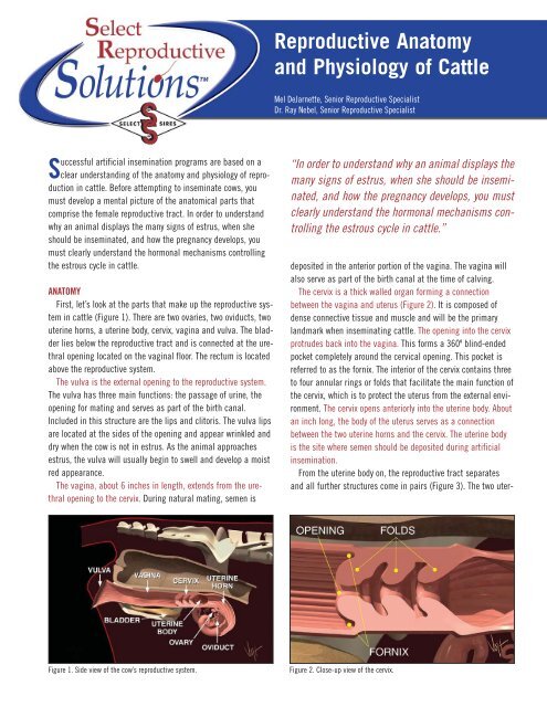
The cervix is a thick walled organ forming a connection<br />
between the vagina <strong>and</strong> uterus (Figure 2). It is composed <strong>of</strong><br />
dense connective tissue <strong>and</strong> muscle <strong>and</strong> will be the primary<br />
l<strong>and</strong>mark when inseminating cattle. The opening into the cervix<br />
protrudes back into the vagina. This forms a 360º blind-ended<br />
pocket completely around the cervical opening. This pocket is<br />
referred to as the fornix. The interior <strong>of</strong> the cervix contains three<br />
to four annular rings or folds that facilitate the main function <strong>of</strong><br />
the cervix, which is to protect the uterus from the external environment.<br />
The cervix opens anteriorly into the uterine body. About<br />
an inch long, the body <strong>of</strong> the uterus serves as a connection<br />
between the two uterine horns <strong>and</strong> the cervix. The uterine body<br />
is the site where semen should be deposited during artificial<br />
insemination.<br />
From the uterine body on, the reproductive tract separates<br />
<strong>and</strong> all further structures come in pairs (Figure 3). The two uter-<br />
Figure 1. Side view <strong>of</strong> the cow’s reproductive system.<br />
Figure 2. Close-up view <strong>of</strong> the cervix.
▼ REPRODUCTIVE ANATOMY AND PHYSIOLOGY OF CATTLE<br />
Figure 3. The short uterine body divides into two long uterine horns.<br />
ine horns consist <strong>of</strong> three layers <strong>of</strong> muscle <strong>and</strong> a heavy network<br />
<strong>of</strong> blood vessels. The main function <strong>of</strong> the uterus is to provide a<br />
suitable environment for fetal development.<br />
When a cow is bred, either naturally or by artificial insemination,<br />
the uterine muscles, under the influence <strong>of</strong> hormones oxytocin<br />
<strong>and</strong> estrogen, rhythmically contract to aid in sperm transport<br />
to the oviducts (Figure 4).<br />
Oviducts, as their name implies, carry ova, the cow’s eggs.<br />
The oviducts are also commonly referred to as the fallopian<br />
tubes. The oviduct has several distinct regions when examined<br />
microscopically.<br />
The lower segment, closest to the uterus is called the isthmus.<br />
The connection between the uterus <strong>and</strong> the isthmus is called the<br />
utero-tubal junction or UTJ. The UTJ functions as a filter <strong>of</strong><br />
abnormal sperm <strong>and</strong> the isthmus as a reservoir for healthy<br />
sperm (Figure 5).<br />
Research suggests that upon gaining access to the isthmus,<br />
healthy spermatozoa attach themselves to the walls. During this<br />
period <strong>of</strong> attachment, many physiological changes occur to<br />
Figure 5. The UTJ, isthmus <strong>and</strong> ampulla are functionally different regions <strong>of</strong> the<br />
oviduct.<br />
sperm membranes, which are essential to attainment <strong>of</strong> fertilization<br />
potential. These changes are collectively referred to as<br />
capacitation <strong>and</strong> are apparently regulated by this very important<br />
attachment to the walls <strong>of</strong> the isthmus. It takes about 5 to 6<br />
hours after insemination before a sufficient number <strong>of</strong> fertile<br />
sperm cells can populate the isthmus <strong>and</strong> complete the capacitation<br />
process.<br />
The upper portion <strong>of</strong> the oviduct, closest to the ovary, is<br />
referred to as the ampulla. The interior <strong>of</strong> the ampulla is more<br />
open than the isthmus allowing for easier passage <strong>of</strong> ova. It is<br />
within this segment <strong>of</strong> the oviduct that fertilization actually<br />
occurs. It is believed that a chemical signal released at the time<br />
<strong>of</strong> ovulation, stimulates the release <strong>of</strong> spermatozoa from the<br />
walls <strong>of</strong> the isthmus allowing them to continue their journey to<br />
the site <strong>of</strong> fertilization in the ampulla.<br />
The large funnel-like structure on the open end <strong>of</strong> the oviduct,<br />
called the infundibulum, surrounds the ovary, to recover the ova<br />
<strong>and</strong> keeps them from falling into the body cavity (Figure 6).<br />
Hair-like structures on the infundibulum <strong>and</strong> within the ampulla<br />
Figure 4. Uterine contractions aid in sperm transport.<br />
Figure 6. The infundibulum catches the egg from the ovary <strong>and</strong> guides it into<br />
the oviduct.
Figure 7. Egg <strong>and</strong> cumulus mass are transported into oviduct by hair-like<br />
structures.<br />
rhythmically beat to move ova <strong>and</strong> a surrounding mass <strong>of</strong> cells<br />
called the cumulus mass down the oviduct to the site <strong>of</strong> fertilization<br />
(Figure 7).<br />
The ovaries are the primary organs in a cow’s reproductive<br />
tract. They have two functions: to produce eggs <strong>and</strong> to produce<br />
hormones, namely estrogen <strong>and</strong> progesterone, throughout the<br />
different stages <strong>of</strong> the estrus cycle. On the surface <strong>of</strong> the ovary,<br />
you will usually find two different types <strong>of</strong> structures.<br />
Follicles are fluid filled, blister like structures that contain<br />
developing oocytes or eggs (Figure 8). Usually you will find<br />
numerous follicles on each ovary that vary in size from barely<br />
visible to ones 18 to 20mm in diameter. The largest follicle present<br />
on one <strong>of</strong> the ovaries is termed the “dominant follicle” <strong>and</strong><br />
is the most likely c<strong>and</strong>idate for ovulation when the animal<br />
comes into heat. Over time, greater than 95% <strong>of</strong> the other follicles<br />
on the ovary regress <strong>and</strong> die without ovulating <strong>and</strong> are<br />
replaced by new growing follicles.<br />
The other structure found on the ovarian surface is the corpus<br />
luteum or CL The CL is the site where ovulation occurred during<br />
Figure 9. Cross section <strong>of</strong> bovine ovary with corpus luteum.<br />
the previous cycle (Figure 9). Unless there were twin ovulations<br />
you should find only one CL located on one <strong>of</strong> the two ovaries.<br />
The CL will usually have a distinct crown protruding from the<br />
ovarian surface, which facilitates identification during rectal<br />
palpation. The CL may also have a fluid filled cavity but usually<br />
has a much thicker wall than a follicle <strong>and</strong> thus a much denser<br />
texture. “Corpus Luteum” is Latin for “yellow body.” While the<br />
outside <strong>of</strong> this structure is usually dark red in appearance, a<br />
cross section reveals a bright yellow to yellow-orange interior.<br />
THE ESTROUS CYCLE<br />
Over a period <strong>of</strong> time, many changes take place in the reproductive<br />
system in response to changing hormone levels. These<br />
changes in normal open females repeat every 18 to 21 days. This<br />
regular repetitive cycle is called the estrous cycle (Figure 10).<br />
Let’s discuss how the estrous cycle works starting with a cow<br />
in heat on day zero. Looking at the reproductive tract, we see<br />
several things happening. One ovary has a large follicle approximately<br />
15 to 20mm in diameter. This follicle has a mature egg<br />
Figure 8. Blister like follicles on the ovaries.<br />
Figure 10. The bovine estrous cycle.
Figure 11. Estrogen from growing follicle is transported through the blood<br />
stream to all parts <strong>of</strong> the body.<br />
inside ready to be released. The cells lining the follicle are producing<br />
the hormone estrogen (Figure 11). Estrogen is transported<br />
in the blood stream to all parts <strong>of</strong> the cow’s body, causing<br />
other organs to react in a number <strong>of</strong> ways. It makes the uterus<br />
more sensitive to stimulation <strong>and</strong> aids in the transport <strong>of</strong> semen<br />
at the time <strong>of</strong> insemination. It causes the cervix to secrete viscous<br />
mucus that flows <strong>and</strong> lubricates the vagina. Estrogen is<br />
also responsible for all signs <strong>of</strong> heat including; a red swollen<br />
vulva, allowing other cows to mount her, going <strong>of</strong>f feed, bellowing<br />
considerably <strong>and</strong> holding her ears erect are but a few <strong>of</strong> the<br />
many signs.<br />
On day 1, the follicle ruptures or “ovulates” releasing the egg<br />
to the waiting infundibulum (Figure 12). Several hours prior to<br />
ovulation estrogen production declines. As a result, the cow no<br />
longer displays the familiar signs <strong>of</strong> heat. After ovulation, new<br />
types <strong>of</strong> cells called, luteal cells, grow in the void on the ovary<br />
where the follicle was located.<br />
Quite rapidly over the next five to six days these cells grow to<br />
form the corpus luteum (CL). The CL produces another hormone,<br />
Figure 13. Progesterone from the corpus luteum prepares the uterus for<br />
pregnancy.<br />
progesterone. Progesterone prepares the uterus for pregnancy<br />
(Figure 13). Under the influence <strong>of</strong> progesterone, the uterus produces<br />
a nourishing substance for the embryo called uterine milk.<br />
At the same time, progesterone causes a thick mucous plug to<br />
form in the cervix, preventing access <strong>of</strong> bacteria or viruses into<br />
the uterus.<br />
Progesterone also prevents the animal from returning to<br />
estrus by regulating the release <strong>of</strong> gonadotropins from the pituitary<br />
gl<strong>and</strong> in the brain (Figure 14). There are two very important<br />
gonadotropins produced, stored <strong>and</strong> released from the pituitary<br />
gl<strong>and</strong>. The first is follicle stimulating hormone, or FSH. As its<br />
name implies FSH stimulates the growth <strong>of</strong> small follicles.<br />
Luteinizing hormone or LH is the other gonadotropin. In addition<br />
to supporting progesterone production by the CL, LH can also<br />
stimulate estrogen production in large folicles. High levels <strong>of</strong><br />
estrogen would bring the animal back into heat <strong>and</strong> make life<br />
difficult for a new embryo if she were pregnant. Thus, progesterone’s<br />
regulation <strong>of</strong> FSH <strong>and</strong> LH is a very important aspect <strong>of</strong><br />
maintaining the pregnancy.<br />
Figure 12. Luteal cells begin to grow in follicle void after ovulation.<br />
Figure 14. Progesterone regulates release <strong>of</strong> FSH <strong>and</strong> LH.
▼ REPRODUCTIVE ANATOMY AND PHYSIOLOGY OF CATTLE<br />
Figure 15. Prostagl<strong>and</strong>in destroys the corpus luteum.<br />
On the other h<strong>and</strong>, if we did not breed the cow, we do want<br />
her to come into heat again. Days 16 through 18 <strong>of</strong> the estrous<br />
cycle are referred to as “the period <strong>of</strong> maternal recognition.”<br />
During this time, the uterus searches itself for the presence <strong>of</strong> a<br />
growing embryo. If no embryo is detected, the uterus begins to<br />
produce another hormone called prostagl<strong>and</strong>in. Prostagl<strong>and</strong>in<br />
begins to destroy the CL (Figure 15). When the CL is destroyed,<br />
no more progesterone is produced <strong>and</strong> the pituitary gl<strong>and</strong> begins<br />
to increase secretion <strong>of</strong> gonadotrophins. Increased secretion <strong>of</strong><br />
LH stimulates the dominant follicle to produce estrogen <strong>and</strong><br />
bring the animal back into estrus (Figure 16).<br />
A full cycle is now completed. The average total time is about<br />
21 days. The estrous cycle is subdivided into two phases based<br />
on the dominant hormone or ovarian structure during each<br />
phase. The luteal phase begins when the corpus luteum is<br />
formed; about 5 to 6 days after the cow was in heat, <strong>and</strong> ends<br />
when the CL regresses, about day 17 to 19 <strong>of</strong> the cycle.<br />
Progesterone levels are high during this phase <strong>of</strong> the cycle <strong>and</strong><br />
estrogen levels are low.<br />
Figure 17. Estrous cycle is divided into two phases.<br />
The other phase <strong>of</strong> the cycle; the follicular phase, begins when<br />
the CL <strong>of</strong> one cycle is regressed <strong>and</strong> ends when the new CL <strong>of</strong><br />
the following cycle is formed. Thus, the follicular phase encompasses<br />
the period <strong>of</strong> time surrounding estrus (Figure 17). During<br />
this phase <strong>of</strong> the cycle estrogen levels are typically high while<br />
progesterone levels are low.<br />
As mentioned earlier, follicles may be present on the ovaries<br />
throughout the estrous cycle. Research using ultrasound technology<br />
has characterized follicular growth as occurring in<br />
“waves.” Normally, an animal will have 2 or 3 waves <strong>of</strong> follicular<br />
growth during a 21-day cycle (Figure 18). The beginning <strong>of</strong> each<br />
wave is characterized by a small rise in FSH followed by rapid<br />
growth <strong>of</strong> numerous follicles. From this wave <strong>of</strong> follicles, one follicle<br />
is selected to grow to a much larger size than the others.<br />
This “dominant” follicle has the ability to regulate or restrict<br />
growth <strong>of</strong> all other follicles on the ovary. Dominant follicles only<br />
remain dominant for a short period <strong>of</strong> time, 3 to 6 days, which is<br />
followed by either cell death <strong>and</strong> regression or ovulation <strong>and</strong><br />
release <strong>of</strong> the egg. Consequently, disappearance <strong>of</strong> the dominant<br />
Luteal Phase<br />
Follicular<br />
Phase<br />
Figure 16. FSH stimulates growth <strong>of</strong> small follicles while LH stimulates progesterone<br />
production by the CL <strong>and</strong> estrogen production by large follicles.<br />
Figure 18. Follicular growth occurs throughout the estrous cycle but estrogen<br />
levels only rise during the follicular phase.
Figure 19. Fetus interferes with prostagl<strong>and</strong>in.<br />
follicle coincides with recruitment <strong>of</strong> the next wave <strong>of</strong> follicular<br />
growth. From this new wave another dominant follicle will be<br />
selected.<br />
Although it is typical for much follicular growth to occur<br />
throughout the estrous cycle, low levels <strong>of</strong> LH during the luteal<br />
phase, prevents these follicles from producing high levels <strong>of</strong><br />
estrogen which would bring the animal back into heat. It is only<br />
the dominant follicle present at the time the CL regresses, when<br />
progesterone levels are low, that is permitted to produce enough<br />
estrogen to bring the animal back into estrus <strong>and</strong> to continue on<br />
to ovulation.<br />
EMBRYO AND FETAL DEVELOPMENT<br />
For the first four to five days, the embryo moves in the oviduct<br />
toward the uterus. Once the embryo gets there, it will be bathed<br />
in uterine fluids <strong>and</strong> continues to grow. While floating free in the<br />
uterus, several membranes, including the amnion, the chorion<br />
<strong>and</strong> the allantois are produced by the early embryo. Collectively,<br />
these membranes are referred to as the placenta.<br />
Hopefully, by the time the period <strong>of</strong> maternal recognition<br />
arrives, days 16 through 18, the fetus <strong>and</strong> growing placenta will<br />
have produced adequate quantities <strong>of</strong> the chemical signal<br />
required to maintain pregnancy. This signal interferes with the<br />
action <strong>of</strong> prostagl<strong>and</strong>in on the corpus luteum (Figure 19). The CL<br />
is thus retained <strong>and</strong> continues to produce progesterone, which is<br />
essential to maintenance <strong>of</strong> the pregnancy.<br />
At about 30 days <strong>of</strong> gestation, the placenta begins to attach<br />
to the uterus at several points. The placental sides <strong>of</strong> these<br />
attachment points are called cotyledons while the uterine side<br />
has caruncles. The attachment <strong>of</strong> cotyledons to caruncles is<br />
similar to Velcro. This greatly increases the surface area within<br />
the attachment point, facilitating exchange <strong>of</strong> nutrients <strong>and</strong><br />
waste products between calf <strong>and</strong> mother by way <strong>of</strong> arteries <strong>and</strong><br />
Figure 20. The end result.<br />
veins leading to <strong>and</strong> through the umbilical cord.<br />
At calving, the muscles in the uterus begin to contract <strong>and</strong><br />
eventually expel the calf <strong>and</strong> membranes through a dilated<br />
cervix <strong>and</strong> vagina. Several hormones including progesterone,<br />
estrogen, prolactin, relaxin <strong>and</strong> corticoids produced by the mother,<br />
the fetus, <strong>and</strong> the placenta, interact to bring about this event<br />
(Figure 20). Calving in a clean environment <strong>and</strong> proper treatment<br />
<strong>of</strong> the cow after a difficult calving will help prevent reproductive<br />
problems.<br />
Becoming familiar with the reproductive anatomy <strong>and</strong> physiology<br />
<strong>of</strong> a cow will help you do a better job artificially inseminating<br />
your cattle. Underst<strong>and</strong>ing how the hormones involved in the<br />
estrous cycle interact to control this phenomenon gives you<br />
clearer underst<strong>and</strong>ing why <strong>and</strong> when animals display the many<br />
signs <strong>of</strong> estrus, how pregnancy is maintained, <strong>and</strong> <strong>of</strong> what you<br />
or your veterinarian must do to treat cows that do not cycle<br />
normally. <br />
Telephone: (614) 873-4683<br />
Fax: (614) 873-5751<br />
www.selectsires.com<br />
SS110-1105-10.0