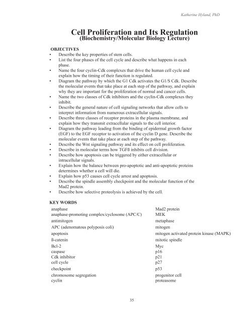
Cell Proliferation and Its Regulation - UCSF Biochemistry ...
Cell Proliferation and Its Regulation - UCSF Biochemistry ...
Cell Proliferation and Its Regulation - UCSF Biochemistry ...

Create successful ePaper yourself
Turn your PDF publications into a flip-book with our unique Google optimized e-Paper software.
Katherine Hyl<strong>and</strong>, PhD<br />
<strong>Cell</strong> <strong>Proliferation</strong> <strong>and</strong> <strong>Its</strong> <strong>Regulation</strong><br />
(<strong>Biochemistry</strong>/Molecular Biology Lecture)<br />
OBJECTIVES<br />
• Describe the key properties of stem cells.<br />
• List the four phases of the cell cycle <strong>and</strong> describe what happens in each<br />
phase.<br />
• Name the four cyclin-Cdk complexes that drive the human cell cycle <strong>and</strong><br />
explain how the timing of their function is regulated.<br />
• Diagram the pathway by which the G1 Cdk activates the G1/S Cdk. Describe<br />
the molecular events that take place at each step of the pathway, <strong>and</strong> explain<br />
why they are important for the proliferation of normal <strong>and</strong> cancer cells.<br />
• Name the two classes of Cdk inhibitors <strong>and</strong> the cyclin-Cdk complexes they<br />
inhibit.<br />
• Describe the general nature of cell signaling networks that allow cells to<br />
interpret information from numerous extracellular signals.<br />
• Describe three classes of receptor proteins in the plasma membrane, <strong>and</strong><br />
explain how they transmit extracellular signals to the cell interior.<br />
• Diagram the pathway leading from the binding of epidermal growth factor<br />
(EGF) to the EGF receptor to activation of the cyclin D gene. Describe the<br />
molecular events that take place at each step of the pathway.<br />
• Describe the Wnt signaling pathway <strong>and</strong> its effect on cell proliferation.<br />
• Describe in molecular terms how TGFß inhibits cell division.<br />
• Describe how apoptosis can be triggered by either extracellular or<br />
intracellular signals.<br />
• Explain how the balance between pro-apoptotic <strong>and</strong> anti-apoptotic proteins<br />
determines whether a cell will die.<br />
• Explain how p53 causes cell cycle arrest <strong>and</strong> apoptosis.<br />
• Describe the spindle assembly checkpoint <strong>and</strong> the molecular function of the<br />
Mad2 protein.<br />
• Describe how selective proteolysis is achieved by the cell.<br />
KEY WORDS<br />
anaphase<br />
anaphase-promoting complex/cyclosome (APC/C)<br />
antimitogen<br />
APC (adenomatous polyposis coli)<br />
apoptosis<br />
ß-catenin<br />
Bcl-2<br />
caspase<br />
Cdk inhibitor<br />
cell cycle<br />
checkpoint<br />
chromosome segregation<br />
cyclin<br />
Mad2 protein<br />
MEK<br />
metaphase<br />
mitogen<br />
mitogen activated protein kinase (MAPK)<br />
mitotic spindle<br />
Myc<br />
p16<br />
p21<br />
p27<br />
p53<br />
progenitor cell<br />
proteasome<br />
35
<strong>Cell</strong> <strong>Proliferation</strong> <strong>and</strong> its <strong>Regulation</strong><br />
KEY WORDS (Continued)<br />
cyclin-dependent kinase (Cdk)<br />
cytochrome c<br />
cytokinesis<br />
E2F transcription factor<br />
epidermal growth factor (EGF)<br />
Grb2<br />
growth factor<br />
GTPase-activating protein (GAP)<br />
GTP-binding protein (G-protein)<br />
guanine nucleotide exchange factor (GEF)<br />
Her2/neu<br />
kinetochore<br />
Rb protein<br />
Raf<br />
Ras<br />
receptor tyrosine kinase<br />
SH2 domain<br />
sister chromatid<br />
sister chromatid cohesion<br />
Sos<br />
spindle pole<br />
stem cell<br />
survival factor<br />
terminal differentiation<br />
transforming growth factor ß (TGFß)<br />
Wnt protein<br />
Optional reading:<br />
Alberts et al. Molecular Biology of the <strong>Cell</strong>; 5th Edition, Garl<strong>and</strong> Science, 2008.<br />
Chapter 17: <strong>Cell</strong> Cycle; Chapter 18: Apoptosis.<br />
Kumar, Abbas, Fausto <strong>and</strong> Mitchell. Robbins Basic Pathology; 8th Edition, Elsevier/Saunders,<br />
2007. Chapter 6 - Neoplasia: <strong>Cell</strong> Cycle, pp 188-198.<br />
I. INTRODUCTION<br />
<strong>Cell</strong> proliferation produces two cells from one, <strong>and</strong> it requires cell growth followed by<br />
cell division. Uncontrolled cell proliferation is a hallmark of cancer. As described in the<br />
overview lecture of cancer biology, multiple mutations that accumulate in somatic cells<br />
over many years eventually remove an elaborate set of controls that would otherwise<br />
prevent cancer cells from dividing unchecked. In this lecture, we will focus on the normal<br />
mechanisms that allow nearly all of the billions of cells in our body to proliferate only<br />
when they should. These mechanisms are subverted in cancer cells, <strong>and</strong> it is impossible to<br />
underst<strong>and</strong> cancer without first underst<strong>and</strong>ing the controls that keep the vast majority of<br />
the 10 14 cells (100,000 billion cells) that form the human body from misbehaving.<br />
In normal tissues, cell proliferation is generally restricted to cells that replenish the tissue.<br />
Most tissues are thought to contain stem cells that have this replenishment function<br />
(Figure 1). Stem cells are self-renewing cells that can divide asymmetrically to yield a<br />
new stem cell <strong>and</strong> a progenitor cell. Progenitor cells may or may not undergo further<br />
divisions, ultimately leading to terminal differentiation. Once cells have terminally<br />
differentiated, they have a specialized function <strong>and</strong> are no longer dividing. Most tissues<br />
are made up of such non-dividing cells. Thus proliferation is normally tightly controlled<br />
so that only particular cells in the body are dividing.<br />
<strong>Cell</strong> number is dependent not only on cell proliferation, but also on cell death.<br />
Programmed cell death, or apoptosis, is the process by which excess or damaged cells in<br />
the body are removed. Apoptosis is an extensive, ongoing process in our bodies. It is the<br />
balance between the production of new cells <strong>and</strong> cell death that maintains the appropriate<br />
36
Katherine Hyl<strong>and</strong>, PhD<br />
Stem <strong>Cell</strong> (self-renewing)<br />
Progenitor (Dividing)<br />
Figure 1. Stem <strong>Cell</strong>s. Stem cells are<br />
self-renewing cells. They can divide<br />
asymmetrically to produce a new<br />
stem cell (indicated by a circle) <strong>and</strong> a<br />
progenitor cell. Progenitor cells divide<br />
to produce cells that undergo terminal<br />
differentiation to produce the mature<br />
cells that make up a tissue or organ.<br />
Terminally Differentiated<br />
<strong>Cell</strong>s (Non-Dividing)<br />
number of cells in a tissue (referred to as homeostasis). Apoptosis is also a key<br />
mechanism by which cancer-prone cells are eliminated. Both normal apoptotic processes<br />
<strong>and</strong> normal cell mechanisms that control proliferation usually need to be altered to<br />
produce enough abnormal cell proliferation to cause cancer.<br />
II.<br />
THE CELL CYCLE<br />
<strong>Cell</strong> division occurs in defined stages, which together comprise the cell cycle. In terms<br />
of the genetic material, cells must replicate their chromosomal DNA once every cell<br />
cycle <strong>and</strong> segregate the sister chromatids produced by DNA replication to yield two<br />
genetically identical daughter cells (Figure 2). During DNA replication, cohesion<br />
proteins attach the replicated sister chromatids to each other, holding them together.<br />
This sister chromatid cohesion is critical for the subsequent alignment of each pair of<br />
sister chromatids on the mitotic spindle (see below), <strong>and</strong> it is therefore essential for the<br />
subsequent segregation of one (<strong>and</strong> only one) chromatid of each pair into each of the two<br />
daughter cells.<br />
14<br />
The cell division cycle is broken up into four stages: G 1<br />
, S, G 2<br />
<strong>and</strong> M (Figure3).<br />
DNA replication occurs during S (“synthesis”) phase. DNA packaging, chromosome<br />
segregation <strong>and</strong> cell division (cytokinesis) occur in M (mitosis). S phase <strong>and</strong> M phase<br />
are separated by Gap phases. G<br />
Thompson & Thompson GENETICS IN MEDICINE 1<br />
is the gap between M <strong>and</strong> S. <strong>Cell</strong> growth is one of the<br />
important events of G1. The transition from G1 to S is the critical control point in the cell<br />
M<br />
G 2<br />
(2-4 hr)<br />
G 1<br />
(10-12 hr)<br />
S<br />
(6-8 hr)<br />
Telomere<br />
Telomere<br />
Sister chromatids<br />
Centromere<br />
Figure 2-9 ■ A typical mitotic cell cycle, described in the<br />
text. The telomeres, the centromere, <strong>and</strong> sister chromatids<br />
are indicated.<br />
37<br />
spend a long time, days or years, in G 1 . In fact, some<br />
cell types, such as neurons <strong>and</strong> red blood cells, do not<br />
divide at all once they are fully differentiated; rather,<br />
The two sister chromatids are held together physically<br />
at the centromere, a region of DNA that associates with<br />
a number of specific proteins to form the kinetochore.<br />
Figure<br />
This complex<br />
2. The chromosome<br />
structure serves<br />
cycle.<br />
to attach<br />
A key<br />
each chromosome<br />
to of the cell microtubules division is of the the duplication mitotic spindle <strong>and</strong> to<br />
purpose<br />
of govern the genetic chromosome material movement carried on during mitosis. DNA<br />
chromosomes synthesis during <strong>and</strong> S its phase accurate is not synchronous segregation throughout<br />
such all chromosomes that each daughter or even cell within acquires a single one chromosome;<br />
copy rather, of each along chromatid. each chromosome, (Reproduced it begins at hundreds<br />
from to thous<strong>and</strong>s Thompson of & sites, Thompson called origins Genetics of DNA in replication.<br />
Medicine, Individual 7th chromosome edition, Nusbbaum, segments have et al, their own characteristic<br />
2007, time with of permission replication from during Elsevier.) the 6- to 8-hour<br />
p.14,<br />
S phase.<br />
By the end of S phase, the DNA content of the cell<br />
has doubled, <strong>and</strong> each cell now contains two copies of<br />
the diploid genome. After S phase, the cell enters a brief<br />
stage called G 2 . Throughout the whole cell cycle, ribonucleic<br />
acids <strong>and</strong> proteins are produced <strong>and</strong> the cell<br />
gradually enlarges, eventually doubling its total mass<br />
before the next mitosis. G 2 is ended by mitosis, which<br />
begins when individual chromosomes begin to condense<br />
<strong>and</strong> become visible under the microscope as thin,<br />
extended threads, a process that is considered in greater
<strong>Cell</strong> <strong>Proliferation</strong> <strong>and</strong> its <strong>Regulation</strong><br />
Figure 3. The cell cycle. Reproduced<br />
with permission from Alberts et<br />
al. Molecular Biology of the <strong>Cell</strong>.<br />
Garl<strong>and</strong> Publishing, 2002.<br />
cycle. G2 is the gap between S <strong>and</strong> M, <strong>and</strong> provides time for proofreading to ensure DNA<br />
is properly replicated <strong>and</strong> packaged prior to cell division. G0 or quiescence occurs when<br />
cells exit the cell cycle due to the absence of growth-promoting signals or presence of<br />
prodifferentiation signals. Ordered progression through each phase is intricately regulated<br />
through both positive <strong>and</strong> negative regulatory signaling molecules. The G 1<br />
, S, <strong>and</strong> G 2<br />
phases comprise interphase, which accounts for most of the time in each cell cycle.<br />
The M phase, mitosis, is relatively short (approximately 1 hour of a 24 hour cell cycle).<br />
Mitosis is itself divided into several steps, described below. (For a review of mitosis, see<br />
the Mitosis <strong>and</strong> Meiosis online module on iROCKET.)<br />
1. Assembly of the mitotic spindle: At the very beginning of M phase (called<br />
prophase), the chromosomes condense while the cytoplasmic microtubules are being<br />
reorganized to build a bipolar mitotic spindle. <strong>Its</strong> purpose is to accurately segregate<br />
the two sister chromatids to opposite poles of the cell.<br />
2. Steps leading to metaphase: The nuclear envelope then breaks down, allowing the<br />
sister chromatids, which are attached to each other through sister chromatid cohesion,<br />
to become linked to the microtubules via attachment sites on each chromatid called<br />
kinetochores. Kinetochores are protein-DNA complexes in which proteins that<br />
can capture microtubules are held tightly by DNA sequences at the centromere<br />
on each sister chromatid pair. The other end of a spindle microtubule is attached<br />
to a centrosome (the major microtubule organizing center in the cell, also called<br />
the spindle pole body), which has duplicated by this time to form the two spindle<br />
poles. Because the two kinetochores on each pair of sister chromatids are attached<br />
to opposite spindle poles, they are under tension due to pulling forces that are<br />
attempting to move them to opposite poles. Eventually, the balance between these<br />
forces causes each chromosome to line up near the center of the spindle, which marks<br />
the metaphase stage of mitosis (Figure 4).<br />
3. Anaphase: After all the chromosomes achieve bipolar attachment to spindle<br />
microtubules in metaphase, sister chromatid cohesion is rapidly dissolved. As a<br />
result, the pulling forces of the microtubules cause the two sister chromatids to move<br />
rapidly to the opposite poles (Figure 5).<br />
4. Cytokinesis: After sister chromatids segregate to opposite poles, cells physically<br />
divide into two daughter cells through a process that involves pinching in of the<br />
plasma membrane (Figure 6).<br />
38
Katherine Hyl<strong>and</strong>, PhD<br />
Figure 4. The mitotic spindle at metaphase. All of the chromosomes are lined up at the equator of<br />
the spindle. Reproduced with permission from Alberts et al. Molecular Biology of the <strong>Cell</strong>. Garl<strong>and</strong><br />
Publishing, 2008.<br />
Figure 5. Anaphase. Only three pairs of sister chromatids are shown; however, in a diploid cell,<br />
this occurs simultaneously for all 46 human chromosomes (that is, for 46 pairs of sister chromatids).<br />
(Reproduced with permission from Alberts et al. Molecular Biology of the <strong>Cell</strong>. Garl<strong>and</strong> Publishing, 2008.)<br />
Figure 6. Cytokinesis. After the two sister chromatids are segregated to opposite poles, cells undergo<br />
cytokinesis by an organized pinching in of the plasma membrane. (Reproduced with permission from<br />
Alberts et al. Molecular Biology of the <strong>Cell</strong>. Garl<strong>and</strong> Publishing, 2008.)<br />
39
<strong>Cell</strong> <strong>Proliferation</strong> <strong>and</strong> its <strong>Regulation</strong><br />
III.<br />
CELL CYCLE CONTROL: ACTIVATORS <strong>and</strong> BRAKES<br />
How is the cell cycle controlled The mechanisms of regulation can be broken down into<br />
two parts: First, how is the cell cycle regulated so that the different phases occur in the<br />
correct order Second, how do extracellular signals activate or inhibit the cell cycle This<br />
section addresses the first question, the next section (IV) addresses the second.<br />
Not until the 1980s was it discovered that a special regulatory system acts like the<br />
controller on a washing machine to drive the cell through each of its stages. This<br />
regulatory system is more than a billion years old, <strong>and</strong> most of its central components<br />
are essentially the same in single-celled eukaryotes such as yeasts <strong>and</strong> humans. This has<br />
made it possible to use the readily accessible yeast cell to dissect many of the details that<br />
underlie the normal regulatory mechanisms that control the growth of the cells in our<br />
bodies.<br />
A. Cyclin Dependent Kinases: The core activators of the cell cycle control system.<br />
The events that occur in each part of the cell cycle are carried out by specific<br />
proteins, <strong>and</strong> these proteins must be synthesized or activated at the correct time in the<br />
cycle. For example, before DNA synthesis can begin, the enzymes that produce the<br />
nucleotides used in DNA synthesis must be activated. This occurs late in G1 phase.<br />
(Remember Nucleotide Metabolism See lecture from M&N.)<br />
<strong>Cell</strong> cycle progression is positively regulated by a family of protein kinases called<br />
cyclin-dependent kinases (Cdks), which function to turn specific proteins on <strong>and</strong> off<br />
at appropriate times in the cell cycle. Like other protein kinases, Cdks turn proteins<br />
on or off by phosphorylating them. Each cyclin-dependent kinase has two subunits<br />
- a kinase subunit (the Cdk catalytic subunit) <strong>and</strong> a cyclin subunit (Figure 7). As a<br />
monomer, the Cdk has no enzymatic activity; activation requires association with a<br />
cyclin protein, which functions as an allosteric activator.<br />
Cyclins were first identified as key cell-cycle regulators when it was observed that<br />
they undergo a cycle of synthesis <strong>and</strong> regulated destruction during each cell cycle.<br />
There are several different Cdks <strong>and</strong> a number of cyclins. The kinase subunits<br />
are present throughout the cell cycle, while the cyclin subunits are produced <strong>and</strong><br />
degraded at specific times in the cell cycle, thus providing temporal regulation of the<br />
cyclin-Cdk complex. As the cyclin subunit is produced, it binds to the kinase subunit<br />
<strong>and</strong> activates it. The cyclin subunit also targets its kinase partner to specific protein<br />
substrates. The key cyclin-Cdk complexes that drive the human cell cycle are listed in<br />
Table I.<br />
The cell cycle can be viewed as a Cdk cycle (Figure 8). Activation of G1-Cdks<br />
by cyclin D turns on the events that occur in the early phase of G1. One of these is<br />
synthesis of cyclin E. As cyclin E is made, it binds to Cdk2, forming G1/S-Cdk. As<br />
the G1/S-Cdk activity accumulates to a critical threshold, it triggers the transition<br />
from late G1 into S phase. Cyclin A is made in S phase. It also binds to Cdk2,<br />
forming the S-Cdk that is required for DNA synthesis. Cyclin B is made during S<br />
phase <strong>and</strong> G2. As it is made, it binds to Cdk1 forming M-Cdk. When M-Cdk reaches<br />
a threshold activity, it triggers the transition from G2 into the prophase stage of<br />
mitosis.<br />
40
Katherine Hyl<strong>and</strong>, PhD<br />
Figure 7. Cyclin-dependent<br />
kinases (Cdks). Cdks are<br />
the key regulators of the cell<br />
division cycle in organisms as<br />
diverse as baker’s yeast <strong>and</strong><br />
humans. Cyclin-dependent<br />
kinases have two subunits, the<br />
kinase (often simply called the<br />
Cdk) <strong>and</strong> a regulatory protein<br />
called a cyclin.<br />
Table I. The four key cyclin-Cdks that drive the cell cycle<br />
Figure 8. The cell cycle as a Cdk cycle. Different phases of the cell cycle are driven by different cyclin-<br />
Cdk complexes. In this simplified view, only a G1/S-Cdk, a S-Cdk <strong>and</strong> a M-Cdk are shown. These act in<br />
sequence, as each cyclin protein is produced, to program the following critical events: the G1-S transition<br />
known as Start, S phase (DNA synthesis), <strong>and</strong> the start of M phase (mitosis). In addition, as described<br />
in the text, a G1-Cdk activated by cyclin D phosphorylates the Rb protein to produce cyclin E, which is<br />
required for G1/S-Cdk activity. Note that the activity of each Cdk disappears rapidly at a specific time<br />
in the cell cycle (as the specific cyclin protein is degraded). The APC/C is a large protein complex that<br />
controls a proteolytic process required for the separation of sister chromatids at anaphase. (Reproduced<br />
with permission from Alberts et al. Molecular Biology of the <strong>Cell</strong>. 5th Edition, Garl<strong>and</strong> Publishing, 2008;<br />
Fig. 17-16, p. 1062)<br />
41
<strong>Cell</strong> <strong>Proliferation</strong> <strong>and</strong> its <strong>Regulation</strong><br />
B. G1 regulation: How the G1-Cdk turns on the G1/S Cdk<br />
During G1, cells prepare for DNA replication. They must synthesize proteins<br />
necessary to replicate their genome, <strong>and</strong> then assemble the various components of<br />
the DNA replication machinery onto the origins of replication. This is coordinated<br />
with nutrient <strong>and</strong> growth factor availability to ensure the cell is in an environment<br />
that supports cell division. The G1 phase of the cell cycle is unique in that it<br />
represents the only time where cells are sensitive to signals from their extracellular<br />
environment. <strong>Cell</strong>s require growth factor-dependent signals up to a point in late G1,<br />
referred to as the “restriction point” or Start, after which the transition is made into S<br />
phase. The transition between early G1 <strong>and</strong> late G1 (“Start”) illustrates one way that<br />
cyclin-dependent kinases regulate the progression of the cell around the cell cycle.<br />
This is a crucial control point that is often dysregulated in cancer.<br />
In order to move from early G1 to late G1, the cell must synthesize cyclin E.<br />
Transcription of the cyclin E gene requires a transcription factor called E2F. In<br />
cells that are not proliferating <strong>and</strong> in cells that are in early G1, the E2F transcription<br />
factor is bound to the promoter for the cyclin E gene, but it is inhibited by a protein<br />
that binds it, called Rb. (Rb st<strong>and</strong>s for Retinoblastoma, a childhood tumor of the<br />
retina – more on this in the Tumor Suppressor <strong>and</strong> Oncogene lecture). Rb is a nuclear<br />
phospho-protein that plays a key role in regulating the cell cycle. It exists in an active<br />
underphosphorylated state <strong>and</strong> an inactive hyperphosphorylated state. In its active<br />
state, Rb serves as a brake that prevents advancement of cells from G1 to S phase.<br />
When G1-Cdk activity increases near the middle of G1, G1-Cdk phosphorylates the<br />
Rb protein <strong>and</strong> inactivates it (Figure 9). Inactive phosphorylated Rb releases from<br />
E2F <strong>and</strong> allows transcription of the cyclin E gene to take place. The cyclin E protein<br />
binds to the Cdk2 kinase to form the G1/S-Cdk. E2F also transcribes a number of<br />
other genes important for S phase, including the genes for DNA polymerase <strong>and</strong><br />
thymidylate synthase.<br />
A. Molecular Events B. Control Relationships<br />
G1-Cdk OFF<br />
Cdk4-cyclin D (G1-Cdk)<br />
Rb<br />
E2F<br />
promoter<br />
cyclin E gene<br />
OFF<br />
Rb<br />
E2F<br />
G1-Cdk ON<br />
Rb<br />
P<br />
Cdk2<br />
cyclin E<br />
E2F<br />
promoter<br />
cyclin E gene<br />
ON<br />
Cdk2-cyclin E (G1/S-Cdk)<br />
Figure 9. How the G1-Cdk activates the G1/S-Cdk. The G1-Cdk (Cdk4-cyclin D) phosphorylates<br />
the Rb (Retinoblastoma) protein releasing it from transcription factor E2F. E2F can now activate<br />
transcription of cyclin E, which in turn results in the production of cyclin E protein <strong>and</strong> formation of<br />
the G1/S Cdk. In describing signaling systems, it is common to use an arrow to indicate an activation<br />
<strong>and</strong> the T-shaped symbol to indicate an inhibition.<br />
42
Katherine Hyl<strong>and</strong>, PhD<br />
Importantly, the production a cyclin E, <strong>and</strong> thus CDK2-cyclin E activity, represents<br />
the transition from mitogen-dependent to mitogen–independent cell cycle progression<br />
(or passage through “start”), irreversibly committing the cells to enter S phase. Once<br />
cells enter S phase, they are committed to divide without additional growth factor<br />
stimulation. As we will discuss in later lectures, cells that acquire mutations that<br />
obviate the need for mitogen- dependent signals will bypass this crucial control point.<br />
C. Brakes on the cell cycle: Cdk inhibitors<br />
The Rb protein can be viewed as a “brake” on the cell cycle because it prevents the<br />
transcription of the gene for cyclin E by inhibiting E2F. Three other proteins that act<br />
as “brakes” on the cell cycle are the Cdk inhibitors p16, p21, <strong>and</strong> p27. These act by<br />
binding directly to Cdk-cyclin complexes <strong>and</strong> blocking their protein kinase activity<br />
(Figure 10).<br />
Cdk inhibitors fall into two classes: specific <strong>and</strong> general. The Ink4 (inhibitors of<br />
Cdk4) family of proteins, including p16, bind exclusively to <strong>and</strong> inhibit the G1 Cdks,<br />
Cdk4/6-cylin D. The Cip/Kip family of Cdk inhibitors, including p21 <strong>and</strong> p27, bind<br />
to a broad range of Cdk-cyclin complexes, shutting off the cell cycle at multiple<br />
points. Functionally, p21 <strong>and</strong> p27 appear to mainly inhibit Cdk2 complexes. As will<br />
be discussed in the Tumor Suppressor Gene <strong>and</strong> Oncogene lecture, alterations in<br />
these inhibitor proteins play an important role in cancer.<br />
Why is the cell cycle controlled by both activators (e.g. cyclins) <strong>and</strong> inhibitors (e.g.<br />
Rb, p16, p21, p27) As we will see, it helps each cell to respond to multiple inputs,<br />
so that it enters the cell cycle only when the correct combination of conditions are<br />
present. The control of cycle entry by both growth activating <strong>and</strong> growth inhibiting<br />
signals is part of a “fail-safe” system for insuring that cell proliferation only occurs<br />
when it is useful to a multicellular organism like ourselves. Without a complex<br />
control system of this type, humans could not exist, because we would all get cancer<br />
at a very early age (probably in utero).<br />
Figure 10. How Cdk inhibitors bind to <strong>and</strong> inactivate Cdks. (Reproduced with permission from Alberts<br />
et al. Molecular Biology of the <strong>Cell</strong>. Garl<strong>and</strong> Publishing, 2008.<br />
43
<strong>Cell</strong> <strong>Proliferation</strong> <strong>and</strong> its <strong>Regulation</strong><br />
IV.<br />
CONTROLLING PROLIFERATION: MITOGENS, ANTI-MITOGENS, <strong>and</strong><br />
CELL SIGNALING<br />
A. General Principles of <strong>Cell</strong> Signaling<br />
<strong>Cell</strong> signaling processes are central to all of human biology <strong>and</strong> medicine. Although<br />
the details of cell signaling pathways can become very complex, the big picture of<br />
cell signaling (e.g. transmitting information from the extra-cellular environment<br />
into the cell so it can respond appropriately) is straightforward. Signaling<br />
pathways are built from a limited set of molecules <strong>and</strong> molecular mechanisms (e.g.<br />
phosphorylation or proteolysis) that allow for communication within <strong>and</strong> between<br />
cells. The underlying molecular mechanisms used in signaling pathways show a<br />
number of common properties. In particular, they allow signaling proteins to undergo<br />
switch-like activation from an inactive to an active state (for example by receptor<br />
clustering, GTP-binding to Ras proteins, <strong>and</strong> stabilization of β catenin, as described<br />
below) <strong>and</strong> they can also be readily reversed (e.g. by receptor down-regulation,<br />
hydrolysis of bound GTP, <strong>and</strong> β catenin degradation). Dr. Fulton introduced the<br />
subject of signaling last year, <strong>and</strong> it is important to review that material for this block<br />
(see lectures on Protein Function <strong>and</strong> Signaling from Prologue). The introductory<br />
lectures focused on one of the two major classes of cell surface receptor proteins<br />
present in all cells: the G-protein-linked receptor family. The other major class is<br />
referred to as the enzyme-linked receptor family. This class includes receptors linked<br />
to protein kinases, which fall into two subgroups: the receptor tyrosine kinases<br />
(RTKs) <strong>and</strong> the receptor serine/threonine kinases. An example of each is discussed<br />
below.<br />
B. Mitogens <strong>and</strong> Anti-Mitogens<br />
Non-dividing cells exist in phase called G0 (G zero). G0 cells can re-enter the cell<br />
cycle in G1 when stimulated by mitogens, which are extracellular proteins that<br />
stimulate cell proliferation by directly controlling the entry of cells into the cell<br />
cycle. (For historical reasons, mitogens are often loosely referred to as growth<br />
factors. Although it is best to reserve the latter term for those signaling molecules<br />
that actually induce cell growth, i.e. the increase in cell mass, these terms are often<br />
used interchangeably). Conversely, cells can be arrested in G1 via the action of<br />
Lig<strong>and</strong><br />
Binding<br />
Domain<br />
Kinase<br />
Domain<br />
Tyrosine<br />
residue<br />
EGF<br />
P SH2<br />
SH3<br />
Grb2<br />
(adaptor protein)<br />
Sos<br />
Figure 11. Activation of the Epidermal<br />
Growth Factor (EGF) receptor tyrosine<br />
kinase. EGF binds to the EGF receptor through<br />
an extracellular lig<strong>and</strong> binding domain, leading<br />
to dimerization of the receptor. Dimerization<br />
causes one subunit to phosphorylate the other<br />
(transphosphorylation) on specific tyrosine<br />
residues. The SH2 domain of the Grb2 adaptor<br />
protein then binds to the region of the EGF<br />
receptor containing the phosphorylated<br />
tyrosines. Grb2 in turn, uses its second<br />
common protein domain, called SH3, to bind<br />
to another protein called Sos. Grp2 is known<br />
as an adapter protein, since it function to hold<br />
two other proteins together. Sos is a member<br />
of a large family of proteins that regulate G<br />
proteins (GTP-binding proteins) by causing the<br />
exchange of a tightly bound GDP molecule for<br />
GTP (see Figure 14).<br />
44
Katherine Hyl<strong>and</strong>, PhD<br />
anti-mitogens (proteins that inhibit the activity of mitogens). Many mitogens <strong>and</strong> a<br />
smaller number of anti-mitogens are known. We will discuss one example of each:<br />
the mitogen epidermal growth factor (EGF) <strong>and</strong> the anti-mitogen transforming<br />
growth factor ß (TGFß). The receptors for these factors are both enzyme-linked<br />
receptors. The EGF receptor, or EGFR, is an example of a receptor tyrosine kinase<br />
(RTK), <strong>and</strong> the TGFß receptor is a receptor serine/threonine kinase,<br />
What are the normal functions of these factors One function of EGF is to promote<br />
wound healing. After a wound is formed, epidermal <strong>and</strong> inflammatory cells secrete<br />
EGF <strong>and</strong> other growth factors. It signals cells at the margins of the wound to<br />
proliferate so that the wound may be healed. TGFß acts as a brake to this process so<br />
that the proliferation is coordinated with other aspects of wound healing.<br />
C. The EGF Signaling Pathway<br />
The EGF receptor belongs to the ErbB family of RTKs, which has four members<br />
capable of homo– or heterodimerization. Each receptor heterodimer can respond to a<br />
distinct set of extracellular lig<strong>and</strong>s <strong>and</strong> has different intracellular signaling properties.<br />
Interestingly, another member of the ErbB family, the ErbB2 receptor (also called<br />
HER2/neu) lacks intrinsic growth factor-binding activity. Consequently, in normal<br />
cells HER2/neu must function as part of a heterodimer with another ErbB family<br />
member, such as EGF. (More about HER2/neu <strong>and</strong> its role in breast cancer in later<br />
lectures.)<br />
EGF functions by binding to the extracellular domain of EGF receptor, a cell<br />
surface protein with a single transmembrane domain (Figure 11). The cytoplasmic<br />
domain of the receptor is the protein tyrosine kinase. When EGF binds to its<br />
receptor, the receptor forms a dimer in which one subunit phosphorylates the other<br />
(transphosphorylation) on particular tyrosine residues in the cytoplasmic part of the<br />
receptor. These phosphorylated tyrosines serve as binding sites for other cytoplasmic<br />
proteins that contain special domains, called SH2 domains. SH2 domains specifically<br />
recognize phosphorylated tyrosines <strong>and</strong> the adjacent amino acids. One protein that<br />
binds to phosphotyrosine residues in the EGF receptor is an adaptor protein called<br />
Grb2. Grb2, in turn, recruits a protein called Sos. Thus binding of EGF to the EGF<br />
receptor recruits both Grb2 <strong>and</strong> Sos to the intracellular portion of the receptor.<br />
Sos<br />
GDP<br />
GTP<br />
Ras-GDP<br />
Ras-GTP<br />
inactive<br />
active<br />
GAP<br />
GTPase-activating protein<br />
45<br />
Figure 12. Sos is a guanine<br />
nucleotide exchange protein (GEF)<br />
that activates the Ras protein. Ras<br />
is a monomeric GTP-binding protein<br />
that is only active in its GTP-bound<br />
form. In its GDP bound form, Ras<br />
is inactive. When Sos binds to Grb2<br />
at the EGF receptor, it is brought<br />
close to membrane-bound Ras-GDP<br />
molecules, causing the Ras to release<br />
its GDP <strong>and</strong> bind a GTP in its place.<br />
A second common type of protein is<br />
a GTPase-activating protein (GAP),<br />
which inactivates Ras by promoting<br />
its GTP hydrolysis. The cell contains<br />
hundreds of monomeric GTP-binding<br />
proteins that serve to regulate many<br />
different functions. Each is regulated<br />
in a similar way by GEFs <strong>and</strong> GAPs.
<strong>Cell</strong> <strong>Proliferation</strong> <strong>and</strong> its <strong>Regulation</strong><br />
MEK<br />
inactive<br />
Ras-Raf<br />
MAPK<br />
inactive<br />
MEK-P<br />
active<br />
MAPK-P<br />
active<br />
Target Target-P<br />
inactive<br />
active<br />
Figure 13. Ras activates the MAP<br />
kinase cascade. Ras-GTP binds<br />
directly to Raf, which activates its<br />
kinase activity. Raf phosphorylates<br />
a kinase called MEK (also called<br />
MAP kinase kinase). After it has<br />
been phosphorylated by Raf, MEK<br />
phosphorylates MAP kinase (mitogen<br />
activated protein kinase, MAPK).<br />
Active MAPK then phosphorylates<br />
its target proteins, including<br />
transcription factors, stimulating the<br />
entry of the cell into the cell cycle.<br />
Sos is a guanine nucleotide exchange factor (GEF). It acts on a small monomeric<br />
GTP binding protein, Ras. The Ras protein is bound to the inner surface of the<br />
plasma membrane. Like the G-proteins discussed by Dr. Fulton in the Prologue, Ras<br />
can exist in two states: an inactive state in which GDP is bound, <strong>and</strong> an active state<br />
in which GTP is bound. Sos activates Ras by promoting the release of its GDP <strong>and</strong><br />
binding of GTP (Figure 14). Recruitment of Sos to the plasma membrane where Ras<br />
is located results in the activation of Ras. Ras can be returned to its inactive form<br />
through the hydrolysis of GTP to GDP. This step occurs when a GTPase-activating<br />
protein (GAP) binds Ras <strong>and</strong> induces the hydrolysis of its GTP (see Figure 12).<br />
In its GTP-bound (active) state, Ras turns on a protein kinase cascade, in which<br />
protein kinases sequentially activate each other through phosphorylation (Figure<br />
13). Active Ras binds to <strong>and</strong> activates a protein kinase called Raf. In turn, Raf<br />
phosphorylates <strong>and</strong> activates another kinase called MEK (MAP kinase kinase).<br />
MEK in turn phosphorylates <strong>and</strong> activates mitogen-activated protein kinase, MAP<br />
kinase. This chain of phosphorylation events is called the MAP kinase cascade.<br />
MAP kinase phosphorylates gene-specific transcription factors in the cell nucleus<br />
that bind to the promoters of genes <strong>and</strong> promote cell proliferation. One important<br />
transcription factor that is up-regulated by the MAP kinase cascade is Myc, which is<br />
the product of the c-MYC gene.<br />
One of the targets of transcription factors that are activated by the MAP kinase<br />
cascade is the cyclin D gene. Thus, a multi-tiered pathway connects the presence of<br />
a mitogen (EGF) outside the cell to increased expression of a key component of the<br />
cell cycle control machinery (the cyclin D gene) in the nucleus (Figure 14). Increased<br />
expression of the cyclin D gene leads to the activation of G1-Cdk, pushing the cell to<br />
proliferate, as explained previously.<br />
MAPK<br />
Transcription<br />
Factors<br />
(e.g. Myc)<br />
cyclin D<br />
Figure 14. Activation of MAP kinase leads to the transcription of cyclin D. MAPK phosphorylates<br />
transcription factors. This in turn leads to the transcription of the Myc gene, which itself encodes a<br />
transcription factor for the cyclin D gene.<br />
46
Katherine Hyl<strong>and</strong>, PhD<br />
D. Wnt signaling<br />
The Wnt proteins are mitogens analogous to EGF. They function in a signaling<br />
pathway that regulates cell proliferation by controlling proteolysis of a key signaling<br />
protein (Figure 15). The Wnt signaling pathway plays a central role during embryonic<br />
development, <strong>and</strong> also serves important functions in adults. For example, Wnt<br />
signaling is necessary for the proliferation of stem cells in the proliferative zones<br />
in the gut epithelium (the crypts that lie between the microvilli of the epithelium)<br />
(Figure 16). Colon cancer is almost invariably associated with the hyperactivation of<br />
this pathway in an early step of tumor evolution.<br />
As illustrated in Figure 15, Wnt proteins bind to a cell surface receptor called<br />
Frizzled. Frizzled controls the stability of a protein called ß-catenin, which functions<br />
together with a protein called TCF to form a transcription factor that activates the<br />
promoter of the cyclin D gene.<br />
When Wnt is bound, Frizzled turns off a protein kinase called GSK-3. GSK-3<br />
normally functions to promote the degradation of ß-catenin, thus preventing it from<br />
activating the cyclin D promoter. Phosphorylation of ß-catenin by the protein kinase<br />
GSK-3 results in its degradation. However, GSK-3 can only phosphorylate ß-catenin<br />
when ß-catenin is bound to a protein called APC (adenomatous polyposis coli).<br />
Thus, APC is necessary to hold ß-catenin in check, <strong>and</strong> loss or inactivation of APC<br />
is associated with development of colorectal cancer (as described in later lectures).<br />
(Note: this APC protein is not to be confused with APC/C, the anaphase promoting<br />
complex/cyclosome, to be described later in this lecture.)<br />
Wnt<br />
Frizzled<br />
GSK-3<br />
P<br />
P<br />
b-catenin<br />
APC<br />
b-catenin<br />
APC<br />
degradation<br />
TCF<br />
APC<br />
b-catenin<br />
TCF<br />
cyclin D<br />
Figure 15. The Wnt signaling pathway (see text for details).<br />
47
<strong>Cell</strong> <strong>Proliferation</strong> <strong>and</strong> its <strong>Regulation</strong><br />
microvillus<br />
microvillus<br />
high<br />
Expression<br />
of APC<br />
crypt<br />
(proliferating<br />
stem cells)<br />
Figure 16. Expression of the APC gene in the gut epithelium. Shown is a<br />
schematic of a microvillus in the gut epithelium showing the zone of proliferation<br />
(crypts) <strong>and</strong> the gradient of expression of the APC gene, whose protein product<br />
inhibits Wnt signaling.<br />
low<br />
Once GSK-3 is inhibited by Frizzled, ß-catenin is no longer degraded, allowing it to<br />
associate with TCF <strong>and</strong> activate the cyclin D promoter <strong>and</strong> promote cell proliferation.<br />
Thus, Wnt signaling promotes cell proliferation through the effect of ß-catenin on<br />
cyclin D production.<br />
While Wnt proteins are the extracellular growth factors that activate this pathway,<br />
cells also control the pathway from within the cell by varying the transcription<br />
of the APC gene, whose protein product inhibits the Wnt signaling pathway. For<br />
example, in the epithelium of the colon, there exists a gradient of APC expression<br />
that is highest in the terminally differentiated nondividing cells in the microvilli <strong>and</strong><br />
lowest in the proliferating stem cells in the crypts (see Figure 16). (More about the<br />
role of APC in colon cancer to come in lectures on Colon Cancer <strong>and</strong> Familial <strong>and</strong><br />
Hereditary Cancer Syndromes.)<br />
E. TGFß-Smad: An anti-mitogenic pathway<br />
Like EGF, TGFß is an extracellular protein that binds a cell surface receptor.<br />
However, instead of causing cell proliferation, this molecule causes cells to<br />
arrest their cell cycle <strong>and</strong> enter G0. How does this occur The TGFß receptor is<br />
a transmembrane serine/threonine kinase. Upon binding to TGFß, the receptor<br />
phosphorylates proteins in the cytoplasm called Smads (Figure 17). Once<br />
phosphorylated, Smad proteins then enter the nucleus <strong>and</strong> function as transcription<br />
factors to turn on specific target genes. A key gene turned on by TGFß is the Cdk<br />
inhibitor p21 discussed above. The activation of p21 blocks G1-S transition by<br />
inhibiting Cdk2-Cyclin E/A, leading to the arrest of the cell cycle. Thus, TGFß<br />
arrests cell division by turning on transcription of the gene for a Cdk inhibitor.<br />
V. APOPTOSIS<br />
As previously explained, the number of cells in a tissue is controlled not only by cell<br />
proliferation, but also by programmed cell death, or apoptosis. For a tissue to stay the<br />
same size, cell proliferation <strong>and</strong> cell death must be perfectly balanced. Apoptosis plays<br />
important roles both during development <strong>and</strong> in mature tissues. For example, during<br />
development of a limb, tissue present between the digits must be removed. This occurs<br />
through localized apoptosis (Figure 18).<br />
48
Katherine Hyl<strong>and</strong>, PhD<br />
TGF<br />
Smad<br />
Smad-P<br />
Smad-P<br />
promoter<br />
kinase<br />
domain<br />
p27 gene<br />
TGF receptor<br />
plasma<br />
membrane<br />
p21<br />
Cdk4-cyclin D<br />
Cdk2-cyclin E<br />
Cdk2-cyclinA<br />
Figure 17. How TGFß<br />
arrests cell division. TGFß<br />
binds to the TGFß receptor.<br />
Binding of TGFß activates<br />
the receptor’s intracellular<br />
protein kinase domain,<br />
leading to phosphorylation of<br />
Smad proteins on serine <strong>and</strong><br />
threonines. Phosphorylated<br />
Smads enter the nucleus <strong>and</strong><br />
bind to promoters of genes to<br />
control transcription. A key<br />
target is the p21 gene. The<br />
p21 protein in turn inhibits<br />
cyclin E/A- cdk2 complexes,<br />
thus leading to cell cycle<br />
arrests.<br />
nucleus<br />
As described in the Prologue block, the process of apoptosis requires the activation of a<br />
special class of proteases inside the cell known as caspases. Caspase molecules normally<br />
exist as inactive procaspase molecules in the cell. Procaspase activation is carefully<br />
controlled, so that the cell only kills itself when this is appropriate for the success of the<br />
organism as a whole.<br />
A. <strong>Cell</strong>-surface death receptors activate an extrinsic apoptotic pathway<br />
Procaspase activation can be initiated from outside the cell, as happens in the immune<br />
system when T cells kill their target cells by producing a signaling protein called<br />
Fas lig<strong>and</strong>. The Fas lig<strong>and</strong> binds to its receptor, Fas, on target cells. The cytoplasmic<br />
domain of a “death receptor” such as Fas is then triggered to bind adaptor proteins<br />
that link the receptor to procaspase-8 molecules. The aggregated procaspase-8<br />
molecules are thereby stimulated to cleave each other, initiating a proteolytic cascade<br />
that leads to apoptosis (Figure 19A).<br />
B. An intrinsic apoptotic pathway depends on mitochondria<br />
When cells are stressed (e.g., hypoxia), damaged (e.g., unrepaired DNA damage), or<br />
become abnormal in other ways, they can activate apoptosis from inside the cell by<br />
triggering a similar process of procaspase aggregation <strong>and</strong> activation. In response to<br />
stress or damage, pro-apoptotic signals induce mitochondria to release cytochrome<br />
c into the cytosol, where it binds <strong>and</strong> activates an adaptor protein called Apaf-1. This<br />
causes Apaf-1 to aggregate into a wheel-like complex called an apoptosome. This<br />
aggregate then recruits a set of procaspase-9 molecules, which become activated to<br />
trigger a caspase cascade causing cell death (Figure 19B).<br />
49
<strong>Cell</strong> <strong>Proliferation</strong> <strong>and</strong> its <strong>Regulation</strong><br />
Apoptosis<br />
Figure 18. Programmed cell death. Fas During lig<strong>and</strong> development of limb, tissue present<br />
Fas<br />
between the digits is removed by apoptosis.<br />
Apoptosis<br />
Killer T-cell<br />
Target <strong>Cell</strong><br />
Figure 19. Induction of apoptosis by either extracellular or intracellular signals. (A) Extracellular<br />
activation. Adaptor proteins bind the intracellular region of aggregated Fas proteins, causing the<br />
aggregation of procaspase-8 molecules. These then cleave one another to initiate the caspase cascade. (B)<br />
Intracellular activation. Mitochondria release cytochrome c, which binds to <strong>and</strong> causes the aggregation of<br />
the adaptor protein Apaf-1. Apaf-1 binds <strong>and</strong> aggregates procaspase-9 molecules, which are activated to<br />
trigger a caspase cascade, leading to apoptotic cell death (From Alberts et al., Molecular Biology of the<br />
<strong>Cell</strong>, 2002)<br />
50
Katherine Hyl<strong>and</strong>, PhD<br />
Domains BH 1, 2, 3 BH 1, 2, 3, 4 BH 3 only<br />
Function Pro-apoptotic Anti-apoptotic Pro-apoptotic<br />
Examples Bak, Bax Bcl-2 Bad, Bid, Puma<br />
Table 2. Three subclasses of proteins in the Bcl-2 family that control apoptosis by the<br />
intracellular (intrinsic) pathway.<br />
The release of cytochrome c from the mitochondria is tightly controlled by members<br />
of the Bcl-2 family of proteins, all of which contain at least one BH protein domain.<br />
Within this family of proteins, there are three sub-classes (Table 2): two subclasses<br />
promote apoptosis (the “pro-apoptotic” BH123 proteins, which contain 3 different<br />
BH protein domains, <strong>and</strong> the BH3-only proteins), <strong>and</strong> one subclass antagonizes<br />
apoptosis (the “anti-apoptotic” Bcl-2 proteins). The BH3 domain is the only domain<br />
shared by all three subclasses of proteins, <strong>and</strong> it can mediate a direct binding<br />
interaction between one pro-apoptotic protein <strong>and</strong> one anti-apoptotic protein to form<br />
heterodimers. The central players are the BH123 family members, Bak <strong>and</strong> Bax,<br />
which can form channels in the mitochondrial outer membrane that cause cytochrome<br />
c <strong>and</strong> other proteins in the mitochondrion’s intermembrane space to be released into<br />
the cytoplasm, thereby activating procaspase-9 via Apaf-1. The anti-apoptotic Bcl-2<br />
proteins appear to bind directly to Bak <strong>and</strong> Bax to inhibit them, thereby serving to<br />
keep the cell alive. The remaining BH3-only pro-apoptotic subclass is composed<br />
of a large number of proteins that bind to various subsets of the anti-apoptotic<br />
Bcl-2 proteins, forming heterodimers with them. If large enough amounts of the<br />
BH3 proteins are present in the right combinations, they will dissociate all of these<br />
inhibitors from Bak <strong>and</strong> Bax, thereby permitting the channel formation <strong>and</strong> inducing<br />
cell death (Figure 20).<br />
In summary, it is the balance between the activities of the set of anti-apoptotic Bcl-2<br />
proteins <strong>and</strong> the two subclasses of pro-apoptotic proteins that determines whether<br />
a mammalian cell lives or dies by the intrinsic pathway of apoptosis. This balance<br />
is determined through a complex <strong>and</strong> poorly understood signaling network that<br />
continually monitors the state of the cell. For example, only if a cell is in its expected<br />
location in the organism will it receive the specific survival signals that it requires to<br />
prevent apoptosis. Thus it is not surprising that cancer cells often acquire mutations<br />
that allow them to alter the balance between pro- <strong>and</strong> anti-apoptotic proteins, making<br />
it less likely for them to commit suicide even under conditions when normal cells<br />
would.<br />
VI.<br />
p53, THE CELL CYCLE, <strong>and</strong> APOPTOSIS<br />
The cell cycle is controlled at certain stages by checkpoints. These are biochemical<br />
mechanisms that stop the cell cycle if certain conditions are not met.<br />
One checkpoint is the G1 DNA damage checkpoint. If cells contain unrepaired damage to<br />
their DNA, the cell cycle is arrested in G1. This arrest requires a key transcription factor,<br />
p53, which is activated by DNA damage (Figure 21). There are three components to the<br />
system: 1) a DNA damage sensor, 2) the Mdm2 protein that normally causes p53 to be<br />
degraded, <strong>and</strong> 3) the p53 protein itself. DNA damage causes phosphorylation of p53 <strong>and</strong><br />
blocks the binding of Mdm2. This leads to the stabilization <strong>and</strong> accumulation of p53. p53<br />
can then bind to the promoter of the p21 Cdk inhibitor described earlier <strong>and</strong> activate its<br />
transcription, causing p21 to accumulate. The resulting inhibition of Cdks leads to cell<br />
cycle arrest.<br />
51
<strong>Cell</strong> <strong>Proliferation</strong> <strong>and</strong> its <strong>Regulation</strong><br />
Figure 20. How pro-apoptotic BH3-<br />
only <strong>and</strong> anti-apoptotic Bcl2 proteins<br />
regulate the intrinsic pathway of<br />
apoptosis. (A) In the absence of an<br />
apoptotic stimulus, anti-apoptotic<br />
Bcl2 proteins bind to <strong>and</strong> inhibit the<br />
BH123 proteins on the mitochondrial<br />
outer membrane (<strong>and</strong> in the cytosol -<br />
not shown). (B) In the presence of an<br />
apoptotic stimulus, BH3-only proteins are<br />
activated <strong>and</strong> bind to the anti-apoptotic<br />
Bcl2 proteins so that they can no longer<br />
inhibit the BH123 proteins, which no<br />
become activated an aggregate in the<br />
outer mitochondrial membrane <strong>and</strong><br />
promote the release of intermembrane<br />
mitochondrial proteins into the cytosol.<br />
Some activated BH3-only proteins may<br />
stimulate mitochondrial protein release<br />
more directly by binding to <strong>and</strong> activating<br />
the BH123 proteins. Although not shown,<br />
the anti-apoptotic Bcl2 protins are bound<br />
to the mitochondrial surface. (Reproduced<br />
with permission from Alberts et al.<br />
Molecular Biology of the <strong>Cell</strong>. Garl<strong>and</strong><br />
Publishing, 2008. Figure e18-11, p. 1124.)<br />
If p53 activation continues for a prolonged period of time, apoptosis ensues. This<br />
process kills cells with damaged DNA that remain unrepaired, <strong>and</strong> serves to remove cells<br />
from tissues that may otherwise accumulate mutations that would be passed on to their<br />
daughter cells. High levels of p53 are thought to activate apoptosis by increasing the<br />
transcription of several genes. One target gene is the BH123 protein Bax, whose gene is<br />
directly activated by p53 (Figure 22).<br />
In light of the important role p53 plays in preventing unrepaired DNA damage to be<br />
passed on to daughter cells, it is not surprising that p53 is found to play a central role in<br />
cancer development. In fact, the p53 pathway is mutated in nearly all cancers, thereby<br />
allowing damaged DNA to remain in cells as they proliferate (more in the lecture on<br />
Tumor Suppressor Genes <strong>and</strong> Oncogenes).<br />
52
Katherine Hyl<strong>and</strong>, PhD<br />
Figure 21. How DNA damage<br />
activates p53 <strong>and</strong> causes cellcycle<br />
arrest. DNA damage<br />
activates a protein kinase that<br />
phosphorylates p53, preventing<br />
its degradation. This leads to the<br />
production of high levels of the<br />
Cdk inhibitor p21. (reproduced<br />
with permission from Alberts et<br />
al. Molecular Biology of the cell.<br />
5th Edition, Garl<strong>and</strong> Publishing,<br />
2008: Fig 17-63.)<br />
Prolonged<br />
p53<br />
Activation<br />
p53<br />
promoter<br />
BAX gene<br />
Figure 22. DNA damage can lead to<br />
apoptosis. Prolonged activation of p53 in<br />
response to DNA damage results in apoptosis.<br />
p53 activates the transcription of<br />
several genes involved in apoptosis including<br />
that for the pro-apoptotic BH123 protein<br />
Bax shown here.<br />
cytochrome C<br />
apoptosis<br />
Bax channel<br />
53
<strong>Cell</strong> <strong>Proliferation</strong> <strong>and</strong> its <strong>Regulation</strong><br />
VII.<br />
THE SPINDLE ASSEMBLY CHECKPOINT: THE IMPORTANCE OF<br />
REGULATED PROTEOLYSIS IN THE CELL<br />
In addition to monitoring the state of DNA in G1 before entering S phase, cells also<br />
monitor the state of the cell at several other checkpoints. One, called spindle assembly<br />
checkpoint, ensures that mitosis does not proceed beyond metaphase until the spindle is<br />
properly assembled. This checkpoint monitors the attachment of spindle microtubules<br />
to each kinetochore through the action of the Mad2 protein (Figure 23). There are two<br />
key features of the checkpoint: 1) Mad2 associates with kinetochores only when they<br />
are not attached to microtubules, <strong>and</strong> 2) Mad2 becomes activated for arresting mitosis<br />
only when bound to such kinetochores. If even one of the 46 human chromosomes<br />
is not attached correctly to microtubules, enough Mad2 is activated to keep the cell<br />
in metaphase. Only when the spindle has been properly assembled with all of the<br />
kinetochores bound to microtubules does Mad2 becomes inactive <strong>and</strong> allow anaphase to<br />
proceed. If there is a problem with spindle assembly, Mad2 will arrest the cell cycle until<br />
the problem is resolved.<br />
Active Mad2 exerts its effects by blocking the key regulator of the metaphase-toanaphase<br />
transition, the anaphase-promoting complex/cyclosome (APC/C). The APC/C<br />
is a member of a large family of important enzymes, called ubiquitin ligases, that trigger<br />
the regulated destruction of target proteins in the cell. The actual proteolysis is carried out<br />
by proteasomes, large protein complexes that pump selective proteins into their interior<br />
in order to cleave them into small fragments.<br />
As a ubiquitin ligase, the APC/C marks proteins for uptake into proteasomes by<br />
covalently adding multiple copies of the small protein called ubiquitin to them. The<br />
polyubiquitin chain added to a protein is recognized by the proteasome, causing the<br />
protein to be destroyed. One of the destroyed proteins is an inhibitor of the protein that<br />
centrosome<br />
microtubules<br />
Mad2<br />
cell cycle arrest<br />
kinetochore<br />
Mad2<br />
inactive<br />
Sister<br />
Chromatids<br />
Figure 23. Spindle assembly checkpoint. This checkpoint functions through the action of the Mad2<br />
protein, which binds to kinetochores that have not attached to microtubules. When bound to kinetochores,<br />
Mad2 triggers cell cycle arrest. Once microtubules are attached to all of the kinetichores, Mad2 is no<br />
longer active <strong>and</strong> the cell cycle proceeds.<br />
54
Katherine Hyl<strong>and</strong>, PhD<br />
cuts the linkages holding the sister chromatid pairs together. The removal of the inhibitor<br />
allows the separation of sisters <strong>and</strong> unleashes anaphase. The S- <strong>and</strong> M-cyclins are the<br />
second major targets of the APC/C. The destruction of these cyclins inactivates the<br />
corresponding Cdks (see Figure 8). As a result, the many proteins phosphorylated by<br />
Cdks from S phase to early mitosis are dephosphorylated by various protein phosphatases<br />
that are present in the anaphase cell. This dephosphorylation of Cdk targets is required<br />
for the completion of M phase, including the final steps in mitosis <strong>and</strong> the process of<br />
cytokinesis. Not surprisingly, cells defective in the spindle assembly checkpoint show<br />
high rates of aneuploidy because of errors in chromosome segregation during mitosis.<br />
Defects in the spindle-assembly checkpoint, <strong>and</strong> specifically in Mad2, have been<br />
associated with tumorigenesis.<br />
VIII.<br />
THE EVOLUTION OF CELL SIGNALING AND CANCER<br />
Now that we have explored the key aspects of normal cell proliferation, we can begin<br />
to consider what goes awry in cancer cells. The following section considers theoretical<br />
aspects of the evolution of cell signaling <strong>and</strong> cancer to provide you with context for<br />
thinking about cancer development <strong>and</strong> treatment. (From Dr. Bruce Alberts)<br />
A. Elaborate cell signaling mechanisms had to evolve in multicellular organisms to<br />
prevent cancer. Various types of evidence suggest that single-celled life was present on<br />
the earth 3.5 billion years ago, about a billion years after the earth formed (prokaryotic<br />
cells such as bacteria). However, it appears to have required another two billion years<br />
to evolve the first multicellular organisms. Initially these were very small aggregates of<br />
eukaryotic cells that had learned how to cooperate, with each cell restraining its own<br />
growth for the good of the entire aggregate. Although this had the advantage of allowing<br />
each type of cell to specialize, it meant that each cell had to send <strong>and</strong> receive an elaborate<br />
set of signals to determine its appropriate behavior, <strong>and</strong> that fail-safe controls had to<br />
evolve to prevent the type of selfish cell behavior that we call cancer.<br />
As larger <strong>and</strong> larger organisms evolved, major improvements to these fail-safe controls<br />
had to develop in the form of multiple, largely redundant systems that prevent aberrant<br />
cell proliferation. Why Even with the overlapping set of proofreading mechanisms that<br />
allow us to replicate the three billion (3 x 10 9 ) nucleotide pairs in the human genome with<br />
an error rate of only about one in a billion (10 -9 ), the fact that humans are formed from<br />
about 10 14 cells means that billions of cells experience mutations every day, potentially<br />
disrupting the normal controls on cell growth. Viewed from this perspective, the<br />
surprising thing about large multicellular organisms is how infrequently cells misbehave<br />
to create a tumor. As we shall see, the reason we do not all die of cancer is that, in<br />
general, many different mutations need to accumulate in a single line of cells to cause<br />
this disease – perhaps 10 to 20. Obtaining a better underst<strong>and</strong>ing the multiple layers of<br />
control that are circumvented during tumorigenesis will be key to controlling cancer.<br />
Unfortunately, there is still much to learn in this critical area of research.<br />
B. <strong>Cell</strong>s integrate the many signals that they receive in deciding whether to survive,<br />
grow <strong>and</strong> divide (proliferate), differentiate, or die (apoptosis). Every cell contains<br />
many different cell surface receptor molecules, each of which recognizes a particular<br />
molecule at the cell exterior. Some of these bind to signaling molecules that have been<br />
secreted by neighboring cells, others bind to protein molecules held in the plasma<br />
membrane of tightly opposed adjacent cells, while others bind to the extracellular matrix.<br />
All of these signal molecules work in combinations to regulate the behavior of the cell,<br />
55
<strong>Cell</strong> <strong>Proliferation</strong> <strong>and</strong> its <strong>Regulation</strong><br />
with each of the hundreds or thous<strong>and</strong>s of different cell types in our bodies responding<br />
to this babble of signals differently. As shown in Figure 24, an individual cell generally<br />
requires multiple signals just to survive. It requires additional signals to grow <strong>and</strong> divide,<br />
<strong>and</strong> a different set of additional signals to differentiate. If deprived of its required survival<br />
signals, a cell will undergo cell suicide (apoptosis). The actual situation is even more<br />
complex than illustrated in Figure 24, since some extracellular signal molecules act to<br />
inhibit these <strong>and</strong> other cell behaviors, or even to induce apoptosis.<br />
How exactly a cell makes each of the all-or-none decisions illustrated in Figure 24 is<br />
not understood in detail. Speaking metaphorically, the decision is analogous to “cell<br />
thinking”. <strong>Cell</strong>s integrate the many signals they receive through a “cross-talk” between<br />
different intracellular events triggered by different cell surface receptors. Some of the<br />
cross talk depends on simple “coincidence detectors”, as in the example shown in Figure<br />
25. Here two different signaling events are needed to activate a single protein inside the<br />
cell, because the protein needs to be phosphorylated at two different sites to become<br />
active. Thus, the activation of this protein occurs if, <strong>and</strong> only if, two specific extracellular<br />
signals are present simultaneously. But much of the cross talk is more complex <strong>and</strong> not<br />
yet decipherable.<br />
Figure 24. How an animal cell depends on multiple extracellular signal molecules.<br />
Reproduced with permission from Alberts et al. Molecular Biology of the <strong>Cell</strong>. Garl<strong>and</strong><br />
Publishing, 2008.<br />
56
Katherine Hyl<strong>and</strong>, PhD<br />
Figure 25. Signal integration inside the cell. Here signals A <strong>and</strong> B each trigger a different intracellular<br />
signaling pathway. Both pathways involve the activation of a protein kinase that phosphorylates protein<br />
Y, but at a different site. Because both sites must be phosphorylated for protein Y to become activated,<br />
protein Y serves as a coincidence detector that indicates that both extracellular molecules A <strong>and</strong> B<br />
are present. (Reproduced with permission from Alberts et al. Molecular Biology of the <strong>Cell</strong>. Garl<strong>and</strong><br />
Publishing, 2008.)<br />
Why is it so important to underst<strong>and</strong> how cells “think” Cancer can be viewed as a<br />
disease in which a cell has accumulated so many changes in its intracellular processes<br />
that it has escaped from all of its normal requirements, thinking that it should proliferate<br />
<strong>and</strong> survive independent of its environment. The ideal cancer therapy would be based<br />
on an underst<strong>and</strong>ing of the exact, highly abnormal intracellular state of the cells in<br />
a particular tumor. One might then be able to induce apoptosis in the cancer cells by<br />
exposing them to a mixture of two or three specific signaling molecules (or inhibitors<br />
of such molecules), with no deleterious effect on normal cells. It is important to keep<br />
in mind, however, that each tumor has its own unique set of mutations <strong>and</strong> aberrant<br />
signaling pathways, resulting from a long evolutionary process of r<strong>and</strong>om mutation <strong>and</strong><br />
natural selection during tumor progression. Thus, we should view cancer as a collection<br />
of different but related diseases, each of which may require its own specific combination<br />
of therapies to treat it.<br />
Acknowledgements:<br />
Significant contributions to this lecture were made by<br />
Hiten Madhani, PhD, Department of <strong>Biochemistry</strong> <strong>and</strong> Biophysics, who originally<br />
developed this lecture (lecturer 2002 – 2006); <strong>and</strong><br />
Bruce Alberts, PhD, Department of <strong>Biochemistry</strong> <strong>and</strong> Biophysics (lecturer 2007).<br />
57
<strong>Cell</strong> <strong>Proliferation</strong> <strong>and</strong> its <strong>Regulation</strong><br />
58