
Create successful ePaper yourself
Turn your PDF publications into a flip-book with our unique Google optimized e-Paper software.
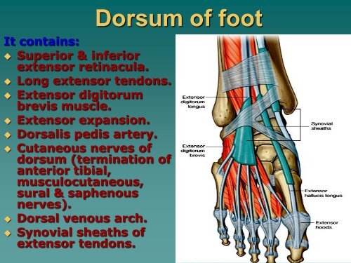
It contains:<br />
Superior & inferior<br />
extensor retinacula.<br />
Long extensor tendons.<br />
Extensor digitorum<br />
brevis muscle.<br />
Extensor expansion.<br />
Dorsalis pedis artery.<br />
Cutaneous nerves <strong>of</strong><br />
dorsum (termination <strong>of</strong><br />
anterior tibial,<br />
musculocutaneous,<br />
sural & saphenous<br />
nerves).<br />
Dorsal venous arch.<br />
Synovial sheaths <strong>of</strong><br />
extensor tendons.<br />
<strong>Dorsum</strong> <strong>of</strong> <strong>foot</strong>
Dorsalis pedis<br />
artery branches:<br />
Medial tarsal<br />
Lateral tarsal<br />
1 st dorsal metatarsal<br />
Arcuate artery; gives<br />
3 dorsal metatarsal<br />
arteries.<br />
1 st planter<br />
metatarsal artery in<br />
sole.
Dorsal venous arch
Cutaneous<br />
innervations <strong>of</strong><br />
dorsum <strong>of</strong> <strong>foot</strong>
Planter<br />
aponeurosis:<br />
Triangular in shape<br />
Apex attached to<br />
calcaneus<br />
Base divided into 5<br />
slips<br />
Its function is:<br />
1. Protects underling<br />
structures.<br />
2. Maintains<br />
longitudinal arch <strong>of</strong><br />
<strong>foot</strong>.<br />
Sole <strong>of</strong> <strong>foot</strong>
1 st layer <strong>of</strong><br />
muscles:<br />
Layers <strong>of</strong> sole<br />
Contains 3 muscles:<br />
1. Abductor hallucis<br />
muscle.<br />
2. Abductor digiti<br />
minimi.<br />
3. Flexor digitorum<br />
brevis.
2 nd layer <strong>of</strong> sole<br />
It contains 2 long<br />
tendons & 2muscles:<br />
‣ Tendon <strong>of</strong> flexor hallucis<br />
longus.<br />
‣ Tendon <strong>of</strong> flexor digitorum<br />
longus.<br />
‣ Flexor digitorum<br />
accessorius.<br />
‣ 4 lumberical muscles.
3 rd layer<br />
It contains 3<br />
muscles:<br />
1. Flexor hallucis brevis.<br />
2. Flexor digiti minimi.<br />
3. Adductor hallucis; has<br />
oblique & transverse<br />
heads.
4th layer <strong>of</strong> sole<br />
It contains 2 long<br />
tendons & 2muscles:<br />
‣ Tendon <strong>of</strong> tibialis<br />
posterior<br />
‣ Tendon <strong>of</strong> peroneus<br />
longus.<br />
‣ 4 dorsal interossei.<br />
‣ 3 planter interossei.
Lateral planter<br />
artery:<br />
<br />
<br />
<br />
<br />
Planter arteries<br />
It arises from posterior tibial<br />
artery deep to flexor<br />
retinaculum.<br />
Course: it runs laterally<br />
between 1 st & 2 nd layers till<br />
base <strong>of</strong> 5 th metatarsal bone<br />
then curves medially between<br />
3 rd & 4 th layers<br />
Ends: by anastmosing with<br />
termination <strong>of</strong> dorsalis pedis<br />
artery.<br />
Branches: muscular,<br />
cutaneous, twigs for<br />
anastmosis around ankle,<br />
lateral planter arch: It gives 4<br />
planter metatarsal, anterior &<br />
posterior perforating branches
medial planter<br />
artery:<br />
Planter arteries<br />
It arises from posterior<br />
tibial artery.<br />
Course: it runs forwards<br />
deep to abductor hallucis<br />
longus .<br />
Ends: by anastmosing<br />
with digital branch to<br />
medial side <strong>of</strong> big toe.<br />
Branches: small<br />
superficial digital<br />
branches.
Planter nerves<br />
medial planter<br />
nerve:<br />
It arises from posterior<br />
tibial nerve deep to<br />
flexor retinaculum.<br />
Course: it runs forwards<br />
between abductor<br />
hallucis longus & flexor<br />
digitorum brevis<br />
Branches:<br />
1. Muscular to 4 muscles.<br />
2. Cutaneous to medial<br />
2/3 <strong>of</strong> sole & medial 3 ½<br />
toes
Lateral planter nerve<br />
It arises from posterior tibial<br />
nerve.<br />
Course: it runs laterally<br />
between 1 st & 2 nd layers till<br />
base <strong>of</strong> 5 th metatarsal bone<br />
then divides into superficial<br />
branch & deep branch which<br />
curves medially between 3 rd &<br />
4 th layers in concavity <strong>of</strong><br />
planter arch.<br />
Ends: divides into superficial<br />
branch & deep branch.<br />
Branches: 1. muscular to 14<br />
muscles.<br />
2. cutaneous to lateral 1/3 <strong>of</strong><br />
sole & lateral 1 ½ toes.<br />
3. Articular to intertarsal &<br />
tarsometatarsal joints.
Articular surfaces<br />
Hip joint<br />
Head <strong>of</strong> femur<br />
acetabulum
Capsule attachment
Synovial membrane
Capsule &<br />
Synovial<br />
membrane
Ligaments <strong>of</strong> hip joint<br />
Ili<strong>of</strong>emoral<br />
ligament<br />
capsule<br />
Pub<strong>of</strong>emoral<br />
ligament<br />
Ischi<strong>of</strong>emoral<br />
ligament
Arterial supply <strong>of</strong> hip joint:
Anterior relation <strong>of</strong> hip joint
Superior & posterior relation <strong>of</strong> hip joint
Knee<br />
joint:<br />
• Synovial <strong>of</strong><br />
condylar or<br />
modified hing<br />
type.<br />
•Articular<br />
surfaces:<br />
•Femoral<br />
codyles, tibial<br />
codyles &<br />
patella
Capsule:<br />
is attached close<br />
to articular<br />
surfaces <strong>of</strong><br />
femoral & tibial<br />
codyles but<br />
replaced anteriorly<br />
by quadriceps<br />
tendon, patella &<br />
ligamentum<br />
patellae
Articular surface <strong>of</strong> patella with<br />
anterior surface <strong>of</strong> femoral condyles
Synovial membrane & bursae
Synovial membrane reflected on cruciate ligaments<br />
(itracapsular, extrasynovial) ,forms suprapatellar bursa,<br />
infrapatellar fold, sheath around popliteus tendone
Extra capsular ligaments <strong>of</strong> knee joint
fibular collateral<br />
ligament<br />
Tibial collateral<br />
ligament
Ligamentum<br />
patellae
Extra capsular ligaments <strong>of</strong> knee joint<br />
Oblique popliteal<br />
ligament
Intra capsular ligaments <strong>of</strong> knee joint
Intracapsular<br />
structures:<br />
•Anterior &<br />
posterior cruciate<br />
ligaments.<br />
•Transverse<br />
ligament.<br />
•Medial & lateral<br />
menesci.<br />
•Tendon <strong>of</strong><br />
popliteus muscle.<br />
•Infrapatellar pad<br />
<strong>of</strong> fat.
Function <strong>of</strong><br />
cruciate<br />
ligaments:<br />
* Anterior cruciate<br />
ligament Prevent<br />
posterior<br />
dislocation <strong>of</strong> femur<br />
on tibia.<br />
* Posterior ligament<br />
Prevents anterior<br />
dislocation & hyper<br />
extension <strong>of</strong> knee.
Arterial<br />
supply:<br />
Anastmosis around<br />
knee joint
Superior & inferior tibi<strong>of</strong>ibular joints & introsseous<br />
membrane
Ankle joint:<br />
Articular surfaces
Capsule &<br />
ligaments
Medial ligament <strong>of</strong> ankle =deltoid<br />
ligament
lateral ligament <strong>of</strong> ankle
Medial relation= structures deep<br />
to flexor retinaculum
anterior relation= structures<br />
deep to superior extensor<br />
retinaculum
lateral relation= structures deep<br />
to peroneal retinaculua
Arches <strong>of</strong> <strong>foot</strong>:<br />
Medial longitudinal arch<br />
lateral longitudinal arch<br />
transverse arch
Factors maintaining <strong>foot</strong> arches
Planter aponeurosis
Long flexor<br />
tendons<br />
maintain<br />
medial<br />
longitudinal<br />
arch & tendon<br />
<strong>of</strong> peroneus<br />
longus<br />
maintains<br />
transverse arch
Ligaments <strong>of</strong> <strong>foot</strong><br />
Long planter<br />
ligament.<br />
Short planter<br />
ligament.<br />
Spring (planter<br />
calceneonavicular).
Bifurcate<br />
ligament<br />
Ligaments <strong>of</strong> <strong>foot</strong>