Olympus BX-51 brochure
Olympus BX-51 brochure
Olympus BX-51 brochure

You also want an ePaper? Increase the reach of your titles
YUMPU automatically turns print PDFs into web optimized ePapers that Google loves.
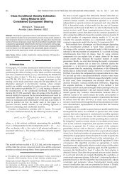
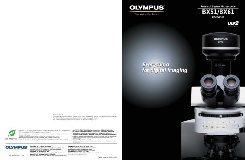
Research System Microscoope<br />
<strong>BX</strong><strong>51</strong>/<strong>BX</strong>61<br />
<strong>BX</strong>2 Series<br />
Everything<br />
for digital imaging<br />
Photos courtesy of:<br />
Junko Kyozuka, Associate Professor, Graduate School of Agricultural and Life Sciences, The Tokyo University (P.11 below)<br />
Prof. Nakatani, Biochemistry course, School of medicine, Showa University (P.13 above)<br />
Prof. Tadokoro, Pathology class, School of medicine, St. Marianna University (P.15)<br />
<strong>BX</strong><strong>51</strong>/<strong>BX</strong>61 are the environmental conscious products according to OLYMPUS's own standards.<br />
Main features of OLYMPUS Eco-products are as follows.<br />
• Lead-free and arsenic-free Eco-glass for optics, such as lenses and prisms.<br />
• Exclusion of hexavalent chrome, mercury, lead and cadmium from metal materials and surface treatment of metal.<br />
• Exclusion of lead solders.<br />
• Adoption of cardboard for packing materials without styrene foam for promoting the recycling.<br />
* A definition of exclusion depends on OLYMPUS standard. Some accessories are inapplicable.<br />
Please visit our web site for further information: http://www.olympus.co.jp/en/eco-products/<br />
• OLYMPUS CORPORATION has obtained the ISO9001/ISO14001.<br />
• OLYMPUS CORPORATION has obtained the MD540624/ISO13485.<br />
• Illumination devices for microscope have suggested lifetimes.<br />
Periodic inspections are required. Please visit our web site for details.<br />
• Windows is a registered trademark of Microsoft Corporation in the United States and other countries.<br />
All other company and product names are registered trademarks and/or trademarks of their<br />
respective owners.<br />
• Images on the PC monitors are simulated.<br />
• Specifications and appearances are subject to change without any notice or obligation on the part of<br />
the manufacturer.<br />
Printed in Japan M1540E-0909B
The UIS2 optical system:<br />
a new evolutionary advance in fluorescence<br />
digital imaging.<br />
The new <strong>BX</strong>2 series addresses the research demands of the future with <strong>Olympus</strong>' most<br />
advanced optical system to date. UIS2 optics deliver the world's highest standard of<br />
fluorescence performance, along with the image quality and clarity needed for progress<br />
of fast-developing life science research programs. With increased S/N ratio, high optical<br />
transmission, and diverse illumination capabilities, the UIS2 optical system provides<br />
excellent performance over a newly extended wavelength range between UV and IR.<br />
This improvement meets all current demands in fluorescence digital imaging and<br />
provides a firm foundation for future developments. As modern research advances to ever<br />
higher levels of complexity and sophistication, the need for quality and dependability<br />
makes the <strong>BX</strong>2 series today's most convincing solution.<br />
<strong>BX</strong><strong>51</strong><br />
System Microscope<br />
<strong>BX</strong>61<br />
Motorized System Microscope<br />
1<br />
2
Higher S/N ratio enables clear capture of<br />
weak fluorescence emissions.<br />
Fluorescence<br />
World leading fluorescence<br />
performance — a vital key to modern<br />
life science research<br />
The ideal in fluorescence observation is to<br />
capture high-contrast images with the<br />
lowest exposure to excitation light, thus<br />
minimizing the chances of cell damage and<br />
fluorescence fading. With increased S/N<br />
ratio, high transmission of the objectives and<br />
the high performance mirror unit, <strong>Olympus</strong>'<br />
UIS2 optics provide excellent performance<br />
in fluorescence by obtaining bright images<br />
from weak fluorescence signals.<br />
Improved performance<br />
S N<br />
of interference type<br />
fluorescence mirror units<br />
The new fluorescence mirror units achieve<br />
high S/N ratio by application of new coating<br />
technology to the filters and optimal design<br />
of excitation and emission filters'<br />
characteristic.<br />
The hard coating, which prolongs the<br />
lifetime of filter, is applied to all <strong>Olympus</strong><br />
fluorescence mirror units.<br />
U-MNIBA3<br />
100<br />
90<br />
BP470-495<br />
80<br />
DM505<br />
70<br />
60<br />
50<br />
40<br />
30<br />
BA<strong>51</strong>0-550<br />
20<br />
10<br />
0<br />
400 450 500 550 600 650<br />
Wavelength (nm)<br />
Transmittance (%)<br />
High-performance filters with high<br />
transmission, optimized to individual<br />
fluorochrome characteristics<br />
Mirror unit with stray<br />
light reducing function<br />
to eliminate noise<br />
High S/N objectives detect even slight<br />
fluorescence emission<br />
Low auto-fluorescence immersion<br />
oil available<br />
Camera adapter compatible<br />
for near infrared region<br />
Trinocular tube compatible for<br />
near infrared region<br />
Lamp housing with aspherical<br />
collector lens providing<br />
excellent excitation efficiency<br />
S<br />
N<br />
Signal Up<br />
Noise Down<br />
Excellent trinocular tube performance<br />
even in the near infrared region<br />
The trinocular tube U-TR30NIR improves<br />
the transmission and compensates for<br />
aberrations over a wider wavelength range.<br />
A new multi-coatings is applied to the<br />
trinocular optical surfaces to widen the IR<br />
spectral characteristics and allow for<br />
observation of newly developed<br />
fluorochromes in the near-infrared region.<br />
Camera adapter suitable for<br />
near infrared region<br />
Users can choose from a variety of low<br />
magnification camera adapters with<br />
C-mount, all IR compatible.<br />
q w e r<br />
q U-TV0.35xC-2 w U-TV0.5xC-3 e U-TV0.63xC<br />
r U-TV1x-2+U-CMAD3<br />
High transmission across a wide<br />
wavelength spectrum<br />
The latest UIS2 objectives achieve a flat,<br />
high transmission over a wide wavelength<br />
spectrum, from visible to near infrared —<br />
thanks to the incorporation of a newlydeveloped<br />
ultra-wide wavelength reflection<br />
prevention coating (UW multi-coatings). The<br />
improvement in transmission in the near<br />
infrared region is especially notable, and<br />
typifies the high performance which makes<br />
UIS2 objectives the natural choice in many<br />
leading-edge research fields.<br />
High-quality<br />
fluorescence mirror units<br />
S N<br />
for fluorescence proteins<br />
The HQ type mirror units are ideal for the<br />
wavelength characteristics of<br />
ECFP/EGFP/EYFP/DsRed. With sharp<br />
upstroke and high transmission, the mirror<br />
unit efficiently transmits the fluorescence<br />
emitted from fluorescence proteins. This<br />
allows bright observation images even with<br />
weak excitation light, while preventing<br />
fluorescence fading and minimizing the<br />
chances of cell damage.<br />
Stray light reducing function N<br />
<strong>Olympus</strong> mirror units are equipped<br />
with an unique function to eliminate<br />
the stray light that could increase the<br />
background noise in fluorescence image.<br />
Light<br />
source<br />
Dichromatic<br />
mirror<br />
Emission filter<br />
Fluorescence light<br />
for observation<br />
Stray light<br />
The best S/N ratio...and<br />
S N<br />
the best fluorescence<br />
performance<br />
<strong>Olympus</strong> UIS2 objectives provide the best<br />
S/N ratio by employment of totally new<br />
design to curtail autofluorescence from all<br />
possible sources — glass material, coating<br />
and cementing material. UIS2 objectives<br />
achieve high N.A. while reducing<br />
autofluorescence, two benefits previously<br />
considered incompatible.<br />
With these improvements, UIS2 objectives<br />
provide the best fluorescence image.<br />
Image captured by the UIS2 objective<br />
High N.A. objectives for S N<br />
fluorescence imaging<br />
The <strong>BX</strong>2 series features the newlydeveloped<br />
PLAPON60XO objective, offering<br />
the world's highest N.A. (1.42) for<br />
fluorescence imaging, and the<br />
UPLSAPO100XO with high 1.4 N.A. and<br />
advanced universal features. In addition to<br />
their outstanding fluorescence S/N ratio,<br />
they enjoy UV transmission. The<br />
UPLSAPO100XO objective is especially<br />
notable for maintaining its transmission<br />
down to the 340 nm wavelength.<br />
Up to near infrared compensation for<br />
chromatic aberration<br />
The Super Apochromat performance of<br />
UPLSAPO series objectives compensates<br />
for all chromatic aberrations, from visible to<br />
up to 1000nm wavelength light. Clear<br />
images without color shift are provided even<br />
in multi-color observations. Imaging all the<br />
way from UV to IR can be performed with a<br />
single objective.<br />
U-MGFPHQ<br />
100<br />
BP460-480<br />
90<br />
DM485<br />
80<br />
BA495-540<br />
70<br />
Ex<br />
60<br />
50<br />
40<br />
30<br />
20<br />
Em<br />
10<br />
0<br />
400 450 500 550 600 650<br />
Wavelength (nm)<br />
Transmittance (%)<br />
Excitation filter<br />
Excitation light:<br />
Illumination light<br />
Objective<br />
Stray light<br />
reducing<br />
function<br />
Specimen<br />
Low auto-fluorescence<br />
N<br />
immersion oil<br />
The ability to reduce auto-fluorescence<br />
normally associated with immersion oil<br />
makes this product well suited for<br />
fluorescence microscopy. Resistance to<br />
crystallization allows it to be used over long<br />
periods of time.<br />
Image captured by a conventional objective<br />
3<br />
4
Fluorescence accessories<br />
Bright, even reflected light<br />
fluorescence illumination<br />
A wide range of illumination<br />
modules for light adjustment<br />
Excitation light balancer<br />
for even illumination<br />
Lamp housing can be selected<br />
according to application<br />
Fluorescence excitation balancers/<br />
U-EXBABG, U-EXBAUB, U-EXBAUG<br />
When observing double and triple stained<br />
specimens, both observation and<br />
photography can be conducted by<br />
arranging or altering the fluorescence<br />
brightness while freely changing the<br />
excitation light for each stained color. An<br />
excitation balancer is attached in the parallel<br />
light path, so there is no unevenness in the<br />
visual field.<br />
Pinhole field stop module/<strong>BX</strong>-RFSPOT<br />
This slider makes it possible to use the light<br />
source as a spotlight, illuminating tiny<br />
individual areas on the fluorescence<br />
specimen — an especially valuable feature<br />
in experimental work.<br />
The slider is attached<br />
to the <strong>BX</strong>-RFA<br />
fluorescence<br />
illuminator in the field<br />
stop position.<br />
Rectangular field stop for<br />
digital imaging/U-RFSS<br />
The rectangular field stop can be set to the<br />
exact size of the imaging sensor to avoid<br />
fading outside of the imaging area and<br />
damaging sensitive<br />
tissue.<br />
Unnecessary exposure area<br />
caused by a round field stop<br />
Neuro glear cells<br />
Green and red<br />
enhanced<br />
Blue and red enhanced<br />
High-rigidity reflected light illuminator<br />
Two types of reflected light illuminator are<br />
provided: the multi-purpose <strong>BX</strong>-RFA,<br />
suitable for a wide range of different research<br />
projects, and the economical <strong>BX</strong>-URA2.<br />
Up to 6 mirror units can be attached in the<br />
cassette, which is especially useful when<br />
observing multi-stained specimens. A click<br />
weight adjustment function is provided for<br />
filter exchange, and a click release function<br />
that eliminates vibration.<br />
<strong>BX</strong>-URA2<br />
<strong>BX</strong>-RFA<br />
Luminous mirror unit indicator for easy<br />
confirmation in dark room<br />
Bright,easy-to-see self-illuminated labels are<br />
used to denote fluorescence filter sets,<br />
easily visible in a dark room. Three filter<br />
positions are<br />
displayed simultaneously<br />
making<br />
selection of the next<br />
filter easy and<br />
intuitive.<br />
Lamphouses according to application<br />
Two 100 W mercury lamphouses<br />
(U-LH100HG and U-LH100HGAPO) are<br />
available, the latter with color correction<br />
extending to UV wavelengths.<br />
The U-LH75XEAPO is available for 75 W<br />
xenon lamps, also with correction extending<br />
to the UV.<br />
U-LH100HGAPO<br />
U-LH100HG<br />
U-LH75XEAPO<br />
Bright, consistent illumination<br />
Twice the brightness provided by<br />
conventional models can be obtained with<br />
the10X ~ 20X objectives, or 2-3 times the<br />
brightness when light is narrowed to F.N.<br />
12. This enables efficient observations at<br />
lower magnifications.<br />
Double lamp housing adapter U-DULHA<br />
for exchange between two light sources<br />
When two different light sources are<br />
attached at the same time, this adapter unit<br />
enables easy exchange between them<br />
according to the user's application.<br />
(Optical path: 100/0, 0/100, F.N.11)<br />
Convenient 6-position filter sliders<br />
U-RSL6/U-RSL6EM<br />
Setting the excitation and emission filters on<br />
this sextuple filter slider enables<br />
synchronous and continuous exchange<br />
between positions.<br />
With ND filters<br />
attached, illumination<br />
can be adjusted in 6<br />
steps.<br />
Confocal laser scanning biological microscope/FV1000<br />
The FluoView/FV1000 is a next-generation imaging system designed for<br />
high-resolution, confocal observation of both fixed and live cells.<br />
The FV1000 offers advances in confocal system performance<br />
while providing the speed and sensitivity required for<br />
live cell imaging with minimal risk of damage to<br />
specimens.<br />
In addition, the FV1000 offers a revolutionary<br />
synchronized laser scanning system called the SIM<br />
Scanner. While one laser stimulates, the second<br />
laser simultaneously provides high-resolution<br />
imaging. This coordination of laser stimulation and<br />
imaging makes the FV1000 an ideal choice for<br />
FRAP, FLIP and photoactivation.<br />
*FV1000 is a class 3B laser product.<br />
FV1000<br />
Blue enhanced<br />
5<br />
6
Varied illumination and advanced optics deliver<br />
top quality digital images.<br />
Digital Imaging<br />
Excellent color reproduction from<br />
daylight illumination<br />
Since <strong>Olympus</strong> microscopes can apply ideal<br />
color temperature at natural daylight (5500 K)<br />
throughout the light source, the objectives<br />
and the CCD camera, the camera captures<br />
color information accurately and provides<br />
faithful reproduction on the display.<br />
Digital camera/ DP72<br />
High-resolution digital images equivalent to 12.8 million pixels* captured in<br />
approx. 2.5 seconds — from brightfield to fluorescence.<br />
Thanks to its high-speed hardware, the DP72 can<br />
capture high-resolution images equivalent to 12.8<br />
million pixels in around 2.5 seconds*. The camera's<br />
high sensitivity and low noise (equivalent to the level of<br />
ISO 1600) ensure clear fluorescence imaging, while the<br />
resolution quality allows precise representation of<br />
particular specimen areas.<br />
By shifting the pixels of the 1.45 million pixel 2/3 inch CCD (one pixel = 6.45 µm), it<br />
is possible to record still images equivalent to the maximum image recording size<br />
(4140 x 3096) or effective image size of 12.5 million pixels.<br />
UIS2 optics provide high transmission<br />
for clear, flat images<br />
In the UIS2 optical system, improved<br />
transmission and compensation for<br />
chromatic aberration over a wide<br />
wavelength spectrum are not only<br />
characteristics of the objectives, but also of<br />
image forming components such as the<br />
trinocular tube and video camera adapter.<br />
As a result, images at all magnification levels<br />
are flat, sharp, clear and free from color<br />
shift.<br />
Digital camera/ DP25<br />
5 megapixel high-precision, high-quality technology for microscopic imaging<br />
In addition to live display at a high frame rate of 8 fps<br />
with exceptional quality (2560 x 1920 pixels), the DP25<br />
is equipped with a color profile that provides full-color<br />
images in real-time, allowing faithful color reproduction<br />
of specimens. It can easily be connected with just 1<br />
cable (6 pin) to a PC with a FireWire (IEEE1394) port.<br />
And it can connected to a laptop PC as well via a<br />
FireWire (IEEE1394a) PC card.<br />
Imaging software* —<br />
Software to support basic functions<br />
DP2-BSW is a simple and easy-to-use, thus<br />
user-friendly image capturing software<br />
package. It can be used to control different<br />
types of motorized units, and to perform<br />
both still time-lapse images and live image<br />
movie recording.<br />
*As per the software for Europe, please contact the nearest<br />
<strong>Olympus</strong> representative office.<br />
• Intuitive, easy-to-use GUI (Graphical User<br />
Interface). Tool bar items can be usercustomized<br />
and menu icons restricted to<br />
frequently used functions.<br />
• A reference scale bar can be displayed,<br />
overlayed, and subsequently burned onto a<br />
saved image. Arrows and text can also be<br />
entered and saved in an image.<br />
• Time-lapse feature lets you set the starting<br />
time of a photo sequence, the duration, the<br />
number of images, and thus the chronological<br />
development of the whole sequence.<br />
• Combine multiple color images (from a<br />
single specimen) from different excitation<br />
wavelengths, into a single final image<br />
(grayscale or color).<br />
100<br />
High transmittance<br />
(UPLSAPO10x+U-TR30NIR)<br />
90<br />
80<br />
Transmittance (%)<br />
70<br />
60<br />
50<br />
40<br />
30<br />
20<br />
10<br />
0<br />
350 400 450 500 550 600 650 700 750 800 850 900<br />
Wavelength (nm)<br />
Optimal trinocular tube for digital<br />
imaging<br />
In digital imaging, the best light intensity<br />
balance between the observation side and<br />
the digital camera side should be equal.<br />
<strong>Olympus</strong>' new trinocular tube U-TR30NIR<br />
provides a choice of three light path<br />
exchange: 100% for binocular, 100% for<br />
camera, or 50% each for binocular and<br />
camera.<br />
Digital camera/ DP20<br />
Providing live image display speed that’s close to real-time while maintaining<br />
high-precision image quality<br />
The DP20 can display high-precision images of<br />
2 megapixels in UXGA (1600 x 1200) format at<br />
15 frames/second. Additionally, it provides faithful 8-bit<br />
RGB color reproduction that is ideal for conferences<br />
both large and small. The handset control unit has<br />
functional key layout for quick and easy control.<br />
• A focusing indicator function makes<br />
focusing in a live image easy; a line profile<br />
function lets you focus accurately on userdefined<br />
regions. Additionally, the region in<br />
focus can be magnified up to 16 times.<br />
• <strong>BX</strong>61 motorized microscopes can be<br />
controlled from a personal computer.<br />
Different conditions can be set for respective<br />
observation methods, and the observation<br />
method can be changed by simply clicking<br />
on a button on the controller screen.<br />
• Several functions for measuring live or still<br />
images, including point measurements,<br />
arbitrary line, polygon, circle and ellipse or<br />
rectangle measurements, are integrated.<br />
For further processing the measurements<br />
can be exported to MS Excel with the<br />
simple click of a mouse.<br />
7<br />
8
The advanced UIS2 system delivers<br />
high performance over a wider wavelength spectrum.<br />
Excellent Optics<br />
UIS2 optics inherit high expandability<br />
As heir to <strong>Olympus</strong>' infinity-corrected optical<br />
system, in which the tube lens is built into<br />
the observation tube, UIS2 optics display no<br />
image deterioration even when many<br />
different optical components or equipment<br />
are inserted in the parallel light path. This<br />
inherent expandability gives users ample<br />
freedom to construct the system in a way<br />
that meets their specific requirements.<br />
UW (Ultra wideband) multi-coatings<br />
reduces autofluorescence and improves<br />
S/N ratio<br />
By using carefully selected raw materials for<br />
glass, and applying advanced UW multicoatings<br />
technology, <strong>Olympus</strong> has reduced<br />
objective autofluorescence and significantly<br />
improved the S/N ratio.<br />
Flat, high transmission over wide<br />
wavelength range from UV to IR<br />
UW multi-coatings also yields a flat, high<br />
transmission over a wide wavelength range,<br />
ensuring high performance in research tasks<br />
using different types of fluorochromes.<br />
Transmittance(%)<br />
High transmittance<br />
UPLSAPO100XO<br />
100 (new) UPLSAPO100XO<br />
90<br />
80<br />
70<br />
60<br />
50<br />
40<br />
30<br />
20<br />
10<br />
(conventional) PLAPO100XO<br />
0<br />
300 400 500 600 700 800<br />
UIS2 objectives<br />
Cover Cor- Iris Water proof<br />
Objective N.A. W.D. F.N. glass Immer- Spring rec- dia- and<br />
(mm) thickness sion tion phragm oil proof<br />
(mm) ring function<br />
UPLSAPO 4X 0.16 13 26.5 —<br />
UPLSAPO 10X2 0.4 3.1 26.5 0.17<br />
UPLSAPO 20X 0.75 0.6 26.5 0.17 _<br />
UPLSAPO 20XO 0.85 0.2 26.5 — Oil _<br />
UPLSAPO 40X2 0.95 0.18 26.5 0.11-0.23 _ _<br />
UPLSAPO 60XW 1.20 0.28 26.5 0.15-0.21 Water _ _ _<br />
UPLSAPO 60XO 1.35 0.15 26.5 0.17 Oil _ _<br />
UPLSAPO 100XO 1.40 0.13 26.5 0.17 Oil _ _<br />
PLAPON 1.25X 0.04 5 26.5 —<br />
PLAPON 2X 0.08 6.2 26.5 —<br />
PLAPON 60XO 1.42 0.15 26.5 0.17 Oil _ _<br />
UPLFLN 4X 0.13 17 26.5 —<br />
UPLFLN 10X2 0.3 10 26.5 —<br />
UPLFLN 20X 0.5 2.1 26.5 0.17 _<br />
UPLFLN 40X 0.75 0.<strong>51</strong> 26.5 0.17 _<br />
UPLFLN 40XO 1.3 0.2 26.5 0.17 Oil _ _<br />
UPLFLN 60X 0.9 0.2 26.5 0.11-0.23 _ _<br />
UPLFLN 60XOI 1.25-0.65 0.12 26.5 0.17 Oil _ _ _<br />
UPLFLN 100XO2 1.3 0.2 26.5 0.17 Oil _ _<br />
UPLFLN 100XOI2 1.3-0.6 0.2 26.5 0.17 Oil _ _ _<br />
UPLFLN 10X2PH 0.3 10 26.5 —<br />
UPLFLN 20XPH 0.5 2.1 26.5 0.17 _<br />
UPLFLN 40XPH 0.75 0.<strong>51</strong> 26.5 0.17 _<br />
UPLFLN 60XOIPH 1.25-0.65 0.12 26.5 0.17 Oil _ _ _<br />
UPLFLN 100XO2PH 1.3 0.2 26.5 0.17 Oil _ _<br />
UPLFLN 4XP 0.13 17 26.5 —<br />
UPLFLN 10XP 0.3 10 26.5 —<br />
UPLFLN 20XP 0.5 2.1 26.5 0.17 _<br />
UPLFLN 40XP 0.75 0.<strong>51</strong> 26.5 0.17 _<br />
UPLFLN 100XOP 1.3 0.2 26.5 0.17 Oil _ _<br />
Cover Cor- Iris Water proof<br />
Objective N.A. W.D. F.N. glass Immer- Spring rec- dia- and<br />
(mm) thickness sion tion phragm oil proof<br />
(mm) ring function<br />
PLN 2X 0.06 5.8 22 —<br />
PLN 4X 0.1 18.5 22 —<br />
PLN 10X 0.25 10.6 22 —<br />
PLN 20X 0.4 1.2 22 0.17 _<br />
PLN 40X 0.65 0.6 22 0.17 _<br />
PLN 50XOI 0.9-0.5 0.2 22 — Oil _ _ _<br />
PLN 100XO 1.25 0.15 22 — Oil _<br />
PLN 10XPH 0.25 10.6 22 —<br />
PLN 20XPH 0.4 1.2 22 0.17 _<br />
PLN 40XPH 0.65 0.6 22 0.17 _<br />
PLN 100XOPH 1.25 0.15 22 — Oil _<br />
PLN 4XP 0.1 18.5 22 —<br />
ACHN 10XP 0.25 6 22 —<br />
ACHN 20XP 0.4 3 22 0.17<br />
ACHN 40XP 0.65 0.45 22 0.17 _<br />
ACHN 100XOP 1.25 0.13 22 — Oil _<br />
MPLAPON 100XO 1.4 0.1 26.5 0 Oil _ _<br />
MPLFLN 40X 0.75 0.63 26.5 0 _<br />
APON 60XOTIRF 1.49 0.1 22 0.13-0.19 Oil _ _<br />
UAPON 100XOTIRF 1.49 0.1 22 0.13-0.19 Oil _ _<br />
UAPON 150XOTIRF 1.45 0.08 22 0.13-0.19 Oil _ _<br />
All UIS2 objectives and WHN eyepieces: lead-free eco-glass<br />
UIS objectives<br />
Cover Cor- Iris Oil<br />
Objective N.A. W.D. F.N. glass Immer- Spring rec- dia- proof<br />
(mm) thickness sion tion phragm cap<br />
(mm)<br />
ring<br />
PLFL 100X 0.95 0.2 26.5 0.14-0.2 _ _<br />
Complete chromatic aberration<br />
compensation up to near infrared<br />
region<br />
UPLSAPO objectives completely eliminate<br />
chromatic aberration up to the near infrared<br />
region, matching the ability of Super<br />
Apochromat objectives to provide clear<br />
images without overlapping colors or color<br />
shift. As a result, a single objective can<br />
perform imaging from UV to IR wavelengths.<br />
2<br />
UPLSAPO series chromatic aberration compensation<br />
Comparing chromatic aberration compensation levels:<br />
(The smaller the figure the better)<br />
■ UPLSAPO series<br />
Thanks to the application of <strong>Olympus</strong>' original UW multicoatings,<br />
these Super Apochromat objectives fully<br />
compensate for both spherical and chromatic aberrations<br />
from the UV to the near infrared region. Their sensitivity to<br />
fluorescence emissions ensures the acquisition of sharp,<br />
clear images, without color shift, even in brightfield and<br />
Nomarski DIC observations. For quality and performance,<br />
they offer an unbeatable solution to every kind of digital<br />
imaging need.<br />
■ UPLFLN (UPLFLN-PH) series<br />
These plan objectives also provide flat images with high<br />
transmission up to the near infrared region of the<br />
spectrum through the employment of UW multi-coatings.<br />
With their high S/N ratio, excellent resolution and high<br />
contrast imaging, they are especially effective in<br />
brightfield and Nomarski DIC observations.<br />
The UPLFLN-PH series is optimized for phase contrast<br />
observation.<br />
Focus (µm)<br />
1.5<br />
1<br />
0.5<br />
(conventional) UPLAPO100XO<br />
0<br />
(new) UPLSAPO100XO<br />
-0.5<br />
400 450 500 550 600 650 700 750 800 850 900<br />
Wavelength (nm)<br />
UPLSAPO100XO<br />
UPLAPO100XO<br />
■ PLAPON series<br />
Designed for unsurpassed resolution and contrast, these<br />
Plan Apochromat objectives keep chromatic aberration<br />
down to an absolute minimum.<br />
The PLAPON60XO, to which the UW multi-coatings is<br />
applied, is the first in the world to achieve N.A. 1.42 for<br />
fluorescence imaging.<br />
■ PLN(PLN-PH) series<br />
Ideal for a range of clinical and research applications,<br />
these high quality objectives feature excellent flatness up<br />
to F.N. 22 in transmitted brightfield (phase contrast)<br />
observation. The PLN-PH series is specifically designed<br />
for phase contrast work.<br />
9 10
New DIC observation system optimizes<br />
the specimen image at wider magnifications.<br />
Nomarski DIC<br />
Clear, high-contrast imaging from low<br />
to high magnifications.<br />
Brightfield<br />
Optimum shearing value according to<br />
the specimen<br />
Three types of prisms with different shearing<br />
value are provided to define contrast and<br />
resolution.<br />
• High contrast for thin specimens<br />
U-DICTHC<br />
High contrast can be obtained even in high<br />
magnification observations of thin<br />
specimens, such as culture cells.<br />
New DIC system allows wider selection<br />
More DIC-compatible objectives are<br />
available in UIS2, and users can select the<br />
most suitable shearing value for a given<br />
specimen from among 10X to 100X<br />
objectives. In addition, combination with<br />
other observation methods and components<br />
is simpler and more convenient.<br />
Analyzer<br />
Objective<br />
DIC slider<br />
(Objective side)<br />
Specimen<br />
DIC prism<br />
(Condenser side)<br />
• High resolution with less glare<br />
U-DICTHR<br />
This unit enables observations with high<br />
resolution but less glare even for thick<br />
specimens used in developmental and<br />
genetic research, such as finely-structured<br />
diatoms, embryos, zebrafish and C. elegans.<br />
PtK2 cell<br />
Polarizer<br />
Universal condenser/U-UCD8<br />
This condenser, with built-in polarizer, allows<br />
simultaneous attachment of up to 8 optical<br />
components, freely combined or easily<br />
switched.<br />
Bone marrow<br />
C.elegance<br />
Kidney<br />
Septuple revolving nosepiece for<br />
DIC/simple POL/U-D7RE<br />
Equipped with a DIC slider slot, the U-D7RE<br />
septuple revolving nosepiece allows<br />
simultaneous attachment of 7 objectives<br />
from low to high magnifications. It is<br />
especially suitable for combined DIC and<br />
fluorescence observations.<br />
• High all-round performance<br />
U-DICT, U-DICTS<br />
Suitable for observing a wide range of<br />
general specimens, such as tissue.<br />
Task-specific brightfield condenser<br />
options<br />
According to their purpose, users can<br />
choose from the U-SC3, a swing-out<br />
condenser suitable for observations from<br />
1.25X-100X; the U-AC2, a highly costefficient<br />
Abbe-type model; the U-AAC,<br />
whose Aplanat-Achromat design<br />
comprehensively eliminates chromatic<br />
aberration; and the U-ULC-2, a special<br />
condenser for ultra low magnifications.<br />
* Select the U-ULC2 condenser for optimal digital imaging with the<br />
1.25X objective.<br />
Clear, high-contrast observation of<br />
stained specimens<br />
Image contrast is significantly enhanced by<br />
combining UIS2 objectives with the UIS2<br />
eyepiece WHN, which features multi-<br />
coatings on all its surfaces. This makes the<br />
image background look whiter, so that the<br />
stained area of the specimen stands out<br />
more clearly.<br />
A shoot apical meristem of rice<br />
U-ULC-2<br />
U-AAC<br />
For high contrast For high resolution General<br />
U-AC2<br />
Specimen Thin Thick —<br />
DIC slider (objective side) U-DICTHC U-DICTHR U-DICT* 1 U-DICTS* 1<br />
*1 Choose upon objective magnification<br />
U-SC3<br />
UIS2 image<br />
Conventional image<br />
11 12
Ideal phase contrast observation with<br />
excellent image clarity.<br />
Phase Contrast<br />
Polarizing observation for wide-area<br />
retardation measurement.<br />
Polarizing<br />
High-contrast observation of internal<br />
structure of live cells/fungus<br />
• UPLFLN-PH series objectives have high<br />
transmission, producing well-balanced<br />
images with high contrast even at low<br />
magnifications. They are suitable for<br />
simultaneous fluorescence, brightfield and<br />
darkfield observations.<br />
Phase contrast accessories<br />
Ovarian cancer<br />
Vitamin C<br />
Amyloid<br />
High-quality darkfield effect at all magnifications.<br />
Darkfield<br />
Observing algae in water, or muscle<br />
tissue<br />
Two darkfield condensers are provided:<br />
dry darkfield condenser U-DCD, for<br />
magnifications from 10X to 100X (up to N.A.<br />
0.80); and oil immersion darkfield condenser<br />
U-DCW, for magnifications from 20X to 100X<br />
(up to N.A. 1.2).<br />
* Please consult your nearest <strong>Olympus</strong> dealer for applicable<br />
objectives.<br />
• With the U-CPA conoscopic observation<br />
attachment, the changeover between<br />
orthoscopic and conoscopic observation<br />
methods is simple and quick — just slide the<br />
Bertrand lens control knob in or out.<br />
U-CPA<br />
U-POC-2<br />
U-OPA<br />
U-P4RE<br />
U-AN360P-2<br />
• UPLFLN-P series objectives, designed for<br />
observation under polarizing light, can be<br />
used with the revolving nosepiece U-P4RE,<br />
which provides a centering function, and the<br />
special polarizing light condenser U-POC-2.<br />
Also available as an option is the sextuple<br />
revolving nosepiece U-P6RE, which allows<br />
perfect alignment of the light path among 3<br />
objectives.<br />
• The circular rotatable graduated stage has<br />
two centering knobs and allows smooth<br />
sample rotation. By setting a click stop every<br />
45 degrees, it enables accurate observation<br />
and measurement.<br />
• Mounting an attachable cross-movement<br />
mechanical stage (U-FMP) onto the circular<br />
rotatable stage makes for improved<br />
observation efficiency. Interference between<br />
the mechanical stage and the objectives is<br />
eliminated, so that images of superb quality<br />
can be effortlessly<br />
observed at all objective<br />
magnification.<br />
U-FMP<br />
U-DCD<br />
U-DCW<br />
Spirogyra<br />
o<br />
q<br />
i<br />
w<br />
u<br />
q U-TP530 w U-TP137 e U-TAD r U-CBRI t U-CBR2<br />
y U-CWE2 u U-CSE i U-CBE o U-CTB<br />
e<br />
y<br />
r<br />
t<br />
Measuring range of compensators<br />
Compensator Measurement range Applications<br />
Thick Berek (U-CTB) 0-11,000 nm (20λ) Measurement of high retardation level (R*>3λ),<br />
(crystals, macromolecules, fiber, etc.)<br />
Berek (U-CBE) 0-1,640 nm (3λ) Measurement of retardation level<br />
(crystals, macromolecules, living organisms, etc.)<br />
Senarmont compensator 0-546 nm (1λ) Measurement of retardation level (crystals, living organisms, etc.)<br />
(U-CSE)<br />
Enhancement of image contrast (living organisms, etc.)<br />
Brace-Koehler compensator 1/10λ 0-55 nm (1/10λ) Measurement of low retardation level (living organisms, etc.)<br />
(U-CBR1)<br />
Brace-Koehler compensator 1/30λ 0-20 nm (1/30λ) Enhancement of image contrast (living organisms, etc.)<br />
(U-CBE2)<br />
Quartz wedge (U-CWE2) 500-2,200 nm (4λ) Approximate measurement of retardation level<br />
(crystal, macromolecules, etc.)<br />
*R= retardation level<br />
For more accurate measurement, it is recommended that compensators (exceptt U-CWE2) be used together with the interference filter<br />
45-IF546.<br />
13 14
New advances in ergonomics secure<br />
improved observation efficiency.<br />
Ergonomics<br />
High-efficiency motorized system<br />
meets more sophisticated research<br />
demands.<br />
Motorized Image Capture Microscope<br />
Rackless stage design<br />
<strong>BX</strong>2 series microscopes feature a wiredriven<br />
stage from which the X-axis guide<br />
does not protrude. This design provides a<br />
rigid and precise X-Y translation. The X-Y<br />
movement weight is freely adjustable. The<br />
stage surface has a ceramic coating which<br />
provides excellent wear resistance and<br />
ensures consistently smooth specimen<br />
movement.<br />
Filter wheels /<br />
U-FWR, U-FWO and U-FWT<br />
Motorized exchange of 6 filters. 3 kinds<br />
of filters can be attached simultaneously:<br />
U-FWR (ø32, 25) for excitation, U-FWO<br />
(ø32, 25) for emission and U-FWT(ø32)<br />
for transmitted light.<br />
Grooved oil stage<br />
For operators who frequently use high<br />
magnification oil immersion objectives,<br />
<strong>Olympus</strong> offers a special stage with a<br />
groove for oil run-off, to prevent glass slides<br />
from sticking to the surface.<br />
Smooth, light rubber knob movement<br />
A rubber cap allowing light and accurate<br />
one-finger operation is available as option.<br />
Reflected light illuminator/<strong>BX</strong>-RFAA<br />
This motorized turret can load up to 6<br />
fluorescence mirror units. Also, equipped<br />
with motorized shutter.<br />
DC power source with no flicker<br />
The microscope body's power source is<br />
direct current, which delivers bright<br />
observation images without flicker.<br />
Swing-out U-SC3 condenser allows<br />
observation over wide area<br />
The swing-out U-SC3 condenser is suitable<br />
for all observations from 1.25X to 100X. No<br />
special condenser is required for work at<br />
ultra low<br />
magnifications.<br />
Up to 4 filters can be mounted<br />
Space is provided for an optional fourth<br />
filter. This allows any filter to be inserted<br />
freely, and the built-in frosted filter to be<br />
changed. Changing to direct light<br />
observation is a<br />
one-touch<br />
operation.<br />
Motorized revolving nosepiece/<br />
U-D6REM<br />
Motorized sextuple revolving nosepiece<br />
with slider slot for Nomarski DIC.<br />
Stage adjustment<br />
buttons<br />
Metal construction for maximum rigidity<br />
The microscope bodies are made from<br />
aluminum alloy to ensure the high rigidity<br />
needed for consistent performance and<br />
long-term durability.<br />
1.25X<br />
4X<br />
10X<br />
40X<br />
100X<br />
Pulmonary<br />
adenocarcinoma<br />
Motorized universal condenser/<br />
U-UCD8A-2<br />
8 position universal condenser. Different<br />
combinations of designated optical<br />
components allow for various kinds of<br />
transmitted light observation. Automatic<br />
control of optical component exchange, top<br />
lens swing out and aperture iris diaphragm.<br />
Light adjustment buttons<br />
<strong>BX</strong>-UCB<br />
U-HSTR2<br />
■ Lamp preset and lamp on/off button<br />
Mounted on the front left side of the<br />
microscope frame.<br />
■ Fine/coarse and stage escape button<br />
Mounted on the left side of the microscope<br />
frame.<br />
■ Hand switch/U-HSTR2<br />
Hand set used to control the microscope<br />
while conducting visual observations.<br />
■ Control box/<strong>BX</strong>-UCB<br />
Motorized modules attached to the<br />
microscope are controlled via this control<br />
box, which is linked to the computer via an<br />
RS232C connector.<br />
15 16
Meticulously selected accessories further enhance new <strong>BX</strong>2 functions.<br />
Accessories<br />
EYEPIECES<br />
OBSERVATION TUBES/EYEPOINT ADJUSTER<br />
GROUP OBSERVATION SYSTEMS<br />
Eyepieces/WHN, WH, SWH<br />
Eyepieces maintain image flatness even<br />
when a reflected light illuminator or other<br />
intermediate tube is attached. The two<br />
available types are F.N. 22 and F.N. 26.5.<br />
A wide range of observation tubes is available for the <strong>BX</strong>2 series, including wide field<br />
binocular and trinocular types, various tilting tubes, and tubes for observation of upright<br />
images in which the specimen and the observed image move in the same direction.<br />
Multi observation bodies/<strong>BX</strong>2N-DO, <strong>BX</strong>2N-SDO, <strong>BX</strong>2N-MDO-5, <strong>BX</strong>2N-MDO-10<br />
<strong>Olympus</strong> discussion systems are invaluable for research studies, lab training, and<br />
education. Multi-view configurations are available to accommodate between 2 and 10<br />
participants. The pointer is powered by LED, so there is no need for concern about<br />
sudden lamp failure.<br />
q<br />
y<br />
ui<br />
<strong>BX</strong>2N-DO<br />
Eyepiece specifications<br />
<strong>BX</strong>2N-SDO<br />
Item Name F.N. Diopter Micrometer (ømm)<br />
Widefield WHN10X 22 24<br />
WHN10X-H 22 -8 — +5 24<br />
er<br />
WH15X 14 24<br />
CROSSWHN10X 22 -8 — +5<br />
w<br />
Super widefield SWH10X-H 26.5 -8 — +2 —<br />
MICROSWH10X 26.5 -8 — +2<br />
CROSSSWH10X 26.5 -8 — +2<br />
* Users who want the SWH10X micrometer: please have your eyepiece adapted by the manufacturer.<br />
t<br />
o<br />
<strong>BX</strong>2N-MDO-10<br />
<strong>BX</strong>2N-MDO-5<br />
q Super widefield erect image trinocular tube/U-SWETR<br />
w Super widefield trinocular tube/U-SWTR-3 e Trinocular tube/U-TR30-2<br />
r Trinocular tube/U-TR30NIR t Binocular tube/U-BI30-2 y Tilting binocular tube/U-TBI-3<br />
u Ergonomic binocular tube/U-ETBI i Ergonomic binocular tube/U-TTBI<br />
o Eyepoint adjuster/U-EPA2<br />
INTERMEDIATE UNITS<br />
CONDENSERS<br />
Universal condensers, ultra low magnification condensers and Abbe type condensers are available to meet all observation needs.<br />
U-DP<br />
U-DP1×C<br />
U-DA<br />
U-POT<br />
U-KPA<br />
U-ULC-2<br />
U-AAC<br />
U-DAL10X<br />
U-ANT<br />
8 position universal condenser/U-UCD8<br />
Top lenses/U-TLD, U-TLO<br />
U-AC2<br />
U-DCD U-DCW<br />
U-SC3<br />
Brightfield condensers Darkfield condensers Phase/darkfield condenser/U-PCD2 Polarizing condenser U-POC-2<br />
Dual port/U-DP<br />
The dual port may be used for a variety of<br />
purposes: separating the image by spectral<br />
composition (e.g. directing fluorescence to one<br />
port, infrared to the other), as an illumination<br />
port for adding a new incident light source or as<br />
a C-mount compatible trinocular port for image<br />
output. A 1X image formation lens is also<br />
provided.<br />
Trinocular intermediate attachments/<br />
U-TRU<br />
This intermediate trinocular attachment can be<br />
used simultaneously with the inclinable binocular<br />
observation tube (U-TBI-3). Two light paths are<br />
selectable: 100% light for binocular observation<br />
or 20% for binocular observation and 80% for<br />
imaging through the trinocular port.<br />
Drawing attachment/U-DA<br />
The drawing attachment projects an image of<br />
the pencil and drawing surface into the visual<br />
field. Tracing of microscopic structures is made<br />
easier and more accurate.<br />
Simple polarizing attachment<br />
Simple polarizing observation can be<br />
accomplished with the combination of U-KPA<br />
intermediate attachment for simple polarizing<br />
observation, U-ANT analyzer for transmitted light<br />
and U-POT polarizer.<br />
STAGES<br />
REVOLVING NOSEPIECES<br />
The U-SHG and U-SHGT rubber grip can be attached to the standard stage handle. Different specimen holders are available for use<br />
with one glass slide or two, making it easy to switch specimens with just one hand. A simple plain stage is available with optional stage<br />
clips. Rotatable stages are available with the option of simple stage clips or attachable mechanical stage mechanisms. A special<br />
grooved stage is available, designed to disperse immersion oil, preventing the glass slide from sticking to the stage surface. Users can<br />
choose according to purpose.<br />
Mechanical stage with left-hand control/U-SVLB-4<br />
Specimen holder/U-HRD-4<br />
Rubber grip/U-SHG<br />
Mechanical stage with right-hand control/U-SVRB-4<br />
Specimen holder/U-HLD-4<br />
Rubber grip/U-SHG<br />
Oil rectangular stage with right-hand control/U-SVRO<br />
Oil rectangular stage with left-hand control/U-SVLO<br />
Specimen holder/U-HLD-4, Rubber grip/U-SHG<br />
Septuple revolving nosepiece for DIC/simple POL/<br />
U-D7RE<br />
Septuple revolving nosepiece with slider slot for<br />
DIC/POL. Use of thick specimen holder may<br />
damage some objectives.<br />
Magnification changer/U-CA<br />
This intermediate magnification changer<br />
expands the capability of UIS2 objectives,<br />
optimizing the imaged field without the<br />
interruption of rotating the objective lens;<br />
1X / 1.25X / 1.6X / 2X<br />
CAMERA ADAPTERS<br />
The single port tube of the trinocular tube<br />
is detachable, and can be used with<br />
various cameras via a range of adapters.<br />
Using the U-TV1X-2, video can be shot<br />
directly with no need for a shooting lens.<br />
The potential of your microscope is<br />
greatly increased by its multiple image<br />
utilization capabilities.<br />
Magnification changer/U-ECA, U-ECA1.6X<br />
This intermediate magnification changer<br />
expands the capability of UIS2 objectives,<br />
optimizing the imaged field without the<br />
interruption of rotating the objective lens;<br />
U-ECA: 1X / 2X, U-ECA1.6X: 1X / 1.6X<br />
q<br />
w<br />
e<br />
Arrow pointer/U-APT<br />
Enables insertion of a red or green LED arrow<br />
for display on a monitor.<br />
y<br />
Filter cassette/U-FC<br />
Use of this cassette enables fast exchange<br />
among three filters (with ø45 mm and below<br />
2.8 mm thickness).<br />
u<br />
qU-SMAD<br />
wU-CMAD3<br />
eU-TMAD<br />
rU-BMAD<br />
tU-FMT<br />
yU-TV0.25×C<br />
uU-TVZ<br />
iU-TV0.35×C-2<br />
oU-TV0.5×C-3<br />
Plain stage/U-SP<br />
Rotatable graduated stage/U-SRG2<br />
Precision rotatable graduated stage/U-SRP,<br />
Mechanical stage/U-FMP<br />
Centerable sextuple revolving nosepiece/<br />
U-P6RE<br />
Sextuple centerable revolving nosepiece allows<br />
centering of three objectives. Use of thick<br />
specimen holder may damage some objectives.<br />
r<br />
t<br />
i o !0<br />
!1<br />
!0U-TV0.63×-C<br />
!1U-TV1×-2<br />
17 18
80<br />
70<br />
60<br />
90<br />
50<br />
0<br />
10<br />
40<br />
20<br />
30<br />
SHUTTER<br />
12<br />
11<br />
10<br />
9<br />
8<br />
6<br />
4<br />
P<br />
See manual<br />
SW1<br />
ON<br />
SW2<br />
RS232C<br />
HS<br />
RMT<br />
ERR<br />
NP<br />
MU<br />
RSHT<br />
AS<br />
FW1<br />
FW2<br />
FW3<br />
TL<br />
CDT<br />
Z/AF<br />
OFF<br />
FOCUS<br />
<strong>BX</strong><strong>51</strong>/61 specifications<br />
<strong>BX</strong><strong>51</strong><br />
<strong>BX</strong>61<br />
Microscope frame Optical system UIS2 optical system<br />
Focus Vertical stage movement: 25 mm Motorized focus/vertical stage movement: 25 mm,<br />
Stage stroke with coarse adjustment limit stopper<br />
0.01µm increments, maximum speed: 3 mm/s,<br />
Torque adjustment for coarse adjustment knobs<br />
coarse/fine changeover button, stage shunting button and<br />
Stage mounting position variable<br />
stage up/down button<br />
High sensitivity fine focusing knob<br />
(minimum adjustment gradations: 1 µm)<br />
Illuminator Built-in Koehler illumination for transmitted light 12 V 100 W halogen bulb Light preset switch<br />
Light intensity LED indicator Built-in filters (LBD-IF, ND6, ND25, option)<br />
Revolving nosepiece<br />
Interchangeable reversed quintuple/sextuple/septuple nosepiece<br />
Motorized sextuple revolving nosepiece with slider slot for DIC<br />
Septuple revolving nosepiece for DIC/simple POL<br />
Observation Widefield •Widefield binocular, inclined 30° •Widefield tilting binocular, inclined 5°-35°<br />
tube (F.N. 22) •Widefield trinocular, inclined 30° •Widefield tilting/telescoping binocular, inclined 0°-25°, telescoping 0–45 mm<br />
Super widefield Super widefield trinocular, inclined 24°<br />
(F.N. 26.5)<br />
Stage<br />
Ceramic-coated coaxial stage with left or right hand low drive control: with rotating mechanism and torque adjustment mechanism,<br />
optional rubber grips available<br />
(Non stick grooved coaxial, plain, rotatable stages are also available)<br />
Condenser •Abbe (N.A. 1.1), for 4×—100× •Swing out Achromatic (N.A. 0.9), for 1.25×—100× (swing-out: 1.25×—4×)<br />
•Achromatic Aplanatic (N.A. 1.4), for 10×—100×<br />
•Universal (N.A. 1.4/0.9), for 2×—100× (swing-out: 2×—4×, with oil top lens: 20×—100×)<br />
Motorized fluorescence illuminator * 3 Motorized reflected fluorescence, 6-position mirror turret unit, motorized shutter changeover speed: shutter speed: 0.1 s<br />
Motorized universal condenser * 3 8-position with motorized AS, turret and top lens swing out mechanism (N.A. 1.4—0.9), for 1.25×* 1 * 2 —100×<br />
Motorized transmitted filter wheel * 3 To be mounted on light exit, 6 positions, ø32, filter thickness: up to 6 mm<br />
Motorized reflected filter wheel * 3 To be mounted between the lamphouse and the frame, 6 positions, ø25/ø32, filter thickness: up to 6 mm<br />
Motorized observation filter wheel * 3 To be mounted between the frame and the observation tube, 6 positions, ø25/ø32, filter thickness: up to 6 mm<br />
Hand switch * 3<br />
Control of septuple revolving nosepiece, 6-position mirror turret illumination unit and 8-position condenser<br />
Control box * 3<br />
Serial interface RS232C, built-in transmitted/reflected halogen power supply<br />
* 1 Slight vignetting may occur in the periphery of the field due to the top lens. This occurs in observation only. * 2 U-FWCO 1.25× should be mounted on U-FWT * 3 Optional<br />
<strong>BX</strong><strong>51</strong> dimensions (unit: mm) <strong>BX</strong>61 dimensions (unit: mm)<br />
175<br />
90<br />
317.5<br />
<strong>BX</strong><strong>51</strong>+<strong>BX</strong>-RFA dimensions<br />
450<br />
175<br />
*412<br />
65<br />
45<br />
209<br />
84<br />
124<br />
*470.8<br />
440.5<br />
45<br />
209<br />
84<br />
*187<br />
*187<br />
70<br />
341<br />
415<br />
Weight: 18 kg Power consumption: 140 W<br />
The length marked with an asterisk (*) may vary according to interpupillary distance.<br />
Distance for figure shown is 62 mm.<br />
317.5<br />
70<br />
341<br />
415<br />
500<br />
Weight: 27 kg Power consumption: 390 W<br />
The length marked with an asterisk (*) may vary according to interpupillary distance.<br />
Distance for figure shown is 62 mm.<br />
(unit: mm)<br />
175<br />
<strong>BX</strong>-UCB dimensions<br />
26<br />
124<br />
*496.8<br />
45<br />
209<br />
84<br />
*187<br />
70<br />
317.5<br />
341<br />
415<br />
Weight: 37 kg Power consumption: 500 W<br />
530.5<br />
The length marked with an asterisk (*) may vary according to interpupillary distance.<br />
Distance for figure shown is 62 mm.<br />
216<br />
212<br />
<strong>BX</strong>-UBC<br />
125<br />
Weight: 5 kg Power consumption: 250 W<br />
15<br />
310<br />
332 (depth)<br />
1<strong>51</strong>.4<br />
(unit: mm)<br />
<strong>BX</strong><strong>51</strong>+U-DO3 dimensions<br />
*191.5<br />
312.8<br />
(unit: mm)<br />
U-HSTR2 dimensions<br />
(unit: mm)<br />
OB<br />
146<br />
105<br />
7°<br />
209<br />
84<br />
*4<strong>51</strong>.5<br />
45<br />
65 45<br />
70<br />
341<br />
415<br />
433.6<br />
Weight: 20.5 kg Power consumption: 160 W<br />
The length marked with an asterisk (*) may vary according to interpupillary distance.<br />
Distance for figure shown is 62 mm.<br />
Weight: 0.4 kg<br />
180<br />
<strong>BX</strong><strong>51</strong>+U-MDO10 dimensions<br />
(unit: mm)<br />
(637.1)<br />
(594.5)<br />
1640.6 (standard interpupillary distance)<br />
415.4<br />
467.1<br />
319<br />
45<br />
(740.6)<br />
(740.6)<br />
600.8<br />
965.6<br />
54<br />
1086.9<br />
1341.7<br />
Weight: 45 kg Power consumption: 160 W<br />
594.5 637.1<br />
19 20
SYSTEM DIAGRAM<br />
TR-Adapter<br />
U-FMT<br />
F mount<br />
adapter<br />
U-TMAD<br />
T mount<br />
adapter<br />
U-SMAD<br />
Sony<br />
mount adapter<br />
U-BMAD<br />
Bayonet<br />
mount adapter<br />
U-CMAD3<br />
C mount<br />
adapter<br />
U-CMDPTS<br />
C mount adapter<br />
for U-DPTS<br />
MICROSCOPE CAMERAS<br />
U-ECA<br />
Magnification changer 2X<br />
U-ECA1.6X<br />
Magnification changer 1.6X<br />
U-ANT<br />
Analyser for transmitted light<br />
U-KPA<br />
Intermediate attachment for<br />
simple polarising observation<br />
U-CA<br />
Magnification changer<br />
U-CPA<br />
Intermediate attachment for conoscopic and<br />
orthoscopic observation<br />
U-OPA<br />
Intermediate attachment for<br />
orthoscopic observation<br />
U-DA<br />
Drawing attachment<br />
U-DO3<br />
Dual observation<br />
attachment<br />
U-AN360P-2<br />
Rotatable<br />
analyser<br />
U-DAL10X<br />
Drawing<br />
attachment 10X<br />
U-TVZ<br />
Zoom camera<br />
port<br />
U-TV1X-2<br />
Direct image<br />
camera port<br />
U-DPCAD<br />
Dual port<br />
tube with<br />
C mounts<br />
U-DPTS<br />
Multi double<br />
port tube<br />
U-TV0.25XC<br />
C mount<br />
camera port<br />
with 0.25X lens<br />
U-TV0.35XC-2<br />
C mount<br />
camera port<br />
with 0.35X lens<br />
U-TV0.5XC-3<br />
C mount<br />
camera port<br />
with 0.5X lens<br />
U-TV0.63XC<br />
C mount<br />
camera port<br />
with 0.63X lens<br />
U-DP* 3<br />
Dual port<br />
U-DP1xC<br />
Dual port 1X<br />
U-TRU* 2<br />
Trinocular intermediate attachment<br />
U-SDO3<br />
Side by side<br />
observation<br />
attachment<br />
U-APT<br />
Arrow pointer<br />
U-TBI-3* 1<br />
Tilting<br />
binocular tube<br />
U-ETBI<br />
Ergonomic<br />
erect image<br />
binocular tube<br />
U-TTBI<br />
Ergonomic<br />
binocular tube<br />
U-BI30-2<br />
Binocular tube<br />
U-ETR-4* 2<br />
Erect image<br />
trinocular tube<br />
U-SWETR<br />
Super widefield<br />
erect image trinocular tube<br />
U-SWTR-3<br />
Super widefield<br />
trinocular tube<br />
U-TR30NIR<br />
Trinocular tube<br />
U-TR30-2<br />
Trinocular tube<br />
U-EPA2<br />
Eyepoint adjuster<br />
U-MDOB3<br />
Multi observation<br />
body<br />
U-MDOSV* 3<br />
Multi observation<br />
side viewer<br />
U-TAD<br />
Plate adapter<br />
COMPENSATORS<br />
OBJECTIVES<br />
for polarising<br />
observation<br />
WHN10X, WHN10X-H,<br />
*<br />
CROSS WHN10X<br />
Eyepieces<br />
U-CT30<br />
Centering eyepiece<br />
SWH10X, SWH10X-H,<br />
*<br />
CROSS SWH10X,<br />
MICRO SWH10X<br />
Eyepieces<br />
U-CT30<br />
Centering eyepiece<br />
U-P4RE<br />
Centerable<br />
revolving<br />
nosepiece<br />
U-ANT<br />
Analyser for<br />
transmitted light<br />
U-GAN<br />
Gout analyser<br />
U-DICT<br />
DIC slider for<br />
transmitted<br />
light<br />
U-ANT<br />
Analyser for<br />
transmitted light<br />
U-DICTS<br />
Shift DIC slider for<br />
transmitted light<br />
U-DICTHR<br />
High resolution DIC<br />
slider for transmitted light<br />
U-DICTHC<br />
High contrast DIC slider<br />
for transmitted light<br />
Illustrations colored cyan shows<br />
motorized units.<br />
U-D6RE<br />
Sextuple<br />
revolving<br />
nosepiece for<br />
DIC/simple<br />
POL<br />
U-POT<br />
Polarizer<br />
U-FC<br />
Filter cassette<br />
OBJECTIVES<br />
<strong>BX</strong><strong>51</strong>TF<br />
<strong>BX</strong><strong>51</strong> transmitted<br />
frame<br />
U-D7RE<br />
Septuple<br />
revolving<br />
nosepiece for<br />
DIC/simple<br />
POL<br />
Filter (ø45)<br />
U-P6RE<br />
Centerable<br />
sextuple<br />
revolving<br />
nosepiece<br />
U-5RE-2<br />
Quintuple<br />
revolving<br />
nosepiece<br />
<strong>BX</strong><strong>51</strong>TRF<br />
<strong>BX</strong><strong>51</strong> transmitted & reflected frame<br />
U-TLO<br />
Oil top lens<br />
U-TLD<br />
Dry top lens<br />
Optical devices<br />
U-FWO<br />
Observation<br />
filter wheel<br />
MIRROR UNITS<br />
<strong>BX</strong>-RFAA<br />
Motorized<br />
fluorescence<br />
illuminator<br />
U-D6REM<br />
Motorized sextuple revolving<br />
nosepiece for DIC/simplePOL<br />
U-UCD8A-2<br />
Motorized<br />
universal<br />
condenser<br />
<strong>BX</strong>61TRF<br />
<strong>BX</strong>61 transmitted &<br />
reflected frame<br />
U-RSL6<br />
6 position filter slider<br />
U-RSL6EM<br />
6 position filter slider<br />
U-FWT<br />
Transmitted<br />
filter wheel<br />
U-EXBABG<br />
Excitation balancer BG<br />
U-EXBAUB<br />
Excitation balancer UB<br />
U-EXBAUG<br />
Excitation balancer UG<br />
<strong>BX</strong>-UCB<br />
<strong>BX</strong> control box<br />
U-FWR<br />
Reflected filter wheel<br />
U-25ND6-2, U-25ND25-2, U-25ND50-2<br />
ND filter<br />
Slider<br />
adapter<br />
U-RFSS<br />
Rectangular field stop<br />
<strong>BX</strong>-RFSPOT<br />
Rectangular field stop<br />
U-AN<br />
Analyser for<br />
reflected light<br />
U-LH100-3<br />
100 W halogen lamp<br />
housing<br />
U-RMT<br />
Extension cord<br />
U-HSTR2<br />
Hand switch<br />
U-MDO10B3<br />
Multi observation body<br />
for 10 persons<br />
U-MDO10R3* 3<br />
Multi observation body for 10 persons<br />
<strong>BX</strong>-URA2<br />
<strong>BX</strong> reflected light illuminator<br />
MIRROR<br />
UNITS<br />
<strong>BX</strong>-RFA<br />
<strong>BX</strong> fluorescence<br />
illuminator<br />
U-RSL6<br />
6 position filter slider<br />
Slider<br />
adapter<br />
U-RSL6EM<br />
6 position filter slider<br />
Stand* 3<br />
U-AN<br />
Analyser for reflected light<br />
U-25ND6-2, U-25ND25-2,<br />
U-25ND50-2<br />
ND filter<br />
U-EXBABG<br />
Excitation balancer BG<br />
U-EXBAUB<br />
Excitation balancer UB<br />
U-EXBAUG<br />
Excitation balancer UG<br />
U-RFSS<br />
Rectangular field stop<br />
<strong>BX</strong>-RFSPOT<br />
Rectangular field stop<br />
U-FMP<br />
Mechanical<br />
stage<br />
U-CST<br />
Centering<br />
target<br />
U-HLD-4,<br />
U-HLDT-4<br />
Specimen<br />
holder<br />
U-HRD-4,<br />
U-HRDT-4<br />
Specimen<br />
holder<br />
U-HLS-4,<br />
U-HLST-4<br />
Specimen<br />
holder<br />
<strong>BX</strong>2-BSW<br />
Control software<br />
PC<br />
U-ZPCB-2<br />
Z control board<br />
* 4 U-RX-T<br />
U-DULHA<br />
Double lamp house<br />
adapter<br />
U-SRG2<br />
Rotatable<br />
stage<br />
U-SRP<br />
Precision<br />
rotatable<br />
stage<br />
U-SP<br />
Plain<br />
stage<br />
U-SHG<br />
Rubber grip<br />
U-SHGT<br />
Rubber grip<br />
U-SVRO<br />
Oil rectangular<br />
stage with<br />
right-hand<br />
control<br />
* 1 Slight vignetting may occur in the periphery of the field of view in combination with an additional intermediate attachment.<br />
* 2 Sight vignetting may occur in the periphery of the field of view in combination with fluorescence illuminator.<br />
U-SVLO<br />
Oil rectangular<br />
stage with<br />
left-hand<br />
control<br />
U-SVRB-4<br />
Mechanical<br />
stages with<br />
right-hand<br />
control<br />
* 3 Stand is a standard equipment of the U-MDOSV and U-MDO10R3.<br />
* 4 Standard accessory of <strong>BX</strong><strong>51</strong>TF and <strong>BX</strong>61TRF frames.<br />
U-SVLB-4<br />
Mechanical<br />
stages with<br />
left-hand<br />
control<br />
U-AC2<br />
Abbe<br />
condenser<br />
U-SC3<br />
Swing-out<br />
condenser<br />
U-AAC<br />
Achromatic/<br />
Aplanatic<br />
condenser<br />
U-ULC-2<br />
Ultra low<br />
condenser<br />
U-UCDTP530<br />
Tint plate for<br />
sensitive color<br />
observation<br />
Optical devices<br />
U-PCD2<br />
Phase/darkfield<br />
condenser<br />
U-POC-2<br />
Polarising<br />
condenser<br />
U-UCD8<br />
8 position<br />
universal condenser<br />
U-DCD<br />
Darkfield<br />
condenser,<br />
dry<br />
U-DCW<br />
Darkfield<br />
condenser,<br />
oil<br />
U-TLO<br />
Oil top lens<br />
U-TLD<br />
Dry top lens<br />
U-LH75XEAPO<br />
75 W xenon apo<br />
lamp housing<br />
U-LH100HGAPO<br />
100 W mercury apo<br />
lamp housing<br />
U-LH100HG<br />
100 W mercury<br />
lamp housing<br />
Power supply unit for<br />
xenon lamp<br />
U-RFL-T<br />
Power supply unit for<br />
mercury lamp<br />
21 22