Kitchen_10_Modified Karydakis.pdf - Pilonidal Disease
Kitchen_10_Modified Karydakis.pdf - Pilonidal Disease
Kitchen_10_Modified Karydakis.pdf - Pilonidal Disease

You also want an ePaper? Increase the reach of your titles
YUMPU automatically turns print PDFs into web optimized ePapers that Google loves.
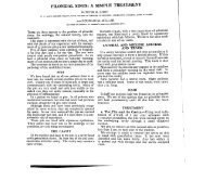
PILONIDAL SINUS – THE MODIFIED KARYDAKIS OPERATION<br />
Dr. Paul <strong>Kitchen</strong>, St Vincent’s Hospital, Melbourne<br />
In 1973, the late Dr. George <strong>Karydakis</strong>, in Athens, published his experience with a simple and<br />
successful operation to cure <strong>Pilonidal</strong> Sinus, 1 and later presented the largest personal series in<br />
the world. 2 He excised the sinus with a simple biconvex ‘elliptical’ excision only just crossing<br />
the midline to excise the sinus. It was based 1-2cm from the midline with excision down to the<br />
sacrum. A thick flap was then created by undercutting the midline side of the wound. This flap<br />
was advanced across the midline to meet the other side of the wound with two layers of catgut<br />
sutures to the fat and sacral fascia around a drain tube. The wound was then closed with skin<br />
sutures.<br />
<strong>Karydakis</strong> believed and taught that hair insertion is the cause of pilonidal sinus and attributed his<br />
extremely low recurrence rate of 1% to two facts. These are:<br />
(a) the whole wound is placed away from the midline (recurrences always occur in the<br />
midline) and<br />
(b) the resulting new natal cleft is shallower (so hairs do not collect so readily).<br />
I had the privilege of meeting Dr. <strong>Karydakis</strong> on three occasions and appreciated his thorough<br />
research and the originality of his ideas.<br />
Despite these good results, the <strong>Karydakis</strong> operation has been criticised for taking too much fat, 3<br />
and for placing sutures into the midline sacral fascia (often causing pain). Most of his patients<br />
required general anaesthesia and stayed in hospital a day or more, though before his death in the<br />
early 1990’s, Dr <strong>Karydakis</strong> was trying to develop ways to simplify the operation for day<br />
surgery. 4<br />
I have followed the <strong>Karydakis</strong> method since seeing it performed by him in London in 1973. I<br />
coined the term the ‘<strong>Karydakis</strong> operation’ to honour him and published my initial experience in<br />
1981. 5 Since 1973, I have done the operation (or a modification) since than on 497 patients of<br />
whom 9 (1.8%) had a recurrence requiring another procedure (curettage or a repeat <strong>Karydakis</strong>)<br />
and 8 (1.6%) had slight insignificant wound problems easily dealt with by simple measures<br />
without more surgery.<br />
Dr. John Bascom in Eugene, Oregon, developed a similar operation that he calls ‘cleft lift.’ 3 It<br />
results in a shallow cleft and a wound off the midline that looks very like a <strong>Karydakis</strong> wound,<br />
and he reports that his operation is very successful in the treatment of difficult recurrent sinuses.<br />
I witnessed this operation performed by Dr. John Bascom and his son Dr. Tom Bascom at their<br />
hospital in July 2004, and learned a number of useful modifications to the technique I have been<br />
using. Since then I have changed my technique for 216 patients (43% of my total) and called it<br />
the ‘<strong>Modified</strong> <strong>Karydakis</strong> operation.’ Since than my patients have gone home sooner (usually<br />
within 24 hours) with less pain. 37 (17%) of these latter have the operation under local<br />
1 <strong>Karydakis</strong> GE ‘New approach to the problem of pilonidal sinus’ Lancet 1973; ii:1414-15<br />
2 <strong>Karydakis</strong> GE: ‘Easy and successful treatment of pilonidal sinus after explanation of its causative<br />
processes Aust NZ J Surg 1992; 62:385-9<br />
3 Bascom JU ‘Failed pilonidal surgery’ Arch Surg 2002; 137:1146-50<br />
4 Personal Communication<br />
5 <strong>Kitchen</strong> P.R.B. ‘<strong>Pilonidal</strong> Sinus:Excision and Primary Closure with a Lateralised Wound - the <strong>Karydakis</strong> Operation’<br />
A.N.Z.J.Surg. 1981; 52:302-305.
anaesthesia (with intravenous sedation), and 4 under spinal anaesthesia and there have been 5<br />
(2%) recurrences.<br />
The modifications I have made after learning from Drs. Bascom include the following:<br />
1. The operation can be performed with a liberal usage of local anaesthesia (and adrenaline)<br />
which is preferable to general anaesthesia in the prone position. This is possible in nonnervous<br />
patients if the sinus not too large, and it can be done as a day-case. Give plenty<br />
of time to insert LA slowly under IV sedation with fine needle. However, my experience<br />
more recently has been that most patients prefer general anaesthesia or spinal anaesthesia<br />
and an overnight stay.<br />
2. The flap should be created first, usually 2cm wide and ~0.75cm deep under the skin<br />
3. Buttock straps should then be removed from the edges of the operating table and the skin<br />
flap gently pulled across the midline with skin hooks to see if the mark of the outer rim<br />
of the ‘ellipse’ has been made correctly on the skin, and to make adjustments to prevent<br />
tension on wound closure. Less may need to be excised from the top end where the cleft<br />
is shallower than lower down.<br />
4. The outer limit of excision is then cut with the scalpel, but instead of going down to the<br />
sacrum (<strong>Karydakis</strong> method), the fat is left and only skin and dermis are excised, until the<br />
sinus is reached.<br />
5. If the cavity is large and deep, the deepest portion of its wall can be left in-situ, and after<br />
curetting it out, and cut into small 1cm cubes so that it will collapse and its sides come<br />
together on closure and help to elevate the cleft (excising a large deep abscess wall may<br />
in fact deepen the cleft on closure rather than make it more shallow).<br />
6. John Bascom has reminded me that <strong>Karydakis</strong> had emphasized the circulation of hairs<br />
from a midline primary pit and through secondary openings. 2 So secondary openings a<br />
distance from the main track do not have to be included in the excision (eg, by making<br />
the wound very large, or by V cuts on one side of the wound to close and result in a T-<br />
shape as I once advocated 6 ). Rather, the tracks can be curetted, and the openings cleaned<br />
out and enlarged a bit, and hair particles removed by curettage and pulling gauze through<br />
them, to and fro. Once the primary pit is dealt with, the secondary pits should left to<br />
drain and will heal, and any hairs in them should make their way out.<br />
7. The flap should fit gently across the midline on the fat rolled in from the other side over<br />
the suction drain tube brought out well laterally. A few fine PDS or vicryl sutures are<br />
used in the fat and need not be inserted down to the sacral fascia.<br />
flap<br />
collapsed<br />
cavity in<br />
depth<br />
I use the term ‘<strong>Modified</strong> <strong>Karydakis</strong>’ rather than ‘Cleft Lift’ or ‘Bascom II’ because the basis of<br />
the operation is the original work of <strong>Karydakis</strong> (who deserves the recognition), and to prevent<br />
confusion with the simpler operation previously described by John Bascom. 7<br />
I have found the modified <strong>Karydakis</strong> operation (or cleft lift) is an improvement on the original<br />
<strong>Karydakis</strong> procedure because it uses sensible plastic surgical principles (in preparation of the<br />
flap first), it only removes what is necessary (preserves most of the fat to help elevate the cleft)<br />
and is less painful (no deep sutures). Also it can sometimes be done under LA as a day-case.<br />
6 <strong>Kitchen</strong> PRB ‘<strong>Pilonidal</strong> Sinus - Experience with the <strong>Karydakis</strong> Flap’ Br J Surg 1996; 83:1452-55<br />
7 Bascom JU ‘<strong>Pilonidal</strong> <strong>Disease</strong>: longterm results from follicle removal’ Dis Colon Rectum 1983; 26:800-7
I would add to the teachings of Dr. John Bascom with the following points:<br />
1. The ‘ellipse’ should be marked out on the operating table by first marking two points on<br />
one side (the most diseased side), a distance of 1.5-2 cm from the midline and placed far<br />
enough apart to allow a gentle curve to be marked out to just cross the midline a few<br />
mms. to include the primary pit in the excision. For small sinuses, use 1.5cm from the<br />
midline.<br />
Point marked 2cm from midline<br />
•<br />
•<br />
width 4cm<br />
• Point marked 2cm from midline<br />
2. The distance of maximum width of the ‘ellipse’ is twice the distance the upper and lower<br />
points are from the midline (3-4 cm). This is measured between the marked lines not<br />
between the midline and the outer edge.<br />
3. After probing the tracks to work out their extent, the use of methylene blue injection into<br />
the midline pit will enable all branches to be easily identified in case any are if severed<br />
or opened during the excision.<br />
4. To avoid a ‘dog-ear’ at the lower end or the wound tending to ‘move’ towards the anus<br />
or midline, a V excision of skin can be taken off the lower end laterally. This will deviate<br />
the lower end a little further from the midline when skin is closed.<br />
5. A fine low-pressure suction drain tube is placed in the fat, brought out lateral to the<br />
wound. Allow no suture holes or drain hole to appear in the new midline.<br />
6. Use a subcuticular 3/0 prolene skin suture loosely knotted as a loop (the wound<br />
lengthens when the patient sits, so loose suture needed to prevent a ‘cheese-cutter’<br />
effect). Support it with a few interrupted 3/0 prolene sutures.<br />
7. IV antibiotics including metronidazole is given during the procedure<br />
8. Patient goes home same or next day after removal of drain tube. All sutures out on tenth<br />
day.<br />
base of flap becomes the midline of a new shallower cleft<br />
excised skin<br />
closed wound<br />
V cut brings lower end away from midline