Oral delivery of chitosan microspheres for peyer's patch targeting ...
Oral delivery of chitosan microspheres for peyer's patch targeting ...
Oral delivery of chitosan microspheres for peyer's patch targeting ...

Create successful ePaper yourself
Turn your PDF publications into a flip-book with our unique Google optimized e-Paper software.
XVIIth International Conference on Bioencapsulation, Groningen, Netherlands ; September 24-26, 2009<br />
<strong>Oral</strong> <strong>delivery</strong> <strong>of</strong> <strong>chitosan</strong> <strong>microspheres</strong> <strong>for</strong> peyer’s <strong>patch</strong> <strong>targeting</strong> against<br />
diseases associated with small instestine<br />
Khare P. 1# and Jain S. K. 2 *<br />
1 Science Stream, Department <strong>of</strong> Pharmaceutical Sciences, 2 Dr. H.S. Gour<br />
University - Sagar- India, 470003<br />
* supervisor # piushkhare@rediffmail.com<br />
INTRODUCTION<br />
Chitosan <strong>microspheres</strong> as drug <strong>delivery</strong> system have attained importance and attracted the attention<br />
<strong>of</strong> researchers recently. Microspheres have been explored extensively <strong>for</strong> their use in the field <strong>of</strong><br />
drug <strong>delivery</strong> and various polymers have been utilized <strong>for</strong> the <strong>for</strong>mulation <strong>of</strong> the <strong>microspheres</strong><br />
which in turn have been assessed <strong>for</strong> different purposes (Woo B.H. 2001). Recently, dosage <strong>for</strong>ms<br />
that can precisely control the release rates and target drugs to a definite body site or organ have<br />
played a pivotal role and provided a major thrust in the development <strong>of</strong> the novel drug <strong>delivery</strong><br />
field. Chitosan which is deacetylated derivative <strong>of</strong> (-4)2- acetaamido-2-deoxy b-d- glucose or<br />
chitin, has been extensively explored <strong>for</strong> its various biomedical and pharmaceutical applications. As<br />
a drug carrier <strong>chitosan</strong> has been investigated <strong>for</strong> the sustained <strong>delivery</strong> <strong>of</strong> many oral <strong>for</strong>mulations<br />
and parentral <strong>for</strong>mulations. In the present project the <strong>chitosan</strong> <strong>microspheres</strong> were developed and<br />
characterized <strong>for</strong> the effective <strong>delivery</strong> <strong>of</strong> drug to small intestine via oral route. The developed<br />
systems can be used to deliver the bioactives <strong>for</strong> the selective uptake by the peyers <strong>patch</strong>es or<br />
release drug in the small intestine <strong>for</strong> diseases associated with this specific part <strong>of</strong> Gastrointestinal<br />
tract.<br />
MATERIAL AND METHODS<br />
Chitosan (purified, viscosity grade 50) obtained from Central Institute <strong>of</strong> Fisheries and Technology,<br />
Cochin India was used without further purification. Span 85 and Glutaraldehyde (biological grade,<br />
25% v/v aqueous solution) and Eudragit S-100 were from Sigma chemical, USA. Light liquid<br />
paraffin, Petroleum ether, Acetone, Acetic acid and other solvents were from Loba Chemie Pvt Ltd.<br />
Mumbai, India and were utilized as received. Deionised Millipore water was used throughout the<br />
study. Cipr<strong>of</strong>loxacin-HCl was a kind gift from Solisto Pharma, Sagar (M.P), India.<br />
Preparation <strong>of</strong> Microspheres<br />
Microspheres were prepared by slight modification <strong>of</strong> emulsification method (Gohel M.C. 1998).<br />
Glutaraldehyde was used as cross linking agent. Microspheres were prepared by taking 1:1 mixture<br />
<strong>of</strong> Petroleum Ether and Light liquid paraffin. Cipr<strong>of</strong>loxacin-HCl was taken as model drug. Briefly,<br />
the drug was already dissolved in 4% acetic acid solution and then <strong>chitosan</strong> was dissolved in the<br />
drug solution to produce 2% w/v concentration <strong>of</strong> the polymer. The drug to polymer ratio was kept<br />
1:2. This aqueous phase (5 ml) was added drop wise to the beaker containing 50 ml <strong>of</strong> oil phase and<br />
0.5% w/v Span 85 as emulsifying agent (previously mixed through stirring) under constant stirring.<br />
Stirring was carried out at 3000 rpm utilizing a three blade mechanical stirrer (Remi Instruments,<br />
Mumbai). 750 !L glutaraldehyde was added after 20 minutes. The stirring was continued <strong>for</strong> 3<br />
hours. The oil phase containing <strong>microspheres</strong> was centrifuged at 1000 g <strong>for</strong> one minute. The<br />
supernatant oil was decanted and the <strong>microspheres</strong> at the bottom were then washed three times with<br />
petroleum ether by centrifugation to remove the oil.<br />
Poster P22 – page 1<br />
XVIIth International Conference on Bioencapsulation, Groningen, Netherlands ; September 24-26, 2009<br />
They were then washed with water to remove traces <strong>of</strong> glutaraldehyde and then they were washed<br />
with acetone and then they were freeze dried in a freeze drier (Heto Hilton, Germany).<br />
Electron Microscopy<br />
Shape and surface morphology <strong>of</strong> the different <strong>microspheres</strong> <strong>for</strong>med was investigated through<br />
Scanning Electron Microscope (Leo 435 VP, Ox<strong>for</strong>d Instruments, England). Briefly, <strong>microspheres</strong><br />
were sprinkled on double sided tape, sputter coated with gold and were examined in the<br />
microscope.<br />
Particle Size<br />
Microspheres were sized using a Malvern mastersizer (model Nano ZF-90, Malvern Instruments<br />
Private Ltd. UK). The volume mean diameter and distribution <strong>of</strong> particle size were measured.<br />
Average mean size <strong>of</strong> different <strong>microspheres</strong> prepared was found to be 3.15±0.04 !m.<br />
Zeta Potential<br />
Zeta potentials were measured by electrophoresis per<strong>for</strong>med on a Malvern Zetasizer (model Nano<br />
ZF-90, Malvern Instruments Private Ltd. UK). Phosphate buffer pH 7.4 was used as the medium to<br />
suspend the <strong>microspheres</strong> <strong>for</strong> the measurement <strong>of</strong> the zeta potential. The zeta potential was found to<br />
be 5.03±0.06 mv.<br />
Drug content and Entrapment Efficiency<br />
Microspheres were crushed in a glass mortar and pestle. The powered <strong>microspheres</strong> were suspended<br />
in 10 ml <strong>of</strong> phosphate buffer (pH 7.4). After 24 hours, the solution was filtered and the filtrate was<br />
analysed <strong>for</strong> the drug content through UV-spectrophotometer (Shimadzu-1601, Kyoto, Japan) at<br />
241.5 nm. The drug entrapment efficiency was calculated using the following <strong>for</strong>mula: Practical<br />
drug content/ Theoretical drug content x 100.<br />
In vitro Drug release<br />
In vitro drug release studies were per<strong>for</strong>med by suspending 50 mg <strong>of</strong> <strong>microspheres</strong> in PBS buffer<br />
(pH 7.4). This suspension was then filled in a dialysis bag and this was placed in release media<br />
(PBS buffer pH 7.4). Aliquots were withdrawn at predetermined intervals <strong>of</strong> time and volume was<br />
made up with an equal amount <strong>of</strong> phosphate saline buffer pH 7.4. The aliquots were analyzed <strong>for</strong><br />
the drug content at 241.5 nm using UV spectrophotometer (Shimadzu-1601, Kyoto, Japan).<br />
Coating <strong>of</strong> Microspheres<br />
The <strong>microspheres</strong> were coated with Eudragit S-100 as reported by Lorenzo-Lamosa et.al., with<br />
slight modifications (Lorenzo-Lamosa M.L. 1998). Briefly the prepared <strong>chitosan</strong> <strong>microspheres</strong> were<br />
dispersed in 5 ml <strong>of</strong> an organic solvent in which Eudragit S-100 was previously to give 10:1 coat<br />
/core ratio. This organic phase was poured into 70 ml <strong>of</strong> liquid paraffin containing Span 85. The<br />
system was maintened under agitation at 1500 rpm at room temperature.The solvent was evaporated<br />
and the coated <strong>microspheres</strong> were collected and rinsed with n-hexane and freeze dried.<br />
Confocal Microscopy study<br />
The CLSM study was per<strong>for</strong>med using Rh-6G loaded <strong>chitosan</strong> <strong>microspheres</strong> which were<br />
administered to the Balbc mice orally. Plain dye was administered to the other group <strong>of</strong> mice and<br />
was treated as control. The studies were per<strong>for</strong>med on Zeiss LSM 510 meta microscope .<br />
Poster P22 – page 2
XVIIth International Conference on Bioencapsulation, Groningen, Netherlands ; September 24-26, 2009<br />
RESULTS AND DISCUSSION<br />
Uni<strong>for</strong>m and small <strong>microspheres</strong> <strong>of</strong> 3.15±0.04 !m and 68.87±1.03 percent drug entrapment were<br />
obtained. Zeta potential <strong>of</strong> the <strong>microspheres</strong> was found to be 5.03±0.06 mv. A higher entrapment<br />
value in this case is due to the hydrophllic nature <strong>of</strong> the drug which got entrapped in the hydrophllic<br />
matrix <strong>of</strong> the <strong>microspheres</strong>. Lower zeta potential values are due to the crosslinking <strong>of</strong> the amine<br />
groups <strong>of</strong> the <strong>chitosan</strong> by glutaraldehyde.<br />
Since the amine groups are positively charged and contribute to the potential <strong>of</strong> the carrier system<br />
and hence the crosslinking reduces the free amine groups and hence the effect. Microspheres were<br />
coated with Eudragit S-100 to prevent the drug release in acidic environment <strong>of</strong> stomach. Eudragit<br />
S-100 is a pH responsive polymer and degrades only at the pH similar to that <strong>of</strong> small intestine.<br />
There<strong>for</strong>e it was used so as to release the contents specifically at small intestine. In vitro drug<br />
release in phosphate buffer pH 7.4 was calculated <strong>for</strong> the <strong>microspheres</strong> which showed 70±2.46%<br />
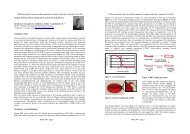
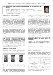
release <strong>of</strong> drug after an interval <strong>of</strong> 7 hrs. Confocal microscopy studies were per<strong>for</strong>med in Balbc<br />
mice where Rhodamine 6G loaded coated <strong>microspheres</strong> as well plain dye was administered orally<br />
to the animals in different groups. The confocal laser scanning microscopy study revealed that Rh-<br />
6G loaded <strong>chitosan</strong> <strong>microspheres</strong> were present in peyers <strong>patch</strong> which confirm the effective uptake<br />
<strong>of</strong> the <strong>microspheres</strong> to peyer’s <strong>patch</strong>es whereas the plain dye did not show any uptake into the<br />
peyers <strong>patch</strong>es (Figure 1a & 1b).<br />
XVIIth International Conference on Bioencapsulation, Groningen, Netherlands ; September 24-26, 2009<br />
through these systems which would elicit the immune response. This work was a part <strong>of</strong> our<br />
ongoing thrust and project to develop microparticulate drug <strong>delivery</strong> system <strong>for</strong> oral <strong>delivery</strong>.<br />
ACKNOWLEDGEMENTS<br />
Financial support from Indian Council <strong>of</strong> Medical Research (ICMR), Govt. <strong>of</strong> India - New Delhi,<br />
India <strong>for</strong> the work is gratefully acknowledged. Authors are thankful to All India Institute <strong>of</strong> Medical<br />
Sciences – New Delhi, India <strong>for</strong> Scanning Electron Microscopy and Director, CIFT, Cochin, India<br />
<strong>for</strong> generous gift <strong>of</strong> Chitosan.<br />
REFERENCES<br />
Woo BH, Jiang G, Jo YW, Deluca PP (2001). Pharm Res 18:1600-1606.<br />
Gohel MC, Amin AF (1998). J Control Rel 51:115-122.<br />
Lorenzo-Lamosa ML, Lopez CR, Vila-Jato JL, Alonso, MJ (1998). J.Control. Rel. 52: 109-118.<br />
1a: Plain dye (Rh6G) 1b: Rh6G loaded <strong>microspheres</strong><br />
Figure 1: CLSM photomicrograph showing uptake <strong>of</strong> the plain dye and <strong>chitosan</strong> microsphers<br />
<strong>microspheres</strong> to peyer’s <strong>patch</strong>es<br />
This could be due the fact that Eudragit coated <strong>microspheres</strong> reached the small intestine and did not<br />
release their content in the stomach. Moreover these microparticles are effectively taken up by the<br />
peyers <strong>patch</strong> tissue where they are trapped depending upon their size. On the contrary the dye may<br />
have been washed <strong>of</strong>f by the gastrointestinal fluid and hence no uptake resulted as a result.<br />
CONCLUSION<br />
Results revealed that <strong>microspheres</strong> effectively localize in the peyer’s <strong>patch</strong> <strong>of</strong> small intestine.<br />
Further this also can be utilized <strong>for</strong> the development <strong>of</strong> the vaccine by delivering the antigen<br />
Poster P22 – page 3<br />
Poster P22 – page 4