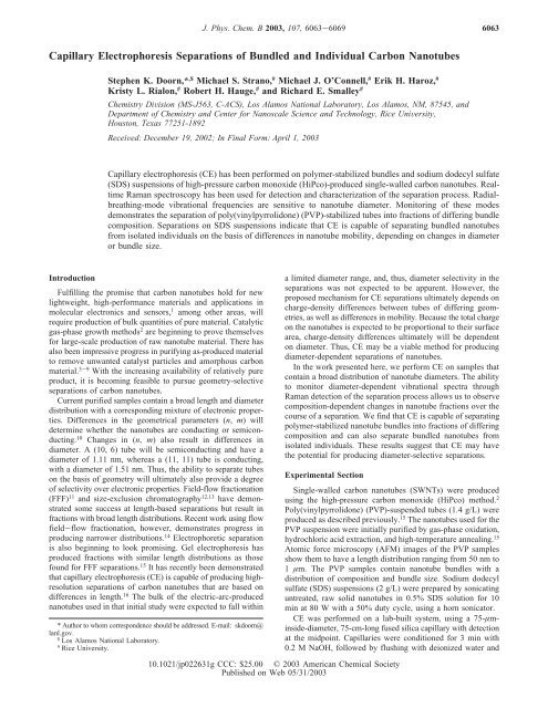
Capillary Electrophoresis Separations of Bundled ... - Rice University
Capillary Electrophoresis Separations of Bundled ... - Rice University
Capillary Electrophoresis Separations of Bundled ... - Rice University
You also want an ePaper? Increase the reach of your titles
YUMPU automatically turns print PDFs into web optimized ePapers that Google loves.
J. Phys. Chem. B 2003, 107, 6063-6069<br />
6063<br />
<strong>Capillary</strong> <strong>Electrophoresis</strong> <strong>Separations</strong> <strong>of</strong> <strong>Bundled</strong> and Individual Carbon Nanotubes<br />
Stephen K. Doorn,* ,$ Michael S. Strano, # Michael J. O’Connell, # Erik H. Haroz, #<br />
Kristy L. Rialon, # Robert H. Hauge, # and Richard E. Smalley #<br />
Chemistry DiVision (MS-J563, C-ACS), Los Alamos National Laboratory, Los Alamos, NM, 87545, and<br />
Department <strong>of</strong> Chemistry and Center for Nanoscale Science and Technology, <strong>Rice</strong> UniVersity,<br />
Houston, Texas 77251-1892<br />
ReceiVed: December 19, 2002; In Final Form: April 1, 2003<br />
<strong>Capillary</strong> electrophoresis (CE) has been performed on polymer-stabilized bundles and sodium dodecyl sulfate<br />
(SDS) suspensions <strong>of</strong> high-pressure carbon monoxide (HiPco)-produced single-walled carbon nanotubes. Realtime<br />
Raman spectroscopy has been used for detection and characterization <strong>of</strong> the separation process. Radialbreathing-mode<br />
vibrational frequencies are sensitive to nanotube diameter. Monitoring <strong>of</strong> these modes<br />
demonstrates the separation <strong>of</strong> poly(vinylpyrrolidone) (PVP)-stabilized tubes into fractions <strong>of</strong> differing bundle<br />
composition. <strong>Separations</strong> on SDS suspensions indicate that CE is capable <strong>of</strong> separating bundled nanotubes<br />
from isolated individuals on the basis <strong>of</strong> differences in nanotube mobility, depending on changes in diameter<br />
or bundle size.<br />
Introduction<br />
Fulfilling the promise that carbon nanotubes hold for new<br />
lightweight, high-performance materials and applications in<br />
molecular electronics and sensors, 1 among other areas, will<br />
require production <strong>of</strong> bulk quantities <strong>of</strong> pure material. Catalytic<br />
gas-phase growth methods 2 are beginning to prove themselves<br />
for large-scale production <strong>of</strong> raw nanotube material. There has<br />
also been impressive progress in purifying as-produced material<br />
to remove unwanted catalyst particles and amorphous carbon<br />
material. 3-9 With the increasing availability <strong>of</strong> relatively pure<br />
product, it is becoming feasible to pursue geometry-selective<br />
separations <strong>of</strong> carbon nanotubes.<br />
Current purified samples contain a broad length and diameter<br />
distribution with a corresponding mixture <strong>of</strong> electronic properties.<br />
Differences in the geometrical parameters (n, m) will<br />
determine whether the nanotubes are conducting or semiconducting.<br />
10 Changes in (n, m) also result in differences in<br />
diameter. A (10, 6) tube will be semiconducting and have a<br />
diameter <strong>of</strong> 1.11 nm, whereas a (11, 11) tube is conducting,<br />
with a diameter <strong>of</strong> 1.51 nm. Thus, the ability to separate tubes<br />
on the basis <strong>of</strong> geometry will ultimately also provide a degree<br />
<strong>of</strong> selectivity over electronic properties. Field-flow fractionation<br />
(FFF) 11 and size-exclusion chromatography 12,13 have demonstrated<br />
some success at length-based separations but result in<br />
fractions with broad length distributions. Recent work using flow<br />
field-flow fractionation, however, demonstrates progress in<br />
producing narrower distributions. 14 Electrophoretic separation<br />
is also beginning to look promising. Gel electrophoresis has<br />
produced fractions with similar length distributions as those<br />
found for FFF separations. 15 It has recently been demonstrated<br />
that capillary electrophoresis (CE) is capable <strong>of</strong> producing highresolution<br />
separations <strong>of</strong> carbon nanotubes that are based on<br />
differences in length. 16 The bulk <strong>of</strong> the electric-arc-produced<br />
nanotubes used in that initial study were expected to fall within<br />
* Author to whom correspondence should be addressed. E-mail: skdoorn@<br />
lanl.gov.<br />
$ Los Alamos National Laboratory.<br />
# <strong>Rice</strong> <strong>University</strong>.<br />
a limited diameter range, and, thus, diameter selectivity in the<br />
separations was not expected to be apparent. However, the<br />
proposed mechanism for CE separations ultimately depends on<br />
charge-density differences between tubes <strong>of</strong> differing geometries,<br />
as well as differences in mobility. Because the total charge<br />
on the nanotubes is expected to be proportional to their surface<br />
area, charge-density differences ultimately will be dependent<br />
on diameter. Thus, CE may be a viable method for producing<br />
diameter-dependent separations <strong>of</strong> nanotubes.<br />
In the work presented here, we perform CE on samples that<br />
contain a broad distribution <strong>of</strong> nanotube diameters. The ability<br />
to monitor diameter-dependent vibrational spectra through<br />
Raman detection <strong>of</strong> the separation process allows us to observe<br />
composition-dependent changes in nanotube fractions over the<br />
course <strong>of</strong> a separation. We find that CE is capable <strong>of</strong> separating<br />
polymer-stabilized nanotube bundles into fractions <strong>of</strong> differing<br />
composition and can also separate bundled nanotubes from<br />
isolated individuals. These results suggest that CE may have<br />
the potential for producing diameter-selective separations.<br />
Experimental Section<br />
Single-walled carbon nanotubes (SWNTs) were produced<br />
using the high-pressure carbon monoxide (HiPco) method. 2<br />
Poly(vinylpyrrolidone) (PVP)-suspended tubes (1.4 g/L) were<br />
produced as described previously. 15 The nanotubes used for the<br />
PVP suspension were initially purified by gas-phase oxidation,<br />
hydrochloric acid extraction, and high-temperature annealing. 15<br />
Atomic force microscopy (AFM) images <strong>of</strong> the PVP samples<br />
show them to have a length distribution ranging from 50 nm to<br />
1 µm. The PVP samples contain nanotube bundles with a<br />
distribution <strong>of</strong> composition and bundle size. Sodium dodecyl<br />
sulfate (SDS) suspensions (2 g/L) were prepared by sonicating<br />
untreated, raw solid nanotubes in 0.5% SDS solution for 10<br />
min at 80 W with a 50% duty cycle, using a horn sonicator.<br />
CE was performed on a lab-built system, using a 75-µminside-diameter,<br />
75-cm-long fused silica capillary with detection<br />
at the midpoint. Capillaries were conditioned for 3 min with<br />
0.2 M NaOH, followed by flushing with deionized water and<br />
10.1021/jp022631g CCC: $25.00 © 2003 American Chemical Society<br />
Published on Web 05/31/2003
6064 J. Phys. Chem. B, Vol. 107, No. 25, 2003 Doorn et al.<br />
Figure 1. Raman spectrum (20 mW at 785 nm excitation, 1-s integration) <strong>of</strong> bulk solid HiPco-produced nanotubes. Inset: Radial breathing mode<br />
(RBM) region <strong>of</strong> bulk solid.<br />
then run buffer before each separation. <strong>Separations</strong> were run<br />
with an applied voltage <strong>of</strong> +15 kV, using 50 mM trizma base<br />
as a run buffer (15 kV was below the limit <strong>of</strong> linear behavior<br />
in a voltage-versus-current plot for our buffer systems). Buffers<br />
for separations <strong>of</strong> the SDS suspensions also contained 0.5%<br />
SDS. Samples were pressure-introduced at 500 mbar for 2sby<br />
pulling sample into a 2-mL void volume with a syringe attached<br />
to the effluent end <strong>of</strong> the capillary.<br />
Raman detection was performed using 20-mW, 785-nm diode<br />
laser excitation through a fiber-optic probe head that incorporated<br />
a 20×, 0.5 N.A. objective for sample illumination and<br />
180° collection <strong>of</strong> Raman scatter through a window burned into<br />
the capillary coating. Scattered light was collected into a Kaiser<br />
Optical HoloSpec f/1.8 imaging spectrograph. An entrance<br />
slit 50 µm wide resulted in a spectral resolution <strong>of</strong> 4 cm -1 .<br />
Detection was accomplished with a back-illuminated redsensitive<br />
Princeton Instruments CCD camera (100 × 1340<br />
pixels), using integration times <strong>of</strong> 1 s. Full spectra were collected<br />
continuously in real time during the course <strong>of</strong> a separation.<br />
Results<br />
Behavior <strong>of</strong> PVP-Stabilized Nanotubes. A bulk Raman<br />
spectrum <strong>of</strong> the purified solid HiPco-produced nanotubes is<br />
shown in Figure 1. Of particular interest for our studies is the<br />
low-frequency region <strong>of</strong> the spectrum (Figure 1, inset), which<br />
is dominated by the nanotube radial breathing mode (RBM)<br />
vibrations. The observed frequencies are inversely proportional<br />
to the diameter <strong>of</strong> the nanotubes 17 and range from 169 to 373<br />
cm -1 in our sample, which translates to a diameter distribution<br />
ranging from 0.6 to 1.3 nm. The seven most-intense bands<br />
(207-267 cm -1 ) correspond to diameters <strong>of</strong> 0.88-1.15 nm. The<br />
presence <strong>of</strong> a broad range <strong>of</strong> observable diameters in the HiPcoproduced<br />
samples makes them attractive for investigating the<br />
potential <strong>of</strong> CE for diameter selectivity.<br />
A representative electropherogram from a separation <strong>of</strong> PVPsuspended<br />
sample is shown in Figure 2a. By monitoring the<br />
intensity <strong>of</strong> the strong 1591 cm -1 nanotube Raman band, we<br />
find that nanotubes first appear ∼17 min into a separation. (Note<br />
that each point in the electropherogram actually represents a<br />
complete Raman spectrum.) Except for the appearance <strong>of</strong> a few<br />
spikes (bands limited in width to the integration time <strong>of</strong> 1 s),<br />
the electropherogram is dominated by primarily broad, incompletely<br />
resolved fractions. This domination is in contrast to the<br />
behavior observed in the original work on SDS-dissolved<br />
nanotubes by Doorn et al., 16 in which well-resolved, extremely<br />
narrow fractions were observed. The time <strong>of</strong> the first appearance<br />
<strong>of</strong> nanotubes is reproducible from run to run, as are the positions<br />
<strong>of</strong> the major features in the electopherogram. However, the<br />
relative intensities <strong>of</strong> these features can change between runs.<br />
This variation may be due to minor differences in the manual<br />
sampling method and also possibly results from the small and<br />
potentially heterogeneous sampling volume (∼100-300 nL).<br />
The presence <strong>of</strong> distinct bands in the electropherogram indicates<br />
that some separation process is occurring; however, the question<br />
<strong>of</strong> whether this is due only to the previously observed lengthbased<br />
mechanism 16 or combines a diameter-based process<br />
requires further analysis <strong>of</strong> the Raman spectra to determine.<br />
Closer inspection <strong>of</strong> how the RBM region <strong>of</strong> the Raman<br />
spectra changes over the course <strong>of</strong> a separation will provide<br />
information on whether diameter-selective separations are<br />
occurring superimposed on the length-based mechanism. Electropherograms<br />
showing changes in the intensity ratios <strong>of</strong> the<br />
267 cm -1 band, relative to the 234 cm -1 band, and <strong>of</strong> the 234<br />
cm -1 band, relative to the 207 cm -1 band, are shown in Figures
CE <strong>Separations</strong> <strong>of</strong> Carbon Nanotubes J. Phys. Chem. B, Vol. 107, No. 25, 2003 6065<br />
Figure 2. Electropherograms <strong>of</strong> PVP-stabilized nanotubes monitoring<br />
(a) the 1591 cm -1 mode intensity, (b) the intensity ratio <strong>of</strong> the 267<br />
cm -1 to 234 cm -1 modes, and (c) the intensity ratio <strong>of</strong> the 234 cm -1 to<br />
207 cm -1 modes.<br />
2b and c, respectively. At early times (1200-1450 s), the band<br />
at 234 cm -1 is the most prominent, with a significant increase<br />
in intensity occurring at 1404 s. At intermediate times, the 267<br />
cm -1 band is strongest. Finally, from ∼1600 s to the end <strong>of</strong> the<br />
separation, the 234 cm -1 mode diminishes in intensity.<br />
Differences in selected spectra within these regions are shown<br />
for the highlighted times in Figure 3 and are also compared to<br />
the bulk, unseparated solution spectrum. Nanotubes observed<br />
at intermediate times display spectra (Figure 3c) very similar<br />
to that observed for the bulk solution (Figure 3a). At early times,<br />
the 234 cm -1 mode is clearly dominant, whereas the 267 cm -1<br />
band is diminished, with respect to all other modes present. At<br />
the end <strong>of</strong> the separation (Figure 3d), all vibrations are weaker<br />
overall in intensity, but the intermediate frequencies (215-234<br />
cm -1 ) are significantly weakened, compared to the 207 and 267<br />
cm -1 modes.<br />
Behavior <strong>of</strong> SDS-Suspended Nanotubes. A typical electropherogram<br />
for the SDS-suspended nanotube sample is shown<br />
in Figure 4. A series <strong>of</strong> baseline-separated, sharp and intense<br />
spikes (widths limited to the 1-s integration time) are observed<br />
throughout the early part <strong>of</strong> the separation (Figure 4a). These<br />
results are similar to those found by Doorn et al. in their<br />
separations <strong>of</strong> SDS-suspended arc-produced nanotubes. 16 Closer<br />
inspection <strong>of</strong> the electropherogram, however, reveals that the<br />
sharp spikes are on top <strong>of</strong> a broad background (Figure 4b) that<br />
more resembles the results obtained for the PVP suspended<br />
tubes.<br />
As seen in the ratio electropherogram <strong>of</strong> the 267 cm -1<br />
intensity divided by the 234 cm -1 intensity (Figure 4c), the<br />
behavior <strong>of</strong> the RBMs undergoes large changes over the course<br />
<strong>of</strong> the separation. Three distinct regions <strong>of</strong> behavior are<br />
Figure 3. (a) Radial breathing mode (RBM) Raman spectrum <strong>of</strong> bulk,<br />
unseparated PVP-stabilized nanotube solution. Selected Raman spectra<br />
from separation shown in Figure 2 are also presented: (b) spectrum at<br />
1404 s, (c) spectrum at 1461 s, and (d) spectrum at 1800 s.<br />
observed. In the first region, ranging from the first appearance<br />
<strong>of</strong> nanotubes to ∼500 s, ratios for both the spikes and the broad<br />
envelope are greater than the baseline ratio observed before the<br />
appearance <strong>of</strong> nanotubes. This observation indicates that spectra<br />
in this region are dominated by the 267 cm -1 mode. At<br />
intermediate times (region 2, 500-677 s), the intense spikes<br />
are still positive going, but the broad envelope begins to trend<br />
to values less than the baseline. At longer times (region 3, from<br />
678 s to the end <strong>of</strong> the separation), the intense spikes disappear<br />
and the broad envelope remains in the region below the baseline.<br />
This later-time region is dominated by the 234 cm -1 mode<br />
throughout the broad envelope, as well as for the weak spikes<br />
observed in this region. The first appearance <strong>of</strong> nanotubes occurs<br />
reproducibly after ∼5 min into a separation. The observed<br />
positions <strong>of</strong> individual spikes and relative intensities <strong>of</strong> the major<br />
features change from run to run, because <strong>of</strong> sampling heterogeneities.<br />
However, the positions and duration <strong>of</strong> the three<br />
regions observed in the ratio electropherogram are reproducible.<br />
Representative spectra for these three regions are compared<br />
to the bulk solution spectrum in Figure 5. A spectrum <strong>of</strong> a spike<br />
at 482 s is shown in Figure 5b. The spectrum is dominated by<br />
the 267 cm -1 band and is similar to that for the bulk solution.<br />
In the spike spectrum, however, the relative intensity near 234<br />
cm -1 is ∼50% <strong>of</strong> that in the bulk spectrum. The spectrum at<br />
482 s is identical to all spikes observed in the first two regions<br />
<strong>of</strong> the electropherogram, except for variations in intensity. The<br />
broad background envelope spectra in this region are also
6066 J. Phys. Chem. B, Vol. 107, No. 25, 2003 Doorn et al.<br />
Figure 4. (a) Electropherogram <strong>of</strong> SDS-suspended nanotubes, monitoring<br />
the 1591 cm -1 intensity. (b) Electropherogram showing expansion<br />
<strong>of</strong> the y-axis. (c) Electropherogram monitoring the intensity ratio <strong>of</strong><br />
the 267 cm -1 to 234 cm -1 modes.<br />
identical to this spikesthe observed frequencies and relative<br />
intensities remain the same. In the second electropherogram<br />
region (500-677 s), the spike spectra also display the same<br />
frequencies and relative intensities as those that are observed<br />
in the first region. However, the background envelope now shifts<br />
to spectra that are dominant in the 234 cm -1 mode, with its<br />
intensity being approximately twice that <strong>of</strong> the 267 cm -1 band.<br />
The spectrum taken at 633 s (Figure 5c) is representative <strong>of</strong><br />
this second region. A further dramatic shift in the relative<br />
intensities is seen when one goes into the third region. Almost<br />
all the RBM intensity is now in the 234 cm -1 mode, plus two<br />
shoulders located at 226 and 215 cm -1 (Figure 5d). Spectra in<br />
this region are also similar to those observed in the fraction at<br />
1404 s in the PVP separations (Figure 3b), although with lower<br />
intensity observed in the 267 cm -1 mode. Low-intensity spikes<br />
are also observed in this third region. Spectra for these spikes<br />
are quite different than those found for the strong spikes in the<br />
first region. Rather than being dominant in the 267 cm -1 mode,<br />
the spectra are quite similar to the envelope spectra observed<br />
in the second region (Figure 5c), with the 234 cm -1 band being<br />
approximately twice the intensity <strong>of</strong> that at 267 cm -1 .<br />
Discussion<br />
The variations in the RBM intensities indicate that there are<br />
diameter-dependent spectral changes occurring over the course<br />
<strong>of</strong> a separation. It is tempting to relate these intensity changes<br />
directly to changes in the relative population <strong>of</strong> the corresponding<br />
diameter tube. However, there are several complicating<br />
Figure 5. (a) Radial breathing mode (RBM) Raman spectrum <strong>of</strong> bulk<br />
unseparated SDS-suspended nanotubes. Selected Raman spectra from<br />
separation shown in Figure 4 are also presented: (b) spectrum at 482<br />
s, (c) spectrum at 633 s, and (d) spectrum at 918 s.<br />
factors. The PVP samples used in this investigation are known<br />
to be bundles that vary in size and composition. Thus, the<br />
behavior observed is not that for separations <strong>of</strong> isolated,<br />
individual nanotubes. Furthermore, Raman spectra for nanotubes<br />
display strongly diameter-dependent resonance enhancement<br />
effects. Spectra for nanotubes <strong>of</strong> differing diameter will be<br />
enhanced to varying degrees for a fixed excitation wavelength.<br />
This is an important consideration for both the PVP and SDS<br />
systems. Only RBMs for nanotubes with a van Hove singularity<br />
near the 785 nm excitation energy will be significantly enhanced.<br />
These enhancement effects will be further perturbed in the<br />
PVP samples, because <strong>of</strong> the strong interaction expected among<br />
nanotubes in a given bundle. Bundling (roping) <strong>of</strong> the nanotubes<br />
gives rise to an orthogonal energy dispersion to the onedimensional<br />
density <strong>of</strong> states found in individual nanotubes,<br />
resulting in a change in energy spacing <strong>of</strong> the van Hove<br />
singularities. 18,19 For semiconducting nanotubes, the absorption<br />
maxima are broadened and shifted to lower energy. 19,20 This is<br />
a particularly strong effect for the 267 cm -1 mode, which can<br />
be used as an indicator <strong>of</strong> the degree <strong>of</strong> roping. For sufficiently<br />
large bundles, the electronic resonance for this mode strongly<br />
overlaps the 785 nm excitation energy. As the bundle size<br />
decreases, the resonance energy shifts to shorter wavelengths.<br />
This shift will result in a decrease in resonance Raman intensity<br />
at 267 cm -1 to a minimum when overlap is minimized between<br />
the 785 nm excitation energy and the electronic resonance for
CE <strong>Separations</strong> <strong>of</strong> Carbon Nanotubes J. Phys. Chem. B, Vol. 107, No. 25, 2003 6067<br />
Figure 6. Radial breathing mode (RBM) spectra for (a) an aqueous<br />
slurry <strong>of</strong> roped nanotubes, (b) a sonicated sample <strong>of</strong> nanotubes in 6.3<br />
mM SDS, and (c) isolated individuals collected via centrifugation (ref<br />
20). Note: the absolute intensities for spectra a and b are directly<br />
comparable; an arbitrary scaling factor was applied to spectrum c.<br />
an individual nanotube. This trend is clearly seen in Figure 6.<br />
The 267 cm -1 mode loses intensity when one goes from a purely<br />
roped sample, through a sonicated mixture <strong>of</strong> roped and isolated<br />
nanotubes, and finally to a sample <strong>of</strong> purely isolated individuals.<br />
Conversely, the 234 cm -1 mode gains in intensity when one<br />
goes from a roped sample to isolated individuals. Resonance<br />
Raman excitation pr<strong>of</strong>iles on samples <strong>of</strong> isolated individuals<br />
show the 234 cm -1 mode to be strongly enhanced with 785 nm<br />
excitation. Thus, enhancement effects and perturbations may<br />
result in spectra that do not accurately indicate the dominant<br />
nanotube type in a given population but may be useful in<br />
indicating the relative nanotube bundle size. Perturbations <strong>of</strong><br />
enhancement behavior are not expected to be dependent on<br />
nanotube length for our samples.<br />
The bundle size/cross section for a collection <strong>of</strong> nanotubes<br />
is defined by the number <strong>of</strong> nanotubes present and by their<br />
individual diameters. The results for the PVP samples demonstrate<br />
that we can separate a sample into fractions <strong>of</strong> differing<br />
composition, that exhibit differing electronic resonance behavior.<br />
The majority <strong>of</strong> PVP-stabilized bundles will contain a random<br />
distribution <strong>of</strong> the available tube diameters. Therefore, spectra<br />
observed over the course <strong>of</strong> a separation might be expected to<br />
be similar to that observed for the bulk solution. This condition<br />
is observed at intermediate times (Figure 3c). Even at very early<br />
and late times, when the ratio electropherograms are showing<br />
some enhancement <strong>of</strong> particular RBMs (Figure 2b, c), the<br />
spectra are still similar to the bulk spectrum. The somewhatreduced<br />
relative intensity <strong>of</strong> the 267 cm -1 mode found in the<br />
region from 1100 to 1400 s (Figure 2b) suggests that these<br />
bundles may be smaller than those eluting at later times. The<br />
sharp loss in intensity <strong>of</strong> the 234 cm -1 mode at late times (Figure<br />
3d) may be due to a decrease in the population <strong>of</strong> corresponding<br />
diameter tubes. Alternatively, the bundle compositions at this<br />
point in the separation may yield a shift <strong>of</strong> the 234 cm -1<br />
resonance energy that results in poor overlap with the excitation<br />
wavelength.<br />
The most-dramatic changes in the spectra are observed for<br />
the sharp, intense fraction occurring at 1404 s (Figure 3b).<br />
Although the dominance <strong>of</strong> the 234 cm -1 mode in this fraction<br />
may mean that the corresponding diameter <strong>of</strong> the nanotube is<br />
being enriched in this fraction, this seems improbable. A more<br />
likely explanation is that the bundle size for this fraction is<br />
significantly smaller than that found in the remainder <strong>of</strong> the<br />
sample, which would result in a significant loss in intensity in<br />
the 267 cm -1 mode, as is observed for this fraction.<br />
The degree to which bundles <strong>of</strong> different composition exist<br />
in a fraction is indicated by the relative intensity and width <strong>of</strong><br />
that fraction. For those that represent a more random distribution<br />
<strong>of</strong> diameters, there should be a small and continuous variation<br />
in cross section/size across the range <strong>of</strong> composition. This<br />
variation will result in relatively broad bands, as observed in<br />
Figure 2a. Areas in which significant enrichment <strong>of</strong> one<br />
combination or diameter <strong>of</strong> nanotubes occurs should yield a<br />
narrower distribution <strong>of</strong> total cross section/size. This will result<br />
in relatively narrow, intense fractions. This is best illustrated<br />
for the fraction occurring at 1404 s. The growth and decay <strong>of</strong><br />
this fraction occurs over a period <strong>of</strong> just 20 s, followed by the<br />
much broader fraction containing spectral distributions similar<br />
to the bulk solution. In addition to broadening from a variation<br />
in cross section or size distributions, all fractions will be further<br />
broadened because <strong>of</strong> the superposition <strong>of</strong> separations that are<br />
based on length differences. This will also cause some overlap<br />
<strong>of</strong> bands that might otherwise represent “pure” compositions.<br />
Thus, the observed spectra will not be pure indicators <strong>of</strong><br />
composition.<br />
Significant changes in RBM spectra over the course <strong>of</strong> a<br />
separation are also observed for the SDS-suspended samples<br />
(Figures 4c, 5). On considering the loss in relative intensity in<br />
the 234 cm -1 mode in the first-region spectra, compared to that<br />
for the bulk solution (Figure 5a, b), it seems likely that this<br />
population is recovered in the spectra observed in the second<br />
and third regions (Figure 5c, d). The presence <strong>of</strong> three different<br />
regions <strong>of</strong> spectral behavior demonstrates that nanotube fractions<br />
<strong>of</strong> at least three different electronic behavior are being separated.<br />
As with the PVP samples, however, one cannot simply say that<br />
the shift in the relative intensities for the different RBMs is<br />
due to a change in the absolute population <strong>of</strong> different diameter<br />
nanotubes. Changes in resonance enhancement conditions and<br />
interaction effects are also likely.<br />
Although the 267 cm -1 mode is almost absent in the third<br />
separation region (Figures 4c, 5d), some weak intensity is still<br />
clearly present. This suggests that, rather than elimination <strong>of</strong><br />
the diameter tube corresponding to the 267 cm -1 band altogether,<br />
the observed intensity differences may instead be due<br />
to a change in resonance excitation conditions for this fraction.<br />
Therefore, changes in the relative intensities <strong>of</strong> the 267 and 234<br />
cm -1 modes throughout the separation are likely due to the same<br />
effects that are observed and discussed for the PVP-suspended<br />
systems. The spectra observed in the first region (Figures 4c,<br />
5b) are likely due to large aggregates/ropes <strong>of</strong> nanotubes,<br />
because the spectra are dominated by the 267 cm -1 “roping”<br />
mode. The weaker intensity observed for this mode in the second<br />
region (Figure 5c) suggests that aggregates present during this<br />
time are significantly smaller. Finally, the near loss <strong>of</strong> intensity<br />
<strong>of</strong> this mode in the third region (Figure 5d) suggests that<br />
nanotubes present at these times are aggregates <strong>of</strong> minimum<br />
size or are individual isolated nanotubes exhibiting minimum<br />
interaction with other tubes in solution. The Raman intensity<br />
observed from the spikes (in some cases, as high as 100 times<br />
greater than that observed for the isolated tubes) also supports<br />
the conclusion that they are due to aggregates. Strong signals<br />
may result from a significant number <strong>of</strong> tubes being present<br />
per aggregate. Weaker intensities observed in the second and<br />
third separation regions are consistent with these nanotubes<br />
being smaller aggregates and individual nanotubes. Finally, there<br />
is a good comparison between spectra <strong>of</strong> known aggregates and
6068 J. Phys. Chem. B, Vol. 107, No. 25, 2003 Doorn et al.<br />
Figure 7. Full Raman spectra from the separation shown in Figure 4:<br />
(a) spectrum at 482 s, (b) spectrum at 633 s, (c) spectrum at 809 s, and<br />
(d) spectrum at 918 s.<br />
isolated individuals (Figure 6a, c) and their proposed counterparts<br />
from the separations.<br />
Additional evidence for these assignments can be found by<br />
examining the complete spectra for the fractions. By comparing<br />
spectra for the three regions (Figure 7), we find that the latetime<br />
spectra (Figure 7c, d) contain fluorescence features that<br />
are absent in the earlier regions. These fluorescence bands occur<br />
on either side <strong>of</strong> the 1591 cm -1 band, with absolute wavelengths<br />
for emission <strong>of</strong> 878 and 915 nm. These emission bands have<br />
only recently been discovered for SWNTs by O’Connell et al. 20<br />
The bands have been assigned as band-gap emission from the<br />
first van Hove transitions that occur in semiconducting tubes.<br />
The key to their discovery was the ability to produce truly<br />
isolated individual nanotubes in solution. These authors found<br />
that the aggregation <strong>of</strong> tubes quenched the fluorescence, because<br />
<strong>of</strong> the presence <strong>of</strong> increased energy-decay pathways or through<br />
direct interaction with metallic tubes in a given bundle.<br />
Fluorescence is not observed at any point in the separation<br />
<strong>of</strong> the PVP samples, which is consistent with the previously<br />
discussed findings that it can only be observed in isolated tubes.<br />
However, the processing used for the PVP samples could lead<br />
to isolated nanotubes that do not fluoresce. The oxidation step<br />
used in their purification will eliminate fluorescence through<br />
the introduction <strong>of</strong> a large number <strong>of</strong> defect sites on the nanotube<br />
sidewalls. If isolated tubes are present in the PVP sample, their<br />
concentration is low, because the RBM intensity patterns<br />
expected for isolated nanotubes are not observed. The closest<br />
candidate for this behavior would be the nanotubes in the<br />
fraction at 1404 s (Figure 2). However, as previously discussed,<br />
the RBM intensities observed for this fraction (Figure 3b) are<br />
more consistent with a small bundle rather than for an isolated<br />
individual. Low concentrations <strong>of</strong> isolated individuals would<br />
be expected for this sample, as a consequence <strong>of</strong> the PVP<br />
suspension procedure. The centrifugal concentration step results<br />
in a high degree <strong>of</strong> roped tubes being present in the PVP<br />
sample. 15,20<br />
The findings for the SDS-suspended samples strongly suggest<br />
that spectra observed for early-to-intermediate times in Figure<br />
4 (the first two separation regions), particularly in the strong<br />
spikes, are from aggregated nanotubes. This observation also<br />
suggests that the strong spikes observed by Doorn et al. 16 are<br />
probably also due to aggregates. Observation <strong>of</strong> fluorescence<br />
in the third separation region indicates that it is dominated by<br />
isolated individual nanotubes. Spikes observed in this region,<br />
although still dominant in the 234 cm -1 RBM, do not contain<br />
the fluorescence bands and are, therefore, assigned as small<br />
aggregates. The large number <strong>of</strong> aggregate fractions observed<br />
in the SDS separations is likely a result <strong>of</strong> the relatively mild<br />
sonication conditions used for sample preparation. This may<br />
also be a contributing factor in run-to-run differences in the<br />
appearance time <strong>of</strong> specific fractions. Roping <strong>of</strong> the nanotubes<br />
will be a dynamic process, resulting in changes in aggregate<br />
composition and size between samplings for separate CE runs.<br />
The information contained in the observed fluorescence bands<br />
emphasizes again that the Raman spectra can be misleading, in<br />
regard to the composition <strong>of</strong> a given fraction. The fluorescence<br />
bands at 878 and 915 nm correspond to nanotubes <strong>of</strong> diameter<br />
0.69 and 0.76 nm, respectively. 21 Nanotubes <strong>of</strong> this diameter<br />
will have RBMs at 336 and 306 cm -1 , respectively. These bands<br />
are found in spectra for the aggregate spikes (Figure 5b);<br />
however, because <strong>of</strong> weak resonance enhancement at 785 nm<br />
excitation for these individual nanotubes, they are not observed<br />
in the third separation region. However, the observation <strong>of</strong> their<br />
fluorescence bands clearly indicates their presence in this<br />
fraction. Nanotubes <strong>of</strong> certain diameters may be present in a<br />
given fraction, yet not be observed, because <strong>of</strong> spectral silence<br />
arising from poor resonance enhancement.<br />
The separations observed in the SDS system build on the<br />
PVP results. In addition to being able to separate fractions with<br />
differing electronic behaviors, fractions that differ in large-scale<br />
geometry can also be obtained. The SDS results demonstrate<br />
that bundles <strong>of</strong> different cross-sectional area can be separated<br />
from each other, and that bundles can be separated from<br />
individual isolated nanotubes. However, the fact that the fraction<br />
<strong>of</strong> isolated nanotubes in the SDS separations contains a wide<br />
range <strong>of</strong> diameters suggests that the separations may not strictly<br />
be a function <strong>of</strong> nanotube diameter or bundle cross section.<br />
Instead, the molecular weight <strong>of</strong> the species in a fraction may<br />
be the important mechanistic factor for separations, although<br />
molecular weight will ultimately depend on bundle cross section<br />
or individual diameter. The fact that separations depend on many<br />
factors is evident in the overlap observed between the three<br />
regions in the SDS separations. Large aggregate spikes continue<br />
to be observed in the region containing primarily small to<br />
moderate-sized aggregates, whereas these smaller aggregates<br />
continue to appear as spikes throughout the region containing<br />
primarily isolated individual nanotubes. The potential for such<br />
overlapping behaviors was recognized in the original work on<br />
length-dependent separations 16 and will be an important problem<br />
to overcome in future CE work on carbon nanotubes.
CE <strong>Separations</strong> <strong>of</strong> Carbon Nanotubes J. Phys. Chem. B, Vol. 107, No. 25, 2003 6069<br />
Conclusion<br />
The results <strong>of</strong> this work indicate that capillary electrophoresis<br />
(CE) has the potential to separate carbon nanotubes, not only<br />
by differences in length (as demonstrated previously 16 ) but also<br />
by differences in size or other geometric factors, such as<br />
diameter or cross section. We have shown, for poly(vinylpyrrolidone)<br />
(PVP)-stabilized tube bundles, that separations result<br />
in fractions containing bundles <strong>of</strong> different electronic properties<br />
and may also be dependent on variations in bundle size. CE on<br />
sodium dodecyl sulfate (SDS) suspensions can separate large<br />
aggregates from smaller bundles and produce a relatively pure<br />
fraction <strong>of</strong> individual isolated nanotubes. Separation <strong>of</strong> the<br />
aggregates from individual nanotubes may be based on differences<br />
in molecular weight or differences in cross section/<br />
diameter. These results are also significant in that they are the<br />
first independent confirmation <strong>of</strong> the ability <strong>of</strong> semiconducting<br />
carbon nanotubes to emit band-gap fluorescence. Our results<br />
were obtained in a completely different approach than what<br />
resulted in the original fluorescence discovery (separation based<br />
on differences in density 20 ). Both results indicate that sonication<br />
is capable <strong>of</strong> producing individual isolated nanotubes and that<br />
this capability is critical to gaining a complete understanding<br />
<strong>of</strong> fundamental nanotube properties. Our results also underscore<br />
the importance <strong>of</strong> recognizing resonance enhancement effects<br />
when interpreting Raman data from a given set <strong>of</strong> nanotubes.<br />
Shifts in resonance enhancement must be weighed against the<br />
possibility <strong>of</strong> changes in sample composition when explaining<br />
variations in the relative intensity.<br />
The results on the SDS suspensions also indicate that the full<br />
potential <strong>of</strong> CE for diameter-selective separations may best be<br />
reached through working with samples <strong>of</strong> fully isolated individual<br />
tubes. Bulk production <strong>of</strong> these samples is just now<br />
becoming feasible, 20 and separations <strong>of</strong> them will be the subject<br />
<strong>of</strong> future work. These types <strong>of</strong> separations will be important,<br />
not only for producing samples consisting <strong>of</strong> a narrow range <strong>of</strong><br />
geometrical and electronic properties, but also for making the<br />
study <strong>of</strong> these pure properties on isolated individual nanotubes<br />
possible, without the need to go to the single nanotube level.<br />
Acknowledgment. Research at Los Alamos National Laboratory<br />
has been supported by internal LDRD funding. Research<br />
at <strong>Rice</strong> <strong>University</strong> has been supported by the National Science<br />
Foundation (NSF) (CHE-9900417), the NSF Focused Research<br />
Group on Fullerene Nanotube Chemistry (DMR-0073046), the<br />
NSF Center for Biological and Environmental Nanotechnology<br />
(EEC-0118007), and the Robert A. Welch Foundation (C-0689<br />
and C-0807). Support from NASA (NCC 9-77) for the development<br />
<strong>of</strong> the HiPco method is also gratefully acknowledged.<br />
References and Notes<br />
(1) Dresselhaus, M. S., Dresselhaus, G., Avouris, P., Eds. Carbon<br />
Nanotubes: Synthesis, Structure, Properties, and Applications; Springer:<br />
Berlin, 2001; Vol. 80.<br />
(2) Nikolaev, P.; Bronikowski, M. J.; Bradley, R. K.; Rohmund, F.;<br />
Colbert, D. T.; Smith, K. A.; Smalley, R. E. Chem. Phys. Lett. 1999, 313,<br />
91.<br />
(3) Rinzler, A. G.; Liu, J.; Dai, H.; Nikolaev, P.; Huffman, C. B.;<br />
Rodriguez-Macias, F. J.; Boul, P. B.; Lu, A. H.; Heymann, D.; Colbert, D.<br />
T.; Lee, R. S.; Fischer, J. E.; Rao, A. M.; Eklund, P. C.; Smalley, R. E.<br />
Appl. Phys. A: Mater. Sci. Process. 1998, 67, 29.<br />
(4) Dillon, A. C.; Gennet, T.; Jones, K. M.; Alleman, J. L.; Parilla, P.<br />
A.; Heben, M. J. AdV. Mater. 1999, 11, 1354.<br />
(5) Li, F.; Cheng, H. M.; Xing, Y. T.; Tan, P. H.; Su, G. Carbon 2000,<br />
38, 2041.<br />
(6) Yudasaka, M.; Zhang, M.; Jabs, C.; Iijima, S. Appl. Phys. A: Mater.<br />
Sci. Process. 2000, 71, 449.<br />
(7) Coleman, J. N.; Dalton, A. B.; Curran, S.; Rubio, A.; Davey, A.<br />
P.; Drury, A.; McCarthy, B.; Lahr, B.; Ajayan, P. M.; Roth, S.; Barklie, R.<br />
C.; Blau, W. J. AdV. Mater. 2000, 12, 213.<br />
(8) Holzinger, M.; Hirsch, A.; Bernier, P.; Duesberg, G. S.; Burghard,<br />
M. Appl. Phys. A: Mater. Sci. Process. 2000, 70, 599.<br />
(9) Niyogi, S.; Hu, H.; Hamon, M. A.; Bhomik, P.; Zhao, B.;<br />
Rozenzhak, S. M.; Chen, J.; Itkis, M. E.; Meier, M. S.; Haddon, R. C. J.<br />
Am. Chem. Soc. 2001, 123, 733.<br />
(10) Dresselhaus, M. S.; Dresselhaus, G.; Pimenta, M. Eur. Phys. J. D<br />
1999, 9, 69.<br />
(11) Liu, J.; Rinzler, A. G.; Dai, H.; Hafner, J. H.; Bradley, R. K.; Boul,<br />
P. J.; Lu, A.; Iverson, T.; Shelimov, K.; Huffman, C. B.; Rodriguez-Macias,<br />
F.; Shon, Y.-S.; Lee, T. R.; Colbert, D. T.; Smalley, R. E. Science 1998,<br />
280, 1253.<br />
(12) Duesberg, G. S.; Muster, J.; Krstic, V.; Burghard, M.; Roth, S.<br />
Appl. Phys. A: Mater. Sci. Process. 1998, 67, 117.<br />
(13) Duesberg, G. S.; Blau, W.; Byrne, H. J.; Muster, J.; Burghard, M.;<br />
Roth, S. Synth. Met. 1999, 103, 2484.<br />
(14) Chen, B.; Selegue, J. P. Anal. Chem. 2002, 74, 4774.<br />
(15) O’Connell, M. J.; Boul, P.; Ericson, L. M.; Huffman, C.; Wang,<br />
Y.; Haroz, E.; Kuper, C.; Tour, J.; Ausman, K. D.; Smalley, R. E. Chem.<br />
Phys. Lett. 2001, 342, 265.<br />
(16) Doorn, S. K.; Fields, R. E., III; Hu, H.; Hamon, M. A.; Haddon,<br />
R. C.; Selegue, J. P.; Majidi, V. J. Am. Chem. Soc. 2002, 124, 3169.<br />
(17) Rao, A. M.; Richter, E.; Bandow, S.; Chase, B.; Eklund, P. C.;<br />
Williams, K. A.; Fang, S.; Subbaswamy, K. R.; Menon, M.; Thess, A.;<br />
Smalley, R. E.; Dresselhaus, G.; Dresselhaus, M. S. Science 1997, 275,<br />
187.<br />
(18) Rao, A. M.; Chen, J.; Richter, E.; Schlecht, U.; Eklund, P. C.;<br />
Haddon, R. C.; Venkateswaran, U. D.; Kwon, Y.-K.; Tomanek, D. Phys.<br />
ReV. Lett. 2001, 86, 3895.<br />
(19) Reich, S.; Thomsen, C.; Ordejon, P. Phys. ReV. B2002, 65, 5411.<br />
(20) O’Connell, M. J.; Bachilo, S. M.; Huffman, C. B.; Moore, V. C.;<br />
Strano, M. S.; Haroz, E. H.; Rialon, K. L.; Boul, P. J.; Noon, W. H.; Kittrell,<br />
C.; Ma, J.; Hauge, R. H.; Weisman, R. B.; Smalley, R. E. Science 2002,<br />
297, 593.<br />
(21) Bachilo, S. M.; Strano, M. S.; Kittrell, C.; Hauge, R. H.; Smalley,<br />
R. E.; Weisman, R. B. Science 2002, 298, 2361.


