VIEW PDF - Wounds UK
VIEW PDF - Wounds UK
VIEW PDF - Wounds UK

Create successful ePaper yourself
Turn your PDF publications into a flip-book with our unique Google optimized e-Paper software.
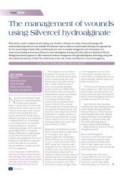
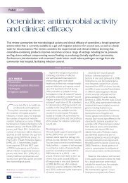
Clinical RESEARCH/AUDIT<br />
Dressing conformability and silvercontaining<br />
wound dressings<br />
Phil Bowler, Samantha Jones, Victoria Towers, Rebecca Booth,<br />
David Parsons, Mike Walker<br />
Abstract<br />
Background: All wounds have unique and irregular topographies, including cavities where fluids and bacteria may collect<br />
and increase the risk of wound infection. Aims: To qualitatively and quantitatively investigate the relationship between the<br />
physical structure of antiseptic wound dressings and their ability to manage bioburden in models that simulate clinical<br />
conditions. Methods: In vitro models were utilised to enable both visualisation of the conformability of silver-containing<br />
dressings with a simulated wound tissue surface, and measure the corresponding antimicrobial effect that these dressings<br />
had on a shallow wound microbial model. Results: Tissue contact and antimicrobial activity was shown with a silvercontaining<br />
Hydrofiber ® dressing (HF-Ag) over a 48-hour contact period. In contrast, the silver-containing foam dressings<br />
tested demonstrated areas of non-conformability which were associated with reduced antimicrobial activity. Conclusions:<br />
These in vitro studies confirm that both dressing conformability and silver availability to bacteria at the wound surface are<br />
critical to the optimum functioning of silver-containing dressings. Conflict of interest: The study was sponsored by ConvaTec.<br />
KEY WORDS<br />
Hydrofiber ® Technology<br />
Foam dressing<br />
Conformability<br />
Silver<br />
Antimicrobial efficacy<br />
Chronic wounds are polymicrobial,<br />
with bacterial colonisation<br />
originating from external<br />
sources such as surrounding skin, the gut<br />
and the mouth. As a consequence, an<br />
often complex bacterial burden exists,<br />
predominantly in superficial wound tissue,<br />
which has the ability to compromise wound<br />
progression (Bowler, 2003). To minimise<br />
the opportunity for infection in chronic<br />
wounds, it is important that the bioburden<br />
is controlled and maintained in a state that<br />
Phil Bowler is Director; Samantha Jones is Microbiology<br />
Laboratory Manager; Victoria Towers and Rebecca Booth<br />
are Research Scientists; David Parsons is Associate Director;<br />
Mike Walker is Senior Research Advisor, all at Antiinfectives<br />
R&D, ConvaTec Global Development Centre<br />
14 <strong>Wounds</strong> uk, 2010, Vol 6, No 2<br />
is not problematic to the host, as increasing<br />
bioburden can lead to chronic inflammation<br />
and increased risk of infection. In this<br />
respect, wound management practices such<br />
as cleansing, sharp debridement and the<br />
use of antimicrobial dressings are important<br />
within a protocol of care.<br />
To minimise the opportunity<br />
for infection in chronic<br />
wounds, it is important that<br />
the bioburden is controlled<br />
and maintained in a state<br />
that is not problematic to<br />
the host.<br />
When considering the use of<br />
antimicrobial dressings, many factors<br />
will influence the likely potency of the<br />
dressing. Wound-related factors include<br />
wound depth and size, amount of<br />
devitalised tissue, wound bioburden and<br />
the presence of biofilm. Dressing-related<br />
factors include the type and concentration<br />
of antimicrobial agent, availability of the<br />
agent from the vehicle in which it is<br />
contained, and the ability of the dressing<br />
to closely contact the surface of a wound<br />
and hence maximise exposure of the<br />
bioburden to the antimicrobial agent.<br />
This latter factor is challenged by the<br />
fact that chronic wounds are anatomically<br />
highly variable; they may vary in depth from<br />
sinuses and penetrating pressure ulcers to<br />
relatively shallow leg ulcers. All wounds have<br />
unique and irregular topographies, including<br />
cavities where fluids may collect, and result<br />
in the creation of ‘dead spaces’ (Edberg,<br />
1981; Snyder, 2005). This fluid may contain<br />
harmful components (e.g. bacteria and<br />
cellular debris) which may increase the risk<br />
of wound infection (Cutting et al, 2009).<br />
In the in vitro studies described in this<br />
paper, models were developed and utilised<br />
that enabled both visualisation of the<br />
conformability of silver-containing dressings<br />
with a simulated wound tissue surface, and<br />
the corresponding antimicrobial effect that<br />
these dressings had on a shallow wound<br />
microbial model.<br />
Figure 1. Porcine muscle tissue fixed to the inside<br />
wall of a 50mm Petri dish to provide an irregular<br />
tissue surface.
Clinical RESEARCH/AUDIT<br />
Materials<br />
The silver-containing wound dressings used<br />
are listed in Table 1.<br />
Tissue conformability model:<br />
Pork muscle tissue (purchased at a local<br />
supermarket); 50mm Petri dish; PVC<br />
tubing (1.65mm; Watson Marlow); dye<br />
solution (5–10mg of methylene blue<br />
dissolved in 50ml of physiological saline);<br />
Cane Crono PCA Pump (Applied Medical<br />
Technology, <strong>UK</strong>); an Olympus SZ61<br />
light microscope with QImaging camera<br />
(Device 3564); Image Pro Plus 5.0 image<br />
analysis software (MediaCybernetics, <strong>UK</strong>).<br />
Shallow wound microbial model:<br />
Microbiological culture media: Tryptone<br />
Soy Agar (TSA), Tryptone Soy Broth<br />
(TSB); gauze dressing (Topper 8, Johnson<br />
& Johnson); physiological saline (0.85%);<br />
Challenge organisms: Pseudomonas<br />
aeruginosa (NCIMB 8626) and<br />
Staphylococcus aureus (NCIMB 9518).<br />
Methods<br />
Tissue conformability model<br />
Porcine muscle tissue sections<br />
(~3x1x1cm) were fixed to the inside<br />
wall of a Petri dish (50mm diameter)<br />
with Loctite glue to provide an<br />
irregular tissue surface. Before fixing the<br />
tissue, a small hole was made in the side<br />
of the Petri dish using a hot needle to<br />
allow the insertion of a syringe needle<br />
(18 gauze) into the simulated wound<br />
tissue, such that its tip was just visible<br />
at the upper surface of the tissue. The<br />
dressings investigated were HF-Ag and<br />
representative adhesive foam dressings<br />
(foams A, B and E). A small strip of each<br />
dressing was applied over the tissue<br />
surface and, where appropriate (i.e.<br />
HF-Ag dressing), a moisture retentive<br />
adhesive Hydrofiber ® cover dressing<br />
[AHCD]) (Figure 1).<br />
The Cane Crono PCA pump was<br />
set to provide a flow rate for the dye<br />
solution of 2ml/hour. Microscopic images<br />
were collected every 45 seconds up to a<br />
maximum of 60 images.<br />
Shallow wound microbial model<br />
Evaluation of antimicrobial performance<br />
was made using an indented agar plate<br />
assay (simulating a shallow wound ~2–<br />
3mm deep).<br />
Table 1<br />
List of silver-containing dressings used<br />
Silver-containing dressing<br />
HF-Ag (AQUACEL ® Ag, ConvaTec)*<br />
Foam A (Allevyn Ag Adhesive, Smith & Nephew)<br />
Foam B (Allevyn Ag Non-adhesive,<br />
Smith & Nephew)<br />
Foam C (Allevyn Ag Gentle, Smith & Nephew)<br />
Foam D (Allevyn Ag Gentle Border,<br />
Smith & Nephew)<br />
Foam E (Mepilex ® Ag, Mölnlycke Healthcare)<br />
*Note: this dressing was covered with an adhesive Hydrofiber ® cover dressing (AHCD)<br />
Preparation of indented agar plates<br />
Within a laminar flow cabinet, molten<br />
TSA (80ml), which had been pre-cooled<br />
to ~45°C, was dispensed into 140mm<br />
Petri dishes and allowed to solidify. Pieces<br />
of gauze (2-ply, 4x4cm) were aseptically<br />
transferred to the centre of each prepoured<br />
TSA plate and gently pressed<br />
down onto the agar surface. A second<br />
piece of gauze (2-ply, 5x5cm) was applied<br />
over the original gauze, after which a 45ml<br />
volume of molten TSA was aseptically<br />
poured over the upper gauze layer and<br />
allowed to solidify overnight. The gauzes<br />
were aseptically removed from each agar<br />
plate to create a graduated indentation<br />
(5x5cm x ~ 2–3mm depth), with an<br />
Dressing type<br />
Hydrofiber ® (HF) with 1.2% w/w ionic silver*<br />
Adhesive foam containing silver sulfadiazine<br />
Non-adhesive foam containing silver sulfadiazine<br />
Soft gel adhesive foam containing<br />
silver sulfadiazine<br />
Silicone gel adhesive foam containing<br />
silver sulfadiazine<br />
Silicone adhesive foam containing silver sulphate<br />
irregular gauze imprinted surface (Figures 2<br />
and 3).<br />
A representative colony of each<br />
challenge bacterium was separately<br />
inoculated into TSB and incubated for<br />
four hours on a roller mixer at 35°C<br />
(±3°C) to achieve an actively growing<br />
population. Each suspension was diluted<br />
in TSB to achieve a concentration of<br />
approximately 1x10 8 cfu/ml, which was<br />
further serially diluted in physiological<br />
saline to provide a final inoculum<br />
concentration of approximately 1x10 3 cfu/<br />
ml (stock suspension). A microbial count<br />
was performed on each stock suspension<br />
to confirm the inoculum level.<br />
Figure 2. Preparation of indented agar model. A: gauze fixed within agar; B: gauze being removed;<br />
C: graduated indentation, showing an irregular agar surface.<br />
Figure 3. Cross-section of the graduated indentation (~2–3mm deep) and irregular surface.<br />
16 <strong>Wounds</strong> uk, 2010, Vol 6, No 2
Clinical RESEARCH/AUDIT<br />
Four millilitres of the stock suspension<br />
was then inoculated directly into the<br />
centre of the indented agar plate; this<br />
volume filled the indentation to mimic<br />
an exuding wound. A silver-containing<br />
dressing (~10x10cm, n=3 for each<br />
dressing) was placed centrally over the<br />
indent in the inoculated agar plate. For the<br />
HF-Ag dressing, an AHCD was applied<br />
as the secondary dressing, as indicated<br />
in the manufacturer’s instructions for<br />
use. An inoculated indented agar plate<br />
containing no dressing was also included<br />
as a positive control (n=1 for each<br />
challenge organism) to confirm the<br />
extent of bacterial growth in the absence<br />
of silver-containing dressings.<br />
All agar plates were incubated<br />
aerobically at 35°C (± 3°C) for 48<br />
hours, after which time dressings were<br />
aseptically removed. Photographs of the<br />
agar plates were taken to record any<br />
immediately obvious bacterial growth<br />
beneath each dressing. All agar plates<br />
were then reincubated for a further<br />
24 hours to allow any remaining viable<br />
bacterial cells to form complete colonies.<br />
This additional incubation step was<br />
necessary to enable image analysis to be<br />
performed and subsequent quantification<br />
of bacterial growth. The total surface area<br />
(mm 2 ) of each indented agar surface, and<br />
proportion of the area showing bacterial<br />
growth was measured using ImageTool<br />
for Windows, version 3.0 (The University<br />
of Texas Health Science Centre). Results<br />
for each silver-containing dressing were<br />
reported as the average value from the<br />
three replicates.<br />
Results<br />
Tissue conformability model<br />
The HF-Ag dressing was shown to<br />
conform well to the irregular surface of<br />
the simulated wound tissue (Figure 4a).<br />
In contrast, the adhesive foam dressings<br />
(foams A, B and E) showed intermittent<br />
contact with the tissue surface and<br />
associated areas of free fluid collection<br />
were evident (i.e. Figures 4b, 4c and 4d<br />
respectively).<br />
Shallow wound microbial model<br />
The positive controls of both challenge<br />
organisms showed confluent growth at the<br />
end of the study period. Figure 5 shows an<br />
example for S. aureus.<br />
Figure 4. Dressing conformability with simulated porcine muscle tissue surface. a: HF-Ag with AHCD; b: foam A;<br />
c: foam b; d. foam E.<br />
The HF-Ag dressing showed<br />
widespread antimicrobial activity against<br />
both P. aeruginosa and S. aureus beneath<br />
the dressing (Figure 6). The image analysis<br />
data indicated that growth of P. aeruginosa<br />
represented 9.1% of the indented<br />
agar surface area (Figure 7), and the<br />
corresponding growth of S. aureus was<br />
negligible (0.4%) (Figure 8). In contrast, all of<br />
the silver-containing foam dressings failed to<br />
inhibit P. aeruginosa (Figure 7). Growth of S.<br />
aureus beneath the silver-containing foam<br />
dressings ranged from 16.8% (foam B) to<br />
73.2% (foam E) of the total indented agar<br />
surface area (Figure 8).<br />
Additionally, extended growth of both P.<br />
aeruginosa and S. aureus beyond the edges<br />
of the indented agar surface was observed<br />
in association with all of the adhesive<br />
silver-containing foam dressings (Figure 9),<br />
and growth was mirrored on the wound<br />
contact surface, in particular with foam E<br />
(Figure 10).<br />
18 <strong>Wounds</strong> uk, 2010, Vol 6, No 2
Clinical RESEARCH/AUDIT<br />
Discussion<br />
Antimicrobial dressings are widely<br />
used in the care of chronic wounds,<br />
with their primary functions being to<br />
control wound bioburden (White et<br />
al, 2006), thus preventing infection<br />
and facilitating wound progression.<br />
Many host and dressing-related factors<br />
will influence the efficacy of a topical<br />
antimicrobial agent within a wound<br />
environment. In this respect, the<br />
relationship between the dressing and<br />
the antimicrobial agent contained within<br />
it is extremely important in ensuring<br />
optimal antimicrobial activity. If the<br />
dressing cannot maximise the availability<br />
of an antimicrobial agent, its efficacy is<br />
likely to be compromised. Similarly, if a<br />
dressing cannot fill the wound space and<br />
conform closely to an irregular wound<br />
surface, exposure of superficial wound<br />
bacteria to the antimicrobial agent in the<br />
dressing is likely to be sub-optimal.<br />
Bearing these factors in mind, in vitro<br />
models were developed to visualise<br />
dressing conformability (i.e. porcine<br />
muscle tissue) and a contaminated<br />
simulated shallow wound to visualise<br />
bacterial growth. While it is accepted<br />
that in vitro models may have limitations<br />
(for example, it is not possible to mimic<br />
in vivo human biological support systems<br />
such as blood supply), these studies were<br />
specifically designed to evaluate the<br />
antimicrobial and tissue conformability<br />
properties of silver-containing wound<br />
dressings over a 48-hour contact period,<br />
and are an extension of previously<br />
published in vitro work (Jones et al, 2005).<br />
The tissue conformability studies<br />
demonstrated that following hydration,<br />
Figure 6. Example of growth of P. aeruginosa (left) and S. aureus (right) beneath HF-Ag.<br />
Bacterial growth (%)<br />
100<br />
80<br />
60<br />
40<br />
20<br />
0<br />
Figure 7. Percentage surface area of P. aeruginosa growth beneath the dressings within the indented agar<br />
surface area.<br />
Bacterial growth (%)<br />
100<br />
80<br />
60<br />
40<br />
20<br />
0<br />
100.00<br />
Foam A<br />
25.71<br />
Foam A<br />
97.78<br />
Foam B<br />
16.77<br />
Foam B<br />
95.82<br />
Foam C<br />
24.64<br />
Foam C<br />
99.79<br />
Foam D<br />
33.54<br />
Foam D<br />
100.00<br />
Foam E<br />
73.20<br />
Foam E<br />
HF-Ag with<br />
AHCD<br />
HF-Ag with<br />
AHCD<br />
Figure 8. Percentage surface area of S. aureus growth beneath the dressings within the indented agar<br />
surface area.<br />
9.06<br />
0.40<br />
100.00<br />
No dressing<br />
(positive control)<br />
100.00<br />
No dressing<br />
(positive control)<br />
Figure 5. Example of growth of S. aureus without<br />
dressing application.<br />
the rapid gelling action of HF-Ag led to<br />
intimate contact between the dressing and<br />
the simulated wound tissue surface. The<br />
importance of such dressing conformability<br />
was demonstrated in the subsequent<br />
antimicrobial model where minimal growth<br />
of both P. aeruginosa and S. aureus was<br />
detected on the indented agar surface<br />
beneath this dressing.<br />
Growth of S. aureus beneath the silvercontaining<br />
foam dressings was evident, but<br />
less extensive than P. aeruginosa. This may be<br />
explained by the fact that S. aureus is a nonmotile<br />
bacterium and therefore has the<br />
tendency to form more discrete colonies<br />
on an agar surface, whereas P. aeruginosa<br />
is a motile bacterium and is more likely to<br />
swarm in a moist environment (Köhler et<br />
al, 2000).<br />
The limited antimicrobial performance<br />
of the adhesive silver-containing foam<br />
dressings was likely to be a consequence<br />
<strong>Wounds</strong> uk, 2010, Vol 6, No 2<br />
19
Clinical RESEARCH/AUDIT<br />
Foam A<br />
Foam C<br />
Foam B<br />
Foam D<br />
Foam E<br />
Foam B<br />
Figure 9. Examples of bacterial growth on the prominent agar surface surrounding the indentation for each<br />
adhesive silver-containing foam dressing (after an additional 24-hour incubation period following dressing<br />
contact). Foams A, C and E, P. aeruginosa; Foam D, S. aureus.<br />
Figure 11. Examples of no spreading bacterial<br />
growth on the prominent agar surface area with the<br />
non-adhesive silver-containing dressing (foam B).<br />
Top right, P. aeruginosa and bottom right, S. aureus.<br />
Figure 10: Example showing growth of S. aureus<br />
upon removal of foam E.<br />
of their less conformable nature, as<br />
observed in the tissue conformability<br />
model where areas of non-contact were<br />
evident (Figure 4). HF-Ag (Figure 6) and the<br />
non-adhesive foam B (Figure 11) showed<br />
areas surrounding the indented area<br />
that remained clear of bacteria, indicating<br />
good conformability with the perfectly flat<br />
prominent agar surface. However, despite<br />
containing an antimicrobial agent, all the<br />
adhesive silver-containing foam dressings<br />
showed lateral bacterial spread beyond<br />
the indentation and onto the surrounding<br />
prominent agar surface.<br />
These observations are supported<br />
by in vitro work undertaken by Buchholtz,<br />
who reported that, ‘The soft silicone<br />
layer dressing [i.e. foam E as used in these<br />
studies] had no noteworthy release of silver<br />
20 <strong>Wounds</strong> uk, 2010, Vol 6, No 2<br />
and no antimicrobial activity suggesting that<br />
the soft silicone layer may be encapsulating<br />
the foam keeping silver from reaching the<br />
wound’ (Buchholtz, 2009).<br />
This reported observation may explain<br />
the growth of S. aureus on foam E (Figure<br />
10). Subsequent investigations (unpublished<br />
data by the authors of this paper) using light<br />
microscopy, scanning electron microscopy<br />
and energy dispersive X-ray analysis<br />
showed no evidence of silver existing on<br />
the wound contact surface of foam E.<br />
Conclusion<br />
Two in vitro models have been used<br />
to investigate the conformability and<br />
activity of a range of silver-containing<br />
antimicrobial dressings in simulated shallow<br />
wounds. Tissue contact and antimicrobial<br />
activity was shown with HF-Ag over a<br />
48-hour contact period. In contrast, the<br />
silver-containing foam dressings tested<br />
demonstrated areas of non-conformability<br />
which were associated with reduced<br />
antimicrobial activity. These in vitro studies<br />
confirm that both dressing conformability<br />
and silver availability to bacteria at the<br />
wound surface are critical to the optimum<br />
functioning of silver-containing dressings.<br />
Hydrofiber ® and AQUACEL are registered<br />
trademarks of ConvaTec Inc. All other<br />
trademarks are the property of their<br />
respective owners. Wuk<br />
References<br />
Bowler PG (2003) Progression towards healing:<br />
Wound infection and the role of an advanced<br />
silver-containing Hydrofiber® dressing. Ostomy<br />
Wound Manage 49(8) Suppl: 2–5<br />
Buchholtz C (2009) An in vitro comparison of<br />
antimicrobial activity and silver release from<br />
foam dressings. Poster presentation at <strong>Wounds</strong><br />
<strong>UK</strong>, Harrogate 2009<br />
Cutting K, White R, Hoekstra H (2009)Topical<br />
silver-impregnated dressings and the importance<br />
of the dressing technology. Int Wound J 6:<br />
396–402<br />
Edberg SC (1981) Methods of quantitative<br />
microbiological analyses that support the<br />
diagnosis, treatment, and prognosis of human<br />
infection. CRC Crit Rev Microbiol 8: 339–97<br />
Jones SA, Bowler PG, Walker M (2005)<br />
Antimicrobial activity of silver-containing<br />
dressings is influenced by dressing<br />
conformability with a wound surface. WOUNDS<br />
17: 263–70<br />
Köhler T, Curty LK, Barja F, van Delden, Pechère<br />
(2000) Swarming of Pseudomonas aeruginosa is<br />
dependent on cell-to-cell signalling and requires<br />
flagella and pili. J Bacteriol 182: 5990–6<br />
Snyder RJ (2005) Managing dead space: an<br />
overview. Podiatry Manage 24: 171–4<br />
White RJ, Cutting K, Kingsley A (2006)<br />
Topical antimicrobials in the control of wound<br />
bioburden. Ostomy Wound Mange 52: 26–58