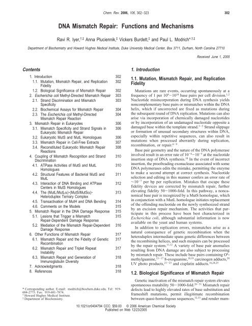
DNA Mismatch Repair: Functions and Mechanisms
DNA Mismatch Repair: Functions and Mechanisms
DNA Mismatch Repair: Functions and Mechanisms

Create successful ePaper yourself
Turn your PDF publications into a flip-book with our unique Google optimized e-Paper software.
Chem. Rev. 2006, 106, 302−323<br />
302<br />
<strong>DNA</strong> <strong>Mismatch</strong> <strong>Repair</strong>: <strong>Functions</strong> <strong>and</strong> <strong>Mechanisms</strong><br />
Ravi R. Iyer, †,‡ Anna Pluciennik, ‡ Vickers Burdett, ‡ <strong>and</strong> Paul L. Modrich* ,†,‡<br />
Department of Biochemistry <strong>and</strong> Howard Hughes Medical Institute, Duke University Medical Center, Box 3711, Durham, North Carolina 27710<br />
Received June 1, 2005<br />
Contents<br />
1. Introduction 302<br />
1.1. Mutation, <strong>Mismatch</strong> <strong>Repair</strong>, <strong>and</strong> Replication 302<br />
Fidelity<br />
1.2. Biological Significance of <strong>Mismatch</strong> <strong>Repair</strong> 302<br />
2. Escherichia coli Methyl-Directed <strong>Mismatch</strong> <strong>Repair</strong> 303<br />
2.1. Str<strong>and</strong> Discrimination <strong>and</strong> <strong>Mismatch</strong><br />
303<br />
Specificity<br />
2.2. Biochemical Assays for <strong>Mismatch</strong> <strong>Repair</strong> 304<br />
2.3. The Escherichia coli Methyl-Directed<br />
304<br />
<strong>Mismatch</strong> <strong>Repair</strong> Reaction<br />
3. <strong>Mismatch</strong> <strong>Repair</strong> in Eukaryotes 306<br />
3.1. <strong>Mismatch</strong> Specificity <strong>and</strong> Str<strong>and</strong> Signals in 306<br />
Eukaryotic <strong>Mismatch</strong> <strong>Repair</strong><br />
3.2. Eukaryotic MutS <strong>and</strong> MutL Homologues 306<br />
3.3. <strong>Mismatch</strong> <strong>Repair</strong> in Cell-Free Extracts 307<br />
3.4. Reconstituted Eukaryotic <strong>Mismatch</strong> <strong>Repair</strong> 308<br />
Reactions<br />
4. Coupling of <strong>Mismatch</strong> Recognition <strong>and</strong> Str<strong>and</strong> 310<br />
Discrimination<br />
4.1. ATPase Activities of MutS <strong>and</strong> MutL<br />
310<br />
Homologues<br />
4.2. Structural Features of Bacterial MutS <strong>and</strong> 311<br />
MutL<br />
4.3. Interaction of <strong>DNA</strong> Binding <strong>and</strong> ATPase 312<br />
Centers in MutS Homologues<br />
4.4. The (MutL/MutLR)‚(MutS/MutSR)‚<br />
313<br />
Heteroduplex Ternary Complex<br />
4.5. Transactivation of MutH <strong>and</strong> <strong>DNA</strong> Bending 314<br />
4.6. Comments on the Models 315<br />
5. <strong>Mismatch</strong> <strong>Repair</strong> in the <strong>DNA</strong> Damage Response 315<br />
5.1. Lesions that Trigger a <strong>Mismatch</strong><br />
315<br />
<strong>Repair</strong>-Dependent Damage Response<br />
5.2. Mediation of the <strong>Mismatch</strong> <strong>Repair</strong>-Dependent 316<br />
Damage Response<br />
6. Other <strong>Functions</strong> of <strong>Mismatch</strong> <strong>Repair</strong> 317<br />
6.1. <strong>Mismatch</strong> <strong>Repair</strong> <strong>and</strong> the Fidelity of Genetic 317<br />
Recombination<br />
6.2. <strong>Mismatch</strong> <strong>Repair</strong> <strong>and</strong> Triplet Repeat<br />
317<br />
Instability<br />
6.3. <strong>Mismatch</strong> <strong>Repair</strong> <strong>and</strong> Generation of<br />
318<br />
Immunoglobulin Diversity<br />
7. Acknowledgments 318<br />
8. References 318<br />
* Corresponding author. E-mail: modrich@biochem.duke.edu. Tel: 919-<br />
684-2775. Fax: 919-681-7874.<br />
† Howard Hughes Medical Institute.<br />
‡ Department of Biochemistry.<br />
1. Introduction<br />
1.1. Mutation, <strong>Mismatch</strong> <strong>Repair</strong>, <strong>and</strong> Replication<br />
Fidelity<br />
Mutations are rare events, occurring spontaneously at a<br />
frequency of 1 per 10 9 -10 10 base pairs per cell division. 1,2<br />
Nucleotide misincorporation during <strong>DNA</strong> synthesis yields<br />
noncomplementary base pairs or mismatches within the <strong>DNA</strong><br />
helix, which if uncorrected are fixed as mutations during<br />
the subsequent round of <strong>DNA</strong> replication. Mutations can also<br />
arise via incorporation of chemically damaged nucleotides<br />
or by incorporation of an undamaged nucleotide opposite a<br />
damaged base within the template str<strong>and</strong>. 3-5 Str<strong>and</strong> slippage<br />
or formation of unusual secondary structures within <strong>DNA</strong>,<br />
especially within repetitive sequences, can also result in<br />
mutations when processed aberrantly during replication,<br />
recombination, or repair. 6-9<br />
Base pair geometry <strong>and</strong> the nature of the <strong>DNA</strong> polymerase<br />
involved result in an error rate of 10 -4 -10 -5 at the nucleotide<br />
insertion step of <strong>DNA</strong> synthesis. 10 In the event of incorrect<br />
insertion, the proofreading exonuclease associated with some<br />
<strong>DNA</strong> polymerases edits the mistake, permitting the enzyme<br />
to make a second attempt at correct synthesis. Nucleotide<br />
selection <strong>and</strong> editing in this manner confers an error rate of<br />
∼10 -7 per bp per replication. Mistakes that escape these<br />
fidelity devices are corrected by mismatch repair, further<br />
elevating fidelity 50-1000-fold. In this pathway, a noncanonical<br />
base pair is recognized by a MutS homologue, which<br />
in conjunction with a MutL homologue initiates replacement<br />
of the offending nucleotide on the newly synthesized str<strong>and</strong><br />
by an excision repair mechanism. The activities that participate<br />
in this process have been best characterized in<br />
Escherichia coli, although substantial information is now<br />
available on the yeast <strong>and</strong> human systems.<br />
In addition to replication errors, mismatches arise as a<br />
natural consequence of genetic recombination when the<br />
heteroduplex intermediate spans genetic differences between<br />
the recombining helices, <strong>and</strong> such mispairs can be processed<br />
by the repair system. 11,12 A variety of base pair anomalies<br />
resulting from <strong>DNA</strong> damage are also subject to processing<br />
by mismatch repair. These include base pairs containing O 6 -<br />
methylguanine, 13-17 8-oxoguanine, 18,19 carcinogen adducts, 20<br />
UV photo products, 21-23 <strong>and</strong> cisplatin adducts. 16,24,25<br />
1.2. Biological Significance of <strong>Mismatch</strong> <strong>Repair</strong><br />
Genetic inactivation of the mismatch repair system elevates<br />
spontaneous mutability 50-1000-fold. 26-31 <strong>Mismatch</strong> repair<br />
defects lead to highly elevated rates of base substitution <strong>and</strong><br />
frameshift mutations, permit illegitimate recombination<br />
between quasi-homologous sequences, 32,33 <strong>and</strong> render mam-<br />
10.1021/cr0404794 CCC: $59.00 © 2006 American Chemical Society<br />
Published on Web 12/23/2005
<strong>DNA</strong> <strong>Mismatch</strong> <strong>Repair</strong> Chemical Reviews, 2006, Vol. 106, No. 2 303<br />
Ravi R. Iyer obtained his B.Sc. degree in Chemistry <strong>and</strong> Biochemistry<br />
from the University of Bombay, India, <strong>and</strong> a M.Sc. in Biotechnology from<br />
Madurai Kamaraj University, Madurai, India. For his Ph.D. degree in<br />
Biochemistry, he studied the mechanisms of trinucleotide repeat instability<br />
under the supervision of Robert D. Wells at Texas A&M University,<br />
Houston, Texas. At the present time, he is a postdoctoral fellow in Paul<br />
Modrich’s laboratory working on the molecular mechanisms of human<br />
<strong>DNA</strong> mismatch repair.<br />
Vickers Burdett received her B.S. in Biology from Chatham College 1969<br />
<strong>and</strong> Ph.D. in Microbiology from Georgetown University under the<br />
mentorship of Stanley Falkow. After postdoctoral training with Donald<br />
Helinski at the University of California, San Diego, she joined the research<br />
faculty of the Department of Microbiology at Duke Univeristy Medical<br />
Center, where she discovered a novel tetracycline resistance mechanism<br />
based on active release of the antibiotic from the ribosomal target. In<br />
1998, she moved to the Department of Biochemistry where she collaborates<br />
with Paul Modrich in the study of genetic stabilization pathways.<br />
Anna Pluciennik received her Master’s degree in Microbiology <strong>and</strong> Genetics<br />
from the University of Lodz, Pol<strong>and</strong>. After receiving her Ph.D. in Molecular<br />
Biology with Adam Jaworski at the University of Lodz, she did postdoctoral<br />
work with Robert D. Wells investigating the processes underlying triplet<br />
repeat instability. Currently, she is a postdoctoral fellow with Paul Modrich,<br />
<strong>and</strong> her research interests lie in underst<strong>and</strong>ing the mechanisms of<br />
mismatch repair in bacteria <strong>and</strong> humans.<br />
malian cells resistant to the cytotoxic effects of several<br />
classes of <strong>DNA</strong> damaging agents. 27,34-36 Inactivation of the<br />
human mismatch repair system is the cause of hereditary<br />
nonpolyposis colon cancer (HNPCC) 37-40 <strong>and</strong> has been<br />
implicated in the development of a subset of sporadic tumors<br />
that occur in a variety of tissues. 39-43<br />
2. Escherichia coli Methyl-Directed <strong>Mismatch</strong><br />
<strong>Repair</strong><br />
The notion that mismatches generated during <strong>DNA</strong><br />
transactions might provoke their own repair was initially<br />
suggested by Holliday 44 <strong>and</strong> Whitehouse 45 to account for<br />
marker effects associated with meiotic recombination. On<br />
the basis of the low transformation efficiency of certain<br />
genetic markers into Streptococcus pneumoniae, Ephrussi-<br />
Taylor <strong>and</strong> colleagues proposed a mismatch rectification<br />
process in this bacterium that was targeted to the incoming<br />
<strong>DNA</strong> str<strong>and</strong>. 46 Direct proof that mismatches can provoke<br />
their own repair was provided by Meselson <strong>and</strong> colleagues<br />
who transfected E. coli with phage λ heteroduplex <strong>DNA</strong>s<br />
Paul Modrich is an Investigator of the Howard Hughes Medical Institute<br />
<strong>and</strong> Professor of Biochemistry at Duke University Medical Center. He<br />
received his undergraduate degree from M.I.T. <strong>and</strong> his Ph.D. in<br />
Biochemistry from Stanford University with Robert Lehman <strong>and</strong> did<br />
postdoctoral work at Harvard Medical School with Charles Richardson.<br />
His research interests are in the molecular mechanisms of <strong>DNA</strong>−protein<br />
interaction <strong>and</strong> the nature of cellular pathways responsible for genetic<br />
stabilization. He is a member of the National Academy of Sciences, the<br />
Institute of Medicine, <strong>and</strong> the American Academy of Arts <strong>and</strong> Sciences.<br />
containing one or more mismatched base pairs. 47,48 These<br />
experiments showed that different mismatches can be rectified<br />
with differing efficiencies, implying that rectification<br />
is dependent on mismatch recognition. They also demonstrated<br />
that co-repair of closely linked mismatches usually<br />
occurs on the same <strong>DNA</strong> str<strong>and</strong>, an effect that was<br />
interpreted in terms of an excision mode of repair with a<br />
tract size of several thous<strong>and</strong> nucleotides.<br />
2.1. Str<strong>and</strong> Discrimination <strong>and</strong> <strong>Mismatch</strong><br />
Specificity<br />
Although early studies of mismatch repair were prompted<br />
by an interest in recombination marker effects, Wagner <strong>and</strong><br />
Meselson postulated that mismatch repair could also contribute<br />
to replication fidelity provided that the reaction could<br />
be directed to the newly synthesized <strong>DNA</strong> str<strong>and</strong>. 48 They<br />
suggested that this could be accomplished by exploitation<br />
of secondary signals within the helix such as the transient
304 Chemical Reviews, 2006, Vol. 106, No. 2 Iyer et al.<br />
absence of methylation on newly synthesized <strong>DNA</strong> or via a<br />
“special relation to the replication complex”. 48 Methyl<br />
direction was confirmed in E. coli when heteroduplex repair<br />
was shown to be controlled by the status of adenine<br />
modification at GATC sequences. 49 Newly synthesized <strong>DNA</strong><br />
is subject to modification at this sequence by the Dam<br />
methylase after a transient delay. 50-52 <strong>Mismatch</strong> repair of<br />
hemi-methylated <strong>DNA</strong> occurs on the unmodified str<strong>and</strong>,<br />
heteroduplex <strong>DNA</strong> lacking the Dam modification on either<br />
str<strong>and</strong> is also processed but with little or no str<strong>and</strong> bias, <strong>and</strong><br />
<strong>DNA</strong> that is modified on both str<strong>and</strong>s is not repaired. 49,53<br />
As expected from such observations, E. coli cells deficient<br />
in Dam methylase are mutators, 54,55 as are strains that<br />
overproduce the enzyme. 56,57 The latter effect has been<br />
attributed to a reduced temporal window during which repair<br />
may occur. It is noteworthy in this context that a single hemimodified<br />
GATC sequence is sufficient to direct E. coli<br />
mismatch repair <strong>and</strong> that this site may reside on either side<br />
of the mismatch. 58-60<br />
Although hemi-modification of GATC sites plays a major<br />
role in str<strong>and</strong> discrimination in vivo, a str<strong>and</strong> break will also<br />
suffice for this purpose. 61,62 In fact, as discussed below, the<br />
function of a hemi-methylated GATC site in E. coli mismatch<br />
repair is to provide a str<strong>and</strong> break, <strong>and</strong> it has been suggested<br />
that GATC modification is responsible for directing only a<br />
subset of mismatch repair events, the remainder attributed<br />
to str<strong>and</strong> discontinuities that occur naturally on daughter<br />
<strong>DNA</strong> str<strong>and</strong>s during the course of <strong>DNA</strong> replication (a 3′-<br />
terminus on the leading str<strong>and</strong>, 3′- <strong>and</strong> 5′-termini on the<br />
lagging str<strong>and</strong>). 63 Str<strong>and</strong> discontinuities are also believed to<br />
be the natural str<strong>and</strong> discrimination signals in Str. pneumoniae.<br />
64,65<br />
The E. coli methyl-directed system recognizes <strong>and</strong> repairs<br />
G-T, A-C, G-A, T-C, A-A, G-G, <strong>and</strong> T-T mismatches,<br />
66-68 although some G-A <strong>and</strong> C-T mispairs are<br />
weak substrates, depending on sequence context. 67,69 The<br />
C-C mismatch is subject to little if any rectification.<br />
Insertion/deletion (ID) mismatches containing up to about<br />
four unpaired bases are also efficiently processed by the<br />
pathway. 70-73<br />
2.2. Biochemical Assays for <strong>Mismatch</strong> <strong>Repair</strong><br />
Molecular analysis of mismatch repair was made possible<br />
by the development of assays that permit mismatch rectification<br />
in vitro to be scored by biochemical or genetic methods.<br />
Both approaches have utilized circular heteroduplexes containing<br />
a mismatch <strong>and</strong> a str<strong>and</strong> discrimination signal. The<br />
biochemical assay relies on placement of a mismatch within<br />
overlapping recognition sites for two restriction endonucleases.<br />
53,68,74,75 The mismatch renders the <strong>DNA</strong> resistant to<br />
cleavage by both endonucleases, but repair restores sensitivity<br />
to one or the other of the two endonucleases, depending on<br />
which str<strong>and</strong> is subject to rectification.<br />
In the genetic method 76 a mismatch is placed within a<br />
β-galactosidase gene that resides within M13 viral <strong>DNA</strong> such<br />
that one str<strong>and</strong> contains a wild-type gene sequence while its<br />
complement contains a mutation that inactivates β-galactosidase<br />
function. After incubation with a cell-free fraction,<br />
<strong>DNA</strong> products are introduced into a mismatch-repair deficient<br />
E. coli strain, which is then plated on media containing<br />
5-bromo-4-chloro-3-indolyl-β-D-galactopyranoside (X-gal),<br />
which yields a blue product upon hydrolysis by β-galactosidase.<br />
The fate of the mismatch is evaluated by scoring blue<br />
or white plaques versus plaques that display blue <strong>and</strong> white<br />
sectors. Plaques of a pure color arise as a consequence of<br />
repair, whereas sectored plaques reflect segregation of the<br />
two str<strong>and</strong>s in the absence of correction.<br />
2.3. The Escherichia coli Methyl-Directed<br />
<strong>Mismatch</strong> <strong>Repair</strong> Reaction<br />
E. coli strains deficient in MutH, MutL, MutS, or <strong>DNA</strong><br />
helicase II (also called UvrD) are deficient in methyl-directed<br />
mismatch repair. 49,53,66,77 Application of the in vitro restriction<br />
endonuclease assay described above permitted isolation of<br />
homogeneous preparations of MutH, MutL, MutS, <strong>and</strong> <strong>DNA</strong><br />
helicase II. 62,78-80 Biochemical <strong>and</strong> genetic studies also<br />
implicated several additional activities in methyl-directed<br />
mismatch repair: exonuclease I (ExoI) exonuclease VII<br />
(ExoVII), RecJ exonuclease, exonuclease X (ExoX), singlestr<strong>and</strong>ed<br />
<strong>DNA</strong> binding protein (SSB), <strong>DNA</strong> polymerase III<br />
holoenzyme, <strong>and</strong> <strong>DNA</strong> ligase. 62,81-83<br />
Analysis of repair in E. coli extracts under conditions<br />
where repair <strong>DNA</strong> synthesis was blocked <strong>and</strong> study of<br />
reactions supported by purified proteins indicate that the<br />
overall repair reaction can be divided into several steps:<br />
mismatch-dependent incision of the unmethylated str<strong>and</strong> at<br />
a hemi-methylated GATC site; excision of that portion of<br />
the incised str<strong>and</strong> spanning the single-str<strong>and</strong> break <strong>and</strong> the<br />
mispair; repair of the ensuing gap by <strong>DNA</strong> synthesis <strong>and</strong><br />
ligation (Figure 1). A key feature of this system is its<br />
bidirectional capability. The finding that a single hemimethylated<br />
GATC sequence is sufficient to direct E. coli<br />
mismatch repair <strong>and</strong> that this site may reside on either side<br />
of the mispair suggested that the pathway could function in<br />
a bidirectional manner. 58-60 This was confirmed by use of<br />
electron microscopy <strong>and</strong> end-labeling methods to map<br />
excision tracts produced under conditions of repair synthesis<br />
block in both E. coli extracts <strong>and</strong> a reconstituted system<br />
comprised of purified forms of the proteins noted above. 84,85<br />
The 6.4 kbp heteroduplexes used in these studies contained<br />
aG-T mismatch <strong>and</strong> a single hemi-methylated GATC site<br />
located about 1000 bp distant (as viewed along the shorter<br />
path linking the two sites on the circular <strong>DNA</strong>). <strong>Mismatch</strong>provoked<br />
excision tracts were localized to the unmodified<br />
str<strong>and</strong> where they extended via the shorter path from the<br />
GATC site to terminate at a number of sites within a 100<br />
nucleotide region beyond the mispair. Localization of excision<br />
tracts to the shorter path between the two <strong>DNA</strong> sites<br />
was observed irrespective of which str<strong>and</strong> of the helix<br />
harbored GATC modification, that is, whether the unmethylated<br />
GATC sequence was located 3′ or 5′ to the mismatch.<br />
These observations led to the suggestion that mismatchprovoked<br />
excision commences at the incised GATC site <strong>and</strong><br />
proceeds toward the mispair. 85 It is pertinent to note in this<br />
context that while a hemi-modified GATC located 1000 bp<br />
from the mismatch can function efficiently in directing<br />
excision, the efficacy of the reaction decreases as the<br />
separation distance increases to 2000 bp. 58-60<br />
MutS is responsible for initiation of E. coli mismatch<br />
repair. This 95 kDa polypeptide, which exists as an equilibrium<br />
mixture of dimers <strong>and</strong> tetramers, 78,86 recognizes<br />
mismatched base pairs. 68,72,78,87 MutL, a 68 kDa polypeptide<br />
that is dimeric in solution, is recruited to the heteroduplex<br />
in a MutS- <strong>and</strong> ATP-dependent fashion. 80,88-92 The MutL‚<br />
MutS‚heteroduplex complex is believed to be a key intermediate<br />
in the initiation of mismatch repair, but as described<br />
below, its nature is not well understood.<br />
Assembly of the MutL‚MutS‚heteroduplex ternary complex<br />
is sufficient to activate several downstream repair
<strong>DNA</strong> <strong>Mismatch</strong> <strong>Repair</strong> Chemical Reviews, 2006, Vol. 106, No. 2 305<br />
Figure 1. Mechanism of E. coli methyl-directed mismatch repair. Details of the reaction are described in the text. Although not shown,<br />
<strong>DNA</strong> ligase restores covalent continuity to the repaired str<strong>and</strong> after <strong>DNA</strong> polymerase III holoenzyme fills in the gap. Green arrows indicate<br />
MutS- <strong>and</strong> MutL-dependent signaling between the two <strong>DNA</strong> sites involved in the reaction.<br />
activities. One of these is MutH, a 25 kDa latent endonuclease<br />
specific for unmodified GATC sequences. MutH is<br />
activated in a mismatch-, MutS-, MutL-, <strong>and</strong> ATP-dependent<br />
manner <strong>and</strong> incises the unmethylated str<strong>and</strong> of a hemimethylated<br />
GATC site 5′ to the G. 79,93 Activated MutH will<br />
also cleave both str<strong>and</strong>s of an unmodified GATC site by a<br />
two-hit mechanism resulting in a double str<strong>and</strong> break. MutH<br />
incision can occur either 3′ or 5′ to the mispair on the<br />
unmodified str<strong>and</strong>, <strong>and</strong> the ensuing str<strong>and</strong> break serves as<br />
the actual signal that directs excision repair to the unmethylated<br />
str<strong>and</strong> (Figure 1). Thus, a preexisting single-str<strong>and</strong><br />
break, which need not be within a GATC sequence, bypasses<br />
the requirements for both MutH <strong>and</strong> a hemi-modified GATC<br />
site in E. coli mismatch repair, an effect that has been<br />
documented both in vivo <strong>and</strong> in vitro. 61,62<br />
Formation of the MutS‚MutL‚heteroduplex complex is also<br />
sufficient to activate the methyl-directed excision system,<br />
which is comprised of <strong>DNA</strong> helicase II <strong>and</strong> several singlestr<strong>and</strong><br />
specific exonucleases. MutS <strong>and</strong> MutL activate<br />
unwinding by helicase II on nicked <strong>DNA</strong> in a mismatchdependent<br />
manner. 94 Use of pre-steady-state methods demonstrated<br />
that helix unwinding in this system initiates at the<br />
str<strong>and</strong> break. 95 Although unwinding in this three-protein<br />
system occurs in both directions from the str<strong>and</strong> break, the<br />
reaction displays a fairly strong bias for unwinding toward<br />
the mismatch. Since this directional bias was observed<br />
without regard to placement of the nick 3′ or 5′ to the mispair,<br />
this finding led to the suggestion that MutS <strong>and</strong> MutL can<br />
coordinate recognition of the two <strong>DNA</strong> sites in a manner<br />
that establishes their relative orientation. This would permit<br />
orientation dependent loading of the helicase at the str<strong>and</strong><br />
break so that unwinding proceeds toward the mismatch. That<br />
portion of the incised str<strong>and</strong> displaced by the helicase is<br />
subject to hydrolysis by an appropriate single-str<strong>and</strong> specific<br />
exonuclease (see below).<br />
In the partial reaction systems described above, MutL plays<br />
a major role in the coupling of mismatch recognition by<br />
MutS to the activation of MutH or helicase II. This<br />
conclusion is based on the finding that under certain<br />
conditions MutL is sufficient to activate <strong>DNA</strong> helicase II<br />
on a substrate that lacks a mismatched base pair. 94,96,97 This<br />
effect has been attributed to physical interaction of the two<br />
proteins with MutL loading the unidirectional 3′ to 5′<br />
helicase 98 onto the appropriate <strong>DNA</strong> str<strong>and</strong> so that unwinding<br />
proceeds toward the mismatch in a manner consistent with<br />
heteroduplex orientation. Under certain conditions, MutL can<br />
also activate the MutH endonuclease in a mismatch- <strong>and</strong><br />
MutS-independent manner, an effect that has also been<br />
attributed to physical interaction of the two proteins. 99,100<br />
As noted above, mapping of excision tracts <strong>and</strong> the<br />
demonstration that MutS <strong>and</strong> MutL activate unwinding by<br />
<strong>DNA</strong> helicase II at a str<strong>and</strong> break have led to the conclusion<br />
that excision initiates at the site of MutH incision. This view<br />
is also consistent with the nature of exonuclease activities<br />
that have been implicated in methyl-directed repair based in<br />
vitro assay. Thus, when MutH incision occurs 5′ to the<br />
mismatch, excision depends on ExoVII or RecJ exonuclease,<br />
82,85 both of which hydrolyze single-str<strong>and</strong>ed <strong>DNA</strong><br />
with 5′ to 3′ polarity. 101,102 When MutH cleavage occurs<br />
3′ to the mispair, excision requires ExoI, ExoVII, or<br />
ExoX, 62,81-83,85 all of which support 3′ to 5′ hydrolysis of<br />
single-str<strong>and</strong>ed <strong>DNA</strong>. 101,103,104 (ExoVII supports both 5′ to<br />
3′ <strong>and</strong> 3′ to 5′ directionality.)<br />
Genetic inactivation of both ExoVII <strong>and</strong> RecJ abolishes<br />
5′-directed mismatch repair in E. coli extracts, whereas<br />
inactivation of ExoI, ExoVII, <strong>and</strong> ExoX is necessary to<br />
eliminate 3′-directed repair in vitro. 82,83 Similar heteroduplex<br />
orientation-dependent requirements for the exonucleases have<br />
been observed in reactions reconstituted using purified<br />
proteins. 82,83,85 ExoVII <strong>and</strong> RecJ thus provide redundant<br />
functions in 5′-directed excision, while ExoI, ExoVII, <strong>and</strong><br />
ExoX provide redundancy in 3′-directed hydrolysis. Redundancy<br />
of ExoI, ExoVII, ExoX, <strong>and</strong> RecJ function in<br />
mismatch repair has also been documented in in vivo genetic
306 Chemical Reviews, 2006, Vol. 106, No. 2 Iyer et al.<br />
studies. Analysis of all possible single, double, <strong>and</strong> triple<br />
mutant strains failed to reveal a defect in mismatch repair<br />
as judged by mutability increase; 82,83,105 however, strains<br />
deficient in all four hydrolytic activities display a 7-fold<br />
increase in mutation rate, a value considerably less than the<br />
50 to 100-fold increase in mutability conferred by helicase<br />
II or MutS defects. 83 While the limited increase in mutability<br />
associated with quadruple exonuclease deficiency might indicate<br />
that these enzymes play only a limited role in mismatch<br />
repair within the cell, this does not appear to be the case.<br />
Rather, the modest mutability of such strains is due to under<br />
recovery of mutations because bacterial chromosomes in<br />
which mismatches occur tend to be lost or destroyed. 81<br />
The single-str<strong>and</strong>ed gap produced by the action of helicase<br />
II <strong>and</strong> a single-str<strong>and</strong> exonuclease is stabilized by SSB. In<br />
the absence of SSB, repair efficiency is reduced substantially.<br />
62 <strong>DNA</strong> polymerase III holoenzyme is sufficient to<br />
support the repair synthesis step of methyl-directed correction<br />
in vitro, <strong>and</strong> extracts prepared from a dnaZ ts mutant have<br />
been shown to be defective in mismatch repair at the<br />
restrictive temperature. 62 In the final step of the reaction,<br />
helix integrity is restored by the action of <strong>DNA</strong> ligase. 62<br />
The dnaZ requirement indicates that the γ complex of pol<br />
III holoenzyme is required for methyl-directed repair in vitro.<br />
The γ complex functions as the loader that places the β clamp<br />
onto the helix. 106 The β clamp functions as a processivity<br />
factor for <strong>DNA</strong> polymerase III but has also recently been<br />
shown to interact physically with MutS. 107 As discussed<br />
below, recent work has demonstrated that PCNA (the<br />
eukaryotic homologue of the β clamp) <strong>and</strong> the RFC clamp<br />
loader (homologue of the γ complex) play important roles<br />
in regulation of mismatch-provoked excision in the human<br />
mismatch repair system. Unfortunately, potential effects of<br />
the γ complex <strong>and</strong> the β clamp on the excision step of<br />
bacterial mismatch repair have not been addressed, although<br />
such studies would appear warranted.<br />
3. <strong>Mismatch</strong> <strong>Repair</strong> in Eukaryotes<br />
3.1. <strong>Mismatch</strong> Specificity <strong>and</strong> Str<strong>and</strong> Signals in<br />
Eukaryotic <strong>Mismatch</strong> <strong>Repair</strong><br />
As noted above, MutH incision at a hemi-methylated<br />
GATC site provides a <strong>DNA</strong> str<strong>and</strong> break that serves as the<br />
actual signal that directs E. coli mismatch repair. In fact,<br />
the initial demonstration of str<strong>and</strong>-directed mismatch repair<br />
in higher cells relied on the use of circular heteroduplex<br />
<strong>DNA</strong>s containing a str<strong>and</strong>-specific single-str<strong>and</strong> break. 75,76<br />
Incubation of such <strong>DNA</strong>s with nuclear extracts derived from<br />
human or Drosophila melanogaster cells results in robust<br />
mismatch correction with the repair being directed to the<br />
incised str<strong>and</strong>. Such extracts support efficient nick-directed<br />
repair of G-T, A-C, A-A, G-A, G-G, T-T, C-T, <strong>and</strong><br />
C-C mismatches, as well as small ID heterologies. 75,76,108-111<br />
Although str<strong>and</strong> discontinuities are sufficient to direct<br />
mismatch repair in vitro, the natural signals that direct the<br />
eukaryotic reaction remain uncertain. Early studies 112-114<br />
suggested that cytosine methylation might be involved in<br />
str<strong>and</strong> discrimination in mammalian cells in a manner<br />
analogous to the role of adenine methylation in E. coli.<br />
However, more recent work has seemingly ruled out this<br />
possibility. 115,116 On the other h<strong>and</strong>, several groups have<br />
found that mouse cells deficient in the <strong>DNA</strong> cytosine<br />
methyltransferase Dnmt1 display instability of mono- <strong>and</strong><br />
dinucleotide repeat sequences, 117,118 a phenotype characteristic<br />
of mismatch repair-deficient cells. 41,119,120 Although a<br />
role for Dnmt1 in mammalian mismatch repair has been<br />
inferred on these grounds, defective or aberrant repair in<br />
Dnmt1-deficient cells has not been directly demonstrated.<br />
Other possible mechanisms for str<strong>and</strong> discrimination in<br />
eukaryotic cells have also been considered. Str<strong>and</strong> discontinuities<br />
occurring naturally as intermediates during the<br />
course of <strong>DNA</strong> replication may provide str<strong>and</strong> signal<br />
functions in bacterial mismatch repair <strong>and</strong> could function in<br />
a similar manner in eukaryotic systems. 27 Wagner <strong>and</strong><br />
Meselson postulated that “special relation to the replication<br />
complex” could effect str<strong>and</strong> discrimination in mismatch<br />
repair. 48 As detailed below, the PCNA replication clamp<br />
interacts with several eukaryotic mismatch repair activities,<br />
<strong>and</strong> it has been suggested that PCNA might provide a<br />
physical link between repair <strong>and</strong> replication that would allow<br />
<strong>DNA</strong> termini at the fork to function as str<strong>and</strong> signals. 121 Yet<br />
another possibility is that noncovalent signals in the form<br />
of proteins that segregate with the individual str<strong>and</strong>s during<br />
replication could conceivably provide a mechanism for<br />
discrimination of parental <strong>and</strong> nascent str<strong>and</strong>s. 27<br />
3.2. Eukaryotic MutS <strong>and</strong> MutL Homologues<br />
Genes encoding homologues of bacterial MutS <strong>and</strong> MutL<br />
have been identified in a variety of eukaryotes including<br />
yeast, plants, insects, nematodes, <strong>and</strong> mammals, 26-30,122-124<br />
although no eukaryotic homologue of MutH has been<br />
identified. The several eukaryotic homologues of MutS have<br />
been designated MSH1-MSH6. MSH1, which has not been<br />
identified in mammalian cells, is required for mitochondrial<br />
<strong>DNA</strong> stability in Saccharomyces cereVisiae. 12,125,126 Eukaryotic<br />
MSH2, MSH3, <strong>and</strong> MSH6 gene products have been<br />
implicated in mitotic genetic stability where they participate<br />
in repair of base-base mismatches <strong>and</strong> ID heterologies;<br />
12,14,27,110,127-141 however, MSH2 has also been implicated<br />
in meiotic gene conversion. 12 Function of the MutS homologues<br />
MSH4 <strong>and</strong> MSH5 is apparently restricted to meiosis<br />
where they play important roles in crossing over in both yeast<br />
<strong>and</strong> mammals. 142-146<br />
Genes that encode MutL homologues have also been<br />
identified in eukaryotes. MLH1 <strong>and</strong> PMS2 (mammalian<br />
PMS2 corresponds to PMS1 in yeast, plants, <strong>and</strong> nematodes)<br />
have been the most extensively characterized. Both have been<br />
implicated in mitotic mutation avoidance 110,128,147-154 <strong>and</strong> in<br />
meiotic recombination. 11,155-157 MLH3, which has been<br />
identified in yeasts <strong>and</strong> mammals 158,159 plays an important<br />
role in meiotic crossing-over 157,160,161 but also functions in<br />
mitotic genetic stabilization in yeast by preventing frameshift<br />
mutations. 158,162 Mitotic functions of MLH3 in mammalian<br />
cells have been the subject of controversy. 159,163-166 Other<br />
MutL homologues have also been identified: MLH2 in<br />
yeast 157,162 <strong>and</strong> PMS1 in humans. 167 The former protein may<br />
provide a meiotic function. 157<br />
Available evidence indicates that eukaryotic MutS <strong>and</strong><br />
MutL homologues function as heterodimers, which allows<br />
for a modular system for recognition <strong>and</strong> processing of<br />
different types of <strong>DNA</strong> lesions. In both human <strong>and</strong> yeast,<br />
MSH2 forms a heterodimer with MSH6 (MutSR) or MSH3<br />
(MutSβ). 130,131,135,136,138,140,141,168-170 In human cells, MSH2<br />
partitions between MSH6 <strong>and</strong> MSH3 such that about 85%<br />
of the MSH2 is found in the MSH2‚MSH6 MutSR complex.<br />
140,141 Human MutSR supports repair of all eight basebase<br />
mismatches including C-C, as well as ID mispairs<br />
containing up to about 10 unpaired nucleotides, whereas
<strong>DNA</strong> <strong>Mismatch</strong> <strong>Repair</strong> Chemical Reviews, 2006, Vol. 106, No. 2 307<br />
Figure 2. Human bidirectional mismatch repair in vitro. Human mismatch repair in vitro can be directed by a str<strong>and</strong> break located either<br />
3′ or 5′ to the mismatch. Activities that have been implicated in several steps of the reaction are shown. Question marks indicate that<br />
unidentified activities may also play significant roles in the reaction.<br />
MutSβ supports correction of ID mismatches containing two<br />
to about 10 nucleotides but is only weakly active on single<br />
nucleotide ID mispairs. 138,141 This specificity is consistent<br />
with the finding that mononucleotide (but not dinucleotide)<br />
repeat instability is diagnostic for MSH6-deficient tumor<br />
cells. 120,171<br />
As judged by mutation spectra <strong>and</strong> in vitro heteroduplex<br />
binding assay, the specificities of yeast MutSR <strong>and</strong> MutSβ<br />
are similar to their human counterparts. Yeast MutSR<br />
recognizes base-base mismatches (C-C is a weak substrate),<br />
as well as ID mispairs of up to about 10 unpaired<br />
nucleotides. 147,169,170,172,173 Yeast MutSβ supports repair of<br />
ID mismatches of one to about 10 unpaired nucleotides.<br />
134,136,173-175<br />
Although not as well studied as their MutS counterparts,<br />
eukaryotic MutL homologues also appear to function as<br />
heterodimers, MLH1 serving as a common subunit. The best<br />
characterized of these has been MutLR, which has been<br />
isolated from both human (MLH1‚PMS2 heterodimer) <strong>and</strong><br />
yeast (MLH1‚PMS1 complex) 153,176-178 <strong>and</strong> is capable of<br />
supporting repair initiated by MutSR or MutSβ. 130,176,179<br />
Formation of MLH1‚MLH2 <strong>and</strong> MLH1‚MLH3 complexes<br />
has been inferred on genetic grounds in S. cereVisiae, the<br />
latter complex cooperating with MutSβ to prevent frameshift<br />
mutations. 157,158,162 A human MutLβ complex of MLH1 <strong>and</strong><br />
PMS1 has also been isolated, but its molecular activities have<br />
not been ascertained. 180,181<br />
3.3. <strong>Mismatch</strong> <strong>Repair</strong> in Cell-Free Extracts<br />
Much of the information on the nature of eukaryotic<br />
str<strong>and</strong>-directed mismatch repair has derived from study of<br />
the nick-directed reaction in human cell-free extracts (Figure<br />
2). As in the bacterial reaction, the nick that directs repair<br />
can be located 3′ or 5′ to the mismatch. 108,182 The rate of<br />
nicked-directed correction diminishes with an increase in<br />
nick-mismatch separation distance from 125 to 1000 bp,<br />
although repair is readily demonstrable at the larger distance.<br />
108 As discussed above, electron microscopy has been<br />
employed to directly visualize excision tracts produced by<br />
the bacterial methyl-directed system when repair <strong>DNA</strong><br />
synthesis is blocked. These experiments demonstrated the<br />
presence of a single-str<strong>and</strong>ed gap spanning the distance<br />
between the mismatch <strong>and</strong> the GATC site that directs repair.<br />
Although excision tracts produced in extracts of human cells<br />
have not been visualized in this manner, several lines of<br />
evidence suggest a similar mode of excision. Radiolabeled<br />
nucleotide incorporation into nicked heteroduplexes occurs<br />
preferentially in the region spanning the nick <strong>and</strong> the<br />
mispair. 75,76 Furthermore, end-labeling studies have demonstrated<br />
that nick-directed, mismatch-provoked excision (under<br />
conditions of repair synthesis block) leads to production of<br />
a new set of <strong>DNA</strong> termini localized to a region 90-170<br />
nucleotides beyond the mispair. 108 A gap extending from the<br />
nick to this set of sites has been inferred on the basis of<br />
conversion of this region to a restriction endonucleaseresistant<br />
form <strong>and</strong> by virtue of its ability to serve as a hybridization<br />
acceptor for complementary oligonucleotides. 108,182<br />
An alternate mode of excision has been suggested on the<br />
basis of analysis of radiolabeled nucleotide incorporation into<br />
nicked heteroduplexes in Xenopus egg extracts. Fine structure<br />
restriction analysis of repair products demonstrated a significantly<br />
higher specific radioactivity in the vicinity of the<br />
mismatch than near the str<strong>and</strong> break. 183 On the basis of this<br />
analysis, Radman <strong>and</strong> colleagues have suggested an alternate<br />
mode of excision wherein the nick that directs repair serves<br />
only as a str<strong>and</strong> signal, rather than an initiation site for<br />
excision. 183 In this model a mismatch-stimulated endonuclease<br />
is postulated to introduce a str<strong>and</strong>-specific nick in the<br />
vicinity of the mismatch. This nick serves as the site for<br />
initiation of excision, which is restricted to the immediate<br />
vicinity of the mispair. 183 It is not clear whether these<br />
different conclusions concerning the modes of excision in<br />
human <strong>and</strong> Xenopus extracts are due to biological or<br />
experimental differences in these two systems. However, it<br />
is pertinent to note that a similar radiolabel incorporation<br />
study in HeLa cell extracts demonstrated that the highest<br />
label enrichment occurred in the vicinity of the str<strong>and</strong> break<br />
that directs repair. 76 On the other h<strong>and</strong>, much of the fine<br />
structure mapping of excision tracts in the human system<br />
has relied on analysis of single-str<strong>and</strong>ed gaps produced under<br />
conditions of repair <strong>DNA</strong> synthesis block, a condition that<br />
could conceivably perturb the experimental outcome. 108,182<br />
Analysis of nick-directed repair in crude <strong>and</strong> partially<br />
fractionated extracts has implicated a number of activities<br />
in the human reaction. MutSR, MutSβ, <strong>and</strong> MutLR were<br />
initially identified on the basis of repair defects in extracts<br />
of hypermutable tumor cell lines resulting from deficiency<br />
of one or more of these activities. 130,131,135,138,140,141,153,184<br />
As discussed above, <strong>DNA</strong> helicase II <strong>and</strong> multiple<br />
exonucleases participate in the excision step of bacterial<br />
mismatch repair. By contrast, there is no compelling evidence<br />
for helicase involvement in eukaryotic mismatch repair, 185-187<br />
<strong>and</strong> only one hydrolytic activity has been convincingly<br />
implicated in the reaction. Exonuclease I (ExoI) is a member<br />
of the Rad2 family that hydrolyzes duplex <strong>DNA</strong> with 5′ to<br />
3′ polarity but also displays 5′ flap endonuclease activity. 188-193
308 Chemical Reviews, 2006, Vol. 106, No. 2 Iyer et al.<br />
Initial evidence for ExoI involvement in mismatch repair was<br />
obtained in yeast. Yeast exoI mutations confer a mutator<br />
phenotype, <strong>and</strong> yeast ExoI has been shown to interact with<br />
yeast MSH2. 189,190,194 Similarly, the human ExoI homologue<br />
has been found to interact with human MSH2, MLH1, <strong>and</strong><br />
MSH3. 195-197 Direct evidence for participation of ExoI in<br />
nick-directed mismatch repair was provided by experiments<br />
in which human nuclear extracts were depleted of ExoI. 198<br />
Surprisingly, depletion of the activity attenuated not only<br />
5′-directed excision <strong>and</strong> repair but 3′-directed reactions as<br />
well, although both could be restored by supplementation<br />
with homogeneous human ExoI. 198 Since a similar requirement<br />
for ExoI in 5′- <strong>and</strong> 3′-directed repair has been observed<br />
in ExoI -/- mouse cells, 199 the 5′ to 3′ exonuclease is evidently<br />
required for both excision directionalities supported by the<br />
system.<br />
Involvement of several other hydrolytic activities in<br />
eukaryotic mismatch repair has also been suggested, but<br />
evidence in these cases is less compelling. Analysis of<br />
dinucleotide repeat instability in S. cereVisiae led to the<br />
suggestion that the RAD27 exonuclease may be involved in<br />
mismatch repair, 200 but subsequent studies demonstrated that<br />
this activity has little if any role in mismatch correction. 201,202<br />
The editing exonuclease functions of <strong>DNA</strong> polymerases δ<br />
<strong>and</strong> ɛ have also been postulated to provide hydrolytic<br />
functions in mismatch repair; 203,204 however, this suggestion<br />
has also been questioned. 205<br />
<strong>Mismatch</strong> repair in nuclear extracts is insensitive to <strong>DNA</strong><br />
polymerase β inhibitors but is abolished by aphidicolin, an<br />
inhibitor of the eukaryotic replicative polymerases R, δ, <strong>and</strong><br />
ɛ. 75,76,108 <strong>Repair</strong> is also reduced by low concentrations of<br />
butylphenyl-dGTP, a nucleotide that inhibits all three <strong>DNA</strong><br />
polymerases but preferentially inhibits polymerase R at the<br />
concentrations used. 76 The nature of the repair synthesis step<br />
of mismatch correction was clarified by development of a<br />
depleted extract system that sustains mismatch-provoked<br />
excision but fails to support the complete repair reaction. 206<br />
A HeLa activity that restored repair to the depleted extract<br />
was isolated <strong>and</strong> shown to be identical to <strong>DNA</strong> polymerase<br />
δ with highly purified fractions devoid of detectable R or ɛ.<br />
Additional evidence for polymerase δ involvement in<br />
mismatch repair has been provided in yeast where genetic<br />
studies have shown that mutations in POL32, which encodes<br />
a noncatalytic subunit of polymerase δ, potentiate the<br />
mutability of ExoI-deficient strains. 194 Thus, <strong>DNA</strong> polymerase<br />
δ is required for eukaryotic mismatch correction, but<br />
supporting roles for polymerases R <strong>and</strong> ɛ have not been ruled<br />
out.<br />
Analysis of nick-directed mismatch repair in nuclear<br />
extracts of human cells has also implicated several <strong>DNA</strong><br />
binding proteins in the reaction. Involvement of the human<br />
single-str<strong>and</strong>ed <strong>DNA</strong> binding protein RPA was established<br />
by immunological methods, <strong>and</strong> the protein has been shown<br />
to stabilize excision intermediates <strong>and</strong> to facilitate repair<br />
<strong>DNA</strong> synthesis in crude fractions. 207,208 Use of a depleted<br />
extract approach similar to those described above has also<br />
suggested involvement of HMGB1, a non-histone chromatin<br />
protein. This 30 kDa protein, which binds to certain types<br />
of <strong>DNA</strong> damage, 209 interacts with MutSR <strong>and</strong> may play an<br />
important role in early steps of the reaction prior to<br />
excision. 210<br />
PCNA, the eukaryotic replication sliding clamp, also plays<br />
several important roles in mismatch repair. 121,211 Given that<br />
PCNA is an important cofactor for <strong>DNA</strong> synthesis by<br />
polymerase δ, 212 its involvement in the repair synthesis step<br />
of mismatch correction is not surprising. 213 However, depletion<br />
of PCNA from human cell extracts by p21, which binds<br />
tightly to PCNA <strong>and</strong> effectively sequesters the protein, 214,215<br />
abolishes 3′-directed, mismatch-provoked excision <strong>and</strong> inhibits<br />
5′-directed excision to a limiting level of about<br />
50%. 121,216,217 Additional support for PCNA involvement in<br />
mismatch repair has been provided in the yeast system with<br />
the identification of mutant alleles within the PCNA structural<br />
gene that display elevated mutability. 121,211,218-220<br />
Much of the work on the nature of PCNA involvement in<br />
early steps of mismatch repair has focused on interaction of<br />
the protein with MutSR <strong>and</strong> MutSβ. Although a robust<br />
interaction between PCNA <strong>and</strong> MutLR has not been demonstrated,<br />
221,222 PCNA interacts strongly with MutSR <strong>and</strong><br />
MutSβ, both of which harbor a PCNA recognition motif<br />
located near the N-terminus of the MSH6 or MSH3 subunit,<br />
respectively. 211,221,223,224 Interestingly, mutations within yeast<br />
structural genes for MSH3 or MSH6 that abolish the<br />
MutSR-PCNA <strong>and</strong> MutSβ-PCNA interactions in vitro<br />
display only a modest mutability increase in vivo, 220,223,224<br />
suggesting that this interaction plays a significant but<br />
nonessential role in mismatch repair.<br />
Similar results have been obtained in the human system<br />
on the basis of analysis of MutSR-PCNA interaction. The<br />
PCNA binding motif of human MutSR resides within the<br />
N-terminal 12 amino acids of the MSH6 subunit. Unlike<br />
native MutSR, a variant lacking the 77 MSH6 N-terminal<br />
amino acids fails to colocalize with PCNA to replication foci<br />
in vivo <strong>and</strong> is defective in its ability to restore nick-directed<br />
repair to extracts derived from an MSH6-deficient cell line. 221<br />
By contrast, two other human MutSR variants (lacking the<br />
N-terminal 12 or 341 amino acid residues of MSH6) that<br />
interact poorly with PCNA have been found to support near<br />
normal levels of mismatch repair upon supplementation of<br />
MSH6-deficient extracts (R. Iyer, T. Pohlhaus, S. Chen, <strong>and</strong><br />
P. Modrich, unpublished). Although the latter findings are<br />
consistent with the yeast studies mentioned above, the<br />
differing results obtained with the different N-terminal MSH6<br />
truncations have not been resolved.<br />
3.4. Reconstituted Eukaryotic <strong>Mismatch</strong> <strong>Repair</strong><br />
Reactions<br />
The functions of ExoI <strong>and</strong> PCNA in eukaryotic mismatch<br />
repair have been further clarified by establishment of several<br />
reconstituted systems that support mismatch-provoked excision<br />
by purified human proteins. The simplest of these<br />
(Figure 3), which is comprised of MutSR, MutLR, ExoI, <strong>and</strong><br />
RPA, supports a mismatch-provoked excision reaction that<br />
occurs exclusively with 5′ to 3′ directionality, 198,216 consistent<br />
with the 5′ to 3′ polarity of ExoI hydrolysis in the absence<br />
of other proteins. 191-193 Although MutLR is not essential for<br />
excision in this system, it does enhance the mismatch<br />
dependence of the reaction.<br />
In the absence of RPA, MutSR stimulates ExoI hydrolysis<br />
of nicked <strong>DNA</strong> in a mismatch- <strong>and</strong> ATP-dependent manner.<br />
216 Under these conditions, MutSR renders ExoI highly<br />
processive, an effect attributed to formation of a complex<br />
between the two proteins on heteroduplex <strong>DNA</strong>. RPA<br />
modulates behavior of this complex, reducing processivity<br />
from ∼2000 to ∼250 nucleotides. This leads to termination<br />
of excision upon mismatch removal as a consequence of two<br />
effects. An RPA-filled gap is an extremely poor substrate<br />
for ExoI, but MutSR promotes ExoI initiation at such sites
<strong>DNA</strong> <strong>Mismatch</strong> <strong>Repair</strong> Chemical Reviews, 2006, Vol. 106, No. 2 309<br />
Figure 3. RPA regulation of mismatch-provoked 5′ to 3′ excision. As described in the text, MutSR confers a high degree of processivity<br />
on ExoI presumably via formation of a molecular complex between the two proteins (left). RPA reduces the processivity of this complex<br />
to about 250 nucleotides (right). An RPA-filled gap is an extremely poor substrate for ExoI reloading, but MutSR can promote reloading<br />
if the <strong>DNA</strong> contains a mismatched base pair. Hence, in the presence of RPA, excision is dramatically attenuated upon mismatch removal<br />
because MutSR can no longer assist in this manner. This effect is potentiated by MutSR <strong>and</strong> MutLR, which act together to suppress ExoI<br />
activity on <strong>DNA</strong> that lacks a mismatch, leading to effective termination of excision.<br />
provided that the <strong>DNA</strong> contains a mismatched base pair.<br />
Hence, excision is dramatically attenuated upon mismatch<br />
removal because MutSR can no longer assist in this manner.<br />
This effect is potentiated by MutSR <strong>and</strong> MutLR, which act<br />
together to suppress ExoI hydrolysis on <strong>DNA</strong> that lacks a<br />
mismatched base pair, leading to effective termination of<br />
excision. 216 Interestingly, this simple mechanism provides a<br />
potential explanation for one of the most puzzling questions<br />
in mismatch repair. In both the bacterial <strong>and</strong> human systems,<br />
excision terminates at a number of sites centered about 100<br />
nucleotides beyond the original location of the mispair. The<br />
mechanism of this four protein purified system suggests that<br />
termination in this manner may be a simple consequence of<br />
the distance separating the nick <strong>and</strong> the mispair coupled with<br />
the degree of processivity of the hydrolytic system, that is,<br />
the number of times the excision system must be reloaded<br />
to effect mismatch removal.<br />
The 5′ to 3′ directionality of this four protein system can<br />
be regarded as a default polarity because hydrolysis always<br />
proceeds 5′ to 3′ from the str<strong>and</strong> break without regard to<br />
nick location 5′ or 3′ to the mismatch. Thus, when the nick<br />
that directs excision is 5′ to the mispair, hydrolysis proceeds<br />
5′ to 3′ toward the mismatch, terminating upon mismatch<br />
removal. However, the four protein system also supports<br />
mismatch-provoked excision on a 3′-heteroduplex, <strong>and</strong> in<br />
this case, hydrolysis also proceeds with 5′ to 3′ polarity,<br />
which is incorrect directionality for mismatch removal. 198,222<br />
This observation led to the finding that supplementation of<br />
MutSR, MutLR, ExoI, <strong>and</strong> RPA with PCNA <strong>and</strong> RFC (the<br />
enzyme that loads the PCNA clamp onto the helix) 225 yields<br />
a system that supports bidirectional excision. 222 When the<br />
nick that directs the reaction is located 5′ to the mismatch,<br />
hydrolysis in this six-component system proceeds 5′ to 3′<br />
as it does in the default pathway. However, when the nick<br />
is located 3′ to the mispair, 5′ to 3′ hydrolysis by ExoI is<br />
suppressed, <strong>and</strong> excision proceeds with apparent 3′ to 5′<br />
polarity, resulting in mismatch removal (Figure 4). The<br />
apparent 3′ to 5′ polarity of excision in this system assumes<br />
that the hydrolytic events leading to mismatch removal<br />
Figure 4. A purified human system that supports bidirectional<br />
excision. A six-component system comprised of MutSR, MutLR,<br />
ExoI, RPA, RFC, <strong>and</strong> PCNA supports mismatch-provoked excision<br />
directed by a str<strong>and</strong> break located either 3′ or 5′ to the mismatch.<br />
Both 3′- <strong>and</strong> 5′-directed excision reactions depend on integrity of<br />
the ExoI active site. The simplest explanation for this finding is<br />
that ExoI mediates both 3′- <strong>and</strong> 5′-directed excision; this point has<br />
not been established. Green arrows indicate that the reaction is<br />
dependent on signaling between the two <strong>DNA</strong> sites. Reprinted with<br />
permission from ref 222. Copyright 2004 Elsevier, Inc.<br />
initiate at the str<strong>and</strong> break that directs the reaction, but this<br />
point has not been established. 222 Nevertheless, this sixcomponent<br />
system displays the key element of bidirectional<br />
excision that has been observed in nuclear extracts, namely,<br />
activation of differential excision events in response to 3′-<br />
or 5′-heteroduplex orientation.<br />
3′-Directed excision in the six-component system requires<br />
MutSR, MutLR, ExoI, PCNA, RFC, <strong>and</strong> ATP with RPA<br />
stimulating the reaction. The ExoI requirement for both 5′-<br />
<strong>and</strong> 3′-directed excision in the reconstituted bidirectional<br />
system is similar to the bidirectional requirement for the<br />
protein in nuclear extracts. 198,199 Although the role of the 5′<br />
to 3′ exonuclease in 3′-directed excision is not clear, analysis<br />
of an active site mutant has demonstrated a requirement for<br />
the ExoI catalytic center in both 5′- <strong>and</strong> 3′-directed reactions.<br />
222 Several possible explanations could account for this<br />
finding. ExoI could play a structural role in the assembly of<br />
a multiprotein repair complex required for activation of 3′-<br />
directed excision by an as yet unidentified activity associated<br />
with another repair protein; 194 however this possibility is<br />
somewhat difficult to reconcile with the ExoI active site<br />
requirement in 3′-directed excision. It has also been suggested
310 Chemical Reviews, 2006, Vol. 106, No. 2 Iyer et al.<br />
Figure 5. <strong>Mechanisms</strong> for signaling between the mismatch <strong>and</strong> the str<strong>and</strong> signal. The models depicting ATP-dependent movement, MutSnucleated<br />
polymerization, <strong>and</strong> <strong>DNA</strong> bending are described in the text.<br />
that ExoI may harbor a cryptic 3′ to 5′ hydrolytic activity<br />
that is activated on a 3′-heteroduplex by other components<br />
of the repair system 222 with this activity presumably sharing<br />
active site residues with the 5′ to 3′ hydrolytic function. A<br />
third possibility is that excision with apparent 3′ to 5′<br />
directionality is dependent in some way on 5′ to 3′ hydrolysis<br />
by ExoI.<br />
Use of a mutant form of the protein <strong>and</strong> domain specific<br />
antibodies has indicated that RFC provides at least two<br />
functions in the reconstituted bidirectional excision system. 222<br />
The amino terminal ligase homology domain of the large<br />
RFC subunit, which is not required for PCNA loading onto<br />
the helix, 226,227 is necessary for suppression of 5′ to 3′<br />
hydrolysis from a 3′-str<strong>and</strong> break but is not required for<br />
activation of 3′-directed excision. Domain B of the large RFC<br />
subunit, which functions in PCNA loading, 228 is not necessary<br />
for suppression of 5′ to 3′ hydrolysis from a 3′-nick but is<br />
required for activation of 3′-directed excision. Based on the<br />
latter finding, it has been inferred that the loaded form of<br />
PCNA is required to activate 3′-directed hydrolysis. 222<br />
Although PCNA has been suggested to function as a str<strong>and</strong><br />
signal during mismatch repair, 121,229 these results suggest that<br />
RFC <strong>and</strong> PCNA function to regulate directionality of<br />
excision. 222 The manner in which PCNA is loaded onto the<br />
helix provides a simple mechanism by which this might<br />
occur. PCNA is loaded at str<strong>and</strong> discontinuities in an<br />
orientation-dependent manner, that is, different faces of the<br />
PCNA clamp would be oriented toward the mismatch in 3′<br />
<strong>and</strong> 5′ heteroduplexes. 212 Inasmuch as PCNA <strong>and</strong> ExoI<br />
interact, the PCNA orientation at the str<strong>and</strong> break could be<br />
exploited to control directionality of excision (Figure 4). 222<br />
As mentioned above, HMGB1 has been implicated in early<br />
steps of nick-directed mismatch in human cell extracts.<br />
However, the reconstituted bidirectional system does not<br />
display an obvious requirement for this protein. The differential<br />
requirement for HMGB1 in the two systems could<br />
be the consequence of the presence of other <strong>DNA</strong> binding<br />
activities in nuclear extract. 222 Such proteins could restrict<br />
access of repair activities to the mismatch or str<strong>and</strong> discontinuity<br />
in heteroduplex <strong>DNA</strong>, <strong>and</strong> HMGB1 may function to<br />
reverse this type of effect in the extract system. The absence<br />
of such activities in the purified system would obviate the<br />
requirement for the protein.<br />
As discussed above, <strong>DNA</strong> polymerase δ has been implicated<br />
in both human <strong>and</strong> yeast mismatch repair systems. In<br />
fact, supplementation of the reconstituted, six component<br />
bidirectional excision system with <strong>DNA</strong> polymerase δ yields<br />
a system that supports mismatch correction directed by either<br />
a 3′- or 5′-str<strong>and</strong> break. 230 While availability of these<br />
reconstituted reactions should facilitate additional work on<br />
the mechanism of eukaryotic mismatch repair, it is important<br />
to note that they should be regarded as minimal systems for<br />
several reasons. Analysis of ExoI-depleted nuclear extracts<br />
<strong>and</strong> the phenotype of ExoI-deficient mouse cells has<br />
indicated that the probable existence of other excision<br />
activities that may function in a redundant manner with<br />
respect to ExoI. 198,199 Furthermore, the mismatch dependence<br />
of reconstituted 5′-directed excision is not as dramatic as<br />
that observed in nuclear extracts 216,222 indicating that one or<br />
more specificity factors may be lacking. These activities, as<br />
well as the ligase responsible for termination of repair, remain<br />
to be identified.<br />
4. Coupling of <strong>Mismatch</strong> Recognition <strong>and</strong> Str<strong>and</strong><br />
Discrimination<br />
Distinct excision responses are elicited in the bacterial <strong>and</strong><br />
human repair systems depending on whether the str<strong>and</strong> break<br />
that directs the reaction is located 3′ or 5′ to the mismatch.<br />
The repair systems must therefore establish the relative<br />
orientation of these two sites on the heteroduplex, which can<br />
be separated by 1000 bp or more. Three types of model have<br />
been proposed to explain the mode of interaction of the two<br />
<strong>DNA</strong> sites (Figure 5). One model posits ATP-dependent<br />
movement of a MutS homolog, as well as the corresponding<br />
MutS‚MutL complex, from the mismatch to the str<strong>and</strong> signal<br />
along the helix contour. 91,231-234 A second model postulates<br />
that the mismatch‚MutS homologue complex serves as a<br />
nucleation site for polymerization of a second protein along<br />
the helix, with the obvious c<strong>and</strong>idate being the corresponding<br />
MutL homolog. 235,236 The common theme of these two<br />
models is signal transduction along the helix contour, which<br />
can in principle account for the capability of the repair system<br />
to respond to heteroduplex orientation, that is, str<strong>and</strong> signal<br />
placement 3′ or 5′ to the mismatch. A third transactivation<br />
model stipulates that MutS <strong>and</strong> MutL homologues remain<br />
bound to the mismatch with activation of downstream<br />
activities at the str<strong>and</strong> signal mediated by a <strong>DNA</strong> bending<br />
mechanism. 90,237,238 Despite extensive work in a number of<br />
laboratories, the molecular mechanism responsible for signaling<br />
in mismatch repair has not been established. The sections<br />
immediately below will highlight work that bears on this<br />
problem.<br />
4.1. ATPase Activities of MutS <strong>and</strong> MutL<br />
Homologues<br />
<strong>Mismatch</strong>-, MutS- <strong>and</strong> MutL-dependent activation of the<br />
bacterial MutH GATC endonuclease requires ATP <strong>and</strong> is
<strong>DNA</strong> <strong>Mismatch</strong> <strong>Repair</strong> Chemical Reviews, 2006, Vol. 106, No. 2 311<br />
inhibited by ATPγS, 93 implicating ATP hydrolysis in the<br />
interaction of the two <strong>DNA</strong> sites involved in the E. coli<br />
methyl-directed reaction. Accordingly, much of the work on<br />
signaling in mismatch repair has addressed modulatory<br />
effects of adenine nucleotides on the interaction of MutS<br />
<strong>and</strong> MutL homologues with <strong>DNA</strong>. Requirements for ATP<br />
hydrolysis (as opposed to nucleotide binding) have been<br />
inferred in such studies on the basis of comparative analysis<br />
of ATP effects with those obtained using nonhydrolyzable<br />
analogues <strong>and</strong> by use of mutant proteins defective in ATP<br />
binding or hydrolysis or both.<br />
MutS homologues are members of the ABC (adenine<br />
nucleotide binding cassette) transporter family of ATPases 239<br />
<strong>and</strong> hydrolyze ATP with modest turnover numbers. 91,93,240-250<br />
In the absence of <strong>DNA</strong>, reported turnover numbers for E.<br />
coli MutS are 2-26 min -1 per dimer, 91,237,245,251,252 80-240<br />
min -1 for the Thermus aquaticus MutS dimer (at 70-80<br />
°C), 247,250 7-14 min -1 for the yeast MutSR, 169,253 <strong>and</strong> 0.2-1<br />
min -1 for human MutSR. 243,244,246,254 These values were<br />
obtained under a variety of experimental conditions, <strong>and</strong><br />
some of the variability, especially those values on the higher<br />
side, may be due to presence of ATPase contaminants in<br />
the preparations used.<br />
The general consensus is that the ATP hydrolytic activity<br />
of MutS homologues is activated by <strong>DNA</strong>, 91,243-246,248-250,255<br />
although the degree of activation depends on the experimental<br />
system. Thus, <strong>DNA</strong> activates the E. coli MutS<br />
ATPase about 4-fold, 91,245 the human MutSR ATPase 10-<br />
20-fold, 243,244,246 <strong>and</strong> the yeast MutSR ATPase about 4-fold. 248<br />
At physiological ionic strength, heteroduplex <strong>DNA</strong> is a<br />
significantly better activator than homoduplex <strong>DNA</strong> in each<br />
of the three systems, 91,244,246,248,256 but T. aquaticus MutS<br />
appears to be an exception to this rule. 250 MutS homologue<br />
mutants defective in ATPase function typically retain<br />
mismatch recognition activity <strong>and</strong> can display a dominant<br />
negative mutator phenotype in vivo. 92,240,242,248,252,254,257-259<br />
MutL homologues belong to a structurally distinct family<br />
of ATP binding proteins, typified by HSP90, type II <strong>DNA</strong><br />
topoisomerases, <strong>and</strong> histidine kinases. 260,261 Like MutS homologues,<br />
bacterial MutL <strong>and</strong> eukaryotic MutLR are weak<br />
ATPases. 89,178,262,263 The E. coli MutL ATPase is dramatically<br />
activated by single-str<strong>and</strong>ed <strong>DNA</strong>, but less so by duplex<br />
<strong>DNA</strong>. 89,262 By contrast, the ATPase activity of human MutLR<br />
does not respond to <strong>DNA</strong>, 178,263 although the N-terminal<br />
ATPase domain of the PMS2 subunit binds <strong>DNA</strong> with a<br />
preference for duplex molecules. 263 Integrity of the MutL<br />
homologue ATP center is required for function in mismatch<br />
repair 89,178,259,264,265 <strong>and</strong>, in the case of the E. coli protein, is<br />
necessary for both MutH activation <strong>and</strong> mismatch-provoked<br />
excision. 89<br />
4.2. Structural Features of Bacterial MutS <strong>and</strong><br />
MutL<br />
Since structures of bacterial MutS <strong>and</strong> MutL have been<br />
the subject of several recent reviews, 31,266-269 only salient<br />
structural features of the proteins will be summarized here.<br />
Structures have been solved for a near full length, C-terminal<br />
truncated form of T. aquaticus MutS complexed with an<br />
unpaired T heteroduplex 270 <strong>and</strong> for a similar E. coli MutS<br />
variant complexed with G-T, C-A, A-A, G-G, <strong>and</strong><br />
unpaired T heteroduplexes. 271,272 These structures are strikingly<br />
similar. In all, the truncated MutS forms a dimeric,<br />
clamp-like structure about the heteroduplex. The shape of<br />
the individual subunits has been likened to a “comma”, 270<br />
<strong>and</strong> the structure of the dimer is similar to the Greek letter<br />
θ, 267 with two large adjacent channels. Heteroduplex <strong>DNA</strong><br />
is threaded through the larger of these. 268,270,271 The functional<br />
significance of the empty channel is not known, but its size<br />
<strong>and</strong> charge suggest that it might also be able to accommodate<br />
a <strong>DNA</strong> segment. 267 Interactions between the highly conserved<br />
ATPase domains of the two subunits, which are located at<br />
the distal end of the dimer relative to the heteroduplex<br />
channel, provide much of the stabilization energy for dimer<br />
formation. 270,271<br />
Heteroduplex binding is mediated by an N-terminal clamp<br />
domain, which is comprised of long R-helical arms <strong>and</strong> a<br />
highly conserved mismatch recognition domain. The former<br />
serves to clamp the <strong>DNA</strong> within the dimer, <strong>and</strong> the latter<br />
provides heteroduplex recognition contacts. 270-272 Although<br />
comprised of two identical polypeptide chains, the heteroduplex-bound<br />
dimer is structurally asymmetric with mismatch<br />
recognition contacts provided by only one subunit.<br />
In both the T. aquaticus <strong>and</strong> E. coli structures, the unpaired<br />
or mispaired base(s) remain intrahelical <strong>and</strong> the phenylalanine<br />
of the Phe-X-Glu mismatch recognition motif within one<br />
subunit intercalates into the helix via the minor groove to<br />
stack with a mispaired base. 270-272 The recognition motif<br />
glutamic acid hydrogen bonds to the same base (to N7 if<br />
the base is a purine <strong>and</strong> to N3 if it is a pyrimidine). These<br />
interactions result in a heteroduplex kink of about 60°, 270,271<br />
an observation that has led to the suggestion that MutS<br />
homologues may exploit helix deformability conferred by a<br />
mispair during the course of mismatch recognition. 267,270,271<br />
However, recent atomic force microscopy studies have<br />
demonstrated that while virtually all MutS‚homoduplex<br />
complexes are kinked with a bend angle of 40°-60°, bend<br />
angles observed with MutS‚heteroduplex complexes are<br />
bimodal with a significant fraction of the complexes largely<br />
unbent. 273 This has led to the proposal that kinked MutS‚<br />
<strong>DNA</strong> complexes, which also form in regions of perfectly<br />
paired helix, represent an intermediate on the path to an<br />
ultimate unbent mismatch recognition complex.<br />
The truncated forms of MutS used in crystallographic<br />
studies may be significantly compromised with respect to<br />
biological activity. E. coli MutS800, which lacks the C-<br />
terminal 53 amino acids, is unable to form tetramers, exhibits<br />
a reduced ability to support MutH activation, <strong>and</strong> is defective<br />
in its ability to suppress homeologous recombination or<br />
mediate a normal cisplatin response (the latter MutS functions<br />
are considered below). 86,274 Estimated dissociation constants<br />
for the tetramer to dimer transition of native MutS range<br />
from 10 -7 M (0.1 M KCl, 5 mM MgCl 2 ,4°C) 86 to 10 -6 M<br />
(0.25 M NaCl, 10 mM MgCl 2 ,20°C), 275 although the latter<br />
value is based on a single concentration at one rotor speed.<br />
These values should be compared to an estimated intracellular<br />
MutS concentration of about 0.5 µM. 276 It is also<br />
noteworthy that the MutS tetramer is not simply a dimer of<br />
dimers; rather, the tetramer can bind only one heteroduplex<br />
molecule. 86<br />
Although a structure is not available for full length MutL,<br />
structures have been determined for a conserved N-terminal<br />
fragment of the E. coli protein that includes the ATPase<br />
domain (residues 1-349) <strong>and</strong> for a C-terminal dimerization<br />
domain (residues 432-615). 99,262,277 The C-terminal domain<br />
of MutL crystallizes as a V-shaped dimer. In the absence of<br />
nucleotide the N-terminal ATPase domain is monomeric <strong>and</strong><br />
elbow shaped, <strong>and</strong> about 60 residues are present in disordered<br />
loops. 99,262 However, in the presence of nonhydrolyzable
312 Chemical Reviews, 2006, Vol. 106, No. 2 Iyer et al.<br />
AMPPNP, these loops become ordered <strong>and</strong> mediate association<br />
of two ATPase monomers to form a saddle-shaped<br />
dimer, which is postulated to disassemble upon nucleotide<br />
hydrolysis. 99,262 The C-terminal dimerization domain has been<br />
modeled to dock on the posterior side of the ATPase dimer,<br />
thus creating a central channel, the dimensions of which are<br />
determined by the 83 amino acid linker of unknown structure<br />
that joins the N-terminal ATPase with the C-terminal<br />
dimerization domain. 277 <strong>DNA</strong> is postulated to thread through<br />
this channel in the ATP-bound, closed form of the dimer.<br />
<strong>DNA</strong> access to the channel would be determined by<br />
nucleotide occupancy of the ATPase domain, with entry or<br />
exit allowed upon ATP hydrolysis, which attenuates interaction<br />
between N-terminal ATPase domains to produce the<br />
open form of the dimer.<br />
Yeast two-hybrid studies have suggested that MutH<br />
interacts with the C-terminal portion of MutL. 100 However,<br />
cross-linking experiments <strong>and</strong> structure-based modeling<br />
studies indicate that MutH interacts with the N-terminal<br />
portion of MutL <strong>and</strong> that this interaction depends on ATPmediated<br />
dimerization of the MutL ATPase domain. 266,278,279<br />
On the other h<strong>and</strong>, both N- <strong>and</strong> C-terminal domains of MutL<br />
are thought to interact with <strong>DNA</strong> helicase II. 277<br />
4.3. Interaction of <strong>DNA</strong> Binding <strong>and</strong> ATPase<br />
Centers in MutS Homologues<br />
Modulatory effects of <strong>DNA</strong> on ATP hydrolysis <strong>and</strong><br />
nucleotide effects on MutS homologue-<strong>DNA</strong> interaction<br />
indicate that the <strong>DNA</strong> binding site <strong>and</strong> ATPase centers<br />
interact strongly. Pre-steady-state chemical quench studies<br />
with E. coli MutS, T. aquaticus MutS, <strong>and</strong> yeast MutSR have<br />
demonstrated an initial burst of ADP formation, implying<br />
that hydrolytic chemistry is fast relative to turnover in the<br />
absence of <strong>DNA</strong>. 245,249,250 Occurrence of the ADP burst is<br />
not altered by homoduplex <strong>DNA</strong>, but it is abolished in the<br />
presence of heteroduplex <strong>DNA</strong>. This implies that mismatch<br />
recognition increases the lifetime of MutS-bound ATP <strong>and</strong><br />
that in the presence of heteroduplex <strong>DNA</strong> the rate-limiting<br />
step for turnover occurs at or prior to hydrolysis. Thus,<br />
occupancy of the <strong>DNA</strong> binding site modulates activity of<br />
the ATP hydrolytic center.<br />
Conversely, nucleotide binding site occupancy regulates<br />
MutS homologue interaction with <strong>DNA</strong>, <strong>and</strong> as might be<br />
expected from such observations, MutS undergoes a conformational<br />
change upon ATP binding. 252,275,280,281 However,<br />
modulatory effects of adenine nucleotides on MutS homologue-<strong>DNA</strong><br />
interaction are not well understood. To some<br />
extent, this may reflect the number of permissible occupancy<br />
states available to the two ATPase centers. If the minimal<br />
functional unit of a MutS homologue is assumed to be an<br />
asymmetric dimer, then the two nucleotide-binding sites can<br />
in principle be filled in nine different ways. Analysis of<br />
nucleotide binding by E. coli MutS, T. aquaticus MutS, yeast<br />
MutSR, <strong>and</strong> human MutSR has demonstrated that in the<br />
absence of <strong>DNA</strong>, each has one high-affinity site for ADP<br />
<strong>and</strong> one for a nonhydrolyzable ATP analogue per dimer<br />
equivalent. Furthermore, the two classes of sites can be<br />
simultaneously occupied by ADP <strong>and</strong> an ATP analogue,<br />
suggesting that the ADP‚S‚ATP (S corresponds to MutS or<br />
MutSR) species may be highly populated in solution. 249,250,282,283<br />
Indeed, examination of nucleotide occupancy during the<br />
course of a single ATP hydrolytic turnover has demonstrated<br />
that the mixed occupancy species have significant lifetimes.<br />
250,283 Despite the intrinsic specificities of the two sites,<br />
high nucleotide concentrations support formation of an<br />
ATPγS‚S‚ATPγS complex with T. aquaticus MutS <strong>and</strong> yeast<br />
MutSR, <strong>and</strong> an ADP‚S‚ADP complex has been demonstrated<br />
for the former protein. 249,250 Neither of these complexes has<br />
been observed with human MutSR, <strong>and</strong> the latter species<br />
has not been detected for yeast MutSR, 249,283 but the failure<br />
to detect such complexes in these instances could be due to<br />
lifetimes too short for their collection on the nitrocellulose<br />
membranes used for their detection. In fact, the ATP‚S‚ATP<br />
<strong>and</strong> ADP‚S‚ADP complexes have been postulated to play<br />
significant roles in MutS homologue function. 250<br />
Initial attempts to evaluate ATP effects on MutS-<strong>DNA</strong><br />
interaction were based on visualization of complexes of the<br />
E. coli protein with 6.4 kbp heteroduplex <strong>and</strong> homoduplex<br />
<strong>DNA</strong>s by electron microscopy. These experiments demonstrated<br />
the mismatch- <strong>and</strong> ATP-dependent formation of<br />
R-shaped <strong>DNA</strong> loop structures up to several kilo-base pairs<br />
in size, which were stabilized by MutS bound at the base. 231<br />
Although the bound protein was initially interpreted to be<br />
the dimer, more recent work has led to the conclusion that<br />
it was probably the tetramer. 86 Loop size was found to<br />
increase with time, <strong>and</strong> in the majority of the molecules, the<br />
mismatch was present in the loop. Nonhydrolyzable ATP<br />
analogues failed to support large loop formation, <strong>and</strong> ongoing<br />
loop growth was suppressed upon their addition to ATPcontaining<br />
reactions. 231 These effects were attributed to a<br />
translocation mechanism in which MutS leaves the mismatch<br />
by bidirectional movement along the helix in a reaction that<br />
depends on ATP-hydrolysis by the <strong>DNA</strong>-bound protein.<br />
Other studies on nucleotide modulation of MutS homologue-<strong>DNA</strong><br />
interaction have relied on use of small <strong>DNA</strong>s<br />
of 20 to 200 bp in size. Nucleotide-free forms of bacterial<br />
MutS or eukaryotic MutSR bind such heteroduplexes with<br />
affinities in the nanomolar to hundred nanomolar range,<br />
depending on buffer conditions <strong>and</strong> the nature of the<br />
mismatch. 68,170,176,232,243,246,254,284 The specificity of this interaction<br />
relative to homoduplex controls is on the order of<br />
10-20-fold. E. coli MutS‚ADP <strong>and</strong> human MutSR‚ADP<br />
complexes bind heteroduplex <strong>DNA</strong> with an affinity similar<br />
to that of the nucleotide-free protein, 243,283,284 although the<br />
specificity of the interaction is somewhat higher due to partial<br />
suppression of homoduplex binding. 284 It is important to note,<br />
however, that specificity values obtained in such experiments<br />
may significantly underestimate the true specificity of MutS<br />
homologue-<strong>DNA</strong> interaction. Recent studies indicate that<br />
binding of MutS homologues to <strong>DNA</strong> termini may contribute<br />
significantly to their apparent affinity for the linear duplexes<br />
routinely used for affinity <strong>and</strong> specificity determination.<br />
91,234,285<br />
While ADP has little effect on the affinity of bacterial<br />
MutS or eukaryotic MutSR for heteroduplex <strong>DNA</strong>, heteroduplex<br />
affinity is reduced in the presence of ATP‚Mg 2+ or nonhydrolyzable<br />
ATP analogues, 80,130,169,176,232,243,248,254,281,283,286,287<br />
although substantial mismatch specificity is retained under<br />
conditions that support ATP hydrolysis. 80,92,170,234,281,284 Analysis<br />
of bacterial MutS under conditions where triphosphate<br />
hydrolysis is blocked indicates different behavior in this<br />
regard. While significant mismatch specificity is retained<br />
under low salt conditions in the presence of AMPPNP‚Mg 2+ ,<br />
or ATP (no Mg 2+ ), this specificity is abolished at physiological<br />
ionic strength. 284<br />
MutS <strong>and</strong> MutSR complexes with short heteroduplexes<br />
(prepared in the absence of nucleotide or in the presence of<br />
ADP) undergo rapid dissociation upon challenge with ATP‚
<strong>DNA</strong> <strong>Mismatch</strong> <strong>Repair</strong> Chemical Reviews, 2006, Vol. 106, No. 2 313<br />
Mg 2+ , ATPγS‚Mg 2+ , or AMPPNP‚Mg 2+ . 92,232,243,248,285 Examination<br />
of the fate of radiolabeled ADP in [ 3 H]ADP‚<br />
MutSR <strong>and</strong> [ 3 H]ADP‚MutS complexes upon <strong>DNA</strong> binding<br />
has demonstrated that bound ADP is exchanged for ATP<br />
upon heteroduplex binding, 91,233 although homoduplex <strong>DNA</strong><br />
also promotes ATP for ADP exchange at 20-30% of the<br />
rate observed with heteroduplex. 19,91,246<br />
The mechanism of triphosphate-promoted dissociation<br />
from short heteroduplexes has been clarified by placement<br />
of physical barriers at ends of the linear <strong>DNA</strong>s. MutS <strong>and</strong><br />
MutSR dissociate rapidly from heteroduplex substrates with<br />
biotin tags at both termini upon challenge with ATP‚Mg 2+ ,<br />
but dissociation is blocked if the terminal biotins are bound<br />
to streptavidin. 232,233,238,284,288,289 Similarly, <strong>DNA</strong> structures<br />
such as four-way junctions <strong>and</strong> hairpins <strong>and</strong> protein‚<strong>DNA</strong><br />
complexes containing the Lac repressor or IHF, when<br />
strategically placed near substrate termini, also inhibit<br />
the ATP-induced release of heteroduplex-bound MutS/<br />
MutSR. 90,92,285,290 Thus, under conditions permissive for ATP<br />
hydrolysis, the dissociation of a MutS homologue from a<br />
short heteroduplex is presumed to occur at <strong>DNA</strong> ends. This<br />
effect, which has been interpreted in terms of movement of<br />
the MutS homologue from the mismatch <strong>and</strong> along the helix<br />
contour, led to the initial suggestion that MutS homologues<br />
may form a clamp-like structure about the helix. 232,233<br />
The formation of long-lived complexes on end-blocked<br />
<strong>DNA</strong>s in the presence of ATP‚Mg 2+ raises questions<br />
concerning the hydrolytic requirements for this effect.<br />
However, attempts to resolve this question have yielded<br />
contradictory results. In one study, challenge of end-blocked<br />
MutSR‚heteroduplex complexes with ATPγS or AMPPNP<br />
failed to yield long-lived complexes. 232 Similar results were<br />
obtained upon ATP challenge of such complexes with a<br />
mutant form of MutSR that binds ATP but is defective in<br />
hydrolysis. 288 These findings led to the conclusion that ATP<br />
hydrolysis by the <strong>DNA</strong>-bound MutSR is necessary to yield<br />
a mobile complex on <strong>DNA</strong> with the protein dissociating<br />
directly into solution if hydrolysis is blocked. However, a<br />
third study has demonstrated formation of the long-lived <strong>and</strong><br />
presumably mobile complex upon ATPγS challenge of endblocked<br />
MutSR‚heteroduplex complexes. 233 Preformed MutSR‚<br />
heteroduplex complexes used in the former two studies were<br />
prepared in the absence of nucleotide, while those used in<br />
the latter were prepared in the presence of ADP. It is not<br />
clear whether this experimental difference accounts for the<br />
differing results obtained in the three studies.<br />
The work summarized above has led to two models for<br />
ATP-dependent movement of MutS homologues along the<br />
helix, both of which have significant shortcomings. The<br />
electron microscopy studies of Allen et al. led to a model in<br />
which MutS movement along <strong>DNA</strong> is dependent on ATP<br />
hydrolysis by the <strong>DNA</strong>-bound protein. 231,232,245 The shortcoming<br />
of this model is that it is difficult to reconcile with<br />
the modest rate of ATP hydrolytic turnover by MutS<br />
homologues. An alternate molecular switch-sliding clamp<br />
model for MutS homologue movement has been proposed<br />
by Fishel <strong>and</strong> colleagues on the basis of two types of<br />
observations: 19,91,233,246 (i) the finding that under some<br />
conditions, ATPγS challenge of end-blocked MutSR‚<br />
heteroduplex complexes results in production of a long-lived,<br />
mobile complex, <strong>and</strong> (ii) the demonstration that binding of<br />
the ADP‚MutSR complex to heteroduplex <strong>DNA</strong> can be<br />
accompanied by ATP exchange for ADP. This two-state<br />
model envisions MutSR as a molecular switch in which<br />
exchange of ATP for ADP upon mismatch binding releases<br />
the protein from the mispair with the ATP form of the protein<br />
free to diffuse along the helix as a sliding clamp. 233,243 This<br />
proposal has two limitations. Implicit in this model is the<br />
assumption that only two nucleotide-bound states are highly<br />
populated, the ADP or ATP forms. However, as discussed<br />
above, there is excellent evidence that ADP‚MutS‚ATP <strong>and</strong><br />
ADP‚MutSR‚ATP complexes are also highly populated. This<br />
model also posits that formation of the ATP-bound sliding<br />
clamp form of the protein is sufficient to recruit a MutL<br />
homologue <strong>and</strong> activate downstream repair activities (see<br />
below). However, the mismatch dependence of ADP/ATP<br />
exchange is only 3-5-fold, 19,91,246 which is insufficient to<br />
account for the known specificity of activation of downstream<br />
repair functions. 82,93,108,182<br />
A hybrid proposal has been described that attempts to<br />
reconcile the shortcomings of these two models. This<br />
proposal invokes two <strong>DNA</strong> binding sites in the functional<br />
form of a MutS homolog, a clamp site through which <strong>DNA</strong><br />
may freely diffuse <strong>and</strong> a latch site, which serves as a<br />
reflecting barrier against which <strong>DNA</strong> diffuses through the<br />
clamp site. Because nucleotide occupancy is postulated to<br />
control the open/closed state of the latch, this model would<br />
support directional movement over a substantial distance with<br />
limited energy input. 232 However, there is no direct evidence<br />
that supports this idea.<br />
4.4. The (MutL/MutLr)‚(MutS/MutSr)‚<br />
Heteroduplex Ternary Complex<br />
The (MutL/MutLR)‚(MutS/MutSR)‚heteroduplex complex<br />
is generally believed to be a key intermediate in mismatch<br />
repair. Binding of bacterial MutS to a mismatch protects<br />
about 20 bp from <strong>DNA</strong>seI cleavage in footprinting experiments.<br />
68,78,90,251,291 In the presence of MutL <strong>and</strong> ATP this<br />
footprint exp<strong>and</strong>s to well over 100 bp, protecting <strong>DNA</strong> to<br />
both sides of the mispair. 80,92,251 It is not known whether the<br />
extended footprint is due to the binding of multiple copies<br />
MutL <strong>and</strong> MutS or wrapping of <strong>DNA</strong> about one of the<br />
proteins or whether <strong>DNA</strong> within the complex adopts an<br />
altered conformation that renders it less sensitive to nuclease<br />
attack. The striking nature of this complex is reflected in a<br />
<strong>DNA</strong> chain length requirement for its formation. Although<br />
specific (MutS/MutSR)‚heteroduplex complexes are readily<br />
formed with <strong>DNA</strong>s as short as 20 bp, formation of the E.<br />
coli MutL‚MutS‚heteroduplex <strong>and</strong> the human MutLR‚MutSR‚<br />
heteroduplex ternary complex is most efficient with <strong>DNA</strong>s<br />
on the order of 100 bp or larger. 90,234<br />
In addition to footprinting methods, (MutL/MutLR)‚<br />
(MutS/MutSR)‚heteroduplex ternary complexes have been<br />
studied by electron microscopy, gel shift, <strong>and</strong> biosensor<br />
methods. 88,90-92,176-178,231,234,256,258,285 These studies have led<br />
to contradictory conclusions concerning the nature of the<br />
complex, <strong>and</strong> it is fair to say that it is not well understood.<br />
Although the consensus view is that ATP is required for<br />
ternary complex formation, the dynamics of the complex <strong>and</strong><br />
ATP hydrolytic requirements for its formation are the subject<br />
of debate.<br />
Several biosensor studies of ternary complexes in the E.<br />
coli, yeast, <strong>and</strong> human systems have indicated that assemblies<br />
are dynamic, undergoing rapid dissociation in the presence<br />
of ATP‚Mg 2+ or in the absence of nucleotide. 88,234,258,285<br />
These observations coupled with the finding that at least<br />
some ternary complexes can be trapped on linear heteroduplexes<br />
with physical barriers at both termini has led to the
314 Chemical Reviews, 2006, Vol. 106, No. 2 Iyer et al.<br />
suggestion that like MutS/MutSR, the MutL‚MutS <strong>and</strong><br />
MutLR‚MutSR complexes may be capable of movement<br />
along the helix contour, such movement serving to carry a<br />
signal between the mismatch <strong>and</strong> the str<strong>and</strong> signal. 91,234 It is<br />
noteworthy in this regard that biosensor analysis of the yeast<br />
ternary assembly has suggested that dissociation occurs not<br />
only at <strong>DNA</strong> ends but from internal sites as well. 285 By<br />
contrast, a gel shift study of MutS‚MutL‚heteroduplex<br />
assembly has led to the conclusion that while MutS is capable<br />
of moving along the helix in the presence of ATP, MutL<br />
suppresses this movement, leading to a long-lived MutS‚<br />
MutL complex that remains at or near the mispair. 90 These<br />
findings have been interpreted in terms of the <strong>DNA</strong> bending<br />
model for signaling between the mismatch <strong>and</strong> the str<strong>and</strong><br />
signal that directs repair, 90,237 but it must be noted that the<br />
gel shift experiments on which this conclusion is based did<br />
not include homoduplex controls. 90 This is of concern<br />
because the mismatch dependence of ternary complex<br />
formation is relatively modest in the bacterial (2-8-fold); 88,258<br />
human (3-4-fold), 234 <strong>and</strong> yeast systems (2-fold). 285<br />
The ATP hydrolytic requirements for ternary complex<br />
formation have also been a subject of controversy. Biosensor<br />
studies have indicated that AMPPNP <strong>and</strong> ATPγS are<br />
generally much less effective than ATP with respect to their<br />
ability to support mismatch-dependent ternary complex<br />
formation in the bacterial, human, <strong>and</strong> yeast systems. 88,234,285<br />
Somewhat different conclusions were reached by Fishel <strong>and</strong><br />
colleagues, who used biosensor methods to study sequential<br />
assembly of MutL‚MutS‚heteroduplex complexes. 91 Challenge<br />
of heteroduplex-bound MutS‚ATP or MutS‚ATPγS<br />
complexes with MutL in the presence of ATP or ATPγS<br />
demonstrated that MutS‚ATPγS complexes were able to<br />
support MutL binding in the presence of the nonhydrolyzable<br />
nucleotide. Unfortunately, the significance of this finding is<br />
uncertain due to a lack of homoduplex controls in this<br />
biosensor study. Analyses of mutant forms of E. coli MutS<br />
defective in ATPase function have also led to conflicting<br />
conclusions. Using <strong>DNA</strong>se footprint assay, Worth <strong>and</strong><br />
colleagues demonstrated that several MutS ATPase mutants<br />
(G619D <strong>and</strong> G614D) are defective in both mismatch repair<br />
<strong>and</strong> ternary complex formation. 251 A similar conclusion has<br />
been reached by Baitinger et al. 258 with MutS E694A, which<br />
binds ATP but is hydrolytically defective <strong>and</strong> fails to support<br />
mismatch- <strong>and</strong> MutL-dependent MutH activation. This<br />
biosensor analysis indicated that although MutS E694A<br />
supports modest levels of ternary complex formation, assembly<br />
of the complex is mismatch-independent. 258 By<br />
contrast, gel shift studies with MutS E694A have indicated<br />
that the protein does support assembly of the ternary<br />
complex, although the complex was not detectable by <strong>DNA</strong>se<br />
footprinting. 92 While it is difficult to reconcile the differing<br />
results <strong>and</strong> conclusions from these various studies, it is<br />
possible that some of the differences could be indicative of<br />
an ability of the MutL‚MutS <strong>and</strong> MutLR‚MutSR complexes<br />
to assume several forms on <strong>DNA</strong>. Indeed, inspection of the<br />
data from several biosensor studies clearly indicates multiphasic<br />
kinetics for dissociation of ternary complexes. 88,234,285<br />
In the case of the human ternary complex, where data were<br />
fit to multiple exponentials, dissociation was described as<br />
triphasic. 234<br />
While examinations of MutS mutants defective in ATPase<br />
function have yielded contradictory conclusions, study of<br />
mutant forms of MutL defective in ATPase function has<br />
yielded consistent results. MutL ATPase integrity is not<br />
required for assembly of the bacterial MutL‚MutS‚heteroduplex<br />
ternary complex as judged either by gel shift or by footprinting<br />
methods. 91,92<br />
4.5. Transactivation of MutH <strong>and</strong> <strong>DNA</strong> Bending<br />
<strong>DNA</strong> bending as a means for interaction of two <strong>DNA</strong> sites<br />
has extensive precedent in the transcription field. A similar<br />
mechanism has been proposed by Yang, Hsieh, <strong>and</strong> colleagues<br />
to account for the interaction of the two sites involved<br />
in mismatch repair. 90,92,237 In this proposal, MutS remains at<br />
or near the mismatch, 90 its ATPase providing a kinetic<br />
proofreading function. MutS is postulated to bind ATP after<br />
recognition of a putative mismatch. If a misrecognition event<br />
has occurred, the nucleotide is hydrolyzed <strong>and</strong> MutS dissociates<br />
from the <strong>DNA</strong>. However, if MutS resides at a bona<br />
fide mispair, ATP binding serves to verify mismatch<br />
recognition <strong>and</strong> is sufficient for recruitment of MutL <strong>and</strong><br />
downstream repair activities. 237<br />
Three lines of evidence have been presented in support<br />
of this model. Although there is unanimity with respect to<br />
the conclusion that ATP binding by mismatch-bound MutS/<br />
MutSR confers mobility on the protein, a gel shift study<br />
described above has led to the suggestion that MutL<br />
recruitment leads to a stable complex that remains at or near<br />
the mispair. 90 A bending model is also consistent with the<br />
demonstration that a mismatch on one oligonucleotide duplex<br />
can activate MutS- <strong>and</strong> MutL-dependent MutH cleavage of<br />
a second duplex <strong>and</strong> that a mismatch on one arm of a fourway<br />
junction can activate MutH incision at a GATC sequence<br />
located on another arm. 90,237 Although integrity of the MutL<br />
ATP hydrolytic center is required for MutH activation in<br />
trans, hydrolytically defective MutS E694A was found to<br />
support the reaction; 237 however, subsequent studies have<br />
shown that MutS E694A is defective in its ability to support<br />
cis activation of MutH on a hemi-methylated 6.4 kbp<br />
heteroduplex that has been used to score methyl-directed<br />
repair in vitro. 258 It should also be noted that the transactivation<br />
<strong>and</strong> four-way junction cleavage reactions are quite<br />
inefficient as compared to cis MutH activation on the 6.4<br />
kbp heteroduplex (0.0002-to 0.002 min -1 per MutH for trans<br />
<strong>and</strong> four-way junction cleavage as compared to 0.1-0.2<br />
min -1 per MutH for the cis reaction on the 6.4 kbp<br />
<strong>DNA</strong>). 90,93,237<br />
Perhaps the most compelling evidence for the bending<br />
model has been provided by Hays <strong>and</strong> co-workers who<br />
constructed nicked circular heteroduplex <strong>DNA</strong>s with physical<br />
barriers (<strong>DNA</strong> hairpins or biotin-streptavidin blocks) located<br />
between the mismatch <strong>and</strong> the str<strong>and</strong> break that directs<br />
repair. 238,290 Analysis of the fate of such molecules in HeLa<br />
nuclear extracts demonstrated that the barriers were without<br />
significant effect on the initiation of mismatch-provoked<br />
excision at the str<strong>and</strong> break, although progress of excision<br />
through the barrier was attenuated substantially. Hence, it<br />
was concluded that signaling between the mismatch <strong>and</strong> the<br />
nick can occur when physical barriers are placed between<br />
the two <strong>DNA</strong> sites. While these experiments are fairly<br />
convincing, several caveats should be noted. The possibility<br />
that a hairpin block itself might provoke a repair response<br />
was not tested. 290 In the case of the avidin experiments, the<br />
blocks were offset from the helix by a 15-carbon linker, <strong>and</strong><br />
the studies did not include controls addressing the possibility<br />
that the repair system or other HeLa extract activities might<br />
be capable of transient displacement of bound avidin from<br />
the <strong>DNA</strong>.
<strong>DNA</strong> <strong>Mismatch</strong> <strong>Repair</strong> Chemical Reviews, 2006, Vol. 106, No. 2 315<br />
4.6. Comments on the Models<br />
While the <strong>DNA</strong> bending model has several attractive<br />
features <strong>and</strong> has garnered significant experimental support,<br />
it has a major shortcoming. Analysis of the bacterial <strong>and</strong><br />
human mismatch repair systems has demonstrated that both<br />
respond to heteroduplex orientation: the excision reaction<br />
elicited at the str<strong>and</strong> break depends on its 3′ or 5′ placement<br />
relative to the mismatch. Function in this manner requires<br />
that the repair system establish heteroduplex orientation, <strong>and</strong><br />
it is not clear how this can be accomplished by a bending<br />
mechanism. Determination of the relative orientation of the<br />
two <strong>DNA</strong> sites would seemingly depend on the transduction<br />
of a signal along the helix contour.<br />
It is well established that MutS homologues are capable<br />
of ATP-dependent movement along the helix, <strong>and</strong> several<br />
studies have attributed a similar mobility to MutS‚MutL<br />
homologue complexes, although our underst<strong>and</strong>ing of this<br />
assembly is at an early stage. ATP-dependent movement in<br />
this manner could in principle serve to establish heteroduplex<br />
orientation, <strong>and</strong> this idea has received much attention in the<br />
literature. However, there is no proof that that this type of<br />
mechanism is responsible for signaling between the two <strong>DNA</strong><br />
sites involved in mismatch repair.<br />
A third model that could serve to establish heteroduplex<br />
orientation has received less attention in the literature. In<br />
this type of mechanism, mismatch recognition by a MutS<br />
homologue serves as a nucleation site for polymerization of<br />
a second protein along the helix. 235 In fact, yeast MutLR<br />
has been shown to cooperatively polymerize on <strong>DNA</strong> in a<br />
manner that dramatically increases with chain length. 236<br />
Although polymerization is readily evident at modest MutLR<br />
concentrations, it is largely abolished at physiological salt<br />
concentrations in the presence of Mg 2+ . Furthermore, electron<br />
microscopic visualization of bacterial MutS <strong>and</strong> MutL on<br />
heteroduplex <strong>DNA</strong> under repair conditions has failed to<br />
reveal evidence for significant polymerization. 231 Nevertheless,<br />
it may be premature to discount this type of model,<br />
because a treadmilling variation has not been excluded. For<br />
example, a mechanism in which addition of a MutL (or<br />
MutS) unit to the head of polymer chain occurs only slightly<br />
faster than dissociation from the tail would yield a polymer<br />
of only modest length. Such a mechanism would not only<br />
suffice to establish heteroduplex orientation but also confer<br />
apparent movement along the helix, as has been described<br />
in a number of the studies described above.<br />
5. <strong>Mismatch</strong> <strong>Repair</strong> in the <strong>DNA</strong> Damage<br />
Response<br />
5.1. Lesions that Trigger a <strong>Mismatch</strong><br />
<strong>Repair</strong>-Dependent Damage Response<br />
The mammalian mismatch repair system has been implicated<br />
in the cellular response to several types of <strong>DNA</strong><br />
damage, including lesions produced by S N 1 <strong>DNA</strong> methylators,<br />
6-thioguanine, 5-fluoro-deoxyuridine, cisplatin, ultraviolet<br />
light, <strong>and</strong> several carcinogens. Recognition <strong>and</strong> perhaps<br />
processing of such lesions by mismatch repair leads to<br />
activation of damage signaling pathways, resulting in cell<br />
cycle arrest <strong>and</strong>, at high lesion load, apoptosis. The versatility<br />
of the repair system in terms of its ability to respond to a<br />
variety of base pair anomalies, including conventional<br />
mismatches, has led to the suggestion that it may function<br />
as a general sensor of <strong>DNA</strong> damage. 14,292<br />
The cytotoxic effects of S N 1 <strong>DNA</strong> methylators (e.g.,<br />
N-methyl N-nitrosourea (MNU), N-methyl N′-nitro N-nitrosoguanidine<br />
(MNNG), temozolomide (8-carbamoyl-3-<br />
methylimidazo[5,1-d]-1,2,3,5-tetrazin-4(3H)-one), procarbazine<br />
(N-isopropyl-R-(2-methylhydrazino)-p-toluamide), <strong>and</strong><br />
dacarbazine (5-(3,3-dimethyl-1-triazenyl)-1H-imidazole-4-<br />
carboxamide) are largely due to production of O 6 -methylguanine,<br />
293-296 which can pair with cytosine or thymine. 297,298<br />
Although 6-thioguanine (6TG) has several modes of<br />
action, 299-302 the purine analogue is incorporated into <strong>DNA</strong><br />
where it is subject to spontaneous methylation by S-<br />
adenosylmethionine, with the resulting 6-MeTG presumed<br />
to pair with cytosine or thymine. 303 5-Fluoro-2′-deoxyuridine<br />
(FdU) can also be incorporated into <strong>DNA</strong> where it may also<br />
promote mispairing. 304,305<br />
Despite their intrinsic mutagenic activity, the cytotoxic<br />
effects of <strong>DNA</strong> methylators, 6-TG, FdU, <strong>and</strong> cisplatin have<br />
been exploited in their use as antitumor drugs. 294,295,304,306,307<br />
The effectiveness of these agents is often mitigated by<br />
development of resistance, a phenomenon that has been<br />
linked to defects in mismatch repair. Association of mismatch<br />
repair defects <strong>and</strong> drug resistance has been documented in<br />
cultured cells 14,15,151,300,308-312 <strong>and</strong> mice, 313 <strong>and</strong> clinical data<br />
are beginning to appear indicating that patients with mismatch<br />
repair-deficient cancers respond poorly to treatment<br />
with at least some of these agents. 314-316<br />
Involvement of mismatch repair in the cytotoxicity of<br />
<strong>DNA</strong> methylators <strong>and</strong> cisplatin was initially described in E.<br />
coli. Although wild-type E. coli are resistant to these<br />
compounds, strains deficient in the Dam methylase are<br />
sensitive to killing by these agents, <strong>and</strong> this sensitivity is<br />
reversed by introduction of mutS or mutL mutations. 13,317,318<br />
Thus, <strong>DNA</strong> methylator <strong>and</strong> cisplatin killing occur in a MutS<strong>and</strong><br />
MutL-dependent manner, but only when <strong>DNA</strong> lacks the<br />
GATC modification that directs repair. This killing effect<br />
has been attributed to production of double-str<strong>and</strong> breaks 77<br />
via cleavage of both str<strong>and</strong>s of an unmethylated GATC site<br />
by activated MutH. 93,319 The pathways leading to cell death<br />
in mammalian cells differ from those in E. coli, but as<br />
discussed below, a common theme in the two systems is the<br />
dependence on lesion recognition by a MutS homolog.<br />
The human lymphoblastoid cell line MT1 was isolated<br />
from TK6 cells by one-step selection for high-level resistance<br />
to the cytotoxic effects of MNNG <strong>and</strong> exhibits a<br />
mutator phenotype. 320 This cell line is defective in str<strong>and</strong>specific<br />
mismatch repair due to genetic inactivation of<br />
both MSH6 alleles <strong>and</strong> consequent MutSR deficiency. 14,130,132<br />
A similar correlation between methylation tolerance <strong>and</strong><br />
inactivation of MutSR was observed in cultured hamster<br />
cells 15 <strong>and</strong> mouse MSH2 -/- ES cells. 308 Subsequent studies<br />
have shown that MutLR is also required for the cytotoxic<br />
response elicited by <strong>DNA</strong> methylators <strong>and</strong> that MutSR <strong>and</strong><br />
MutLR defects also confer resistance to 6-TG, cisplatin, <strong>and</strong><br />
FdU. 151,154,300,305,309-311,321,322 It is noteworthy that MutSβ is<br />
not required for the response to <strong>DNA</strong> methylator damage.<br />
130,323,324<br />
The mismatch repair system has also been implicated in<br />
the cellular response to lesions produced by several chemical<br />
carcinogens, as well as those produced by ultraviolet<br />
irradiation. Deficiency of MutSR or MutLR renders human<br />
cells resistant to apoptotic killing by N-acetoxy-2-acetyl-2-<br />
aminofluorene (AAAF) <strong>and</strong> benzo[a]pyrene-7,8-dihydrodiol-<br />
9,10-epoxide (B[a]PDE). 325 AAAF derivatizes the C-8 of<br />
deoxyguanosine, whereas B[a]PDE attacks the exocyclic
316 Chemical Reviews, 2006, Vol. 106, No. 2 Iyer et al.<br />
amino group of this base. Evidence for involvement of<br />
mismatch repair in the ultraviolet response was initially<br />
obtained in E. coli where recombinational rescue of UVirradiated<br />
λ bacteriophage was shown to be dependent on a<br />
functional mismatch repair system. 21,326 The response of<br />
mismatch repair-deficient mammalian cells to UV irradiation<br />
has not been extensively examined; however, several recent<br />
studies have demonstrated that a functional MSH2 is required<br />
for a normal apoptotic response to UVB radiation in mouse<br />
cells <strong>and</strong> whole animals. In the latter case, it has also been<br />
shown that MSH2-null mice develop UVB-induced skin<br />
tumors at reduced levels of radiation exposure. 327,328<br />
5.2. Mediation of the <strong>Mismatch</strong> <strong>Repair</strong>-Dependent<br />
Damage Response<br />
Key elements of the eukaryotic <strong>DNA</strong> damage response<br />
are the G1 <strong>and</strong> G2 checkpoints. 329 Exposure of normal<br />
mammalian cells to S N 1 <strong>DNA</strong> methylators, 6-TG, or cisplatin<br />
leads to cell cycle arrest at the G2 checkpoint, 300,320,330-332<br />
<strong>and</strong> cells with a high load of <strong>DNA</strong> methylator lesions proceed<br />
from G2 arrest into apoptosis. 323,331,332 In the case of <strong>DNA</strong><br />
methylator damage, these cellular responses are bypassed<br />
in mismatch repair-deficient cells, 14,320 <strong>and</strong> they are presumably<br />
attenuated for other classes of damage where cytotoxicity<br />
depends on a functional repair system.<br />
Two models have been proposed to account for function<br />
of mammalian MutSR <strong>and</strong> MutLR in the damage response.<br />
One model invokes translesion <strong>DNA</strong> synthesis upon replication<br />
fork encounter of damage within the template str<strong>and</strong><br />
with the resulting base pair anomaly activating the mismatch<br />
repair system. Since action of this system is restricted to the<br />
new str<strong>and</strong>, the offending lesion cannot be removed, leading<br />
to abortive turnover of newly synthesized <strong>DNA</strong>. 320 Intermediates<br />
occurring during such futile cycling could serve as a<br />
scaffold for recruitment of damage signaling kinases. This<br />
model is consistent with the finding that <strong>DNA</strong> methylator<br />
treatment of repair-proficient cells results in G2 arrest in the<br />
second cell cycle after methylator exposure, implying occurrence<br />
of replicative bypass of template damage. 320,331 The<br />
alternate model posits recruitment of MutSR, MutLR, <strong>and</strong><br />
perhaps other activities to the site of a damaged base with<br />
this complex sufficient to initiate damage signaling in the<br />
absence of excision repair. 14,16,24<br />
Both of these models invoke action of MutSR <strong>and</strong> MutLR<br />
during the earliest steps of the damage response. Several lines<br />
of evidence support this view. <strong>DNA</strong>s containing O 6 -methylguanine<br />
(O 6 -MeG) are processed by the human mismatch<br />
repair system in nuclear extracts. 324,333,334 Furthermore, human<br />
MutSR has been shown to specifically recognize a variety<br />
of lesions produced by the damaging agents described above<br />
including O 6 -MeG-C <strong>and</strong> O6-MeG-T base pairs, the<br />
cisplatin 1,2-intrastr<strong>and</strong> GpG cross-link (but not the nontoxic<br />
1,3 transplatin intrastr<strong>and</strong> cross-link), AAAF, <strong>and</strong> B[a]PDE<br />
lesions, as well as cyclobutane pyrimidine dimers <strong>and</strong> [6,4]-<br />
photoproducts produced by ultraviolet irradiation. 16,17,20,325<br />
In the case of cisplatin cross-links <strong>and</strong> UV photoproducts,<br />
which are normally processed by nucleotide excision repair,<br />
presence of a noncomplementary base (e.g., a T opposite a<br />
G in the cisplatin 1,2 GpG cross-link) enhances MutSR<br />
affinity for the lesion. 22,23,25,335 Similarly, an activity present<br />
in mismatch repair proficient cells has been identified that<br />
binds 6-MeTG-T base pairs. Inasmuch as this activity is<br />
lacking in MSH2 -/- or MSH6 -/- cells, it has been presumed<br />
to be MutSR. 301,303 In fact, duplexes containing 6-TG or<br />
6-MeTG are bound by MutSR with a hierarchy of affinities:<br />
6-TG-T > G-T > 6-MeTG-C > 6-MeTG-T . G-C ><br />
6-TG-C (ref 16, C. Koh, S. Dunham, C. Baitinger, <strong>and</strong> P.<br />
Modrich, unpublished).<br />
Action of MutSR <strong>and</strong> MutLR during the earliest steps of<br />
the damage response implies that the repair proteins would<br />
act upstream of damage signaling kinases that trigger the<br />
cellular <strong>DNA</strong> damage response. This possibility has been<br />
addressed using p53 as an in vivo substrate to score activation<br />
of damage signaling kinase response to <strong>DNA</strong> methylator<br />
damage. Treatment of human cells with MNU or MNNG<br />
results in p53 phosphorylation at Ser-15 <strong>and</strong> Ser-392.<br />
Phosphorylation at both sites was shown to be dependent<br />
on functional MutSR <strong>and</strong> MutLR, implying that these<br />
activities act upstream of the kinases responsible for these<br />
p53 modifications. 324 Residues Ser-15 <strong>and</strong> Ser-392 of p53<br />
are preferred substrates for ATM <strong>and</strong> ATR kinases, 336,337<br />
which play key roles in the <strong>DNA</strong> damage response via<br />
parallel pathways involving phosphorylation-dependent activation<br />
of the downstream checkpoint kinases Chk1 (by<br />
ATR) <strong>and</strong> Chk2 (by ATM). 329 Indeed, exposure to S N 1<br />
methylators or 6-TG has been variously reported to involve<br />
MutSR- <strong>and</strong> MutLR-dependent phosphorylation of Chk1,<br />
Chk2, or both, providing further evidence for ATM or ATR<br />
involvement or both in this process. 338-345 Additional evidence<br />
for functional involvement of MutSR <strong>and</strong> MutLR in<br />
ATM <strong>and</strong> ATR activation has been provided by observations<br />
suggesting direct interactions between the mismatch repair<br />
proteins <strong>and</strong> kinases involved in these damage-signaling<br />
cascades. Thus, MSH2 has been reported to interact with<br />
ATR, MLH1 with ATM, <strong>and</strong> MSH2 with Chk2. 339,346 In<br />
addition to ATM- <strong>and</strong> ATR-dependent signaling pathways,<br />
the mitogen-activated protein kinase p38 has also been implicated<br />
in damage signaling triggered by S N 1 <strong>DNA</strong> methylator<br />
lesions. Protein p38 is activated in response to <strong>DNA</strong><br />
methylator damage in an MLH1-dependent fashion, <strong>and</strong><br />
down-regulation of p38 by siRNA silencing or by use of<br />
pharmacological inhibitors abrogates G2 checkpoint arrest. 347<br />
The p73 tumor suppressor regulates a p53-independent<br />
apoptotic pathway. 348,349 Wang <strong>and</strong> colleagues 350,351 have<br />
demonstrated a p73 requirement for an MLH1-dependent<br />
response to cisplatin damage. Levels of p73 are elevated upon<br />
cisplatin treatment of mismatch repair-proficient cells, an<br />
effect that is accompanied by activation of the tyrosine kinase<br />
c-Abl, which phosphorylates p73, thereby increasing its halflife.<br />
350 These responses are defective in MLH1-deficient cells.<br />
This signaling system may involve physical interaction<br />
between PMS2 <strong>and</strong> p73 because association between the<br />
MutLR subunit <strong>and</strong> p73 is enhanced upon cellular exposure<br />
to cisplatin. 351 Possible involvement of p73 <strong>and</strong> c-Abl in the<br />
damage response to S N 1 methylators has not been reported.<br />
The simplest interpretation of these findings is that the<br />
mismatch repair system provides a critical lesion sensing<br />
function that permits mammalian cells to respond appropriately<br />
to several classes of <strong>DNA</strong> damage. As noted<br />
above, two models have been proposed to explain function<br />
of the repair system in this regard: (i) activation of damage<br />
signaling kinases via a lesion bypass mechanism that triggers<br />
MutSR- <strong>and</strong> MutLR-dependent excision <strong>and</strong> (ii) MutSR- <strong>and</strong><br />
MutLR-dependent assembly of a damage signaling complex<br />
at the site of a lesion that may reside in nonreplicating <strong>DNA</strong>.<br />
These models are not necessarily mutually exclusive, <strong>and</strong><br />
experimental support is available for both ideas. Cell cycle<br />
arrest in the second G2 following S N 1 <strong>DNA</strong> methylator
<strong>DNA</strong> <strong>Mismatch</strong> <strong>Repair</strong> Chemical Reviews, 2006, Vol. 106, No. 2 317<br />
exposure <strong>and</strong> the demonstration that cisplatin <strong>and</strong> pyrimidine<br />
dimer compound lesions are preferentially recognized by<br />
MutSR are most consistent with the lesion bypass model. In<br />
addition, the finding that ExoI deficiency confers partial<br />
resistance to 6-TG in mouse cells suggests involvement of<br />
excision in the response to this compound (E. Avdievich <strong>and</strong><br />
W. Edelmann, unpublished). On the other h<strong>and</strong>, mouse cells<br />
that harbor a G674A missense mutation within the ATPase<br />
domain of MSH2 display differential defects in mismatch<br />
repair <strong>and</strong> the cisplatin-induced damage response. In contrast<br />
to MSH2-null cells, which are defective in mismatch repair<br />
<strong>and</strong> cisplatin-resistant, cells harboring the G674A allele are<br />
repair defective but remain cisplatin-sensitive, 352 suggesting<br />
that excision is not required for the damage response elicited<br />
by cisplatin. Elucidation of the molecular details responsible<br />
for MutSR- <strong>and</strong> MutLR-dependent damage signaling should<br />
further clarify these issues.<br />
These studies also have implications with respect to genetic<br />
stabilization afforded by the mismatch repair system. Virtually<br />
all of the agents considered in this section, including<br />
the antitumor drugs mentioned above, are mutagens <strong>and</strong><br />
include several well-known carcinogens. Somatic mutations<br />
are believed to contribute in a major way to tumor<br />
development, 353-356 <strong>and</strong> the cancer predisposition associated<br />
with mismatch repair defects has generally been attributed<br />
to the failure to correct <strong>DNA</strong> replication errors. However,<br />
the damage signaling functions of mismatch repair provide<br />
another mechanism by which the system can effectively<br />
stabilize the genome. It is reasonable to assume that<br />
inactivation of the damage-signaling functions of the pathway<br />
also contribute to the cancer predisposition associated with<br />
mismatch repair defects. 325<br />
6. Other <strong>Functions</strong> of <strong>Mismatch</strong> <strong>Repair</strong><br />
6.1. <strong>Mismatch</strong> <strong>Repair</strong> <strong>and</strong> the Fidelity of Genetic<br />
Recombination<br />
MutS <strong>and</strong> MutL homologues modulate the outcome of<br />
mitotic <strong>and</strong> meiotic recombination events in a number of<br />
ways, <strong>and</strong> comprehensive descriptions of these effects can<br />
be found in several recent reviews. 32,33,357 Such effects are<br />
generally not well understood in terms of mechanism.<br />
Consequently, this section will be restricted to consideration<br />
of MutS <strong>and</strong> MutL homologue function in the suppression<br />
of recombination between quasi-homologous <strong>DNA</strong> sequences,<br />
so-called homeologous recombination, a phenomenon<br />
for which mechanistic information is available that can<br />
at least partially account for the observed biological effects.<br />
The genomes of virtually all organisms harbor multiple<br />
copies of related sequence elements; however, the fact that<br />
chromosome rearrangements are rare implies that recombination<br />
between such quasi-homologous sequences is infrequent.<br />
The puzzling nature of this stability became evident with<br />
the demonstration that the activities responsible for recombinational<br />
str<strong>and</strong> transfer readily promote exchange between<br />
<strong>DNA</strong>s that differ significantly at the sequence level. 358,359<br />
Initial clues to the nature of this barrier to exchange between<br />
diverged sequences was provided by E. coli studies, which<br />
demonstrated that inactivation of MutS or MutL dramatically<br />
increases the frequency of homeologous exchanges between<br />
quasi-homologous sequences. 360-363 The biological importance<br />
of this mismatch repair function is dramatically<br />
illustrated by the work of Radman <strong>and</strong> colleagues. 360,363 E.<br />
coli <strong>and</strong> Salmonella typhimurium do not normally exchange<br />
genetic information; however, the presence of a mutS or mutL<br />
mutation within one of these species allows it to incorporate<br />
<strong>DNA</strong> content from the other into its genome. In other words,<br />
the repair system is a major determinant responsible for<br />
definition of the species barrier in bacteria. Subsequent<br />
studies with yeast <strong>and</strong> mammalian cells have demonstrated<br />
similar functions of eukaryotic MutS <strong>and</strong> MutL homologues<br />
in the suppression of homeologous recombination. 32,33 In the<br />
case of human cells, MutSR or MutLR deficiency increases<br />
the rate of gene duplication 50-100-fold, a genetic destabilization<br />
effect attributed to illegitimate recombination that<br />
may contribute to the cancer predisposition conferred by<br />
mismatch repair deficiency. 364<br />
Several molecular features of the anti-recombination<br />
activity of E. coli MutS <strong>and</strong> MutL were revealed by in vitro<br />
analysis of the effects of the two proteins on RecA-mediated<br />
str<strong>and</strong> transfer as a function of the degree of homology of<br />
the <strong>DNA</strong>s involved in the reaction. Although without effect<br />
on RecA-mediated str<strong>and</strong> transfer reactions in which the two<br />
participating <strong>DNA</strong>s were identical (i.e., reactions in which<br />
both <strong>DNA</strong>s are derived from bacteriophage fd or both from<br />
M13), MutS <strong>and</strong> MutL block homeologous str<strong>and</strong> transfer<br />
between fd <strong>and</strong> M13, which differ by 3% at the sequence<br />
level. 365 High concentrations of MutS alone are sufficient to<br />
suppress fd-M13 str<strong>and</strong> exchange, but MutL dramatically<br />
potentiates this effect at reduced MutS concentrations. Mutant<br />
forms of MutS, which retain mismatch recognition activity<br />
but lack ATPase function due to amino acid substitutions<br />
within the Walker A motif, also suppress fd-M13 str<strong>and</strong><br />
transfer. However, MutL fails to enhance this effect, suggesting<br />
that MutS ATP hydrolysis is required for MutL<br />
function in this manner. 251 Examination of str<strong>and</strong> transfer<br />
intermediates produced during fd-M13 str<strong>and</strong> exchange has<br />
demonstrated that str<strong>and</strong> transfer initiates in the presence of<br />
MutS <strong>and</strong> MutL <strong>and</strong> that the mismatch repair proteins block<br />
the branch migration step of str<strong>and</strong> assimilation. 365 This<br />
suggests that MutS <strong>and</strong> MutL are able to access mismatched<br />
base pairs within early str<strong>and</strong> transfer intermediates <strong>and</strong> that<br />
this serves to block branch migration. Inasmuch as RecA<br />
str<strong>and</strong> transfer products reside within a nucleoprotein filament,<br />
366 MutS <strong>and</strong> MutL are apparently able to access<br />
mispairs within this structure, but the mechanism by which<br />
this occurs is not clear. It is noteworthy that yeast genetic<br />
studies support the view that MutS <strong>and</strong> MutL homologues<br />
intervene in homeologous exchange by blocking the branch<br />
migration step of str<strong>and</strong> transfer. Thus, gene conversion tracts<br />
produced by exchange between quasi-homologous sequences<br />
in mismatch repair-deficient cells are substantially longer<br />
than those observed in otherwise isogenic repair-proficient<br />
cells. 367,368<br />
The homeologous branch migration intermediates that are<br />
trapped by MutS <strong>and</strong> MutL have been postulated to undergo<br />
a disassembly reaction, 365 but the fate of such structures has<br />
not been established. It also is pertinent to note in this regard<br />
that the fate of mispairs within a homeologous str<strong>and</strong> transfer<br />
intermediate need not be the same as that for mismatches<br />
that arise at the replication fork. For example, the fate of<br />
the MutS‚MutL‚mismatch complex that occurs within a<br />
recombination intermediate could be dictated by its presence<br />
within the context of the RecA nucleoprotein filament.<br />
6.2. <strong>Mismatch</strong> <strong>Repair</strong> <strong>and</strong> Triplet Repeat<br />
Instability<br />
Trinucleotide repeat expansion is the cause of several<br />
common neurodegenerative diseases such as myotonic
318 Chemical Reviews, 2006, Vol. 106, No. 2 Iyer et al.<br />
dystrophy, Huntington’s disease, fragile-X syndrome, <strong>and</strong><br />
Friedreich’s ataxia. 369 The expansion of CTG‚CAG, CGG‚<br />
CCG, <strong>and</strong> GAA‚TTC repeat tracts located in the vicinity of<br />
specific genes has been attributed to the propensity of these<br />
sequences to form unusual secondary structures during <strong>DNA</strong><br />
replication, recombination, <strong>and</strong> repair. 369,370 In a very surprising<br />
finding, Jaworski et al. demonstrated that long CTG‚<br />
CAG repeat tracts (>100 repeats) exp<strong>and</strong> <strong>and</strong> delete less<br />
frequently in E. coli strains deficient in MutS, MutL, or MutH<br />
as compared to otherwise isogenic repair-proficient strains. 371<br />
Detailed examination of the mismatch repair dependence of<br />
CTG‚CAG repeat instability has revealed two distinct classes<br />
of events. Whereas small expansions <strong>and</strong> deletions (1-5<br />
repeats) are prevented by mismatch repair, the occurrence<br />
of large changes (>5 repeats) depends on the functional<br />
integrity of the MutHLS pathway. 372-375<br />
Subsequent studies with mice have led to similar conclusions.<br />
Somatic <strong>and</strong> germline expansions at CAG‚CTG repeat<br />
loci within the Huntington’s gene are dependent on a wildtype<br />
MSH2 gene. 376-378 Similarly, CTG‚CAG repeat expansions<br />
within a myotonic dystrophy protein kinase (DMPK)<br />
transgene depend on functional MSH2 <strong>and</strong> PMS2 loci, 379,380<br />
the latter requirement implicating MutLR. Interestingly,<br />
instability is reduced substantially in MSH3-null mice but is<br />
enhanced in an MSH6-null background, 381 suggesting that<br />
MutSβ plays a major role in triplet repeat destabilization.<br />
Although several models have been proposed to account for<br />
the mismatch repair-dependent destabilization of triplet<br />
repeats, 371,374,377,378,380 the molecular events responsible for<br />
expansion in this manner remain to be established.<br />
6.3. <strong>Mismatch</strong> <strong>Repair</strong> <strong>and</strong> Generation of<br />
Immunoglobulin Diversity<br />
Antibody diversity is achieved through genetic alteration<br />
of immunoglobulin genes by V(D)J recombination, class<br />
switch recombination, <strong>and</strong> somatic hypermutation (SHM)<br />
during clonal expansion of B-lymphocytes. 382 The variable<br />
regions of immunoglobulin genes in B-lymphocytes are<br />
targets for a mutagenic process that results in a highly<br />
elevated mutation rate of 10 -3 -10 -5 per base pair per cell<br />
generation. 383 While mismatch repair defects enhance mutability<br />
in a typical somatic cell, genetic inactivation of<br />
MSH2, MSH6, PMS2, MLH1,orEXOI reduces the recovery<br />
of one class of mutation that occurs within immunoglobulin<br />
gene variable regions as a consequence of SHM. 384-393 Thus,<br />
although mismatch repair defects do not alter the incidence<br />
of mutations occurring at G‚C base pairs within variable<br />
region hotspots, they significantly reduce the recovery of<br />
alterations that occur at A‚T base pairs.<br />
To account for these <strong>and</strong> other observations, Neuberger,<br />
Milstein, <strong>and</strong> their colleagues have proposed a two-stage<br />
model for SHM. 387 Stage I mutations are largely restricted<br />
to G‚C base pairs within the RGYA/T consensus. The first<br />
stage of somatic hypermutation requires activation-induced<br />
cytidine deaminase (AID), which deaminates cytosines to<br />
yield G-U mispairs. 394 These G-U mismatches are postulated<br />
to have several alternative fates. They may be fixed as<br />
transition mutations by replication, or upon action of uracil<br />
<strong>DNA</strong> glycosylase 5 the abasic site product may serve as<br />
template for lesion bypass <strong>DNA</strong> synthesis, resulting in<br />
transition <strong>and</strong> transversion mutations. 395 As discussed below,<br />
a third possibility involves recognition of these mismatches<br />
by MutSR 23,396<br />
Subsequent stage two mutations are primarily localized<br />
to A‚T base pairs within the T/AA consensus <strong>and</strong> are<br />
dependent upon integrity of the mismatch repair system. As<br />
compared to wild-type mice, the SHM spectrum of MSH2-<br />
or MSH6-deficient animals is dramatically shifted toward<br />
occurrence at G‚C base pairs. 387,390,392 ExoI -/- animals also<br />
exhibit a bias against mutations occurring at A‚T base<br />
pairs, 393 <strong>and</strong> modest alterations of the mutation spectrum have<br />
been described for MLH1- or PMS2-deficient mice. 388,397,398<br />
These findings suggest a mutagenic process occurring at A‚T<br />
base pairs that is dependent on MutSR, ExoI, <strong>and</strong> to a lesser<br />
extent, MutLR, 395 which may be initiated by MutSR recognition<br />
of G-U mispairs. 23,396 The mechanism by which<br />
mismatch repair dependent A‚T mutations arise is unclear,<br />
although the limited MutLR dependence of stage II mutagenesis<br />
at A‚T base pairs is reminiscent of the in vitro<br />
studies described above, which have demonstrated that<br />
mismatch-provoked excision directed by a 5′-str<strong>and</strong> break<br />
can proceed in a MutLR-independent manner. It has been<br />
suggested that repair of the ensuing gap in an error prone<br />
manner may account for mutations targeted to A‚T base<br />
pairs. 395,396,399 Indeed, recent studies have suggested that<br />
MutSR may interact with <strong>DNA</strong> polymerase η, thereby<br />
stimulating its activity as it synthesizes across gaps generated<br />
by G-U provoked, MutSR-dependent excision. 396,399<br />
7. Acknowledgments<br />
Work in the authors’ laboratory was supported in part by<br />
Grants R01 GM23719, R01 GM45190, <strong>and</strong> P01 CA92584<br />
from the National Institutes of Health. P.M. is an Investigator<br />
of the Howard Hughes Medical Institute.<br />
8. References<br />
(1) Drake, J. W. Proc. Natl. Acad. Sci. U.S.A. 1991, 88, 7160.<br />
(2) Drake, J. W. Ann. N. Y. Acad. Sci. 1999, 870, 100.<br />
(3) Friedberg, E. C.; Walker, G. C.; Siede, W. <strong>DNA</strong> <strong>Repair</strong> <strong>and</strong><br />
Mutagenesis; ASM Press: Washington, DC, 1995.<br />
(4) Echols, H.; Goodman, M. F. Mutat. Res. 1990, 236, 301.<br />
(5) Barnes, D. E.; Lindahl, T. Annu. ReV. Genet. 2004, 38, 445.<br />
(6) Streisinger, G.; Okada, Y.; Emrich, J.; Newton, J.; Tsugita, A.;<br />
Terzaghi, E.; Inouye, M. Cold Spring Harbor Symp. Quant. Biol.<br />
1966, 31, 77.<br />
(7) Levinson, G.; Gutman, G. A. Mol. Biol. EVol. 1987, 4, 203.<br />
(8) Trinh, T. Q.; Sinden, R. R. Nature 1991, 352, 544.<br />
(9) Sinden, R. R. <strong>DNA</strong> Structure <strong>and</strong> Function; Academic Press: San<br />
Diego, CA, 1994.<br />
(10) Kunkel, T. A. J. Biol. Chem. 2004, 279, 16895.<br />
(11) Lichten, M.; Goyon, C.; Schultes, N. P.; Treco, D.; Szostak, J. W.;<br />
Haber, J. E.; Nicolas, A. Proc. Natl. Acad. Sci. U.S.A. 1990, 87,<br />
7653.<br />
(12) Reenan, R. A.; Kolodner, R. D. Genetics 1992, 132, 975.<br />
(13) Karran, P.; Marinus, M. Nature 1982, 296, 868.<br />
(14) Kat, A.; Thilly, W. G.; Fang, W. H.; Longley, M. J.; Li, G. M.;<br />
Modrich, P. Proc. Natl. Acad. Sci. U.S.A. 1993, 90, 6424.<br />
(15) Branch, P.; Aquilina, G.; Bignami, M.; Karran, P. Nature 1993, 362,<br />
652.<br />
(16) Duckett, D. R.; Drummond, J. T.; Murchie, A. I. H.; Reardon, J. T.;<br />
Sancar, A.; Lilley, D. M. J.; Modrich, P. Proc. Natl. Acad. Sci. U.S.A.<br />
1996, 93, 6443.<br />
(17) Rasmussen, L. J.; Samson, L. Carcinogenesis 1996, 17, 2085.<br />
(18) Ni, T. T.; Marsischky, G. T.; Kolodner, R. D. Mol. Cell 1999, 4,<br />
439.<br />
(19) Mazurek, A.; Berardini, M.; Fishel, R. J. Biol. Chem. 2002, 277,<br />
8260.<br />
(20) Li, G. M.; Wang, H.; Romano, L. J. J. Biol. Chem. 1996, 271, 24084.<br />
(21) Feng, W. Y.; Lee, E. H.; Hays, J. B. Genetics 1991, 129, 1007.<br />
(22) Mu, D.; Tursun, M.; Duckett, D. R.; Drummond, J. T.; Modrich, P.;<br />
Sancar, A. Mol. Cell. Biol. 1997, 17, 760.<br />
(23) Wang, H.; Lawrence, C. W.; Li, G. M.; Hays, J. B. J. Biol. Chem.<br />
1999, 274, 16894.<br />
(24) Mello, J. A.; Acharya, S.; Fishel, R.; Essigmann, J. M. Chem. Biol.<br />
1996, 3, 579.
<strong>DNA</strong> <strong>Mismatch</strong> <strong>Repair</strong> Chemical Reviews, 2006, Vol. 106, No. 2 319<br />
(25) Yamada, M.; O’Regan, E.; Brown, R.; Karran, P. Nucleic Acids Res.<br />
1997, 25, 491.<br />
(26) Kolodner, R. Genes DeV. 1996, 10, 1433.<br />
(27) Modrich, P.; Lahue, R. Annu. ReV. Biochem. 1996, 65, 101.<br />
(28) Jiricny, J. EMBO J. 1998, 17, 6427.<br />
(29) Kolodner, R. D.; Marsischky, G. T. Curr. Opin. Genet. DeV. 1999,<br />
9, 89.<br />
(30) Buermeyer, A. B.; Deschenes, S. M.; Baker, S. M.; Liskay, R. M.<br />
Annu. ReV. Genet. 1999, 33, 533.<br />
(31) Schofield, M. J.; Hsieh, P. Annu. ReV. Microbiol. 2003, 57, 579.<br />
(32) Harfe, B. D.; Jinks-Robertson, S. Annu. ReV. Genet. 2000, 34, 359.<br />
(33) Surtees, J. A.; Argueso, J. L.; Alani, E. Cytogenet. Genome Res. 2004,<br />
107, 146.<br />
(34) Li, G. M. Oncol. Res. 1999, 11, 393.<br />
(35) Karran, P. Carcinogenesis 2001, 22, 1931.<br />
(36) Stojic, L.; Brun, R.; Jiricny, J. <strong>DNA</strong> <strong>Repair</strong> (Amsterdam) 2004, 3,<br />
1091.<br />
(37) Kolodner, R. D. Trends Biochem. Sci. 1995, 20, 397.<br />
(38) Lynch, H. T.; de la Chapelle, A. J. Med. Genet. 1999, 36, 801.<br />
(39) de la Chapelle, A. Nat. ReV. Cancer 2004, 4, 769.<br />
(40) Rowley, P. T. Annu. ReV. Med. 2005, 56, 539.<br />
(41) Eshleman, J. R.; Markowitz, S. D. Curr. Opin. Oncol. 1995, 7, 83.<br />
(42) Peltomaki, P. Mutat. Res. 2001, 488, 77.<br />
(43) Peltomaki, P. J. Clin. Oncol. 2003, 21, 1174.<br />
(44) Holliday, R. A. Genet. Res. 1964, 5, 282.<br />
(45) Whitehouse, H. L. K. Sci. Prog. 1965, 53, 285.<br />
(46) Ephrussi-Taylor, H.; Gray, T. C. J. Gen. Physiol. 1966, 49 (Part 2),<br />
211.<br />
(47) Wildenberg, J.; Meselson, M. Proc. Natl. Acad. Sci. U.S.A. 1975,<br />
72, 2202.<br />
(48) Wagner, R.; Meselson, M. Proc. Natl. Acad. Sci. U.S.A. 1976, 73,<br />
4135.<br />
(49) Pukkila, P. J.; Peterson, J.; Herman, G.; Modrich, P.; Meselson, M.<br />
Genetics 1983, 104, 571.<br />
(50) Marinus, M. G. J. Bacteriol. 1976, 128, 853.<br />
(51) Geier, G. E.; Modrich, P. J. Biol. Chem. 1979, 254, 1408.<br />
(52) Lyons, S. M.; Schendel, P. F. J. Bacteriol. 1984, 159, 421.<br />
(53) Lu, A.-L.; Clark, S.; Modrich, P. Proc. Natl. Acad. Sci. U.S.A. 1983,<br />
80, 4639.<br />
(54) Marinus, M. G.; Morris, N. R. J. Mol. Biol. 1974, 85, 309.<br />
(55) Glickman, B. W. Mutat. Res. 1979, 61, 153.<br />
(56) Herman, G. E.; Modrich, P. J. Bacteriol. 1981, 145, 644.<br />
(57) Marinus, M. G.; Poteete, A.; Arraj, J. A. Gene 1984, 28, 123.<br />
(58) Lahue, R. S.; Su, S. S.; Modrich, P. Proc. Natl. Acad. Sci. U.S.A.<br />
1987, 84, 1482.<br />
(59) Lu, A.-L. J. Bacteriol. 1987, 169, 1254.<br />
(60) Bruni, R.; Martin, D.; Jiricny, J. Nucleic Acids Res. 1988, 16, 4875.<br />
(61) Längle-Rouault, F.; Maenhaut, M. G.; Radman, M. EMBO J. 1987,<br />
6, 1121.<br />
(62) Lahue, R. S.; Au, K. G.; Modrich, P. Science 1989, 245, 160.<br />
(63) Claverys, J. P.; Mejean, V. Mol. Gen. Genet. 1988, 214, 574.<br />
(64) Claverys, J.-P.; Mejean, V.; Gasc, A.-M.; Sicard, A. M. Proc. Natl.<br />
Acad. Sci. U.S.A. 1983, 80, 5956.<br />
(65) Lacks, S. A.; Dunn, J.; Greenberg, B. Cell 1982, 31, 327.<br />
(66) Kramer, B.; Kramer, W.; Fritz, H.-J. Cell 1984, 38, 879.<br />
(67) Dohet, C.; Wagner, R.; Radman, M. Proc. Natl. Acad. Sci. U.S.A.<br />
1985, 82, 503.<br />
(68) Su, S.-S.; Lahue, R. S.; Au, K. G.; Modrich, P. J. Biol. Chem. 1988,<br />
263, 6829.<br />
(69) Jones, M.; Wagner, R.; Radman, M. Genetics 1987, 115, 605.<br />
(70) Dohet, C.; Wagner, R.; Radman, M. Proc. Natl. Acad. Sci. U.S.A.<br />
1986, 83, 3395.<br />
(71) Learn, B. A.; Grafstrom, R. H. J. Bacteriol. 1989, 171, 6473.<br />
(72) Parker, B. O.; Marinus, M. G. Proc. Natl. Acad. Sci. U.S.A. 1992,<br />
89, 1730.<br />
(73) Carraway, M.; Marinus, M. G. J. Bacteriol. 1993, 175, 3972.<br />
(74) Fishel, R. A.; Kolodner, R. Cold Spring Harbor Symp. Quant. Biol.<br />
1984, 49, 603.<br />
(75) Holmes, J.; Clark, S.; Modrich, P. Proc. Natl. Acad. Sci. U.S.A. 1990,<br />
87, 5837.<br />
(76) Thomas, D. C.; Roberts, J. D.; Kunkel, T. A. J. Biol. Chem. 1991,<br />
266, 3744.<br />
(77) Glickman, B. W.; Radman, M. Proc. Natl. Acad. Sci. U.S.A. 1980,<br />
77, 1063.<br />
(78) Su, S.-S.; Modrich, P. Proc. Natl. Acad. Sci. U.S.A. 1986, 83, 5057.<br />
(79) Welsh, K. M.; Lu, A.-L.; Clark, S.; Modrich, P. J. Biol. Chem. 1987,<br />
262, 15624.<br />
(80) Grilley, M.; Welsh, K. M.; Su, S.-S.; Modrich, P. J. Biol. Chem.<br />
1989, 264, 1000.<br />
(81) Burdett, V.; Baitinger, C.; Viswanathan, M.; Lovett, S. T.; Modrich,<br />
P. Proc. Natl. Acad. Sci. U.S.A. 2001, 98, 6765.<br />
(82) Cooper, D. L.; Lahue, R. S.; Modrich, P. J. Biol. Chem. 1993, 268,<br />
11823.<br />
(83) Viswanathan, M.; Burdett, V.; Baitinger, C.; Modrich, P.; Lovett, S.<br />
T. J. Biol. Chem. 2001, 276, 31053.<br />
(84) Su, S.-S.; Grilley, M.; Thresher, R.; Griffith, J.; Modrich, P. Genome<br />
1989, 31, 104.<br />
(85) Grilley, M.; Griffith, J.; Modrich, P. J. Biol. Chem. 1993, 268, 11830.<br />
(86) Bjornson, K. P.; Blackwell, L. J.; Sage, H.; Baitinger, C.; Allen, D.;<br />
Modrich, P. J. Biol. Chem. 2003, 278, 34667.<br />
(87) Jiricny, J.; Su, S.-S.; Wood, S. G.; Modrich, P. Nucleic Acids Res.<br />
1988, 16, 7843.<br />
(88) Galio, L.; Bouquet, C.; Brooks, P. Nucleic Acids Res. 1999, 27, 2325.<br />
(89) Spampinato, C.; Modrich, P. J. Biol. Chem. 2000, 275, 9863.<br />
(90) Schofield, M. J.; Nayak, S.; Scott, T. H.; Du, C.; Hsieh, P. J. Biol.<br />
Chem. 2001, 22, 28291.<br />
(91) Acharya, S.; Foster, P. L.; Brooks, P.; Fishel, R. Mol. Cell 2003, 12,<br />
233.<br />
(92) Selmane, T.; Schofield, M. J.; Nayak, S.; Du, C.; Hsieh, P. J. Mol.<br />
Biol. 2003, 334, 949.<br />
(93) Au, K. G.; Welsh, K.; Modrich, P. J. Biol. Chem. 1992, 267, 12142.<br />
(94) Yamaguchi, M.; Dao, V.; Modrich, P. J. Biol. Chem. 1998, 273, 9197.<br />
(95) Dao, V.; Modrich, P. J. Biol. Chem. 1998, 273, 9202.<br />
(96) Hall, M. C.; Jordan, J. R.; Matson, S. W. EMBO J. 1998, 17, 1535.<br />
(97) Mechanic, L. E.; Frankel, B. A.; Matson, S. W. J. Biol. Chem. 2000,<br />
275, 38337.<br />
(98) Matson, S. W. J. Biol. Chem. 1986, 261, 10169.<br />
(99) Ban, C.; Yang, W. Cell 1998, 95, 541.<br />
(100) Hall, M. C.; Matson, S. W. J. Biol. Chem. 1999, 274, 1306.<br />
(101) Chase, J. W.; Richardson, C. C. J. Biol. Chem. 1974, 249, 4553.<br />
(102) Lovett, S. T.; Kolodner, R. D. Proc. Natl. Acad. Sci. U.S.A. 1989,<br />
86, 2627.<br />
(103) Lehman, I. R.; Nussbaum, A. L. J. Biol. Chem. 1964, 239, 2628.<br />
(104) Viswanathan, M.; Lovett, S. T. J. Biol. Chem. 1999, 274, 30094.<br />
(105) Harris, R. S.; Ross, K. J.; Lombardo, M. J.; Rosenberg, S. M. J.<br />
Bacteriol. 1998, 180, 989.<br />
(106) Jeruzalmi, D.; O’Donnell, M.; Kuriyan, J. Curr. Opin. Struct. Biol.<br />
2002, 12, 217.<br />
(107) Lopez de Saro, F. J.; O’Donnell, M. Proc. Natl. Acad. Sci. U.S.A.<br />
2001, 98, 8376.<br />
(108) Fang, W.-h.; Modrich, P. J. Biol. Chem. 1993, 268, 11838.<br />
(109) Fang, W.-h.; Li, G.-M.; Longley, M.; Holmes, J.; Thilly, W.; Modrich,<br />
P. Cold Spring Harbor Symp. Quant. Biol. 1993, 58, 597.<br />
(110) Parsons, R.; Li, G. M.; Longley, M. J.; Fang, W. H.; Papadopoulos,<br />
N.; Jen, J.; de la Chapelle, A.; Kinzler, K. W.; Vogelstein, B.;<br />
Modrich, P. Cell 1993, 75, 1227.<br />
(111) Umar, A.; Boyer, J. C.; Kunkel, T. A. Science 1994, 266, 814.<br />
(112) Hare, J. T.; Taylor, J. H. Proc. Natl. Acad. Sci. U.S.A. 1985, 82,<br />
7350.<br />
(113) Hare, J. T.; Taylor, J. H. Gene 1988, 74, 159.<br />
(114) Hare, J. T.; Taylor, J. H. Cell. Biophys. 1989, 15, 29.<br />
(115) Drummond, J. T.; Bellacosa, A. Nucleic Acids Res. 2001, 29, 2234.<br />
(116) Petranovic, M.; Vlahovic, K.; Zahradka, D.; Dzidic, S.; Radman, M.<br />
Neoplasma 2000, 47, 375.<br />
(117) Guo, G.; Wang, W.; Bradley, A. Nature 2004, 429, 891.<br />
(118) Wang, K. Y.; James Shen, C. K. Oncogene 2004, 23, 7898.<br />
(119) Akiyama, Y.; Sato, H.; Yamada, T.; Nagasaki, H.; Tsuchiya, A.; Abe,<br />
R.; Yuasa, Y. Cancer Res. 1997, 57, 3920.<br />
(120) Verma, L.; Kane, M. F.; Brassett, C.; Schmeits, J.; Evans, D. G.;<br />
Kolodner, R. D.; Maher, E. R. J. Med. Genet. 1999, 36, 678.<br />
(121) Umar, A.; Buermeyer, A. B.; Simon, J. A.; Thomas, D. C.; Clark,<br />
A. B.; Liskay, R. M.; Kunkel, T. A. Cell 1996, 87, 65.<br />
(122) Fishel, R.; Kolodner, R. D. Curr. Opin. Genet. DeV. 1995, 5, 382.<br />
(123) Fishel, R.; Wilson, T. Curr. Opin. Genet. DeV. 1997, 7, 105.<br />
(124) Marti, T. M.; Kunz, C.; Fleck, O. J. Cell. Physiol. 2002, 191, 28.<br />
(125) Reenan, R. A.; Kolodner, R. D. Genetics 1992, 132, 963.<br />
(126) Chi, N. W.; Kolodner, R. D. J. Biol. Chem. 1994, 269, 29984.<br />
(127) Miret, J. J.; Milla, M. G.; Lahue, R. S. J. Biol. Chem. 1993, 268,<br />
3507.<br />
(128) Str<strong>and</strong>, M.; Prolla, T. A.; Liskay, R. M.; Petes, T. D. Nature 1993,<br />
365, 274.<br />
(129) New, L.; Liu, K.; Crouse, G. F. Mol. Gen. Genet. 1993, 239, 97.<br />
(130) Drummond, J. T.; Li, G.-M.; Longley, M. J.; Modrich, P. Science<br />
1995, 268, 1909.<br />
(131) Palombo, F.; Gallinari, P.; Iaccarino, I.; Lettieri, T.; Hughes, M.;<br />
D’Arrigo, A.; Truong, O.; Hsuan, J. J.; Jiricny, J. Science 1995, 268,<br />
1912.<br />
(132) Papadopoulos, N.; Nicolaides, N. C.; Liu, B.; Parsons, R.; Lengauer,<br />
C.; Palombo, F.; D’Arrigo, A.; Markowitz, S.; Willson, J. K.; Kinzler,<br />
K. W.; Jiricny, J.; Vogelstein, B. Science 1995, 268, 1915.<br />
(133) Alani, E.; Chi, N. W.; Kolodner, R. Genes DeV. 1995, 9, 234.<br />
(134) Str<strong>and</strong>, M.; Earley, M. C.; Crouse, G. F.; Petes, T. D. Proc. Natl.<br />
Acad. Sci. U.S.A. 1995, 92, 10418.<br />
(135) Acharya, S.; Wilson, T.; Gradia, S.; Kane, M. F.; Guerrette, S.;<br />
Marsischky, G. T.; Kolodner, R.; Fishel, R. Proc. Natl. Acad. Sci.<br />
U.S.A. 1996, 93, 13629.
320 Chemical Reviews, 2006, Vol. 106, No. 2 Iyer et al.<br />
(136) Habraken, Y.; Sung, P.; Prakash, L.; Prakash, S. Curr. Biol. 1996,<br />
6, 1185.<br />
(137) Johnson, R. E.; Kovvali, G. K.; Prakash, L.; Prakash, S. J. Biol. Chem.<br />
1996, 271, 7285.<br />
(138) Palombo, F.; Iaccarino, I.; Nakajima, E.; Ikejima, M.; Shimada, T.;<br />
Jiricny, J. Curr. Biol. 1996, 6, 1181.<br />
(139) Risinger, J. I.; Umar, A.; Boyd, J.; Berchuck, A.; Kunkel, T. A.;<br />
Barrett, J. C. Nat. Genet. 1996, 14, 102.<br />
(140) Drummond, J. T.; Genschel, J.; Wolf, E.; Modrich, P. Proc. Natl.<br />
Acad. Sci. U.S.A. 1997, 94, 10144.<br />
(141) Genschel, J.; Littman, S. J.; Drummond, J. T.; Modrich, P. J. Biol.<br />
Chem. 1998, 273, 19895.<br />
(142) Ross-Macdonald, P.; Roeder, G. S. Cell 1994, 79, 1069.<br />
(143) Hollingsworth, N. M.; Ponte, L.; Halsey, C. Genes DeV. 1995, 9,<br />
1728.<br />
(144) Bocker, T.; Barusevicius, A.; Snowden, T.; Rasio, D.; Guerrette, S.;<br />
Robbins, D.; Schmidt, C.; Burczak, J.; Croce, C. M.; Copel<strong>and</strong>, T.;<br />
Kovatich, A. J.; Fishel, R. Cancer Res. 1999, 59, 816.<br />
(145) Kneitz, B.; Cohen, P. E.; Avdievich, E.; Zhu, L.; Kane, M. F.; Hou,<br />
H., Jr.; Kolodner, R. D.; Kucherlapati, R.; Pollard, J. W.; Edelmann,<br />
W. Genes DeV. 2000, 14, 1085.<br />
(146) Snowden, T.; Acharya, S.; Butz, C.; Berardini, M.; Fishel, R. Mol.<br />
Cell 2004, 15, 437.<br />
(147) Kramer, B.; Kramer, W.; Williamson, M. S.; Fogel, S. Mol. Cell.<br />
Biol. 1989, 9, 4432.<br />
(148) Bishop, D. K.; Andersen, J.; Kolodner, R. D. Proc. Natl. Acad. Sci.<br />
U.S.A. 1989, 86, 3713.<br />
(149) Bronner, C. E.; Baker, S.; Morrison, P. T.; Warren, G.; Smith, L.<br />
G.; Lescoe, M. K.; Kane, M.; Earabino, C.; Lipford, J.; Lindblom,<br />
A.; Tannergard, P.; Bollag, R. J.; Godwin, A. R.; Ward, D. C.;<br />
Nordenskjold, M.; Fishel, R.; Kolodner, R.; Liskay, R. M. Nature<br />
1994, 368, 258.<br />
(150) Hemminki, A.; Peltomäki, P.; Mecklin, J.-P.; Järvinen, H.; Salovaara,<br />
R.; Nyström-Lahti, M.; de la Chapelle, A.; Aaltonen, L. A. Nat.<br />
Genet. 1994, 8, 405.<br />
(151) Koi, M.; Umar, A.; Chauhan, D. P.; Cherian, S. P.; Carethers, J. M.;<br />
Kunkel, T. A.; Bol<strong>and</strong>, C. R. Cancer Res. 1994, 54, 4308.<br />
(152) Prolla, T. A.; Pang, Q.; Alani, E.; Kolodner, R. D.; Liskay, R. M.<br />
Science 1994, 265, 1091.<br />
(153) Li, G.-M.; Modrich, P. Proc. Natl. Acad. Sci. U.S.A. 1995, 92, 1950.<br />
(154) Risinger, J. I.; Umar, A.; Barrett, J. C.; Kunkel, T. A. J. Biol. Chem.<br />
1995, 270, 18183.<br />
(155) Baker, S. M.; Plug, A. W.; Prolla, T. A.; Bronner, C. E.; Harris, A.<br />
C.; Yao, X.; Christie, D. M.; Monell, C.; Arnheim, N.; Bradley, A.;<br />
Ashley, T.; Liskay, R. M. Nat. Genet. 1996, 13, 336.<br />
(156) Edelmann, W.; Cohen, P. E.; Kane, M.; Lau, K.; Morrow, B.; Bennett,<br />
S.; Umar, A.; Kunkel, T.; Cattoretti, G.; Chaganti, R.; Pollard, J.<br />
W.; Kolodner, R. D.; Kucherlapati, R. Cell 1996, 85, 1125.<br />
(157) Wang, T. F.; Kleckner, N.; Hunter, N. Proc. Natl. Acad. Sci. U.S.A.<br />
1999, 96, 13914.<br />
(158) Flores-Rozas, H.; Kolodner, R. D. Proc. Natl. Acad. Sci. U.S.A. 1998,<br />
95, 12404.<br />
(159) Lipkin, S. M.; Wang, V.; Jacoby, R.; Banerjee-Basu, S.; Baxevanis,<br />
A. D.; Lynch, H. T.; Elliott, R. M.; Collins, F. S. Nat. Genet. 2000,<br />
24, 27.<br />
(160) Lipkin, S. M.; Moens, P. B.; Wang, V.; Lenzi, M.; Shanmugarajah,<br />
D.; Gilgeous, A.; Thomas, J.; Cheng, J.; Touchman, J. W.; Green,<br />
E. D.; Schwartzberg, P.; Collins, F. S.; Cohen, P. E. Nat. Genet.<br />
2002, 31, 385.<br />
(161) Santucci-Darmanin, S.; Neyton, S.; Lespinasse, F.; Saunieres, A.;<br />
Gaudray, P.; Paquis-Flucklinger, V. Hum. Mol. Genet. 2002, 11, 1697.<br />
(162) Harfe, B. D.; Minesinger, B. K.; Jinks-Robertson, S. Curr. Biol. 2000,<br />
10, 145.<br />
(163) Wu, Y.; Berends, M. J.; Sijmons, R. H.; Mensink, R. G.; Verlind,<br />
E.; Kooi, K. A.; van der Sluis, T.; Kempinga, C.; van dDer Zee, A.<br />
G.; Hollema, H.; Buys, C. H.; Kleibeuker, J. H.; Hofstra, R. M. Nat.<br />
Genet. 2001, 29, 137.<br />
(164) Hienonen, T.; Laiho, P.; Salovaara, R.; Mecklin, J. P.; Jarvinen, H.;<br />
Sistonen, P.; Peltomaki, P.; Lehtonen, R.; Nupponen, N. N.;<br />
Launonen, V.; Karhu, A.; Aaltonen, L. A. Int. J. Cancer 2003, 106,<br />
292.<br />
(165) Liu, H. X.; Zhou, X. L.; Liu, T.; Werelius, B.; Lindmark, G.; Dahl,<br />
N.; Lindblom, A. Cancer Res. 2003, 63, 1894.<br />
(166) de Jong, M. M.; Hofstra, R. M.; Kooi, K. A.; Westra, J. L.; Berends,<br />
M. J.; Wu, Y.; Hollema, H.; van der Sluis, T.; van der Graaf, W. T.;<br />
de Vries, E. G.; Schaapveld, M.; Sijmons, R. H.; te Meerman, G. J.;<br />
Kleibeuker, J. H. Cancer Genet. Cytogenet. 2004, 152, 70.<br />
(167) Papadopoulos, N.; Nicolaides, N. C.; Wei, Y.-F.; Ruben, S. M.;<br />
Carter, K. C.; Rosen, C. A.; Haseltine, W. A.; Fleischmann, R. D.;<br />
Fraser, C. M.; Adams, M. D.; Venter, J. C.; Hamilton, S. R.; Peterson,<br />
G. M.; Watson, P.; Lynch, H. T.; Peltomäki, P.; Mecklin, J.-P.; de<br />
la Chapelle, A.; Kinzler, K. W.; Vogelstein, B. Science 1994, 263,<br />
1625.<br />
(168) Marra, G.; Iaccarino, I.; Lettieri, T.; Roscilli, G.; Delmastro, P.;<br />
Jiricny, J. Proc. Natl. Acad. Sci. U.S.A. 1998, 95, 8568.<br />
(169) Alani, E. Mol. Cell. Biol. 1996, 16, 5604.<br />
(170) Marsischky, G. T.; Kolodner, R. D. J. Biol. Chem. 1999, 274, 26668.<br />
(171) Kolodner, R. D.; Tytell, J. D.; Schmeits, J. L.; Kane, M. F.; Gupta,<br />
R. D.; Weger, J.; Wahlberg, S.; Fox, E. A.; Peel, D.; Ziogas, A.;<br />
Garber, J. E.; Syngal, S.; Anton-Culver, H.; Li, F. P. Cancer Res.<br />
1999, 59, 5068.<br />
(172) Iaccarino, I.; Palombo, F.; Drummond, J.; Totty, N. F.; Hsuan, J. J.;<br />
Modrich, P.; Jiricny, J. Curr. Biol. 1996, 6, 484.<br />
(173) Marsischky, G. T.; Filosi, N.; Kane, M. F.; Kolodner, R. Genes DeV.<br />
1996, 10, 407.<br />
(174) Greene, C. N.; Jinks-Robertson, S. Mol. Cell. Biol. 1997, 17, 2844.<br />
(175) Sia, E. A.; Kokoska, R. J.; Dominska, M.; Greenwell, P.; Petes, T.<br />
D. Mol. Cell. Biol. 1997, 17, 2851.<br />
(176) Habraken, Y.; Sung, P.; Prakash, L.; Prakash, S. J. Biol. Chem. 1998,<br />
273, 9837.<br />
(177) Bowers, J.; Tran, P. T.; Joshi, A.; Liskay, R. M.; Alani, E. J. Mol.<br />
Biol. 2001, 306, 957.<br />
(178) Raschle, M.; Dufner, P.; Marra, G.; Jiricny, J. J. Biol. Chem. 2002,<br />
277, 21810.<br />
(179) Habraken, Y.; Sung, P.; Prakash, L.; Prakash, S. Curr. Biol. 1997,<br />
7, 790.<br />
(180) Raschle, M.; Marra, G.; Nystrom-Lahti, M.; Schar, P.; Jiricny, J. J.<br />
Biol. Chem. 1999, 274, 32368.<br />
(181) Leung, W. K.; Kim, J. J.; Wu, L.; Sepulveda, J. L.; Sepulveda, A.<br />
R. J. Biol. Chem. 2000, 275, 15728.<br />
(182) Wang, H.; Hays, J. B. J. Biol. Chem. 2002, 277, 26136.<br />
(183) Varlet, I.; Canard, B.; Brooks, P.; Cerovic, G.; Radman, M. Proc.<br />
Natl. Acad. Sci. U.S.A. 1996, 93, 10156.<br />
(184) Umar, A.; Risinger, J. I.; Glaab, W. E.; Tindall, K. R.; Barrett, J. C.;<br />
Kunkel, T. A. Genetics 1998, 148, 1637.<br />
(185) Bennett, S. E.; Umar, A.; Oshima, J.; Monnat, R. J., Jr.; Kunkel, T.<br />
A. Cancer Res. 1997, 57, 2956.<br />
(186) Langl<strong>and</strong>, G.; Kordich, J.; Creaney, J.; Goss, K. H.; Lillard-Wetherell,<br />
K.; Bebenek, K.; Kunkel, T. A.; Groden, J. J. Biol. Chem. 2001,<br />
276, 30031.<br />
(187) Pedrazzi, G.; Perrera, C.; Blaser, H.; Kuster, P.; Marra, G.; Davies,<br />
S. L.; Ryu, G. H.; Freire, R.; Hickson, I. D.; Jiricny, J.; Stagljar, I.<br />
Nucleic Acids Res. 2001, 29, 4378.<br />
(188) Szankasi, P.; Smith, G. R. J. Biol. Chem. 1992, 267, 3014.<br />
(189) Szankasi, P.; Smith, G. R. Science 1995, 267, 1166.<br />
(190) Tishkoff, D. X.; Boerger, A. L.; Bertr<strong>and</strong>, P.; Filosi, N.; Gaida, G.<br />
M.; Kane, M. F.; Kolodner, R. D. Proc. Natl. Acad. Sci. U.S.A. 1997,<br />
94, 7487.<br />
(191) Wilson, D. M., 3rd; Carney, J. P.; Coleman, M. A.; Adamson, A.<br />
W.; Christensen, M.; Lamerdin, J. E. Nucleic Acids Res. 1998, 26,<br />
3762.<br />
(192) Lee, B. I.; Wilson, D. M., 3rd J. Biol. Chem. 1999, 274, 37763.<br />
(193) Lee, B.; Nguyen, L. H.; Barsky, D.; Fern<strong>and</strong>es, M.; Wilson, D. M.,<br />
3rd Nucleic Acids Res. 2002, 30, 942.<br />
(194) Amin, N. S.; Nguyen, M. N.; Oh, S.; Kolodner, R. D. Mol. Cell.<br />
Biol. 2001, 21, 5142.<br />
(195) Tishkoff, D. X.; Amin, N. S.; Viars, C. S.; Arden, K. C.; Kolodner,<br />
R. D. Cancer Res. 1998, 58, 5027.<br />
(196) Schmutte, C.; Marinescu, R. C.; Sadoff, M. M.; Guerrette, S.;<br />
Overhauser, J.; Fishel, R. Cancer Res. 1998, 58, 4537.<br />
(197) Schmutte, C.; Sadoff, M. M.; Shim, K. S.; Acharya, S.; Fishel, R. J.<br />
Biol. Chem. 2001, 276, 33011.<br />
(198) Genschel, J.; Bazemore, L. R.; Modrich, P. J. Biol. Chem. 2002,<br />
277, 13302.<br />
(199) Wei, K.; Clark, A. B.; Wong, E.; Kane, M. F.; Mazur, D. J.; Parris,<br />
T.; Kolas, N. K.; Russell, R.; Hou, H., Jr.; Kneitz, B.; Yang, G.;<br />
Kunkel, T. A.; Kolodner, R. D.; Cohen, P. E.; Edelmann, W. Genes<br />
DeV. 2003, 17, 603.<br />
(200) Johnson, R. E.; Kovvali, G. K.; Prakash, L.; Prakash, S. Science 1995,<br />
269, 238.<br />
(201) Tishkoff, D. X.; Filosi, N.; Gaida, G. M.; Kolodner, R. D. Cell 1997,<br />
88, 253.<br />
(202) Kokoska, R. J.; Stefanovic, L.; Tran, H. T.; Resnick, M. A.; Gordenin,<br />
D. A.; Petes, T. D. Mol. Cell. Biol. 1998, 18, 2779.<br />
(203) Tran, H. T.; Gordenin, D. A.; Resnick, M. A. Mol. Cell. Biol. 1999,<br />
19, 2000.<br />
(204) Wang, H.; Hays, J. B. J. Biol. Chem. 2002, 277, 26143.<br />
(205) Datta, A.; Schmeits, J. L.; Amin, N. S.; Lau, P. J.; Myung, K.;<br />
Kolodner, R. D. Mol. Cell 2000, 6, 593.<br />
(206) Longley, M. J.; Pierce, A. J.; Modrich, P. J. Biol. Chem. 1997, 272,<br />
10917.<br />
(207) Lin, Y.-L.; Shivji, M. K. K.; Chen, C.; Kolodner, R.; Wood, R. D.;<br />
Dutta, A. J. Biol. Chem. 1998, 273, 1453.<br />
(208) Ramilo, C.; Gu, L.; Guo, S.; Zhang, X.; Patrick, S. M.; Turchi, J. J.;<br />
Li, G. M. Mol. Cell. Biol. 2002, 22, 2037.<br />
(209) Zamble, D. B.; Lippard, S. J. Trends Biochem. Sci. 1995, 20, 435.
<strong>DNA</strong> <strong>Mismatch</strong> <strong>Repair</strong> Chemical Reviews, 2006, Vol. 106, No. 2 321<br />
(210) Yuan, F.; Gu, L.; Guo, S.; Wang, C.; Li, G. M. J. Biol. Chem. 2004,<br />
279, 20935.<br />
(211) Johnson, R. E.; Kovvali, G. K.; Guzder, S. N.; Amin, N. S.; Holm,<br />
C.; Habraken, Y.; Sung, P.; Prakash, L.; Prakash, S. J. Biol. Chem.<br />
1996, 271, 27987.<br />
(212) Tsurimoto, T. Front. Biosci. 1999, 4, D849.<br />
(213) Gu, L.; Hong, Y.; McCulloch, S.; Watanabe, H.; Li, G. M. Nucleic<br />
Acids Res. 1998, 26, 1173.<br />
(214) Waga, S.; Stillman, B. Nature 1994, 369, 574.<br />
(215) Warbrick, E.; Lane, D. P.; Glover, D. M.; Cox, L. S. Curr. Biol.<br />
1995, 5, 275.<br />
(216) Genschel, J.; Modrich, P. Mol. Cell 2003, 12, 1077.<br />
(217) Guo, S.; Presnell, S. R.; Yuan, F.; Zhang, Y.; Gu, L.; Li, G. M. J.<br />
Biol. Chem. 2004, 279, 16912.<br />
(218) Chen, C.; Merrill, B. J.; Lau, P. J.; Holm, C.; Kolodner, R. D. Mol.<br />
Cell. Biol. 1999, 19, 7801.<br />
(219) Kokoska, R. J.; Stefanovic, L.; Buermeyer, A. B.; Liskay, R. M.;<br />
Petes, T. D. Genetics 1999, 151, 511.<br />
(220) Lau, P. J.; Flores-Rozas, H.; Kolodner, R. D. Mol. Cell. Biol. 2002,<br />
22, 6669.<br />
(221) Kleczkowska, H. E.; Marra, G.; Lettieri, T.; Jiricny, J. Genes DeV.<br />
2001, 15, 724.<br />
(222) Dzantiev, L.; Constantin, N.; Genschel, J.; Iyer, R. R.; Burgers, P.<br />
M.; Modrich, P. Mol. Cell 2004, 15, 31.<br />
(223) Flores-Rozas, H.; Clark, D.; Kolodner, R. D. Nat. Genet. 2000, 26,<br />
375.<br />
(224) Clark, A. B.; Valle, F.; Drotschmann, K.; Gary, R. K.; Kunkel, T.<br />
A. J. Biol. Chem. 2000, 275, 36498.<br />
(225) Waga, S.; Stillman, B. Annu. ReV. Biochem. 1998, 67, 721.<br />
(226) Uhlmann, F.; Cai, J.; Gibbs, E.; O’Donnell, M.; Hurwitz, J. J. Biol.<br />
Chem. 1997, 272, 10058.<br />
(227) Gomes, X. V.; Gary, S. L.; Burgers, P. M. J. Biol. Chem. 2000, 275,<br />
14541.<br />
(228) Fotedar, R.; Mossi, R.; Fitzgerald, P.; Rousselle, T.; Maga, G.;<br />
Brickner, H.; Messier, H.; Kasibhatla, S.; Hubscher, U.; Fotedar, A.<br />
EMBO J. 1996, 15, 4423.<br />
(229) Pavlov, Y. I.; Mian, I. M.; Kunkel, T. A. Curr. Biol. 2003, 13, 744.<br />
(230) Constantin, N.; Dzantiev, L.; Kadyrov, F. A.; Modrich, P. J. Biol.<br />
Chem., in press.<br />
(231) Allen, D. J.; Makhov, A.; Grilley, M.; Taylor, J.; Thresher, R.;<br />
Modrich, P.; Griffith, J. D. EMBO J. 1997, 16, 4467.<br />
(232) Blackwell, L. J.; Martik, D.; Bjornson, K. P.; Bjornson, E. S.;<br />
Modrich, P. J. Biol. Chem. 1998, 273, 32055.<br />
(233) Gradia, S.; Subramanian, D.; Wilson, T.; Acharya, S.; Makhov, A.;<br />
Griffith, J.; Fishel, R. Mol. Cell 1999, 3, 255.<br />
(234) Blackwell, L. J.; Wang, S.; Modrich, P. J. Biol. Chem. 2001, 276,<br />
33233.<br />
(235) Modrich, P. Annu. ReV. Biochem. 1987, 56, 435.<br />
(236) Hall, M. C.; Wang, H.; Erie, D. A.; Kunkel, T. A. J. Mol. Biol. 2001,<br />
312, 637.<br />
(237) Junop, M. S.; Obmolova, G.; Rausch, K.; Hsieh, P.; Yang, W. Mol.<br />
Cell 2001, 7, 1.<br />
(238) Wang, H.; Hays, J. B. EMBO J. 2004, 23, 2126.<br />
(239) Aravind, L.; Walker, D. R.; Koonin, E. V. Nucleic Acids Res. 1999,<br />
27, 1223.<br />
(240) Haber, L. T.; Walker, G. C. EMBO J. 1991, 10, 2707.<br />
(241) Wu, T. H.; Marinus, M. G. J. Bacteriol. 1994, 176, 5393.<br />
(242) Alani, E.; Sokolsky, T.; Studamire, B.; Miret, J. J.; Lahue, R. S.<br />
Mol. Cell. Biol. 1997, 17, 2436.<br />
(243) Gradia, S.; Acharya, S.; Fishel, R. Cell 1997, 91, 995.<br />
(244) Blackwell, L. J.; Bjornson, K. P.; Modrich, P. J. Biol. Chem. 1998,<br />
273, 32049.<br />
(245) Bjornson, K. P.; Allen, D. J.; Modrich, P. Biochemistry 2000, 39,<br />
3176.<br />
(246) Gradia, S.; Acharya, S.; Fishel, R. J. Biol. Chem. 2000, 275, 3922.<br />
(247) Biswas, I.; Obmolova, G.; Takahashi, M.; Herr, A.; Newman, M.<br />
A.; Yang, W.; Hsieh, P. J. Mol. Biol. 2001, 305, 805.<br />
(248) Hess, M. T.; Gupta, R. D.; Kolodner, R. D. J. Biol. Chem. 2002,<br />
277, 25545.<br />
(249) Antony, E.; Hingorani, M. M. Biochemistry 2003, 42, 7682.<br />
(250) Antony, E.; Hingorani, M. M. Biochemistry 2004, 43, 13115.<br />
(251) Worth, L., Jr.; Bader, T.; Yang, J.; Clark, S. J. Biol. Chem. 1998,<br />
273, 23176.<br />
(252) Lamers, M. H.; Winterwerp, H. H.; Sixma, T. K. EMBO J. 2003,<br />
22, 746.<br />
(253) Studamire, B.; Quach, T.; Alani, E. Mol. Cell. Biol. 1998, 18,<br />
7590.<br />
(254) Iaccarino, I.; Marra, G.; Palombo, F.; Jiricny, J. EMBO J. 1998, 17,<br />
2677.<br />
(255) Dufner, P.; Marra, G.; Raschle, M.; Jiricny, J. J. Biol. Chem. 2000,<br />
275, 36550.<br />
(256) Bowers, J.; Tran, P. T.; Liskay, R. M.; Alani, E. J. Mol. Biol. 2000,<br />
302, 327.<br />
(257) Drotschmann, K.; Yang, W.; Kunkel, T. A. <strong>DNA</strong> <strong>Repair</strong> (Amsterdam)<br />
2002, 1, 743.<br />
(258) Baitinger, C.; Burdett, V.; Modrich, P. J. Biol. Chem. 2003, 278,<br />
49505.<br />
(259) Junop, M. S.; Yang, W.; Funchain, P.; Clendenin, W.; Miller, J. H.<br />
<strong>DNA</strong> <strong>Repair</strong> (Amsterdam) 2003, 2, 387.<br />
(260) Mushegian, A. R.; Bassett, D. E., Jr.; Boguski, M. S.; Bork, P.;<br />
Koonin, E. V. Proc. Natl. Acad. Sci. U.S.A. 1997, 94, 5831.<br />
(261) Bergerat, A.; de Massy, B.; Gadelle, D.; Varoutas, P. C.; Nicolas,<br />
A.; Forterre, P. Nature 1997, 386, 414.<br />
(262) Ban, C.; Junop, M.; Yang, W. Cell 1999, 97, 85.<br />
(263) Guarne, A.; Junop, M. S.; Yang, W. EMBO J. 2001, 20, 5521.<br />
(264) Tran, P. T.; Liskay, R. M. Mol. Cell. Biol. 2000, 20, 6390.<br />
(265) Tomer, G.; Buermeyer, A. B.; Nguyen, M. M.; Liskay, R. M. J. Biol.<br />
Chem. 2002, 277, 21801.<br />
(266) Yang, W. Mutat. Res. 2000, 460, 245.<br />
(267) Hopfner, K. P.; Tainer, J. A. Struct. Folding Des. 2000, 8, R237.<br />
(268) Sixma, T. K. Curr. Opin. Struct. Biol. 2001, 11, 47.<br />
(269) Kunkel, T. A.; Erie, D. A. Annu. ReV. Biochem. 2004.<br />
(270) Obmolova, G.; Ban, C.; Hsieh, P.; Yang, W. Nature 2000, 407,<br />
703.<br />
(271) Lamers, M. H.; Perrakis, A.; Enzlin, J. H.; Winterwerp, H. H.; de<br />
Wind, N.; Sixma, T. K. Nature 2000, 407, 711.<br />
(272) Natrajan, G.; Lamers, M. H.; Enzlin, J. H.; Winterwerp, H. H.;<br />
Perrakis, A.; Sixma, T. K. Nucleic Acids Res. 2003, 31, 4814.<br />
(273) Wang, H.; Yang, Y.; Schofield, M. J.; Du, C.; Fridman, Y.; Lee, S.<br />
D.; Larson, E. D.; Drummond, J. T.; Alani, E.; Hsieh, P.; Erie, D.<br />
A. Proc. Natl. Acad. Sci. U.S.A. 2003, 100, 14822.<br />
(274) Calmann, M. A.; Nowosielska, A.; Marinus, M. G. Nucleic Acids<br />
Res. 2005, 33, 1193.<br />
(275) Lamers, M. H.; Georgijevic, D.; Lebbink, J. H.; Winterwerp, H. H.;<br />
Agianian, B.; de Wind, N.; Sixma, T. K. J. Biol. Chem. 2004, 279,<br />
43879.<br />
(276) Feng, G.; Tsui, H. C.; Winkler, M. E. J. Bacteriol. 1996, 178,<br />
2388.<br />
(277) Guarne, A.; Ramon-Maiques, S.; Wolff, E. M.; Ghirl<strong>and</strong>o, R.; Hu,<br />
X.; Miller, J. H.; Yang, W. EMBO J. 2004, 23, 4134.<br />
(278) Ban, C.; Yang, W. EMBO J. 1998, 17, 1526.<br />
(279) Giron-Monzon, L.; Manelyte, L.; Ahrends, R.; Kirsch, D.; Spengler,<br />
B.; Friedhoff, P. J. Biol. Chem. 2004, 279, 49338.<br />
(280) Biswas, I.; Vijayvargia, R. Biochem. J. 2000, 347 Pt 3, 881.<br />
(281) Joshi, A.; Sen, S.; Rao, B. J. Nucleic Acids Res. 2000, 28, 853.<br />
(282) Bjornson, K. P.; Modrich, P. J. Biol. Chem. 2003, 278, 18557.<br />
(283) Martik, D.; Baitinger, C.; Modrich, P. J. Biol. Chem. 2004, 279,<br />
28402.<br />
(284) Blackwell, L. J.; Bjornson, K. P.; Allen, D. J.; Modrich, P. L. J.<br />
Biol. Chem. 2001, 276, 34339.<br />
(285) Mendillo, M. L.; Mazur, D. J.; Kolodner, R. D. J. Biol. Chem. 2005,<br />
280, 22245.<br />
(286) Wilson, T.; Guerrette, S.; Fishel, R. J. Biol. Chem. 1999, 274, 21659.<br />
(287) Das Gupta, R.; Kolodner, R. D. Nat. Genet. 2000, 24, 53.<br />
(288) Iaccarino, I.; Marra, G.; Dufner, P.; Jiricny, J. J. Biol. Chem. 2000,<br />
275, 2080.<br />
(289) Joshi, A.; Rao, B. J. Biochemistry 2002, 41, 3654.<br />
(290) Wang, H.; Hays, J. B. J. Biol. Chem. 2003, 278, 28686.<br />
(291) Biswas, I.; Hsieh, P. J. Biol. Chem. 1997, 272, 13355.<br />
(292) Modrich, P. J. Biol. Chem. 1997, 272, 24727.<br />
(293) Loveless, A. Nature 1969, 223, 206.<br />
(294) Bignami, M.; O’Driscoll, M.; Aquilina, G.; Karran, P. Mutat. Res.<br />
2000, 462, 71.<br />
(295) Hurley, L. H. Nat. ReV. Cancer 2002, 2, 188.<br />
(296) Drablos, F.; Feyzi, E.; Aas, P. A.; Vaagbo, C. B.; Kavli, B.; Bratlie,<br />
M. S.; Pena-Diaz, J.; Otterlei, M.; Slupphaug, G.; Krokan, H. E. <strong>DNA</strong><br />
<strong>Repair</strong> (Amsterdam) 2004, 3, 1389.<br />
(297) Patel, D. J.; Shapiro, L.; Kozlowski, S. A.; Gaffney, B. L.; Jones, R.<br />
A. Biochemistry 1986, 25, 1027.<br />
(298) Patel, D. J.; Shapiro, L.; Kozlowski, S. A.; Gaffney, B. L.; Jones, R.<br />
A. Biochemistry 1986, 25, 1036.<br />
(299) Elion, G. B. Science 1989, 244, 41.<br />
(300) Hawn, M. T.; Umar, A.; Carethers, J. M.; Marra, G.; Kunkel, T. A.;<br />
Bol<strong>and</strong>, C. R.; Koi, M. Cancer Res. 1995, 55, 3721.<br />
(301) Waters, T. R.; Swann, P. F. Biochemistry 1997, 36, 2501.<br />
(302) Glaab, W. E.; Risinger, J. I.; Umar, A.; Barrett, J. C.; Kunkel, T. A.;<br />
Tindall, K. R. Carcinogenesis 1998, 19, 1931.<br />
(303) Swann, P. F.; Waters, T. R.; Moulton, D. C.; Xu, Y. Z.; Zheng, Q.;<br />
Edwards, M.; Mace, R. Science 1996, 273, 1109.<br />
(304) Meyers, M.; Hwang, A.; Wagner, M. W.; Boothman, D. A. EnViron.<br />
Mol. Mutagen. 2004, 44, 249.<br />
(305) Meyers, M.; Wagner, M. W.; Mazurek, A.; Schmutte, C.; Fishel,<br />
R.; Boothman, D. A. J. Biol. Chem. 2005, 280, 5516.<br />
(306) Chu, G. J. Biol. Chem. 1994, 269, 787.<br />
(307) Siddik, Z. H. Oncogene 2003, 22, 7265.
322 Chemical Reviews, 2006, Vol. 106, No. 2 Iyer et al.<br />
(308) de Wind, N.; Dekker, M.; Berns, A.; Radman, M.; te Riele, H. Cell<br />
1995, 82, 321.<br />
(309) Aebi, S.; Kurdi-Haidar, B.; Gordon, R.; Cenni, B.; Zheng, H.; Fink,<br />
D.; Christen, R. D.; Bol<strong>and</strong>, C. R.; Koi, M.; Fishel, R.; Howell, S.<br />
B. Cancer Res. 1996, 56, 3087.<br />
(310) Drummond, J. T.; Anthoney, A.; Brown, R.; Modrich, P. J. Biol.<br />
Chem. 1996, 271, 19645.<br />
(311) Fink, D.; Nebel, S.; Aebi, S.; Zheng, H.; Cenni, B.; Nehme, A.;<br />
Christen, R. D.; Howell, S. B. Cancer Res. 1996, 56, 4881.<br />
(312) Vaisman, A.; Varchenko, M.; Umar, A.; Kunkel, T. A.; Risinger, J.<br />
I.; Barrett, J. C.; Hamilton, T. C.; Chaney, S. G. Cancer Res. 1998,<br />
58, 3579.<br />
(313) Friedman, H. S.; Johnson, S. P.; Dong, Q.; Schold, S. C.; Rasheed,<br />
B. K.; Bigner, S. H.; Ali-Osman, F.; Dolan, E.; Colvin, O. M.;<br />
Houghton, P.; Germain, G.; Drummond, J. T.; Keir, S.; Marcelli, S.;<br />
Bigner, D. D.; Modrich, P. Cancer Res. 1997, 57, 2933.<br />
(314) Brooks, K. R.; To, K.; Joshi, M. B.; Conlon, D. H.; Herndon, J. E.,<br />
2nd; D’Amico, T. A.; Harpole, D. H., Jr. Ann. Thorac. Surg. 2003,<br />
76, 187.<br />
(315) Carethers, J. M.; Smith, E. J.; Behling, C. A.; Nguyen, L.; Tajima,<br />
A.; Doctolero, R. T.; Cabrera, B. L.; Goel, A.; Arnold, C. A.; Miyai,<br />
K.; Bol<strong>and</strong>, C. R. Gastroenterology 2004, 126, 394.<br />
(316) Ribic, C. M.; Sargent, D. J.; Moore, M. J.; Thibodeau, S. N.; French,<br />
A. J.; Goldberg, R. M.; Hamilton, S. R.; Laurent-Puig, P.; Gryfe,<br />
R.; Shepherd, L. E.; Tu, D.; Redston, M.; Gallinger, S. N. Engl. J.<br />
Med. 2003, 349, 247.<br />
(317) Jones, M.; Wagner, R. Mol. Gen. Genet. 1981, 184, 562.<br />
(318) Fram, R. J.; Cusick, P. S.; Wilson, J. M.; Marinus, M. G. Mol.<br />
Pharmacol. 1985, 28, 51.<br />
(319) Grafstrom, R. H.; Amsterdam, A.; Zachariasewycz, K. J. Bacteriol.<br />
1988, 170, 3485.<br />
(320) Goldmacher, V. S.; Cuzick, R. A.; Thilly, W. G. J. Biol. Chem. 1986,<br />
261, 12462.<br />
(321) Papouli, E.; Cejka, P.; Jiricny, J. Cancer Res. 2004, 64, 3391.<br />
(322) Meyers, M.; Wagner, M. W.; Hwang, H. S.; Kinsella, T. J.;<br />
Boothman, D. A. Cancer Res. 2001, 61, 5193.<br />
(323) Hickman, M. J.; Samson, L. D. Proc. Natl. Acad. Sci. U.S.A. 1999,<br />
96, 10764.<br />
(324) Duckett, D. R.; Bronstein, S. M.; Taya, Y.; Modrich, P. Proc. Natl.<br />
Acad. Sci. U.S.A. 1999, 96, 12384.<br />
(325) Wu, J.; Gu, L.; Wang, H.; Geacintov, N. E.; Li, G. M. Mol. Cell.<br />
Biol. 1999, 19, 8292.<br />
(326) Feng, W.-Y.; Hays, J. B. Genetics 1995, 140, 1175.<br />
(327) Peters, A. C.; Young, L. C.; Maeda, T.; Tron, V. A.; Andrew, S. E.<br />
<strong>DNA</strong> <strong>Repair</strong> (Amsterdam) 2003, 2, 427.<br />
(328) Young, L. C.; Thulien, K. J.; Campbell, M. R.; Tron, V. A.; Andrew,<br />
S. E. Carcinogenesis 2004, 25, 1821.<br />
(329) Sancar, A.; Lindsey-Boltz, L. A.; Unsal-Kacmaz, K.; Linn, S. Annu.<br />
ReV. Biochem. 2004, 73, 39.<br />
(330) Sorenson, C. M.; Barry, M. A.; Eastman, A. J. Natl. Cancer Inst.<br />
1990, 82, 749.<br />
(331) Tominaga, Y.; Tsuzuki, T.; Shiraishi, A.; Kawate, H.; Sekiguchi,<br />
M. Carcinogenesis 1997, 18, 889.<br />
(332) D’Atri, S.; Tentori, L.; Lacal, P. M.; Graziani, G.; Pagani, E.;<br />
Benincasa, E.; Zambruno, G.; Bonmassar, E.; Jiricny, J. Mol.<br />
Pharmacol. 1998, 54, 334.<br />
(333) Karran, P.; Macpherson, P.; Ceccotti, S.; Dogliotti, E.; Griffin, S.;<br />
Bignami, M. J. Biol. Chem. 1993, 268, 15878.<br />
(334) Ceccotti, S.; Aquilina, G.; Macpherson, P.; Yamada, M.; Karran, P.;<br />
Bignami, M. Curr. Biol. 1996, 6, 1528.<br />
(335) Zdraveski, Z. Z.; Mello, J. A.; Farinelli, C. K.; Essigmann, J. M.;<br />
Marinus, M. G. J. Biol. Chem. 2002, 277, 1255.<br />
(336) Bode, A. M.; Dong, Z. Nat. ReV. Cancer 2004, 4, 793.<br />
(337) Abraham, R. T. <strong>DNA</strong> <strong>Repair</strong> (Amsterdam) 2004, 3, 883.<br />
(338) Adamson, A. W.; Kim, W. J.; Shangary, S.; Baskaran, R.; Brown,<br />
K. D. J. Biol. Chem. 2002, 277, 38222.<br />
(339) Wang, Y.; Qin, J. Proc. Natl. Acad. Sci. U.S.A. 2003, 100, 15387.<br />
(340) Stojic, L.; Mojas, N.; Cejka, P.; Di Pietro, M.; Ferrari, S.; Marra,<br />
G.; Jiricny, J. Genes DeV. 2004, 18, 1331.<br />
(341) Caporali, S.; Falcinelli, S.; Starace, G.; Russo, M. T.; Bonmassar,<br />
E.; Jiricny, J.; D’Atri, S. Mol. Pharmacol. 2004, 66, 478.<br />
(342) Yamane, K.; Taylor, K.; Kinsella, T. J. Biochem. Biophys. Res.<br />
Commun. 2004, 318, 297.<br />
(343) Yan, T.; Desai, A. B.; Jacobberger, J. W.; Sramkoski, R. M.; Loh,<br />
T.; Kinsella, T. J. Mol. Cancer Ther. 2004, 3, 1147.<br />
(344) Debiak, M.; Nikolova, T.; Kaina, B. <strong>DNA</strong> <strong>Repair</strong> (Amsterdam) 2004,<br />
3, 359.<br />
(345) Adamson, A. W.; Beardsley, D. I.; Kim, W. J.; Gao, Y.; Baskaran,<br />
R.; Brown, K. D. Mol. Biol. Cell 2005, 16, 1513.<br />
(346) Brown, K. D.; Rathi, A.; Kamath, R.; Beardsley, D. I.; Zhan, Q.;<br />
Mannino, J. L.; Baskaran, R. Nat. Genet. 2003, 33, 80.<br />
(347) Hirose, Y.; Katayama, M.; Stokoe, D.; Haas-Kogan, D. A.; Berger,<br />
M. S.; Pieper, R. O. Mol. Cell. Biol. 2003, 23, 8306.<br />
(348) Melino, G.; De Laurenzi, V.; Vousden, K. H. Nat. ReV. Cancer 2002,<br />
2, 605.<br />
(349) Yang, A.; Kaghad, M.; Caput, D.; McKeon, F. Trends Genet. 2002,<br />
18, 90.<br />
(350) Gong, J. G.; Costanzo, A.; Yang, H. Q.; Melino, G.; Kaelin, W. G.,<br />
Jr.; Levrero, M.; Wang, J. Y. Nature 1999, 399, 806.<br />
(351) Shimodaira, H.; Yoshioka-Yamashita, A.; Kolodner, R. D.; Wang,<br />
J. Y. Proc. Natl. Acad. Sci. U.S.A. 2003, 100, 2420.<br />
(352) Lin, D. P.; Wang, Y.; Scherer, S. J.; Clark, A. B.; Yang, K.;<br />
Avdievich, E.; Jin, B.; Werling, U.; Parris, T.; Kurihara, N.; Umar,<br />
A.; Kucherlapati, R.; Lipkin, M.; Kunkel, T. A.; Edelmann, W.<br />
Cancer Res. 2004, 64, 517.<br />
(353) Loeb, L. A.; Springate, C. F.; Battula, N. Cancer Res. 1974, 34, 2311.<br />
(354) Nowell, P. C. Science 1976, 194, 23.<br />
(355) Nowell, P. C. In Boundaries between Promotion <strong>and</strong> Progression<br />
during Carcinogenesis; Sudilovsky, O., Ed.; Plenum Press: New<br />
York, 1991.<br />
(356) Loeb, L. A. Cancer Res. 2001, 61, 3230.<br />
(357) Matic, I.; Taddei, F.; Radman, M. Methods Mol. Biol. 2000, 152,<br />
149.<br />
(358) DasGupta, C.; Radding, C. M. Nature 1982, 295, 71.<br />
(359) Iype, L. E.; Wood, E. A.; Inman, R. B.; Cox, M. M. J. Biol. Chem.<br />
1994, 269, 24967.<br />
(360) Rayssiguier, C.; Thaler, D. S.; Radman, M. Nature 1989, 342, 396.<br />
(361) Shen, P.; Huang, H. V. Mol. Gen. Genet. 1989, 218, 358.<br />
(362) Petit, M.-A.; Dimpfl, J.; Radman, M.; Echols, H. Genetics 1991, 129,<br />
327.<br />
(363) Matic, I.; Radman, M.; Rayssiguier, C. Genetics 1994, 136, 17.<br />
(364) Chen, S.; Bigner, S. H.; Modrich, P. Proc. Natl. Acad. Sci. U.S.A.<br />
2001, 98, 13802.<br />
(365) Worth, L., Jr.; Clark, S.; Radman, M.; Modrich, P. Proc. Natl. Acad.<br />
Sci. U.S.A. 1994, 91, 3238.<br />
(366) Pugh, B. F.; Cox, M. M. J. Biol. Chem. 1987, 262, 1337.<br />
(367) Chen, W.; Jinks-Robertson, S. Mol. Cell. Biol. 1998, 18, 6525.<br />
(368) Chen, W.; Jinks-Robertson, S. Genetics 1999, 151, 1299.<br />
(369) Genetic Instabilities <strong>and</strong> Hereditary Neurological Diseases; Wells,<br />
R. D., Warren, S. T., Eds.; Academic Press: San Diego, CA, 1998.<br />
(370) Bacolla, A.; Wells, R. D. J. Biol. Chem. 2004, 279, 47411.<br />
(371) Jaworski, A.; Rosche, W. A.; Gellibolian, R.; Kang, S.; Shimizu,<br />
M.; Bowater, R. P.; Sinden, R. R.; Wells, R. D. Proc. Natl. Acad.<br />
Sci. U.S.A. 1995, 92, 11019.<br />
(372) Wells, R. D.; Parniewski, P.; Pluciennik, A.; Bacolla, A.; Gellibolian,<br />
R.; Jaworski, A. J. Biol. Chem. 1998, 273, 19532.<br />
(373) Schumacher, S.; Fuchs, R. P.; Bichara, M. J. Mol. Biol. 1998, 279,<br />
1101.<br />
(374) Parniewski, P.; Jaworski, A.; Wells, R. D.; Bowater, R. P. J. Mol.<br />
Biol. 2000, 299, 865.<br />
(375) Schmidt, K. H.; Abbott, C. M.; Leach, D. R. Mol. Microbiol. 2000,<br />
35, 463.<br />
(376) Manley, K.; Shirley, T. L.; Flaherty, L.; Messer, A. Nat. Genet. 1999,<br />
23, 471.<br />
(377) Kovtun, I. V.; McMurray, C. T. Nat. Genet. 2001, 27, 407.<br />
(378) Wheeler, V. C.; Lebel, L. A.; Vrbanac, V.; Teed, A.; te Riele, H.;<br />
MacDonald, M. E. Hum. Mol. Genet. 2003, 12, 273.<br />
(379) Savouret, C.; Garcia-Cordier, C.; Megret, J.; te Riele, H.; Junien,<br />
C.; Gourdon, G. Mol. Cell. Biol. 2004, 24, 629.<br />
(380) Gomes-Pereira, M.; Fortune, M. T.; Ingram, L.; McAbney, J. P.;<br />
Monckton, D. G. Hum. Mol. Genet. 2004, 13, 1815.<br />
(381) van den Broek, W. J.; Nelen, M. R.; Wansink, D. G.; Coerwinkel,<br />
M. M.; te Riele, H.; Groenen, P. J.; Wieringa, B. Hum. Mol. Genet.<br />
2002, 11, 191.<br />
(382) de Villartay, J. P.; Fischer, A.; Dur<strong>and</strong>y, A. Nat. ReV. Immunol. 2003,<br />
3, 962.<br />
(383) Li, Z.; Woo, C. J.; Iglesias-Ussel, M. D.; Ronai, D.; Scharff, M. D.<br />
Genes DeV. 2004, 18, 1.<br />
(384) Cascalho, M.; Wong, J.; Steinberg, C.; Wabl, M. Science 1998, 279,<br />
1207.<br />
(385) Frey, S.; Bertocci, B.; Delbos, F.; Quint, L.; Weill, J. C.; Reynaud,<br />
C. A. Immunity 1998, 9, 127.<br />
(386) Phung, Q. H.; Winter, D. B.; Cranston, A.; Tarone, R. E.; Bohr, V.<br />
A.; Fishel, R.; Gearhart, P. J. J. Exp. Med. 1998, 187, 1745.<br />
(387) Rada, C.; Ehrenstein, M. R.; Neuberger, M. S.; Milstein, C. Immunity<br />
1998, 9, 135.<br />
(388) Kim, N.; Bozek, G.; Lo, J. C.; Storb, U. J. Exp. Med. 1999, 190, 21.<br />
(389) Kong, Q.; Maizels, N. Mol. Immunol. 1999, 36, 83.<br />
(390) Wiesendanger, M.; Kneitz, B.; Edelmann, W.; Scharff, M. D. J. Exp.<br />
Med. 2000, 191, 579.<br />
(391) Martin, A.; Li, Z.; Lin, D. P.; Bardwell, P. D.; Iglesias-Ussel, M.<br />
D.; Edelmann, W.; Scharff, M. D. J. Exp. Med. 2003, 198, 1171.<br />
(392) Martomo, S. A.; Yang, W. W.; Gearhart, P. J. J. Exp. Med. 2004,<br />
200, 61.
<strong>DNA</strong> <strong>Mismatch</strong> <strong>Repair</strong> Chemical Reviews, 2006, Vol. 106, No. 2 323<br />
(393) Bardwell, P. D.; Woo, C. J.; Wei, K.; Li, Z.; Martin, A.; Sack, S. Z.;<br />
Parris, T.; Edelmann, W.; Scharff, M. D. Nat. Immunol. 2004, 5, 224.<br />
(394) Pham, P.; Bransteitter, R.; Goodman, M. F. Biochemistry 2005, 44,<br />
2703.<br />
(395) Neuberger, M. S.; Di Noia, J. M.; Beale, R. C.; Williams, G. T.;<br />
Yang, Z.; Rada, C. Nat. ReV. Immunol. 2005, 5, 171.<br />
(396) Wilson, T. M.; Vaisman, A.; Martomo, S. A.; Sullivan, P.; Lan, L.;<br />
Hanaoka, F.; Yasui, A.; Woodgate, R.; Gearhart, P. J. J. Exp. Med.<br />
2005, 201, 637.<br />
(397) Winter, D. B.; Phung, Q. H.; Umar, A.; Baker, S. M.; Tarone, R. E.;<br />
Tanaka, K.; Liskay, R. M.; Kunkel, T. A.; Bohr, V. A.; Gearhart, P.<br />
J. Proc. Natl. Acad. Sci. U.S.A. 1998, 95, 6953.<br />
(398) Phung, Q. H.; Winter, D. B.; Alrefai, R.; Gearhart, P. J. J. Immunol.<br />
1999, 162, 3121.<br />
(399) Goodman, M. F.; Scharff, M. D. J. Exp. Med. 2005, 201, 493.<br />
CR0404794