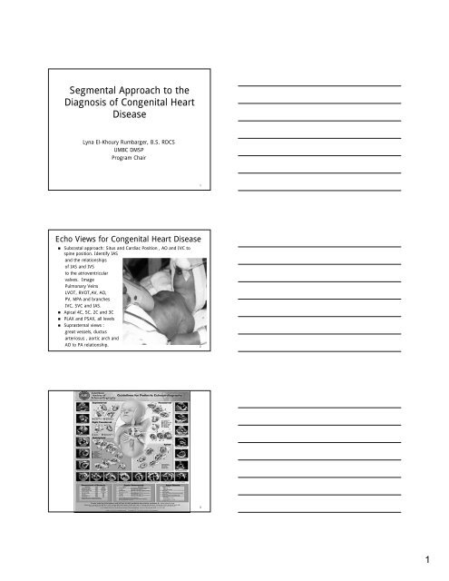
Segmental Approach to the Diagnosis of Congenital Heart Disease
Segmental Approach to the Diagnosis of Congenital Heart Disease
Segmental Approach to the Diagnosis of Congenital Heart Disease

Create successful ePaper yourself
Turn your PDF publications into a flip-book with our unique Google optimized e-Paper software.
<strong>Segmental</strong> <strong>Approach</strong> <strong>to</strong> <strong>the</strong><br />
<strong>Diagnosis</strong> <strong>of</strong> <strong>Congenital</strong> <strong>Heart</strong><br />
<strong>Disease</strong><br />
Lyna El-Khoury Rumbarger, B.S. RDCS<br />
UMBC DMSP<br />
Program Chair<br />
1<br />
Echo Views for <strong>Congenital</strong> <strong>Heart</strong> <strong>Disease</strong><br />
• Subcostal approach: Situs and Cardiac Position , AO and IVC <strong>to</strong><br />
spine position. Identify IAS<br />
and <strong>the</strong> relationships<br />
<strong>of</strong> IAS and IVS<br />
<strong>to</strong> <strong>the</strong> atrioventricular<br />
valves. Image<br />
Pulmonary Veins<br />
LVOT, RVOT,AV, AO,<br />
PV, MPA and branches<br />
IVC, SVC and IAS.<br />
• Apical 4C, 5C, 2C and 3C<br />
• PLAX and PSAX, all levels<br />
• Suprasternal views :<br />
great vessels, ductus<br />
arteriosus , aortic arch and<br />
AO <strong>to</strong> PA relationship. 2<br />
3<br />
1
Adults with CHD<br />
• The number <strong>of</strong> adults with known congenital heart<br />
disease (CHD) has increased dramatically over <strong>the</strong> past<br />
few decades because <strong>of</strong> significant advances in cardiac<br />
imaging, diagnosis and medical and surgical care.<br />
• For <strong>the</strong> past 10 years, <strong>the</strong> number <strong>of</strong> adults with<br />
congenital cardiovascular malformations has equaled <strong>the</strong><br />
number <strong>of</strong> children with <strong>the</strong>se disorders. The majority <strong>of</strong><br />
children born with CHD are surviving in<strong>to</strong> adulthood.<br />
• With additional improvements in surgical techniques and<br />
with repairs occurring at an earlier age, this patient<br />
group is likely <strong>to</strong> increase even fur<strong>the</strong>r.<br />
4<br />
Adult CHD Centers<br />
• Infants born with complex CHD are not always cured.<br />
• Residual problems are common and predictable.<br />
• There are many challenges in <strong>the</strong> transition <strong>of</strong> care from<br />
pediatric <strong>to</strong> adult practitioners.<br />
• Adults with moderate and complex CHD should undergo<br />
regular evaluation at a regional ACHD center.<br />
• In order <strong>to</strong> properly care for <strong>the</strong>se patients, we must<br />
have detailed knowledge <strong>of</strong> CHD, both repaired and<br />
unrepaired.<br />
5<br />
Indications for Echo in Adults with CHD<br />
1. Patients with suspected CHD due <strong>to</strong> clinical<br />
signs and symp<strong>to</strong>ms such as murmurs, cyanosis<br />
or unexplained arterial desaturation, abnormal<br />
ECG or chest X-Ray.<br />
2. Patients with known CHD on follow-up due <strong>to</strong><br />
change in clinical findings.<br />
3. Patients with known CHD for whom <strong>the</strong>re is<br />
uncertainty as <strong>to</strong> <strong>the</strong> original diagnosis,<br />
structural abnormalities and hemodynamics.<br />
6<br />
2
Indications for Echo in Adults with CHD<br />
4. Patients with known, repaired or unrepaired<br />
CHD for whom ventricular function, valve<br />
regurgitation, residual shunts and/or pulmonary<br />
artery pressure must be followed.<br />
5. Patients undergoing Echo guided interventional<br />
ca<strong>the</strong>ter procedures: valvo<strong>to</strong>my, ablation, septal<br />
defect closure…<br />
6. Follow-up Echo exams annually or once every 2<br />
years, in patients with known CHD without<br />
evident change in clinical condition.<br />
7<br />
<strong>Segmental</strong> <strong>Approach</strong> <strong>to</strong> Ana<strong>to</strong>my<br />
• The initial echo exam <strong>of</strong> <strong>the</strong> patient with<br />
suspected CHD requires a sequential and<br />
systematic approach <strong>to</strong> cardiac ana<strong>to</strong>my.<br />
• This approach is based on following <strong>the</strong> blood<br />
flow in<strong>to</strong> <strong>the</strong> heart (systemic venous and<br />
pulmonary venous), through <strong>the</strong> heart (atria,<br />
atrioventricular valves and ventricles) and <strong>the</strong>n<br />
out <strong>of</strong> <strong>the</strong> heart (through semilunar valves and<br />
great vessels).<br />
8<br />
<strong>Segmental</strong> <strong>Approach</strong> <strong>to</strong> <strong>the</strong><br />
<strong>Diagnosis</strong> <strong>of</strong> CHD<br />
• The first step is <strong>to</strong> determine <strong>the</strong> atrial situs and<br />
assess <strong>the</strong> connecting veins and <strong>the</strong>ir inflow<br />
pattern in<strong>to</strong> <strong>the</strong> atria.<br />
• Then <strong>the</strong> ventricular arrangement, morphology<br />
and position are determined and <strong>the</strong><br />
atrioventricular connections are defined.<br />
• Finally <strong>the</strong> arrangement <strong>of</strong> <strong>the</strong> great arteries is<br />
determined and <strong>the</strong>ir connections <strong>to</strong> <strong>the</strong><br />
ventricles (ventriculoarterial relationships).<br />
9<br />
3
10<br />
Sequential <strong>Segmental</strong> <strong>Approach</strong><br />
• Identify <strong>the</strong> situs <strong>of</strong> <strong>the</strong> thoraco-abdominal organs.<br />
• Determine cardiac position within <strong>the</strong> thorax.<br />
• Segment by segment analysis <strong>of</strong> cardiac ana<strong>to</strong>my.<br />
• Atrial Situs.<br />
• Ventricular Situs.<br />
• Atrioventricular connection and alignment.<br />
• Ventriculo-arterial connection and alignment.<br />
• Conal (infundibular) ana<strong>to</strong>my.<br />
• Relationship between <strong>the</strong> great arteries.<br />
• Description <strong>of</strong> associated malformations.<br />
11<br />
12<br />
4
Viscero-Abdominal Situs<br />
• First examine <strong>the</strong> abdominal viscera: positions <strong>of</strong><br />
liver, s<strong>to</strong>mach, spleen, and abdominal great<br />
vessels (AO/IVC).<br />
1. Situs solitus: Aorta lies <strong>to</strong> <strong>the</strong> left <strong>of</strong> <strong>the</strong> spine<br />
and <strong>the</strong> IVC <strong>to</strong> <strong>the</strong> right.<br />
2. Situs inversus: Aorta lies <strong>to</strong> <strong>the</strong> right <strong>of</strong> <strong>the</strong><br />
spine and <strong>the</strong> IVC <strong>to</strong> <strong>the</strong> left.<br />
3. Right isomerism: both vessels lie on <strong>the</strong> same<br />
side with <strong>the</strong> Aorta posterior.<br />
4. Left isomerism: both vessels lie on <strong>the</strong> same<br />
side with <strong>the</strong> Aorta anterior.<br />
13<br />
Abdominal Situs Solitus<br />
A<br />
R<br />
L<br />
Liver<br />
IVC<br />
AO<br />
Spine<br />
P<br />
14<br />
Thoraco-Abdominal Situs<br />
• Situs Solitus: normal arrangement, left s<strong>to</strong>mach,<br />
left spleen, right liver, right lung with 3 lobes,<br />
left lung with 2 lobes.<br />
• Situs Inversus: inverted arrangement, right<br />
s<strong>to</strong>mach, right spleen, left liver, right lung with 2<br />
lobes, left lung with 3 lobes.<br />
• Right Isomerism: Asplenia (no spleen), bilateral<br />
right-sidedness, both lungs have 3 lobes.<br />
• Left Isomerism: Polysplenia (multiple spleens),<br />
bilateral left-sidedness, both lungs have 2 lobes,<br />
<strong>of</strong>ten interrupted IVC.<br />
15<br />
5
16<br />
Situs Solitus<br />
• This is <strong>the</strong> normal<br />
arrangement in most<br />
patients with <strong>the</strong><br />
organs on <strong>the</strong>ir<br />
appropriate sides.<br />
• Atrial Situs Solitus<br />
also seen.<br />
17<br />
Situs Inversus<br />
• This is sometimes<br />
know as a mirror<br />
image arrangement.<br />
• Atrial Situs Inversus is<br />
also seen.<br />
18<br />
6
Right Isomerism: Asplenia<br />
• Known as asplenic syndrome.<br />
The lungs are both tri-lobed<br />
(right morphology) with short<br />
main bronchi. The atria are<br />
both <strong>of</strong> right sided<br />
morphology. 2 SA nodes may<br />
occur. Cardiac problems<br />
include anomalous pulmonary<br />
venous return, septation &<br />
abnormal cardiac position.<br />
Infections are a serious risk<br />
as <strong>the</strong> spleen is absent.<br />
19<br />
Left Isomerism: Polysplenia<br />
• Known as polysplenic syndrome.<br />
The lungs are both bi-lobed (left<br />
morphology) with long main<br />
bronchi. The atria are both <strong>of</strong><br />
left sided morphology. No true<br />
SA node exists. Cardiac<br />
problems include anomalies <strong>of</strong><br />
systemic venous return,<br />
septation & abnormal cardiac<br />
position. The IVC is incomplete<br />
& continues as <strong>the</strong> azygous and<br />
hemiazygos veins. The liver is<br />
usually central and polysplenia is<br />
common.<br />
20<br />
Slightly Central Liver<br />
with Polysplenia<br />
21<br />
7
Bronchial Tree<br />
• In Situs Solitus, <strong>the</strong> right<br />
main bronchus is shorter<br />
than <strong>the</strong> left and takes <strong>of</strong>f<br />
at a more acute angle<br />
from <strong>the</strong> trachea.<br />
• The reverse is true in<br />
situs inversus.<br />
• There are two<br />
symmetrical short main<br />
bronchi in right isomerism<br />
and two longer bronchi in<br />
left isomerism.<br />
22<br />
Cardiac Position & Orientation<br />
• Position <strong>of</strong> <strong>the</strong> heart in <strong>the</strong> chest with regard <strong>to</strong> its<br />
location, and <strong>the</strong> orientation <strong>of</strong> its apex.<br />
• Location <strong>of</strong> <strong>the</strong> heart in <strong>the</strong> chest:<br />
1. Levoposition - <strong>to</strong> <strong>the</strong> left<br />
2. Mesoposition - central<br />
3. Dextroposition - <strong>to</strong> <strong>the</strong> right<br />
• Cardiac orientation is <strong>the</strong> base <strong>to</strong> apex orientation <strong>of</strong> <strong>the</strong><br />
heart:<br />
1. Levocardia - apex directed <strong>to</strong> <strong>the</strong> left <strong>of</strong> <strong>the</strong> midline<br />
2. Mesocardia - apex oriented inferiorly in <strong>the</strong> midline<br />
3. Dextrocardia - apex directed <strong>to</strong> <strong>the</strong> right <strong>of</strong> <strong>the</strong> midline<br />
23<br />
24<br />
8
Imaging Patients with<br />
Cardiac Malposition<br />
• When confronted with a patient with known (or suspected)<br />
cardiac malposition, begin scanning from <strong>the</strong> subcostal<br />
transverse imaging plane. The transducer index mark and<br />
image (screen) index mark should both be oriented <strong>to</strong> <strong>the</strong><br />
patient's left.<br />
• Define relative positions <strong>of</strong> AO, IVC, and spine.<br />
• Sweep and tilt up <strong>to</strong> demonstrate <strong>the</strong> position <strong>of</strong> <strong>the</strong> heart<br />
in <strong>the</strong> chest and <strong>the</strong> direction <strong>the</strong> apex is pointing.<br />
• Continue tilting anteriorly <strong>to</strong> determine <strong>the</strong> atrioventricular<br />
and ventriculoarterial connections and positions <strong>of</strong> <strong>the</strong><br />
great vessels.<br />
25<br />
Dextrocardia<br />
26<br />
Imaging Patients with Dextrocardia<br />
• Patients with Dextrocardia should be positioned in <strong>the</strong><br />
right lateral decubitus position, and <strong>the</strong> sonographer<br />
should be positioned such that scanning can be<br />
comfortably performed with <strong>the</strong> patient in that position.<br />
• Imaging patients with Dextrocardia should include a<br />
clear illustration <strong>of</strong> an empty left chest. The orientation<br />
<strong>of</strong> <strong>the</strong> transducer over <strong>the</strong> right chest should be <strong>the</strong><br />
same as it usually is over <strong>the</strong> left chest.<br />
• A subcostal view should always document <strong>the</strong> abdominal<br />
situs which may be variable.<br />
• Dextrocardia should be differentiated from Cardiac<br />
Dextroposition, which is defined as displacement <strong>of</strong> <strong>the</strong><br />
heart <strong>to</strong> <strong>the</strong> right secondary <strong>to</strong> extracardiac causes such<br />
as right lung hypoplasia or diaphragmatic hernia.<br />
27<br />
9
Variable Situs & Dextrocardia<br />
S I A<br />
28<br />
Dextrocardia in Adults<br />
• Because we are concerned with Dextrocardia in adults, we<br />
will consider only a limited number <strong>of</strong> possibilities:<br />
Dextrocardia with L-loop ventricles and inverted great<br />
vessels (Situs Solitus or Situs inversus <strong>to</strong>talis with<br />
corrected TGA also called Mirror-Image Dextrocardia).<br />
Generally discovered in <strong>the</strong> adult on routine chest x-ray or<br />
physical exam performed for o<strong>the</strong>r reasons.<br />
• Patients experience normal longevity <strong>of</strong> life and have<br />
similar risks <strong>of</strong> getting acquired heart disease as do o<strong>the</strong>r<br />
patients <strong>of</strong> <strong>the</strong> same age and sex. If angina pec<strong>to</strong>ris or<br />
myocardial infarction occurs, <strong>the</strong> pain is located in <strong>the</strong> right<br />
anterior chest and radiates <strong>to</strong> <strong>the</strong> right shoulder and right<br />
arm.<br />
29<br />
Dextrocardia with Situs Inversus<br />
and Corrected TGA<br />
30<br />
10
Dextrocardia with Situs Solitus and Corrected TGA<br />
31<br />
Atrial Morphology<br />
• The right and left atria are identified morphologically by<br />
<strong>the</strong>ir respective atrial appendages and veins emptying<br />
in<strong>to</strong> <strong>the</strong>m.<br />
• The RA receives IVC, SVC and coronary sinus.<br />
• The LA receives all 4 pulmonary veins.<br />
• The RA has a triangular, broad based, anterior<br />
appendage while <strong>the</strong> LA has a narrow, fingerlike<br />
posterior appendage.<br />
• The septum secundum (limbus <strong>of</strong> <strong>the</strong> fossa ovale) lies on<br />
<strong>the</strong> RA side. The septum primum (flap) lies on <strong>the</strong> LA<br />
side. The Crista Terminalis is in <strong>the</strong> RA. LA is smooth<br />
with fewer trabeculations.<br />
• Almost invariably 2 atria are present although sometimes<br />
<strong>the</strong>re may be a common atrium if <strong>the</strong> IAS is absent.<br />
32<br />
Normal Atrial Morphology (TEE)<br />
33<br />
11
TEE views showing<br />
triangular and broad<br />
based<br />
RA appendage.<br />
Narrow, fingerlike LA<br />
appendage.<br />
34<br />
Normal Subcostal Images<br />
IVC-RA Connection<br />
35<br />
Atrial Situs (Arrangement)<br />
• There are 3 possible atrial situs nomenclatures:<br />
Situs Solitus (S), Situs Inversus (I), Situs Ambiguous (A)<br />
= right isomerism (bilateral right atria) or left isomerism<br />
(bilateral left atria).<br />
• Atrial Situs Solitus (S): IVC, SVC and coronary sinus on<br />
<strong>the</strong> right.<br />
• Atrial Situs Inversus (I): IVC, SVC and coronary sinus on<br />
<strong>the</strong> left.<br />
• Atrial Situs Ambiguous (A): IVC is interrupted between<br />
<strong>the</strong> renal and hepatic segments, hepatic veins drain in<strong>to</strong><br />
common atrium, persistent left SVC (bilateral SVC) may<br />
drain in<strong>to</strong> unro<strong>of</strong>ed coronary sinus and a muscle bar<br />
similar <strong>to</strong> a crista terminalis may be present in <strong>the</strong> left<br />
SVC-atrial junction. Variable pulmonary vein connections.<br />
36<br />
12
37<br />
Atrial Situs Ambiguous<br />
38<br />
Ventricular Morphology<br />
• The RV is distinguished by its triangular shape, coarse<br />
trabeculations, heavy apical modera<strong>to</strong>r band and chordal<br />
attachments <strong>of</strong> <strong>the</strong> TV leaflets <strong>to</strong> <strong>the</strong> trabeculated septal<br />
surface. Normal RV has a pumping chamber (RV sinus)<br />
and an outflow tract (infundibulum).<br />
• The LV is oval shaped with a finely trabeculated apex<br />
(trabeculae carnea) smooth septal wall and MV chordal<br />
attachments only <strong>to</strong> <strong>the</strong> free wall papillary muscles.<br />
• The common situation is when two, well formed<br />
ventricles are present. True “single” ventricles do exist<br />
but it is usually possible <strong>to</strong> identify a rudimentary<br />
ventricle attached <strong>to</strong> <strong>the</strong> side <strong>of</strong> <strong>the</strong> main ventricle. The<br />
surgical outcome is better in those with a dominant LV.<br />
39<br />
13
Ventricular Situs or Looping<br />
• Normal rightward direction <strong>of</strong> looping is termed Dextro or<br />
D-Looping (right hand <strong>to</strong>pology)<br />
• If <strong>the</strong> heart loops abnormally <strong>to</strong> <strong>the</strong> left, Levo or L-Looping<br />
(left hand <strong>to</strong>pology) occurs.<br />
40<br />
Determination <strong>of</strong> Ventricular Situs by<br />
Chirality (Handedness)<br />
D-loop: palm <strong>of</strong> right hand over RV<br />
septal surface, thumb in TV and fingers<br />
in RVOT.<br />
L-loop: palm <strong>of</strong> left hand over RV<br />
septal surface, thumb in TV and fingers<br />
in RVOT.<br />
41<br />
Concordant or Discordant<br />
Connections<br />
• Connection : refers <strong>to</strong> <strong>the</strong> sequence <strong>of</strong> ana<strong>to</strong>mic<br />
structures. Normally, RA is connected <strong>to</strong> RV by means <strong>of</strong><br />
TV. RV is <strong>the</strong>n connected <strong>to</strong> <strong>the</strong> PA by means <strong>of</strong> <strong>the</strong> PV.<br />
Therefore, <strong>the</strong>re are atrio-ventricular connections and<br />
ventriculo-great arterial connections <strong>to</strong> identify.<br />
• Concordance: describes <strong>the</strong> relationship between <strong>the</strong><br />
various chambers, valves, and great vessels. In <strong>the</strong><br />
normal heart all <strong>the</strong> connections and relationships in <strong>the</strong><br />
ana<strong>to</strong>mic sequence are concordant.<br />
• Discordance : describes abnormal relationships between<br />
<strong>the</strong> various chambers and great vessels.<br />
42<br />
14
Concordant or Discordant<br />
Connections<br />
43<br />
AV Connection/Alignment<br />
• AV Concordance<br />
• AV Discordance<br />
• Tricuspid Atresia<br />
• Mitral Atresia<br />
• Common AV Valve<br />
• Overriding AV Valve<br />
• Straddling AV Valve<br />
• Double Inlet Ventricle<br />
44<br />
45<br />
15
Arterial Morphology<br />
• The definition <strong>of</strong> an aorta<br />
is an artery that gives rise<br />
<strong>to</strong> <strong>the</strong> coronary arteries<br />
and <strong>the</strong> brachiocephalic<br />
vessels.<br />
• In contrast <strong>the</strong> pulmonary<br />
artery branches in<strong>to</strong> two<br />
but does not give rise <strong>to</strong><br />
any vessels.<br />
46<br />
Ventriculo-Arterial Connection<br />
Concordant or Discordant<br />
• When <strong>the</strong> aorta is connected <strong>to</strong> <strong>the</strong> LV and <strong>the</strong><br />
pulmonary artery <strong>to</strong> <strong>the</strong> RV <strong>the</strong> connection is described<br />
as concordant.<br />
• If <strong>the</strong> aorta is connected <strong>to</strong> <strong>the</strong> RV and <strong>the</strong> pulmonary<br />
artery <strong>to</strong> <strong>the</strong> LV <strong>the</strong>n <strong>the</strong> connection is discordant. This<br />
is most commonly seen in transposition <strong>of</strong> <strong>the</strong> great<br />
arteries (TGA)<br />
47<br />
Ventriculo-Arterial Connection<br />
Double Outlet<br />
• If both great arteries arise from a single ventricle <strong>the</strong><br />
connection is described as double outlet.<br />
• The great arteries may be normally related <strong>to</strong> each o<strong>the</strong>r<br />
or mal-positioned (TGA).<br />
• The situation is still described as double outlet even if<br />
both vessels are not completely over a single ventricle.<br />
• If more than 50% <strong>of</strong> an artery overrides a ventricle it is<br />
said <strong>to</strong> be committed <strong>to</strong> it.<br />
48<br />
16
Ventriculo-Arterial Connection<br />
Single Outlet<br />
One vessel only may arise from<br />
<strong>the</strong> heart. This may be because a<br />
vessel is absent (Aortic or<br />
Pulmonary Atresia) as is <strong>the</strong><br />
case with <strong>the</strong> drawing on <strong>the</strong> left<br />
or because <strong>the</strong> two vessels<br />
have fused <strong>to</strong> form a common<br />
outlet as seen in <strong>the</strong> drawing <strong>of</strong> a<br />
Persistent Truncus on <strong>the</strong> right.<br />
49<br />
Concordance or<br />
Discordance<br />
Double Outlet<br />
Connection<br />
Single Outlet<br />
Connection<br />
50<br />
Commitment<br />
• Commitment fur<strong>the</strong>r describes possible abnormalities <strong>of</strong><br />
flow through valves in<strong>to</strong> ventricles and great vessels.<br />
• In Tetralogy <strong>of</strong> Fallot, <strong>the</strong> atria, atrioventricular valves,<br />
and ventricles are positioned normally, and concordant.<br />
But <strong>the</strong> aorta overrides a VSD and is doubly committed<br />
<strong>to</strong> both ventricles.<br />
• In cases where <strong>the</strong>re is only one ventricle (univentricular<br />
heart), both atrioventricular valves are usually doubly<br />
committed <strong>to</strong> <strong>the</strong> single ventricle.<br />
51<br />
17
Ambiguous, Inlet, Outlet<br />
• Ambiguous is used when precise identification <strong>of</strong> a<br />
ventricle or o<strong>the</strong>r structure cannot be made. For<br />
example, in a univentricular heart with a doubly<br />
committed atrioventricular connection it may not be<br />
possible <strong>to</strong> always identify clearly whe<strong>the</strong>r it is <strong>the</strong> right<br />
or left ventricle. Thus, <strong>the</strong> single ventricle would be<br />
ambiguous.<br />
• Inlet refers <strong>to</strong> anomalies <strong>of</strong> <strong>the</strong> structures and flow in<strong>to</strong><br />
<strong>the</strong> ventricle.<br />
• Outlet refers <strong>to</strong> anomalies <strong>of</strong> <strong>the</strong> structures and flow out<br />
<strong>of</strong> <strong>the</strong> ventricles in<strong>to</strong> <strong>the</strong> great vessels.<br />
52<br />
Types <strong>of</strong> Conus<br />
• Subpulmonary: absence <strong>of</strong> subaortic infundibular free<br />
wall, found in <strong>the</strong> normal heart.<br />
• Subaortic: absence <strong>of</strong> subpulmonary infundibular free<br />
wall, found in D-loop TGA.<br />
• Bilateral: bilaterally present infundibular free wall, found<br />
in double outlet RV but rarely in TGA.<br />
• Bilaterally Absent: no infundibular free wall, found in<br />
double outlet LV.<br />
53<br />
54<br />
18
Interrelations between <strong>the</strong><br />
Semilunar Valves<br />
• Solitus (normal).<br />
• Inversus (mirror image)<br />
• D-malposition (aortic valve anterior and <strong>to</strong> <strong>the</strong> right).<br />
• L-malposition (aortic valve anterior and <strong>to</strong> <strong>the</strong> left).<br />
• Anterior malposition (aortic valve anterior in <strong>the</strong> middle).<br />
• Parasternal and high parasternal short axis views as well<br />
as subcostal short axis views are used <strong>to</strong> recognize <strong>the</strong><br />
positions <strong>of</strong> <strong>the</strong> AV and PV.<br />
• The origins or <strong>the</strong> coronary arteries are also<br />
documented.<br />
55<br />
56<br />
Formulation: Step 1<br />
• The visceroatrial situs is determined: S, I or A.<br />
• Visceroatrial situs refers <strong>to</strong> <strong>the</strong> position <strong>of</strong> <strong>the</strong><br />
atria in relation <strong>to</strong> <strong>the</strong> nearby ana<strong>to</strong>my<br />
(including <strong>the</strong> s<strong>to</strong>mach, liver, spleen, and<br />
bronchi).<br />
• Three different ana<strong>to</strong>mic configurations may be<br />
observed: S (situs solitus), I (situs inversus), or<br />
A (situs ambiguous).<br />
57<br />
19
Formulation: Step 2<br />
• Ventricular Situs: D or L<br />
The leftward L-loop (abnormal) or rightward D-<br />
loop (normal) orientation <strong>of</strong> ventricular looping<br />
is evaluated, and <strong>the</strong> positions <strong>of</strong> <strong>the</strong> ventricles<br />
are identified on <strong>the</strong> basis <strong>of</strong> <strong>the</strong>ir internal<br />
morphologic features.<br />
58<br />
Formulation: Step 3<br />
• The position <strong>of</strong> <strong>the</strong> great vessels is determined<br />
first, and any abnormalities are noted: S, I, D L<br />
or A. (S = Solitus)<br />
• Abnormalities in <strong>the</strong> origin and position <strong>of</strong> <strong>the</strong><br />
great vessels (conotruncal anomalies) are<br />
predominantly <strong>of</strong> 4 types:<br />
• Inverted (I)<br />
• D-Malposition (D)<br />
• L-Malposition (L)<br />
• Anterior (A)<br />
59<br />
60<br />
20
Normal {S,D,S} Ana<strong>to</strong>my<br />
The Van Praagh segmental approach documents<br />
<strong>the</strong> three major cardiac segments {atria, ventricles,<br />
great arteries} in a venoarterial sequence.<br />
Letters are coded in brackets { } <strong>to</strong> describe <strong>the</strong><br />
visceroatrial situs [S = situs solitus, I = situs<br />
inversus, A = ambiguous], <strong>the</strong> ventricular loop [D =<br />
D-loop, L = L-loop]), and <strong>the</strong> great artery position<br />
[S = normally related great arteries, I = inverted<br />
normally related great arteries, D = D-malposition,<br />
A= Anterior malposition and L = L-malposition].<br />
Thus, a normal heart would default <strong>to</strong> {S,D,S}<br />
ana<strong>to</strong>my. Any o<strong>the</strong>r combination is abnormal.<br />
61<br />
Main Sources<br />
• <strong>Segmental</strong> <strong>Approach</strong> and Nomenclature. Tal Geva, MD <strong>of</strong><br />
<strong>Congenital</strong> <strong>Heart</strong> <strong>Disease</strong>. Department <strong>of</strong> Cardiology.<br />
Children's Hospital Bos<strong>to</strong>n.<br />
• Van Praagh R: The segmental approach <strong>to</strong> diagnosis <strong>of</strong><br />
congenital heart disease. In Birth Defects: Original<br />
Article Series. Baltimore, Williams & Wilkins.<br />
• <strong>Approach</strong> <strong>to</strong> Dextrocardia in Adults: Review<br />
Pierre D. Maldjian and Muhamed Saric.<br />
62<br />
21