

Medexpert Arteriograph IrDA - User Manual
Medexpert Arteriograph IrDA - User Manual
Medexpert Arteriograph IrDA - User Manual

You also want an ePaper? Increase the reach of your titles
YUMPU automatically turns print PDFs into web optimized ePapers that Google loves.
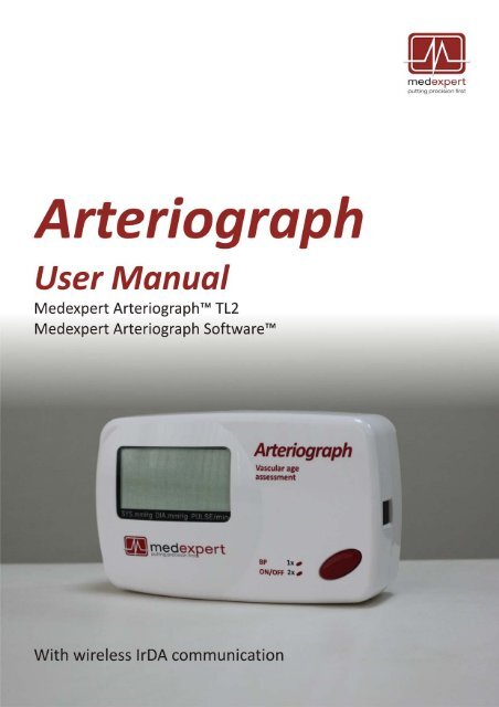
<strong>Medexpert</strong> <strong>Arteriograph</strong><br />
<strong>Medexpert</strong> <strong>Arteriograph</strong> Software<br />
Copyright © 2011 <strong>Medexpert</strong> Ltd., Budapest, Hungary. All rights reserved. Under the copyright laws,<br />
this manual cannot be reproduced in any form without prior written permission of <strong>Medexpert</strong> Ltd.<br />
Every effort has been made to ensure that the information in this manual is accurate. Succeeding<br />
models and manuals are subject to change without notice.<br />
<strong>Medexpert</strong> is not responsible for printing or clerical errors.<br />
This <strong>Manual</strong> is produced on the assumption that the operator is an experienced user of the Windows<br />
2000 / XP / Vista / 7 Operating Systems.<br />
If the operator is not familiar with Windows operations, please refer to the on‐line Help of Windows<br />
or the Windows <strong>User</strong> <strong>Manual</strong>.<br />
<strong>Arteriograph</strong> is an unregistered trademark of <strong>Medexpert</strong> Ltd.<br />
Other company and product names mentioned herein may be trademarks of their respective<br />
companies.<br />
Head office and service:<br />
<strong>Medexpert</strong> Ltd.<br />
Törrökkö u. 5‐7.<br />
1037 Budapest, HUNGARY<br />
Hungary<br />
Phone: +36 1 250 4490<br />
Fax: +36 1 250 3795<br />
Web: www.medexpert.hu<br />
E‐mail: arterialcare@medexpert.hu<br />
4
Contents<br />
1 Introduction.................................................................................................................................... 7<br />
1.1 Preface ................................................................................................................................................. 7<br />
1.2 Contents of the manual ....................................................................................................................... 7<br />
1.3 General information............................................................................................................................. 7<br />
1.4 Guarantee ............................................................................................................................................ 7<br />
1.5 Warnings and precautions ................................................................................................................... 8<br />
2 The <strong>Medexpert</strong> TensioClinic <strong>Arteriograph</strong> device .......................................................................... 9<br />
2.1 The device at a glance..........................................................................................................................9<br />
2.2 The accessories of the Device............................................................................................................ 11<br />
2.3 Installing the Device........................................................................................................................... 11<br />
2.4 Operation instructions....................................................................................................................... 12<br />
2.5 System (Infrared communication) setup ........................................................................................... 12<br />
3 The software................................................................................................................................. 14<br />
3.1 <strong>Medexpert</strong> TensioClinic Software installation and setup .................................................................. 14<br />
3.1.1 Preliminary information about the software ................................................................................ 14<br />
3.1.2 Minimum system requirements.................................................................................................... 14<br />
3.1.3 Software Installation ..................................................................................................................... 15<br />
3.2 The software at a glance.................................................................................................................... 17<br />
3.2.1 The Menu of the Program............................................................................................................. 18<br />
3.2.2 The Toolbar of the Program.......................................................................................................... 18<br />
3.2.3 The examinations window ............................................................................................................ 18<br />
3.2.4 The results block ...........................................................................................................................18<br />
3.2.5 Patient details field ....................................................................................................................... 19<br />
3.2.6 The measurement data window ................................................................................................... 19<br />
3.3 First step – Register a user................................................................................................................. 19<br />
4 Performing a single blood pressure measurement ...................................................................... 20<br />
4.1 Patient preparation............................................................................................................................ 20<br />
4.2 Performing the measurement ........................................................................................................... 20<br />
4.3 Acoustic signals .................................................................................................................................. 21<br />
5 Performing pulse wave recording and analysis............................................................................ 22<br />
5.1 Hemodynamic parameters measured by the <strong>Arteriograph</strong>............................................................... 22<br />
5.2 System setup...................................................................................................................................... 23<br />
5.3 Patient preparation............................................................................................................................ 24<br />
5.4 Performing the measurement ........................................................................................................... 26<br />
5.4.1 Device and measurement setup ................................................................................................... 26<br />
5.4.2 Select a patient.............................................................................................................................. 28<br />
5.4.2.1 How to select an existing patient ........................................................................................ 28<br />
5.4.2.1.1 How to modify a patient’s data ...................................................................................... 28<br />
5.4.2.1.2 How to search for a patient ............................................................................................ 28<br />
5.4.2.2 How to enter a new patient’s data...................................................................................... 29<br />
5.4.3 Medical Examination with <strong>Medexpert</strong> TensioClinic <strong>Arteriograph</strong> ................................................ 30<br />
5.4.4 Quality control of the measurement............................................................................................. 32<br />
5.4.4.1 Quality control index: the SD PWVao .................................................................................. 32<br />
5.4.4.2 Quality control by visual inspection..................................................................................... 32<br />
5.4.5 How to analyse the data ............................................................................................................... 35<br />
5.4.5.1 Tools for visual inspection ................................................................................................... 38<br />
5.4.5.2 Oscillation amplitudes during blood pressure measurement ............................................. 39<br />
5.4.5.3 Vascular age assessment ..................................................................................................... 40<br />
5.4.6 The measurement Report ............................................................................................................. 40<br />
5.4.6.1 Edit the Report..................................................................................................................... 41<br />
5.4.6.2 Print the Report ................................................................................................................... 42<br />
5.4.6.3 How to send a Report in PDF format................................................................................... 45<br />
5.4.7 Exporting the measurement data ................................................................................................. 45<br />
5
5.4.8 Backup and restore the database ................................................................................................. 47<br />
5.4.8.1 Backup data ......................................................................................................................... 47<br />
5.4.8.2 Restore data ........................................................................................................................ 48<br />
5.4.8.3 Import previous database.................................................................................................... 48<br />
6 Troubleshooting............................................................................................................................ 49<br />
7 Technical Specification ................................................................................................................. 51<br />
7.1 Electromagnetic compatibility ........................................................................................................... 52<br />
6
1. Introduction<br />
1.1 Preface<br />
Thank you for choosing <strong>Medexpert</strong>® <strong>Arteriograph</strong>. This state‐of‐the‐art device and method enables<br />
recording and analysing arterial pulse waves in an exceptionally easy, reliable and accurate way. With<br />
this entirely new implementation of oscillometric method a complete arterial function evaluation can<br />
be achieved.<br />
1.2 Contents of the manual<br />
This manual helps you in setting up and starting to use the <strong>Medexpert</strong> <strong>Arteriograph</strong> TL1 device as<br />
well as installing and setting up the <strong>Medexpert</strong> <strong>Arteriograph</strong> Software up to versions 3.0.0.0.<br />
Attention! Before first use, please read and understand this document carefully.<br />
1.3 General information<br />
• When active, the Device does not cause any electromagnetic disturbance and its immunity to<br />
environmental effects is also sufficient. The measurement data are downloaded into the PC<br />
through wireless infrared communication. The electromagnetic compatibility between the<br />
Device and the PC is guaranteed (see Section 7.1 for details).<br />
• To ensure the optimal performance and precise operation the Device should be maintained<br />
at the manufacturer’s service every second year.<br />
• The cuff and the Device itself can be cleaned with a wet cloth by making sure that no liquid<br />
enter into the Device.<br />
• The manufacturer’s products must be handled, stored, packaged, protected and shipped in<br />
compliance with the general quality requirements.<br />
Attention! Federal Law restricts this device to use by or on the order of a physician,<br />
or other licensed practitioner (US only).<br />
1.4 Guarantee<br />
<strong>Medexpert</strong> Ltd. offers a two‐year guarantee period for the Device. Any repair within or beyond the<br />
guarantee period will be performed by <strong>Medexpert</strong> Ltd. at the company’s site as indicated above.<br />
7
1.5 Warnings and precautions<br />
! Caution!<br />
Patient safety<br />
The device has an integrated safety mechanism, which prevents the cuff pressure to exceed<br />
300 mmHg. If however the inflation continues above this value or the pressurization lasts too<br />
long, unplug the tube of the cuff from the device and remove the cuff from the subject.<br />
Do not use the device on an arm, which is being injected with intravenous injection.<br />
Do not use on neonates.<br />
Device use<br />
Only use with cuffs supplied by <strong>Medexpert</strong> Ltd. Use of cuffs supplied by a third party can lead<br />
to erroneous measurement results.<br />
Confirm blood pressure measurement with auscultation when erroneous results are<br />
suspected.<br />
Do not use a microwave device (e.g. mobile phone) near the unit during measurement.<br />
Do not use the device when it is exposed to mechanical vibration (e.g. in vehicles).<br />
Prevent the device to be exposed to direct sunlight, to get in contact with liquid or from<br />
excessive mechanical impact.<br />
Do not disassemble the device.<br />
8
2. The <strong>Medexpert</strong> <strong>Arteriograph</strong> device<br />
This section will give an introduction on the device along with installation information and operation<br />
instructions.<br />
2.1 The device at a glance<br />
Figure 1 shows the front view of the device:<br />
1. The name of the Device<br />
2. Push button for entering two commands to control the Device, as per Section 2.4<br />
3. Symbols standing for the above two commands<br />
4. LCD<br />
1<br />
4<br />
2<br />
3<br />
Figure 2 shows the rear view of the device:<br />
5. Battery socket (cover removed)<br />
6. Device label<br />
7. Pneumatic connector<br />
Figure 1<br />
7<br />
5<br />
6<br />
Figure 2<br />
9
Figure 3 shows the device label. The meanings of the symbols are the following:<br />
8. Name of the Device<br />
9. Name of the Manufacturer<br />
10. Range of ambient operating temperature<br />
11. Manufacturing serial number<br />
12. Applicable nominal range of supply voltage with the batteries<br />
13. Year of manufacturing<br />
14. Safety classification of electric shock protection (Classification: Patient side: BF)<br />
15. Warning on the importance of reading this <strong>User</strong> <strong>Manual</strong> thoroughly<br />
16. Certification mark as a guarantee of the Device’s conformity with EU requirements<br />
17. Type ID of the Device<br />
8 9<br />
18<br />
16<br />
10<br />
13<br />
12<br />
11<br />
17<br />
14<br />
Figure 3<br />
10<br />
See Figure 4 for the side view of the Device:<br />
18. Infrared communication window<br />
18<br />
Figure 4<br />
10
2.2 The accessories of the Device<br />
<strong>Arteriograph</strong> has the following accessories:<br />
• 4 pcs. AA size alkaline batteries<br />
• Three D‐ring cuffs of different sizes (see below)<br />
• <strong>Medexpert</strong> <strong>Arteriograph</strong> Software installation CD<br />
• A tape measure for measuring the jugulum – symphysis distance<br />
• <strong>IrDA</strong> adapter kit (<strong>IrDA</strong> adapter, Driver CD, USB extension cable)<br />
• A pouch with an integrated console for holding the <strong>IrDA</strong> adapter<br />
• A <strong>User</strong> <strong>Manual</strong><br />
The cuff sizes are, as follows:<br />
Cuff type Bladder dimensions Sleeve dimensions Arm circumference range<br />
01 34 × 8 cm 62 × 9 cm 34 ‐ 43 cm<br />
02 26 × 8 cm 52 × 9 cm 26 ‐ 33 cm<br />
03 18 x 6 cm 38 x 7 cm 18 ‐ 25 cm<br />
2.3 Installing the Device<br />
<strong>Arteriograph</strong> is a battery operated device.<br />
• Insert 4 durable alkaline AA batteries into the Device with taking care of the right polarity<br />
(see Figure 3)<br />
• Or insert 4 AA sized rechargeable Ni‐MH or NiCd batteries as per the above instruction.<br />
(Please note that new batteries must be pre‐charged.)<br />
o For problem‐free operation minimum 1,500 mAh chargeable Ni‐MH or NiCd batteries<br />
are recommended.<br />
Figure 3<br />
Note: The clock circuits of the Device are fed by integrated NiCd HA 35 battery cells, therefore<br />
the operation of the clock is independent from the AA battery supply.<br />
Attention! If you do not intend to use the Device for a longer period of time, remove<br />
the batteries and store them in a dry and cool place out of little children’s reach. Do<br />
not expose them to intense heat because it may cause a short circuit. Regarding both<br />
rechargeable and alkaline batteries, the policies of use and waste collection as<br />
governed by detailed environmental and labour safety requirements are to be<br />
considered.<br />
11
Once the batteries are inserted in the Device, the following checks are performed automatically:<br />
• Voltage control of the batteries. The measured value appears on the<br />
LCD for a few seconds. The supply voltage is sufficient if the<br />
measured value is between 6.0 V and 5.4 V for alkaline batteries and<br />
between 4.8 V and 4.6 V for rechargeable batteries.<br />
• If the voltage drops below 4.4V, the batteries must be replaced. A<br />
warning symbol of low battery (crossed battery icon) appears on the<br />
LCD.<br />
5.6V<br />
LOw Batt<br />
• The Device is ready for operation if the current PC time appears on<br />
the LCD.<br />
D 09-39<br />
2.4 Operation instructions<br />
There is a single push button available for operating the Device (Section 2.1). The patient can use the<br />
only push button for issuing a total of two commands.<br />
1. By pressing the button shortly manual blood pressure measuring will start. Note! In this case<br />
the Device performs blood pressure measurement without any arterial function (e.g. PWVao,<br />
Aix) evaluation.<br />
2. The Device can be turned off by pressing the button twice. In OFF<br />
state the device cannot be operated either manually or by PC. To<br />
turn it on the button must be pressed twice again.<br />
OFF<br />
2.5 System (Infrared communication) setup<br />
For installing the driver of the <strong>IrDA</strong> adapter please perform the following steps (the same description<br />
can be found in the Installation <strong>User</strong>s <strong>Manual</strong>).<br />
!<br />
Attention! Please note that the <strong>IrDA</strong> adapter must not be plugged in the PC before<br />
or during installation! To complete the driver installation the computer must be<br />
restarted!<br />
1. Insert the <strong>IrDA</strong> driver CD into the CD tray of your PC. The installation starts automatically.<br />
2. Please choose your preferred language.<br />
3. Click “Next” to run the procedure.<br />
4. Click “Install” to begin the installation and to exit the install program.<br />
5. Restart the computer to complete the install procedure. (Without restarting the PC the driver<br />
will not work properly. All open data should be saved before restart!)<br />
12
6. After restart plug in the <strong>IrDA</strong> adapter. (Do not plug in the adapter before or during the<br />
installation!)<br />
7. The <strong>IrDA</strong> device is ready to communicate.<br />
The Device communicates with the PC using standard infrared protocol (<strong>IrDA</strong>) with a transmission<br />
rate of 115,200bps. We recommend the <strong>IrDA</strong> communication to be set up by a person experienced in<br />
wireless communication.<br />
For connecting <strong>Arteriograph</strong> to the PC see Section 5.2.<br />
Data transmission can only be initialized with the <strong>Arteriograph</strong> Software.<br />
13
3. The software<br />
3.1 <strong>Medexpert</strong> <strong>Arteriograph</strong> Software installation and setup<br />
3.1.1 Preliminary information about the software<br />
The <strong>Medexpert</strong> <strong>Arteriograph</strong> Software (<strong>Medexpert</strong> SW) is a Windows‐based software. The common<br />
Windows mouse operations are to be used in the program (left single‐click, double click, drag, etc.).<br />
Button and command names ending in … indicate that a dialog window will open upon clicking on<br />
those items.<br />
<strong>Medexpert</strong> SW allows you to perform arterial function measurement, to print and share the<br />
measurement report and to modify the measurement settings.<br />
3.1.2 Minimum system requirements<br />
• Pentium IV PC, 256MB memory, 2GB available HDD capacity, CDROM, 1024*768 resolution,<br />
• Windows 2000 operating system with Service Pack 4, Windows XP with Service Pack 2,<br />
Windows Vista or Windows 7 installed,<br />
• Active infrared port.<br />
Although the Program can start in a less powerful environment, in that case we cannot take<br />
responsibility for its fast and reliable operation.<br />
14
3.1.3 Software Installation<br />
For installing the <strong>Medexpert</strong> SW on the computer the user must have administrator privileges. After<br />
the installation is completed, please visit our website (www.<strong>Medexpert</strong>.com) for the latest software<br />
upgrades. Installation instructions for those can also be found on our site.<br />
Step 1<br />
Insert the CD into the CD tray of your PC. The installation procedure will start<br />
automatically. In case it does not, start the installation by double‐clicking the<br />
setup.exe file on the disc with use of a file manager program.<br />
Should a security warning pop up, simply press Run to start the installation<br />
Step 2<br />
To start the Setup Wizard, choose a preferred language for installation and click OK.<br />
Step 3<br />
The wizard starts with a welcome screen. Please close all the other running Windows<br />
applications and click Next.<br />
15
Step 4<br />
Select the components to be installed from the drop‐down list. The type of<br />
installation can either be Full or Upgrade.<br />
When the preferred component is selected, click Next.<br />
Step 5<br />
The following screen offers you to create an icon on the desktop. This task can be<br />
deselected. Click Next to start the installation process.<br />
Step 6<br />
The status of the automatic installation procedure can be tracked on a progress bar.<br />
If you want to interrupt it, click Cancel.<br />
Step 7<br />
When the automatic installation procedure is finished the following screen appears.<br />
Click Finish to exit the Wizard and start the <strong>Arteriograph</strong> Software.<br />
16
3.2 The software at a glance<br />
To start the Program double‐click the <strong>Arteriograph</strong> icon on your desktop, or use the<br />
Windows Start Menu to navigate to the program in the Program Files.<br />
A splash screen appears along with a login dialog window.<br />
When started for the first time, simply press OK and the Program logs in the user with the default<br />
“ARTERIOGRAM” login name. On every login after that, however, the user name must be specified.<br />
The program window appears with the following components. (For a better understanding the<br />
picture below shows the program window after a completed measurement.)<br />
1. Menu bar<br />
2. Toolbar<br />
3. Examinations window<br />
4. Results field<br />
5. Patient details field<br />
6. Data window<br />
17
3.2.1 The Menu of the Program<br />
The menu bar of the software can be found under the title bar and contains the menu items through<br />
which all the major functions of the program can be accessed.<br />
The File Menu<br />
The basic functions of <strong>Medexpert</strong> SW can be accessed through the File<br />
menu. You can enter new patients, open, edit or delete existing<br />
database records, save the result of an analysis; furthermore edit, print<br />
and send medical reports, as well as log out and exit.<br />
The View Menu<br />
The View Menu enables you to choose to display the pulse waves<br />
recorded or the maximum amplitudes of the oscillatory signals at each<br />
cuff pressure step (deflated stepwise with 10 mmHg) during blood<br />
pressure measurement. Furthermore you can set the scale both<br />
horizontally and vertically, hide the Toolbar, and get information about<br />
the currently studied patient.<br />
The Tools Menu<br />
The Tools Menu enables you to start automatic pulse wave data<br />
collection by means of the <strong>Arteriograph</strong> Device, as well as interrupt an<br />
ongoing data collection process. Previous versions of databases can be<br />
converted and imported into the new database format; furthermore<br />
exports and backups of the actual database can be created. All the<br />
Program and Device settings can also be accessed and modified through<br />
this Menu.<br />
3.2.2 The Toolbar of the Program<br />
The toolbar can be found under the menu bar and contains icons, which allow direct access to the<br />
major functions of the program. The Toolbar can be displayed or hidden by the Toolbar command of<br />
the View Menu.<br />
3.2.3 The examinations window<br />
All the previous – if there are any – examinations of the selected patient are listed here by the date<br />
and time of recording.<br />
3.2.4 The results block<br />
The calculated hemodynamic parameters are displayed here when an analysis of measurement data<br />
is performed (automatically or manually).<br />
18
3.2.5 Patient details field<br />
The name and date of birth of the patient are displayed in this field.<br />
3.2.6 The measurement data window<br />
During the measurement the data are displayed here real‐time in the form of non‐calibrated<br />
pressure curves (oscillations) as recorded by the device. The recorded and saved pressure waves can<br />
be opened and displayed at any later time. The <strong>User</strong> can also select to display the amplitudes of the<br />
oscillometric curves throughout the blood pressure measurement phase, or the estimated vascular<br />
age of the patient based on the arterial function assessment.<br />
3.3 First step – Register a user<br />
If the program is used by several users with different groups of patients, it may be necessary that<br />
users can access their own respective patients’ data only after login. To this end a new user will have<br />
to register in the database by specifying the name and login.<br />
<strong>Medexpert</strong> SW can be used with the default login of “ARTERIOGRAM”; it is, however highly<br />
recommended to create and use your own user login.<br />
Registration of a new user<br />
To register a new user, open the Options… window under the Tools menu and select the<br />
<strong>User</strong>s tab. To enter a new user click on Add New and then enter the first and family names of<br />
the user and specify a login name (user ID). Click Add to register the new user in the database<br />
or click Cancel to interrupt the registration. The new user can log in with the login name<br />
specified.<br />
Note! The registered users’ data cannot be edited.<br />
How to delete registered users<br />
To delete registered users, select Options… under Tools and click on the <strong>User</strong>s tab in the<br />
pop‐up window. The Delete button can be used for deleting a pre‐selected item from the<br />
users list. In case the list contains only one user, the Delete function will not be available.<br />
19
4. Performing a single blood pressure measurement<br />
The <strong>Medexpert</strong> <strong>Arteriograph</strong> device can be used as a simple automatic blood pressure meter. (For<br />
this function no computer is needed.)<br />
4.1 Patient preparation<br />
Sitting position is recommended for standard blood pressure measurement. The patient should be<br />
comfortably seated with the legs uncrossed and the back and arm supported. The cuff on the upper<br />
arm should be at the level of the heart.<br />
Allow the patient to sit in a relaxed state for at least 5 minutes before the measurement. Ensure<br />
optimal room temperature and eliminate excessive background noise.<br />
Choose a cuff of appropriate size by considering the followings:<br />
• The length of the inflatable bladder of the cuff should be at least 80% of upper arm<br />
circumference.<br />
• The width of the inflatable bladder of the cuff should be at least 40% of upper arm length.<br />
(Please read and follow the latest guidelines on blood pressure measurement.)<br />
4.2 Performing the measurement<br />
To operate <strong>Arteriograph</strong> first install the device (see Section 2.3). There is a single push button<br />
(Section 2.1) available for operating the Device. The measured values and the status of the Device<br />
are shown on the LCD.<br />
• The Device is ready for operation if the current PC time<br />
appears on the LCD.<br />
D 09-39<br />
By pressing the button shortly manual blood pressure measuring will start.<br />
Note: In this case the Device performs blood pressure measurement without arterial function<br />
(e.g. PWVao, Aix) evaluation.<br />
By doing so, the current time will disappear from the display and then:<br />
• The LCD test pattern is displayed,<br />
• The supply voltage is checked<br />
5.6V<br />
• The Device calibrates itself by setting the zero pressure<br />
CAL 0<br />
20
The measurement starts with the inflation of the cuff. This procedure<br />
87<br />
is indicated by an arrowhead pointing upwards.<br />
During deflation an arrowhead pointing downwards indicates the<br />
procedure.<br />
69<br />
When measurement is completed, the Device displays the systolic and<br />
diastolic values,<br />
128/96<br />
and then the heart rate. PUL 68<br />
The measuring process can be disrupted any time by a single press on<br />
the button. The text “OFF” will appear and remain visible for about 5<br />
seconds on the display. Thereafter the time is displayed again, which<br />
indicates the ready‐to‐operate status for another manual or<br />
programmed measurement.<br />
OFF<br />
4.3 Acoustic signals<br />
When the Device is active, a gentle acoustic signal is emitted when its button is pressed down.<br />
21
5. Performing pulse wave recording and analysis<br />
Pulse wave registration can only be initialized by <strong>Medexpert</strong> SW that is running on a PC with<br />
wireless infrared data transmission link to the Device (in this case the button of the device should not<br />
be used)!<br />
The oscillatory signals recorded during the whole measuring process are displayed real‐time on the<br />
PC screen.<br />
5.1 Hemodynamic parameters measured by the <strong>Arteriograph</strong><br />
The following hemodynamic parameters are computed during the measurement:<br />
Sys – Brachial systolic blood pressure (mmHg)<br />
Dia – Brachial diastolic blood pressure (mmHg)<br />
HR – Heart Rate (beat/min)<br />
MAP – Mean Arterial Pressure (mmHg)<br />
As calculated from the brachial systolic and diastolic blood pressure.<br />
MAP = diastolic blood pressure + (systolic – diastolic blood pressure)/3<br />
PP – Brachial Pulse Pressure (mmHg)<br />
PP is the difference between the systolic and diastolic blood pressure in mmHg.<br />
ABI – Ankle – Brachial Index (Dimensionless index)<br />
The ratio of the ankle systolic blood pressure and brachial systolic blood pressure values.<br />
Aix brachial – brachial augmentation index (%)<br />
Augmentation index = The difference between the amplitudes of the late (backward) systolic wave<br />
(P 2 ) and the early (forward) systolic wave (P 1 ) over the pulse pressure (PP) and multiplied with 100<br />
Aix = (P 2 ‐P 1 )/PP*100<br />
Aix aortic – central augmentation index (%)<br />
Calculated on the basis of a very strong (R>0.9) linear relationship between the brachial and central<br />
augmentation index.<br />
ED – Ejection Duration of the left ventricle (ms)<br />
It is the period of the mechanical systole, i.e. the time‐span between the opening and closing of the<br />
aortic valves.<br />
RT – Return time (ms)<br />
The RT is the time of the pulse wave travelling from the aortic root to the bifurcation and back.<br />
PWVao – aortic pulse wave velocity (m/s)<br />
The velocity of the pulse wave in the aorta.<br />
This is calculated from the travelled distance (measured as the suprasternal notch – pubic bone<br />
distance) of the pulse wave in the aorta divided by the measured transit time (RT/2).<br />
22
SD PWVao (m/s)<br />
A parameter informing about the quality of the measurement. The PWVao is calculated from each<br />
pulse recorded and the standard deviation is featured. For more details, see Section 5.4.4.1.<br />
SBPao – central systolic blood pressure (mmHg)<br />
Calculated on the basis of the physiological relationship between diastolic BP, mean arterial BP,<br />
peripheral and central Aix and central SBP.<br />
PPao – central pulse pressure (mmHg)<br />
The difference between aortic systolic and diastolic BP values.<br />
DRA ‐ Diastolic Reflection Area (Dimensionless index)<br />
DRA provides information about the quality of diastolic filling in the coronary arteries, taking into<br />
consideration the duration of the diastole and the area between the expected (theoretical) diastolic<br />
pressure curve without reflection and the truly measured diastolic curve with reflection.<br />
SAI – Systolic Area Index (%)<br />
SAI is the systolic part of the area under the entire pulse curve.<br />
DAI – Diastolic Area Index (%)<br />
DAI is the diastolic part of the area under the entire pulse curve.<br />
5.2 System setup<br />
Enable the infrared communication on the PC (see Section 2.5). Place the Device by directing its<br />
infrared window towards the infrared port of the PC.<br />
Please make sure that the infrared window of the Device and the <strong>IrDA</strong> adapter plugged into the PC<br />
are facing each other, nothing blocks the path of the ray and that the device and the adapter are on<br />
the same level. The distance between the Device and the sensor cannot be more than 1 m.<br />
Then – once the device moves into the operating range of the infrared communication adapter – the<br />
Device automatically connects to the PC.<br />
To ensure optimal conditions for <strong>IrDA</strong> communication please use the supplied pouch with integrated<br />
console for holding the <strong>IrDA</strong> adapter (see Figure 6). Insert the device into the pouch with its infrared<br />
window pointing towards the console. The <strong>IrDA</strong> adapter can be fastened on the console with use of<br />
the velcro strip attached on each item.<br />
!<br />
Attention! Direct light (either sunlight or artificial light) or mirroring surfaces (e.g.<br />
glass table) can cause infrared communication malfunction.<br />
Other infrared devices (e.g. mobile phone) operating in the range of the system also<br />
may cause error.<br />
23
<strong>IrDA</strong> Adapter<br />
Pouch<br />
Console<br />
Figure 6<br />
Infrared communication takes place in the following way: when an active – i.e.<br />
enabled on the PC ‐ infrared communication adapter moves in the Device’s<br />
efficient range, the Device and the PC will automatically connect. The sign<br />
shown by the Figure on the right appears on the LCD. No actual data<br />
transmission takes place yet.<br />
An icon appears in the system tray of the PC indicating a detected infrared connection. By default it is<br />
accompanied with a Windows sound effect.<br />
Data transfer can only be initiated with use of the <strong>Medexpert</strong> SW.<br />
5.3 Patient preparation<br />
Wrap the cuff around the dominant upper arm. When doing this procedure, please consider the<br />
followings:<br />
• Use the cuff suggested by the software (see Section 5.4.2)<br />
• Make sure that the cuff is tightened properly, without causing harm to the patient. The cuff<br />
should fit the patient’s arm evenly. Tightness is sufficient if your finger cannot or can only<br />
hardly be inserted under the cuff.<br />
• Make sure that the patient’s skin is not pinched by the cuff.<br />
Note: If the cuff is too loose or big, the SW does not provide measurement results and gives an<br />
error message (“Small amplitudes!”).<br />
The cuff should not contact the patient’s chest during the measurement because the respiratory<br />
movements may cause pressure alterations and artefacts in the cuff.<br />
According to the guidelines for measuring arterial function routinely the supine position is<br />
recommended for measurements. In case the examination is performed while the patient is seated,<br />
indicate this fact in the “Comment” field of the SW when the patient’s data are entered.<br />
24
• Place the cuff in a way that the hose is in anterior position.<br />
• To avoid the irritation of the skin a long‐sleeved shirt made of some thin material can be<br />
worn under the cuff.<br />
• Connect the hose of the cuff to the air connector of the Device. For this, push the plug on the<br />
hose into the air connector of the device and twist the plug slightly until it securely clicks.<br />
Make sure that the cuff is properly attached by checking the tightness of the connection.<br />
• Measure the suprasternal notch – pubic bone distance (Jug‐Sy) with use of the supplied tape<br />
measure.<br />
Note: The suprasternal notch (jugulum) – pubic bone (symphysis) distance should be measured in<br />
a straight line. Measuring on the body surface can lead to overestimation (e.g. in case of obese<br />
patients).<br />
• Ensure optimal measuring conditions:<br />
o Speaking, muscle movements, especially the movement of arm muscles must be avoided<br />
as these activities significantly increase the measuring time, may result in measurement<br />
failure or may reduce the precision rate thereof. Muscle movements (tremor) distort the<br />
form of the measured pulse wave and render its evaluation impossible.<br />
o Within 3 hours before the examination the patient should not consume a greater<br />
amount of food, drink coffee or smoke. Additionally within 10 hours before the<br />
examination alcohol consumption should be avoided.<br />
o The patient should not sleep during the measuring phase.<br />
o At least a ten‐minute period of physical and mental rest is required before the<br />
examination.<br />
o Any circumstance (e.g. noise, phone ringing, persons’ movement in the examining room)<br />
that may disturb the patient’s calm must be avoided.<br />
o Should “White Coat Effect” be detected the measurement must be repeated several<br />
times with a few minutes’ intervals or the examination should be repeated in another<br />
time.<br />
25
5.4 Performing the measurement<br />
To start the Program double‐click the <strong>Arteriograph</strong> icon on the desktop or use the Windows Start<br />
Menu to navigate to the program. Log in to the software with the previously specified user name<br />
(see Section 3.3). (When started for the first time, the Program logs in the user with the default<br />
“ARTERIOGRAM” login name. On every login after that, however, the user name must be specified.)<br />
In case a wrong or non‐existent user name is given, the Program will offer the default name.<br />
5.4.1 Device and measurement setup<br />
Before starting the measurement changing or checking setup parameters may be needed.<br />
For setting up the Device select Options… under the Tools menu.<br />
In the pop‐up window the following parameters can be set in the System tab:<br />
• Initial inflation pressure is the value <strong>Arteriograph</strong> inflates the cuff to when starting a blood<br />
pressure measurement. This can be set as a fixed or a relative value. In this latter case the<br />
Device adds 35 mmHg to the systolic value measured previously and inflates the cuff to this<br />
pressure. The relative value is the default setting.<br />
• Offset of systolic pressure level determines the value which is added to the measured<br />
systolic pressure in order to obtain the suprasystolic pressure step where the Device will<br />
record pulse wave data. 35mmHg is the default setting, which means that data will be<br />
collected at a pressure level of the measured systolic value + 35mmHg. For diagnostic<br />
examinations this value should not be set lower than 35 mmHg.<br />
• Offset of diastolic pressure level determines the value which is added to the measured<br />
diastolic pressure in order to obtain the pressure step where the Device will record diastolic<br />
pulse wave data (volume curve). Zero (0) is the default setting, which means that data will be<br />
26
collected at the pressure step corresponding to the measured diastolic value. Diagnostic<br />
examinations should not deviate from this value.<br />
• Data transfer time at dia and sys pressure level determines the time‐span (in seconds) of<br />
pulse wave data collection at the above‐specified pressure levels. The default value is 8 sec.<br />
Diagnostic examinations may deviate from this value in the 7‐10 sec range.<br />
• Data interface: GDT; The GDT interface enables data transfer between medical devices and<br />
general practice IT systems according to the standard produced by the German Quality<br />
Assurance Software (QMS). A practice software system that supports GDT is required for the<br />
communication. For setting up the GDT interface, please read and follow the GDT interface<br />
manual located on the <strong>Arteriograph</strong> installation CD.<br />
• Calculation of Ankle Brachial Index; if selected, Ankle‐Brachial Index is calculated. For this<br />
the ankle systolic blood pressure has to be measured before the pulse wave recording.<br />
• Communication port; A default communication port number can be defined here. This way<br />
the user can avoid the more time consuming process of automatic port search. (The proper<br />
port number can be checked under Control Panel ‐> Telephones and modems ‐> Modems.) If<br />
this field is left blank, the software performs an automatic port search when starting the<br />
measurement. This process may take up to one minute.<br />
• <strong>User</strong> defined parameter; One new parameter can be added to the patient’s data‐set through<br />
this function. Enter the name of the parameter into the input field (the parameter will be<br />
displayed in the Patient details field on the print‐out, see 5.4.5.2) and press Set.<br />
Note: A new parameter can be added to a patient’s data only once and cannot be modified<br />
or deleted later. The new parameter wil automatically be added to all the patients in the<br />
database. After setting it into either a new patient’s data or an existing patient’s data, the<br />
patient characteristics will feature this newly specified parameter, as well.<br />
Non‐Device‐specific settings:<br />
• Program language; the language of the <strong>Arteriograph</strong> SW can be selected. The change only<br />
takes place upon the next SW start.<br />
• Date format; one of the following date formats can be selected: Year‐month‐day, daymonth‐year,<br />
and month‐day‐year.<br />
Default settings can be restored by clicking on Default Settings. The settings must be downloaded<br />
into the Device to take effect. For this use the Downloading into the Device button.<br />
The download of the settings to the Device can be tracked on the screen. The<br />
text “CONNECT” will be displayed on the LCD of the Device while communication<br />
is in progress.<br />
CONNECT<br />
Successful data download will be confirmed on the PC.<br />
Note: The downloaded settings cannot be cancelled. They can be overwritten through reprogramming.<br />
27
5.4.2 Select a patient<br />
5.4.2.1 How to select an existing patient<br />
After logging in to the program use the Open command in the File menu or click on the Open<br />
button (see on the left) on the toolbar for displaying the list of the patients that belong to the<br />
user logged in. Find and select the patient you are interested in (by double‐clicking on the row which<br />
contains the patient’s details or by highlighting the row with a single click and then click on OK).<br />
The selected patient’s full name and date of birth are displayed in the Patient Details block under the<br />
Toolbar. In addition, the patient’s previous examinations will also appear in a list, if there are any.<br />
5.4.2.1.1 How to modify a patient’s data<br />
Select the patient (as described above) whose data you wish to modify (by a single click in the<br />
Patients’ List) and press Edit. Click on OK to save the modified data.<br />
5.4.2.1.2 How to search for a patient<br />
In case there is a great number of patients, a particular patient can be found by using the search<br />
function of the software.<br />
For a quick search click anywhere in the patient list and type in the initials of the family name of the<br />
given patient. The quick search is not case sensitive.<br />
An advanced search can be done by name or date of birth with use of the “Search for” function.<br />
Select “Name”, “Date of Birth” or “ID” and type in the name, date or ID you want to find into the<br />
“Search for” field. This function is case sensitive. When searching by name, the search is made in<br />
both the first and last names and even a part of the name can be searched for not necessarily<br />
including the initial of the name.<br />
28
5.4.2.2 How to enter a new patient’s data<br />
If you want to enter a new patient into the database, select the New patient command in the<br />
File menu or click on the New patient button (see on the left) on the Toolbar. For entering a<br />
new patient only the date of birth, the height, the patient’s jugulum – symphysis (JUG‐SY) distance<br />
and the upper arm circumference are required to be filled out. The jugulum – symphysis distance and<br />
the upper arm circumference can be measured by means of the tape measure provided as an<br />
accessory (see Section 5.3). The former data is necessary for calculating the pulse wave velocity while<br />
the latter one is needed for calculating the proper cuff size. After filling out the arm circumference<br />
field the suggested cuff type and its size will automatically appear next to it.<br />
Note: To achieve good measurement quality always use the cuff suggested by the software.<br />
The cholesterol unit can be selected from a drop‐down list (mmol/l or mg/dl).<br />
The Comment field is advised to be filled out with important information on the measuring<br />
conditions (e.g. the measuring position, pregnancy).<br />
Fill out the Risk profile field with information on the known cardiovascular risk factors, such as family<br />
history, duration and frequency of smoking, etc.<br />
29
5.4.3 Medical Examination with <strong>Medexpert</strong> <strong>Arteriograph</strong><br />
To start a single measurement select PW read under Tools or press the START button<br />
on the toolbar.<br />
To start a scheduled sequence of measurements click the Repeated Measurements…<br />
button on the toolbar. In the dialog window the length of the test and the measurement<br />
frequency can be set. To start the measurements, click Start. The measurement process can be<br />
interrupted by pressing the Stop button.<br />
When starting a measurement a confirmation window will pop up with some important<br />
measurement information. If the listed data are correct, press Yes to start the measurement process.<br />
If these data need to be modified, press No and edit the patient’s characteristics (see Section<br />
5.4.2.1.1).<br />
The measurement process starts with the SW connecting to the <strong>Arteriograph</strong> device. The scanning of<br />
the active ports and the communication process can be tracked on the monitor. While this<br />
communication is in progress, “CONNECT” (See Section 5.4.1) will appear on the LCD of the Device.<br />
After a short time measurement will start automatically by performing a blood pressure<br />
measurement in the first phase. During measurement the recorded pulse wave curves can be tracked<br />
on the screen.<br />
In the second phase the pulse wave recording for pulse waveform analysis is performed.<br />
• First the Device will inflate back to the diastolic pressure level and register the pulse waves<br />
for a previously determined duration (8 sec by default).<br />
• Second, the Device inflates further to the suprasystolic pressure level (by default to the<br />
measured systolic value + 35mmHg) occluding the brachial artery completely in this way and<br />
registers the pulse waves for a previously determined duration (8 sec by default).<br />
(The time and pressure offset values can be modified through the system settings function in the<br />
above‐described manner. See Section 5.4.1)<br />
During the measurement there is a real‐time data transfer enabling the user to see the recorded<br />
oscillatory waves on the screen. At the end of the examination the Device transmits the blood<br />
pressure values to the PC, as well. The SW displays the followings: systolic and diastolic blood<br />
pressure values, heart rate, MAP, pulse pressure, and the entire registered pulse wave curve.<br />
30
If you want to interrupt the measurement use the Abort command. An<br />
Abort button appears automatically next to the PW button when starting a<br />
measurement, and the same command can be found under the Tools menu. Please note that this<br />
function only disrupts the data collection procedure of the software and does not affect the<br />
operation of the Device. To interrupt a measurement on the device the push button of the<br />
<strong>Medexpert</strong> <strong>Arteriograph</strong> must also be pressed once.<br />
If the Calculation of Ankle – Brachial Index was selected (See Section 5.4.1) a dialog window appears<br />
after the suprasystolic pulse wave recording phase is completed. Enter the formerly measured ankle<br />
systolic blood pressure here and press OK. The calculated value will appear under the blood pressure<br />
values in the Results field (see Section 3.2). If calculation of ABI is not needed or the ankle blood<br />
pressure could not be measured, press Cancel.<br />
Measurement of the ankle blood pressure is advised to be performed preceding the pulse wave<br />
recording. For measuring systolic blood pressure on the ankle, use of Doppler technique is suggested.<br />
However, if it is not available blood pressure measurement can also be performed on the ankle with<br />
<strong>Medexpert</strong> <strong>Arteriograph</strong>, using the simple blood pressure measurement function (see Section 4). In<br />
the latter case please use the appropriate cuff size (the same rules apply as for arm cuffs, see Section<br />
4.1).<br />
After the measurement is completed the automatically calculated blood pressure and arterial<br />
function data appear in the Results field (see Section 3.2), while the recorded pulse wave curves are<br />
displayed in the Data window (see Section 3.2).<br />
Every examination is saved automatically. An already saved examination can be deleted by selecting<br />
the row, which contains its date and time in the Examinations list and then pressing the Delete<br />
button on the keyboard.<br />
31
5.4.4 Quality control of the measurement<br />
Conditions (movement, arrhythmia, respiration, etc.) disturbing the measuring process may distort<br />
the shape of the recorded pulse wave, which may result in false hemodynamic parameter values.<br />
After the measurement is completed its quality should be checked to decide whether the result of<br />
the subsequent analysis can be accepted.<br />
5.4.4.1 Quality control index: the SD PWVao<br />
SD PWVao (see Section 5.1) is the standard deviation of the PWVao values calculated from the pulse<br />
waves that could be analysed by the algorithm of the software. This variable shows how much the<br />
PWVao data of each analysed pulse curve differ, therefore reflects on the identity of the recorded<br />
pulse wave forms. Large deviation of the measured data shows some disturbance in the<br />
measurement.<br />
Normal SD<br />
If the SD PWVao is greater than or equal to 0 and less than or equal to 1.0 m/s,<br />
the measurement is considered to be of good quality.<br />
High SD<br />
If the SD PWVao (SD) is greater than 1.0 m/s, the measurement is considered<br />
to be of unacceptable quality and the results of the current analysis should be<br />
rejected. The SD appears in red colour in the Results field to warn the <strong>User</strong> on<br />
the quality problem. For advanced users the use of manual evaluation (see<br />
Section 5.4.5) can help to find a good quality sequence in the suprasystolic<br />
measurement phase. If manual evaluation is still not acceptable, the<br />
measurement should be repeated.<br />
No SD<br />
If the automatic analysis calculated arterial function data but no SD PWVao,<br />
then only one evaluable pulse curve could be found in the suprasystolic<br />
measurement sequence. The SD appears in red colour in the Results field to<br />
warn the <strong>User</strong> on the quality problem. The results of the analysis should be<br />
rejected and the measurement should be repeated.<br />
5.4.4.2 Quality control by visual inspection<br />
Although the SD PWVao performs considerably well in detecting erroneous measurements, further<br />
visual inspection can help categorizing the type of the error. Consequently it can be avoided when<br />
repeating the measurement. By visual inspection disturbances can also be detected in measurements<br />
that were categorized to be of good quality by the SD PWVao parameter.<br />
32
Improper cuff size or tightness<br />
In this case no early and late systolic wave can be differentiated on the pulse wave form. Due to<br />
the larger cuff size or the looser fit, the cuff system loses its sensitivity to pick up higher<br />
frequency components of the oscillatory waves. This often leads to recording small amplitudes,<br />
which case is always indicated by the software with a warning message.<br />
How to recognize: no reflected component can be seen on the wave.<br />
How to avoid: use a smaller cuff or tighten the cuff according to the instructions (see Section<br />
5.3).<br />
Arrhythmia<br />
The oscillatory measurement method is highly affected by arrhythmia. In such cases the beat‐tobeat<br />
variance of the pulse wave forms renders the analysis inaccurate.<br />
How to recognize: the heart beats do not show regularity.<br />
How to avoid: this condition cannot be affected by the examiner. <strong>Manual</strong> evaluation (see Section<br />
5.4.5) can help to find a regular sequence in the suprasystolic measurement phase.<br />
33
Tremor or mechanical vibration<br />
Tremor introduces an unwanted high frequency noise into the measurement. This often leads to<br />
unrecognizable and not evaluable waveforms.<br />
How to recognize: the measurement is noisy, a sawtooth‐like signal is superimposed to the pulse<br />
waves.<br />
How to avoid: tremor cannot be affected by the examiner. Make sure, however that the patient<br />
is isolated from external mechanical vibration (e.g. do not place the patient’s arm onto a<br />
vibrating machine, like a PC).<br />
Movement artefacts<br />
Movement artefacts distort the waveforms and lead either to not analysable measurements or<br />
to results with inflated variation.<br />
How to recognize: the baseline of the recording undulates or some pulse waves noticeably differ<br />
from the others.<br />
How to avoid: emphatically ask the patient not to move. <strong>Manual</strong> evaluation (see Section 5.4.5)<br />
can help to find a regular sequence in the suprasystolic measurement phase.<br />
34
5.4.5 How to analyse the data<br />
Select a patient and an examination thereof you intend to analyse. You have two evaluation options<br />
available: automatic and manual.<br />
The former is fast and convenient. Automatic evaluation will take<br />
place immediately after a patient’s examination is selected. The<br />
same can be achieved by clicking first on Automatic analysis S35 (for suprasystolic data) and then on<br />
button D (for diastolic data) after the results have been displayed. Most of the hemodynamic<br />
parameters described above (Section 5.1) will appear in the results window, under the list of<br />
examinations. During the automatic analysis the algorithm will analyse pulse waves of several<br />
heartbeats and calculate the mean of the results as well as the standard deviation of the PWVao (SD<br />
PWVao). However if there is only one evaluable pulse wave, no SD PWVao can be calculated.<br />
For manual evaluation you have to go to the end of the whole measurement sequence by using the<br />
horizontal scroll bar at the bottom of the graphic area. (The pulse wave curves recorded at the<br />
suprasystolic pressure step are located at the end of the measurement sequence.) You can use the<br />
mouse for selecting arbitrarily any (or several) pulse wave recorded at the diastolic and suprasystolic<br />
pressure steps. Curves can be selected even from those recorded in the blood pressure measuring<br />
phase, but only that were recorded above the measured systole or in a ±10 mmHg range of the<br />
measured diastole.<br />
To select a series of pulse waves (or a single one) highlight (hold down the left mouse button while<br />
moving the mouse) those from the beginning to the end of the sequence. Only pulses (i.e. heart<br />
cycles) that are highlighted in their entirety are included in the analysis (see the picture below).<br />
• When several pulse waves are selected for manual analysis the results are displayed in the<br />
same way as in a normal automatic analysis.<br />
• In case a pulse wave of only a single heartbeat is highlighted – of either those that were<br />
registered at the suprasystolic or those registered at the diastolic pressure level – it will<br />
appear in a separate window displayed on the millimetre net. (Please note that in this case<br />
SD PWVao cannot be calculated.)<br />
35
The said separate window can have two kinds of content:<br />
o<br />
The one highlighted pulse wave at the suprasystolic pressure step is represented by a<br />
green curve. There are four markers shown on the curve and these markers indicate<br />
the location of the incident wave’s highest point, the location of the first reflected<br />
wave’s foot and peak, and the point of dicrotic notch (ED value, i.e. the closing of the<br />
aorta valve).<br />
o<br />
When selecting only one pulse wave at the pressure step corresponding to the<br />
diastolic value several curves appear in the window: the original pulse wave<br />
(represented by a red curve), the systolic component (green) and the diastolic wave<br />
component (black). The curve shows two markers indicating – from left to right – the<br />
locations of the peak of the systolic wave and the start of the diastolic wave.<br />
36
With use of the tools in the separate window the single pulse wave can further be examined.<br />
The curve can be enlarged and reduced in size within pre‐set limits; see the relevant value of this in<br />
the upper left‐hand corner of the window. Furthermore, you can examine the curves from point to<br />
point by means of a cursor that can be activated.<br />
The window can be closed separately.<br />
Characteristic points of the pulse wave curve are marked in this window. Blank blue circles represent<br />
the first and reflected wave peaks and the ejection duration (Dicrotic notch), respectively. Filled red<br />
dot represents the calculated return time. The return time is calculated as the time difference<br />
between specific characteristic points of the curve, therefore it does not necessarily fall to a<br />
characteristic point itself.<br />
Note: Results of the evaluation are not saved automatically. For saving the results into the<br />
database the Save results… command should be used. This allows the examiner to save the<br />
results of only those measurements that are of good enough quality to be exported later.<br />
The findings of the evaluation can be saved into the database by selecting Save results… in the<br />
File menu or by clicking the Save results… button on the toolbar. Any previous result will be<br />
overwritten.<br />
This even allows saving results different from the ones calculated by the automatic evaluation.<br />
When an examination is opened it appears with the formerly saved arterial stiffness results. If no<br />
result is saved, the software displays the results of the automatic evaluation. However, in this latter<br />
case no values will be exported with the Export command (see Section 5.4.7).<br />
37
5.4.5.1 Tools for visual inspection<br />
X axis step / Horizontal axis<br />
The scale of the horizontal, time axis of the pulse wave curve as<br />
displayed on the millimetre net can be set from 10 ms/cm to<br />
1,600 ms/cm in the pre‐defined steps. This function can be accessed directly from the toolbar (X axis<br />
step) or by selecting a value under Horizontal axis in the View menu.<br />
Y axis compression / Vertical axis compression<br />
Here the vertical, amplitude axis of the pulse wave curve, as<br />
displayed on the millimetre net, can be set. The set value indicates<br />
the vertical compression rate of the curve. In case the set value is 1, the curve will be displayed in full<br />
size. This function can be accessed directly from the toolbar (Y axis step) or by selecting a value<br />
under Vertical axis compression in the View menu.<br />
Baseline shift<br />
The pulse wave curve on the millimetre net can be<br />
moved upwards or downwards relative to the zero line.<br />
The original position can be restored by clicking on the Reset button.<br />
View pressure curve<br />
To display the actual cuff pressure values, press the View pressure curve button on the<br />
toolbar. When it is on, the baseline cuff pressure can be seen during the whole measurement<br />
on the millimetre net.<br />
Cursor<br />
is on).<br />
Use the Cursor function for reading the co‐ordinates of the oscillatory signal (pulse<br />
waves) and the value of the baseline cuff pressure (if the View pressure curve function<br />
When the button is on the control tools appear. With use of these the<br />
cursor can be moved in small or big steps.<br />
Note: The Cursor feature is only available at the default X axis step of 200 ms/cm.<br />
Note: The oscillatory signals are not calibrated and therefore should not be considered to<br />
represent true pressure values.<br />
38
The above mentioned functions are illustrated on the picture below.<br />
5.4.5.2 Oscillation amplitudes during blood pressure measurement<br />
By clicking on the Amps – pressure values tab of the measurement window the maximum<br />
amplitudes of the oscillations recorded during the blood pressure measuring phase appear. The same<br />
function can be accessed by selecting the Amps – pressure values option under View menu.<br />
By using this feature the quality of blood pressure measurement can be evaluated visually.<br />
39
5.4.5.3 Vascular age assessment<br />
By clicking on the Vascular age assessment tab of the measurement window the estimated vascular<br />
age of the measured patient can be displayed. The same function can be accessed by selecting the<br />
Vascular age assessment command under View menu.<br />
This tab shows the Age – PWVao relationship with smoothed reference percentile curves.<br />
Note: Percentile is a value below which a certain percentage of the known measurements fall;<br />
e.g. the 75 th percentile is the value below which 75% of the observations can be found. Agespecific<br />
percentile curves show certain percentiles in different ages.<br />
Note: The reference curves were derived from a healthy central ‐ European population including<br />
approx. 10 000 subjects.<br />
The current measurement is indicated with a coloured dot in the graph, with the colour depending<br />
on which range the measured PWVao value falls. The ranges are as follows:<br />
• Normal – green colour. The measured value is lower than or equal to the 50 th percentile for<br />
that specific age group.<br />
• Elevated – yellow colour. The measured value is higher than the 50 th percentile but lower<br />
than or equal to the 90 th percentile for that specific age group.<br />
• High – red colour. The measured value is higher than the 90 th percentile for that specific age<br />
group.<br />
5.4.6 The measurement Report<br />
The Report is a printed summary of the measurement results. It includes patient details, all the<br />
calculated hemodynamic parameters, characteristic pressure curves and vascular age assessment.<br />
The following functions are only enabled if an examination of a patient has been selected.<br />
40
5.4.6.1 Edit the Report<br />
To edit the Report, press the Edit report… button on the toolbar or select the Edit report<br />
command under the File menu. In the report window you can enter your medical advice /<br />
treatment suggestion, which can be saved by clicking on Save. The current date can be inserted to<br />
the report by using the Insert date button. The edited Report will appear on the third page of the<br />
print‐out.<br />
Note: The report is assigned to a specific examination, and can be modified later. For other<br />
measurements of the patient this medical advice will not be saved or displayed.<br />
To display institutional data on the printed Report, select Options under Tools. In the dialog box<br />
choose the Printed Report tab where you can specify the following:<br />
• Doctor’s identification data<br />
• Department details<br />
• Operator’s name<br />
Department details along with the doctor’s name and registration number will appear in the footer<br />
of the printed Report. These data are user specific and therefore will appear on the report of every<br />
patient of the user.<br />
The Operator’s name will appear in the Study data section of the printed report. It is measurement<br />
specific, therefore can be modified for each measurement. By default the Operator’s name is the<br />
login name of the <strong>User</strong>. The Operator’s name for the default user (ARTERIOGRAM) cannot be<br />
modified.<br />
41
5.4.6.2 Print the Report<br />
You can print out a Report by choosing Print preview and print under the File menu or clicking<br />
the Print preview and print button on the toolbar.<br />
A Print options dialog window appears where printer selection and printer settings can be made and<br />
other common print options can be set. Click OK to continue.<br />
The print preview screen appears:<br />
With use of the toolbar on this screen the following functions can be accessed:<br />
Zoom tool<br />
Navigation tool<br />
The print preview page can be zoomed to fit the screen height (Zoom to fit), to fit<br />
the screen width (Zoom to width) and to the original size (100%).<br />
This tool can be used to navigate to the First page, Previous page, Next<br />
page or to the Last page of the medical report.<br />
Search tool<br />
With use of these tools a search can be performed by page number (Go to page) or by<br />
a keyword (Search for text).<br />
42
Copy tool<br />
The current page of the report can be copied to clipboard with this tool.<br />
Print tool<br />
A printer can be selected an set up with use of the first icon. The page can be printed<br />
with the previously determined settings by clicking on the second icon.<br />
Load / Save tool<br />
The Report can also be saved in a file by clicking on Save report. The destination<br />
directory and the name of the saved file can be selected in a pop‐up dialog window.<br />
Note: This procedure generates a QuickReport file (.qrp), which can be viewed only in the<br />
TensioMed SW.<br />
The patient’s previously saved Reports can be selected and displayed by clicking on Load Report in<br />
the Print preview dialog box.<br />
Close<br />
The Print preview screen can be closed by using this button.<br />
The Report printed out by the <strong>Medexpert</strong> Software will contain the following:<br />
43
Page 1:<br />
• Patient details. The data specified when entering the patient’s data are shown here including<br />
a user‐defined parameter and its value provided it is set (Section 5.4.1). The space for<br />
missing or undefined data will remain blank.<br />
• Risk profile. The previously entered risk profile (see Section 5.4.2.2) appears here.<br />
• Medication. The list of medications entered previously (see Section 5.4.2.2) appears here.<br />
• Measurement data. This part contains the date and time of the examination, the name of<br />
the operator, and the mandatory patient data.<br />
• Suprasystolic record. Peripheral and central hemodynamic parameters are listed here,<br />
excluding pulse wave velocity. The calculated Ankle‐Brachial Index (ABI) is also represented<br />
here, if this option was selected (see Section 5.4.1). A characteristic pulse wave is shown with<br />
the points of the Return Time (RTS35) and Dicrotic notch (Ejection Duration; ED) indicated on<br />
it. The return time does not necessarily fall to a characteristic point on the curve (see Section<br />
5.4.4).<br />
• Diastolic record. This part contains the parameters resulting from the analysis of the pulse<br />
wave curves recorded at the diastolic pressure step. There are also two graphs showing a<br />
section of the pulse wave curves and a single curve (the last wave if several waves are<br />
selected for analysis). The DRA, SAI, and DAI parameters can be defined only if an ED value is<br />
available from the previous phase (Suprasystolic record).<br />
• Footer:<br />
- Institutional and operator data on the left‐hand side, if set (Section 5.4.5.1).<br />
- Manufacturer’s data complemented with the version number of the SW on the righthand<br />
side.<br />
Page 2:<br />
• Suprasystolic record. The first part of this page contains the quality control of the<br />
measurement. A section of pulse wave curves recorded on the suprasystolic pressure step<br />
are shown in a graph to enable visual control over the quality of the waveforms. The value of<br />
SD PWVao is also represented here.<br />
• Pulse Wave Velocity measurement. This part highlights the measured aortic pulse wave<br />
velocity and return time values.<br />
• PWV – Arterial Age Assessment. The current measurement is placed in an age – PWVao<br />
graph with smoothed reference percentile curves (see Section 5.4.4.3). The point of the<br />
measurement is coloured according to the ranges in the graph legend. The estimated arterial<br />
age of the patient is also given here in decades except for pooling over the vascular age of<br />
60. A more precise estimation of the patient’s arterial age can be seen on the SD PWVao<br />
parameter graph, which is a simplified visualization of the reference percentile values for the<br />
patient’s age. It shows the patient’s position relative to the reference population. The<br />
measured value is indicated in the graph, also showing the estimated arterial age at the top.<br />
Page 3 (optional):<br />
• Medical Report. Printed only if the Edit report function was used (see Section 5.4.5.1). This<br />
page includes only the patient details and the edited medical report.<br />
44
Note: The Report is always printed out in A4 size. It will contain only as much information as was<br />
entered previously or was calculated in the course of the analyses, which also means that a<br />
Report can be printed out with only the patient’s data appearing.<br />
Note: Cut‐off (critical) values for the different parameters can be found in the available scientific<br />
literature addressing vascular age and arterial function.<br />
5.4.6.3 How to send a Report in PDF format<br />
You can convert the above detailed Report to PDF format by Send PDF Report under the File menu<br />
and simply e‐mail it. After the command is selected, the program converts the Report to PDF format<br />
automatically. The generated .pdf file can be saved to a selected destination with the following file<br />
name: Tcrep_fullname_date_time.pdf, where fullname is the name of the patient and date and time<br />
are the date and time of the examination in the pre‐set date and time format (e.g.<br />
TCrep_TestElisabeth_26032007_07_43.pdf).<br />
As a next step the default e‐mail client – if there is any – on the PC is loaded automatically with the<br />
generated .pdf file attached to a new letter.<br />
5.4.7 Exporting the measurement data<br />
Formerly saved measurement results (see Section 5.4.4) can be exported into a formatted text<br />
file by choosing Export data under the Tools menu or clicking the Export data… button on the<br />
toolbar.<br />
Step 1<br />
Select a <strong>User</strong> whose patients’ measurement you would like to export by clicking the<br />
checkbox next to it. The Select all option is a quick way to select all the listed users. All<br />
the registered users are listed in this window. Selection is confirmed by clicking on the<br />
arrow.<br />
Step 2<br />
Select the patients whose measurements you would like to export by clicking on its row.<br />
All the patients belonging to the selected user are listed here in alphabetical order of the<br />
family name.<br />
In case there is a great number of patients, a particular patient can be found by using the<br />
built‐in search function. For a quick search click anywhere in the patient list and type in<br />
the initials of the family name of the given patient. The built‐in search is not case<br />
45
sensitive.<br />
Multiply selection can be done in the common Windows way (using Ctrl or Shift while<br />
clicking). The Select all option is a quick way to select all the listed subjects. Selection is<br />
confirmed by clicking on the arrow.<br />
Step 3<br />
By setting a time range here, only those measurements that were performed in the<br />
specified range will be exported. By selecting Export all measurements, every<br />
measurement of the formerly selected patients will be exported. Click Export to start the<br />
process.<br />
Step 4<br />
The status of the export process is shown by a progress bar. In case of a large database<br />
the process can take several minutes.<br />
Step 5<br />
When the export is completed a message window will pop up confirming on the<br />
successful save.<br />
46
The save destination of the completed text file can be selected. The exported file is a tab delimited<br />
text file, which can easily be opened or imported in common spreadsheet applications (e.g. Microsoft<br />
Excel) or statistical packages (e.g. SPSS, SAS).<br />
Note: The exported text file contains each measurement in a different row. Several<br />
measurements for one patient are listed under each other. If the result were not saved for a<br />
given measurement the exported text file will only contain the patient characteristics and the<br />
blood pressure measurement data in that row.<br />
5.4.8 Backup and restore the database<br />
To protect your measurements against accidental data loss, the database should regularly be backed<br />
up. For this use the Backup command under Tools menu.<br />
The Backup and restore dialog window appears, where you can set whether to make or to restore a<br />
database. The destination directory can also be selected.<br />
5.4.8.1 Backup data<br />
In the Backup field select Make to start the backup process. Also select the destination directory for<br />
the backup file.<br />
In case there is a previously saved backup file in the selected database, a confirmation window pops<br />
up for overwriting the file. If you don’t want to overwrite it, press No and rename the previous<br />
backup file or choose another destination directory. If you approve overwriting, simply click on Yes.<br />
An information window appears if the backup is successfully completed.<br />
47
5.4.8.2 Restore data<br />
In the Backup field select Restore to start the restoration process. Also select the destination<br />
directory for the backup file.<br />
A confirmation window pops up for approving the restoration. Simply press No to cancel the process.<br />
To start the restoration, click on Yes.<br />
An information window appears when the restoration is successfully completed.<br />
5.4.8.3 Import previous database<br />
If you have a previous version of <strong>Arteriograph</strong> database on your computer the Convert previous<br />
database and import data command under Tools menu helps to convert the measurements into the<br />
new database format.<br />
Select the patient list (.pat) file in the Regisztratumok folder under the previous installation directory<br />
(by default: C:\TensioClinic\Regisztratumok) and click Open to start the import procedure.<br />
Note: Only a database in the original installation folder can be imported.<br />
Note: For a large database the conversion can take several minutes. The status of the procedure<br />
is shown by a progress bar.<br />
Successful conversion is confirmed by a pop‐up window.<br />
48
6. Troubleshooting<br />
Problem Possible causes What to do<br />
Communication adapter is not<br />
connected<br />
Plug in the adapter and follow the<br />
instructions of Section 2.5.<br />
No communication<br />
No arterial function<br />
parameters are<br />
displayed<br />
No print preview<br />
The PDF report cannot<br />
be opened<br />
Database import<br />
cannot be completed<br />
Cannot send e‐mail<br />
successfully<br />
The device is not recognized by the<br />
PC<br />
No or low batteries in the device<br />
The device was turned off<br />
Loose cuff<br />
Arrhythmia<br />
Tremor<br />
No printer have been installed to<br />
the PC.<br />
Adobe Reader is not installed to<br />
the PC.<br />
Not the proper database was<br />
opened or the database was<br />
damaged.<br />
No e‐mail client is installed to the<br />
PC.<br />
Go to Control Panel/Telephones<br />
and Modems/Modems and check<br />
whether the device is present. If<br />
not, follow the instructions of<br />
Section 5.2.<br />
Replace the batteries.<br />
Turn on the device by pressing its<br />
button twice.<br />
Tighten the cuff.<br />
The method cannot reliably assess<br />
arterial function parameters in<br />
arrhythmic patients.<br />
If possible, try to terminate<br />
tremor.<br />
Install a printer or use the built‐in<br />
virtual printer of Windows.<br />
Download and install the latest<br />
version of Adobe Reader.<br />
Import the database by following<br />
the instructions of Section 5.4.8.3.<br />
Install an e‐mail client and set it to<br />
be the default one.<br />
Error messages<br />
Error codes, as shown on the LCD, and their explanation are specified below. It<br />
should be noted that errors can occur due to patient movement causing artefacts<br />
in the reading. In case the Device cannot measure blood pressure (e.g. due to<br />
motion), measuring will be disrupted.<br />
Example:<br />
The codes of the error flags indicated by the Device shall mean the following:<br />
1 “measuring time exceeded” The Device could not measure the patient’s blood pressure<br />
within the pre‐set time.<br />
3 “low battery” Measuring was disrupted because of low battery.<br />
31 “cuff missing” The cuff is not connected to the Device.<br />
49
32 “cuff clogged” The rubber hose of the cuff is blocked or something is choking<br />
the hose.<br />
33 “air leakage” The cuff (or the Device) leaks.<br />
34 “the cuff is not on the arm” While connected to the Device, the cuff is not put on the<br />
patient.<br />
35 “measuring disrupted” For some reason (e.g. the user presses the button) measuring is<br />
disrupted.<br />
90‐99 “Device error” Blood pressure measuring failed for some Device error or the<br />
battery is low.<br />
100 “Faulty measuring result” The calculated results cannot be considered as a real blood<br />
pressure value or the patient is having arrhythmia.<br />
101 “motion” The measuring conditions, e.g. the patient’s movement,<br />
interfere with the process of measuring.<br />
102 “no pulse detected” For some reason the Device does not detect the cardiac pulses.<br />
110 “Faulty measuring result” The signals are insufficient for blood pressure calculation.<br />
(Artefacts, arrhythmia.)<br />
111<br />
112<br />
113<br />
“Faulty measuring result”<br />
The calculated results cannot be considered as a real blood<br />
pressure value. (The most likely reason is noisy measurement<br />
signals.)<br />
115 “Faulty measuring result” The pulse rate cannot be calculated or cannot be considered as<br />
a real value.<br />
50
7. Technical Specification<br />
Power supply:<br />
Four AA long‐life alkaline batteries or rechargeable batteries<br />
Protection against electric shock:<br />
Internal supply by batteries<br />
Grade of protection against electric shock:<br />
BF type on patient’s side<br />
Display:<br />
Liquid Crystal Display (LCD)<br />
Data storage:<br />
EEPROM<br />
Data transmission:<br />
Wireless, infrared<br />
PC Interface:<br />
<strong>IrDA</strong> Communication Adapter<br />
PC system requirements:<br />
Windows XP with Service Pack 2 or newer OS installed<br />
Ambient temperature:<br />
10 – 40 °C<br />
Dimensions:<br />
116 x 94 x 47mm<br />
Weight (incl. batteries):<br />
250g<br />
Blood pressure measuring method:<br />
Oscillometric<br />
Sampling frequency:<br />
200Hz<br />
Blood pressure measuring limits:<br />
30 – 280mmHg<br />
Static accuracy:<br />
± 3mmHg or ±2% of the measured value (Stability: 2 years)<br />
Measuring accuracy:<br />
Systolic:<br />
91 (91%) out of 99 comparative checks fall within 5 mmHg,<br />
For 33 patients out of 33, 2 of 3 comparative checks fall within 5 mmHg,<br />
0 of 33 patients did not have a value within 5 mmHg after 3 measurements.<br />
Diastolic:<br />
97 (98%) out of 99 comparative checks fall within 5 mmHg,<br />
For 33 patients/33, 2 of 3 comparative checks within 5 mmHg,<br />
0 of 33 patients did not have a value within 5 mmHg after 3 measurements.<br />
Average deviation from auscultatory measurements (Korotkov sounds):<br />
(systolic/diastolic): 0.4/‐0.3 mmHg<br />
Standard deviation (systolic/diastolic): 2.7/2.3 mmHg<br />
Pressure sensor:<br />
Piezo‐resistive<br />
Inflation:<br />
Automatic motor‐driven pump<br />
Safety:<br />
Maximum cuff pressure: 280mmHg<br />
Deflation:<br />
Automatic, stepwise<br />
51
7.1 Electromagnetic compatibility<br />
Electromagnetic emissions<br />
The TL1 device is intended for use in the electromagnetic environment specified below. The customer or the user of the TL1 device<br />
should assure that it is used in such an environment.<br />
Emissions test Compliance Electromagnetic environment ‐ guidance<br />
RF emissions<br />
CISPR 11<br />
RF emissions<br />
CISPR 11<br />
Harmonic emissions<br />
IEC 61000‐3‐2<br />
Voltage fluctuations / flicker emissions<br />
IEC 61000‐3‐3<br />
Group 1<br />
Class B<br />
Not applicable<br />
Not applicable<br />
The TL1 device uses RF energy only for its internal<br />
function. Therefore, its RF emissions are very low and<br />
are not likely to cause any interference in nearby<br />
electronic equipment.<br />
The TL1 device is suitable for use in all establishments,<br />
including domestic establishments and those directly<br />
connected to the public low‐voltage power supply<br />
network that supplies buildings used for domestic<br />
purposes.<br />
Electromagnetic immunity<br />
The TL1 device is intended for use in the electromagnetic environment specified below The customer or the user of the TL1 device should<br />
assure that it is used in such an environment.<br />
Electromagnetic environment ‐<br />
Immunity test IEC 60601 test level Compliance level<br />
guidance<br />
Electrostatic discharge (ESD)<br />
IEC 61000‐4‐2<br />
Electrical fast transient/burst<br />
IEC 61000‐4‐4<br />
Surge<br />
IEC 61000‐4‐5<br />
Voltage dips, short interruptions<br />
and voltage variations on power<br />
supply input lines<br />
IEC 61000‐4‐11<br />
± 6 kV contact<br />
± 8 kV air<br />
± 2 kV for power supply lines<br />
± 1 kV for input/output lines<br />
± 1 kV line(s) to line(s)<br />
± 2 kV line(s) to earth<br />
95% dip in U T )<br />
for 0,5 cycle<br />
40% U T<br />
(60% dip in U T )<br />
for 5 cycles<br />
± 6 kV contact<br />
± 8 kV air<br />
Not applicable<br />
Not applicable<br />
Not applicable<br />
Floors should be wood, concrete or ceramic<br />
tile. If floors are covered with synthetic<br />
material, the relative humidity should be at<br />
least 30 %.<br />
70% U T<br />
(30% dip in U T )<br />
for 25 cycles<br />
Power frequency<br />
(50/60 Hz)<br />
magnetic field<br />
IEC 61000‐4‐8<br />
NOTE: U T is the AC mains voltage prior to application of the test level.<br />
95% dip in U T )<br />
for 5 s<br />
3 A/m 3 A/m If image distortion occurs, it may be<br />
necessary to position the TL1 device further<br />
from sources of power frequency magnetic<br />
fields or to install magnetic shielding. The<br />
power frequency magnetic field should be<br />
measured in the intended installation<br />
location to assure that it is sufficiently low.<br />
52
Electromagnetic immunity<br />
The TL1 device is intended for use in the electromagnetic environment specified below. The customer or the user of the TL1 device<br />
should assure that it is used in such an environment.<br />
Immunity test IEC 60601 test level Compliance level Electromagnetic environment — guidance<br />
Portable and mobile RF communications<br />
equipment should be used no closer to any part of<br />
the TL1 device, including cables, than the<br />
recommended separation distance calculated from<br />
the equation applicable to the frequency of the<br />
transmitter.<br />
Recommended separation distance<br />
Conducted RF<br />
IEC 61000‐4‐6<br />
3 V eff<br />
150 kHz – 80 MHz<br />
3 V<br />
d=1,2√P<br />
Radiated RF<br />
IEC 61000‐4‐3<br />
3 V/m<br />
80 MHz – 2,5 GHz<br />
3 V/m<br />
d=1,2√P 80 MHz – 800 MHz<br />
d=2,3√P 800 MHz – 2,5 GHz<br />
where P is the maximum output power rating<br />
of the transmitter in watts (W) according to<br />
the transmitter manufacturer and d is the<br />
recommended separation distance in metres<br />
(m).<br />
Field strengths from fixed RF transmitters, as<br />
determined by an electromagnetic site<br />
survey, a should be less than the compliance<br />
level in each frequency range. b<br />
Interference may occur in the vicinity of<br />
equipment marked with the following symbol:<br />
NOTE 1 At 80 MHz and 800 MHz, the higher frequency range applies.<br />
NOTE 2 These guidelines do not apply in all situations. Electromagnetic propagation is affected by absorption and reflection from<br />
structures, objects and people.<br />
a<br />
Field strengths from fixed transmitters, such as base stations for radio (cellular/cordless) telephones and land mobile radios, amateur<br />
radio, AM and FM radio broadcast and TV broadcast cannot be predicted theoretically with accuracy. To assess the electromagnetic<br />
environment due to fixed RF transmitters, an electromagnetic site survey should be considered. If the measured field strength in the<br />
location in which the TL1 device is used exceeds the applicable RF compliance level above, the TL1 device should be observed to verify<br />
normal operation. If abnormal performance is observed, additional measures may be necessary, such as re‐orienting or relocating the<br />
TL1 device.<br />
b<br />
Over the frequency range 150 kHz to 80 MHz, field strengths should be less than 3 V/m.<br />
Recommended separation distances between portable and mobile RF communications equipment and the<br />
TL1 device<br />
The TL1 device is intended for use in an electromagnetic environment in which radiated RF disturbances are controlled. The customer or<br />
the user of the TL1 device can help prevent electromagnetic interference by maintaining a minimum distance between portable and<br />
mobile RF communications equipment (transmitters) and the TL1 device as recommended below, according to the maximum output<br />
power of the communications equipment.<br />
Separation distance according to frequency of transmitter<br />
Rated maximum output<br />
m<br />
power of transmitter 150 kHz – 80 MHz<br />
80 MHz – 800 MHz<br />
800 MHz – 2,5 GHz<br />
W<br />
d=[3,5/3]√P<br />
d=[3,5/3]√P<br />
d=[7/3]√P<br />
0,01 0,12 0,12 0,23<br />
0,1 0,38 0,38 0,73<br />
1 1,2 1,2 2,3<br />
10 3,8 3,8 7,3<br />
100 12 12 23<br />
For transmitters rated at a maximum output power not listed above, the recommended separation distance d in metres (m) can be<br />
estimated using the equation applicable to the frequency of the transmitter, where P is the maximum output power rating of the<br />
transmitter in watts (W) according to the transmitter manufacturer.<br />
NOTE 1 At 80 MHz and 800 MHz, the separation distance for the higher frequency range applies.<br />
NOTE 2 These guidelines may not apply in all situations. Electromagnetic propagation is affected by absorption and reflection from<br />
structures, objects and people.<br />
53