CSF rhinorrhea Newsletter B - ENT & Allergy Associates
CSF rhinorrhea Newsletter B - ENT & Allergy Associates
CSF rhinorrhea Newsletter B - ENT & Allergy Associates

You also want an ePaper? Increase the reach of your titles
YUMPU automatically turns print PDFs into web optimized ePapers that Google loves.
S P R I N G 2 0 1 2<br />
Rhinorrhea: The medical term for<br />
A Runny Nose<br />
When is it a sign of a more serious medical condition<br />
B. Todd Schaeffer, M.D., F.A.C.S.<br />
Lake Success, NY<br />
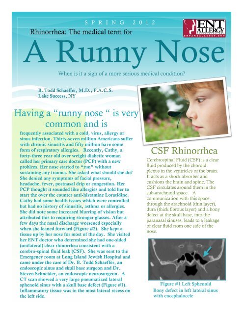
Having a “runny nose “ is very<br />
common and is<br />
frequently associated with a cold, virus, allergy or<br />
sinus infection. Thirty-seven million Americans suffer<br />
with chronic sinusitis and fifty million have some<br />
form of respiratory allergies. Recently, Cathy, a<br />
forty-three year old over weight diabetic woman<br />
called her primary care doctor (PCP) with a new<br />
problem. Her nose started to “run” without<br />
sustaining any trauma. She asked what should she do<br />
She denied any symptoms of facial pressure,<br />
headache, fever, postnasal drip or congestion. Her<br />
PCP thought it sounded like allergies and told her to<br />
start the over the counter anti-histamine Loratidine.<br />
Cathy had some health issues which were controlled<br />
but had no history of sinusitis, asthma or allergies.<br />
She did note some increased blurring of vision but<br />
attributed this to requiring stronger glasses. After a<br />
few days the nasal discharge worsened especially<br />
when she leaned forward (Figure #2). She kept a<br />
tissue up by her nose for most of the day. She visited<br />
her <strong>ENT</strong> doctor who determined she had one-sided<br />
(unilateral) clear <strong>rhinorrhea</strong> consistent with a<br />
cerebro-spinal fluid leak (<strong>CSF</strong>). She was sent to the<br />
Emergency room at Long Island Jewish Hospital and<br />
came under the care of Dr. B. Todd Schaeffer, an<br />
endoscopic sinus and skull base surgeon and Dr.<br />
Steven Schneider, an endoscopic neurosurgeon. A<br />
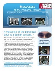
CT scan showed a very large pneumatized lateral<br />
sphenoid sinus with a skull base defect (Figure #1).<br />
Inflammatory tissue was in the most lateral recess on<br />
the left side.<br />
<strong>CSF</strong> Rhinorrhea<br />
Cerebrospinal Fluid (<strong>CSF</strong>) is a clear<br />
fluid produced by the choroid<br />
plexus in the ventricles of the brain.<br />
It acts as a shock absorber and<br />
cushions the brain and spine. The<br />
<strong>CSF</strong> circulates around them in the<br />
sub-arachnoid space. A<br />
communication with this space<br />
through the arachnoid (thin layer),<br />
dura (thick fibrous layer) and a bony<br />
defect at the skull base, into the<br />
paranasal sinuses, leads to a leakage<br />
of clear fluid from one side of the<br />
nose.<br />
Figure #1 Left Sphenoid<br />
Bony defect in left lateral sinus<br />
with encephalocele
THE LOREM IPSUMS SUMMER 2012<br />
SPRING 2012<br />
<strong>CSF</strong> Rhinorrhea<br />
Traumatic vs Spontaneous<br />
Motor Vehicle Accidents<br />
Gun Shot Wounds<br />
Elevated Intracranial Pressure<br />
Pseudotumor Cerebri<br />
Figure #2: Clear <strong>rhinorrhea</strong><br />
Surgery i.e tumor<br />
removal, sinus surgery<br />
<strong>CSF</strong> RHINORRHEA<br />
A lumbar drain was placed and an eye<br />
exam confirmed papilledema consistent<br />
with benign intracranial hypertension most<br />
likely due to pseudotumor cerebri.<br />
Cerebrospinal Fluid (<strong>CSF</strong>) is a clear fluid<br />
produced by the chorid plexus located in<br />
the ventricles of the brain. It acts as a<br />
shock absorber and cushions the brain and<br />
spine. The <strong>CSF</strong> circulates in the<br />
subarachnoid space. A communication<br />
with this space through the arachnoid (thin<br />
layer), dura (thick fibrous layer) and a<br />
bony defect at the skull base, (in the<br />
paranasal sinuses), causes the unilateral<br />
clear fluid “runny nose.”<br />
Cross contamination of nasal contents<br />
with <strong>CSF</strong> is a set-up for meningitis and<br />
intracranial infection and therefore sealing<br />
the leak is paramount.<br />
A CT cisternogram confirmed the sphenoid<br />
sinus as the site of leakage. An endoscopic<br />
transnasal trans-sphenoidal repair with naso<br />
septal flap was performed. This was extremely<br />
difficult since the lateral recess where the leak<br />
was found was lateral and posterior to the<br />
pterygopalatine fossa in the infratemporal<br />
fossa. This was especially challenging since<br />
the repair was lateral and inferior to foramen<br />
rotundum and ovale. The instruments were<br />
just barely able to reach with visualization<br />
supplied with angled scopes transnasally.<br />
Pseudotumor Cerebri is benign intracranial<br />
hypertension found in obese females with<br />
complaints of headache, nausea, vomiting,<br />
tinnitus, double vision that can cause<br />
papilledema and eventually visual loss. <strong>CSF</strong><br />
leak is a complication due to chronically high<br />
intracranial pressure leading to bony defects<br />
and spontaneous leaks.<br />
Figure # 3: Second patient with spontaneous cribriform <strong>CSF</strong> leak.<br />
Skull base defect with meningocele. This was repaired<br />
endoscopically transnasally with local mucosal flap. Repaired with<br />
Dr. Mark Eisenberg (Endoscopic Skull Base Neurosurgeon).<br />
2
THE LOREM IPSUMS SUMMER 2012<br />
SPRING 2012<br />
Diagnostic Testing For <strong>CSF</strong> Leak<br />
<strong>CSF</strong> <strong>rhinorrhea</strong> can easily be<br />
misdiagnosed because it is often left<br />
off the differential diagnosis of<br />
<strong>rhinorrhea</strong>. While a “runny nose” is<br />
commonly thought to be from an<br />
allergy, cold, virus or sinus infection,<br />
a careful history of unilateral crystal<br />
clear watery <strong>rhinorrhea</strong> may help<br />
elucidate the correct diagnosis. If the<br />
diagnosis is in doubt, a nuclear<br />
medicine pledget test can confirm<br />
there is a leak. After a lumbar<br />
puncture, a radioactive tagged<br />
isotope is placed back into the <strong>CSF</strong>.<br />
If a cotton pledget placed in the nose<br />
is positive for the isotope this<br />
confirms an active leak and the side.<br />
A CT Cisternogram helps<br />
confirm the site of a leak. After a<br />
lumbar puncture, contrast material is<br />
injected into the subarachnoid space<br />
where the <strong>CSF</strong> circulates. The<br />
patient is tilted upside down<br />
followed by a CT scan with the head<br />
down leaning forward. The contrast<br />
material seen in the nasal cavity<br />
elucidates the area of leak.<br />
Beta-2 transferrin is found<br />
almost exclusively in <strong>CSF</strong> and not in<br />
blood, mucous or tears. When clear<br />
nasal fluid collected is positive for<br />
this marker, it confirms a <strong>CSF</strong> leak<br />
on that side. The specific site is<br />
determined by CT, CT cisternogram<br />
or endoscopy. Intraoperative<br />
localization using fluorescein<br />
produces greenish/yellow <strong>CSF</strong> fluid.<br />
An off-label use of fluorescein is<br />
used when this is injected<br />
preoperatively into the <strong>CSF</strong> in<br />
diluted amounts.<br />
Figure #4: Third patient with <strong>CSF</strong><br />
leak at Left Supraorbital ethmoid.<br />
This was an encephalocele repaired<br />
through a transnasal endoscopic<br />
approach<br />
Etiology of <strong>CSF</strong> Leaks<br />
Trauma<br />
Tumors<br />
Iatrogenic<br />
Spontaneous/Unknown<br />
Key Points to <strong>CSF</strong> Repair<br />
Transnasal endoscopic repair with<br />
navigation. Lumbar drain as needed<br />
Materials used for multilayer closure<br />
Bone<br />
Fat<br />
Fascia<br />
Duragen/Dural repair<br />
Tisseel TM (glue)<br />
DuraSeal TM ( non-toxic hydrogel)<br />
Nasoseptal Flap<br />
3
SPRING 2012<br />
B. Todd Schaeffer, M.D., F.A.C.S<br />
is an Endoscopic Sinus and Skull Base Surgeon who has<br />
been performing advanced endoscopic sinus surgery for<br />
twenty years. He has performed more endoscopic skull<br />
base surgery than any other sinus surgeon on Long Island.<br />
He commonly works with skull base neurosurgeon Dr.<br />
Mark Eisenberg. As a team, they have<br />
successfully treated pituitary tumor removal,<br />
closure of <strong>CSF</strong> leaks, removal of encephaloceles,<br />
chordomas, clival tumors, meningiomas,<br />
craniopharyngiomas, odontoidectomy, spinal cord<br />
decompression, biopsies at the skull base, removal of<br />
malignant sinus/nasal tumors and skull base<br />
reconstruction. The key to their success is collaboration<br />
together and the support staff of North Shore University<br />
Hospital and Long Island Jewish Medical Center.<br />
Experience and team collaboration counts. Visit<br />
NOSEMD at You Tube or Google NOSEMD.<br />
www.PituitaryMD.com www.SchaefferMD.com<br />
LIJ<br />
<strong>ENT</strong> and <strong>Allergy</strong> <strong>Associates</strong>, LLP<br />
3003 New Hyde Park Road<br />
Lake Success, NY 11042<br />
(516) 775-2800<br />
[Recipient]