

jacobson glut tendon 8-23-12 syllabus
jacobson glut tendon 8-23-12 syllabus
jacobson glut tendon 8-23-12 syllabus
- No tags were found...

You also want an ePaper? Increase the reach of your titles
YUMPU automatically turns print PDFs into web optimized ePapers that Google loves.
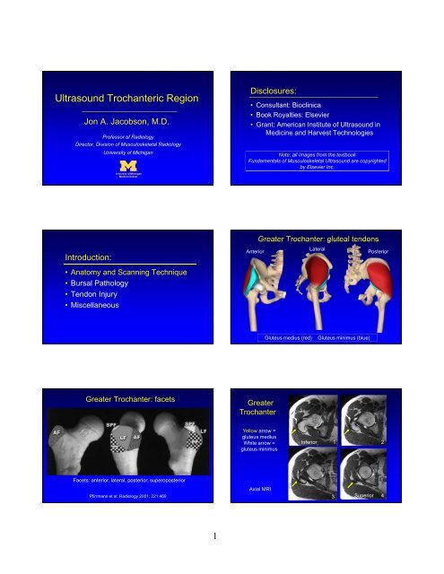
Ultrasound Trochanteric Region<br />
Jon A. Jacobson, M.D.<br />
Professor of Radiology<br />
Director, Division of Musculoskeletal Radiology<br />
University of Michigan<br />
Disclosures:<br />
• Consultant: Bioclinica<br />
• Book Royalties: Elsevier<br />
• Grant: American Institute of Ultrasound in<br />
Medicine and Harvest Technologies<br />
Note: all images from the textbook<br />
Fundamentals of Musculoskeletal Ultrasound are copyrighted<br />
by Elsevier Inc.<br />
Introduction:<br />
• Anatomy and Scanning Technique<br />
• Bursal Pathology<br />
• Tendon Injury<br />
• Miscellaneous<br />
Greater Trochanter: <strong>glut</strong>eal <strong>tendon</strong>s<br />
Anterior<br />
Lateral<br />
Posterior<br />
Gluteus medius (red)<br />
Gluteus minimus (blue)<br />
Greater Trochanter: facets<br />
Greater<br />
Trochanter<br />
Yellow arrow =<br />
<strong>glut</strong>eus medius<br />
White arrow =<br />
<strong>glut</strong>eus minimus<br />
Inferior<br />
1 2<br />
Facets: anterior, lateral, posterior, superoposterior<br />
Pfirrmann et al. Radiology 2001; 221:469<br />
Axial MRI<br />
3 Superior 4<br />
1
Greater Trochanter<br />
Greater Trochanter<br />
Anterior<br />
Posterior<br />
Yellow arrow = <strong>glut</strong>eus medius<br />
White arrow = <strong>glut</strong>eus minimus<br />
Pfirrmann et al. Radiology 2001; 221:469<br />
Gluteus Minimus: Long Axis<br />
Gluteus Medius: Long Axis<br />
Gmed<br />
Gmed<br />
AF<br />
Iliotibial<br />
Tract<br />
LF<br />
Anterior<br />
Facet<br />
Lateral<br />
Facet<br />
Sonographic Technique: Hip<br />
• Lateral Femoral<br />
Cutaneous Nerve<br />
Sartorius<br />
Lateral Femoral Cutaneous Nerve<br />
Rectus<br />
Femoris<br />
Inguinal Ligament<br />
Short Axis<br />
Long Axis<br />
2
Lateral Femoral Cutaneous Nerve<br />
Lateral<br />
Sartorius<br />
Medial<br />
Introduction:<br />
• Anatomy and Scanning Technique<br />
• Bursal Pathology<br />
• Tendon Injury<br />
• Miscellaneous<br />
Transverse<br />
Trochanteric Pain Syndrome:<br />
• Most commonly caused by <strong>glut</strong>eus<br />
minimus and medius <strong>tendon</strong><br />
abnormalities 1<br />
• Trochanteric bursitis: rare<br />
– Not actually inflamed 2<br />
Trochanteric Bursal Fluid:<br />
• Bursal fluid not normally seen<br />
• Fluid distention:<br />
– simple fluid: anechoic<br />
– complicated fluid: mixed echogenicity<br />
– Not associated with pain 3 1 Eur Rad 2007; 17:1772.<br />
2 J Clin Rheumatol 2008; 14:82<br />
3 Skeletal Radiol 2008; 37:903<br />
Trochanteric Bursitis<br />
Trochanteric Bursitis<br />
PF<br />
PF<br />
Transverse<br />
Coronal<br />
Transverse<br />
Coronal<br />
3
Trochanteric Bursitis<br />
Trochanteric Bursitis<br />
Transverse<br />
Arthrogram<br />
Metal-on-Metal Arthroplasty: pseudotumor<br />
Introduction:<br />
Cup<br />
Neck<br />
Cup<br />
Troch<br />
• Anatomy and Scanning Technique<br />
• Bursal Pathology<br />
• Tendon Injury<br />
• Miscellaneous<br />
Anterior<br />
Lateral<br />
Tendinosis: Gluteus Medius<br />
Gluteal Tendon Pathology:<br />
• Tendinosis: hypoechoic, no defects<br />
• Partial tear: anechoic clefts<br />
• Complete tear: discontinuous <strong>tendon</strong><br />
• >2 mm cortical irregularity is associated with<br />
<strong>tendon</strong> tear<br />
– Positive predictive value = 90% (xray)*<br />
AF LF SPF LF<br />
*Steinert et al. Radiology 2010; 257:754<br />
4
Tendinosis: Gluteus Minimus<br />
Tear: Gluteus Medius<br />
AF<br />
LF<br />
AF<br />
AF LF LF<br />
>2 mm cortical irregularity (x-ray) =<br />
90% positive predictive value for<br />
<strong>glut</strong>eus <strong>tendon</strong> tear<br />
Steinert et al. Radiology 2010; 257:754<br />
Post-operative: Gluteus Medius<br />
Calcific Tendinosis: Gluteus Medius<br />
AF LF SPF LF<br />
AF<br />
LF<br />
LF<br />
Long Axis<br />
Short Axis<br />
Potential Treatment Algorithm:<br />
• If bursa: aspirate, inject steroids<br />
• If tendinosis:<br />
– Tenotomy or fenestration<br />
– Inject steroids superficial to <strong>tendon</strong><br />
• 72% of patients significantly improved 1<br />
• If <strong>tendon</strong> tear: platelet-rich plasma injection<br />
Trochanteric<br />
Region Bursae<br />
• Trochanteric: deep to<br />
<strong>glut</strong>eus maximus<br />
• Sub<strong>glut</strong>eus medius<br />
• Sub<strong>glut</strong>eus minimus<br />
• Axial plane<br />
PF<br />
LF<br />
1<br />
Labrosse, et al. 2010 AJR 2010; 194:202<br />
5
Gluteus Medius Tenotomy<br />
Introduction:<br />
Greater<br />
Trochanter<br />
• Anatomy and Scanning Technique<br />
• Bursal Pathology<br />
• Tendon Injury<br />
• Miscellaneous<br />
Snapping Hip Syndrome<br />
• Painful snap with hip motion<br />
• Intraarticular<br />
• Extraarticular:<br />
– Medial: iliopsoas <strong>tendon</strong><br />
– Lateral: iliotibial tract or <strong>glut</strong>eus maximus<br />
Snapping Hip: iliotibial tract<br />
• Transverse over<br />
greater trochanter<br />
• Hip external rotation /<br />
flexion<br />
• Abrupt motion of<br />
iliotibial tract over<br />
greater trochanter<br />
Snapping Hip Syndrome: iliotibial tract<br />
Morel-Lavallee Lesion:<br />
• Thigh and hip region<br />
• Fluid collection:<br />
– Between subcutaneous fat and fascia<br />
– Closed de-gloving injury<br />
• Trauma<br />
Mellado, AJR 2004; 182:<strong>12</strong>89<br />
6
Morel-Lavallee Lesion<br />
Take-home points:<br />
Muscle<br />
Muscle<br />
Sub-Q Fat<br />
Muscle<br />
• Trochanteric anatomy<br />
• Bursitis: rare<br />
• Gluteal <strong>tendon</strong>s abnormalities: frequent<br />
• Snapping hip: dynamic<br />
Coronal Transverse Normal<br />
7