A Novel Modified Semi-direct Method for Total Leukocyte Count in ...
A Novel Modified Semi-direct Method for Total Leukocyte Count in ...
A Novel Modified Semi-direct Method for Total Leukocyte Count in ...

You also want an ePaper? Increase the reach of your titles
YUMPU automatically turns print PDFs into web optimized ePapers that Google loves.
Research Articles<br />
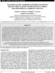
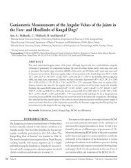
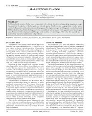
Figure 4: Photographs of a blood smear of an African Grey parrot, sta<strong>in</strong>ed with the tested eos<strong>in</strong>-based quick sta<strong>in</strong> ( J-322A-2, Jorgensen<br />
Laboratories Inc. Loveland, Colorado, USA) which is <strong>in</strong>cluded <strong>in</strong> quick sta<strong>in</strong><strong>in</strong>g kit (A). The same smear was later counter-sta<strong>in</strong>ed with the<br />
methylene blue-based sta<strong>in</strong> ( J-322A-3, Jorgensen Laboratories Inc. Loveland, Colorado, USA) also <strong>in</strong>cluded <strong>in</strong> the kit. B: the area of the smear<br />
as shown <strong>in</strong> A, after the counter sta<strong>in</strong><strong>in</strong>g. The areas marked by the red and green boxes (marked 1 and 2) correspond to the areas marked by<br />
the red and green boxes <strong>in</strong> B (marked 3 and 4, respectively). These areas are magnified <strong>in</strong> the correspond<strong>in</strong>g photos A1, A2, B3 and B4. A1:<br />
the shadow of a lymphocyte (red arrow) and two heterophils (black arrows) are seen. The latter are recognized by absorption of the eos<strong>in</strong>-based<br />
dye by their granules; B3: the same lymphocyte (red arrow) and the two heterophils (black arrows) <strong>in</strong> A1 after counter sta<strong>in</strong><strong>in</strong>g (arrow); A2:<br />
the shadows of clumped thrombocytes (arrows); B4: the same thrombocytes <strong>in</strong> A2 after counter sta<strong>in</strong><strong>in</strong>g (arrows).<br />
ulocytes, lymphocytes and thrombocytes that were sta<strong>in</strong>ed<br />
with the tested sta<strong>in</strong> was confirmed after counter-sta<strong>in</strong><strong>in</strong>g<br />
the smear with the methylene-blue-based component of the<br />
quick-sta<strong>in</strong> and re-exam<strong>in</strong><strong>in</strong>g the marked microscopic fields<br />
(Figure 2-B3, B4; Figure 3-B4, B5, B6 and Figure 4-B3, B4.<br />
The optimally diluted sta<strong>in</strong><strong>in</strong>g solution was later tested<br />
<strong>in</strong> several samples of different non-mammalian species, with<br />
similar sta<strong>in</strong><strong>in</strong>g quality (data not shown).<br />
Precision of the MSDTLC<br />
The MSDTLC was repeated twice by two different persons<br />
<strong>for</strong> each of the 13 samples obta<strong>in</strong>ed from psittac<strong>in</strong>e birds<br />
(Table 1). There were no significant differences between<br />
the two MSDTLC’s (P=0.319). The absolute agreement<br />
between the two MSDTLC’s was very good (ICC 0.93,<br />
P