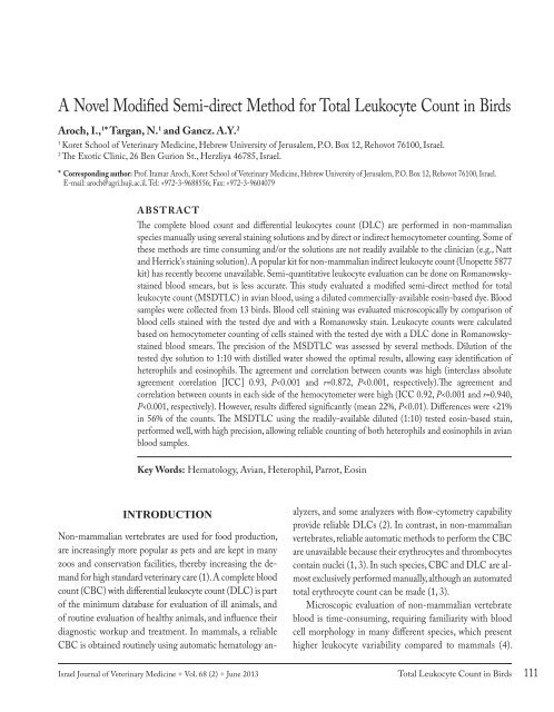
A Novel Modified Semi-direct Method for Total Leukocyte Count in ...
A Novel Modified Semi-direct Method for Total Leukocyte Count in ...
A Novel Modified Semi-direct Method for Total Leukocyte Count in ...

You also want an ePaper? Increase the reach of your titles
YUMPU automatically turns print PDFs into web optimized ePapers that Google loves.
A <strong>Novel</strong> <strong>Modified</strong> <strong>Semi</strong>-<strong>direct</strong> <strong>Method</strong> <strong>for</strong> <strong>Total</strong> <strong>Leukocyte</strong> <strong>Count</strong> <strong>in</strong> Birds<br />
Aroch, I., 1 * Targan, N. 1 and Gancz. A.Y. 2<br />
1<br />
Koret School of Veter<strong>in</strong>ary Medic<strong>in</strong>e, Hebrew University of Jerusalem, P.O. Box 12, Rehovot 76100, Israel.<br />
2<br />
The Exotic Cl<strong>in</strong>ic, 26 Ben Gurion St., Herzliya 46785, Israel.<br />
* Correspond<strong>in</strong>g author: Prof. Itamar Aroch, Koret School of Veter<strong>in</strong>ary Medic<strong>in</strong>e, Hebrew University of Jerusalem, P.O. Box 12, Rehovot 76100, Israel.<br />
E-mail: aroch@agri.huji.ac.il. Tel: +972-3-9688556; Fax: +972-3-9604079<br />
ABSTRACT<br />
The complete blood count and differential leukocytes count (DLC) are per<strong>for</strong>med <strong>in</strong> non-mammalian<br />
species manually us<strong>in</strong>g several sta<strong>in</strong><strong>in</strong>g solutions and by <strong>direct</strong> or <strong>in</strong><strong>direct</strong> hemocytometer count<strong>in</strong>g. Some of<br />
these methods are time consum<strong>in</strong>g and/or the solutions are not readily available to the cl<strong>in</strong>ician (e.g., Natt<br />
and Herrick's sta<strong>in</strong><strong>in</strong>g solution). A popular kit <strong>for</strong> non-mammalian <strong>in</strong><strong>direct</strong> leukocyte count (Unopette 5877<br />
kit) has recently become unavailable. <strong>Semi</strong>-quantitative leukocyte evaluation can be done on Romanowskysta<strong>in</strong>ed<br />
blood smears, but is less accurate. This study evaluated a modified semi-<strong>direct</strong> method <strong>for</strong> total<br />
leukocyte count (MSDTLC) <strong>in</strong> avian blood, us<strong>in</strong>g a diluted commercially-available eos<strong>in</strong>-based dye. Blood<br />
samples were collected from 13 birds. Blood cell sta<strong>in</strong><strong>in</strong>g was evaluated microscopically by comparison of<br />
blood cells sta<strong>in</strong>ed with the tested dye and with a Romanowsky sta<strong>in</strong>. <strong>Leukocyte</strong> counts were calculated<br />
based on hemocytometer count<strong>in</strong>g of cells sta<strong>in</strong>ed with the tested dye with a DLC done <strong>in</strong> Romanowskysta<strong>in</strong>ed<br />
blood smears. The precision of the MSDTLC was assessed by several methods. Dilution of the<br />
tested dye solution to 1:10 with distilled water showed the optimal results, allow<strong>in</strong>g easy identification of<br />
heterophils and eos<strong>in</strong>ophils. The agreement and correlation between counts was high (<strong>in</strong>terclass absolute<br />
agreement correlation [ICC] 0.93, P
Research Articles<br />
There<strong>for</strong>e, many veter<strong>in</strong>arians refer blood samples to reference<br />
laboratories. However, leukocytes may distort if analysis<br />
is delayed (5, 6). In such cases, prolonged exposure to ethylenediam<strong>in</strong>e<br />
tetraacetic acid (EDTA) anticoagulant may<br />
cause erythrolysis, viscosity changes and leukocyte morphology<br />
alterations <strong>in</strong> blood samples of crowned cranes, crows,<br />
jays, brush turkeys, hornbills, magpies and some ratites (5,<br />
7).In-cl<strong>in</strong>ic blood smear evaluation has several advantages: it<br />
is cost-efficient, samples are fresh, avoid<strong>in</strong>g time-dependent<br />
deterioration and morphologic artifacts (5) and results are<br />
obta<strong>in</strong>ed and <strong>in</strong>terpreted immediately, thereby promot<strong>in</strong>g<br />
early diagnosis and treatment.<br />
The DLC <strong>in</strong> non-mammalian species is per<strong>for</strong>med by<br />
count<strong>in</strong>g 100-200 leukocytes <strong>in</strong> sta<strong>in</strong>ed blood smears, and<br />
has been studied mostly <strong>in</strong> the domestic chicken (Gallus gallus)<br />
(1). In non-mammalian species, differentiation between<br />
eos<strong>in</strong>ophils and heterophils, small lymphocytes and thrombocytes,<br />
and large lymphocytes and monocytes is often difficult<br />
(4). Eos<strong>in</strong>ophil granule color varies between species<br />
of parrots and iguanas, from red to light blue, purple or grey<br />
and depends on the sta<strong>in</strong><strong>in</strong>g type and technique (1, 8, 9, 10).<br />
The total leukocyte count (TLC) <strong>in</strong> non-mammalian<br />
vertebrates can be counted us<strong>in</strong>g three methods; 1) <strong>direct</strong><br />
hemocytometer count<strong>in</strong>g with Natt and Herrick's or toluid<strong>in</strong>e<br />
blue sta<strong>in</strong> solutions (1, 7, 11, 12); 2) semi-<strong>direct</strong> hemocytometer<br />
count with phlox<strong>in</strong>e-B dye or the (now discont<strong>in</strong>ued)<br />
commercial eos<strong>in</strong>ophil Unopette 5877 (Becton<br />
- Dick<strong>in</strong>son, Ruther<strong>for</strong>d, NJ) (1, 3, 8, 11-14), comb<strong>in</strong>ed with<br />
manual DLC <strong>in</strong> Romanowsky-sta<strong>in</strong>ed blood smears (4, 516,<br />
17); 3) <strong>Semi</strong>-quantitative TLC evaluation <strong>in</strong> Romanowskysta<strong>in</strong>ed<br />
blood smears, which is considered less accurate (1,<br />
3). The latter can be done by averag<strong>in</strong>g leukocyte number<br />
<strong>in</strong> 10 monolayer, X400 fields and multiply<strong>in</strong>g it by 2000,<br />
yield<strong>in</strong>g the TLC <strong>in</strong> cell/µL. Alternatively, one can determ<strong>in</strong>e<br />
the leukocyte average number <strong>in</strong> five X1000 monolayer<br />
fields, multiply it by 3,500,000 (the approximate number of<br />
erythrocytes <strong>in</strong> 1 µL of blood <strong>in</strong> birds when the packed cell<br />
volume (PCV) is 35-55%), and divid<strong>in</strong>g the result by 1000<br />
(the average number of erythrocytes <strong>in</strong> 5 X1000 monolayer<br />
fields). The result should be corrected if PCV is abnormally<br />
high or low (1, 3, 4).<br />
Direct leukocyte count is a complex, time-consum<strong>in</strong>g<br />
procedure, <strong>in</strong>volv<strong>in</strong>g preparation of the sta<strong>in</strong> solution, and is<br />
complicated by the need to differentiate lymphocytes from<br />
thrombocytes, and with presence of sta<strong>in</strong>ed erythrocytes<br />
<strong>in</strong> the hemocytometer (1, 4, 5). The semi-<strong>direct</strong> count<strong>in</strong>g<br />
method relies on the positive sta<strong>in</strong><strong>in</strong>g of heterophils and eos<strong>in</strong>ophils<br />
by the dye, allow<strong>in</strong>g their count<strong>in</strong>g <strong>in</strong> the hemocytometer.<br />
Calculat<strong>in</strong>g their total number per microliter, and<br />
then the TLC is achieved by per<strong>for</strong>m<strong>in</strong>g a DLC us<strong>in</strong>g a<br />
Romanowsky-sta<strong>in</strong>ed blood smear. This method is more precise<br />
and less time consum<strong>in</strong>g than the <strong>direct</strong> hemocytometer<br />
count (1, 3, 4, 8); however, it becomes progressively less accurate<br />
<strong>for</strong> estimation of the TLC with <strong>in</strong>creas<strong>in</strong>g proportion<br />
of mononuclears to granulocytes (1). Currently, the Unopette<br />
5877 kit has been discont<strong>in</strong>ued (Ady Gancz, personal communication),<br />
while the other sta<strong>in</strong><strong>in</strong>g solutions mentioned<br />
are not readily available <strong>in</strong> most veter<strong>in</strong>ary cl<strong>in</strong>ics. In contrast,<br />
Romanowsky-based quick sta<strong>in</strong><strong>in</strong>g solutions are readily<br />
available and are used <strong>in</strong> many veter<strong>in</strong>ary cl<strong>in</strong>ics <strong>for</strong> blood<br />
smear sta<strong>in</strong><strong>in</strong>g. Based on the sta<strong>in</strong><strong>in</strong>g aff<strong>in</strong>ity of heterophils<br />
and eos<strong>in</strong>ophils, eos<strong>in</strong>-based sta<strong>in</strong><strong>in</strong>g solutions are expected<br />
to sta<strong>in</strong> these cells similarly as phlox<strong>in</strong>e-B.<br />
The aim of the present study was to evaluate a modified<br />
semi-<strong>direct</strong> method <strong>for</strong> TLC (MSDTLC) <strong>in</strong> non-mammalian<br />
blood, us<strong>in</strong>g a diluted eos<strong>in</strong>-based dye, <strong>in</strong>cluded <strong>in</strong><br />
a Romanowsky-based quick sta<strong>in</strong> ( J-322A-2, Jorgensen<br />
Laboratories Inc. Loveland, Colorado, USA), which is readily<br />
available, <strong>in</strong>stead of phlox<strong>in</strong>e-B as the sta<strong>in</strong><strong>in</strong>g diluent,<br />
and to assess its accuracy and variability.<br />
MATERIALS AND METHODS<br />
Selected animals and blood samples collection<br />
Blood samples were collected from the jugular or brachial<br />
ve<strong>in</strong>s <strong>in</strong> lithium-hepar<strong>in</strong> pediatric tubes (M<strong>in</strong>iCollect,<br />
Gre<strong>in</strong>er, Gre<strong>in</strong>er Bio-One North America, Inc., Monroe,<br />
NC), from 13 psittac<strong>in</strong>e birds (Table 1) presented to the<br />
Hebrew University Veter<strong>in</strong>ary Teach<strong>in</strong>g Hospital, Beit<br />
Dagan, and to The Exotic Cl<strong>in</strong>ic, Herzeliya. Blood smears<br />
were prepared immediately, and later sta<strong>in</strong>ed us<strong>in</strong>g a<br />
Romanowsky-based sta<strong>in</strong> (modified Wright's solution).<br />
Assessment of the ideal sta<strong>in</strong><strong>in</strong>g solution dilution and<br />
blood cell sta<strong>in</strong><strong>in</strong>g and hemocytometer count<strong>in</strong>g<br />
Four different dilutions of the dye ( J-322A-2) were tested,<br />
<strong>in</strong> distilled water, and <strong>in</strong> sal<strong>in</strong>e (1:5, 1:10, 1:25 and 1:50) to<br />
sta<strong>in</strong> a s<strong>in</strong>gle fresh blood sample obta<strong>in</strong>ed <strong>in</strong> lithium-hepar<strong>in</strong><br />
from a parrot which was presented to the hospital at the time<br />
the study was <strong>in</strong>itiated. The blood was exam<strong>in</strong>ed with<strong>in</strong> five<br />
112<br />
Aroch, I.<br />
Israel Journal of Veter<strong>in</strong>ary Medic<strong>in</strong>e • Vol. 68 (2) • June 2013
Research Articles<br />
Table 1: Results of the modified semi-<strong>direct</strong> total leukocyte count, estimated leukocyte count based on Romanowsky-sta<strong>in</strong>ed blood smears,<br />
differential leukocyte count* and packed cell volume <strong>in</strong> 13 different birds<br />
Bird<br />
number<br />
Bird species<br />
MSDTLC<br />
count #1<br />
(x10 3 /µL)<br />
MSDTLC<br />
count #2<br />
(x10 3 /µL)<br />
Sta<strong>in</strong>ed blood<br />
smear evaluation<br />
of leukocytes<br />
(x10 3 /µL)<br />
H+E 1<br />
(%)<br />
L 2<br />
(%)<br />
M 3<br />
(%)<br />
B 4<br />
(%)<br />
Packed cell<br />
volume<br />
(%)<br />
1<br />
Cockatoo (Cacatua galerita),<br />
26.59<br />
36.36<br />
40.8<br />
66<br />
12<br />
22<br />
0<br />
30<br />
2<br />
Cockatoo (Cacatua galerita)<br />
9.12<br />
9.41<br />
18.6<br />
68<br />
17<br />
15<br />
0<br />
42<br />
3<br />
Rose-r<strong>in</strong>ged Parakeet (Psittacula krameri)<br />
12.53<br />
11.65<br />
13.8<br />
79<br />
12<br />
7<br />
2<br />
49<br />
4<br />
Cockatiel (Nymphicus holandicus)<br />
12.13<br />
13.13<br />
15<br />
80<br />
11<br />
7<br />
2<br />
40<br />
5<br />
Cockatiel (Nymphicus holandicus)<br />
38.83<br />
26.33<br />
63<br />
60<br />
28<br />
12<br />
0<br />
54<br />
6<br />
African gray parrot (Psittacus erithacus)<br />
26.90<br />
20.83<br />
18.8<br />
84<br />
9<br />
6<br />
1<br />
50<br />
7<br />
Cockatiel (Nymphicus holandicus)<br />
6.92<br />
7.54<br />
7.4<br />
65<br />
25<br />
10<br />
0<br />
35<br />
8<br />
Eclectus parrot (Eclectus roratus)<br />
8.46<br />
5.00<br />
12.2<br />
52<br />
33<br />
6<br />
9<br />
45<br />
9<br />
Budgerigar, (Melapsittacus undulates)<br />
7.14<br />
2.86<br />
6.4<br />
21<br />
65<br />
13<br />
1<br />
65<br />
10<br />
Budgerigar (Melapsittacus undulates)<br />
15.00<br />
6.11<br />
10.36<br />
18<br />
52<br />
21<br />
9<br />
60<br />
11<br />
African gray parrot (Psittacus erithacus)<br />
12.69<br />
11.73<br />
24.6<br />
52<br />
35<br />
7<br />
6<br />
40<br />
12<br />
Cockatiel (Nymphicus holandicus)<br />
4.12<br />
3.24<br />
7.8<br />
34<br />
52<br />
9<br />
5<br />
54<br />
13<br />
Sun conure (Arat<strong>in</strong>ga solastitalis)<br />
10.57<br />
9.43<br />
19.6<br />
35<br />
49<br />
5<br />
11<br />
43<br />
* Presented as means of the two counts; 1) Heterophils and Eos<strong>in</strong>ophils; 2) Lymphocytes; 3) Monocytes; 4) Basophils<br />
m<strong>in</strong>utes from application of the sta<strong>in</strong><strong>in</strong>g solution. The assessment<br />
of the sta<strong>in</strong><strong>in</strong>g properties was subjective, and was done<br />
us<strong>in</strong>g the hemocytometer at X100 and X200 microscopic<br />
magnification. The follow<strong>in</strong>g criteria were used: 1) quality<br />
of the contrast between the granulocytes and the background;<br />
2) the <strong>in</strong>tensity of granulocytes sta<strong>in</strong><strong>in</strong>g; 3) sta<strong>in</strong><strong>in</strong>g<br />
<strong>in</strong>tensity of other blood cells (i.e., other leukocytes, thrombocytes<br />
and erythrocytes). This sta<strong>in</strong><strong>in</strong>g was deemed undesirable<br />
<strong>for</strong> our purposes. In order to assess the granulocytes<br />
sta<strong>in</strong><strong>in</strong>g with the tested dye, a blood smear of a parrot was<br />
sta<strong>in</strong>ed <strong>in</strong>itially only with the tested sta<strong>in</strong>, and three fields<br />
were microscopically screened and photographed. The same<br />
smear was later sta<strong>in</strong>ed with the methylene-blue-based sta<strong>in</strong><br />
of the Romanowsky-based reagent kit ( J-322-3, Jorgensen<br />
Laboratories Inc. Loveland, Colorado, USA), and the same<br />
fields were rescreened and photographed <strong>for</strong> comparison.<br />
The improved Neubauer Bright-L<strong>in</strong>e hemocytometer<br />
(Reichert, Buffalo, NY) was used <strong>in</strong> this study. Four different<br />
dilutions of the sta<strong>in</strong> ( J-322A-2) were used (see above).<br />
For each test, 10µL of blood were mixed with 990 µL of<br />
the diluted sta<strong>in</strong> and loaded onto the hemocytometer chamber<br />
to its full 1 µL capacity. The heterophils and eos<strong>in</strong>ophils<br />
were counted with<strong>in</strong> five m<strong>in</strong>utes, <strong>in</strong> five large squares (the<br />
central one and the four <strong>in</strong> the corners) <strong>in</strong> both sides of the<br />
hemocytometer's grid area. Thus, the total volume of diluted<br />
(1:100) blood used <strong>for</strong> this count constitutes 1 µL (10 squares<br />
of 0.1 µL). The <strong>for</strong>mula used to calculate the total number<br />
of heterophils and eos<strong>in</strong>ophils (THEC) was as follows:<br />
THEC [cells/µL] =100 [i.e., dilution factor] X(heterophils<br />
+ eos<strong>in</strong>ophils counted <strong>in</strong> 10 squares of the hemocytometer)<br />
(18). The next step was per<strong>for</strong>med us<strong>in</strong>g a manual DLC <strong>in</strong><br />
a Romanowsky-sta<strong>in</strong>ed blood smear by differentially count<strong>in</strong>g<br />
100 leukocytes. The TLC was then calculated as follows:<br />
TLC [cells/µL] = (THEC [cells/µL] X100 [i.e., dilution<br />
factor]) / (% heterophils + % eos<strong>in</strong>ophils) (1).<br />
Determ<strong>in</strong>ation of precision of the MSDTLC<br />
In order to assess the effect of the person per<strong>for</strong>m<strong>in</strong>g the<br />
MSDTLC, 13 samples of different avian species were counted<br />
twice, each time by a different person (NT and AG) and<br />
the agreement and correlation between counts were determ<strong>in</strong>ed.<br />
Additionally, <strong>for</strong> each of the above 13 avian samples,<br />
Israel Journal of Veter<strong>in</strong>ary Medic<strong>in</strong>e • Vol. 68 (2) • June 2013 <strong>Total</strong> <strong>Leukocyte</strong> <strong>Count</strong> <strong>in</strong> Birds 113
Research Articles<br />
the TLC was also determ<strong>in</strong>ed based on the number of heterophils<br />
and eos<strong>in</strong>ophils counted <strong>in</strong> five squares <strong>in</strong> one chamber<br />
of the ruled area of the hemocytometer, as well as based<br />
on the number of these cells <strong>in</strong> the correspond<strong>in</strong>g five squares<br />
<strong>in</strong> its other chamber. This procedure was repeated 15 times<br />
<strong>for</strong> one of the samples, and two to three times <strong>for</strong> the other<br />
12 samples (a total of 45counts). The follow<strong>in</strong>g <strong>for</strong>mula was<br />
used to calculate the TLC: TLC [cells/µL] = (heterophils +<br />
eos<strong>in</strong>ophils counted <strong>in</strong> five hemocytometer squares) X100<br />
[i.e., dilution factor]) X 2 X 100 / (% heterophils + % eos<strong>in</strong>ophils<br />
obta<strong>in</strong>ed by DLC <strong>in</strong> Romanowsky-sta<strong>in</strong>ed blood<br />
smears of the bird). The correlation and agreement between<br />
the TLC’s calculated, based on each chamber of the hemocytometer<br />
were determ<strong>in</strong>ed. F<strong>in</strong>ally, a s<strong>in</strong>gle sample of avian<br />
blood was counted 15 times by a s<strong>in</strong>gle person (NT) us<strong>in</strong>g<br />
the MSDTLC and the agreement between counts was<br />
determ<strong>in</strong>ed.<br />
Statistical methods<br />
Descriptive statistics <strong>for</strong> each cont<strong>in</strong>uous variable were per<strong>for</strong>med.<br />
The distribution of data (normal vs. non-normal)<br />
was assessed us<strong>in</strong>g Kolmogorov-Smirnov test. Student's<br />
t- and Wilcoxon-rank-sum tests were used to determ<strong>in</strong>e<br />
whether the mean difference of two test results was different<br />
from zero. The one-sample Student's t-test was used to<br />
determ<strong>in</strong>e if the absolute difference between the mean of<br />
two repeated counts was different from zero. Pearson’s correlations<br />
were calculated between pairs of the test results.<br />
An <strong>in</strong>terclass absolute agreement correlation (ICC) between<br />
tests was also calculated. An ICC of 0.7 was considered as<br />
good agreement, while higher values were considered as very<br />
good. All tests were two-tailed, and a P ≤0.05 was considered<br />
significant. Statistical analyses were per<strong>for</strong>med us<strong>in</strong>g a<br />
statistical software package (SPSS 15.0 <strong>for</strong> W<strong>in</strong>dows, SPSS<br />
Inc. Chicago, IL).<br />
Assessment of the ideal sta<strong>in</strong><strong>in</strong>g solution dilution and<br />
blood cell sta<strong>in</strong><strong>in</strong>g<br />
Dilution of the sta<strong>in</strong> solution with sal<strong>in</strong>e led to <strong>in</strong>tense erythrocyte<br />
sta<strong>in</strong><strong>in</strong>g compared to the granulocytes, mak<strong>in</strong>g differentiation<br />
between them difficult us<strong>in</strong>g X200 magnification,<br />
and there<strong>for</strong>e necessitat<strong>in</strong>g the use of higher magnifications,<br />
and thereby protract<strong>in</strong>g the count. Based on the criteria mentioned<br />
above, the optimal sta<strong>in</strong> dilution was 1:10. This diluted<br />
sta<strong>in</strong> solution was mixed with the birds' blood sample (990<br />
and 10 µL, respectively, thus yield<strong>in</strong>g a f<strong>in</strong>al blood dilution of<br />
1:100). At that dilution, the heterophils and eos<strong>in</strong>ophils were<br />
readily recognizable, with good contrast and background,<br />
while sta<strong>in</strong><strong>in</strong>g of the erythrocytes was weak, allow<strong>in</strong>g easy<br />
differentiation of the granulocytes from erythrocytes (Figure<br />
1). A good quality granulocyte sta<strong>in</strong><strong>in</strong>g was also observed<br />
us<strong>in</strong>g a 1:5 dilution, however, erythrocytes sta<strong>in</strong>ed more <strong>in</strong>tensely,<br />
and this slowed the granulocyte count. Dilutions of<br />
1:25 and 1:50 were judged to provide poor, unacceptable results,<br />
due to low contrast and difficulty to accurately identify<br />
the granulocytes.<br />
Us<strong>in</strong>g the 1:10 dilution, the cytoplasm of erythrocytes,<br />
heterophils and eos<strong>in</strong>ophils sta<strong>in</strong>ed clearly, allow<strong>in</strong>g their<br />
identification (Figure 2-A2; Figure 3-A1, A2). Lymphocytes,<br />
monocytes and thrombocytes did not sta<strong>in</strong>, and only their<br />
shadows were apparent, not allow<strong>in</strong>g clear def<strong>in</strong>ite identification<br />
of the cell type (Figure 2-A1, Figure 3-A3; Figure<br />
4-A1, A2). Several selected microscopic fields were marked<br />
us<strong>in</strong>g the coord<strong>in</strong>ates of the microscope tray and photographed<br />
at different magnifications. The identity of the gran-<br />
RESULTS<br />
Figure 1: Photograph of the hemocytometer field at X200<br />
magnification with a blood sample from bird, diluted with the<br />
sta<strong>in</strong><strong>in</strong>g solution at the chosen, best dilution (1:10). The arrows po<strong>in</strong>t<br />
to heterophils or eos<strong>in</strong>ophils, which are easily recognizable, because<br />
their granules absorb the eos<strong>in</strong>-based dye. The black circles mark area<br />
<strong>in</strong> which erythrocytes can be seen.<br />
114<br />
Aroch, I.<br />
Israel Journal of Veter<strong>in</strong>ary Medic<strong>in</strong>e • Vol. 68 (2) • June 2013
Research Articles<br />
Figure 2: Photographs of a blood smear of an African Grey parrot, sta<strong>in</strong>ed with the tested eos<strong>in</strong>-based quick sta<strong>in</strong> ( J-322A-2, Jorgensen<br />
Laboratories Inc. Loveland, Colorado, USA), which is <strong>in</strong>cluded <strong>in</strong> quick sta<strong>in</strong><strong>in</strong>g kit (A). The same smear was later counter-sta<strong>in</strong>ed with the<br />
methylene blue-based sta<strong>in</strong> ( J-322A-3, Jorgensen Laboratories Inc. Loveland, Colorado, USA) also <strong>in</strong>cluded <strong>in</strong> the kit. B: the area of the<br />
smear as shown <strong>in</strong> A, after the counter sta<strong>in</strong><strong>in</strong>g. The areas marked by the green and red boxes (A-1 and A-2, respectively) correspond to the<br />
areas marked <strong>in</strong> the green and red boxes <strong>in</strong> B (B3 and B4, respectively). These areas are magnified <strong>in</strong> the correspond<strong>in</strong>g photos A1, A2, B3 and<br />
B4. A1: the shadow of a lymphocyte is seen (arrow); B3: the same lymphocyte as <strong>in</strong> A1 after counter sta<strong>in</strong><strong>in</strong>g (arrow); A2: two heterophils<br />
(arrows) can be seen, and can be recognized by absorption of the eos<strong>in</strong>-based dye by their granules; B4: the same heterophils as <strong>in</strong> A2 (arrows)<br />
after counter sta<strong>in</strong><strong>in</strong>g.<br />
Figure 3: Photographs of a blood smear of an African<br />
Grey parrot, sta<strong>in</strong>ed with the tested eos<strong>in</strong>-based quick<br />
sta<strong>in</strong> ( J-322A-2, Jorgensen Laboratories Inc. Loveland,<br />
Colorado, USA) which is <strong>in</strong>cluded <strong>in</strong> quick sta<strong>in</strong><strong>in</strong>g<br />
kit (A). The same smear was later counter-sta<strong>in</strong>ed with<br />
the methylene blue-based sta<strong>in</strong> ( J-322A-3, Jorgensen<br />
Laboratories Inc. Loveland, Colorado, USA) also <strong>in</strong>cluded<br />
<strong>in</strong> the kit. B: the area of the smear as shown <strong>in</strong> A, after the<br />
counter sta<strong>in</strong><strong>in</strong>g. The areas marked by the red, green and<br />
blueboxes (A-1, A-2 and A-3, respectively) correspond to<br />
the areas marked <strong>in</strong> the red, green and blue boxes <strong>in</strong> B (B4<br />
and B5 and B6, respectively). These areas are magnified <strong>in</strong><br />
the correspond<strong>in</strong>g photos A1, A2, A3, B4, B5 and B6.A1:<br />
two heterophils (arrows) can be recognized by absorption<br />
of the eos<strong>in</strong>-based dye by their granules; B4: the same<br />
heterophils as <strong>in</strong> A2 (arrows) after counter sta<strong>in</strong><strong>in</strong>g; A2:<br />
a heterophil (arrow) can be seen, and can be recognized by<br />
absorption of the eos<strong>in</strong>-based dye by its granules; B4: the<br />
same heterophils as <strong>in</strong> A2 (arrows) after counter sta<strong>in</strong><strong>in</strong>g;<br />
A3: the shadow of a lymphocyte is seen (arrow); B5: the<br />
same hereophil as <strong>in</strong> A2 after counter sta<strong>in</strong><strong>in</strong>g; B6: the<br />
same lymphocyte as <strong>in</strong> A3 after counter sta<strong>in</strong><strong>in</strong>g (arrow).<br />
Israel Journal of Veter<strong>in</strong>ary Medic<strong>in</strong>e • Vol. 68 (2) • June 2013 <strong>Total</strong> <strong>Leukocyte</strong> <strong>Count</strong> <strong>in</strong> Birds 115
Research Articles<br />
Figure 4: Photographs of a blood smear of an African Grey parrot, sta<strong>in</strong>ed with the tested eos<strong>in</strong>-based quick sta<strong>in</strong> ( J-322A-2, Jorgensen<br />
Laboratories Inc. Loveland, Colorado, USA) which is <strong>in</strong>cluded <strong>in</strong> quick sta<strong>in</strong><strong>in</strong>g kit (A). The same smear was later counter-sta<strong>in</strong>ed with the<br />
methylene blue-based sta<strong>in</strong> ( J-322A-3, Jorgensen Laboratories Inc. Loveland, Colorado, USA) also <strong>in</strong>cluded <strong>in</strong> the kit. B: the area of the smear<br />
as shown <strong>in</strong> A, after the counter sta<strong>in</strong><strong>in</strong>g. The areas marked by the red and green boxes (marked 1 and 2) correspond to the areas marked by<br />
the red and green boxes <strong>in</strong> B (marked 3 and 4, respectively). These areas are magnified <strong>in</strong> the correspond<strong>in</strong>g photos A1, A2, B3 and B4. A1:<br />
the shadow of a lymphocyte (red arrow) and two heterophils (black arrows) are seen. The latter are recognized by absorption of the eos<strong>in</strong>-based<br />
dye by their granules; B3: the same lymphocyte (red arrow) and the two heterophils (black arrows) <strong>in</strong> A1 after counter sta<strong>in</strong><strong>in</strong>g (arrow); A2:<br />
the shadows of clumped thrombocytes (arrows); B4: the same thrombocytes <strong>in</strong> A2 after counter sta<strong>in</strong><strong>in</strong>g (arrows).<br />
ulocytes, lymphocytes and thrombocytes that were sta<strong>in</strong>ed<br />
with the tested sta<strong>in</strong> was confirmed after counter-sta<strong>in</strong><strong>in</strong>g<br />
the smear with the methylene-blue-based component of the<br />
quick-sta<strong>in</strong> and re-exam<strong>in</strong><strong>in</strong>g the marked microscopic fields<br />
(Figure 2-B3, B4; Figure 3-B4, B5, B6 and Figure 4-B3, B4.<br />
The optimally diluted sta<strong>in</strong><strong>in</strong>g solution was later tested<br />
<strong>in</strong> several samples of different non-mammalian species, with<br />
similar sta<strong>in</strong><strong>in</strong>g quality (data not shown).<br />
Precision of the MSDTLC<br />
The MSDTLC was repeated twice by two different persons<br />
<strong>for</strong> each of the 13 samples obta<strong>in</strong>ed from psittac<strong>in</strong>e birds<br />
(Table 1). There were no significant differences between<br />
the two MSDTLC’s (P=0.319). The absolute agreement<br />
between the two MSDTLC’s was very good (ICC 0.93,<br />
P
Research Articles<br />
DISCUSSION<br />
This study has evaluated a novel modified semi-<strong>direct</strong> technique<br />
<strong>for</strong> TLC <strong>for</strong> use <strong>in</strong> non-mammalian vertebrate blood.<br />
Orig<strong>in</strong>ally, it was our <strong>in</strong>tention to assess the MSDTLC with<br />
an exist<strong>in</strong>g commercial sta<strong>in</strong><strong>in</strong>g system that has been widely<br />
used <strong>for</strong> this purpose that utilizes phlox<strong>in</strong>e-B as the sta<strong>in</strong><strong>in</strong>g<br />
reagent (Eos<strong>in</strong>ophil Unopette 5877, Becton-Dick<strong>in</strong>son,<br />
Ruther<strong>for</strong>d, NJ) (12-14). However, just be<strong>for</strong>e <strong>in</strong>itiation of<br />
the study, production and market<strong>in</strong>g of this system ceased,<br />
exemplify<strong>in</strong>g the need <strong>for</strong> a simple, cost-efficient and readily<br />
available sta<strong>in</strong><strong>in</strong>g technique <strong>for</strong> TLC <strong>in</strong> non-mammalian<br />
blood.<br />
Hemocytometer-based counts have several built-<strong>in</strong> errors<br />
(17). First, the 'error of the field' refers to the random cell<br />
sett<strong>in</strong>g <strong>in</strong> the count<strong>in</strong>g area of the chamber, even <strong>in</strong> properly<br />
mixed sample, and is subject to chance. Second, the 'error of<br />
the chamber' refers to the count<strong>in</strong>g variations result<strong>in</strong>g from<br />
two separate fill<strong>in</strong>gs of the hemocytometer chamber. Third,<br />
the 'error of the pipette ' refers to the use of different pipettes<br />
<strong>for</strong> fill<strong>in</strong>g the chambers. The total <strong>in</strong>herent error <strong>in</strong> the procedure<br />
of TLC us<strong>in</strong>g a hemocytometer <strong>for</strong> a TLC of 7,000/<br />
µL at two SDs from the mean is ± 21% (17). Such variance<br />
is to be expected <strong>in</strong> cl<strong>in</strong>ical sett<strong>in</strong>gs, although it can be m<strong>in</strong>imized<br />
when experienced technicians per<strong>for</strong>m the count, and<br />
when counts are repeated several times. In contrast, the automated<br />
TLC <strong>in</strong> mammalian blood is much more accurate<br />
and is considered a 'gold standard' <strong>in</strong> both human and veter<strong>in</strong>ary<br />
medic<strong>in</strong>e. Because such a TLC cannot be per<strong>for</strong>med<br />
<strong>in</strong> non-mammalian blood samples (1, 3), the <strong>in</strong>herent errors<br />
and <strong>in</strong>accuracy of the hemocytometer-based TLCs must be<br />
accepted. In the present study, the absolute agreement and<br />
correlation between counts of the same sample <strong>in</strong> the two<br />
hemocytometer chambers were significant and very high,<br />
suggest<strong>in</strong>g that the MSDTLC technique is relatively precise.<br />
Additionally, if one accepts the 21% <strong>in</strong>herent error of<br />
hemocytometer-based TLC methods, <strong>in</strong> 56% of the present<br />
counts, the difference between chambers counts was below<br />
21%. Nevertheless, other sources suggest that the difference<br />
between chambers should not exceed 10% (1), and <strong>in</strong> any<br />
such event, the count should be repeated. This recommendation<br />
was not followed <strong>in</strong> the present study. Only 23% of the<br />
counts complied with the 10% difference recommendation,<br />
while the mean difference was 22%. Future assessment of the<br />
MSDTLC should probably follow this recommendation and<br />
improve its accuracy.<br />
In a cl<strong>in</strong>ical sett<strong>in</strong>g, repetitions of manual counts is<br />
time consum<strong>in</strong>g, and one has to f<strong>in</strong>d the optimal balance<br />
between maximal accuracy and the time allocated <strong>for</strong> the<br />
count. Per<strong>for</strong>m<strong>in</strong>g the MSDTLC 15 times <strong>in</strong> the same sample<br />
has resulted <strong>in</strong> a cl<strong>in</strong>ically acceptable SD of 1.4 x 10 3 /<br />
µL, however, its range was 4.0 x 10 3 /µL, which is cl<strong>in</strong>ically<br />
significant, and might <strong>in</strong>fluence therapeutic and prognostic<br />
considerations. We can thus recommend that the MSDTLC<br />
should be done <strong>in</strong> three repetitions, compar<strong>in</strong>g both chambers<br />
of the hemocytometer, and if the differences between<br />
counts or between chamber counts exceed 20%, the count<br />
should be repeated. The f<strong>in</strong>al TLC should be the average of<br />
all counts. Care should be taken to use the same pipette <strong>for</strong><br />
both chambers, and <strong>in</strong>cubat<strong>in</strong>g and mix<strong>in</strong>g of the blood with<br />
the sta<strong>in</strong><strong>in</strong>g solution should be thorough (17).<br />
<strong>Semi</strong>-<strong>direct</strong> counts of non-mammalian blood, such as the<br />
MSDTLC, are not <strong>in</strong>tended to replace an automated TLC,<br />
as this is not commonly available <strong>for</strong> such species. Rather,<br />
the MSDTLC should be an alternative <strong>for</strong> other semi-<strong>direct</strong><br />
count<strong>in</strong>g methods, such as those based on phlox<strong>in</strong>e-B<br />
or toluid<strong>in</strong>e blue, or to the estimation of the TLC based<br />
on Romanowsky-sta<strong>in</strong>ed blood smears. The present results<br />
show that the latter and the MSDTLC have a good absolute<br />
agreement and a positively significant high correlation. This<br />
is probably due to the fact that as <strong>for</strong> any semi-<strong>direct</strong> count<strong>in</strong>g<br />
method, the MSDTL relies on a differential count made<br />
<strong>in</strong> Romanowsky-sta<strong>in</strong>ed blood smears. Hemocytometerbased<br />
counts of avian blood, such as the MSDTLC, are considered<br />
more accurate compared to blood smear-based TLC<br />
estimations (5).This is because the <strong>for</strong>mer are done <strong>in</strong> a fixed,<br />
known blood volume, while when exam<strong>in</strong><strong>in</strong>g blood smears,<br />
it is impossible to determ<strong>in</strong>e the blood volume used <strong>for</strong> the<br />
count (5). The latter varies considerably, depend<strong>in</strong>g on the<br />
<strong>in</strong>itial sample volume used to prepare the smear, the length<br />
of the smear and the hematocrit (1, 4, 5).<br />
This study has several limitations. First, it <strong>in</strong>cluded a<br />
small number of birds, and the agreement was based on two<br />
repetitions of full hemocytometer counts, thereby weaken<strong>in</strong>g<br />
the power of our statistical analyses. We have tried to partially<br />
overcome this limitation by <strong>in</strong>troduc<strong>in</strong>g comparison of<br />
counts between hemocytometer chambers. Second, selected<br />
birds were of seven species, which may have <strong>in</strong>troduced some<br />
variance due to the different morphological characteristics<br />
of the granulocytes. However, we experienced no difficulties<br />
<strong>in</strong> identification of these cells us<strong>in</strong>g the tested sta<strong>in</strong>. Ideally<br />
Israel Journal of Veter<strong>in</strong>ary Medic<strong>in</strong>e • Vol. 68 (2) • June 2013 <strong>Total</strong> <strong>Leukocyte</strong> <strong>Count</strong> <strong>in</strong> Birds 117
Research Articles<br />
the study should have <strong>in</strong>cluded a large number of birds, with<br />
more birds of each species. Third, we elected to count cells <strong>in</strong><br />
10 squares of the hemocytometer, 5 <strong>in</strong> each chamber), rather<br />
than <strong>in</strong> 18 squares (9 <strong>in</strong> each chamber) as some veter<strong>in</strong>ary<br />
texts recommend (1). The number of squares counted represents<br />
a compromise between the goal of gett<strong>in</strong>g an accurate<br />
result and an attempt to keep the count with<strong>in</strong> a realistic<br />
time frame <strong>for</strong> a busy cl<strong>in</strong>ic or laboratory (i.e.