Chromatin Structure & Function - Abcam
Chromatin Structure & Function - Abcam
Chromatin Structure & Function - Abcam

Create successful ePaper yourself
Turn your PDF publications into a flip-book with our unique Google optimized e-Paper software.
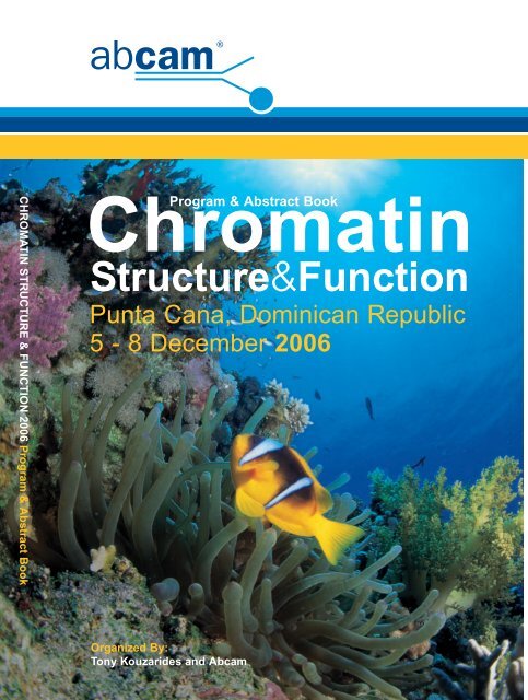
CHROMATIN STRUCTURE & FUNCTION 2006 Program & Abstract Book<br />
<strong>Chromatin</strong><br />
Program & Abstract Book<br />
<strong>Structure</strong>&<strong>Function</strong><br />
Punta Cana, Dominican Republic<br />
5 - 8 December 2006<br />
Organized By:<br />
Tony Kouzarides and <strong>Abcam</strong>
Punta Cana Prog 30/10/06 10:08 Page 2
Punta Cana Prog 30/10/06 10:08 Page 1<br />
Program & Abstract Book<br />
The third<br />
<strong>Chromatin</strong><br />
<strong>Structure</strong> & <strong>Function</strong><br />
Punta Cana, Dominican Republic<br />
5 - 8 December 2006<br />
Organizers:<br />
Tony Kouzarides<br />
(University of Cambridge)<br />
and <strong>Abcam</strong><br />
Table of contents<br />
Conference Program . . . . . . . . . . . . . . . . . . . . . . . . . . . . . . . . . . . .Page 2<br />
Poster Index . . . . . . . . . . . . . . . . . . . . . . . . . . . . . . . . . . . . . . . . . .Page 6<br />
Abstracts - Oral . . . . . . . . . . . . . . . . . . . . . . . . . . . . . . . . . . . . . . .Page 18<br />
Abstracts - Poster . . . . . . . . . . . . . . . . . . . . . . . . . . . . . . . . . . . . .Page 51<br />
Resort Information . . . . . . . . . . . . . . . . . . . . . . . . . . . . . . . . . . .Page 179<br />
Disclaimer: Material contained within this booklet should be citied only with permission from the author(s).<br />
No live recording or photography is permitted during the oral or poster sessions.<br />
Copyright © 2006 <strong>Abcam</strong>, All Rights Reserved. The <strong>Abcam</strong> logo is a registered trademark.<br />
All information / detail is correct at time of going to print.<br />
1
Punta Cana Prog 30/10/06 10:08 Page 2<br />
<strong>Chromatin</strong> <strong>Structure</strong> & <strong>Function</strong> Punta Cana, Dominican Republic, 5 - 8 December 2006<br />
Conference Program<br />
Tuesday 5th December<br />
Meeting room - Allegro Plaza<br />
Keynote Speaker - introduced by Tony Kouzarides<br />
18:00 - 19:00 Steve Henikoff . . . . . . . . . . . . . . . . . . . . . . . .Page 18<br />
Epigenetic patterns generated by histone replacement<br />
Welcome reception and buffet at poolside.<br />
Allegro Live resort show<br />
Wednesday 6th December<br />
Chair: Jerry Workman<br />
09:00 - 09:30 Yang Shi . . . . . . . . . . . . . . . . . . . . . . . . . . . . .Page 19<br />
The identification of histone demethylases established the<br />
dynamic and reversible nature of histone methylation<br />
regulation<br />
09:30 - 09:45 Jesper Christensen . . . . . . . . . . . . . . . . . . . . .Page 20<br />
The retinoblastoma tumor suppressor binding protein RBP2 is<br />
a transcriptional repressor demethylating tri- and dimethylated<br />
lysine 4 on Histone H3<br />
09:45 - 10:00 Mischa Machius . . . . . . . . . . . . . . . . . . . . . . .Page 21<br />
Structural basis for CoREST-dependent demethylation of<br />
nucleosomes by the human LSD1 histone demethylase<br />
10:00 - 10:30 Ramin Shiekhattar . . . . . . . . . . . . . . . . . . . . .Page 22<br />
<strong>Function</strong>al and biochemical characterization of histone<br />
demethylase complexes<br />
Drinks break in hotel lobby<br />
11:00 - 11:30 Yi Zhang . . . . . . . . . . . . . . . . . . . . . . . . . . . . .Page 23<br />
Histone demethylation by the JmjC domain-containing proteins<br />
11:30 - 11:45 Francis Stewart . . . . . . . . . . . . . . . . . . . . . . . .Page 24<br />
Epigenetic aspects of lineage commitment<br />
2
Punta Cana Prog 30/10/06 10:08 Page 3<br />
Conference Program<br />
11:45 - 12:00 Henk Stunnenberg . . . . . . . . . . . . . . . . . . . . .Page 25<br />
Title and abstract unavailable<br />
12:00 - 12:30 Ali Shilatifard . . . . . . . . . . . . . . . . . . . . . . . . . .Page 26<br />
H2B monoubiquitination and H3K4 methylation via COMPASS<br />
Lunch at the Beach Buffet Restaurant and free time<br />
Chair: Yang Shi<br />
16:00 - 16:30 Paulo Sassone-Corsi . . . . . . . . . . . . . . . . . . .Page 27<br />
A chromatin remodeling clock<br />
16:30 - 16:45 Sung Hee Baek . . . . . . . . . . . . . . . . . . . . . . .Page 28<br />
A Novel Link between SUMO Modification of a <strong>Chromatin</strong><br />
Remodeling Complex and Cancer Metastasis<br />
16:45 - 17:00 Laszlo Tora . . . . . . . . . . . . . . . . . . . . . . . . . . .Page 29<br />
The simultaneously dimethylated Lys-9 and phosphorylated<br />
Ser-10 tails of histone H3 adopt different conformations<br />
during mitosis<br />
17:00 - 17:30 Tony Kouzarides . . . . . . . . . . . . . . . . . . . . . . .Page 30<br />
Characterisation of novel histone modifications<br />
18:00 - 21:00 Posters and buffet by the pool<br />
Allegro Live resort show<br />
Thursday 7th December<br />
Chair: Genevieve Almouzni<br />
09:00 - 09:30 Thomas Jenuwein . . . . . . . . . . . . . . . . . . . . . .Page 31<br />
Epigenetic control by histone methylation<br />
09:30 - 09:45 Roberta Benetti . . . . . . . . . . . . . . . . . . . . . . .Page 32<br />
The role of Dicer in the regulation of chromatin at telomeres<br />
3
Punta Cana Prog 30/10/06 10:08 Page 4<br />
<strong>Chromatin</strong> <strong>Structure</strong> & <strong>Function</strong> Punta Cana, Dominican Republic, 5 - 8 December 2006<br />
09:45 - 10:00 Mareike Puschendorf . . . . . . . . . . . . . . . . . . .Page 33<br />
Ezh2 independent targeting of PRC1 proteins to paternal<br />
constitutive heterochromatin in mouse pre-implantation embryos<br />
10:00 - 10:30 Edith Heard . . . . . . . . . . . . . . . . . . . . . . . . . . .Page 34<br />
The nuclear and epigenetic dynamics of X-chromosome<br />
inactivation in the mouse<br />
Drinks break in hotel lobby<br />
11:00 - 11:30 Adrian Bird . . . . . . . . . . . . . . . . . . . . . . . . . . .Page 35<br />
MeCP2: molecular interactions and phenotypic stability in a<br />
mouse model of Rett Syndrome<br />
11:30 - 11:45 Jon Penterman . . . . . . . . . . . . . . . . . . . . . . . .Page 36<br />
DNA demethylation in Arabidopsis thaliana<br />
11:45 - 12:00 Francois Fuks . . . . . . . . . . . . . . . . . . . . . . . . .Page 37<br />
The Polycomb Group protein EZH2 is recruited to promoters by<br />
MECP2<br />
12:00 - 12:30 Shelley Berger . . . . . . . . . . . . . . . . . . . . . . . .Page 38<br />
Factor and histone covalent modifications in genome regulation<br />
Lunch at the Beach Buffet Restaurant and free time<br />
Chair: Ramin Shiekhattar<br />
16:00 - 16:30 Danny Reinberg . . . . . . . . . . . . . . . . . . . . . . .Page 39<br />
A molecular understanding of epigenetics<br />
16:30 - 16:45 Jessica Tyler . . . . . . . . . . . . . . . . . . . . . . . . . .Page 40<br />
The mechanistic basis for the requirement of promoter<br />
chromatin disassembly for transcriptional activation<br />
16:45 - 17:00 Gratien Prefontaine . . . . . . . . . . . . . . . . . . . . .Page 41<br />
Epigenetic mechanisms influencing pituitary gene expression<br />
17:00 - 17:30 Bob Kingston . . . . . . . . . . . . . . . . . . . . . . . . .Page 42<br />
Possible roles in silencing for piRNAs<br />
4
Punta Cana Prog 30/10/06 10:08 Page 5<br />
Conference Program<br />
18:00 - 19:30 Posters and drinks by the pool<br />
Beach barbeque and live band on the sand<br />
Friday 8th December<br />
Chair: Edith Heard<br />
09:00 - 09:30 Michael Grunstein . . . . . . . . . . . . . . . . . . . . . .Page 43<br />
Deacetylation of histone H4 K16 regulates gene activity in yeast<br />
09:30 - 09:45 Ann Ehrenhofer-Murray . . . . . . . . . . . . . . . . .Page 44<br />
A role for the HDAC Rpd3 in establishing eurchromatinheterochromatin<br />
boundaries at yeast telomeres<br />
09:45 - 10:00 Wyatt Yue . . . . . . . . . . . . . . . . . . . . . . . . . . . .Page 45<br />
CARM1 and histone methylation - a structural study<br />
10:00 - 10:30 Sharon Dent . . . . . . . . . . . . . . . . . . . . . . . . . .Page 46<br />
Common and unique factors regulate Set1-mediated<br />
methylation of the Dam1 kinetochore protein and histone H3<br />
Drinks break in hotel lobby<br />
11:00 - 11:30 Genevieve Almouzni . . . . . . . . . . . . . . . . . . . .Page 47<br />
<strong>Chromatin</strong> assembly factors, histone H3 variants and cell cycle<br />
11:30 - 11:45 Dmitry Fyodorov . . . . . . . . . . . . . . . . . . . . . . .Page 48<br />
ATP-dependant deposition of Histone H3.3 by Drosophila CHD1<br />
in vivo<br />
11:45 - 12:00 Roberto Mantovani . . . . . . . . . . . . . . . . . . . . .Page 49<br />
The histone fold trimer NF-Y is required to define positive<br />
histone marks in CCAAT-promotors: a genome wide analysis<br />
12:00 - 12:30 Jerry Workman . . . . . . . . . . . . . . . . . . . . . . . .Page 50<br />
Histone modification and chromatin remodeling in transcription<br />
Lunch at the Beach Buffet Restaurant<br />
Conference ends<br />
5
Punta Cana Prog 30/10/06 10:08 Page 6<br />
<strong>Chromatin</strong> <strong>Structure</strong> & <strong>Function</strong> Punta Cana, Dominican Republic, 5 - 8 December 2006<br />
Poster Index<br />
Abstract P1 Karl Agger . . . . . . . . . . . . . . . . . . . . . . . . . . . .Page 51<br />
The role of the polycomb group protein RYBP in oncogene<br />
induced senescence<br />
Abstract P2 Helena Ahlfors . . . . . . . . . . . . . . . . . . . . . . . .Page 52<br />
A novel player in T helper cell differentiation<br />
Abstract P3 Barbara Alberter . . . . . . . . . . . . . . . . . . . . . . .Page 53<br />
Histone modification pattern of the T lymphotropic Herpesvirus<br />
saimiri genome in latency<br />
Abstract P4 Marco Alvarez . . . . . . . . . . . . . . . . . . . . . . . . .Page 54<br />
Histone variant macroH2A is an epigenetic factor involved in<br />
the modulation of ribosomal gene expression during seasonal<br />
adaptation of carp fish<br />
Abstract P5 Terra Arnason . . . . . . . . . . . . . . . . . . . . . . . . .Page 55<br />
Rsp5 is required for nuclear shuttling of the Snf1 kinase<br />
complex in yeast<br />
Abstract P6 Stuart Atkinson . . . . . . . . . . . . . . . . . . . . . . . .Page 56<br />
Epigenetic mechanisms of pluripotency and differentiation<br />
Abstract P7 Joanne Attema . . . . . . . . . . . . . . . . . . . . . . . .Page 57<br />
Epigenetic features of hematopoietic stem cells using small<br />
numbers of highly purified primary cells<br />
Abstract P8 Kristin Baetz . . . . . . . . . . . . . . . . . . . . . . . . . .Page 58<br />
NuA4 is a cellular “Hub”: an integrative map of physical and<br />
genetic interactions mediated by the NuA4 histone<br />
acetyltransferase<br />
Abstract P9 Slobodan Barbaric . . . . . . . . . . . . . . . . . . . . .Page 59<br />
<strong>Chromatin</strong> remodeling activities at the yeast PHO84 promoter<br />
Abstract P10 Vivian Bardwell . . . . . . . . . . . . . . . . . . . . . . . .Page 60<br />
Polycomb group and SCF ubiquitin ligases are found in a novel<br />
BCOR complex that is recruited to BCL6 targets<br />
Abstract P11 Amrita Basu . . . . . . . . . . . . . . . . . . . . . . . . . .Page 61<br />
Computational prediction of histone and non-histone proteins<br />
6
Punta Cana Prog 30/10/06 10:08 Page 7<br />
Poster Index<br />
Abstract P12 Mark Bedford . . . . . . . . . . . . . . . . . . . . . . . . .Page 62<br />
Screening for the methylated proteome<br />
Abstract P13 Sukesh R. Bhaumik . . . . . . . . . . . . . . . . . . . .Page 63<br />
Regulation of transcriptional activation by mRNA cap-binding<br />
complex in vivo<br />
Abstract P14 Marjorie Brand . . . . . . . . . . . . . . . . . . . . . . . .Page 64<br />
The Ash2L/MLL2 methyltransferase complex is important for ß-<br />
globin transcription during erythroid differentiation<br />
Abstract P15 Lauren Buro . . . . . . . . . . . . . . . . . . . . . . . . . .Page 65<br />
Histone methylation patterns at interferon-gamma inducible<br />
gene loci<br />
Abstract P16 Jill Butler . . . . . . . . . . . . . . . . . . . . . . . . . . . . .Page 66<br />
CXXC-finger protein 1 regulates Dnmt1 protein expression<br />
Abstract P17 Jim Cakouros . . . . . . . . . . . . . . . . . . . . . . . . .Page 67<br />
Identification of a novel enzyme which regulates the kinetics of<br />
histone arginine methylation in Drosophila melanogaster<br />
Abstract P18 Raymond Camahort . . . . . . . . . . . . . . . . . . . .Page 68<br />
Genome-wide analysis of the budding yeast histone variant<br />
Cse4 reveals occupancy at a single centromeric nucleosome as<br />
well as additional non-centromeric locations<br />
Abstract P19 Dylan Carney . . . . . . . . . . . . . . . . . . . . . . . . .Page 69<br />
The RAG2 PHD Finger links the histone code to V(D)J<br />
recombination<br />
Abstract P20 Beverly Chilton . . . . . . . . . . . . . . . . . . . . . . . .Page 70<br />
Analysis of RUSH/SMARCA3 isoforms and their interactions<br />
with Egr-1 and c-Rel in the regulation of transcription<br />
Abstract P21 Alexandra Chittka . . . . . . . . . . . . . . . . . . . . . .Page 71<br />
Signalling by a novel p75 neurotrophin receptor interacting<br />
protein, SC1/PRDM4<br />
Abstract P22 Leslie Chu . . . . . . . . . . . . . . . . . . . . . . . . . . . .Page 72<br />
Inheritance of epigenetic chromatin states<br />
7
Punta Cana Prog 30/10/06 10:08 Page 8<br />
<strong>Chromatin</strong> <strong>Structure</strong> & <strong>Function</strong> Punta Cana, Dominican Republic, 5 - 8 December 2006<br />
Abstract P23 Mair Churchill . . . . . . . . . . . . . . . . . . . . . . . . .Page 73<br />
Structural basis for the histone chaperone activity of Asf1<br />
Abstract P24 Jeffrey Craig . . . . . . . . . . . . . . . . . . . . . . . . . .Page 74<br />
What makes centromeres localise and cluster in interphase<br />
nuclei<br />
Abstract P25 Valerie Crusselle-Davis . . . . . . . . . . . . . . . . . .Page 75<br />
Regulation of beta-globin expression through the recruitment<br />
of chromatin modifying enzymes by TFII-I and USF<br />
Abstract P26 Eullia de Nadal . . . . . . . . . . . . . . . . . . . . . . . .Page 76<br />
Control of gene expression by the yeast Hog1 MAPK.<br />
Abstract P27 Foteini Davrazou . . . . . . . . . . . . . . . . . . . . . . .Page 77<br />
Molecular mechanism of histone H3K4me3 recognition by the<br />
PHD finger of ING2<br />
Abstract P28 Roger Deal . . . . . . . . . . . . . . . . . . . . . . . . . . .Page 78<br />
Repression of flowering in Arabidopsis requires histone H2A.Z<br />
deposition by a putative SWR1 complex<br />
Abstract P29 Laurent Delva . . . . . . . . . . . . . . . . . . . . . . . . .Page 79<br />
The Transcription Intermediary Factor 2 is required for<br />
zebrafish development<br />
Abstract P30 Luisa Di Stefano . . . . . . . . . . . . . . . . . . . . . .Page 80<br />
Lsd1 mutation in Drosophila disrupt normal level of H3K4<br />
methylation and affects viability and fertility<br />
Abstract P31 Stephan Diekmann . . . . . . . . . . . . . . . . . . . . .Page 81<br />
In vivo dynamic (FRAP, FCS) and neighbourhood relation (AB-<br />
FRET, FLIM) studies of human inner kinetochore proteins<br />
Abstract P32 Jeffrey Dilworth . . . . . . . . . . . . . . . . . . . . . . . .Page 82<br />
MEF2 helps establish muscle specific pattern of gene<br />
expression by recruiting Trithorax Group proteins to specific<br />
promoters<br />
Abstract P33 Ivana Djuretic . . . . . . . . . . . . . . . . . . . . . . . . .Page 83<br />
T-bet and Runx3 cooperate to activate Interferon gamma and<br />
silence Interleukin-4 in T helper-1 cells<br />
8
Punta Cana Prog 30/10/06 10:08 Page 9<br />
Poster Index<br />
Abstract P34 Tom Donndelinger . . . . . . . . . . . . . . . . . . . . . .Page 84<br />
Seeing cells in a new light: Improving resolution with a<br />
scientific approach to tissue processing<br />
Abstract P35 Bojan Drobic . . . . . . . . . . . . . . . . . . . . . . . . . .Page 85<br />
Characterization of Histone H3 kinases, MSK1 and MSK2<br />
Abstract P36 Danielle Ellis . . . . . . . . . . . . . . . . . . . . . . . . . .Page 86<br />
Histone acetylation of SRC and p21 promoters in response to<br />
histone deacetylase inhibitor treatment; implications of HDAC<br />
activity and SRC expression<br />
Abstract P37 Alexander Erkine . . . . . . . . . . . . . . . . . . . . . .Page 87<br />
Differential mechanisms of nucleosome displacement at yeast<br />
heat shock gene promoters<br />
Abstract P38 Ragnhild Eskeland . . . . . . . . . . . . . . . . . . . . .Page 88<br />
HP1 binding to chromatin methylated at H3K9 is enhanced by<br />
auxiliary factors<br />
Abstract P39 George Feehery . . . . . . . . . . . . . . . . . . . . . . .Page 89<br />
CpG methylated DNA standards and control primers for use in<br />
methyl sensitive PCR and bisulphite sequencing<br />
Abstract P40 Barna Fodor . . . . . . . . . . . . . . . . . . . . . . . . . .Page 90<br />
Identification of novel pericentric proteins by their localization<br />
Abstract P41 Maria Fousteri . . . . . . . . . . . . . . . . . . . . . . . . .Page 91<br />
Cockayne syndrome A and B proteins differentially regulate<br />
recruitment of chromatin remodeling and repair factors to<br />
stalled RNA polymerase II in vivo<br />
Abstract P42 Robert Gillespie . . . . . . . . . . . . . . . . . . . . . . .Page 92<br />
Retinoid regulated association of transcriptional coregulators<br />
and the polycomb group protein SUZ12 with the retinoic acid<br />
response elements of Hoxa1, RARß2, and Cyp26A1 in F9<br />
embryonal carcinoma cells<br />
Abstract P43 Clara Goday . . . . . . . . . . . . . . . . . . . . . . . . . .Page 93<br />
<strong>Chromatin</strong> modifications in germline chromosomes of sciarid flies<br />
9
Punta Cana Prog 30/10/06 10:08 Page 10<br />
<strong>Chromatin</strong> <strong>Structure</strong> & <strong>Function</strong> Punta Cana, Dominican Republic, 5 - 8 December 2006<br />
Abstract P44 Aaron Goldberg . . . . . . . . . . . . . . . . . . . . . . .Page 94<br />
HIRA-dependent incorporation of histone H3.3 marks active<br />
genes in mouse embryonic stem cells<br />
Abstract P45 Elizabeth Goneska . . . . . . . . . . . . . . . . . . . . .Page 95<br />
Phosphorylation of the SQ H2A.X motif is required for proper<br />
meiosis and mitosis in Tetrahymena thermophila<br />
Abstract P46 Susana Gonzalo . . . . . . . . . . . . . . . . . . . . . . .Page 96<br />
Telomere epigenetic modifications: a control of telomere length<br />
and a stop on recombination<br />
Abstract P47 Tanya Gustafson . . . . . . . . . . . . . . . . . . . . . . .Page 97<br />
Epigenetic silencing of Singleminded-2 in breast cancer<br />
Abstract P48 Soon-Ki Han . . . . . . . . . . . . . . . . . . . . . . . . . .Page 98<br />
Role of plant CBP/p300-like genes in the regulation of<br />
flowering time<br />
Abstract P49 Christin Hanigan . . . . . . . . . . . . . . . . . . . . . . .Page 99<br />
Identification of an HDAC2 mutation in colorectal cancer and<br />
its consequences<br />
Abstract P50<br />
Troy Harkness . . . . . . . . . . . . . . . . . . . . . . . .Page100<br />
Rsp5 is required for nuclear shuttling of the Snf1 kinase<br />
complex in yeast<br />
Abstract P51 Tiffany Hung . . . . . . . . . . . . . . . . . . . . . . . . .Page 101<br />
ING4 recognition of histone H3 trimethylated at lysine 4<br />
Abstract P52 David Johnson . . . . . . . . . . . . . . . . . . . . . . .Page 102<br />
E2F1 and GCN5 facilitate the recruitment of nucleotide excision<br />
repair factors to sites of UV-induced DNA damage<br />
Abstract P53 Paul Kalitsis . . . . . . . . . . . . . . . . . . . . . . . . .Page 103<br />
Nucleosome spacing analysis of repeat DNA regions in the<br />
mouse genome<br />
Abstract P54 Min-Jeong Kang . . . . . . . . . . . . . . . . . . . . . .Page 104<br />
Role of a RPD3/HDA1 family histone deacetylase in the regulation<br />
of phytochrome-mediated light respases in Arabidopsis<br />
10
Punta Cana Prog 30/10/06 10:08 Page 11<br />
Poster Index<br />
Abstract P55 Panagiota Karagianni . . . . . . . . . . . . . . . . . .Page 105<br />
ICBP90, a putative link between histone ubiquitination and cell<br />
cycle progression<br />
Abstract P56 Emmanuel Kas . . . . . . . . . . . . . . . . . . . . . . .Page 106<br />
Altering the structure and functional properties of<br />
heterochromatin with satellite-specific minor-groove binders<br />
Abstract P57 Chul Geun Kim . . . . . . . . . . . . . . . . . . . . . . .Page 107<br />
PIAS1 confers erythroid cell specific α-globin gene regulation<br />
by the CP2 transcription factor family<br />
Abstract P58 Keun Il Kim . . . . . . . . . . . . . . . . . . . . . . . . . .Page 108<br />
A novel link between SUMO modification of a chromatin<br />
remodeling complex and cancer metastasis<br />
Abstract P59 Sarah Kimmins . . . . . . . . . . . . . . . . . . . . . . .Page 109<br />
Methylation of Histone H3 at lysine 4 is dynamic and tightly<br />
regulated during male germ cell development<br />
Abstract P60 Robert Klose . . . . . . . . . . . . . . . . . . . . . . . . .Page 110<br />
JmjC-domain-containing proteins and histone demethylation<br />
Abstract P61 Christoph Koch . . . . . . . . . . . . . . . . . . . . . . .Page 111<br />
The landscape of activating histone modifications across 1% of<br />
the human genome<br />
Abstract P62 Ryoki Kujiki . . . . . . . . . . . . . . . . . . . . . . . . . .Page 112<br />
1alpha,25(OH)2D3-induced transrepression on 1alphahydroxylase<br />
gene promoter mediates chromatin remodeling<br />
through WINAC<br />
Abstract P63 Sharmistha Kundu . . . . . . . . . . . . . . . . . . . .Page 113<br />
SWI/SNF establishes transcriptional memory at the<br />
Saccharomyces cerevisiae GAL1 gene<br />
Abstract P64 Georg Kustatscher . . . . . . . . . . . . . . . . . . . .Page 114<br />
Metabolite-sensitive and metabolite-insensitive chromatin<br />
surfaces through the human histone macroH2A<br />
Abstract P65 Hyockman Kwon . . . . . . . . . . . . . . . . . . . . . .Page 115<br />
BAF53-dependent higher-order chromatin structure as the<br />
compartment of replication and repair foci<br />
11
Punta Cana Prog 30/10/06 10:08 Page 12<br />
<strong>Chromatin</strong> <strong>Structure</strong> & <strong>Function</strong> Punta Cana, Dominican Republic, 5 - 8 December 2006<br />
Abstract P66 Monika Lachner . . . . . . . . . . . . . . . . . . . . . .Page 116<br />
Studying lysine methylation in non-histone proteins<br />
Abstract P67 Brian Larsen . . . . . . . . . . . . . . . . . . . . . . . . .Page 117<br />
Caspase 3 mediated DNA strand breaks contribute to genomic<br />
reorganization during skeletal muscle terminal differentiation<br />
Abstract P68 Richard Lawrence . . . . . . . . . . . . . . . . . . . . .Page 118<br />
Mechanisms controlling dynamic Swi6/HP1 binding in S.<br />
pombe facilitate de novo heterochromatin formation<br />
Abstract P69 Frederic Leduc . . . . . . . . . . . . . . . . . . . . . . .Page 119<br />
Presence of gamma-H2AX in elongating spermatids:<br />
involvement of NHEJ<br />
Abstract P70 Min Gyu Lee . . . . . . . . . . . . . . . . . . . . . . . . .Page 120<br />
<strong>Function</strong>al association of a trimethyl H3K4 demethylase and<br />
Ring6a/MBLR, a polycomb-like protein<br />
Abstract P71 Niraj Lodhi . . . . . . . . . . . . . . . . . . . . . . . . . . .Page 121<br />
Histone acetylation (H3K9) and methylation (H3K4) of the<br />
nucleosome over core promoter are associated with the<br />
induction of tobacco PR-1a gene<br />
Abstract P72 Mattias Mannervik . . . . . . . . . . . . . . . . . . . .Page 122<br />
An HDAC3/SMRTER/Ebi complex required for Snail repressor<br />
function in Drosophila development<br />
Abstract P73 Robert Martin . . . . . . . . . . . . . . . . . . . . . . . .Page 123<br />
<strong>Chromatin</strong> labeling and distribution in living cells<br />
Abstract P74 Peter McKeown . . . . . . . . . . . . . . . . . . . . . . .Page 124<br />
<strong>Chromatin</strong> components of the Arabidopsis thaliana nucleolus<br />
Abstract P75 Rosalind Meldrum . . . . . . . . . . . . . . . . . . . .Page 125<br />
Visualisation of DNA repair and chromatin dynamics<br />
Abstract P76 Brendon Monahan . . . . . . . . . . . . . . . . . . . .Page 126<br />
Purification and characterization of the fission yeast Swi/Snf<br />
and RSC chromatin remodeling complexes<br />
12
Punta Cana Prog 30/10/06 10:08 Page 13<br />
Poster Index<br />
Abstract P77 Antonin Morillon . . . . . . . . . . . . . . . . . . . . . .Page 127<br />
Transcriptional co-suppression in S. cerevisiae<br />
Abstract P78 Ashby Morrison . . . . . . . . . . . . . . . . . . . . . . .Page 128<br />
Mec1/Tel1-dependent phosphorylation of a chromatin<br />
remodeling complex influences the DNA damage checkpoint<br />
pathway<br />
Abstract P79 Raul Mostoslavsky . . . . . . . . . . . . . . . . . . . .Page 129<br />
Genomic instability and aging-like phenotype in the absence of<br />
mammalian SIRT6<br />
Abstract P80 Takahiro Nakayama . . . . . . . . . . . . . . . . . . .Page 130<br />
Drosophila GAGA factor promotes histone H3.3 replacement<br />
that prevents the heterochromatin spreading<br />
Abstract P81 Zuyao Ni . . . . . . . . . . . . . . . . . . . . . . . . . . . .Page 131<br />
The tumor suppressor BRG1 silences the distal silencers at<br />
interferon-responsive genes<br />
Abstract P82 Olivia Osborn . . . . . . . . . . . . . . . . . . . . . . . .Page 132<br />
Transcriptional targets of Af4<br />
Abstract P83 Julia Pagan . . . . . . . . . . . . . . . . . . . . . . . . . .Page 133<br />
A novel corepressor, BCOR-L1, functions through CTBP and<br />
class 2 HDACs<br />
Abstract P84 Maria Panchenko . . . . . . . . . . . . . . . . . . . . .Page 134<br />
Role of Jade-1 in the HAT HBO1 complex<br />
Abstract P85 Tej Pandita . . . . . . . . . . . . . . . . . . . . . . . . . .Page 135<br />
Mammalian ortholog of Drosophila MOF is critical for<br />
embryogenesis and DNA repair<br />
Abstract P86 Maëlle Pannetier . . . . . . . . . . . . . . . . . . . . . .Page 136<br />
Imprinting perturbation in mouse hepatocarcinoma: link<br />
between DNA methylation and histone methylation<br />
Abstract P87 Janet Partridge . . . . . . . . . . . . . . . . . . . . . . .Page 137<br />
Establishment and maintenance of centromeric<br />
heterochromatin in fission yeast are functionally separable<br />
13
Punta Cana Prog 30/10/06 10:08 Page 14<br />
<strong>Chromatin</strong> <strong>Structure</strong> & <strong>Function</strong> Punta Cana, Dominican Republic, 5 - 8 December 2006<br />
Abstract P88 Kelly Perkins . . . . . . . . . . . . . . . . . . . . . . . . .Page 138<br />
Activated HIV-1 provirus forms a gene loop, connecting viral<br />
transcriptional initiation with termination<br />
Abstract P89 David Picketts . . . . . . . . . . . . . . . . . . . . . . . .Page 139<br />
SNF2L-mediated control of cell number in the developing brain<br />
Abstract P90 Romina Ponzielli . . . . . . . . . . . . . . . . . . . . . .Page 140<br />
Optimization of experimental design parameters of ChIP-onchip<br />
studies<br />
Abstract P91 Ryan Raisner . . . . . . . . . . . . . . . . . . . . . . . .Page 141<br />
Single nucleosome resolution mapping of the histone variant<br />
H2A.Z in a developing organism<br />
Abstract P92 Rama Natarajan . . . . . . . . . . . . . . . . . . . . . .Page 142<br />
Genome-wide analysis of histone lysine methylation variations<br />
caused by diabetic conditions in human monocytes<br />
Abstract P93 Edward Ramos . . . . . . . . . . . . . . . . . . . . . . .Page 143<br />
Global characterization and function of Gypsy-like endogenous<br />
insulators in Drosophila melanogaster<br />
Abstract P94 William Renthal . . . . . . . . . . . . . . . . . . . . . . .Page 144<br />
Class II histone deacetylases regulate the behavioral<br />
adaptations to chronic cocaine and stress<br />
Abstract P95 Karsten Rippe . . . . . . . . . . . . . . . . . . . . . . . .Page 145<br />
Activities of histone chaperone NAP1: Association states and<br />
interactions with histones, nucleosome assembly and effect on<br />
the chromatin fiber conformation<br />
Abstract P96 Charles Roberts . . . . . . . . . . . . . . . . . . . . . .Page 146<br />
The Swi/Snf chromatin remodeling complex regulates lineage<br />
specific transcription programs during development and<br />
impairment of this activity causes cancer<br />
Abstract P97 Paul Sadowski . . . . . . . . . . . . . . . . . . . . . . .Page 147<br />
Post-translational modification of the insulator protein, CTCF<br />
Abstract P98 Teresa Sanchez Alcaraz . . . . . . . . . . . . . . . .Page 148<br />
Role of USP7 and GMP synthetase in deubiquitination of<br />
human histone H2B<br />
14
Punta Cana Prog 30/10/06 10:08 Page 15<br />
Poster Index<br />
Abstract P99 Annette Scharf . . . . . . . . . . . . . . . . . . . . . . .Page 149<br />
Dynamics of histone modifications during chromatin assembly<br />
Abstract P100 Stefan Schoeftner . . . . . . . . . . . . . . . . . . . . .Page 150<br />
Screening for miRNAs regulating mammalian telomeres<br />
Abstract P101 Gunnar Schotta . . . . . . . . . . . . . . . . . . . . . .Page 151<br />
A genome-wide transition to H4K20 mono-methylation impairs<br />
stress-induced and programd DNA damage response in the<br />
mouse<br />
Abstract P102 David Schrump . . . . . . . . . . . . . . . . . . . . . . .Page 152<br />
Brother of the Regulator of Imprinted Sites (BORIS) recruits<br />
Sp1 to modulate NY-ESO-1 expression in lung cancer cells<br />
Abstract P103 Bonnie Scott . . . . . . . . . . . . . . . . . . . . . . . . .Page 153<br />
Evolution of centromere-binding proteins and their interactions<br />
with centromere DNA in Arabidopsis<br />
Abstract P104 David Shechter . . . . . . . . . . . . . . . . . . . . . . .Page 154<br />
Histone H2A arginine3 is mono- and symmetrically-di<br />
methylated by a complex of PRMT5 and the WD-repeat protein<br />
MEP50 in Xenopus laevis eggs<br />
Abstract P105 Yoichi Shinkai . . . . . . . . . . . . . . . . . . . . . . . .Page 155<br />
H3K9 methylation and germ cell development<br />
Abstract P106 Krishna Sinha . . . . . . . . . . . . . . . . . . . . . . . .Page 156<br />
Inhibition of the transcriptional activity of osterix by<br />
interactions with NO66, a jumonji family chromatin protein<br />
Abstract P107 Karen Smith . . . . . . . . . . . . . . . . . . . . . . . . .Page 157<br />
Identification and characterization of novel HDAC-associated<br />
proteins that regulate cancer cell growth<br />
Abstract P108 Matthew Smith . . . . . . . . . . . . . . . . . . . . . . .Page 158<br />
<strong>Chromatin</strong>- mediated silencing of immune response genes<br />
Abstract P109 Hae-Ryong Song . . . . . . . . . . . . . . . . . . . . .Page 159<br />
Coordination of transcriptional regulation and chromatin<br />
modification of Arabidopsis circadian clock genes<br />
15
Punta Cana Prog 30/10/06 10:08 Page 16<br />
<strong>Chromatin</strong> <strong>Structure</strong> & <strong>Function</strong> Punta Cana, Dominican Republic, 5 - 8 December 2006<br />
Abstract P110 Stacey Southall . . . . . . . . . . . . . . . . . . . . . . .Page 160<br />
Structural studies of histone methyltransferases<br />
Abstract P111 Maike Stam . . . . . . . . . . . . . . . . . . . . . . . . . .Page 161<br />
Molecular analysis of chromatin changes involved in b1<br />
paramutation, an allele-dependent transfer of epigenetic<br />
information<br />
Abstract P112 Sean Taverna . . . . . . . . . . . . . . . . . . . . . . . .Page 162<br />
Connecting H3 methylation and acetylation: The role of Yng1 in<br />
transcription<br />
Abstract P113 Tage Thorstensen . . . . . . . . . . . . . . . . . . . . .Page 163<br />
The Arabidopsis SUVR proteins define a novel subgroup of<br />
SET domain proteins associated with the nucleolus<br />
Abstract P114 Christopher Topp . . . . . . . . . . . . . . . . . . . . .Page 164<br />
Unusually-sized centromeric RNAs associate with maize<br />
centromeric chromatin<br />
Abstract P115 Martin Tribus . . . . . . . . . . . . . . . . . . . . . . . . .Page 165<br />
Molecular mechanisms of histone variant H3.3 assembly by the<br />
motor protein CHD1<br />
Abstract P116 Christopher Vakoc . . . . . . . . . . . . . . . . . . . .Page 166<br />
A profile of histone lysine methylation generated by<br />
mammalian gene transcription<br />
Abstract P117 Claudius Vincenz . . . . . . . . . . . . . . . . . . . . .Page 167<br />
Visualizing polycomb group protein interactions with histones<br />
in vivo<br />
Abstract P118 Vikki Weake . . . . . . . . . . . . . . . . . . . . . . . . .Page 168<br />
The SAGA histone acetyltransferase complex functions in the<br />
development of neuronal connectivity in the Drosophila<br />
compound eye<br />
Abstract P119 Stephanie Williams . . . . . . . . . . . . . . . . . . . .Page 169<br />
Mechanistic insights into promoter chromatin disassembly<br />
Abstract P120 Jon Wilson . . . . . . . . . . . . . . . . . . . . . . . . . .Page 170<br />
Structural studies of SET domain methyltransferases<br />
16
Punta Cana Prog 30/10/06 10:08 Page 17<br />
Poster Index<br />
Abstract P121 Zhaodong Xu . . . . . . . . . . . . . . . . . . . . . . . .Page 171<br />
Remote elements critical for cytokine induced gene expression<br />
Abstract P122 Xiaofang Yang . . . . . . . . . . . . . . . . . . . . . . .Page 172<br />
Dissecting SWI/SNF ATP-dependent chromatin remodeling<br />
complex in Saccharomyces cerevisiae<br />
Abstract P123 Juan I. Young . . . . . . . . . . . . . . . . . . . . . . . .Page 173<br />
Post-transcriptional functions of MeCP2<br />
Abstract P124 Veronica Yu . . . . . . . . . . . . . . . . . . . . . . . . . .Page 174<br />
Over-expression of Cks proteins causes gene derepression in<br />
Saccharomyces cerevisiae<br />
Abstract P125 Rebekah Zinn . . . . . . . . . . . . . . . . . . . . . . . .Page 175<br />
hTERT is expressed in cancer despite promoter DNA<br />
methylation by preservation of unmethylated DNA and active<br />
chromatin around the transcription start site<br />
Additional poster submissions<br />
Abstract P126 Yoshimitsu Takahashi . . . . . . . . . . . . . . . . . .Page 176<br />
Degree of SUMO modification as a differential tag for targeting<br />
to specific chromosomal domains<br />
Abstract P127 Marna S. Costanzo . . . . . . . . . . . . . . . . . . . .Page 177<br />
The evolutionary conservation of chromatin modifying proteins<br />
in malaria<br />
Abstract P128 Philippe Prochasson . . . . . . . . . . . . . . . . . . .Page 178<br />
<strong>Function</strong>al characterization of the HIR corepressor complex<br />
17
Punta Cana Prog 30/10/06 10:08 Page 18<br />
<strong>Chromatin</strong> <strong>Structure</strong> & <strong>Function</strong> Punta Cana, Dominican Republic, 5 - 8 December 2006<br />
Abstracts – Oral<br />
Steve Henikoff Abstract 1<br />
Epigenetic patterns generated by histone replacement<br />
Fred Hutchinson Cancer Research Center 1100 Fairview Avenue North, Seattle, WA 98109-<br />
1024, U.S.A.<br />
Histone H3 is deposited at replication, but it is replaced at active genes by the constitutive<br />
histone variant, H3.3. We have used chromatin affinity purification of biotin-tagged H3.3 to<br />
map histone replacement throughout the Drosophila genome. Replacement is especially<br />
prominent at active genes, corresponding to sites of abundant RNA polymerase II and<br />
methylated H3 lysine-4 throughout the genome. Active genes are depleted of histones at<br />
promoters and are enriched in H3.3 from upstream to downstream of transcription units.<br />
Histone replacement patterns differ between the dosage compensated X-chromosome and<br />
autosomes downstream of gene promoters, suggesting that dosage compensation is<br />
achieved by modulating transcriptional elongation. Histone replacement is low overall at the<br />
Bithorax Complex, but surprisingly, Polycomb Response Elements are sites of<br />
conspicuously high histone turnover, whose peaks precisely correspond to nuclease<br />
hypersensitive sites. We also observe high levels of histone turnover at the “poised”<br />
promoters of heat shock genes. We propose that the remodeling process responsible for<br />
histone replacement patterns at cis-regulatory elements maintains continuous accessibility<br />
of DNA to trans-acting factors, providing a simple general mechanism for cellular memory.<br />
18
Punta Cana Prog 30/10/06 10:08 Page 19<br />
Abstracts - Oral<br />
Yang Shi Abstract 2<br />
The identification of histone demethylases established the<br />
dynamic and reversible nature of histone methylation regulation<br />
In this presentation, I will discuss our continued efforts to catalog histone demethylases and<br />
to understand their roles in development and human diseases.<br />
19
Punta Cana Prog 30/10/06 10:08 Page 20<br />
<strong>Chromatin</strong> <strong>Structure</strong> & <strong>Function</strong> Punta Cana, Dominican Republic, 5 - 8 December 2006<br />
Jesper Christensen Abstract 3<br />
The retinoblastoma tumor suppressor binding protein RBP2<br />
is a transcriptional repressor demethylating tri- and<br />
dimethylated lysine 4 on Histone H3<br />
Jesper Christensen 1 , Karl Agger 1 , Paul A. C. Cloos 1 , Diego Passini 1 , Klaus<br />
H. Hansen 1 and Kristian Helin 1, 2<br />
1<br />
Biotech Research & Innovation Centre, Fruebjergvej 3,2100 Copenhagen, Denmark;<br />
2<br />
Faculty of Health Sciences, University of Copenhagen, Blegdamsvej 3, 2200 Copenhagen,<br />
Denmark.<br />
The Retinoblastoma tumor suppressor protein, pRB, is a key regulator of cell-cycle<br />
progression, differentiation and senescence, and is often found deregulated in cancer. To<br />
repress transcription upon cell-cycle exit induced by differentiation or oncogene-induced<br />
senescence, pRB binds directly to members of the E2F transcription factor family and<br />
cellular factors, thereby bridging chromatin modifiers to E2F-regulated genes causing<br />
chromatin condensation. Here we show that the Jumonji domain containing protein,<br />
Retinoblastoma Binding Protein 2 (RBP2), is a transcriptional co-repressor, which modifies<br />
chromatin by demethylating tri- and dimethylated lysine 4 on histone 3 (H3K4), a chromatin<br />
mark present on active genes. Ectopic expression of RBP2 in human TIG3 fibroblasts<br />
induced a senescent-like phenotype with reduced H3K4 methylation. Similarly, ectopic<br />
expresion of RBP2 in U2OS cells strongly reduced H3K4 methylation when analyzed by<br />
immunofluorescence. Mutation of the Jumonji domain of RBP2 abolished the demethylation<br />
activity. Furthermore, purified recombinant RBP2 efficiently demethylated tri- or dimethylated<br />
H3K4 in vitro using purified calf thymus histones or HeLa cell nucleosomes as substrate for<br />
the enzyme reactions, while other histone methylation marks at H3K9, H3K27, H3K36 and<br />
H4K20 were unaffected. Finally, the enzymatic specificity of RBP2 was confirmed by testing<br />
tri-, di-, and monomethylated H3K4 peptides as substrate and subsequent mass<br />
spectrometry analysis of the reaction products. The biological function of RBP2 is currently<br />
not fully elucidated. However, considering the pRB binding and the demethylation activity of<br />
RBP2, a role for RBP2 in chromatin demethylation and repression of pRB regulated genes<br />
is a possibility and is currently being explored.<br />
20
Punta Cana Prog 30/10/06 10:08 Page 21<br />
Abstracts - Oral<br />
Mischa Machius Abstract 4<br />
Structural Basis for CoREST-Dependent Demethylation of<br />
Nucleosomes by the Human LSD1 Histone Demethylase<br />
Mischa Machius, Maojun Yang, Christian B. Gocke, Xuelian Luo,<br />
Dominika Borek, Diana R. Tomchick, Zbyszek Otwinowski and Hongtao Yu<br />
University of Texas Southwestern Medical Center, 5323 Harry Hines Blvd., Dallas, TX,<br />
75390, U.S.A.<br />
Histone methylation regulates diverse chromatin-templated processes, including<br />
transcription. Many transcriptional corepressor complexes contain lysine-specific<br />
demethylase 1 (LSD1) and CoREST that collaborate to demethylate mono- and dimethylated<br />
H3-K4 of nucleosomes. We report the crystal structure of the LSD1-CoREST<br />
complex. LSD1-CoREST forms an elongated structure with a long stalk connecting the<br />
catalytic domain of LSD1 and the CoREST SANT2 domain. LSD1 likely recognizes a large<br />
segment of the H3 tail through a deep, negatively charged pocket at the active site and a<br />
shallow groove on its surface. CoREST SANT2 interacts with DNA. Disruption of the<br />
SANT2-DNA interaction diminishes CoRESTdependent demethylation of nucleosomes by<br />
LSD1. The shape and dimension of LSD1-CoREST suggest its bivalent binding to<br />
nucleosomes, allowing efficient H3-K4 demethylation. This spatially separated, multivalent<br />
nucleosome-binding mode may apply to other chromatin-modifying enzymes that generally<br />
contain multiple nucleosome-binding modules.<br />
21
Punta Cana Prog 30/10/06 10:08 Page 22<br />
<strong>Chromatin</strong> <strong>Structure</strong> & <strong>Function</strong> Punta Cana, Dominican Republic, 5 - 8 December 2006<br />
Ramin Shiekhattar Abstract 5<br />
<strong>Function</strong>al and biochemical characterization of histone<br />
demethylase complexes<br />
Ramin Shiekhattar<br />
The Wistar Institute, 3601 Spruce Street, Philadelphia, PA 19104<br />
Schizosaccharomyces pombe contains two proteins, SWIRM1 and SWIRM2, with close<br />
homology to human histone H3 lysine 4 demethylase. Both proteins contain the amino<br />
oxidase catalytic domain and a recently described DNA interaction SWIRM domain. Our<br />
results indicate that while SWIRM2 is an essential gene, cells lacking SWIRM1 are viable.<br />
We found that SWIRM1 and SWIRM2 are stably associated in a multiprotein complex, but<br />
intriguingly, unlike their human counterpart, S. pombe SWIRM complex contains neither a<br />
histone deacetylase (HDAC) nor any detectable demethylase activity. Genome-wide<br />
chromatin immunoprecipitation unexpectedly showed the absence of both SWIRM proteins<br />
from heterochromatic domains. Instead, consistent with biochemical analyses, SWIRM1 and<br />
SWIRM2 co-localize to a common set of target gene promoters whose functions are<br />
implicated in diverse processes including mitochondrial metabolism and transcriptional<br />
regulation. Importantly, we show that SWIRM1 is not only required for optimum transcription<br />
of its target genes but also display a global role in regulation of antisense transcription.<br />
22
Punta Cana Prog 30/10/06 10:08 Page 23<br />
Abstracts - Oral<br />
Yi Zhang Abstract 6<br />
Histone demethylation by the JmjC domain-containing<br />
proteins<br />
Yi Zhang<br />
Howard Hughes Medical Institute, University of North Carolina at Chapel Hill, Chapel Hill,<br />
NC 27599, U.S.A.<br />
Posttranslational histone modifications play an important role in regulating chromatin<br />
dynamics and function. One of the modifications, methylation, occurs on both lysine and<br />
arginine residues and participates in diverse range of biological processes including<br />
heterochromatin formation, X-chromosome inactivation, and transcriptional regulation. While<br />
acetylation, phosphorylation, and ubiquitylation are dynamically regulated by enzymes that<br />
catalyze the addition and removal of a particular modification, enzymes that are capable of<br />
removing methyl groups were not known until recently. Using a novel demethylase assay, we<br />
have identified a family of JmjC domain-containing histone demethylases. The mechanism<br />
of demethylation and biological significance of these demethylases will be discussed.<br />
23
Punta Cana Prog 30/10/06 10:08 Page 24<br />
<strong>Chromatin</strong> <strong>Structure</strong> & <strong>Function</strong> Punta Cana, Dominican Republic, 5 - 8 December 2006<br />
Francis Stewart Abstract 7<br />
Epigenetic aspects of lineage commitment<br />
Glaser, S., Lubitz, S., Anastassiadis, K., Siebler, J., Schwenk, F. and<br />
Stewart, A.F.<br />
BioInnovationsZentrum, Technische Universitaet Dresden, Germany; Artemis<br />
Pharmaceuticals, Cologne, Germany<br />
In higher eukaryotes, somatic cells differ epigenetically from the pluripotent cells of early<br />
development. Consequently it is possible that epigenetic mechanisms play important roles<br />
in lineage commitment and cellular differentiation. In mammals, the simplest model<br />
suggests that the epiblast is pluripotent because its chromatin is epigenetically naive and<br />
lineage commitment restricts pluripotency via the imposition of epigenetic marks. A potential<br />
corollary to this model suggests that cellular identity in the adult is maintained, in part, by<br />
epigenetic mechanisms.<br />
Recent progress has highlighted the importance of three histone lysine methylations in<br />
epigenetics. Whereas methylation of histone 3 lysine 9 (H3 K9) and H3 K27 direct<br />
inheritable states of gene silencing, methylation of H3 K4 is associated with gene<br />
expression. Mammals have multiple enzymes for each of these methylations, including at<br />
least six for H3 K4. It is therefore possible that different gene expression programs are<br />
regulated by different methyltransferases.<br />
To explore these issues, we are studying two of the H3 K4 methyltransferases, Mll and Mll2,<br />
in mouse development. These two sister genes have arisen by a gene duplication and are<br />
closely related in many ways. However they regulate different genes. Notably Mll regulates<br />
the Hoxa complex whereas Mll2 the Hoxb complex. Based on experiments with conditional<br />
mutagenesis and studies in utero and in ES cells, we conclude that epigenetic mechanisms<br />
are not essential for lineage commitment decisions, rather they contribute to securing and<br />
co-ordinating decisions with notable effects on timing and the regulation of apoptosis.<br />
24
Punta Cana Prog 30/10/06 10:08 Page 25<br />
Abstracts - Oral<br />
Henk Stunnenberg Abstract 8<br />
Title and abstract unavailable<br />
25
Punta Cana Prog 30/10/06 10:08 Page 26<br />
<strong>Chromatin</strong> <strong>Structure</strong> & <strong>Function</strong> Punta Cana, Dominican Republic, 5 - 8 December 2006<br />
Ali Shilatifard Abstract 9<br />
H2B monoubiquitination and H3K4 methylation via COMPASS<br />
Ali Shilatifard<br />
Saint Louis University Cancer Center, Saint Louis University School of Medicine<br />
Saint Louis, MO 63104<br />
Chromosomal rearrangements and translocations play a major role in the pathogenesis of<br />
hematological malignancies. The trithorax related mixed lineage leukemia (MLL) gene<br />
located on chromosome 11q23 is rearranged in a variety of aggressive human B and T<br />
lymphoid tumors as well as acute myeloid leukemia (AML) in both children and adults. In<br />
order to better define the role of MLL in pathogenesis of leukemia, we have been studying<br />
the biochemical properties of MLL and MLL-related proteins from several different<br />
organisms. We have demonstrated that the MLL homologue in yeast, the Set1 protein, exist<br />
in a macromolecular complex we call COMPASS. COMPASS is a histone<br />
methyltransferases capable of mono- di and trimethylating the fourth lysine of histone H3.<br />
Previously, we demonstrated that the ubiquitin-conjugating enzyme Rad6 and its E3 ligase<br />
Bre1 and several other factors are required for COMPASS mediated methylation of H3K4<br />
through regulation of monoubiquitination of H2B at K123. Here, I will discuss our recent<br />
findings regarding the molecular mechanism and the role of H2B monoubiquitination in the<br />
regulation of H3K4 methylation by COMPASS.<br />
26
Punta Cana Prog 30/10/06 10:08 Page 27<br />
Abstracts - Oral<br />
Paulo Sassone-Corsi Abstract 10<br />
A chromatin remodeling clock<br />
Jun Hirayama, Masao Doi, Saurabh Sahar, Benedetto Grimaldi, David<br />
Gauthier, Yasukazu Nakahata and Paolo Sassone-Corsi<br />
Department of Pharmacology, School of Medicine, University of California, Irvine.<br />
Circadian rhythms are the overt consequences of biological clocks, endogenous timers<br />
acting within cells. At the molecular level, circadian clocks are constituted by ‘clock genes’,<br />
some of which encode proteins able to feedback and inhibit their own transcription<br />
Circadian rhythms are regulated by clocks located in specific structures of the central<br />
nervous system – such as the suprachiasmatic nucleus (SCN) in mammals – but also by<br />
peripheral oscillators present in various other tissues. It is now established that an intrinsic<br />
circadian pacemaker functions in virtually each cell. Importantly, about 15% of all genes are<br />
expressed in a circadian manner. It is thereby conceivable to invoke large-scale events of<br />
chromatin remodeling in order to accommodate these global changes in gene expression.<br />
The molecular machinery that governs circadian rhythmicity comprises proteins whose<br />
interplay generates time-specific transcription of clock genes. The role of chromatin<br />
remodeling in a physiological setting such as the circadian clock has been unclear. We have<br />
shown that the protein CLOCK, a central component of the circadian pacemaker, has<br />
histone acetyltransferase (HAT) activity. CLOCK shares homology with acetyl-coenzyme A<br />
binding motifs within the MYST family of HATs. CLOCK displays high sequence similarity to<br />
ACTR, a member of SRC family of HATs, with which it shares also enzymatic specificity for<br />
histones H3 and H4. BMAL1, the heterodimerization partner of CLOCK, enhances HAT<br />
function. The HAT activity of CLOCK is essential to rescue circadian rhythmicity and<br />
activation of clock genes in Clock-mutant cells. Identification of CLOCK as a novel type of<br />
DNA-binding HAT reveals that chromatin remodeling is crucial for the core clock mechanism<br />
and identifies unforeseen links between histone acetylation and cellular physiology.<br />
27
Punta Cana Prog 30/10/06 10:08 Page 28<br />
<strong>Chromatin</strong> <strong>Structure</strong> & <strong>Function</strong> Punta Cana, Dominican Republic, 5 - 8 December 2006<br />
Sung Hee Baek Abstract 11<br />
A Novel Link between SUMO Modification of a <strong>Chromatin</strong><br />
Remodeling Complex and Cancer Metastasis<br />
Jung Hwa Kim 1,3 , Hyejin Nam 1,3 , Hee June Choi 1 , Bogyou Kim 1 , Ji Min<br />
Lee 1 , Ik Soo Kim 1 , Keun Il Kim 2 , and Sung Hee Baek 1<br />
1<br />
Department of Biological Sciences, Seoul National University, Seoul 151-746, South Korea,<br />
2<br />
Department of Biological Sciences, Sookmyung Women's University, Seoul 140-742, South<br />
Korea<br />
3<br />
These authors contributed equally<br />
Defining the functional modules with transcriptional regulatory factors that govern switching<br />
between repression and activation events is a central issue in biology. We have reported<br />
the dynamic role of a b-catenin/reptin chromatin remodeling complex to regulate a<br />
metastasis suppressor gene KAI1, which is capable of inhibiting the progression of tumor<br />
metastasis, and further which signaling factors confer repressive function on reptin and<br />
hence maintain a repressed state of KAI1 (Kim et al., Nature 434, 921-6; Kim et al., Nature<br />
Cell Biol. 8, 631-9). Biochemical purification of a reptin-containing complex has revealed<br />
the presence of specific deSUMOylating enzymes that reverse the SUMOylation of reptin<br />
that underlies its repressor function. DeSUMOylation of reptin alters the repressive function<br />
of reptin and its association with HDAC1. Further, SUMOylation status of reptin modulates<br />
the invasive activity in cancer cells with metastatic potential. This provides a clear definition<br />
of the functional model and a novel insight for linking SUMO modification to cancer<br />
metastasis. As a follow-up study, we will address novel findings on the function of newly<br />
identified histone methyltransferase as a component of reptin, linking chromatin remodeling<br />
process and cancer metastasis.<br />
28
Punta Cana Prog 30/10/06 10:08 Page 29<br />
Abstracts - Oral<br />
Laszlo Tora Abstract 12<br />
The simultaneously dimethylated Lys-9 and phosphorylated<br />
Ser-10 tails of histone H3 adopt different conformations<br />
during mitosis<br />
Tora L., Eberlin A., Oulad-Abdelghani M., Robert F., Grauffel C., Spehner<br />
D., Wurtz J-M., Schultz P. and Dejaegere A.<br />
Institut de Genetique et de Biologie Moleculaire et Cellulaire (IGMBC), UMR 7104 CNRS,<br />
ULP, INSERM, INSERM U.596, Parc d’Innovation,1, rue Laurent Fries, BP 10142, 67404<br />
Illkirch Cedex, C.U. de Strasbourg, France<br />
Eukaryotic cells possess mechanisms for condensing and decondensing chromatin.<br />
<strong>Chromatin</strong> condensation is particularly evident during mitosis and cell death induced by<br />
apoptosis, whereas chromatin decondensation is necessary for replication, repair,<br />
recombination and transcription. Histones are among the numerous DNA binding proteins<br />
that control the level of DNA condensation, and post-translational modification of histone<br />
tails plays a critical role in the dynamic condensation/decondensation that occur at<br />
numerous cellular processes. Post-translational modifications, alone or in combination, can<br />
direct distinct downstream events. The association of lysine (K) 9 dimethylation (di-Me), a<br />
hallmark of the heterochromatin, with serine (S) 10 phosphorylation (P), a marker of<br />
mitosis, on the same histone H3 tail, as well as the idea of a structured histone-tail, has<br />
long been controversial. Interestingly, by using a specific antibody, we detect a histone H3<br />
tail conformation, which contains simultaneously K9(di-Me) and S10(P) that appears only<br />
between the late prophase and the early anaphase steps, being the strongest during<br />
metaphase. This H3 tail conformation is different from another state, where the K9(di-Me)<br />
S10(P) modifications are also simultaneously recognized, but more widely during mitosis.<br />
Furthermore, results obtained by confocal and electron microscopy suggest that the<br />
conformation of K9(di-Me) and S10(P) histone H3 tails changes during mitosis and can<br />
adopt at least two different conformations. This observation has also been confirmed by<br />
biostructural docking and molecular dynamics modelling as well as by competition tests,<br />
using various modified peptides. The localisation and the role of these different<br />
conformations in gene regulation and mitotic chromosome condensation will be discussed.<br />
29
Punta Cana Prog 30/10/06 10:08 Page 30<br />
<strong>Chromatin</strong> <strong>Structure</strong> & <strong>Function</strong> Punta Cana, Dominican Republic, 5 - 8 December 2006<br />
Tony Kouzarides Abstract 13<br />
Characterisation of novel histone modifications<br />
Claire Pike, Chris Nelson, Paul Hurd, Andy Bannister and Tony Kouzarides<br />
The Gurdon Institute, Cambridge University, Tennis Court Road, Cambridge, U.K.<br />
We are investigating the mechanism of action of several novel modifications on histone H3.<br />
We have previously identified an enzyme FPR4 in yeast that can isomerise prolines in the tail<br />
of H3. A mammalian enzyme that can accomplish similar functions has been identified and is<br />
under characterisation. In addition, we have identified a new phosphorylation site on human<br />
H3 by mass spectrometry. Specific antibodies raised against this site are now being used to<br />
establish the kinase pathways that mediate this phosphorylation event.<br />
30
Punta Cana Prog 30/10/06 10:08 Page 31<br />
Abstracts - Oral<br />
Thomas Jenuwein Abstract 14<br />
Epigenetic control by histone methylation<br />
Thomas Jenuwein<br />
IMP Vienna, Dr. Bohrgasse 7, Austria<br />
Epigenetic mechanisms, such as histone modifications, control eukaryotic development<br />
beyond DNA-stored information. We are analyzing histone lysine methylation in mammalian<br />
chromatin to further dissect its role(s) in chromosome organization, gene regulation,<br />
genome stability and overall epigenetic control. While there is under-representation of<br />
repressive histone marks in quiescent (resting), stem and regenerating cells, there is a<br />
selective accumulation of aberrant histone lysine methylation profiles in aging, ‘stressed’<br />
and tumor cells. We have generated mutant mice that lack crucial HMTases, such as the<br />
Suv4-20h enzymes. In these Suv4-20h double-null mice, there is a genome-wide transition<br />
from H4K20 tri- to H4K20 mono-methylation, which appears to impair stress-induced and<br />
programd DNA damage response. In addition, we have screened chemical libraries (in<br />
collaboration with Boehringer Ingelheim, Ridgefield U.S.A.) and identified a small molecule<br />
inhibitor for the G9a HMTase. This novel compound, BIX-01294, is the first HMTase inhibitor<br />
that can be used to transiently modulate H3K9me2 levels in mammalian chromatin. Finally,<br />
we have been characterizing jumonjiC-containing proteins that represent hydroxylases with<br />
the potential to remove repressive H3K9me3 marks. Together, these approaches promise to<br />
yield new insights into the plasticity of cell fate decisions and may offer novel strategies to<br />
revert aberrant development.<br />
31
Punta Cana Prog 30/10/06 10:08 Page 32<br />
<strong>Chromatin</strong> <strong>Structure</strong> & <strong>Function</strong> Punta Cana, Dominican Republic, 5 - 8 December 2006<br />
Roberta Benetti Abstract 15<br />
The role of Dicer in the regulation of chromatin at telomeres<br />
Roberta Benetti 1 *, Susana Gonzalo 1,4 *, Stefan Schoeftner 1 , Isabel Jaco 1 ,<br />
Purificacion Muntildedoz 1 , Elizabeth Murchison 2 , Thomas Andl 3 , Peter<br />
Klatt 1 , Sarah Millar 3 , Gregory Hannon 2 and Maria A. Blasco 1<br />
1<br />
Telomeres and Telomerase Group, Molecular Oncology Program, Spanish National Cancer<br />
Centre (CNIO), Madrid E-28029, SPAIN; 2 Cold Spring Harbor Laboratory, NY 11724, U.S.A.;<br />
3<br />
Department of Dermatology, University of Pennsylvania, Philadelphia, PA 19104-6100,<br />
U.S.A.; 4 Radiation and Cancer Biology Division, Department of Radiation Oncology,<br />
Washington University School of Medicine, St. Louis, MO 63108, U.S.A.<br />
Dicer has been proposed to have a role in the maintenance of silencing at centromeres in<br />
several organisms, including mammals. Here we describe a role for Dicer in regulating<br />
mammalian telomeric chromatin. In particular, mouse ES cells and skin keratinocytes<br />
conditionally deleted for Dicer show aberrantly elongated telomeres compared to wild-type<br />
controls, concomitant with increased telomeric recombination. This occurs in the absence of<br />
changes in TRF1 and TRF2 expression and with decreased telomerase activity in Dicer-null<br />
cells. The long-telomere phenotype of Dicer-null cells is accompanied by an increased<br />
density of histone heterochromatic marks at telomeric chromatin, such as histone 3 lysine 9<br />
(H3K9) and histone 4 lysine 20 (H4K20) tri-methylation, and by decreased histone<br />
acetylation, supporting the idea of a silencing of telomeric chromatin in the absence of<br />
Dicer. In support of this, we observed a decreased abundance of telomeric RNA transcripts<br />
in Dicer-null cells. All together, these results demonstrate an unprecedented role for Dicer in<br />
the regulation of mammalian telomeric chromatin. Dicer has been proposed to have a role<br />
in the maintenance of silencing at centromeres in several organisms, including mammals.<br />
Here we describe a role for Dicer in regulating mammalian telomeric chromatin. In<br />
particular, mouse ES cells and skin keratinocytes conditionally deleted for Dicer show<br />
aberrantly elongated telomeres compared to wild-type controls, concomitant with increased<br />
telomeric recombination. This occurs in the absence of changes in TRF1 and TRF2<br />
expression and with decreased telomerase activity in Dicer-null cells. The long-telomere<br />
phenotype of Dicer-null cells is accompanied by an increased density of histone<br />
heterochromatic marks at telomeric chromatin, such as histone 3 lysine 9 (H3K9) and<br />
histone 4 lysine 20 (H4K20) tri-methylation, and by decreased histone acetylation,<br />
supporting the idea of a silencing of telomeric chromatin in the absence of Dicer. In support<br />
of this, we observed a decreased abundance of telomeric RNA transcripts in Dicer-null<br />
cells. All together, these results demonstrate an unprecedented role for Dicer in the<br />
regulation of mammalian telomeric chromatin<br />
32
Punta Cana Prog 30/10/06 10:08 Page 33<br />
Abstracts - Oral<br />
Mareike Puschendorf Abstract 16<br />
Ezh2 independent targeting of PRC1 proteins to paternal<br />
constitutive heterochromatin in mouse pre-implantation<br />
embryos<br />
Mareike Puschendorf<br />
Friedrich Miescher Institute, Maulbeerstrasse 66, CH-4057 Basel Switzerland<br />
In mammals, fertilization triggers a cascade of events leading to the formation of a<br />
totipotent embryo from two highly specialized gametes. During this process both parental<br />
genomes undergo major epigenetic reprogramming, suggesting a potential causal<br />
relationship between the two events. Several immunofluorescence studies indicate that<br />
chromatin states of maternal and paternal genomes are initially highly asymmetric.<br />
Whereas the maternal genome inherits many distinct types of histone lysine methylation,<br />
the paternal genome is de novo methylated at different lysine residues in a highly spatially<br />
and temporally coordinated manner after the protamine to histone exchange. At the<br />
maternal genome, constitutive heterochromatin is labeled by modifications characteristic of<br />
the Suv39h pathway (such as H3K9 and H4K20 tri-methylation and binding of HP1b).<br />
Importantly, in proliferating somatic cells Suv39h function is required to maintain mitotic<br />
genome stability. Surprisingly, paternal constitutive heterochromatin in early embryos is<br />
devoid of the canonical Suv39h-dependent chromatin marks. Instead, we observe that<br />
various proteins of the Polycomb Repressive Complex 1 (PRC1) are targeted to constitutive<br />
heterochromatin of only the paternal genome. By using embryos maternally and zygotically<br />
deficient for Ezh2, we demonstrate that the parental-origin-specific labeling is independent<br />
of Ezh2 function and H3K27 tri-methylation. PRC1 binding to paternal heterochromatin is<br />
stably transmitted over several mitotic divisions suggesting the existence of a memory of<br />
parental identity of constitutive heterochromatin in pre-implantation embryos.<br />
33
Punta Cana Prog 30/10/06 10:08 Page 34<br />
<strong>Chromatin</strong> <strong>Structure</strong> & <strong>Function</strong> Punta Cana, Dominican Republic, 5 - 8 December 2006<br />
Edith Heard Abstract 17<br />
The nuclear and epigenetic dynamics of X-chromosome<br />
inactivation in the mouse<br />
Edith Heard<br />
Mammalian Developmental Epigenetics Group, CNRS UMR 218 - Nuclear Dynamics and<br />
Genome Plasticity Curie Institute, 26 rue d’Ulm, 75248 Paris Cedex 05, France<br />
In female mammals, one of the two X chromosomes is converted from the active euchromatic<br />
state into inactive heterochromatin during early embryonic development. This process, known<br />
as X-chromosome inactivation, results in the transcriptional silencing of over a thousand<br />
genes and ensures dosage compensation between the sexes. X inactivation is a dramatic<br />
example of mammalian epigenetics, involving differential regulation of two homologous<br />
chromosomes within the same nucleus, in a mitotically heritable but developmentally<br />
reversible manner. We are interested in the mechanisms and kinetics of this process in early<br />
mouse embryos and differentiating embryonic stem (ES) cells. X inactivation is a highly<br />
dynamic process during early development (Okamoto et al, 2004) and we are interested in<br />
defining the epigenetic marks that underlie its initiation and its maintenance. Given the monoallelic<br />
character of X inactivation, we are also investigating the role of sub-nuclear compartmentalization<br />
in this process, both at the level of the master control locus of X inactivation,<br />
the Xic, and the non-coding Xist transcript it produces, that is responsible for inducing<br />
transcriptional silencing in cis. In this context, we have recently discovered that the two Xics<br />
transiently co-localise just prior to random monoallelic up-regulation of Xist and the onset of<br />
X inactivation (Bacher et al, 2006). This co-localisation seems to be important for ensuring<br />
that X inactivation is triggered when more than one Xic is present. Recent evidence will be<br />
presented for a new region of the Xic that seems to be critical for bringing the two loci together<br />
in trans and that is characterized by specific histone modifications.<br />
Refs:<br />
Okamoto,I., Otte,A., Allis,C.D., Reinberg,D. and Heard,E. (2004) Epigenetic dynamics of<br />
imprinted X inactivation during early mouse development. Science, 303, 644-649<br />
Bacher,C., Guggiari,M., Brors,B., Augui,S., Avner,P., Eils,R. and Heard,E. (2006) Transient<br />
colocalization of X-inactivation centres accompanies the initiation of X inactivation. Nature<br />
Cell Biology, 8, 293-239.<br />
34
Punta Cana Prog 30/10/06 10:08 Page 35<br />
Abstracts - Oral<br />
Adrian Bird Abstract 18<br />
MeCP2: molecular interactions and phenotypic stability in a<br />
mouse model of Rett Syndrome<br />
Jacky Guy 1 , Xinsheng Nan 1 , Jianghui Hou 2 , Skirmantas Kriaucionis 1 and<br />
Adrian Bird 1<br />
1<br />
Wellcome Trust Centre for Cell Biology, The University of Edinburgh, The Kings Buildings,<br />
Edinburgh EH9 3JR, U.K., 2 Molecular Medicine Centre, The University of Edinburgh,<br />
Western General Hospital, Edinburgh EH4 2XU, U.K.<br />
Rett Syndrome (RTT) is a profound neurological disorder that almost exclusively affects<br />
girls. More than 80% of patients carry a new mutation in one copy of the X-linked MECP2<br />
gene and this is now established as the primary cause of the condition. Overt symptoms<br />
show delayed onset in girls between 6 and 18 months of age and include developmental<br />
delay, loss of purposeful limb use and breathing abnormalities. As there is no obvious<br />
neurodegeneration in post-mortem brains of RTT patients, the question of reversibility<br />
arises and is of obvious relevance for therapeutic approaches to RTT. We earlier created a<br />
mouse model for RTT that lacks an intact Mecp2 gene and mimicks several features of the<br />
disorder including late inset. Using a mouse with an Mecp2 allele that can be conditionally<br />
activated, we are asking whether neuronal defects in the young adult can be rectified if<br />
MeCP2 is provided after abnormal neuronal morphology and symptoms have arisen. Can<br />
switching on MeCP2 in these animals reverse the phenotype, or is it too late In addition to<br />
these physiological studies, we have identified the Swi/Snf motor protein ATRX as an<br />
MeCP2 binding partner. Mutations in both MECP2 and ATRX genes cause X-linked mental<br />
retardation and we have preliminary evidence for interdependence in the mouse brain.<br />
35
Punta Cana Prog 30/10/06 10:08 Page 36<br />
<strong>Chromatin</strong> <strong>Structure</strong> & <strong>Function</strong> Punta Cana, Dominican Republic, 5 - 8 December 2006<br />
Jon Penterman Abstract 19<br />
DNA demethylation in Arabidopsis thaliana<br />
Jon Penterman 1 , Daniel Zilberman 2 , Jin Hoe Huh 1 , Tracy Ballinger 2,3 ,<br />
Steven Henikoff 2,3 , Robert Fischer 1<br />
1<br />
Department of Plant and Microbial Biology, University of California, Berkeley, California<br />
94720, U.S.A. 2 Basic Sciences Division, Fred Hutchinson Cancer Research Center, Seattle,<br />
Washington, 98109, U.S.A. 3 Howard Hughes Medical Institute<br />
Cytosine DNA methylation is an epigenetic modification that functions in a number of<br />
processes, one of which is genome defense against transposons and repetitive elements. In<br />
Arabidopsis thaliana DNA is methylated by methyltransferases whose specificity is<br />
determined by parental methylation patterns, histone modifications, and/or small RNAs.<br />
Here we show that methylation at many loci throughout the genome is actively removed by<br />
a DNA demethylation pathway. The DEMETER-LIKE (DML) DNA glycosylases, which excise<br />
5-methylcytosine and initiate the base excision DNA repair pathway, mediate this process.<br />
Using genome-tiling arrays, we detected nearly two hundred discrete loci that are<br />
demethylated in a DML-dependent manner. We find that DML demethylation primarily<br />
occurs at the 5’ and 3’ ends of genes, a pattern opposite to the overall distribution of wildtype<br />
DNA methylation. Our results show that DML-dependent DNA demethylation is a<br />
fundamental pathway that edits the Arabidopsis methylation profile. We believe that DML<br />
demethylation provides a protective buffer against the methylation pathway of Arabidopsis,<br />
which might indirectly enable Arabidopsis to have a robust defense pathway for repressing<br />
transposons and repetitive elements.<br />
36
Punta Cana Prog 30/10/06 10:08 Page 37<br />
Abstracts - Oral<br />
Francois Fuks Abstract 20<br />
The Polycomb Group protein EZH2 is recruited to promoters<br />
by MECP2<br />
Emmanuelle Vire 1 , Helene Denis 1 , Esteban Ballestar 2 , Yvan de Launoit 3 ,<br />
Manel Esteller 2 and Francois Fuks 1<br />
1<br />
Free University of Brussels, Faculty of Medicine, Laboratory of Cancer Epigenetics, 808<br />
route de Lennik, 1070 Brussels, Belgium; 2 CNIO, Cancer Epigenetics Group, C/ Melchor<br />
Fernandez Almagro 3,28029-Madrid, Spain; 3 Institut de Biologie de Lille, 1 rue Calmette,<br />
59021 Lille, Cedex, France<br />
Polycomb Group (PcG) proteins and DNA methylation are fundamental epigenetic systems<br />
involved in gene silencing. Recently we have uncovered a close connection between these<br />
two systems: the PcG protein EZH2 can control DNA methylation (1).<br />
Here we show that conversely, CpG methylation can influence EZH2 function through the<br />
methyl-CpG-binding protein MECP2. We demonstrate that EZH2 interacts physically with<br />
MECP2 in vivo. <strong>Chromatin</strong> immunoprecipitations indicate that the presence of MECP2 is<br />
required for binding of EZH2 to target promoters. Genome-wide location analysis with<br />
antibodies against these proteins are under way to explore whether there is a cross-talk<br />
between EZH2 and MECP2 across promoters within the genome.<br />
Our results suggest that MECP2 may act as a molecular scout for PcG recruitment to<br />
chromatin. They could shed light on the poorly understood mechanisms by which<br />
mammalian Polycomb Group proteins are targeted to promoters.<br />
Ref.<br />
(1) Vire E, Brenner C, Deplus R, Blanchon L, Fraga M, Didelot C, Morey L, Van Eynde A,<br />
Bernard D, Vanderwinden JM, Bollen M, Esteller M, Di Croce L, de Launoit Y, Fuks F.<br />
Nature. 2006 Feb 16:871-4.<br />
37
Punta Cana Prog 30/10/06 10:08 Page 38<br />
<strong>Chromatin</strong> <strong>Structure</strong> & <strong>Function</strong> Punta Cana, Dominican Republic, 5 - 8 December 2006<br />
Shelley Berger Abstract 21<br />
Factor and histone covalent modifications in genome regulation<br />
Shelley L. Berger<br />
The Wistar Institute, Philadelphia, PA, 19104, U.S.A.<br />
Genomic structure and function is regulated in part through covalent post-translational<br />
modifications (PTMs) of factors and histones, including acetylation (ac), methylation (me),<br />
phosphorylation (ph), ubiquitylation (ub), and sumoylation (su). There are an enormous<br />
number of factor and histone PTMs. To make sense of this bewildering complexity, we<br />
focuses on patterns, temporal sequences, and cross-talk between PTMs in the yeast S.<br />
cerevisiae and mammalian cells.<br />
In mammals we study PTMs of DNA-bound transcription factors, using the tumor<br />
suppressor and transcription factor p53 as a model. We currently focus on methylation by<br />
the SET domain methyltransferase Smyd2. Smyd2 methylates p53 at K370, adjacent to the<br />
previously identified methylation site at K372. K372me is associated with transcriptional<br />
activation by p53, and inhibits K370me, which in contrast, is associated with transcriptional<br />
repression by p53. Thus, there is regulatory cross-talk between activating and repressing<br />
lysine methylation. Our most recent results indicate that demethylation occurs within p53.<br />
A methylation/demethylation pathway will be discussed in detail.<br />
38
Punta Cana Prog 30/10/06 10:08 Page 39<br />
Abstracts - Oral<br />
Danny Reinberg Abstract 22<br />
A molecular understanding of epigenetics<br />
Danny Reinberg<br />
NYU-School of Medicine, NY, U.S.A.<br />
Epigenetics encompasses changes in gene expression profiles that occur without<br />
alterations in the genomic DNA sequence of a cell. This arises from the dynamic processes<br />
that structure regions of chromosomal DNA through a range of compaction in eukaryotes.<br />
The altered pattern of gene expression is pivotal to cellular differentiation and development<br />
and is inherited by daughter cells thereby maintaining the integrity, specifications, and<br />
functions for a given cell type. Aberrancies in this epigenetic process give rise to<br />
perturbations that are also inherited and disruptive to normal cellular properties. The histone<br />
proteins that package DNA into chromatin are subject to post-translational modifications<br />
generating different chromatin structures. While euchromatin has a relaxed structure<br />
permissive to transcription, constitutive heterochromatin is densely packed and inaccessible<br />
to transcription factors. On the other hand, facultative heterochromatin is repressive, but can<br />
be altered in its properties to become active. Our goals are to identify the molecular<br />
mechanisms controlling the formation of facultative heterochromatin and the epigenetic<br />
parameters that ensure its propagation through cell divisions.<br />
39
Punta Cana Prog 30/10/06 10:08 Page 40<br />
<strong>Chromatin</strong> <strong>Structure</strong> & <strong>Function</strong> Punta Cana, Dominican Republic, 5 - 8 December 2006<br />
Jessica Tyler Abstract 23<br />
The mechanistic basis for the requirement of promoter<br />
chromatin disassembly for transcriptional activation<br />
Jessica K Tyler 1 , Stephanie Williams 1 , Melissa Adkins 1 , Christine English 1<br />
and Mair Churchill 2<br />
1<br />
Department of Biochemistry and Molecular Genetics, 2 Department of Pharmacology,<br />
University of Colorado at Denver and Health Sciences Center, Aurora CO 80045<br />
Nucleosomes appear to be disassembled from the promoters of transcriptionally active<br />
genes in Eukaryotic species spanning from yeast to humans. We have previously shown<br />
that this promoter chromatin disassembly is essential for transcriptional activation of the<br />
budding yeast PHO5 and PHO8 genes. Promoter chromatin disassembly is mediated by<br />
the highly conserved histone H3/H4 chaperone Anti-silencing function 1 (Asf1) during<br />
activation of the PHO5 and PHO8 genes upon phosphate depletion and during activation of<br />
the ADY2 and ADH2 genes upon glucose removal. Using PHO5 as our model system to<br />
learn how promoter chromatin disassembly activates gene expression, we have discovered<br />
that Asf1-mediated promoter chromatin disassembly is required for recruitment of TBP and<br />
RNA polymerase II, but not for recruitment of the Pho4 and Pho2 activators. Furthermore,<br />
accumulation of SWI/SNF and SAGA at PHO5 required promoter chromatin disassembly.<br />
We have also uncovered a novel requirement for SWI/SNF and SAGA in chromatin<br />
disassembly to facilitate activator recruitment to the nucleosome-buried binding site in the<br />
PHO5 promoter that is distinct from the stable recruitment of SWI/SNF and SAGA after<br />
chromatin disassembly.<br />
Towards addressing the mechanism whereby Asf1 mediates chromatin disassembly, we<br />
have solved the crystal structure of Asf1 in complex with histones H3/H4 to 1.7 Angstrom<br />
resolution; this is the first time histone proteins have been “seen” outside of the nucleosome<br />
or octamer structures (see Mair Churchill’s abstract for more details). Using mutants<br />
designed from the Asf1-H3/H4 structure, we show that Asf1 needs to bind to the H3:H3<br />
dimerization surface of the H3/H4 heterodimer in order to achieve chromatin disassembly<br />
from the PHO5 promoter. Furthermore, binding of Asf1 to the C-terminal beta strand of<br />
histone H4, which in itself induces an 180˚ flipping-out of this region of H4 as compared to<br />
the nucleosome, is also required for chromatin disassembly by Asf1. From these results we<br />
propose a “strand capture” model for chromatin disassembly.<br />
40
Punta Cana Prog 30/10/06 10:08 Page 41<br />
Abstracts - Oral<br />
Gratien Prefontaine Abstract 24<br />
Epigenetic mechanisms influencing pituitary gene<br />
expression.<br />
Prefontaine G.G., Lunyak, V. and Rosenfeld M.G.<br />
Department of Medicine, University of California, San Diego, 9500 Gilman Drive, CMM-<br />
West #345, La Jolla, CA, 92093-0648<br />
During development, tissue-specific transcription factors direct covalent modifications of<br />
chromatin, laying down the foundation for regulated gene transcription. These factors act<br />
through cis-acting elements located in promoter proximal and distal elements. The pituitary<br />
gland provides an excellent model system for studying epigenetic changes in gene<br />
regulation. Common pluripotent primordial ectodermal cells differentiate to produce a<br />
pituitary gland composed of 5 major cell types characterized by types of hormone they<br />
produce and secrete, including: lactotropes, somatotropes, thyrotropes, corticotropes and<br />
gonadotropes. I have performed a comprehensive analysis of the CpG DNA methylation<br />
status of the growth hormone (GH) promoter in mouse pituitary. Early in development the<br />
embyonic murine GH promoter was largely CpG methylated. Later postnatally, the GH<br />
promoter was demethylated in a subset of cell types. I have linked the DNA demethylation<br />
of the promoter, genetically and spatially with the occupancy of a cell-type specific factor.<br />
Furthermore, using a combination of techniques, I show the CpG methylation status of the<br />
promoter associated with specific histone modifications. These results have allowed us to<br />
propose a model where the GH gene is differentially repressed by polycomb (long term<br />
silencing) and HP1 (short term repression) proteins in distinct pituitary cell types. Using<br />
BAC recombination to substitute a fluorescent gene into the GH gene locus and to delete<br />
upstream distally located regulatory elements, I have created multiple transgenic mice that<br />
demonstrate the importance of a GH enhancer or locus control region and a repetitive<br />
element that are critical for proper regulation of the GH locus.<br />
41
Punta Cana Prog 30/10/06 10:08 Page 42<br />
<strong>Chromatin</strong> <strong>Structure</strong> & <strong>Function</strong> Punta Cana, Dominican Republic, 5 - 8 December 2006<br />
Bob Kingston Abstract 25<br />
Possible roles in silencing for piRNAs<br />
Bob Kingston, Anita Seto, Nelson Lau, Jinkuk Kim, David Bartel,<br />
Jonathan Dennis and Caroline Woo<br />
Massachusetts General Hospital, 185 Cambridge Street, CPZN7250, Boston MA 02114, U.S.A.<br />
Maintaining a silent state requires the targeting of epigenetic regulatory complexes to<br />
specific genes. Small noncoding RNAs have been proposed to play a role in this targeting,<br />
based on studies in numerous model organisms. Because these RNAs appeared to be<br />
enriched meiotic cells in model organsims, we made extracts from Rat testes to try to<br />
identify RNAs that might be involved in silencing in mammals. From these extracts, we<br />
purified a mammalian complex that might function in transcriptional gene silencing (TGS).<br />
This complex, called piRC, contains small RNAs and Riwi, the rat homolog to human Piwi.<br />
The RNAs, frequently 29–30 nt in length, are called Piwi-interacting RNAs (piRNAs), 94% of<br />
which map to 100 small (
Punta Cana Prog 30/10/06 10:08 Page 43<br />
Abstracts - Oral<br />
Michael Grunstein Abstract 26<br />
Deacetylation of histone H4 K16 regulates gene activity in<br />
yeast<br />
Wei Xie, Amy Wang and Michael Grunstein<br />
Department of Biological Chemistry and the Molecular Biology Institute, Boyer Hall, UCLA,<br />
Los Angeles, CA. 90095, U.S.A.<br />
We have found that of all the H4 sites of acetylation, K16 when deacetylated is uniquely<br />
involved in the silencing of heterochromatin through its interaction with the silencing protein<br />
Sir3. This site is also uniquely involved in gene activation in euchromatin. Surprisingly, while<br />
histone hyperacetylation is generally correlated with gene activity, it is the hypoacetylation of<br />
K16 that is associated with gene activity genome wide. This occurs in part through its<br />
recruitment of the bromodomain containing transcription factor, Bdf1. We describe here the<br />
enzymes which deacetylate and acetylate K16 and their recruitment during gene activity to<br />
dynamically regulate transcription.<br />
43
Punta Cana Prog 30/10/06 10:08 Page 44<br />
<strong>Chromatin</strong> <strong>Structure</strong> & <strong>Function</strong> Punta Cana, Dominican Republic, 5 - 8 December 2006<br />
Ann Ehrenhofer-Murray Abstract 27<br />
A role for the HDAC Rpd3 in establishing eurchromatinheterochromatin<br />
boundaries at yeast telomeres<br />
Stefan Ehrentraut and Ann E. Ehrenhofer-Murray<br />
Universität Duisburg-Essen, 45117 Essen, Germany<br />
Eukaryotic genomes are organized into euchromatic and heterochromatic regions. Cells<br />
need to ensure that the respective chromatin states are restricted to their location in order<br />
to prevent inappropriate gene expression.<br />
In Saccharomyces cerevisiae, spreading of the telomeric Sir2/ Sir3/ Sir4 heterochromatin<br />
complex is prevented by the activity of the HAT complex SAS-I, which acetylates lysine 16<br />
of histone H4, a residue that is required in the deacetylated state for the SIR complex to<br />
bind to chromatin. In sas2-delete(D) cells, SIR spreads to more centromere-proximal<br />
positions and causes repression of subtelomeric genes. However, the SAS2 deletion is not<br />
lethal in yeast.<br />
Here, we performed a genetic screen to identify factors that become lethal in the absence of<br />
Sas2. Surprisingly, we found that the absence of the HDAC Rpd3 was synthetically lethal in<br />
combination with sas2D. The lethality was specific for rpd3D and sas2D in that no other<br />
HDAC deletion was lethal with sas2D, and no other HAT deletion was lethal with rpd3D.<br />
Furthermore, the lethality depended on the components of the Rpd3(L) complex and on all<br />
SAS-I components. Our observations suggest parallel functions of the two protein<br />
complexes despite their opposing enzymatic activities.<br />
Significantly, we found that the lethality of sas2D rpd3D cells was caused by inappropriate<br />
spreading of SIR complexes, because the lethality was suppressed by sir2/3/4D. In line with<br />
this, we found by ChIP analysis that Sir2 was more abundant at telomeres and in<br />
subtelomeric regions in rpd3D cells than in wild-type. Furthermore, subtelomeric genes<br />
were more repressed in rpd3D cells than in wild-type as measured by qRT-PCR, and the<br />
repression was abrogated in the absence of SIRs.<br />
Altogether, our data show a novel and unexpected role for Rpd3 in preventing spreading of<br />
SIR complexes into euchromatic regions, indicating that Rpd3 exerted a boundary function<br />
at yeast telomeres.<br />
44
Punta Cana Prog 30/10/06 10:08 Page 45<br />
Abstracts - Oral<br />
Wyatt Yue Abstract 28<br />
CARM1 and Histone Methylation - a Structural Study<br />
W.W. Yue 1 , V. Thompson Vale 1 , M. Hassler 1 , S. Kisakye-Nambozo 1 ,<br />
M. Roe 1 , T. Kouzarides 2 and L.H. Pearl 1<br />
1<br />
Section of Structural Biology, Institute of Cancer Research, 237 Fulham Road, London<br />
SW3 6JB, UK; 2 Gurdon Institute and Department of Pathology, Tennis Court Road,<br />
Cambridge CB2 1QN, U.K.<br />
Covalent histone modifications have important roles in transcriptional regulation by effecting<br />
changes in the chromatin structure. Coactivator-associated arginine methyltransferase 1<br />
(CARM1) methylates histone H3 at Arg17 and Arg26 upon hormonal activation of nuclear<br />
receptors. It enhances transcriptional activation through synergistic interactions with<br />
coactivators such as GRIP-1 and the histone acetyltransferase CBP.<br />
CARM1 contains a conserved protein arginine methyltransferase (PRMT) catalytic domain<br />
flanked by unique N- and C-terminal extensions. We have determined the 2.8 Å structures<br />
of the CARM1 catalytic domain alone, and in complex with the cofactor product AdoHcy.<br />
The catalytic domain consists of a cofactor binding region and a nine-stranded ß-barrel.<br />
The binding of AdoHcy allows the ordering of the N-terminal helix α1, which forms<br />
extensive contacts with AdoHcy. This almost completely buries the cofactor and limits the<br />
accessibility of the arginine substrate to a narrow channel. Helix α1 also forms the upper<br />
ridge of a proposed substrate binding groove lining the entrance to the active site. The<br />
bottom ridge of this groove is formed by the first 10 residues of the CARM1 C-terminal<br />
extension, which is not present in other PRMT structures.<br />
Using in vitro methylation and pull-down assays, we have demonstrated that the N- and C-<br />
terminal extensions are important in contributing to the enzyme activity of CARM1. In<br />
addition, CARM1 activity towards histone H3 Arg17 is potentiated by pre-acetylation at<br />
Lys18, suggesting a possible cross-talk mechanism between histone acetylation and<br />
methylation. We are in the process of determining the structure of a CARM1-cofactorsubstrate<br />
complex, in order to establish the molecular basis for the substrate specificity of<br />
CARM1 towards histone H3, which is unique among PRMTs.<br />
45
Punta Cana Prog 30/10/06 10:08 Page 46<br />
<strong>Chromatin</strong> <strong>Structure</strong> & <strong>Function</strong> Punta Cana, Dominican Republic, 5 - 8 December 2006<br />
Sharon Dent Abstract 29<br />
Common and unique factors regulate Set1-mediated<br />
methylation of the Dam1 kinetochore protein and histone H3<br />
Sharon Y.R. Dent, John A. Latham and Ke Zhang<br />
Univ. Texas M.D. Anderson Cancer Center, Dept. Biochemistry and Molecular Biol., Unit<br />
1000, 1515 Holcombe Blvd, Houston, Texas 77030, U.S.A.<br />
We reported last year that deletion of the SET1 methyltransferase gene suppresses defects<br />
in chromosome segregation caused by mutations in the IPL1 aurora kinase in yeast (Zhang<br />
et al. Cell 122, 2005). Mutations in other components of the COMPASS complex also<br />
suppress the ipl1-2 mutation, but mutations in PAF1 or H2B K123 do not. These results<br />
indicate that Set1 and the COMPASS complex normally oppose functions of Ipl1, but that<br />
these effects are independent of the functions of Set1 in transcription initiation and<br />
elongation that are mediated by Paf1 and H2B ubiquitylation. Moreover, we determined<br />
ipl1-2 suppression is not related to loss of H3 K4 methylation in set1 mutant cells. Rather,<br />
Set1 is required for methylation of a kinetochore protein, Dam1, and Dam1 methylation at<br />
K233 limits phosphorylation of neighboring serines. We have now defined the effects of<br />
mutations in individual COMPASS components on Dam1 methylation in vivo. We have also<br />
begun to define upstream factors that are required for methylation of both H3 K4 and<br />
Dam1, and as well as unique factors required for methylation of each of these substrates.<br />
Our findings demonstrate that Set1 has important functions in mitosis, and they suggest<br />
that antagonism between lysine methylation and serine phosphorylation is a fundamental<br />
mechanism for controlling protein function.<br />
46
Punta Cana Prog 30/10/06 10:08 Page 47<br />
Abstracts - Oral<br />
Geneviève Almouzni Abstract 30<br />
<strong>Chromatin</strong> assembly factors, histone H3 variants and cell cycle<br />
Dominique Ray-Gallet, Sophie Polo, Jean-Pierre Quivy, Anja Groth,<br />
Danièle Roche and Geneviève Almouzni<br />
UMR 218 CNRS, Institut Curie – Recherche, 26 rue d’Ulm, F-75248 Paris cedex 05, France<br />
The ordered assembly of chromatin produces a nucleoprotein template capable of<br />
regulating the expression and maintenance of the genome functions.<br />
Factors have been isolated from cell extracts that stimulate early steps in chromatin<br />
assembly in vitro. One such factor, chromatin assembly factor-1 (CAF-1), facilitates<br />
nucleosome formation coupled to DNA synthesis. It is thought to participate in a marking<br />
system at the crossroads of DNA replication and repair to monitor genome integrity and to<br />
define particular epigenetic states. We have begun to approach its critical importance during<br />
early development in Xenopus laevis and using mammalian cell systems. In addition, we<br />
have now identified a chromatin assembly pathway independent of DNA synthesis. The<br />
HIRA protein appears critical for this pathway in Xenopus egg extracts. Notably, CAF-1 was<br />
part of the the histone H3 complex, H3.1 complex (replicative form) and HIRA of the H3.3<br />
complex (replacement form) (Tagami et al, 2004, Nakatani et al, 2004). A major goal in our<br />
laboratory is now to better integrate the function of these factors in vivo during development<br />
and also in connection with replication, repair and control of histone pools.<br />
We will discuss our recent findings on this topic and the interrelationships with other<br />
assembly factors.<br />
Refs.<br />
Groth A., Ray-Gallet D., Quivy J.P., Lukas J., Bartek J. & Almouzni G. (2005) Human Asf1<br />
regulates the flow of S-phase histones during replicational stress. Mol. Cell, 17, 301-311.<br />
Polo S. & Almouzni G. (2006) <strong>Chromatin</strong> assembly : a basic recipe with various flavors.<br />
Current Opinion in Genetics and Development, 16, 104-111.<br />
Gérard A., Koundrioukoff S., Ramillon V., Sergère J.C., Mailand N., Quivy J.P. & Almouzni G.<br />
(2006) The replication kinase Cdc7-Dbf4 promotes the interaction of the p150 subunit of<br />
<strong>Chromatin</strong> Assembly Factor 1 with proliferating cell nuclear antigen. EMBO Reports , 7, 817-823.<br />
Polo S., Roche D. & Almouzni G. (2006) Evidence for new histone incorporation marking<br />
sites of UV-repair in human cells. Cell (in press).<br />
Loyola A., Bonaldi T., Roche D., Imhof A. & Almouzni G. (2006) Modifications on histone H3<br />
variants before chromatin assembly potentiate their final epigenetic state. Mol. Cell (in press).<br />
47
Punta Cana Prog 30/10/06 10:08 Page 48<br />
<strong>Chromatin</strong> <strong>Structure</strong> & <strong>Function</strong> Punta Cana, Dominican Republic, 5 - 8 December 2006<br />
Dmitry Fyodorov Abstract 31<br />
ATP-dependent deposition of Histone H3.3 by Drosophila<br />
CHD1 in vivo<br />
Alexander Konev 1 , Martin Tribus 2 , Alexandra Lusser 2 and Dmitry Fyodorov 1<br />
1<br />
Albert Einstein College of Medicine, Bronx, NY 10461, U.S.A.; 2 Innsbruck Medical<br />
University, Innsbruck, A-6020, Austria.<br />
The assembly of nucleosomes in vitro is mediated by the concerted action of two groups of<br />
factors – histone chaperones, such as CAF-1, NAP-1, Asf1 and HIRA, and ATP-utilizing<br />
enzymes, such as ACF/CHRAC, RSF and CHD1. However, the respective roles of these<br />
factors in the chromatin assembly process in vivo remain controversial. For instance, it is<br />
possible that ATP-dependent factors are dispensable for the histone deposition and<br />
participate only in the spacing of the nascent nucleosomes.<br />
Drosophila CHD1, in conjunction with the histone chaperone NAP-1, can mediate the<br />
assembly of periodic arrays of nucleosomes in vitro. To elucidate the biological function of<br />
CHD1 we generated mutant alleles of Chd1. Homozygous null Chd1 female flies survive to<br />
adulthood and lay fertilized eggs, but the embryos die before hatching. We have analyzed<br />
the chromosome structure in developing Chd1 embryos and found that the absence of<br />
CHD1 impedes decondensation of paternal sperm chromatin and consequently results in<br />
the development of haploid embryos.<br />
Sperm decondensation is the earliest developmental instance of genome-scale chromatin<br />
assembly. The male pronucleus undergoes profound chromatin reorganization, as paternal<br />
protamines are replaced with maternal histones. This process occurs in a replicationindependent<br />
manner and involves H3.3 variant but not the canonical H3. We found that in<br />
the absence of maternal CHD1, H3.3 fails to become incorporated into male chromatin.<br />
Thus, we demonstrate that CHD1 directly mediates the loading of H3.3 onto DNA in vivo.<br />
The function of CHD1 in histone H3.3 deposition, combined with the recent finding that the<br />
delivery of H3.3 to the male pronucleus is dependent on the chaperone HIRA, support a<br />
molecular model in which CHD1 utilizes HIRA-delivered histones to assemble H3.3-<br />
containing nucleosomes. The combined action of CHD1 and HIRA defines a novel pathway<br />
for the replication-independent deposition of variant histones into chromatin.<br />
The nucleosome assembly in the male pronucleus takes place in the transcriptionally silent<br />
phase of Drosophila embryonic development. However, both H3.3 and CHD1 are expected<br />
to function in transcription-coupled nucleosome assembly. Therefore, we analyzed H3.3<br />
incorporation into chromatin during later developmental stages, after the onset of zygotic<br />
transcription. We discovered that elimination of maternal CHD1 blocks the assembly of<br />
H3.3-containing nucleosomes in transcriptionally active chromatin.<br />
Our work provides the first conclusive evidence that ATP-dependent mechanisms are<br />
utilized for histone deposition during chromatin assembly in vivo. Hence, molecular motor<br />
proteins, such as CHD1, function not only in remodeling of existing nucleosomes but also in<br />
de novo nucleosome assembly from DNA and histones.<br />
48
Punta Cana Prog 30/10/06 10:08 Page 49<br />
Abstracts - Oral<br />
Roberto Mantovani Abstract 32<br />
The histone-fold trimer NF-Y is required to define positive<br />
histone marks in CCAAT-promoters: a genome-wide analysis.<br />
Michele Ceribelli, Giacomo Donati, Diletta Dolfini, Giulio Pavesi,<br />
Alesaandra Viganò and Roberto Mantovani<br />
Dipartimento di Scienze Biomolecolari, U. di Milano, Via Celoria 26 20133 Milano, Italy<br />
The CCAAT box is a promoter element, bound by the NF-Y trimer, composed of an H2A-<br />
H2B-like dimer and a sequence-specific subunit, NF-YA. To gain an unbiased view of NF-Y<br />
binding in vivo, we performed ChIP on chips with anti-NF-YB antibodies on 3 platforms. (i)<br />
CpG islands arrays identified 300 genes targeted by NF-Y. Surprisingly, 41% of NF-Y sites<br />
are not in promoters, but in introns or at distant 3’ or 5’ loci (1). (ii) A more sensitive oligobased<br />
chip containing 179 human promoters indicated that NF-Y binds to 40/54% of<br />
promoters, essentially all containing one -or more- CCAAT boxes (2). (iii) We performed<br />
location analysis on the Nimblegen tiling arrays of chromosomes 20/21/22. This method<br />
proved to be highly specific, as none of 24 negative locations scored positive, but missed<br />
some 30% of sites. Consistent with the CpG array, only a minority of the sites -15/20%- are<br />
in promoters. A positive correlation with active histone –H3-Acetylation and H3-K4-<br />
trimethylation- was established in parallel ChIP on Chip experiments on the same platform.<br />
ChIP analysis of cells infected with a dominant negative NF-YA expressing Adenovirus<br />
showed a remarkable decrease in NF-YB local promoter binding and in the above<br />
mentioned histone modifications, as well as H3-K79-dimethylation. Similar results were<br />
obtained transfecting cells with siRNA for NF-YB. Consequently, elimination of NF-Y binding<br />
leads to transcriptional impairment. These data establish NF-Y as a crucial factor in the<br />
formation of a positive epigenetic environment around the transcription start sites.<br />
Refs<br />
1) Testa et al. J. Biol Chem. 280, 13606-13615 (2005).<br />
2) Cerebelli et al. Cell Cycle, In press (2006).<br />
49
Punta Cana Prog 30/10/06 10:08 Page 50<br />
<strong>Chromatin</strong> <strong>Structure</strong> & <strong>Function</strong> Punta Cana, Dominican Republic, 5 - 8 December 2006<br />
Jerry Workman Abstract 33<br />
Histone modification and chromatin remodeling in transcription<br />
Bing Li, Kenneth Lee, Mark Chandy, Michael Carey and Jerry Workman<br />
Stowers Institute for Medical Research 1000 East 50th Street Kansas City, MO 64110 U.S.A.<br />
Nucleosome remodeling and histone modification play crucial roles in the process of gene<br />
transcription. Sequence-specific DNA binding transcription activators recruit the SAGA<br />
histone acetyltransferase complex to gene promoters during activation. Targeted acetylation<br />
of promoter nucleosomes by the SAGA complex marks them for subsequent displacement<br />
by the Swi/Snf nucleoosome remodeling complex. This generates nucleosome free regions<br />
where the general transcription factors and RNA polymerase II can form a preinitiation<br />
complex. Following initiation the elongating RNA polymerase associates with<br />
acetyltransferases that co-transcriptionally acetylate nucleosomes in the coding region. This<br />
acetylation is recognize by the bromo-domain containing RSC complex which remodels<br />
nucleosomes to assist polymerase passage. The Set2 histone methyltransferase also<br />
travels with the polymerase and co-transcriptionally methylates histone H3. This methylation<br />
is subsequently recognized by the chromodomain containing Rpd3S histone deacetylase<br />
complex which removes the co-transcriptional histone acetylation marks returning the<br />
stability of nucleosomes within the coding region.<br />
50
Punta Cana Prog 30/10/06 10:08 Page 51<br />
Abstracts – Poster<br />
Karl Agger<br />
Abstract P1<br />
The role of the polycomb group protein RYBP in oncogene<br />
induced senescence<br />
Karl Agger 1 , Paul Cloos 1 , Michael Lees 1 and Kristian Helin 1,2<br />
1<br />
Biotech Research & Innovation Centre, Fruebjergvej 3 Copenhagen, Denmark;<br />
2<br />
Faculaty of Health Sciences, University of Copenhagen, Blegdamsvej 3, 2200<br />
Copenhagen, Denmark<br />
Abstracts - Poster<br />
Several links between deregulated chromatin modifying activities and cancer have been<br />
described. In our laboratory special attention has been applied to the role of PcG genes in<br />
the regulation of cell cycle progression and cancer because of their well- established<br />
oncogenic potential. In an attempt to identify additional proteins involved in PcG related<br />
chromatin modification we performed multiple Yeast two hybrid (YTH) screens using PcG<br />
genes as baits. From the results of these screens we constructed a protein interaction map<br />
of the human Polycomb group proteins. We chose to focus on one of the identified<br />
interactions between the PcG gene RYBP and the HMT SUV4-20H1. This interaction is<br />
interesting because it links a new repressive enzymatic activity to the PcG protein family. To<br />
further characterise the interaction we confirmed the binding between the two proteins by<br />
co-immunoprecipitation. We found that RYBP and SUV4-20H1 over-expression induced<br />
heterochromatin foci in U2OS cells. These foci resemble Senescence associated<br />
heterochromatin foci (SAHF). SAHFs are dapi dense heterochromatin foci that appears in<br />
cells undergoing senescence, a proliferative arrest that provides a barrier to malignant<br />
transformation and contributes to the antitumor activity of certain chemotherapies. We found<br />
that both RYBP and SUV4-20H1 can induce senescence in diploid fibroblasts upon overexpression.<br />
Additionally we found that ectopic RYBP activity can induce SAHF-like<br />
structures and the protein RYBP co-localise with these. Hence, RYBP could be directly<br />
involved in the formation of SAHF at the chromatin level and additionally be involved in<br />
senescence induction.<br />
51
Punta Cana Prog 30/10/06 10:08 Page 52<br />
<strong>Chromatin</strong> <strong>Structure</strong> & <strong>Function</strong> Punta Cana, Dominican Republic, 5 - 8 December 2006<br />
Helena Ahlfors<br />
A novel player in T helper cell differentiation<br />
Abstract P2<br />
Helena Ahlfors 1 , Soile Tuomela 1,2 , Tiina Henttinen 1 , Riikka Lund 1 and Riitta<br />
Lahesmaa 1<br />
1<br />
Turku Centre for Biotechnology, University of Turku and Åbo Akademi University, Turku,<br />
Finland, 2 Turku Graduate School for Biomedical Sciences, Turku, Finland<br />
Naïve CD4+ T helper lymphocytes can differentiate into two distinct subsets, termed as Th1<br />
and Th2, according their cytokine expression profiles. These subsets of T helper cells are<br />
responsible for specific immune functions; Th1 cells contribute to cell-mediated<br />
inflammatory immunity, while Th2 cells are responsible for humoral responses. Defects in<br />
the T helper cell differentiation may result in the pathogenesis of various immune mediated<br />
diseases such as asthma and allergies. We have carried out genome wide gene expression<br />
profiling during various stages of Th cell polarization to Th1 or Th2 cells to identify potential<br />
new players involved in the process. In this study we have focused on one Th2-specific<br />
transcription factor (acronym TF2). The differential expression of TF2 observed with<br />
microarray experiments was confirmed both at mRNA and protein level during the early Th1<br />
and Th2 cell polarization. TF2 is clearly induced by interleukin-4 at early time points both on<br />
mRNA and protein level, and its expression remains at increased level throughout the early<br />
differentiation process. As STAT6 plays a key role in Th2 cell differentiation, we investigated<br />
whether downregulation of STAT6 would regulate the expression of TF2. We transfected<br />
primary human Th cells with plasmid-based constructs of STAT6 siRNAs contaning a<br />
marker for cell selection and cultured the cells in polarizing conditions. Here we show that<br />
downregulation of STAT6 by siRNA downregulates the expression of TF2. Characterization<br />
of putative STAT6 binding sites in the promoter region of TF2 is in progress. In addition, we<br />
designed and optimized vector-based siRNAs to downregulate the expression of TF2.<br />
These constructs were transfected to primary human Th cells to elucidate the dosedependent<br />
effects of TF2 in T helper cell differentiation, growth, proliferation and survival.<br />
After 24 and 48 hours and 7 days of polarization the cytokine production of the transfected<br />
cells was analyzed. Interestingly, the knockdown of TF2 changed the cytokine production<br />
profiles of cells polarized to Th1 and Th2 directions. Samples from the early time points<br />
were hybridized into Illumina beadarrays for genome wide detection of the genes regulated<br />
by TF2. Studies in progress aim at further characterization of the role of TF2 in Th cell<br />
differentiation.<br />
52
Punta Cana Prog 30/10/06 10:08 Page 53<br />
Abstracts - Poster<br />
Barbara Alberter<br />
Abstract P3<br />
Histone modification pattern of the T lymphotropic<br />
Herpesvirus saimiri genome in latency<br />
Barbara Alberter and Armin Ensser<br />
Institut für Klinische und Molekulare Virologie, Friedrich-Alexander Universität Erlangen-<br />
Nürnberg, D-91054 Erlangen, Germany<br />
Herpesvirus saimiri (HVS) is the prototypic γ2-herpesvirus. Its circular dsDNA genome (155<br />
kb) consists of an AT-rich coding region harbouring at least 77 open reading frames (orf)<br />
and a non-coding region which is made up of tandem repetitive elements with high GC<br />
content. After infection HVS establishes latency in its natural host, the new world primate<br />
Saimiri sciureus. Experimentally infected and thereby growth transformed human T cells<br />
also retain latent HVS genomes. Here, only the viral orf1 (bicistronic transcript, StpC and<br />
Tip) and orf73 (LANA homolog) are transcribed. StpC and Tip are essential for the<br />
transformation of T cells to antigen-independent growth and the orf73/LANA supports the<br />
episomal maintenance of the viral genome in those cells. It is not known how gene<br />
expression of latent genes on the one hand and repression of the major group of lytic<br />
genes on the other hand are epigenetically regulated in γ2-herpesviruses. In this study, we<br />
performed chromatin immunoprecipitation with seven different acetylation or methylation<br />
specific antibodies followed by quantitative SYBR Green PCR to profile the histone<br />
modifications in the herpesviral genome. Four studied promoters of lytic genes carry<br />
repressive marks, whereas the orf73 promoter revealed a variable modification pattern. As<br />
expected, the promoter of orf1 was found to be wrapped up in permissive chromatin, but to<br />
our surprise the most permissive chromatin structure was revealed in the non-coding<br />
repetitive elements at the ends of the genome.<br />
53
Punta Cana Prog 30/10/06 10:08 Page 54<br />
<strong>Chromatin</strong> <strong>Structure</strong> & <strong>Function</strong> Punta Cana, Dominican Republic, 5 - 8 December 2006<br />
Marco Alvarez<br />
Abstract P4<br />
Histone variant macroH2A is an epigenetic factor involved in<br />
the modulation of ribosomal gene expression during<br />
seasonal adaptation of carp fish<br />
Pinto R., Bouvet P. 1 , Dimitrov S. 2 , Molina A., Vera M.I. and Alvarez M.<br />
Departamento de Ciencias Biologicas, Universidad Andres Bello and Millennium Institute for<br />
Fundamental and Applied Biology, Santiago, Chile, 1<br />
Ecole Normale Superieure, Lyon,<br />
France, 2 Institut Albert Bonniot, Grenoble, France<br />
Cyprinus carpio acclimatization is a process mediated through molecular mechanisms that<br />
coordinate a homeostatic state in response to cyclic environmental changes. Consequently,<br />
molecular and cellular functions are reprogramd during seasonal adaptation of the fish. This<br />
“phenotypic plasticity” is the result of fine and coordinated regulation of gene expression.<br />
MacroH2A is a histone variant associated with epigenetic mechanisms of gene silencing.<br />
Previously, we have reported that high levels of macroH2A correlate with hypermethylation of<br />
the carp rDNA gene promoter during winter, concomitant with a lower transcription level of<br />
ribosomal genes. Altogether these observations seem to suggest that histone variant<br />
macroH2A could be involved into the seasonal regulation mechanisms of the carp ribosomal<br />
biogenesis.<br />
In the present work, by means of chromatin immunoprecipitation (CHIP), we demonstrated<br />
that macroH2A is present in the rDNA cistron during the cold season. Furthermore, realtime<br />
PCR analyses of these experiments confirmed that macroH2A is mainly enriched in<br />
the promoter region of the rDNA compared to summer season. Moreover, we tested the<br />
nuclease accessibility of carp rDNA promoter and here we show that the enzyme<br />
accessibility decrease during winter suggesting a more compact state of the chromatin. In<br />
conclusion, we postulate that an epigenetic mechanism like histone replacement by its<br />
variants, particularly the replacement of H2A by macroH2A, plays a central role in the<br />
regulation of ribosomal genes expression in carp fish.<br />
FONDECYT 1040197; DI-UNAB 37-04<br />
54
Punta Cana Prog 30/10/06 10:08 Page 55<br />
Abstracts - Poster<br />
Terra G. Arnason<br />
Abstract P5<br />
Rsp5 is required for nuclear shuttling of the Snf1 kinase<br />
complex in yeast<br />
Terra G. Arnason, Megan D. Dash, Gerald F. Davies and Troy A. A. Harkness<br />
Department of Anatomy and Cell Biology, University of Saskatchewan, Saskatoon, SK, Canada<br />
<strong>Chromatin</strong> assembly in yeast is regulated by a complex molecular network governed in part<br />
by the ubiquitin ligases Rsp5p, the Anaphase Promoting Complex (APC) and the SCF. We<br />
have shown that Rsp5p, localized exclusively to the plasma membrane and adjacent to<br />
vacuoles, triggers nuclear APC activity by blocking the activity of APC inhibitors, such as<br />
the SCF. The APC then initiates replication-independent, but CAF-I-dependent, chromatin<br />
assembly. Here, we demonstrate another mechanism leading to Rsp5p-dependent APC<br />
activity. Mutation to RSP5 leads to increased histone H3 phosphorylation and decreased<br />
histone H3 acetylation at elevated temperatures. We show that the histone H3 kinase,<br />
Snf1p, is required for the rsp5 phenotype. Interestingly, we previously demonstrated that the<br />
Snf1 kinase complex, which shuttles across the nuclear membrane, is required for APC<br />
activity. Thus, we propose that Rsp5p is required for the transit of Snf1p across the nuclear<br />
membrane. In support of this theory, we show that GFP-tagged Snf1p, Snf4p (activator<br />
subunit) and Gal83p (localizing subunit) all fail to localize to the nucleus upon carbon stress<br />
in rsp5 mutant cells. Similarly, the GFP-tagged Snf1p target, Mig1p, failed to exit the nucleus<br />
in rsp5 mutants. We next asked whether Snf4p, which requires ubiquitination for stability<br />
and function, requires Rsp5p or any of the Rsp5p associated E2 enzymes. In ubiquitin<br />
coimmunoprecipitation (CoIP) experiments, we recovered GST-Snf4p bound to ubiquitin, but<br />
not GST alone. We observed that carbon stress induced an increase in ubiquitinated GST-<br />
Snf4p in wild type cells. When ubiquitin was CoIPed from ubc4p ubc5p cells, GST-Snf4p<br />
was again recovered, but we failed to observe induction of ubiquitinated GST-Snf4p upon<br />
carbon stress. The influence of i) Rsp5p, ii) the Snf1p and Rsp5p interacting protein, Rod1p,<br />
and iii) Ubc7p, an E2 that physically interacts with Rsp5p, on Snf4p ubiquitination will be<br />
discussed.<br />
55
Punta Cana Prog 30/10/06 10:08 Page 56<br />
<strong>Chromatin</strong> <strong>Structure</strong> & <strong>Function</strong> Punta Cana, Dominican Republic, 5 - 8 December 2006<br />
Stuart P. Atkinson<br />
Abstract P6<br />
Epigenetic mechanisms of pluripotency and differentiation<br />
Stuart P. Atkinson and Anna Golebiewska<br />
Institute of Human Genetics, International Centre for Life, Central Parkway, Newcastle upon<br />
Tyne, NE1 3BZ. U.K.<br />
A great deal of recent research has concentrated on the epigenetic basis of pluripotency of<br />
human and mouse embryonic stem cells (ESCs) and studies have also suggested that the<br />
differences between various cell types may be due to differences in global epigenetic<br />
profiles. Understanding the epigenetic differences between pluripotent cell, such as human<br />
ESCs, and differentiated cell lines may allow further understanding of the role epigenetics<br />
plays in development. Human embryonal carcinoma (EC) cells are first utilised as a model.<br />
The chromatin environment of promoters of genes involved in pluripotency, self renewal and<br />
early stages of differentiation were studied in great detail in undifferentiated EC cells as<br />
well as after ATRA-mediated differentiation. An active chromatin configuration, such as high<br />
levels of H3K4 methylation and histone acetylation, was observed in EC cells at the<br />
promoters of genes involved in maintenance of pluripotency and self-renewal (e.g. Sox2,<br />
Oct4, and Nanog). A similar pattern was observed at the promoters of genes involved in<br />
early stages of differentiation (e.g. Gata2, Pax6) with additional H3K27 methylation<br />
suggesting that such genes were primed for expression, but still kept in repressed state in<br />
pluripotent EC cells. During differentiation, such promoter configurations become more<br />
active linking this to expression of such genes in differentiated ECs, whereas more<br />
repressed pattern was observed at promoters of genes involved in pluripotency and selfrenewal<br />
(e.g. presence of H3K9me2). These studies and the growing knowledge of the<br />
epigenetic state of hES cells could potentially be used to devise reprogramming strategies<br />
to induce ‘stem-like’ phenotypes in differentiated cells through the modulation of epigenetic<br />
mechanisms. In the future these studies may facilitate the production of isogenic tissues for<br />
regenerative therapy without the ethical and logistical problems associated with therapeutic<br />
cloning in humans.<br />
56
Punta Cana Prog 30/10/06 10:08 Page 57<br />
Abstracts - Poster<br />
Joanne L. Attema<br />
Abstract P7<br />
Epigenetic features of hematopoietic stem cells using small<br />
numbers of highly purified primary cells<br />
Joanne L. Attema 1 , Peter Papathanasiou 1 , E. Camilla Forsberg 1 , Jian Xu 2 ,<br />
Stephen T. Smale 2 and Irving L. Weissman 1<br />
1<br />
Institute of Stem Cell Biology and Regenerative Medicine, Departments of Pathology and<br />
Developmental Biology, Stanford University School of Medicine, Stanford, California, U.S.A;<br />
2<br />
Howard Hughes Medical Institute, Molecular Biology Institute and Department of<br />
Microbiology, Immunology, and Molecular Genetics, University of California, Los Angeles,<br />
California, U.S.A.<br />
Hematopoietic stem cells (HSC) are endowed with the ability to produce all blood cell<br />
lineages through their capacity to self-renew and differentiate to descendent progenitors<br />
with restricted potential. Recent studies have shown that HSC express many different<br />
lineage-affiliated genes at low levels, leading to the hypothesis that epigenetic marks may<br />
exist at these loci for their critical expression during differentiation. We investigated this by<br />
examining histone and DNA modifications at lineage-affiliated genes in prospectively<br />
purified hematopoietic stem and progenitor cells. Here, we describe a method that allows for<br />
the analysis of DNA and associated histones from as few as 50,000 primary cells. We<br />
found that histone modifications and unmethylated CpG dinucleotides co-localize across<br />
defined regulatory regions of key lineage-affiliated genes in HSC that were either<br />
maintained or lost in the committed progenitors consistent with their expression. These data<br />
support a model in which epigenetic histone modifications are present at lineage-affiliated<br />
genes in HSC and could serve as an epigenetic-based mechanism that underlies<br />
multipotency.<br />
57
Punta Cana Prog 30/10/06 10:08 Page 58<br />
<strong>Chromatin</strong> <strong>Structure</strong> & <strong>Function</strong> Punta Cana, Dominican Republic, 5 - 8 December 2006<br />
Kristin Baetz<br />
Abstract P8<br />
NuA4 is a cellular “Hub”: an integrative map of physical and<br />
genetic interactions mediated by the NuA4 histone<br />
acetyltransferase<br />
Leslie Mitchell, Wendan Chen, Maria Gerdes, Jean-Philippe Lambert,<br />
Daniel Figeys and Kristin Baetz<br />
Ottawa Institute of Systems Biology, Department of Biochemistry, Microbiology and<br />
Immunology, University of Ottawa, Ottawa, Ontario, Canada K1H 8M5<br />
NuA4 is the only essential histone acetyltransferase in the budding yeast Saccharomyces<br />
cerevisiae and has established roles in transcription, DNA repair and faithful chromosome<br />
segregation. NuA4 acetylates histone H4, H2A and the histone variant Htz1, however the<br />
exact molecular mechanisms by which NuA4 mediates its cellular processes are poorly<br />
understood. Nor is it known if NuA4 has additional cellular roles or acetylation targets. To<br />
better understand the cellular functions of NuA4 we have exploited the biochemical and<br />
genetic amenabilities of the seven non-essential subunits of NuA4 – Eaf1, Eaf3, Eaf5,<br />
Eaf6, Eaf7, Yaf9 and Yng2. Physical and genetic interactions centered on the non-essential<br />
subunits of NuA4 were mapped at high resolution using systematic proteomic and genomic<br />
methods. Physical interactions were identified using large scale affinity purification of NuA4<br />
complex in all seven non-essential NuA4 mutant backgrounds to establish the contribution<br />
of each subunit to NuA4 complex integrity and their role in mediating interactions with non-<br />
NuA4 proteins. Genetic interactions for each of the non-essential NuA4 subunits were<br />
uncovered using genome-wide synthetic genetic array technology. An extended network,<br />
consisting of more then 300 genetic and physical interactions, was found to connect NuA4<br />
to a wide range of cellular functions and has identified several novel cellular roles for NuA4,<br />
including golgi-vacuole protein transport. The NuA4 network is helping to define roles for<br />
each of the non-essential subunits of NuA4 and we determined that there is a direct<br />
correlation between a subunit’s contribution to NuA4 complex integrity and the extent of<br />
genetic interactions identified. For example unlike most of the non-essential subunits, Eaf1<br />
is crucial to NuA4 complex stability and displays greater than100 genetic interactions<br />
suggesting that Eaf1 may be a scaffold protein for the NuA4 complex. Our extensive<br />
network indicates that NuA4 is a “hub” of fundamental importance in the cell.<br />
58
Punta Cana Prog 30/10/06 10:08 Page 59<br />
Abstracts - Poster<br />
Slobodan Barbaric<br />
Abstract P9<br />
<strong>Chromatin</strong> remodeling activities at the yeast PHO84 promoter<br />
B. Silic 1 , T. Luckenbach 2 , S. Stuerzl 2 ,P. Korber 2 and S. Barbaric 1<br />
1<br />
Laboratory of Biochemistry, Faculty of Food Technology and Biotechnology, University of<br />
Zagreb, 10000 Zagreb, Croatia and 2 Adolf-Butenandt-Institut, Universitaet Muenchen,<br />
Schillerstr. 44, 80336 Muenchen, Germany<br />
The yeast PHO84 promoter, which is coregulated with the well studied PHO5 and PHO8<br />
promoters in response to phosphate availability, is the strongest promoter of the PHO family,<br />
containing five binding sites for the specific activator Pho4. Under repressive conditions there<br />
is a short hypersensitive region in the promoter containing two closely positioned Pho4<br />
binding sites, but upon induction the promoter chromatin structure is altered, so that at least<br />
one nucleosome upstream and one downstream from the hypersensitive region are<br />
remodeled. Remodeling of chromatin structure leads to histone depletion from the promoter<br />
region. The rate of histone eviction and consequently the rate of promoter activation are<br />
strongly delayed in mutants deleted for either Snf2 or Gcn5. Nonetheless, after prolonged<br />
induction full activation is achieved, but in the absence of Snf2 chromatin remodeling is only<br />
partial, resulting in displacement of the downstream but not of the upstream nucleosome.<br />
Therefore, eviction of these two nucleosomes requires different chromatin remodeling<br />
activities.<br />
In contrast to Snf2, Gcn5 is not required for efficient remodeling of both nucleosomes upon<br />
full induction, either in the presence or absence of Snf2. Similarly to Gcn5, the absence of<br />
Ino80 also causes delay in the promoter activation without affecting the final extent of<br />
chromatin remodeling and the same is true for the histone chaperone Asf1. Taken together,<br />
Gcn5, Ino80, or Asf1 affects only the rate of remodeling at the PHO84 promoter, similar as<br />
we previously found for the PHO5 promoter. However, with respect to the requirement for Snf2,<br />
the PHO84 promoter chromatin structure possesses hybrid characteristics compared to the<br />
two coregulated promoters: the PHO8 is essentially dependent on Snf2 and at the PHO5 only<br />
the rate of chromatin remodeling is reduced in the absence of Snf2.<br />
We have also examined a possible role of the histone variant H2A.Z in regulation of chromatin<br />
remodeling at PHO promoters. Interestingly, the absence of H2A.Z has only a slight effect on<br />
activation of the PHO5 promoter, but causes a strong delay in the activation kinetics of the<br />
PHO84 promoter. This provides additional evidence that chromatin remodeling process at the<br />
two coregulated promoters involves different mechanisms.<br />
59
Punta Cana Prog 30/10/06 10:08 Page 60<br />
<strong>Chromatin</strong> <strong>Structure</strong> & <strong>Function</strong> Punta Cana, Dominican Republic, 5 - 8 December 2006<br />
Vivian Bardwell<br />
Abstract P10<br />
Polycomb group and SCF ubiquitin ligases are found in a<br />
novel BCOR complex that is recruited to BCL6 targets<br />
Micah Gearhart, Connie Corcoran, Joseph Wamstad and Vivian Bardwell<br />
Department of Genetics, Cell Biology and Development and the Cancer Center, University<br />
of Minnesota, Minneapolis, MN 55455, U.S.A.<br />
The corepressor BCOR potentiates transcriptional repression by the proto-oncoprotein<br />
BCL6 and suppresses the transcriptional activity of a common mixed-lineage leukemia<br />
fusion partner, AF9. Mutations in human BCOR cause male lethal, X-linked oculofaciocardiodental<br />
syndrome. We identified a BCOR complex containing Polycomb group (PcG) and<br />
Skp-Cullin-F-box subcomplexes. The PcG proteins include RING1, RYBP, NSPC1, a<br />
Posterior Sex Combs homolog, and RNF2, an E3 ligase for the mono-ubiquitylation of H2A.<br />
BCOR complex components and mono-ubiquitylated H2A localize to BCL6 targets,<br />
indicating that the BCOR complex employs PcG proteins to expand the repertoire of<br />
enzymatic activities that can be recruited by BCL6. This also suggests that BCL6 can target<br />
PcG proteins to DNA. In addition, the BCOR complex contains components of a second<br />
ubiquitin E3 ligase, namely, SKP1 and FBXL10 (JHDM1B). We show that BCOR coimmunoprecipitates<br />
isoforms of FBXL10 which contain a JmjC domain that recently has been<br />
determined to have histone H3K36 demethylase activity. The recruitment of two distinct<br />
classes of E3 ubiquitin ligases and a histone demethylase by BCOR suggests that BCOR<br />
uses a unique combination of epigenetic modifications to direct gene silencing.<br />
60
Punta Cana Prog 30/10/06 10:08 Page 61<br />
Abstracts - Poster<br />
Amrita Basu<br />
Abstract P11<br />
Computational prediction of histone and non-histone proteins<br />
A. Basu 1 , Y. Zhao 2 ,S.Hake 1 , K. Rose 3 , B. Ueberheide 3 , D. Hunt 3 ,C.D.<br />
Allis 1 and E. Segal 4<br />
1<br />
Laboratory of <strong>Chromatin</strong> Biology, The Rockefeller University, New York NY 10021,U.S.A.,<br />
2<br />
Department of Biochemistry, UT Southwestern Medical Center at Dallas, Dallas, TX<br />
75390,U.S.A., 3 Department of Chemistry, University of Virginia, Charlottesville, VA 22904,<br />
U.S.A., 4 Department of Computer Science and Applied Mathematics, The Weizmann<br />
Institute of Science, Rehovoth 76100, Israel<br />
Acetylation, a well-studied post-translational modification,.plays essential regulatory roles in<br />
a broad spectrum of biological processes, notably gene regulation. Although many studies<br />
have been contributed on the molecular mechanism of acetylation dynamics, the intrinsic<br />
features of substrate site specificity are still elusive and remain to be critically defined to a<br />
point where predictions of unknown acetylation sites can be made with reasonable<br />
accuracy. Since many enzymatic processes that modify histones have been identified and<br />
characterized, it is highly probable that these histone-modifying enzymes target nonhistones<br />
in mammalian cells, as illustrated by the p300/CBP requirement for p53 and HIV1<br />
acetylation in human cells (1,2). Whether or not these enzymes target the substrate in a<br />
specific manner and whether sequence composition surrounding the modified amino acids<br />
plays an important role remains to be elucidated. In this work, using a sequence based<br />
bioinformatics approach, we show that there is a bias towards specific amino acids<br />
surrounding an acetylated lysine that may result from HAT recognition in histones and<br />
nuclear non-histone proteins. We hope that our approaches can be used as a predictive<br />
method in identifying acetylated substrates in the human proteome.<br />
Refs:<br />
(1) Gu W and Roeder R.G, Activation of p53 sequence-specific DNA binding by acetylation<br />
of the p53 C-terminal domain. Cell 1997. 90: pp. 595–606.<br />
(2) Kiernan R.E. et al., HIV-1 tat transcriptional activity is regulated by acetylation. EMBO J,<br />
1999, 18: pp. 6106–6118.<br />
61
Punta Cana Prog 30/10/06 10:08 Page 62<br />
<strong>Chromatin</strong> <strong>Structure</strong> & <strong>Function</strong> Punta Cana, Dominican Republic, 5 - 8 December 2006<br />
Mark T. Bedford<br />
Screening for the methylated proteome<br />
Abstract P12<br />
Donghang Cheng and Mark T. Bedford<br />
University of Texas M.D. Anderson Cancer Center, Department of Carcinogenesis<br />
PO Box 389, 1808 Park Road 1-C, Smithville, TX 78957, U.S.A.<br />
The coactivator associated arginine methyltransferase, CARM1, is recruited by many<br />
different transcription factors as a positive regulator. To understand the mechanism by which<br />
CARM1 functions, we sought to isolate its substrates. We developed a small-pool screening<br />
approach for this purpose and identified CA150, SAP49, SmB and U1C as splicing factors<br />
that are specifically methylated by CARM1. We further showed that CA150, a molecule that<br />
links transcription to splicing, interacts with the tudor domain of the spinal muscular atrophy<br />
protein SMN, in a CARM1-dependent fashion. Experiments with an exogenous splicing<br />
reporter and the endogenous CD44 gene revealed that CARM1 promotes exon skipping in<br />
an enzyme-dependent manner. The identification of splicing factors that are methylated by<br />
CARM1, and protein-protein interactions that are regulated by CARM1, strongly implicate<br />
this enzyme in the regulation of alternative splicing and points towards its involvement in<br />
spinal muscular atrophy pathogenesis.<br />
62
Punta Cana Prog 30/10/06 10:08 Page 63<br />
Abstracts - Poster<br />
Sukesh R. Bhaumik<br />
Abstract P13<br />
Regulation of transcriptional activation by mRNA cap-binding<br />
complex in vivo<br />
Pratibha Bajwa, Abhijit Shukla, Nadia Stanojevic and Sukesh R. Bhaumik<br />
Department of Biochemistry and Molecular Biology, Southern Illinois University School of<br />
Medicine, Carbondale, IL-62901, U.S.A.<br />
Eukaryotic gene regulation is largely controlled at the level of transcriptional activation by<br />
gene-specific activators which function by stimulating the assembly of general transcription<br />
factors to form a preinitiation complex (PIC) at the promoters of the active genes.<br />
Interestingly, we show here that mRNA cap-binding complex (CBC) dramatically stimulates<br />
formation of the PIC assembly and subsequently transcription in vivo. However, CBC is<br />
recruited to the active gene following formation of the PIC assembly. These results reveal<br />
that PIC and CBC are intimately coupled via their reciprocal synergism, providing a novel<br />
regulatory pathway of transcriptional activation by CBC in a positive feedback mechanism.<br />
63
Punta Cana Prog 30/10/06 10:08 Page 64<br />
<strong>Chromatin</strong> <strong>Structure</strong> & <strong>Function</strong> Punta Cana, Dominican Republic, 5 - 8 December 2006<br />
Marjorie Brand<br />
Abstract P14<br />
The Ash2L/MLL2 methyltransferase complex is important for<br />
ß-globin transcription during erythroid differentiation<br />
Patrick Lai 1 , Jeffrey A. Ranish 2 , Celina Demers 1 , Gaetan Juban 3 , Francois<br />
Morle 3 , Ruedi Aebersold 2,4 , F. Jeffrey Dilworth 1 , Mark Groudine 5 and<br />
Marjorie Brand 1<br />
1<br />
Sprott Center for Stem Cell Research, Ottawa Health Research Institute, Ottawa, ON,<br />
Canada, 2 Institute for Systems Biology, Seattle, WA, USA, 3 Centre de Genetique Moleculaire<br />
et Cellulaire, Villeurbanne, France, 4 Institute for Molecular Systems Biology, Zurich,<br />
Switzerland, 5 Fred Hutchinson Cancer Research Center, Seattle, WA, U.S.A.<br />
The mammalian ß-globin locus is comprised of five clustered ß-like globin genes, whose<br />
developmental-specific expression is regulated by the distal locus control region (LCR),<br />
located dozens of kilobases upstream of the genes. Methylation of histone H3 at lysine 4<br />
(H3-K4) occurs at the ßMaj-globin gene during erythroid differentiation and is dependent<br />
upon the hematopoietic-specific transcription factor NF-E2-p45. However, the enzyme<br />
responsible for H3-K4 methylation at the ß-globin locus is unknown.<br />
Here, we set out to identify methyltransferase(s) interacting with NF-E2-p45 in erythroid<br />
cells using immunoprecipitation and mass spectrometry. Strikingly we found that NF-E2-<br />
p45 associates with 2 distinct histone H3 methyltransferases: G9a which modifies lysine 9,<br />
and MLL2 which is specific for lysine 4. Interestingly, in vitro these 2 methyltransferases<br />
display competing activities, which appear to be regulated by the acetylation status of their<br />
target, suggesting a new element to the histone-code. Using chromatin<br />
immunoprecipitation, we then showed that, during erythroid differentiation, the MLL2<br />
complex is recruited to the ß-globin LCR, 38kb upstream of the ßMaj-Globin gene.<br />
Interestingly, MLL2 (but not the Ash2L subunit of the MLL2 complex) is also recruited<br />
across the entire ß-globin locus from the LCR to the transcribed ß-Majglobin area,<br />
suggesting that the H3-K4 methyltransferase is transported across the ß-globin locus via a<br />
spreading mechanism. Finally, in contrast to the recent proposition that trimethylation of H3-<br />
K4 and H3-K9 co-exists within the active ßMaj-globin gene, we demonstrate that these 2<br />
modifications are mutually exclusive, trimethylated H3-K4 being instead strongly correlated<br />
to acetylated H3-K9. In fact, we found that the ß-Majglobin transcribed area (methylated on<br />
H3-K4 and acetylated on H3-K9) is flanked by regions that are enriched in trimethyl-H3-K9,<br />
while being poorly trimethylated on H3-K9 itself. This is in agreement with earlier<br />
suggestions that one role of trimethyl-H3-K4 is to prevent spreading of the repressive H3-<br />
K9 trimethyl mark. Thus altogether our results provide new insights into the regulation of<br />
transcription via recruitment and spreading of the H3-K4 methyltransferase MLL2 to specific<br />
genes during development.<br />
64
Punta Cana Prog 30/10/06 10:08 Page 65<br />
Abstracts - Poster<br />
Lauren Buro<br />
Abstract P15<br />
Histone methylation patterns at interferon-gamma inducible<br />
gene loci<br />
Lauren Buro and Melissa Henriksen<br />
Department of Biological Sciences, Fordham University, Bronx, NY 10458, U.S.A.<br />
Here we examine the lysine methylation profile of histone H3 (H3) at several interferon<br />
gamma (IFN-() induced, STAT1 dependent gene loci. STATs are a family of transcription<br />
factors that, in response to a variety of extracellular ligands, are rapidly and transiently<br />
recruited from their latent state in the cytoplasm to the nucleus where they drive<br />
transcription of target genes, affecting growth, differentiation, homeostasis and the immune<br />
response. The accessibility of the activated STAT to its DNA binding site and the<br />
succeeding gene expression are intimately tied to chromatin structure. Post-translational<br />
modifications of the residues in the amino terminal tails of the lysine residues of histones<br />
define the functional state of the chromatin. Methylation of lysine residues of H3 is<br />
described as either activating or repressive of gene expression, with the activity correlated<br />
to the position of the lysine residue (K4 or K9), the level of methylation (mono, di or<br />
trimethylation) and the location of the nucleosome itself (promoter or transcribed regions,<br />
euchromatin or heterochromatin). <strong>Chromatin</strong> immunoprecipitation followed by real-time<br />
PCR revealed K4 dimethylation (K4M2) and K4 trimethylation (K4M3) primarily in the<br />
control regions of all the IFN-( inducible genes assayed, independent of transcriptional<br />
activity. That such methylation was not dynamic suggests a role for these modifications only<br />
in poising these genes for transcription. Further experiments to determine if K4M2 and/or<br />
K4M3 are permissive for gene induction are underway. H3 K9 trimethylation (K9M3), a<br />
modification reportedly associated with transcription elongation was not observed in the<br />
coding regions of the IFN-gamma inducible genes.<br />
65
Punta Cana Prog 30/10/06 10:08 Page 66<br />
<strong>Chromatin</strong> <strong>Structure</strong> & <strong>Function</strong> Punta Cana, Dominican Republic, 5 - 8 December 2006<br />
Jill S. Butler<br />
Abstract P16<br />
CXXC-finger Protein 1 regulates Dnmt1 protein expression<br />
Jill S. Butler and David G. Skalnik<br />
Indiana University School of Medicine, Herman B Wells Center for Pediatric Research<br />
1044 W. Walnut Street, R4 Room 324, Indianapolis, IN 46202, U.S.A.<br />
Cytosine methylation and histone tail modifications are two epigenetic modifications that<br />
influence gene expression. Elucidation of epigenetic regulation is becoming increasingly<br />
important as deregulation of epigenetic processes is observed in many diseases, including<br />
cancer. The CXXC1 gene encodes CXXC finger protein 1 (CFP1), a transcriptional activator<br />
that specifically binds unmethylated CpG dinucleotides. This DNA binding activity of CFP1<br />
makes it unique in that most CpG binding proteins bind methylated CpG dinucleotides and<br />
facilitate heterochromatin formation. CFP1 has recently been identified as a component of<br />
the mammalian SET1 histone H3 lysine 4 methyltransferase complex (Lee and Skalnik, JBC<br />
(2005) 280:41725-31). Disruption of CXXC1 in mice results in an early embryonic lethal<br />
phenotype (Carlone and Skalnik, MCB (2001) 21:7601-6), and embryonic stem (ES) cells<br />
lacking CFP1 exhibit multiple epigenetic defects including altered histone modifications and<br />
reduced global cytosine methylation (Carlone, et al, MCB (2005) 21:4881-91). DNA<br />
methyltransferase 1 (Dnmt1) is the major source of maintenance DNA methyltransferase<br />
activity in mammalian cells and is primarily responsible for copying cytosine methylation<br />
patterns during DNA replication. Dnmt1 protein level and DNA methyltransferase activity<br />
are decreased by ~50% in CXXC1-/- ES cells and are rescued by stable expression of<br />
murine CFP1. Northern blot analysis along with real-time PCR experiments revealed<br />
Dnmt1 transcript level is elevated ~ 40% in CXXC1 -/- ES cells. Additionally,<br />
immunoprecipitation experiments revealed an interaction between CFP1 and Dnmt1 in vivo.<br />
Regulation of Dnmt1 protein level by CFP1 is the first example of reduced Dnmt1 protein<br />
without direct disruption of Dnmt1 gene function. The functional significance of this novel<br />
intersection of epigenetic regulatory proteins is currently under investigation.<br />
66
Punta Cana Prog 30/10/06 10:08 Page 67<br />
Abstracts - Poster<br />
Jim Cakouros<br />
Abstract P17<br />
Identification of a novel enzyme which regulates the kinetics<br />
of histone arginine methylation in Drosophila melanogaster<br />
D. Cakouros, T. Daish and S. Kumar<br />
Hanson Institue, Department of Haematology, Frome Road, Adelaide, Australia<br />
The sequential modifications of histones form the basis of the histone code which<br />
translates into either gene activation or repression. The dynamic fluctuations of histone<br />
methylation occurs in response to specific signals, regulated by the interplay of histone<br />
methyltransferases and histone demethylases. Nuclear receptors recruit a cohort of histone<br />
modifying enzymes in response to ligand binding and regulate proliferation, differentiation<br />
and programd cell death (PCD). In Drosophila, coactivators for the nuclear ecdysone<br />
receptor (EcR/UsP) have not been extensively examined. We have identified a novel<br />
cofactor for the EcR which is normally involved in amino acid catabolism. This enzyme<br />
contains two enzymatic domains in its amino and carboxyl termini respectively, binds<br />
EcR/UsP directly and potentiates ecdysone mediated transcription. We found that it can<br />
inhibit H3-R17 dimethylation mediated by the drosophila methyltransferase CARMER<br />
(DART4) by directly binding the amino tail of histone H3 specifically and this inhibition is<br />
dependant on the cofactors ketoglutarate and NADH. In drosophila cells this protein<br />
regulates the kinetics of H3-R17 dimethylation and knockdown by RNAi can compromise<br />
ecdysone mediated transcription and PCD. The in vivo function of this enzyme is currently<br />
being examined in flies.<br />
67
Punta Cana Prog 30/10/06 10:08 Page 68<br />
<strong>Chromatin</strong> <strong>Structure</strong> & <strong>Function</strong> Punta Cana, Dominican Republic, 5 - 8 December 2006<br />
Raymond Camahort<br />
Abstract P18<br />
Genome-wide analysis of the budding yeast histone variant<br />
Cse4 reveals occupancy at a single centromeric nucleosome<br />
as well as additional non-centromeric locations<br />
Raymond Camahort 1,2 , Bing Li 1 , Brian Fleharty 1 , Jerry L. Workman 1 , Chris<br />
Seidel 1 , Jennifer L. Gerton 1,2<br />
1<br />
Stowers Institute for Medical Research, Kansas City, MO 64110, U.S.A., 2 Department of<br />
Biochemistry and Molecular Biology, 4011 Wahl Hall East, 3901 Rainbow Blvd., University<br />
of Kansas Medical Center, Kansas City, KS 66160, U.S.A.<br />
Cse4 is the S.cerevisiae centromeric H3 histone variant, also known as CENP-A. This<br />
histone variant is incorporated into nucleosomes that are located at centromeres in budding<br />
yeast and are required for proper kinetochore assembly and chromosome segregation.<br />
Centromeres in budding yeasts are defined by a specific 125 bp sequence that contains the<br />
elements CDEI, CDEII, and CDEIII. We demonstrate the localization of Cse4 to the sixteen<br />
centromeres of budding yeast chromosomes, as expected, and additionally the surprising<br />
result that Cse4 nucleosomes are located at other regions in the yeast genome, mainly<br />
repetitive regions including telomeres, rDNA, and Ty elements. To verify the localization of<br />
Cse4 to these regions, we fused Cse4 to a transcriptional activation domain and<br />
demonstrate that this chimeric protein can activate transcription from the rDNA and Ty<br />
elements. Using high resolution quantitative PCR we demonstrate that there is a single<br />
Cse4-containing nucleosome at centromeres in vivo and this nucleosome is positioned over<br />
centromere sequences. Finally, using purified components, we assemble canonical and<br />
Cse4-containing nucleosomes in vitro and demonstrate that they can assemble with high<br />
efficiency on non-centromeric DNA. Our results demonstrate that Cse4 alone is not<br />
sufficient to nucleate a kinetochore. We suggest that Cse4, in addition to its critical role at<br />
the centromere, may participate in repressing DNA metabolic processes such as<br />
transcription and recombination.<br />
68
Punta Cana Prog 30/10/06 10:08 Page 69<br />
Abstracts - Poster<br />
Dylan Carney<br />
Abstract P19<br />
The RAG2 PHD finger links the histone code to V(D)J<br />
recombination<br />
Dylan Carney, Alex Kuo, Tina Christakos and Or Gozani<br />
Gilbert Biological Sciences, 371 Serra Mall, Stanford, CA 94306, U.S.A.<br />
The PHD finger (Plant Homeodomain) module is a signature chromatin-associated domain<br />
that is found throughout eukaryotic proteomes and is mutated in several human diseases.<br />
One such PHD-containing protein, the Recombination Activating Gene 2 (RAG2), is<br />
necessary for the V(D)J recombination reaction that is responsible for generating<br />
immunoglobulin and T-Cell Receptor (TCR) diversity among lymphocytes. Mutations within<br />
the RAG2 PHD finger have been implicated in Omenns Syndrome, which is a result of an<br />
inability to effectively carry out V(D)J recombination. Preliminary work has shown that the<br />
RAG2 PHD domain binds specifically to histone H3 trimethylated at lysine 4 (H3K4me3).<br />
Furthermore, we have shown that the very same mutation (W453R) implicated in several<br />
cases of Omenns Syndrome abrogates this binding. Our work suggests that the recognition<br />
of H3K4me3 by the RAG2 PHD finger plays an important role in V(D)J recombination and<br />
thus proper immune system function.<br />
69
Punta Cana Prog 30/10/06 10:08 Page 70<br />
<strong>Chromatin</strong> <strong>Structure</strong> & <strong>Function</strong> Punta Cana, Dominican Republic, 5 - 8 December 2006<br />
Beverly S. Chilton<br />
Abstract P20<br />
Analysis of RUSH/SMARCA3 isoforms and their interactions<br />
with Egr-1 and c-Rel in the regulation of transcription<br />
Aveline Hewetson and Beverly S. Chilton<br />
Department of Cell Biology & Biochemistry, Texas Tech University Health Sciences Center,<br />
3601 4th Street – MS6540, Lubbock, TX 79430, U.S.A.<br />
RUSH, a SWI/SNF-related regulator of chromatin, was cloned from rabbit endometrium. Steroiddependent<br />
alternative splicing yields two distinct isoforms (alpha and beta). Alpha has seven highly<br />
conserved DNA-dependent ATPase domains, and beta has four. Although both isoforms are capable of<br />
DNA binding, no studies have addressed their individual involvement in any biological process. RUSH is<br />
highly conserved in eukaryotes and was independently identified as HIP116 (human) and P113 (mouse).<br />
These names were replaced by the gene name, SMARCA3. In addition to alternative splicing,<br />
progesterone-induced transcriptional activation is mediated by a bipartite progesterone receptor (PRE)<br />
half-site/overlapping Y-box combination (-38/-26) in the proximal promoter (-162/+90). Activation by<br />
progesterone is achieved via the PRE half-site, and attenuated by NF-Y binding to the Y-box. Repression<br />
is also achieved at two GC-rich sequences in the proximal promoter and a distal RUSH site (-616/-611).<br />
At each GC-rich site, specific binding of Sp3 was 15-17-fold more abundant than Sp1. The rate of<br />
complex disassociation (off rate) for the distal A site (-131/-126) with the consensus Sp binding<br />
sequence (GGGGCGGGG) was half that of the proximal B site (-62/-53) with a variant (GGGGCGGAG)<br />
sequence. MatInspector (Genomatix) analysis revealed the complexity of the stronger A site. A putative<br />
Egr-1/Sp/MAZ/MZF1 site on the positive strand overlaps a putative c-Rel site on the negative strand<br />
(matrix similarity values ( 0.91). Supershift assays with nuclear extracts from progesterone-treated<br />
animals confirmed the binding of each candidate protein to the composite A site. In contrast, only Sp3<br />
binds the A site in nuclear extracts from estrous animals, and protein binding to the B site is negligible. At<br />
the RUSH site, isoform-specific binding was demonstrated with supershift assays. Exclusive binding of<br />
alpha was confirmed with isoform-specific antibodies and nuclear extract from progesterone-treated<br />
animals. Exclusive binding of beta to the same site was confirmed with isoform-specific antibodies and<br />
nuclear extract from estrous animals. TransSignal TF-TF Interaction Arrays showed strong physical<br />
associations between alpha and DNA-bound Egr-1 and c-Rel. No physical interactions occurred between<br />
alpha and DNA-bound Sp1, MZF1 or NF-Y. Supershift assays confirmed alpha interacts physically with<br />
Egr-1 and c-Rel bound to DNA at the A site, and conversely Egr-1 and c-Rel interact with alpha bound to<br />
DNA at the distal RUSH site. ChIP assays confirmed alpha interacts physically with Egr-1 and c-Rel at<br />
each site in the transcriptionally active promoter. RUSH/Egr-1 and RUSH/c-Rel interactions were<br />
visualized by confocal microscopy. The proposed model for this interaction includes the fact that the<br />
authentic RUSH site is separated from the composite A site by 500-bp of 5(-sequence with a series of<br />
alternating pyrimidine-purine (CA) elements, which are anisotropically flexible and promote DNA bending.<br />
Thus progesterone acts via the PRE to induce transactivation. Fine-tuning the magnitude of this<br />
response includes isoform-specific autorepression in which newly synthesized alpha binds DNA in a<br />
sequence selective manner, and interacts physically with ligand-bound Egr-1 and c-Rel in the proximal<br />
promoter. Conversely, when the gene is silent (estrous), beta replaces alpha, and the Egr-1 and c-Rel<br />
partners are unavailable for binding interactions. Alternative splicing is a powerful means of generating<br />
macromolecular complexity. Understanding the functional capabilities of alternatively spliced isoforms of<br />
specific genes that are transcription factors with intrinsic helicase activity will enhance our understanding<br />
of isoform-distinctive mechanisms of transcriptional regulation.<br />
70
Punta Cana Prog 30/10/06 10:08 Page 71<br />
Abstracts - Poster<br />
Alexandra Chittka<br />
Abstract P21<br />
Signalling by a novel p75 neurotrophin receptor interacting<br />
protein, SC1/PRDM4<br />
Alexandra Chittka 1* , Juan Carlos Arevalo 2 , Maria Rodriguez-Guzman 1 ,<br />
Pilar Perez 3 , Moses V. Chao 2 and Michael Sendtner 1<br />
1<br />
*MRC centre for developmental neurobiology, King’s College, Guy’s Campus, London<br />
Bridge, London SE1 1UL, 1 Institute for Clinical Neurobiology, University of Wuerzburg,<br />
97080 Wuerzburg, Germany, 2 Molecular Neurobiology Program, Skirball Institute of<br />
Biomolecular Medicine, New York University School of Medicine, New York, NY 10016,<br />
3<br />
Instituto de Microbiologia Bioquimica, CSIC/Departamento de Microbiologia y Genetica,<br />
Universidad de Salamanca, 37007 Salamanca, Spain<br />
Schwann cell factor 1 (SC1), a p75 neurotrophin receptor-interacting protein, is a member of<br />
the positive regulatory/suppressor of variegation, enhancer of zeste, trithorax (PR/SET)<br />
domain-containing zinc finger protein family, and it has been shown to be regulated by<br />
serum and neurotrophins. SC1 shows a differential cytoplasmic and nuclear distribution,<br />
and its presence in the nucleus correlates strongly with the absence of bromodeoxyuridine<br />
(BrdU) in these nuclei. Here, we investigated potential transcriptional activities of SC1 and<br />
analyzed the function of its various domains. We show that SC1 acts as a transcriptional<br />
repressor when it is tethered to Gal4 DNA-binding domain. The repressive activity requires<br />
a trichostatin A-sensitive histone deacetylase (HDAC) activity, and SC1 is found in a<br />
complex with HDACs 1, 2, and 3. Transcriptional repression exerted by SC1 requires the<br />
presence of its zinc finger domains and the PR domain. Additionally, these two domains are<br />
involved in the efficient block of BrdU incorporation by SC1. The zinc finger domains are<br />
also necessary to direct SC1’s nuclear localization. Lastly, SC1 represses the promoter of a<br />
promitotic gene, cyclin E, suggesting a mechanism for how growth arrest is regulated by<br />
SC1.<br />
Currently, we are searching for additional target genes of SC1/PRDM4 to address the<br />
mechanistic role it plays during the development of the nervous system.<br />
71
Punta Cana Prog 30/10/06 10:08 Page 72<br />
<strong>Chromatin</strong> <strong>Structure</strong> & <strong>Function</strong> Punta Cana, Dominican Republic, 5 - 8 December 2006<br />
Leslie Chu<br />
Inheritance of epigenetic chromatin states<br />
Abstract P22<br />
Leslie Chu and Joachim Li<br />
University of California San Francisco, 600 16th Street, Genetech Hall, Rm S376, San<br />
Francisco, CA 94158 U.S.A.<br />
In eukaryotes, DNA is packaged into either euchromatin or heterochromatin. Euchromatin<br />
is classically defined as actively transcribed regions of DNA, while heterochromatin is<br />
defined as transcriptionally silenced regions of DNA. This regional silencing requires the<br />
formation of a highly compact chromatin structure. Once established, the silent chromatin<br />
structure is stably maintained throughout the cell cycle and inherited in each subsequent<br />
generation. The inheritance of silent chromatin is surprisingly stable, given the stresses<br />
placed on chromatin through out the cell cycle. Previous work has shown that in each cell<br />
cycle, DNA replication disrupts nucleosomes, the fundamental unit of chromatin. This<br />
suggests that DNA replication also disrupts the silent chromatin structure. During each cell<br />
cycle, however, transcriptional silencing remains intact. These studies imply that if DNA<br />
replication disrupts silent chromatin, then the restoration of silencing is tightly coupled to<br />
replication. Our studies focus on understanding if DNA replication disrupts silencing, what<br />
serves as the molecular memory for silent chromatin and identifying proteins required for<br />
the inheritance of transcriptional silencing.<br />
Using Saccharomyces cerevisiae, we show that Sir1, a heterochromatin establishment<br />
protein, and Asf1, a nucleosome deposition factor, are required for the inheritance of<br />
HMLalpha transcriptional silencing. We also show that progression through S phase, in the<br />
absence of Sir1 and Asf1, disrupts transcriptional silencing. This finding demonstrates that<br />
an S phase event, possibly DNA replication, perturbs silencing. We used chromatin<br />
immunoprecipitation to analyze the chromatin structure of HMLalpha. Interestingly, when<br />
silencing is not inherited (lost with progression through one cell cycle), an intermediate<br />
chromatin structure is formed. This intermediate structure contains both euchromatin and<br />
heterochromatin features. These remaining heterochromatin features may serve as a<br />
molecular memory to restore the silent chromatin structure. These results suggest that S<br />
phase disrupts transcriptional silencing, resulting in an intermediate chromatin structure.<br />
This intermediate chromatin structure may provide the molecular memory that directs Sir1<br />
and Asf1 restorative silencing activity, resulting in the faithful inheritance of silencing.<br />
72
Punta Cana Prog 30/10/06 10:08 Page 73<br />
Abstracts - Poster<br />
Mair Churchill<br />
Abstract P23<br />
Structural basis for the histone chaperone activity of Asf1<br />
Mair E.A. Churchill, Christine M. English, Melissa W. Adkins, Joshua J.<br />
Carson and Jessica K. Tyler<br />
Department of Pharmacology, Department of Biochemistry and Molecular Genetics, U.<br />
Colorado Health Sciences Center, U.S.A.<br />
Asf1 is a highly conserved chaperone of histones H3/H4 that assembles or disassembles<br />
chromatin during transcription, replication, and repair. The structure of the globular domain<br />
of Asf1 bound to H3/H4 determined by X-ray crystallography to a resolution of 1.7 shows<br />
how Asf1 binds the H3/H4 heterodimer, enveloping the C-terminus of histone H3 and<br />
physically blocking formation of the H3/H4 heterotetramer. Unexpectedly, the C-terminus of<br />
histone H4 that forms a mini-beta sheet with histone H2A in the nucleosome, undergoes a<br />
major conformational change upon binding to Asf1 and adds a beta strand to the Asf1 betasheet<br />
sandwich. Interactions with both H3 and H4 were required for Asf1 histone<br />
chaperone function in vivo and in vitro. The Asf1-H3/H4 structure suggests a strandcapture<br />
mechanism whereby the H4 tail acts as a lever to facilitate chromatin disassembly /<br />
assembly that may be used ubiquitously by histone chaperones.<br />
73
Punta Cana Prog 30/10/06 10:08 Page 74<br />
<strong>Chromatin</strong> <strong>Structure</strong> & <strong>Function</strong> Punta Cana, Dominican Republic, 5 - 8 December 2006<br />
Jeffrey Craig<br />
Abstract P24<br />
What makes centromeres localise and cluster in interphase<br />
nuclei<br />
Irina Solovei 1 , Claudia Weierich 1 , Tatyana Karamysheva 1 , Paul Canham 2 ,<br />
K.H. Andy Choo 3 , Thomas Cremer 1 and Jeffrey M. Craig 2<br />
1<br />
Biozentrum (LMU), Grosshaderner Str. 2, Planegg-Martinsried, 82152 Germany,<br />
2<br />
Epigenetics Research Laboratory and 3 Chromosome and <strong>Chromatin</strong> Research Laboratory,<br />
Murdoch Childrens Research Institute, Royal Children’s Hospital, Flemington Road,<br />
Parkville, Victoria 3052, Australia<br />
Centromeres lie at the heart of chromosomes where they choreograph the multiple events<br />
of cell division. Centromeres are also a prime example of intergenerational epigenetic<br />
inheritance – their chromatin structure is preserved from one generation to the next. They<br />
contain large regions of heterochromatin and this has been shown to play a major role in<br />
establishing structured nuclear domains which can control gene expression. In mammals,<br />
these domains are composed of clusters of centromeres and are often located close to the<br />
nuclear periphery. Furthermore, centromere are also localise to the periphery of their<br />
individual chromosome territories. The roles of centromeres per se and of heterochromatin<br />
in this nuclear localisation and clustering are unclear. To separate out these roles we have<br />
looked at the nuclear localisation of human centromeres without tandemly-repetitive DNA.<br />
These neocentromeres contain only small domains of pericentric heterochromatin. We have<br />
looked at the three dimensional position of neocentromeres in the nuclei of human cell lines<br />
using in situ hybridisation and immunolofluorescence using antibodies to centromere<br />
proteins. We found that consistently, neocentromeres exhibited the same localisation as<br />
repetitive centromeres within a territory and within the whole nucleus. However, ongoing<br />
findings are showing that neocentromeres may not cluster with other centromeres in the<br />
interphase nucleus. These results imply that large regions of heterochromatin exert more<br />
influence on centromere clustering and the creation of large heterochromatic domains in<br />
interphase and less influence on positioning with chromosomes and nuclei.<br />
74
Punta Cana Prog 30/10/06 10:08 Page 75<br />
Abstracts - Poster<br />
Valerie J. Crusselle-Davis<br />
Abstract P25<br />
Regulation of beta-globin expression through the recruitment<br />
of chromatin modifying enzymes by TFII-I and USF<br />
Valerie J. Crusselle-Davis, Archana Anantharaman, Tihomir Dodev and<br />
Jurg Bungert<br />
Department of Biochemistry and Molecular Biology, Powell Gene Therapy Center, Center for<br />
Mammalian Genetics, and Shands Cancer Center, University of Florida, Gainesville, FL<br />
32610 U.S.A.<br />
The human beta-globin locus contains five functional genes which are arranged in the order<br />
of their developmental expression. Gene proximal cis-regulatory DNA elements and<br />
interacting proteins restrict expression of the genes to the embryonic, fetal, or adult stages<br />
of erythropoiesis. In addition, the relative order of the genes with respect to the locus<br />
control region also contributes to the temporal regulation of the genes. To more fully<br />
understand adult beta-globin gene regulation, we examined the downstream promoter and<br />
found that transcription factors TFII-I and USF interact with elements within this region in<br />
erythroid cells. TFII-I was found to act as a repressor of beta-globin expression while USF<br />
proteins were found to act as activators of beta-globin expression. It is becoming<br />
increasingly clear that one role DNA binding proteins play is to recruit co-activators or corepressors<br />
that modify histones or mobilize nucleosomes at regulatory sites. Therefore, to<br />
understand the mechanism behind the regulation of beta-globin expression by TFII-I and<br />
USF we investigated the recruitment of chromatin modifying enzymes to the beta-globin<br />
gene locus by these proteins in both an embryonic and adult environment. TFII-I was found<br />
to interact with HDAC3 exclusively in embryonic environment. Suz12, a component of the<br />
Polycomb group complexes 2,3, and 4 which contains histone methylase activity, was also<br />
found to interact at the beta-globin promoter in an embryonic environment but not in an<br />
adult environment suggesting a role in repression. USF was found to interact with<br />
activators in an adult environment. The role of USF in beta-globin expression was also<br />
further investigated in transgenic mice which express a dominant-negative protein to USF<br />
exclusively in erythroid cells.<br />
75
Punta Cana Prog 30/10/06 10:08 Page 76<br />
<strong>Chromatin</strong> <strong>Structure</strong> & <strong>Function</strong> Punta Cana, Dominican Republic, 5 - 8 December 2006<br />
Foteini Davrazou<br />
Abstract P26<br />
Molecular mechanism of histone H3K4me3 recognition by<br />
the PHD finger of ING2<br />
Pedro V. Peña 1 , Foteini Davrazou 1 , Xiaobing Shi 2 , Kay L. Walter 2 , Vladislav<br />
V. Verkhusha 3 , Or Gozani 2 , Rui Zhao 4 and Tatiana G. Kutateladze 1<br />
1<br />
Department of Pharmacology, University of Colorado Health Sciences Center, Aurora,<br />
Colorado 80045, U.S.A. 2 Department of Biological Sciences, Stanford University, Stanford,<br />
California 94305, U.S.A. 3 Department of Anatomy and Structural Biology, Albert Einstein<br />
College of Medicine, Bronx, New York 10461, U.S.A. 4 Department of Biochemistry and<br />
Molecular Genetics, University of Colorado Health Sciences Center, Aurora, Colorado<br />
80045, U.S.A.<br />
The PHD (plant homeodomain) finger is found in many chromatin remodeling complexes<br />
however its function remains unknown. We found that a subset of PHD fingers targets trimethylated<br />
H3 histone (H3K4me3) tail representing a novel family of protein-effectors that<br />
recognize this epigenetic mark1,2. We have determined the structure of the PHD finger of<br />
ING2 (inhibitor of growth) tumor suppressor in complex with a histone H3K4me3 peptide<br />
and characterized its specificity toward post-translationally modified histone tails. The<br />
H3K4me3 peptide is bound in an extended conformation in a deep and extensive binding<br />
site consisting of elements that are conserved among other PHD fingers. The<br />
trimethylammonium group of Lys 4 is recognized by aromatic residues of the domain,<br />
whereas the intermolecular hydrogen-bonding and complementary surface interactions,<br />
involving five peptide residues, account for the PHD finger’s high specificity and affinity.<br />
Substitution of the binding site residues disrupts H3K4me3 interaction in vitro and impairs<br />
the ability of ING2 to induce apoptosis, suggesting a novel tumor suppressive mechanism.<br />
Strong binding of other human ING and yeast YNG PHD fingers indicates that the<br />
recognition of the H3K4me3 histone code is a general function of this protein family.<br />
76
Punta Cana Prog 30/10/06 10:08 Page 77<br />
Abstracts - Poster<br />
Eullia de Nadal<br />
Abstract P27<br />
Control of gene expression by the yeast Hog1 MAPK<br />
Eullia de Nadal, Meritxell Zapater, Glria Mas, Nuria Noriega, lex Vendrell,<br />
Sergi Regot and Francesc Posas<br />
Cell Signaling Unit, Departament de Cincies Experimentals i de la Salut, Universitat<br />
Pompeu Fabra (UPF), E-08003 Barcelona, Spain<br />
Mitogen-activated protein kinase (MAP) cascades are common signaling modules found in<br />
both higher and lower eukaryotic cells. Budding yeast has several MAP kinase cascades<br />
one of which contains a relative of the p38 family of stress activated MAP kinases. This<br />
kinase coordinates cellular responses to increases in external osmolarity by inducing<br />
diverse osmo-adaptative response. Recent genome-wide transcriptional studies revealed<br />
that a great number of genes are regulated by osmotic stress in a Hog1 dependent manner,<br />
suggesting a key role for the MAP kinase in stress-induced gene expression. However, there<br />
is not a uniform mechanism by which stress-activated MAP kinase modulates gene<br />
expression. It has been reported that MAPK can modify gene regulation by direct<br />
phosphorylation of transcription factors, activators and repressors, such Smp1 and Sko1<br />
proteins. Apart from the role of Hog1 in the modification of transcription factors, this kinase<br />
is associated specifically to chromatin in stress responsive promoters. Binding of the MAPK<br />
is critical for RNA Pol II and chromatin remodeling factors recruitment to osmostress<br />
responsive promoters and for gene expression. These data suggest a new dimension to<br />
gene regulation by signaling kinases and it prompted us to further study alternative<br />
mechanisms by which the MAP kinase Hog1 could regulate osmostress gene expression.<br />
77
Punta Cana Prog 30/10/06 10:08 Page 78<br />
<strong>Chromatin</strong> <strong>Structure</strong> & <strong>Function</strong> Punta Cana, Dominican Republic, 5 - 8 December 2006<br />
Roger Deal<br />
Abstract P28<br />
Repression of flowering in Arabidopsis thaliana requires<br />
histone H2A.Z deposition by a putative SWR1 complex<br />
Roger B. Deal, Christopher N. Topp, Elizabeth C. McKinney and Richard<br />
B. Meagher<br />
Department of Genetics (Deal, McKinney, Meagher) and Department of Plant Biology<br />
(Topp), University of Georgia, Athens, GA 30602, U.S.A.<br />
In addition to the bulk histones that package the nascent genome during S phase,<br />
eukaryotes also encode variant histones that are deposited independently of DNA<br />
replication and serve to functionally specialize particular chromatin regions. The histone<br />
variant H2A.Z is universally conserved and has been implicated in a wide variety of<br />
chromatin-mediated processes including transcriptional activation and euchromatin<br />
maintenance in yeast, and heterochromatin formation in metazoans. In budding yeast and<br />
humans H2A.Z is deposited into chromatin through the action of a conserved protein<br />
complex known as SWR1 or SRCAP, respectively. Here we show that the Arabidopsis<br />
thaliana homologs of two components of this complex, ACTIN-RELATED PROTEIN 6<br />
(ARP6) and the Snf2 protein PHOTOPERIOD-INDEPENDENT EARLY FLOWERING 1<br />
(PIE1), are part of a single protein complex and are each required for deposition of H2A.Z<br />
into chromatin at multiple loci. One of these loci is the FLOWERING LOCUS C (FLC) gene,<br />
a central repressor of the transition from vegetative to reproductive development. Loss of<br />
H2A.Z from chromatin in arp6 and pie1 mutants results in reduced FLC expression and<br />
premature flowering, indicating that H2A.Z is required for transcriptional activation of FLC to<br />
levels that inhibit flowering. Collectively these results support the existence of a SWR1-like<br />
complex in Arabidopsis thaliana and show that, similar to its role in yeast, H2A.Z can serve<br />
to potentiate transcriptional activation in plants.<br />
78
Punta Cana Prog 30/10/06 10:08 Page 79<br />
Abstracts - Poster<br />
Laurent Delva<br />
Abstract P29<br />
The Transcription Intermediary Factor 2 is required for<br />
zebrafish development<br />
Julia Zhuravleva 1 , Bernard Thisse 2 , Christine Thisse 2 , Jean-Noël Bastie 1<br />
and Laurent Delva 1<br />
1<br />
Inserm U517, Faculté de Médecine, Université de Bourgogne, 7, bd Jeanne d’Arc, 21000<br />
Dijon, France, 2 Institut de Génétique et de Biologie Moléculaire et Cellulaire,<br />
CNRS/Inserm/ULP, 1, rue Laurent Fries, BP10142, 67404 Illkirch Cedex, France<br />
The TIF2 (Transcription Intermediary Factor 2) gene, encoding a histone acetyl transferase,<br />
belongs to the p160 family. TIF2 interacts with liganded nuclear receptors, enhancing<br />
transcription activity of the receptors. Because of partial genetic compensation effect, knock<br />
out TIF2 mice did not inform completely about the role of TIF2 in development. Therefore,<br />
we decided to use zebrafish as an animal model. tif2 is ubiquitously expressed in zebrafish<br />
development. tif2-knock down zebrafish embryos present embryonic alterations such as a<br />
smaller tail size, abnormalities in the notochord and mesenchyme. In addition, tif2<br />
morphants did not harbor posterior intermediate cell mass (ICM) indicating putative<br />
hematopoietic differentiation defects. To better understand the nature of the defects<br />
observed in tif2 morphants, we performed molecular analysis of various differentiation<br />
markers by using whole-mount RNA in situ hybridization. Our results suggest the<br />
involvement of tif2 in embryonic development, particularly in primitive hematopoiesis.<br />
Furthermore, we observed that loss-of-function of zebrafish tif2 results in massive apoptosis<br />
in the mesenchyme.<br />
79
Punta Cana Prog 30/10/06 10:08 Page 80<br />
<strong>Chromatin</strong> <strong>Structure</strong> & <strong>Function</strong> Punta Cana, Dominican Republic, 5 - 8 December 2006<br />
Luisa Di Stefano<br />
Abstract P30<br />
Lsd1 mutation in Drosophila disrupt normal level of H3K4<br />
methylation and affects viability and fertility<br />
Di Stefano L, Moon NS, Ji JY and Dyson N.<br />
Massachusetts General Hospital Cancer Research Center and Harvard Medical School,<br />
Charlestown, Massachusetts, U.S.A.<br />
Covalent modifications of histone tails have fundamental roles in determining the chromatin<br />
structure and in regulating gene expression. One such modification, histone lysine<br />
methylation was considered irreversible until the recent discovery of histone demethylases.<br />
Two distinct classes of histone lysine demethylases have been characterized so far, the<br />
Jumonji-domain containing proteins and Lsd1. In mammalian cells, Lsd1 was shown to<br />
specifically demethylate mono and di-methyl histone H3 lysine 4 and, when associated with<br />
the androgen receptor, to act on dimethyl-H3K9. Lsd1 is highly conserved between<br />
organisms from yeast to human but its role has yet to be studied in vivo. Here we describe<br />
the effects of Lsd1 mutation in Drosophila. We find that mutation of dLsd1 strongly affects<br />
global level of methylation of mono and dimethyl H3K4 revealing that specificity towards<br />
these residues is conserved throughout evolution. In contrast the global levels of dimethyl-<br />
H3K9 are not affected in dLsd1 mutant suggesting either a functional difference to the<br />
human counterpart or that this regulation is restricted to specific tissues. As a consequence<br />
of dLsd1 mutation, animal viability is strongly affected in a gender specific manner. dLsd1<br />
mutant flies are sterile and ovary development is strongly impaired. dLsd1 mutation strongly<br />
suppresses positional effect variegation (PEV) and affects gene expression. Taken together<br />
our results support an important role for histone H3K4 lysine demethylation in the regulation<br />
of chromatin structure and gene transcription in Drosophila development.<br />
80
Punta Cana Prog 30/10/06 10:08 Page 81<br />
Abstracts - Poster<br />
Stephan Diekmann<br />
Abstract P31<br />
In vivo dynamic (FRAP, FCS) and neighbourhood relation<br />
(AB-FRET, FLIM) studies of human inner kinetochore<br />
proteins<br />
S. Diekmann, P. Hemmerich, S. Orthaus, S. Weidtkamp-Peters and<br />
C. Hoischen<br />
Molecular Biology, FLI, Beutenbergstr. 11, D-07745 Jena, Germany<br />
The kinetochore specifies a DNA/protein assembly at the surface of chromosomes that<br />
plays an essential role in faithful segregation of the genetic material. To gain a precise<br />
dynamic understanding of this complex, the mobility of GFP-tagged inner kinetochore<br />
proteins CENP-A, CENP-B, CENP-C, CENP-H, CENP-I, and hMis12 were analyzed in living<br />
human cells using fluorescence recovery after photobleaching (FRAP) and fluorescence<br />
correlation spectroscopy (FCS). In interphase cells, CENP-A, CENP-B, CENP-C, CENP-H<br />
and CENP-I are stable components of the kinetochore over hours while hMis12 rapidly and<br />
completely exchanges within seconds. FCS detected soluble pools of kinetochore proteins<br />
in interphase cells with protein-specific diffusion coefficients indicating the absence of preassembled<br />
kinetochore protein subcomplexes. During mitosis CENP-A, CENP-C, CENP-H,<br />
and CENP-I remain stably associated with the kinetochores, while CENP-B becomes mobile<br />
and, strikingly, hMis12 becomes completely immobilized at the kinetochores. Thus, unlike all<br />
other chromatin binding complexes analysed so far, the kinetochore is not maintained by a<br />
constant flux of rapidly exchanging components but rather by a static assembly mechanism.<br />
Alterations in the mobility of specific kinetochore proteins such as Mis12, however, appear<br />
to be associated with the changing functional properties of kinetochores during mitosis.<br />
Many of the proteins involved in kinetochore formation are known, however, little information<br />
is available on molecular structures and complex architecture although structural<br />
phenomena seem to play an important role for kinetochore function. In addition to their<br />
dynamic behaviour, we also determined the neighbourhood relation (in the < 10 nm range)<br />
of the inner kinetochore proteins CENP-A, CENP-B, CENP-C and CENP-I as well as<br />
histones in living human HEp-2 cells by energy transfer (FRET and FLIM). The data can be<br />
well explained by a centromeric chromatin 30 nm fiber model. Our results elucidate the<br />
architecture of the human inner kinetochore complex.<br />
81
Punta Cana Prog 30/10/06 10:08 Page 82<br />
<strong>Chromatin</strong> <strong>Structure</strong> & <strong>Function</strong> Punta Cana, Dominican Republic, 5 - 8 December 2006<br />
Jeffrey Dilworth<br />
Abstract P32<br />
MEF2 helps establish muscle specific pattern of gene<br />
expression by recruiting Trithorax Group proteins to specific<br />
promoters<br />
Shravanti Rampalli, Marjorie Brand and F. Jeffrey Dilworth<br />
Sprott Stem Cell Research Center, Ottawa Health Research Institute, Ottawa, Ontario, K1H<br />
8L6., Canada<br />
Pluripotency of stem cells is proposed to be due to the fact that all genes that have the<br />
potential to be transcribed lie in open chromatin. As cells divide and differentiate, epigenetic<br />
marking acts as a cellular memory to decide which genes should be expressed, or<br />
repressed through chromatin condensation. These epigenetic signals are generated by<br />
polycomb (repression) and trithorax (activation) group proteins which mark genes by<br />
methylating hisone H3 on lysine residue 9/27 and 4 respectively. In an effort to understand<br />
how the muscle specific pattern of gene expression is established during differentiation, we<br />
set out to determine whether trithorax proteins play a role in establishing muscle specific<br />
gene expression. Initially, we used chromatin- immunoprecipitation (ChIP) to show that upon<br />
differentiation, several specific muscle specific genes are marked by H3K4 trimethylation. To<br />
determine how trithorax proteins are targeted to these promoters, we used coimmunoprecipitation<br />
and found that the Mef2 family of transcriptional regulators interact with<br />
the trithorax group protein Ash2L. In addition, pull-down studies suggest that in cellular<br />
extracts from muscle cells, only specific Mef2 isoforms (Mef2C and Mef2D but not Mef2A)<br />
interact with Ash2L. Importantly, this interaction is significantly increased by pre-treating<br />
Mef2C, or Mef2D with p38 kinase in the presence of ATP, suggesting that phosphorylation<br />
of Mef2 proteins enhances its binding to Ash2L. Finally, we demonstrate by ChIP that the<br />
timing of Histone H3K4 trimethylation (epigenetic mark established by Ash2L complex)<br />
during differentiation of muscle cells coincides with the binding of Mef2 to several muscle<br />
specific promoters. Thus, it appears that the transcriptional activator Mef2 is helping<br />
establish the muscle specific pattern of gene expression though epigenetic marking of<br />
specific genes.<br />
82
Punta Cana Prog 30/10/06 10:08 Page 83<br />
Abstracts - Poster<br />
Ivana Djuretic<br />
Abstract P33<br />
T-bet and Runx3 cooperate to activate Interferon gamma<br />
and silence Interleukin-4 in T helper-1cells<br />
Ivana Djuretic 1 , Ditsa Levanon 2 , Varda Negreanu 2 , Yoram Groner 2 , Anjana<br />
Rao 1 and K. Mark Ansel 1<br />
1<br />
Harvard Medical School and the CBR Institute for Biomedical Research, Boston, MA, USA<br />
02115, U.S.A., 2 The Weizmann Institute of Science, Rehovot, Israel 76100<br />
Cell differentiation requires activation of lineage-appropriate genes and silencing of lineageinappropriate<br />
genes. Naive CD4+ T cells can differentiate down one of the two effector<br />
pathways: T helper-1 (Th1) or T helper-2 (Th2). T-bet, a T-box family transcription factor, is a<br />
central regulator during Th1 differentiation because it directly activates Th1-specific genes<br />
such as Interferon gamma (Ifng), and silences Th2-specific genes, such as Interleukin-4<br />
(Il4). Here we demonstrate that T-bet induces another transcription factor, Runx3, with which<br />
it cooperates in both the activation of Ifng and silencing of Il4. Although each factor was<br />
capable of functioning on their own, optimal gene activation and rtpression depended on<br />
the presence of both T-bet and Runx3. The mechanim of repression likely involves a direct<br />
co-operation on DNA, as the two factors were able to form a complex on the Il4 silencerderived<br />
probe in vitro. Current studies are aimed in further defining the nature of T-<br />
bet/Runx3 cooperation, including their ability to cause context-dependent gene activation<br />
and repression. In addition, since Il4 silencing has been associated with the appearance of<br />
H3K27 methylation, an important goal of this study will be to dettermine if direct<br />
cooperation of T-bet and Runx3 mediates the process of H3K27-methylation dependent<br />
silencing in the Il4 locus as well as other relevant loci.<br />
83
Punta Cana Prog 30/10/06 10:08 Page 84<br />
<strong>Chromatin</strong> <strong>Structure</strong> & <strong>Function</strong> Punta Cana, Dominican Republic, 5 - 8 December 2006<br />
Tom Donndelinger<br />
Abstract P34<br />
Seeing cells in a new light: Improving resolution with a<br />
scientific approach to tissue processing<br />
Tom Donndelinger and Elizabeth Oldenkamp<br />
BI-Biomics, 1512 12th Ave Rd, Nampa, Idaho, U.S.A.<br />
One of the greatest shortcomings of fluorescent microscopy is the lack of resolution.<br />
Fluorescence studies alone cannot be solid proof of sub-cellular localization. Although this<br />
problem will never be entirely overcome, our methods for tissue fixation combined with<br />
fluorescent technology return more conclusive results for localization studies with resolution<br />
up to 2400x. We also show that superimposition of fluorescent images with H&E stained<br />
images creates a much more informative and cohesive picture of cell biology than<br />
traditional images with DAPI counterstains. Further, our studies have revealed<br />
unprecedented nuclear and nucleolar detail as well as multiple novel cellular processes that<br />
have never been seen through light microscopy. This technology, while a simple upgrade to<br />
an existing procedure, is revealing data that could seriously alter currently accepted<br />
scientific paradigms.<br />
84
Punta Cana Prog 30/10/06 10:08 Page 85<br />
Abstracts - Poster<br />
Bojan Drobic<br />
Abstract P35<br />
Characterization of Histone H3 kinases, MSK1 and MSK2<br />
Bojan Drobic and James R. Davie<br />
CancerCare MB, Manitoba Institute of Cell Biology, 675 McDermot Ave., Winnipeg,<br />
Manitoba, R3E 0V9, Canada<br />
Stimulation of the Ras-MAPK signal transduction pathway by growth factors (EGF) or<br />
phorbol esters (TPA) in parental (10T1/2) and oncogene (H-ras)-transformed (Ciras-3)<br />
mouse fibroblasts induces rapid phosphorylation of histone H3. Phosphorylation of H3<br />
occurs on Ser10 and Ser28 in the NH2-terminal tail. This phosphorylation event is<br />
implicated in the regulation of immediate early genes such as c-fos and c-jun. Constitutive<br />
activation of the Ras-MAPK pathway in ras-transformed mouse fibroblasts increases<br />
phosphorylation of H3 at Ser10 and Ser28 and we have shown that this increase is due to<br />
enhanced activity of histone H3 kinase, mitogen- and stress-activated protein kinase 1<br />
(MSK1). Characterization of the MSK complex will be undertaken in 10T1/2, Ciras-3 and<br />
HEK293. Preliminary results demonstrate that MSK1 is associated with SWI/SNF ATPase<br />
(Brg1) and histone H3 acetyltransferase (HAT) [PCAF], as well as with c-Fos/c-Jun, p65<br />
sub-unit of NFKB, 14-3-3 proteins, but not with HDAC1. Furthermore, chromatin remodeling<br />
activity of the MSK complex will be investigated by isolating the MSK complex from 10T1/2,<br />
Ciras-3 and HEK293 (cycling, serum-starved, TPA/EGF treated) cells and performing HAT<br />
and mononucleosome disruption assays. Preliminary HAT assay results suggest that the<br />
MSK complex contains HAT activity. Dynamics of MSK1/2 association with c-fos and c-jun<br />
promoters will be investigated via ChIP assay. Since the Ras-MAPK signaling is frequently<br />
deregulated in cancer (30% of human cancers contain aberrant ras), characterization of the<br />
Ras-MAPK activated MSK complex and its associating activities (chromatin remodeling and<br />
modifying activities) could provide a basis for the assessment of MSK as a novel<br />
therapeutic target to treat cancer.<br />
85
Punta Cana Prog 30/10/06 10:08 Page 86<br />
<strong>Chromatin</strong> <strong>Structure</strong> & <strong>Function</strong> Punta Cana, Dominican Republic, 5 - 8 December 2006<br />
Danielle Ellis<br />
Abstract P36<br />
Histone acetylation of SRC and p21 promoters in response<br />
to histone deacetylase inhibitor treatment; implications of<br />
HDAC activity and SRC expression<br />
Danielle Ellis 1 and Keith Bonham 2<br />
1<br />
Department of Biochemistry, University of Saskatchewan. and 2 Cancer Research Unit,<br />
Health Research Division, Saskatchewan Cancer Agency, 20 Campus Drive, Saskatoon,<br />
SK, Canada, S7N 4H4<br />
Histone deacetylase (HDAC) inhibitors (HDIs), such as trichostatin A (TSA), are well<br />
documented for their ability to induce apoptosis, growth arrest and differentiation in a variety<br />
of neoplasms. These effects are mediated through the activation and/or repression of gene<br />
expression. Some classic examples of this phenomenon include the transcriptional<br />
activation of p21WAF1 and the transcriptional repression of SRC. Currently, it is believed<br />
that HDAC inhibitors act at the histone level to alter chromatin dynamics through the<br />
inactivation of HDACs thereby resulting in histone hyperacetylation and increased<br />
transcriptional activation. However, transcriptional repression of gene expression is not so<br />
easily explained by this model. Indeed, changes in the acetylation status of histones<br />
associated with genes repressed by HDAC inhibitors, such as SRC, have not been reported.<br />
Therefore, we carried out a systematic investigation of the changes in histone H3 and H4<br />
acetylation status at the promoter regions of two genes differentially affected by HDAC<br />
inhibitors. Treatment of HT29 colon cancer cells with TSA led to similar changes in the<br />
acetylation of discreet H3 and H4 lysine residues at the SRC1A, SRC1alpha and p21<br />
promoter regions. Differential promoter specific acetylation changes were also observed;<br />
whereby, the SRC1alpha and p21WAF1 promoter regions demonstrated differential changes<br />
in acetylation as compared to SRC1A. The observations that SRC is repressed by HDIs and<br />
demonstrates rapid changes in histone acetylation upon HDI treatment suggests that an<br />
HDAC(s) may be localized at the SRC promoter regions and may be involved in SRC<br />
activation. Through the use of RNA interference, we observed that the knockdown of each<br />
class I HDAC (HDAC 1, 2, 3 and 8) did not repress SRC but actually resulted in increased<br />
SRC expression. Taken together these results suggest that histone acetylation is not a<br />
simple predictor of promoter activity and class I HDACs have an inhibitory affect on SRC<br />
expression. Further study of class II HDACs may aid in elucidating the mechanism by which<br />
SRC is repressed by HDIs.<br />
86
Punta Cana Prog 30/10/06 10:08 Page 87<br />
Abstracts - Poster<br />
Alexander Erkine<br />
Abstract P37<br />
Differential mechanisms of nucleosome displacement at<br />
yeast heat shock gene promoters<br />
T. Y. Erkina and A. M. Erkine<br />
Division of Basic Biomedical Sciences, Sanford School of Medicine, University of South<br />
Dakota, 414 E. Clark St., Vermillion, SD 57069-2390, U.S.A.<br />
<strong>Chromatin</strong> remodeling at promoters of co-regulated and highly inducible heat shock genes<br />
HSP12, HSP82, and SSA4 is characterized by robust histone displacement during induction<br />
of transcription. These promoters reach their maximally histone stripped state via two<br />
distinct chromatin remodeling pathways: one associated (for the HSP12 and HSP82<br />
promoters) and the other one not associated (for the SSA4 promoter) with robust histone<br />
H3 specific acetylation. This observation implies that a histone acetylated platform<br />
recognized by bromodomain containing chromatin remodeling complexes is not uniformly<br />
required for the robust histone displacement at gene promoters during induction of<br />
transcription. SNF2 deletion causes elimination of histone displacement from the HSP12<br />
promoter but not from the HSP82 and SSA4 promoters. Out of three analyzed heat shock<br />
genes only HSP12 is characterized by inducible binding of HSF to the promoter, while<br />
HSP82 and SSA4 have HSF preloaded before heat shock. The SNF2 deletion prevents<br />
HSF binding to the HSP12 promoter. Knowing that HSF cannot bind to chromatinized DNA<br />
we speculate that in wild type cells the SWI/SNF complex determines sliding or relocation of<br />
nucleosomes along the HSP12 promoter allowing HSF to establish promoter binding, while<br />
in the snf2 mutant strain nucleosomes lose their dynamic behavior and block HSF loading,<br />
thus eliminating histone displacement.<br />
87
Punta Cana Prog 30/10/06 10:08 Page 88<br />
<strong>Chromatin</strong> <strong>Structure</strong> & <strong>Function</strong> Punta Cana, Dominican Republic, 5 - 8 December 2006<br />
Ragnhild Eskeland<br />
Abstract P38<br />
HP1 binding to chromatin methylated at H3K9 is enhanced<br />
by auxiliary factors<br />
Ragnhild Eskeland, Anton Eberharter and Axel Imhof<br />
Adolf-Butenandt Institut, University of Munich, Schillerstr. 44, 80336 Muenchen Germany<br />
A large portion of the eukaryotic genome is packaged into transcriptionally silent<br />
heterochromatin. Several factors have been identified that play important roles during the<br />
establishment and maintenance of this condensed form. Methylation of lysine 9 within<br />
histone H3 and the subsequent binding of the chromo-domain protein HP1 is thought to<br />
initiate heterochromatin formation in vivo and to propagate a heterochromatic state through<br />
several cell divisions. Here we analysed the binding of HP1 to methylated chromatin in a<br />
fully reconstituted system. In contrast to its strong binding to methylated peptides, HP1 only<br />
weakly binds to methylated chromatin. However, the addition of recombinant SU(VAR)<br />
proteins such as ACF1 or SU(VAR)3-9 facilitates HP1 binding to chromatin methylated at<br />
H3K9. We propose that HP1 has multiple target sites that contribute to its recognition of<br />
chromatin only one of them being H3K9me. These findings have implications for the<br />
mechanisms of how specific chromatin modifications are recognized in vivo.<br />
88
Punta Cana Prog 30/10/06 10:08 Page 89<br />
Abstracts - Poster<br />
George Feehery<br />
Abstract P39<br />
CpG methylated DNA standards and control primers for use<br />
in methyl sensitive PCR and bisulphite sequencing<br />
George R. Feehery, Pierre-Olivier Esteve, Hang Gyeong Chin and<br />
Sriharsa Pradhan<br />
New England Biolabs, 240 County Road, Ipswich, MA 01938-2723. U.S.A.<br />
Methylation-specific PCR (MSP), is a technology for the sensitive detection of gene<br />
methylation in the vertebrate genome. The procedure employs an initial bisulfite reaction<br />
that modifies the genomic DNA, or converts unmethylated cytosines to uracils while 5-<br />
methylcytosines remain unaltered. The bisulphate modified DNA is amplified by PCR with<br />
specific primers designed to distinguish methylated from unmethylated sequences. Because<br />
this is a sensitive PCR-based assay, the use of DNA and primer controls are necessary to<br />
determine the quality of the bisulfite conversion, the identification of artifacts such as<br />
primer-dimer pairing, and mispriming to undesired target DNA that can cloud the<br />
interpretation of results.<br />
To create a methylation-positive DNA control, we have enzymatically methylated Hela, NIH-<br />
3T3, and Jurkat genomic DNA with a prokaryotic CpG Methylase (M.SssI). All the cytosine<br />
residues (C) within the double-stranded dinucleotide recognition sequence 5CG3 are<br />
methylated cytosine (C5). To test the extent of CpG methylation, we challenged the various<br />
modified DNAs with [3H]AdoMet and an excess of bacterial methyltransferase M.SssI. No<br />
further incorporation of tritiated AdoMet was observed in the DNA substrates even after<br />
extensive overnight incubation. MSP-PCR using 10 different primer sets also revealed<br />
complete methylation of all CpG dinucleotides. Bisulphite sequencing of methylated DNA<br />
displayed ~99.5% of CpG methylation of candidate genes.<br />
We created a reduced methylation DNA control by incubating Jurkat cells with a 2M<br />
concentration of the methyltransferase inhibitor 5-aza-2-deoxycytidine (5-Azadc) for eight<br />
days. The genomic DNA derived from cells treated with this drug exhibited some lower<br />
molecular weight smearing when visualized on a 0.8% agarose gel. Bisulfite conversion and<br />
sequencing of a section of intergenic (IGS) repetitive DNA (rDNA) that is normally<br />
methylated revealed significant CpG demethylation. Used in conjunction CpG methylated<br />
and unmethylated genomic DNA controls may serve as powerful tools in the investigation of<br />
DNA methylation in the genome.<br />
89
Punta Cana Prog 30/10/06 10:08 Page 90<br />
<strong>Chromatin</strong> <strong>Structure</strong> & <strong>Function</strong> Punta Cana, Dominican Republic, 5 - 8 December 2006<br />
Barna Fodor<br />
Abstract P40<br />
Identification of novel pericentric proteins by their localization<br />
Barna D. Fodor, Mario Richter, Manuela Scaranaro and Thomas Jenuwein<br />
Research Institute of Molecular Pathology (IMP) Dr. Bohrgasse 7, A-1030 Vienna, Austria<br />
The eukaryotic genome is organized into distinct euchromatic and heterochromatic<br />
subdomains. Heterochromatin has fundamental roles in the structural organization of<br />
chromosomes, genome stability, and controlling epigenetic programs. Genetic screens in S.<br />
pombe and Drosophila have identified a number of genes called suppressors of variegation<br />
[Su(var)s] most of which encode components of heterochromatin. These screens exploit<br />
the transcriptional silencing effect of heterochromatin on juxtaposed reporter genes, which<br />
is compromised in Su(var) mutants (Schotta et al. 2003). Another principle to discriminate<br />
heterochromatin components is their characteristic localization in cells. In mouse cells<br />
pericentric heterochromatin forms typical (DAPI dense) foci. Co-localization of epitope<br />
tagged factors to these foci can easily be detected. Localization screens based on this<br />
principle, were also successful in identifying heterochromatin associated proteins (Dellaire<br />
et al. 2003). However, the potential of this approach was not yet fully exploited.<br />
In this study we applied genetrapping to identify novel components of heterochromatin. The<br />
transduced genetrap constructs randomly integrate in the genome, generating translational<br />
fusions of endogenous loci and the GFP transgene. We observed characteristic localization<br />
patterns of the expressed GFP fusion product for a number of clones, proving the utility of<br />
our experimental design. The interesting clones were picked, and expanded. The identity of<br />
the trapped genes can be determined by 5’- and 3’-races. We are now extending our studies<br />
to a number of cell lines with a variety of genetrap constructs.<br />
90
Punta Cana Prog 30/10/06 10:08 Page 91<br />
Abstracts - Poster<br />
Maria Fousteri<br />
Abstract P41<br />
Cockayne syndrome A and B proteins differentially regulate<br />
recruitment of chromatin remodeling and repair factors to<br />
stalled RNA polymerase II in vivo<br />
Anne Jensen and Leon H.F. Mullenders<br />
Department of Toxicogenetics, Leiden University Medical Center, Einthovenweg 20, 2333<br />
RC Leiden, The Netherlands<br />
Restoration of UV-inhibited transcription requires removal of transcription-blocking DNA<br />
lesions by transcription-coupled repair (TCR), a specialized repair pathway that removes UV<br />
induced photolesions from transcriptional active genes. In mammals TCR is dependent on<br />
CSA and CSB proteins, however, their function and interactions are not well understood.<br />
CSA is a WD-40 repeat protein and a component of an E3-ubiquitin ligase, while CSB is a<br />
DNA-dependent ATPase that shares homology with SW2/SNF2 chromatin remodelers.<br />
Mutations in any of the two genes that encode for CSB and CSA lead to a rare recessive<br />
neuro-developmental progeroid-like disorder, Cockayne syndrome (CS).<br />
Currently, information is lacking on the exact composition and molecular interactions of an<br />
active TCR complex. Our aim was to improve understanding of TCR in vivo by isolation and<br />
analysis of lesion-stalled transcription elongation complexes. We have used in vivo<br />
crosslinking and ChIP and we examined the recruitment of specific repair and chromatin<br />
remodeling factors to a UV-stalled RNAPII in TCR-proficient and -deficient human cells. The<br />
protocol was modified in such a way that it enabled the analysis of co-immunoprecipitated<br />
proteins that reside in close proximity on damage containing chromatin fragments. Our<br />
study revealed that CSB and CSA display differential roles in recruitment of TCR-specific<br />
factors and that assembly for TCR in vivo occurs without disruption of the UV-stalled<br />
RNAPIIo. CSB fulfills a key role as coupling factor in the assembly of a chromatin-bound<br />
TCR complex that involves histone acetyltransferase p300, nucleotide excision repair (NER)<br />
proteins and CSA-DDB1 E3-ubiquitin ligase complex with the COP9 signalosome. CSA is<br />
dispensable for attraction of NER proteins, yet is required to recruit XAB2, the nucleosomal<br />
binding protein HMGN1 and transcription cleavage factor TFIIS. This approach highlights<br />
the essential roles of CS proteins in TCR complex formation and provides a molecular link<br />
between damage recognition, chromatin remodelling and NER.<br />
91
Punta Cana Prog 30/10/06 10:08 Page 92<br />
<strong>Chromatin</strong> <strong>Structure</strong> & <strong>Function</strong> Punta Cana, Dominican Republic, 5 - 8 December 2006<br />
Robert Gillespie<br />
Abstract P42<br />
Retinoid regulated association of transcriptional coregulators<br />
and the polycomb group protein SUZ12 with the retinoic acid<br />
response elements of Hoxa1, RARß2, and Cyp26A1 in F9<br />
embryonal carcinoma cells<br />
Robert F. Gillespie and Lorraine J. Gudas<br />
Molecular Biology Program of Weill Graduate School of Medical Sciences and<br />
Pharmacology Department of Weill Medical College of Cornell University, New York, New<br />
York 10021, U.S.A.<br />
Hox gene expression is activated by retinoic acid (RA) binding to Retinoic Acid Receptor-<br />
Retinoid X Receptor (RAR-RXR) heterodimers bound at RA response elements (RAREs) of<br />
target genes. Hox genes are also repressed by polycomb group proteins (PcG), though how<br />
these proteins are targeted is unclear. We used chromatin immunoprecipitation assays to<br />
investigate the association of RXRα, cofactors, and the PcG protein SUZ12 with the Hoxa1,<br />
RARß2, and Cyp26A1 RAREs in F9 embryonal carcinoma cells during RA treatment. We<br />
demonstrate that the association of RARγ-RXRα with RAREs before and during RA<br />
treatment remains relatively constant. pCIP, p300, and RNA polymerase II levels at target<br />
RAREs also varied by gene, though RA increased the associaton of these proteins with<br />
RAREs. Conversely, SUZ12 was associated with all RAREs studied and this association was<br />
attenuated by RA. Upon RA removal, SUZ12 re-associated with RAREs. H3ac, H3K4me2,<br />
and H3K27me3 marks were simultaneously detected at target loci, indicative of a bivalent<br />
domain chromatin structure. During RA mediated differentiation, H3K27me3 levels decreased<br />
at target RAREs whereas H3ac and H3K4me2 levels remained constant. These studies<br />
provide insight into the dynamics of association of coregulators with RAREs and<br />
demonstrate a novel link between RA signaling and PcG repression.<br />
92
Punta Cana Prog 30/10/06 10:08 Page 93<br />
Abstracts - Poster<br />
Clara Goday<br />
Abstract P43<br />
<strong>Chromatin</strong> modifications in germline chromosomes of<br />
sciarid flies<br />
P.G. Greciano and C. Goday<br />
Ramiro de Maeztu 9, Centro de Investigaciones Biologicas, CSIC, 28040 Madrid, Spain<br />
A classic example of programd chromosome elimination and genomic imprinting is found<br />
in sciarid flies (Diptera, Sciaridae), where whole chromosomes of paternal origin are<br />
selectively discarded from the genome during development. In early germ cells a single<br />
paternal X chromosome is eliminated in embryos of both sexes and in male meiotic cells<br />
the whole paternal complement is discarded. In sciarids, differential acetylation of histones<br />
H3 and H4 occurs between chromosomes of different parental origin, both in early germ<br />
nuclei and in male meiotic cells. We here investigated histone methylation modifications<br />
between chromosomes in germline cells of Sciara ocellaris. In early germ nuclei, maternal<br />
chromosomes show high levels of di- and trimethylated histone H3 at lysine 4, whereas<br />
this histone modification is not detected in paternal chromosomes. In male meiosis, only<br />
the eliminated paternal chromosomes exhibit high levels of di- and trimethylated histones<br />
H3 at lysine 4 and dimethylated H4 at lysine 20. In early germ nuclei, RNA polymerase II<br />
associates to maternally-derived chromosomes but lacks phosphorylation of the carboxyterminal<br />
domain on serine 2. The results suggest that histone H3 methylation at lysine 4<br />
does not correlates with transcriptional activity in early Sciara germline nuclei. Our results<br />
supports that specific covalent chromatin modifications such as histone<br />
acetylation/methylation are involved in the imprinted behaviour of germline chromosomes<br />
in Sciara.<br />
93
Punta Cana Prog 30/10/06 10:08 Page 94<br />
<strong>Chromatin</strong> <strong>Structure</strong> & <strong>Function</strong> Punta Cana, Dominican Republic, 5 - 8 December 2006<br />
Aaron D. Goldberg<br />
Abstract P44<br />
HIRA-dependent incorporation of histone H3.3 marks active<br />
genes in mouse embryonic stem cells<br />
Aaron D. Goldberg 1 , Fyodor D. Urnov 4 , Ileana M. Cristea 2 , Chingwen<br />
Yang 3 , Jeffrey Miller 4 , Carlos Ramos 5 , Marco Seandel 5,6 , Daylon James 5 ,<br />
Sandra B. Hake 1,7 , Peter J. Scambler 8 , Brian T. Chait 2 , Philip D. Gregory 4 ,<br />
Shahin Rafii 5,6,9 and C. David Allis 1<br />
1<br />
Laboratory of <strong>Chromatin</strong> Biology, 2 Laboratory of Mass Spectrometry and Gaseous Ion<br />
Chemistry, and 3 Gene Targeting Resource Center, The Rockefeller University, 1230 York<br />
Avenue, New York, NY, 10021, U.S.A. 4<br />
Sangamo BioSciences, Inc. Pt. Richmond Tech<br />
Center 501, Canal Blvd, Suite A100 Richmond, California 94804, U.S.A. 5<br />
Department of<br />
Genetic Medicine, 6 Department of Medicine, Division of Hematology-Medical Oncology,<br />
Weill Medical College of Cornell University, 1300 York Avenue, New York, New York 10021,<br />
U.S.A. 7<br />
Adolf-Butenandt-Institut, Molekularbiologie, Ludwig-Maximiliams-Universitaet,<br />
Schillerstr. 44, 80336 Muenchen, Germany. 8<br />
Molecular Medicine Unit, Institute of Child<br />
Health, London WC1N 1EH, U.K. 9<br />
Howard Hughes Medical Institute, Weill Medical College<br />
of Cornell University, 1300 York Avenue, New York, New York 10021, U.S.A.<br />
The histone H3 variant H3.3 has been shown to be associated with transcriptionally active genes in<br />
multiple organisms, and to be retained in some loci after transcription has ceased. (Ahmad and<br />
Henikoff 2002; Schwartz and Ahmad 2005) It is tempting to speculate that H3.3 associates with<br />
transcriptionally permissive chromatin in differentiating mammalian cells, and that this localization<br />
has functional importance in the establishment or maintenance of gene activation. However, it is<br />
difficult to address these questions due to the lack of available tools or antibodies to distinguish<br />
H3.3 from the other H3 variants H3.2 or H3.1. Here we describe a rapid and efficient method that<br />
uses engineered zinc finger protein nucleases and a short, selection-less, promoter-less targeting<br />
construct to introduce an enhanced yellow fluorescent protein (EYFP) sequence into the C-terminal<br />
coding exon of the endogenous histone H3.3B gene in mouse embryonic stem (ES) cells. In work<br />
in progress, we have used these heterozygous H3.3B-EYFP tagged ES cells to show that H3.3 is<br />
excluded from H3K9me3-rich pericentric heterochromatin on metaphase chromosomes, and<br />
partially but incompletely co-localizes with regions of H3K4me3. Through chromatin<br />
immunoprecipitation (ChIP) assays, we show that H3.3 is significantly enriched at the promoters,<br />
depleted at the transcriptional start sites, and enriched into the 3’ ends of pluripotency genes Oct4<br />
and Nanog in undifferentiated ES cells. As a control for the specificity of H3.3 localization, we have<br />
also used our method to introduce point mutations in H3.3B, generating EYFP-tagged H3.2<br />
expressed from the endogenous histone H3.3B locus. Unlike H3.3, H3.2-EYFP is diffusely<br />
localized throughout metaphase chromosomes, and is not excluded from pericentric<br />
heterochromatin. Finally, we have used our method to visualize the subcellular localization of H3.3<br />
in ES cells homozygous for a knockout of the putative H3.3 chaperone HIRA, demonstrating that<br />
H3.3 incorporation into metaphase chromosomes is HIRA-dependent. These studies confirm and<br />
extend studies carried out in other non-mammalian models and provide a “proof-of-principle” of an<br />
approach permitting the tagging of key chromatin constituents for analyses in mammalian cells.<br />
94
Punta Cana Prog 30/10/06 10:08 Page 95<br />
Abstracts - Poster<br />
Elizabeth Goneska<br />
Abstract P45<br />
Phosphorylation of the SQ H2A.X motif is required for proper<br />
meiosis and mitosis in Tetrahymena thermophila<br />
Xiaoyuan Song* 1,3 , Elizabeth Goneska* 1,2 , Qinghu Ren* 1,4 , Sean D. Taverna 2 ,<br />
C. David Allis 2 and Martin A. Gorovsky 1<br />
1<br />
Department of Biology, University of Rochester, Rochester, NY, U.S.A., 2 Laboratory of<br />
<strong>Chromatin</strong> Biology, The Rockefeller University, New York, NY, U.S.A., 3 School of Medicine,<br />
UCSD, La Jolla, CA, U.S.A., 4 The Institute for Genomic Research, Rockville, MD,<br />
U.S.A.,*These authors contributed equally to this work<br />
Phosphorylation of the H2A.X C-terminal SQ motif is required for efficient DNA doublestrand<br />
break (DSB) repair in diverse organisms. Here we show that H2A.X, one of the two<br />
major H2As in Tetrahymena, is phosphorylated at serine 134 in response to DSBs induced<br />
by chemical agents and during prophase of meiosis I. Using strains containing a mutation<br />
(S134A) that abolishes this phosphorylation, we demonstrate that phosphorylation of the<br />
SQ motif is required for normal micronuclear meiosis and mitosis, and to a lesser extent, for<br />
normal amitotic macronuclear division. H2A.X phosphorylation is also important for<br />
Tetrahymena cells to recover from exogenous DNA damage, and its absence, while not<br />
lethal, leads to extensive accumulation of DSBs in both micro- and macronuclei.<br />
95
Punta Cana Prog 30/10/06 10:08 Page 96<br />
<strong>Chromatin</strong> <strong>Structure</strong> & <strong>Function</strong> Punta Cana, Dominican Republic, 5 - 8 December 2006<br />
Susana Gonzalo<br />
Abstract P46<br />
Telomere epigenetic modifications: a control of telomere<br />
length and a stop on recombination<br />
Susana Gonzalo 1,3 , Isabel Jaco 1 , Roberta Benetti 1 , Gunnar Schotta 2 , Peter<br />
Klatt 1 , Thomas Jenuwein 2 and María A. Blasco 1<br />
1<br />
Telomeres and Telomerase Group. Molecular Oncology Program. Spanish National Cancer<br />
Centre (CNIO). Madrid E-28029, Spain., 2 Research Institute of Molecular Pathology (IMP).<br />
The Vienna Biocenter. A-1030 Vienna, Austria, 3 Department of Radiation Oncology.<br />
Washington University School of Medicine. St. Louis, MO 63108, U.S.A.<br />
Telomeres protect chromosome ends from being recognized as double strand breaks. Alterations<br />
in the binding of telomere proteins or erosion of telomeric DNA below a critical threshold lead to<br />
telomere instability, activation of the DNA damage response pathway, cell cycle arrest, and<br />
senescence or apoptosis. To avoid growth inhibition, cells maintain telomere length either by<br />
telomerase or alternative lengthening of telomeres mechanism (ALT). Recently, we and other<br />
investigators have demonstrated that telomere metabolism can also be regulated epigenetic<br />
modifications. Our studies identified a number of repressive chromatin-modifying activities that<br />
participate in the assembly of telomeric chromatin in mouse cells. In particular, the abrogation of<br />
histone methyltransferases (HMTases) Suv39h1 and h2, results in defective trimethylation of<br />
telomeric histone H3 at lysine 9 (H3K9me3), leading to telomere elongation. These observations,<br />
together with a report showing telomere erosion upon heterochromatin protein 1 (HP1) overexpression<br />
in human cells, was the first demonstration that epigenetic alterations of mammalian<br />
telomeres can lead to telomere length deregulation. Furthermore, loss of DNA methyltransferases<br />
Dnmt1 or Dnmt3a/3b, resulting in DNA hypomethylation of subtelomeres, or loss of<br />
Retinoblastoma (Rb) family function, affecting trimethylation of telomeric histone H4 at lysine 20<br />
(H4K20me3) and global DNA methylation, lead to telomere elongation. Most recently, we have<br />
observed decreased telomeric H4K20me3 and telomere length deregulation in cells deficient for<br />
Suv4-20h1 and h2 HMTases, suggesting that these enzymes are responsible for this chromatin<br />
modification at telomeres. Based on the available data, we propose the hypothesis that the<br />
acquisition of a heterochromatic “condensed” structure at the telomere restricts the access of<br />
telomere elongating machineries in order to ensure telomere homeostasis. The “opening” of<br />
telomeric chromatin by loss of repressive chromatin marks or an excessive heterochromatinization<br />
by up-regulation of these marks could facilitate or further restrict the accessibility of telomere<br />
elongating activities, leading to telomere elongation or erosion, respectively. In fact, we have<br />
recently found an increased frequency of telomeric sister chromatid exchange (T-SCE) events in<br />
cells lacking Dnmt1 or Dnmt3a/3b, and also in cells lacking HMTases Suv39h1/h2 or Suv4-<br />
20h1/h2, by the Chromosome Orientation FISH technique (CO-FISH). This technique is a read-out<br />
of recombination among sister telomeres, a hallmark of activation of ALT mechanism of telomere<br />
elongation. In summary, we conclude that modifications of telomeric chromatin serve to control<br />
telomere length and impose a stop on recombination.<br />
96
Punta Cana Prog 30/10/06 10:08 Page 97<br />
Abstracts - Poster<br />
Tanya Gustafson<br />
Abstract P47<br />
Epigenetic silencing of Singleminded-2 in breast cancer<br />
Tanya Gustafson 1 , Keelan Anderson 1 , Mike Kladde 2 and Weston Porter 1<br />
1<br />
Texas A&M College of Veterinary Medicine, Department of Integrative Biosciences, College<br />
Station, TX 77843, 2 Texas A&M University, Department of Biochemistry and Biophysics,<br />
College Station, TX 77843, U.S.A.<br />
Epigenetic gene regulation has been identified as a crucial event in breast cancer<br />
development. Epigenetic mechanisms of regulation include covalent modifications of<br />
histones and DNA methylation. These events occur throughout all stages of breast<br />
tumorigenesis and are a more common alternative to deletions or mutations for inactivating<br />
breast tumor suppressor genes. The human gene Singleminded-2 (Sim2) is a member of<br />
the basic helix-loop-helix Per-Arnt-Sim (bHLH/PAS) family of transcription factors, which<br />
includes genes responsible for maintenance of circadian rhythms (Per), sensors of hypoxia<br />
(Hif1α) and environmental contaminants (AhR). We have recently shown that Sim2 has<br />
tumor suppressor activity in the breast. Sim2 is expressed highly in normal breast cell lines,<br />
but expression is lost in invasive breast cancer cells. The purpose of this work is to<br />
elucidate the epigenetic mechanisms of Sim2 silencing in breast cancer cells.<br />
To determine whether Sim2 was silenced through epigenetic mechanisms, highly invasive<br />
breast cancer cells (MDA435) and normal breast epithelial cells (MCF10A) were treated<br />
with a demethylating agent and a histone deacetylase inhibitor. Sim2 expression was<br />
increased by treatment in MDA435 cells, but did not change in MCF10A cells. Bisulfite<br />
sequencing of the CpG islands in MCF10A, MCF7 (mildly invasive breast cancer cells) and<br />
MDA435 cells revealed that methylation of a large island in exon 1 correlates with Sim2<br />
expression levels. <strong>Chromatin</strong> immunoprecipitation demonstrated that the Sim2 promoter is<br />
hyperacetylated in MCF10A cells and heterochromatic in MDA435 cells. The novel<br />
technique, MAP-IT, has been used to simultaneously characterize DNA methylation and<br />
chromatin structure on individual molecules. Overexpression of DNA methyltransferases<br />
has led to methylation, decreased expression and decreased histone acetylation of the<br />
Sim2 gene. The data support Sim2 silencing through epigenetic mechanisms during breast<br />
cancer progression.<br />
97
Punta Cana Prog 30/10/06 10:08 Page 98<br />
<strong>Chromatin</strong> <strong>Structure</strong> & <strong>Function</strong> Punta Cana, Dominican Republic, 5 - 8 December 2006<br />
Soon-Ki Han<br />
Abstract P48<br />
Role of plant CBP/p300-like genes in the regulation of<br />
flowering time<br />
Soon-Ki Han 1,3 , Ju-Dong Song 2,3 , Yoo-Sun Noh 2,3 and Bosl Noh 1,3<br />
1<br />
Environmental Biotechnology National Core Research Center, Gyeongsang National<br />
University, Jinju 660-701, Korea. 2 Department of Biological Sciences, Seoul National<br />
University, Seoul 151-742, Korea. 3 Global Research Laboratory for Flowering at SNU and<br />
UW, Seoul National University, Seoul 151-742, Korea<br />
CREB-binding protein (CBP) and its homolog p300 possess histone acetyltransferase (HAT)<br />
activity and function as key transcriptional coactivators in the regulation of gene expression<br />
that controls differentiation and development in animals. However, the role of CBP/p300-like<br />
genes in plants is not yet elucidated. Here, we show that Arabidopsis CBP/p300-like genes<br />
promote flowering through affecting the expression of a major floral repressor FOWERING<br />
LOCUS C (FLC). Although animal CBP/p300 generally function as coactivators, Arabidopsis<br />
CBP/p300-like proteins are required for the negative regulation of FLC. This CBP/p300-<br />
mediated FLC repression might involve reversible protein acetylation independent of histone<br />
modification within FLC chromatin.<br />
98
Punta Cana Prog 30/10/06 10:08 Page 99<br />
Abstracts - Poster<br />
Christin Hanigan<br />
Abstract P49<br />
Identification of an HDAC2 mutation in colorectal cancer and<br />
its consequences<br />
Christin L Hanigan, Manon van Engeland, James Eshleman and<br />
James Herman<br />
Cancer Biology Program, Department of Oncology, The Sidney Kimmel Comprehensive<br />
Cancer Center at Johns Hopkins, Baltimore, MD 21231, U.S.A., Department of Pathology,<br />
University Maastricht, 6200 MD Maastricht, The Netherlands<br />
Histone deacetylases are an important component of chromatin remodeling machinery<br />
associated with transcriptional repression. Specifically, these proteins have been shown to<br />
aid in silencing tumor suppressor genes. While these proteins regulate a number of genes<br />
and cellular processes, we are only beginning to understand what genes and processes<br />
regulate them. One recent study showed mutations in the Wnt signaling pathway can lead<br />
to an upregulation of a histone deacetylase, HDAC2. Microsatellite instability can lead to<br />
increased mutation rates in genes that contain mono, di, and tri-nucleotide repeat tracts.<br />
We have found taht HDAC2 has a poly(A) tract in exon1, which in some MSI+ colon cancer<br />
cells is mutated leading to a frameshift and premature stop codon. We have observed the<br />
apoptotic resistance fo pharmacologically inhibiting HDACs in cell lines lacking HDAC2. We<br />
propose that HDAC2’s regulation of APAF-1 may play a role in this resistance. We have<br />
found HDAC2 on the APAF-1 promoter and see up-regulation of APAF-1 at the mRNA level<br />
only in cell lines that have functional HDAC2 and are responsive to HDAC inhibition. We<br />
suggest that SAHA treatment up-regulates APAF-1 as one mechanism of inducing cell<br />
death. Cell lines that have lost HDAC2 already have compensated for high levels of APAF-<br />
1 and are resistant to HDAC inhibition induced cell death.<br />
99
Punta Cana Prog 30/10/06 10:08 Page 100<br />
<strong>Chromatin</strong> <strong>Structure</strong> & <strong>Function</strong> Punta Cana, Dominican Republic, 5 - 8 December 2006<br />
Troy Harkness<br />
Abstract P50<br />
Rsp5 is required for nuclear shuttling of the Snf1 kinase<br />
complex in yeast<br />
Terra G. Arnason, Megan D. Dash, Gerald F. Davies and Troy A. A.<br />
Harkness<br />
Department of Anatomy and Cell Biology, University of Saskatchewan, Saskatoon, SK, Canada<br />
<strong>Chromatin</strong> assembly in yeast is regulated by a complex molecular network governed at<br />
least in part by the ubiquitin ligases Rsp5p, the Anaphase Promoting Complex (APC) and<br />
the SCF. We have shown that Rsp5p, localized exclusively to the plasma membrane and<br />
adjacent to vacuoles, triggers nuclear APC activity by blocking the activity of APC inhibitors,<br />
such as the SCF. The APC then initiates replication-independent, but CAF-I-dependent,<br />
chromatin assembly. Here, we demonstrate another mechanism leading to Rsp5pdependent<br />
APC activity. Mutation to RSP5 leads to increased histone H3 phosphorylation<br />
and decreased histone H3 acetylation at elevated temperatures. We show that the histone<br />
H3 kinase, Snf1p, is required for the rsp5 phenotype. Interestingly, we previously<br />
demonstrated that the Snf1 kinase complex, which shuttles across the nuclear membrane,<br />
is required for APC activity. Thus, we propose that Rsp5p is required for the transit of Snf1p<br />
across the nuclear membrane. In support of this theory, we show that GFP-tagged Snf1p,<br />
Snf4p (activator subunit) and Gal83p (localizing subunit) all fail to localize to the nucleus<br />
upon carbon stress in rsp5 mutant cells. Similarly, the GFP-tagged Snf1p target, Mig1p,<br />
failed to exit the nucleus in rsp5 mutants. We next asked whether Snf4p, which requires<br />
ubiquitination for stability and function, requires Rsp5p or any of the Rsp5p associated E2<br />
enzymes. In ubiquitin coimmunoprecipitation (CoIP) experiments, we recovered GST-Snf4p<br />
bound to ubiquitin, but not GST alone. We observed that carbon stress induced an increase<br />
in ubiquitinated GST-Snf4p in wild type cells. When ubiquitin was CoIPed from ubc4∆ ubc5∆<br />
cells, GST-Snf4p was again recovered, but we failed to observe induction of ubiquitinated<br />
GST-Snf4p upon carbon stress. The influence of i) Rsp5p, ii) the Snf1p and Rsp5p<br />
interacting protein, Rod1p, and iii) Ubc7p, an E2 that physically interacts with Rsp5p, on<br />
Snf4p ubiquitination will be discussed.<br />
100
Punta Cana Prog 30/10/06 10:08 Page 101<br />
Abstracts - Poster<br />
Tiffany Hung<br />
Abstract P51<br />
ING4 recognition of histone H3 trimethylated at lysine 4<br />
Tiffany Hung, Xiaobing Shi and Or Gozani<br />
Stanford University, 371 Serra Mall, Stanford, CA 94305, U.S.A.<br />
ING4 is a candidate tumor suppressor protein and member of the evolutionarily conserved<br />
ING (Inhibitor of Growth) family of chromatin-regulatory proteins. ING4 is a native subunit<br />
of an HBO1 histone acetyltransferase (HAT) complex and is thought to link HAT activity and<br />
tumor suppression. Here we present in vitro and in vivo evidence that the PHD finger (plant<br />
homeodomain) module of ING4 specifically recognizes histone H3 trimethylated at lysine 4<br />
(H3K4me3). This modification is an epigenetic hallmark of active transcription, and we are<br />
testing the model that recognition of H3K4me3 by the ING4 PHD finger is important for<br />
gene activation via an increase in HBO1-dependent acetylation at nearby nucleosomes.<br />
101
Punta Cana Prog 30/10/06 10:08 Page 102<br />
<strong>Chromatin</strong> <strong>Structure</strong> & <strong>Function</strong> Punta Cana, Dominican Republic, 5 - 8 December 2006<br />
David Johnson<br />
Abstract P52<br />
E2F1 and GCN5 facilitate the recruitment of nucleotide<br />
excision repair factors to sites of UV-induced DNA damage<br />
Ruifeng Gou, David L. Mitchell, Thomas R. Berton and David G. Johnson<br />
The University of Texas M.D. Anderson Cancer Center, Department of Carcinogenesis, Science<br />
Park-Research Division, PO Box 389, 1808 Park Road 1-C, Smithville, TX 78957, U.S.A.<br />
The E2F1 transcription factor regulates the expression of genes involved in cell cycle<br />
progression, apoptosis, differentiation, and DNA repair. In addition, emerging data suggests<br />
that E2F1 has a transcription-independent function in response to at least some forms of<br />
DNA damage. We find that E2F1 localizes to sites of DNA damage caused by ultraviolet<br />
(UV) radiation and that this requires the ATR kinase and phosphorylation of E2F1 at serine<br />
31. In the absence of E2F1 the recruitment of nucleotide excision repair (NER) factors, such<br />
as XPC and XPA, to sites of UV damage is impaired. This correlates with a defect in DNA<br />
repair and an increased sensitivity to UV-induced apoptosis in cells lacking E2F1. The GCN5<br />
histone acetyltransferase also accumulates at sites of UV-induced DNA damage. Moreover,<br />
GCN5 associates with E2F1 in response to UV exposure and knocking down E2F1<br />
expression impairs co-localization of GCN5 with damaged DNA. Like E2F1, GCN5 is also<br />
important for the efficient recruitment of repair proteins to sub-nuclear regions containing UV<br />
damage. These findings indicate that phosphorylation mediated by ATR converts E2F1 into a<br />
DNA repair factor that localizes to UV damaged DNA. E2F1 then promotes DNA repair by<br />
recruiting GCN5, and in turn, the NER machinery to sites of damage.<br />
102
Punta Cana Prog 30/10/06 10:08 Page 103<br />
Abstracts - Poster<br />
Paul Kalitsis<br />
Abstract P53<br />
Nucleosome spacing analysis of repeat DNA regions in the<br />
mouse genome<br />
Paul Kalitsis, Sheena Rigby and K.H. Andy Choo<br />
Murdoch Children’s Research Institute, Royal Children’s Hospital, Melbourne 3052, Australia<br />
Mammalian genomes consist of highly repeated satellite DNAs that are found in gene-poor<br />
regions such as centromeres and telomeres. In the mouse genome the proximal telomere is<br />
within 11 kb of the centromeric minor satellite DNA. These chromosome structures have<br />
separate and unique roles in chromosome segregation and stability. To investigate the<br />
relationship between these two domains we have examined the primary chromatin structure<br />
by partially digesting mouse chromatin with micrococcal nuclease from a variety of cell lines<br />
and tissues. Each digested chromatin extract was run on an agarose gel and hybridised<br />
with a representative repeat probe from telomeric, centromeric and peri-centromeric<br />
domains. Furthermore, we examined the effects on nucleosomal spacing in cell lines that<br />
were treated with chemical agents that perturb chromatin structure and differentiate cells.<br />
103
Punta Cana Prog 30/10/06 10:08 Page 104<br />
<strong>Chromatin</strong> <strong>Structure</strong> & <strong>Function</strong> Punta Cana, Dominican Republic, 5 - 8 December 2006<br />
Min-Jeong Kang<br />
Abstract P54<br />
Role of a RPD3/HDA1 family histone deacetylase in the<br />
regulation of phytochrome-mediated light respases in<br />
Arabidopsis<br />
Min-Jeong Kang 1,3 , Soon-Ki Han 2,3 , Yoo-Sun Noh 1,3 and Bosl Noh 2,3<br />
1<br />
Department of Biological Sciences, Seoul National University, Seoul 151-742, Korea.<br />
2<br />
Environmental Biotechnology National Core Research Center, Gyeongsang National<br />
University, Jinju 660-701, Korea. 3 Global Research Laboratory for Flowering at SNU and<br />
UW, Seoul National University, Seoul 151-742, Korea<br />
Posttranslational acetylation of histone N-terminal tail is well known to influence gene<br />
transcription by changing chromatin structures: it relaxes the association of histone proteins<br />
with DNA, displacing nucleosomes from the promoters of genes. Lately, histone acetylation<br />
has also been thought to provide surfaces which transcription activators or repressors<br />
recognize and bind to. Histone acetylation is reversibly regulated by histone deacetylases<br />
(HDACs) that are categorized into three major groups; the RPD3/HDA1 superfamily, the<br />
SIR2 family, and the HD2 family. Arabidopsis has 10 members of HDACs belonging to the<br />
RPD3/HDA1 superfamily. In order to address the biological roles of the RPD3/HDA1 family<br />
HDACs in plants, we isolated the loss of function mutants of Arabidopsis HDACs by a<br />
reverse genetics and have characterized their phenotypes. We found the mutation in one of<br />
the HDACs causes hypersensitivity to the light-mediated inhibition of hypocotyl elongation.<br />
Genetic analyses showed the mutation in the photoreceptor phyB is epistatic to the hdac<br />
mutation in the hypocotyl elongation. More data indicating the role of the HDAC in the<br />
regulation of light responses will be presented.<br />
104
Punta Cana Prog 30/10/06 10:08 Page 105<br />
Abstracts - Poster<br />
Panagiota Karagianni<br />
Abstract P55<br />
ICBP90, a putative link between histone ubiquitination and<br />
cell cycle progression<br />
Panagiota Karagianni, Jun Qin and Jiemin Wong<br />
Department of Molecular and Cellular Biology, Baylor College of Medicine, One Baylor<br />
Plaza, Houston TX 77030, U.S.A.<br />
ICBP90 was initially identified as a transcription factor that can regulate Topoisomerase IIα<br />
expression. The primary sequence of ICBP90 contains a Ubiquitin like, a SET and RING<br />
Associated (SRA), a PHD and two RING domains. It was recently shown that ICBP90<br />
exhibits auto-ubiquitination activity. Since a murine protein with high sequence identity with<br />
ICBP90, Np95, has been shown to bind chromatin through the SRA domain as well as<br />
ubiquitinate core histones in vitro, we wanted to examine if ICBP90 can also function as a<br />
ubiquitin ligase for histones. We have found that ICBP90 can bind to chromatin and<br />
ubiquitinate histone H3 both in vitro as well as in transfected cells, in a RING domaindependent<br />
fashion. In fact, ICBP90 can poly-ubiquitinate H3 in vitro, which provides, to our<br />
knowledge, the first paradigm of histone H3 poly-ubiquitination. By using mass<br />
spectrometry we have mapped Lysine 79 of histone H3 as the site of covalent ubiquitin<br />
linkage. ICBP90 has been previously shown to be required for G1/S as well as G2/M<br />
transition. We are currently addressing the potential role of ICBP90-mediated histone<br />
ubiquitination in cell cycle progression.<br />
105
Punta Cana Prog 30/10/06 10:08 Page 106<br />
<strong>Chromatin</strong> <strong>Structure</strong> & <strong>Function</strong> Punta Cana, Dominican Republic, 5 - 8 December 2006<br />
Emmanuel Kas<br />
Abstract P56<br />
Altering the structure and functional properties of<br />
heterochromatin with satellite-specific minor-groove binders<br />
Roxane Blattes, Guillaume Susbielle, Caroline Monod and Emmanuel Kas<br />
LBME - UMR5099 CNRS/UPS, IBCG, 118 route de Narbonne, 31062 Toulouse cedex 9, France<br />
Repeated AT-rich sequences constitute an enormous target for DNA minor-groove binders<br />
such as oligopyrrole polyamides and diamidines. These sequence arrays are highly<br />
enriched in pericentric heterochromatin, but also in the genomes of certain pathogenic<br />
parasitic microorganisms, or portions thereof as in the kinetoplast DNA of trypanosomes.<br />
We have previously shown that targeting of these sequences is easily achieved in vivo and<br />
is extraordinarily specific. The D1 protein of Drosophila melanogaster is associated with the<br />
359-bp 1.688 g/cm3 satellite III (SAT III) repeats that are cleaved by topoisomerase II (topo<br />
II) in vivo. We show that synthetic minor-groove binders that selectively target SAT III<br />
sequences alter the nuclear localization of topo II or interfere with its enzymatic activity to<br />
induce modifications of position-effect variegation (PEV). P9, a satellite-specific polyamide,<br />
affects a D1-dependent pathway that directs topo II to the SAT III array and also perturbs<br />
the association of HP1 with heterochromatin. In contrast, synthetic diamidines uncouple the<br />
topo II/D1 interaction and cause a massive D1-independent relocalization of topoisomerase<br />
II to AT-rich heterochromatin. This mobilization in turn results in a suppression of PEV. We<br />
propose that synthetic and natural AT-specific minor groove binders act coordinately with<br />
topo II to effect assembly of specialized nucleoprotein structures such as heterochromatin.<br />
The antiparasitic diamidines used here also target AT-rich DNA sequences in their target<br />
microorganisms. Their effects on the localization of topo II provide keys to understanding<br />
their mechanism of biological action, which most likely reflects an anti-genome activity that<br />
interferes with essential nucleoprotein complexes involving AT-rich sequences.<br />
106
Punta Cana Prog 30/10/06 10:08 Page 107<br />
Abstracts - Poster<br />
Chul Geun Kim<br />
Abstract P57<br />
PIAS1 confers erythroid cell specific α-globin gene regulation<br />
by the CP2 transcription factor family<br />
Ho Chul Kang, Kyung Sook Choi, Hyen Seok Heo, Tae Ho Jeon and<br />
Chul Geun Kim<br />
Department of Life Science, Hanyang University, Seoul 133-791, Korea<br />
CP2c, a member of CP2 transcription factor family genes, was discovered initially in mouse<br />
as a transcription factor that binds to and stimulates transcription from the α-globin<br />
promoter. We previously demonstrated that ubiquitously expressed CP2c exerts potent<br />
erythroid-specific transactivation of α-globin through interactions with CP2b, which is<br />
identical to CP2a except that it has an additional 36 amino acids encoded by an extra exon,<br />
and protein inhibitor of activated STAT1 (PIAS1) (Kang et al., 2005). Indeed, significant<br />
reduction of α-globin expression was observed in RNAi-mediated knockdown of CP2c,<br />
CP2b or PIAS1 in erythroid cells, indicating that these three factors are indispensable<br />
components for α-globin expression. However, the mechanisms by which how these three<br />
factors confer erythroid-specific activation of α-globin in vivo are unsolved. Here we report<br />
that PIAS1 confers erythroid cell specific α-globin gene regulation by the CP2 transcription<br />
factor family. We find that CP2c is mostly present in the cytoplasm, whereas both CP2b and<br />
PIAS1 are solely in the nucleus and CP2a is in the cytoplasm. Interestingly, overexpression<br />
of CP2b or PIAS1 induces nuclear translocation of CP2c. Furthermore, the strong DNA<br />
binding activity of CP2c which was destabilized by CP2b is restored by supplementation of<br />
PIAS1 into the EMSA reaction, suggesting that PIAS1 induces the DNA/CP2b/CP2c/PIAS1<br />
quaternary complex formation in the nucleus. We confirmed that all three factors interact<br />
with each other using their two discrete binding domains, and proteins containing minimally<br />
two tethered CP2b/CP2c binding domains of PIAS1 are sufficient to maintain the high level<br />
of DNA binding and transcriptional activities of CP2b/CP2c. Taken together, our data<br />
suggest that PIAS1 commands the protein-protein interaction, subcellular localization, and<br />
DNA binding abilities of the CP2 transcription factor family to confer erythroid cell specific<br />
α-globin gene regulation.<br />
107
Punta Cana Prog 30/10/06 10:08 Page 108<br />
<strong>Chromatin</strong> <strong>Structure</strong> & <strong>Function</strong> Punta Cana, Dominican Republic, 5 - 8 December 2006<br />
Keun Il Kim<br />
Abstract P58<br />
A novel link between SUMO modification of a chromatin<br />
remodeling complex and cancer metastasis<br />
Sung Hee Baek 1 , Jung Hwa Kim 1 , Hee June Choi 1 , Bogyou Kim 1 , Ji Min<br />
Lee 1 , Ik Soo Kim 1 and Keun Il Kim 2<br />
1<br />
Department of Biological Sciences, Seoul National University, Seoul 151-746, South<br />
Korea, 2 Department of Biological Sciences, Sookmyung Womens University, Seoul 140-<br />
742, South Korea<br />
Defining the functional modules with transcriptional regulatory factors that govern switching<br />
between repression and activation events is a central issue in biology. We have reported<br />
the dynamic role of a b-catenin/reptin chromatin remodeling complex to regulate a<br />
metastasis suppressor gene KAI1, which is capable of inhibiting the progression of tumor<br />
metastasis, and further which signaling factors confer repressive function on reptin and<br />
hence maintain a repressed state of KAI1 (Kim et al., Nature 434, 921-6; Kim et al., Nature<br />
Cell Biol. 8, 631-9). Biochemical purification of a reptin-containing complex has revealed<br />
the presence of specific deSUMOylating enzymes that reverse the SUMOylation of reptin<br />
that underlies its repressor function. DeSUMOylation of reptin alters the repressive function<br />
of reptin and its association with HDAC1. Further, SUMOylation status of reptin modulates<br />
the invasive activity in cancer cells with metastatic potential. This provides a clear definition<br />
of the functional model and a novel insight for linking SUMO modification to cancer<br />
metastasis. As a follow-up study, we will address novel findings on the function of newly<br />
identified histone methyltransferase as a component of reptin, linking chromatin remodeling<br />
process and cancer metastasis.<br />
108
Punta Cana Prog 30/10/06 10:08 Page 109<br />
Abstracts - Poster<br />
Sarah Kimmins<br />
Abstract P59<br />
Methylation of Histone H3 at lysine 4 is dynamic and tightly<br />
regulated during male germ cell development<br />
Véronik Auger, Maren Godmann and Sarah Kimmins<br />
Departments of Animal Science and Pharmacology and Therapurtics, McGill University,<br />
Montreal, Canada<br />
Idiopathic male infertility is associated with genetic and epigenetic abnormalities. Histones<br />
can undergo epigenetic modifications on the N-terminus including methylation, acetylation<br />
and phosphorylation, among others. These histone modifications are hypothesized to signal<br />
changes in chromatin structure leading to altered gene expression and recruitment of<br />
regulatory transcription complexes. To date there is little information on the distribution and<br />
significance of histone modifications in spermatogenesis. We have identified the cellular<br />
localization patterns and developmental regulation of histone H3 mono-, di-, and trimethylation<br />
at lysine 4 (K4), and the epigenetic modifiers implicated in the, removal and<br />
reading of these epigenetic marks namely, LSD1, MBD2a/b, and HDAC1, during<br />
mammalian spermatogenesis. Testis were collected from the first wave of spermatogenesis<br />
in mice at postnatal days 6 (type A spermatogonia), 8 (type A and B spermatogonia), 10<br />
(pre-leptotene and leptotene), 12, (zygotene), 14 (early pachytene), and 20 (late pachytene)<br />
for immunolocalization and Western blot analysis. Patterns of distribution were further<br />
confirmed using highly specific cell isolation and staging methods. Histone H3-K4 mono-,<br />
di- and tri-methylation is dynamic and widely distributed in spermatogenic cell types and is<br />
associated with euchromatic regions. This strongly suggests that as in other tissues, in the<br />
testis, methylation of histone H3 at lysine 4 serves key functions in the regulation of gene<br />
transcription. Expression of LSD1 is tightly regulated during germ cell differentiation.<br />
Remarkably, in comparison to somatic tissues, LSD1 is preferentially expressed in the testis.<br />
Interaction studies reveal unique transcriptional regulatory complexes associated with H3-<br />
K4 methylation in the testis including the association of LSD1 and MBD2b in a complex with<br />
HDAC1, presumably forming transcriptional repressor complexes. These studies serve to<br />
enhance our understanding of epigenetic control of the transcriptional program governing<br />
male germ cell differentiation in normal and pathological states.<br />
109
Punta Cana Prog 30/10/06 10:08 Page 110<br />
<strong>Chromatin</strong> <strong>Structure</strong> & <strong>Function</strong> Punta Cana, Dominican Republic, 5 - 8 December 2006<br />
Robert Klose<br />
Abstract P60<br />
JmjC-domain-containing proteins and histone demethylation<br />
Robert J. Klose and Yi Zhang<br />
Howard Hughes Medical Institute, Lineberger Comprehensive Cancer Center, Department of<br />
Biochemistry and Biophysics, University of North Carolina at Chapel Hill, U.S.A.<br />
Histone methylation plays important roles in regulation of gene expression, maintenance of<br />
genome integrity, and epigenetic inheritance. A wealth of understanding is available about<br />
the histone methyltransferase enzymes which place these covalent modifications, but, until<br />
recently, enzymes capable of reversing histone methylation remained elusive and histone<br />
methylation was thought to be a relatively static modification. The identification of lysine<br />
specific demethylase1 (LSD1) revealed that histone methylation could be dynamically<br />
regulated in a manner similar to histone acetylation and phosphorylation. Recently the<br />
Jumonji C (JmjC) domain has been shown to possesses Fe(II)/alpha-ketoglutarate<br />
dependent histone demethylase activity. Bioinformatic analysis of the extended JmjCdomain<br />
containing family of proteins has enabled us to categorize these proteins into seven<br />
evolutionarily conserved groupings based on homology within the JmjC-domain and overall<br />
protein domain architecture. By analysing the predicted co-factor binding sites within<br />
individual JmjC-domain groupings we have been able to utilize a targeted approach to<br />
characterize additional JmjC-domain containing histone demethylases. Based on our<br />
analysis we recently identified a novel histone demethylase, JHDM3A, which has the<br />
capacity to reverse the tri-methyl lysine modification mark on histone H3K9/36. Our<br />
continued functional analysis has revealed additional histone demethylases within the JmjCdomain<br />
containing family of proteins.<br />
110
Punta Cana Prog 30/10/06 10:08 Page 111<br />
Abstracts - Poster<br />
Christoph M. Koch<br />
Abstract P61<br />
The landscape of activating histone modifications across 1%<br />
of the human genome<br />
Christoph M. Koch, Robert M. Andrews, Paul Flicek, Shane C. Dillon,<br />
Ulas Karaoz, Gayle K. Clelland, Sarah Wilcox, Dave M Beare, Joanna C.<br />
Fowler, Phillippe Couttet, Keith D. James, Gregory C. Lefebvre, Alexander<br />
W. Bruce, Oliver M. Dovey, Peter D. Ellis, Pawandeep Dhami, Cordelia F.<br />
Langford, Cordelia F. Langford, Zhiping Weng, Ewan Birney, Nigel P.<br />
Carter, David Vetrie and Ian Dunham<br />
The Wellcome Trust Sanger Institute and European Bioinformatics Institute Wellcome Trust<br />
Genome Campus, Hinxton, Cambridge, U.K., Bioinformatics Program and Biomedical<br />
Engineering Department Boston University, 24/44 Cummington St., Boston, MA 02215, U.S.A.<br />
The NHGRI has established a pilot project (ENCODE) to explore computational and<br />
experimental methods to develop an encyclopaedia of DNA elements in the human<br />
genome. Initially the project targets 1% of the genome chosen according to the criteria<br />
outlined at http://www.genome.gov/10506161. We have constructed a microarray<br />
representing the 44 ENCODE regions consisting of 24005 PCR fragments with an average<br />
size of ~1 kb. It covers ~80% of the targeted regions including repetitive elements where<br />
possible. We are using this microarray to assay DNA samples enriched for sequences<br />
involved in specific biological processes and functions generated by chromatin<br />
immunoprecipitation (ChIP). ChIP experiments are being performed with a variety of<br />
antibodies for specific histone modifications in a lymphoblastoid cell line (GM06990), an<br />
erythroleukemia cell line (K562) , foetal lung fibroblastoid cell lines (IMR90, HFL-1), cervix<br />
carcinoma cell line (HeLaS3), a T-cell line (MOLT4) and a chimpanzee cell line PTR8. We<br />
correlate maps of histone modifications with a range of genomic DNA features including<br />
C+G content, genes/exons, repeat elements, SNP density and regions of conserved DNA<br />
sequence identified by comparative sequencing across multiple species as well as the<br />
expression profiles of the cell lines. Preliminary analysis reveals strong enrichments of<br />
acetylated histone H3 and di- and tri-methylated histone H3 (H3K4me2 and H3K4me3) at 5<br />
ends of transcriptional start sites. Mono-methylated histone H3 (H3K4me1) was found to be<br />
widely distributed and not exclusive focussed to transcriptional start sites similar to<br />
acetylated histone H4 (H4ac). While comparing enrichments between different cell lines we<br />
found a correlation of the expression status of genes and the absence or presence of<br />
H3K4me3 at the transcriptional start site/promoter. Active promoters in each cell line show a<br />
robust enrichment of H3K4me3 while inactive promoter do not.<br />
111
Punta Cana Prog 30/10/06 10:08 Page 112<br />
<strong>Chromatin</strong> <strong>Structure</strong> & <strong>Function</strong> Punta Cana, Dominican Republic, 5 - 8 December 2006<br />
Ryoki Kujiki<br />
Abstract P62<br />
1alpha,25(OH)2D3-induced transrepression on 1alphahydroxylase<br />
gene promoter mediates chromatin remodeling<br />
through WINAC<br />
R. Fujiki 1 , M. Kim 1 , Y. Sasaki 1 , H. Kitagawa 1 and S. Kato 1,2<br />
1<br />
Laboratory of Nuclear Signaling, Institute of Molecular and Cellular Biosciences, University<br />
of Tokyo, Tokyo, Japan, 2 ERATO, Japan Science and Technology Agency<br />
Vitamin D receptor (VDR) is a nuclear receptor (NR) regulating bone metabolism and<br />
calcium homeostasis. VDR modulates many target genes in a ligand-dependent manner.<br />
VDR-mediated gene expression requires a large number of co-regulator complexes.<br />
Recently, we identified WINAC as a novel VDR co-regulater complex that reconfigures<br />
nucleosomal array around vitamin D response element (VDRE) (Cell, 113, 905, 2003).<br />
While mechanism of transactivation by NRs is well understood, the molecular basis of the<br />
ligand-induced repression remains to be uncovered.<br />
To address this issue, we focused on the 1a,25(OH)2D3-induced transrepression<br />
mechanism of 25(OH) D3 1a-hydroxylase [1a(OH)ase] gene, encoding a key enzyme of<br />
vitamin D metabolism. The transactivation function by VDIR, a transcriptional factor<br />
recognizing negative VDRE in 1a(OH)ase promoter (1anVDRE) (EMBO J. 23, 1598, 2004),<br />
was found to be suppressed by liganded VDR and/or WSTF, a WINAC major component. In<br />
co-immunoprecipitation assay, WSTF physically interacts with both unliganded VDR and<br />
acetylated histones. ChIP assay showed that this interaction presumably allowed DNAunbound<br />
VDR to associate with the 1anVDRE region prior to ligand binding. Using mouse<br />
embryonic fibloblasts from VDR knockout mice, we further found that VDR is required for<br />
the association of WSTF with 1a(OH)ase promoter, implying 1anVDRE-specific association<br />
of WSTF is defined by VDR. Next, we showed in vitro that the association of WSTF with<br />
acetylated histones is required for assembly of liganded VDR with VDIR bound to the<br />
1anVDRE. WSTF bromodomain was then mapped as interaction surface to acetylated<br />
histone H3. Interestingly, WSTF with deleted bromodomain acts as a dominant negative<br />
mutant in the transrepression of 1a(OH)ase gene. All together, WSTF association with both<br />
unliganded VDR and acetylated nucleosomes, appears to be indispensable for this ligandinduced<br />
transrepression (EMBO J. 24, 3881, 2005). Thus, we have identified a novel<br />
mechanism of ligand-induced tranrepression by NRs that links transrepression and<br />
promoter histone acetylation.<br />
112
Punta Cana Prog 30/10/06 10:08 Page 113<br />
Abstracts - Poster<br />
Sharmistha Kundu<br />
Abstract P63<br />
SWI/SNF establishes transcriptional memory at the<br />
Saccharomyces cerevisiae GAL1 gene<br />
Sharmistha Kundu, Peter J. Horn and Craig L. Peterson<br />
Program in Molecular Medicine, University of Massachusetts Medical School, 373<br />
Plantation St., Biotech 2, Suite 210, Worcester, 01605, U.S.A.<br />
<strong>Chromatin</strong> remodeling enzymes that chemically modify histones or DNA have long been<br />
linked with transcriptional memory in diverse eukaryotes. We show that the ATP-dependent<br />
chromatin remodeling enzyme, SWI/SNF is essential for establishing transcriptional memory<br />
in the yeast, Saccharomyces cerevisiae. We observe memory in the regulation of the yeast<br />
GAL1 gene expression. Though SWI/SNF is dispensable for inducing GAL1 transcription, its<br />
recruitment allows much rapid kinetics of GAL1 reinduction, after being transitorily<br />
repressed with glucose. This phenomenon requires the ATPase activity of SWI/SNF and is<br />
epigenetically inherited by daughter cells. Significantly, SWI/SNF appears to antagonize the<br />
ISWI complexes to establish transcriptional memory at GAL1. Upon further examination we<br />
find that deleting the ISW2 complex specifically rescues growth defect of swi2- cells in<br />
galactose and also rescues expression of another SWI/SNF dependent gene. Further<br />
examples of SWI/SNF - ISW2 antagonism are presented.<br />
113
Punta Cana Prog 30/10/06 10:08 Page 114<br />
<strong>Chromatin</strong> <strong>Structure</strong> & <strong>Function</strong> Punta Cana, Dominican Republic, 5 - 8 December 2006<br />
Georg Kustatscher<br />
Abstract P64<br />
Metabolite-sensitive and metabolite-insensitive chromatin<br />
surfaces through the human histone macroH2A<br />
Georg Kustatscher, Judith Sporn, Michael Hothorn, Bjoern Fritz, Miriam<br />
Bortfeld, Klaus Scheffzek and Andreas Ladurner<br />
Gene Expression and Structural Biology Unit, EMBL Heidelberg, Germany<br />
Small-molecule metabolites play an important role in the regulation of gene expression. We<br />
discovered that human chromatin might be a receptor for NAD-metabolites produced by Sir2<br />
deacetylases. We are now studying whether this histone might provide a regulatory<br />
connection between metabolism and vertebrate chromatin.<br />
Unlike other deacetylases, Sir2 and its metazoan homologues use NAD to carry out the<br />
chemically straightforward deacetylation reaction. Sir2 may thus respond to the redox state<br />
of a cell. In fact, evidence links Sir2 to metabolism, chromatin and aging by mediating the<br />
benefits of life-extension through caloric restriction in yeast, C. elegans and Drosophila. The<br />
mammalian orthologue, SirT1, is induced under caloric restriction and regulates fat storage<br />
and the insulin pathway through PPARgamma and FOXO deacetylation. Sir2/SirT1 might<br />
thus play a conserved role in the metabolic control of gene expression and in<br />
heterochromatin formation.<br />
We find that its metabolite, O-acetyl-ADP-ribose (AAR), binds the macro domain of the<br />
human histone macroH2A1.1. The novel crystal structure of the protein-nucleotide complex<br />
reveals how chromatin may be a direct target for endogenous metabolites. Further,<br />
macroH2A1 is subject to alternative splicing, where two mutually exclusive exons produce<br />
two distinct proteins. Crucially, these exons encode a region critical for ligand-binding. In<br />
fact, the splice variant cannot bind AAR. Structural plasticity between the isoforms thus<br />
results in proteins that adopt the same fold, but show binary sensitivity to AAR. Human<br />
chromatin may thus bear metabolite-sensitive and metabolite-insensitive surfaces.<br />
We present in vivo evidence, including human tumors, that the two macroH2A1 isoforms<br />
show cell-type specific expression, in particular with regard to proliferation. Could there be<br />
metabolic control of gene activity through macroH2A-containing chromatin and how is this<br />
linked to proliferation and gene repression in mammals<br />
114
Punta Cana Prog 30/10/06 10:08 Page 115<br />
Abstracts - Poster<br />
Hyockman Kwon<br />
Abstract P65<br />
BAF53-dependent higher-order chromatin structure as the<br />
compartment of replication and repair foci<br />
Ki Won Lee 1 , Su Jin Kwon 1 , Phan Kyu Park 1 , Jae Yong Kim 1 , Yunhee Kim<br />
Kwon 2 , and Hyockman Kwon 1<br />
1<br />
Department of Bioscience and Biotechnology, Hankuk University of Foreign Studies,<br />
Yongin 449-791, Republic of Korea, 2 Department of Biology, Kyunghee University, Seoul<br />
130-701, Republic of Korea<br />
It is becoming evident that higher-order chromatin structure plays a critical role in many<br />
aspects of gene regulation in interphase mammalian nuclei. The emerging view is that<br />
chromosomes are compartmentalized into discrete chromosome territories in which<br />
chromatins are packaged into compact chromosomal subdomains with diameters of 100 to<br />
450 nm. Chromosomal subdomain may define the functional compartment as well as the<br />
structural compartment. In mammalian cell nucleus, DNA replication and DNA doublestrand<br />
break repair occur at discrete sub-nuclear structure called replication foci and repair<br />
foci, respectively. Interestingly, the average sizes of replication and repair foci, ~1 Mb, are<br />
similar each other, suggesting that they could represent different functional states of<br />
chromosomal subdomains. However, this possibility has not been fully appreciated yet.<br />
Previously, we showed that BAF53 is required for the higher-order chromatin structure.<br />
BAF53 knockdown resulted in the expansion of chromosome territories and the remarkable<br />
increase in the micrococcal nuclease sensitivity of chromatin. Here we found that BAF53<br />
knockdown suppressed the formation of replication foci and repair foci. Although DNA<br />
replication proceeded normally in the BAF53-knockdowned cells, the early S-phase<br />
replication foci were not observed. Instead, a diffused pattern of BrdU incorporation was<br />
found in the nucleoplasm. Interestingly, the mid and late S-phase replication foci remained<br />
intact. Reduction of H3-K9 dimethylation foci in the nucleoplasm in the BAF53-<br />
knockdowned cells supports the specific disappearance of the early S-phase replication<br />
foci. In addition, the formation of H2AX foci in response to DNA damage by adriamycin was<br />
largely reduced in the BAF53-knockdowned cells. Activation of ATM appeared unchanged in<br />
the BAF53-knockdowned cells. Taken together, these results raised the possibility that<br />
replication foci and repair foci are originated from the same structural entity such as<br />
chromosomal subdomain whose formation requires BAF53. We discussed our results based<br />
on the multi-loop subcompartment model for chromosomal subdomain.<br />
115
Punta Cana Prog 30/10/06 10:08 Page 116<br />
<strong>Chromatin</strong> <strong>Structure</strong> & <strong>Function</strong> Punta Cana, Dominican Republic, 5 - 8 December 2006<br />
Monika Lachner<br />
Abstract P66<br />
Studying lysine methylation in non-histone proteins<br />
Monika Lachner 1 , Kristie L. Rose 2 , Jeffrey Shabanowitz 2 , Karl Mechtler 3 ,<br />
Thomas Jenuwein 3 , Donald F. Hunt 3 and C. David Allis 1<br />
1<br />
The Rockefeller University, 1230 York Avenue, New York, NY 10021, U.S.A., 2 Department of<br />
Chemistry, University of Virginia, Charlottesville, VA 22901, U.S.A., 3 Research Institute of<br />
Molecular Pathology, Dr. Bohrgasse 7, 1030 Wien, Austria<br />
While the impact of histone lysine methylation on chromatin structure and function has been<br />
extensively studied, there is little known about the occurrence and function of this posttranslational<br />
modification in non-histone proteins. This obvious lack of understanding<br />
sparked our interest in exploring lysine methylation in non-histone proteins.<br />
In order to identify novel lysine methylation sites in non-histone proteins, we employed an<br />
approach that combines immunoprecipitation with a pan-methyl-lysine antibody and massspectrometry.<br />
These experiments provided several interesting candidate proteins that are<br />
involved in a variety of cellular processes. For a more detailed analysis we chose two of<br />
these proteins (Eset and mAM), since they have a well-documented function. Eset and<br />
mAM form an enzymatic complex that specifically trimethylates histone H3 on lysine 9.<br />
Interestingly, immunoprecipitations with the pan-methyl-lysine antibody suggested that both<br />
complex members are lysine-methylated proteins. At this point, we have confirmed this<br />
finding and we have been able to map one methylation site in mAM. Experiments are<br />
currently underway that will address the identity of the respective methyltransferase as well<br />
as the potential functional implications of this post-translational modification (e.g.<br />
modulation of the enzymatic activity of the complex, regulation of protein-protein<br />
interactions).<br />
We anticipate that further investigation of lysine methylation in non-histone proteins will<br />
demonstrate the importance of this post-translational modification in numerous biological<br />
processes.<br />
116
Punta Cana Prog 30/10/06 10:08 Page 117<br />
Abstracts - Poster<br />
Brian D. Larsen<br />
Abstract P67<br />
Caspase 3 mediated DNA strand breaks contribute to<br />
genomic reorganization during skeletal muscle terminal<br />
differentiation<br />
Brian D. Larsen 1,2 and Lynn A. Megeney 1,2<br />
1<br />
Ottawa Health Research Institute, Centre for Stem Cell Research, Molecular Medicine<br />
Program, Ottawa Hospital General Campus, Ottawa ON, Canada. 2 Department of Cellular<br />
and Molecular Medicine, University of Ottawa, Ottawa ON, Canada<br />
Skeletal muscle differentiation is dependent on chromatin remodelling and genome wide<br />
reorientation of gene expression programs. However, the factors controlling this transition<br />
have not been fully elucidated. One mechanism known to influence the differentiation<br />
process is the activity of pro-apoptotic proteins. The apoptotic process is itself associated<br />
with genomic reprogramming events i.e. DNA strand breaks, suggesting that similar<br />
mechanisms may influence the genomic transition during cell differentiation. Here we<br />
explored the role of strand break formation in skeletal muscle differentiation. In situ nick<br />
translation (ISNT) was used to detect DNA strand breaks during differentiation of an in vitro<br />
model system (C2C12 myoblast cell line). Transient DNA strand breaks were detected early<br />
during differentiation between 12 and 24 hours. The formation of DNA strands breaks<br />
coincided with caspase 3 activation, a requirement for skeletal muscle differentiation.<br />
Moreover, treatment of differentiating C2C12 myoblasts with the caspase 3 inhibitor (z-<br />
DEVD-fmk) blocked the detection of DNA strand breaks by ISNT coincident with an<br />
inhibition of differentiation/myotube formation. A well characterized function of caspase 3 is<br />
the activation of the caspase activated nuclease (CAD) through proteolytic cleavage of its<br />
inhibitor (ICAD). Western blot analysis revealed cleavage of the long isoform of ICAD in<br />
C2C12 cells at 12 and 24 hours following low serum induction of differentiation; these<br />
results suggest the stand breaks are mediated through caspase 3 activity. The formation of<br />
transient DNA strand breaks implicates the necessity of a DNA repair mechanism during<br />
the differentiation process. As such we monitored the phosphorylation status of the histone<br />
variant H2AX, a well characterized marker of DNA double strand breaks, during myoblast<br />
differentiation. Phosphorylation of H2AX coincided with the formation of DNA strand<br />
breaks during myoblast differentiation and also appeared to be dependent on caspase 3<br />
activity. These results suggest a role for pro-apoptotic proteins in regulating gene expression<br />
and chromatin remodelling to promote myoblast differentiation.<br />
117
Punta Cana Prog 30/10/06 10:08 Page 118<br />
<strong>Chromatin</strong> <strong>Structure</strong> & <strong>Function</strong> Punta Cana, Dominican Republic, 5 - 8 December 2006<br />
Richard Lawrence<br />
Abstract P68<br />
Mechanisms controlling dynamic Swi6/HP1 binding in S.<br />
pombe facilitate de novo heterochromatin formation<br />
Richard J. Lawrence and Thomas A. Volpe<br />
Department of Cell and Molecular Biology, Feinberg School of Medicine, Northwestern<br />
University, 303 E. Chicago Avenue, Chicago, IL 60611, U.S.A.<br />
Heterochromatin plays a critical role in genome stability and organization, and has long<br />
been regarded as statically condensed and inert. Contrarily, recent evidence suggests that<br />
heterochromatin is maintained by dynamic binding of heterochromatin protein 1 (HP1).<br />
However, the mechanisms, if any, that mediate dynamic HP1 binding and the in vivo<br />
function of HP1 mobility are unknown. We are studying a jmjC domain protein that<br />
modulates binding of the HP1 homolog, Swi6, to heterochromatin in S. pombe thereby<br />
antagonizing epigenetic stability. Interestingly, the protein lacks histone demethylase activity.<br />
In the absence of this protein a reporter gene in centromere heterochromatin is more<br />
enriched with Swi6 and in vivo binding studies indicate that Swi6 is more tightly bound to<br />
heterochromatin. Paradoxically, this “hyper-heterochromatization” promotes epigenetic<br />
heterochromatin stability but inhibits de novo heterochromatin nucleation. These results<br />
ultimately suggest that Swi6 binding is actively antagonized by this protein; thus, it functions<br />
to mobilize a pool of Swi6 that targets heterochromatin nucleation, most likely through a<br />
novel mechanism that is independent of histone demethylase activity.<br />
118
Punta Cana Prog 30/10/06 10:08 Page 119<br />
Abstracts - Poster<br />
Frederic Leduc<br />
Abstract P69<br />
Presence of gamma-H2AX in elongating spermatids:<br />
involvement of NHEJ<br />
Frederic Leduc, Veronique Lauziere, Marilyne Joly, Leila Jaouad and<br />
Guylain Boissonneault<br />
Faculte de Medecine, Departement de Biochimie, 3001, 12eme avenue Nord, Sherbrooke,<br />
Qc J1H 5N4, Canada<br />
The histone variant H2AX is involved in early DNA damage response and controls the<br />
recruitment of DNA repair proteins at sites of double-stranded breaks. In testis, the active,<br />
phosphorylated form of H2AX (gamma-H2AX) is thought to be involved in two processes<br />
namely the inactivation of sex chromosomes (at the sex vesicle) and the control of genome<br />
integrity during meiotic recombination. We used confocal microscopy applied to both<br />
immunofluorescence and TUNEL (terminal deoxynucleotidyl transferase dUTP nick-end<br />
labeling) on squash preparations of seminiferous tubules. Stage-specific sections were<br />
obtained according to their light absorption pattern. Here, we demonstrate the presence of<br />
gamma-H2AX in the whole population of elongating spermatids in mouse (steps 8-9),<br />
coincident with the onset of transient DNA strand breakage and chromatin remodeling as<br />
shown by the hyperacetylation of histone H4. These results strongly suggest that a<br />
complex DNA repair system is recruited and required during the chromatin remodeling steps<br />
in elongating spermatids. Given their haploid character, we hypothesize that the nonhomologous<br />
end-joining (NHEJ) is responsible for the DNA repair during step 8 through 13.<br />
The presence of NHEJ-associated factors is being investigated.<br />
Funded by Canadian Institutes of Health Research (Grant# MOP-74500)<br />
119
Punta Cana Prog 30/10/06 10:08 Page 120<br />
<strong>Chromatin</strong> <strong>Structure</strong> & <strong>Function</strong> Punta Cana, Dominican Republic, 5 - 8 December 2006<br />
Min Gyu Lee<br />
Abstract P70<br />
<strong>Function</strong>al association of a trimethyl H3K4 demethylase and<br />
Ring6a/MBLR, a polycomb-like protein<br />
Min Gyu Lee 1 , Jessica Norman 1 , Anne llvarsonn 2 , Joel C Eissenberg 2 , Ali<br />
Shilatifard 2 and Ramin Shiekhattar 1<br />
1<br />
The Wistar Institute, 3601 Spruce Street, Philadelphia, PA 19104 U.S.A., 2 Department of<br />
Biochemistry and Molecular Biology, Saint Louis University Health Sciences Center, Saint<br />
Louis, MI 63104, U.S.A.<br />
Histone methylation is a post-transcriptional mark regulating chromatin structure and gene<br />
regulation. Once deemed irreversible, recent findings have identified two classes of<br />
enzymes capable of demethylating lysine residues. BHC110/LSD1, the catalytic heart of<br />
multiple co-repressor complexes, was the first of such demethylases shown to reverse<br />
dimethyl histone H3K4. However, due to intrinsic limitations of BHC110/LSD1 mode of<br />
action, it is unable to remove trimethyl H3K4 marks. Here we show that JARID1d, a member<br />
of a second class of histone demethylases containing JmjC-domain, can specifically<br />
demethylate trimethyl H3K4. Detailed mapping analysis revealed that besides the JmjCdomain,<br />
the BRIGHT and zinc-finger-like C5HC2 domains are required for maximum<br />
catalytic activity. Importantly, isolation of native JARID1d complexes from human cells<br />
revealed the association of the demethylase with a polycomb-like protein Ring6a/MBLR.<br />
Ring6a/MBLR not only directly interacts with JARID1d but also regulates its enzymatic<br />
activity. We show that JARID1d occupies human Engrailed 2 promoter and regulates its<br />
expression and H3K4 methylation levels. Finally, we show that the single Drosophila<br />
homolog of JARID1d, little imaginal discs (Lid), is also a trimethyl H3K4 demethylase<br />
attesting to the cross-species conserved function for this family of histone demethylases.<br />
120
Punta Cana Prog 30/10/06 10:08 Page 121<br />
Abstracts - Poster<br />
Niraj Lodhi<br />
Abstract P71<br />
Histone acetylation (H3K9) and methylation (H3K4) of the<br />
nucleosome over core promoter are associated with the<br />
induction of tobacco PR-1a gene<br />
Niraj Lodhi, C. P. Chaturvedi, Suraiya A. Ansari, Rakesh Srivastava,<br />
Samir V. Sawant and Rakesh Tuli<br />
National Botanical Research Institute India, Rana Pratap Marg, Lucknow, India 226001<br />
The PR-1a gene encodes one of the major defense related protein in tobacco (Nicotiana<br />
tabacum) and it is tightly regulated at the transcription level. Detailed studies on PR-1a<br />
promoter in tobacco leaves have shown that it is induced specifically on pathogen attack or<br />
after Salicylic Acid (SA) induction. Our work suggested that the core promoter region of PR-<br />
1a gene plays an important role in determining SA induced transcription. We have mapped<br />
a nucleosome which spanned the core promoter of PR-1a gene. The nucleosome spanned<br />
the region between -103 to +55 relative to the transcription initiation site of the PR-1a gene.<br />
We carried out Chomatin Immunoprecitation (ChIP) with H3K9 acetylation specific and<br />
H3K4 methylation specific antibodies to identify the role of acetylation and methylation of<br />
nucleosome over the core promoter region. Our results revealed that the nucleosome over<br />
the core promoter results in a repressive chromatin architecture which is remodeled by<br />
histone modifications concomitant with the gene activation after SA induction. The SA<br />
induction leads to shifting of nucleosome from the core promoter which allows the assembly<br />
of the pre-initiation complex and initiation of transcription. The results related to our new<br />
findings will be presented at the conference.<br />
121
Punta Cana Prog 30/10/06 10:08 Page 122<br />
<strong>Chromatin</strong> <strong>Structure</strong> & <strong>Function</strong> Punta Cana, Dominican Republic, 5 - 8 December 2006<br />
Mattias Mannervik<br />
Abstract P72<br />
An HDAC3/SMRTER/Ebi complex required for Snail<br />
repressor function in Drosophila development<br />
Mattias Mannervik, Dai Qi and Mattias Bergman<br />
Stockholm University, Wenner-Gren Institute, Dept. of Developmental Biology,<br />
Arrheniuslaboratories E3, Stockholm, Sweden<br />
The zinc-finger transcription factor Snail is critical for Drosophila embryo development by<br />
preventing expression of neuroectoderm-specific genes in the mesoderm. We found that in<br />
embryos devoid of maternal Ebi protein, Snail repressor function is impaired. Drosophila Ebi<br />
and its mammalian homolog TBL1 are WD40 proteins with a divergent F-box domain.<br />
Previous studies have linked Ebi and TBL1 to two different pathways; ubiquitin conjugation<br />
through SCF-type ligases, and N-CoR/SMRT/HDAC3-mediated transcriptional repression. In<br />
ebi mutant embryos, Snail target genes are de-repressed in the mesoderm. De-repression<br />
of a Snail-dependent reporter gene in ebi mutant embryos, and genetic interactions with<br />
snail support a requirement for Ebi in Snail function. Snail-mediated repression was<br />
previously shown to depend on another co-repressor, CtBP. We found that both CtBP and<br />
Ebi can interact with Snail protein in vitro, but through different interaction domains. A<br />
minimal Ebi-interaction domain that fails to bind CtBP constitutes a potent repression<br />
domain in both S2 cells and in transgenic embryos. This suggests that Snail uses Ebi as corepressor<br />
independently of CtBP. The repression activity of this domain can be attenuated<br />
either by knockdown of HDAC3 or by TSA treatment, indicating an involvement of histone<br />
deacetylation. By contrast, inhibition of proteasome activity does not affect Snail-mediated<br />
repression. We suggest that Ebi as part of a SMRTER/HDAC3 co-repressor complex is<br />
required for Snail function in Drosophila, and that histone deacetylation is part of the<br />
mechanism by which Snail represses transcription.<br />
122
Punta Cana Prog 30/10/06 10:08 Page 123<br />
Abstracts - Poster<br />
Robert Martin<br />
<strong>Chromatin</strong> labeling and distribution in living cells<br />
Abstract P73<br />
Martin, R.M., Leonhardt, H. and Cardoso, M.C.<br />
Max Delbrueck Center for Molecular Medicine, Robert Roessle Str. 10, 13125 Berlin,<br />
Germany and Ludwig Maximilians University Munich,Department of Biology II, 82152<br />
Planegg-Martinsried, Germany<br />
Live cell fluorescence microscopy experiments often require visualization of the nucleus and<br />
the chromatin to determine the nuclear morphology or the localization of nuclear<br />
compartments. We compared five different DNA dyes, TOPRO-3, TOTO-3, propidium iodide,<br />
Hoechst 33258, and DRAQ5, to test their usefulness in live cell experiments with continuous<br />
imaging and photobleaching in widefield epifluorescence and confocal laser scanning<br />
microscopy. In addition, we compared the DNA stainings with fluorescent histones as an<br />
independent fluorescent label to mark chromatin. From the dyes tested, only Hoechst and<br />
DRAQ5 could be used to stain DNA in living cells. However, DRAQ5 had several<br />
advantages, namely low photobleaching, labeling of the chromatin compartments<br />
comparable to that of H2B-GFP fusion proteins, and deep red excitation/emission<br />
compatible with available genetically encoded fluorescent proteins such as C/G/YFP or<br />
mRFP. The DNA dye DRAQ5 is well suited for chromatin visualization in living cells and can<br />
easily be combined with other fluorophores with blue to orange emission. It could be used in<br />
a variety of cells of different species including primary cultures after a few minutes<br />
incubation in the culture medium.<br />
Furthermore the effect on histones and other chromatin proteins will be discussed. DRAQ5<br />
is a useful molecular tool for cell biology that allows a fast and non invasive labeling as well<br />
as microscopic visualization of DNA structures in living cells.<br />
Ref:<br />
Martin, R. M., Leonhardt, H., and Cardoso, M. C. (2005) Cytometry A 67, 45-52<br />
123
Punta Cana Prog 30/10/06 10:08 Page 124<br />
<strong>Chromatin</strong> <strong>Structure</strong> & <strong>Function</strong> Punta Cana, Dominican Republic, 5 - 8 December 2006<br />
Peter McKeown<br />
Abstract P74<br />
<strong>Chromatin</strong> components of the Arabidopsis thaliana nucleolus<br />
Peter C. McKeown, Alison F. Pendle and Peter J. Shaw<br />
Department of Cell and Developmental Biology, John Innes Centre, Norwich Research Park,<br />
Colney Lane, Norwich, Norfolk U.K., NR4 7UH, John Innes Centre, Colney Lane, Norwich,<br />
Norfolk, NR4 7UH, U.K.<br />
Eukaryote DNA is packaged into chromatin through association with highly basic histone<br />
proteins. The presence of histone variants and multiple covalent modifications engender a<br />
‘histone code’ which controls the expression of the packaged DNA. This chromatin is not<br />
purely informational – it also has a structural existence within the nucleus. This is welldemonstrated<br />
in the largest nuclear structure, the nucleolus, which forms through the<br />
chromatin-regulated expression of rRNA genes and acts as the site of pre-ribosome<br />
formation and many RNA processing pathways. However, few distinctive chromatin marks<br />
have been identified in the nucleolus, in contrast to other structures such as centromeres,<br />
telomeres and sites of DNA breaks.<br />
We have now identified a group of nucleolar histones and histone modifications in the<br />
model plant, Arabidopsis thaliana thaliana, through a combination of techniques. Previously,<br />
we used mass spectrometry of extracted nucleoli to analyse the organellar proteome<br />
(Pendle et al., Mole. Biol. Cell, 16:260-269, 2005) identifying several nucleolar histones in<br />
the process. We have now used further MS techniques to determine how these proteins are<br />
modified, and demonstrated nucleolar specificity with GFP-fusion proteins.<br />
Immunofluorescence has identified other modifications, and confirmed the presence of a<br />
nucleolus-specific linker histone which may bind inactive rRNA genes and acetylated<br />
histone H2B which colocalises with sites of RNA polymerase I-mediated transcription. Such<br />
chromatin components may represent a source of nucleolus-specific information additional<br />
to the standard histone code.<br />
We conclude that the nucleolus is a good model for assessing the links between the<br />
informational content of the histone code and the its effects upon nuclear structures, and<br />
are currently determining the roles of the chromatin marks identified in both rRNA<br />
transcription and nucleolar structure.<br />
124
Punta Cana Prog 30/10/06 10:08 Page 125<br />
Abstracts - Poster<br />
Rosalind Meldrum<br />
Abstract P75<br />
Visualisation of DNA repair and chromatin dynamics<br />
Rosalind Meldrum<br />
School of Biosciences, University of Birmingham, Edgbaston, Birmingham B15 2TT U.K.<br />
The area of research describing interactions between chromatin structure and the DNA<br />
damage response is receiving an increasing amount of attention. It is likely that DNA<br />
damage has a considerable influence on the physical structure and movements of<br />
chromatin. It is with out doubt that new high-resolution microscopy techniques, that can<br />
visualise detailed geometrical and spatial changes that take place in the cell in response to<br />
DNA damage, will play a critical role in this area of research.<br />
The most useful approaches to detecting and visualising chromatin dynamics are those<br />
where DNA damage can be induced in predetermined patterns and target specific<br />
structures in a cell nucleus.<br />
To be able to see the details of structural and dynamic changes it is desirable to induce and<br />
visualise DNA damage with very high resolution.<br />
UV lesions can be induced with nanoscale resolution in cell nuclear DNA by triple-photon<br />
infra-red absorption. The number or lesions induced is much smaller than when total cell<br />
irradiation by a mercury lamp is used or when localised damage is induced by irradiation of<br />
cells with UV light through a micro-filter. Because the UV photoproducts can be induced in a<br />
defined pattern, during a period of incubation following irradiation, distinctive movement of<br />
the damage DNA is seen to take place and the damage distributed over the cell nucleus<br />
forms individual clusters of lesions. The characteristics of the clustering of lesions are being<br />
investigated in relation to cell cycle, histone modification, ATP-dependent remodelling and<br />
incorporation of histone variants. A further interesting observation revealed that higher<br />
levels of damage immobilise and preserve the induced pattern for some hours following<br />
irradiation.<br />
Ref:<br />
R.A.Meldrum, S.W.Botchway, C.W.Wharton, G.J.Hirst. (2003) Nanoscale induction of UV<br />
photoproducts in cellular DNA by 3-photon near infra-red absorption EMBO Reports 4, 12<br />
1144-1149<br />
125
Punta Cana Prog 30/10/06 10:08 Page 126<br />
<strong>Chromatin</strong> <strong>Structure</strong> & <strong>Function</strong> Punta Cana, Dominican Republic, 5 - 8 December 2006<br />
Brendon Monahan<br />
Abstract P76<br />
Purification and characterization of the fission yeast Swi/Snf<br />
and RSC chromatin remodeling complexes<br />
Brendon J. Monahan 1 , Judit Villen 2 , Samuel Marguerat 3 , Jurg Bahler 3 ,<br />
Steve Gygi 2 and Fred Winston 1<br />
1<br />
Department of Genetics, Harvard Medical School, Boston MA, U.S.A, 2 Taplin Biological<br />
Mass Spectrometry Facility, Department of Cell Biology, Harvard Medical School, Boston<br />
MA, U.S.A., 3 Wellcome Trust Sanger Institute, Cambridge, U.K.<br />
The Swi/Snf type of ATP-dependent chromatin remodeling complexes are molecular motors<br />
that modify chromatin structure thereby regulating transcription and mediating other cellular<br />
processes such as DNA repair. Two evolutionarily conserved and distinct subclasses of<br />
Swi/Snf, named Swi/Snf and RSC, have been extensively studied in Saccharomyces<br />
cerevisiae. Here we present the purification and characterization of the Swi/Snf and RSC<br />
chromatin remodeling complexes from the fission yeast Schizosaccharomyces pombe. S.<br />
pombe, which is as closely related evolutionary to humans as it is to S. cerevisiae, is of<br />
interest as its chromatin shares several important similarities to mammalian chromatin that<br />
do not occur in S. cerevisiae and also provides a basis for valuable comparative analysis.<br />
The S. pombe Swi/Snf and RSC complexes were purified using tandem affinity purification<br />
(TAP) methodology and components identified by mass spectrometry. The S. pombe<br />
Swi/Snf complex is composed of 12 subunits, of which six are shared with the 14-member<br />
RSC complex. Deletion and tetrad analysis has shown that the core subunit genes in RSC<br />
are essential for growth whereas deletion mutants of the paralogous Swi/Snf genes were<br />
viable. Four of the six genes shared between the two complexes are essential for cell<br />
growth, the exceptions being the two arp (actin related protein) genes, arp4+ and arp9+. To<br />
investigate the global effect Swi/Snf has on S. pombe gene expression, whole genome<br />
expression analysis was done using deletion mutants of two core Swi/Snf complex subunit<br />
genes, snf22 and snf5. Overall, the expression levels of approximately 2.5% of S. pombe<br />
genes were altered greater than 2-fold in the swi/snf mutants. Interestingly, genes involved<br />
in sugar uptake and iron homeostasis were significantly enriched in the up-regulated gene<br />
set. This work has established the foundation for further detailed analysis into the role of<br />
these chromatin remodeling complexes in transcription activation and repression and other<br />
cellular processes in fission yeast.<br />
126
Punta Cana Prog 30/10/06 10:08 Page 127<br />
Abstracts - Poster<br />
Antonin Morillon<br />
Transcriptional co-suppression in S. cerevisiae<br />
Abstract P77<br />
Julia Berreta, Benjamin Pernot-Cornu and Antonin Morillon<br />
CGM-CNRS, Gif/Yvette, France<br />
Cosuppression has been defined as high gene copy number-triggered, homologydependent,<br />
gene silencing and may have evolved in eukaryotic cells as a defensive strategy<br />
against viral infections and to control activity of transposons. In S. cerevisiae, cosuppression<br />
was first unveiled in 2002 (Y. Jiang, 2002) and was shown to act at the<br />
transcriptional level controlling the Ty1 retrotransposon. As the main actors of<br />
Transcriptional Gene Silencing are not conserved in baker yeast, co-suppression must be<br />
mediated by an original pathway which we try to characterize in this work. We hypothesize<br />
that it is mediated by regulatory RNAs which may inhibit Ty1 expression. We show that the<br />
expression of Ty1 is dependent upon the presence of several proteins involved in RNA<br />
degradation, in particular the 5-3 exonuclease Xrn1. Furthermore, the drop of Ty1<br />
expression correlates with the accumulation of an antisense non coding RNA<br />
corresponding to the 5 Long Terminal Repeat (LTR) region of Ty1. Through ChIP<br />
experiments, we show that the decrease of Ty1 RNA does not correlate to a reduction of<br />
RNAPII occupancy on the Ty1 gene suggesting that Ty1 RNA is affected at a posttranscriptional<br />
step. Finally, it has been shown that the retrotransposon is very sensitive to<br />
the histone levels and to <strong>Chromatin</strong> Remodeling Complex activity. We will discuss our<br />
preliminary results on nucleosome positions and histone modifications on Ty1 elements<br />
upon co-suppression conditions.<br />
127
Punta Cana Prog 30/10/06 10:08 Page 128<br />
<strong>Chromatin</strong> <strong>Structure</strong> & <strong>Function</strong> Punta Cana, Dominican Republic, 5 - 8 December 2006<br />
Ashby Morrison<br />
Abstract P78<br />
Mec1/Tel1-dependent phosphorylation of a chromatin<br />
remodeling complex influences the DNA damage<br />
checkpoint pathway<br />
Ashby J. Morrison 1 , Jung-Ae Kim 2 , Maria D. Person 3 , Jessica Highland 1 ,<br />
Tammy S. Wehr 1 , Sean Hensley 1 , Jianjun Shen 1 , Sean R. Collins 5 ,Jeff<br />
Delrow 4 , Nevan J. Krogan 5 , James E. Haber 2 and Xuetong Shen 1<br />
1<br />
Department of Carcinogenesis, Science Park Research Division, University of Texas M.D.<br />
Anderson Cancer Center, Smithville, Texas 78957, 2 Rosentiel Center and Department of<br />
Biology Brandeis University, Waltham, Massachusetts 02454, 3 College of Pharmacy,<br />
Division of Pharmacology & Toxicology, University of Texas at Austin, Austin, Texas 78712,<br />
4<br />
Division of Basic Sciences Fred Hutchinson Cancer Research Center, Seattle, Washington<br />
98109, 5 Department of Cellular and Molecular Pharmacology, University of California San<br />
Francisco, San Francisco, California 94143 U.S.A.<br />
The Mec1/Tel1 kinases in yeast, ATM/ATR in mammals, play central roles in coordinating<br />
the DNA damage response by phosphorylating proteins involved in DNA repair and<br />
checkpoint pathways. Recently, ATP-dependent chromatin remodeling complexes, such as<br />
the yeast INO80 complex, which were originally characterized as transcriptional regulators,<br />
have also been implicated in the DNA damage response. Here, we show that the Ies4<br />
subunit of the INO80 complex is phosphorylated in a Mec1/Tel1-dependent manner during<br />
exposure to DNA damaging agents. The phosphorylation status of Ies4 does not<br />
significantly affect transcription or DNA repair processes, such as homologous<br />
recombination. However, DNA damage checkpoint pathways are influenced by the<br />
phosphorylation status of Ies4. These findings establish a chromatin remodeling complex<br />
as a functional component in the Mec1/Tel1 DNA damage signaling pathway that modulates<br />
checkpoint responses, and suggest that post-translational modification of chromatin<br />
remodeling complexes may regulate their involvement in distinct nuclear processes.<br />
128
Punta Cana Prog 30/10/06 10:08 Page 129<br />
Abstracts - Poster<br />
Raul Mostoslavsky<br />
Abstract P79<br />
Genomic instability and aging-like phenotype in the absence<br />
of mammalian SIRT6<br />
Raul Mostoslavsky 1 , David B. Lombard 1,2 , Katrin F. Chua 1 , Jennifer Kim 1 ,<br />
Lionel Gellon 4 , Bruce Demple 4 , George Yancopoulos 3 and Frederick W. Alt 1<br />
1<br />
Howard Hughes Medical Institute, The Children’s Hospital, CBR Institute for Biomedical<br />
Research, and 2 Department of Genetics, Harvard Medical School, Boston, Massachusetts<br />
021151, 3 Department of Pathology, Brigham and Women’s Hospital, Boston, Massachusetts<br />
021152, 4 Regeneron Pharmaceuticals, Inc., 777 Old Saw Mill River Road, Tarrytown, New<br />
York 10591-67073, 5 Department of Genetics and Complex Diseases, Harvard School of<br />
Public Health, Boston Massachussets 021154<br />
In yeast, the histone deacetylase/ADP-ribosyltransferase Sir2 inhibits DNA recombination,<br />
promoting longevity. Seven mammalian Sir2 homologs, termed SIRT1-SIRT7, have been<br />
described. Here we show that SIRT6 is a nuclear, chromatin-associated protein, expressed<br />
in multiple tissues. At the cellular level, SIRT6 promotes resistance to DNA damage and<br />
suppresses genomic instability, in association with a role in Base Excision Repair (BER).<br />
SIRT6-deficient mice are small and at 2-3 weeks of age develop abnormalities that include<br />
acute lymphocyte depletion, loss of subcutaneous fat, lordokyphosis and severe metabolic<br />
defects, eventually dying at about 4 weeks.<br />
We conclude that one function of SIRT6 is to promote normal DNA repair, and that SIRT6<br />
loss in mice leads to defects in lymphocyte homeostasis and abnormalities that overlap with<br />
aging-associated degenerative processes. Recent progress in understanding the role of<br />
SIRT6 in DNA repair and metabolism will be discussed.<br />
129
Punta Cana Prog 30/10/06 10:08 Page 130<br />
<strong>Chromatin</strong> <strong>Structure</strong> & <strong>Function</strong> Punta Cana, Dominican Republic, 5 - 8 December 2006<br />
Takahiro Nakayama<br />
Abstract P80<br />
Drosophila GAGA factor promotes histone H3.3 replacement<br />
that prevents the heterochromatin spreading<br />
Takahiro Nakayama, Kenichi Nishioka, Yi-Xin Dong, Tsukasa Shimojima<br />
and Susumu Hirose<br />
Department of Developmental Genetics, National Institute of Genetics, and Department of<br />
Genetics, SOKENDAI, Mishima, Shizuoka-ken 411-8540, Japan<br />
Drosophila white gene that is normally located euchromatin region governs a red eye<br />
phenotype. When the white gene is juxtaposed with centromeric heterochromatin regions by<br />
chromosomal rearrangement, its expression is subject to variable in a heritable silencing<br />
manner, giving rise to white mottled eye color. This phenomenon termed position effect<br />
variegation (PEV) provides evidence for a crucial role of chromatin structure in gene<br />
expression. Previously, Karch group showed that the Trithorax-like gene encoding GAGA<br />
factor is a dominant enhancer of PEV, suggesting that the GAGA factor plays a role in the<br />
active maintenance of white under heterochromatin environment. However, little is known<br />
about the molecular mechanism. Here, we demonstrate that the GAGA factor binds to a site<br />
just downstream of the white gene and this binding site is necessary and sufficient to block<br />
heterochromatin spreading. Interestingly there are a dip of histone H3 Lysine 9 methylation<br />
and a peak of H3 Lysine 4 methylation at this site. Furthermore, the GAGA factor promotes<br />
chromatin remodeling and replacement of histone H3 with H3.3 through recruitment of<br />
HIRA at this site, and maintains white expression under the heterochromatin environment.<br />
Based on these findings, we propose that the GAGA factor- dependent replacement of<br />
Lysine 9-methylated histone H3 by H3.3 counteracts the spreading of silent chromatin.<br />
130
Punta Cana Prog 30/10/06 10:08 Page 131<br />
Abstracts - Poster<br />
Zuyao Ni<br />
Abstract P81<br />
The tumor suppressor BRG1 silences the distal silencers at<br />
interferon-responsive genes<br />
Zuyao Ni*, Mohamed A I Abou El Hassan*, Zhaodong Xu, Tao Yu, and<br />
Rod Bremner<br />
Toronto Western Research Institute, University Health Network, 399 Bathurst Street,<br />
Toronto, Ontario, M5T 2S8, Departments of Lab Medicine and Pathobiology and<br />
Ophthalmology and Vision Science, University of Toronto, Ontario, Canada<br />
Communication between distal regulatory DNA elements is essential for control of many<br />
nuclear processes in higher eukaryotes. Long-range gene regulation has been associated<br />
primarily with step-wise activation of differentiation genes over days. Recently, our group<br />
implicated long-range effects in rapid IFNgamma (IFNg)-mediated gene induction. However,<br />
the mediators of such long-range effects are unknown. Previously, we showed that the<br />
tumor suppressor and chromatin remodeling factor BRG1 has a primary role in regulating<br />
IFNg-responsive genes, so we hypothesized it may act through remote elements. Here, we<br />
show that BRG1 constitutively binds distal regulatory sites and binding increases upon IFNg<br />
treatment. BRG1 is required for transcription factor (STAT1 and IRF1) binding, histone<br />
acetylation, and chromatin remodeling at the distal<br />
IFNg sensitive sites at several target loci. At both CIITA and SOCS1 loci distal elements<br />
loop and interact with target promoters and/or with each other in a BRG1 dependent<br />
manner. To determine the functional relevance of these events in the appropriate context,<br />
we constructed a 200 kb bacterial artificial chromosome (BAC) reporter vector containing<br />
the entire CIITA locus. Remarkably, deleting any one of several distal elements induced<br />
basal CIITA activity to levels similar to those seen in IFNg-treated cells, even in the absence<br />
of BRG1. Thus, multiple remote elements cooperate to silence CIITA, and IFNg-overcomes<br />
their effect, derepressing CIITA in a BRG1-dependent manner. Negative regulation of distal<br />
silencing complexes is a novel mechanism of action of BRG1.<br />
(* Equal contributors)<br />
131
Punta Cana Prog 30/10/06 10:08 Page 132<br />
<strong>Chromatin</strong> <strong>Structure</strong> & <strong>Function</strong> Punta Cana, Dominican Republic, 5 - 8 December 2006<br />
Olivia Osborn<br />
Transcriptional targets of Af4<br />
Abstract P82<br />
Osborn O, Oliver P.L., Bitoun E. and Davies K.E.<br />
Dept of Physiology Anatomy & Genetics, Oxford University<br />
The putative transcription factor Af4, known to be translocated in acute leukaemia, is also<br />
expressed in the Purkinje cells of the cerebellum. The robotic mouse mutant has an ataxic<br />
gait, is smaller than its littermates and shows distinct and progressive pattern of Purkinje<br />
cells loss from 8 weeks of age.<br />
This phenotype is caused by a gain of function mutation (V280A) in Af4 which prevents its<br />
normal rapid turnover by the proteosome and consequently its accumulation in numerous<br />
tissues as well as the brain. The cause of the cell loss in robotic and the normal function of<br />
Af4 is, however, unknown.<br />
<strong>Chromatin</strong> immunoprecipitation coupled with microarrays have been used with the aim of<br />
finding the direct targets of this transcription factor along with expression analysis to<br />
determine the downstream targets that contribute to the ataxic phenotype.<br />
132
Punta Cana Prog 30/10/06 10:08 Page 133<br />
Abstracts - Poster<br />
Julia Pagan<br />
Abstract P83<br />
A novel corepressor, BCOR-L1, functions through CTBP and<br />
class 2 HDACs<br />
Julia K. Pagan 1,2 , Jeremy Arnold 1 , Kim J. Hanchard 1 , Mathew J.K. Jones 1,2 ,<br />
Derek J. Richard 1 , Alistair Forrest 3 , Amanda Spurdle 1 , Eric Verdin 4 , Merlin<br />
Crossley 5 , Georgia Chenevix- Trench 1 , David B. Young* 1 and Kum Kum<br />
Khanna* 1<br />
1<br />
Queensland Institute of Medical Research, 300 Herston Rd, Herston 4029, Queensland,<br />
Australia, 2<br />
School of Medicine, Central Clinical Division, University of Queensland, Royal<br />
Brisbane Hospital, Herston 4029, Queensland, Australia, 3 Institute for Molecular Bioscience,<br />
University of Queensland, Brisbane, QLD 4072, Australia, 4 Gladstone Institute of Virology<br />
and Immunology, University of California San Francisco, San Francisco, California, United<br />
States of America, 5 School of Molecular and Microbial Biosciences, G08, University of<br />
Sydney, New South Wales 2006, Australia<br />
Corepressors play a crucial role in negative gene regulation and are defective in several<br />
diseases. BCoR is a corepressor for the BCL6 repressor protein. Here we describe and<br />
functionally characterize BCoR-L1, a homolog of BCoR corepressor. When tethered to a<br />
heterologous promoter, BCoR-L1 is capable of strong repression. Most corepressors<br />
function by associations with histone deacetylase (HDAC) activity. BCoR-L1 coprecipitates<br />
with Class II HDACs; HDAC4, HDAC5 and HDAC7, suggesting they are involved in its role<br />
as a transcriptional repressor. BCoR-L1 interacts with the CtBP corepressor through a<br />
CtBP-interacting motif in its amino-terminus. Abrogation of the CtBP binding site within<br />
BCoR-L1 partially relieves BCoR-L1-mediated transcriptional repression. Furthermore,<br />
BCoR-L1 is located on the E-Cadherin promoter, a known CtBP regulated promoter, and is<br />
involved in repression of E-Cadherin.<br />
133
Punta Cana Prog 30/10/06 10:08 Page 134<br />
<strong>Chromatin</strong> <strong>Structure</strong> & <strong>Function</strong> Punta Cana, Dominican Republic, 5 - 8 December 2006<br />
Maria V. Panchenko<br />
Role of Jade-1 in the HAT HBO1 complex<br />
Abstract P84<br />
Rebecca L. Foy, Vipul C. Chitalia, Herbert T. Cohen and Maria V. Panchenko<br />
Boston University School of Medicine, Evans Biomedical Research Center, MA 02118-<br />
2393, U.S.A.<br />
Regulation of global chromatin acetylation is important in processes requiring chromatin<br />
remodeling, including DNA replication. We demonstrated that PHD zinc finger protein Jade-<br />
1 is localized to the nucleus, activates rates of transcription when tethered to a<br />
heterologous promoter, and is associated with endogenous HAT activity. It has been recently<br />
reported (J. Coté’s lab, 2006) that Jade-1 co-purifies with a novel HAT complex, consisting<br />
of three additional proteins, including HBO1, ING4/5 and Eaf6. In mammalian cells, HBO1<br />
provides the basal level of global histone H4 acetylation, is required for DNA replication,<br />
and its activity is regulated during the cell cycle. We thus investigated a functional role for<br />
Jade-1 in the HBO1 complex. We demonstrated that while overexpression of HBO1 did not<br />
alter levels of endogenous histone H4 acetylation, co-transfection of even low sub-sufficient<br />
amounts of Jade-1 resulted in a dramatic, up to a 50-fold upregulation of histone H4<br />
acetylation, strongly suggesting that Jade-1 plays a crucial role in HBO1-mediated<br />
acetylation of nucleosomal histones. Interestingly, while PHD fingers were indispensable for<br />
Jade-1 to synergize with HBO1, they were dispensable for Jade-1-HBO1 physical<br />
interactions. We proposed that Jade-1 might promote histone acetylation by binding<br />
chromatin via its PHD fingers and targeting the HAT HBO1 complex to the proximity of<br />
histone substrates. Because Jade-1 partner HBO1 is involved in DNA replication, we<br />
investigated an effect of cell cycle progression on endogenous Jade-1 expression and<br />
nuclear distribution. We found that Jade-1 nuclear distribution and presumably chromatin<br />
association is strongly regulated by cell growth arrest and during cell cycle progression. In<br />
addition, Jade-1 undergoes phosphorylation followed by dephosphorylation in synchronized<br />
cycling cells. The data suggest a role for cdks in this posttranslational modification of Jade-<br />
1 and implicates a potential mechanism for the regulation of HBO1 HAT activity during the<br />
cell cycle and DNA synthesis.<br />
134
Punta Cana Prog 30/10/06 10:08 Page 135<br />
Abstracts - Poster<br />
Tej Pandita<br />
Abstract P85<br />
Mammalian ortholog of Drosophila MOF is critical for<br />
embryogenesis and DNA repair<br />
Arun Gupta 1 , Geraldine Guerin-Peyrou 2 , Girdhar G. Sharma 1 , Manjula<br />
Agarwal 1 , Raj K. Pandita 1 , Raju Kucheralapati 3 , Thomas Ludwig 2 and<br />
Tej K. Pandita 1<br />
1<br />
Washington University School of Medicine, Saint Louis, MO 63108; 2 College of Physicians<br />
and Surgeons, Columbia University, New York, NY 10032, 3 Harvard Medical School, Boston,<br />
MA 02115, U.S.A.<br />
Human ortholog (hMOF) of the Drosophila MOF gene (males absent on the first) is histone<br />
H4 lysine K16-specific acetyltransferase. It is involved in transcription as it is a component of<br />
a functional dosage compensation complex required for male killing in Drosophila and DNA<br />
damage response. Cellular exposure to ionizing radiation enhances hMOF-dependent<br />
acetylation of its target substrate, lysine 16 of histone H4. Depletion of hMOF results in<br />
abrogation of ATM autophosphorylation, ATM kinase activity and DNA repair as well as<br />
increases cell killing after IR exposure. Based on these preliminary studies, we hypothesize<br />
that hMOF is involved in the regulation of DNA damage-induced ATM activation. In addition,<br />
MOF function is indispensable for development, because Mof-deficiency in mouse embryos<br />
results in early embryonic lethality which cannot be overcome by inactivation of p53 or ATM.<br />
Mice with haploinsufficiency of mMof in Atm null background are smaller in size and die<br />
early mostly because of leukemia. MOF over expression results in enhanced oncogenic<br />
transformation and is over expressed in most of the human tumors. These results<br />
demonstrate that ‘MOF’ is not only required for early embryonic development but plays a<br />
critical role in cell growth during oncogenic transformation in absence of Atm. We will<br />
discuss the role of MOF in regulation of DNA double strand break repair.<br />
135
Punta Cana Prog 30/10/06 10:08 Page 136<br />
<strong>Chromatin</strong> <strong>Structure</strong> & <strong>Function</strong> Punta Cana, Dominican Republic, 5 - 8 December 2006<br />
Maëlle Pannetier<br />
Abstract P86<br />
Imprinting perturbation in mouse hepatocarcinoma: link<br />
between DNA methylation and histone methylation<br />
Maëlle Pannetier, Katia Delaval, Alexandre Wagschal and Robert Feil<br />
Institut de Génétique Moléculaire de Montpellier, CNRS UMR 5535 et Université Montpellier<br />
II, 1919 route de Mende, 34293 Montpellier cedex 5, France<br />
Genomic imprinting is an essential mechanism in development that gives rise to mono-allelic<br />
expression of genes depending on the parental origin of the allele. This mono-allelic<br />
expression is controlled by imprinting control regions (ICRs), short CpG-rich sequences that<br />
are differentially methylated. In addition to differential DNA methylation, allelic histone<br />
modifications, like lysine methylation and acetylation, are observed. We found that<br />
trimethylation of H3K27, H3K9 and H4K20 are associated with the DNA-methylated allele,<br />
whereas dimethylation of H3K4 and acetylation of H3 are associated with the unmethylated<br />
one. Cancer cells are characterized by prominent epigenetic dysregulation, including altered<br />
DNA methylation patterns, chromatin modifications and loss of genomic imprinting. Using a<br />
murine model of hepatocarcinoma, we study such epigenetic alterations at two imprinted<br />
domains located on chromosome 7: the Kcnq1 and Igf2/H19 domains. Transgenic mice<br />
showing liver-specific expression of a c-myc transgene were crossed with P53 knock-out<br />
mice. Thus, c-myc overexpression, in a hemizygous state for P53, gives rise to liver tumours<br />
at about 9 months of age. Moreover, these mice have a paternal chromosome 7 from M.<br />
Spretus and a maternal one from M. Domesticus, allowing us to discriminate the maternal<br />
allele from the paternal one. In this context, we determine if imprinting is perturbed in thusinduced<br />
hepatocarcinomas, studying DNA methylation and histone modifications. 1/4 of<br />
tested tumours showed a gain of DNA methylation at the Igf2/H19 ICR, whereas 1/16<br />
showed a loss of DNA methylation at the Kcnq1 ICR. Analysis of histone modifications by<br />
ChIP will allow us to determine how DNA and histone modifications are correlated during<br />
epigenetic perturbation.<br />
136
Punta Cana Prog 30/10/06 10:08 Page 137<br />
Abstracts - Poster<br />
Janet Partridge<br />
Abstract P87<br />
Establishment and maintenance of centromeric<br />
heterochromatin in fission yeast are functionally separable<br />
Janet F. Partridge, Jennifer L. DeBeauchamp, Michael Hadler, Dagny L.<br />
Ulrich and Victoria J.P. Noffsinger<br />
Dept. Biochemistry, St. Jude Children’s Research Hospital, Memphis, TN. U.S.A.<br />
Both the establishment and maintenance of centromeric heterochromatin in fission yeast<br />
require the RITS complex. Comprised of centromeric siRNAs, the chromodomain protein<br />
Chp1, Argonaute (Ago1) and Tas3, RITS couples the cellular RNAi pathway with assembly<br />
of constitutive heterochromatin. However, it remains unclear if mechanisms governing RITSdependent<br />
establishment of centromeric heterochromatin differ from its maintenance. Here,<br />
we generate a Tas3 protein, mutated in a highly conserved GW-rich Argonaute-binding<br />
domain, which cannot bind Ago1. This mutant exhibits near normal maintenance of<br />
centromeric heterochromatin, but cannot support its establishment. We show that Ago1 can<br />
be maintained at centromeres through binding siRNA, but to establish centromeric<br />
heterochromatin, Ago1 must bind Tas3-Chp1. Our results support a model whereby<br />
recruitment of RITS to centromeres is initiated by Chp1 binding to K9-methylated histone<br />
H3, with the RNAi pathway responsible for maintenance of RITS at centromeres.<br />
137
Punta Cana Prog 30/10/06 10:08 Page 138<br />
<strong>Chromatin</strong> <strong>Structure</strong> & <strong>Function</strong> Punta Cana, Dominican Republic, 5 - 8 December 2006<br />
Kelly Perkins<br />
Abstract P88<br />
Activated HIV-1 provirus forms a gene loop, connecting viral<br />
transcriptional initiation with termination<br />
Kelly J. Perkins, Marina Lusic 2 , Mauro Giacca 2 and Nick J. Proudfoot 1<br />
1<br />
Sir William Dunn School of Pathology, South Parks Road, University of Oxford, Oxford OX1<br />
3RE, U.K., 2 Laboratory of Molecular Medicine, International Center for Genetic Engineering<br />
and Biotechnology (ICGEB), Padriciano, 99-34012 Trieste, Italy<br />
We are investigating high order chromatin structure and factor recruitment to integrated HIV-<br />
1 provirus following re-activation from latency. Both chromosome conformation capture (3C)<br />
and chromatin immunoprecipitation (ChIP) techniques are being utilized. 3C analysis of promonocytic<br />
cell line U937 cell line U1/HIV-1 (U1) chromatin induced by TPA treatment or<br />
exogenous viral Tat expression indicated that activated provirus exists in a transcriptiondependent<br />
loop conformation that juxtaposes the 5 long terminal repeat (LTR) promoter and<br />
3LTR terminator regions. Based on ChIP analysis, RNA polymerase II (Pol II) and factors<br />
involved in transcriptional initiation and elongation (Cdk9 and USF) were detected at both<br />
ends of the HIV-1 provirus, supporting the existence of LTR-LTR interaction upon activation<br />
of proviral reservoirs. To determine whether proviral loop formation occurs before or after<br />
Cdk9-mediated Pol II CTD phosphorylation, we are currently performing 3C and ChIP<br />
analysis on chromatin treated with flavopiridol, a specific inhibitor of Cdk9 kinase activity.<br />
We propose that when latent integrated HIV-1 proviral DNA is transcriptionally activated in<br />
reponse to cellular or Tat-mediated stimulation, a structural formation is created where the<br />
flanking LTRs reside in close spatial proximity. This permits “cross-talk” between initiation<br />
and termination factors and so may allow efficient recycling of transcriptional machinery.<br />
This is the first study to identify the spatial arrangement of active integrated HIV-1 provirus<br />
and may provide valuable insight into the physical properties of HIV-1 proviral reservoirs<br />
upon transcriptional activation.<br />
138
Punta Cana Prog 30/10/06 10:08 Page 139<br />
Abstracts - Poster<br />
David Picketts<br />
Abstract P89<br />
SNF2L-mediated control of cell number in the developing<br />
brain<br />
Darren J. Yip 1,3 , Stephen Rennick 1,3 , Adriana de Maria 1 , Josée Coulombe 1 ,<br />
Michael Rudnicki 1,2 and David J. Picketts 1,3<br />
1<br />
Molecular Medicine Program, Ottawa Health Research Institute, 501 Smyth Road, Ottawa,<br />
ON, 2 Department of Cellular and Molecular Medicine and 3 Department of Biochemistry,<br />
Microbiology and Immunology, University of Ottawa, Ottawa, ON<br />
Epigenetic modification of the genome is becoming recognized as a major point of<br />
regulation governing many developmental processes. Certainly, defective epigenetic<br />
regulation is already implicated in a wide range of genetic disorders including mental<br />
retardation, autoimmune disorders, and cancer. We have shown that the ISWI family of<br />
chromatin remodelling proteins are abundantly expressed during mammalian brain<br />
development and thus, may be key participants in establishing chromatin domains that are<br />
characteristic of neurons. Here we used a conditional gene-targeting approach to inactivate<br />
the murine Snf2l gene in order to assess its role in neurodevelopment. Heterozygous<br />
females (Snf2lf/x) were crossed to mice expressing Cre-recombinase under the control of<br />
the GATA-1 promoter. SNF2L-null male mice were viable and born at classic Mendelian<br />
ratios. These mice displayed no overt developmental or behavioral abnormalities; however,<br />
the loss of Snf2l resulted in a 2-fold increase in the brain weight to body weight ratio. This<br />
was accompanied by a concomitant increase in cell number in the hippocampus and<br />
cerebral cortex ranging from 0.5-2-fold in the six distinct cortical layers and a 2-fold increase<br />
in all hippocampal strata. Using a combination of in situ BrdU labeling experiments and<br />
primary neuroprogenitor cultures we demonstrate that the increased cell number results<br />
from a delay in terminal differentiation. Indeed, neuroprogenitor cultures isolated from<br />
knockout animals continue to incorporate BrdU twice as long as wild type cells when<br />
induced to differentiate. As a result, fewer cells stain positive for neuronal or astrocytic<br />
markers at earlier times (4 days) but are equivalent by the end of the 7-day differentiation<br />
time course. Taken together, our results suggest that Snf2l has an important role in<br />
regulating the switch from proliferation to differentiation as a mechanism to control cell<br />
number and brain size during neuronal development. Moreover, we propose that Snf2l<br />
regulation of brain size occurs through direct effects on genes involved in cell cycle exit<br />
and/or terminal differentiation.<br />
139
Punta Cana Prog 30/10/06 10:08 Page 140<br />
<strong>Chromatin</strong> <strong>Structure</strong> & <strong>Function</strong> Punta Cana, Dominican Republic, 5 - 8 December 2006<br />
Romina Ponzielli<br />
Abstract P90<br />
Optimization of experimental design parameters of ChIP-onchip<br />
studies<br />
Romina Ponzielli 1 Paul C. Boutros 1,2,3 , Sigal Katz 1 , Igor Jurisica 1,3,4 and<br />
Linda Z. Penn 1,2<br />
1<br />
Division of Cancer Genomics and Proteomics, Ontario Cancer Institute, University Health<br />
Network, Toronto, Canada M5G 2M9, 2 Department of Medical Biophysics, University of<br />
Toronto, Canada, 3 Division of Signaling Biology, Ontario Cancer Institute, University Health<br />
Network, Toronto, Canada M5G 2M9, 4 Department of Computer Science, University of<br />
Toronto, Canada<br />
<strong>Chromatin</strong> immunoprecipitation (ChIP) is an antibody-based technique for determining<br />
transcription-factor binding in vivo. ChIP-chip technology combines the sensitivity and<br />
specificity of ChIP with high-throughput analysis, by exploiting microarray platforms spotted<br />
with promoter region DNA. In yeast, this technique has been used to map transcriptionregulatory<br />
networks; in mammals, ChIP-on-Chip has determined transcription-factor binding<br />
in normal and cancerous cells and tissues. While ChIP-on-Chip studies are yielding useful<br />
results, their bioinformatic analysis is not yet fully realized. In this study we characterize<br />
both the statistical and experimental-design features of ChIP-on-Chip.<br />
To profile the signal-to-noise characteristics of ChIP-on-Chip data we performed a series of<br />
validation studies. First, we compared the performance of different amplification methods.<br />
Second, we determined optimal sample-to-array allocation. Third, we profiled the effect of<br />
dye-bias. Fourth, we evaluated effects of array batch variability. Finally, we determined the<br />
importance of antibody purity for successful ChIP-on-Chip studies. These studies<br />
encompass over 100 arrays, the data from which was exploited in a large-scale empirical<br />
study of statistical pre-processing methods. We assessed 84 distinct analysis methods for<br />
sensitivity, stability, and selectivity. Through these analyses we have optimized the major<br />
design parameters of ChIP-on-Chip studies. Our rigorous characterization of ChIP-on-Chip<br />
data is a key step towards exploiting this important technology for the rapid elucidation of<br />
regulatory networks.<br />
140
Punta Cana Prog 30/10/06 10:08 Page 141<br />
Abstracts - Poster<br />
Ryan Raisner<br />
Abstract P91<br />
Single nucleosome resolution mapping of the histone variant<br />
H2A.Z in a developing organism<br />
Ryan M Raisner and Hiten D Madhani<br />
UCSF Department of Biochemistry and Biophysics, U.S.A.<br />
The conserved histone variant H2A.Z is an essential protein in higher organisms that is<br />
required early in development. It is poorly understood where H2A.Z is localized in<br />
metazoans, and furthermore what it’s essential biological function is. It has been shown by<br />
several groups that in the budding yeast Saccharomyces cerevisiae, H2A.Z is present<br />
throughout the genome, and more specifically it marks the promoter regions of the majority<br />
of genes, regardless of their transcription rate. More recently, there has been evidence that<br />
H2A.Z can selectively mark active genes in vertebrates. We have chosen the model<br />
organism Danio rerio (zebrafish) to study the distribution of H2A.Z using chromatin<br />
immunoprecipitation along with high resolution tiling microarrays at a range of genes,<br />
including the developmentally important Hox gene clusters at several stages of the<br />
developing animal. In addition to H2A.Z, we have also profiled the chromatin modifications<br />
H3 tri-methyl lysine 27 and tri-methyl lysine 4 at the same regions at single nucleosome<br />
resolution. These modifications have been shown recently to be present predominantly at<br />
transcription factor promoters in pre-differentiated cells. Accordingly, their coincidence with<br />
H2A.Z, along with the dynamics of all three chromatin marks at the Hox gene clusters<br />
throughout development is of great general interest. Our localization studies of H2A.Z in the<br />
developmentally tractable vertebrate system of zebrafish will allow us to better understand<br />
the nature of the activity of H2A.Z in the context of a complex developing organism.<br />
141
Punta Cana Prog 30/10/06 10:08 Page 142<br />
<strong>Chromatin</strong> <strong>Structure</strong> & <strong>Function</strong> Punta Cana, Dominican Republic, 5 - 8 December 2006<br />
Rama Natarajan<br />
Abstract P92<br />
Genome-wide analysis of histone lysine methylation<br />
variations caused by diabetic conditions in human monocytes<br />
Feng Miao 1 , Xiwei Wu 2 , Lingxiao Zhang 1 , Yate-Ching Yuan 2 , Arthur D.<br />
Riggs 3 and Rama Natarajan 1<br />
Departments of Diabetes 1 , Biomedical Informatics 2 and Biology 3 , Beckman Research<br />
Institute of City of Hope, 1500 East Duarte Road, Duarte, CA 91010, U.S.A.<br />
Histone modifications in chromatin, particularly histone lysine methylations, play key<br />
epigenetic roles in gene expression and constitute a new layer of transcription regulation.<br />
Aberrant methylation patterns that change chromatin structure can promote dysregulated<br />
gene expression associated with disease progression. Current approaches for acquiring<br />
genome-wide information of histone modifications involve the use of chromatinimmunoprecipitation<br />
linked to DNA arrays (ChIP–arrays). This method generates data sets<br />
of chromatin status and provides snapshot views of the cell at the layer of histone<br />
methylation.<br />
Diabetic conditions such as high glucose (HG) can alter key pathologic pathways and<br />
genes. However, their impact on cellular histone lysine methylation is not known. We<br />
hypothesized that chronic HG can induce aberrant changes in histone H3 lysine-4 and<br />
lysine-9 dimethylation (H3K4me2 and H3K9me2), key histone marks normally associated<br />
with active and repressed genes respectively. We used antibodies to H3K4me2 and<br />
H3K9me2 in ChIP-arrays to compare their profiles and variations in human THP-1<br />
monocytes cultured in normal glucose (NG) and HG separately. We used human 12K cDNA<br />
and 12K CpG arrays representing coding and promoter regions respectively. After statistical<br />
analyses of the ChIP-microarray data, we identified key candidate genes relevant to<br />
diabetes that displayed changes in H3K4me2 and H3K9me2 in HG relative to NG and also<br />
validated them with follow-up conventional ChIPs. Relevance to diabetes was further<br />
demonstrated by examining these modifications by ChIPs in peripheral blood monocytes<br />
isolated from patients with type 1 diabetes relative to normal controls. In addition, regular<br />
mRNA profiling with the cDNA arrays revealed both anticipated and unanticipated<br />
correlations between mRNA expression, H3K4me2 and H3K9me2 levels. These results<br />
showing HG-and diabetes-induced variations in histone methylation genome-wide suggest<br />
that the diseased cells may have distinct epigenomes and also provide new insights into<br />
diabetic changes in the context of histone methylation. This may lead to an epigenetic<br />
memory of transcription history responsible for the “metabolic memory” and sustained<br />
clinical complications of diabetes.<br />
142
Punta Cana Prog 30/10/06 10:08 Page 143<br />
Abstracts - Poster<br />
Edward Ramos<br />
Abstract P93<br />
Global characterization and function of Gypsy-like<br />
endogenous insulators in Drosophila melanogaster<br />
Edward Ramos and Victor Corces<br />
Johns Hopkins Univ, Dept of Biol Mudd Hall, 3400 N Charles St, Baltimore, MD 21218,<br />
U.S.A.<br />
Higher-order chromatin organization is essential for the proper expression of eukaryotic<br />
genes. The chromatin domains established through this organization are thought to regulate<br />
gene expression by controlling intra-chromosomal domain communication. The gypsy<br />
chromatin insulator proteins of Drosophila melanogaster have been implicated in the<br />
establishment of these domains. However, the mechanism governing the genomic<br />
organization and maintenance of these elements is not well understood. To better<br />
understand the role of insulators in chromatin organization, we investigated the function of<br />
the insulator protein Su(Hw) at the global level. To this end we used a combination of<br />
traditional molecular, biochemical and genetic approaches along with novel computational<br />
tools for our studies. We have thoroughly characterized a number of endogenous Su(Hw)<br />
binding sites and determined, through gel shift binding assays, direct interaction between<br />
predicted Su(Hw) binding sites and Su(Hw) insulator protein. In addition, in vivo experiments<br />
demonstrated that these predicted endogenous Su(Hw) binding sites co-localize with<br />
Su(Hw) protein on polytene chromosomes. Finally and most importantly, these endogenous<br />
Su(Hw) binding sites function as bona fide insulators as they can prevent communication<br />
between enhancers and promoters as demonstrated in an enhancer blocking assay. Using<br />
computational analysis we are in the process of mapping these Su(Hw) binding site to the<br />
entire Drosophila genome to determine the global function of the Su(Hw) protein during fly<br />
development and everyday function. It is our hope to find a functional correlation between<br />
our endogenous insulator binding sites and global genomic regulation<br />
143
Punta Cana Prog 30/10/06 10:08 Page 144<br />
<strong>Chromatin</strong> <strong>Structure</strong> & <strong>Function</strong> Punta Cana, Dominican Republic, 5 - 8 December 2006<br />
William Renthal<br />
Abstract P94<br />
Class II histone deacetylases regulate the behavioral<br />
adaptations to chronic cocaine and stress<br />
William Renthal 1 , Arvind Kumar 1 , Vaishnav Krishnan 1 , Scott J. Russo 1 ,<br />
David E. Theobald 1 , Kwang-Ho Choi 1 , Nadia Tsankova 1 , Rachel Neve 2 ,<br />
Eric N. Olson 3 and Eric J. Nestler 1<br />
1<br />
Department of Psychiatry, UT Southwestern Medical Center, 2 Department of Psychiatry,<br />
McLean Hospital, Harvard Medical Center, 3 Department of Molecular Biology, UT<br />
Southwestern Medical Center, U.S.A.<br />
Addiction and depression are chronic psychiatric disorders in which long-lasting changes in<br />
gene expression are thought to contribute to the behavioral pathologies. Previous studies<br />
from our lab have identified alterations in histone acetylation in rodent models of addiction<br />
and depression that may contribute to the longevity of these disorders. Since<br />
neurotransmission is highly dependent on calcium signaling, we focused on the calciumregulated<br />
class II histone decacetylases (HDACs) as potential mediators of the addiction<br />
and depression-induced changes in chromatin structure. Calcium influx results in the rapid<br />
phosphorylation and nuclear exportation of these HDACs, which in turn leads to a<br />
subsequent increase of histone acetylation on target genes. Mice lacking the class II<br />
HDACs, HDAC5 or HDAC9, are completely normal in a variety of behavioral tests from<br />
learning and memory to acute cocaine reward. However, after chronic cocaine treatments<br />
or chronic stress, they demonstrate increased sensitivity to both stimuli. These mice seem<br />
to hyper-adapt, suggesting that proper balance of histone acetylation is important in the<br />
behavioral adaptations to chronic but not acute stimuli. We have also found that cocaine<br />
and stress regulate HDAC5 phosphorylation in wild type mice, which corresponds with<br />
changes in histone acetylation of HDAC5 target genes that were identified by gene<br />
expression microarrays and ChIP on chip technology. We are currently investigating the<br />
role of these HDAC5 target genes individually using viral-mediated gene transfer into<br />
specific nuclei in the brain. These studies together with analogous findings in models of<br />
chronic cardiac stress, implicate class II HDACs in the molecular machinery needed for<br />
adapting to a variety of chronic environmental stimuli.<br />
144
Punta Cana Prog 30/10/06 10:08 Page 145<br />
Abstracts - Poster<br />
Karsten Rippe<br />
Abstract P95<br />
Activities of histone chaperone NAP1: Association states and<br />
interactions with histones, nucleosome assembly and effect<br />
on the chromatin fiber conformation<br />
Karsten Rippe, Jacek Mazurkiewicz, Felix Kepert and Katalin Fejes Toth<br />
Kirchhoff-Institut fur Physik, Molecular Biophysics Group, Ruprecht-Karls-Universitat<br />
Heidelberg, Im Neuenheimer Feld 227, D-69120 Heidelberg, Germany<br />
The nucleosome assembly protein 1 (NAP1) is a histone chaperone that functions as a<br />
carrier of histones during nuclear import, nucleosome assembly and chromatin remodeling.<br />
We have examined (i) the association states of NAP1 alone and in complexes with histones<br />
[1], (ii) the process of mononucleosome assembly mediated by NAP1 on DNA fragments of<br />
146 and 207 bp length containing a 5 S rDNA nucleosome positioning sequence [2], and<br />
(iii) how the interaction of NAP1 with core and linker histones affects the chromatin fiber<br />
organization [3]. Taken together these results provide complementary insight into the<br />
mechanisms, by which NAP1 exerts its different biological activites.<br />
Refs:<br />
1. Fejes Toth, K., Mazurkiewicz, J. & Rippe, K. (2005). J. Biol. Chem. 280, 15690-15699.<br />
2. Mazurkiewicz, J., Kepert, J.F. & Rippe, K. (2006). J. Biol. Chem. 281, 16462-16472.<br />
3. Kepert, J.F., Mazurkiewicz, J., Heuvelman, G., Fejes Toth, K. & Rippe, K. (2005). J. Biol.<br />
Chem. 280, 34063-34072.<br />
145
Punta Cana Prog 30/10/06 10:08 Page 146<br />
<strong>Chromatin</strong> <strong>Structure</strong> & <strong>Function</strong> Punta Cana, Dominican Republic, 5 - 8 December 2006<br />
Charles Roberts<br />
Abstract P96<br />
The Swi/Snf chromatin remodeling complex regulates<br />
lineage specific transcription programs during development<br />
and impairment of this activity causes cancer<br />
Xi Wang, Miriam B. Werneck, Courtney G. Sansam, Michael S. Isakoff<br />
and Julia A. Evans<br />
Harvey Cantor and Charles W. M. Roberts. Dana-Farber Cancer Inst., Boston, MA, U.S.A.<br />
<strong>Chromatin</strong> remodeling complexes control nucleosome positioning and are dynamic<br />
regulators of transcription. While the biochemical role of these complexes in nucleosome<br />
mobilization is beginning to be elucidated, little understanding exists with respect to the role<br />
of the complexes in coordinated control of gene expression programs such as those<br />
required for lineage specific development. Snf5/Ini1/Baf47/Smarcb1 is a core member of the<br />
Swi/Snf chromatin remodeling complex and is a potent tumor suppressor that is inactivated<br />
in aggressive childhood cancers. As many transcription factors involved in oncogenic<br />
transformation are master regulators of lineage specific development, we hypothesized that<br />
the Swi/Snf complex, which also regulates transcription, may serve a similar role and that<br />
this activity may underlie its role in tumor suppression. We therefore sought to identify the<br />
developmental function of Snf5 and to elucidate the mechanistic basis of its tumor<br />
suppressor activity. Since the progressive stages of T cell development are well<br />
characterized and because Snf5 loss gives rise to T cell lymphomas, we chose to<br />
investigate the role of Snf5 in T cell development. We found that deletion of Snf5 in mice<br />
leads to aberrant gene expression in T cells, abnormal developmental progression with<br />
imbalanced CD4:CD8 bifurcation and culminates in a specific developmental block. This<br />
dysfunction and imbalance is coupled with the extremely rapid formation of mature CD8+ T<br />
cell lymphomas in 100% of mice. This developmental role is highly specific for alpha-beta T<br />
cells as we show that Snf5 is largely dispensable for T cells of the gamma-delta lineage and<br />
that it possesses no tumor suppressor activity within the gamma-delta or B cell lineages.<br />
Lastly, we show that the tumor suppressor activity is likely derived from the role of Snf5 in<br />
restricting cell cycle progression/proliferation and promoting differentiation. Loss of Snf5<br />
leads to aberrant stimulation of the cell cycle by causing down-regulation of p16Ink4a and<br />
activation of E2F targets which in turn directly drive replication. These changes promote<br />
cancer formation. Collectively, our data demonstrate a role for the Swi/Snf chromatin<br />
remodeling complex in controlling lineage specific developmental programs and provide<br />
insight into how loss of this control leads to oncogenic transformation.<br />
146
Punta Cana Prog 30/10/06 10:08 Page 147<br />
Abstracts - Poster<br />
Paul Sadowski<br />
Abstract P97<br />
Post-translational modification of the insulator protein, CTCF<br />
Melissa MacPherson, Linda Beatty, Wenjing Zhou and Paul Sadowski<br />
Department of Medical Genetics and Microbiology, University of Toronto, Toronto<br />
M5S 1A8 Canada<br />
The CTCF protein is a highly conserved zinc finger protein that has been implicated in many<br />
aspects of gene regulation and nuclear organization. It acts as a repressor of some genes<br />
such as the c-myc gene but it activates others such as the ß-amyloid precursor protein<br />
gene. It also plays a key role in regulating genomic imprinting of the IGF2 and H19 genes<br />
by binding to a differentially methylated insulator sequence that lies between the two genes.<br />
The CTCF protein may be over expressed or mutated in some cancers. It is posttranslationally<br />
modified by poly(ADP) ribosylation and is phosphorylated by casein kinase II.<br />
We now report that CTCF is also post-translationally modified by the ubiquitin-like proteins,<br />
SUMO. We have co-transfected HEK293 cells with plasmids encoding tagged versions of<br />
CTCF and SUMOs 1, 2 or 3 followed by immunoprecipitation and western blotting to reveal<br />
that CTCF is SUMOylated by SUMOs 1, 2 and 3. The reaction is dependent upon the C-<br />
terminal diglycine amino acids of SUMOs 1 and 2 and is sensitive to the SUMO proteases<br />
SENP1 and SENP5 (vectors supplied by Dr. E. Yeh). The SUMOylation reaction is<br />
insensitive to 3-aminobenzamide, an inhibitor of poly (ADP) ribose polymerase. Thus far, we<br />
have identified one strong SUMO acceptor site in the C-terminus of CTCF. SUMOylation of<br />
CTCF also occurs efficiently in vitro and SUMOylation does not affect CTCF’s ability to bind<br />
to DNA that contains CTCF-binding consensus sequences from the H19 insulator.<br />
We are studying the interrelationship of SUMOylation of CTCF with other post-translational<br />
modifications of the protein and its influence upon the multitude of transcriptional activities<br />
of this protein.<br />
(Supported by the Canadian Institutes of Health Research)<br />
147
Punta Cana Prog 30/10/06 10:08 Page 148<br />
<strong>Chromatin</strong> <strong>Structure</strong> & <strong>Function</strong> Punta Cana, Dominican Republic, 5 - 8 December 2006<br />
Teresa Sanchez Alcaraz<br />
Abstract P98<br />
Role of USP7 and GMP synthetase in deubiquitination of<br />
human histone H2B<br />
Teresa Sanchez Alcaraz 1 , Melissa Holowaty 1 , Yi Sheng 2 , Cheryl<br />
Arrowsmith 2 and Lori Frappier 1<br />
1<br />
Department of Medical Genetics and Microbiology and 2 Banting and Best Department of<br />
Medical Research, University of Toronto, Toronto, Canada<br />
One of the post-translational modifications that histones can undergo is monoubiquitination.<br />
This modification occurs predominantly on H2A and H2B but is poorly understood in terms<br />
of its effect on chromatin structure and function, as well as the enzymes controlling the<br />
ubiquitination/deubiquitination process. In mammalian cells, histone H2B has been shown<br />
to be ubiquitinated by Mdm2 and RFN20/40 but the ubiquitin-specific protease (USP) that<br />
reverses this modification is not known. We have been studying the structure and function of<br />
USP7 in human cells (also called HAUSP), which is the target of herpes simplex and<br />
Epstein-Barr virus proteins (ICP0 and EBNA1) and plays roles in p53 regulation (2005 Mol.<br />
Cell 18, 25; 2006 Nature Struc. Mol. Biol. 13, 285). To more completely determine how<br />
USP7 affects cellular processes, we investigated protein interactions of USP7 using an<br />
affinity column approach. We found that the most predominant interaction of USP7 is with<br />
human GMP synthetase. Interestingly, recent studies in Drosophila indicate that H2B is<br />
deubiquitinated by USP7 in complex with GMP synthetase (2005 Mol Cell 17, 695). We<br />
found that the USP7-GMP synthetase interaction occurs through the USP7 N-terminal<br />
domain, which is also responsible for binding p53, Mdm2 and EBNA1. We have shown that<br />
purified USP7 has some capacity to cleave ubiquitin from monoubiquitinated H2B isolated<br />
from human cells, but that this activity is greatly stimulated by purified GMP synthetase.<br />
This effect was specific for H2B, as deubiquitination of polyubiquitinated p53 by USP7 was<br />
not affected by GMP synthetase. Consistent with these observation, cellular levels of<br />
ubiquitinated H2B were decreased when USP7 was silenced in Hela cells by a hairpin RNA.<br />
The results suggest that a complex of USP7 and GMP synthetase is responsible for<br />
deubiquitination of H2B in higher eukaryotes.<br />
148
Punta Cana Prog 30/10/06 10:08 Page 149<br />
Abstracts - Poster<br />
Annette Scharf<br />
Abstract P99<br />
Dynamics of histone modifications during chromatin<br />
assembly<br />
Annette ND Scharf, Ana Villar-Garea and Axel Imhof<br />
Adolf Butenandt Institute, Schillerstrasse 44, 80336 Munich, Germany<br />
Histone tails undergo a variety of posttranslational modifications that change their<br />
interaction with DNA and nuclear proteins. Combinations of modifications are thought to<br />
serve as a ‘histone code’. Changes in combinatorial histone modifications mark unique<br />
downstream events such as gene expression and DNA repair. Moreover histone<br />
modifications are thought to be transmitted through mitosis and therefore serve as<br />
mediators of cellular memory. However the way histone modifications are passed on from<br />
one cell generation to the other remains elusive. Using well-established MALDI-TOF and<br />
ESI MS/MS techniques our goal is to describe the changes in histone modifications during<br />
chromatin assembly. Therefore we use a Drosophila melanogaster assembly extract derived<br />
from preblastoderm embryos to assemble chromatin in vitro. Here, we find that there is a<br />
gradual deacetylation of H4K5 and H4K12 during the assembly, and that the deacetylation<br />
is dependent on ATP. We also confirm that the deacetylation is sensitive to HDAC inhibitors.<br />
We are currently investigating these findings in vivo by means of stable isotope labeling with<br />
amino acids in cell culture (SILAC).<br />
149
Punta Cana Prog 30/10/06 10:08 Page 150<br />
<strong>Chromatin</strong> <strong>Structure</strong> & <strong>Function</strong> Punta Cana, Dominican Republic, 5 - 8 December 2006<br />
Stefan Schoeftner<br />
Abstract P100<br />
Screening for miRNAs regulating mammalian telomeres<br />
Stefan Schoeftner 1 , Susana Gonzalez 1 , Manuel Serrano 1 , Gregory J.<br />
Hannon 2 and Maria A. Blasco 1<br />
1<br />
Spanish National Cancer Center (CNIO), 28029 Madrid, Spain; 2 Cold Spring Harbor<br />
Laboratory, Cold Spring Harbor, New York 11724, U.S.A.<br />
Mammalian telomeres are specialized chromatin structures at the ends of chromosomes<br />
that are essential for genomic stability. Telomeres shorten witch age due to the so called<br />
“end-replication problem”, a process underlying ageing.<br />
Telomere length is regulated by proteins binding directly to telomeric TTAGGG tandem<br />
repeats but also by chromatin modifying enzymatic activities such as histone lysine<br />
methyltransferases (HMTases) and DNA methyltransferases (DNMTs).<br />
Recently, microRNAs (miRNAs) were identified to play a central role in regulating animal<br />
development and physiology. miRNAs are ~21-26 nucleotide RNAs that regulate their target<br />
RNA by triggering endonuclease driven cleavage or translational repression. Mammalian<br />
miRNAs are generated from PolII transcribed precursor RNAs and processed into an active<br />
form by cleavage by the RNase III enzymes Drosha and Dicer. Importantly, mouse ES cells<br />
lacking Dicer show dramatically elongated telomeres with altered chromatin structure*. This<br />
finding suggests that miRNAs could be directly or indirectly involved in the regulation of<br />
telomere function and contribute to telomere associated diseases.<br />
In order to identify miRNAs involved in the regulation of telomeres we are taking advantage<br />
of a miRNA library comprising a set of 150 predicted miRNAs.<br />
The miRNA containing vectors were transiently transfected in to a Hela cell line carrying a<br />
luciferase reporter gene in close proximity to a telomere. miRNA induced changes in<br />
telomeric or subtelomeric chromatin result in altered expression of the reporter gene, a<br />
phenomenon known as “telomere position effect” (TPE). Additionally, the miRNA library was<br />
transfected into a standard Hela cell line to study changes in telomere length and global<br />
chromatin structure using high throughput quantitative fluorescence hybridisation (Q-FISH)<br />
and quantitative immunofluorescence (Q-IF).<br />
The poster outlines the primary screening as well functional validation of candidate miRNAs<br />
regulating telomere length and telomeric chromatin.<br />
*Benetti et al., unpublished data<br />
150
Punta Cana Prog 30/10/06 10:08 Page 151<br />
Abstracts - Poster<br />
Gunnar Schotta<br />
Abstract P101<br />
A genome-wide transition to H4K20 mono-methylation<br />
impairs stress-induced and programd DNA damage response<br />
in the mouse<br />
Gunnar Schotta, Roopsha Sengupta, Stefan Kubicek, Stephen Malin,<br />
Michaela Pagani, Monika Kauer, Alexsandra Espejo, Mark Bedford,<br />
Meinrad Busslinger and Thomas Jenuwein<br />
Research Institute of Molecular Pathology (IMP), The Vienna Biocenter, A-1030 Vienna,<br />
Austria; University of Texas, M.D. Anderson Cancer Center, Science Park Research<br />
Division, PO box 389, Smithville, Texas 79857, U.S.A.<br />
H4K20 methylation is a broad epigenetic modification that has been linked with gene<br />
silencing, heterochromatin formation and response of the chromatin template to<br />
environmental signals. To analyze its function during mammalian development, we have<br />
disrupted the two Suv4-20h HMTases in the mouse. Whereas Suv4-20h1 null mutants<br />
display perinatal lethality, Suv4-20h2 deficient mice develop normally. Using conditional<br />
alleles, we generated Suv4-20h double null (dn) mice, in which nearly all H4K20me3 and<br />
H4K20me2 states are lost, resulting in a genome-wide transition to H4K20me1 chromatin.<br />
Suv4-20h dn mouse embryonic fibroblasts (MEFs) display impaired proliferation, have<br />
reduced S-phase ratios and enter crisis at early passage numbers. The DNA damage<br />
checkpoint protein 53bp1 selectively associates with H4K20me2 nucleosomes, and<br />
recruitment of 53bp1 to DSBs is significantly impaired in Suv4-20h dn cells. Importantly,<br />
Suv4-20h dn B-cells are also defective in class switch recombination, which reflects a<br />
developmentally programd pathway for DSB-mediated antibody isotype diversification. Thus,<br />
an H4K20me1 chromatin is insufficient to ensure mammalian genome function and<br />
H4K20me2 is a major signal in processing stress-induced and programd DNA damage<br />
response in the mouse.<br />
151
Punta Cana Prog 30/10/06 10:08 Page 152<br />
<strong>Chromatin</strong> <strong>Structure</strong> & <strong>Function</strong> Punta Cana, Dominican Republic, 5 - 8 December 2006<br />
David Schrump<br />
Abstract P102<br />
Brother of the Regulator of Imprinted Sites (BORIS) recruits<br />
Sp1 to modulate NY-ESO-1 expression in lung cancer cells<br />
Yang Kang, Julie A. Hong, G. Aaron Chen, Dao M. Nguyen and David S.<br />
Schrump<br />
Thoracic Oncology Section, Surgery Branch, Center for Cancer Research, National Cancer<br />
Institute, Bethesda, MD, U.S.A.<br />
Epigenetic alterations during malignant transformation facilitate de-repression of a variety of<br />
germ cell-restricted genes, such as NY-ESO-1, which encodes a cytoplasmic protein that is<br />
immunogenic in cancer patients. Recently we reported that the paralogous zinc finger<br />
proteins- CTCF and BORIS, directly contribute to transcriptional regulation of NY-ESO-1 in<br />
lung cancer cells. To further examine mechanisms that mediate NY-ESO-1 expression, we<br />
performed software-guided analysis of the NY-ESO-1 promoter region, which revealed<br />
several potential Sp1 binding motifs. Promoter-reporter assays demonstrated that deletions,<br />
which sequentially eliminated the putative BORIS/CTCF recognition sequence and a<br />
prototypic Sp1 binding site markedly decreased NY-ESO-1 promoter activity. Transient<br />
transfection experiments using promoter-reporter constructs, electromobility shift assays,<br />
and chromatin immunoprecipitation experiments revealed that NY-ESO-1 promoter activity<br />
coincided with increased occupancy of the proximal Sp1 binding site in lung cancer cells.<br />
Mutations within the Sp1 recognition sequence specifically eliminated binding of Sp1 to this<br />
motif in vitro. siRNA-mediated inhibition of Sp1 expression coincided with markedly<br />
diminished NY-ESO-1 promoter activity in lung cancer cells. In contrast, abrogation of CTCF<br />
expression resulted in pronounced augmentation of NY-ESO-1 promoter activity. Coimmunoprecipitation<br />
experiments indicated that Sp1 physically interacts with BORIS but not<br />
with CTCF in vivo. Collectively, these findings suggest that NY-ESO-1 expression is<br />
governed by distinct transcriptional complexes during pulmonary carcinogenesis, and that<br />
BORIS recruits Sp1 to augment NY-ESO-1 expression in lung cancer cells.<br />
152
Punta Cana Prog 30/10/06 10:08 Page 153<br />
Abstracts - Poster<br />
Bonnie Scott<br />
Abstract P103<br />
Evolution of centromere-binding proteins and their<br />
interactions with centromere DNA in Arabidopsis<br />
Bonnie Scott, Song Luo, Sarah Hall and Daphne Preuss<br />
Howard Hughes Medical Institute and Department of Molecular Genetics and Cell Biology,<br />
GCIS W519, University of Chicago, 929 E. 57th Street, Chicago, IL 60637, U.S.A.<br />
During cell division, proper chromosome segregation involves assembling a kinetochore<br />
protein complex at the centromere region of every chromosome. A fundamental yet<br />
unresolved question is how inner kinetochore proteins initially choose the site for<br />
kinetochore assembly and subsequently maintain its identity as a centromere throughout<br />
the cell cycle. The challenge in understanding these phenomena is that inner kinetochore<br />
proteins, namely centromere-binding proteins (CENP) A and C, share high sequence<br />
similarity between plants, yeast, and animals, yet the centromere DNA to which they<br />
associate lacks a conserved sequence and is instead composed of repetitive DNA,<br />
including rapidly evolving satellite DNA. These observations have led to the hypothesis that<br />
CENPs evolve adaptively across species to maintain protein-DNA interactions at the<br />
centromere for chromosome inheritance. The goal of this study is to identify structural<br />
features within plant centromere-binding proteins that are required for species-specific<br />
centromere function in vivo. To accomplish this, we have first cloned CENP-A and CENP-C<br />
sequences from increasingly diverged Arabidopsis species and used phylogenetic tools to<br />
understand their evolutionary history and identify putative regions important for speciesspecificity.<br />
Second, we are expressing these CENP sequences into corresponding<br />
Arabidopsis thaliana mutants to define the degree of natural variation able to rescue CENP<br />
function at the centromere.<br />
153
Punta Cana Prog 30/10/06 10:08 Page 154<br />
<strong>Chromatin</strong> <strong>Structure</strong> & <strong>Function</strong> Punta Cana, Dominican Republic, 5 - 8 December 2006<br />
David Shechter<br />
Abstract P104<br />
Histone H2A arginine3 is mono- and symmetrically-di<br />
methylated by a complex of PRMT5 and the WD-repeat<br />
protein MEP50 in Xenopus laevis eggs<br />
David Shechter 1 , Raghu Chitta 2 , Eileen Woo 3 , Brian Chait 3 , Jeffrey<br />
Shabanowitz 2 , Donald F. Hunt 2 and C. David Allis 1<br />
1<br />
The Laboratory of <strong>Chromatin</strong> Biology, Rockefeller University, New York, NY 10021;<br />
2<br />
Chemistry Department, University of Virginia, Charlottesville, VA 22904; 3 The Laboratory of<br />
Mass Spectrometry and Gaseous Ion Chemistry, Rockefeller University, New York, NY<br />
10021, U.S.A.<br />
A wealth of emerging literature demonstrate that post-translational modification of histones, the<br />
protein components of nucleosomes which are the fundamental repeating unit of chromatin, serve<br />
to regulate a wide variety of DNA-templated processes, notably gene regulation. Methylation of<br />
arginines in histones has been observed in many contexts, including gene activation and<br />
repression [1]. Histone arginine methylation has been correlated with events during development,<br />
especially in germ cells. Here we report the identification of the predominant histone<br />
methyltransferase in extracts of the large eggs of Xenopus laevis. The activity is composed of, at<br />
minimum, a complex of the methyltransferase PRMT5 (also known as Hsl7 in Xenopus and<br />
yeast) and the WD-Repeat Protein MEP50. It specifically catalyzes mono- and symmetric-di<br />
methylation on free histone H2A on arginine 3, as determined by mass spectrometry and by<br />
immunoblotting with H2A/H4R3me-specific antibodies. The complex methylates H2A and H4 in<br />
nucleosomes at a much lower activity. The methylation activity towards H2A is inhibited by H2A<br />
Serine-1 phosphorylation, although we did observe H2AS1phos and H2AR3Me on the same<br />
protein molecule by mass spectrometry. AMI-1, a general small molecule inhibitor of PRMTs,<br />
inhibits this complex at approximately 100µM. This activity and H2AR3Me2S modification is<br />
abundant in the egg and disappears rapidly during development. In conclusion, H2AR3<br />
methylation is a major histone modification in the early development of Xenopus laevis.<br />
Experiments are in progress to gain insight into the biological function(s) of PRMT5/MEP50<br />
complex in the context of early development. We also are exploring whether MEP50 serves to<br />
present H2A to PRMT5, analogous to the function for the WD-Repeat Protein WDR5 in MLL<br />
(mixed lineage leukemia) complex isolated from human cells as recently described by our<br />
laboratory [2, 3].<br />
Refs<br />
1. Bedford, M.T. and S. Richard, Arginine methylation an emerging regulator of protein function.<br />
Mol Cell, 2005. 18(3): p. 263-72.<br />
2. Dou, Y., et al., Regulation of MLL1 H3K4 methyltransferase activity by its core components. Nat<br />
Struct Mol Biol, 2006. 13(8): p. 713-9.<br />
3. Ruthenburg, A.J., et al., Histone H3 recognition and presentation by the WDR5 module of the<br />
MLL1 complex. Nat Struct Mol Biol, 2006. 13(8): p. 704-12.<br />
154
Punta Cana Prog 30/10/06 10:08 Page 155<br />
Abstracts - Poster<br />
Yoichi Shinkai<br />
H3K9 methylation and germ cell development<br />
Abstract P105<br />
Makoto Tachibana 1 , Masami Nozaki 2 , Naoki Takeda 3 and Yoichi Shinkai 1<br />
1<br />
Institute for Virus Research, Kyoto University, 2 Research Institute for Microbial Disease, Osaka<br />
University, 3 Center for Animal Resources and Development, Kumamoto University, Japan<br />
It is known that epigenetic cellular memories are dynamically reprogramd during germ cell<br />
development. G9a is one of major histone H3 lysine 9 (H3K9) methyltransferases at<br />
euchromatin. To elucidate how G9a and G9a-mediated H3K9 methylation are crucial for<br />
germ cell development, we established germ-lineage specific G9a deficient mice. In the<br />
absence of G9a, male and most female mice are sterile. Even TNAP (tissue non-specific<br />
alkaline phosphatase) promoter-driven Cre enzyme supposedly inactivates a G9a gene (and<br />
G9a-mediated H3K9 methylation) before the migration stage into testis, spermatogenesis in<br />
G9a-deficient mice until a gonia type stage seems to be intact. However, spermatocytes<br />
later a pachytene stage are missing. Furthermore, abnormal meiotic-prophase progression<br />
and synaptonemal complex formation are frequently observed in G9a-deficient<br />
spermatocytes. Oogenesis is also impaired around the pachytene stage. DNA microarray<br />
analysis described that multiple genes are activated or up-regulated in G9a-deficient<br />
testicular cells. These results emphasize a crucial function for G9a (and G9a-mediated<br />
H3K9 methylation) in controlling germ cell development and meiotic-prophase progression.<br />
155
Punta Cana Prog 30/10/06 10:08 Page 156<br />
<strong>Chromatin</strong> <strong>Structure</strong> & <strong>Function</strong> Punta Cana, Dominican Republic, 5 - 8 December 2006<br />
Krishna Sinha<br />
Abstract P106<br />
Inhibition of the transcriptional activity of osterix by<br />
interactions with NO66, a jumonji family chromatin protein<br />
Krishna M Sinha, Xin Zhou, Chi Zhang, Lingna Zhang and Benoit de<br />
Crombrugghe<br />
Department of Molecular Genetics, UT M. D. Anderson Cancer Center, Houston,<br />
TX 77030, U.S.A.<br />
Osterix (Osx) is a critical osteoblast-specific transcription factor required for bone formation.<br />
Genetic inactivation of Osx in mice leads to complete arrest of osteoblast differentiation,<br />
although chondrocyte differentiation and cartilage formation are normal. However, the<br />
mechanism of Osx function during osteoblast differentiation is not well understood.<br />
In the present study, the Jumonji (JmjC) domain containing protein, NO66, was identified as<br />
an Osx-interacting protein using an Osx expressing stable osteoblast cell line by tandem<br />
immunoaffinity chromatography and mass spectrometry. Co-immunoprecipitation and GSTpulldown<br />
assays showed that NO66 physically interacts with Osx. NO66 interacts through<br />
its JmjC domain with the transcription activation domain of Osx. In situ RNA hybridization<br />
experiments revealed that NO66 and Osx are co-expressed in skeletal elements of hind<br />
limbs and forelimbs, in vertebrae, ribs, and craniofacial bones during mouse embryonic<br />
development. In DNA transfection assays, Osx strongly stimulates the activity of a 1kb<br />
osteocalcin promoter and that of an osteoblast specific 2.3-kb Col1a1 promoter. Cotransfection<br />
of NO66 results in a strong inhibition of the Osx-dependent activity of these<br />
promoters, whereas the activity of several other promoters was not affected by NO66. In<br />
addition to this, transfection of NO66 in osteoblasts also inhibits the activity of the<br />
endogenous osteocalcin gene. Furthermore, knockdown of Obelix expression in osteoblasts<br />
by specific siRNAs results in increased expression of several osteoblast-specific marker<br />
genes including Col1a1, Osteocalcin and Bone Sialoprotein (BSP) without apparent<br />
changes in the level of Osx expression.<br />
Overall our data suggest the hypothesis that NO66 is a negative regulator of Osx and,<br />
hence, of osteoblast differentiation or function. The presence of a JmjC domain in NO66<br />
raises the intriguing possibility that inhibitory effect of NO66 is mediated by demethylation of<br />
specific lysine residues of histones within the chromatin environment of osteoblasts.<br />
156
Punta Cana Prog 30/10/06 10:08 Page 157<br />
Abstracts - Poster<br />
Karen Smith<br />
Abstract P107<br />
Identification and characterization of novel HDAC-associated<br />
proteins that regulate cancer cell growth<br />
Karen T. Smith, Skylar A. Martin-Brown, Laurence Florens, Michael P.<br />
Washburn and Jerry L. Workman<br />
The Stowers Institute for Medical Research, Kansas City, MO 64110 U.S.A.<br />
Histone deacetylase (HDAC) inhibitors are one class of chemotherapeutic drugs. However,<br />
these inhibitors exhibit undesirable side effects in cancer patients due to their inability to<br />
discriminate among the several structurally similar HDACs in humans. HDACs reside in<br />
multi-subunit protein complexes and some of these HDAC-associated proteins are needed<br />
for full activity of the HDACs themselves. We propose that a single HDAC can be inhibited<br />
in the cell by targeting HDAC-associated proteins rather than the HDACs themselves. We<br />
have taken a proteomics approach to identify proteins differentially associated with human<br />
HDAC1 and HDAC3 in 293T cells. Preliminary experiments have identified several known<br />
members of HDAC-containing complexes and many novel HDAC-interacting proteins.<br />
Importantly, many of these proteins differentially associate with HDAC1 or HDAC3. Several<br />
of these newly identified HDAC1 and HDAC3-interacting proteins have a connection to<br />
chromatin or cell growth, while others have not yet been described. We are currently<br />
confirming these interactions in 293T cells and testing the conservation of these<br />
interactions in cancer cell lines. We then will test if these HDAC-interacting proteins are<br />
important for their associated HDAC’s activity. Proteins that have a significant effect on<br />
HDAC1 or HDAC3 activity will then be abrogated in cancer cell lines to determine if loss of<br />
these HDAC-associated proteins can inhibit cancer cell growth.<br />
157
Punta Cana Prog 30/10/06 10:08 Page 158<br />
<strong>Chromatin</strong> <strong>Structure</strong> & <strong>Function</strong> Punta Cana, Dominican Republic, 5 - 8 December 2006<br />
Matthew Smith<br />
Abstract P108<br />
<strong>Chromatin</strong> - mediated silencing of immune response genes<br />
Matthew Smith, Jian Wu and Kenneth Wright<br />
University of South Florida College of Medicine, Department of Molecular Medicine and<br />
Moffitt Cancer Center Tampa, FL, U.S.A.<br />
The transcriptional repressor PRDI-BF1 (Blimp-1) is required for terminal differentiation of<br />
plasma cells, T-cell homeostasis and silencing of interferon beta expression upon viral<br />
induction. The repressive functions of PRDI-BF1 are mediated through interactions with<br />
additional co-factors, such as Groucho, HDAC1/2 and G9a. Here, we demonstrate a role for<br />
PRDI-BF1 in the maturation of dendritic cells (DCs). Considerable phenotypic differences<br />
exist between immature and mature DC populations. Immature DCs function in immune<br />
surveillance via antigen uptake and processing, while mature DCs present antigen in the<br />
context of MHC Class II molecules to immune effector molecules. This phenotypic transition<br />
involves an orchestrated change in gene expression profiles, exemplified by the downregulation<br />
of CIITA and MRC1 upon maturation. CIITA, encoded by MHC2TA, is the master<br />
regulator of MHC Class II transcription which is known to be silenced upon maturation.<br />
MRC1 (CD206) recognizes extracellular pathogens via oligosaccharide domains and is<br />
capable of activating the innate immune response. PRDI-BF1 is induced upon DC<br />
maturation (antigen encounter) and inversely correlates with expression of MHC2TA and<br />
MRC1 mRNA. Using in vivo genomic footprinting and chromatin immunoprecipitation<br />
experiments, we show that transcriptional co-activators are displaced by PRDI-BF1 at the<br />
promoter regions of MHC2TA and MRC1. Upon maturation, these regions demonstrate<br />
hallmarks of silent chromatin, such as decreased acetylation of histones H3 and H4,<br />
decreased methylation of histone H3 lysine 4 and increased di-methylation of histone H3<br />
lysine 9. Furthermore, this silencing is mediated by the G9a histone methyltransferase and<br />
the heterochromatin protein, HP1 gamma. These results provide novel insight into the<br />
molecular mechanisms underlying the maturation process in DCs.<br />
158
Punta Cana Prog 30/10/06 10:08 Page 159<br />
Abstracts - Poster<br />
Hae-Ryong Song<br />
Abstract P109<br />
Coordination of transcriptional regulation and chromatin<br />
modification of Arabidopsis circadian clock genes<br />
Hae-Ryong Song 1,3 , Ju-Hee Jeong 1,3 , Bosl Noh 2,3 and Yoo-Sun Noh 1,3<br />
1<br />
Department of Biological Sciences, Seoul National University, Seoul 151-742, Korea.,<br />
2<br />
Environmental Biotechnology National Core Research Center, Gyeongsang National<br />
University, Jinju 660-701, Korea. 3 Global Research Laboratory for Flowering at SNU and<br />
UW, Seoul National University, Seoul 151-742, Korea<br />
Circadian clock genes are regulated through a transcriptional-translational feedback loop. In<br />
Arabidopsis, LHY and CCA1 transcripts are highly expressed in the early morning.<br />
Translated LHY and CCA1 proteins repress the expression of TOC1 transcript which peaks<br />
in the evening. TOC1 protein elevates the expression of LHY and CCA1 mRNAs, forming a<br />
negative feedback loop that is believed to constitute the oscillatory mechanism of the clock.<br />
Recently the rhythmic oscillation of mouse clock genes, mPER1 and mPER2, was shown to<br />
be correlated with the regular alteration of chromatin structure through histone<br />
acetylation/deacetylation. However, little is known about the chromatin modificationmediated<br />
transcriptional regulation of Arabidopsis circadian clock genes. Here we propose<br />
a possibility that Arabidosis clock-associated genes, LHY, CCA1, and TOC1 might be<br />
regulated by rhythmic histone modifications. Our results show that certain type of histone<br />
modifications either has positive or negative correlations with the expression of LHY, CCA1,<br />
and TOC1 transcripts. Therefore, the rhythmic transcription of these clock genes might<br />
depend on regular histone modifications within their chromatin and the fine-tuning of the<br />
feedback loop comprising an oscillator in plants might be accomplished by an ordered<br />
modification of histones.<br />
159
Punta Cana Prog 30/10/06 10:08 Page 160<br />
<strong>Chromatin</strong> <strong>Structure</strong> & <strong>Function</strong> Punta Cana, Dominican Republic, 5 - 8 December 2006<br />
Stacey Southall<br />
Structural studies of histone methyltransferases<br />
Abstract P110<br />
Stacey M. Southall and Jonathon R. Wilson<br />
<strong>Chromatin</strong> Regulation Team, Section of Structural Biology, Institute of Cancer Research,<br />
Chester Beatty Laboratories, 237 Fulham Road, Chelsea, London, SW3 6JB<br />
Histone lysine methylation is an important epigenetic marker that can profoundly influence<br />
delineation of chromatin regions determining patterns of gene expression and consequently<br />
defining cell state. This precisely targeted modification is catalysed by a family of histone<br />
methyltransferases (HMTs) containing the evolutionarily conserved SET domain. Our focus<br />
is on the methylation of lysine 20 of histone H4. Multiple SET-containing methyltransferases<br />
are able to specifically mono-, di- and/or tri-methylate this lysine residue. Although the<br />
precise downstream effects of these modifications have not been determined, it is clear that<br />
each methylation state has a different physiological role. Structural analysis of SET domain<br />
methyltransferases has given insight into the molecular mechanism of methyltransfer,<br />
however to fully understand epigenetic regulation both in the normal cell and in disease we<br />
need to obtain a better understanding of how different HMTs are targeted to the same<br />
residue and how their activity is regulated at the molecular level.<br />
Our strategy is to apply biochemical, biophysical and structural techniques to different H4-<br />
K20 HMTs such as nuclear receptor-binding SET domain-containing proteins (NSD family)<br />
and the Suv4-20h family. It is now understood that many cancers have an epigenetic<br />
component and so modifying enzymes such as HMTs are likely to become drug targets. Our<br />
studies will therefore help to inform the rational design of such drugs.<br />
160
Punta Cana Prog 30/10/06 10:08 Page 161<br />
Abstracts - Poster<br />
Maike Stam<br />
Abstract P111<br />
Molecular analysis of chromatin changes involved in b1<br />
paramutation, an allele-dependent transfer of epigenetic<br />
information<br />
Maike Stam, Max Haring, Rechien Bader and Marieke Louwers<br />
SILS, University of Amsterdam, Kruislaan 318, 1098 SM Amsterdam, The Netherlands<br />
We investigate the molecular mechanism underlying paramutation, a mitotically and<br />
meiotically heritable change in gene expression induced by allele interactions in trans.<br />
Paramutation is observed in a wide variety of plants and more recently in fungi and<br />
mammals, emphasizing its significance. We study paramutation at the b1 locus in maize,<br />
where it affects the pigmentation phenotype. The low expressed B’ epiallele imposes its low<br />
transcription rate onto the high expressed B-I epiallele in trans. This change correlates with<br />
changes in DNA methylation and chromatin structure.<br />
A regulatory element, containing seven tandem repeats located ~100 kb upstream of the b1<br />
coding region, is essential for b1 paramutation and functions as an enhancer for the B-I<br />
epiallele. We hypothesize a physical interaction between the repeats and the b1 promoter,<br />
and are currently using 3C technology (Dekker et al., 2002) to identify spatial, long range, in<br />
cis interactions within the 100 kb b1 chromatin domain. The regions spanning the repeat<br />
junctions show differential DNA methylation (only methylated in B’) and nuclease sensitivity<br />
(nuclease hypersensitive in B-I), suggesting differential binding of chromatin factors. DNA<br />
methylation analyses of mutants preventing paramutation indicate that the repeat junctions<br />
play an important role in trans-inactivation, while the regions flanking the junctions play a<br />
role in enhancement of b1 expression. ChIP experiments using an anti-H3K4me2 antibody<br />
indicates that both the B’ and B-I coding regions are transcription competent throughout<br />
development. Upon transcriptional enhancement of b1, the repeat region shows<br />
H3K9ac/14ac in high expressing B-I, and H3K9me2 in low expressing B’ tissue, indicating a<br />
developmentally regulated crosstalk between the transcription activation machinery and the<br />
heritable B-I and B’ epigenetic states. Intriguingly, B’ is H3K27 dimethylated at the promoter<br />
and coding region throughout development, suggesting a role for H3K27me2 in the<br />
heritability of the B’ expression state. We are currently using mutants to dissect the role of<br />
histone modifications in paramutation.<br />
161
Punta Cana Prog 30/10/06 10:08 Page 162<br />
<strong>Chromatin</strong> <strong>Structure</strong> & <strong>Function</strong> Punta Cana, Dominican Republic, 5 - 8 December 2006<br />
Sean Taverna<br />
Abstract P112<br />
Connecting H3 methylation and acetylation : The role of<br />
Yng1 in transcription<br />
Sean D. Taverna 1 , Serge Ilin 3 , Richard S. Rogers 4,5 , Jason C. Tanny 1 ,<br />
Heather Lavender 6 , Haitao Li 3 , Lindsey Baker 1 , John Boyle 4,5 , Lauren P.<br />
Blair 6 , Brian T. Chait 2 , Dinshaw J. Patel 3 , John D. Aitchison 4,5 , Alan J.<br />
Tackett 6 and C. David Allis 1<br />
1<br />
Laboratory of <strong>Chromatin</strong> Biology, The Rockefeller University, New York, NY 10021, U.S.A.,<br />
2<br />
Laboratory of Mass Spectrometry and Gaseous Ion Chemistry, The Rockefeller University,<br />
New York, NY 10021, U.S.A., 3 Structural Biology Program, Memorial Sloan-Kettering Cancer<br />
Center, New York, NY 10021, U.S.A., 4 Institute for Systems Biology, Seattle, WA 98103,<br />
U.S.A., 5 Department of Cell Biology, University of Alberta, Edmonton, AB T6G 2H7,<br />
Canada, 6 Department of Biochemistry and Molecular Biology, University of Arkansas for<br />
Medical Sciences, Little Rock, AR 72205, U.S.A.<br />
Post-translational histone modifications participate in modulating the structure and function<br />
of chromatin. Promoters of transcribed genes are enriched with K4 trimethylation and<br />
hyperacetylation on the N-terminal tail of histone H3. Recently, PHD finger proteins, like the<br />
Yng1 component in the NuA3 HAT complex, were shown to interact with H3K4me3,<br />
indicating a biochemical link between K4 methylation and hyperacetylation. Using a<br />
combination of mass spectrometry, biochemistry, and NMR, we detail the Yng1 PHD-<br />
H3K4me3 interaction and the importance of NuA3-dependent acetylation at K14.<br />
Furthermore, genome-wide ChIP-Chip analysis demonstrates co-localization of Yng1 and<br />
H3K4me3 in vivo. Disrupting the K4me3-binding capacity of Yng1 altered K14ac and<br />
transcription at certain genes, thereby demonstrating direct in vivo evidence of sequential<br />
trimethyl-binding, acetyltransferase activity, and gene regulation by NuA3. Our data support<br />
a general mechanism of transcriptional control through which histone acetylation upstream<br />
of gene activation is promoted partially through availability of H3K4me3, “read” by binding<br />
modules in select subunits.<br />
162
Punta Cana Prog 30/10/06 10:08 Page 163<br />
Abstracts - Poster<br />
Tage Thorstensen<br />
Abstract P113<br />
The Arabidopsis SUVR proteins define a novel subgroup of<br />
SET domain proteins associated with the nucleolus<br />
Tage Thorstensen, Andreas Fischer 1 , Silje V. Sandvik, Sylvia S. Johnsen,<br />
Paul E. Grini, Gunter Reuter 1 and Reidunn B. Aalen<br />
Department of Molecular Biosciences, University of Oslo P.O. Box 1041 Blindern, N-0316 Oslo,<br />
Norway, 1 Institute of Genetics, Biologicum, Martin Luther University Halle Halle, Germany<br />
The methylation pattern of the different lysine residues on histone tails has been shown to<br />
be part of the so called “histone code” and to be important in the regulation of eukaryotic<br />
gene expression and chromatin structure. The proteins responsible for the majority of this<br />
methylation contain the evolutionary conserved SET domain. SET domain proteins related<br />
to the Drosophila SU(VAR)3-9 protein have been associated with gene repression and heterochromatinization.<br />
There are 10 SUVH and 5 SUVR genes encoding proteins similar to<br />
SU(VAR)3-9 in Arabidopsis, and 4 SUVH proteins have been shown to control<br />
heterochromatic silencing by its HMTase activity and by directing DNA methylation. The<br />
SUVR proteins differ from the SUVH proteins in their domain structure, and we show that<br />
the closely related SUVR1, SUVR2 and SUVR4 proteins contain a novel WIYLD domain at<br />
their N-terminus, and a SUVR specific region preceding the SET domain. Green fluorescent<br />
protein (GFP)-fusions of these SUVR proteins preferably localize to the nucleolus,<br />
suggesting involvement in regulation of rRNA expression, in contrast to other SET-domain<br />
proteins studied so far which are associated with heterochromatin. The subnuclear<br />
localization of SUVR proteins is regulated by alternative splicing and we found that the<br />
SUVR4 protein is a histone lysine methyltransferase (HKMTase) with preference for<br />
monomethylated histone H3K9 in vitro.<br />
163
Punta Cana Prog 30/10/06 10:08 Page 164<br />
<strong>Chromatin</strong> <strong>Structure</strong> & <strong>Function</strong> Punta Cana, Dominican Republic, 5 - 8 December 2006<br />
Christopher Topp<br />
Abstract P114<br />
Unusually-sized centromeric RNAs associate with maize<br />
centromeric chromatin<br />
Christopher N. Topp 1 and R. Kelly Dawe 1,2<br />
1<br />
Department of Plant Biology and 2 Department of Genetics, University of Georgia, Athens,<br />
GA 30602<br />
Centromeres control the strict inheritance of genetic information, yet are themselves<br />
genetically ill-defined. The repetitive DNA sequences of most eukaryotic centromeres are<br />
disposed to rapid evolutionary change, differing among even recently diverged species.<br />
Currently the best definition for a functional centromere is epigenetic: the presence of a<br />
centromere-specific histone variant, CENH3. When highly overexpressed, CENH3 can<br />
apparently nucleate de novo kinetochore formation at ectopic locations (Heun et al 2006).<br />
However the normal mechanisms that specify CENH3 deposition and the composition of<br />
centromeric chromatin remain unclear. Here we demonstrate that unusually-sized small,<br />
non-coding centromeric RNA species are physically associated with maize centromeric<br />
chromatin, as assayed by chromatin immunoprecipitation with CENH3. We discuss the<br />
implications of these data for centromere function and evolution.<br />
164
Punta Cana Prog 30/10/06 10:08 Page 165<br />
Abstracts - Poster<br />
Martin Tribus<br />
Abstract P115<br />
Molecular mechanisms of histone variant H3.3 assembly by<br />
the motor protein CHD1<br />
Martin Tribus 1 , Alexander Konev 2 , Valerie Podhraski 1 , Dmitry Fyodorov 2<br />
and Alexandra Lusser 1<br />
1<br />
Division of Molecular Biology, Biocenter, Innsbruck Medical University, A-6020 Insbruck,<br />
Austria, 2 Department of Cell Biology, Albert Einstein College of Medicine, Bronx, NY<br />
10461, U.S.A.<br />
The Drosophila Snf2-related ATPase CHD1 functions as an ATP-dependent chromatin<br />
assembly factor. We have previously shown that CHD1 mediates the assembly of extended<br />
periodic nucleosome arrays in a purified system containing recombinant histone chaperone<br />
NAP-1, purified Drosophila core histones and relaxed circular DNA.<br />
We have begun to examine the biological functions of CHD1. To this end, we generated<br />
three mutant alleles of Chd1 in Drosophila. Two mutations result in a loss of CHD1 protein,<br />
while the third allele gives rise to a C-terminally truncated polypeptide. We discovered that<br />
all three alleles are maternal effect embryonic lethal mutations as embryos laid by<br />
homozygous mutant females die before hatching. Intriguingly, we discovered that the<br />
absence of maternal CHD1 blocks the incorporation of the histone variant H3.3 into the<br />
paternal chromatin and thus results in the exclusion of the paternal genome from zygote<br />
formation. Furthermore, CHD1 is required for H3.3 deposition in transcriptionally active<br />
chromatin of late syncytial embryos.<br />
It was recently shown that the histone chaperone HIRA is required for delivery of H3.3 into<br />
the male pronucleus in Drosophila.<br />
Here we present evidence that CHD1 can utilize HIRA as a chaperone to mediate the<br />
assembly of H3.3 containing nucleosomes in vitro. We could not detect direct or histone<br />
mediated interactions between recombinant CHD1 and HIRA. However, coimmunoprecipitation<br />
from Drosophila embryonic extracts revealed a weak association of<br />
CHD1 with HIRA. Thus, our in vivo and in vitro data uncover a CHD1 and HIRA-dependent<br />
pathway for the assembly of the histone variant H3.3.<br />
CHD1 contains two chromodomains that have recently been shown to be H3-methylK4<br />
binding modules. Binding of CHD1 to methylated H3 is thought to be critical for the function<br />
of CHD1 as an elongation factor. To examine the contribution of the chromodomains of<br />
CHD1 to its transcription-independent function in chromatin assembly during early<br />
embryonic development, we have generated transgenes that carry point mutations at<br />
conserved amino acid residues critical for H3-methylK4 binding. We will present data that<br />
show that the chromodomains are required for some but not all in vivo functions of CHD1.<br />
165
Punta Cana Prog 30/10/06 10:08 Page 166<br />
<strong>Chromatin</strong> <strong>Structure</strong> & <strong>Function</strong> Punta Cana, Dominican Republic, 5 - 8 December 2006<br />
Christopher Vakoc<br />
Abstract P116<br />
A profile of histone lysine methylation generated by<br />
mammalian gene transcription<br />
Christopher R. Vakoc, Mira M. Sachdeva, Hongxin Wang, and Gerd A. Blobel<br />
Children’s Hospital of Philadelphia,Division of Hematology, Philadelphia, PA 19104, and the<br />
University of Pennsylvania School of Medicine, Philadelphia, PA 19104, U.S.A.<br />
Complex patterns of histone methylation encode distinct functions within chromatin. Lysine<br />
methylation displays the highest degree of complexity among known covalent histone<br />
modifications, with each site of methylation regulating the association of different effector<br />
molecules. We and others previously reported that tri-methylation of lysine 9 of histone H3<br />
occurs at both silent heterochromatin and at the transcribed region of active mammalian<br />
genes, suggesting that the extent of histone lysine methylation involved in mammalian gene<br />
activation is not completely defined. To identify additional sites of histone methylation that<br />
respond to mammalian gene activity, we describe here a comparative assessment of all six<br />
known positions of lysine methylation and relate them to gene transcription. For our<br />
studies we used the highly expressed housekeeping gene PABPC1 that spans more than 19<br />
kb thus permitting high-resolution comparison of histone lysine methylation landscapes. In<br />
addition, we used genes that can be transcriptionally induced or repressed. We observed<br />
high trimethylation of H3K4, H3K9, H3K36, andH3K79 in actively transcribed regions,<br />
consistent with previous findings. H4K20 mono-methylation, a modification previously linked<br />
with repression, we identified as a mark of transcription elongation in mammalian cells. In<br />
contrast, H3K27 monomethylation, a modification enriched at peri-centromeric<br />
heterochromatin, was observed broadly distributed throughout all euchromatic sites<br />
analyzed, with selective depletion in the vicinity of the transcription start site at active genes.<br />
Together, these results underscore that similar to other described methyl-lysine<br />
modifications, H4K20 and H3K27 mono-methylation are versatile and dynamic with respect<br />
to gene activity, suggesting the existence of novel site-specific methyltransferases and<br />
demethylases coupled to the transcription cycle.<br />
166
Punta Cana Prog 30/10/06 10:08 Page 167<br />
Abstracts - Poster<br />
Claudius Vincenz<br />
Abstract P117<br />
Visualizing polycomb group protein interactions with histones<br />
in vivo<br />
Claudius Vincenz and Tom Kerppola<br />
1150 West Medical Ctr. Dr., 4574 MSRBII, Ann Arbor, MI 48109, U.S.A.<br />
The Polycomb Group proteins consist of about three dozen structurally unrelated proteins<br />
that were originally identified by their ability to produce a common phenotype in Drosophila.<br />
More recent mechanistic studies have identified chromatin as the target for the activity of<br />
these proteins. Specifically, these proteins both methylate lysine 27 of H3 as well as bind to<br />
this post-translational modification to repress transcription of the underlying DNA. This<br />
repression is maintained through cell division producing a molecular memory that enables<br />
the cell to have a stable phenotype.<br />
In Drosophila the Polycomb protein has been shown to be the anchor that recruits a whole<br />
complex of Polycomb Group proteins to the methylated lysine. In mammalian cells a gene<br />
expansion has produced five homologues of this anchor protein. Here we show that these<br />
structural homologues, termed CBX proteins in vertebrates, display a nuclear distribution<br />
pattern characteristic for each protein. This suggests that methylation of lysine 27 cannot<br />
be the only determinant of their binding to chromatin. Furthermore, we use Bimolecular<br />
Fluorescence Complementation (BiFC) to document that homologous domains have<br />
different roles in mediating Histone H3 binding of each CBX protein. For example, deletion<br />
of the chromodomain of CBX4 reduces interaction with H3 greatly, confirming in vitro<br />
studies. However, the homologous mutation in CBX6 and CBX7 does not affect H3 binding.<br />
Both, CBX6&7 are highly expressed in Embryonic Stem cells. These results are discussed<br />
in the context of the unusual distribution of H3K4 and H3K27 methylation in the genome of<br />
ES cells. We postulate that this atypical binding mode has evolved to accommodate the<br />
rapid genome wide changes in transcription that are induced upon differentiation in ES cells.<br />
167
Punta Cana Prog 30/10/06 10:08 Page 168<br />
<strong>Chromatin</strong> <strong>Structure</strong> & <strong>Function</strong> Punta Cana, Dominican Republic, 5 - 8 December 2006<br />
Vikki Weake<br />
Abstract P118<br />
The SAGA histone acetyltransferase complex functions in the<br />
development of neuronal connectivity in the Drosophila<br />
compound eye<br />
Vikki M Weake, Kenneth K. Lee, Sebastian Guelman and Jerry L. Workman<br />
Stowers Institute for Medical Research, Kansas City, Missouri, U.S.A.<br />
The SAGA histone acetyltransferase complex is involved in transcriptional regulation via the<br />
covalent modification of histones. A proteomics approach is being used in our lab to identify<br />
and characterize the components of Drosophila SAGA. This approach has identified two<br />
novel components of the Drosophila SAGA complex: nonstop and CG13379. Nonstop is<br />
homologous to yeast Ubp8, which catalyzes removal of the ubiquitin group from histone<br />
H2B. A critical balance in the level of ubiquitylated H2B is required for correct transcription<br />
of SAGA regulated genes in yeast, and crosstalk between histone ubiquitylation and<br />
methylation has been observed in previous studies. CG13379 is homologous to yeast<br />
Sgf11, which together with Ubp8 constitutes a functional module within yeast SAGA. We<br />
have confirmed that nonstop and Sgf11 are stable components of Drosophila SAGA by coimmunoprecipitation,<br />
and a pupal-lethal mutation in sgf11 has been isolated. Furthermore,<br />
we show that nonstop can complement the ubp8 deletion strain in yeast and incorporates<br />
into yeast SAGA. The deubiquitiylation activity of nonstop is currently being characterized<br />
both in yeast and Drosophila. Independently of our studies, a mutation in nonstop was<br />
isolated during a screen for mutations disrupting neuronal connectivity in the Drosophila<br />
visual system. In order to determine whether the neuronal defect of the nonstop mutant is<br />
due to its role in SAGA, neuronal connectivity of other Drosophila SAGA mutants has been<br />
analyzed. Mutation of other components of SAGA results in an identical phenotype to that<br />
of nonstop, indicating that SAGA function is required for correct axon targeting in the<br />
developing fly eye. The eventual aim of this work is to identify the underlying genetic<br />
pathways regulated by SAGA that are required for the establishment of correct neuronal<br />
connectivity in the developing Drosophila eye.<br />
168
Punta Cana Prog 30/10/06 10:08 Page 169<br />
Abstracts - Poster<br />
Stephanie Williams<br />
Abstract P119<br />
Mechanistic insights into promoter chromatin disassembly<br />
Stephanie Williams, Melissa Adkins and Jessica Tyler<br />
UCHSC, Dept. of Biochemistry, RC-1S, Rm 10403, 12801 E. 17th Ave., Aurora, CO<br />
80010, U.S.A.<br />
The disassembly of chromatin from promoter regions is a recently discovered mechanism<br />
for regulation of Eukaryotic gene expression. This process is best understood at the<br />
budding yeast PHO5 gene, where the histone H3/H4 chaperone Anti-silencing function 1<br />
(Asf1) is essential for removing 3-4 nucleosomes from the PHO5 promoter upon phosphate<br />
depletion - the signal for activation of the PHO5 gene (Adkins et al., Molecular Cell 2004).<br />
We now show that Asf1-mediated promoter chromatin disassembly is also required for the<br />
activation of additional yeast genes. To understand the mechanism whereby promoter<br />
chromatin disassembly mediates transcriptional activation, we examined factor occupancy in<br />
vivo. Somewhat surprisingly, promoter chromatin disassembly is not required to enable<br />
access of the transcriptional activators to the promoter, but is required to enable access by<br />
the general transcription machinery, including TBP and RNA polymerase II. We will also<br />
present our recent analyses of the involvement of chromatin remodelers and histone<br />
acetyltransferases in promoter chromatin disassembly during transcriptional activation.<br />
169
Punta Cana Prog 30/10/06 10:08 Page 170<br />
<strong>Chromatin</strong> <strong>Structure</strong> & <strong>Function</strong> Punta Cana, Dominican Republic, 5 - 8 December 2006<br />
Jon Wilson<br />
Abstract P120<br />
Structural studies of SET domain methyltransferases<br />
Jon Wilson<br />
Institute of Cancer Research, Section of Structural Biology, 237 Fulham Road, Chelsea,<br />
London, SW3 6JB, U.K.<br />
The methylation of lysine residues on histone tails is a potent signalling mechanism for<br />
defining patterns of gene expression via variation of chromatin organisation. Only a limited<br />
number of the lysines in histone tails are subject to modification but the possibility of the<br />
discreet addition of either one, two or three methyl groups allows for a high combinatorial<br />
potential. Correctly regulating the activity of histone methyltransferase enzymes is crucial as<br />
the consequence of mis-regulation can effect the expression of a large number of genes<br />
downstream. It is becoming increasingly apparent that epigenetic processes are involved in<br />
disease especially cancer. Whether lysine methylation leads to activation or repression is<br />
context specific. Disease can be caused by either activation of oncogenes or repression of<br />
tumour suppressors.<br />
Building upon previous analysis on Set7/9[1] and PR-Set7[2] we are establishing a better<br />
understanding of the molecular mechanisms involved in SET domain catalysed methyl<br />
transfer. The major area of interest is targeting of a particular lysine residue substrate and<br />
the determination of activity in terms of how many methyl groups a particular enzyme can<br />
add. This is a complex area given that some SET domain methyltransferase enzymes seem<br />
to have varying specificity depending on interactions within the multi-protein complexes of<br />
which they are components.<br />
Refs:<br />
[1] Xiao, B., Jing, C., Wilson, J.R., Walker, P.A., Vasisht, N., Kelly, G., Howell, S.A., Taylor,<br />
I.A., Blackburn, G.M. & Gamblin, S.J. (2003) <strong>Structure</strong> and catalytic mechanism of the<br />
human histone methyltransferase SET7/9. Nature 421, 652-656.<br />
[2] Xiao,B., Jing, C., Kelly, G., Walker, P.A., Muskett, F.W., Frenkiel, T.A., Martin, S.R., Sarma,<br />
K., Reinberg, D., Gamblin, S.J. and Wilson, J.R. (2005) Specificity and Mechanism of the<br />
Histone Methyltransferase Pr-Set7. Genes & Development 19, 1444-1454.<br />
170
Punta Cana Prog 30/10/06 10:08 Page 171<br />
Abstracts - Poster<br />
Zhaodong Xu<br />
Abstract P121<br />
Remote elements critical for cytokine induced gene expression<br />
Zhaodong Xu, Zuyao Ni, Mohamed Abou El Hassan, Tao Yu, Monika<br />
Sangwan, Mohamad Ahmad and Rod Bremner<br />
Toronto Western Research Institute, Toronto Western Research Institute, 399 Bathurst<br />
Street, Mc6-424,Toronto, Ontario M5T 2S8, Canada<br />
The role played by long-range elements in the regulation of gene transcription is still<br />
underappreciated. Hunting for these elements is a daunting task and developing a rapid and<br />
easy procedure is essentially important. Towards this end, we utilized histone acetylation to<br />
precisely mark interferon gamma (IFNgamma) specific regulatory elements throughout<br />
megabase (Mb) regions around 56 known IFN-responsive genes. IFNgamma induced<br />
STAT1 and IRF1 binding specifically at promoter proximal and remote sites from gene starts.<br />
Remarkably the vast majority of IFNgamma-induced STAT1 and IRF1 binding at promoters<br />
as well as remote sites was flagged by acetylated histones, verifying the role of acetylated<br />
chromatin in the prediction of promoters and most importantly of long-range elements. The<br />
activity at novel long-range elements, residing at as far as 70 kb of gene starts, were<br />
confirmed at the CIITA and SOCS1 loci using ChIP, reporter and looping assays. Thus, longrange<br />
elements are a novel aspect of IFNgamma-mediated gene induction and mapping<br />
acetylated chromatin is an excellent tool to find them, implying the specificity of the present<br />
method in finding remote regulatory elements in large genomic scales.<br />
171
Punta Cana Prog 30/10/06 10:08 Page 172<br />
<strong>Chromatin</strong> <strong>Structure</strong> & <strong>Function</strong> Punta Cana, Dominican Republic, 5 - 8 December 2006<br />
Xiaofang Yang<br />
Abstract P122<br />
Dissecting SWI/SNF ATP-dependent chromatin remodeling<br />
complex in Saccharomyces cerevisiae<br />
Xiaofang Yang 1 , Roser Zaurin 2 , Sharmistha Kundu 1 , Miguel Beato 2 and<br />
Craig Peterson 1<br />
1<br />
Program in Molecular Medicine, University of Massachusetts Medical School, Worcester,<br />
MA 01605. 2 Centre de Regulacio Genomica, Universitat Pompeu Fabra, Passeig Maritim 37-<br />
49, E-08003 Barcelona, Spain<br />
Yeast SWI/SNF is the founding member of an ATP-dependent chromatin remodeling<br />
superfamily involved in transcriptional regulation for a subset of genes. Most SWI/SNF-like<br />
chromatin remodeling enzymes have been purified as multiprotein complexes that contain<br />
an ATPase catalytic subunit highly homologous to yeast Swi2/Snf2. The fact that ATPase<br />
subunit like human Brg1 alone is active for chromatin remodeling raised the question on the<br />
role of other subunits for chromatin remodeling. Interestingly, mutations of core subunits of<br />
hSWI/SNF such as hSwi2/BRG1, hSnf5/INI1, or hSWI3 are found in many types of cancers.<br />
In this study, we investigated the role of different subunits for yeast SWI/SNF function<br />
through a partial deletion of the SANT domain of Swi3. Genome-wide transcriptional profile<br />
suggested that SWI3deltaSANT was basically a weak null allele of SWI3. SWI3deltaSANT<br />
crippled the recruitment of SWI/SNF to target promoter of CDC6, SIC1 and HO. In the<br />
absence of an intact SANT domain, tethering SWI/SNF via LexA-Swi2 was insufficient for<br />
transcriptional activation of a LexAop-GAL1TATA-LacZ reporter in vivo. Surprisingly,<br />
SWI3deltaSANT, SWI3R564E or swi3 caused dissociation of SWI/SNF into at least 4 stable<br />
subcomplexes, indicating that the SANT domain of Swi3 is critical for SWI/SNF assembly.<br />
Intriguingly, a triplex of Swi2/Arp7/Arp9, the minimal complex of SWI/SNF, was completely<br />
competent for ATP hydrolysis, generating superhelical torsion, inducing restriction enzyme<br />
accessibility and catalyzing nucleosome mobility change in vitro. However, the minimal<br />
complex was defective to catalyze histone H2A/H2B dimer loss from MMTV<br />
mononucleosomes. Finally, we found that the minimal SWI/SNF was functional to regulate<br />
SRG1 transcription in vivo, based on Northern blot and ChIP. We propose that SWI/SNF is<br />
an integration of at least 4 functional modules that are responsible for multi-step chromatin<br />
remodeling process. Our data help to understand both structural and functional organization<br />
of SWI/SNF ATP-dependent chromatin remodeling complex.<br />
172
Punta Cana Prog 30/10/06 10:08 Page 173<br />
Abstracts - Poster<br />
Juan I. Young<br />
Post-transcriptional functions of MeCP2<br />
Abstract P123<br />
Mauricio A. Saez, Matias Alvarez-Saavedra, Huda Y. Zoghbi and<br />
Juan I. Young<br />
Centro de Estudios Cientificos and Universidad Austral de Chile, Valdivia, 905-9100, Chile.<br />
Department of Molecular and Human Genetics, Department of Neuroscience and Howard<br />
Hughes Medical Institute, Baylor College of Medicine, Houston, TX 77030, U.S.A.<br />
Epigenetic modifications of DNA provide an extra level of genetic information. The main<br />
epigenetic modification of mammalian genomes is cytosine methylation, commonly<br />
associated with gene silencing. The importance of this process for chromatin function has<br />
been underscored by the discovery that Rett syndrome, a disabling neurodevelopmental<br />
disease, is caused by mutations in MeCP2. MeCP2 (methyl-CpG-binding protein 2) is a<br />
methylation-dependent transcriptional repressor. We found that MeCP2 is a multifunctionalmultilevel<br />
regulator of gene expression; interacts with proteins involved in RNA processing<br />
and regulates alternative splicing in addition to its role as a transcriptional repressor.<br />
173
Punta Cana Prog 30/10/06 10:08 Page 174<br />
<strong>Chromatin</strong> <strong>Structure</strong> & <strong>Function</strong> Punta Cana, Dominican Republic, 5 - 8 December 2006<br />
Veronica Yu<br />
Abstract P124<br />
Over-expression of Cks proteins causes gene derepression<br />
in Saccharomyces cerevisiae<br />
Roman Holic and Veronica Yu<br />
MRC Clinical Sciences Centre, Imperial College Hammersmith Campus, Du Cane Road,<br />
London, W12 0NN, U.K.<br />
Cks (cyclin-dependent kinase interacting) proteins are evolutionarily conserved and are<br />
frequently over-expressed in multiple cancers of high grade and stage.<br />
It has previously been demonstrated that the Saccharomyces cerevisiae cell cycle<br />
regulatory cyclin-dependent kinase interacting protein, Cks1 and its kinase partner, Cdc28<br />
(Cdk1); are involved in the control of transcription at multiple gene loci. This transcriptional<br />
role is mediated through a requirement of Cdc28/Cks1 for recruiting proteasomes to coding<br />
regions. However, it is independent of the protein kinase activity of Cdc28.<br />
We over-expressed yeast and mammalian Cks proteins in S. cerevisiae in order to mimic<br />
the protein over-expression pattern in tumour cells. We found that over-expression of Cks<br />
proteins caused derepression of a wide-range of genes under repressive conditions.<br />
Possible mechanisms and implications for this observation will be discussed.<br />
174
Punta Cana Prog 30/10/06 10:08 Page 175<br />
Abstracts - Poster<br />
Rebekah Zinn<br />
Abstract P125<br />
hTERT is expressed in cancer despite promoter DNA<br />
methylation by preservation of unmethylated DNA and active<br />
chromatin around the transcription start site<br />
Rebekah L. Zinn 1,2 , Kevin Pruitt 1 , Sayaka Eguchi 1 , Stephen B. Baylin 1,2 and<br />
James G. Herman 1,2<br />
1<br />
The Sidney Kimmel Comprehensive Cancer Center at Johns Hopkins, Baltimore, Maryland<br />
21231 2 The Graduate Program in Cellular and Molecular Medicine, The Johns Hopkins<br />
University School of Medicine, Baltimore, Maryland 21231<br />
hTERT, which encodes the catalytic subunit of telomerase and is expressed in most<br />
immortalized and cancer cells, has been reported to have increased DNA methylation in its<br />
promoter region in many cancers. This pattern is inconsistent with observations that DNA<br />
methylation of promoter CpG islands is typically associated with gene silencing. Here we<br />
provide a comprehensive analysis of promoter DNA methylation, chromatin patterns, and<br />
expression of hTERT in cancer and immortalized cells. Methylation specific PCR (MSP) and<br />
bisulfite sequencing of the hTERT promoter in breast, lung, and colon cancer cells shows<br />
that all cancer cell lines retain alleles with little or no methylation around the transcription<br />
start site, despite being densely methylated in a region 600 bp upstream of the transcription<br />
start site. By real-time RT-PCR, all cancer cell lines express hTERT. <strong>Chromatin</strong><br />
immunoprecipitation (ChIP) analysis reveals that both active (acetyl-H3K9, dimethyl-H3K4)<br />
and inactive (trimethyl-H3K9, trimethyl-H3K27) chromatin marks are present across the<br />
hTERT promoter. However, using a novel approach combining methylation analysis of ChIP<br />
DNA, we show that active chromatin marks are associated with unmethylated DNA, while<br />
inactive marks of chromatin are associated with methylated DNA in the region around the<br />
transcription start site. These results demonstrate that DNA methylation patterns of the<br />
hTERT promoter (-150 to +150 around the transcription start) are not inconsistent with the<br />
usual dynamics of gene expression in that the absence of methylation in this region and the<br />
association with active chromatin marks allow for the continued expression of hTERT.<br />
175
Punta Cana Prog 30/10/06 10:08 Page 176<br />
<strong>Chromatin</strong> <strong>Structure</strong> & <strong>Function</strong> Punta Cana, Dominican Republic, 5 - 8 December 2006<br />
Additional poster submissions<br />
Yoshimitsu Takahashi<br />
Abstract P126<br />
Degree of SUMO modification as a differential tag for targeting<br />
to specific chromosomal domains<br />
Yoshimitsu Takahashi and Alexander Strunnikov<br />
NIH, NICHD, LGRD, Bethesda, MD, U.S.A.<br />
SUMO is a ubiquitin-like modifier that regulates many proteins by direct conjugation. E1, E2<br />
and E3 enzymes are needed to transfer SUMO to a specific target. Recently, numerous<br />
potential SUMO substrates were identified by proteomic approaches. However, functional<br />
roles of individual modification are obscure, as only a small fraction of a given protein is<br />
sumoylated. In order to overcome this technical difficulty, we designed a novel technique<br />
Constitutive SUMO Modification (CSM), which allows to track only the modified form of the<br />
protein. We validated this method using Top2p as a model substrate. The advantages of<br />
Top2p for application of this technique are: (1) sumoylation sites are in a non-essential C-<br />
terminal domain. (2) Top2p expression is abundant. (3) Top2 modification is relatively strong<br />
both in vivo and in vitro. We have evidence that sumoylation of Top2p to different degrees<br />
changes intranuclear targeting of this essential protein.<br />
176
Punta Cana Prog 30/10/06 10:08 Page 177<br />
Abstracts - Poster<br />
Marna S. Costanzo<br />
Abstract P127<br />
The evolutionary conservation of chromatin modifying<br />
proteins in malaria<br />
Marna S. Costanzo 1 , Szymon Kaczanowski 2 , Thanat Chookajorn 3 , and<br />
Daniel L. Hartl 1<br />
1<br />
Department of Organismic and Evolutionary Biology, Harvard University, Cambridge, MA<br />
02138, USA; 2 Institute of Biochemistry and Biophysics, Polish Academy of Sciences, 02-<br />
106 Warsaw, Poland; 3 Department of Biochemistry, Faculty of Science, Mahidol University,<br />
Bangkok 10400, Thailand<br />
The malaria parasite is responsible for most of the deaths related to infectious disease in<br />
the world today. This single cell eukaryote has a complex life-cycle consisting of two hosts<br />
and many morphologically different forms. The majority of genes expressed during the<br />
pathogenic bloodstages correlate to the cell cycle. Transcription of these genes is achieved<br />
monocistronically despite the apparent lack of specific transcription factors and covalent<br />
histone modification provides a possible mechanism of expression control. We investigate<br />
the apparent conservation of histone lysine methyltransferase function in malaria, despite<br />
an evolutionary history that is far diverged from most eukaryotes. The implications of these<br />
findings are discussed in context of the parasite biology and evolutionary implications.<br />
177
Punta Cana Prog 30/10/06 10:08 Page 178<br />
<strong>Chromatin</strong> <strong>Structure</strong> & <strong>Function</strong> Punta Cana, Dominican Republic, 5 - 8 December 2006<br />
Philippe Prochasson<br />
Abstract P128<br />
<strong>Function</strong>al characterization of the HIR corepressor complex<br />
Philippe Prochasson, Laurence Florens and Jerry L. Workman<br />
The Stowers Institute for Medical Research, Kansas City, Missouri 64110, U.S.A.<br />
The histone regulatory (HIR) and histone promoter control (HPC) repressor proteins<br />
regulate three of the four histone gene loci during the Saccharomyces cerevisiae cell cycle.<br />
Previously, we showed that Hir1, Hir2, Hir3, and Hpc2 proteins form a stable HIR<br />
corepressor complex. The HIR complex promotes histone deposition onto DNA in vitro and<br />
constitutes a novel nucleosome assembly complex. The HIR complex stably binds to DNA<br />
and nucleosomes. Furthermore, we demonstrated that the HIR complex binding to<br />
nucleosomes forms a distinct protein/DNA complex resistant to remodeling by SWI/SNF.<br />
Thus, the HIR complex is a novel nucleosome assembly complex which functions with<br />
SWI/SNF to regulate transcription.<br />
We are now pursuing the functional characterization of the HIR complex to better understand<br />
its role in gene regulation and its interplay with the SWI/SNF chromatin remodeling complex.<br />
We will present data showing that the HIR complex is directly involved in the transcriptional<br />
regulation of the SUC2 gene and that the presence of the HIR proteins at the SUC2<br />
promoter renders its transcriptional activation swi2/snf2 dependent. In a second part, we will<br />
focus on the identification of post-translational modifications (PTMs) of the HIR proteins<br />
during the cell cycle and their role on the histone genes transcriptional regulation.<br />
178
Punta Cana Prog 30/10/06 10:08 Page 179<br />
Resort Information<br />
Resort Information<br />
Occidental Allegro Hotel telephone numbers<br />
Hotel Doctor: Dr. Ramón Pineyro, Ext 1901<br />
Hotel Security: Nicolas González, Ext 1812<br />
Local Hospitals: Hopiten, 809-686-1414<br />
Hotel Front desk: Erick Demorais, Ext 1503<br />
Concierge: José Ramón Sanz, Ext 1519<br />
Local telephone numbers<br />
Police: 809-455-1041<br />
Fire: 809-455-1041<br />
24hr pharmacy: 809-395-0848<br />
Airport: 809-959-2376<br />
Taxi: 809-5520617<br />
Organizers<br />
Conference organisers: <strong>Abcam</strong><br />
At the resort from 4th December<br />
+44(0)7968 024628<br />
179
Punta Cana Prog 30/10/06 10:08 Page 180<br />
<strong>Chromatin</strong> <strong>Structure</strong> & <strong>Function</strong> Punta Cana, Dominican Republic, 5 - 8 December 2006<br />
Occidental Allegro Hotel Map<br />
180
Punta Cana Prog 30/10/06 10:08 Page 181<br />
Sponsor of 2006 <strong>Chromatin</strong> <strong>Structure</strong> & <strong>Function</strong><br />
Keynote Presentation<br />
Chroma Therapeutics (Oxford, UK) is a privately-held clinical stage<br />
company focused on the discovery and development of novel small<br />
molecule drugs based upon chromatin biology. Chroma's highly<br />
experienced management team has built a broad and innovative product<br />
pipeline of first-in-class and best-in-class targeted approaches to treat<br />
cancer and inflammatory disorders.<br />
Chroma's lead program, CHR-2797, is an aminopeptidase inhibitor that<br />
has demonstrated strong anti-tumour activity in a number of models of<br />
cancer, both as a single agent and in synergy with cytotoxic agents. The<br />
safety and tolerability of CHR-2797 is currently being assessed in a<br />
Phase I study in patients with treatment refractory metastatic cancer.<br />
Phase I studies of CHR-2797 as monotherapy in haematological cancer<br />
patients and in combination with paclitaxel in patients with treatmentrefractory<br />
solid tumours have also been initiated. Following this, Chroma<br />
has a best-in-class HDAC inhibitor and a Aurora kinase inhibitor in<br />
preclinical development.<br />
In parallel to its leadership position in chromatin modifying enzyme<br />
inhibitors, Chroma has developed a proprietary chemistry approach that<br />
enchances the accumulation and retention of a therapeutic agent inside<br />
cells leading to significant improvements in potency and pharmacokinetics<br />
properties. Usings this technology, Chroma has rapidly expanded its<br />
preclinical pipeline in both oncology and inflammation.
Punta Cana Prog 30/10/06 10:08 Page 182<br />
Antibodies and online resources<br />
for <strong>Chromatin</strong> researchers<br />
• Cancer Abwire | over 5700 antibodies<br />
www.abcam.com/cancer<br />
• <strong>Chromatin</strong> Abwire | over 2200 antibodies<br />
www.abcam.com/chromatin<br />
• Nuclear Signaling Abwire | over 4900 antibodies<br />
www.abcam.com/nuclearsignal<br />
• RNAi Abwire | over 130 antibodies<br />
www.abcam.com/rnai<br />
Abwires for antibodies,<br />
protocols, exclusive articles<br />
and guides, conferences, and<br />
references. Look out for image<br />
galleries and news updates.<br />
We offer 100% satisfaction with our<br />
Abpromise SM , customers’ candid<br />
Abreviews SM , ChIP Grade approval<br />
and batch testing for many antibodies.