Study of the biosynthesis pathway of the geosmin in Penicillium ...
Study of the biosynthesis pathway of the geosmin in Penicillium ...
Study of the biosynthesis pathway of the geosmin in Penicillium ...
- No tags were found...

Create successful ePaper yourself
Turn your PDF publications into a flip-book with our unique Google optimized e-Paper software.
Acknowledgements<br />
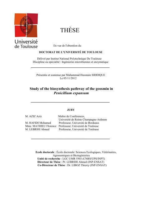
THÈSE<br />
En vue de l'obtention du<br />
DOCTORAT DE L’UNIVERSITÉ DE TOULOUSE<br />
Délivré par Institut National Polytechnique De Toulouse<br />
Discipl<strong>in</strong>e ou spécialité : Ingénieries microbiennes et enzymatique<br />
Présentée et soutenue par Muhammad Hussna<strong>in</strong> SIDDIQUE<br />
Le 05/11/2012<br />
<strong>Study</strong> <strong>of</strong> <strong>the</strong> biosyn<strong>the</strong>sis <strong>pathway</strong> <strong>of</strong> <strong>the</strong> <strong>geosm<strong>in</strong></strong> <strong>in</strong><br />
<strong>Penicillium</strong> expansum<br />
JURY<br />
M. AZIZ Aziz Maître de Conférences,<br />
Université de Reims Champagne-Ardenne<br />
M. HAFIDI Mohamed Pr<strong>of</strong>esseur, Université de Bordeaux<br />
Mme. MATHIEU Florence Pr<strong>of</strong>esseur, Université de Toulouse<br />
M. LEBRIHI Ahmed Pr<strong>of</strong>esseur, Université de Toulouse<br />
Ecole doctorale : École doctorale: Sciences Ecologiques, Vétér<strong>in</strong>aires,<br />
Agronomiques et Bio<strong>in</strong>génieries<br />
Unité de recherche : LGC UMR 5503 (CNRS/UPS/INPT)<br />
Directeur de Thèse : Pr. LEBRIHI Ahmed (INP-ENSAT)<br />
Co-Directeur de Thèse : Dr. LIBOZ Thierry (INP-ENSAT)<br />
1
Acknowledgements<br />
Acknowledgements<br />
First <strong>of</strong> all I am thankful to <strong>the</strong> almighty ALLAH, whose bless<strong>in</strong>gs are always with<br />
me.<br />
I <strong>of</strong>fer my humble thanks from <strong>the</strong> deepest core <strong>of</strong> my heart to Holy Prophet<br />
Muhammad (Peace be upon him) who is forever a torch <strong>of</strong> guidance and knowledge for<br />
humanity as a whole.<br />
I have <strong>the</strong> deepest sense <strong>of</strong> gratitude to my Saa<strong>in</strong> Gee So<strong>of</strong>i Nisar Ahmad Dogar<br />
Naqshbandi Khaliqi who has always been a source <strong>of</strong> elevation <strong>in</strong> my whole life.<br />
My s<strong>in</strong>cere appreciation goes to my supervisor Pr<strong>of</strong>essor Ahmed LEBRIHI and cosupervisor<br />
Doctor Thierry LIBOZ, whose scientific approach, careful read<strong>in</strong>g and<br />
constructive comments were valuable. Their timely and efficient contributions helped me to<br />
shape my research work <strong>in</strong>to its f<strong>in</strong>al form and I express my s<strong>in</strong>cerest appreciation for <strong>the</strong>ir<br />
assistance <strong>in</strong> any way that I may have asked.<br />
I deem it utmost pleasure to avail this opportunity to express <strong>the</strong> heartiest gratitude<br />
and deep sense <strong>of</strong> obligation to Ahmed LEBRIHI for <strong>the</strong>ir dexterous guidance, critical<br />
judgment, sympa<strong>the</strong>tic attitude and <strong>in</strong>spir<strong>in</strong>g efforts to <strong>in</strong>culcate <strong>in</strong> me <strong>the</strong> spirit <strong>of</strong> motivation<br />
dur<strong>in</strong>g <strong>the</strong> course <strong>of</strong> my research work. They were always available whenever I need <strong>the</strong>ir<br />
assistance and guidance, especially, dur<strong>in</strong>g <strong>the</strong>sis writ<strong>in</strong>g.<br />
S<strong>in</strong>cere thanks are due to Pr<strong>of</strong>essor Florence MATHIEU for her k<strong>in</strong>dness and help<strong>in</strong>g<br />
<strong>in</strong> <strong>the</strong>sis work.<br />
I also wish to thank <strong>the</strong> ―Higher Education Commission (HEC)‖ <strong>of</strong> Pakistan, its<br />
leadership and <strong>the</strong> staff who <strong>in</strong> <strong>the</strong>ir limited resources supported me f<strong>in</strong>ancially for my studies<br />
<strong>in</strong> France. I must also mention <strong>the</strong> services provided by SFERE (Société Française d'<br />
Exportation des Resources Educatives) to facilitate my liv<strong>in</strong>g <strong>in</strong> France.<br />
I fervently extend my zealous thanks to <strong>the</strong> members <strong>of</strong> my <strong>the</strong>sis jury Dr. AZIZ<br />
Aziz, Pr. HAFIDI Mohamed, Pr. MATHIEU Florence and Pr. Ahmed LEBRIHI .<br />
I would like to reflect my gratitude to Pr<strong>of</strong>essor Nasserd<strong>in</strong>e SABAOU, Pr<strong>of</strong>essor<br />
Abdelghani ZITOUNI and all my colleagues <strong>of</strong> Laboratoire de Génie Chimique especially<br />
2
Acknowledgements<br />
Atika MEKLAT, Nafees BACHA, Saima MUZAMMIL, Philippe ANSON, Rafik, Hafsa,<br />
Elida, Carol, Marion, Safwan, Patricia and Rayenne who always helped me and gave me<br />
strong support.<br />
I am also <strong>in</strong>debted to my friends who were always with me <strong>in</strong> every situation and<br />
helped me morally and I would like to reflect my gratitude particularly Ali, Saqla<strong>in</strong>, Ramiz,<br />
Tusawar, Tausif and Imran.<br />
F<strong>in</strong>ally, I am forever <strong>in</strong>debted to my family: my fa<strong>the</strong>r Noor MUHAMMAD and my<br />
mo<strong>the</strong>r Hameeda BIBI. A special thanks to my wife Dr. Saima MUZAMMIL who always<br />
shares my problems with an ultimate solution, and cooperated at each and every crucial step<br />
<strong>of</strong> my life. My humble thanks to my bro<strong>the</strong>rs: Shaukat Ali (late), Abdul Razzaq and<br />
Muhammad Riaz and my sisters: Shagufta Parveen, Tasleem Kousar and Afshan Sahar, and<br />
my niece Tehm<strong>in</strong>a. Last but not <strong>the</strong> least I feel pleasure to acknowledge <strong>of</strong> those who love me<br />
and whom I love.<br />
I dedicate this <strong>the</strong>sis <strong>in</strong> honor <strong>of</strong> my family especially my dear bro<strong>the</strong>r Abdul Razzaq<br />
whose love, affection and confidence enabled me to achieve my goals.<br />
3
summary<br />
Summary<br />
Résumé ............................................................................................................................. 7<br />
Abstract ............................................................................................................................ 8<br />
List <strong>of</strong> Abbreviations ....................................................................................................... 9<br />
List <strong>of</strong> Tables .................................................................................................................. 11<br />
List <strong>of</strong> Figures ................................................................................................................ 12<br />
Chapter I: Literature Review ...................................................................................... 14<br />
1.1. Fungal secondary metabolites .................................................................................. 15<br />
1.2. <strong>Penicillium</strong> expansum .............................................................................................. 18<br />
1.2.1. Classification and morphological description ....................................................... 18<br />
1.2.2. Hosts ...................................................................................................................... 21<br />
1.2.3. Secondary metabolites produced by P. expansum ................................................ 21<br />
1.3. Cytochrome P450 monooxygenase .......................................................................... 23<br />
1.3.1. Characteristics <strong>of</strong> cytochrome P450s .................................................................... 23<br />
1.3.2. Structure <strong>of</strong> P450 ................................................................................................... 23<br />
1.3.3. Reactions catalyzed by P450s ............................................................................... 26<br />
1.3.4. Involvement <strong>of</strong> P450s <strong>in</strong> biosyn<strong>the</strong>sis <strong>of</strong> secondary metabolites and <strong>the</strong>ir different<br />
functions .......................................................................................................................... 26<br />
1.4. Geosm<strong>in</strong> .................................................................................................................. 27<br />
1.4.1. General characteristics .......................................................................................... 27<br />
1.4.2. Production <strong>of</strong> <strong>geosm<strong>in</strong></strong> by microorganisms ........................................................... 28<br />
1.4.3. Different methods to analyze <strong>geosm<strong>in</strong></strong>s ................................................................ 31<br />
1.4.4. Treatments to control <strong>geosm<strong>in</strong></strong> .............................................................................. 33<br />
1.4.5. Biosyn<strong>the</strong>sis <strong>pathway</strong>s <strong>of</strong> gesm<strong>in</strong> ......................................................................... 36<br />
1.4.5.1. Biocchemical <strong>pathway</strong> <strong>of</strong> <strong>geosm<strong>in</strong></strong> syn<strong>the</strong>sis <strong>in</strong> bacteria ................................... 36<br />
1.4.5.2. Genes <strong>in</strong>volved <strong>in</strong> <strong>geosm<strong>in</strong></strong> biosyn<strong>the</strong>tic <strong>pathway</strong> <strong>in</strong> bacteria ........................... 38<br />
1.4.5.3. Mechanism and stereochemistry <strong>of</strong> <strong>the</strong> conversion <strong>of</strong> farnesyldiphosphate to<br />
germacradienol and germacrene D .................................................................................. 40<br />
Objectives ………………………………………………………………………………42<br />
Chapter II: Materials and Methods ............................................................................ 43<br />
4
summary<br />
Chapter III: Results and Discussion ............................................................................ 63<br />
3.1. Whe<strong>the</strong>r cytocrome P450 monooxygenase genes can be <strong>in</strong>volved <strong>in</strong> <strong>geosm<strong>in</strong></strong><br />
production ...................................................................................................................... 64<br />
3.1.1. Bio<strong>in</strong>formatics analysis to identify germacradienol/<strong>geosm<strong>in</strong></strong> synthase <strong>in</strong><br />
<strong>Penicillium</strong> ...................................................................................................................... 64<br />
3.1.2.Role <strong>of</strong> P450 <strong>in</strong> terpens biosyn<strong>the</strong>sis ..................................................................... 65<br />
3.1.3. Amplification <strong>of</strong> P. expansum P450 (gpe1)gene sequence by PCR ..................... 66<br />
3.1.4. Alignment <strong>of</strong> gpe1 with o<strong>the</strong>r cytochrom P450 monooxygenases ....................... 69<br />
3.1.5. gpe1 gene presence <strong>in</strong> <strong>geosm<strong>in</strong></strong> produc<strong>in</strong>g <strong>Penicillium</strong> species ............................ 71<br />
3.2. How to explore different aspects <strong>of</strong> <strong>the</strong> gpe1 gene us<strong>in</strong>g bio<strong>in</strong>formatics tools ...... 72<br />
3.2.1. Implication <strong>of</strong> P450 enzymes <strong>in</strong> <strong>the</strong> biosyn<strong>the</strong>sis <strong>of</strong> different secondary<br />
metabolites… .................................................................................................................. 72<br />
3.2.2. Conserved doma<strong>in</strong>s <strong>of</strong> cytochrome P450s ............................................................ 73<br />
3.2.3.Which functional doma<strong>in</strong>s <strong>of</strong> cytochrome P450 monooxygenase enzymes are<br />
present <strong>in</strong> gpe1. ............................................................................................................... 74<br />
3.3. Does <strong>the</strong> gpe1 gene require for <strong>geosm<strong>in</strong></strong> biosyn<strong>the</strong>sis <strong>in</strong> P. expansum. ................. 76<br />
3.3.1. Production <strong>of</strong> mutants by <strong>the</strong> gene disruption method and <strong>the</strong>ir screen<strong>in</strong>g by<br />
PCRs. ............................................................................................................................... 76<br />
3.2.3. Production <strong>of</strong> reverse compliments and <strong>the</strong>ir screen<strong>in</strong>g by PCRs. ....................... 77<br />
3.3.3. Quantifictaion <strong>of</strong> <strong>geosm<strong>in</strong></strong> by gas chromatography-mass spectrometry(GC/MS)..78<br />
3.4. Where does <strong>the</strong> gpe1 gene <strong>in</strong>tervene <strong>in</strong> <strong>the</strong> biosyn<strong>the</strong>sis <strong>pathway</strong> <strong>of</strong> <strong>geosm<strong>in</strong></strong>. ..... 79<br />
3.4.1. Dependence <strong>of</strong> secondary metabolites <strong>pathway</strong>s upon <strong>the</strong> relative abundance <strong>of</strong><br />
<strong>the</strong>ir precursors. ............................................................................................................... 79<br />
3.4.2: Is <strong>the</strong> biosyn<strong>the</strong>sis <strong>pathway</strong> <strong>of</strong> <strong>geosm<strong>in</strong></strong> same <strong>in</strong> P. expansum as that suggested <strong>in</strong><br />
bacteria.. ........................................................................................................................ 81<br />
3.4.3. Use <strong>of</strong> <strong>Penicillium</strong> marneffei genome to know about <strong>the</strong> neighbor genes <strong>of</strong> <strong>the</strong><br />
gpe1. ................................................................................................................................ 81<br />
3.5. Chapter IV: General Conclusions and Future Prospects. .................................. 84<br />
5
summary<br />
References. ..................................................................................................................... 89<br />
Annexes ........................................................................................................................ 109<br />
6
Résumé<br />
Résumé<br />
La géosm<strong>in</strong>e est un terpénoïde, provoquant un goût moisi-terreux associée à des<br />
flaveurs atypiques dans l'eau et le v<strong>in</strong>. Chez les bactéries, la voie de biosynthèse de la<br />
géosm<strong>in</strong>e est bien caractérisée, mais peu de connaissance sont disponibles au sujet de sa<br />
biosynthèse chez les eucaryotes, en particulier dans les champignons filamenteux. L'orig<strong>in</strong>e<br />
de la géosm<strong>in</strong>e dans la vigne est en grande partie attribuable à la présence de <strong>Penicillium</strong><br />
expansum sur les rais<strong>in</strong>s. Dans cette thèse, af<strong>in</strong> de mieux comprendre la voie de biosynthèse<br />
de la géosm<strong>in</strong>e chez <strong>Penicillium</strong> expansum, nous avons décrit la caractérisation et l'analyse<br />
de "gpe1", un gène codant pour une cytochrome P450 monooxygénase impliquée dans la<br />
biosynthèse de la géosm<strong>in</strong>e.<br />
Nous avons démontré que les deux fragments d'ADN: p450-1 et p450-2<br />
appartiennent à un seul gène du cytochrome p450 (gpe1). La séquence d'acides am<strong>in</strong>és<br />
déduite de gpe1 a une identité moyenne de 40 % avec les enzymes PbP450-2 et P450-4 qui<br />
ont été trouvées impliquées respectivement dans la synthèse d'<strong>in</strong>dole diterpène et dans la<br />
synthèse des gibbérell<strong>in</strong>es. Les amplifications par PCR effectuée sur quatorze espèces de<br />
<strong>Penicillium</strong> ont montré que seules les espèces producteurices de la géosm<strong>in</strong>e ont donné le<br />
même fragment de ~1,2 kb que gpe1. L'analyse du gène gpe1 nous a permis d'identifier la<br />
présence de certa<strong>in</strong>s doma<strong>in</strong>es conservés de cytochromes P450 monooxygénases. Ensuite, la<br />
caractérisation fonctionnelle du gène gpe1 chez P. expansum M2230 a été décrite. Nous<br />
avons montré que les mutants de gpe1 ont perdus leur pouvoir de produire la géosm<strong>in</strong>e alors<br />
que les révertants de gpe1 ont rétablis leur pouvoir de production. Enf<strong>in</strong>, nous avons<br />
démontré qu'une polykétide synthase putative et une putative NRPS sont présentes sur le<br />
côté droit du gène gpe1 proposant que le gène gpe1 pourrait être une partie d'un "Cluster"<br />
codant pour la biosynthèse de métabolites secondaires.<br />
Mots clés: Cytochrome P450 monooxygénase, géosm<strong>in</strong>e, gpe1, <strong>Penicillium</strong><br />
expansum.<br />
7
Abstract<br />
Abstract<br />
Geosm<strong>in</strong> is a terpenoid, an earthy-musty compound associated with <strong>of</strong>f-flavors <strong>in</strong><br />
water and w<strong>in</strong>e. In bacteria, <strong>the</strong> biosyn<strong>the</strong>sis <strong>pathway</strong> <strong>of</strong> <strong>geosm<strong>in</strong></strong> is well characterized, but<br />
little is known about its biosyn<strong>the</strong>sis <strong>in</strong> eukaryotes, especially <strong>in</strong> filamentous fungi. The<br />
orig<strong>in</strong> <strong>of</strong> <strong>geosm<strong>in</strong></strong> <strong>in</strong> grapev<strong>in</strong>e is largely attributable to <strong>the</strong> presence <strong>of</strong> <strong>Penicillium</strong><br />
expansum on grapes. In this <strong>the</strong>sis, we have described <strong>the</strong> characterization and analysis <strong>of</strong><br />
―gpe1‖, a gene encod<strong>in</strong>g a cytochrome P450 monooxygenase probably <strong>in</strong>volved <strong>in</strong> <strong>the</strong><br />
biosyn<strong>the</strong>sis <strong>of</strong> <strong>geosm<strong>in</strong></strong> <strong>in</strong> P. expansum M2230, <strong>in</strong> order to better understand <strong>of</strong> <strong>the</strong><br />
biosyn<strong>the</strong>sis <strong>pathway</strong> <strong>of</strong> <strong>geosm<strong>in</strong></strong> <strong>in</strong> this species.<br />
. We demonstrated that <strong>the</strong> two DNA fragments i.e. p450-1 and p450-2 belong to a<br />
s<strong>in</strong>gle cytochrome p450 gene (gpe1). We showed that <strong>the</strong> deduced am<strong>in</strong>o acid sequence <strong>of</strong><br />
gpe1 has an average identity <strong>of</strong> 40 % with PbP450-2 and P450-4 enzymes which have been<br />
found <strong>in</strong>volved <strong>in</strong> <strong>in</strong>dole diterpene syn<strong>the</strong>sis and <strong>in</strong> gibberell<strong>in</strong> syn<strong>the</strong>sis respectively. Then,<br />
<strong>the</strong> results <strong>of</strong> PCRs performed on <strong>the</strong> fourteen <strong>Penicillium</strong> species showed that only<br />
<strong>Penicillium</strong> species which were producers <strong>of</strong> <strong>geosm<strong>in</strong></strong> gave <strong>the</strong> same fragment <strong>of</strong> ~1.2 kb<br />
like gpe1. Analysis <strong>of</strong> <strong>the</strong> gpe1 gene enabled us to identify <strong>the</strong> presence <strong>of</strong> some conserved<br />
doma<strong>in</strong>s <strong>of</strong> cytochromes P450 monoxygenases <strong>in</strong> <strong>the</strong> am<strong>in</strong>o acid sequence <strong>of</strong> gpe1. Then,<br />
<strong>the</strong> functional characterization <strong>of</strong> <strong>the</strong> gpe1 gene <strong>in</strong> P. expansum M2230 was described. We<br />
illustrated that <strong>the</strong> mutants <strong>of</strong> gpe1 lost <strong>the</strong>ir potential to produce <strong>geosm<strong>in</strong></strong> whereas <strong>the</strong><br />
reverse complements <strong>of</strong> gpe1 restored <strong>the</strong>ir potential to produce <strong>geosm<strong>in</strong></strong>. F<strong>in</strong>ally, we<br />
demonstrated that a putative polyketide synthase and a putative NRPS-like enzyme are<br />
present on <strong>the</strong> right side <strong>of</strong> <strong>the</strong> gpe1 gene suggest<strong>in</strong>g that gpe1 gene might be <strong>the</strong> part <strong>of</strong> a<br />
gene cluster encod<strong>in</strong>g <strong>the</strong> biosyn<strong>the</strong>sis <strong>of</strong> secondary metabolites.<br />
Key words: Cytochrome P450 monooxygenase, <strong>geosm<strong>in</strong></strong>, gpe1, <strong>Penicillium</strong><br />
expansum.<br />
8
List <strong>of</strong> Abbreviations<br />
List <strong>of</strong> Abbreviations<br />
2-MIB:<br />
aa:<br />
BLAST:<br />
bp:<br />
CoA:<br />
CPR:<br />
CTAB:<br />
CYA<br />
CYP<br />
DNA:<br />
EDTA:<br />
ER:<br />
FAD:<br />
FCPD:<br />
FMN:<br />
FPP:<br />
GAC:<br />
GC–MS:<br />
GGPP:<br />
hph:<br />
LQ:<br />
MEA:<br />
NADPH:<br />
NRPS:<br />
OD:<br />
PAC:<br />
PCR:<br />
PKS:<br />
RNA:<br />
rpm:<br />
2-methylisoborneol<br />
Am<strong>in</strong>o acid<br />
Basic local alignment search tool<br />
Base pair<br />
Coenzyme-A<br />
Cytochrome P450 reductase<br />
Cetyltrimethylammonium bromide<br />
Czapek yeast agar<br />
Cytochrome P450<br />
Deoxyribonucleic acid<br />
Ethylenediam<strong>in</strong>etetraacetic acid<br />
Endoplasmatic reticulum<br />
Flav<strong>in</strong>e aden<strong>in</strong>e d<strong>in</strong>ucleotide<br />
Fungal cytochrome P450 database<br />
Flav<strong>in</strong> mononucleotide<br />
Farnesyl diphosphate<br />
Granular activated carbon<br />
Gas chromatography-mass spectrometry<br />
Geranylgeranyl diphosphate<br />
Hygromyc<strong>in</strong> B phosphotransferase<br />
Limit <strong>of</strong> quantification<br />
Malt Agar Extract<br />
Nicot<strong>in</strong>amide aden<strong>in</strong>e d<strong>in</strong>ucleotide phosphate (reduced form)<br />
Non-ribosomal peptide synthase<br />
Optical Density<br />
Powdered activated carbon<br />
Polymerase cha<strong>in</strong> reaction<br />
Polyketide synthase<br />
Ribonucleic acid<br />
Rotation per m<strong>in</strong>ute<br />
9
List <strong>of</strong> Abbreviations<br />
SDS:<br />
TOC:<br />
UV:<br />
YES:<br />
Sodium dodecyl sulfate<br />
Total organic carbon<br />
Ultraviolet<br />
Yeast extract sucrose<br />
10
List <strong>of</strong> Tables<br />
List <strong>of</strong> Tables<br />
Table 1: Production <strong>of</strong> <strong>geosm<strong>in</strong></strong> by different organisms ................................................. 30<br />
Table 2: Extraction / enrichment techniques used to preconcentrate <strong>geosm<strong>in</strong></strong> prior<br />
to quantification by gas chromatography-mass spectrometry ......................................... 32<br />
Table 3: Different treatments to control <strong>geosm<strong>in</strong></strong> <strong>in</strong> water ............................................. 35<br />
Table 4: Dosage <strong>of</strong> <strong>geosm<strong>in</strong></strong> ............................................................................................ 79<br />
11
List <strong>of</strong> Figures<br />
List <strong>of</strong> Figures<br />
Figure 1: <strong>Penicillium</strong> expansum A-C, 7-days-old colonies on A. CYA, B. MEA, C. YES<br />
at 25 °C. .......................................................................................................................... 20<br />
Figure 2: <strong>Penicillium</strong> expansum, D-H. Conidiophores. I. Conidia. Scale bar = 10 µm. 20<br />
Figure 3: Schematic representation <strong>of</strong> <strong>the</strong> eukaryotic endoplasmatic reticulum type<br />
cytochrome P450 enzyme system .................................................................................. 25<br />
Figure 4: Chemical structure <strong>of</strong> <strong>geosm<strong>in</strong></strong> ...................................................................... 28<br />
Figure 5: Simplified biosyn<strong>the</strong>tic scheme for <strong>the</strong> formation <strong>of</strong> <strong>geosm<strong>in</strong></strong> <strong>in</strong> streptomycetes<br />
and myxobacteria. .......................................................................................................... 37<br />
Figure 6: Mechanism <strong>of</strong> cyclization <strong>of</strong> FPP (2) to Germacradienol (3), Germacrene D (4),<br />
Hydrocarbon 5, and Geosm<strong>in</strong> (1) ................................................................................... 39<br />
Figure 7: Organization <strong>of</strong> prote<strong>in</strong> doma<strong>in</strong> and conserved Mg 2+ -b<strong>in</strong>d<strong>in</strong>g motifs <strong>in</strong> S.<br />
coelicolor germcradienol-<strong>geosm<strong>in</strong></strong> synthase .................................................................. 40<br />
Figure 8: Mechanism and stereochemistry <strong>of</strong> <strong>the</strong> cyclization <strong>of</strong> FPP (2) to germacradienol<br />
(3), germacrene D (4), octal<strong>in</strong> (5) and <strong>geosm<strong>in</strong></strong> (1)......................................................... 41<br />
Figure 9: Schematic representation <strong>of</strong> transformation vector formation and gpe1 gene<br />
disruption. ....................................................................................................................... 58<br />
Figure 10: A BLAST search with <strong>the</strong> prote<strong>in</strong> sequence <strong>of</strong> <strong>the</strong> S. peucetius strept13<br />
(ATCC 27952) as a query did not show any gene hav<strong>in</strong>g homology with <strong>the</strong> genes<br />
encod<strong>in</strong>g germacradienol/<strong>geosm<strong>in</strong></strong> synthase, <strong>in</strong> <strong>the</strong> genus <strong>Penicillium</strong> .......................... 65<br />
Figure 11: Alignment <strong>of</strong> <strong>the</strong> deduced am<strong>in</strong>o acid sequences <strong>of</strong> p450-1 and p450-2 <strong>of</strong><br />
<strong>Penicillium</strong> expansum with CYP619C2 and CYP619C3 <strong>of</strong> Aspergillus clavatus <strong>in</strong>volved<br />
<strong>in</strong> patul<strong>in</strong> biosyn<strong>the</strong>sis .................................................................................................... 67<br />
12
List <strong>of</strong> Figures<br />
Figure 12: Alignment <strong>of</strong> <strong>the</strong> deduced am<strong>in</strong>o acid sequence <strong>of</strong> p450-1 and p450-2 <strong>of</strong><br />
<strong>Penicillium</strong> expansum with P450-4 <strong>of</strong> Gibberella fujikuroi <strong>in</strong>volved <strong>in</strong> <strong>the</strong> biosyn<strong>the</strong>sis <strong>of</strong><br />
gibberell<strong>in</strong>s ...................................................................................................................... 68<br />
Figure 13: Amplification <strong>of</strong> <strong>the</strong> gpe1 us<strong>in</strong>g <strong>the</strong> primers mhsF and mhsR <strong>in</strong> P. expansum.<br />
......................................................................................................................................... 69<br />
Figure 14: Alignment <strong>of</strong> <strong>the</strong> deduced am<strong>in</strong>o acid sequence <strong>of</strong> gpe1 with o<strong>the</strong>r cytochrome<br />
P450 monooxygenases .................................................................................................... 70<br />
Figure 15: gpe1 PCR amplification on <strong>geosm<strong>in</strong></strong> productive and non-productive<br />
<strong>Penicillium</strong> species .......................................................................................................... 71<br />
Figure 16: Typical features <strong>of</strong> an ER-bound P450 prote<strong>in</strong> ............................................. 74<br />
Figure17: Conserved doma<strong>in</strong>s <strong>of</strong> cytochromes P450 monooxygenases present <strong>in</strong> gpe .. 75<br />
Figure 18: InterPro Scan visual output show<strong>in</strong>g different doma<strong>in</strong>s <strong>of</strong> gep1………….76<br />
Figure 19: PCR transformants screen<strong>in</strong>g ........................................................................ 78<br />
Figure 20: Metabolic <strong>pathway</strong> diagram, <strong>the</strong> mevalonate (MVA) and non-mevalonate<br />
(MEP) <strong>pathway</strong>s that l<strong>in</strong>k <strong>geosm<strong>in</strong></strong> syn<strong>the</strong>sis ................................................................. 80<br />
Figure21: A simple diagram show<strong>in</strong>g neighbor genes <strong>of</strong> <strong>the</strong> particular gene <strong>of</strong><br />
<strong>Penicillium</strong> marneffei show<strong>in</strong>g highest resemblance with gpe1<strong>of</strong> P. expansum<br />
as a result <strong>of</strong> BLAST search ............................................................................................ 83<br />
.<br />
13
Chapter I Literature Review<br />
Chapter I<br />
Literature Review<br />
14
Chapter I Literature Review<br />
1. Literature Review<br />
1.1. Fungal Secondary Metabolite<br />
The primary metabolism <strong>of</strong> an organism is <strong>the</strong> summation <strong>of</strong> an <strong>in</strong>terrelated series <strong>of</strong><br />
enzyme-catalyzed chemical reactions (both degradative and syn<strong>the</strong>tic) which provide <strong>the</strong><br />
organism with its energy, its syn<strong>the</strong>tic <strong>in</strong>termediates and its key macromolecules such as<br />
prote<strong>in</strong> and DNA. On <strong>the</strong> o<strong>the</strong>r hand, secondary metabolism <strong>in</strong>volves ma<strong>in</strong>ly syn<strong>the</strong>tic<br />
processes whose end-products, <strong>the</strong> secondary metabolites, play no obvious role <strong>in</strong> <strong>the</strong><br />
<strong>in</strong>ternal economy <strong>of</strong> <strong>the</strong> organism.<br />
Plants and microorganisms produce a vast number <strong>of</strong> natural compounds known as<br />
secondary metabolites. Kossel (1891) <strong>in</strong>troduced <strong>the</strong> concept <strong>of</strong> ―secondary metabolites‖ to<br />
dist<strong>in</strong>guish <strong>the</strong>se compounds from primary metabolites that <strong>the</strong>se products are not necessary<br />
for <strong>the</strong> growth, survival or reproduction <strong>of</strong> <strong>the</strong>ir producers. Secondary metabolites are<br />
substances <strong>of</strong> limited molecular weight (normally < 3000 Daltons) which display an<br />
enormous structural diversity. However each <strong>of</strong> <strong>the</strong>m is syn<strong>the</strong>sized only by a limited<br />
taxonomic group <strong>of</strong> organisms whereas primary metabolites are found <strong>in</strong> all liv<strong>in</strong>g<br />
organisms s<strong>in</strong>ce <strong>the</strong>y perform essential functions <strong>in</strong> growth and development.<br />
Many species <strong>in</strong> <strong>the</strong> fungal k<strong>in</strong>gdom have unique and unusual biochemical<br />
<strong>pathway</strong>s. Important pharmaceuticals such as penicill<strong>in</strong>, cyclospor<strong>in</strong> and stat<strong>in</strong>s; potent<br />
poisons, <strong>in</strong>clud<strong>in</strong>g aflatox<strong>in</strong>s and tricho<strong>the</strong>cenes; and some Janus-faced metabolites that are<br />
both toxic and pharmaceutically useful, such as <strong>the</strong> ergot alkaloids are <strong>the</strong> products <strong>of</strong> <strong>the</strong>se<br />
<strong>pathway</strong>s. All <strong>of</strong> <strong>the</strong>se natural products, along with many o<strong>the</strong>r low-molecular-weight fungal<br />
metabolites, are classified toge<strong>the</strong>r as secondary metabolites. Secondary metabolites are<br />
produced as families <strong>of</strong> related compounds at restricted parts <strong>of</strong> <strong>the</strong> life cycle, with<br />
production <strong>of</strong>ten correlated with a specific stage <strong>of</strong> morphological differentiation. Secondary<br />
metabolites have restricted taxonomic distribution as only a small group <strong>of</strong> organisms<br />
15
Chapter I Literature Review<br />
produces each metabolite and <strong>the</strong> producer organisms can grow without syn<strong>the</strong>siz<strong>in</strong>g <strong>the</strong>se<br />
metabolites. Secondary metabolites are, <strong>of</strong>ten syn<strong>the</strong>sized after active growth has ceased,<br />
which do not have an obvious function <strong>in</strong> producer species (Keller et al., 2005).<br />
In fungi, <strong>in</strong> <strong>the</strong> case <strong>of</strong> <strong>the</strong> Basidiomycetes and <strong>the</strong> larger Ascomycetes, secondary<br />
metabolites may be obta<strong>in</strong>ed simply by extraction <strong>of</strong> <strong>the</strong> organism collected <strong>in</strong> <strong>the</strong> field. But<br />
<strong>the</strong> great advantage <strong>of</strong> <strong>the</strong> fungi as sources <strong>of</strong> secondary metabolites is <strong>the</strong>ir ability to<br />
produce <strong>the</strong> compounds on aqueous media. As a result, secondary metabolites <strong>of</strong> diverse<br />
type are conveniently available <strong>in</strong> <strong>the</strong> laboratory for chemical, biochemical and biological<br />
studies, and a few are manufactured on a commercial scale. In some cases <strong>the</strong> same<br />
secondary metabolites have been obta<strong>in</strong>ed from fruit<strong>in</strong>g-bodies and from aqueous culture <strong>of</strong><br />
Basidomycetes, though <strong>in</strong> most cases <strong>the</strong> compounds have so far only been obta<strong>in</strong>ed from<br />
one <strong>of</strong> <strong>the</strong> sources. The laboratory cultures <strong>of</strong> Basidiomycetes are, <strong>of</strong> course, mycelial;<br />
Basidiomycetes do not normally form fruit<strong>in</strong>g-bodies under laboratory conditions and <strong>in</strong><br />
some cases have resisted all attempts to <strong>in</strong>duce <strong>the</strong>m to do so. In aqueous cultures,<br />
secondary metabolites accumulate both <strong>in</strong> <strong>the</strong> medium and <strong>in</strong> <strong>the</strong> mycelium. For related<br />
compounds, <strong>the</strong> distribution between medium and mycelium can <strong>of</strong>ten be correlated with<br />
water-solubility, though this apparent correlation may be a result <strong>of</strong> some o<strong>the</strong>r factor such<br />
as ease <strong>of</strong> transport across cell membranes (Turner, 1971).<br />
Fungal secondary metabolites encompass over 30,000 known compounds with an<br />
extremely diverse array <strong>of</strong> chemical structures. It is <strong>in</strong>trigu<strong>in</strong>g that all <strong>the</strong>se secondary<br />
metabolites orig<strong>in</strong>ate from a few common biosyn<strong>the</strong>tic <strong>pathway</strong>s utiliz<strong>in</strong>g precursors (small<br />
biosyn<strong>the</strong>tic units or build<strong>in</strong>g blocks) formed dur<strong>in</strong>g primary metabolism. The <strong>in</strong>termediates<br />
result<strong>in</strong>g from condensation <strong>of</strong> <strong>the</strong>se small biosyn<strong>the</strong>tic units are fur<strong>the</strong>r elaborated<br />
(―tailored‖ or ―decorated‖) by numerous enzyme-catalyzed reactions, lead<strong>in</strong>g to products<br />
with a diversity <strong>of</strong> structures. Thus, fungal secondary metabolites are conveniently classified<br />
16
Chapter I Literature Review<br />
based on <strong>the</strong>ir biosyn<strong>the</strong>tic orig<strong>in</strong> as polyketides (e.g. aflatox<strong>in</strong> and fumonis<strong>in</strong>s),<br />
nonribosomal peptides (e.g. sirodesm<strong>in</strong>, peram<strong>in</strong>e and siderophores such as ferricroc<strong>in</strong>),<br />
terpenes (e.g. T-2 tox<strong>in</strong>, deoxynivalenol (DON)), and <strong>in</strong>dole alkaloids (e.g. paxill<strong>in</strong>e,<br />
fumigaclav<strong>in</strong>es and fumitremorgens) (Keller et al., 2005; Gunatilaka, 2006).<br />
Biosyn<strong>the</strong>sis <strong>of</strong> fungal secondary metabolites <strong>of</strong>ten <strong>in</strong>volves elaborate biochemical<br />
<strong>pathway</strong>s and is regulated by a group <strong>of</strong> genes known as biosyn<strong>the</strong>tic genes. The <strong>in</strong>sights<br />
that have been ga<strong>in</strong>ed from recent advances <strong>in</strong> genetics, genomics, molecular biology, and<br />
bio<strong>in</strong>formatics have contributed to <strong>the</strong> understand<strong>in</strong>g and manipulation <strong>of</strong> <strong>the</strong>se genes for<br />
improved production, or <strong>in</strong>hibition <strong>of</strong> production, <strong>of</strong> fungal secondary metabolites.<br />
Fungal secondary metabolites are well known for <strong>the</strong>ir biological activity and<br />
represent some <strong>of</strong> today's important and useful pharmaceuticals and agrochemicals. Among<br />
<strong>the</strong> pharmaceuticals, most noteworthy are penicill<strong>in</strong>s, cephalospor<strong>in</strong>s, and fusidic acid with<br />
antibacterial activity; ech<strong>in</strong>ocand<strong>in</strong> B, pneumocand<strong>in</strong>s, grise<strong>of</strong>ulv<strong>in</strong>, and strobilur<strong>in</strong>s with<br />
antifungal activity; <strong>in</strong>tegric acid and <strong>in</strong>tegresone with antiviral activity; cyclospor<strong>in</strong> A and<br />
mycophenolic acid with immunosuppressive activity; fumagill<strong>in</strong> and rhizox<strong>in</strong> with<br />
antitumor activity; lovastat<strong>in</strong> and pravastat<strong>in</strong> with cholesterol-lower<strong>in</strong>g activity; and ergot<br />
alkaloids (for example, ergotam<strong>in</strong>e) with antimigra<strong>in</strong>e activity. Gibberell<strong>in</strong>s and<br />
zearalenones are fungal secondary metabolites used <strong>in</strong> agriculture as plant growth hormones<br />
and <strong>in</strong> animal husbandry as growth promoters, respectively. Some fungal secondary<br />
metabolites such as mycotoxic aflatox<strong>in</strong>s and mutagenic fusar<strong>in</strong> C possess potent toxic and<br />
carc<strong>in</strong>ogenic activities and are <strong>the</strong>refore important <strong>in</strong> human, animal, and plant health<br />
(V<strong>in</strong><strong>in</strong>g, 1990; Fox and Howlett, 2008) whereas some volatile non-toxic secondary<br />
metabolites such as <strong>geosm<strong>in</strong></strong> and 2-methylisoborneol (2-MIB) haves also concerns for<br />
humans as <strong>the</strong>y are responsible for <strong>of</strong>f-flavors <strong>in</strong> dr<strong>in</strong>k<strong>in</strong>g water and w<strong>in</strong>es (Gerber, 1979;<br />
Darriet et al., 2000).<br />
17
Chapter I Literature Review<br />
In some fungi, secondary metabolism (<strong>the</strong> process that results <strong>in</strong> <strong>the</strong> production <strong>of</strong><br />
secondary metabolites) has been found to commence dur<strong>in</strong>g <strong>the</strong> stationary or rest<strong>in</strong>g phase<br />
<strong>of</strong> <strong>the</strong>ir development and is <strong>of</strong>ten associated with sporulation and colony formation. Some<br />
well-documented functions <strong>of</strong> fungal secondary metabolites <strong>in</strong>clude enhancement <strong>of</strong> spore<br />
survival by act<strong>in</strong>g as virulence factors and protect<strong>in</strong>g aga<strong>in</strong>st ultraviolet (UV) light, and<br />
augmentation <strong>of</strong> <strong>the</strong>ir fitness and competitive ability aga<strong>in</strong>st o<strong>the</strong>r fast-grow<strong>in</strong>g organisms.<br />
Fungal metabolites associated with sporulation may activate sporulation (for example,<br />
l<strong>in</strong>oleic acid analogs produced by Aspergillus nidulans), provide pigmentation required for<br />
sporulation structures (for example, melan<strong>in</strong>s produced by Alternaria alternata), or have<br />
toxic properties to ward <strong>of</strong>f compet<strong>in</strong>g organisms (for example, mycotox<strong>in</strong>s produced by<br />
some Aspergillus species). The relationship between production <strong>of</strong> secondary metabolites<br />
and regulation <strong>of</strong> asexual sporulation by a G-prote<strong>in</strong>–mediated growth <strong>pathway</strong><br />
<strong>in</strong> Aspergillus species was established over a decade ago. Also, it has been speculated that<br />
secondary metabolites <strong>in</strong> fungi function as metal chelators (comb<strong>in</strong><strong>in</strong>g with metal ions and<br />
remov<strong>in</strong>g <strong>the</strong>m from <strong>the</strong>ir sphere <strong>of</strong> action), which is important <strong>in</strong> m<strong>in</strong>eral nutrition, and that<br />
<strong>pathway</strong>s lead<strong>in</strong>g to <strong>the</strong>ir formation act as safety-valve shunts that prevent <strong>the</strong> accumulation<br />
<strong>of</strong> toxic <strong>in</strong>termediates <strong>of</strong> primary metabolism under conditions <strong>of</strong> unbalanced growth (Calvo<br />
et al., 2002; Fox and Howlett, 2008).<br />
1.2. <strong>Penicillium</strong> expansum<br />
1.2.1. Classification and morphological description<br />
<strong>Penicillium</strong> expansum is <strong>the</strong> typical fungus <strong>of</strong> <strong>the</strong> genus <strong>Penicillium</strong> and is <strong>the</strong>refore<br />
also one <strong>of</strong> <strong>the</strong> most studied species <strong>in</strong> <strong>the</strong> genus (Pitt, 1979). This fungus belongs to phylum<br />
Ascomycota, class Eurotiomycetes, subclass Eurotiomycetidae, order Eurotiales and family<br />
Trichocomaceae.<br />
18
Chapter I Literature Review<br />
After 7 days <strong>of</strong> <strong>in</strong>cubation on Petri dish conta<strong>in</strong><strong>in</strong>g CYA (Czapek Yeast Agar)<br />
medium at 25 °C, a colony <strong>of</strong> P. expansum atta<strong>in</strong>s a diameter <strong>of</strong> 26-50 mm, on MEA (Malt<br />
Agar Extract) medium, its colony could be 16-34 mm <strong>in</strong> diameter, while on YES (Yeast<br />
Extract Sucrose) agar medium, <strong>the</strong> diameter <strong>of</strong> a colony could be 38-65 mm (Figure 1) but<br />
<strong>the</strong>re is no growth at 37 °C (Frisvad and Samson, 2004). Cultural characteristics <strong>of</strong> this<br />
fungus <strong>in</strong>clude: <strong>the</strong> colonies grow rapidly on <strong>the</strong> culture media, with radial wr<strong>in</strong>kles up to 2<br />
mm deep, spore heavily, very variable, from velvety with conidiophores occurr<strong>in</strong>g s<strong>in</strong>gly to<br />
granular with conidiophores grouped toge<strong>the</strong>r <strong>in</strong> fascicles or produc<strong>in</strong>g quite dist<strong>in</strong>ct<br />
coremia, <strong>of</strong>ten show<strong>in</strong>g radial zonation; white, rapidly becom<strong>in</strong>g dull yellow green to<br />
greyish green with <strong>the</strong> production <strong>of</strong> conidia (Figure 2); reverse variable, colorless to yellow<br />
brown to deep brown. The conidial heads are asymmetric, once or twice branched, elongate,<br />
bear<strong>in</strong>g long tangled cha<strong>in</strong>s <strong>of</strong> conidia. The conidiophores are smooth or <strong>in</strong> some stra<strong>in</strong>s<br />
slightly roughened, moderately long, up to 400 µm long but occasionally up to 600-700 µm<br />
long and 3-3.5 µm wide; branches 15-25 x 2.5-3.5 µm, occasionally longer. But <strong>the</strong> metulae<br />
aris<strong>in</strong>g from branches at about <strong>the</strong> same level, 3 to 6 <strong>in</strong> number, and about 10-15 x 2-3 µm.<br />
The phialides are <strong>in</strong> groups <strong>of</strong> 5-9, <strong>of</strong>ten about 8-12 x 2-2.5 µm, occasionally longer. The<br />
conidia are smooth, elliptical to cyl<strong>in</strong>drical when first formed and usually rema<strong>in</strong> elliptical,<br />
generally 4-5 x 2.5-3.5 µm (L<strong>in</strong>k, 1809; Onions, 1966)<br />
(http://www.mycobank.org/MycoTaxo.aspxL<strong>in</strong>k=T&Rec=159382).<br />
19
Chapter I Literature Review<br />
YES at 25 °C.<br />
Figure1: <strong>Penicillium</strong> expansum, A-C. 7-days-old colonies on A. CYA, B. MEA, C.<br />
Figure 2: <strong>Penicillium</strong> expansum, D-H. Conidiophores. I. Conidia. Scale bar = 10 µm.<br />
20
Chapter I Literature Review<br />
1.2.2. Host<br />
<strong>Penicillium</strong> expansum is commonly present <strong>in</strong> soil and <strong>in</strong> a wide variety <strong>of</strong> organic<br />
material <strong>in</strong>clud<strong>in</strong>g gra<strong>in</strong>s and cereal products, and though generally isolated from mouldy<br />
fruit, particularly apples, it also occurs on o<strong>the</strong>r pomaceous fruits such as cherries, peaches,<br />
pears, grapes, olives, p<strong>in</strong>eapple and sometimes on citrus and avocado. It is also common on<br />
walnuts, pecans, hazelnuts and acorns. P. expansum is responsible for <strong>the</strong> postharvest decay<br />
<strong>of</strong> <strong>the</strong>se fruits lead<strong>in</strong>g to important economic losses <strong>in</strong> <strong>the</strong> fruit <strong>in</strong>dustry (Filtenborg et al.,<br />
1996; Karabulat et al., 2002; Karabulat and Bakyal, 2002; Ventur<strong>in</strong>i et al., 2002).<br />
1.2.3. Secondary metabolites produced by P. expansum<br />
P. expansum has been reported to produce many secondary metabolites such as:<br />
chaetoglobos<strong>in</strong>s A and C, communes<strong>in</strong> B which are cytotoxic metabolites (Bridge et al.,<br />
1989; Frisvad and Filtenborg, 1989; Frisvad, 1992; Andersen et al., 2004), <strong>the</strong> bioactive<br />
compounds expansolides A and B (Massias et al., 1990; Andersen et al., 2004), an antibiotic<br />
penicillic acid (Leistner and Pitt, 1977), roquefort<strong>in</strong>e C which is neurotoxic (Frisvad and<br />
Filtenborg, 1983; Bridge et al., 1989), patul<strong>in</strong> which is carcenogenic, citr<strong>in</strong><strong>in</strong> which is<br />
nephrotoxic (Leistner and Pitt, 1977; Frisvad and Filtenborg, 1983; Paterson et al., 1987;<br />
Andersen et al., 2004) and <strong>geosm<strong>in</strong></strong> which is an aromatic volatile secondary metabolite<br />
(Mat<strong>the</strong>is and Roberts, 1992). Among <strong>the</strong> above mentioned extrolites <strong>of</strong> P. expansum,<br />
chaetoglobos<strong>in</strong>s A and C, penicillic acid, patul<strong>in</strong> and citr<strong>in</strong><strong>in</strong> belong to polyketides.<br />
Geosm<strong>in</strong> and expansolides A and B are terpenes whereas roquefort<strong>in</strong>e C and communes<strong>in</strong> B<br />
belong to <strong>in</strong>dole alkaloid family.<br />
In literature, it has been reported that citr<strong>in</strong><strong>in</strong> biosyn<strong>the</strong>sis was orig<strong>in</strong>ated from a<br />
pentaketide <strong>in</strong> <strong>Penicillium</strong> and Aspegillus species (Barber and Staunton, 1980; Sankawa et<br />
al., 1983). It was demonstrated <strong>in</strong> literature that <strong>in</strong> <strong>the</strong> genus Aspergillus, <strong>the</strong> condensation<br />
21
Chapter I Literature Review<br />
<strong>of</strong> one acetyl coenzyme A (acetyl-CoA) molecule with four malonyl-CoA molecules,<br />
followed by <strong>the</strong> addition <strong>of</strong> three methyl units has syn<strong>the</strong>sized <strong>the</strong> citr<strong>in</strong><strong>in</strong> (Colombo et al.,<br />
1981; Hill et al., 1981). In <strong>the</strong> contrary, Hajjaj et al. (1999) revealed that citr<strong>in</strong><strong>in</strong> is formed<br />
from a tetraketide precursor aris<strong>in</strong>g from <strong>the</strong> condensation <strong>of</strong> one acetyl-CoA molecule with<br />
three malonyl-CoA molecules <strong>in</strong> <strong>the</strong> filamentous fungus Monascus ruber <strong>in</strong>stead <strong>of</strong> a<br />
pentaketide as reported <strong>in</strong> <strong>Penicillium</strong> and Aspergillus. The patul<strong>in</strong> production <strong>pathway</strong> from<br />
<strong>the</strong> polyketide, 6-methylsalicylic acid (6-MSA) has been established and is thought to<br />
<strong>in</strong>volve at least 10 different enzymatic steps (Moake et al., 2005). However, two <strong>of</strong> <strong>the</strong><br />
genes namely <strong>the</strong> 6-methylsalicylic acid synthase (6-msas) gene (Beck et al., 1990) and <strong>the</strong><br />
isoepoxydon dehydrogenase gene (idh) (Gaucher & Fedeshko, 2000) encod<strong>in</strong>g <strong>the</strong>se<br />
enzymes have been cloned and sequenced, both from <strong>Penicillium</strong> urticae. Precursor feed<strong>in</strong>g<br />
experiments revealed that tryptophan, histid<strong>in</strong>e, and mevalonate are <strong>in</strong>volved <strong>in</strong> <strong>the</strong><br />
biosyn<strong>the</strong>sis <strong>of</strong> roquefort<strong>in</strong>e C (Barrow et al., 1979; Gorst-Allman et al., 1982). Garcia-<br />
Estrada et al. (2011) cloned 5 genes from a s<strong>in</strong>gle gene cluster <strong>of</strong> <strong>Penicillium</strong> chrysogenum<br />
<strong>in</strong>volved <strong>in</strong> <strong>the</strong> biosyn<strong>the</strong>sis and secretion <strong>of</strong> <strong>the</strong> mycotox<strong>in</strong> roquefort<strong>in</strong>e C and proved that<br />
<strong>the</strong> roquefort<strong>in</strong>e C derive from a s<strong>in</strong>gle <strong>pathway</strong>. Communes<strong>in</strong>s are <strong>of</strong> mixed biosyn<strong>the</strong>tic<br />
orig<strong>in</strong>, predictably derived from tryptophan, mevalonate, acetate and a methyl group from<br />
methion<strong>in</strong>e (Wigley et al., 2006). In bacteria, MEP or/and MVA <strong>pathway</strong> may lead to <strong>the</strong><br />
syn<strong>the</strong>sis <strong>of</strong> <strong>the</strong> <strong>geosm<strong>in</strong></strong> (Dickschat et al., 2005; Jüttner and Watson, 2007). Biosyn<strong>the</strong>sis <strong>of</strong><br />
<strong>the</strong> <strong>geosm<strong>in</strong></strong> has been discussed <strong>in</strong> detail <strong>in</strong> <strong>the</strong> next part.<br />
The cytochtochromes P450 monooxygenases could be <strong>in</strong>volved <strong>in</strong> <strong>the</strong> biosyn<strong>the</strong>sis<br />
<strong>of</strong> <strong>geosm<strong>in</strong></strong> <strong>in</strong> P. expansum. Therefore, an <strong>in</strong>clusive <strong>in</strong>troduction <strong>of</strong> <strong>the</strong>se enzymes has been<br />
given <strong>in</strong> <strong>the</strong> follow<strong>in</strong>g section.<br />
22
Chapter I Literature Review<br />
1.3. Cytochrome P450 monooxygenase<br />
1.3.1. Charecteristics <strong>of</strong> cytochrome P450s<br />
Cytochrome P450 (CYP) genes encode a superfamily <strong>of</strong> heme-thiolate-conta<strong>in</strong><strong>in</strong>g<br />
enzymes. These enzymes are found <strong>in</strong> all life forms from prokaryotes (archea, bacteria) and<br />
lower eukaryotes (fungi and <strong>in</strong>sects) to higher eukaryotes (plants and animals <strong>in</strong>clud<strong>in</strong>g<br />
humans) (Cresnar and Petric, 2011) and reported to be <strong>in</strong>volved <strong>in</strong> an array <strong>of</strong> diverse<br />
endogenous and exogenous oxidative processes (Guengerich, 1991). Cytochromes P450 are<br />
external monooxygenases. Monooxygenases (mixed function oxidases) catalyse <strong>the</strong><br />
<strong>in</strong>corporation <strong>of</strong> a s<strong>in</strong>gle atom <strong>of</strong> molecular oxygen <strong>in</strong>to a substrate with <strong>the</strong> concomitant<br />
reduction <strong>of</strong> <strong>the</strong> o<strong>the</strong>r atom to water. There are two classes <strong>of</strong> monooxygenases: <strong>the</strong> <strong>in</strong>ternal<br />
and <strong>the</strong> external monooxygenases. Their character as hemoprote<strong>in</strong>s and <strong>the</strong>ir unusual<br />
spectral properties display<strong>in</strong>g a typical absorption maximum <strong>of</strong> <strong>the</strong> reduced CO-bound<br />
complex at 450 nm gave <strong>the</strong>m a name as cytochromes P450: cytochrome stands for a<br />
hemoprote<strong>in</strong>, P for pigment and 450 reflects <strong>the</strong> absorption peak <strong>of</strong> <strong>the</strong> CO complex at 450<br />
nm. The follow<strong>in</strong>g reaction is catalysed by cytochrome P450 systems:<br />
RH + O 2 + NAD(P)H + H+ → ROH + H 2 O + NAD(P)+<br />
A separate electron donat<strong>in</strong>g system donates <strong>the</strong> electrons needed for <strong>the</strong> oxygen<br />
<strong>in</strong>sertion <strong>in</strong> <strong>the</strong> substrate molecule (R). The electron donat<strong>in</strong>g system is ei<strong>the</strong>r a two-prote<strong>in</strong><br />
system (adrenodox<strong>in</strong> and adrenodox<strong>in</strong> reductase) for mitochondrial and prokaryotic P450s<br />
or a s<strong>in</strong>gle prote<strong>in</strong> (cytochrome P450 reductase, CPR) for cytochrome P450 enzymes that<br />
are located <strong>in</strong> <strong>the</strong> endoplasmatic reticulum (ER). Most fungal cytochrome P450s identified<br />
thus far are expected to be located <strong>in</strong> <strong>the</strong> ER.<br />
1.3.2. Structure <strong>of</strong> P450<br />
Three dimensional structures <strong>of</strong> cytochrome P450s have shown somewhat similarity<br />
although cytochrome P450 am<strong>in</strong>o acid sequences are not well conserved between different<br />
23
Chapter I Literature Review<br />
families. The conserved structures <strong>of</strong> cytochrome P450s <strong>in</strong>clude <strong>the</strong> heme b<strong>in</strong>d<strong>in</strong>g region at<br />
<strong>the</strong> C-term<strong>in</strong>us <strong>of</strong> <strong>the</strong> prote<strong>in</strong> and <strong>the</strong> putative substrate b<strong>in</strong>d<strong>in</strong>g region (Figure 3) (van den<br />
Br<strong>in</strong>k et al., 1998). An additional N-term<strong>in</strong>al peptide is present <strong>in</strong> eukaryotic endoplasmic<br />
reticulum (ER) localized cytochrome P450s. This noncleavable signal peptide is responsible<br />
for <strong>the</strong> localization <strong>in</strong> <strong>the</strong> ER membrane.<br />
The o<strong>the</strong>r component <strong>of</strong> <strong>the</strong> cytochrome P450 enzyme system is cytochrome P450<br />
reductase (CPR). This prote<strong>in</strong> is able to reduce cytochrome P450 enzymes. CPR is a<br />
flavoprote<strong>in</strong> <strong>of</strong> about 78 kDa, conta<strong>in</strong><strong>in</strong>g 1 mol each <strong>of</strong> <strong>the</strong> pros<strong>the</strong>tic factors FAD (flav<strong>in</strong>e<br />
aden<strong>in</strong>e d<strong>in</strong>ucleotide) and FMN (flav<strong>in</strong> mononucleotide) per mole prote<strong>in</strong> (Figure 3). CPR<br />
consists <strong>of</strong> a small membrane spann<strong>in</strong>g region <strong>of</strong> 6 kDa (TR) and a hydrophilic,<br />
cytoplasmatic part <strong>of</strong> approximately 72 kDa (Black et al., 1979). The hydrophilic part can be<br />
divided <strong>in</strong>to four structural doma<strong>in</strong>s <strong>in</strong>teract<strong>in</strong>g with <strong>the</strong> cytochrome P450, NADPH, and <strong>the</strong><br />
c<strong>of</strong>actors FAD and FMN (Porter and Kasper, 1986; Shen et al., 1989). The c<strong>of</strong>actors are<br />
important for <strong>the</strong> electron flow from NADPH to FAD to FMN and f<strong>in</strong>ally to <strong>the</strong> electron<br />
acceptor cytochrome P450 (Vermillion et al., 1981; Kurzban and Strobel, 1986; Porter,<br />
1991).<br />
24
Chapter I Literature Review<br />
Figure 3. Schematic representation <strong>of</strong> <strong>the</strong> eukaryotic endoplasmatic reticulum type<br />
cytochrome P450 enzyme system (van den Br<strong>in</strong>k et al., 1998). (A) Cytochrome P450.<br />
Indicated are <strong>the</strong> membrane-spann<strong>in</strong>g doma<strong>in</strong> (TR), <strong>the</strong> two regions <strong>in</strong>volved <strong>in</strong> heme<br />
b<strong>in</strong>d<strong>in</strong>g (HR1 and HR2), and <strong>the</strong> completely conserved cyste<strong>in</strong>e residue (C). SB <strong>in</strong>dicates<br />
<strong>the</strong> putative substrate b<strong>in</strong>d<strong>in</strong>g region. (B) Cytochrome P450 reductase (CPR). The<br />
transmembrane region is <strong>in</strong>dicated by TR. FMN and FAD <strong>in</strong>dicate regions <strong>in</strong>volved <strong>in</strong> <strong>the</strong><br />
<strong>in</strong>teraction with <strong>the</strong>se pros<strong>the</strong>tic factors. P450 <strong>in</strong>dicates charged regions putatively <strong>in</strong>volved<br />
<strong>in</strong> <strong>in</strong>teraction with cytochrome P450 enzymes and NADPH <strong>in</strong>dicates <strong>the</strong> region <strong>in</strong>volved <strong>in</strong><br />
NADPH b<strong>in</strong>d<strong>in</strong>g and recognition.<br />
25
Chapter I Literature Review<br />
1.3.3. Reactions catalyzed by P450s<br />
They are found <strong>in</strong>volved <strong>in</strong> reactions as diverse as e.g. hydroxylation, N-, O- and S-<br />
dealkylation, sulphoxidation, epoxidation, deam<strong>in</strong>ation, desulphuration, dehalogenation,<br />
peroxidation, and N-oxide reduction. More than 20 different reactions, which can be<br />
catalysed by cytochromes P450s have been listed: hydrocarbon hydroxylation, alkene<br />
epoxidation, alkyne oxygenation, arene epoxidation, aromatic hydroxylation, N-<br />
dealkylation, S-dealkylation, O-dealkylation, N-hydroxylation, N-oxidation, S-oxidation,<br />
oxidative deam<strong>in</strong>ation, oxidative dehalogenation, alcohol and aldehyde oxidations,<br />
dehydrogenation, dehydrations, reductive dehalogenation, N-oxide reduction, epoxide<br />
reduction, reductive β-scission <strong>of</strong> alkyl peroxide, NO reduction, isomerizations, oxidative C-<br />
C bond cleavage (Sono et al., 1996). They have different substrates as: fatty acids, steroids,<br />
prostagland<strong>in</strong>s, as well as a multitude <strong>of</strong> foreign compounds such as drugs, anaes<strong>the</strong>tics,<br />
organic solvents, ethanol, alkylaryl hydrocarbon products, pesticides, and carc<strong>in</strong>ogens.<br />
1.3.4. Involvement <strong>of</strong> P450s <strong>in</strong> biosyn<strong>the</strong>sis <strong>of</strong> secondary metabolites and<br />
different functions<br />
33 cytochromes P450 (CYPs) and 18 CYP genes have been identified <strong>in</strong><br />
Streptomyces avermitilis and Streptomyces coelicolor A3, respectively. At least one-third <strong>of</strong><br />
<strong>the</strong>m were proposed to be <strong>in</strong>volved <strong>in</strong> <strong>the</strong> biosyn<strong>the</strong>sis <strong>of</strong> secondary metabolites, <strong>in</strong> both<br />
organisms. The probable contribution <strong>of</strong> many <strong>of</strong> <strong>the</strong> rema<strong>in</strong><strong>in</strong>g CYP genes to secondary<br />
metabolite production was also reported but <strong>the</strong>y were not l<strong>in</strong>ked to a specified gene cluster<br />
(Lamb et al., 2003). In literature, cytochrome P450 enzymes have been reported to <strong>in</strong>volve<br />
<strong>in</strong> many metabolic <strong>pathway</strong>s, <strong>in</strong>clud<strong>in</strong>g terpenes and <strong>the</strong>ir derivatives (Nelson et al., 1993;<br />
Werck-Reichhart and Feyereisen, 2000; Bernhardt, 2006). White et al. (2006) have cloned<br />
and characterized part <strong>of</strong> two putative cytochrome P450 monooxygenase genes P-450 1 and<br />
P-450 2 <strong>in</strong> <strong>Penicillium</strong> expansum. They reported <strong>the</strong> <strong>in</strong>volvement <strong>of</strong> <strong>the</strong>se genes <strong>in</strong> patul<strong>in</strong><br />
26
Chapter I Literature Review<br />
biosyn<strong>the</strong>sis as <strong>the</strong>ir <strong>in</strong>creased expression was observed under patul<strong>in</strong>-permissive<br />
conditions. Saikia et al. (2007) demonstrated <strong>the</strong> <strong>in</strong>volvement <strong>of</strong> two cytochrome P450<br />
monooxygenases, PaxP and PaxQ <strong>in</strong> paxill<strong>in</strong>e biosyn<strong>the</strong>sis <strong>in</strong> <strong>Penicillium</strong> paxilli.<br />
Cytochrome P450 enzymes have been reported <strong>in</strong>volved <strong>in</strong> many complex fungal<br />
bioconversion processes (van den Br<strong>in</strong>k et al., 1998). The conversion <strong>of</strong> hydrophobic<br />
<strong>in</strong>termediates <strong>of</strong> primary and secondary metabolic <strong>pathway</strong>s <strong>of</strong> fungi is catalyzd by<br />
cytochrome P450 monooxygenases. They also detoxify natural and environmental pollutants<br />
and allow fungi to grow under different conditions. 4,538 putative P450 genes have been<br />
identified <strong>in</strong> <strong>the</strong> genomes <strong>of</strong> 66 fungal and 4 oomycete species. The systematic identification<br />
and multifaceted analyses <strong>of</strong> P450s at multiple taxon levels via <strong>the</strong> web are facilitated by <strong>the</strong><br />
Fungal Cytochrome P450 Database (FCPD). All data and functions are available at <strong>the</strong> web<br />
site http://p450.riceblast.snu.ac.kr/ (Park et al., 2008).<br />
1.4. Geosm<strong>in</strong><br />
1.4.1. General characteristics<br />
Geosm<strong>in</strong> (trans-1,10-dimethyl-trans-9-decalol) (Figure 4)<br />
is a small aromatic<br />
volatile secondary metabolite responsible for <strong>the</strong> characteristic odor <strong>of</strong> freshly plowed earth<br />
and belongs to <strong>the</strong> class <strong>of</strong> sesquiterpenes. The name <strong>geosm<strong>in</strong></strong> is derived from two Greek<br />
words: ―ge‖ mean<strong>in</strong>g earth and ―osme‖ mean<strong>in</strong>g odor. This compound was first isolated by<br />
Gerber and Lechevalier <strong>in</strong> 1965 (Gerber and Lechevalier, 1965). Geosm<strong>in</strong> exists as (+) and<br />
(–) enantiomers and odor outbreaks are caused by biological production <strong>of</strong> <strong>the</strong> naturally<br />
occurr<strong>in</strong>g (–) enantiomers which are some 10 times more potent than <strong>the</strong> (+) molecules<br />
(Watson et al., 2007). The molecular formula <strong>of</strong> <strong>geosm<strong>in</strong></strong> is C 12 H 22 O hav<strong>in</strong>g a molecular<br />
mass <strong>of</strong> 182.3 g / mol. Geosm<strong>in</strong> is responsible for undesirable musty or <strong>of</strong>f-flavors <strong>in</strong><br />
dr<strong>in</strong>k<strong>in</strong>g water, w<strong>in</strong>e, grape juices, fish and o<strong>the</strong>r food stuffs (Gerber, 1979; Heil and<br />
L<strong>in</strong>dsay, 1988; Darriet et al., 2000; La Guerche et al., 2005). Geosm<strong>in</strong> has been identified,<br />
27
Chapter I Literature Review<br />
<strong>of</strong>ten associated with ano<strong>the</strong>r volatile secondary metabolite i.e 2-methylisoborneol (2-MIB)<br />
which is also found responsible for <strong>the</strong> earthy/musty smell (Buttery and Garibaldi, 1976).<br />
Figure 4. Chemical structure <strong>of</strong> <strong>geosm<strong>in</strong></strong><br />
1.4.2. Production <strong>of</strong> <strong>geosm<strong>in</strong></strong> by microorganisms<br />
Geosm<strong>in</strong> can be produced by a wide variety <strong>of</strong> microorganisms (Table 1). The<br />
act<strong>in</strong>omycetes: Streptomyces coelicolor, S. avermitilis, S. peucetius and S. griseus, which are<br />
a complex group <strong>of</strong> bacteria present <strong>in</strong> a wide variety <strong>of</strong> environments produce <strong>geosm<strong>in</strong></strong><br />
(Gerber, 1971; Zaitl<strong>in</strong> and Watson 2006). Several species <strong>of</strong> cyanobacteria e.g. Oscillatoria<br />
simplicissima and Anabaena scheremetievi were found produc<strong>in</strong>g <strong>geosm<strong>in</strong></strong> (Izaguirre et al.,<br />
1982). Dickschat et al. (2004) found that <strong>the</strong> characteristic odor <strong>of</strong> <strong>the</strong> myxobacterium<br />
Myxococcus xanthus was due to <strong>the</strong> <strong>geosm<strong>in</strong></strong>. Geosm<strong>in</strong> is notably found <strong>in</strong> dr<strong>in</strong>k<strong>in</strong>g water<br />
(Gerber, 1979) and grape juice (Darriet et al., 2000; 2001). In case <strong>of</strong> water, contam<strong>in</strong>ation<br />
is strictly bacterial as <strong>geosm<strong>in</strong></strong> is produced by several groups <strong>of</strong> benthic and pelagic aquatic<br />
microorganisms, ma<strong>in</strong>ly cyanobacteria and act<strong>in</strong>omycetes which are found <strong>in</strong> eutrophic<br />
surface waters such as dr<strong>in</strong>k<strong>in</strong>g water reservoirs (Jüttner and Watson, 2007). Two groups <strong>of</strong><br />
superior fungi have been reported to produce <strong>geosm<strong>in</strong></strong>: some basidiomycetes species<br />
(Cort<strong>in</strong>arius herculeus, Cystoderma amianth<strong>in</strong>um, and Cy. carcharias) (Breheret et al.,<br />
1999) and various species <strong>of</strong> <strong>Penicillium</strong>, such as P. citr<strong>in</strong>um (Pisarnitskii and Egorov,<br />
1988), P. expansum (Mat<strong>the</strong>is and Roberts, 1992), P. vulp<strong>in</strong>um (Börjesson et al., 1993), P.<br />
28
Chapter I Literature Review<br />
aethiopicum, P. clavigerum, P. discolor, P. ech<strong>in</strong>ulatum, P. formosanum, P. hirsutum, and<br />
P. roqueforti (Larsen and Frisvad, 1995). Spiteller et al. (2002) demonstrated <strong>the</strong> syn<strong>the</strong>sis<br />
<strong>of</strong> <strong>geosm<strong>in</strong></strong> by <strong>the</strong> liverwort Fossombronia pusilla. Lu et al. (2003) concluded that <strong>the</strong> red<br />
beets (Beta vulgaris L.) are capable <strong>of</strong> endogenous syn<strong>the</strong>sis <strong>of</strong> <strong>geosm<strong>in</strong></strong>. Hayes et al. (1991)<br />
found <strong>the</strong> amoeba Vannella as potential producers <strong>of</strong> <strong>the</strong> <strong>geosm<strong>in</strong></strong>. Geosm<strong>in</strong> can also be<br />
syn<strong>the</strong>sised by <strong>in</strong>sects. Omura et al. (2002) expla<strong>in</strong>ed that <strong>the</strong> small millipede Niponia<br />
nodulosa (Polydesmida: Cryptodesmidae) emits <strong>geosm<strong>in</strong></strong> when disturbed.<br />
In a study forty-three <strong>Penicillium</strong>-related species isolated from rotten grapes <strong>of</strong> <strong>the</strong><br />
Bordeaux v<strong>in</strong>eyards have been analyzed by gas chromatography-mass spectrometry (GC–<br />
MS) for <strong>the</strong>ir <strong>geosm<strong>in</strong></strong> production. It was found that all stra<strong>in</strong>s produc<strong>in</strong>g <strong>geosm<strong>in</strong></strong> belonged<br />
to only one species i.e. <strong>Penicillium</strong> expansum (La Guerche et al., 2004). Its presence <strong>in</strong> juice<br />
obta<strong>in</strong>ed from rotten grapes suggested that <strong>Penicillium</strong> expansum that developed on <strong>the</strong><br />
grapes contributed to <strong>the</strong> presence <strong>of</strong> <strong>geosm<strong>in</strong></strong> <strong>in</strong> w<strong>in</strong>es. La Guerche et al. (2005)<br />
demonstrated <strong>the</strong> necessary and complementary action <strong>of</strong> Botrytis c<strong>in</strong>erea and <strong>Penicillium</strong><br />
expansum <strong>in</strong> <strong>geosm<strong>in</strong></strong> production <strong>in</strong> grape juice and <strong>in</strong> crushed grape berries. Botrytis<br />
c<strong>in</strong>erea was largely present <strong>in</strong> earthy grapes. The authors illustrated that P. expansum alone<br />
was able to produce <strong>geosm<strong>in</strong></strong> on a model medium but not on grapes, but <strong>the</strong> grape juice<br />
became favourable to <strong>geosm<strong>in</strong></strong> production by P. expansum after 7 days pre-culture <strong>of</strong> some<br />
B. c<strong>in</strong>erea stra<strong>in</strong>s on this juice. La Guerche et al. (2007) reported that two groups <strong>of</strong> stra<strong>in</strong>s<br />
<strong>of</strong> B. c<strong>in</strong>erea ([bot +] and [bot _ ]) <strong>in</strong>duced significantly higher production <strong>of</strong> <strong>geosm<strong>in</strong></strong> from<br />
P. expansum. Morales-Valle et al. (2011) also demonstrated that some <strong>of</strong> B. c<strong>in</strong>erea stra<strong>in</strong>s<br />
<strong>in</strong>duced detectable <strong>geosm<strong>in</strong></strong> production on grape broth medium by P. expansum. So, <strong>in</strong> <strong>the</strong><br />
case <strong>of</strong> w<strong>in</strong>e, orig<strong>in</strong> <strong>of</strong> <strong>geosm<strong>in</strong></strong> is ma<strong>in</strong>ly due to <strong>the</strong> development <strong>of</strong> <strong>Penicillium</strong> expansum<br />
and/or a comb<strong>in</strong>ation <strong>of</strong> P. expansum and B. c<strong>in</strong>erea stra<strong>in</strong>s on grapes.<br />
29
Chapter I Literature Review<br />
Table 1: Production <strong>of</strong> <strong>geosm<strong>in</strong></strong> by different organisms.<br />
Organisms Species References<br />
Bacteria<br />
Cyanobacteria<br />
Streptomyces oelicolor Jiang et al., 2006<br />
Streptomyces vemitilis Cane et al., 2006<br />
Streptomyces peucetius Ghimire et al., 2008<br />
Streptomyces griseus Whitmore and Denny, 1992<br />
Oscillatoria plicissima<br />
Anabaena heremetievi<br />
Izaguirre et al., 1982<br />
Myxobacteria Myxococcus xanthus Dickschat et al., 2004<br />
Fungi<br />
<strong>Penicillium</strong> expansum<br />
Mat<strong>the</strong>is and Roberts, 1992<br />
La Guerche et al., 2004<br />
P. citr<strong>in</strong>um<br />
Pisarnitskii and Egorov,<br />
1988<br />
P. vulp<strong>in</strong>um Börjesson et al., 1993<br />
P. aethipicum<br />
P. clavigerum<br />
P. sclerotiorum<br />
P. discolor<br />
P. ech<strong>in</strong>ulatum<br />
P. formosanum<br />
P. hirsutum<br />
P. roqueforti<br />
Cort<strong>in</strong>arius herculeus<br />
Cystoderma ianth<strong>in</strong>um<br />
Cystoderma archarias<br />
Larsen and Frisvad, 1995<br />
Breheret et al., 1999<br />
Liverwort Fossombronia pusilla Spiteller et al., 2002<br />
Red beets Beta vulgaris L. Lu et al., 2003<br />
Millipede Niponia nodulosa Omura et al., 2002<br />
30
Chapter I Literature Review<br />
1.4.3. Different methods to analyze <strong>geosm<strong>in</strong></strong><br />
In general, <strong>the</strong> human taste and odor sensitivity threshold for <strong>geosm<strong>in</strong></strong> is<br />
extraord<strong>in</strong>ary low as 10 ng / L (Cook et al., 1991; Suffet et al., 1999; Watson et al., 2000).<br />
To date, numerous methods to analyze <strong>geosm<strong>in</strong></strong> are available. Johnsen and Kuan (1987)<br />
described a simple and rapid method for <strong>the</strong> extraction and quantification <strong>of</strong> <strong>geosm<strong>in</strong></strong> from<br />
pond water and microbial culture media. They used methylene chloride extraction and gas<br />
chromatography (GC) elim<strong>in</strong>at<strong>in</strong>g <strong>the</strong> costly stripp<strong>in</strong>g devices. This procedure has<br />
approximately 65 % recovery efficiency. Darriet et al. (2000) performed quantification <strong>of</strong><br />
<strong>geosm<strong>in</strong></strong> by gas chromatography–mass spectrometry (GC/MS) us<strong>in</strong>g <strong>the</strong> HP5890-I-HP5970<br />
mass selective detector, <strong>in</strong> <strong>the</strong> selected ion monitor<strong>in</strong>g mode (SIM). Benanou et al. (2003)<br />
analyzed <strong>geosm<strong>in</strong></strong> <strong>in</strong> water samples by stir bar sorptive extraction (SBSE) followed by onl<strong>in</strong>e<br />
<strong>the</strong>rmal desorption (TD) capillary gas chromatography–mass spectrometry (GC/MS).<br />
Quantification was performed us<strong>in</strong>g <strong>the</strong> MS <strong>in</strong> <strong>the</strong> s<strong>in</strong>gle-ion-monitor<strong>in</strong>g mode (SIM) with<br />
2,4,6-trichloroanisole-D5 as <strong>in</strong>ternal standard. Quantification limit was 0.5 ng / L and more<br />
than twenty samples per day can be analyzed by this technique. A method constitut<strong>in</strong>g micro<br />
extraction and gas chromatography-mass spectrometry with limit <strong>of</strong> quantification as 15.6<br />
ng / L was used for <strong>the</strong> analysis <strong>of</strong> <strong>geosm<strong>in</strong></strong> <strong>in</strong> grape juice samples <strong>in</strong>oculated with P.<br />
expanusm and B. c<strong>in</strong>erea (Moralles-Valle et al., 2010).<br />
For rapid, selective, and sensitive analysis <strong>of</strong> <strong>geosm<strong>in</strong></strong> and 2-methyisoborneol, a preconcentration<br />
step is usually necessary. Among <strong>the</strong> extraction / enrichment techniques<br />
(Table 2), closed loop-stripp<strong>in</strong>g analysis (CLSA) and some <strong>of</strong> its modified versions have<br />
been <strong>the</strong> most frequently used method for <strong>geosm<strong>in</strong></strong> and 2- MIB analysis (Zander and P<strong>in</strong>gert,<br />
1997; Hassett and Rohwer, 1999). Also, liquid–liquid extraction (LLE) (Wood and<br />
Snoey<strong>in</strong>k, 1977; Sh<strong>in</strong> and Ahn, 2004; Xiaoyan et al., 2007), solid phase extraction (SPE)<br />
(Xiaoyan et al., 2007), solid phase microextraction (SPME) (Lloyd et al., 1998; Watson et<br />
31
Chapter I Literature Review<br />
al., 2000; Nakamura and Daishima, 2005; Sung et al., 2005; Boutou and Chatonnet, 2007),<br />
purge and trap (PT) (Salemi et al., 2006), stir bar sorptive extraction (SBSE) (Nakamura et<br />
al., 2001), and recently headspace s<strong>in</strong>gle drop microextraction (SDME) (Bagheri and<br />
Salemi, 2006) have been developed. All <strong>of</strong> <strong>the</strong> above mentioned techniques present some<br />
drawbacks. CLSA, PT, SPME and SBSE use expensive materials, are time-consum<strong>in</strong>g and<br />
usually have carryover effects. Fur<strong>the</strong>rmore, SPME and SBSE have long-time sorbent<br />
condition<strong>in</strong>g. On <strong>the</strong> o<strong>the</strong>r hand, LLE and SPE use large amounts <strong>of</strong> potentially toxic and<br />
normally expensive organic solvents, time-consum<strong>in</strong>g and <strong>the</strong> high manipulation <strong>of</strong> <strong>the</strong><br />
sample can lead to undesirable contam<strong>in</strong>ations. In <strong>the</strong> case <strong>of</strong> SDME fast stirr<strong>in</strong>g speed and<br />
air bubbles cause a drop <strong>in</strong>stability and tend to break up <strong>the</strong> organic drop, and equilibrium<br />
could not be atta<strong>in</strong>ed after a long time <strong>in</strong> most cases.<br />
Table 2: Extraction / enrichment techniques used to preconcentrate <strong>geosm<strong>in</strong></strong> prior to<br />
quantification by gas chromatography-mass spectrometry.<br />
Methods<br />
References<br />
Closed loop-stripp<strong>in</strong>g analysis<br />
(CLSA)<br />
Liquid–liquid extraction (LLE)<br />
Zander and P<strong>in</strong>gert, 1997;<br />
Hassett and Rohwer, 1999<br />
Wood and Snoey<strong>in</strong>k, 1977;<br />
Sh<strong>in</strong> and Ahn, 2004;<br />
Xiaoyan et al., 2007<br />
Solid phase extraction (SPE) Xiaoyan et al., 2007<br />
Solid phase microextraction<br />
(SPME)<br />
Nakamura and Daishima, 2005;<br />
Boutou and Chatonnet, 2007<br />
Stir bar sorptive extraction (SBSE) Nakamura et al., 2001<br />
Headspace s<strong>in</strong>gle drop<br />
microextraction (SDME)<br />
Ultrasound-assisted dispersive<br />
liquid–liquid microextraction<br />
(USADLLME)<br />
Bagheri and Salemi, 2006<br />
Cortada et al., 2011<br />
32
Chapter I Literature Review<br />
Cortada et al. (2011), <strong>the</strong>refore, to overcome <strong>the</strong> above mentioned drawbacks has<br />
developped a fast, simple and environment friendly ultrasound-assisted dispersive liquid–<br />
liquid microextraction (USADLLME) procedure to preconcentrate <strong>geosm<strong>in</strong></strong> and 2-<br />
methylisoborneol from water and w<strong>in</strong>e samples prior to quantification by gas<br />
chromatography-mass spectrometry. The use <strong>of</strong> ultrasound energy to disrupt <strong>the</strong> extractant<br />
phase reduces <strong>the</strong> consumption <strong>of</strong> organic solvent because <strong>the</strong> disperser solvent is not<br />
needed, be<strong>in</strong>g ultrasound-assisted dispersive liquid–liquid microextraction (USADLLME) a<br />
more environment friendly technique.<br />
1.4.4. Treatments to control <strong>geosm<strong>in</strong></strong><br />
Different physical, chemical and biological treatments exist to control <strong>the</strong> earthymusty<br />
odors <strong>in</strong> public water supplies (Table 3). Geosm<strong>in</strong> is relatively stable to chemical<br />
(Westerh<strong>of</strong>f et al., 2006; Peter and Von Gunten, 2007) and biological degradation and can<br />
persist <strong>in</strong> <strong>the</strong> open water <strong>in</strong> <strong>the</strong> dissolved form for some time. This is an important factor to<br />
consider when attempt<strong>in</strong>g to understand and trace <strong>the</strong> distribution, transport, and fate <strong>of</strong> this<br />
volatile organic compound <strong>in</strong> aquatic systems and its response to water treatment. Dissolved<br />
<strong>geosm<strong>in</strong></strong> is slowly degraded by bacterioplankton <strong>in</strong> oxic freshwater (Durrer et al., 1999), but<br />
little is known about <strong>the</strong> fate <strong>of</strong> this compound under anoxic water conditions.<br />
Kutschera et al. (2009) <strong>in</strong>vestigated <strong>the</strong> degradation <strong>of</strong> <strong>geosm<strong>in</strong></strong> by UV irradiation at<br />
different wavelengths under vary<strong>in</strong>g boundary conditions. They found that conventional UV<br />
radiation (254 nm) is <strong>in</strong>effective <strong>in</strong> remov<strong>in</strong>g this compound from water. In contrast to <strong>the</strong><br />
usual UV radiation, UV/VUV radiation (254 + 185 nm) that creates advanced oxidation<br />
conditions was more effective <strong>in</strong> <strong>the</strong> removal <strong>of</strong> <strong>the</strong> taste and odor compound <strong>in</strong> ultrapure as<br />
well as <strong>in</strong> raw water. Additionally, <strong>the</strong>y also studied <strong>the</strong> formation <strong>of</strong> <strong>the</strong> byproduct nitrite.<br />
In <strong>the</strong> UV/VUV irradiation process up to 0.6 mg / L nitrite was formed dur<strong>in</strong>g <strong>the</strong> complete<br />
photo <strong>in</strong>itiated oxidation <strong>of</strong> <strong>the</strong> odor compounds. However, <strong>the</strong> addition <strong>of</strong> low ozone doses<br />
33
Chapter I Literature Review<br />
as 20 µg / m<strong>in</strong> (with ozone concentration 5 µg / L) could prevent <strong>the</strong> formation <strong>of</strong> nitrite <strong>in</strong><br />
<strong>the</strong> UV/VUV irradiation experiments.<br />
Geosm<strong>in</strong> can effectively be removed to concentrations below than 10 ng / L by<br />
powdered activated carbon (PAC) when <strong>the</strong> correct dose is applied. Powdered activated<br />
carbon is <strong>of</strong>ten used <strong>in</strong> treatment plants for <strong>the</strong> mitigation <strong>of</strong> problem odors e.g. produced by<br />
<strong>geosm<strong>in</strong></strong> and 2-MIB, as it is relatively <strong>in</strong>expensive and can be applied only when required<br />
(Cook et al., 2001). Elhadi et al. (2004) conducted experiments us<strong>in</strong>g two parallel filter<br />
columns conta<strong>in</strong><strong>in</strong>g fresh and exhausted granular activated carbon (GAC) / sand media. The<br />
GAC media was exhausted <strong>in</strong> terms <strong>of</strong> total organic carbon (TOC) removal. Typical<br />
ozonation by-products were fed to <strong>the</strong> filters along with <strong>the</strong> target odor compounds <strong>in</strong> order<br />
to simulate <strong>the</strong> effect on a bi<strong>of</strong>ilter <strong>of</strong> upstream ozonation. Ozonation alone can at least<br />
partially destroy <strong>geosm<strong>in</strong></strong> with removals be<strong>in</strong>g dependent on transferred ozone dose and<br />
water characteristics (e.g. availability <strong>of</strong> OH radical precursors). Bi<strong>of</strong>iltration follow<strong>in</strong>g<br />
ozonation has <strong>the</strong> potential to fur<strong>the</strong>r significantly reduce <strong>the</strong> concentration <strong>of</strong> <strong>the</strong>se highly<br />
degradable ozonation products. Additionally, higher biological compounds formed by<br />
ozonated water will <strong>in</strong>crease biomass <strong>in</strong> <strong>the</strong> filter and <strong>the</strong>reby enhance <strong>the</strong> bi<strong>of</strong>ilter’s ability<br />
to degrade <strong>the</strong> residual <strong>geosm<strong>in</strong></strong> as well as reduc<strong>in</strong>g <strong>the</strong> biological <strong>in</strong>stability. 76 to 100 %<br />
<strong>geosm<strong>in</strong></strong> was removed us<strong>in</strong>g fresh granular activated carbon whereas <strong>the</strong> removal <strong>of</strong><br />
<strong>geosm<strong>in</strong></strong> was <strong>in</strong>itially less us<strong>in</strong>g <strong>the</strong> exhausted granular activated carbon but removal<br />
<strong>in</strong>creased over time. So, <strong>the</strong> use <strong>of</strong> bi<strong>of</strong>iltration follow<strong>in</strong>g ozonation as a means <strong>of</strong> <strong>geosm<strong>in</strong></strong><br />
removal is quite encourag<strong>in</strong>g.<br />
Hoefel et al. (2009) reported <strong>the</strong> isolation <strong>of</strong> a Gram-negative bacterium, Geo48,<br />
from <strong>the</strong> bi<strong>of</strong>ilm <strong>of</strong> a water treatment plant (WTP) sand filter and demonstrated to degrade<br />
<strong>geosm<strong>in</strong></strong>. Eaton and Sandusky (2010) identified two terpene-degrad<strong>in</strong>g bacteria:<br />
Pseudomonas sp. SBR3-tpnb and Rhodococcus wratislaviensis DLC-cam which convert<br />
34
Chapter I Literature Review<br />
(+/–)-<strong>geosm<strong>in</strong></strong> to several oxidation products; <strong>the</strong> major products are keto<strong>geosm<strong>in</strong></strong>s which<br />
have no odors.<br />
Presence <strong>of</strong> <strong>geosm<strong>in</strong></strong> is highly detrimental to <strong>the</strong> aromatic quality <strong>of</strong> w<strong>in</strong>es due to its<br />
low olfactory perception threshold and stability dur<strong>in</strong>g ag<strong>in</strong>g (Darriet et al., 2000; 2001).<br />
Removal or degradation processes will be detrimental to <strong>the</strong> organoleptic quality <strong>of</strong> w<strong>in</strong>es,<br />
and cannot be applied. Nowadays, predictive models <strong>of</strong> fungal growth are <strong>the</strong>refore <strong>the</strong> best<br />
way to control <strong>geosm<strong>in</strong></strong> production. Judet-Correia et al. (2010) validated a model for<br />
predict<strong>in</strong>g <strong>the</strong> comb<strong>in</strong>ed effect <strong>of</strong> temperature and water activity on <strong>the</strong> radial growth rate <strong>of</strong><br />
Botrytis c<strong>in</strong>erea and <strong>Penicillium</strong> expansum on grape berries. This approach allowed<br />
validation <strong>of</strong> <strong>the</strong> model over a wide range <strong>of</strong> variation <strong>of</strong> temperature and water activity, but<br />
also <strong>the</strong> estimation <strong>of</strong> <strong>the</strong> optimal growth rate on grape berries under non optimal conditions.<br />
This facility was particularly useful for <strong>the</strong> exam<strong>in</strong>ation <strong>of</strong> fast grow<strong>in</strong>g fungi on small<br />
fruits.<br />
Table 3: Different treatments to control <strong>the</strong> <strong>geosm<strong>in</strong></strong> <strong>in</strong> water.<br />
Treatment Pr<strong>in</strong>ciple References<br />
UV/VUV<br />
irradiation<br />
by photo<strong>in</strong>itiated oxidation Kutschera et al., 2009<br />
Powdered activated<br />
carbon<br />
by adsorption Cook et al., 2001<br />
Bi<strong>of</strong>iltration<br />
followed by<br />
ozonation<br />
Terpene-degrad<strong>in</strong>g<br />
bacteria<br />
<strong>in</strong>creased biomass <strong>in</strong> <strong>the</strong><br />
filter by ozonation enhances<br />
<strong>the</strong> bi<strong>of</strong>ilter's ability to<br />
degrade <strong>the</strong> <strong>geosm<strong>in</strong></strong><br />
by conversion <strong>of</strong> <strong>geosm<strong>in</strong></strong> to<br />
keto<strong>geosm<strong>in</strong></strong><br />
Elhadi et al., 2004<br />
Eaton and Sandusky,<br />
2010<br />
35
Chapter I Literature Review<br />
1.4.5. Biosyn<strong>the</strong>sis <strong>pathway</strong> <strong>of</strong> <strong>geosm<strong>in</strong></strong><br />
1.4.5.1. Biochemical <strong>pathway</strong> <strong>of</strong> <strong>geosm<strong>in</strong></strong> syn<strong>the</strong>sis <strong>in</strong> bacteria<br />
Farnesyl diphosphate (FPP) is <strong>the</strong> immediate precursor <strong>of</strong> cyclic sesquiterpenes<br />
(Cane et al., 2006). Geosm<strong>in</strong> was produced by Streptomyces when labeled 1-deoxy-Dxylulose<br />
(Spiteller et al., 2002) was added, while labeled mevalolactone and leuc<strong>in</strong>e were<br />
applied successfully with <strong>the</strong> myxobacteria Myxococcus xanthus and Stigmatella aurantica<br />
(Dickschat et al., 2005). From <strong>the</strong>se studies, it is evident that <strong>the</strong>re are several different<br />
biosyn<strong>the</strong>tic <strong>pathway</strong>s <strong>of</strong> isoprenoid syn<strong>the</strong>sis <strong>in</strong> microorganisms, one or more <strong>of</strong> which may<br />
lead to <strong>the</strong> production <strong>of</strong> <strong>geosm<strong>in</strong></strong> by different taxa (Figure 5). In many bacterial groups, <strong>the</strong><br />
MEP <strong>pathway</strong> is <strong>the</strong> major biosyn<strong>the</strong>tic isoprenoid route; never<strong>the</strong>less, <strong>the</strong>re is some<br />
evidence that <strong>the</strong> MVA <strong>pathway</strong> is also used. The MVA <strong>pathway</strong> may function exclusively<br />
<strong>in</strong> <strong>the</strong> syn<strong>the</strong>sis <strong>of</strong> <strong>geosm<strong>in</strong></strong> and o<strong>the</strong>r isoprenoids <strong>in</strong> some groups such as myxobacteria and<br />
contribute to <strong>geosm<strong>in</strong></strong> production <strong>in</strong> <strong>the</strong> stationary growth phase <strong>of</strong> streptomycetes (Seto et<br />
al., 1998; Dickschat et al., 2005; Jüttner and Watson, 2007).<br />
36
Chapter I Literature Review<br />
Figure 5: Simplified biosyn<strong>the</strong>tic scheme for <strong>the</strong> formation <strong>of</strong> <strong>geosm<strong>in</strong></strong> <strong>in</strong><br />
streptomycetes and myxobacteria.<br />
37
Chapter I Literature Review<br />
1.4.5.2. Genes <strong>in</strong>volved <strong>in</strong> <strong>geosm<strong>in</strong></strong> biosyn<strong>the</strong>tic <strong>pathway</strong> <strong>in</strong> bacteria<br />
Gust et al. (2003) reported <strong>the</strong> <strong>in</strong>volvement <strong>of</strong> a sesquiterpene synthase doma<strong>in</strong> <strong>of</strong> a<br />
prote<strong>in</strong> encoded by <strong>the</strong> gene, SCO6073 (cyc2) <strong>in</strong> <strong>geosm<strong>in</strong></strong> biosyn<strong>the</strong>sis <strong>in</strong> Streptomyces<br />
coelicolor. This prote<strong>in</strong> has two sesquiterpene doma<strong>in</strong>s but only <strong>the</strong> am<strong>in</strong>o-term<strong>in</strong>al<br />
sesquiterpene synthase doma<strong>in</strong> <strong>of</strong> cyc2 was found essential for <strong>geosm<strong>in</strong></strong> biosyn<strong>the</strong>sis from<br />
farnesyl diphosphate. Later, <strong>the</strong> characterization <strong>of</strong> a Streptomyces coelicolor<br />
germacradienol/<strong>geosm<strong>in</strong></strong> synthase was done by Cane and Watt (2003). The S. coelicolor A3<br />
(SCO6073), encodes a prote<strong>in</strong> <strong>of</strong> 726 am<strong>in</strong>o acids that was showed to catalyze <strong>the</strong> Mg 2+ -<br />
dependent conversion <strong>of</strong> farnesyl diphosphate to <strong>the</strong> germacradienol. In <strong>geosm<strong>in</strong></strong><br />
biosyn<strong>the</strong>sis, <strong>the</strong> formation <strong>of</strong> <strong>the</strong> germacradienol seemed to be <strong>the</strong> committed step. Cane et<br />
al. (2006) performed clon<strong>in</strong>g and expression analysis <strong>of</strong> <strong>the</strong> S. avermitilis gene SAV2163<br />
(geoA) that encodes a germacradienol/<strong>geosm<strong>in</strong></strong> synthase. The S. avermitilis geoA is<br />
extremely similar to <strong>the</strong> S. coelicolor A3 gene. Ano<strong>the</strong>r gene spterp13 hav<strong>in</strong>g 2,199 bp<br />
encod<strong>in</strong>g a germacradienol/<strong>geosm<strong>in</strong></strong> synthase was functionally characterized from<br />
Streptomyces peucetius ATCC 27952. The am<strong>in</strong>o acid sequence <strong>of</strong> Spterp13 showed 66 %<br />
identity with GeoA from S. avermitilis and 65 % identity with A3 from S. coelicolor, which<br />
were reported to produce <strong>geosm<strong>in</strong></strong> (Ghimire et al., 2008). Giglio et al., 2008 reported <strong>the</strong><br />
characterization <strong>of</strong> <strong>the</strong> 1893-bp npun02003620 gene <strong>of</strong> Nostoc punctiforme PCC 73102<br />
(ATCC 29133) also encod<strong>in</strong>g germacradienol/<strong>geosm<strong>in</strong></strong> synthase. They demonstrated for <strong>the</strong><br />
first time that <strong>geosm<strong>in</strong></strong> biosyn<strong>the</strong>sis <strong>in</strong> a model cyanobacterium, Nostoc punctiforme PCC<br />
73102 (ATCC 29133) utilized a s<strong>in</strong>gle enzyme to catalyze <strong>the</strong> cyclization <strong>of</strong> farnesyl<br />
diphosphate to <strong>geosm<strong>in</strong></strong>. Ludwig et al. (2007) amplified two genes geoA1 and geoA2 <strong>in</strong><br />
Phormidium sp., by PCR. These two amplified genes were similar to <strong>the</strong> cyc2 and geoA<br />
genes <strong>of</strong> Streptomyces <strong>in</strong>volved <strong>in</strong> <strong>geosm<strong>in</strong></strong> biosyn<strong>the</strong>sis.<br />
38
Chapter I Literature Review<br />
Germacradienol/<strong>geosm<strong>in</strong></strong> synthase is a bifonctional enzyme and catalyze <strong>the</strong><br />
conversion <strong>of</strong> farnesyldiphosphate <strong>in</strong>to <strong>geosm<strong>in</strong></strong> <strong>in</strong> a two-step process. The<br />
germacradienol/<strong>geosm<strong>in</strong></strong> synthase catalyzes <strong>the</strong> Mg 2+ -dependent conversion <strong>of</strong><br />
farnesyldiphosphate to a mixture <strong>of</strong> germacradienol, germacrene D, octal<strong>in</strong> and <strong>geosm<strong>in</strong></strong>,<br />
without <strong>in</strong>volvement <strong>of</strong> any cosubstrates or redox c<strong>of</strong>actors (Figure 6). Infact, <strong>the</strong> Mg 2+ -<br />
dependent cyclization <strong>of</strong> FPP to germacradienol and germacrene D is catalysed by <strong>the</strong><br />
recomb<strong>in</strong>ant N-term<strong>in</strong>al half <strong>of</strong> S. coelicolor<br />
SCO6073 prote<strong>in</strong>, while <strong>the</strong> highly<br />
homologous C-term<strong>in</strong>al doma<strong>in</strong>, previously thought to be catalytically silent, catalyzes <strong>the</strong><br />
Mg 2+ -dependent conversion <strong>of</strong> germacradienol to <strong>geosm<strong>in</strong></strong>. Both <strong>the</strong> N- and C-term<strong>in</strong>al<br />
halves <strong>of</strong> <strong>the</strong> S. coelicolor SCO6073 prote<strong>in</strong> harbor variants <strong>of</strong> <strong>the</strong> canonical aspartate rich<br />
doma<strong>in</strong>, with a DDHFLE motif <strong>in</strong> <strong>the</strong> N-term<strong>in</strong>al half and an unusual DDYYP motif <strong>in</strong> <strong>the</strong><br />
C-term<strong>in</strong>al half. Typical NSE motifs: NDLFSYQRE and NDVFSYQKE are also present <strong>in</strong><br />
both halves. An unusual repeat <strong>of</strong> <strong>the</strong> upstream NSE motif NDVLTSRLHQFE is also<br />
located <strong>in</strong> <strong>the</strong> N-term<strong>in</strong>al half. (Figure 7) (Jiang et al., 2006; 2007).<br />
Figure 6. Mechanism <strong>of</strong> cyclization <strong>of</strong> FPP (2) to Germacradienol (3), Germacrene<br />
D (4), Hydrocarbon 5, and Geosm<strong>in</strong> (1).<br />
39
Chapter I Literature Review<br />
Figure 7: Organization <strong>of</strong> prote<strong>in</strong> doma<strong>in</strong> and conserved Mg 2+ -b<strong>in</strong>d<strong>in</strong>g motifs <strong>in</strong> S.<br />
coelicolor germcradienol-<strong>geosm<strong>in</strong></strong> synthase. The N-term<strong>in</strong>al doma<strong>in</strong>, corresmond<strong>in</strong>g to<br />
am<strong>in</strong>o acids 1-319 is highlighted <strong>in</strong> red. The C-term<strong>in</strong>al doma<strong>in</strong>, corresmond<strong>in</strong>g to am<strong>in</strong>o<br />
acids 374-726, is shown <strong>in</strong> blue.<br />
1.4.5.3. Mechanim and stereochemistry <strong>of</strong> <strong>the</strong> conversion <strong>of</strong> farnesyldiphosphate to<br />
germacradienol and germacrene D<br />
Intermediate products and catalytic mechanisms have been also documented <strong>in</strong> several<br />
bacterial species (Dickschat et al., 2005; Jiang and Cane, 2008). The pr<strong>of</strong>ile <strong>of</strong> volatile<br />
compounds emitted by <strong>the</strong> myxobacterium Myxococcus xanthus (stra<strong>in</strong> DK1622) has been<br />
<strong>in</strong>vestigated by use <strong>of</strong> a modified closed-loop stripp<strong>in</strong>g apparatus (CLSA). One <strong>of</strong> <strong>the</strong> major<br />
terpenoid compounds produced by this stra<strong>in</strong> was <strong>the</strong> <strong>geosm<strong>in</strong></strong>. O<strong>the</strong>r terpenoids released by<br />
M. xanthus were germacradienol, <strong>of</strong>ten produced <strong>in</strong> large amounts, and germacrene D<br />
(Dickschat et al., 2005).<br />
Jiang and Cane, 2008 have <strong>in</strong>vestigated <strong>in</strong> detail <strong>the</strong> mechanism and stereochemistry<br />
<strong>of</strong> <strong>the</strong> conversion <strong>of</strong> farnesyldiphophate to germacradienol (3) and germacrene D (4), which<br />
is shown to <strong>in</strong>volve <strong>the</strong> partition<strong>in</strong>g <strong>of</strong> a common germacradienyl cation <strong>in</strong>termediate (6)<br />
(Figure 8). A 1,3-hydride shift <strong>of</strong> <strong>the</strong> orig<strong>in</strong>al H-1si <strong>of</strong> FPP results <strong>in</strong> <strong>the</strong> formation <strong>of</strong><br />
germacrene D. The alternative germacradienol formation, which <strong>in</strong>volves compet<strong>in</strong>g loss <strong>of</strong><br />
<strong>the</strong> H-1si proton <strong>of</strong> FPP (2), can occur by cyclization <strong>of</strong> 6 to an enzyme-bound, trans-fused<br />
40
Chapter I Literature Review<br />
bicyclic <strong>in</strong>termediate, isolepidozene (7). By proton-<strong>in</strong>itiated r<strong>in</strong>g open<strong>in</strong>g and capture <strong>of</strong> <strong>the</strong><br />
result<strong>in</strong>g homoallyl cation by water, isolepidozene would be converted to germacradienol.<br />
The formation <strong>of</strong> octal<strong>in</strong> (5) and release <strong>of</strong> <strong>the</strong> 2-propanol side cha<strong>in</strong> as acetone is due to<br />
proton-<strong>in</strong>itiated cyclization <strong>of</strong> germacradienol and retro-Pr<strong>in</strong>s fragmentation. Then, <strong>geosm<strong>in</strong></strong><br />
is produced by reprotonation <strong>of</strong> 5 followed by 1, 2-hydride shift <strong>of</strong> <strong>the</strong> bridgehead proton<br />
<strong>in</strong>to r<strong>in</strong>g B and quench<strong>in</strong>g <strong>of</strong> <strong>the</strong> result<strong>in</strong>g cation by water. The isolation <strong>of</strong> octal<strong>in</strong> (5) as a<br />
coproduct <strong>of</strong> <strong>in</strong>cubations <strong>of</strong> FPP with germacradienol/<strong>geosm<strong>in</strong></strong> synthase gave a support to<br />
this model.<br />
Figure 8: Mechanism and stereochemistry <strong>of</strong> <strong>the</strong> cyclization <strong>of</strong> FPP (2) to<br />
germacradienol (3), germacrene D (4), octal<strong>in</strong> (5) and <strong>geosm<strong>in</strong></strong> (1).<br />
41
Objectives<br />
Objectives<br />
The objectives <strong>of</strong> <strong>the</strong> <strong>the</strong>sis were:<br />
• <strong>Penicillium</strong> expansum is responsible for <strong>geosm<strong>in</strong></strong> production <strong>in</strong> w<strong>in</strong>es and<br />
grape juices. In order to better understand <strong>the</strong> production <strong>of</strong> <strong>geosm<strong>in</strong></strong> by P.<br />
expansum, we proposed a study <strong>of</strong> <strong>the</strong> biosyn<strong>the</strong>sis <strong>pathway</strong> <strong>of</strong> <strong>the</strong> <strong>geosm<strong>in</strong></strong> <strong>in</strong><br />
P. expansum. The objective <strong>of</strong> our work was <strong>the</strong> characterization <strong>of</strong> <strong>the</strong> genes<br />
implicated <strong>in</strong> <strong>the</strong> biosyn<strong>the</strong>sis <strong>of</strong> <strong>the</strong> <strong>geosm<strong>in</strong></strong> <strong>in</strong> P. expansum.<br />
• To searche genetic biomarkers <strong>of</strong> <strong>the</strong> <strong>geosm<strong>in</strong></strong> to manage <strong>the</strong> presence <strong>of</strong><br />
<strong>geosm<strong>in</strong></strong> <strong>in</strong> <strong>the</strong> w<strong>in</strong>e <strong>in</strong>dustry.<br />
42
Chapter II<br />
Materials and Methods<br />
43
Chapter II Materials and Methods<br />
2.1. Materials<br />
2.1.1. List <strong>of</strong> products utilised<br />
• Agarose Mol Bio Grade (Promega, Charbonniéres, France)<br />
• Agar (Difco, Fisher Bioblock Scientific, Illkirch, France)<br />
• Acetic acid (Sigma Aldrich, Sa<strong>in</strong>t Quent<strong>in</strong> Fallavier, France)<br />
• Ampicill<strong>in</strong> (Sigma Aldrich, Sa<strong>in</strong>t Quent<strong>in</strong> Fallavier, France)<br />
• CTAB (Sigma Aldrich, Sa<strong>in</strong>t Quent<strong>in</strong> Fallavier, France)<br />
• Chlor<strong>of</strong>orm (Fisher Bioblock Scientific, Illkirch, France)<br />
• Copper sulfate (Fluka, Sa<strong>in</strong>t Quent<strong>in</strong> Fallavier, France)<br />
• Ethanol (Fluka, Sa<strong>in</strong>t Quent<strong>in</strong> Fallavier, France)<br />
• EDTA (Sigma Aldrich, Sa<strong>in</strong>t Quent<strong>in</strong> Fallavier, France)<br />
• Ethidium bromide (Sigma Aldrich, Sa<strong>in</strong>t Quent<strong>in</strong> Fallavier, France)<br />
• Fructose (Fluka, Sa<strong>in</strong>t Quent<strong>in</strong> Fallavier, France)<br />
• Glycerol (Fluka, Sa<strong>in</strong>t Quent<strong>in</strong> Fallavier, France)<br />
• Hygromyc<strong>in</strong> B (Calbiochem, VWR International, Fontenay sous Bois, France)<br />
• Hydrochloric acid (Fluka, Sa<strong>in</strong>t Quent<strong>in</strong> Fallavier, France)<br />
• Iron sulfate (Fluka, Sa<strong>in</strong>t Quent<strong>in</strong> Fallavier, France)<br />
• Isopropanol (Sigma Aldrich, Sa<strong>in</strong>t Quent<strong>in</strong> Fallavier, France)<br />
• LB medium (Luria-Bertani) (Difco, Fisher Bioblock Scientific, Illkirch, France)<br />
• Lys<strong>in</strong>g enzymes (Sigma Aldrich, Sa<strong>in</strong>t Quent<strong>in</strong> Fallavier, France)<br />
• Magnesium sulfate heptahydrat (Fluka, Sa<strong>in</strong>t Quent<strong>in</strong> Fallavier, France)<br />
• Maltose (Fluka, Sa<strong>in</strong>t Quent<strong>in</strong> Fallavier, France)<br />
• Peptone (Fisher Bioblock Scientific, Illkirch, France)<br />
• Potassium phosphate monohydrogen (Fluka, Sa<strong>in</strong>t Quent<strong>in</strong> Fallavier, France)<br />
• Potassium phosphate dihydrogen (Fluka, Sa<strong>in</strong>t Quent<strong>in</strong> Fallavier, France)<br />
44
Chapter II Materials and Methods<br />
• Potassium chloride (Sigma Aldrich, Sa<strong>in</strong>t Quent<strong>in</strong> Fallavier, France)<br />
• Potassium hydroxyde (Sigma Aldrich, Sa<strong>in</strong>t Quent<strong>in</strong> Fallavier, France)<br />
• Polyethylene glycol 6000 (Sigma Aldrich, Sa<strong>in</strong>t Quent<strong>in</strong> Fallavier, France)<br />
• Phenol-chlor<strong>of</strong>orm-isoamyl alcohol : 25/24/1 (v/v/v) (MP Biomedicals &<br />
Qbiogene,Illkirch, France)<br />
• Restriction enzymes: Ecor1, Not1, Sal1, Sma1. (MP Biomedicals & Q-biogene,<br />
Illkirch, France)<br />
• Sodium nitrate (Fluka, Sa<strong>in</strong>t Quent<strong>in</strong> Fallavier, France)<br />
• Sodium chloride (Sigma Aldrich, Sa<strong>in</strong>t Quent<strong>in</strong> Fallavier, France)<br />
• Sodium hydroxyde (Fluka, Sa<strong>in</strong>t Quent<strong>in</strong> Fallavier, France)<br />
• Sucrose (Fluka, Sa<strong>in</strong>t Quent<strong>in</strong> Fallavier, France)<br />
• Sodium dodecyl sulfate (Euromedex, Paris, France)<br />
• Sorbitol (Sigma Aldrich, Sa<strong>in</strong>t Quent<strong>in</strong> Fallavier, France)<br />
• Tween 80 (Fisher Bioblock Scientific, Illkirch, France)<br />
• Tris (Sigma Aldrich, Sa<strong>in</strong>t Quent<strong>in</strong> Fallavier, France)<br />
• Tris-Acetate, EDTA 50X (MP Biomedicals & Qbiogene, Illkirch, France)<br />
• X-Gal (Euromedex, Paris, France)<br />
• Yeast extract (Difco, Fisher Bioblock Scientific, Illkirch, France)<br />
• Ammonium Sulfate (Fluka, Sa<strong>in</strong>t Quent<strong>in</strong> Fallavier, France)<br />
• Ammonium nitrate (Fluka, Sa<strong>in</strong>t Quent<strong>in</strong> Fallavier, France)<br />
• Ammonium chloride (Sigma Aldrich, Sa<strong>in</strong>t Quent<strong>in</strong> Fallavier, France)<br />
• Manganese sulfate (Fluka, Sa<strong>in</strong>t Quent<strong>in</strong> Fallavier, France)<br />
• Malt Extrait (Difco, Fisher Bioblock Scientific, Illkirch, France)<br />
• Potassium chloride (Sigma Aldrich, Sa<strong>in</strong>t Quent<strong>in</strong> Fallavier, France)<br />
• Sodium borate (Fluka, Sa<strong>in</strong>t Quent<strong>in</strong> Fallavier, France)<br />
• Sodium sulfite (Na 2 SO 3 ) (Fluka, Sa<strong>in</strong>t Quent<strong>in</strong> Fallavier, France)<br />
45
Chapter II Materials and Methods<br />
• Tartaric acid (Sigma Aldrich, Sa<strong>in</strong>t Quent<strong>in</strong> Fallavier, France)<br />
• Z<strong>in</strong>c sulfate (Fluka, Sa<strong>in</strong>t Quent<strong>in</strong> Fallavier, France)<br />
2.1.2. List <strong>of</strong> Kit utilized<br />
Kits<br />
Fast DNA SPIN kit<br />
EZNA Fungal DNA M<strong>in</strong>iprep<br />
Kit<br />
Purel<strong>in</strong>k Quick Plasmid<br />
M<strong>in</strong>iprep Kit<br />
Producers<br />
MP Biomedical and Qbiogene,<br />
Illkrich, France<br />
Bi<strong>of</strong>idel, Vaulex en Vel<strong>in</strong>,<br />
France<br />
Invitrogen, Cergy Pontoise,<br />
France<br />
QIAquick Gel Extraction Kit QIAGEN, Courtaboeuf, France<br />
QIAquick Nucleotide Removal<br />
Kit<br />
QIAGEN, Courtaboeuf, France<br />
QIAquick PCR Purification Kit QIAGEN, Courtaboeuf, France<br />
TOPO TA Clon<strong>in</strong>g Kit<br />
Invitrogen, Cergy Pontoise,<br />
France<br />
SuperCos 1 Cosmid Vector Kit Stratagene USA and Canada<br />
Gigapack III gold packag<strong>in</strong>g<br />
extract<br />
Stratagene USA and Canada<br />
DNeasy plant Maxi kit Qiagen, Courtaboeuf, France<br />
2.1.3. Apparatus used<br />
• pH-meter (Corn<strong>in</strong>g- EEL modèle 109)<br />
• Autoclave<br />
• Vacuum concentrator (Certomat RB Braun, Unimax 2010, Heidolph,<br />
Germany)<br />
• Water bath (Bioblock Scientific, Polystat I ref. 33194 220 V)<br />
• Mixer (Type 418, Braun, Spa<strong>in</strong>)<br />
• Centrifuge (Jouan)<br />
• Micro centrifuge (112, Sigma Aldrich, Sa<strong>in</strong>t Quent<strong>in</strong> Fallavier, France)<br />
46
Chapter II Materials and Methods<br />
• Precision balance (OSI, M-220 D, Drnver Instrument)<br />
• Normal balance: Sartorius B 610 S, (Mettler, type B6C 200, Mettere E<br />
Mettler)<br />
• Incubator (Héraeus)<br />
• Microscope (Leica, Leitz DM RB)<br />
• Homogenizer (Ultra Thurax)<br />
• UV-visible spectrophotometer (Philips PU 8600)<br />
• Electrophoresis tank, horizontal system (Embi Tec, San Diego, CA)<br />
• Thermo cycler (Robocycler gradient 96 BioRad)<br />
• Table-top UV transillum<strong>in</strong>ator<br />
• Quantity one analysis s<strong>of</strong>tware (BioRad, France)<br />
2.1.4. Culture media<br />
2.1.4.1. Yeast Extract Saccharose (YES)<br />
Composition for 1 liter<br />
Saccharose (1M ) : 341 g<br />
Yeast extract : 20 g<br />
Agar : 15 g<br />
Distilled Water : qsp 1L<br />
Autoclave for 15 m<strong>in</strong> at 121°C<br />
2.1.4.2. Czapek Yeast Extract Agar (CYA)<br />
Preparation <strong>of</strong> CYA medium needs an advanced preparation <strong>of</strong> three solutions i.e.<br />
solution A, C and Cu+Zn.<br />
Composition for 1 liter<br />
47
Chapter II Materials and Methods<br />
Saccharose : 30 g<br />
Yeast extract : 5 g<br />
Solution A : 50 mL<br />
Solution C : 50 mL<br />
Solution Cu+Zn : 1 mL<br />
Agar : 15 g<br />
Distilled Water : qsp 1L<br />
Autoclave for 15 m<strong>in</strong> at 121 o C<br />
Solution A (500 mL)<br />
NaNO3 : 20 g<br />
KCl : 5 g<br />
MgSO4.7 H2O : 5 g<br />
FeSO4.7 H2O : 0,1 g<br />
Distilled Water : qsp 1L<br />
Solution C (500 mL)<br />
K2HPO4 : 10 g<br />
Distilled Water : qsp 500 mL<br />
Solution Cu+Zn (100 mL)<br />
ZnSO4, 7H2O : 1 g<br />
CuSO4, 5H2O : 0,5 g<br />
Distilled Water : qsp 500 mL<br />
48
Chapter II Materials and Methods<br />
2.1.4.3. LB Broth<br />
Composition for 1 liter<br />
NaCl : 10 g<br />
Trypton : 10 g<br />
Yeast extract : 5 g<br />
Adjust a pH 7.0 with 5 N NaOH<br />
Add distilled water to adjust f<strong>in</strong>al volume to 1 liter<br />
Autoclave for 15 m<strong>in</strong> at 121°C.<br />
2.1.5. Oligonucleotides primers used<br />
Primer name<br />
mhsF<br />
mhsR<br />
hphF<br />
hphR<br />
M13F<br />
M13R<br />
Sequence (5' -3')<br />
CGAAATTCTGCTGGAAAGCG<br />
ATTGGCTTTTCCCGTTCACG<br />
GAATTCAGCGAGAGCCTGAC<br />
ACATTGTTGGAGCCGAAATC<br />
ACGTTGTAAAACGACGGCCAG<br />
CAGGAAACAGCTATGACCATG<br />
2.2. Methodology<br />
2.2.1. Preparation <strong>of</strong> <strong>in</strong>oculum and <strong>the</strong>ir conservation<br />
On a Petri plate conta<strong>in</strong><strong>in</strong>g YES medium, a spore suspension (10 6 spores / mL) <strong>of</strong><br />
<strong>Penicillium</strong> expansum was spread. 5 to 10 mL <strong>of</strong> 0.01 % Tween 80 solution was spread on<br />
each plate after 10 days <strong>of</strong> <strong>in</strong>cubation at 28 °C. The spores were scratched with a sterile<br />
blade and filtered with a gaze. The count<strong>in</strong>g <strong>of</strong> <strong>the</strong> spores was done through Thoma Bright<br />
l<strong>in</strong>e count<strong>in</strong>g chamber.<br />
49
Chapter II Materials and Methods<br />
The spore solutions (glycerol 50% - spores suspension (50:50 (v/v))) <strong>of</strong> <strong>the</strong> fungal<br />
stra<strong>in</strong> were made <strong>in</strong> cryogenic tubes <strong>of</strong> 1.8 mL (Nalgene, Fisher Bioblock Scientific,<br />
Illkirch, France) and conserved at -20 °C.<br />
2.2.2. Fungal nucleic acid extraction<br />
a. Preparation <strong>of</strong> fungal material<br />
Inoculation <strong>of</strong> spores suspension (f<strong>in</strong>al concentration <strong>of</strong> 10 6 spores / mL) was done<br />
<strong>in</strong> an Erlenmeyer flask (250 mL) conta<strong>in</strong><strong>in</strong>g liquid YES medium (100 mL) and <strong>in</strong>cubated at<br />
28 °C without agitation for 3 days. Then, filtration <strong>of</strong> liquid culture was performed through<br />
a nitrocellulose membrane <strong>of</strong> 0.45μm. The recovered fungal material was grounded <strong>in</strong><br />
liquid nitrogen and stored at -80 °C for subsequent extraction <strong>of</strong> nucleic acids.<br />
b. Extraction <strong>of</strong> genomic DNA<br />
The DNA molecules were released by lyses <strong>of</strong> <strong>the</strong> fungal cell wall and cell<br />
membrane. The process<strong>in</strong>g <strong>of</strong> RNA and prote<strong>in</strong>s were performed by <strong>the</strong> RNase A and<br />
prote<strong>in</strong>ase K (Promega, Charbonnieres, France) respectively. The genomic DNA was resuspended<br />
<strong>in</strong> ultra high quality water. The follow<strong>in</strong>g three methods were used to extract<br />
genomic DNA.<br />
I. Extraction <strong>of</strong> high molecular weight DNA<br />
Mycelium was r<strong>in</strong>sed several times with distilled water, excess water was removed<br />
and one gram <strong>of</strong> mycelium was grounded by mortar and pestle after be<strong>in</strong>g froze <strong>in</strong> liquid<br />
nitrogen. The extraction <strong>of</strong> DNA was performed <strong>in</strong> a 250 mL Erlenmayer flask with 25 ml<br />
<strong>of</strong> TSE buffer (150 mM NaCl, 100 mM EDTA, 50 mM Tris HCl) conta<strong>in</strong><strong>in</strong>g 2 % SDS and<br />
0.2 volumes <strong>of</strong> toluene. The samples were centrifuged at 2000 x g for 15 m<strong>in</strong>utes after<br />
<strong>in</strong>cubation for 72 hours at room temperature with shak<strong>in</strong>g (rotary shaker at about 1 cycle<br />
per second) and <strong>the</strong>n supernatant was collected. The classical phenol-chlor<strong>of</strong>orm technique,<br />
<strong>in</strong>volv<strong>in</strong>g extraction through 1 volume <strong>of</strong> phenol, followed by 1 volume <strong>of</strong> phenol – 1<br />
50
Chapter II Materials and Methods<br />
volume chlor<strong>of</strong>orm and <strong>the</strong>n by a f<strong>in</strong>al volume <strong>of</strong> chlor<strong>of</strong>orm was used for <strong>the</strong> purification<br />
<strong>of</strong> <strong>the</strong> DNA by extraction. After phenol chlor<strong>of</strong>orm extraction, 0.6 volume <strong>of</strong> isopropanol<br />
was used to precipitate (<strong>in</strong> <strong>the</strong> form <strong>of</strong> a clot) <strong>the</strong> DNA <strong>in</strong> <strong>the</strong> supernatant. The DNA clot<br />
was removed with <strong>the</strong> help <strong>of</strong> a sterile Pasture pipette, r<strong>in</strong>sed gently with 70% ethanol, dried<br />
<strong>in</strong> a desiccator and dissolved <strong>in</strong> TE buffer (10 mM Tris HCl, 1 mM EDTA, pH 8.0).<br />
II. CTAB method<br />
A lyses solution or extraction buffer with composition as: CTAB (1%), EDTA (pH<br />
8, 20 mM), NaCl (1.4M), Tris-HCl (pH 8, 100mM) and ultra pure H 2 O to adjust <strong>the</strong><br />
volume, was prepared to extract <strong>the</strong> DNA. The filtration <strong>of</strong> <strong>the</strong> extraction buffer was<br />
performed through a 0.22 µm filter (Millipore). The mycelium was grounded <strong>in</strong> liquid<br />
nitrogen. Put about 300 mg <strong>of</strong> grounded mycelium <strong>in</strong> an Eppendorf tube, added 700 µL <strong>of</strong><br />
extraction buffer <strong>in</strong> it and mixed vigorously, <strong>the</strong>n <strong>in</strong>cubated at 50 °C for 10 m<strong>in</strong>utes and<br />
f<strong>in</strong>ally transferred to 4 °C for one hour. Added equal volume <strong>of</strong> phenol-chlor<strong>of</strong>orm-isoamyl<br />
alcohol (v / v) and vortexed for about 30 seconds to obta<strong>in</strong> an emulsion. A centrifugation<br />
was performed for 15 m<strong>in</strong> at 13,000 rpm and <strong>the</strong>n, <strong>the</strong> upper phase was collected <strong>in</strong> a new<br />
Eppendorf tube. Then, equal volume <strong>of</strong> chlor<strong>of</strong>orm (v / v) was added to remove residual<br />
phenol. A centrifugation was aga<strong>in</strong> performed for 5 m<strong>in</strong>utes at 13,000 rpm after vortex<strong>in</strong>g<br />
<strong>the</strong> sample. The upper phase was transferred to a new Eppendorf tube. To precipitate <strong>the</strong><br />
DNA, 0.7 volume <strong>of</strong> isopropanol was added and mixed thoroughly. A centrifugation for 10<br />
m<strong>in</strong>utes at 13,000 rpm was performed to have <strong>the</strong> DNA <strong>in</strong> a pellet. Then, washed <strong>the</strong> DNA<br />
with about 500 μl <strong>of</strong> 70 % (v / v) ethanol to remove salts by centrifugation for 5 m<strong>in</strong>utes at<br />
13,000 rpm. Air dried <strong>the</strong> pellet <strong>in</strong> <strong>the</strong> Eppendorf tube and re-suspended <strong>in</strong> 50 μl <strong>of</strong> ultra<br />
pure sterile water.<br />
The optical density (OD) <strong>of</strong> <strong>the</strong> DNA was measured <strong>in</strong> a spectrophotometer to<br />
check <strong>the</strong> quantity and quality <strong>of</strong> <strong>the</strong> extracted DNA. F<strong>in</strong>ally, a horizontal agarose gel<br />
51
Chapter II Materials and Methods<br />
electrophoresis was performed to visualize <strong>the</strong> DNA.<br />
III. DNA extraction by quick method<br />
To a 1.5 mL Eppendorf tube conta<strong>in</strong><strong>in</strong>g 500 µL <strong>of</strong> lyses buffer (400 mM Tris HCl<br />
[pH 8.0], 60 mM EDTA [pH 8.0], 150 mM NaCl, 1 % sodium dodecyl sulfate), a small<br />
quantity <strong>of</strong> mycelium was taken with <strong>the</strong> help <strong>of</strong> a sterile pipette tip and was grounded with<br />
<strong>the</strong> same sterile pipette tip. The tube was <strong>in</strong>cubated for 10 m<strong>in</strong>utes at room temperature.<br />
Then, added 150 µL <strong>of</strong> potassium acetate buffer (composition for a f<strong>in</strong>al volume <strong>of</strong> 100 mL<br />
is; 5M Potassium acetate 60 mL, Glacial acetic acid 11.5 mL and distilled water 28.5 mL)<br />
<strong>in</strong>to <strong>the</strong> tube, vortexed briefly and centrifuged for 1 m<strong>in</strong>ute at 12,000 rpm. The clear<br />
supernatant was transferred to ano<strong>the</strong>r tube and equal volume <strong>of</strong> isopropanol was added <strong>in</strong>to<br />
it. A centrifugation was performed for 2 m<strong>in</strong>utes at 12,000 rpm after mix<strong>in</strong>g by <strong>in</strong>version.<br />
Discarded <strong>the</strong> supernatant and washed <strong>the</strong> pellet with 300 µL <strong>of</strong> 70 % (v / v) ethanol. A<br />
centrifugation was performed for 1 m<strong>in</strong>ute at 12,000 rpm. The supernatant was discarded<br />
and <strong>the</strong> DNA was resuspended <strong>in</strong> 50 µL <strong>of</strong> sterile water.<br />
c. DNA quantification<br />
Optical density (OD) <strong>the</strong> DNA was measured <strong>in</strong> a quartz vessel at two different<br />
wavelengths i.e. at 260 nm, <strong>the</strong> wavelength <strong>of</strong> nucleic acids absorption and at 280 nm, <strong>the</strong><br />
wavelength <strong>of</strong> prote<strong>in</strong>s absorption. The ratio <strong>of</strong> OD (OD260 nm/OD280 nm) is a mean to<br />
check <strong>the</strong> purity <strong>of</strong> DNA samples. There could be a contam<strong>in</strong>ation <strong>of</strong> prote<strong>in</strong>s <strong>in</strong> <strong>the</strong> sample<br />
if this ratio is less than 1.6 and <strong>the</strong>re could be a contam<strong>in</strong>ation <strong>of</strong> RNAs if it exceeds 1.9.<br />
The follow<strong>in</strong>g formula gives <strong>the</strong> amount <strong>of</strong> DNA <strong>in</strong> <strong>the</strong> sample:<br />
[DNA] = OD260 nm × 50* × dilution factor<br />
* 1 unit <strong>of</strong> OD 260 nm = 50 μg / mL DNA<br />
2.2.3. Horizontal agarose gel electrophoresis<br />
Nucleic acids move through <strong>the</strong> pores <strong>in</strong> <strong>the</strong> agarose gel from <strong>the</strong> cathode to <strong>the</strong><br />
52
Chapter II Materials and Methods<br />
anode due to <strong>the</strong> electric field present <strong>in</strong> <strong>the</strong> horizontal electrophoresis tank. The separation<br />
<strong>of</strong> nucleic acids is based on its size and congestion level, concentration <strong>of</strong> agarose gel,<br />
voltage and ionic strength <strong>of</strong> <strong>the</strong> buffer.<br />
a. Gel preparation and migration<br />
1X TAE: Tris-acetate, EDTA (ethylene diam<strong>in</strong>e tetra-acetic acid) buffer was used<br />
both for <strong>the</strong> preparation and <strong>the</strong> migration <strong>of</strong> <strong>the</strong> gel. The salt <strong>in</strong> <strong>the</strong> buffer permitted <strong>the</strong><br />
conduction <strong>of</strong> <strong>the</strong> electric field <strong>in</strong> <strong>the</strong> horizontal electrophoresis tank. A 0.8 % to 2 %<br />
agarose gel was used. The powdered agarose was added <strong>in</strong> 1X TAE buffer, heated <strong>in</strong> a<br />
microwave till to become a homogeneous solution, let it be cooled a bit and poured <strong>in</strong>to <strong>the</strong><br />
horizontal tank. Samples were weighed down by load<strong>in</strong>g buffer "Blue / Orange Load<strong>in</strong>g<br />
Dye, 6X (Promega)‖ and were deposited <strong>in</strong> <strong>the</strong> wells <strong>of</strong> agarose gel submerged <strong>in</strong> 1X TAE<br />
buffer. To estimate size <strong>of</strong> different fragments <strong>of</strong> nucleic acid, <strong>the</strong> size marker: 1kb DNA<br />
Ladder (Promega) was used as a reference.<br />
b. Visualisation <strong>of</strong> <strong>the</strong> gel after electrophoresis<br />
Let <strong>the</strong> gel be submerged <strong>in</strong> a tray conta<strong>in</strong><strong>in</strong>g 1X TAE buffer + ethidium bromide<br />
(BET) at a f<strong>in</strong>al concentration <strong>of</strong> 0.5 ug / ml after migration. BET is a mutagen which <strong>in</strong>ters<br />
to fix itself between <strong>the</strong> bases <strong>of</strong> nucleic acids. It allows <strong>the</strong> visualization <strong>of</strong> <strong>the</strong> bands <strong>of</strong><br />
nucleic acids <strong>in</strong> its fluorescence under UV (254 nm). The use <strong>of</strong> BET should be done with<br />
great caution as it is highly carc<strong>in</strong>ogenic. Twenty m<strong>in</strong>utes after submersion <strong>in</strong> solution <strong>of</strong><br />
BET, <strong>the</strong> gel was analyzed by us<strong>in</strong>g an automated trans-illum<strong>in</strong>ator (Quantity one 3-D<br />
analyzer, BIO-RAD).<br />
2.2.4. Polymerase cha<strong>in</strong> reaction (PCR)<br />
Polymerase cha<strong>in</strong> reaction (PCR) is a technique to amplify a s<strong>in</strong>gle or few copies <strong>of</strong><br />
a piece <strong>of</strong> DNA across several orders <strong>of</strong> magnitude, generat<strong>in</strong>g millions or more copies <strong>of</strong> a<br />
particular DNA sequence. The three steps <strong>of</strong> a PCR cycle were conducted at specific<br />
53
Chapter II Materials and Methods<br />
temperatures <strong>in</strong> a <strong>the</strong>rmocycler (BIO-RAD Robocycler gradient 96), which is an automatic<br />
and programmable mach<strong>in</strong>e allow<strong>in</strong>g <strong>the</strong> shift <strong>of</strong> one temperature to ano<strong>the</strong>r and equally<br />
repeat<strong>in</strong>g <strong>the</strong> cycle.<br />
Protocol<br />
A PCR kit supplied by MP Biomedicals was utilized to prepare <strong>the</strong> reaction mix.<br />
This kit is composed <strong>of</strong> an enzyme i.e. Taq DNA polymerase, dNTPs mix and 10 X PCR<br />
buffer with MgCl 2 . The reaction was composed <strong>of</strong>:<br />
Primer 1 (10 μM) : 0.5μl<br />
Primer 2 (10 μM) : 0.5μl<br />
dNTPs mix (10 mM each) : 0.5μl<br />
10 X PCR buffer with<br />
MgCl2<br />
: 2.5μl<br />
Taq DNA polymerase 5U/μl : 0,2 unit<br />
DNA : ~ 100 ng<br />
Sterilized H2O : qsp 25 μl<br />
The PCR reaction was programmed as follow:<br />
First step : Initial denaturation at 94 °C for 4 m<strong>in</strong>.<br />
Second step: 30-35 cycles<br />
- Denaturation at 94 °C for 45 sec.<br />
- Hybridation at T 1 °C for 45 sec.<br />
- Elongation at 72 °C for t 2 m<strong>in</strong>.<br />
Third step: - F<strong>in</strong>al elongation at 72 °C for 10 m<strong>in</strong>.<br />
1 T depends on <strong>the</strong> primer melt<strong>in</strong>g temperature<br />
2 t depends on <strong>the</strong> size <strong>of</strong> <strong>the</strong> fragment to amplify<br />
54
Chapter II Materials and Methods<br />
2.2.5. Clon<strong>in</strong>g<br />
a. Clon<strong>in</strong>g pr<strong>in</strong>ciple<br />
The clon<strong>in</strong>g <strong>of</strong> a DNA fragment consists <strong>of</strong> its <strong>in</strong>sertion <strong>in</strong> a DNA vector: plasmid <strong>in</strong><br />
our case, <strong>the</strong>n <strong>the</strong> recomb<strong>in</strong>ant vector is <strong>in</strong>troduced <strong>in</strong>to a host cell (Escherichia coli) by<br />
<strong>the</strong>rmal shock, which is <strong>the</strong>n spread on Petri dishes conta<strong>in</strong><strong>in</strong>g solid LB agar medium. The<br />
bacteria will form a colony <strong>of</strong> identical cells conta<strong>in</strong><strong>in</strong>g <strong>the</strong> DNA fragment <strong>in</strong>serted <strong>in</strong> <strong>the</strong><br />
start<strong>in</strong>g plasmid vector by divid<strong>in</strong>g <strong>the</strong>mselves.<br />
b. Preparation <strong>of</strong> Petri dishes conta<strong>in</strong><strong>in</strong>g LB agar<br />
Two Petri dishes conta<strong>in</strong><strong>in</strong>g LB agar (LB: 25 g / L; Agar: 15 g / L) medium<br />
supplemented with 50 μg / mL <strong>of</strong> ampicill<strong>in</strong> were prepared and placed <strong>in</strong> <strong>the</strong> <strong>in</strong>cubator at 37<br />
°C for 30 m<strong>in</strong>. On each plate, 40 μl <strong>of</strong> X-gal (5-Bromo-4-chloro-3-<strong>in</strong>dolyl-b-Dgalactopyranoside)<br />
was equally spread and left <strong>the</strong> plates at 37 °C for 30 m<strong>in</strong>utes before<br />
use.<br />
c. Clon<strong>in</strong>g Protocol<br />
The TOPO TA Clon<strong>in</strong>g kit (Invitrogen) was used was for clon<strong>in</strong>g. The reactants<br />
supplied with <strong>the</strong> kit <strong>in</strong>clude:<br />
- TOPO-pCR2.1 vector<br />
Competent cells: <strong>the</strong>se are <strong>the</strong> Escherichia coli (TOP10F’ One Shot E. coli) cells.<br />
- Sal<strong>in</strong>e solution (1.2 m NaCl, 0.06 M MgCl 2 )<br />
SOC medium (2% Tryptone, 0.5% Yeast Extract, 0.05% NaCl, 2.5 mM KCl, 10 mM<br />
MgCl 2 , 20 mM glucose)<br />
d. ligation<br />
In order to perform <strong>the</strong> ligation, 1 μl <strong>of</strong> TOPO-pCR2.1 vector, 4 μl <strong>of</strong> DNA<br />
fragment (PCR product or gel purified DNA fragment) and 1 μl <strong>of</strong> sal<strong>in</strong>e solution were<br />
added <strong>in</strong> a sterile 0.2 mL Eppendorf tube. Mixed gently and put at room temperature for 30<br />
55
Chapter II Materials and Methods<br />
m<strong>in</strong>utes.<br />
e. Transformation<br />
The tube conta<strong>in</strong><strong>in</strong>g ligation mixture was put on <strong>the</strong> ice after giv<strong>in</strong>g a little sp<strong>in</strong><br />
through a table top centrifugation mach<strong>in</strong>e. Then, removed <strong>the</strong> competent cells from - 80 °<br />
C and put <strong>the</strong>m on ice till thawed. Transferred 4 μl <strong>of</strong> ligation mixture <strong>in</strong>to <strong>the</strong> tube <strong>of</strong><br />
competent cells, mixed by gentle shak<strong>in</strong>g and <strong>the</strong>n placed on ice for 30 m<strong>in</strong>utes. The tube<br />
was placed for 40 seconds <strong>in</strong> a water bath at 42 ° C (without stirr<strong>in</strong>g) to produce a <strong>the</strong>rmal<br />
shock and <strong>the</strong>n, immediately transferred to ice for 5 m<strong>in</strong>utes. Added 250μl <strong>of</strong> SOC medium<br />
<strong>in</strong>to <strong>the</strong> tube at room temperature and <strong>the</strong>n <strong>in</strong>cubated <strong>the</strong> tube at 37 °C under agitation (200<br />
rpm) for 1h. 20 μl and 40 μl <strong>of</strong> <strong>the</strong> transformation mixture were spread <strong>in</strong>dependently on<br />
two LB agar Petri dishes conta<strong>in</strong><strong>in</strong>g ampicill<strong>in</strong> at concentration <strong>of</strong> 50 μg / mL. Then, <strong>the</strong><br />
plates were <strong>in</strong>cubated overnight (16 to 24 hours) at 37 °C.<br />
f. Analysis and conservation <strong>of</strong> clones<br />
After <strong>in</strong>cubation, two types <strong>of</strong> clones were present on <strong>the</strong> Petri dishes. The white<br />
clones (10 clones) were analyzed by PCR us<strong>in</strong>g <strong>the</strong> primer pair M13F / M13R. Then,<br />
cultures <strong>of</strong> <strong>the</strong> positive clones were made <strong>in</strong> liquid LB medium conta<strong>in</strong><strong>in</strong>g ampicill<strong>in</strong> at<br />
concentration <strong>of</strong> 50 μg / mL to perform m<strong>in</strong>i preparation. Conserved <strong>the</strong> recomb<strong>in</strong>ant clones<br />
<strong>in</strong> glycerol at a f<strong>in</strong>al concentration <strong>of</strong> 25 %, <strong>in</strong> cryogenic tubes <strong>of</strong> 1.8 mL (Nalgene, Fisher)<br />
and stored at -20 °C.<br />
e. M<strong>in</strong>i preparation <strong>of</strong> plasmid DNA<br />
M<strong>in</strong>i preparation <strong>of</strong> plasmid DNA was performed by us<strong>in</strong>g a kit (Purel<strong>in</strong>k Quick<br />
Plasmid M<strong>in</strong>iprep Kit, Invitrogen) accord<strong>in</strong>g to <strong>the</strong> protocol described by <strong>the</strong> supplier.<br />
2.2.6. Transformation vectors formation and transformation <strong>of</strong> P.expansum<br />
a. Formation <strong>of</strong> transformation vector<br />
A 1182 bp fragment <strong>of</strong> <strong>the</strong> gene <strong>of</strong> <strong>in</strong>terest was cloned <strong>in</strong>to <strong>the</strong> plasmid pCR2.1-<br />
56
Chapter II Materials and Methods<br />
TOPO. Besides <strong>the</strong> hygromyc<strong>in</strong> B phosphotransferase (hph) cassette <strong>of</strong> resistance from<br />
Escherichia coli was generated from plasmid pID2.1 (Tang et al., 1992) and was <strong>in</strong>serted<br />
<strong>in</strong>to <strong>the</strong> plasmid pCR2.1-TOPO 1.2 kb fragment thanks to <strong>the</strong> Sma1 digest to form <strong>the</strong><br />
transformation vector (Figure 9). Performed <strong>the</strong> f<strong>in</strong>al transformation <strong>of</strong> P. expansum by<br />
fusion <strong>of</strong> transformation vector with protoplasts <strong>of</strong> P. expansum.<br />
a.<br />
57
Chapter II Materials and Methods<br />
b.<br />
c.<br />
Figure 9. Schematic representation <strong>of</strong> transformation vector formation and gpe1<br />
gene disruption. (a) Us<strong>in</strong>g primer pair mhsF/mhsR, 1182 bp gpe1 gene conta<strong>in</strong><strong>in</strong>g SmaI<br />
restriction site (<strong>in</strong>dicated by triangle) was amplified. PCR product was cloned <strong>in</strong>to <strong>the</strong><br />
PCR2.1–Topo plasmid to generate <strong>the</strong> plasmid TopoP. The PID2.1 plasmid vector was<br />
restricted with <strong>the</strong> restriction enzyme PmlI (<strong>in</strong>dicated by triangle) to obta<strong>in</strong> hph cassette<br />
58
Chapter II Materials and Methods<br />
(1032 bp). The plasmid TopoP was restricted with Sac1 and ligated with hph cassette to<br />
generate <strong>the</strong> TopoPhph transformation vector. (b) Protoplasts <strong>of</strong> P. expansum wild type<br />
was prepared and gpe1 gene was disrupted us<strong>in</strong>g <strong>the</strong> TopoPhph vector to obta<strong>in</strong> Δgpe1<br />
mutants. (c) Protoplasts <strong>of</strong> gpe1 mutant was prepared and gpe1 gene was brought <strong>in</strong> its<br />
orig<strong>in</strong>al form us<strong>in</strong>g <strong>the</strong> TopoP vector to obta<strong>in</strong> gpe1 complementary mutants.<br />
b. Formation <strong>of</strong> protoplasts<br />
4 x 10 6 conidia / mL <strong>of</strong> <strong>the</strong> wild type stra<strong>in</strong> or mutant stra<strong>in</strong> were <strong>in</strong>oculated <strong>in</strong> a<br />
liquid YES medium for 15 hours until <strong>the</strong> germ<strong>in</strong>ation <strong>of</strong> spores (microscopic verification)<br />
at 28 °C under agitation (125 rpm) for <strong>the</strong> preparation <strong>of</strong> protoplasts. Recovered <strong>the</strong> newly<br />
germ<strong>in</strong>ated mycelium by filtration on <strong>the</strong> sterilized musl<strong>in</strong> paper (miracloth, Calbiochem)<br />
and <strong>the</strong>n washed with 200 mL <strong>of</strong> solution 1 (0.6M MgSO 4 ). Dried <strong>the</strong> musl<strong>in</strong> paper<br />
conta<strong>in</strong><strong>in</strong>g mycelium between sterile paper towels and weighed <strong>the</strong> dried mycelium <strong>in</strong> a<br />
sterile Petri dish. Then, transferred <strong>the</strong> mycelium <strong>in</strong>to a sterilized 15 mL tube and resuspended<br />
<strong>in</strong> 5 mL <strong>of</strong> solution 2 (1.2 M MgSO 4 <strong>in</strong> 10mM Na 2 HPO 4 /NaH 2 PO 4 , pH 5.8).<br />
Add lyses enzymes and <strong>the</strong>n placed on ice for 5 m<strong>in</strong>. The preparation was <strong>the</strong>n <strong>in</strong>cubated at<br />
30 °C under shak<strong>in</strong>g at100 rpm for one hour. The formations <strong>of</strong> protoplasts were confirmed<br />
by observation under microscope.<br />
NB: <strong>the</strong> f<strong>in</strong>al concentration <strong>of</strong> mycelium is 1 g / 10 mL and that <strong>of</strong> <strong>the</strong> lyses<br />
enzymes is 40 mg / mL.<br />
c. Purification <strong>of</strong> protoplasts<br />
One volume <strong>of</strong> separation buffer A (0.6 M sorbitol, 100 mM Tris-Cl, pH 7.0) was<br />
added <strong>in</strong> <strong>the</strong> protoplasts suspension and centrifuged at 3000 rpm for 15 m<strong>in</strong>utes. Recovered<br />
protoplasts from <strong>the</strong> <strong>in</strong>terphase and transferred <strong>in</strong>to a new falcon tube. The volume was<br />
completed to 10 mL by <strong>the</strong> addition <strong>of</strong> separation buffer B (1.2 mM sorbitol, 10 mM Tris-<br />
HCl, pH 7.5). Then, a centrifugation at 2600 rpm for 10 m<strong>in</strong>utes was performed to collect<br />
59
Chapter II Materials and Methods<br />
<strong>the</strong> protoplasts <strong>in</strong> <strong>the</strong> form <strong>of</strong> a pellet. A second wash was performed with 10 mL <strong>of</strong><br />
separation buffer B. F<strong>in</strong>ally, <strong>the</strong> protoplasts were resuspended <strong>in</strong> 0.2 mL <strong>of</strong> solution 7 (1.0<br />
M sorbitol, 10 mM CaCl 2 , 10 mM Tris-HCl, pH 7.5). Determ<strong>in</strong>ed <strong>the</strong> concentration <strong>of</strong><br />
protoplasts and <strong>the</strong>n, adjusted <strong>the</strong> concentration to 2 x 10 6 protoplasts / mL with solution 7.<br />
d. Transformation<br />
The digestion <strong>of</strong> <strong>the</strong> prepared transformation vector was performed by EcoR1<br />
restriction enzyme and <strong>the</strong>n purified <strong>the</strong> plasmid DNA with ethanol precipitation. To a<br />
sterilized Eppendorf tube, added about 3 μg <strong>of</strong> plasmid DNA, <strong>the</strong>n, 150 μl <strong>of</strong> protoplasts<br />
solution (2 x 10 6 protoplasts / mL) was added <strong>in</strong> <strong>the</strong> tube and put <strong>the</strong> tube on ice for 20 m<strong>in</strong><br />
after mix<strong>in</strong>g gently. Then, 500 μl <strong>of</strong> solution 8 (60% PEG, 10mM CaCl 2 , 10mM Tris-HCl)<br />
were added <strong>in</strong>to <strong>the</strong> tube and placed at room temperature for 20 m<strong>in</strong>utes. 500 μl <strong>of</strong> solution<br />
7 was added to <strong>the</strong> mixture and was <strong>the</strong>n gently suspended <strong>in</strong> 30 mL <strong>of</strong> overlay medium<br />
(0.3 % agar YES). Then, distributed this mixture suspended <strong>in</strong> overlay medium on 10 Petri<br />
dishes conta<strong>in</strong><strong>in</strong>g solid YES medium. Incubated all <strong>the</strong> plates at room temperature for one<br />
day. For mutants, about 3ml <strong>of</strong> 0.3 % agar YES medium conta<strong>in</strong><strong>in</strong>g hygromyc<strong>in</strong> B at a f<strong>in</strong>al<br />
concentration <strong>of</strong> 150 μg / mL was added to each Petri dish. The dishes were <strong>the</strong>n <strong>in</strong>cubated<br />
at 28 °C for 4 days until <strong>the</strong> appearance <strong>of</strong> colonies. The colonies were <strong>the</strong>n sub-cultured on<br />
fresh solid YES medium supplemented with hygromyc<strong>in</strong> B at a concentration <strong>of</strong> 150 μg /<br />
mL on Petri dishes.<br />
The <strong>in</strong>itial screen<strong>in</strong>g <strong>of</strong> <strong>the</strong> genetically complemented mutants without selection<br />
cassette was performed by grow<strong>in</strong>g <strong>the</strong>m on Petri dishes conta<strong>in</strong><strong>in</strong>g YES medium. After 48<br />
hours <strong>of</strong> <strong>in</strong>cubation at room temperature, each <strong>of</strong> <strong>the</strong> <strong>in</strong>dividual grown was divided <strong>in</strong>to two<br />
halves. Transferred <strong>the</strong> one part <strong>of</strong> <strong>the</strong> divided colony on to a Petri dish conta<strong>in</strong><strong>in</strong>g YES<br />
medium without hygromyc<strong>in</strong> and <strong>the</strong> o<strong>the</strong>r part on a dish conta<strong>in</strong><strong>in</strong>g YES medium<br />
supplemented with hygromyc<strong>in</strong> at concentration <strong>of</strong> 150 μg / mL. The successfully<br />
60
Chapter II Materials and Methods<br />
complemented mutants lost <strong>the</strong>ir efficiency to grow aga<strong>in</strong>st hygromyc<strong>in</strong>.<br />
The positive mutants and complemented mutants which proved positive by <strong>in</strong>itial<br />
screen<strong>in</strong>g were selected for fur<strong>the</strong>r screen<strong>in</strong>g through PCR.<br />
2.2.7. Confirmation <strong>of</strong> gene disruption and analyses <strong>of</strong> mutants<br />
The transformation efficiency was analyzed by several PCR tests utiliz<strong>in</strong>g several<br />
hph specific and gene <strong>of</strong> <strong>in</strong>terest specific primers (to confirm disruption <strong>of</strong> <strong>the</strong><br />
correspond<strong>in</strong>g gene <strong>in</strong> <strong>the</strong> genome <strong>of</strong> subject fungal stra<strong>in</strong>).<br />
2.2.8. Quantification <strong>of</strong> Geosm<strong>in</strong> production<br />
The production <strong>of</strong> <strong>geosm<strong>in</strong></strong> was quantified from 10 days old culture <strong>of</strong> P. expansum<br />
wild type, ∆gpe1 mutant and gpe1 complementary mutant stra<strong>in</strong> grown <strong>in</strong> Petri dishes<br />
conta<strong>in</strong><strong>in</strong>g YES agar medium. We put all <strong>the</strong> mycelium along with medium <strong>in</strong> a tube after<br />
cutt<strong>in</strong>g <strong>in</strong>to small pieces with a sterile surgical blade. 10 mL <strong>of</strong> 20 % ethanol was added <strong>in</strong><br />
each tube conta<strong>in</strong><strong>in</strong>g all <strong>the</strong> mycelium <strong>of</strong> relevant stra<strong>in</strong>. After vortex<strong>in</strong>g, <strong>the</strong> tubes were<br />
<strong>in</strong>cubated at room temperature at 200 rpm for 1 hour. Then, filtered samples were sent to<br />
Exact Laboratory at Macon, France for quantification <strong>of</strong> <strong>geosm<strong>in</strong></strong> production. They<br />
quantified <strong>the</strong> <strong>geosm<strong>in</strong></strong> by gas chromatography–mass spectrometry (GC–MS) with limit <strong>of</strong><br />
quantification as 10 ng / L. Here is <strong>the</strong> methodology used for <strong>geosm<strong>in</strong></strong> quantification.<br />
SPME extraction <strong>of</strong> samples <strong>of</strong> w<strong>in</strong>e / hydroalcoholic macerate:<br />
5 ml <strong>of</strong> w<strong>in</strong>e / hydroalcoholic macerate was put <strong>in</strong> a glass vial <strong>of</strong> 20 ml, saturated<br />
with 3 g <strong>of</strong> sodium chloride and diluted to 50 % <strong>in</strong> HPLC grade water previously acidified<br />
to pH = 3. Then, 100 ng / L <strong>geosm<strong>in</strong></strong> d5 (<strong>in</strong>ternal standard) was also added to mixture. The<br />
vial was crimped us<strong>in</strong>g a magnetic capsule. The adsorption was carried out on <strong>the</strong> SPME<br />
headspace <strong>of</strong> <strong>the</strong> above mixture. It was performed on a fiber <strong>of</strong> poly-dimethylsiloxane<br />
(PDMS) 100μm thick, at 40 ° C for 30 m<strong>in</strong>utes with stirr<strong>in</strong>g.<br />
61
Chapter II Materials and Methods<br />
Chromatographic analysis:<br />
After adsorption <strong>of</strong> <strong>the</strong> headspace <strong>of</strong> <strong>the</strong> sample, <strong>the</strong> volatiles were desorbed for 10<br />
m<strong>in</strong>utes <strong>in</strong> <strong>the</strong> <strong>in</strong>jector <strong>of</strong> a gas chromatograph VARIAN 3900 coupled to a mass<br />
spectrometer ion trap VARIAN 2100. The <strong>in</strong>jection was performed <strong>in</strong> splitless mode at 260<br />
° C and separation <strong>of</strong> volatile compounds was carried out on a capillary column DB5-MS<br />
(Varian) <strong>of</strong> dimensions: length 30 m, <strong>in</strong>ternal diameter 0.25 mm, film thickness 0.25<br />
microns. The programm<strong>in</strong>g <strong>of</strong> oven temperature was as follows: 50 ° C – 1 m<strong>in</strong>ute; 8 ° /<br />
m<strong>in</strong> up to 170 ° C, 25 ° / m<strong>in</strong> up to 280 ° C, 280 ° C – 4; 60 m<strong>in</strong>utes.<br />
The analysis <strong>of</strong> <strong>geosm<strong>in</strong></strong> and <strong>geosm<strong>in</strong></strong> d5 was performed <strong>in</strong> MS / MS mode by<br />
selective fragmentation <strong>of</strong> ions 112 and 114 respectively. The quantification was performed<br />
on <strong>the</strong> ions son 97 (<strong>geosm<strong>in</strong></strong>) and 99 (<strong>geosm<strong>in</strong></strong> d5).<br />
2.2.8. Data analyses<br />
The deduced am<strong>in</strong>o acid sequence was determ<strong>in</strong>ed us<strong>in</strong>g <strong>the</strong><br />
http://www.expasy.org/tools/dna.html site while BLAST searches were conducted at <strong>the</strong><br />
GenBank database: http://www.ncbi.nlm.nih.gov. The alignments were conducted us<strong>in</strong>g <strong>the</strong><br />
website http://multal<strong>in</strong>.toulouse.<strong>in</strong>ra.fr/multal<strong>in</strong>.<br />
62
Chapter II Materials and methods<br />
Chapter III<br />
Results and Discussions<br />
63
Chapter III Results and Discussions<br />
3. Results and Discussions<br />
3.1. Whe<strong>the</strong>r cytochrome P450 monooxygenase genes can be <strong>in</strong>volved<br />
<strong>in</strong> <strong>geosm<strong>in</strong></strong> production<br />
3.1.1. Bio<strong>in</strong>formatics analysis to identify germacradienol/<strong>geosm<strong>in</strong></strong> synthase <strong>in</strong><br />
<strong>Penicillium</strong><br />
The biosyn<strong>the</strong>sis <strong>pathway</strong> <strong>of</strong> <strong>geosm<strong>in</strong></strong> has been well characterized <strong>in</strong> bacteria<br />
particularly <strong>in</strong> <strong>the</strong> genus Streptomyces. A bifunctional enzyme germacradienol/<strong>geosm<strong>in</strong></strong><br />
synthase has been found <strong>in</strong>volved <strong>in</strong> biosyn<strong>the</strong>sis <strong>of</strong> <strong>geosm<strong>in</strong></strong> <strong>in</strong> bacteria, <strong>in</strong> which <strong>the</strong> N-<br />
term<strong>in</strong>al doma<strong>in</strong> <strong>of</strong> <strong>the</strong> prote<strong>in</strong> converts farnesyl diphosphate (FPP) which is <strong>the</strong> immediate<br />
precursor <strong>of</strong> cyclic sesquiterpenes, <strong>in</strong>to germacradienol and germacrene D, while <strong>the</strong> C-<br />
term<strong>in</strong>al doma<strong>in</strong> catalyzes <strong>the</strong> transformation <strong>of</strong> germacradiennol to <strong>geosm<strong>in</strong></strong> (Jiang et al.,<br />
2007). In bacteria, four genes named as: Streptomyces coelicolor A3 (SCO 6063),<br />
Streptomyces avermitilis geoA (SAV 2163), Streptomyces peucetius strept13 (ATCC 27952)<br />
and Nostoc punctiforme pcc 73102 (ATCC 29133) encod<strong>in</strong>g germacradienol/<strong>geosm<strong>in</strong></strong><br />
synthase have been identified and characterized (Cane et al., 2006; Ghimire et al., 2008;<br />
Jiang et al., 2006; Giglio et al., 2008).<br />
Accord<strong>in</strong>g to our knowledge no gene encod<strong>in</strong>g germacradienol/<strong>geosm<strong>in</strong></strong> synthase<br />
has been characterized <strong>in</strong> <strong>the</strong> eukaryotes until today. In order to determ<strong>in</strong>e <strong>the</strong> presence <strong>of</strong><br />
germacradienol/<strong>geosm<strong>in</strong></strong> synthase <strong>in</strong> <strong>the</strong> databases <strong>of</strong> <strong>the</strong> genus <strong>Penicillium</strong>, we undertook a<br />
bio<strong>in</strong>formatics search. A BLAST search with <strong>the</strong> prote<strong>in</strong> sequence <strong>of</strong> <strong>the</strong> S. peucetius<br />
strept13 (ATCC 27952) as a query did not show any gene hav<strong>in</strong>g homology with <strong>the</strong> genes<br />
encod<strong>in</strong>g germacradienol/<strong>geosm<strong>in</strong></strong> synthase, <strong>in</strong> <strong>the</strong> genus <strong>Penicillium</strong> (Figure 10). So, no<br />
presence <strong>of</strong> germacradienol/<strong>geosm<strong>in</strong></strong> synthase <strong>in</strong> <strong>the</strong> genus <strong>Penicillium</strong> suggest <strong>the</strong> presence<br />
<strong>of</strong> a different biosyn<strong>the</strong>tic <strong>pathway</strong> <strong>of</strong> <strong>geosm<strong>in</strong></strong> <strong>in</strong> <strong>the</strong> P. expansum.<br />
64
Chapter III Results and Discussions<br />
Figure 10. A BLAST search with <strong>the</strong> prote<strong>in</strong> sequence <strong>of</strong> <strong>the</strong> S. peucetius strept13<br />
(ATCC 27952) show<strong>in</strong>g absence <strong>of</strong> <strong>the</strong> genes encod<strong>in</strong>g germacradienol/<strong>geosm<strong>in</strong></strong> synthase, <strong>in</strong><br />
<strong>the</strong> genus <strong>Penicillium</strong>.<br />
3.1.2. Role <strong>of</strong> P450s <strong>in</strong> terpenes biosyn<strong>the</strong>sis<br />
No evidence <strong>of</strong> <strong>the</strong> presence <strong>of</strong> germacradienol/<strong>geosm<strong>in</strong></strong> synthase <strong>in</strong> <strong>the</strong> P. expansum<br />
force to th<strong>in</strong>k that some o<strong>the</strong>r enzymes might be <strong>in</strong>volved <strong>in</strong> <strong>the</strong> <strong>geosm<strong>in</strong></strong> biosyn<strong>the</strong>sis.<br />
Geosm<strong>in</strong> structure and <strong>the</strong> presence <strong>of</strong> one hydroxyl group (Figure 4) may lead to <strong>the</strong> role <strong>of</strong><br />
o<strong>the</strong>r enzymes <strong>in</strong> <strong>geosm<strong>in</strong></strong> production. These enzymes could also be cytochrome P450<br />
monoxygenases as one cytochrome P450 (CYP180A1) has already been predicted to be<br />
<strong>in</strong>volved <strong>in</strong> <strong>the</strong> biosyn<strong>the</strong>sis <strong>of</strong> <strong>geosm<strong>in</strong></strong> (Lamb et al., 2003). Tricho<strong>the</strong>cenes are a group <strong>of</strong><br />
sesquiterpenes produced by Fusarium species. Four cytochrome P450 enzymes have been<br />
found <strong>in</strong>volved <strong>in</strong> ticho<strong>the</strong>cenes biosyn<strong>the</strong>sis. Three P450 enzymes: Tri11 (CYP65 family),<br />
Tri13 (CYP526 family) and Tri1 (CYP68 family) catalyze <strong>the</strong> hydroxylation reactions <strong>in</strong> <strong>the</strong><br />
biosyn<strong>the</strong>sis <strong>pathway</strong> at carbons C-15, C-4 and C-8 respectively, while <strong>the</strong> fourth P450<br />
65
Chapter III Results and Discussions<br />
(Tri4, CYP58 family) is responsible for four consecutive early oxygenation steps that allow<br />
tricho<strong>the</strong>cene skeleton formation (Kimura et al., 2007).<br />
In cyanobacteria, terpene synthases are part <strong>of</strong> an apparent m<strong>in</strong>icluster that <strong>in</strong>cludes<br />
a P450 and a putative hybrid two-component prote<strong>in</strong> located downstream <strong>of</strong> <strong>the</strong> terpene<br />
synthases. Coexpression <strong>of</strong> P450 genes with <strong>the</strong>ir adjacent located terpene synthase genes <strong>in</strong><br />
E. coli demonstrated that <strong>the</strong> P450 oxygenates <strong>the</strong> terpene product germacrene A (Agger et<br />
al., 2008). A cytochrome P450, CYP170A1 has also been found show<strong>in</strong>g terpenenoid<br />
synthase activity to generate farnesene isomers from farnesyl diphosphate (FPP) (Zhao et<br />
al., 2009).<br />
3.1.3. Amplification <strong>of</strong> P. expansum P450 (gpe1) gene sequence by PCR<br />
It has been already a known fact that P. expansum is one <strong>of</strong> <strong>the</strong> producers <strong>of</strong><br />
<strong>geosm<strong>in</strong></strong> and this earthy-musty compound belongs to <strong>the</strong> terpene family. On <strong>the</strong> o<strong>the</strong>r hand,<br />
cytochrome P450s enzymes have been found <strong>in</strong>volved <strong>in</strong> metabolic <strong>pathway</strong>s <strong>of</strong> terpenes<br />
and <strong>the</strong>ir derivatives (Nelson et al., 1993; Werck-Reichhart and Feyereisen, 2000;<br />
Bernhardt, 2006). The two cytochrome P450 gene fragments i.e. p450-1 and p450-2 have<br />
been proposed to be <strong>in</strong>volved <strong>in</strong> patul<strong>in</strong> biosyn<strong>the</strong>sis <strong>in</strong> P. expansum (White et al., 2006).<br />
Later, <strong>the</strong> functional characterization <strong>of</strong> <strong>the</strong> two cytochrome P450 genes i.e. CYP619C2 and<br />
CYP619C3 <strong>in</strong>volved <strong>in</strong> <strong>the</strong> biosyn<strong>the</strong>sis <strong>of</strong> patul<strong>in</strong> <strong>in</strong> Aspergillus clavatus have also been<br />
reported (Artigot et al., 2009). We did an alignment <strong>of</strong> <strong>the</strong> deduced am<strong>in</strong>o acid sequences <strong>of</strong><br />
<strong>the</strong> cytochrome P450 gene fragments i.e. p450-1 and p450-2 <strong>of</strong> P. expansum with <strong>the</strong><br />
CYP619C2 and CYP619C3 (cytochrome P450s) <strong>in</strong>volved <strong>in</strong> patul<strong>in</strong> biosyn<strong>the</strong>sis <strong>in</strong><br />
Aspergillus clavatus. This alignment showed a very weak identity (Figure 11) among <strong>the</strong><br />
P450 genes <strong>of</strong> P. expansum and <strong>the</strong> P450 genes <strong>of</strong> Aspergillus clavatus needed for patul<strong>in</strong><br />
syn<strong>the</strong>sis. The results <strong>of</strong> this alignment suggested ano<strong>the</strong>r role <strong>of</strong> p450-1 and p450-2 <strong>in</strong> P.<br />
expansum ra<strong>the</strong>r than <strong>the</strong> <strong>in</strong>volvement <strong>in</strong> patul<strong>in</strong> syn<strong>the</strong>sis. So, keep<strong>in</strong>g <strong>in</strong> view <strong>the</strong><br />
66
Chapter III Results and Discussions<br />
production <strong>of</strong> <strong>geosm<strong>in</strong></strong> by <strong>the</strong> P. expansum, <strong>the</strong> belong<strong>in</strong>g <strong>of</strong> <strong>the</strong> <strong>geosm<strong>in</strong></strong> to <strong>the</strong> terpene<br />
family and <strong>in</strong>volvement <strong>of</strong> cytochrome P450s genes <strong>in</strong> <strong>the</strong> biosyn<strong>the</strong>sis <strong>pathway</strong> <strong>of</strong> terpenes,<br />
we were wonder<strong>in</strong>g about <strong>the</strong> <strong>in</strong>volvement <strong>of</strong> p450-1 and p450-2 <strong>in</strong> <strong>the</strong> biosyn<strong>the</strong>sis <strong>of</strong><br />
<strong>geosm<strong>in</strong></strong>. Therefore, we performed <strong>the</strong> alignments <strong>of</strong> p450-1 and p450-2 gene fragment <strong>of</strong><br />
P. expansum with a gene <strong>in</strong>volved <strong>in</strong> terpene biosyn<strong>the</strong>sis.<br />
Figure 11. Alignment <strong>of</strong> <strong>the</strong> deduced am<strong>in</strong>o acid sequence <strong>of</strong> p450-1 (Accession<br />
No. DQ084389) and p450-2 (Accession No. DQ084390) <strong>of</strong> <strong>Penicillium</strong> expansum with<br />
CYP619C2 (Accession No. EU678353) and CYP619C3 (Accession No. EU678354) <strong>of</strong><br />
Aspergillus clavatus <strong>in</strong>volved <strong>in</strong> patul<strong>in</strong> biosyn<strong>the</strong>sis.<br />
These alignments showed an identity <strong>of</strong> about 40 % <strong>of</strong> p450-1 and p450-2 with <strong>the</strong><br />
terpene synthase (Figure 12a, b). The results <strong>of</strong> <strong>the</strong>se alignments also lead to a different role<br />
<strong>of</strong> p450-1 and p450-2 <strong>in</strong> P. expansum contrary to <strong>in</strong>volvement <strong>in</strong> patul<strong>in</strong> biosyn<strong>the</strong>sis.<br />
Moreover, <strong>the</strong> partial sequences <strong>of</strong> p450-1 and p450-2 seemed to match with different parts<br />
<strong>of</strong> <strong>the</strong> same prote<strong>in</strong>. Therefore, we made <strong>the</strong> hypo<strong>the</strong>sis that <strong>the</strong> two previously cloned DNA<br />
fragments belong to <strong>the</strong> same gene. In order to test this hypo<strong>the</strong>sis, we designed two primers<br />
67
Chapter III Results and Discussions<br />
i.e. forward primer mhsF correspond<strong>in</strong>g to <strong>the</strong> 5’ end <strong>of</strong> p450-2 and reverse primer mhsR<br />
correspond<strong>in</strong>g to <strong>the</strong> 3’ end <strong>of</strong> p450-1. A PCR from this pair <strong>of</strong> primers was conducted on P.<br />
expansum (stra<strong>in</strong> M2230). This PCR resulted <strong>in</strong>to <strong>the</strong> amplification <strong>of</strong> a s<strong>in</strong>gle p450 gene<br />
(gpe1) fragment <strong>of</strong> ~1.2 kb (Figure 13). By sequenc<strong>in</strong>g, we got <strong>the</strong> complete nucleotide<br />
sequence <strong>of</strong> <strong>the</strong> gpe1 gene consist<strong>in</strong>g <strong>of</strong> 1182 bp.<br />
a.<br />
b<br />
Figure 12: a. Alignment <strong>of</strong> <strong>the</strong> deduced am<strong>in</strong>o acid sequence <strong>of</strong> p450-1 (Accession<br />
No. DQ084389) <strong>of</strong> <strong>Penicillium</strong> expansum with P450-4 (Accession No. Q701P2.1) <strong>of</strong><br />
Gibberella fujikuroi <strong>in</strong>volved <strong>in</strong> <strong>the</strong> biosyn<strong>the</strong>sis <strong>of</strong> gibberell<strong>in</strong>s. b. Alignment <strong>of</strong> <strong>the</strong><br />
68
Chapter III Results and Discussions<br />
deduced am<strong>in</strong>o acid sequence <strong>of</strong> p450-2 (Accession No. DQ084390) <strong>of</strong> <strong>Penicillium</strong><br />
expansum with P450-4 (Accession No. Q701P2.1) <strong>of</strong> Gibberella fujikuroi <strong>in</strong>volved <strong>in</strong> <strong>the</strong><br />
biosyn<strong>the</strong>sis <strong>of</strong> gibberell<strong>in</strong>s.<br />
Figure 13: Amplification <strong>of</strong> <strong>the</strong> gpe1 us<strong>in</strong>g <strong>the</strong> primers mhsF and mhsR <strong>in</strong> P.<br />
expansum. M: 1 kb DNA ladder.<br />
3.1.4. Alignment <strong>of</strong> gpe1 with o<strong>the</strong>r cytochrome P450 monooxygenases<br />
In order to identify <strong>the</strong> o<strong>the</strong>r genes hav<strong>in</strong>g resemblance with gpe1 gene <strong>in</strong> <strong>the</strong><br />
database, we performed a Blastx. As a result <strong>of</strong> this BLAST we identified seven cytochrome<br />
P450 monooxygenase: Pax P <strong>of</strong> <strong>Penicillium</strong> paxilli, PbP450-2 <strong>of</strong> Phoma betae, P450-4 <strong>of</strong><br />
Gibberella fujikuroi and P450-4 <strong>of</strong> Sphaceloma manihoticola, ltm K and ltm J <strong>of</strong><br />
Neotyphdium lolii and P450-1 <strong>of</strong> Botryot<strong>in</strong>ia fuckeliana show<strong>in</strong>g a high resemblance with<br />
gpe1 <strong>of</strong> P. expansum. Then, we performed <strong>the</strong> alignment <strong>of</strong> <strong>the</strong> deduced am<strong>in</strong>o acid<br />
sequence (394 residues) <strong>of</strong> gpe1 with <strong>the</strong> seven identified cytochrome P450<br />
monooxygenases. This alignment showed an average identity <strong>of</strong> 40 % <strong>of</strong> <strong>the</strong> am<strong>in</strong>o acid<br />
sequence <strong>of</strong> gpe1 to <strong>the</strong> central and N-term<strong>in</strong>al parts <strong>of</strong> PbP450-2 and P450-4 enzymes<br />
69
Chapter III Results and Discussions<br />
which have been found <strong>in</strong>volved <strong>in</strong> <strong>in</strong>dole diterpene syn<strong>the</strong>sis and <strong>in</strong> gibberell<strong>in</strong> syn<strong>the</strong>sis<br />
respectively and an average identity 37 % with o<strong>the</strong>r cytochromes P450 genes (Figure 14).<br />
So, <strong>the</strong>se results lead to <strong>the</strong> formation <strong>of</strong> a hypo<strong>the</strong>sis that <strong>the</strong> gpe1 gene could be <strong>in</strong>volved<br />
<strong>in</strong> <strong>the</strong> syn<strong>the</strong>sis <strong>pathway</strong> <strong>of</strong> <strong>geosm<strong>in</strong></strong>.<br />
Figure 14: Alignment <strong>of</strong> <strong>the</strong> deduced am<strong>in</strong>o acid sequence <strong>of</strong> gpe1 with o<strong>the</strong>r<br />
cytochrome P450 monooxygenases genes: Pax P (Accession No. AAK11528) <strong>of</strong><br />
<strong>Penicillium</strong> paxilli <strong>in</strong>volved <strong>in</strong> <strong>the</strong> biosyn<strong>the</strong>sis <strong>of</strong> paxill<strong>in</strong>e, PbP450-2 (Accession No.<br />
BAD29968) <strong>of</strong> Phoma betae <strong>in</strong>volved <strong>in</strong> <strong>the</strong> biosyn<strong>the</strong>sis <strong>of</strong> aphidicol<strong>in</strong>, P450-4 (Accession<br />
No. Q701P2.1) <strong>of</strong> Gibberella fujikuroi and P450-4 (Accession No. CAP07652.1) <strong>of</strong><br />
Sphaceloma manihoticola <strong>in</strong>volved <strong>in</strong> <strong>the</strong> biosyn<strong>the</strong>sis <strong>of</strong> gibberell<strong>in</strong>s, ltm K (Accession No.<br />
AAW88512) and ltm J (Accession No. ABF20221.1) <strong>of</strong> Neotyphdium lolii <strong>in</strong>volved <strong>in</strong> <strong>the</strong><br />
70
Chapter III Results and Discussions<br />
biosyn<strong>the</strong>sis <strong>of</strong> lolitrem and P450-1 (Accession No. CAP58781.1) <strong>of</strong> Botryot<strong>in</strong>ia fuckeliana,<br />
was <strong>the</strong> direct target <strong>of</strong> Ga subunit BCG1.<br />
3.1.5. gpe1 gene presence <strong>in</strong> <strong>geosm<strong>in</strong></strong> produc<strong>in</strong>g <strong>Penicillium</strong> species<br />
To streng<strong>the</strong>n <strong>the</strong> hypo<strong>the</strong>sis i.e. a possible role <strong>of</strong> gpe1 prote<strong>in</strong> as a CYP <strong>in</strong>volved<br />
<strong>in</strong> <strong>geosm<strong>in</strong></strong> biosyn<strong>the</strong>sis, we performed o<strong>the</strong>r PCRs on <strong>the</strong> fourteen <strong>Penicillium</strong> spp. us<strong>in</strong>g<br />
<strong>the</strong> same primers mhsF and mhsR. The results <strong>of</strong> <strong>the</strong>se PCRs showed that <strong>the</strong> ten <strong>Penicillium</strong><br />
spp. <strong>in</strong>clud<strong>in</strong>g P. expansum which were producers <strong>of</strong> <strong>geosm<strong>in</strong></strong> gave <strong>the</strong> same ~1.2 kb<br />
fragment whereas, <strong>in</strong> <strong>the</strong> rest <strong>of</strong> <strong>Penicillium</strong> spp. which were non-producers <strong>of</strong> <strong>geosm<strong>in</strong></strong>,<br />
ei<strong>the</strong>r <strong>the</strong> band was not detected or it was less than 1.2 kb & very feeble (Figure 15). These<br />
results were quite <strong>in</strong>trigu<strong>in</strong>g to <strong>the</strong> <strong>in</strong>volvement <strong>of</strong> gpe1 <strong>in</strong> production <strong>of</strong> <strong>geosm<strong>in</strong></strong>.<br />
Figure 15: gpe1 PCR amplification on <strong>geosm<strong>in</strong></strong> productive (1-10) and nonproductive<br />
(11-14) <strong>Penicillium</strong> species: 1. P. aureo-c<strong>in</strong>namomeum, 2. P. sclerotiorum, 3. P.<br />
sp<strong>in</strong>ulosum, 4. P. bilaiae, 5. P. ech<strong>in</strong>alutum, 6. P. canescens, 7. P. paraherquei, 8. P.<br />
71
Chapter III Results and Discussions<br />
expansum, 9. P. m<strong>in</strong>ioluteum, 10. P. geastrivorus, 11. P. brevicompactum, 12. P.<br />
ochrochoron, 13. P. restrictum, 14. P. crustosum, M: 1 kb DNA ladder.<br />
3.2. How to explore different aspects <strong>of</strong> <strong>the</strong> gpe1 gene us<strong>in</strong>g<br />
bio<strong>in</strong>formatics tools<br />
3.2.1. Implication <strong>of</strong> P450 enzymes <strong>in</strong> <strong>the</strong> biosyn<strong>the</strong>sis <strong>of</strong> different secondary<br />
metabolites<br />
The biosyn<strong>the</strong>tic <strong>pathway</strong>s <strong>of</strong> fungi lead<strong>in</strong>g to <strong>the</strong> production <strong>of</strong> secondary<br />
metabolites are ra<strong>the</strong>r complex. These fungal biosyn<strong>the</strong>sis <strong>pathway</strong>s consist <strong>of</strong> several<br />
oxygenation steps catalyzed by different enzymes. Mycotox<strong>in</strong>s (i.e. aflatox<strong>in</strong>s,<br />
tricho<strong>the</strong>cenes, and fumonis<strong>in</strong>s) and higher plant hormones (i.e. gibberell<strong>in</strong>s) are <strong>the</strong> best<br />
studied fungal secondary metabolite <strong>pathway</strong>. Five cytochrome P450 monooxygenases:<br />
CYP58, CYP59, CYP60, CYP62 and CYP64 are responsible for <strong>the</strong> biosyn<strong>the</strong>sis <strong>of</strong><br />
aflatox<strong>in</strong> <strong>in</strong> Aspergillus parasiticus. These enzymes are mostly encoded <strong>in</strong> gene clusters and<br />
<strong>the</strong>ir expression is usually co-regulated. Four <strong>of</strong> <strong>the</strong>se enzymes: CYP58, CYP59, CYP60<br />
and CYP64 catalyze <strong>the</strong> oxidative reactions like epoxidation, oxidation, hydroxylation and<br />
desaturation respectively, whereas a reaction with a yet unknown mechanism is catalyzed by<br />
CYP62 (Bhatnagar et al., 2003; Ehrlich et al., 2004; Wen et al., 2005).<br />
Two P450 enzymes i.e. Fum2 and Fum6 are <strong>in</strong>volved <strong>in</strong> <strong>the</strong> biosyn<strong>the</strong>sis <strong>of</strong><br />
fumonis<strong>in</strong>s <strong>in</strong> Fusarium verticillioides. The enzyme Fum2 is responsible for <strong>the</strong> <strong>in</strong>troduction<br />
<strong>of</strong> a hydroxyl group at carbon C-10 <strong>of</strong> <strong>the</strong> polyketide cha<strong>in</strong> <strong>of</strong> <strong>the</strong>se mycotox<strong>in</strong>s whereas,<br />
Fum6 catalyzes two consecutive hydroxylations at carbons C-14 and C-15 (Bojja et al.,<br />
2004; Butchko et al., 2006). Four P450 enzymes i.e. P450-1, P450-2, P450-3 and P450-4 are<br />
<strong>in</strong>volved <strong>in</strong> <strong>the</strong> biosyn<strong>the</strong>sis <strong>of</strong> gibberell<strong>in</strong>s <strong>in</strong> Fusarium fujikuroi. These P450 enzymes are<br />
72
Chapter III Results and Discussions<br />
multifunctional and catalyze 10 <strong>of</strong> <strong>the</strong> 15 reaction steps (Rojas et al., 2001; Tudzynski et al.,<br />
2001; 2002).<br />
In Streptomyces coelicolor A3, <strong>the</strong> sco5223 gene encodes a unique bifunctional<br />
P450 i.e. CYP170A1. This enzyme can catalyze two sequential oxidation reactions <strong>of</strong> <strong>the</strong><br />
terpenoid epi-isozizaene through two epimers <strong>of</strong> albaflavenol to <strong>the</strong> s<strong>in</strong>gle keto<br />
sesquiterpene product, albaflavenone. Interest<strong>in</strong>gly, a terpenenoid synthase activity to<br />
generate farnesene isomers from farnesyl diphosphate is also shown by CYP170A1. It is less<br />
clear how and when <strong>the</strong> organism might trigger farnesene syn<strong>the</strong>sis. Therefore, CYP170A1<br />
is clearly a moonlight<strong>in</strong>g prote<strong>in</strong> (Zhao and Waterman 2011).<br />
3.2.2. Conserved doma<strong>in</strong>s <strong>of</strong> cytochrome P450s<br />
In P450s, <strong>the</strong> highest structural conservation is found <strong>in</strong> <strong>the</strong> core <strong>of</strong> <strong>the</strong> prote<strong>in</strong><br />
around <strong>the</strong> heme. A four-helix (D, E, I and L) bundle, helices J and K, two sets <strong>of</strong> β sheets,<br />
and a coil called <strong>the</strong> meander form <strong>the</strong> conserved core. These regions comprise <strong>the</strong> hemeb<strong>in</strong>d<strong>in</strong>g<br />
loop, Glu-X-X-X-Arg motif and <strong>the</strong> central part <strong>of</strong> <strong>the</strong> I helix, conta<strong>in</strong><strong>in</strong>g a<br />
consensus sequence considered as P450 signature (Ala/Gly-Gly-X-Asp/Glu-Thr-Thr/Ser),<br />
which corresponds to <strong>the</strong> proton transfer groove on <strong>the</strong> distal side <strong>of</strong> <strong>the</strong> heme. Most<br />
eukaryotic P450s are associated with microsomal membranes, and very frequently have a<br />
cluster <strong>of</strong> prol<strong>in</strong>es (Pro-Pro-X-Pro) that form a h<strong>in</strong>ge, preceded by a cluster <strong>of</strong> basic residues<br />
between <strong>the</strong> hydrophobic am<strong>in</strong>o-term<strong>in</strong>al membrane anchor<strong>in</strong>g segment and <strong>the</strong> globular<br />
part <strong>of</strong> <strong>the</strong> prote<strong>in</strong> (Figure 16). The most variable regions are associated with ei<strong>the</strong>r am<strong>in</strong>oterm<strong>in</strong>al<br />
anchor<strong>in</strong>g or target<strong>in</strong>g <strong>of</strong> membrane-bound prote<strong>in</strong>s, or substrate b<strong>in</strong>d<strong>in</strong>g and<br />
recognition (Werck-Reichhart and Feyereisen, 2000).<br />
73
Chapter III Results and Discussions<br />
Figure 16: Typical features <strong>of</strong> an ER-bound P450 prote<strong>in</strong>.<br />
3.2.3. Which functional doma<strong>in</strong>s <strong>of</strong> cytochrome P450 monooygenase enzymes<br />
are present <strong>in</strong> gpe1<br />
The gpe1 gene was analyzed to know about its structural conservation. Among <strong>the</strong><br />
absolutely conserved doma<strong>in</strong>s <strong>of</strong> cytochromes P450 monooxygenases (CYPs), <strong>the</strong> hemeb<strong>in</strong>d<strong>in</strong>g<br />
loop (327-336 aa), <strong>the</strong> Glu-X-X-Arg motif (240-243 aa) and <strong>the</strong> Trp-X-X-X-Arg<br />
(116-120 aa) are also found present <strong>in</strong> <strong>the</strong> correspond<strong>in</strong>g am<strong>in</strong>o acid sequence (394 residues)<br />
<strong>of</strong> gpe1 (Figure 17). The heme-b<strong>in</strong>d<strong>in</strong>g loop, conta<strong>in</strong><strong>in</strong>g <strong>the</strong> most characteristic P450<br />
consensus sequence, located on <strong>the</strong> proximal face <strong>of</strong> <strong>the</strong> heme just, with <strong>the</strong> absolutely<br />
conserved cyste<strong>in</strong>e that serves as fifth ligand to <strong>the</strong> heme iron. The conserved Glu-X-X-Arg<br />
motif <strong>in</strong> helix K, also on <strong>the</strong> proximal side <strong>of</strong> heme is probably needed to stabilize <strong>the</strong> core<br />
structure (Werck-Reichhart and Feyereisen, 2000).<br />
74
Chapter III Results and Discussions<br />
Figure 17: Conserved doma<strong>in</strong>s <strong>of</strong> cytorochromes P450 monooxygenases present <strong>in</strong><br />
gpe1. Trp-X-X-X-Arg motif shown <strong>in</strong> blue color and underl<strong>in</strong>ed, Glu-X-X-Arg motif shown<br />
<strong>in</strong> green color and underl<strong>in</strong>ed and heme b<strong>in</strong>d<strong>in</strong>g loop shown <strong>in</strong> red color and under l<strong>in</strong>ed. W<br />
(Trp): tryptophan, R (Arg): argan<strong>in</strong>, (E) Glu: glutamic acid.<br />
Then, we searched different motif databases us<strong>in</strong>g different sites, <strong>in</strong> order to f<strong>in</strong>d <strong>the</strong><br />
motifs <strong>of</strong> gpe1 sequence. The InterPro Search (European Bio<strong>in</strong>formatics Institute, United<br />
K<strong>in</strong>gdom) us<strong>in</strong>g FPr<strong>in</strong>tScan matched <strong>the</strong> gpe1 prote<strong>in</strong> with a cytochrome P450 <strong>of</strong> class E<br />
and group IV which showed four conserved motifs (aa 231-247, aa 284-302, aa 318-334<br />
and aa 334-352) hav<strong>in</strong>g activities as monooxygenase activity, iron ion b<strong>in</strong>d<strong>in</strong>g, heme<br />
b<strong>in</strong>d<strong>in</strong>g and oxidation-reduction process (Figure 18).<br />
75
Chapter III Results and Discussions<br />
Figure 18: InterPro Scan visual output show<strong>in</strong>g different doma<strong>in</strong> <strong>of</strong> gep1.<br />
3.3. Does <strong>the</strong> gpe1 gene require for <strong>the</strong> <strong>geosm<strong>in</strong></strong> biosyn<strong>the</strong>sis <strong>in</strong> P.<br />
expansum<br />
3.3.1. Production <strong>of</strong> mutants by <strong>the</strong> gene disruption method and <strong>the</strong>ir screen<strong>in</strong>g<br />
by PCRs<br />
To verify <strong>the</strong> <strong>in</strong>volvement <strong>of</strong> gpe1 gene <strong>in</strong> <strong>the</strong> biosyn<strong>the</strong>sis <strong>of</strong> <strong>the</strong> <strong>geosm<strong>in</strong></strong>, <strong>the</strong> gpe1<br />
was functionally characterized <strong>in</strong> P. expansum M 2230. The first step <strong>of</strong> this characterization<br />
was <strong>the</strong> construction <strong>of</strong> mutants <strong>of</strong> <strong>the</strong> gpe1 gene by gene disruption method. In order to<br />
produce mutants <strong>of</strong> gpe1 gene, P. expansum M 2230 protoplasts were transformed with<br />
TopoPhph vector (<strong>the</strong> vector conta<strong>in</strong><strong>in</strong>g <strong>the</strong> gpe1 gene disrupted by <strong>the</strong> <strong>in</strong>tegration <strong>of</strong> hph<br />
cassette <strong>in</strong> it) (Figure 9b). Forty two transformants which were able to grow on YES<br />
medium supplemented with hygromyc<strong>in</strong> were subsequently screened by three consecutive<br />
PCRs to monitor <strong>in</strong>tegration <strong>of</strong> hph cassette <strong>in</strong> <strong>the</strong> genome <strong>of</strong> P. expansum.<br />
A PCR us<strong>in</strong>g primer pair hphF/hphR on positive tranformants resulted <strong>in</strong>to a<br />
fragment <strong>of</strong> ~0.37 kb correspond<strong>in</strong>g to hph cassette <strong>in</strong> only four transformants (Mt) (Figure<br />
19a). These four transformants (Mt) were subjected to <strong>the</strong> second PCR us<strong>in</strong>g primers mhsF<br />
and hphR that gave a ~1.5 kb gpe1/hph shared fragment (Figure 19b) and <strong>the</strong>n, a third PCR<br />
on <strong>the</strong> same four transformants (Mt) us<strong>in</strong>g primers hphF and mhsR resulted <strong>in</strong>to <strong>the</strong><br />
amplification <strong>of</strong> a ~1.1 kb hph/gpe1 shared fragment (Figure 19b). No amplification was<br />
76
Chapter III Results and Discussions<br />
observed <strong>in</strong> <strong>the</strong> wild type P. expansum (Wt) with any <strong>of</strong> <strong>the</strong> primers comb<strong>in</strong>ation (Figure<br />
19a and 19b).<br />
3.3.2. Production <strong>of</strong> reverse complements and <strong>the</strong>ir screen<strong>in</strong>g by PCRs<br />
To obta<strong>in</strong> reverse complements <strong>of</strong> gpe1 gene, protoplasts <strong>of</strong> Δgpe1 mutant were<br />
transformed with TopoP vector (<strong>the</strong> vector conta<strong>in</strong><strong>in</strong>g <strong>the</strong> gpe1 gene) (Figure 9c). The<br />
transformants which grew only on YES medium but not on YES medium supplemented with<br />
hygromyc<strong>in</strong> were selected. These selected transformants were subjected to <strong>the</strong> same three<br />
screen<strong>in</strong>g PCRs us<strong>in</strong>g primer pairs hphF/hphR, mhsF/hphR and hphF /mhsR. No<br />
amplification product <strong>in</strong> complementary mutants (Rev) with any <strong>of</strong> <strong>the</strong> primer pairs depicted<br />
<strong>the</strong> removal <strong>of</strong> <strong>the</strong> hph cassette from <strong>the</strong> genome <strong>of</strong> P. expansum (Figure 19a and 19b).<br />
a.<br />
77
Chapter III Results and Discussions<br />
b.<br />
Figure 19: PCR transformants screen<strong>in</strong>g. a. P.<br />
expansum wild type (Wt), gpe1<br />
complementary mutant (Rev) and Δgpe1 mutants (Mt) with primers hphF/hphR. b. P.<br />
expansum wild type (Wt), gpe1 complementary mutant (Rev) and Δgpe1 mutants (Mt) with<br />
primers mhsF/hphR and hphF/mhsR. - : negative control. M: 1kb DNA ladder.<br />
3.3.3. Quantification <strong>of</strong> <strong>geosm<strong>in</strong></strong> by gas chromatography–mass spectrometry<br />
(GC/MS)<br />
By <strong>the</strong> construction <strong>of</strong> Δgpe1 mutant, <strong>the</strong> gpe1 gene rema<strong>in</strong>ed no more functional <strong>in</strong><br />
<strong>the</strong> P. expansum 2230. Therefore, to know about its role <strong>in</strong> <strong>the</strong> production <strong>of</strong> <strong>geosm<strong>in</strong></strong> <strong>in</strong> P.<br />
expansum, we launched a culture <strong>of</strong> Δgpe1 mutant to see <strong>the</strong> production <strong>of</strong> <strong>geosm<strong>in</strong></strong>. To<br />
quantify <strong>the</strong> production <strong>of</strong> <strong>geosm<strong>in</strong></strong>, <strong>the</strong> samples were sent to Exact Laboratory at Macon,<br />
France where <strong>the</strong>y were analyzed by gas chromatography–mass spectrometry (GC/MS). The<br />
results <strong>of</strong> <strong>the</strong> <strong>geosm<strong>in</strong></strong> quantification analysis showed that Δgpe1 mutant lost its power to<br />
produce <strong>geosm<strong>in</strong></strong> (Table 4). The lost <strong>of</strong> <strong>geosm<strong>in</strong></strong> production by Δgpe1 mutant described <strong>the</strong><br />
78
Chapter III Results and Discussions<br />
<strong>in</strong>volvement <strong>of</strong> gpe1 gene <strong>in</strong> <strong>the</strong> <strong>geosm<strong>in</strong></strong> biosyn<strong>the</strong>sis <strong>pathway</strong>. Then, <strong>the</strong> gpe1 gene was<br />
made functional by produc<strong>in</strong>g its reverse complements. The same <strong>geosm<strong>in</strong></strong> quantification<br />
analysis was performed for <strong>the</strong> reverse complement <strong>of</strong> <strong>the</strong> gpe1 gene. The <strong>geosm<strong>in</strong></strong> was<br />
produced <strong>in</strong> <strong>the</strong> reverse complement <strong>of</strong> gpe1 like that <strong>of</strong> P. expansum wild type (Table 4)<br />
which means <strong>the</strong> reverse complement rega<strong>in</strong>ed its power to produce <strong>geosm<strong>in</strong></strong> which was lost<br />
<strong>in</strong> Δgpe1 mutant. So, <strong>in</strong> light <strong>of</strong> above results, <strong>the</strong> <strong>in</strong>volvement <strong>of</strong> gpe1 gene <strong>in</strong> <strong>the</strong><br />
biosyn<strong>the</strong>tic <strong>pathway</strong> <strong>of</strong> <strong>geosm<strong>in</strong></strong> is evident.<br />
Table 4: Dosage <strong>of</strong> <strong>geosm<strong>in</strong></strong>.<br />
Compound Geosm<strong>in</strong><br />
Unit<br />
(ng/L)<br />
LQ 10<br />
Wild type 14<br />
Reverse<br />
complement<br />
14<br />
Mutant<br />
Chapter III Results and Discussions<br />
production <strong>of</strong> many secondary metabolites. The biosyn<strong>the</strong>tic <strong>pathway</strong>s for secondary<br />
metabolism, <strong>the</strong>refore, depend upon <strong>the</strong> relative abundance <strong>of</strong> <strong>the</strong>se precursors. Block<strong>in</strong>g <strong>the</strong><br />
consumption <strong>of</strong> precursors <strong>in</strong> one <strong>pathway</strong> may <strong>in</strong>duce ano<strong>the</strong>r biosyn<strong>the</strong>tic <strong>pathway</strong>.<br />
S<strong>in</strong>gh et al. (2009) observed an elevated production <strong>of</strong> <strong>the</strong> <strong>in</strong>tracellular pool <strong>of</strong><br />
acetyl-CoA after deletion <strong>of</strong> <strong>the</strong> doxorubic<strong>in</strong> biosyn<strong>the</strong>tic <strong>pathway</strong> led to enhanced growth<br />
and longer survival <strong>of</strong> cell culture. Likewise, <strong>the</strong> greater accumulation <strong>of</strong> acetyl-CoA led to<br />
<strong>the</strong> biosyn<strong>the</strong>sis <strong>of</strong> <strong>geosm<strong>in</strong></strong> <strong>in</strong> Streptomyces peucetius. As <strong>the</strong> concentration <strong>of</strong> <strong>geosm<strong>in</strong></strong><br />
synthase was <strong>in</strong>creased, production <strong>of</strong> <strong>geosm<strong>in</strong></strong> <strong>in</strong>creased concurrently <strong>in</strong> <strong>the</strong> presence <strong>of</strong><br />
sufficient acetyl-CoA. The rate <strong>of</strong> enzyme activity <strong>in</strong>creases <strong>in</strong> direct proportion to <strong>the</strong><br />
<strong>in</strong>crease <strong>in</strong> substrate concentration.<br />
The <strong>in</strong>crease <strong>in</strong> enzyme concentration <strong>in</strong>creases <strong>the</strong> products simultaneously. The<br />
nysF <strong>in</strong>activation that <strong>in</strong>creases nystat<strong>in</strong> production <strong>in</strong> S. noursei is ano<strong>the</strong>r case <strong>of</strong> deletion<br />
<strong>of</strong> biosyn<strong>the</strong>tic genes lead<strong>in</strong>g to raise secondary metabolite production (Volokhan et al.,<br />
2005). So, keep<strong>in</strong>g <strong>in</strong> view <strong>the</strong> production <strong>of</strong> <strong>geosm<strong>in</strong></strong> depend<strong>in</strong>g on <strong>the</strong> availability <strong>of</strong> acetyl<br />
CoA, with an <strong>in</strong>teraction with doxorubic<strong>in</strong> syn<strong>the</strong>sis (S<strong>in</strong>g et al., 2009), <strong>the</strong> fact that <strong>the</strong><br />
<strong>in</strong>itial DNA fragments p450-1 and p450-2 were isolated from population <strong>of</strong> transcripts<br />
preferentially expressed under patul<strong>in</strong>-permissive conditions is compatible with our<br />
proposition.<br />
Figure 20: Metabolic <strong>pathway</strong> diagram <strong>the</strong> mevalonate (MVA) and non-mevalonate<br />
(MEP) <strong>pathway</strong>s that l<strong>in</strong>k <strong>geosm<strong>in</strong></strong> syn<strong>the</strong>sis.<br />
80
Chapter III Results and Discussions<br />
3.4.2. Is <strong>the</strong> biosyn<strong>the</strong>sis <strong>pathway</strong> <strong>of</strong> <strong>geosm<strong>in</strong></strong> same <strong>in</strong> P. expansum as that<br />
suggested <strong>in</strong> bacteria<br />
In bacteria, <strong>the</strong> <strong>geosm<strong>in</strong></strong> biosyn<strong>the</strong>tic <strong>pathway</strong> is well expla<strong>in</strong>ed <strong>in</strong> which a s<strong>in</strong>gle<br />
bifunctional enzyme i.e. germacradienol/<strong>geosm<strong>in</strong></strong> synthase catalyzes <strong>the</strong> conversion <strong>of</strong><br />
farnesyl diphosphate (FPP) which is <strong>the</strong> precursor <strong>of</strong> sesquiterpenes, <strong>in</strong>to <strong>geosm<strong>in</strong></strong> <strong>in</strong> a two<br />
step reaction (Cane et al., 2006; Ghimire et al., 2008; Jiang et al., 2006; Giglio et al., 2008).<br />
On <strong>the</strong> contrary, <strong>the</strong> biosyn<strong>the</strong>sis <strong>pathway</strong> <strong>of</strong> <strong>geosm<strong>in</strong></strong> has not yet been characterized <strong>in</strong><br />
eukaryotes till today. Therefore, <strong>the</strong> results <strong>of</strong> <strong>the</strong> BLAST search with <strong>the</strong> prote<strong>in</strong> sequence<br />
<strong>of</strong> <strong>the</strong> S. coelicolor A3 (SCO 6063) which encodes <strong>the</strong> germacradienol/<strong>geosm<strong>in</strong></strong> synthase as<br />
a query show<strong>in</strong>g no genes <strong>in</strong> <strong>the</strong> genus <strong>Penicillium</strong> hav<strong>in</strong>g homology with <strong>the</strong> genes<br />
encod<strong>in</strong>g germacradienol/<strong>geosm<strong>in</strong></strong> synthase are quiet sufficient to propose that <strong>the</strong><br />
biosyn<strong>the</strong>sis <strong>of</strong> <strong>geosm<strong>in</strong></strong> may follow a different <strong>pathway</strong> <strong>in</strong> P. expansum <strong>in</strong> place <strong>of</strong> that one<br />
followed <strong>in</strong> bacteria.<br />
3.4.3. Use <strong>of</strong> <strong>Penicillium</strong> marneffei genome to know about <strong>the</strong> neighbor genes <strong>of</strong><br />
<strong>the</strong> gpe1<br />
In order to know that what type <strong>of</strong> genes are present <strong>in</strong> <strong>the</strong> neighborhood <strong>of</strong> gpe1<br />
gene, we used <strong>the</strong> genome <strong>of</strong> <strong>Penicillium</strong> marneffei whose genome has been completely<br />
sequenced. We took <strong>the</strong> nucleotide sequence <strong>of</strong> gpe1 and performed a blastx <strong>in</strong> <strong>Penicillium</strong><br />
marneffei (taxid:37727). This blast showed <strong>the</strong> highest value <strong>of</strong> identity with<br />
PMAA_088100 a gene (consist<strong>in</strong>g <strong>of</strong> 1611 bp) encod<strong>in</strong>g a putative cytochrome P450<br />
monooxygenase <strong>of</strong> P. marneffei ATCC 18224 (Figure 21). Then, we took <strong>the</strong> nucleotide<br />
sequence <strong>of</strong> <strong>the</strong> gene on <strong>the</strong> right side <strong>of</strong> <strong>the</strong> PMAA_088100 and performed a tblastx. In this<br />
way, we searched 5 genes on each side <strong>of</strong> <strong>the</strong> PMAA_088100 (Figurers 21). The purpose <strong>of</strong><br />
this search was to see <strong>the</strong> some neighbor<strong>in</strong>g genes <strong>of</strong> <strong>the</strong> gpe1 which could help us <strong>in</strong> better<br />
81
Chapter III Results and Discussions<br />
understand<strong>in</strong>g <strong>of</strong> <strong>geosm<strong>in</strong></strong> <strong>pathway</strong> <strong>in</strong> P. expansum. On <strong>the</strong> right side <strong>of</strong> <strong>the</strong> cytochrome<br />
P450 monooxygenase (PMAA_088100), two <strong>in</strong>terest<strong>in</strong>g genes encod<strong>in</strong>g a putative<br />
polyketide synthase (PMAA_088150) and a putative NRPS-like enzyme were found.<br />
Polyketide synthases (PKS) are large multi-enzyme prote<strong>in</strong> complexes that conta<strong>in</strong><br />
one and/or many <strong>of</strong> <strong>the</strong> follow<strong>in</strong>g functional doma<strong>in</strong>s: ketosynthase (KS), acyl transferase<br />
(AT), ketoreductase (KR), dehydratase (DH), enoyl reductase (ER), methyltransferase (MT),<br />
acyle carrier prote<strong>in</strong> (ACP) and thioesterase (TE). However, <strong>the</strong> three pr<strong>in</strong>ciple doma<strong>in</strong>s are<br />
AT, KS and ACP while <strong>the</strong> rema<strong>in</strong>g doma<strong>in</strong>s are optional (Fujii et al., 1998). The PKS act<br />
<strong>in</strong> a step-wise manner to biosyn<strong>the</strong>size <strong>the</strong> correspond<strong>in</strong>g polyketides, such as pigments and<br />
mycotox<strong>in</strong>s, from simple 2-, 3-, 4-carbon build<strong>in</strong>g blocks such as acetyl-CoA, propionyl<br />
CoA, butyryl-CoA and <strong>the</strong>ir activated derivatives, malonyl-, methylmalonyl- and<br />
ethylmalonyl-CoA (Khosla et al., 1999).<br />
Nonribosomal peptide syn<strong>the</strong>tases (NRPS) are multimodular enzymes that make<br />
nonribosomal peptides through a thiotemplate mechanism <strong>in</strong>dependent <strong>of</strong> ribosomes. A<br />
unique feature <strong>of</strong> NRPS system is <strong>the</strong> ability to syn<strong>the</strong>size peptides conta<strong>in</strong><strong>in</strong>g prote<strong>in</strong>ogenic<br />
as well as non-prote<strong>in</strong>ogenic am<strong>in</strong>o acids. In many cases <strong>the</strong>se enzymes work <strong>in</strong> conjunction<br />
with polyketides synthases giv<strong>in</strong>g hybrid products. The products <strong>of</strong> <strong>the</strong>se multifunctional<br />
enzymes have a broad spectrum <strong>of</strong> biological activities, some <strong>of</strong> which have been useful <strong>in</strong><br />
medic<strong>in</strong>e, agriculture, and biological research (Kle<strong>in</strong>kauf and Doehren, 1996; Schwarzer and<br />
Marahiel, 2001; Smith et al., 1990).<br />
All <strong>of</strong> three i.e. <strong>the</strong> cytochrome P450 monooxygenase, PKS and NRPS are <strong>the</strong><br />
enzymes <strong>in</strong>volved <strong>in</strong> <strong>the</strong> biosyn<strong>the</strong>sis <strong>of</strong> different secondary metabolites. So, on <strong>the</strong> basis <strong>of</strong><br />
this search <strong>in</strong> silico, we can say that gpe1 gene might be <strong>the</strong> part <strong>of</strong> a gene cluster encod<strong>in</strong>g<br />
<strong>the</strong> enzymes <strong>in</strong>volved <strong>in</strong> <strong>the</strong> biosyn<strong>the</strong>sis <strong>of</strong> secondary metabolites.<br />
82
Chapter III Results and Discussions<br />
Figure 21: A simple diagram show<strong>in</strong>g neighbor genes <strong>of</strong> <strong>the</strong> particular gene <strong>of</strong><br />
<strong>Penicillium</strong> marneffei show<strong>in</strong>g highest resemblance with gpe1<strong>of</strong> P. expansum as a result <strong>of</strong><br />
BLAST search. PMAA_088100: a 1611 bp gene encod<strong>in</strong>g a putative cytochrome P450<br />
monooxygenase. PMAA_088110: a 1035 bp gene encod<strong>in</strong>g a hypo<strong>the</strong>tical prote<strong>in</strong>.<br />
PMAA_088120: a 1689 bp gene encod<strong>in</strong>g a putative NRPS-like enzyme. PMAA_088130: a<br />
1295 bp gene encod<strong>in</strong>g a metallo-beta-lactamase superfamily prote<strong>in</strong>. PMAA_088140: a<br />
2084 bp gene encod<strong>in</strong>g an abc1 doma<strong>in</strong> prote<strong>in</strong>. PMAA_088150: a 11961 bp gene encod<strong>in</strong>g<br />
a putative polyketide synthase. PMAA_088090 (2476 bp), PMAA_088080 (786 bp),<br />
PMAA_088070 (600 bp) and PMAA_088060 (1014 bp), all encode a hypo<strong>the</strong>tical prote<strong>in</strong><br />
whereas PMAA_088050 (888 bp) encodes a putative NAD dependent<br />
epimerase/dehydratase.<br />
83
Chapter IV General Conclusions and Future Prospects<br />
Chapter IV<br />
General Conclusions and Future Prospects<br />
84
Chapter IV General Conclusions and Future Prospects<br />
4.1. General Conclusions<br />
Geosm<strong>in</strong> is a powerful aromatic compound hav<strong>in</strong>g an earthy smell and is associated<br />
with <strong>of</strong>f-flavors <strong>in</strong> water and w<strong>in</strong>e (Gerber and Lechevalier, 1965; Darriet et al., 2000). In<br />
case <strong>of</strong> water, contam<strong>in</strong>ation is strictly caused by bacteria (Jüttner and Watson, 2007). In case<br />
<strong>of</strong> w<strong>in</strong>e, <strong>the</strong> development <strong>of</strong> <strong>Penicillium</strong> expansum on grapes, with a possible <strong>in</strong>teraction <strong>of</strong><br />
Botrytis c<strong>in</strong>erea is <strong>the</strong> pr<strong>in</strong>cipal cause <strong>of</strong> <strong>geosm<strong>in</strong></strong> orig<strong>in</strong> (La Guerche et al., 2004; Morales-<br />
Valle et al., 2011). Its presence is highly detrimental to <strong>the</strong> aromatic quality <strong>of</strong> w<strong>in</strong>e due to its<br />
low olfactory perception threshold and stability dur<strong>in</strong>g ag<strong>in</strong>g (Darriet et al., 2000; 2001).<br />
Different removal or degradation processes <strong>of</strong> <strong>geosm<strong>in</strong></strong> cannot be applied <strong>in</strong> w<strong>in</strong>es as <strong>the</strong>y<br />
would be detrimental to <strong>the</strong>ir organoleptic quality.<br />
In this context, a better knowledge <strong>of</strong> <strong>the</strong> <strong>geosm<strong>in</strong></strong> biosyn<strong>the</strong>sis <strong>pathway</strong> <strong>in</strong><br />
filamentous fungi P. expansum will help to def<strong>in</strong>e new strategies to reduce contam<strong>in</strong>ation <strong>in</strong><br />
grapev<strong>in</strong>e products. The <strong>geosm<strong>in</strong></strong> biosyn<strong>the</strong>sis <strong>pathway</strong> has been well characterized <strong>in</strong><br />
bacteria particularly <strong>in</strong> <strong>the</strong> genus Streptomyces. In bacteria, a bifunctional enzyme<br />
germacradienol/<strong>geosm<strong>in</strong></strong> synthase catalyzes <strong>the</strong> Mg 2+ -dependent conversion <strong>of</strong> farnesyl<br />
diphosphate (precursor <strong>of</strong> cyclic sesquiterpenes) <strong>in</strong>to germacradienol and germacrene D and<br />
<strong>geosm<strong>in</strong></strong> (Jiang et al., 2007). Accord<strong>in</strong>g to our knowledge no gene encod<strong>in</strong>g<br />
germacradienol/<strong>geosm<strong>in</strong></strong> synthase has been characterized <strong>in</strong> <strong>the</strong> eukaryotes until today. We<br />
performed a bio<strong>in</strong>formatics search <strong>in</strong> order to determ<strong>in</strong>e <strong>the</strong> presence <strong>of</strong><br />
germacradienol/<strong>geosm<strong>in</strong></strong> synthase <strong>in</strong> <strong>the</strong> databases <strong>of</strong> <strong>the</strong> genus <strong>Penicillium</strong>, which did not<br />
show any gene hav<strong>in</strong>g homology with <strong>the</strong> genes encod<strong>in</strong>g germacradienol/<strong>geosm<strong>in</strong></strong> synthase.<br />
So, it is evident that <strong>the</strong>re is no gene encod<strong>in</strong>g germacradienol/<strong>geosm<strong>in</strong></strong> synthase, present <strong>in</strong><br />
<strong>the</strong> genus <strong>Penicillium</strong>. Therefore, some o<strong>the</strong>r enzymes might be <strong>in</strong>volved <strong>in</strong> <strong>geosm<strong>in</strong></strong><br />
production. These enzymes could also be cytochrome P450 monoxygenases as one<br />
cytochrome P450 (CYP180A1) has already been predicted to be <strong>in</strong>volved <strong>in</strong> <strong>the</strong> biosyn<strong>the</strong>sis<br />
85
Chapter IV General Conclusions and Future Prospects<br />
<strong>of</strong> <strong>geosm<strong>in</strong></strong> (Lamb et al., 2003). Moreover, cytochrome P450s have also been found <strong>in</strong> terpene<br />
syn<strong>the</strong>sis (Kimura et al., 2007; Agger et al., 2008; Zhao et al., 2009). In <strong>the</strong> contrary, White et<br />
al. (2006) proposed that two cytochrome P450 gene fragments i.e. p450-1 and p450-2<br />
<strong>in</strong>volved <strong>in</strong> patul<strong>in</strong> biosyn<strong>the</strong>sis <strong>in</strong> P. expansum. We performed an alignment <strong>of</strong> <strong>the</strong> deduced<br />
am<strong>in</strong>o acid sequences <strong>of</strong> <strong>the</strong> cytochrome P450 gene fragments i.e. p450-1 and p450-2 <strong>of</strong> P.<br />
expansum with <strong>the</strong> CYP619C2 and CYP619C3 (cytochrome P450s) <strong>in</strong>volved <strong>in</strong> patul<strong>in</strong><br />
biosyn<strong>the</strong>sis <strong>in</strong> Aspergillus clavatus (Artigot et al., 2009), which showed a very weak identity<br />
(Figure 10) among <strong>the</strong> P450 genes <strong>of</strong> P. expansum and <strong>the</strong> P450 genes <strong>of</strong> Aspergillus clavatus<br />
needed for patul<strong>in</strong> syn<strong>the</strong>sis. As we were wonder<strong>in</strong>g about <strong>the</strong> <strong>in</strong>volvement <strong>of</strong> p450-1 and<br />
p450-2 <strong>in</strong> <strong>the</strong> biosyn<strong>the</strong>sis <strong>of</strong> <strong>geosm<strong>in</strong></strong>, <strong>the</strong>refore, we performed <strong>the</strong> alignments <strong>of</strong> p450-1 and<br />
p450-2 gene fragments <strong>of</strong> P. expansum with a gene <strong>in</strong>volved <strong>in</strong> terpene biosyn<strong>the</strong>sis which<br />
showed a high identity (about 40 %) <strong>of</strong> p450-1 and p450-2 with <strong>the</strong> terpene synthase (Figure<br />
11a, b). So, <strong>in</strong> <strong>the</strong> light <strong>of</strong> <strong>the</strong> results <strong>of</strong> <strong>the</strong>se alignments, we can say that p450-1 and p450-2<br />
may have ano<strong>the</strong>r role <strong>in</strong> P. expansum ra<strong>the</strong>r than <strong>the</strong> <strong>in</strong>volvement <strong>in</strong> patul<strong>in</strong> syn<strong>the</strong>sis. The<br />
result <strong>of</strong> <strong>the</strong> PCR performed on p450-1 and p450-2 illustrated that <strong>the</strong>se DNA fragment<br />
belongs to a s<strong>in</strong>gle cytochrome p450 gene <strong>of</strong> 1182 bp (Figure 12) that we named as gpe1.<br />
An alignment <strong>of</strong> <strong>the</strong> deduced am<strong>in</strong>o acid sequence (394 residues) <strong>of</strong> <strong>the</strong> gpe1 with <strong>the</strong><br />
seven identified cytochrome P450 monooxygenases displayed an average identity <strong>of</strong> 40 % <strong>of</strong><br />
<strong>the</strong> am<strong>in</strong>o acid sequence <strong>of</strong> gpe1 with PbP450-2 and P450-4 enzymes which have been found<br />
<strong>in</strong>volved <strong>in</strong> <strong>in</strong>dole diterpene syn<strong>the</strong>sis and <strong>in</strong> gibberell<strong>in</strong>s syn<strong>the</strong>sis respectively, and an<br />
average identity <strong>of</strong> 37 % with o<strong>the</strong>r cytochromes P450 genes (Figure 13). Then, <strong>the</strong> results <strong>of</strong><br />
PCRs performed on <strong>the</strong> fourteen <strong>Penicillium</strong> spp. us<strong>in</strong>g <strong>the</strong> same primers which were used for<br />
gpe1 amplification showed that only <strong>Penicillium</strong> spp. which were producers <strong>of</strong> <strong>geosm<strong>in</strong></strong> gave<br />
<strong>the</strong> same ~1.2 kb fragment (Figure 14) like gpe1 and <strong>the</strong> nucleotide sequences <strong>of</strong> <strong>the</strong>se DNA<br />
86
Chapter IV General Conclusions and Future Prospects<br />
fragments had > 98 % similarity with gpe1 <strong>of</strong> P. expansum. So, from above results, we can<br />
<strong>in</strong>fer that <strong>the</strong> gpe1 gene could be <strong>in</strong>volved <strong>in</strong> <strong>the</strong> <strong>geosm<strong>in</strong></strong> syn<strong>the</strong>sis <strong>pathway</strong>.<br />
We performed an analysis <strong>of</strong> <strong>the</strong> gpe1 gene <strong>in</strong> order to explore its different aspects<br />
which enabled us to identify <strong>the</strong> presence <strong>of</strong> some conserved doma<strong>in</strong>s <strong>of</strong> cytochrome P450s <strong>in</strong><br />
gpe1 sequence (Werck-Reichhart and Feyereisen, 2000). Then, we performed <strong>the</strong> functional<br />
characterization <strong>of</strong> <strong>the</strong> gpe1 gene <strong>in</strong> P. expansum M2230. The loss <strong>of</strong> <strong>the</strong> potential to produce<br />
<strong>geosm<strong>in</strong></strong> <strong>in</strong> Δgpe1 mutant and <strong>the</strong>n, rega<strong>in</strong> <strong>the</strong> potential to produce <strong>geosm<strong>in</strong></strong> by <strong>the</strong> reverse<br />
complement <strong>of</strong> gpe1 was sufficient to conclude <strong>the</strong> <strong>in</strong>volvement <strong>of</strong> <strong>the</strong> gpe1 gene encod<strong>in</strong>g a<br />
cytochrome P450 monooxygenase <strong>in</strong> <strong>the</strong> biosyn<strong>the</strong>sis <strong>of</strong> <strong>geosm<strong>in</strong></strong>. In order to know that<br />
which type <strong>of</strong> genes are present <strong>in</strong> <strong>the</strong> neighborhood <strong>of</strong> gpe1 gene, we performed a search<br />
with nucleotide sequence <strong>of</strong> gpe1 <strong>in</strong> <strong>the</strong> genome <strong>of</strong> <strong>Penicillium</strong> marneffei us<strong>in</strong>g<br />
bio<strong>in</strong>formatics tools. The presence <strong>of</strong> two <strong>in</strong>terest<strong>in</strong>g genes encod<strong>in</strong>g a putative polyketide<br />
synthase and a putative NRPS-like enzyme on <strong>the</strong> right side <strong>of</strong> <strong>the</strong> cytochrome P450<br />
monooxygenase suggested to say that gpe1 gene might be <strong>the</strong> part <strong>of</strong> a gene cluster encod<strong>in</strong>g<br />
<strong>the</strong> biosyn<strong>the</strong>sis <strong>of</strong> secondary metabolites as all <strong>of</strong> three i.e. <strong>the</strong> cytochrome P450<br />
monooxygenase, PKS and NRPS are <strong>the</strong> enzymes <strong>in</strong>volved <strong>in</strong> <strong>the</strong> biosyn<strong>the</strong>sis <strong>of</strong> different<br />
secondary metabolites.<br />
Our proposition is compatible with <strong>the</strong> fact that <strong>the</strong> <strong>in</strong>itial DNA fragments p450-1 and<br />
p450-2 were isolated from population <strong>of</strong> transcripts preferentially expressed under patul<strong>in</strong>permissive<br />
conditions, as numerous studies have shown <strong>the</strong> <strong>in</strong>teractions between different<br />
secondary metabolic <strong>pathway</strong>s. If <strong>geosm<strong>in</strong></strong> derives from farnesyldiphosphate, its biosyn<strong>the</strong>sis<br />
probably starts with acetyl CoA, via <strong>the</strong> mevalonate <strong>pathway</strong>, suggest<strong>in</strong>g concerted regulation<br />
process. S<strong>in</strong>gh et al. (2009) has already described a phenomenon for <strong>geosm<strong>in</strong></strong> production<br />
depend<strong>in</strong>g on <strong>the</strong> availability <strong>of</strong> acetyl CoA, with an <strong>in</strong>teraction with doxorubic<strong>in</strong> syn<strong>the</strong>sis.<br />
87
Chapter IV General Conclusions and Future Prospects<br />
4.2. Future Prospects<br />
We have performed <strong>the</strong> functional characterization <strong>of</strong> a gene <strong>in</strong>volved <strong>in</strong> <strong>the</strong><br />
biosyn<strong>the</strong>sis <strong>of</strong> <strong>the</strong> <strong>geosm<strong>in</strong></strong> <strong>in</strong> P. expansum. As prospectives <strong>of</strong> this <strong>the</strong>sis, different po<strong>in</strong>ts<br />
can be considered.<br />
1. The characterization <strong>of</strong> <strong>the</strong> o<strong>the</strong>r genes <strong>of</strong> <strong>the</strong> biosyn<strong>the</strong>sis <strong>pathway</strong> <strong>of</strong> <strong>the</strong> <strong>geosm<strong>in</strong></strong> <strong>in</strong><br />
<strong>the</strong> P. expansum would be done.<br />
2. In this context, <strong>the</strong> gene bank <strong>of</strong> P. expansum would be constructed.<br />
3. Then, screen<strong>in</strong>g <strong>of</strong> this gene bank <strong>of</strong> P. expansum would be performed us<strong>in</strong>g different<br />
radioactive probes <strong>in</strong>clud<strong>in</strong>g gpe1.<br />
4. If some <strong>in</strong>terest<strong>in</strong>g genes will be found <strong>in</strong> <strong>the</strong> cluster encod<strong>in</strong>g <strong>the</strong> biosyn<strong>the</strong>sis <strong>of</strong> <strong>the</strong><br />
<strong>geosm<strong>in</strong></strong> as a result <strong>of</strong> screen<strong>in</strong>g <strong>of</strong> <strong>the</strong> gene bank <strong>of</strong> <strong>the</strong> P. expansum, <strong>the</strong>n, <strong>the</strong><br />
expression <strong>of</strong> those genes would be studied by produc<strong>in</strong>g <strong>the</strong>ir mutants and reverse<br />
complements.<br />
5. The verification <strong>of</strong> <strong>the</strong> sequence and distribution <strong>of</strong> gpe1 gene <strong>in</strong> different isolates <strong>of</strong><br />
P. expansum producers and non-producers <strong>of</strong> <strong>geosm<strong>in</strong></strong> would be performed.<br />
6. It would be <strong>in</strong>terest<strong>in</strong>g to develop a rapid test for <strong>the</strong> characterization <strong>of</strong> P. expansum<br />
<strong>in</strong> order to characterize this species directly on grapes by us<strong>in</strong>g a s<strong>in</strong>gle molecular test.<br />
7. It would also be <strong>in</strong>terest<strong>in</strong>g to use <strong>the</strong> Q-PCR for <strong>the</strong> <strong>in</strong>dicrect quantification <strong>of</strong><br />
<strong>geosm<strong>in</strong></strong> from <strong>the</strong> grape samples would be performed.<br />
88
Chapter IV General Conclusions and Future Prospects<br />
References<br />
89
References<br />
A-<br />
Agger S.A., Lopez-Gallego F., Hoye T.R. and Schmidt-Dannert C. (2008)<br />
Identification <strong>of</strong> sesquiterpene synthases from Nostoc punctiforme PCC 73102 and Nostoc<br />
sp. stra<strong>in</strong> PCC 7120. The Journal <strong>of</strong> Bacteriology 190: 6084-6096.<br />
Andersen B., Smedsgaard J. and Frisvad J.C. (2004) <strong>Penicillium</strong> expansum:<br />
Consistent Production <strong>of</strong> Patul<strong>in</strong>, Chaetoglobos<strong>in</strong>s, and O<strong>the</strong>r Secondary Metabolites <strong>in</strong><br />
Culture and Their Natural Occurrence <strong>in</strong> Fruit Products. Journal <strong>of</strong> Agricultura and Food<br />
Chemistry 52: 2421-2428.<br />
Artigot M.P., Loiseau N., Laffitte J., Mas-Reguieg L., Tadrist S., Oswald I.P.<br />
and Puel O. (2009) Molecular clon<strong>in</strong>g and functional characterization <strong>of</strong> two CYP619<br />
cytochrome P450s <strong>in</strong>volved <strong>in</strong> biosyn<strong>the</strong>sis <strong>of</strong> patul<strong>in</strong> <strong>in</strong> Aspergillus clavatus. Microbiology<br />
155: 1738-1747.<br />
-B-<br />
Bagheri H. and Salemi A. (2006) Headspace solvent microextraction as a simple<br />
and highly sensitive sample pretreatment technique for ultra trace determ<strong>in</strong>ation <strong>of</strong> <strong>geosm<strong>in</strong></strong><br />
<strong>in</strong> aquatic media. Journal <strong>of</strong> Seperation Science 29: 57-65.<br />
Barber J. and Staunton J. (1980) New <strong>in</strong>sights <strong>in</strong>to polyketide metabolism; <strong>the</strong><br />
use <strong>of</strong> protium as a tracer <strong>in</strong> <strong>the</strong> biosyn<strong>the</strong>sis <strong>of</strong> citr<strong>in</strong><strong>in</strong> by <strong>Penicillium</strong> citr<strong>in</strong>um. Journal <strong>of</strong><br />
<strong>the</strong> Chemical Society, Perk<strong>in</strong> Transactions 1 2244-2248.<br />
Barrow K.D., Colley P.W. and Tribe D.E. (1979) Biosyn<strong>the</strong>sis <strong>of</strong> <strong>the</strong> neurotox<strong>in</strong><br />
alkaloid roquefort<strong>in</strong>e. Journal <strong>of</strong> Chemical Society, Chemical Communications pp. 225-226.<br />
Beck J., Ripka S., Siegner A., Schiltz E. and Schweizer E. (1990) The<br />
multifunctional 6-methylsalicylic acid synthase gene <strong>of</strong> <strong>Penicillium</strong> patulum. Its gene<br />
90
References<br />
structure relative to that <strong>of</strong> o<strong>the</strong>r polyketide synthases. European Journal <strong>of</strong> Biochemistry<br />
192: 487-498.<br />
Benanou D., Acobas F., de Roub<strong>in</strong> M.R., David F. and Sandra P. (2003)<br />
Analysis <strong>of</strong> <strong>of</strong>f-flavors <strong>in</strong> <strong>the</strong> aquatic environment by stir bar sorptive extraction–<strong>the</strong>rmal<br />
desorption–capillary GC/MS/olfactometry. Analytical and Bioanalytical Chemistry 37: 69-<br />
77.<br />
Bernhardt R. (2006) Cytochromes P450 as versatile biocatalysts. Journal <strong>of</strong><br />
Biotechnology 124: 128-145.<br />
Bhatnagar D., Ehrlich K.C. and Cleveland T.E. (2003) Molecular genetic<br />
analysis and regulation <strong>of</strong> aflatox<strong>in</strong> biosyn<strong>the</strong>sis. Applied Microbiology and Biotechnology<br />
61: 83-93.<br />
Black S.D., French J.S., Williams C.H.Jr. and Coon M.J. (1979) Role <strong>of</strong> a<br />
hydrophobic polypeptide <strong>in</strong> <strong>the</strong> N-term<strong>in</strong>al region <strong>of</strong> NADPH cytochrome P450 reductase <strong>in</strong><br />
complex formation with P450LM. Biochemicam and Biophysical Research<br />
Communicatiions 91: 1528-1535.<br />
Bojja R.S., Cerny R.L., ProctorR.H. and Du L. (2004) Determ<strong>in</strong><strong>in</strong>g <strong>the</strong><br />
biosyn<strong>the</strong>tic sequence<br />
Börjesson T.S., Stollman U.M. and Schnürer J.L. (1993) Offodorous compounds<br />
produced by molds on oatmeal agar: identification and relation to o<strong>the</strong>r growth<br />
characteristics. Journal <strong>of</strong> Agriculture and Food Chemistry 41: 2104–2111.<br />
Boutou S. and Chatonnet P. (2007) Rapid headspace solid-phase<br />
microextraction/gas chromatographic/mass spectrometric assay for <strong>the</strong> quantitative<br />
determ<strong>in</strong>ation <strong>of</strong> some <strong>of</strong> <strong>the</strong> ma<strong>in</strong> odorants caus<strong>in</strong>g <strong>of</strong>f-flavours <strong>in</strong> w<strong>in</strong>e.<br />
Journal <strong>of</strong><br />
Chromatography A 1141: 1-9.<br />
91
References<br />
Breheret S., Talou T.,<br />
Rapior S. and Bessière J.M. (1999) Geosm<strong>in</strong>, a<br />
sesquiterpenoid compound responsible for <strong>the</strong> musty-earthy odor <strong>of</strong> Cort<strong>in</strong>arius herculeus,<br />
Cystoderma amianth<strong>in</strong>um, and Cy. carcharias. Mycologia 91: 117-120.<br />
Bridge P.D., Hawksworth D.L., Kozakiewicz Z., Onions A.H.A., Paterson<br />
R.R.M., Sack<strong>in</strong> M.J. and Sneath P.H.A. (1989) A reappraisal <strong>of</strong> <strong>the</strong> terverticillate<br />
Penicillia us<strong>in</strong>g biochemical, physiological and morphological features. I. Numerical<br />
taxonomy. Journal <strong>of</strong> General Microbiology 135: 2941-2966.<br />
Butchko R.A., Plattner R.D. and Proctor R.H. (2006) Deletion analysis <strong>of</strong> FUM<br />
genes <strong>in</strong>volved <strong>in</strong> tricarballylic ester formation dur<strong>in</strong>g fumonis<strong>in</strong> biosyn<strong>the</strong>sis. Journal <strong>of</strong><br />
Agriculture and Food Chemistry 54: 9398-9404.<br />
Buttery R.G. and Garibaldi J.A. (1976) Geosm<strong>in</strong> and methylisoborneol <strong>in</strong> garden<br />
soil. Journal <strong>of</strong> Agriculture and Food Chemistry 24: 1246-1247.<br />
-C-<br />
Calvo A.M., Wilson R.A., Bok J.W. and Keller N.P. (2002) Relationship between<br />
Secondary Metabolism and Fungal Development. Microbiology and Molecular Biology<br />
Reviews 66: 447-459.<br />
Cane D.E. and Watt R.M. (2003) Expression and mechanistic analysis <strong>of</strong> a<br />
germacradienol synthase from Streptomyces coelicolor implicated <strong>in</strong> <strong>geosm<strong>in</strong></strong> biosyn<strong>the</strong>sis.<br />
Proceed<strong>in</strong>gs <strong>of</strong> <strong>the</strong> National Academy <strong>of</strong> Sciences <strong>of</strong> <strong>the</strong> United States <strong>of</strong> America 100:<br />
1547-1551.<br />
Cane D.E., He X., Kobayashi S., Omura S. and Ikeda H. (2006) Geosm<strong>in</strong><br />
biosyn<strong>the</strong>sis <strong>in</strong> Streptomyces avermitilis. Molecular clon<strong>in</strong>g, expression, and mechanistic<br />
study <strong>of</strong> <strong>the</strong> germacradienol/<strong>geosm<strong>in</strong></strong> synthase. The Journal <strong>of</strong> Antibiotics 59: 471-479.<br />
92
References<br />
Colombo L., Gennari C., Potenza D. and Scolastico C. (1981) Biosyn<strong>the</strong>sis <strong>of</strong><br />
citr<strong>in</strong><strong>in</strong> and syn<strong>the</strong>sis <strong>of</strong> its biogenetic precursors. Journal <strong>of</strong> <strong>the</strong> Chemical Society, Perk<strong>in</strong><br />
Transactions 1 2594-2597.<br />
Cook D., Newcombe G. and Sztajnbok P. (2001) The application <strong>of</strong> powdered<br />
activated carbon for MIB and <strong>geosm<strong>in</strong></strong> removal: predict<strong>in</strong>g PAC doses <strong>in</strong> four raw waters.<br />
Water Research 35: 1325-1333.<br />
Cortada C., Vidal L. and Canals A. (2011) Determ<strong>in</strong>ation <strong>of</strong> <strong>geosm<strong>in</strong></strong> and 2–<br />
methylisoborneol <strong>in</strong> water and w<strong>in</strong>e samples by ultrasound–assisted dispersive liquid–liquid<br />
microextraction coupled to gas chromatography–mass spectrometry. Journal <strong>of</strong><br />
Chromatography A 1218: 17-22.<br />
Cresnar B. and Petric S. (2011). Cytochrome P450 enzymes <strong>in</strong> <strong>the</strong> fungal<br />
k<strong>in</strong>gdom. Biochimica et Biophysica Acta 1814: 29-35.<br />
-D-<br />
Darriet P., Lamy S., Guerche S.L., Pons M., Dubourdieu D., Blancard D.,<br />
Steliopoulos P. and Mosandl A. (2001) Stereodifferentiation <strong>of</strong> <strong>geosm<strong>in</strong></strong> <strong>in</strong> w<strong>in</strong>e. European<br />
Food Research and Technology 213: 122-125.<br />
Darriet P., Pons M., Lamy S. and Dubourdieu D. (2000) Identification and<br />
quantification <strong>of</strong> <strong>geosm<strong>in</strong></strong>, an earthy odorant contam<strong>in</strong>at<strong>in</strong>g w<strong>in</strong>es. Journal <strong>of</strong> Agriculture<br />
and Food Chemistry 48: 4835-4838.<br />
Dickschat J.S., Bode H.B., Mahmud T., Müller R. and Schulz S. (2005) A novel<br />
type <strong>of</strong> <strong>geosm<strong>in</strong></strong> biosyn<strong>the</strong>sis <strong>in</strong> myxobacteria. The Journal <strong>of</strong> Organic Chemistry 70: 5174-<br />
5182.<br />
93
References<br />
Dickschat J.S., Wenzel S.C., Bode H.B., Muller R. and Schulz S. (2004)<br />
Biosyn<strong>the</strong>sis <strong>of</strong> volatiles by <strong>the</strong> myxobacterium Myxococcus xanthus. ChemBioChem 5:<br />
778-787.<br />
Durrer M., Zimmermann U. and Jüttner F. (1999) Dissolved and particle-bound<br />
<strong>geosm<strong>in</strong></strong> <strong>in</strong> a mesotrophic lake (lake Zürich): spatial and seasonal distribution and <strong>the</strong> effect<br />
<strong>of</strong> grazers. Water Research 33: 3628-3636.<br />
-E-<br />
Eaton R.W. and Sandusky P. (2010) Biotransformations <strong>of</strong> (+/–)-<strong>geosm<strong>in</strong></strong> by<br />
terpene–degrad<strong>in</strong>g bacteria. Biodegradation 21: 71-79.<br />
Ehrlich K.C., Chang P.K., Yu J. and Cotty P.J. (2004) Aflatox<strong>in</strong> biosyn<strong>the</strong>sis<br />
cluster gene cypA is required for G aflatox<strong>in</strong> formation. Applied and Environmental<br />
Microbiology 70: 6518-6524.<br />
Elhadi S.L.N., Huck P.M. and Slawson R.M. (2004) Removal <strong>of</strong> <strong>geosm<strong>in</strong></strong> and 2-<br />
methylisoborneol by biological Filtration. Water Science and Technology 49: 273-280.<br />
-F-<br />
Filtenborg O., Frisvad J.C. and Thrane U. (1996) Moulds <strong>in</strong> food spoilage.<br />
International Journal <strong>of</strong> Food Microbiology 33: 85-102.<br />
Fox E.M. and Howlett B.J. (2008) Secondary metabolism, regulation, and role <strong>in</strong><br />
fungal biology. Current Op<strong>in</strong>ion <strong>in</strong> Microbiology 11: 481-487.<br />
Frisvad J.C. (1992) Chemometrics and chemotaxonomy: a comparison <strong>of</strong><br />
multivariate statistical methods for <strong>the</strong> evaluation <strong>of</strong> b<strong>in</strong>ary fungal secondary metabolite<br />
data. Chemometrics and Intelligent Laboratory Systems 14: 253-269.<br />
94
References<br />
Frisvad J.C. and Filtenborg O. (1983) Classification <strong>of</strong> Terverticillate penicillia<br />
based on pr<strong>of</strong>iles <strong>of</strong> mycotox<strong>in</strong>s and o<strong>the</strong>r secondary metabolites. Applied and<br />
Environmental Microbiology 46: 1301-1310.<br />
Frisvad J.C. and Filtenborg O. (1989) Terverticillate penicillia: chemotaxonomy<br />
and mycotox<strong>in</strong> production. Mycologia 81: 837-861.<br />
Frisvad J.C. and Samson R.A. (2004) Polyphasic taxonomy <strong>of</strong> <strong>Penicillium</strong><br />
subgenus <strong>Penicillium</strong>. A guide to identification <strong>of</strong> food and air-borne terverticillate<br />
Penicillia and <strong>the</strong>ir mycotox<strong>in</strong>s. <strong>Study</strong> <strong>in</strong> Mycology 49: 1-173.<br />
Fujii I., Watanabe A., Mori Y. and Ebizuka Y. (1998) Structures and functional<br />
analyses <strong>of</strong> fungal polyketide synthase genes. Act<strong>in</strong>omycetology 12: 1-14.<br />
-G-<br />
Garcia-Estrada C., Ullan R.V., Albillos S.M., Fernandez-Bodega M. A., Durek<br />
P., Döhren H. and Mart<strong>in</strong> J.F. (2011) S<strong>in</strong>gle Cluster <strong>of</strong> Coregulated Genes Encodes <strong>the</strong><br />
Biosyn<strong>the</strong>sis <strong>of</strong> <strong>the</strong> Mycotox<strong>in</strong>s Roquefort<strong>in</strong>e C and Meleagr<strong>in</strong> <strong>in</strong> <strong>Penicillium</strong> chrysogenum.<br />
Chemistry and Biology 18: 1499-1512.<br />
Gaucher G.M. and Fedeshko R.W. (2000) Isolation and characterization <strong>of</strong> <strong>the</strong><br />
isoepoxydon dehydrogenase gene <strong>in</strong> <strong>Penicillium</strong> urticae. Direct submission to NCBI<br />
Gerber N.N. (1971) Sesquiterpenoids from act<strong>in</strong>omycetes: Cad<strong>in</strong>-4-ene-1-ol.<br />
Phytochemistry 10: 185-189.<br />
Gerber N.N. (1979) Volatile substances from act<strong>in</strong>omycetes: <strong>the</strong>ir role <strong>in</strong> <strong>the</strong> odor<br />
pollution <strong>of</strong> water. CRC Critical Review <strong>in</strong> Microbiology 7: 191-214.<br />
Gerber N.N. and Lechevalier H.A. (1965) Geosm<strong>in</strong>, an earthly-smell<strong>in</strong>g substance<br />
isolated from act<strong>in</strong>omycetes. Applied Microbiology 13: 935-938.<br />
95
References<br />
Ghimire G.P., Oh T.J., Lee H.C. Kim B.G. and Sohng J.K. (2008) Clon<strong>in</strong>g and<br />
functional characterization <strong>of</strong> <strong>the</strong> germacradienol synthase (spterp13) from Streptomyces<br />
peucetius ATCC 27952. Journal <strong>of</strong> Microbiology and Biotechnology 18: 1216-1220.<br />
Giglio S., Jiang J., Sa<strong>in</strong>t C.P., Cane D.E. and Monis P.T. (2008) Isolation and<br />
characterization <strong>of</strong> <strong>the</strong> gene associated with <strong>geosm<strong>in</strong></strong> production <strong>in</strong> cyanobacteria.<br />
Environmental Science and Technolology 42: 8027-8032.<br />
Gorst-Allman C.P., Steyn P.S. and Vleggaar R. (1982). The biosyn<strong>the</strong>sis <strong>of</strong><br />
roquefort<strong>in</strong>e. An <strong>in</strong>vestigation <strong>of</strong> acetate and mevalonate <strong>in</strong>corporation us<strong>in</strong>g high field<br />
n.m.r. spectroscopy. Journal <strong>of</strong> Chemical Society, Chemical Communications pp. 652-653.<br />
Guengerich F.P. (1991) Reactions and significance <strong>of</strong> cytochrome P-450 enzymes.<br />
Journal <strong>of</strong> Biology and Chemistry 266: 10019-10022.<br />
Gunatilaka A.A.L. (2006) Natural products from plant-associated microorganisms:<br />
Distribution, structural diversity, bioactivity, and implications <strong>of</strong> <strong>the</strong>ir occurrence. Journal<br />
<strong>of</strong> Natural Products 69: 509-526.<br />
Gust B., Challis G.L., Fowler K., Kieser T. and Chater K.F. (2003) PCRtargeted<br />
Streptomyces gene replacement identifies a prote<strong>in</strong> doma<strong>in</strong> needed for biosyn<strong>the</strong>sis<br />
<strong>of</strong> <strong>the</strong> sesquiterpene soil odor <strong>geosm<strong>in</strong></strong>. Proceed<strong>in</strong>gs <strong>of</strong> <strong>the</strong> National Academy <strong>of</strong> Sciences<br />
100. 1541-1546.<br />
-H-<br />
Hajjaj H., Klaébé A., Loret M.O., Goma G., Blanc P.J. and François J. (1999)<br />
Biosyn<strong>the</strong>tic <strong>pathway</strong> <strong>of</strong> citr<strong>in</strong><strong>in</strong> <strong>in</strong> <strong>the</strong> filamentous fungus Monascus ruber as revealed by<br />
13C nuclear magnetic resonance. Applied and Environmental Microbiology 65: 311-314.<br />
96
References<br />
Hassett A.J. and Rohwer E.R. (1999) Analysis <strong>of</strong> odorous compounds <strong>in</strong> water by<br />
isolation by closed-loop stripp<strong>in</strong>g with a multichannel silicone rubber trap followed by gas<br />
chromatography–mass spectrometry. Journal <strong>of</strong> Chromatography A 849: 521-528.<br />
Hayes S.J., Hayes K.P. and Rob<strong>in</strong>son B.S. (1991) Geosm<strong>in</strong> as an odorous<br />
metabolite <strong>in</strong> cultures <strong>of</strong> a free-liv<strong>in</strong>g amoeba, Vannella species (Gymnamoebia,<br />
Vannellidae). Journal <strong>of</strong> Protozoology 38: 44-47.<br />
Heil T.P. and L<strong>in</strong>dsay R.C. (1988) Volatile compounds <strong>in</strong> flavor-ta<strong>in</strong>ted fish from<br />
<strong>the</strong> Upper Wiscons<strong>in</strong> River. Journal <strong>of</strong> Environmental Science and Health 23: 489-512.<br />
Hill R.A., Carter R.H. and Staunton J. (1981) Biosyn<strong>the</strong>sis <strong>of</strong> fungal metabolites.<br />
Terre<strong>in</strong>, a metabolite <strong>of</strong> Aspergillus terreus Thom1. Journal <strong>of</strong> <strong>the</strong> Chemical Society, Perk<strong>in</strong><br />
Transactions 1 2570-2576.<br />
Hoefel D., Ho L., Monisa P.T., Newcombe G. and Sa<strong>in</strong>t C.P. (2009)<br />
Biodegradation <strong>of</strong> <strong>geosm<strong>in</strong></strong> by a novel Gram-negative bacterium; isolation, phylogenetic<br />
characterization and degradation rate determ<strong>in</strong>ation. Water Research 43: 2927-2935.<br />
<strong>in</strong> <strong>the</strong> early steps <strong>of</strong> <strong>the</strong> fumonis<strong>in</strong> <strong>pathway</strong> by use <strong>of</strong> three gene-disruption mutants<br />
<strong>of</strong> Fusarium verticillioides. Journal <strong>of</strong> Agriculture and Food Chemistry 52: 2855-2860.<br />
-I-<br />
Izaguirre G., Hwang C.J., Krasner S.W. and Mcguire M.J. (1982) Geosm<strong>in</strong> and<br />
2–Methylisoborneol from cyanobacteria <strong>in</strong> three water supply systems. Applied and<br />
Environmental Microbiology 43: 708-714.<br />
97
References<br />
-J-<br />
Jiang J. and Cane D.E.<br />
(2008) Geosm<strong>in</strong> biosyn<strong>the</strong>sis. Mechanism <strong>of</strong> <strong>the</strong><br />
fragmentation-rearrangement <strong>in</strong> <strong>the</strong> conversion <strong>of</strong> germacradienol to <strong>geosm<strong>in</strong></strong>. Journal <strong>of</strong> <strong>the</strong><br />
American Chemical Society 130: 428-429.<br />
Jiang J., He X. and Cane D.E. (2006) Geosm<strong>in</strong> biosyn<strong>the</strong>sis. Streptomyces<br />
coelicolor germacradienol/germacrene D synthase converts farnesyl diphosphate to<br />
<strong>geosm<strong>in</strong></strong>. Journal <strong>of</strong> <strong>the</strong> American Chemical Society 128: 8128-8129.<br />
Jiang J., He X. and Cane D.E. (2007) Biosyn<strong>the</strong>sis <strong>of</strong> <strong>the</strong> earthy odorant <strong>geosm<strong>in</strong></strong><br />
by a bifunctional Streptomyces coelicolor enzyme. Nature Chemical Biology 3: 711-715.<br />
Johnsen P.B. and Kuan J.C.W. (1987) Simplified method to quantify <strong>geosm<strong>in</strong></strong><br />
and 2-methylisoborneol concentrations <strong>in</strong> water and microbiological cultures. Journal <strong>of</strong><br />
Chromatography A 409: 337-342.<br />
Judet-Correia D., Bollaert S., Duquenne A., Charpentier C., Bensoussan M.<br />
andd Dantigny P. (2010) Validation <strong>of</strong> a predictive model for <strong>the</strong> growth <strong>of</strong> Botrytis<br />
c<strong>in</strong>erea and <strong>Penicillium</strong> expansum on grape berries. International Journal <strong>of</strong> Food<br />
Microbiology 142: 106-113.<br />
Jüttner F. and Watson S.B. (2007) Biochemical and ecological control <strong>of</strong> <strong>geosm<strong>in</strong></strong><br />
and 2–methyisoborneol <strong>in</strong> source waters. Applied and Environmental Microbiology 73:<br />
4395-4406.<br />
-K-<br />
Karabulut O.A. and Baykal N. (2002) Evaluation <strong>of</strong> <strong>the</strong> use <strong>of</strong> microwave power<br />
for <strong>the</strong> control <strong>of</strong> postharvest diseases <strong>of</strong> peaches. Postharvest Biology and Technology 26:<br />
237-240.<br />
98
References<br />
Karabulut O.A., Cohen L., Wiess B., Daus A., Lurie S. and Droby S. (2002)<br />
Control <strong>of</strong> brown rot and blue mold <strong>of</strong> peach and nectar<strong>in</strong>e by short hot water brush<strong>in</strong>g and<br />
yeast antagonists. Postharvest Biology and Technology 24: 103-111.<br />
Keller G. Turner N.P. and Bennett J.W. (2005) Fungal secondary metabolism—<br />
from biochemistry to genomics. Nature Reviews Microbiology 3: 937-947.<br />
Khosla C., Gokhale R.S., Jacobsen J.R. and Cane D.E. (1999). Tolerance and<br />
specificity <strong>of</strong> polyketide synthases. Annual Review <strong>of</strong> Biochemistry 68: 219-253.<br />
Kimura M., Tokai T., Takahashi A.N., Ohsato S. and Fujimura M. (2007)<br />
Molecular and genetic studies <strong>of</strong> Fusarium tricho<strong>the</strong>cene biosyn<strong>the</strong>sis: Pathways, genes, and<br />
evolution. Bioscience, Biotechnology and Biochemistry 71: 2105-2123.<br />
Kle<strong>in</strong>kauf H. and Doehren H.V. (1996) A nonribosomal system <strong>of</strong> peptide<br />
biosyn<strong>the</strong>sis. European Journal <strong>of</strong> Biochemistry 236: 335-351.<br />
Kossel A. (1891) Archives <strong>of</strong> Analytical Physiology, Physiol Abteilung, 181-186.<br />
Kurzban G.P. and Strobel H.W. (1986) Preparation and characterization <strong>of</strong> FAD<br />
dependent NADPH cytochrome P450 reductase. The Journal <strong>of</strong> Biological Chemistry 261:<br />
7824-7830.<br />
Kutschera K., Börnick H. and Worch E. (2009) Photo<strong>in</strong>itiated oxidation <strong>of</strong><br />
<strong>geosm<strong>in</strong></strong> and 2-methylisoborneol by irradiation with 254 nm and 185 nm UV light. Water<br />
Research 43: 2224-2232.<br />
-L-<br />
La Guerche S., Chamont S., Blancard D., Dubourdieu D. and Darriet P. (2005)<br />
Orig<strong>in</strong> <strong>of</strong> (-)-<strong>geosm<strong>in</strong></strong> on grapes: on <strong>the</strong> complementary action <strong>of</strong> two fungi, Botrytis c<strong>in</strong>erea<br />
and <strong>Penicillium</strong> expansum. Antonie van Leeuwenhoek 88: 131-139.<br />
99
References<br />
La Guerche S., De Senneville L., Blancard D. and Darriet P. (2007) Impact <strong>of</strong><br />
<strong>the</strong> Botrytis c<strong>in</strong>erea stra<strong>in</strong> and metabolism on ()-<strong>geosm<strong>in</strong></strong> production by <strong>Penicillium</strong><br />
expansum <strong>in</strong> grape juice. Anto<strong>in</strong>e Van Leeuwenhoek 92: 331-341.<br />
La Guerche S., Garcia C., Darriet P., Dubourdieu D. and Labarère J. (2004)<br />
Characterization <strong>of</strong> <strong>Penicillium</strong> species isolated from grape berries by <strong>the</strong>ir <strong>in</strong>ternal<br />
transcribed spacer (ITS1) sequences and by gas chromatography–mass spectrometry<br />
analysis <strong>of</strong> <strong>geosm<strong>in</strong></strong> production. Current Microbiology 48: 405-411.<br />
Lamb D.C., Ikeda H., Nelson D.R., Ishikawa J., Skaug T., Jackson C., Omura<br />
S., Waterman M. R. and Kelly S.L. (2003) Cytochrome P450 complement (CYPome) <strong>of</strong><br />
<strong>the</strong> avermect<strong>in</strong>-producer Streptomyces avermitilis and comparison to that <strong>of</strong> Streptomyces<br />
coelicolor A3(2). Biochemical and Biophysical Research Communications 307: 610-619.<br />
Larsen T.O. and Frisvad J.C. (1995) Characterisation <strong>of</strong> volatile metabolites from<br />
47 <strong>Penicillium</strong> taxa. Mycology Research 99: 1153-1166.<br />
Leistner L. and Pitt J.I. (1977) Miscellaneous <strong>Penicillium</strong> tox<strong>in</strong>s. In: Mycotox<strong>in</strong>s<br />
<strong>in</strong> Human and Animal Health (Rodricks J.V., Hesselt<strong>in</strong>e C.W., Mehlman M.A., Eds),<br />
Pathotox Publishers, Park Forest South, IL, pp. 639-653.<br />
L<strong>in</strong>k H.F. (1809) Observationes <strong>in</strong> ord<strong>in</strong>es plantarum naturales. Gesellschaft<br />
Naturforschender Freunde: Berl<strong>in</strong>, Germany 3: 1-42.<br />
Lloyd S.W., Lea J.M., Zimba P.V. and Grimm C.C. (1998) Rapid analysis <strong>of</strong><br />
<strong>geosm<strong>in</strong></strong> and 2-methylisoborneol <strong>in</strong> water us<strong>in</strong>g solid phase micro extraction procedures.<br />
Water Research 32: 2140-2146.<br />
Lu G., Edwards C.G., Fellman J.K., Matt<strong>in</strong>son D.S. and Navazio J. (2003)<br />
Biosyn<strong>the</strong>tic orig<strong>in</strong> <strong>of</strong> <strong>geosm<strong>in</strong></strong> <strong>in</strong> red beets (Beta vulgaris L.). Journal <strong>of</strong> Agriculture and<br />
Food Chemistry 51: 1026-1029.<br />
100
References<br />
Ludwig F., Medger A., Bornick H., Opitz M., Lang K., Gottfert M. and Roske<br />
K. (2007) Identification and expression analyses <strong>of</strong> putative sesquiterpene synthase genes <strong>in</strong><br />
Phormidium sp. and prevalence <strong>of</strong> geoA-like genes <strong>in</strong> a dr<strong>in</strong>k<strong>in</strong>g water reservoir. Applied<br />
and Environmental Microbiology 73: 6988-6993.<br />
-M-<br />
Massias M., Rebuffat S., Molho L., Chiaroni A., Riche C and Bodo B. (1990)<br />
Expansolides A and B: Tetracyclic sesquiterpene lactones from <strong>Penicillium</strong> expansum.<br />
Journal <strong>of</strong> <strong>the</strong> Americal Chemical Society 112: 8112-8115.<br />
Mat<strong>the</strong>is J.P. and Roberts R.G. (1992) Identification <strong>of</strong> <strong>geosm<strong>in</strong></strong> as a volatile<br />
metabolite <strong>of</strong> <strong>Penicillium</strong> expansum. Applied and Environmental Microbiology 58: 3170-<br />
3172.<br />
Moake M.M., Padilla-Zakour O.I. and Worobo R.W. (2005) Comprehensive<br />
review <strong>of</strong> patul<strong>in</strong> control methods <strong>in</strong> foods. Comprehensive Review <strong>of</strong> Food Science and<br />
Food Safety 4: 8-22.<br />
Morales-Valle H., Silva L.C., Paterson R., Venâncio A. and Lima N. (2010)<br />
Microextraction and gas chromatography/mass spectrometry for improved analysis <strong>of</strong><br />
<strong>geosm<strong>in</strong></strong> and o<strong>the</strong>r fungal ―<strong>of</strong>f‖ volatiles <strong>in</strong> grape juice. Journal <strong>of</strong> Microbiological Methods<br />
83: 48-52.<br />
Morales-Valle H., Silva L.C., Paterson R.R.M., Venâncio A. and Lima N.<br />
(2011) Effects <strong>of</strong> <strong>the</strong> orig<strong>in</strong>s <strong>of</strong> Botrytis c<strong>in</strong>erea on earthy aromas from grape broth media<br />
fur<strong>the</strong>r <strong>in</strong>oculated with <strong>Penicillium</strong> expansum. Food Microbiology 28: 1048-1053.<br />
101
References<br />
-N-<br />
Nakamura S. and Daishima S. (2005) Simultaneous determ<strong>in</strong>ation <strong>of</strong> 22 volatile<br />
organic compounds, methyl-tert-butyl e<strong>the</strong>r, 1,4-dioxane, 2-methylisoborneol and <strong>geosm<strong>in</strong></strong><br />
<strong>in</strong> water by headspace solid phase microextraction-gas chromatography–mass spectrometry.<br />
Analytica Chimica Acta. 548: 79-85.<br />
Nakamura S., Nakamura N. and Ito S. (2001) Determ<strong>in</strong>ation <strong>of</strong> 2-<br />
methylisoborneol and <strong>geosm<strong>in</strong></strong> <strong>in</strong> water by gas chromatography-mass spectrometry us<strong>in</strong>g<br />
stir bar sorptive extraction. Journal <strong>of</strong> Seperation Science 24: 674-677.<br />
Nelson D.R., Kamataki T., Waxman D.J., Guengerich F.P., Estabrook R.W.,<br />
Feyereisen R., Gonzalez F.J., Coon M.J., Gunsalus I.C., Gotoh O., Okuda K. and<br />
Nebert D.W. (1993) The P450 superfamily : Update on new sequences, gene mapp<strong>in</strong>g,<br />
accession numbers, early trivial names <strong>of</strong> enzymes, and nomenclature, DNA and Cell<br />
Biology 12: 1-51.<br />
-O-<br />
Omura H., Kuwahara Y. and Tanabe T. (2002) 1–Octen–3–OL toge<strong>the</strong>r with<br />
<strong>geosm<strong>in</strong></strong>: new secretion compounds from a polydesmid millipede, Niponianodulosa. Journal<br />
<strong>of</strong> Chemical Ecology 28: 2601-2612.<br />
Onions A.H.S. (1966) P.expansum. CMI Description <strong>of</strong> Pathogenic Fungi and<br />
Bacteria 97: 1-2.<br />
-P-<br />
Park J., Lee S., Choi J., Ahn K., Park B., Park J., Kang S. and Lee Y.H. (2008)<br />
Fungal Cytochrome P450 Database. BMC Genomics 9: 402.<br />
102
References<br />
Paterson R.R.M., Simmonds M.S.J., and Blaney W.M. (1987) Mycopesticidal<br />
effects <strong>of</strong> characterized extracts <strong>of</strong> <strong>Penicillium</strong> isolates and purified secondary metabolites<br />
(<strong>in</strong>clud<strong>in</strong>g mycotox<strong>in</strong>s) on Drosophila melanogaster and Spodoptora littoralis. Journal <strong>of</strong><br />
Invertebrate Pathology 50: 124-133.<br />
Peter A. and Von Gunten U. (2007) Oxidation k<strong>in</strong>etics <strong>of</strong> selected taste and odor<br />
compounds dur<strong>in</strong>g ozonation <strong>of</strong> dr<strong>in</strong>k<strong>in</strong>g water. Environmental Science and Technology 41:<br />
626-631.<br />
Pisarnitskii A.F. and Egorov I.A. (1988) Low molecular weight metabolites<br />
produced by various <strong>Penicillium</strong> species. Prikl Biokhim Mikrobiol 24: 760-764.<br />
Pitt J.I. (1979) The Genus <strong>Penicillium</strong> and Its Teleomorphic States Eupenicillium<br />
and Talaromyces, Academic Press: London, U.K. pp. 324.<br />
Porter T.D. (1991) An unusual yet strongly conserved flavoprote<strong>in</strong> reductase <strong>in</strong><br />
bacteria and mammals. Trends <strong>in</strong> Biochemical Sciences 16: 154-158.<br />
Porter T.D. and Kasper C.D. (1986) NADPH-cytochrome P450 oxidoreductase:<br />
Flav<strong>in</strong> mononucleotide and flav<strong>in</strong> aden<strong>in</strong>e d<strong>in</strong>ucleotide doma<strong>in</strong>s evolved from different<br />
flavoprote<strong>in</strong>s. Biochemistry 29: 1682-1687.<br />
-R-<br />
Rohmer M. (2003) Mevalonate-<strong>in</strong>dependent methylerythritol phosphate <strong>pathway</strong><br />
for isoprenoid biosyn<strong>the</strong>sis, Elucidation and distribution. Pure Applied Chemistry 75: 375-<br />
387.<br />
Rojas M.C., Hedden P., Gask<strong>in</strong> P. and Tudzynski B. (2001) The P450-1 gene <strong>of</strong><br />
Gibberella fujikuroi encodes a multifunctional enzyme <strong>in</strong> gibberell<strong>in</strong> biosyn<strong>the</strong>sis.<br />
Proceed<strong>in</strong>gs <strong>of</strong> <strong>the</strong> National Academy <strong>of</strong> Sciences <strong>of</strong> <strong>the</strong> United States <strong>of</strong> America 98: 5838-<br />
5843.<br />
103
References<br />
-S-<br />
Saikia S., Parker E.J., Koulman A. and Scott B. (2007) Def<strong>in</strong><strong>in</strong>g Paxill<strong>in</strong>e<br />
Biosyn<strong>the</strong>sis <strong>in</strong> <strong>Penicillium</strong> paxilli, Functional characterization <strong>of</strong> two cytochrom P450<br />
monooxygenases. The journal <strong>of</strong> Biological Chemistry 23: 16829-16837.<br />
Salemi A., Lacorte S., Bagheri H. and Barceló D. (2006) Automated trace<br />
determ<strong>in</strong>ation <strong>of</strong> earthy-musty odorous compounds <strong>in</strong> water samples by on-l<strong>in</strong>e purge-andtrap–gas<br />
chromatography–mass spectrometry. Journal <strong>of</strong> Chromatography A 1136: 170-175.<br />
Sankawa U., Ebizuka Y., Noguchi H., Isikawa Y., Kitaghawa S., Yamamoto Y.,<br />
Kobayashi T. and Iitak Y. (1983) Biosyn<strong>the</strong>sis <strong>of</strong> citr<strong>in</strong><strong>in</strong> <strong>in</strong> Aspergillus terreus.<br />
Tetrahedron 39: 3583-3591.<br />
Schwarzer D. and Marahiel M.A. (2001) Multimodular biocatalysts for natural<br />
product assembly. Naturwissenschaften 88: 93-101.<br />
Seto H., Orihara N. and Furihata K. (1998) Studies on <strong>the</strong> biosyn<strong>the</strong>sis <strong>of</strong><br />
terpenoids produced by act<strong>in</strong>omycetes. 4. Formation <strong>of</strong> BE-40644 by <strong>the</strong> mevalonate and<br />
nonmevalonate <strong>pathway</strong>s. Tetrahedron Letters 39: 9497-9500.<br />
Shen A.L., Porter T.D., Wilson T.E. and Kasper C.B. (1989) Structural analysis<br />
<strong>of</strong> <strong>the</strong> FMN b<strong>in</strong>d<strong>in</strong>g doma<strong>in</strong> <strong>of</strong> NADPH cytochrome P450 oxidoreductase by site directed<br />
mutagenesis. The Journal <strong>of</strong> Biological Chemistry 264: 7584-7589.<br />
Sh<strong>in</strong> H.-S. and Ahn H.-S. (2004) Simple, Rapid, and Sensitive Determ<strong>in</strong>ation <strong>of</strong><br />
Odorous Compounds <strong>in</strong> Water by GC–MS. Chromatographia 59: 107-113.<br />
S<strong>in</strong>gh B., Oh T.J. and Sohng J.K. (2009). Exploration <strong>of</strong> <strong>geosm<strong>in</strong></strong> synthase from<br />
Streptomyces peucetius ATCC 27952 by deletion <strong>of</strong> doxorubic<strong>in</strong> biosyn<strong>the</strong>tic gene cluster.<br />
Journal <strong>of</strong> Industrial Microbiology and Biotechnology 36: 1257-1265.<br />
104
References<br />
Smith D.J., Burnham M.K., Bull J.H., Hodgson J.E., Ward J.M., Browne P.,<br />
Browne J., Barton B., Earl A.J. and Turner G. (1990) Beta-lactam antibiotic biosyn<strong>the</strong>tic<br />
genes have been conserved <strong>in</strong> clusters <strong>in</strong> prokaryotes and eukaryotes. The EMBO Journal 9:<br />
741-747.<br />
Sono M., Roach M.P., Coulter E.D., and Dawson J.H (1996) Heme-conta<strong>in</strong><strong>in</strong>g<br />
oxygenases. Chemical Reviews 96: 2841-2888.<br />
Spiteller D., Jux A., Piel J. and Boland W. (2002) Feed<strong>in</strong>g <strong>of</strong> [5,5- 2 H 2 ]-1-desoxy-<br />
D-xylulose and [4,4,6,6,6- 2 H 5 ]-mevalolactone to a <strong>geosm<strong>in</strong></strong>-produc<strong>in</strong>g Streptomyces sp. and<br />
Fossombronia pusilla. Phytochemistry 61: 827-834.<br />
Suffet I.H., Khiari D. and Bruchet A. (1999) The dr<strong>in</strong>k<strong>in</strong>g water taste and odor<br />
wheel for <strong>the</strong> millennium: beyond <strong>geosm<strong>in</strong></strong> and 2-methylisoborneol. Water Science and<br />
Technology 40: 1-13.<br />
Sung Y.H., Li T.Y. and Huang S.D. (2005) Analysis <strong>of</strong> earthy and musty odors <strong>in</strong><br />
water samples by solid-phase microextraction coupled with gas chromatography/ion trap<br />
mass spectrometry. Talanta 65: 518-524.<br />
-T-<br />
Takahashi S., Kuzuyama T., Watanabe H. and Seto H. (1998) A 1-deoxy-Dxylulose<br />
5-phosphate reductoisomerase catalyz<strong>in</strong>g <strong>the</strong> formation <strong>of</strong> 2-C-methyl-D-erythritol<br />
4-phosphate <strong>in</strong> an alternative nonmevalonate <strong>pathway</strong> for terpenoid biosyn<strong>the</strong>sis.<br />
Proceed<strong>in</strong>gs <strong>of</strong> <strong>the</strong> National Academy <strong>of</strong> Sciences <strong>of</strong> <strong>the</strong> United States <strong>of</strong> America 95: 9879-<br />
9884.<br />
Tang C.M., Cohen J. and Holden D.W. (1992) An Aspergillus fumigatus alkal<strong>in</strong>e<br />
protease mutant constructed by gene disruption is deficient <strong>in</strong> extracellular elastase activity.<br />
Molecular Microbiology 6: 1663-1671.<br />
105
References<br />
Tudzynski B., Rojas M.C., Gask<strong>in</strong> P. and Hedden P. (2002) The gibberell<strong>in</strong> 20-<br />
oxidase <strong>of</strong> Gibberella fujikuroi is a multifunctional monooxygenase. The Journal <strong>of</strong><br />
Biological Chemistry 277: 21246-21253.<br />
Tudzynski B., Hedden P., Carrera E. and Gask<strong>in</strong> P. (2001) The P450-4 gene <strong>of</strong><br />
Gibberella fujikuroi encodes ent-kaurene oxidase <strong>in</strong> <strong>the</strong> gibberell<strong>in</strong> biosyn<strong>the</strong>sis <strong>pathway</strong>.<br />
Applied and Environmental Microbiology 67: 3514-3522.<br />
Turner W.B. (1971) Fungal Metabolites. Academic Press, London, U.K. pp. 219-<br />
235.<br />
-V-<br />
Van den Br<strong>in</strong>k H.J.M., Van Gorcom R.F.M., Van den Hondel C.A.M.J.J. and<br />
Punt P.J. (1998) Cytochrome P450 Enzyme Systems <strong>in</strong> Fungi. Fungal Genetics and<br />
Biology 23: 1-17.<br />
Ventur<strong>in</strong>i M.E., Oria R. and Blanco D. (2002) Micr<strong>of</strong>lora <strong>of</strong> two varieties <strong>of</strong><br />
sweet cherries: Burlat and Swee<strong>the</strong>art. Food Microbiology 19: 15-21.<br />
Vermillion J.L., Ballou D.P., Massey V. and Coon M.J. (1981) Separate roles for<br />
FMN and FAD <strong>in</strong> catalysis by liver microsomal NADPH cytochrome P450 reductase. The<br />
Journal <strong>of</strong> Biological Chemistry 256: 266-277.<br />
V<strong>in</strong><strong>in</strong>g L.C. (1990) Functions <strong>of</strong> secondary metabolites. Annual Review <strong>of</strong><br />
Microbialogy 44: 395-427.<br />
Volokhan O., Sletta H., Sekurova O.N., Ell<strong>in</strong>gsen T.E. and Zotchev S.B. (2005)<br />
An unexpected role for <strong>the</strong> putative 4’-phosphopante<strong>the</strong><strong>in</strong>yl transferase-encod<strong>in</strong>g gene nysF<br />
<strong>in</strong> <strong>the</strong> regulation <strong>of</strong> nystat<strong>in</strong> biosyn<strong>the</strong>sis <strong>in</strong> Streptomyces noursei ATCC 11455. FEMS<br />
Microbiol Letters 249: 57-64.<br />
106
References<br />
-W-<br />
Watson S.B., Brownlee B., Satchwill T. and Hargesheimer E.E. (2000)<br />
Quantitative analysis <strong>of</strong> trace levels <strong>of</strong> <strong>geosm<strong>in</strong></strong> and 2-MIB <strong>in</strong> source and dr<strong>in</strong>k<strong>in</strong>g water<br />
us<strong>in</strong>g headspace SPME. Water Research 34: 2818-2828.<br />
Watson S.B., Charlton M., Yerubandi R., Howell T., Ridal J., Brownlee B.,<br />
Marv<strong>in</strong> C. and Millard S. (2007) Off flavour <strong>in</strong> large waterbodies: physics, chemistry and<br />
biology <strong>in</strong> synchrony. Water Science and Technology 55: 1-8.<br />
Wen Y., Hatabayashi H., Arai H., Kitamoto H.K. and Yabe K. (2005) Function<br />
<strong>of</strong> <strong>the</strong> cypX and moxY genes <strong>in</strong> aflatox<strong>in</strong> biosyn<strong>the</strong>sis <strong>in</strong> Aspergillus parasiticus. Applied<br />
and Environmental Microbiology 71: 3192-3198.<br />
Werck-Reichhart D. and Feyereisen R. (2000) Cytochromes P450: a success<br />
story. Genome Biology 6: 1-9.<br />
Westerh<strong>of</strong>f P., Nal<strong>in</strong>akumari B. and Pei, P. (2006) K<strong>in</strong>etics <strong>of</strong> MIB and <strong>geosm<strong>in</strong></strong><br />
oxidation dur<strong>in</strong>g ozonation. Ozone: Science and Eng<strong>in</strong>eer<strong>in</strong>g 28: 277-286<br />
White S., O'Callaghan J. and Dobson A.D.W. (2006) Clon<strong>in</strong>g and molecular<br />
characterization <strong>of</strong> <strong>Penicillium</strong> expansum genes up regulated under conditions permissive<br />
for patul<strong>in</strong> biosyn<strong>the</strong>sis. FEMS Microbiology Letters 225: 17-26.<br />
Whitmore T.N. and Denny S. (1992) The effect <strong>of</strong> dis<strong>in</strong>fectants on a <strong>geosm<strong>in</strong></strong>produc<strong>in</strong>g<br />
stra<strong>in</strong> <strong>of</strong> Streptomyces griseus. Journal <strong>of</strong> AppIied Bacteriology 72: 160-165.<br />
Wigley L.J., Mantle P.G. and Perry D.A. (2006) Natural and directed biosyn<strong>the</strong>sis<br />
<strong>of</strong> communes<strong>in</strong> alkaloids. Phytochemistry 67: 561-569.<br />
Wood N.F. and Snoey<strong>in</strong>k V.L. (1977) 2-Methylisoborneol, improved syn<strong>the</strong>sis and<br />
quantitative gas chromatographic method for trace concentrations produc<strong>in</strong>g odor <strong>in</strong> water.<br />
Journal <strong>of</strong> Chromatography A 132: 405-420.<br />
107
References<br />
-Z-<br />
Zaitl<strong>in</strong> B. and Watson S.B. (2006). Act<strong>in</strong>omycetes <strong>in</strong> relation to taste and odour <strong>in</strong><br />
dr<strong>in</strong>k<strong>in</strong>g water: Myths, tenets and truths. Water Research 40: 1741-1753.<br />
Zander A.K. and P<strong>in</strong>gert P. (1997) Membrane-based extraction for detection <strong>of</strong><br />
tastes and odors <strong>in</strong> water. Water Research 31: 301-309.<br />
Zhao B. and Waterman M.R. (2011) Moonlight<strong>in</strong>g cytochrome P450<br />
monooxygenases. International union <strong>of</strong> Biochemistry and molecular Biology Life 63: 473-<br />
477.<br />
Zhao B., Lei L., Vassylyev D.G., L<strong>in</strong> X., Cane D.E., Kelly S.L., Yuan H., Lamb<br />
D.C. and Waterman M.R. (2009) Crystal structure <strong>of</strong> albaflavenone monooxygenase<br />
conta<strong>in</strong><strong>in</strong>g a moonlight<strong>in</strong>g terpene synthase active site. Journal <strong>of</strong> Biological Chemistry 284:<br />
36711-36719.<br />
108
Annexes<br />
Annexes<br />
Annexe 1. Publication accepted<br />
109
Annexes<br />
Annexe 1. Publication accepted<br />
The follow<strong>in</strong>g article has been published <strong>in</strong> <strong>the</strong> African Journal <strong>of</strong> Microbiology Research<br />
Vol. 6(19) pp. 4122-4127, 23 May, 2012. This article is available onl<strong>in</strong>e at<br />
http://www.academicjournals.org/AJMR.<br />
DOI: 10.5897/AJMR11.1361<br />
ISSN 1996-0808 ©2012 Academic Journals<br />
Characterization <strong>of</strong> a cytochrome p450 monooxygenase gene <strong>in</strong>volved <strong>in</strong><br />
<strong>the</strong> biosyn<strong>the</strong>sis <strong>of</strong> <strong>geosm<strong>in</strong></strong> <strong>in</strong> <strong>Penicillium</strong> expansum<br />
Muhammad Hussna<strong>in</strong> Siddique 1,2 , Thierry Liboz 1,2 , Nafees Bacha 3 , Olivier Puel 4 ,<br />
Florence Mathieu 1,2 , Ahmed Lebrihi 1,2,5*<br />
1<br />
Université de Toulouse, INPT-UPS, Laboratoire de Génie Chimique, avenue de<br />
l’Agrobiopole, 31326 Castanet-Tolosan Cedex, France.<br />
2<br />
Le Centre national de la recherche scientific (CNRS), Laboratoire de Génie Chimique,<br />
31030 Toulouse, France.<br />
3<br />
Centre <strong>of</strong> Biotechnology and Microbiology, University <strong>of</strong> Peshawar, Pakistan.<br />
4<br />
Institut National de la Recherche Agronomique (INRA), Laboratoire de Pharmacologie<br />
Toxicologie, 31931 Toulouse, France.<br />
5<br />
Université Moulay Ismail, Marjane 2, BP 298, Meknes, Maroc.<br />
Accepted 14 February, 2012<br />
Abstract<br />
Geosm<strong>in</strong> is a terpenoid, an earthy-smell<strong>in</strong>g substance associated with <strong>of</strong>f-flavors <strong>in</strong><br />
water and w<strong>in</strong>e. The biosyn<strong>the</strong>sis <strong>of</strong> <strong>geosm<strong>in</strong></strong> is well characterized <strong>in</strong> bacteria, but little is<br />
known about its production <strong>in</strong> eukaryotes, especially <strong>in</strong> filamentous fungi. The orig<strong>in</strong> <strong>of</strong><br />
110
Annexes<br />
<strong>geosm<strong>in</strong></strong> <strong>in</strong> grapev<strong>in</strong>e is largely attributable to <strong>the</strong> presence <strong>of</strong> <strong>Penicillium</strong> expansum on<br />
grapes. Here<strong>in</strong>, we describe <strong>the</strong> characterization <strong>of</strong> ―gpe1”, a gene encod<strong>in</strong>g a cytochrome<br />
P450 monooxygenase probably <strong>in</strong>volved <strong>in</strong> <strong>the</strong> biosyn<strong>the</strong>sis <strong>of</strong> <strong>geosm<strong>in</strong></strong> <strong>in</strong> this species. A<br />
gpe1 knockout mutant <strong>of</strong> P. expansum M2230 lost <strong>the</strong> capacity to produce <strong>geosm<strong>in</strong></strong>, while<br />
<strong>the</strong> genetically complemented mutant restored it. The deduced gpe1 prote<strong>in</strong> sequence shows<br />
identities with o<strong>the</strong>r cytochrome P450 monooxygenases <strong>in</strong>volved <strong>in</strong> diterpene biosyn<strong>the</strong>sis.<br />
These enzymes catalyze <strong>the</strong> addition <strong>of</strong> hydroxyl groups to <strong>the</strong> diterpene compounds. gpe1<br />
prote<strong>in</strong> could work <strong>in</strong> <strong>the</strong> same way, with sesquiterpenes as substrates. This gene seems to<br />
be only present <strong>in</strong> <strong>geosm<strong>in</strong></strong>-produc<strong>in</strong>g <strong>Penicillium</strong> species. To our knowledge, this is <strong>the</strong> first<br />
characterization <strong>of</strong> a fungal gene encod<strong>in</strong>g an enzyme <strong>in</strong>volved <strong>in</strong> <strong>geosm<strong>in</strong></strong> biosyn<strong>the</strong>sis.<br />
Key Words: <strong>Penicillium</strong> expansum, cytochrome P450 monooxygenase, <strong>geosm<strong>in</strong></strong>.<br />
Introduction<br />
Geosm<strong>in</strong> (trans-1,10-dimethyl-trans-9-decalol) is a small volatile isoprenoid<br />
compound responsible for an earthy-smell<strong>in</strong>g <strong>of</strong>f-flavor <strong>in</strong> water and foodstuffs, <strong>of</strong>ten<br />
associated with 2-methylisoborneol (Gerber and Lechevalier, 1965; Buttery and Garibaldi,<br />
1976). It can be produced by many microorganisms, <strong>in</strong>clud<strong>in</strong>g act<strong>in</strong>omycetes,<br />
cyanobacteria, myxobacteria, several filamentous fungi, and may also be directly<br />
syn<strong>the</strong>sized by liverworts, red beet, and <strong>in</strong>sects (Izaguirre et al., 1982; Mat<strong>the</strong>is and Roberts,<br />
1992; Omura et al., 2002; Spiteller et al., 2002; Lu et al., 2003; Dickschat et al., 2004;<br />
Zaitl<strong>in</strong> and Watson, 2006). Geosm<strong>in</strong> has a very low odor threshold, and numerous analysis<br />
methods are available (Cortada et al., 2011). Geosm<strong>in</strong> is notably found <strong>in</strong> dr<strong>in</strong>k<strong>in</strong>g water<br />
and <strong>in</strong> grape juice. In <strong>the</strong> case <strong>of</strong> water, contam<strong>in</strong>ation is strictly bacterial (Jüttner and<br />
Watson, 2007), In this case, this molecule can be removed by <strong>the</strong> physical, chemical and<br />
biological treatments (Cook et al., 2001; Kutschera et al., 2009; Eaton and Sandusky, 2010).<br />
In <strong>the</strong> case <strong>of</strong> w<strong>in</strong>e, orig<strong>in</strong> <strong>of</strong> <strong>geosm<strong>in</strong></strong> is ma<strong>in</strong>ly due to <strong>the</strong> development <strong>of</strong> <strong>Penicillium</strong><br />
111
Annexes<br />
expansum on grapes, with a possible impact <strong>of</strong> Botrytis c<strong>in</strong>erea (La Guerche et al., 2004;<br />
Morales-Valle et al., 2011). Removal or degradation processes will be detrimental to <strong>the</strong><br />
organoleptic quality <strong>of</strong> w<strong>in</strong>es, and cannot be applied. Nowadays, predictive models <strong>of</strong> fungal<br />
growth are <strong>the</strong>refore <strong>the</strong> best way to control <strong>geosm<strong>in</strong></strong> production (Judet-Correia et al., 2010).<br />
In this context, a better knowledge <strong>of</strong> <strong>the</strong> <strong>geosm<strong>in</strong></strong> biosyn<strong>the</strong>sis <strong>pathway</strong> <strong>in</strong><br />
filamentous fungi will help to def<strong>in</strong>e new strategies to reduce contam<strong>in</strong>ation <strong>in</strong> grapev<strong>in</strong>e<br />
products. This <strong>pathway</strong> is well characterized <strong>in</strong> bacteria, especially <strong>in</strong> <strong>the</strong> genus<br />
Streptomyces. A bifunctional germacradienol/<strong>geosm<strong>in</strong></strong> synthase catalyze <strong>the</strong> conversion <strong>of</strong><br />
farnesyldiphosphate, a primary metabolite, <strong>in</strong>to <strong>geosm<strong>in</strong></strong> <strong>in</strong> a two-step process (Jiang et al.,<br />
2007). Until today, no germacradienol/<strong>geosm<strong>in</strong></strong> synthase has been characterized <strong>in</strong> fungi<br />
species to our knowledge. Bio<strong>in</strong>formatics analysis (screen<strong>in</strong>g <strong>of</strong> genes encod<strong>in</strong>g this<br />
enzyme) gave no results <strong>in</strong> <strong>the</strong> genus <strong>Penicillium</strong>, suggest<strong>in</strong>g that this enzyme, if present <strong>in</strong><br />
P. expansum, is different from those described <strong>in</strong> bacteria. Geosm<strong>in</strong> structure, and <strong>the</strong><br />
presence <strong>of</strong> one hydroxyl group (Figure 1), may lead to o<strong>the</strong>r enzymes, like cytochrome<br />
P450 monooxygenases, as has been suggested <strong>in</strong> bacteria (Lamb et al., 2003). These<br />
enzymes are <strong>in</strong>volved <strong>in</strong> many metabolic <strong>pathway</strong>s, <strong>in</strong>clud<strong>in</strong>g <strong>the</strong> biosyn<strong>the</strong>sis <strong>of</strong> terpenes<br />
and <strong>the</strong>ir derivatives (Cresnar and Petric, 2011).<br />
Dur<strong>in</strong>g 2006 White et al. (2006) characterize two DNA fragments, p450-1 and<br />
p450-2, correspond<strong>in</strong>g to parts <strong>of</strong> putative cytochrome P450 monooxygenase genes <strong>in</strong> P.<br />
expansum (stra<strong>in</strong> IBT 21771) by suppression subtractive hybridization. As <strong>the</strong>se two<br />
fragments were isolated from population <strong>of</strong> transcripts preferentially expressed under<br />
patul<strong>in</strong>-permissive conditions, <strong>the</strong> authors concluded to <strong>the</strong>ir <strong>in</strong>volvement <strong>in</strong> patul<strong>in</strong><br />
biosyn<strong>the</strong>sis.<br />
More recently, <strong>the</strong> two cytochrome P450 genes needed for patul<strong>in</strong> biosyn<strong>the</strong>sis were<br />
functionally characterized <strong>in</strong> Aspergillus clavatus (Artigot et al., 2009). Sequences<br />
112
Annexes<br />
alignments revealed weak identities (28%) between <strong>the</strong>se two genes and those from P.<br />
expansum, suggest<strong>in</strong>g ano<strong>the</strong>r role for p450-1 and p450-2. The latter showed higher<br />
similarities (40% on average) with cytochrome P450 <strong>in</strong>volved <strong>in</strong> terpene metabolism and<br />
lower (less than 30%) with those <strong>in</strong>volved <strong>in</strong> polyketide metabolism (as patul<strong>in</strong> for<br />
example).<br />
In this study, we report <strong>the</strong> characterization <strong>of</strong> a P450 gene (gpe1) required for <strong>the</strong><br />
<strong>geosm<strong>in</strong></strong> biosyn<strong>the</strong>sis <strong>in</strong> P. expansum.<br />
Materials and methods<br />
Fungal stra<strong>in</strong> and culture conditions<br />
<strong>Penicillium</strong> expansum M2230 stra<strong>in</strong> was grown for sporulation at 28 °C on Yeast<br />
Extract Sucrose (YES) agar medium (Yeast extract, 20 g; Sucrose, 150 g; Agar, 20 g;<br />
Distilled water, 1 L) for 7 days. Spores were collected us<strong>in</strong>g a solution <strong>of</strong> 0.01% (v/v)<br />
Tween 80, counted and stored at -20°C <strong>in</strong> 25% (v/v) glycerol before use. Conidia were<br />
<strong>in</strong>oculated (density ~ 10 6 /mL) <strong>in</strong>to 250 mL Erlenmeyer flasks conta<strong>in</strong><strong>in</strong>g 100 mL YES<br />
broth medium, and <strong>in</strong>cubated at 28 °C for 4 days, without shak<strong>in</strong>g. Mycelium was harvested<br />
by filtration through a 0.45 µM filter, grounded <strong>in</strong> liquid nitrogen and <strong>the</strong>n stored at –80 °C<br />
before nucleic acid extraction.<br />
DNA extraction and purification<br />
Extraction <strong>of</strong> genomic fungal DNA was done by a rapid extraction method (Liu et<br />
al., 2000). The extraction <strong>of</strong> DNA from plasmids was done by us<strong>in</strong>g a Pure L<strong>in</strong>k Plasmid<br />
M<strong>in</strong>iprep Kit (Invitrogen, France). The extraction <strong>of</strong> DNA from gel was performed by <strong>the</strong><br />
QIAquick Gel Extraction Kit (QIAGEN, France). The PCR products were purified us<strong>in</strong>g <strong>the</strong><br />
QIAquick PCR Purification Kit (QIAGEN, France). The quality and quantity <strong>of</strong> DNA were<br />
estimated by measur<strong>in</strong>g optical density (OD) i.e OD 260 nm / OD 280 nm and OD 260 nm<br />
respectively and by agarose (Promega, France) gel electrophoresis.<br />
113
Annexes<br />
PCR amplifications<br />
PCR amplifications were performed <strong>in</strong> 25 µL reaction mixtures conta<strong>in</strong><strong>in</strong>g 2.5 µL <strong>of</strong><br />
Taq polymerase 10 X buffer with MgCl 2 , 0.5 µL <strong>of</strong> dNTPs mix 10 mM each, 0.5 µL <strong>of</strong> each<br />
primer 10 mM, 1 U <strong>of</strong> Taq polymerase (MP Biomedicals, France), ~ 200 ng <strong>of</strong> genomic<br />
DNA, sterile deionized H 2 O upto 25 µL. Reaction conditions were: 94 °C for 4 m<strong>in</strong> (<strong>in</strong>itial<br />
denaturation), 30 cycles at 94 °C for 45 s (denaturation), 2-5 degrees Celsius below <strong>the</strong> T m<br />
<strong>of</strong> both primers for 45 s (anneal<strong>in</strong>g), and 72 °C for 1 m<strong>in</strong> (elongation). A f<strong>in</strong>al elongation for<br />
10 m<strong>in</strong> at 72 °C was added.<br />
Disruption <strong>of</strong> gpe1 P450 gene <strong>in</strong> P. expansum M2230<br />
The disruption <strong>of</strong> gpe1 was done by <strong>in</strong>sert<strong>in</strong>g <strong>the</strong> E. coli hygromyc<strong>in</strong> B<br />
phosphotransferase gene (hph) flanked by A. nidulans trpC promoter and term<strong>in</strong>ator<br />
sequences from plasmid pID2.1, as previously described by Bacha et al. (2009) and as<br />
illustrated <strong>in</strong> Figure 2. After construction <strong>of</strong> <strong>the</strong> transformation vector (Figure 2a), gpe1<br />
<strong>in</strong>activation was achieved by transformation <strong>of</strong> P. expansum M2230 protoplasts with<br />
TopoPhph (Figure 2b). Complementary mutants were obta<strong>in</strong>ed by transformation <strong>of</strong> Δgpe1<br />
protoplasts with TopoP (Figure 2c). 40 mg/mL lys<strong>in</strong>g enzymes (Sigma, France) were used<br />
for <strong>the</strong> preparation <strong>of</strong> protoplasts.<br />
Screen<strong>in</strong>g <strong>of</strong> <strong>the</strong> transformants<br />
Hygromyc<strong>in</strong>-resistant transformants were selected on YES medium (20 g/L <strong>of</strong> yeast<br />
extract, 1 M sucrose, 15 g/L <strong>of</strong> agar) supplemented with 150 µg/mL <strong>of</strong> hygromyc<strong>in</strong> B.<br />
Transformant plates were <strong>in</strong>cubated at room temperature for 24 h and <strong>the</strong>n transferred to 28<br />
°C for 4 days. Hygromyc<strong>in</strong> resistant transformants were fur<strong>the</strong>r screened through a PCR,<br />
us<strong>in</strong>g hph gene specific primers hphF and hphR (Table 1).Positive transformants were <strong>the</strong>n<br />
subjected to a second PCR us<strong>in</strong>g P450 gene specific primer mhsF with hphR. To screen <strong>the</strong><br />
genetically complemented mutants, each <strong>of</strong> <strong>the</strong> colonies grown after 48 hours <strong>of</strong> <strong>in</strong>cubation<br />
114
Annexes<br />
was divided <strong>in</strong>to two parts. One part was transferred to a Petri dish conta<strong>in</strong><strong>in</strong>g YES medium<br />
without hygromyc<strong>in</strong> and <strong>the</strong> o<strong>the</strong>r part to ano<strong>the</strong>r Petri dish conta<strong>in</strong><strong>in</strong>g YES medium with<br />
hygromyc<strong>in</strong> (f<strong>in</strong>al concentration <strong>of</strong> 150 µg/mL). The colonies which grew successfully on<br />
YES medium without hygromyc<strong>in</strong> but not on YES medium with hygromyc<strong>in</strong> were subjected<br />
to different PCRs (as described above <strong>in</strong> case <strong>of</strong> mutants) for fur<strong>the</strong>r screen<strong>in</strong>g.<br />
Quantification <strong>of</strong> <strong>geosm<strong>in</strong></strong> Production<br />
The production <strong>of</strong> <strong>geosm<strong>in</strong></strong> was quantified from 10 days old culture <strong>of</strong> P. expansum<br />
wild type, ∆gpe1 mutant and gpe1 complementary mutant stra<strong>in</strong>s grown <strong>in</strong> Petri dishes<br />
conta<strong>in</strong><strong>in</strong>g YES medium. We put all <strong>the</strong> mycelium along with medium <strong>in</strong> a tube after cutt<strong>in</strong>g<br />
it <strong>in</strong>to small pieces with a sterile surgical blade. 10 mL <strong>of</strong> 20 % ethanol were added <strong>in</strong> each<br />
tube conta<strong>in</strong><strong>in</strong>g all <strong>the</strong> mycelium <strong>of</strong> relevant stra<strong>in</strong>. After vortex<strong>in</strong>g, <strong>the</strong> tubes were<br />
<strong>in</strong>cubated at room temperature at 200 rpm for 1 hour. Then, filtered samples were sent to<br />
Exact Laboratory at Macon (France) for quantification <strong>of</strong> <strong>geosm<strong>in</strong></strong> production, done by gas<br />
chromatography-mass spectrometry (GC-MS), with a limit <strong>of</strong> quantification <strong>of</strong> 10 ng/L.<br />
Data analysis<br />
The deduced am<strong>in</strong>o acid sequence was determ<strong>in</strong>ed us<strong>in</strong>g <strong>the</strong><br />
http://www.expasy.org/tools/dna.html site while prote<strong>in</strong>–prote<strong>in</strong> Blast (Blastp) searches<br />
were conducted at <strong>the</strong> GenBank database http://www.ncbi.nlm.nih.gov. The alignments<br />
were conducted us<strong>in</strong>g <strong>the</strong> website http://multal<strong>in</strong>.toulouse.<strong>in</strong>ra.fr/multal<strong>in</strong>.<br />
The sequence obta<strong>in</strong>ed was deposited <strong>in</strong> Genbank under <strong>the</strong> accession number<br />
JN126314.<br />
115
Annexes<br />
Results and discussions<br />
Consider<strong>in</strong>g that P. expansum also produce <strong>geosm<strong>in</strong></strong>, and that this molecule belongs<br />
to <strong>the</strong> terpene family, so what about <strong>the</strong> <strong>in</strong>volvement <strong>of</strong> p450-1 and p450-2 <strong>in</strong> <strong>geosm<strong>in</strong></strong><br />
biosyn<strong>the</strong>sis Moreover, <strong>the</strong>se two partial sequences seemed to match with different parts <strong>of</strong><br />
<strong>the</strong> same prote<strong>in</strong>.<br />
For this, two primers were designed, mhsF correspond<strong>in</strong>g to <strong>the</strong> 5’ end <strong>of</strong> p450-2<br />
and mhsR correspond<strong>in</strong>g to <strong>the</strong> 3’ end <strong>of</strong> p450-1 (Table 1), to test <strong>the</strong> hypo<strong>the</strong>sis that <strong>the</strong> two<br />
previously cloned DNA fragments belong to <strong>the</strong> same gene. This allowed <strong>the</strong> amplification<br />
and <strong>the</strong> sequenc<strong>in</strong>g <strong>of</strong> a s<strong>in</strong>gle 1182 bp P. expansum (stra<strong>in</strong> M2230) gene fragment. The<br />
correspond<strong>in</strong>g am<strong>in</strong>o acid sequence (394 residues) displayed conserved doma<strong>in</strong>s <strong>of</strong><br />
cytochromes P450 monooxygenases (CYP) like <strong>the</strong> heme-b<strong>in</strong>d<strong>in</strong>g loop and <strong>the</strong> Glu-X-X-<br />
Arg motif (Werck-Reichhart and Feyereisen, 2000), and showed no similarities with flav<strong>in</strong>conta<strong>in</strong><strong>in</strong>g<br />
monooxygenases (FMO).<br />
Alignment <strong>of</strong> gpe1 with o<strong>the</strong>r cytochromes P450 monooxygenases displayed an<br />
average identity <strong>of</strong> 40% to <strong>the</strong> central and N-term<strong>in</strong>al parts <strong>of</strong> enzymes <strong>in</strong>volved <strong>in</strong> <strong>in</strong>dole<br />
diterpene syn<strong>the</strong>sis and <strong>in</strong> gibberell<strong>in</strong> syn<strong>the</strong>sis (Figure 3). These enzymes catalyze <strong>the</strong><br />
addition <strong>of</strong> hydroxyl groups after cyclization <strong>of</strong> <strong>the</strong> diterpenes (Saikia et al., 2008).<br />
Replacement <strong>of</strong> geranylgeranyl diphosphate (diterpene) as a precursor by<br />
farnesyldiphosphate (sesquiterpene) can probably lead to <strong>the</strong> formation <strong>of</strong> <strong>geosm<strong>in</strong></strong> <strong>in</strong> a<br />
similar process. Farnesyldiphosphate is also an <strong>in</strong>termediate <strong>in</strong> <strong>geosm<strong>in</strong></strong> biosyn<strong>the</strong>sis <strong>in</strong><br />
bacteria (Jiang et al., 2007), and some cyanobacteria have cytochromes P450<br />
monooxygenases <strong>in</strong>volved <strong>in</strong> <strong>the</strong> production <strong>of</strong> sesquiterpenes (Robert et al., 2010). All <strong>of</strong><br />
<strong>the</strong>se data suggest a possible role <strong>of</strong> gpe1 prote<strong>in</strong> as a CYP <strong>in</strong>volved <strong>in</strong> <strong>geosm<strong>in</strong></strong><br />
biosyn<strong>the</strong>sis.<br />
116
Annexes<br />
To confirm this hypo<strong>the</strong>sis, we first used <strong>the</strong> same primers mhsF and mhsR for PCR<br />
amplifications <strong>in</strong> fourteen <strong>Penicillium</strong> species. The ten <strong>geosm<strong>in</strong></strong>-produc<strong>in</strong>g species<br />
(<strong>in</strong>clud<strong>in</strong>g P. expansum) showed <strong>the</strong> same 1.2 kb PCR product, whereas <strong>the</strong> four nonproduc<strong>in</strong>g<br />
species gave no signal, or a weaker smaller band (Figure 4).<br />
Therefore <strong>the</strong> gpe1 gene was functionally characterized <strong>in</strong> P. expansum, by <strong>the</strong> gene<br />
disruption method. To obta<strong>in</strong> mutants <strong>of</strong> gpe1, protoplasts issued from P. expansum M2230<br />
cells were transformed with TopoPhph vector (Figure 2). Forty two transformants which<br />
were able to grow on YES medium added with hygromyc<strong>in</strong> were subsequently screened by<br />
two consecutive PCRs to monitor <strong>the</strong> <strong>in</strong>tegration <strong>of</strong> hph cassette <strong>in</strong> <strong>the</strong> genome <strong>of</strong> P.<br />
expansum. Us<strong>in</strong>g primer pair hphF/hphR, a PCR product <strong>of</strong> ~0.37 kb (correspond<strong>in</strong>g to hph<br />
cassette) was obta<strong>in</strong>ed <strong>in</strong> only five transformants (Figure 5, lane 6). These five transformants<br />
were <strong>the</strong>n subjected to a second PCR us<strong>in</strong>g primers mhsF and hphR. All gave a ~1.5 kb<br />
gpe1/hph fragment (Figure 5, lane 3). No PCR amplification was observed <strong>in</strong> <strong>the</strong> wild type<br />
P. expansum with any <strong>of</strong> <strong>the</strong> primers comb<strong>in</strong>ation (Figure 5, lanes 1 and 4).<br />
Geosm<strong>in</strong> was not detected (limit <strong>of</strong> quantification 10 ng/L) <strong>in</strong> each <strong>of</strong> <strong>the</strong> mutants,<br />
while <strong>the</strong> production <strong>of</strong> <strong>the</strong> wild P. expansum M2230 stra<strong>in</strong> was 14 ng/L.<br />
To produce reverse complements, Δgpe1 mutant protoplasts were transformed with<br />
TopoP vector. The transformants which only grew on YES medium but not on YES medium<br />
supplemented with hygromyc<strong>in</strong> were selected. These selected transformants were subjected<br />
to <strong>the</strong> same two screen<strong>in</strong>g PCRs us<strong>in</strong>g primer pairs hphF/hphR and mhsF/hphR. No<br />
amplification product <strong>in</strong> complementary mutants with any <strong>of</strong> <strong>the</strong> primer pairs depicts <strong>the</strong><br />
removal <strong>of</strong> hph cassette (Figure 5, lanes 2 and 5). Geosm<strong>in</strong> production by <strong>the</strong> reverse<br />
complements was identical to <strong>the</strong> production <strong>of</strong> <strong>the</strong> wild P. expansum M2230 stra<strong>in</strong> (14<br />
ng/L). So <strong>the</strong> conclusion <strong>of</strong> this is <strong>the</strong> proposition that gpe1 encodes a cytochrome P450<br />
monooxygenase <strong>in</strong>volved <strong>in</strong> <strong>the</strong> biosyn<strong>the</strong>sis <strong>of</strong> <strong>geosm<strong>in</strong></strong>.<br />
117
Annexes<br />
The fact that <strong>the</strong> <strong>in</strong>itial DNA fragments p450-1 and p450-2 were isolated from<br />
population <strong>of</strong> transcripts preferentially expressed under patul<strong>in</strong>-permissive conditions is<br />
compatible with our proposition: numerous studies have shown <strong>the</strong> <strong>in</strong>teractions between<br />
different secondary metabolic <strong>pathway</strong>s. If <strong>geosm<strong>in</strong></strong> derives from farnesyldiphosphate, its<br />
biosyn<strong>the</strong>sis probably starts with acetyl CoA, via <strong>the</strong> mevalonate <strong>pathway</strong>, suggest<strong>in</strong>g<br />
concerted regulation process. Such a phenomenon depend<strong>in</strong>g on <strong>the</strong> availability <strong>of</strong> acetyl<br />
CoA was already described for <strong>geosm<strong>in</strong></strong>, with an <strong>in</strong>teraction with doxorubic<strong>in</strong> syn<strong>the</strong>sis<br />
(S<strong>in</strong>gh et al., 2009).<br />
In fur<strong>the</strong>r studies, <strong>the</strong> use <strong>of</strong> gpe1 gene, as a probe, could allow to <strong>the</strong><br />
characterization <strong>of</strong> o<strong>the</strong>r genes <strong>in</strong>volved <strong>in</strong> <strong>the</strong> biosyn<strong>the</strong>tic <strong>pathway</strong> <strong>of</strong> <strong>geosm<strong>in</strong></strong>.<br />
Acknowledgments<br />
We are grateful to <strong>the</strong> Higher Education Commission <strong>of</strong> Pakistan for grant<strong>in</strong>g a<br />
Ph.D scholarship to Mr. Muhammad Hussna<strong>in</strong> Siddique.<br />
118
Annexes<br />
References<br />
Artigot MP, Loiseau N, Laffitte J, Mas-Reguieg L, Tadrist S, Oswald IP, Puel O (2009).<br />
Molecular clon<strong>in</strong>g and functional characterization <strong>of</strong> two CYP619 cytochrome P450s<br />
<strong>in</strong>volved <strong>in</strong> biosyn<strong>the</strong>sis <strong>of</strong> patul<strong>in</strong> <strong>in</strong> Aspergillus clavatus. Microbiol., 155: 1738-1747.<br />
Bacha N, Atoui A, Mathieu F, Liboz T, Lebrihi A (2009). Aspergillus westerdijkiae<br />
polyketide synthase gene aoks1 is <strong>in</strong>volved <strong>in</strong> <strong>the</strong> biosyn<strong>the</strong>sis <strong>of</strong> ochratox<strong>in</strong> A. Fungal<br />
Genet Biol., 46: 77-84.<br />
Buttery RG, Garibaldi JA (1976). Geosm<strong>in</strong> and methylisoborneol <strong>in</strong> garden soil. J Agric<br />
Food Chem., 24: 1246-1247.<br />
Cook D, Newcombe G, Sztajnbok P (2001). The application <strong>of</strong> powdered activated carbon<br />
for MIB and <strong>geosm<strong>in</strong></strong> removal: predict<strong>in</strong>g PAC doses <strong>in</strong> four raw waters. Water Res., 35:<br />
1325-1333.<br />
Cortada C, Vidal L, Canals A (2011). Determ<strong>in</strong>ation <strong>of</strong> <strong>geosm<strong>in</strong></strong> and 2–methylisoborneol<br />
<strong>in</strong> water and w<strong>in</strong>e samples by ultrasound–assisted dispersive liquid–liquid microextraction<br />
coupled to gas chromatography–mass spectrometry. J Chromatogr A., 1218: 17-22.<br />
Cresnar B, Petric S (2011). Cytochrome P450 enzymes <strong>in</strong> <strong>the</strong> fungal k<strong>in</strong>gdom. Biochim<br />
et BiophysActa., 1814: 29-35.<br />
Dickschat JS, Wenzel SC, Bode HB, Muller R, Schulz S (2004). Biosyn<strong>the</strong>sis <strong>of</strong><br />
volatiles by <strong>the</strong> myxobacterium Myxococcus xanthus. Chembiochem., 5: 778-787.<br />
Eaton RW, Sandusky P (2010). Biotransformations <strong>of</strong> (+/–)-<strong>geosm<strong>in</strong></strong> by terpene–<br />
degrad<strong>in</strong>g bacteria. Biodegradation., 21: 71-79.<br />
Gerber NN, Lechevalier HA (1965). Geosm<strong>in</strong>, an earthly–smell<strong>in</strong>g substance isolated<br />
from act<strong>in</strong>omycetes. Appl Microbiol., 13: 935-938.<br />
119
Annexes<br />
Izaguirre G, Hwang CJ, Krasner SW, Mcguire MJ (1982). Geosm<strong>in</strong> and 2–<br />
Methylisoborneol from cyanobacteria <strong>in</strong> three water supply systems. Appl Environ<br />
Microbiol., 43: 708-714.<br />
Jiang J, He X, Cane DE (2007). Biosyn<strong>the</strong>sis <strong>of</strong> <strong>the</strong> earthy odorant <strong>geosm<strong>in</strong></strong> by a<br />
bifunctional Streptomyces coelicolor enzyme. Nat Chem Biol., 3: 711-715.<br />
Judet-Correia D, Bollaert S, Duquenne A, Charpentier C, Bensoussan M, Dantigny P<br />
(2010). Validation <strong>of</strong> a predictive model for <strong>the</strong> growth <strong>of</strong> Botrytis c<strong>in</strong>erea and<br />
<strong>Penicillium</strong> expansum on grape berries. Int J Food Microbiol., 142: 106-113.<br />
Jüttner F, Watson SB (2007). Biochemical and ecological control <strong>of</strong> <strong>geosm<strong>in</strong></strong> and 2–<br />
methyisoborneol <strong>in</strong> source waters. Appl Environ Microbiol., 73: 4395-4406.<br />
Kutschera K, Börnick H, Worch E (2009). Photo<strong>in</strong>itiated oxidation <strong>of</strong> <strong>geosm<strong>in</strong></strong> and 2–<br />
methylisoborneol by irradiation with 254 nm and 185 nm UV light. Water Res., 43:<br />
2224-2232.<br />
La Guerche S, Garcia C, Darriet P, Dubourdieu D, Labarère J (2004). Characterization <strong>of</strong><br />
<strong>Penicillium</strong> species isolated from grape berries by <strong>the</strong>ir <strong>in</strong>ternal transcribed spacer (ITS1)<br />
sequences and by gas chromatography–mass spectrometry analysis <strong>of</strong> <strong>geosm<strong>in</strong></strong><br />
production. Curr Microbiol., 48: 405-411.<br />
Lamb DC, Ikeda H, Nelson DR, Ishikawa J, Skaug T, Jackson C, Omura S, Waterman<br />
MR, Kelly SL (2003). Cytochrome P450 complement (CYPome) <strong>of</strong> <strong>the</strong> avermect<strong>in</strong>–<br />
producer Streptomyces avermitlis and comparison to that <strong>of</strong> Streptomyces coelicolor<br />
A3(2). Biochem Bioph Res Commun., 307: 610-619.<br />
Liu D, Coloe C, Baird R, Pedersen J (2000). Rapid m<strong>in</strong>i–preparation <strong>of</strong> fungal DNA for<br />
PCR. J Cl<strong>in</strong> Microbiol., 38: 471.<br />
120
Annexes<br />
Lu G, Edwards CG, Fellman JK, Matt<strong>in</strong>son DS, Navazio J (2003). Biosyn<strong>the</strong>tic orig<strong>in</strong><br />
<strong>of</strong> <strong>geosm<strong>in</strong></strong> <strong>in</strong> red beets (Beta vulgaris L.). J Agric Food Chem., 51: 1026-1029.<br />
Mat<strong>the</strong>is JP, Roberts RG (1992). Identification <strong>of</strong> <strong>geosm<strong>in</strong></strong> as a volatile metabolite <strong>of</strong><br />
<strong>Penicillium</strong> expansum. Appl Environ Microbiol., 58: 3170-3172.<br />
Morales-Valle H, Silva LC, Paterson RRM, Venâncio A, Lima N (2011). Effects <strong>of</strong> <strong>the</strong><br />
orig<strong>in</strong>s <strong>of</strong> Botrytis c<strong>in</strong>erea on earthy aromas from grape broth media fur<strong>the</strong>r <strong>in</strong>oculated<br />
with <strong>Penicillium</strong> expansum. Food Microbiol., 28: 1048-1053.<br />
Omura H, Kuwahara Y, Tanabe T (2002). 1–Octen–3–OL toge<strong>the</strong>r with <strong>geosm<strong>in</strong></strong>: new<br />
secretion compounds from a polydesmid millipede, Niponianodulosa. J Chem Ecol., 28:<br />
2601-2612.<br />
Robert FO, Pandhal J, Wright PC (2010). Exploit<strong>in</strong>g cyanobacterial P450 <strong>pathway</strong>s. Curr<br />
Op<strong>in</strong> Microbiol., 13: 301-306.<br />
Saikia S, Nicholson MJ, Young C, Parker EJ, Scott B (2008). The genetic basis for<br />
<strong>in</strong>dole-diterpene chemical diversity <strong>in</strong> filamentous fungi. Mycol Res., 112: 184-199.<br />
S<strong>in</strong>gh B, Oh TJ, Sohng JK (2009). Exploration <strong>of</strong> <strong>geosm<strong>in</strong></strong> synthase from Streptomyces<br />
peucetius ATCC 27952 by deletion <strong>of</strong> doxorubic<strong>in</strong> biosyn<strong>the</strong>tic gene cluster. J Ind<br />
Microbiol Biotechnol., 36: 1257-1265.<br />
Spiteller D, Jux A, Piel J, Boland W (2002). Feed<strong>in</strong>g <strong>of</strong> [5,5- 2 H 2 ]-1-desoxy-D-xylulose<br />
and [4,4,6,6,6- 2 H 5 ]-mevalolactone to a <strong>geosm<strong>in</strong></strong>-produc<strong>in</strong>g Streptomyces sp.<br />
and Fossombronia pusilla. Phytochem., 61: 827-834.<br />
Werck-Reichhart D, Feyereisen R (2000). Cytochromes P450: a success story. Genome<br />
Biol., 1: 3003.1-9. <br />
121
Annexes<br />
White S, O'Callaghan J, Dobson ADW (2006). Clon<strong>in</strong>g and molecular characterization <strong>of</strong><br />
<strong>Penicillium</strong> expansum genes upregulated under conditions permissive for patul<strong>in</strong><br />
biosyn<strong>the</strong>sis. FEMS Microbiol Lett., 255: 17-26.<br />
Zaitl<strong>in</strong> B, Watson SB (2006). Act<strong>in</strong>omycetes <strong>in</strong> relation to taste and odour <strong>in</strong> dr<strong>in</strong>k<strong>in</strong>g<br />
water: Myths, tenets and truths. Water Res., 40: 1741-1753.<br />
Table 1: PCR primers used <strong>in</strong> this study<br />
Primer name Sequence (5’–3’)<br />
mhsF<br />
mhsR<br />
hphF<br />
hphR<br />
CGAAATTCTGCTGGAAAGCG<br />
ATTGGCTTTTCCCGTTCACG<br />
GAATTCAGCGAGAGCCTGAC<br />
ACATTGTTGGAGCCGAAATC<br />
122
Annexes<br />
Figure legends<br />
Figure 1. Chemical structure <strong>of</strong> <strong>geosm<strong>in</strong></strong><br />
Figure 2. Schematic representation <strong>of</strong> transformation vector formation and gpeI<br />
gene disruption. (a) Us<strong>in</strong>g primer pair mhsF/mhsR (Table 1), 1182 bp gpe1 gene conta<strong>in</strong><strong>in</strong>g<br />
SmaI restriction site (<strong>in</strong>dicated by triangle) was amplified. PCR product was cloned <strong>in</strong>to<br />
PCR2.1–Topo plasmid to generate plasmid TopoP. PID2.1 plasmid vector was restricted<br />
with PmlI (<strong>in</strong>dicated by triangle) to obta<strong>in</strong> hph cassette (1032 bp). TopoP was restricted with<br />
Sma1 and ligated with hph cassette to generate TopoPhph transformation vector. (b)<br />
Protoplasts <strong>of</strong> P. expansum (wt) were prepared and gpe1 gene was disrupted us<strong>in</strong>g<br />
TopoPhph vector to obta<strong>in</strong> Δgpe1 mutant. (c) Protoplasts <strong>of</strong> Δgpe1 mutant were prepared<br />
and gpe1 gene was restored us<strong>in</strong>g TopoP vector to obta<strong>in</strong> gpe1 complementary mutant.<br />
Figure 3. Alignment <strong>of</strong> <strong>the</strong> deduced am<strong>in</strong>o acid sequence <strong>of</strong> gpe1 with o<strong>the</strong>r<br />
cytochrome P450 monooxygenases genes: Pax P (Accession No. AAK11528) <strong>of</strong><br />
<strong>Penicillium</strong> paxilli <strong>in</strong>volved <strong>in</strong> <strong>the</strong> biosyn<strong>the</strong>sis <strong>of</strong> paxill<strong>in</strong>e, ltm K (Accession No.<br />
AAW88512) <strong>of</strong> Neotyphdium lolii <strong>in</strong>volved <strong>in</strong> <strong>the</strong> biosyn<strong>the</strong>sis <strong>of</strong> lolitrem, PbP450-2<br />
(Accession No. BAD29968) <strong>of</strong> Phoma betae <strong>in</strong>volved <strong>in</strong> <strong>the</strong> biosyn<strong>the</strong>sis <strong>of</strong> aphidicol<strong>in</strong> and<br />
P450-4 (Accession No. Q701P2.1) <strong>of</strong> Gibberella fujikuroi <strong>in</strong>volved <strong>in</strong> <strong>the</strong> biosyn<strong>the</strong>sis <strong>of</strong><br />
gibberell<strong>in</strong>.<br />
Figure 4. gpe1 PCR amplification on <strong>geosm<strong>in</strong></strong> productive (1-10) and non-productive<br />
(11-14) <strong>Penicillium</strong> species: 1. P. aureo-c<strong>in</strong>namomeum, 2. P. sclerotiorum, 3. P.<br />
sp<strong>in</strong>ulosum, 4. P. bilaiae, 5. P. sp<strong>in</strong>ulosum, 6. P. canescens, 7. P. paraherquei, 8. P.<br />
expansum, 9. P. m<strong>in</strong>ioluteum, 10. P. geastrivorus, 11. P. brevicompactum, 12. P.<br />
ochrochoron, 13. P. restrictum, 14. P. crustosum, M: 1 kb DNA ladder.<br />
123
Annexes<br />
Figure 5. PCR transformants screen<strong>in</strong>g : 1. P. expansum wild type with primers<br />
mhsF/hphR, 2. gpe1 complementary mutant with primers mhsF/hphR, 3. Δgpe1 mutant with<br />
mhsF/hphR, 4. P. expansum wild type with primers hphF/hphR, 5. gpe1 complementary<br />
mutant with primers hphF/hphR, 6. Δgpe1 mutant with hphF/hphR. M: 1kb DNA ladder.<br />
124
Annexes<br />
125
Annexes<br />
126
Annexes<br />
127
Annexes<br />
128
Annexes<br />
129