AFC-230 / 210 - Sevest AS
AFC-230 / 210 - Sevest AS
AFC-230 / 210 - Sevest AS
- No tags were found...

Create successful ePaper yourself
Turn your PDF publications into a flip-book with our unique Google optimized e-Paper software.
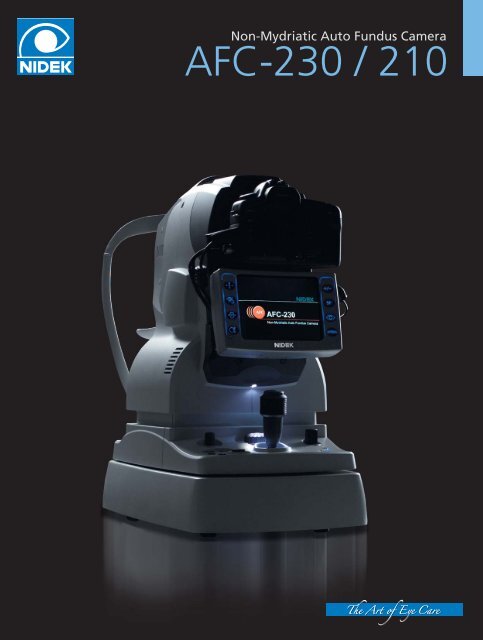
Non-Mydriatic Auto Fundus Camera<br />
<strong>AFC</strong>-<strong>230</strong> / <strong>210</strong>
Auto Fundus Camera<br />
<strong>AFC</strong>-<strong>230</strong> / <strong>210</strong><br />
Next Generation Non-Mydriatic Auto Fundus Camera<br />
offering High Quality Image and Advanced Operation<br />
NIDEK delivers the innovative non-mydriatic digital<br />
fundus camera that integrates every function required<br />
for easy retinal screening. Customized built-in<br />
functions of the <strong>AFC</strong>-<strong>230</strong> / <strong>210</strong> improve the quality and<br />
efficiency of medical examinations.<br />
High Quality Retinal Imaging<br />
Integrating the innovative imaging optical system, this technologically advanced <strong>AFC</strong>-<strong>230</strong> / <strong>210</strong><br />
realizes digital fundus imaging of high resolution and fine gradation. The fine gradation<br />
provides clear and detailed display of the entire fundus image from the light optic disc to a<br />
darkened disease area. With noise greatly reduced, the system offers retinal photography with<br />
minimum flash exposure allowing quick and efficient fundus photography of both eyes, thereby<br />
minimizing patient discomfort.<br />
With advanced optical system with large<br />
sensor, the <strong>AFC</strong>-<strong>230</strong> / <strong>210</strong> offers high quality<br />
image at true 45field of view.<br />
* Conventional frame size camera is also attachable by<br />
utilizing adapter (factory option) with true 45º field<br />
of view.<br />
Full frame size<br />
36mm24mm<br />
Optic disc(closeup)<br />
First in the world full automatic Non-Mydriatic Camera<br />
The <strong>AFC</strong>-<strong>230</strong>/<strong>210</strong> is the first in the world full automatic Non-Mydriatic Camera with advanced<br />
auto-tracking, auto-focus and Auto Shot technology. Auto-tracking technology allows easy and<br />
accurate alignment to the anterior corneal center. Also, the auto focus system provides automatic<br />
switching from anterior to retina, realizing high performance focusing. Once you have retina in<br />
focus, Auto Shot function offers high quality imaging without manually shooting the retina.<br />
Stress Free Photography Management<br />
The <strong>AFC</strong>-<strong>230</strong> / <strong>210</strong>'s advanced technologies reduce every day problems.<br />
High resolution Image Photography<br />
Always in focus<br />
Minimum retakes<br />
Patient friendly<br />
User Friendly
High-Performance Retinal Image Filing System - NAVIS-Lite<br />
The <strong>AFC</strong>-<strong>230</strong> / <strong>210</strong> system incorporates the sophisticated and user-friendly data filing software -<br />
NAVIS-Lite - allowing easy patient data management.<br />
Key Features of NAVIS-Lite<br />
Images that are automatically imported from the <strong>AFC</strong>-<strong>230</strong> / <strong>210</strong> are sorted by patient name.<br />
Easy-care pathway protocols in place for displaying patient information.<br />
Sophisticated imaging functions are incorporated, including Image Processing, Drawing,<br />
Measurement, and Panoramic Imaging for large field analysis.<br />
Zoom :<br />
Effects :<br />
Color control :<br />
Rotate / Reverse :<br />
Measurement :<br />
Drawing :<br />
Images can be zoomed freely<br />
Sharp, Combination, Edge enhancement<br />
Gray scale, Contrast RGB, Red-free, Channel split, Inverting Color,<br />
Brightness, Contrast, Histogram, Gamma control, Intensity selection<br />
Image can be rotated / reversed at any angle<br />
C / D ratio, Disc HV, Cup HV, Two point distance, Selected area<br />
Text / objects can be inserted into the image<br />
Flexible print layout display for patient reports<br />
Data back-up function<br />
Easy Image export<br />
E-mail function allowing to send message text with images files<br />
The data can be transferred to a DICOM (Digital Imaging and Communications in Medicine) 3.0<br />
compatible server (optional).<br />
Sample Screens<br />
Patient List<br />
Patient Data<br />
Image Editing<br />
Full-Screen Display
Pro Photographer<br />
Features of <strong>AFC</strong>-<strong>230</strong> / <strong>210</strong><br />
Accurate Anterior Eye Observation before<br />
Photography<br />
The <strong>AFC</strong>-<strong>230</strong> / <strong>210</strong> integrates the 5.7-inch TFT LCD<br />
(640 x 480) monitor in addition to the special<br />
optic system, CCD camera and high resolution<br />
monitor for anterior eye observation, allowing<br />
accurate confirmation of the anterior eye status<br />
(blepharoptosis, in-growing eyelashes,<br />
nystagmus, cataract, corneal disorder, etc). This<br />
assures high quality retinal photography.<br />
Smaller Pupil Photography Mode<br />
In addition to the regular minimum Pupil<br />
Diameter ø4.0 mm, the <strong>AFC</strong>-<strong>230</strong> / <strong>210</strong> is also<br />
highly capable of detecting a smaller Pupil<br />
Diameter - Minimum 3.7 mm. When the patient's<br />
Pupil Diameter is detected to be smaller than the<br />
required 4.0 mm, the <strong>AFC</strong>-<strong>230</strong> / <strong>210</strong> automatically<br />
switches its mode to the smaller pupil diameter<br />
mode.<br />
Stereo Photography Mode*<br />
Stereo fundus photography is also possible.<br />
*Requires stereo viewer (optional).<br />
Flexible Field Angle<br />
Without an adapter, the <strong>AFC</strong>-<strong>230</strong> / <strong>210</strong> can<br />
provide detailed image of smaller field of view in<br />
high quality by utilizing full frame 35mm in 45º<br />
field of view.<br />
Unique Blink Control<br />
With the automatic blink detection, the <strong>AFC</strong>-<strong>230</strong> /<br />
<strong>210</strong> automatically stops the photography when<br />
the patient blinks.<br />
Anterior Eye Photography Mode<br />
When the button for anterior eye photography is<br />
pressed, the <strong>AFC</strong>-<strong>230</strong> / <strong>210</strong> automatically switches<br />
its mode and provides clear anterior eye<br />
photography.<br />
High-Speed Image Transfer to a PC<br />
Connection to a PC through USB 2.0 allows quick<br />
and easy transfer of the images. The data can also<br />
be saved in an outside electronic chart system<br />
through the NAVIS-Lite.<br />
Ergonomic Design for Easy Operation in<br />
Darkened Room<br />
Layout of the buttons, lever and dial is<br />
ergonomically designed to allow intuitive<br />
operation. These allow the<br />
operator to take a photograph<br />
easily even in a dark room.<br />
Compact Body<br />
All necessary functions are integrated into this<br />
compact body, offering greater portability.
Pro Photographer<br />
Various System Configurations<br />
1<br />
Stand-Alone or Review-Station capability<br />
Easy connection with a Laptop or PC using USB2.0 interface.<br />
Quick and easy software installation to a laptop or PC using<br />
NAVIS-Lite installer.<br />
NAVIS-Lite software is available in stand-alone or review-station<br />
capable edition.<br />
OS requirement: Windows XP or later<br />
2<br />
Communication with Existing Modules<br />
<br />
Existing Software<br />
Module<br />
Patient Data<br />
Image Data<br />
Nidek Advanced Vision<br />
Information System<br />
Image files and XML files can be automatically / manually exported to a designated folder. XML files<br />
include information that links the patient data and image files, allowing data export to the NAVIS and<br />
other existing software modules.<br />
3<br />
DICOM Connection (Optional)<br />
Image files<br />
DICOM Storage<br />
HIS Server<br />
Work List<br />
DICOM connection can be achieved. Downloading work lists from the HIS Server is also possible.
<strong>AFC</strong>-<strong>230</strong> / <strong>210</strong> Specifications<br />
Model<br />
Type<br />
Picture angle<br />
Working distance<br />
Minimum pupil diameter<br />
Display<br />
Dioptric compensation for patient's eyes<br />
Focusing method<br />
Light source<br />
For observation<br />
For photography<br />
Illumination adjustment<br />
Internal fixation target<br />
External fixation target<br />
Horizontal movement<br />
Vertical movement<br />
Chinrest movement<br />
Auto tracking / Auto shot<br />
Interface<br />
External camera<br />
Power supply<br />
Power consumption<br />
Dimensions / Weight<br />
Standard accessories<br />
Optional accessories<br />
<strong>AFC</strong>-<strong>230</strong><br />
Non-mydriatic auto fundus camera<br />
45º (in smaller pupil diameter mode: 37º )<br />
45.7 mm (from camera lens to cornea)<br />
ø4.0 mm (in smaller pupil diameter mode: ø3.7 mm)<br />
5.7-inch TFT color LCD<br />
-33 to +35 D total<br />
-33 to -7 D with minus dioptric lens<br />
-12 to +15 D with no dioptric lens<br />
+11 to +35 D with plus dioptric lens<br />
Infrared split bright target coincidence<br />
(Auto / Manual, in -12 to +15 D range)<br />
Halogen lamp 12V 50W with infrared filter<br />
Xenon flash lamp (max. 300 Ws)<br />
17 levels : F1 (F4.0 + 0.7 EV) to F17 (F22 + 0.5 EV)<br />
0.5 EV increments<br />
LED (70 points)<br />
Free-arm (optional)<br />
40 mm (back and forth)<br />
85 mm (left and right)<br />
32 mm<br />
62 mm (up and down, motorized)<br />
X-Y-Z direction<br />
Auto shot<br />
USB 2.0<br />
High resolution digital SLR camera<br />
AC 100-240 V ±10%<br />
50 / 60 Hz<br />
Normal 150 VA, Max. 300 VA<br />
280 (W) x 505 (D) x 507 (H) mm / 25 kg<br />
11.0 (W) x 19.9 (D) x 20.0 (H) " / 55 lbs.<br />
Power cord (x1), USB cable for fundus camera (x1),<br />
USB cable for digital camera (x1), Cable sleeve (x1),<br />
Tie wrap (x2), Dust cover (x1), Chin rest paper (x1),<br />
Pin for chin rest paper (x2), Objective lens cap (x1),<br />
Camera mount cap (x1), Operator's manual (x1),<br />
Short operator's manual (x1), Digital camera<br />
installation manual (x1), Blower brush (x1), External<br />
fixation target (x1 for US)<br />
NAVIS-Lite, External fixation target, Stereo viewer,<br />
Conventional frame size camera adapter (Factory option)<br />
<strong>AFC</strong>-<strong>210</strong><br />
Y direction<br />
Auto shot<br />
Caution : U.S. Federal Law restricts this device to sale, distribution and use by or on the order of a<br />
physician or other licensed eye care practitioner.<br />
*Specifications and design are subject to change without notice for improvement.<br />
HEAD OFFICE<br />
34-14 Maehama, Hiroishi<br />
Gamagori, Aichi 443-0038, Japan<br />
Telephone : 81-533-67-6611<br />
Facsimile : 81-533-67-6610<br />
URL : http://www.nidek.co.jp<br />
[ Manufacturer ]<br />
TOKYO OFFICE<br />
(International Div.)<br />
3F Sumitomo Fudosan Hongo Bldg.,<br />
3-22-5 Hongo, Bunkyo-ku, Tokyo,<br />
113-0033 Japan<br />
Telephone : 81-3-5844-2641<br />
Facsimile : 81-3-5844-2642<br />
URL : http://www.nidek.com<br />
NIDEK INC.<br />
47651 Westinghouse Drive<br />
Fremont, CA 94539, U.S.A.<br />
Telephone : 1-510-226-5700<br />
: 1-800-223-9044 (US only)<br />
Facsimile : 1-510-226-5750<br />
URL : http://www.usa.nidek.com<br />
NIDEK SOCIETE ANONYME<br />
Europarc<br />
13, rue Auguste Perret<br />
94042 Creteil, France<br />
Telephone : 33-1-49 80 97 97<br />
Facsimile : 33-1-49 80 32 08<br />
URL : http://www.nidek.fr<br />
NIDEK TECHNOLOGIES SRL.<br />
Via dell'Artigianato, 6 / A<br />
35020 Albignasego (Padova), Italy<br />
Telephone : 39 049 8629200 / 8626399<br />
Facsimile : 39 049 8626824<br />
URL : http://www.nidektechnologies.it<br />
CNIDEK 2008 Printed in Japan <strong>AFC</strong>-<strong>230</strong> / <strong>210</strong> NQEEM6