Baicalein antagonizes rotenone-induced ... - Chinese Medicine
Baicalein antagonizes rotenone-induced ... - Chinese Medicine
Baicalein antagonizes rotenone-induced ... - Chinese Medicine

You also want an ePaper? Increase the reach of your titles
YUMPU automatically turns print PDFs into web optimized ePapers that Google loves.
Song et al. <strong>Chinese</strong> <strong>Medicine</strong> 2012, 7:1<br />
http://www.cmjournal.org/content/7/1/1<br />
RESEARCH Open Access<br />
<strong>Baicalein</strong> <strong>antagonizes</strong> <strong>rotenone</strong>-<strong>induced</strong><br />
apoptosis in dopaminergic SH-SY5Y cells related<br />
to Parkinsonism<br />
Ju-Xian Song 1 , Mandy Yuen-Man Choi 1 , Kavin Chun-Kit Wong 1 , Winkie Wing-Yan Chung 1 , Stephen Cho-Wing Sze 1 ,<br />
Tzi-Bun Ng 2 and Kalin Yan-Bo Zhang 1*<br />
Abstract<br />
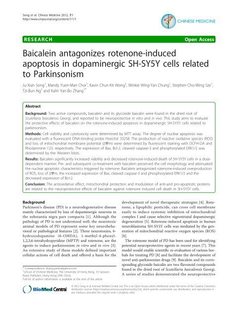
Background: Two active compounds, baicalein and its glycoside baicalin were found in the dried root of<br />
Scutellaria baicalensis Georgi, and reported to be neuroprotective in vitro and in vivo. This study aims to evaluate<br />
the protective effects of baicalein on the <strong>rotenone</strong>-<strong>induced</strong> apoptosis in dopaminergic SH-SY5Y cells related to<br />
parkinsonism.<br />
Methods: Cell viability and cytotoxicity were determined by MTT assay. The degree of nuclear apoptosis was<br />
evaluated with a fluorescent DNA-binding probe Hoechst 33258. The production of reactive oxidative species (ROS)<br />
and loss of mitochondrial membrane potential (ΔΨm) were determined by fluorescent staining with DCFH-DA and<br />
Rhodanmine 123, respectively. The expression of Bax, Bcl-2, cleaved caspase-3 and phosphorylated ERK1/2 was<br />
determined by the Western blots.<br />
Results: <strong>Baicalein</strong> significantly increased viability and decreased <strong>rotenone</strong>-<strong>induced</strong> death of SH-SY5Y cells in a dosedependent<br />
manner. Pre- and subsequent co-treatment with baicalein preserved the cell morphology and attenuated<br />
the nuclear apoptotic characteristics triggered by <strong>rotenone</strong>. <strong>Baicalein</strong> antagonized <strong>rotenone</strong>-<strong>induced</strong> overproduction<br />
of ROS, loss of ΔΨm, the increased expression of Bax, cleaved caspase-3 and phosphorylated ERK1/2 and the<br />
decreased expression of Bcl-2.<br />
Conclusion: The antioxidative effect, mitochondrial protection and modulation of anti-and pro-apoptotic proteins<br />
are related to the neuroprotective effects of baicalein against <strong>rotenone</strong> <strong>induced</strong> cell death in SH-SY5Y cells.<br />
Background<br />
Parkinson’s disease (PD) is a neurodegenerative disease<br />
mainly characterized by loss of dopaminergic neurons in<br />
the substantia nigra pars compacta [1]. Although the<br />
pathology of PD is not understood well, the neurotoxic<br />
animal models of PD represent some key neurobehavioral<br />
or pathological features [2]. Three neurotoxins, 6hydroxydopamine<br />
(6-OHDA), 1-methyl-4-phenyl-<br />
1,2,3,6-tetrahydropyridine (MPTP) and <strong>rotenone</strong>, are the<br />
agents to induce parkinsonism in vitro and in vivo [3].<br />
An extensive study of these models defined important<br />
cellular actions of cell death and offered a basis for the<br />
* Correspondence: zhang.yanbo@yahoo.com<br />
1 School of <strong>Chinese</strong> <strong>Medicine</strong>, The University of Hong Kong, 10 Sassoon<br />
Road, Pokfulam, Hong Kong SAR, China<br />
Full list of author information is available at the end of the article<br />
development of novel therapeutic strategies [4]. Rotenone,<br />
a lipophilic pesticide, can cross cell membrane<br />
easily to induce systemic inhibition of mitochondrial<br />
complex I and cause selective nigrostriatal dopaminergic<br />
degeneration [5]. Rotenone-<strong>induced</strong> apoptosis in human<br />
neuroblastoma SH-SY5Y cells was mediated by the generation<br />
of mitochondrial reactive oxygen species (ROS)<br />
[6].<br />
The <strong>rotenone</strong> model of PD has been used for identifying<br />
potential neuroprotective agents in recent years [7]. This<br />
model would enable scientific re-evaluation of various herbals<br />
for treating PD [8] and facilitate the development of<br />
novel anti-parkinsonian drugs [9]. <strong>Baicalein</strong> and its corresponding<br />
glycoside baicalin are two flavonoid compounds<br />
found in the dried root of Scutellaria baicalensis Georgi.<br />
A series of studies demonstrated the neuroprotective<br />
© 2012 Song et al; licensee BioMed Central Ltd. This is an Open Access article distributed under the terms of the Creative Commons<br />
Attribution License (http://creativecommons.org/licenses/by/2.0), which permits unrestricted use, distribution, and reproduction in<br />
any medium, provided the original work is properly cited.
Song et al. <strong>Chinese</strong> <strong>Medicine</strong> 2012, 7:1<br />
http://www.cmjournal.org/content/7/1/1<br />
effects of baicalein or baicalin in experimental models of<br />
Alzheimer’s disease [10,11], ischemic stroke [12-15] and<br />
PD [16-19]. <strong>Baicalein</strong> was reported to be effective on 6-<br />
OHDA models [18,20,21] and MPTP models [19,22] of<br />
PD. This study aims to investigate the neuroprotective<br />
effects of baicalein or baicalin on <strong>rotenone</strong>-<strong>induced</strong> cellular<br />
toxicities (SH-SY5Y cells) in vitro and in vivo.<br />
Methods<br />
Materials<br />
<strong>Baicalein</strong> and baicalin (Figure 1) with purity > 98% were<br />
purchased from Shanghai Innovative Research Center of<br />
Traditional <strong>Chinese</strong> <strong>Medicine</strong> (SIRC/TCM). Stock solutions<br />
(100 mM) were prepared in DMSO and diluted with<br />
serum-free medium. Dulbecco’s Modified Eagle Medium<br />
with Nutrient Mixture F-12 (DMEM/F-12), Fetal Bovine<br />
Serum (FBS) and Penicillin-Streptomycin were purchased<br />
from GIBCO BRL (Grand Island, NY, USA). 2,7-Dichlorofluorescein<br />
diacetate (DCFH-DA) and Rhodanmine 123<br />
(Rh123) were purchased from Molecular Probes (Invitrogen,<br />
CA, USA). Rotenone, Hoechst 33258, 3-(4,5dimethylthiazol-2-yl)-2,5-diphenyltetrazolium<br />
bromide<br />
(MTT),RIPAbuffer,BCAProteinAssayKitandother<br />
chemicals were obtained from Sigma-Aldrich (St. Louis,<br />
MO, USA). PVDF membrane was purchased from Millipore<br />
(MA, USA). Primary antibodies against Bax (D21),<br />
Bcl-2 (C21), b-actin and horseradish peroxidase (HRP)conjugated<br />
secondary antibodies were purchased from<br />
Santa Cruz Biotechnology (Santa Cruz, CA, USA). Primary<br />
antibodies against phospho-p44/42 MAPK (ERK1/2)<br />
(Thr202/Tyr204) and cleaved caspase-3 (Asp175) were<br />
purchased from Cell Signaling (Beverly, MA, USA). ECL<br />
Western blotting detection system was purchased from<br />
Amersham Biosciences (Piscataway, NJ, USA).<br />
Cell culture and treatments<br />
Human neuroblastoma SH-SY5Y cells (passage ≤ 25)<br />
were cultured as described in our previous study [21]<br />
and then treated with different concentrations of <strong>rotenone</strong>,<br />
baicalein or baicalin respectively in serum-free<br />
medium for 24 hours to determine their cytotoxicity. To<br />
Figure 1 The chemical structure of (A) baicalein and (B)<br />
baicalin.<br />
Page 2 of 9<br />
evaluate the protective effects, we pretreated SH-SY5Y<br />
cells with different concentrations of baicalein or baicalin<br />
for 1 hour and subsequently <strong>rotenone</strong> was added to<br />
the cells for another 24 hours. The final concentration<br />
of DMSO in the medium was 0.5%, and showed no<br />
cytotoxicity to the cells.<br />
MTT assay<br />
SH-SY5Y cells seeded on 96-well plates at 80-90% confluence<br />
were used in the MTT assay as described in our previous<br />
study [21]. In brief, the medium was removed after<br />
the treatment. MTT solution (50 μl, 0.5 mg/ml in<br />
DMEM/F12) was added to each well and incubated for<br />
4 hours at 37°C. MTT lysis buffer containing 50 μl of 20%<br />
SDS (sodium dodecyl sulfate), 50% DMF (N, N-dimethylformamide),<br />
adjusted to pH4.7 by HCl (hydrogen chloride)<br />
was then added before overnight incubation of the cells at<br />
37°C to dissolve formazan. The absorbance at 570 nm was<br />
measured by a microplate reader (Model 680, Bio-Rad<br />
Laboratories, UK). The cell viability was expressed as percentage<br />
of the control.<br />
Cellular morphology and nuclear apoptosis<br />
SH-SY5Y cells were incubated with different concentrations<br />
of baicalein in serum-free medium for 1 hour, followed<br />
by the co-treatment with <strong>rotenone</strong> (20 μM) for<br />
another 24 hours. Chromosomal DNA was stained with a<br />
fluorescent DNA-binding probe Hoechst 33258 (5 μg/ml)<br />
for 5 minutes, washed with PBS and then observed by a<br />
Axiovert S-100 Zeiss fluorescent microscope (Carl Zeiss,<br />
Zürich, Switzerland) at 20×. The morphological changes<br />
were visualized by phase-contrast imaging at 20×.<br />
ROS and mitochondrial membrane potential<br />
SH-SY5Y cells were pretreated with different concentrations<br />
of baicalein for 1 hour and then co-treated with <strong>rotenone</strong><br />
(20 μM) for another 6 hours in serum-free medium.<br />
According to the protocols described in our previous<br />
study [21], the fluorescent probes DCFH-DA and Rh123<br />
were used to determine the generation of intracellular<br />
ROS and mitochondrial membrane potential (ΔΨm),<br />
respectively. The total cell counts and fluorescent intensity<br />
were calculated with Image J software (ImageJ 1.45, http://<br />
rsbweb.nih.gov/ij). Mean fluorescent intensity (MFI) was<br />
calculated for each group using the following formula:<br />
MFI = total fluorescent intensity × 100/total cell counts<br />
Western blots analysis<br />
SH-SY5Y cells were pre-incubated for 1 hour with different<br />
concentrations of baicalein and then co-treated<br />
with <strong>rotenone</strong> (20 μM) for another 24 hours in serumfree<br />
medium. Total proteins were extracted using RIPA
Song et al. <strong>Chinese</strong> <strong>Medicine</strong> 2012, 7:1<br />
http://www.cmjournal.org/content/7/1/1<br />
buffer. Protein determination was by a BCA Protein<br />
Assay Kit. Denatured proteins (30 μg) were size fractionated<br />
by 12.5% SDS-polyacrylamide gels. Proteins were<br />
transferred to PVDF membrane at 80 V for 3 hours.<br />
The blots were blocked for 1 hour at room temperature<br />
in fresh blocking buffer (0.1% Tween-20 in Tris-buffered<br />
saline,pH7.4,containing5%BSA).Themembranewas<br />
incubated overnight at 4°C with primary antibodies<br />
against Bax, Bcl-2, cleaved caspase-3 and phosphorylated<br />
ERK1/2 at dilution of 1:1000. b-actinwasusedasa<br />
loading control. The membrane was incubated for<br />
2 hours with HRP-conjugated secondary antibodies at a<br />
dilution of 1:2000. Signals were detected using ECL<br />
Western blotting detection system. Protein bands were<br />
semi-quantified by densitometric analysis using Image J<br />
software.<br />
Statistical analysis<br />
Each experiment was performed at least three times, and<br />
the results were presented as means or means ± standard<br />
deviations (SD). One-way analysis of variance (ANOVA)<br />
followed by Student-Newman-Keuls test for multiple comparison<br />
was performed using the SigmaPlot 11.0 software<br />
packages (Systat Software Inc., San Jose, CA, USA). Exact<br />
P values were unavailable due to the software features<br />
(Additional file 1 provides a screen snapshot for example).<br />
Dose dependence was visually determined from the dose-<br />
Page 3 of 9<br />
response graphs. A probability value of P < 0.05 was considered<br />
to be statistically significant.<br />
Results<br />
In this study, we evaluated the effects of baicalein and baicalin<br />
on <strong>rotenone</strong>-<strong>induced</strong> cell death, nuclear apoptosis,<br />
production of intracellular ROS, loss of ΔΨm, expressions<br />
of Bax, Bcl-2 and caspase-3, and phosphorylation of<br />
ERK1/2 in SH-SY5Y cells.<br />
Cell death<br />
The cytotoxicity of <strong>rotenone</strong>, baicalein and baicalin were<br />
determined by MTT assay, Figure 2A shows that the cell<br />
viability was decreased in a dose-dependent manner (P <<br />
0.01) by the treatment with <strong>rotenone</strong> for 24 hours. Rotenone<br />
(20 μM) triggered about 50% cell death and this concentration<br />
was chosen for subsequent experiments. Both<br />
baicalein and baicalin showed no cytotoxicity at the concentrations<br />
ranged 10-100 μM. Figure 2B shows that baicalein<br />
increased cell viability by 20-40% (P
Song et al. <strong>Chinese</strong> <strong>Medicine</strong> 2012, 7:1<br />
http://www.cmjournal.org/content/7/1/1<br />
0.01). In consistency with the MTT result, the morphological<br />
observations revealed that baicalein significantly<br />
reversed the cellular damage triggered by <strong>rotenone</strong>, as<br />
shown in Figure 2E. However, baicalin showed no statistically<br />
significant protective effect against <strong>rotenone</strong>-<strong>induced</strong><br />
cell death.<br />
Nuclear apoptosis<br />
Compared with the control, the apoptotic characteristics<br />
<strong>induced</strong> by the <strong>rotenone</strong> treatment, such as<br />
Page 4 of 9<br />
nuclear condensation and fragmentation, could be attenuated<br />
by pre- and subsequent co-treatment with<br />
increasing concentrations of baicalein (as shown in<br />
Figure 3). The statistical data showed 4.29 ± 0.69 folds<br />
of increase in the ratio of apoptotic cells triggered by<br />
<strong>rotenone</strong>, which could be reduced to the control level<br />
by pre- and subsequent co-treatment with increasing<br />
concentrations of baicalein (P < 0.01). <strong>Baicalein</strong> treatment<br />
for 24 hours had no significantly effect on<br />
nuclear apoptosis.<br />
Figure 3 Effects of baicalein (Bai) on <strong>rotenone</strong> (RT)-<strong>induced</strong> nuclear apoptosis. Cells were pretreated with Bai for 1 hour and then cotreated with<br />
20 μM RT for 24 hours in serum-free medium. (A) Representative nuclear morphology. Scale bar: 50 μm. (B) Statistical analysis of apoptotic cells. At<br />
least 600 randomly selected cells were counted in each experiment (n = 3, # P < 0.01 versus control, *P < 0.01 versus RT treatment).
Song et al. <strong>Chinese</strong> <strong>Medicine</strong> 2012, 7:1<br />
http://www.cmjournal.org/content/7/1/1<br />
Intracellular ROS<br />
Figure 4 demonstrates that <strong>rotenone</strong> treatment <strong>induced</strong><br />
2.19 ± 0.36 folds of increase in the intracellular ROS compared<br />
with the control (P < 0.01). Pre- and subsequent cotreatment<br />
with baicalein reduced the production of ROS<br />
in a dose-dependent manner (P < 0.01) down to the control<br />
level. <strong>Baicalein</strong> treatment for 6 hours showed no<br />
Page 5 of 9<br />
significant effect on ROS production as compared with<br />
the control.<br />
Loss of ΔΨm<br />
The inhibition of complex I by <strong>rotenone</strong> may induce loss<br />
of ΔΨm and the release of pro-apoptotic proteins [23]. As<br />
shown in Figure 5, <strong>rotenone</strong> treatment led to about 2 folds<br />
Figure 4 Effects of baicalein (Bai) on <strong>rotenone</strong> (RT)-<strong>induced</strong> ROS overproduction. Cells were pretreated with Bai for 1 hour and then<br />
cotreated with 20 μM RT for 6 hours in serum-free medium. The ROS generation was determined by the mean fluorescent intensity (MFI) of<br />
DCFH-DA. (A) Representative fluorescent images. Scale bar: 50 μm. (B) Statistical analysis. At least 600 randomly selected cells were counted in<br />
each experiment (n = 3, # P < 0.01 versus control, *P < 0.01 versus RT treatment).
Song et al. <strong>Chinese</strong> <strong>Medicine</strong> 2012, 7:1<br />
http://www.cmjournal.org/content/7/1/1<br />
Figure 5 Effects of baicalein (Bai) on <strong>rotenone</strong> (RT)-<strong>induced</strong> loss of ΔΨm. Cells were pretreated with Bai for 1 hour and then cotreated with<br />
20 μM RT for 6 hours in serum-free medium. The ΔΨm was determined by the mean fluorescent intensity (MFI) of Rh123. (A) Representative<br />
fluorescent images. Scale bar: 50 μm. (B) Statistical analysis. At least 600 randomly selected cells were counted in each experiment (n = 3, # P<<br />
0.01 versus control, *P < 0.01 versus RT treatment).<br />
of decrease in Rh123 fluorescence (P < 0.01), reflecting the<br />
loss of ΔΨm. Pre- and subsequent co-treatment with baicalein<br />
significantly inhibited the loss of ΔΨm inadosedependent<br />
manner (P < 0.01). <strong>Baicalein</strong> treatment for 6<br />
hours showed no significant effect on ΔΨm as compared<br />
with the control.<br />
Page 6 of 9<br />
Expression of Bax, Bcl-2 and cleaved caspase-3<br />
To further characterize the mechanism of baicalein inhibition<br />
on <strong>rotenone</strong>-<strong>induced</strong> apoptosis, we determined<br />
the effect of baicalein on the expression of anti- and<br />
pro-apoptotic proteins by Western blots. As shown in<br />
Figure 6, the expression of Bax and cleaved caspase-3
Song et al. <strong>Chinese</strong> <strong>Medicine</strong> 2012, 7:1<br />
http://www.cmjournal.org/content/7/1/1<br />
A B<br />
Figure 6 Effects of baicalein (Bai) on <strong>rotenone</strong> (RT)-<strong>induced</strong> imbalance in the expression of Bax, Bcl-2, cleaved caspase-3 and phopho-<br />
ERK1/2. Cells were pretreated with Bai for 1 hour and then cotreated with 20 μM RT for 24 hours in serum-free medium. Blots were stripped<br />
and reprobed for b-actin as a loading control. (A) Representative protein bands. (B) Statistical analysis. The corresponding bar graph represented<br />
data quantified from three independent experiments (n = 3, # P < 0.05 versus control, *P < 0.05 versus RT treatment, **P < 0.05 versus control).<br />
was increased while the expression of Bcl-2 was significantly<br />
decreased by the treatment with <strong>rotenone</strong> (20<br />
μM) for 24 hours (P < 0.05), compared with the control.<br />
Pre- and subsequent co-treatment with increasing concentrations<br />
of baicalein gradually restored the imbalanced<br />
expression profile of these proteins. Interestingly,<br />
baicalein treatment alone for 24 hours could reduce the<br />
base levels of Bax (0.86 ± 0.07) and cleaved caspase-3<br />
(0.71 ± 0.09) (P < 0.05).<br />
ERK1/2 phosphorylation<br />
It was reported that <strong>rotenone</strong> <strong>induced</strong> ERK1/2 phosphorylation<br />
and neuronal degeneration in hippocampus<br />
neurons [24]. Similar to this finding, we detected 2.47 ±<br />
0.18 folds of increase in the expression of phosphorylated<br />
ERK1/2 in SH-SY5Y cells by treatment with <strong>rotenone</strong><br />
for 24 hours, as shown in Figure 6 (P < 0.05). Preand<br />
subsequent co-treatment with baicalein reduced the<br />
expression of phosphorylated ERK1/2 down to the control<br />
level in a dose-dependent manner. <strong>Baicalein</strong> treatment<br />
alone for 24 hours could also significantly reduce<br />
the base level of ERK1/2 phosphorylation.<br />
Discussion<br />
In the study, we evaluated the neuroprotective effects of<br />
baicalein on <strong>rotenone</strong>-<strong>induced</strong> SH-SY5Y cell apoptosis.<br />
In the neurotoxic models (6-OHDA and MPTP/MPP + )<br />
of PD, either baicalein or baicalin has been reported to<br />
be effective [18,22,25]. However, we found that only baicalein<br />
showed a significant inhibition on <strong>rotenone</strong><strong>induced</strong><br />
cytotoxicity as demonstrated in Figure 2D. Choi<br />
Page 7 of 9<br />
et al. [26] demonstrated that baicalein was protective<br />
against endoplasmic reticulum (ER) stress-<strong>induced</strong> ROS<br />
accumulation and apoptosis. The difference between baicalein<br />
and baicalin in antioxidative potential and cellular<br />
permeability might contribute to their difference in cytoprotective<br />
effects against ER stress-inducers [26]. These<br />
two factors may also account for the different effects of<br />
baicalein and baicalin on <strong>rotenone</strong>-<strong>induced</strong> cytotoxicity.<br />
MTT cell viability assay showed that baicalein antagonized<br />
<strong>rotenone</strong>-<strong>induced</strong> cell death, which may be due to<br />
the ability of baicalein in increasing the cell viability of<br />
normal cells, as indicated in Figure 2B. The cell viability<br />
was reduced to 62.64% (P < 0.01) by treatment with<br />
<strong>rotenone</strong> alone for 24 hours while pre- and subsequent<br />
co-treatment with baicalein (100 μM) increased the cell<br />
viability to 137.01% (P < 0.01), as shown in Figure 2C.<br />
<strong>Baicalein</strong> (100 μM) treatment alone <strong>induced</strong> 43.46%<br />
increase (P < 0.01) in cell viability (Figure 2B) and the<br />
difference in cell viability (Figure 2C) between <strong>rotenone</strong><br />
treatment alone (62.64%) and baicalein (100 μM) pre<br />
and co-treatment (137.01%) is 74.37%, suggesting that<br />
the cell proliferating activity of baicalein (43.46%<br />
increase) does not account for its protection against<br />
<strong>rotenone</strong>-<strong>induced</strong> cell death (74.37% increase). In other<br />
words, the protection of baicalein against <strong>rotenone</strong><strong>induced</strong><br />
cell death may be independent of its cell proliferation<br />
activity. These results suggested that baicalein<br />
had protection against <strong>rotenone</strong>-<strong>induced</strong> cytotoxicity<br />
independent of its cell proliferation activity.<br />
Oxidative injury was proposed to be a primary<br />
mechanism of mitochondrial toxicity in the <strong>rotenone</strong>-
Song et al. <strong>Chinese</strong> <strong>Medicine</strong> 2012, 7:1<br />
http://www.cmjournal.org/content/7/1/1<br />
<strong>induced</strong> degeneration of dopaminergic neurons [27,28].<br />
Impairment of complex I activity by <strong>rotenone</strong> led to<br />
excess ROS formation, which <strong>induced</strong> loss of ΔΨm and<br />
initiated apoptotic cell death [27,28]. It was reported<br />
that baicalein suppressed the mitochondrial dysfunction<br />
<strong>induced</strong> by hydrogen peroxide and 6-OHDA, and the<br />
initiation of the loss of ΔΨm in PC12 cells and SH-<br />
SY5Y cells, respectively [17,29]. This study confirmed<br />
these findings that baicalein inhibited ROS production<br />
and loss of ΔΨm triggered by <strong>rotenone</strong> in SH-SY5Y<br />
cells, resulting in cellular resistance against the initiating<br />
steps of apoptosis. This protection was mediated in part<br />
by its antioxidative ability and preservation of mitochondrial<br />
function.<br />
The balance of Bax and Bcl-2 proteins relates to the cell<br />
viability [30]. Loss of ΔΨm increases the mitochondrial<br />
permeability and results in the release of cytochrome c<br />
from the mitochondria, which triggers activation of caspase-9/3<br />
and ultimate cell death [31]. In this study, we<br />
found that baicalein restored the imbalance of the expression<br />
profiles of Bax, Bcl-2 and cleaved caspase-3; baicalein<br />
treatment alone could also decrease the expression of Bax<br />
and cleaved caspase-3; and modulation of the pro- and<br />
anti-apoptotic proteins would be involved in the protective<br />
effects of baicalein against <strong>rotenone</strong>-<strong>induced</strong> neurotoxicity.<br />
Sustained ERK activation was reported to promote cell<br />
death in neuronal cells treated with neurotoxins [32-34].<br />
Figure 6 demonstrates that <strong>rotenone</strong> triggering significant<br />
phosphorylation and activation of ERK1/2 was<br />
antagonized by baicalein pretreatment, indicating that<br />
inactivation of ERK1/2 pathway was involved in the neuroprotective<br />
effects of baicalein against <strong>rotenone</strong><strong>induced</strong><br />
neurotoxicity.<br />
Conclusion<br />
Inhibition of ROS overproduction, preservation of mitochondrial<br />
function, modulation of anti- and pro-apoptotic<br />
proteins and inactivation of ERK1/2 pathway are related to<br />
the neuroprotective effects of baicalein against <strong>rotenone</strong><strong>induced</strong><br />
apoptosis in dopaminergic SH-SY5Y cells.<br />
Additional material<br />
Additional file 1: A screen snapshot demonstrating the statistical<br />
analysis using SigmaPlot 11.0. The detailed procedures are illustrated<br />
for Figure 2C. Exact P values were unavailable due to the software<br />
features.<br />
Abbreviations<br />
DCFH-DA: 2,7-Dichlorofluorescein diacetate; DMEM/F-12, Dulbecco’s Modified<br />
Eagle Medium: Nutrient Mixture F-12; DMF: N, N-dimethylformamide; DMSO:<br />
dimethyl sulfoxide; ERK1/2: extracellular signal-regulated kinases 1 and 2; FBS:<br />
fetal bovine serum; HCl: hydrogen chloride; HRP: horseradish peroxidase;<br />
MAPK: mitogen activated protein kinases; MPP + : 1-methyl-4-phenyl<br />
pyridinium; MPTP: 1-methyl-4-phenyl-1,2,3,6-tetrahydropyridine; MTT: 3-(4,5dimethylthiazol-2-yl)-<br />
2,5-diphenyltetrazolium bromide; PD: Parkinson’s<br />
disease; Rh123: Rhodanmine 123; ROS: reactive oxygen species; SDS: sodium<br />
dodecyl sulfate; 6-OHDA: 6-hydroxydopamine; ΔΨm: mitochondrial<br />
membrane potential.<br />
Acknowledgements<br />
This study was supported by grants from Seed Funding Programme for<br />
Basic Research from HKU (Project No. 201011159206) and funding from<br />
Stanley Ho Alumni Challenge (SHAC) Development and Alumni affairs office,<br />
HKU (Project No. 22100.20830019).<br />
Author details<br />
1 School of <strong>Chinese</strong> <strong>Medicine</strong>, The University of Hong Kong, 10 Sassoon<br />
Road, Pokfulam, Hong Kong SAR, China. 2 School of Biomedical Sciences, The<br />
<strong>Chinese</strong> University of Hong Kong, Shatin, New Territories, Hong Kong, SAR,<br />
China.<br />
Authors’ contributions<br />
JXS and KYBZ designed the study and drafted the manuscript. JXS, MYMC,<br />
KCKW and WWYC conducted the experiments and analyzed the data. SCWS<br />
and TBN revised the manuscript. All authors read and approved the final<br />
version of the manuscript.<br />
Competing interests<br />
The authors declare that they have no competing interests.<br />
Received: 3 October 2011 Accepted: 21 January 2012<br />
Published: 21 January 2012<br />
Page 8 of 9<br />
References<br />
1. Schapira AH, Bezard E, Brotchie J, Calon F, Collingridge GL, Ferger B,<br />
Hengerer B, Hirsch E, Jenner P, Le Novere N, Obeso JA, Schwarzschild MA,<br />
Spampinato U, Davidai G: Novel pharmacological targets for the<br />
treatment of Parkinson’s disease. Nat Rev Drug Discov 2006, 5:845-854.<br />
2. Cannon JR, Greenamyre JT: Neurotoxic in vivo models of Parkinson’s<br />
disease recent advances. Prog Brain Res 2010, 184:17-33.<br />
3. Beal MF: Experimental models of Parkinson’s disease. Nat Rev Neurosci<br />
2001, 2:325-334.<br />
4. Fox SH, Brotchie JM: The MPTP-lesioned non-human primate models of<br />
Parkinson’s disease. Past, present, and future. Prog Brain Res 2010,<br />
184:133-157.<br />
5. Betarbet R, Sherer TB, MacKenzie G, Garcia-Osuna M, Panov AV,<br />
Greenamyre JT: Chronic systemic pesticide exposure reproduces features<br />
of Parkinson’s disease. Nat Neurosci 2000, 3:1301-1306.<br />
6. Watabe M, Nakaki T: Mitochondrial complex I inhibitor <strong>rotenone</strong>-elicited<br />
dopamine redistribution from vesicles to cytosol in human<br />
dopaminergic SH-SY5Y cells. J Pharmacol Exp Ther 2007, 323:499-507.<br />
7. Greenamyre JT, Cannon JR, Drolet R, Mastroberardino PG: Lessons from the<br />
<strong>rotenone</strong> model of Parkinson’s disease. Trends Pharmacol Sci 2010,<br />
31:141-142.<br />
8. Manyam BV, Sanchez-Ramos JR: Traditional and complementary therapies<br />
in Parkinson’s disease. Adv Neurol 1999, 80:565-574.<br />
9. Li Q, Zhao D, Bezard E: Traditional <strong>Chinese</strong> medicine for Parkinson’s<br />
disease: a review of <strong>Chinese</strong> literature. Behav Pharmacol 2006, 17:403-410.<br />
10. Lebeau A, Esclaire F, Rostene W, Pelaprat D: <strong>Baicalein</strong> protects cortical<br />
neurons from beta-amyloid (25-35) <strong>induced</strong> toxicity. Neuroreport 2001,<br />
12:2199-2202.<br />
11. Wang SY, Wang HH, Chi CW, Chen CF, Liao JF: Effects of baicalein on<br />
beta-amyloid peptide-(25-35)-<strong>induced</strong> amnesia in mice. Eur J Pharmacol<br />
2004, 506:55-61.<br />
12. Cui L, Zhang X, Yang R, Liu L, Wang L, Li M, Du W: <strong>Baicalein</strong> is<br />
neuroprotective in rat MCAO model: role of 12/15-lipoxygenase,<br />
mitogen-activated protein kinase and cytosolic phospholipase A2.<br />
Pharmacol Biochem Behav 2010, 96:469-475.<br />
13. Liu C, Wu J, Xu K, Cai F, Gu J, Ma L, Chen J: Neuroprotection by baicalein<br />
in ischemic brain injury involves PTEN/AKT pathway. J Neurochem 2010,<br />
112:1500-1512.<br />
14. Xue X, Qu XJ, Yang Y, Sheng XH, Cheng F, Jiang EN, Wang JH, Bu W,<br />
Liu ZP: Baicalin attenuates focal cerebral ischemic reperfusion injury
Song et al. <strong>Chinese</strong> <strong>Medicine</strong> 2012, 7:1<br />
http://www.cmjournal.org/content/7/1/1<br />
through inhibition of nuclear factor kappaB p65 activation. Biochem<br />
Biophys Res Commun 2010, 403:398-404.<br />
15. Zhang Z, Wu R, Li P, Liu F, Zhang W, Zhang P, Wang Y: Baicalin<br />
administration is effective in positive regulation of twenty-four<br />
ischemia/reperfusion-related proteins identified by a proteomic study.<br />
Neurochem Int 2009, 54:488-496.<br />
16. Jiang M, Porat-Shliom Y, Pei Z, Cheng Y, Xiang L, Sommers K, Li Q,<br />
Gillardon F, Hengerer B, Berlinicke C, Smith WW, Zack DJ, Poirier MA,<br />
Ross CA, Duan W: <strong>Baicalein</strong> reduces E46K alpha-synuclein aggregation in<br />
vitro and protects cells against E46K alpha-synuclein toxicity in cell<br />
models of familiar Parkinsonism. J Neurochem 2010, 114:419-429.<br />
17. Lee HJ, Noh YH, Lee DY, Kim YS, Kim KY, Chung YH, Lee WB, Kim SS:<br />
<strong>Baicalein</strong> attenuates 6-hydroxydopamine-<strong>induced</strong> neurotoxicity in SH-<br />
SY5Y cells. Eur J Cell Biol 2005, 84:897-905.<br />
18. Mu X, He G, Cheng Y, Li X, Xu B, Du G: <strong>Baicalein</strong> exerts neuroprotective<br />
effects in 6-hydroxydopamine-<strong>induced</strong> experimental parkinsonism in<br />
vivo and in vitro. Pharmacol Biochem Behav 2009, 92:642-648.<br />
19. Mu X, He GR, Yuan X, Li XX, Du GH: <strong>Baicalein</strong> protects the brain against<br />
neuron impairments <strong>induced</strong> by MPTP in C57BL/6 mice. Pharmacol<br />
Biochem Behav 2011, 98:286-291.<br />
20. Im HI, Joo WS, Nam E, Lee ES, Hwang YJ, Kim YS: <strong>Baicalein</strong> prevents 6hydroxydopamine-<strong>induced</strong><br />
dopaminergic dysfunction and lipid<br />
peroxidation in mice. J Pharmacol Sci 2005, 98:185-189.<br />
21. Song JX, Shaw PC, Sze CW, Tong Y, Yao XS, Ng TB, Zhang YB:<br />
Chrysotoxine, a novel bibenzyl compound, inhibits 6-hydroxydopamine<br />
<strong>induced</strong> apoptosis in SH-SY5Y cells via mitochondria protection and NFkappaB<br />
modulation. Neurochem Int 2010, 57:676-689.<br />
22. Cheng Y, He G, Mu X, Zhang T, Li X, Hu J, Xu B, Du G: Neuroprotective<br />
effect of baicalein against MPTP neurotoxicity: behavioral, biochemical<br />
and immunohistochemical profile. Neurosci Lett 2008, 441:16-20.<br />
23. Hu LF, Lu M, Wu ZY, Wong PT, Bian JS: Hydrogen sulfide inhibits<br />
<strong>rotenone</strong>-<strong>induced</strong> apoptosis via preservation of mitochondrial function.<br />
Mol Pharmacol 2009, 75:27-34.<br />
24. Sai Y, Chen J, Wu Q, Liu H, Zhao J, Dong Z: Phosphorylated-ERK 1/2 and<br />
neuronal degeneration <strong>induced</strong> by <strong>rotenone</strong> in the hippocampus<br />
neurons. Environ Toxicol Pharmacol 2009, 27:366-372.<br />
25. Chen X, Zhang N, Zou HY: Protective effect of baicalin on mouse with<br />
Parkinson’s disease <strong>induced</strong> by MPTP. Zhongguo Zhong Xi Yi Jie He Za Zhi<br />
2007, 27:1010-1012.<br />
26. Choi JH, Choi AY, Yoon H, Choe W, Yoon KS, Ha J, Yeo EJ, Kang I: <strong>Baicalein</strong><br />
protects HT22 murine hippocampal neuronal cells against endoplasmic<br />
reticulum stress-<strong>induced</strong> apoptosis through inhibition of reactive<br />
oxygen species production and CHOP induction. Exp Mol Med 2010,<br />
42:811-822.<br />
27. Radad K, Rausch WD, Gille G: Rotenone induces cell death in primary<br />
dopaminergic culture by increasing ROS production and inhibiting<br />
mitochondrial respiration. Neurochem Int 2006, 49:379-386.<br />
28. Testa CM, Sherer TB, Greenamyre JT: Rotenone induces oxidative stress<br />
and dopaminergic neuron damage in organotypic substantia nigra<br />
cultures. Brain Res Mol Brain Res 2005, 134:109-118.<br />
29. Zhang S, Ye J, Dong G: Neuroprotective effect of baicalein on hydrogen<br />
peroxide-mediated oxidative stress and mitochondrial dysfunction in<br />
PC12 cells. J Mol Neurosci 2009, 40:311-320.<br />
30. Borner C: The Bcl-2 protein family: sensors and checkpoints for life-ordeath<br />
decisions. Mol Immunol 2003, 39:615-647.<br />
31. Chinnaiyan AM, Orth K, O’Rourke K, Duan H, Poirier GG, Dixit VM: Molecular<br />
ordering of the cell death pathway. Bcl-2 and Bcl-xL function upstream<br />
of the CED-3-like apoptotic proteases. J Biol Chem 1996, 271:4573-4576.<br />
32. Gomez-Santos C, Ferrer I, Reiriz J, Vinals F, Barrachina M, Ambrosio S: MPP +<br />
increases alpha-synuclein expression and ERK/MAP-kinase<br />
phosphorylation in human neuroblastoma SH-SY5Y cells. Brain Res 2002,<br />
935:32-39.<br />
33. Kulich SM, Horbinski C, Patel M, Chu CT: 6-Hydroxydopamine induces<br />
mitochondrial ERK activation. Free Radic Biol Med 2007, 43:372-383.<br />
34. Zhu JH, Horbinski C, Guo F, Watkins S, Uchiyama Y, Chu CT: Regulation of<br />
autophagy by extracellular signal-regulated protein kinases during 1methyl-4-phenylpyridinium-<strong>induced</strong><br />
cell death. Am J Pathol 2007,<br />
170:75-86.<br />
doi:10.1186/1749-8546-7-1<br />
Cite this article as: Song et al.: <strong>Baicalein</strong> <strong>antagonizes</strong> <strong>rotenone</strong>-<strong>induced</strong><br />
apoptosis in dopaminergic SH-SY5Y cells related to Parkinsonism.<br />
<strong>Chinese</strong> <strong>Medicine</strong> 2012 7:1.<br />
Submit your next manuscript to BioMed Central<br />
and take full advantage of:<br />
• Convenient online submission<br />
• Thorough peer review<br />
• No space constraints or color figure charges<br />
• Immediate publication on acceptance<br />
• Inclusion in PubMed, CAS, Scopus and Google Scholar<br />
• Research which is freely available for redistribution<br />
Submit your manuscript at<br />
www.biomedcentral.com/submit<br />
Page 9 of 9