SAIB SAB
SAIB SAB
SAIB SAB
- No tags were found...

Create successful ePaper yourself
Turn your PDF publications into a flip-book with our unique Google optimized e-Paper software.
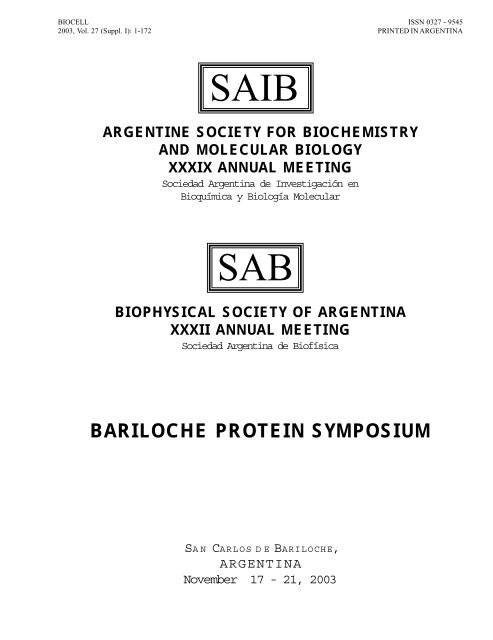
BIOCELL<br />
2003, Vol. 27 (Suppl. I): 1-172<br />
ISSN 0327 - 9545<br />
PRINTED IN ARGENTINA<br />
<strong>SAIB</strong><br />
ARGENTINE SOCIETY FOR BIOCHEMISTRY<br />
AND MOLECULAR BIOLOGY<br />
XXXIX ANNUAL MEETING<br />
Sociedad Argentina de Investigación en<br />
Bioquímica y Biología Molecular<br />
<strong>SAB</strong><br />
BIOPHYSICAL SOCIETY OF ARGENTINA<br />
XXXII ANNUAL MEETING<br />
Sociedad Argentina de Biofísica<br />
BARILOCHE PROTEIN SYMPOSIUM<br />
SAN CARLOS D E B ARILOCHE,<br />
ARGENTINA<br />
November 17 - 21, 2003
2 BIOCELL, 27 (Suppl. I), 2003
BIOCELL, 27 (Suppl. I), 2003<br />
3<br />
MEMBERS OF THE <strong>SAIB</strong> BOARD<br />
PRESIDENT<br />
DRA. NORMA STERIN DE SPEZIALE<br />
IQUIFIB, Facultad de Farmacia y Bioquímica<br />
Universidad de Buenos Aires<br />
Junin 956, 1er. Piso. Buenos Aires - ARGENTINA<br />
Tel.: +54 11 4964 8238<br />
E-mail: speziale@qb.ffyb.uba.ar<br />
SECRETARY<br />
DR. JOSÉ LUIS BOCCO<br />
Departamento de Bioquímica Clínica<br />
Facultad de Ciencias Químicas<br />
Universidad Nacional de Córdoba<br />
Ciudad Universitaria 5000 Córdoba - ARGENTINA<br />
Tel.: +54 351 4334164/Fax: +54 351 4344971<br />
E-mail: jbocco@fcq.unc.edu.ar<br />
VICE-PRESIDENT<br />
DR. ERNESTO PODESTA<br />
Facultad de Medicina.<br />
Universidad Nacional de Buenos Aires<br />
Buenos Aires - ARGENTINA<br />
E-mail: biohrdc@fmed.uba.ar<br />
TREASURER<br />
DRA. ESTELA VALLE<br />
IBR, Facultad de Ciencias Bioquímicas y Farmacéuticas<br />
Universidad Nacional de Rosario<br />
Suipacha 531, S2002LRK Rosario - ARGENTINA<br />
Tel.: +54 341 4350661/4350596/4351235<br />
Fax: +54-341-4390465<br />
E-mail: evalle@arnet.com.ar<br />
PAST-PRESIDENT<br />
DR. RICARDO WOLOSIUK<br />
Instituto de Investigaciones Bioquímicas<br />
Fundación Luis Leloir<br />
Av. Patricias Argentinas 435 - ARGENTINA<br />
C1405BUE – Buenos Aires<br />
E-mail: RWolosiuk@Leloir.org.ar<br />
PRO-SECRETARY<br />
DR. CARLOS ARGARAÑA<br />
CIQUIBIC, Facultad de Ciencias Químicas<br />
Universidad Nacional de Córdoba<br />
Ciudad Universitaria 5000 Córdoba - ARGENTINA<br />
E-mail: carga@dqb.fcq.unc.edu.ar<br />
PRO-TREASURER<br />
DR. LORENZO LAMATTINA<br />
Instituto de Investigaciones Biológicas<br />
Facultad de Ciencias Exactas y Naturales<br />
Universidad Nacional de Mar del Plata - ARGENTINA<br />
E-mail: lolama@bart.mdp.edu.ar
4 BIOCELL, 27 (Suppl. I), 2003<br />
AUDITOR<br />
DRA. CRISTINA NOWICKI<br />
Facultad de Farmacia y Bioquímica<br />
Universidad Nacional de Buenos Aires<br />
Junin 956, 1er. Piso. Buenos Aires - ARGENTINA<br />
E-mail: cnowicki@criba.edu.ar<br />
AUDITOR<br />
DR. ANTONIO UTTARO<br />
IBR-Dto. Microbiología<br />
Facultad de Ciencias Bioquímicas y Farmacéuticas<br />
Universidad Nacional de Rosario - ARGENTINA<br />
E-mail: uttaro@infovia.com.ar<br />
REPRESENTANTS OF SCIENTIFIC SECTIONS<br />
CELL BIOLOGY<br />
DRA. MARIA I<strong>SAB</strong>EL COLOMBO<br />
Laboratorio de Biología Celular y Molecular<br />
IHEM-CONICET. Facultad de Ciencias Médicas.<br />
Universidad Nacional de Cuyo.<br />
Casilla de Correo 56, 5500-Mendoza.<br />
Tel: +54 261-4494143/Fax: +54 261-4494117.<br />
E-mail: mcolombo@fcm.uncu.edu.ar<br />
LIPIDS<br />
DR. CARLOS MARRA<br />
Instituto de Investigaciones Bioquímicas de La Plata<br />
CONICET-UNLP, Facultad de Ciencias Médicas.<br />
Universidad Nacional de La Plata.<br />
E-mail: camarra@atlas.med.unlp.edu.ar<br />
PLANTS<br />
DR. ALBERTO IGLESIAS<br />
Instituto Tecnológico de Chascomús<br />
IIB-INTECH. Chascomús - Buenos Aires.<br />
E-mail: iglesias@criba.edu.ar<br />
MICROBIOLOGY<br />
DR. JUAN DIAZ RICCI<br />
Departamento de Bioquimica de la Nutricion<br />
Instituto Superior de Investigaciones Biológicas<br />
San Miguel de Tucumán.<br />
E-mail: juan@unt.edu.ar
BIOCELL, 27 (Suppl. I), 2003<br />
5<br />
MEMBERS OF THE <strong>SAB</strong> BOARD<br />
PRESIDENT<br />
SECRETARY<br />
VICE-PRESIDENT<br />
TREASURER<br />
PAST-PRESIDENT<br />
COUNCIL MEMBER<br />
DR. ROLANDO ROSSI<br />
IQUIFIB<br />
Facultad de Farmacia y Bioquímica<br />
Universidad de Buenos Aires<br />
Junin 956, 1er. Piso. Buenos Aires - ARGENTINA<br />
Tel.: +54 11 4964 8238<br />
E-mail: rcr@qb.ffyb.uba.ar<br />
DR. SILVIA ALONSO DE ROMANOWSKI<br />
Laboratorio de Biomembranas (LBM)<br />
Departamento de Ciencia y Tecnología<br />
Universidad Nacional de Quilmes<br />
Roque Saenz Peña 180<br />
(1876)Bernal<br />
Buenos Aires - ARGENTINA<br />
FAX:54-11 4365 7132<br />
T.E.:54-11 4365 7100 ext. 123<br />
e-mail: salonso@unq.edu.ar<br />
DR. HORACIO GARDA<br />
INIBIOLP<br />
Facultad de Ciencias Médicas<br />
Universidad de La Plata<br />
Calles 60 y 120, 1900 La Plata - ARGENTINA<br />
E-mail: hgarda@atlas.med.unlp.edu.ar<br />
DR. MARIO ERMACORA<br />
Departamento de Ciencia y Tecnología<br />
Universidad Nacional de Quilmes<br />
Roque Saenz Peña 180<br />
(1876)Bernal<br />
Buenos Aires - ARGENTINA<br />
Phone: +54 (114) 365 7100 ext. 169<br />
Fax: +54 (114) 365 7132<br />
E-mail: ermacora@unq.edu.ar<br />
DR. BRUNO MAGGIO<br />
CIQUIBIC<br />
Departamento de Química Biológica<br />
Facultad de Ciencias Químicas,<br />
Universidad Nacional de Córdoba<br />
Córdoba - ARGENTINA<br />
E-mail: bmaggio@dqb.fcq.unc.edu.ar<br />
DR. ALEJANDRO VILA<br />
Area Biofisica<br />
Facultad de Ciencias Bioquimicas y Farmaceuticas<br />
Universidad Nacional de Rosario<br />
Suipacha 531 - S2002LRK Rosario - ARGENTINA<br />
TE: 54-341-4350661/4350596/4351235 (Ext.24)<br />
Fax: 54-341-4390465<br />
E-mail: vila@arnet.com.ar
6 BIOCELL, 27 (Suppl. I), 2003<br />
COUNCIL MEMBER<br />
COUNCIL MEMBER<br />
IUPAB COUNCIL MEMBER<br />
DRA. GABRIELA AMODEO<br />
Laboratorio de Biomembranas<br />
Departamento de Fisiología<br />
Facultad de Medicina<br />
Universidad de Buenos Aires<br />
Paraguay 2155 Piso 7 Buenos Aires - ARGENTINA<br />
E-mail: amodeo@dna.uba.ar<br />
DR. ROBERTO MORERO<br />
INSIBIO (CONICET UNT)<br />
Departamento de Bioquímica de la Nutrición<br />
Universidad Nacional de Tucumán<br />
Tucumán - ARGENTINA<br />
E-mail: rdmorero@fbqf.unt.edu.ar<br />
DR. JORGE PONCE HORNOS<br />
Labs. de Metabolismo y Energética Cardíaca<br />
Instituto de Investigaciones Cardiológicas<br />
Facultad de Medicina, UBA<br />
Marcelo T. de Alvear 2270<br />
1122 Buenos Aires - ARGENTINA<br />
TE/FAX: 54-11-4508-3881/0<br />
E-mail: pohornos@mail.retina.ar<br />
PROTEIN SYMPOSIUM ORGANIZING COMMITTEE<br />
DR. JOSE MARIA DELFINO<br />
IQUIFIB<br />
Facultad de Farmacia y Bioquímica<br />
Universidad de Buenos Aires<br />
Junin 956, 1er. Piso. Buenos Aires - ARGENTINA<br />
Tel.: +54 11 4964 8238<br />
E-mail: rcr@qb.ffyb.uba.ar<br />
DR. FERNANDO GOLDBAUM<br />
Instituto de Investigaciones Bioquímicas<br />
Fundación Luis Leloir<br />
Av. Patricias Argentinas 435 - ARGENTINA<br />
C1405BUE – Buenos Aires<br />
E-mail: RWolosiuk@Leloir.org.ar<br />
DR. GONZALO DE PRAT GAY<br />
Instituto de Investigaciones Bioquímicas<br />
Fundación Luis Leloir<br />
Av. Patricias Argentinas 435 - ARGENTINA<br />
C1405BUE – Buenos Aires<br />
E-mail: RWolosiuk@Leloir.org.ar<br />
DR. ALEJANDRO VILA<br />
Area Biofisica<br />
Facultad de Ciencias Bioquímicas y Farmacéuticas<br />
Universidad Nacional de Rosario<br />
Suipacha 531 - S2002LRK Rosario - ARGENTINA<br />
TE: 54-341-4350661/4350596/4351235 (Ext.24)<br />
Fax: 54-341-4390465<br />
E-mail: vila@arnet.com.ar
BIOCELL, 27 (Suppl. I), 2003<br />
7<br />
“Algunos creen que la ciencia es un lujo<br />
y que los grandes países gastan en ella<br />
porque son ricos. ¡Grave error!<br />
Los países ricos gastan en Ciencia<br />
porque es un gran negocio y porque<br />
de esta forma se enriquecen.<br />
No gastan en Ciencia porque son ricos y prósperos;<br />
son ricos y prósperos porque gastan en Ciencia.<br />
¡Nada da dividendos comparables a los que<br />
proporciona la investigación científica y tecnológica!”<br />
Dr. Bernardo A. Houssay<br />
Premio Nobel de Fisiología y Medicina (1947)
8 BIOCELL, 27 (Suppl. I), 2003<br />
ACKNOWLEDGMENTS<br />
INSTITUTIONS<br />
• Consejo Nacional de Investigaciones Científicas y Técnicas (CONICET)<br />
• Agencia Nacional de Promoción Científica y Tecnológica<br />
• Fundación ANTORCHAS<br />
• Instituto Fundación Leloir UBA<br />
• Secretaría de Ciencia y Tecnología – Universidad Nacional de Buenos Aires<br />
• Universidad Nacional de Quilmes<br />
• FONCYT (RC)
BIOCELL, 27 (Suppl. I), 2003<br />
9<br />
Monday 17-Nov<br />
DAY ONE<br />
110:00<br />
Registration<br />
15:00<br />
15:00-16:00 Opening Lecture<br />
Dr. Dalla-Favera<br />
16:00-16:15 Coffee Break<br />
Symposium I<br />
16:15-18:35 Glycobiology<br />
18:45-19:45 Lecture: Dr.Gierasch<br />
19:45-21:00 Science & Politics<br />
21:00 Dinner<br />
<strong>SAIB</strong><br />
PS<br />
General<br />
<strong>SAB</strong><br />
OVERVIEW<br />
Tuesday 18-Nov<br />
Wednesday 19-Nov<br />
DAY TWO<br />
Symposium II<br />
8:30-10:30 Lipid-Prot & Prot-Prot Inter<br />
DAY THREE<br />
Symposium IV<br />
8:30-10:30 Channels & Transporters<br />
10:30-11:00<br />
10:30-11:00<br />
11:00-12:00 Lecture: Dr. JM Ruysschaert<br />
11:00-12:00 Lecture: Dr. E. Gratton<br />
12:00-14:00 Lunch<br />
12:00-14:00 Lunch<br />
14:00-15:00 Lecture: Dr. V. Ruiz<br />
Symposium III<br />
14:00-16:30 Protein Folding & Design<br />
15:15-16:15 Lecture: Dr. E. Sztul<br />
16:15-16:30 Coffee Break<br />
16:30-17:00 Coffee Break<br />
17:00-18:00 Lecture: Dr. A. Johnson<br />
CO LIPIDS Sala A<br />
18:00-20:00 CO CHANNELS Sala B<br />
CO PLANTS Sala C<br />
POSTERS: Salón Los Jardines<br />
16:30-20:00 CO MICROBIOL Sala A<br />
CO CELL BIOL Sala B<br />
CO STRUCT BIOL Sala C<br />
POSTERS: Salón Los Jardines<br />
20:00-22:00 Dinner<br />
20:00-22:00 Dinner<br />
22:00-24:00 Assemblies <strong>SAB</strong> & <strong>SAIB</strong><br />
CO: Communications<br />
Sala A: Salon Los Maitenes<br />
Sala B: Salon Los Radales<br />
Sala C: Salon Los Arrayanes<br />
Thursday 20-Nov<br />
DAY FOUR<br />
Symposium V<br />
8:30-10:30 Microbiology<br />
10:30-11:00<br />
11:00-12:00 Lecture: Dr. S. Moreno Perez<br />
12:00-14:00 Lunch<br />
14:00-14:30 Lecture: Dr. U. Hellman<br />
Symposium VI<br />
14:30-16:00 Plant Biochemistry<br />
16:00-16:30 Coffee Break<br />
16:30-17:00 Session in mem A Stoppani<br />
17:00-18:00 Lecture: Dr. J. Johnson<br />
CO SIGNAL TRANSD Sala A<br />
18:00-20:00 CO BIOTECNOL Sala B<br />
CO PLANT/MICROB Sala C<br />
POSTERS: Salón Los Jardines<br />
20:00-24:00 Dinner
10 BIOCELL, 27 (Suppl. I), 2003
BIOCELL, 27 (Suppl. I), 2003<br />
11<br />
PROGRAM<br />
MONDAY, November 17, 2003.<br />
11:00 - 15:00 (Hall Salón Auditorio) Registration<br />
15:00 - 16:00 (Salón Auditorio)<br />
OPENING LECTURE<br />
Dr. Riccardo Dalla-Favera<br />
(Institute for Cancer Genetics, Columbia University, USA)<br />
"Molecular genetics of cancer: lessons from B cell lymphoma"<br />
Chairperson: Dr. Ernesto Podestá - (Facultad de Medicina, Universidad de Buenos Aires) - Argentina<br />
16:00 - 16:15 Coffee break<br />
Symposium I (Salón Auditorio)<br />
"Luis Federico Leloir" Symposium: Glycobiology<br />
Chairpersons: Dr. Armando Parodi and Dr. Ricardo Wolosiuk (Instituto Fundación Leloir, Buenos<br />
Aires). Argentina.<br />
Speakers:<br />
16:15 - 16:50 S1 STRUCTURAL BASIS OF GLYCOGEN SYNTHESIS<br />
Dr. Pedro Alzari (Institut Pasteur, Paris, France)<br />
16:50 - 17:25 S2 THE MULTI-FACETED MANNOSE 6-PHOSPHATE RECEPTORS<br />
Dr. Nancy Dahms (Medical College of Wisconsin, USA)<br />
17:25 - 18:00 S3 THE NUCLEOTIDE SUGAR TRANSPORT/ANTIPORT CYCLE OF THE<br />
ENDOPLASMIC RETICULUM AND GOLGI APPARATUS: FROM<br />
BASIC SCIENCE TO DISEASE<br />
Dr. Carlos Hirschberg (Boston University, USA)<br />
18:00 - 18:35 S4 DOMAIN ORGANIZATION AND PATTERN RECOGNITION OF UDP-<br />
GLC:GLYCOPROTEIN GLUCOSYL TRANSFERASE (GT)<br />
Dr. Armando Parodi (Instituto Fundación Leloir Buenos Aires, Argentina)<br />
18:45 - 19:45 (Salón Auditorio) PLENARY LECTURE<br />
Dr. Lila M. Gierasch<br />
(University of Massachusetts at Amherst, USA)<br />
"Folding of a predominantly beta-sheet protein in vitro and in vivo"<br />
Chairperson: Dr. Gonzalo de Prat Gay (Instituto Fundación Leloir, Buenos Aires). Argentina.
12 BIOCELL, 27 (Suppl. I), 2003<br />
19:45- 20:30 (Salón Auditorio) Science and Politics Workshop<br />
Chairperson: Dra. Norma Sterin de Speziale (Facultad de Farmacia y Bioquímica, Universidad de Buenos<br />
Aires). Argentina<br />
Invited Members of Science Administration Organisms<br />
Dr. Lino Barañao. Presidente de la Agencia Nacional de Promoción Científica y Tecnológica.<br />
Dr. Juan Carlos Pugliese. Secretario de Políticas Universitarias.<br />
Dr. Ricardo Farías. Miembro del Directorio del CONICET.<br />
Lic. Mario José Lattuada. Vicepresidente de Asuntos Tecnológicos del CONICET.<br />
20:30 Dinner<br />
TUESDAY, November 18, 2003.<br />
Symposium II (Salón Auditorio)<br />
Lipid-Protein and Protein-Protein Interactions<br />
Chairperson: Dr. Silvia Alonso (Universidad Nacional de Quilmes) and Dr. Horacio Garda<br />
(Universidad Nacional de La Plata). Argentina.<br />
Speakers<br />
8:30 - 9:00 S5 STRUCTURAL STUDIES OF MELANOCORTIN PEPTIDES IN<br />
AQUEOUS AND LIPID MEDIA<br />
Dr. M. Teresa Lamy-Freund (Instituto de Física, Universidade de São Paulo, Brazil)<br />
9:00 - 9:30 S6 LIPID-PROTEIN INTERACTIONS AT THE OUTER AND MIDDLE RINGS<br />
OF THE ACETYLCHOLINE RECEPTOR TRANSMEMBRANE DOMAINS<br />
Dr. F.J. Barrantes (INIBIBB, Univ. Nac. del Sur, Bahía Blanca, Argentina)<br />
9:30 - 10:00 S7 PROTEIN-PROTEIN AND PROTEIN-LIGAND INTERACTIONS OF<br />
THE GTPASE DYNAMIN<br />
Dr. David Jameson (University of Hawaii, Honolulu, Hawaii,USA)<br />
10:00 - 10:30 S8 PROTEIN-LIPID AND PROTEIN-PROTEIN INTERACTIONS: DIRECT<br />
VISUALIZATION BY 2-PHOTON MICROSCOPY<br />
Dr. Susana Sanchez (University of Illinois at Urbana-Champaign, USA)<br />
10:30 - 11:00 Interval<br />
11:00 - 12:00 (Salón Auditorio) PLENARY LECTURE<br />
Dr. Jean Marie Ruysschaert<br />
(Université Libre de Bruxelles, Belgium)<br />
"Detection of conformational changes in multidrug transporters"<br />
Chairperson: Dra. Silvia. Alonso (Universidad Nacional de Quilmes). Argentina
BIOCELL, 27 (Suppl. I), 2003<br />
13<br />
12:00 - 14:00 Lunch<br />
Symposium III (Salón Auditorio)<br />
Protein Folding and Design<br />
Chairpersons: Dr. Jose Maria Delfino (IQUIFIB, Universidad de Buenos Aires) and<br />
Dr. Alejandro J. Vila (Universidad de Rosario). Argentina.<br />
Speakers:<br />
14:00 - 14:35 S9 METALLOPROTEIN DESIGN: ENGINEERING METAL-BINDING<br />
SITES INTO NATIVE PROTEIN SCAFFOLDS<br />
Dr. Yi Lu (University of Illinois at Urbana-Champaign, USA)<br />
14:35 - 15:10 S10 FROM SEQUENCE TO CONSEQUENCE<br />
Dr. Dagmar Ringe (Brandeis University, USA)<br />
15:10 - 15:45 S11 STRUCTURAL PROPERTIES AND KINETIC ROLE OF EARLY<br />
INTERMEDIATES IN PROTEIN FOLDING<br />
Dr. Heinrich Roder (Fox Chase Cancer Center, Philadelphia, USA)<br />
15:45 - 16:20 S12 HOW DO MUTANT COPPER-ZINC SUPEROXIDE DISMUTASE<br />
PROTEINS KILL MOTOR NEURONS <br />
Dr. Joan S. Valentine (UCLA, Los Angeles, California, USA)<br />
16:20 - 16:30 Conclusions Symposium III<br />
16:30 - 17:00 Coffee Break<br />
17:00 - 18:00 (Salón Auditorio) PLENARY LECTURE<br />
Dr. Arthur E. Johnson<br />
(The Texas A&M University System Health Sciences Center, USA)<br />
"Cholesterol-dependent structural transitions initiate oligomerization and<br />
beta-barrel pore formation by a bacterial protein toxin"<br />
Chairperson: Dr. Fernando Goldbaum (Instituto Fundación Leloir, Buenos Aires, Argentina)<br />
COMMUNICATIONS<br />
SALA A:<br />
Lipids<br />
Chairpersons: Dr. Carlos Alberto Marra (INIBIOLP, CONICET-UNLP, Cátedra de Bioquímica,<br />
Facultad de Ciencias Médicas, UNLP. La Plata, Argentina)<br />
Dra. María del Carmen Fernández Tomé (IQUIFIB-CONICET, Cátedra de Biología<br />
Celular, Facultad de Farmacia y Bioquímica, UBA. Buenos Aires, Argentina).
14 BIOCELL, 27 (Suppl. I), 2003<br />
18:00 - 18:15 LI-C1 VANADYL SULFATE, AN INSULIN-MIMETIC, DOES NOT ALTER<br />
UNSATURATED FATTY ACID BIOSYNTHESIS IN NORMAL OR<br />
STREPTOZOTOCIN RATS<br />
Brenner, Rodolfo R.; González, María S.; Basabe, Juan C. and Bernasconi, Ana M.<br />
18:15 - 18:30 LI-C2 SELECTIVE PROTECTION OF C20:4 n6 AND C22:6 n3 BY<br />
MELATONIN DURING NON ENZYMATIC LIPID PEROXIDATION<br />
OF RAT LIVER, KIDNEY AND BRAIN MICROSOMES AND<br />
MITOCHONDRIA<br />
Leaden, Patricio and Catalá, Angel<br />
18:30 - 18:45 LI-C3 PHOSPHATIDYLCHOLINE SYNTHESIS REGULATION BY PGD 2<br />
IS<br />
MEDIATED BY MAPK ACTIVATION<br />
Fernández-Tome, M.; Favale, N.; Speziale, E. and Sterin-Speziale, Norma<br />
18:45 - 19:00 LI-C4 HEAT INDUCED CHANGES IN TESTICULAR LIPIDS CONTAINING<br />
VERY LONG CHAIN POLYUNSATURATED FATTY ACIDS (VLCPUFA)<br />
Furland, N.E.; Maldonado, E.N. and Aveldaño, M.I.<br />
19:00 - 19:15 LI-C5 GLUCOSE-INDUCED DECREASE OF PHOSPHATIDYL-<br />
ETHANOLAMINE AFFINITY FOR THE TRANS-MEMBRANE<br />
SURFACE OF MEMBRANE PROTEINS<br />
Levi, Valeria; Villamil Giraldo, Ana M.; Castello, Pablo R.; Rossi, Juan P.F.C.<br />
and González Flecha, F. Luis.<br />
19:15 - 19:30 LI-C6 c-FOS REORGANIZES PHOSPHOLIPIDS AT THE INTERFACE<br />
Borioli, Graciela; Rosetti, Carla; and Maggio, Bruno<br />
19:30 - 19:45 LI-C7 INTERACTION OF AN ASPARTYL PROTEASE WITH LIPID<br />
INTERFACES CONTAINING PHOSPHATIDYLGLYCEROL OR<br />
PHOSPHATIDYL ETHANOLAMINES<br />
Martini, M. Florencia; and Disalvo, E. Anibal.<br />
19:45 - 20:00 LI-C8 COLLISIONAL TRANSFER OF FATTY ACIDS FROM IFABP TO<br />
MEMBRANES: IMPORTANCE OF THE LYSINE RESIDUES IN THE -<br />
HELICAL DOMAIN<br />
Laborde, Lisandro; Falomir, Lisandro; Corsico, Betina; Garda, Horacio A. and<br />
Storch, Judith.<br />
SALA B:<br />
Channels and Transporters<br />
Chairpersons: Dr. Graciela Berberian (INIMEC, Universidad Nacional de Cordoba) and<br />
Dr. Luis Gonzalez Flecha (IQUIFIB, Universidad de Buenos Aires).
BIOCELL, 27 (Suppl. I), 2003<br />
15<br />
Speakers:<br />
18:00 - 18:15 CA-C1 MEASUREMENT OF CELL VOLUME CHANGES IN WILD TYPE<br />
AND AQUAPORIN TRANSFECTED RENAL CELLS<br />
Ford, Paula; Rivarola, Valeria; Chara, Osvaldo; Parisi, Mario and Capurro,<br />
Claudia.<br />
18:15 - 18:30 CA-C2 NICOTINIC RECEPTOR M3 TRANSMEMBRANE DOMAIN: ROLE<br />
IN CHANNEL ACTIVATION<br />
De Rosa M.J. and Bouzat C.<br />
18:30 - 18:45 CA-C3 ACTIVATION OF PLANT PLASMA MEMBRANE H + -ATPase (AHA2)<br />
BY 14-3-3 PROTEIN MEDIATED OLIGOMERIZATION<br />
Yudowski G, Berberian G, Palmgren M, Beaugé L, and Roberts G.<br />
18:45 - 19:00 CA-C4 SITES INVOLVED IN THE SPONTANEOUS OCCLUSION OF K + IN<br />
THE Na,K ATPase<br />
González-Lebrero RM, Kaufman SB, Garrahan PJ, Rossi RC.<br />
19:00 - 19:15 CA-C5 FORSKOLINE ACTIVATED CURRENT IN WILD TYPE AND<br />
VINCRISTINE RESISTANT K562 CELL LINE<br />
Assef, Yanina A; Cavarra, Soledad; Damiano, Alicia; Zotta, Elsa; Ibarra,<br />
Cristina; Kotsias, Basilio<br />
19:15 - 19:30 CA-C6 ARABIDOPSIS THALIANA PLANT PLASMA MEMBRANE<br />
AQUAPORINS SHUT DOWN AT LOW CYTOPLASMIC pH<br />
Sutka, Moira; Alleva, Karina; Tournaire-Roux, Colette; Parisi, Mario; Maurel,<br />
Christophe; and Amodeo, Gabriela.<br />
19:30 - 19:45 CA-C7 LIPID PHASE DISTRIBUTION OF NICOTINIC ACETYLCHOLINE<br />
RECEPTOR PROTEIN<br />
Wenz, Jorge J. and Barrantes, F. J.<br />
19:45 - 20:00 CA-C8 A MODEL ACCOUNTING FOR SARCOPLASMIC RETICULUM Ca-<br />
ATPase MAXIMAL ACTIVATION AT HIGH ATP AND METAL<br />
CONCENTRATIONS<br />
González Débora A., Ostuni Mariano A. and Alonso Guillermo L.<br />
SALA C<br />
Plant Biochemistry and Physiology<br />
Chairpersons: Dr. Alberto A. Iglesias (Fac. Bioquímica y Cs. Biol., Univ. Nac. Litoral) and<br />
Dr. Lorenzo Lamattina (Universidad Nacional de Mar del Plata). Argentina.<br />
18:00 - 18:15 PL-C1 POTASSIUM UPTAKE-KINETICS AND GENE EXPRESSION ALONG<br />
BARLEY ROOT AXIS<br />
Vallejo AJ, Peralta MI, Danna CH and Santa-María GE.
16 BIOCELL, 27 (Suppl. I), 2003<br />
18:15 - 18:30 PL-C2 CELL WALL DEGRADING ENZYMES DURING RIPENING OF<br />
STRAWBERRY FRUIT<br />
Rosli HG, Civello PM, Martínez GA.<br />
18:30 - 18:45 PL-C3 ENHANCED TOLERANCE TO IRON DEFICIENCY IN TRANSGENIC<br />
TOBACCO PLANTS EXPRESSING A BACTERIAL FLAVODOXIN<br />
Tognetti VB, Zubriggen MD, Valle EM, Carrillo NJ, Morandi E, Fillat M.<br />
18:45 - 19:00 PL-C4 INDUCTION OF TWO ENDOPROTEOLYTIC ACTIVITIES IN<br />
SECESCENT WHEAT LEAVES<br />
Roberts IN, Passeron S, Barneix AJ.<br />
19:00 - 19:15 PL-C5 CLONING AND CHARACTERIZATION OF FRUCTOSYLTRANSFERASES<br />
GENES IN GRAMINEAE<br />
del Viso F, Heinz R, Puebla AF. Poster PL-P56 (p. 147) (INTERVAL)<br />
19:15 - 19:30 PL-C6 PECTINOLYTIC ACTIVITIES IN POTATO-FUSARIUM INTERACTION<br />
Olivieri FP, Machinandiarena MF, Daleo GR.<br />
19:30 - 19:45 PL-C7 INVOLVEMENT OF LeCDPK AND PP2A IN RESPONSE TO ABIOTIC<br />
STRESS IN TOMATO PLANTS<br />
Capiati D, Pais SM, Coluccio MP, Téllez-Iñón MT.<br />
19:45 - 20:00 PL-C8 CLONING, EXPRESSION AND BIOCHEMICAL CHARACTERIZATION<br />
OF THE FRATAXIN HOMOLOG FROM ARABIDOPSIS THALIANA<br />
(Athfh)<br />
IIB-INTECH, Argentina: Busi MV, Burgos JL, Zabaleta, E, Gomez Casati DF.<br />
18:00 - 20:00 (Salón "Los Jardines") POSTER SESSION I<br />
Microbiology:<br />
Biotechnology:<br />
Structural Biology:<br />
Cell Biology:<br />
MI-P1 to MI-P48<br />
BT-P1 to BT-P35<br />
BE-P1 to BE-P38<br />
BC-P1 to BC-P19<br />
WEDNESDAY, November 19, 2003.<br />
Symposium IV (Salón Auditorio)<br />
Channels and Transporters: Structure and Function<br />
Chairperson: Dr. Cecilia Bouzat (INIBIBB, Bahía Blanca, Argentina) and<br />
Dr. Gabriela Amodeo (Universidad de Buenos Aires, Argentina).
BIOCELL, 27 (Suppl. I), 2003<br />
17<br />
Speakers:<br />
8:30 - 9:00 S13 INTRASTERIC REGULATION OF THE CA2+ TRANSPORTER<br />
FROM PLASMA MEMBRANES<br />
Dr. Hugo Adamo (IQUIFIB, Buenos Aires, Argentina)<br />
9:00 - 9:30 S14 REGULATION AND FUNCTIONAL ROLE OF AQUAPORIN WATER<br />
CHANNELS IN HEPATOCYTES<br />
Dr. Raúl Marinelli (Universidad Nacional de Rosario, Argentina)<br />
9:30 - 10:00 S15 NICOTINIC RECEPTORS OF COCHELAR AND VESTIBULAR<br />
SENSORY SYSTEMS: FORM MOLECULAR STRUCTURE TO<br />
FUNCTION<br />
Dr. Belen Elgoyhen (INGEBI Buenos Aires, Argentina)<br />
10:00 - 10:30 S16 MOLECULAR BASIS OF CHANNEL GATING OF CYS-LOOP<br />
RECEPTORS<br />
Dr. Cecilia Bouzat (INIBIBB, Bahía Blanca, Argentina)<br />
10:30 - 11:00 Interval<br />
11:00 - 12:00 (Salón Auditorio) Gregorio Weber PLENARY LECTURE<br />
Dr. Enrico Gratton<br />
(University of Illinois at Urbana-Champaign, USA)<br />
"3-D Particle Tracking in a Two-photon Microscope"<br />
Chairperson: Dr. David Jameson (University of Hawaii, Honolulu, Hawaii, USA)<br />
12:00 - 14:00 Lunch<br />
14:00 - 15:00 (Salón Auditorio) PLENARY LECTURE<br />
Dr. Valentina Ruiz Gutierrez<br />
(Instituto de de la Grasa (CSIC). Seville, Spain)<br />
"Influence of virgin olive oil on cardiovascular risk factor"<br />
Chairperson: Dr. Angel Catala (Universidad Nacional de La Plata). Argentina<br />
15:00 - 15:15 Interval
18 BIOCELL, 27 (Suppl. I), 2003<br />
15:15 - 16:15 (Salón Auditorio) PLENARY LECTURE<br />
Dr. Elizabeth Sztul<br />
(University of Alabama at Birmingham, USA)<br />
"Molecular regulation of membrane traffic between the ER and the Golgi"<br />
Chairperson: Dr. Cecilia Alvarez (Universidad Nacional de Córdoba). Argentina<br />
16:15 - 16:30 Coffee Break<br />
COMMUNICATIONS<br />
SALA A:<br />
Microbiology<br />
Chairpersons: Dra. Cristina Nowicki (Universidad Nacional de Buenos Aires) and<br />
Eleonora García Véscovi (Universidad Nacional de Rosario) Argentina.<br />
16:30 - 16:45 MI-C1 CHEMICAL MODIFICATION OF AN α-MANNOSYL-TRANSFERASE<br />
FROM Acetobacter xylium<br />
Abdian, Patricia L.; Barreras, Máximo; Geremia, Roberto A.; and Ielpi, Luis.<br />
16:45 - 17:00 MI-C2 Rhodobacter capsulatus SOD MUTANT DISPLAYS INCREASED<br />
SPONTANEOUS DNA MUTAGENESIS<br />
Bortolotti, Ana; Bittel, Cristian; Tabares, Leandro C.; and Cortez, Néstor.<br />
17:00 - 17:15 MI-C3 THE RPON GENE OF BRUCELLA ABORTUS IS IMPORTANT FOR<br />
THE BACTERIAL PERSISTENCE IN MICE<br />
Iannino F, Ciocchini A, Vidal Russell R, Ugalde RA, and Iñón de Iannino N.<br />
17:15 - 17:30 MI-C4 MOLECULAR CLONING, DNA SEQUENCING AND EXPRESSION<br />
OF AN UBIQUITIN CONJUGATING ENZYME GENE FROM<br />
Trypanosoma cruzi<br />
Pravia, Carlos; Búa, Jaqueline; Bontempi, Esteban; Ruiz, Andrés.<br />
17:30 - 17:45 MI-C5 EXPRESSION OF OAT ARGININE DECARBOXYLASE (ADC) GENE<br />
IN Trypanosoma cruzi EPIMASTIGOTES<br />
Serra MP, Carrillo C, Huber A, González NS, and Algranati ID.<br />
17:45 - 18:00 MI-C6 IDENTIFICATION AND ANALYSIS OF TRYPOMASTIGOTE STAGE-<br />
SPECIFIC GENES IN Trypanosoma cruzi<br />
Tekiel V, Agüero F, and Sánchez D.<br />
18:00 - 18:15 MI-C7 CLONING AND EXPRESSION OF A PLAUSIBLE CYTOSOLIC TYPE<br />
MALATE DEHYDROGENASE (cMDH) FROM Leishmania mexicana<br />
Aranda A, Leroux A, Cazzulo JJ, and Nowicki C.
BIOCELL, 27 (Suppl. I), 2003<br />
19<br />
18:15 - 18:30 MI-C8 TRANSCRIPTIONAL REGULATION OF SUCROSE BIOSYNTHESIS<br />
IN Anabaena sp., A NITROGEN- FIXING CYANOBACTERIUM<br />
Cumino, Andrea C.; Giarrocco, Laura E.; Salerno, Graciela L.<br />
18:30 - 18:45 Interval<br />
18: 45 - 19:00 MI-C9 IDENTIFICATION BY MICROARRAYS OF A NOVEL DELETION IN<br />
A MEMBER OF Mycobacterium tuberculosis COMPLEX: M.microti<br />
Caimi K, García-Pelayo C, Bigi F, Romano MI, Gordon S, and Cataldi A.<br />
19:00 - 19:15 MI-C10 Vibrio cholerae-INDUCED APOPTOSIS OF MAMMALIAN CELLS<br />
MEDIATED BY EL TOR HAEMOLYSIN<br />
Saka Héctor A., Bidinost Carla, Echenique José, Chinen Isabel, Bonacci<br />
Gustavo and Bocco José L.<br />
19:15 - 19: 30 MI-C11 CHARACTERIZATION OF AN ADAPTIVE ACID-TOLERANCE<br />
MECHANISM IN Streptococcus pneumoniae: ANALYSIS OF QUORUM-<br />
SENSING MUTANTS AND IDENTIFICATION OF ACID-INDUCED<br />
PROTEINS<br />
Cortes, Paulo; Piñas, Germán E. and Echenique, José R.<br />
19:30 - 19:45 MI-C12 A SALMONELLA SPECIFIC TRANSCRIPTIONAL REGULATOR<br />
THAT REPONDS TO GOLD<br />
Checa, Susana K.; Botta, Pablo E.; Spinelli, Silvana V. and Soncini, Fernando C.<br />
19:45 - 20:00 MI-C13 REGULATION OF SPORULATION AND ENTEROTOXIN<br />
PRODUCTION OF THE GAS GANGRENE PRODUCER Clostridium<br />
perfringens TYPE A FOOD-POISONING<br />
Philippe Valeria, Orsaria Lelia and Grau Roberto.<br />
20:00 - 20:15 MI-C14 EFFECT OF THE MUTATIONS ON THE Cys RESIDUES IN THE<br />
Escherichia coli NADH-DEHYDROGENASE-2 ACTIVITIES<br />
Volentini, Sabrina; Solbiati, José; Rapisarda, Viviana; Rodríguez Montelongo,<br />
Luisa and Farías, Ricardo.<br />
SALA B:<br />
Chairpersons:<br />
Section I (16:30 - 17:45):<br />
Section II (18:00 - 19:15):<br />
Section III (19:30 - 20:30):<br />
Cell Biology<br />
Dr. María I. Colombo (Universidad Nacional de Cuyo) and<br />
Dr. Luis Mayorga (Universidad Nacional de Cuyo)<br />
Dr. Cecilia Alvarez (Universidad Nacional de Córdoba) and<br />
Dr. José Mordoh (Instituto L. F. Leloir)<br />
Dr. Beatriz Caputto (Universidad Nacional de Córdoba) and<br />
Dr. Silvia Moreno (Universidad de Buenos Aires).
20 BIOCELL, 27 (Suppl. I), 2003<br />
16:30 - 16:45 BC-C1 DYNAMICS PROPERTIES OF THE GTPase RAB1 IN LIVING CELLS<br />
Monetta Pablo, and Alvarez Cecilia.<br />
16:45 - 17:00 BC-C2 GANGLIOSIDE GLYCOSYLTRANSFERASES ORGANIZE IN<br />
DISTINCT MULTIENZYME COMPLEXES IN CHO-K1 CELLS<br />
Giraudo, Claudio G. and Maccioni, Hugo J.F.<br />
17:00 - 17:15 BC-C3 AUTOPHAGY AS A NEW TARGET FOR CONTROL OF COXIELLA<br />
AND MYCOBACTERIUM REPLICATION<br />
Gutierrez, M.; Vazquez, C.; Munafó D.; Berón, W.; Master, S.; Deretic, V.;<br />
and. Colombo, M.I.<br />
17:15 - 17:30 BC-C4 NUCLEOSIDE DIPHOSPHATASE AND GLYCOSYLTRANSFERASE<br />
ACTIVITIES CAN LOCALIZE TO DIFFERENT SUBCELLULAR<br />
COMPARTMENTS<br />
D´Alessio, Cecilia; and Parodi, Armando J.<br />
17:30 - 17:45 BC-C5 DYNAMICS OF SNARE ASSEMBLY AND DISASSEMBLY DURING<br />
HUMAN SPERM EXOCITOSIS<br />
De Blas, G; Tomes, C; Yunes, R and Mayorga, LS.<br />
17:45 - 18:00 Interval<br />
18:00 - 18:15 BC-C6 THE CATION-DEPENDENT MANNOSE-6-PHOSPHATE RECEPTOR<br />
IS NECESSARY FOR DEVELOPMENT OF RAT LIVER LYSOSOMES<br />
Romano P, López C, Carvelli L, Sartor T, and Sosa MA.<br />
18:15 - 18:30 BC-C7 ASSOCIATION OF TETRASPANIN CD63 WITH INTEGRINS IN<br />
HUMAN DENDRITIC CELLS: IMPLICATIONS FOR CELL<br />
MIGRATION. A COMPARATIVE STUDY WITH OTHER<br />
TETRASPANINS<br />
Mantegazza, Adriana R.; Barrio, Marcela and Mordoh, José.<br />
18:30 - 18:45 BC-C8 RESTRUCTURING OF FOCAL CONTACTS BY BRADYKININ IN<br />
RAT RENAL PAPILLA<br />
Marquez, Gabriela; Serrano, Diego; Gagliano, Laura; Sterin-Speziale, Norma.<br />
18:45 - 19:00 BC-C9 ACTH-INDUCED CAVEOLIN-1 PHOSPHORYLATION IS RELATED<br />
TO PODOSOME ASSEMBLY IN Y1 ADRENAL CELLS<br />
Colonna C. and Podestá E.<br />
19:00 - 19:15 BC-C10 IDENTIFICATION OF TARGETING SEQUENCES WITHIN PROTEIN<br />
TYROSINE PHOSPHATASE 1B (PTP1B) TO DIFFERENT<br />
CYTOPLASMIC COMPARTMENTS<br />
Davies Sala, Georgina, and Arregui, Carlos O.<br />
19:15 - 19:30 Interval
BIOCELL, 27 (Suppl. I), 2003<br />
21<br />
19:30 - 19:45 BC-C11 INFLUENCE OF SPHINGOLIPIDS ON NICOTINIC ACETYLCHOLINE<br />
RECEPTOR ASSEMBLY, TRAFFICKING AND CELL-SURFACE<br />
TARGETTING<br />
Baier CJ, and Barrantes FJ.<br />
19:45 - 20:00 BC-C12 POSSIBLE MECHANISMS INVOLVED IN c-FOS ACTIVATION OF<br />
PHOSPHOLIPID SYNTHESIS<br />
Portal MM, Gil GA, Renner ML, and Caputto BL.<br />
20:00 - 20:15 BC-C13 CYTOPLASMIC c-FOS: A NOVEL TARGET FOR CANCER<br />
THERAPY<br />
Silvestre D, Gil GA, and Caputto BL.<br />
20:15 - 20:30 BC-C14 NUCLEO-CYTOPLASMIC LOCALIZATION OF P8, CELL CYCLE<br />
AND SIGNAL TRANSDUCTION PATHWAYS<br />
Valacco P, Varone C, Iovanna J, and Moreno S.<br />
SALA C:<br />
Structural Biology and Enzymology<br />
Chairpersons: Dr. Mario Ermácora (Universidad Nacional de Quilmes) and<br />
Fernando Goldbaum (Fundación Instituto Leloir, Universidad de Buenos Aires).<br />
16:30 - 16:45 BE-C1 ENERGETIC MAPPING OF A PROTEIN-DNA INTERFACE<br />
Ferreiro, Diego U.; Dellarole, Mariano; Centeno, Juan M.; Nadra, Alejandro D.<br />
and Prat Gay, Gonzalo.<br />
16:45 - 17:00 BE-C2 MULTIPLE PARTIALLY FOLDED STATES OF β-LACTAMASE AT<br />
EQUILIBRIUM<br />
Santos, Javier; Risso, Valeria A.; Ferreyra, Raúl G.; Gebhard, Leopoldo G. and<br />
Ermácora, Mario R.<br />
17:00 - 17:15 BE-C3 UDP-GLC:GLYCOPROTEIN GLUCOSYLTRANSFERASE<br />
RECOGNIZES SUBSTRATES WITH MINOR STRUCTURAL<br />
PERTURBATIONS<br />
Caramelo, Julio J.; Meras, Andrea A.; Castro, Olga C. and Parodi, Armando J.<br />
17:15 - 17:30 BE-C4 A SIALIDASE MUTANT DISPLAYING TRANS-SIALYLATION ACTIVITY<br />
Paris, Gastón; Ratier, Laura and Frasch, Alberto C.C.<br />
17:30 - 17:45 BE-C5 CHARACTERIZATION OF A TRANSCRIPTION FACTOR-<br />
ANTIBODY INTERACTION<br />
Cerutti, M.Laura; Ferreiro, Diego U.; Prat Gay, Gonzalo; and Goldbaum, Fernando<br />
17:45 - 18:00 BE-C6 THE HPV16 E7 ONCOPROTEIN CAN FORM SPHERE-LIKE<br />
PARTICLES THAT RESEMBLE RING PROTEINS<br />
Alonso, Leonardo; García Alai, Maria; Smal, Clara; Iacono, Rubén; Castaño,<br />
Eduardo and Prat Gay, Gonzalo.
22 BIOCELL, 27 (Suppl. I), 2003<br />
18:00 - 18:15 BE-C7 STUDYING INTERFACES IN PROTEIN-PROTEIN COMPLEXES<br />
WITH A PHOTOCHEMICAL PROBE<br />
Gómez GE, Cauerhff AA, Craig PO, Goldbaum FA, Delfino JM.<br />
18:15 - 18:30 BE-C8 ANALIZYNG LOCAL PROTEIN STRUCTURE PERTURBATIONS<br />
WITH COLORES<br />
Lema, Martin A. and Echave, Julian.<br />
18:30 - 18:45 Interval<br />
18:45 - 19:00 BE-C9 NMR CHARACTERIZATION OF POLYAMINE COMPLEXES WITH<br />
α-SYNUCLEIN<br />
Fernández CO, Hoyer W, Zweckstetter M, Jares-Erijman EA, Subramaniam V,<br />
Griesinger C, and Jovin TM.<br />
19:00 - 19:15 BE-C10 MOLECULAR DYNAMICS SIMULATIONS OF LIVER BASIC FATTY-<br />
ACID BINDING PROTEIN (LB-FABP) IN LIPID MEMBRANES<br />
Montich, Guillermo G.; Nolan, Veronica; Perduca, Massimiliano; Monaco,<br />
Hugo L, and Villarreal, Marcos A.<br />
19:15 - 19:30 BE-C11 ANALYSIS OF THE CHOLESTEROL-DEPENDENT INTERACTION<br />
OF PERFRINGOLYSIN O WITH MEMBRANES USING<br />
FLUORESCENCE SPECTROSCOPY<br />
Heuck AP, Ramachandran R, Johnson A.<br />
19:30 - 19:45 BE-C12 DIRECTED MOLECULAR EVOLUTION OF A METALLO-β-<br />
LACTAMASE: DEVELOPING BAD MANNERS<br />
Tomatis, Pablo E. and Vila, Alejandro J.<br />
19:45 - 20:00 BE-C13 PURIFICATION, CRYSTALLIZATION AND PRELLIMINARY X-RAY<br />
ANALYSIS OF TRIATOMA VIRUS (TrV) FROM TRIATOMA<br />
INFESTANS<br />
Costabel, Marcelo D.; Rozas-Dennis, Gabriela S.; Guérin, Diego M.A.;<br />
Squires, Gaëlle; Lepault, Jean; Navaza, Jorge; and Rey, Félix A.<br />
20:00 - 20:15 BE-C14 CHARACTERIZATION OF THE SUBSTRATE BINDING DOMAIN IN<br />
BACTERIAL ADP-GLUCOSE PYROPHOSPHORYLASE<br />
Erben, Esteban; Figueroa, Carlos; Fusari, Corina; Demonte, Ana; Aleanzi,<br />
Mabel; Iglesias, Alberto A.<br />
18:00 - 20:00 (Salón Los Jardines) POSTER SESSION II<br />
Signal Transduction<br />
Plants<br />
Lipids<br />
Channels and Transporters<br />
TS-P1 to TS-P27<br />
PL-P1 to PL-P55<br />
LI-P1 to LI-P37<br />
CA-P1 to CA-P22
BIOCELL, 27 (Suppl. I), 2003<br />
23<br />
20:15 - 22:00 Dinner<br />
22:00 <strong>SAIB</strong> Ordinary General Assembly (Sala A: "Los Maitenes")<br />
<strong>SAB</strong> Ordinary General Assembly (Sala B: "Los Radales")<br />
THURSDAY, November 20, 2003.<br />
Symposium V (Salón Auditorio)<br />
Microbiology<br />
Chairperson: Dr. Antonio Uttaro (Universidad Nacional de Rosario) and<br />
Dr. Juan Díaz Ricci (Universidad Nacional de Tucumán). Argentina.<br />
Speakers:<br />
9:00 - 9:30 S17 MOLECULAR EVIDENCES FOR THE ACQUISITION OF<br />
ENDOSIMBIONTS BY THE Euglenozoa<br />
Dr. Fred R Opperdoes (Universite Catholique de Louvain, Brussels, Belgium)<br />
9:30 - 10:00 S18 THE Salmonella enterica MAGNESIUM STIMULON<br />
Dr. Fernando Soncini, (IBR-CONICET, Facultad de Cs. Bioquimicas y<br />
Farmaceuticas, UN Rosario, Argentina)<br />
10:00 - 10:30 S19 REGULATION OF Xanthomonas campestris VIRULENCE FACTORS<br />
AND THEIR ROLE IN THE INTERACTION WITH PLANTS<br />
Dr. Adrián A. Vojnov (Fundación Instituto Leloir, Buenos Aires, Argentina)<br />
10:30 - 11:00 Interval<br />
11:00 - 12:00 (Salón Auditorio) Alberto Sols PLENARY LECTURE<br />
Dr. Sergio Moreno Pérez<br />
(Instituto de Biología Molecular y Celular del Cáncer, Spain)<br />
"Molecular mechanisms regulating cell cycle exit"<br />
Chairperson: Dr. José Luis Bocco (Universidad Nacional de Córdoba). Argentina<br />
12:00 - 14:00 Lunch
24 BIOCELL, 27 (Suppl. I), 2003<br />
14:00 - 14:30 (Salón Auditorio) CONFERENCE<br />
Dr. Ulf Hellman<br />
(Ludwig Institute for Cancer Research, Uppsala Branch, Sweden)<br />
"Peptide Sequencing by PSD MALDI-ToF MS using CAF methodology"<br />
Chairperson: Dr. Juan José Cazzulo (Universidad Nacional San Martín). Argentina<br />
Symposium VI<br />
SAFV-<strong>SAIB</strong> Symposium: Plant Biochemistry and Physiology<br />
Organized by the Argentinean Plant Physiology Society and the Argentinean<br />
Society for Research in Biochemistry and Molecular Biology<br />
Chaipersons: Dr. Horacio Tigier (Universidad Nacional de Rio Cuarto, Córdoba) and<br />
Dr. Alberto A. Iglesias (Universidad Nacional del Litoral, Santa Fe). Argentina<br />
Speakers:<br />
14:45 - 15:15 S20 PHYTOREMEDIATION EMPLOYING IN VITRO CULTURES<br />
Dr. Elizabeth Agostini (Universidad Nacional de Río Cuarto, Argentina)<br />
15:15 - 15:45 S21 STRUCTURE AND REGULATION OF THE NDH COMPLEX FROM<br />
CHLOROPLASTS<br />
Dr. Hernan Ramiro Lascano (IFFIVE INTA, Cordoba, Argentina)<br />
15:45 - 16:15 S22 PA AND NO ARE TWO SECOND MESSENGERS INVOLVED IN<br />
PLANT-PATHOGEN INTERACTIONS<br />
Dr. Ana Laxalt (Instituto de Investigaciones Biológicas, FCEyN, Universidad<br />
Nacional de Mar del Plata, Argentina)<br />
16:15 - 16:30 Coffee Break<br />
16:30 - 17:00 (Salón Auditorio)<br />
"HOMAGE TO DR. ANDRES STOPPANI - His Personality and Works"<br />
Dr. Rodolfo R. Brenner.<br />
Universidad Nacional de La Plata, Buenos Aires, Argentina<br />
17:00 - 18:00 (Salón Auditorio) PLENARY LECTURE<br />
Dr. John E. Johnson<br />
(The Scripps Research Institute, USA)<br />
"RNA and DNA virus capsids as nano platforms and nano machines"<br />
Chairperson: Dr. José María Delfino (IQUIFIB, Argentina)
BIOCELL, 27 (Suppl. I), 2003<br />
25<br />
COMMUNICATIONS<br />
SALA A:<br />
Signal Transduction<br />
Chairpersons: Dra. Cristina del Valle Paz (Fac. Medicina, Univ. Bs. As.) and<br />
Dra. Ana Russo de Boland (Dpto. Biología, Bioq. y Farmacia, Univ. Nacional del Sur).<br />
18:00 - 18:15 TS-C1 ISOLATION OF THE STYLE PARTNER OF POLLEN RECEPTOR<br />
KINASES LePRK1 AND LePRK2 FROM TOMATO (LYCOPERSICON<br />
ESCULENTUM)<br />
Wengier, Diego; Cabanas, María; Salem, Tamara; Sanchez, Sabrina; de Paz<br />
Sierra, Pablo and Muschietti, Jorge.<br />
18:15 - 18:30 TS-C2 CDK4/6 INHIBITOR p19INK4d INCREASES THE DNA REPAIR<br />
ABILITY IN FIBROBLAST<br />
Cánepa, Eduardo; Julio, Miguel; Ceruti, Julieta; Carcagno, Abel; Guberman,<br />
Alejandra and Scassa, María.<br />
18:30 - 18:45 TS-C3 STUDIES ON ACTIVATION AND PHOSPHORYLATION OF PROTEIN<br />
KINASE A DURING THE TRANSITION FROM RESPIRATORY TO<br />
FERMENTATIVE METABOLISM IN Saccharomyces cerevisiae<br />
Portela P, and Moreno S.<br />
18:45 - 19:00 TS-C4 TYROSINE PHOSPHATASES ACT ON STEROIDOGENESIS<br />
THROUGH THE ACTIVATION OF AA RELEASE<br />
Cornejo Maciel F, Cano F, Podestá EJ.<br />
19:00 - 19:15 TS-C5 EXPRESSION OF PROTEIN PHOSPHATASES FROM THE PP2A<br />
FAMILY IN POTATO PLANTS<br />
Vozza N, Raíces M, Téllez-Iñón MT.<br />
19:15 - 19:30 TS-C6 INSULIN MODULATES PHOSPHATIDIC ACID METABOLISM IN<br />
CEREBRAL CORTEX SYNAPTOSOMES<br />
Salvador GA, Pasquaré SJ, Ilincheta de Boschero M, and Giusto NM.<br />
19:30 - 19:45 TS-C7 1α25(OH) 2<br />
D 3<br />
AND PTH SIGNALING IN RAT INTESTINAL CELLS:<br />
ACTIVATION OF CYTOSOLIC PLA2<br />
Gentili, Claudia; Morelli, Susana; and Russo de Boland, Ana.<br />
19:45 - 20:00 TS-C8 NITRIC OXIDE, cGMP, CDPKs AND MAPKs ARE INVOLVED IN THE<br />
IAA-INDUCED ADVENTITIOUS ROOT FORMATION IN CUCUMBER<br />
Lanteri, M. Luciana, Pagnussat, Gabriela C. and Lamattina, Lorenzo.<br />
SALA B:<br />
Biotechnology<br />
Chairpersons: Dr. Estela Valle (Facultad de Ciencias Bioquímicas y Farmacéuticas, Universidad<br />
Nacional de Rosario, Argentina) and<br />
Dr. Carlos Argaraña (CIQUIBIC, Universidad Nacional de Córdoba, Argentina).
26 BIOCELL, 27 (Suppl. I), 2003<br />
18:00 - 18:15 BT-C1 SELECTIVE AND COVALENT IMMOBILIZATION OF PROTEINS ONTO<br />
POLYMERIC SURFACES. BIOTECHNOLOGYCAL APPLICATIONS<br />
Carbajal ML, Santos J, Ermacora MR, and Grasselli M.<br />
18:15 - 18:30 BT-C2 ERYTHROMYCIN A BIOSYNTHESIS IN HETEROLOGOUS HOST<br />
Peirú, Salvador; Menzella, Hugo G.; Kurth, Daniel G. and Gramajo, Hugo C.<br />
18:30 - 18:45 BT-C3 EVALUATION OF THE EFFECT OF N-ACETYL-GLUCOSAMINE ON<br />
THE PRODUCTIVITY AND THE GLYCOSYLATION PATTERN OF<br />
rhEPO<br />
Didier, Caroline; Etcheverrigaray, Marina and Kratje, Ricardo<br />
18:45 - 19:00 BT-C4 UNCULTURED γ- PROTEOBACTERIA DOMINATE 16S RDNA<br />
CLONE LIBRARIES FROM NONYLPHENOL ETHOXYLATE-<br />
ENRICHED ACTIVATED SLUDGE<br />
Figuerola, Eva; Itria, Raúl; Lozada, Mariana; de Tullio, Luis; Erijman,<br />
Leonardo.<br />
19:00 - 19:15 BT-C5 PLASMID DESIGN TO PRODUCE MEMBRANE-PERMEANT<br />
RECOMBINANT PROTEINS<br />
Lopez, Cecilia; Magadán, Javier; Mesa, Rosana and Mayorga Luis.<br />
19:15 - 19:30 BT-C6 DEVELOPMENT OF DNA VACCINES AGAINST HEMOLYTIC<br />
UREMIC SYNDROME (HUS)<br />
Bentancor, Leticia; Pistone Creydt, V.; Giambartolomei, Guillermo; Meiss,<br />
Roberto; Ghiringhelli, Daniel; Palermo, Marina.<br />
19:30 - 19:45 BT-C7 SELECTION OF CAMELID ANTI-IDIOTYPIC VHHs BEARING DNA<br />
INTERNAL IMAGE BY PHAGE-DISPLAY<br />
Zarebski LM, Urrutia M, Vila Melo G, Goldbaum FA.<br />
19:45 - 20:00 BT-C8 PROTEOMIC ANALYSIS OF A MELANOMA CELL LINE: INSIGHT<br />
INTO A MOLECULAR PATHWAY OF TUMORIGENESIS<br />
Sosa, María S.; López, Juan A.; Camafeita, Emilio; Juárez, Silvia; Albar, Juan<br />
P.; Podhajcer, Osvaldo and Llera, Andrea S.<br />
SALA C<br />
Plants - Microbiology<br />
Chairpersons: Dr. Lorenzo Lamattina (Universidad Nacional de Mar del Plata) and<br />
Dr. Mario Lozano (Universidad Nacional de Quilmes), Argentina.<br />
18:00 - 18:15 PL-C9 EASTERN BLOTTING. A HIGH-THROUGHPUT FUNCTIONAL<br />
ASSAY OF PHOSPHATASE ACTIVITY<br />
Senn AM, and Wolosiuk RA.
BIOCELL, 27 (Suppl. I), 2003<br />
27<br />
18:15 - 18:30 PL-C10 POLYAMINE METABOLISM IN NODULES AND ROOTS OF<br />
SOYBEAN PLANTS UNDER CADMIUM STRESS<br />
Balestrasse KB, Benavides MP and Tomaro ML<br />
18:30 - 18:45 PL-C11 FUNCTIONAL ANALYSIS OF THE MAIZE PHOTOSYNTHETIC<br />
NADP-MALIC ENZYME BY SITE DIRECTED MUTAGENESIS<br />
Detarsio E, Andreo CS and Drincovich MF<br />
18:45 - 19:00 PL-C12 PURIFICATION AND ANTIFUNGAL ACTIVITY OF A SUNFLOWER<br />
LIPID TRANSFER PROTEIN EXPRESSED IN Escherichia coli AS A<br />
GST-FUSION<br />
Espinosa Vidal, Esteban; Martín, Mariana; de la Canal, Laura.<br />
19:00 - 19:15 MI-C15 THE SPECIFICITY AND ARCHITECTURE OF ACYL-COA<br />
CARBOXYLASE β SUBUNIT IN Streptomyces coelicolor A3(2)<br />
Diacovich L, Gago G, Tsai S-C.(S), Khosla C, and Gramajo H.<br />
19:15 - 19:30 MI-C16 GENETIC VARIABILITY AND RECOMBINATION IN ARENAVIRUSES<br />
Goñi S, Posik D, Romanowski V, Ghiringhelli PD, and Lozano ME.<br />
19:30 - 19:45 MI-C17 GALACTOSIDES METABOLISM OF L. plantarum, GENES AND<br />
THEIR FUNCTION: REGULATION IS THE KEY<br />
Silvestroni A, Connes C, LeBlanc J-G., Piard J-C, Sesma F, Savoy de Giori G..<br />
19:45 - 20:00 MI-C18 REGULATION OF EXPRESSION OF THE TWO-COMPONENT<br />
SYSTEM CitST IN Bacillus subtilis<br />
Sender, Pablo D.; Blancato, Victor S.; Lolkema, Juke; and Magni, Christian.<br />
18:00 - 20:00 (Salón Los Jardines) POSTER SESSION III<br />
Microbiology:<br />
Cell Biology:<br />
Structural Biology:<br />
Bioenergetic<br />
New Technologies<br />
Plants<br />
MI-P49 to MI-P95<br />
BC-P20 to BC-P63<br />
BE-P39 to BE-P77<br />
BG-P1 to BG-P4<br />
NT-P1 to NT-P3<br />
PL-P56
28 BIOCELL, 27 (Suppl. I), 2003
BIOCELL, 27 (Suppl. I), 2003<br />
29<br />
L1.<br />
MOLECULAR GENETICS OF CANCER: LESSONS FROM B CELL LYMPHOMA<br />
Riccardo Dalla-Favera (USA)<br />
L2.<br />
FOLDING OF A PREDOMINANTLY β-SHEET PROTEIN IN VITRO AND IN VIVO<br />
Lila M. Gierasch.<br />
Departments of Biochemistry & Molecular Biology and Chemistry. University of Massachusetts at Amherst. USA. E-mail:<br />
gierasch@biochem.umass.edu<br />
This talk will describe ongoing studies of the mechanism by which cellular retinoic acid binding protein I (CRABP I) takes up its native<br />
structure, and how local and global sequence information specifies its fold. Additionally, we seek to observe the folding of CRABP I in<br />
cells, including assessing its thermodynamic stability, kinetics of folding, the nature of folding intermediates and the energy landscape<br />
of folding, and the effects of mutations, and recent progress in this area will be described. CRABP I is a member of the large family of<br />
intracellular lipid binding proteins, whose structures are comprised of a short helix-turn-helix and two nearly orthogonal five-strand β-<br />
sheets wrapped around a central cavity. Kinetic analysis by stopped-flow fluorescence and CD, hydrogen exchange, and probing of<br />
ligand binding has provided a description of the landscape of refolding of CRABP I. In a 250 µs kinetic phase, upon dilution from urea<br />
into folding conditions, the ensemble of CRABP I conformers is hydrophobically collapsed and contains significant local secondary<br />
structure. Native-like topology, as indicated by ligand binding, develops in a ca. 100 ms kinetic phase. Strikingly, stable hydrogen<br />
bonding in the β-sheets forms in a fully cooperative manner in a later (1 s) phase, during which specific packing interactions also<br />
develop. The presence of significant secondary structure along with hydrophobic collapse suggests that both global and local forces are<br />
acting in the earliest folding events. We observed previously that the formation of the helix-turn-helix sub-domain of CRABP I is<br />
dictated by local sequence, and more recently examination of peptides corresponding to the turns in CRABP I reveals that two (turns III<br />
and IV) are strongly biased to native structure by local sequence. Hence, local sequence may limit the conformational space available<br />
to CRABP I in the early folding phases. Subsequent kinetic phases likely arise as the conformational ensemble forms longer-range<br />
contacts that specify native topology (~100 ms) and finally native interstrand hydrogen bonds and tertiary structure (1 s). Analysis of<br />
sequences and structures for CRABP I and homologues has identified a network of conserved pairwise hydrophobic interactions that is<br />
likely to specify native-like global topology. Recently, CRABP I was mutated to incorporate in a surface-exposed Ω-loop the sequence<br />
CCGPCC, which binds specifically to a membrane-permeable, biarsenical fluoroscein dye. Unfolding of labeled 'tetra-Cys CRABP I' is<br />
accompanied by enhancement of dye fluorescence, which made it possible to determine the free energy of unfolding by urea titration in<br />
cells and to follow in real time the formation of inclusion bodies by the slow-folding, aggregation-prone mutant. Aggregation in vivo<br />
displayed a concentration-dependent lag time characteristic of protein aggregation in purified in vitro model systems. Carrying out<br />
studies of folding and aggregation with a protein such as CRABP I, whose folding is well-characterized in vitro, will provide insight<br />
into the mechanism of folding in cells and the nature of the species that initiate the cellular aggregation process.
30 BIOCELL, 27 (Suppl. I), 2003<br />
L3.<br />
DETECTION OF CONFORMATIONAL CHANGES IN<br />
MULTIDRUG TRANSPORTERS<br />
C. Vigano, L. Manciu and J.-M. Ruysschaert*<br />
Structure et Fonction des Membranes Biologiques, Centre de<br />
Biologie Structurale et de Bioinformatique, Université Libre de<br />
Bruxelles. E-mail: jmruyss@ulb.ac.be<br />
The multidrug resistance is mainly due to the overexpression in<br />
tumor cells of proteins that use the energy derived from ATP<br />
hydrolysis to transport drugs, out of the cell, against a concentration<br />
gradient.These proteins are composed of two homologous halves,<br />
each formed by six putative transmembrane helices and one<br />
nucleotide-binding domain.The mechanism of coupling ATP<br />
hydrolysis, at the cytoplasmic nucleotide binding domain to drug<br />
transport involves conformational changes in the protein structure.<br />
To gain further insight into the mechanism by which multidrug<br />
transporters-mediated drug transport occurs, our group investigates<br />
the different transient conformations adopted by the protein in the<br />
presence of nucleotide ligands and drugs. Multidrug transport<br />
proteins (Pgp, MRP, LmrA) are reconstituted into proteoliposomes<br />
in such a way that they conserve their ATPase and drug transport<br />
activity. Our studies including infrared spectroscopy, tryptophane<br />
quenching, enzymatic proteolysis demonstrate that multidrug<br />
transport proteins change their conformation during the catalytic<br />
cycle and that several conformational states are involved in the<br />
drug transport. Structure changes are transmitted between the<br />
cytosolic domains and the membrane domains. This coupling<br />
between the drug binding site and the catalytic site plays a crucial<br />
role in the transport mechanism. Interestingly, it is differently<br />
affected by drugs which accumulate or do not accumulate in<br />
resistant cells.<br />
L4.<br />
CHOLESTEROL-DEPENDENT STRUCTURAL<br />
TRANSITIONS INITIATE OLIGOMERIZATION AND<br />
BETA-BARREL PORE FORMATION BY A BACTERIAL<br />
PROTEIN TOXIN<br />
Arthur E. Johnson.<br />
The Texas A&M University System Health Sciences Center, College<br />
Station, TX 77843-1114, USA. E-mail: aejohnson@tamu.edu<br />
Perfringolysin O (PFO) is secreted from the Gram-positive<br />
bacterium, Clostridium perfringens, as a water-soluble and stable<br />
monomeric protein. But upon encountering a mammalian cell<br />
membrane that contains cholesterol, PFO binds, oligomerizes, and<br />
forms a very large hole in the bilayer with a diameter near 300 Å.<br />
Using multiple independent fluorescence techniques, we showed<br />
previously that the transition of PFO from a water-soluble monomer<br />
to a membrane-inserted oligomer containing about 50 subunits<br />
involves the conversion of six short α-helices in each monomer<br />
into two transmembrane β-hairpins in the oligomer. We have now<br />
found that this major structural transition is initiated by the binding<br />
of one end of the PFO molecule to the membrane surface. This<br />
association elicits a conformational change at the other end of the<br />
molecule that exposes an otherwise-protected region of PFO that<br />
forms the interface between adjacent proteins in the oligomer.<br />
The cholesterol-dependent structural changes in PFO therefore<br />
extend throughout the entire molecule and are required to initiate<br />
oligomerization. The oligomerized monomers then act<br />
cooperatively to puncture the membrane. The initial binding of<br />
PFO to cholesterol at the membrane surface therefore triggers a<br />
sophisticated sequence of coupled and cooperative intramolecular<br />
and intermolecular conformational changes that ultimately lead<br />
to pore formation on the appropriate target membrane.<br />
L5.<br />
3-D PARTICLE TRACKING IN A TWO-PHOTON<br />
MICROSCOPE<br />
Gratton, Enrico; Levi, Valeria and Ruan, Qiaoqiao.<br />
Laboratory for Fluorescence Dynamics. University of Illinois at<br />
Urbana-Champaign. 1110 W Green St, Urbana, IL 61801. E-mail:<br />
egratton22@yahoo.com<br />
L6.<br />
INFLUENCE OF VIRGIN OLIVE OIL ON<br />
CARDIOVASCULAR RISK FACTOR<br />
Valentina Ruiz Gutierrez (Spain)<br />
Particle tracking in a cell offer the possibility to determine the<br />
diffusive or non-diffusive behavior of a particle over large<br />
distances. The presence of obstacles, flow and particle interaction<br />
with the substrate is recognized by the analysis of the particle<br />
trajectory. Conventionally, particle tracking has been performed<br />
using cameras and reflecting or fluorescing particles. In addition,<br />
interferometric or reflecting techniques have been used to<br />
determine the particle position. In this work, we explore the<br />
possibility to perform particle tracking in 3-D using the 2-photon<br />
excitation microscope. We show that rapid tracking is achievable<br />
over large distances. The tracking bandwidth is essentially limited<br />
by the number of photons that can be collected during the interval<br />
of time used for the feedback of the particle position. For small<br />
movements, it is not necessary to center feedback the particle<br />
position, but the 3-D position can be determined by a simple<br />
algorithm. We present result of particle tracking in the nucleus of<br />
cells.
BIOCELL, 27 (Suppl. I), 2003<br />
L7.<br />
MOLECULAR REGULATION OF MEMBRANE TRAFFIC<br />
BETWEEN THE ER AND THE GOLGI<br />
Elizabeth Sztul.<br />
Department of Cell Biology. University of Alabama at<br />
Birmingham. Birmingham, AL. USA. E-mail: esztul@uab.edu<br />
31<br />
L8.<br />
MOLECULAR MECHANISMS REGULATING CELL<br />
CYCLE EXIT<br />
Sergio Moreno Perez (Spain)<br />
Protein transfer between membrane bound compartments of<br />
eukaryotic cells occurs by membrane traffic. Proteins traverse the<br />
secretory pathway through progressive steps of vesicle formation,<br />
movement, targeting and fusion. Over the last decade, significant<br />
insight has been gained into traffic from the Endoplasmic<br />
Reticulum (ER) to the Golgi, the first “membrane station” in the<br />
secretory pathway. It is now apparent that proteins exit the ER at<br />
specialized subdomains called ER exit sites. Vesicles bud from<br />
ER exit sites and form new compartments called the vesiculotubular<br />
clusters (VTCs). VTCs become transport-competent after<br />
they are remodeled by the removal of select proteins. VTCs then<br />
attach to microtubules and in a motor-mediated process move to<br />
the Golgi region where they fuse. One of the key challenges in<br />
cell biology is to uncover the spatial and temporal sequence of<br />
events that occur at each step. The progress has been on two fronts:<br />
the description of the compartments through which proteins move,<br />
and the identification and functional characterization of molecules<br />
that mediate traffic. Among them are small GTPases, activators<br />
and inactivators of those GTPases, COPII and COPI coat<br />
components, tethering factors and SNAREs. Despite significant<br />
progress, active investigation continues to uncover the relationships<br />
between these molecules. This lecture will highlight some of the<br />
molecular events regulating ER to Golgi transport.<br />
L9.<br />
PEPTIDE SEQUENCING BY PSD MALDI-TOF MS USING<br />
CAF METHODOLOGY<br />
Ulf Hellman.<br />
Ludwig Institute for Cancer Research, Uppsala Branch, Sweden.<br />
L10.<br />
RNA AND DNA VIRUS CAPSIDS AS NANO PLATFORMS<br />
AND NANO MACHINES<br />
John E. Johnson.<br />
Department of Molecular Biology, MB31, The Scripps Research<br />
Institute, La Jolla, CA, 92037. E-mail: jackj@scripps.edu<br />
Viruses are two-edged swords. Many are dangerous pathogens that<br />
cause extraordinary human suffering, mortality and economic loss<br />
(e.g HIV and SARS), but benign viruses are now recognized as<br />
viable reagents for applications in nano technology, chemistry and<br />
biology. We used a static, icosahedral, 30nM plant virus as an<br />
addressable "nano block" for molecular electronics, protein<br />
immobilization, and novel particle patterning on gold surfaces.<br />
RNA and DNA virus capsids that exhibit large-scale, pH sensitive,<br />
protein reorganization have recently been mechanistically<br />
characterized by biophysics and molecular genetics for the<br />
harnessing of these properties for nano devices. The characteristics<br />
of these remarkable nano machines will be presented and potential<br />
applications discussed.<br />
- Wang Q, Lin T, Johnson J, Finn M (2002). Icosahedral Virus Particles<br />
as Addressable Nanoscale Building Blocks. Angew Chem Int Ed. 41: 459-<br />
462.<br />
- Canady M, Tsuruta H, Johnson J (2001). Analysis of Rapid, Large-Scale<br />
Protein Quaternary Structural Changes: Time-Resolved X-ray Solution<br />
Scattering of Nudaurelia capensis w Virus (NwV) Maturation. J Mol Biol<br />
311: 803-814.<br />
- Conway J, Wikoff W, Cheng N, Duda R, Hendrix R, Johnson J, Steven A<br />
(2001). Virus maturation via large subunit rotations and local refolding.<br />
Science 292: 744-748.
32 BIOCELL, 27 (Suppl. I), 2003<br />
S1.<br />
STRUCTURAL BASIS OF GLYCOGEN SYNTHESIS<br />
Pedro M. Alzari, Alejandro Buschiazzo, Marcelo E. Guerin, Juan<br />
E. Ugalde 1 and Rodolfo A. Ugalde 1 .<br />
Institut Pasteur, Paris, France; and 1 IIB, UNSAM, Argentina.<br />
E-mail: alzari@pasteur.fr<br />
Glycogen and starch are the major carbon and energy storage<br />
compounds in most living organisms. The understanding of<br />
glycogen metabolism is an important subject in general<br />
biochemistry. Glycogen synthase [EC 2.4.1.21] catalyzes the<br />
addition of individual glucosyl subunits to the growing chain of<br />
glycogen. It is a key component of the enzymatic machinery<br />
involved in glycogen metabolism, together with glycogen<br />
phosphorylase and the branching/debranching enzymes. Glycogen<br />
synthases from bacteria and higher plants (starch synthases) are<br />
α-retaining family 5 glycosyl transferases (for a classification of<br />
glycosyl transferases, see http://afmb.cnrs-mrs.fr/CAZY) that use<br />
ADP-glucose as sugar donor and have MW around 50 KDa.<br />
Mammalian and yeast GSs belong to family 3 glycosyl transferases,<br />
are larger enzymes (~80 KDa) and prefer UDP-glucose to ADPglucose<br />
as the sugar donor. We now present the 3D structure of a<br />
bacterial GS at 2.3 Å resolution. The recombinant enzyme from<br />
Agrobacterium tumefaciens was purified to homogeneity and<br />
crystallized. The structure was determined by single-wavelength<br />
anomalous diffraction methods, revealing a two-domain α/β<br />
folding topology. Crystals of GS in complex with specific ligands<br />
identified the catalytic center and its architecture strongly suggest<br />
that glycogen synthase and glycogen phosphorylase are<br />
evolutionary related enzymes, indicating that glycogen is<br />
synthesized and degraded by homologous enzymes. The<br />
implications of the 3D structure in terms of protein folding,<br />
catalytic mechanism and activity regulation will be discussed.<br />
S2.<br />
THE MULTI-FACETED MANNOSE 6-PHOSPHATE<br />
RECEPTORS<br />
N.M. Dahms.<br />
Medical College of Wisconsin.USA. E-mail: ndahms@mcw.edu<br />
The 46kDa cation-dependent mannose 6-phosphate receptor (CD-<br />
MPR) and the 300kDa cation-independent mannose 6-phosphate/<br />
insulin-like growth factor II (IGF-II) receptor (CI-MPR) are the<br />
sole members of the P-type lectin family and are distinguished<br />
from all other lectins by their ability to recognize phosphorylated<br />
mannose residues. These receptors play an essential role in the<br />
generation of functional lysosomes within the cells of higher<br />
eukaryotes by directing newly synthesized lysosomal enzymes<br />
bearing the mannose 6-phosphate (M6P) signal from the trans<br />
Golgi network to lysosomes. The CI-MPR has been implicated in<br />
several other processes, including cell growth, apoptosis, and cell<br />
migration, due to its ability to bind a wide range of M6P-containing<br />
(e.g., latent transforming growth factor-beta, granzyme B,<br />
proliferin) and non-M6P-containing (IGF-II, retinoic acid,<br />
urokinase-type plasminogen activator receptor (uPAR),<br />
plasminogen) molecules at the cell surface. The ability of the CI-<br />
MPR to interact with many different proteins and a lipophilic<br />
molecule is facilitated by the receptor’s ~2,270-residue<br />
extracytoplasmic region comprised of 15 homologous domains in<br />
which several binding sites have been localized to individual<br />
domains. Our studies have provided a detailed view of the<br />
mechanism of high affinity (nM) phosphomonoester recognition<br />
by both MPRs. Our recent structural studies have provided insights<br />
into the nature of plasminogen and uPAR recognition by the CI-<br />
MPR and has allowed us to generate a model of the entire<br />
extracytoplasmic region that provides a context with which to<br />
envision the numerous binding interactions carried out by this<br />
multi-faceted receptor.<br />
S3.<br />
THE NUCLEOTIDE SUGAR TRANSPORT/ANTIPORT<br />
CYCLE OF THE ENDOPLASMIC RETICULUM AND<br />
GOLGI APPARATUS: FROM BASIC SCIENCE TO<br />
DISEASE<br />
Carlos B. Hirschberg.<br />
Boston University. USA. E-mail: chirschb@bu.edu<br />
Approximately half of the proteins in eukaryotes are either<br />
membrane bound or secreted. Eighty percent undergo<br />
posttranslational modifications, such a glycosylation, sulfation and<br />
phosphorylation in the lumen of the endoplasmic reticulum and<br />
Golgi. Nucleotide sugars, nucleotide sulfate and ATP are substrates<br />
for these reactions and must first be transported from the cytosol<br />
into the lumen of the above organelles. This transport is coupled<br />
to the exchange with the corresponding nucleoside monophosphate,<br />
an antiport. We have purified, reconstituted into liposomes and<br />
cloned several transporters of the Golgi as well as the enzymes<br />
responsible for generating the antiporter nucleoside<br />
monophosphate molecules. While transporters are<br />
multitransmembrane spanning proteins nucleotide disphosphatases<br />
are type II membrane proteins. Proteins of very different amino<br />
acid sequences may transport the same substrates. Genetic studies<br />
have shown that mutations or deletion of transporter or<br />
diphosphatase genes result in defects of glycosylation of proteins<br />
and lipids. In C. elegans we found that several transporters of<br />
dual substrate specificities have very different protein sequences,<br />
cell type differential expression and biological functions. One of<br />
the nucleotide diphosphatase mRNAs is upregulated by conditions<br />
leading to endoplasmic reticulum stress suggesting that it plays a<br />
role in quality control of glycoprotein folding. . Finally a disease<br />
has been recently described in which the Golgi apparatus GDPfucose<br />
transporters is partially defective leading to severe growth<br />
and developmental impairments.<br />
S4.<br />
DOMAIN ORGANIZATION AND PATTERN<br />
RECOGNITION OF UDP-GLC:GLYCOPROTEIN<br />
GLUCOSYL TRANSFERASE (GT)<br />
Julio J. Caramelo, Olga A. Castro, Gonzalo de Prat-Gay and<br />
Armando J. Parodi.<br />
Fundación Instituto Leloir, Buenos Aires, Argentina. E-mail:<br />
aparodi@leloir.org.ar<br />
GT is the key element of the quality control of glycoprotein folding<br />
occurring in the endoplasmic reticulum (ER). The enzyme only<br />
glucosylates glycoproteins not displaying their native structures.<br />
Structures created by GT (monoglucosylated N-glycans) are<br />
specifically recognised by two ER resident lectins (calnexin and<br />
calreticulin) thus precluding transit of not properly folded<br />
glycoproteins to the Golgi. GT proved to be formed by at least two<br />
tightly associated domains, the C-terminal or catalytic domain<br />
(20% of the molecule) and the N-terminal, presumably involved<br />
in conformation recognition. In spite of showing a poor similarity<br />
at the primary sequence level (16.3%), the GT N-terminal domains<br />
derived from two different species could be functionally<br />
interchanged. Using chemically glycosylated derivatives of<br />
chymotrypsin inhibitor 2 displaying several truncations from the<br />
N-terminus, and thus different conformations, as GT substrates<br />
we determined that GT recognises hydrophobic amino acid patches<br />
exposed in molten globule-like conformations. Moreover, the<br />
enzyme was able to differentially glucosylate conformers showing<br />
minor structural differences.
BIOCELL, 27 (Suppl. I), 2003<br />
S5.<br />
STRUCTURAL STUDIES OF MELANOCORTIN PEPTIDES<br />
IN AQUEOUS AND LIPID MEDIA<br />
Roberto M. Fernandez 1 , Clovis R. Nakaie 2 , Helgi B. Schiöth 3 ,<br />
Amando S. Ito 4 and M. Teresa Lamy 1<br />
1<br />
Instituto de Física, Universidade de São Paulo, São Paulo, SP,<br />
Brazil; 2 Departamento de Biofísica, Universidade Federal de São<br />
Paulo, São Paulo, SP, Brazil; 3 Department of Neuroscience,<br />
Uppsala University, Sweden; 4 Faculdade de Filosofia Ciências e<br />
Letras de Ribeirão Preto, Universidade de São Paulo, Ribeirão<br />
Preto, SP, Brazil. E-mail: mtlamy@if.usp.br<br />
In most vertebrates the cationic linear tridecapeptide α-melanocyte<br />
stimulating hormone (α-MSH) is known as the most relevant<br />
physiological hormone regulating skin darkening. It is also involved<br />
in a variety of other physiological and neurological processes. The<br />
hormone α-MSH, and several of its derivatives will be discussed<br />
regarding the peptides biological activities, their partition and<br />
interaction with lipid bilayers, and their structures in aqueous and<br />
lipid media. For such studies, the peptides natural fluorescent probe<br />
Trp 9 was monitored, via steady state and time-resolved fluorescence<br />
spectroscopy. Besides Trp, some of the derivatives contain the<br />
aromatic fluorescent amino acid β-(2-naphtyl)-D-alanine (D-Nal)<br />
and the paramagnetic amino acid probe 2,2,6,6-<br />
tetramethylpiperidine-N-oxyl-4-amino-4-carboxylic acid (Toac).<br />
Hence, the fluorescence of Trp and D-Nal and the electron<br />
paramagnetic resonance (EPR) signal of Toac were used to analyze<br />
structural properties of the peptides in different media. To monitor<br />
the effect the peptides cause in the lipid bilayer, the ESR signal of<br />
spin labels intercalated in the membrane were investigated.<br />
33<br />
S6.<br />
LIPID-PROTEIN INTERACTIONS AT THE OUTER AND<br />
MIDDLE RINGS OF THE ACETYLCHOLINE RECEPTOR<br />
TRANSMEMBRANE DOMAINS<br />
F. J. Barrantes.<br />
UNESCO chair of biophysics & mol. neurobiology and INIBIBB,<br />
Universidad Nacional del Sur-CONICET, Bahía Blanca.<br />
Argentina. E-mail: rtfjb@criba.edu.ar<br />
Fluorescence spectroscopy and structural studies are beginning to<br />
delineate a topographic map of the nicotinic acetylcholine receptor<br />
(AChR) transmembrane (TM) region. Site-directed mutagenesis<br />
and single-channel electrophysiology help dissect the involvement<br />
of lipid-sensitive residues in channel gating, opening and closing<br />
mechanisms. Recent structural data reveal that the TM segments<br />
form three concentric layers around the ion channel. An inner ring,<br />
shaped by five M2 segments (one from each subunit) excluded<br />
from contact with the lipid, forms the walls of the pore proper.<br />
The middle ring is formed by M1 and M3 segments, which exhibit<br />
contact with lipids, and the M4 TM domains constitute an outer<br />
ring, distant from the channel and loosely separated from the<br />
middle ring. Although they are not part of the ion conduction<br />
pathway, the lipid-exposed middle and outer rings appreciably<br />
modulate AChR function. Establishing structural-functional<br />
correlations of the lipid-exposed TM rings requires the<br />
identification of sites for lipid modulation and more precise<br />
knowledge of the TM structure. Examples of current strategies<br />
used in our laboratory will be discussed in the presentation.<br />
Work supported by grants from the UNS, FONCYT, Argentina,<br />
and FIRCA 1-RO3-TW01225-01 (NIH).<br />
S7.<br />
PROTEIN-PROTEIN AND PROTEIN-LIGAND<br />
INTERACTIONS OF THE GTPase DYNAMIN<br />
Jameson, David M.<br />
Dept. Cell and Mol. Biol., University of Hawaii, Honolulu, Hawaii,<br />
USA. E-mail: djameson@hawaii.edu<br />
Dynamins are closely related GTPases of approximately 100,000<br />
MW that are essential for receptor-mediated endocytosis and<br />
synaptic vesicle recycling. Two forms of dynamin have been<br />
extensively characterized: a neuron-specific form called dynamin<br />
I and an ubiquitously expressed form called dynamin II. These<br />
two proteins have a similar organization of functional domains<br />
which include an N-terminal GTPase domain (residues 1-300), a<br />
pleckstrin-homology (PH) domain that interacts with lipids<br />
(residues 510-620) a GTPase effector domain (GED) which<br />
stimulates dynamin GTPase activity (residues 620-750) and a C-<br />
terminal proline/arginine-rich domain (PRD) which targets<br />
dynamin to the clathrin-coated pit and binds to a subset of SH3-<br />
domain containing proteins, negatively-charged phospholipids, and<br />
microtubules. Dynamin’s role in endocytosis has been variously<br />
attributed to being a force-generating enzyme or a signalling<br />
protein. Here we review our studies on the oligomeric state of<br />
dynamin at high ionic strength conditions. We also review our<br />
work on the elementary processes of the dynamin GTPase at high<br />
ionic strength and compare these to the ATPase of the force<br />
generating protein myosin and the GTPase of the signalling protein<br />
Ras. Data on the interaction of dynamin with a giant unilamellar<br />
vesicles, studied using two-photon fluorescence microscopy, will<br />
also be discussed.<br />
S8.<br />
PROTEIN-LIPID AND PROTEIN-PROTEIN INTER-<br />
ACTIONS: DIRECT VISUALIZATION BY 2-PHOTON<br />
MICROSCOPY<br />
Sánchez, Susana A.<br />
Laboratory for Fluorescence Dynamics. University of Illinois at<br />
Urbana-Champaign. 1110 W Green St, Urbana, IL 61801. USA.<br />
E-mail: sasanche@mail.physics.uiuc.edu<br />
We have used two-photon excitation fluorescent microscopy to<br />
study the interaction of Phospholipase A2 (PLA2) and HDL<br />
particles with lipids. The interaction of PLA2 with Giant<br />
Unilamellar Vesicles of different lipid composition show<br />
morphological and fluidity changes as a manifestation of the<br />
enzymatic activity. Our studies on the monomer-dimer equilibrium<br />
of PLA2 using micelles and Fluorescence Correlation Spectroscopy<br />
indicate a very strong dimer that will not dissociate until 10 -11 M.<br />
A second system we have studied is the interaction between<br />
reconstituted particles of HDL with membranes; in this case we<br />
have used the microscopy version of Laurdan Generalized<br />
Polarization technique to study the capability of these particles to<br />
remove cholesterol from Giant Unilamellar Vesicles of different<br />
lipid composition.
34 BIOCELL, 27 (Suppl. I), 2003<br />
S9.<br />
METALLOPROTEIN DESIGN: ENGINEERING METAL-<br />
BINDING SITES INTO NATIVE PROTEIN SCAFFOLDS<br />
Lu, Yi<br />
Department of Chemistry, University of Illinois at Urbana-<br />
Champaign, Urbana, Illinois 61801, USA. E-mail: yi-lu@uiuc.edu<br />
Metalloproteins catalyze some of the most difficult biological<br />
reactions and can fine-tune the reactivity at the highest level. While<br />
much progress has been made on the study of native metalloproteins<br />
and their variants, little is known about how to design a<br />
metalloprotein with desired structure and activity. By using stable,<br />
easy-to-produce, and well-characterized proteins as scaffolds, we<br />
have successfully designed protein models of Cu A<br />
and Cu B<br />
-heme<br />
centers from cytochrome c oxidase, a Mn(II)-binding center from<br />
manganese peroxidase and a heme-thiolate center from cytochrome<br />
P450. 1 Our successes provide an alternative approach to de novo<br />
protein design. Nature is known to use only a limited number of<br />
thermodynamically stable protein scaffolds and yet is able to<br />
achieve diverse functions by designing different active sites into<br />
the same scaffold. Therefore, as in nature, we can choose stable<br />
scaffolds for designing metalloproteins. More importantly, the<br />
study of the designed proteins has contributed to the understanding<br />
of the structural and functional properties of several complex<br />
metalloenzymes and has helped resolve long-standing issues in<br />
the field. Recent progress in this endeavor will be presented.<br />
S10.<br />
FROM SEQUENCE TO CONSEQUENCE<br />
Ringe, Dagmar<br />
Departments of Biochemistry and Chemistry and Rosenstiel Basic<br />
Medical Sciences Research Center, Brandeis University MS 029,<br />
415 South Street, Waltham, MA 02454-9110, USA.<br />
Genomics, the study of the properties of genes and gene products<br />
on a whole-organism scale, is revolutionizing all aspects of biology.<br />
The ultimate goals are the determinations of the functions of all<br />
gene products of an organism within the context of the organism,<br />
at all levels from the molecular to the organism as a whole. Among<br />
the fields that has the potential to aid in that process is the field of<br />
structural biology. Structural genomics has as its stated goals the<br />
filling-in of the catalog of known protein folds and the assignment<br />
of function to gene products whose functions are not known by<br />
structural similarity to proteins of known function. How realistic<br />
are these goals How robust are the predictive methods that take<br />
structure to function How can computational methods be used to<br />
predict function These questions will be discussed in light of<br />
the methods we have developed to address them.<br />
1. Lu Y, Berry SM, Pfister TD (2001). Chem. Rev. 101: 3047-3080.<br />
S11.<br />
STRUCTURAL PROPERTIES AND KINETIC ROLE OF<br />
EARLY INTERMEDIATES IN PROTEIN FOLDING<br />
Heinrich Roder, Hong Cheng, Ramil Latypov, Kosuke Maki and<br />
M.C. Ramachandra Shastry.<br />
Basic Science Division, Fox Chase Cancer Center, Philadelphia,<br />
PA 19111. USA. E-mail: Roder@fccc.edu<br />
While small globular proteins typically require milliseconds to<br />
seconds to complete the process of folding, there is growing<br />
evidence that important conformational changes, including<br />
secondary structure formation and chain collapse, occur on a much<br />
shorter time scale 1,2 . These early events are crucial for<br />
understanding how protein folding is initiated and directed along<br />
productive channels. A highly efficient capillary mixer 3 coupled<br />
with optical probes or NMR-detected H-D exchange has enabled<br />
us to follow structure formation during folding over the time range<br />
from tens of microseconds to minutes. Our observations of rapid<br />
changes in fluorescence, absorbance and amide protection on the<br />
sub-millisecond time scale provide insight into the structural<br />
characteristics and kinetic role of early intermediates populated<br />
during the folding of a diverse set of proteins, including cytochrome<br />
c 4,5 , protein G 6 , Im7 7 , β-lactoglobulin 8 , staphylococcal nuclease 9<br />
and acyl-CoA binding protein 10 .<br />
S12.<br />
HOW DO MUTANT COPPER-ZINC SUPEROXIDE<br />
DISMUTASE PROTEINS KILL MOTOR NEURONS<br />
Valentine, Joan Selverstone<br />
Department of Chemistry and Biochemistry, UCLA, Los Angeles,<br />
California, USA. E-mail: jsv@chem.ucla.edu<br />
Amyotrophic lateral sclerosis (ALS ) is caused by selective death<br />
of motor neurons. In a small fraction of the cases (familial ALS or<br />
FALS), it is inherited as a autosomal dominant trait associated<br />
with mutations in the gene encoding copper-zinc superoxide<br />
dismutase (CuZnSOD). These mutations are known to confer new<br />
and toxic properties on the protein and thereby cause the disease,<br />
although the nature of the toxic property(ies) is still in dispute.<br />
We have purified a series of ALS mutant human CuZnSODs and<br />
found altered metal binding properties, chemical reactivities,<br />
structures, and stabilities in the mutant proteins relative to the<br />
wild type proteins. These and related observation may help to<br />
explain what is perhaps the most perplexing question in CuZnSODassociated<br />
FALS, how such a diverse set of mutations could result<br />
in the same gain of function that causes motor neurons to die.<br />
1 Curr. Opin. Struct. Biol. 9, 620-626 (1999). 2 Mechanisms of protein folding,<br />
65-104 (Oxford University Press, New York, 2000). 3 Biophys. J. 74, 2714-<br />
2721 (1998). 4 Nature Struct. Biol. 5, 385-392 (1998). 5 J. Mol. Biol. 330,<br />
1145-52 (2003). 6 Nature Struct. Biol. 6, 943-947 (1999). 7 Nature Struct.<br />
Biol. 8, 68-72 (2001). 8 Nature Struct. Biol. 8, 151-5. (2001). 9 Protein Sci.<br />
11, 82-91 (2002). 10 Proc. Natl. Acad. Sci. USA 99, 9807-12 (2002).
BIOCELL, 27 (Suppl. I), 2003<br />
S13.<br />
INTRASTERIC REGULATION OF THE Ca 2+<br />
TRANSPORTER FROM PLASMA MEMBRANES<br />
Hugo P. Adamo.<br />
Instituto de Química y Fisicoquímica Biológicas (IQUIFIB),<br />
Facultad de Farmacia y Bioquímica (UBA). Argentina. E-mail:<br />
hpadamo@qb.ffyb.uba.ar<br />
The term intrasteric regulation was introduced to describe<br />
autoinhibition of protein kinases and phosphatases by internal<br />
sequences acting directly at the active site. it is now clear that<br />
this powerful mechanism of control extends to diverse enzymes<br />
including the Ca 2+ transporter from plasma membranes (PMCA)<br />
that is responsible of fine tuning the cytosolic Ca 2+ concentration<br />
in animal cells. The PMCA is a highly regulated ion transport P-<br />
ATPase, with a well characterized calmodulin bindingautoinhibitory<br />
segment located at the C-terminal end of the<br />
molecule. It has been proposed that in the apoPCMA (without<br />
calmodulin) the autoinhibitory segment blocks the access of<br />
substrates to the catalytic site. Calmodulin binding to a sequence<br />
partially overlapping the autoinhibitory segment would result in<br />
not yet well defined conformational changes that disengage the<br />
inhibitor from the active site. We have been able to detect<br />
calmodulin dependent changes in FRET between the blue and<br />
green fluorescent proteins fused to both ends of the PMCA which<br />
are indicative of important rearrangements of the protein upon<br />
activation. however different intermediate states seem possible<br />
as suggested by the fact that substitution of aspartate 170 by<br />
asparagine activates the enzyme without completely separate the<br />
autoinhibitory sequence from the catalytic core.<br />
Supported by ANPCYT, UBA and CONICET.<br />
35<br />
S14.<br />
REGULATION AND FUNCTIONAL ROLE OF<br />
AQUAPORIN WATER CHANNELS IN HEPATOCYTES<br />
Raúl A. Marinelli.<br />
Instituto de Fisiología Experimental, IFISE (CONICET-UNR)<br />
Rosario, Argentina. E-mail: rmarinel@fbioyf.unr.edu.ar<br />
Hepatocytes express aquaporin water channels, a family of integral<br />
proteins that increase cell membrane water permeability,<br />
facilitating the osmotically driven movement of water. The water<br />
channel aquaporin-8 is located primarily within the interior of<br />
hepatocytes in a vesicular compartment (1,2). Glucagon, via the<br />
cAMP-dependent protein kinase A signal transduction pathway,<br />
stimulates the microtubule-mediated polarized trafficking of<br />
aquaporin-8 vesicles to the canalicular plasma membrane domain<br />
(i.e., the bile secretory pole of hepatocytes) (3). Water transport<br />
studies support the corresponding hormone-induced increase of<br />
membrane water permeability (3,4). The hepatocyte water channel<br />
aquaporin-9 is present exclusively in sinusoidal membranes not<br />
undergoing regulated trafficking (2). Thus, hepatocytes seem to<br />
control their canalicular water permeability by changing the<br />
number of aquaporin molecules. This mechanism would allow<br />
efficient coupling of osmotically active solutes and water transport<br />
during canalicular bile formation.<br />
References:<br />
1. García et al. J Biol Chem 276:12147-52, 2001.<br />
2. Huebert et al. J Biol Chem 277:22710-17, 2002.<br />
3. Gradilone et al. Hepatology 37:1435-41, 2003.<br />
4. Marinelli et al. J Biol Chem 2003 (in press).<br />
S15.<br />
NICOTINIC RECEPTORS OF COCHLEAR AND<br />
VESTIBULAR SENSORY SYSTEMS: FROM MOLECULAR<br />
STRUCTURE TO FUNCTION<br />
Ana Belén Elgoyhen.<br />
Instituto de Investigaciones en Ingeniería Genética y Biología<br />
Molecular, INGEBI (CONICET-UBA). Argentina. E-mail:<br />
elgoyhen@dna.uba.ar<br />
The sensory epithelia of organs responsible for hearing (the<br />
cochlea) and balance (vestibular labyrinth) share a unique subset<br />
of hair cells which transduce mechanical stimuli into electrical<br />
signals. These cells are under the influence of efferent fibers<br />
originating in the brain, which modulate the dynamic range of<br />
afferent fibers. Acetylcholine is the principal neurotransmitter<br />
released by efferent axons. The existent data suggest a central<br />
role for an atypical, nicotinic subtype of receptors (nAChRs)<br />
located at the synapse between efferent fibers and hair cells. Over<br />
the recent years we have cloned two novel nAChR genes involved<br />
in hair cell physiology: α9 and α10. We demonstrate the existence<br />
of two functional nAChRs: homomeric α9 and heteromeric α9<br />
α10. While both α9 and α9α10 nAChRs exhibit similar<br />
pharmacological profiles, the presence of α10 modifies key<br />
biophysical characteristics of α9 nAChRs. Both α9 and α10<br />
transcripts are observed in adult cochlear outer hair cells and<br />
sensory epithelia of the otolithic organs and the semicircular canals.<br />
However, while cochlear inner hair cells express α9 from<br />
embryonic through adult stages, α10 transcripts are only observed<br />
during early development, before the onset of hearing. Our results<br />
indicate that efferent modulation of vestibular and outer hair cell<br />
function occurs via heteromeric nAChRs assembled from both α9<br />
and α10 subunits.<br />
S16.<br />
MOLECULAR BASIS OF CHANNEL GATING OF CYS-<br />
LOOP RECEPTORS<br />
Cecilia Bouzat.<br />
Inst. Invest. Bioquímicas, UNS-CONICET, Bahía Blanca,<br />
Argentina. E-mail: inbouzat@criba.edu.ar<br />
Cys-loop receptors of the ligand-gated ion channel superfamily<br />
play key roles in synaptic transmission. Their strategic positions<br />
in the pathway of information flow makes them molecular targets<br />
for drugs and neurological diseases. Neurotransmitters interact<br />
with a ligand-binding site triggering a conformational change in<br />
the protein that results in the opening of an ion channel. The<br />
detailed structural mechanism of this process, which is known as<br />
gating, remains a mystery. The cys-loop channels are pentameric<br />
proteins composed of homologous subunits. Each subunit contains<br />
an amino terminal extracellular domain which carries the signature<br />
disulfide loop and the binding sites and four transmembrane<br />
domains (M1-M4). Our goal has been to understand the molecular<br />
arrengements underlying channel gating. To this end, we have<br />
combined site-directed mutagenesis and chimeric subunits with<br />
single-channel and macroscopic current recordings. We identified<br />
residues in the less studied M1 and M3 domains as well as in the<br />
lipid-exposed M4 domain of the nicotinic receptor (AChR) that<br />
are involved in channel gating and described their mechanistic<br />
contributions. Our results revealed that these domains govern<br />
opening and closing rates in a subunit-selective manner. We also<br />
clarified the fundamental mechanistic steps that are altered in<br />
several myasthenic syndromes associated with mutations in the<br />
AChR. We constructed a chimeric subunit linking the soluble ACh<br />
binding protein (AChBP) to the channel of the 5-HT 3<br />
receptor.<br />
Replacement of domains in AChBP until gating is achieved is our<br />
current strategy to identify residues involved in channel activation.
36 BIOCELL, 27 (Suppl. I), 2003<br />
S17.<br />
MOLECULAR EVIDENCES FOR THE ACQUISITION OF<br />
ENDOSIMBIONTS BY THE Euglenozoa<br />
Opperdoes, Fred R.<br />
Universite Catholique de Louvain, Brussels, Belgium.<br />
S18.<br />
THE Salmonella enterica MAGNESIUM STIMULON<br />
Soncini, Fernando C.; Castelli, M. Eugenia; Lejona, Sergio;<br />
Spinelli, Silvana; Aguirre, Andrés; Cabeza, M. Laura, and García<br />
Véscovi, Eleonora.<br />
Instituto de Biología Molecular y Celular de Rosario (IBR-<br />
CONICET), and Facultad de Cs. Bioquímicas y Farmacéuticas<br />
(UNR), Rosario, Argentina. E-mail: pat-bact@citynet.net.ar<br />
In the pathogenic bacterium Salmonella enterica, magnesium plays<br />
a fundamental role during the infection process. Salmonella utilizes<br />
the level of environmental Mg 2+ as a signal to control the expression<br />
of several essential virulence determinants. We have identified<br />
that this sensing process is mediated by a specific signaltransduction<br />
system known as PhoP/PhoQ. Extracellular Mg 2+<br />
controls a specific response-regulator (PhoP) phosphatase activity<br />
in the membrane-associated sensor protein PhoQ, inactivating the<br />
transcriptional regulator. Phosphorylation of the response regulator<br />
promotes its self-association, allowing the protein to recognize<br />
and interact with a direct-repeat sequence located in the promoter<br />
region of a set of PhoP-activated genes (pags). On the other hand,<br />
pags that do not harbor the PhoP-box at their promoter regions<br />
are indirectly controlled by this system. We are currently<br />
investigating putative intermediaries in their Mg 2+ -controlled<br />
regulation. Interestingly, most of these indirectly-regulated genes<br />
are Salmonella-specific and were probably acquired by horizontal<br />
transfer. Recently, we have uncovered a novel Mg 2+ regulatory<br />
system that coordinates with PhoP/PhoQ the cellular distribution<br />
and homeostasis of Mg 2+ . The analysis of this system is also<br />
relevant to understand how intracellular Mg 2+ is kept within a<br />
precise range to guarantee the proper function of vital intracellular<br />
mechanisms.<br />
S19.<br />
REGULATION OF Xanthomonas campestris VIRULENCE<br />
FACTORS AND THEIR ROLE IN THE INTERACTION<br />
WITH PLANTS<br />
Adrián A. Vojnov.<br />
Fundación Instituto Leloir, Argentina. E-mail: avojnov@leloir.org.ar<br />
The phytopathogenic bacterium Xanthomonas campestris pv.<br />
campestris (Xcc) is the causal agent of black rot disease of<br />
cruciferous plant. Xcc produces a range of extracellular enzymes<br />
and extracellular polysaccharide (EPS) which are collectively<br />
essential for pathogenesis. The production of these factors is<br />
regulated by a cluster of genes called rpf (for regulation of<br />
pathogenicity factors). Two of the genes, rpfF and rpfB, have<br />
previously been implicated in the synthesis of a diffusible<br />
regulatory molecule, DSF. The gum gene cluster of Xcc comprises<br />
12 genes whose products are involved in the biosynthesis of the<br />
polysaccharide xanthan. These genes are expressed as an operon<br />
from a promoter upstream of the first gene, gumB. A reporter<br />
plasmid was constructed in which the promoter region of the gum<br />
operon was fused to gusA. The expression of the gumgusA fusion<br />
in bacteria recovered from inoculated turnip leaves was maximal<br />
at the later phases of growth and was subjected to regulation by<br />
rpfF. In addition, Xcc produces a neutral cyclic glucan containing<br />
16 glucose residues. Mutations in both rpfF and rpfC lead to<br />
reductions in levels of cyclic glucan. â-1,2-glucan were infiltrated<br />
as a pre-treatment on Nicotiana benthamiana leaves 24h previously<br />
to Xanthomonas infection, restoring the necrosis production in all<br />
mutants. By northern blot experiment, glucans turn out to be a<br />
suppressor of PR1, a defense gene marker, and hence of the vegetal<br />
immunity system.<br />
S20.<br />
PHYTOREMEDIATION EMPLOYING IN VITRO<br />
CULTURES<br />
Agostini, Elizabeth.<br />
Dpto. de Biología Molecular, FCEFQyN, Univ. Nacional de Río<br />
Cuarto. Argentina. E-mail: eagostini@exa.unrc.edu.ar<br />
Phytoremediation consists in the use of plants to remove, destroy<br />
or sequester hazardous substances from the environment and offers<br />
many potential advantages over traditional remediation<br />
technologies, particularly its public acceptance and considerably<br />
lower cost.<br />
Recently, the number of results obtained with the help of in vitro<br />
cultures, such as hairy roots, is rapidly increasing. These cultures<br />
could be used for screening plants suitable for the removal of<br />
organic and inorganic pollutants and they allow to study the<br />
enzymatic catalysis involved in the bioconversion of pollutants to<br />
non-toxic metabolites without the interference of other plant organs<br />
or the microbiota. We have investigated the use of hairy root<br />
cultures as inexpensive enzyme-containing tissues that are easy<br />
to apply in the removal of phenolic compounds from aqueous<br />
solutions. The optimum conditions (H 2<br />
O 2<br />
concentration, pH, time<br />
of exposure) to obtain high efficiencies in the process were<br />
established. Appropriate controls were included. Peroxidase and<br />
laccase activities and the remaining amount of phenols, were<br />
determined. The polymerization catalyzed by peroxidases would<br />
be the main mechanism involved in the removal process. We also<br />
determined the possibility of re-using the roots for various<br />
consecutive cycles, with high removal efficiency. These features<br />
make this system an interesting alternative for use in continuos<br />
detoxification programs.
BIOCELL, 27 (Suppl. I), 2003<br />
S21.<br />
STRUCTURE AND REGULATION OF NDH COMPLEX<br />
FROM CHLOROPLASTS<br />
Lascano, Hernán Ramiro.<br />
IFFIVE-INTA.Córdoba, Argentina. E-mail: hrlascano@hotmail.com<br />
The plastid Ndh complex, analogous to the Complex I of the<br />
mitochondrial respiratory chain, catalyses the transfer of electrons<br />
from NADH to plastoquinone. The Ndh complex activity together<br />
with Mehler reaction and a plastoquinol peroxidase might poise<br />
the redox level of the photosynthetic electron carriers under<br />
different environmental conditions. Hydrogen peroxide and<br />
calcium mediate plastid-encoded NDH polypeptides and Ndh<br />
complex activity increases under photooxidative stress. Changes<br />
in Ndh complex activity could not only be explained by changes<br />
in Ndh protein levels. The in vivo phosphorylation at threonine<br />
residue(s) of the NDH-F polypeptide was demonstrated in both<br />
thylakoids and immunopurified Ndh complex using monoclonal<br />
phosphoamino acid antibodies. The Ndh complex phosphorylation<br />
level, modulated by H 2<br />
O 2<br />
and calcium, closely correlated with its<br />
activity. The understanding of the molecular basis of the Ndh<br />
complex function regulation requires structural studies. The<br />
topologies of Ndh complex and its NDH-F subunit were<br />
investigated using bioinformatic tools, proteolitic assays on intact<br />
and permeabilised thylakoids and antibodies against specific<br />
sequences of different NDH subunits. The Ndh complex structure<br />
may be similar to that of respiratory Complex I. NDH-F subunit<br />
would have up to 15 transmembrane helices. The 181-Thr of NDH-<br />
F seems to be the phosphorylation site of Ndh complex since its<br />
highly conserved and is the unique potential Thr phosphorylation<br />
site located in a stromal hydrophilic. Bioinformatic predictions<br />
and the conserved 349-His in the X transmembrane helix, suggest<br />
that NDH-F could be a proton channel.<br />
37<br />
S22.<br />
PA AND NO ARE TWO SECOND MESSENGERS<br />
INVOLVED IN PLANT-PATHOGEN INTERACTIONS<br />
A. Laxalt, C. de Jong 1 , N. Raho, T. Munnik 1 , L. Lamattina.<br />
IIB, FCEyN, UNMdP, CC 1245, Mar del Plata. Argentina 1 Plant<br />
Physiology, UVA, The Netherlands.<br />
Nitric oxide (NO) and phosphatidic acid (PA), two emerging<br />
molecules in plant stress signaling, are both involved in the<br />
induction of plant defense. We are interested in how NO and PA<br />
exerts their effect. In the last few years, evidence have been<br />
provided that plant cells contain a variety of phospholipid-based<br />
signaling pathways. These pathways include phospholipase C<br />
(PLC) and D (PLD) activities, which generate the emerging plant<br />
second messenger PA. PA accumulates in tomato cell suspensions<br />
treated with aspecific elicitors such us xylanase, chitotetraose and<br />
flagellin. PA also accumulates in tobacco cells expressing tomato<br />
CF-4 + resistance gene treated with the specific C. fulvum elicitor<br />
AVR4. Inhibition of phospholipases responsible for this PA<br />
accumulation, blocks responses associated with plant defense like<br />
the generation of reactive oxygen species (ROS). Moreover,<br />
exogenously applied PA has been shown to generate ROS, which<br />
in turn can induce the hypersensitive response (HR), a form of<br />
programmed cell death (PCD). In animals, NO and ROS act<br />
together triggering PCD. In plants, NO accumulates during plantpathogen<br />
interactions and inhibitors of NO accumulation<br />
compromise HR and PCD. The first experimental evidence show<br />
that-NO-treated cells accumulate PA, and that xylanase-triggered<br />
PA accumulation depends on the NO presence. This opens an<br />
exciting new field in signal transduction pathways that could drive<br />
plant disease resistance.<br />
Supported by F. Antorchas, CONICET, ANPCyT, UNMdP, TWAS, NWO and<br />
KNAW.<br />
LI-C1.<br />
VANADYL SULFATE, AN INSULIN-MIMETIC, DOES NOT<br />
ALTER UNSATURATED FATTY ACID BIOSYNTHESIS IN<br />
NORMAL OR STREPTOZOTOCIN RATS<br />
Brenner, Rodolfo R.; González, María S.; Basabe, Juan C. and<br />
Bernasconi, Ana M.<br />
INIBIOLP (CONICET-UNLP), Facultad de Ciencias Médicas, La<br />
Plata and CEDIE, Hospital R. Gutiérrez, Buenos Aires, Argentina.<br />
E-mail: rbrenner@atlas.med.unlp.edu.ar<br />
It is widely accepted that Vanadium, either in V +4 (vanadyl) or V +5<br />
salts, shows insulinmimetic properties in experimental animals,<br />
isolated tissues and cell preparations. In streptozotocin (STZ) rats,<br />
Vanadyl normalizes the glycemia and corrects the gene expression<br />
for glucokinase, 6-phosphofructokinase, fatty acid synthetase, etc.<br />
On the other hand, insulin recovers the ∆9, ∆6 and ∆5 desaturase<br />
activities depressed in insulin-dependent diabetes. In consequence<br />
to compare effects, we examined the action of vanadyl sulfate<br />
added to the drinking water (0.5 g/L for 1 week, and 1g/L for<br />
three weeks) on control and STZ rats. Glycemia, insulinemia, ∆6<br />
and ∆5 desaturase activities, and fatty composition of liver<br />
microsomes were determined. In diabetic rats vanadyl alleviated<br />
the hyperglycemia without modifying the insulinemia. However,<br />
neither the liver ∆6 and ∆5 desaturation activities nor the fatty<br />
acid composition were altered. Comparing these results to the upto-now<br />
recognized insulin and vanadium downstream biochemical<br />
events, we may deduce: Vanadyl controls glucose homeostasis,<br />
but undoubtedly it is unable to use the pathways of insulin that<br />
lead to the regulation of the desaturases. The activation of insulin<br />
receptor and insulin receptor tyrosine phosphorylation mechanism<br />
would be essential.<br />
LI-C2.<br />
SELECTIVE PROTECTION OF C20:4 n6 AND C22:6 n3 BY<br />
MELATONIN DURING NON ENZYMATIC LIPID<br />
PEROXIDATION OF RAT LIVER, KIDNEY AND BRAIN<br />
MICROSOMES AND MITOCHONDRIA<br />
Leaden, Patricio and Catalá, Angel.<br />
Cátedra de Bioquímica, Facultad de Ciencias Veterinarias,<br />
Universidad Nacional de La Plata, Argentina. E-mail:<br />
acatala@fcv.unlp.edu.ar<br />
Melatonin (MLT) (N-acetyl-5-methoxytryptamine), the main<br />
secretory product of the pineal gland, is a free radical scavenger<br />
that has been found to protect against lipid peroxidation in many<br />
experimental models. In the present study the effect of MLT on<br />
lipid peroxidation of long chain polyunsaturated fatty acids located<br />
in rat liver, kidney and brain microsomes and mitochondria was<br />
determined. The incubation of rat liver, kidney and brain<br />
microsomes or mitochondria in the presence of ascorbate-Fe 2+<br />
resulted in lipid-peroxidation of membranes as evidenced by light<br />
emission and decrease of docosahexaenoic acid 22:6 n-3 and<br />
arachidonic acid 20:4 n-6. In the presence of MLT (0.5, 1.0, 1.5<br />
mM), light emission percent inhibition of microsomes was: (liver-<br />
3.33, 9.98, 39.40) (kidney-46.79, 61.88, 68.36) and (brain-33.36,<br />
28.89, 43.32), whereas light emission percent inhibition of<br />
mitochondria was: (liver-2.74, 19.62, 33.91) (kidney-8.41, 14.66,<br />
44.31) and (brain-4.43, 7.90, 23.55). Not all fatty acids were<br />
equally protected after the addition of melatonin to the incubation<br />
medium. Our results indicate a selective protection of C20:4 n6<br />
and C22:6 n3 by melatonin during non enzymatic lipid peroxidation<br />
of rat liver, kidney and brain microsomes and mitochondria.
38 BIOCELL, 27 (Suppl. I), 2003<br />
LI-C3.<br />
PHOSPHATIDYLCHOLINE SYNTHESIS REGULATION<br />
BY PGD 2<br />
IS MEDIATED BY MAPK ACTIVATION<br />
Fernández-Tome M.; Favale N.; Speziale E.; Sterin-Speziale N.<br />
Cátedra de Biología Celular. Facultad de Farmacia y Bioquímica.<br />
UBA. IQUIFIB-CONICET. E-mail: fertome@mail.retina.ar<br />
Phosphatidylcholine (PC) is a main lipid of biomembranes with<br />
important structural and functional roles. In previous works, we<br />
have demonstrated that renal papillary PC synthesis is regulated<br />
by endogenous-synthesised prostaglandin D 2<br />
(PGD 2<br />
) and that such<br />
regulation occurs on PC biosynthetic enzymes. The purpose of the<br />
present work was to evaluate the mechanism by which PGD 2<br />
exerts<br />
its modulatory action on PC synthesis. We first studied which<br />
receptor could be activated by PGD 2<br />
interaction and found that<br />
sulprostone (a PGE 2<br />
receptor, EP3 full-agonist) mimicked the effect<br />
of PGD 2<br />
on PC synthesis. Hence, we studied if MAPK activation,<br />
which was reported to be involved in EP3 actions, was mediating<br />
PGD 2<br />
regulatory action on papillary PC synthesis. Thus, we<br />
evaluated renal papillary PC biosynthesis as 32 Pi incorporation to<br />
PC in the absence or in the presence of PGD 2<br />
with or without the<br />
addition of U0126, a selective MEK inhibitor. We found that PGD 2<br />
induced MAPK-phosphorylation which was prevented by U0126.<br />
This inhibitor also blocked the PGD 2<br />
effect on PC biosynthesis<br />
with the same pattern observed for MAPK activation, thus<br />
suggesting the involvement of this pathway in the maintenance of<br />
PC synthesis in renal papillary membranes.<br />
LI-C4.<br />
HEAT INDUCED CHANGES IN TESTICULAR LIPIDS<br />
CONTAINING VERY LONG CHAIN POLYUNSATURATED<br />
FATTY ACIDS (VLCPUFA)<br />
Furland, N.E.; Maldonado, E.N. and Aveldaño, M.I.<br />
INIBIBB, CONICET-UNS, 8000 Bahía Blanca, Argentina. E-mail:<br />
nfurland@criba.edu.ar<br />
Hyperthermia is known to result in adverse consequences for the<br />
normal adult testis of several mammals, including a reduction of<br />
testicular weight and a period of partial or complete infertility,<br />
since it selectively damages germ cells. We observed previously<br />
that experimental cryptorchidism in rats resulted in a marked<br />
reduction of germinal cells, concomitantly with a decreased amount<br />
of testicular lipids containing polyunsaturated fatty acids (PUFA)<br />
with long and very long chains. In this work we aimed at studying<br />
the long-term effects on lipids of a single exposure (15 min) of<br />
the testis to moderate heat (43ºC). Four days after the heat shock,<br />
there was a significant increase in the amount of cholesterol esters<br />
and 1-alkyl-2,3 diacylglycerols. The fatty acid profile of both<br />
showed a slight increase in pentaenoic PUFA (22:5n-6) and a<br />
concomitant decrease of pentanoic VLCPUFA (especially 28:5n-<br />
6). The earliest effect observed (6 hours posttreatment) was a<br />
significant, apparently transient, increase in the amount of<br />
ceramides (Cer). The increased Cer showed a peculiar fatty acid<br />
pattern, with an increase in fatty acids other than VLCPUFA. After<br />
four days, the proportions of PUFA and VLCPUFA were<br />
significantly reduced, and after 10 days the VLCPUFA were<br />
virtually absent from the Cer remaining in the testis.<br />
LI-C5.<br />
GLUCOSE-INDUCED DECREASE OF PHOSPHATIDYL-<br />
ETHANOLAMINE AFFINITY FOR THE TRANS-<br />
MEMBRANE SURFACE OF MEMBRANE PROTEINS<br />
Levi, Valeria; Villamil Giraldo, Ana M.; Castello, Pablo R.; Rossi,<br />
Juan P.F.C. and González Flecha, F. Luis.<br />
IQUIFIB, FFYB, Universidad de Buenos Aires, Argentina. E-mail:<br />
lgf@qb.ffyb.uba.ar<br />
Integral membrane proteins span biological membranes defining<br />
a lipid-protein interface. The amphiphiles located in contact with<br />
the protein surface have a restricted mobility as a consequence of<br />
interactions with surface residues of the transmembrane domain.<br />
Hence, the transmembrane segments detect modifications in the<br />
bilayer and induce changes on protein conformation, which -in<br />
some cases- are translated into functional changes. We have<br />
recently developed a simple resonance energy transfer method to<br />
quantify amphiphile-membrane protein interactions [Levi et.al.,<br />
Anal Biochem 317: 171-179, 2003], which uses a phospholipidic<br />
fluorescent probe that competes with the unlabeled amphiphiles<br />
for positions on the transmembrane surface of the protein. In this<br />
study, the plasma membrane calcium pump was reconstituted in<br />
mixed micelles composed of the detergent C 12<br />
E 10<br />
,<br />
phosphatidylcholine and phosphatidylethanolamine (PE) or PE<br />
preincubated with glucose. The protein fluorescence spectrum was<br />
registered at different mole fractions of the micelle components,<br />
and the exchange constants were determined being 1.8 ± 0.2 for<br />
the control and 1.0 ± 0.1 for glucose treated lipids. These values<br />
indicate that incubation of PE with glucose produce a significant<br />
decrease of PE-affinity for the transmembrane domain of the<br />
protein. Similar results were obtained using the Na,K-ATPase<br />
reconstituted in the same micellar systems.<br />
LI-C6.<br />
c-FOS REORGANIZES PHOSPHOLIPIDS AT THE<br />
INTERFACE<br />
Graciela Borioli, Carla Rosetti and Bruno Maggio.<br />
Facultad de Ciencias Químicas, Universidad Nacional de<br />
Córdoba. E-mail: gborioli@dqb.fcq.unc.edu.ar<br />
The transcription factor c-Fos has recently been studied from<br />
viewpoints unrelated to nuclear transcription. It has been shown<br />
that it activates phospholipid biosynthesis in vivo and in vitro<br />
through a citoplasmic activity associated with endoplasmic<br />
reticulum. It also is tensioactive, interacts with phospholipids,<br />
especially phosphatidyl inositoldiphosphate (PIP 2<br />
), and undergoes<br />
two reversible molecular reorganizations in response to lateral<br />
pressure changes in monolayers. Furthermore, it modulates PLA 2<br />
,<br />
sphingomyelinase and PLC activity on lipid monolayers in a finely<br />
tuned way depending on surface lateral pressure. Aimed at<br />
elucidating the mechanism by which c-Fos could play a role in<br />
phospholipid metabolism, we analyse the compressional behavior<br />
of lipid-protein film mixtures seen by surface lateral pressure and<br />
surface potential monitoring, epifluorescence and Brewster angle<br />
microscopy. We find that c-Fos affects phospholipid intermolecular<br />
organization. Mixed films with either PIP 2<br />
or<br />
dilauroylphosphatidylcholine (PC) are respectively condensed or<br />
expanded. The contribution to deviation from ideality of each<br />
component in the mixture depends on the film’s packing and<br />
composition. In addition, mixtures with PIP 2<br />
show domain<br />
segregation and shifts in both of the protein’s interfacial<br />
reorganizations, while those with PC are largely homogeneous with<br />
protein reorganizations that remain independent of the lipid. The<br />
capacity of c-Fos to affect interface structuring according to packing<br />
state, lipid nature and the protein’s own proportion, further support<br />
the idea that it could participate as a signal transducer in membrane<br />
function.
BIOCELL, 27 (Suppl. I), 2003<br />
LI-C7.<br />
INTERACTION OF AN ASPARTYL PROTEASE WITH<br />
LIPID INTERFACES CONTAINING<br />
PHOSPHATIDYLGLYCEROL OR PHOSPHATIDYL<br />
ETHANOLAMINES<br />
M. Florencia Martini and E. Anibal Disalvo.<br />
Laboratorio de Fisicoquímica de Membranas Lipídicas y<br />
Liposomas, Cátedra de Química General e Inorgánica, Facultad<br />
de Farmacia y Bioquímica, Universidad de Buenos Aires. E-mail:<br />
flormartini@yahoo.com.ar<br />
Aspartyl proteases are proteolytic enzymes with two aspartyl<br />
groups in the active site that are involved in various human<br />
diseases.<br />
Previous papers of our laboratory have shown that an aspartyl<br />
protease has a significant adsorption on lipid interfaces depending<br />
on the net charges and the lipid composition.<br />
The penetration of the protein is higher in phosphatidylcholine<br />
than in phosphatidylethanolamine monolayers at the same initial<br />
surface pressure. The penetration in PC monolayers was correlated<br />
with a slight decrease in the enzyme activity at 25°C with respect<br />
to its activity in water. In contrast, the activity increased when the<br />
lipid interface was composed by phosphatidylethanolamine in<br />
which the penetration was much lower. A comparable result was<br />
obtained when the activity was measured in the presence of<br />
membranes composed by phosphatidylglycerol.<br />
For these reasons, our interest was to explore the changes in the<br />
structural properties of both the protein and the membrane when<br />
they are composed by phosphatidylethanolamine or<br />
phosphatidylglycerol.<br />
39<br />
LI-C8.<br />
COLLISIONAL TRANSFER OF FATTY ACIDS FROM<br />
IFABP TO MEMBRANES: IMPORTANCE OF THE LYSINE<br />
RESIDUES IN THE α-HELICAL DOMAIN<br />
Lisandro Laborde 1 , Lisandro Falomir 1 , Betina Corsico 1 , Horacio<br />
A. Garda 1 and Judith Storch 2 .<br />
1<br />
Instituto de Investigaciones Bioquímicas de La Plata (INIBIOLP),<br />
CONICET-UNLP, Fac. de Cs Médicas, calles 60 y 120, 1900-La<br />
Plata, Argentina. 2 Dpt. of Nutritional Sciences, Rutgers University,<br />
New Brunswick NJ 08901-8525.<br />
Fatty acid (FA) transfer from intestinal FABP (IFABP) to PL<br />
membranes occurs by a collisional mechanism. Electrostatic<br />
interactions are crucial in this process, so lysines residues of the<br />
IFABP surface could be interacting with acidic groups in<br />
membrane. We have shown that the helical domain of IFABP is<br />
critical in determining the FA transfer mechanism. To analyze the<br />
role of the Lys residues of the α-helical domain on the transfer<br />
mechanism, we eliminated and reverted the charge of these<br />
residues (K16I, K16E, K20I, K20E, K27I, K27E, K29I and K29E)<br />
by site directed mutagenesis. Analysis of the structural integrity<br />
of point mutants by CD spectra and quantum yield showed no<br />
major differences compared to the wild-type protein. Binding to<br />
the native ligand and anthroyloxy-oleate (12AO) were also<br />
unaltered.Transfer rates for 12AO to zwiterionic membranes<br />
revealed a collisional transfer mechanism for all the mutants<br />
except for K27I. Positions 27 and 29 showed the largest decrease<br />
in transfer rate. More drastic changes were observed for vesicles<br />
containing anionic phospholipids; the highest decrease was shown<br />
by K27E and K29E mutants, with a 78% and 90% decrease in<br />
transfer rate to cardiolipin-containing vesicles respectively. These<br />
results indicate that lysines 27 and 29 have a important role in<br />
this process.<br />
CA-C1.<br />
MEASUREMENT OF CELL VOLUME CHANGES IN WILD<br />
TYPE AND AQUAPORIN TRANSFECTED RENAL CELLS<br />
Ford, Paula; Rivarola, Valeria; Chara, Osvaldo; Parisi, Mario<br />
and Capurro, Claudia.<br />
Laboratorio de Biomembranas, Departamento de Fisiología y<br />
Biofísica, Facultad de Medicina, Universidad de Buenos Aires,<br />
Argentina. E-mail: pford@fmed.uba.ar.<br />
Measurement of water transport across cell membranes, with high<br />
temporal resolution and sensitivity, is essential for determining<br />
osmotic water permeability and changes in cell volume associated<br />
with stimulation or inhibition of ion transport mechanisms under<br />
isotonic conditions. Various techniques have been employed to<br />
study cellular volume changes. The use of fluorescent dye-dilution,<br />
based on an inversely proportional relationship between relative<br />
fluorescence intensity and relative cell water volume, provides<br />
excellent sensitivity for measuring volume changes in single cells.<br />
However the application of this technique in confluent monolayers<br />
of epithelial cells results on a directly proportional relationship,<br />
opposited to what was expected. These results were interpreted<br />
by other authors as a consequence of fluorescence self-quenching.<br />
In the present study, we investigated in detail whether this<br />
relationship depends on the region of the cell studied. To do this<br />
we used a cellular line established from rat cortical collecting<br />
duct (RCCD 1<br />
), which maintains the cellular types and the transport<br />
characteristics of the native duct. We found that the relationship<br />
between relative fluorescence intensity and relative cell water<br />
volume does change with the region of the cell studied.<br />
Comparative studies between wild type and water channel<br />
(aquaporin-2 transfected) cells were made using this technique.<br />
CA-C2.<br />
NICOTINIC RECEPTOR M3 TRANSMEMBRANE<br />
DOMAIN: ROLE IN CHANNEL ACTIVATION<br />
De Rosa M.J. and Bouzat C.<br />
Instituto de Investigaciones Bioquímicas, UNS-CONICET, Bahía<br />
Blanca, Argentina. E-mail: mjderosa@criba.edu.ar<br />
The nicotinic acetylcholine receptor (AChR) is a pentamer of<br />
homologous subunits. The primordial AChR presumably contained<br />
only one type of α subunit and evolution led to subunit diversity<br />
resulting in a wide spectrum of structurally and functionally<br />
different AChRs. AChRs in the adult muscle have a composition<br />
of α 2<br />
βεδ. Each subunit contains 4 transmembrane domains (M1-<br />
M4). The role of M3 in channel gating is not well understood. To<br />
determine its contribution to AChR activation, we constructed<br />
muscle α chimeric subunits containing M3 segments of the<br />
neuronal α7 and the ε subunit. The chimeric subunits were<br />
coexpressed with non-α subunits and the functional changes were<br />
evaluated at the single channel and macroscopic current level.<br />
Single-channel recordings of AChRs in which αM3 was replaced<br />
by α7M3 showed briefer open durations and prolonged closing<br />
intervals within clusters. Kinetic analysis revealed a 6-fold<br />
decrease in the channel opening rate and a 2-fold increase in the<br />
closing rate. Qualitatively different changes were observed in the<br />
AChR containing the chimeric αεM3α subunit. These channels<br />
showed significatively prolonged durations, indicating a ~ 20-fold<br />
decrease in the closing rate, and brief mean closed times within<br />
clusters. The open probability is about 1 at ACh concentrations as<br />
low as 1 µM. Taken together our results add new experimental<br />
evidence that gives support to the involvement of the M3 domain<br />
in channel gating and gives new insights into the structural<br />
functional relationships of its contribution to channel gating.
40 BIOCELL, 27 (Suppl. I), 2003<br />
CA-C3.<br />
ACTIVATION OF PLANT PLASMA MEMBRANE H + -<br />
ATPase (AHA2) BY 14-3-3 PROTEIN MEDIATED<br />
OLIGOMERIZATION<br />
G. Yudowski 1 , G. Berberian 1 , M. Palmgren 2 , L. Beaugé 1 and G.<br />
Roberts 1 .<br />
1<br />
INIMEC, Córdoba, Argentina. 2 The Royal Veterinary and<br />
Agricultural University, Denmark. E-mail: groberts@immf.uncor.edu<br />
Plant plasma membrane H + -ATPase is regulated by an<br />
autoinhibitory C-terminal domain that serves as binding site for<br />
scaffolding protein 14-3-3. The 14-3-3 protein releases the<br />
constraint exerted by this domain. We had established that<br />
activation of the catalysis by C-terminal blocking or deletion<br />
produces a strong negative cooperativity (or double michaelian)<br />
on the substrate kinetic. In this work we have employed fluorescent<br />
resonance energy transfer (FRET) between a donor-acceptor<br />
fluorescent pair to follow self-association between H + -ATPase<br />
monomers in response to treatments related with activation of the<br />
pump. We used recombinant AHA2 y aha∆92 (C-terminal truncated<br />
form). The results show that loss of cooperativity occurs in parallel<br />
with disruption of protein-protein interactions. The activation of<br />
H + -ATPase by 14-3-3 protein changes the substrate kinetics towards<br />
a negative cooperativity and produces oligomerization. Also,<br />
oligomerization was observed when lipids restore the negative<br />
cooperativity (lost after solubilization) in C-terminal truncated<br />
enzyme. These results suggest that there are specific<br />
supramolecular arrangements involved in the optimal performance<br />
of this pump and the role of the C-terminal domain of H + -ATPase<br />
might be related to prevent self-association and further interaction.<br />
Supported by FONCYT-Argentina (PICT99 05-05158) and Agencia<br />
Córdoba Ciencia (181/01).<br />
CA-C4.<br />
SITES INVOLVED IN THE SPONTANEOUS OCCLUSION<br />
OF K + IN THE Na,K-ATPase<br />
González-Lebrero Rm, Kaufman Sb, Garrahan Pj, Rossi Rc<br />
IQUIFIB, Facultad de Farmacia Y Bioquímica, Universidad De<br />
Buenos Aires, Argentina. E-mail: gonlebre@mail.retina.ar<br />
The Na + /K + -ATPase is a protein of the cell membrane that exports<br />
3 Na + and imports 2 K + ions in each transport cycle, during which<br />
these ions become temporarily occluded (trapped within the<br />
protein). Occluded K + can also be formed by mixing the enzyme<br />
with the cation in media lacking Na + , Mg 2+ and ATP (“direct<br />
route”). While K + occlusion in physiological conditions occurs<br />
through extracellular sites of the enzyme, it is believed that<br />
occlusion via the direct route takes place through intracellular<br />
sites, but this is still unclear. We here investigated whether the<br />
sites of the enzyme implicated in the occlusion of K + via the direct<br />
route are extracelullar or intracellular. Under these conditions,<br />
K + occlusion and deocclusion occur through ordered processes with<br />
a fast and a slow components of similar sizes. This behaviour<br />
resembles that found for K + deocclusion in the presence of Mg 2+<br />
and orthophosphate (MgPi) where the sites involved are<br />
extracellular. Using 86 Rb + as a K + congener, and a double incubation<br />
sequence (with 86 Rb + or Rb + ) we labelled the slow- or fast-exchange<br />
pools. Depending on whether the deocclusion velocity of 86 Rb + in<br />
the presence of MgPi was slow or fast after the double incubation<br />
sequence, we could infer if the occlusion via the direct route<br />
occurred through extracelullar or intracellular sites of the enzyme.<br />
Results suggest that the release of Rb + in media with MgPi take<br />
place through the same sites as those from which the cation entered<br />
the enzyme, i.e. the external ones.<br />
With grants from Fundación Antorchas, ANPCyT and CONICET.<br />
CA-C5.<br />
FORSKOLINE ACTIVATED CURRENT IN WILD TYPE<br />
AND VINCRISTINE RESISTANT K562 CELL LINE<br />
Assef, Yanina A.; Cavarra, Soledad; Damiano, Alicia; Zotta, Elsa;<br />
Ibarra, Cristina; Kotsias, Basilio A.<br />
Inst. Inv. Médicas A. Lanari, UBA and Fac. Medicina, UBA,<br />
Argentina. E-mail: yaniassef@yahoo.com.ar<br />
The cell line K562 obtained from a chronic myeloid leukemia<br />
express integral membrane proteins such as CFTR (cystic fibrosis<br />
transmembrane regulator) and MDR1 (multidrug resistance). We<br />
studied with patch clamp and RT-PCR techniques, the relationship<br />
between ion currents and the pattern of expression of these<br />
proteins, in wild type (K562WT) and vincristine resistant<br />
(K562vinc) cell lines. The amplified products in K562WT showed<br />
two bands of 300 bp and 170 bp corresponding to CFTR and<br />
MDR1, respectively. These two bands were of smaller and greater<br />
intensity in K562vinc cells. In the whole cell configuration,<br />
forskoline (20 ì M), an activator of adenylate cyclase, added to<br />
the extracelular side turned on an outward current in both cell<br />
lines, 8 out of 10 experiments in K562WT and 5 out of 9 in<br />
K562vinc. The cAMP-activated current was blocked by<br />
diphenylamine-2-carboxylate (DPC, 0.5 mM) added to the bath.<br />
Normalized current (I/Imax) at 80 mV were: 3.92±0.95 and<br />
2.69±0.56 for forskoline and DPC in K562WT, respectively. In<br />
K562vinc the currents were: 9.62±4.81 and 4.56±2.14 for<br />
forskoline and DPC, respectively. The presence of an outward<br />
cAMP-activated current blocked by DPC in both cell lines suggest<br />
that these currents are not altered in spite of the different pattern<br />
of expression of CFTR and MDR1.<br />
CA-C6.<br />
ARABIDOPSIS THALIANA PLANT PLASMA MEMBRANE<br />
AQUAPORINS SHUT DOWN AT LOW CYTOPLASMIC pH<br />
Moira Sutka 1 , Karina Alleva 1 , Colette Tournaire-Roux 2 , Mario<br />
Parisi 1 , Christophe Maurel 2 and Gabriela Amodeo 1 .<br />
1<br />
Biomembranas, Fisiología, Fac. Medicina, UBA, Argentina.<br />
2<br />
Biochim. et Physiol. Moléc. des Plantes, CNRS/INRA/Agro-M,<br />
Montpellier, France. E-mail: moira@fmed.uba.ar<br />
Mechanisms that regulate water permeability (Pf) in plants are<br />
basic to understand plant responses under limiting factors like<br />
abiotic stress. One of the proposed candidates involved in<br />
aquaporin modulation is pH (J. Memb. Biol., 2002 187:175-184;<br />
Plant. J., 2002 30:71-81). In order to understand its action we<br />
performed the following approaches: 1) At the cell level, isolated<br />
leaf protoplasts showed very low Pf values (10-20 µm.s -1 ) in the<br />
presence of an acidic bath medium (pH 5.5) when employing a<br />
developed micropipette technique combined with video<br />
microscopy. 2) At the membrane level, when plasma membrane<br />
vesicles were purified and studied using stopped flow<br />
spectrophotometry, we found that Pf was blocked by protons when<br />
medium pH was lowered on both sides of the membrane. In these<br />
experiments, when vesicles were equilibrated at different pH (final<br />
concentration 10 mM of buffer) Pf showed a half-inhibition at pH<br />
values of 7.2-7.5. 3) At the molecular level, PIPs were expressed<br />
in Xenopus oocytes. In osmotic swelling assays, Pf was markedly<br />
sensitive (85%) to a 10 min pre-treatment by sodium acetate (pH<br />
6.0) which induced a marked cytosolic acidification.<br />
These findings clearly demonstrate not only that pH allows shortterm<br />
regulation of aquaporin activity but also that these proteins<br />
can be directly blocked by an intracellular acidification.
BIOCELL, 27 (Suppl. I), 2003<br />
CA-C7.<br />
LIPID PHASE DISTRIBUTION OF NICOTINIC<br />
ACETYLCHOLINE RECEPTOR PROTEIN<br />
Wenz, Jorge J. and Barrantes, F.J.<br />
UNESCO Chair Biophys. & Molec. Neurobiol. and INIBIBB,<br />
B8000FWB Bahía Blanca, Argentina. E-mail: jwenz@criba.edu.ar<br />
Fluorescence quenching, fluorescence polarization, and Förster<br />
resonance energy transfer (FRET) techniques in combination with<br />
a detergent insolubility assay were used to investigate the ability<br />
of purified nicotinic acetylcholine receptor (AChR) to partition in<br />
lipid environments having defined phase characteristics.<br />
Fluorescence data indicate that in reconstituted bilayers composed<br />
of DPPC, DOPC, the fluorescent probe DPH, and the quenching<br />
lipid 12-SLPC, which form a distinct two-phase system, the AChR<br />
favours the saturated lipid (DPPC)-enriched phase. This<br />
segregation becomes more evident in the presence of cholesterol.<br />
In addition, the presence of AChR or cholesterol enhances Triton<br />
X-100 insolubility of the phospholipid systems, supporting the<br />
hypothesis that the protein is preferentially distributed in the<br />
saturated DPPC-rich, Triton-insoluble, ordered lipid phase.<br />
Work supported in part by grants from UNS, FONCYT, and FIRCA<br />
1-RO3-TW01225-01 (NIH).<br />
41<br />
CA-C8.<br />
A MODEL ACCOUNTING FOR SARCOPLASMIC<br />
RETICULUM Ca-ATPase MAXIMAL ACTIVATION AT<br />
HIGH ATP AND METAL CONCENTRATIONS<br />
González Débora A, Ostuni Mariano A. and Alonso Guillermo L.<br />
Cátedra de Biofísica, Facultad de Odontología, Universidad de<br />
Buenos Aires, Argentina. E-mail: debora@biofis.odon.uba.ar<br />
The sarcoplasmic reticulum Ca-ATPase uses Mg 2+ as ATP<br />
cosubstrate. Results already presented strongly suggest the<br />
participation of 2 Me 2+ and 2 ATP for maximal enzyme activation.<br />
Previously, we showed that 2 Mn 2+ (Mg 2+ analogue) are involved<br />
in maximal phosphorylation with Pi. To evaluate the stoichiometry<br />
for the binding of Me 2+ and ATP, we studied, in the absence of<br />
enough Ca 2+ to activate the enzyme, the ATP effect on micromolar<br />
( 54 Mn)Mn binding and the Mn 2+ effect on the intrinsic fluorescence<br />
changes induced by ATP binding. We also measured ATPase activity<br />
in an extended range of [Mg 2+ ] and [Mn 2+ ], in the presence of<br />
different [ATP]. The explanation of all these results required the<br />
participation of 2 Me 2+ and 2 ATP. Here we present a model for<br />
the ATPase cycle that includes species with 2 Me 2+ and 2 ATP<br />
simultaneously bound. We assigned the kinetic constants values<br />
and the initial concentrations of the species in the model to obtain,<br />
by numerical simulation, the steady state concentrations of the<br />
species at a given time. Pi production is the index of the ATPase<br />
activity. The simulated curves for activity as a function of [Mn 2+ ]<br />
satisfactorily reproduce the experimental results.<br />
PL-C1.<br />
POTASSIUM UPTAKE -KINETICS AND GENE<br />
EXPRESSION ALONG BARLEY ROOT AXIS<br />
Vallejo A.J., Peralta M.L., Danna C.H. and Santa-María G.E.<br />
Instituto de Investigaciones Biotecnológicas, Universidad<br />
Nacional de San Martín. Bs.As., Argentina. E-mail:<br />
avallejo@iib.unsam.edu.ar<br />
Potassium acquisition by higher plants requires the involvement<br />
of outer root cell layers. It has been postulated that the AKT1<br />
inward rectifier K + channel and HAK1 transporters play a major<br />
role in this process. While evidence supporting this assertion has<br />
been already provided for AKT1 in Arabidopsis thaliana, it remains<br />
scant for HAK1. In situ studies performed with barley roots shown<br />
that HvHAK1 is preferentially expressed at the root epidermis. A<br />
detailed examination of the longitudinal AKT1 and HAK1<br />
expression patterns showed that both genes are expressed at lower<br />
levels in the first centimeter from the apex relative to mature zones.<br />
A similar expression pattern was observed for the putative K +<br />
transporters HvHAK1b, HvHAK2 and HvHAK3. If the contribution<br />
of different transport entities differs along the root, and if these<br />
entities differ in their kinetic properties, a change in Rb + transport<br />
characteristics along the root could be expected. We observed that<br />
all root zones exhibit a saturable high-affinity component and a<br />
linear “non-saturable” one, being the Vmax minimal in the growing<br />
region. No differences were observed for the Rb + Km and the K +<br />
Ki among root segments. Cs + and NH 4<br />
+<br />
inhibited high-affinity<br />
rubidium uptake in all the segments, while Ba 2+ and TEA did not.<br />
However, 10 mM Na + exerted a differential inhibition along the<br />
root that matches the HvHAK1b/HvHAK1 expression quotient.<br />
These results indicate that high-affinity K + transport in all root<br />
zones is mediated by several HAK1-like transporters, which share<br />
most, but not all, kinetic attributes.<br />
PL-C2.<br />
CELL WALL DEGRADING ENZYMES DURING RIPENING<br />
OF STRAWBERRY FRUIT<br />
Rosli, Hernán Guillermo; Civello, Pedro Marcos; Martínez<br />
Gustavo Adolfo.<br />
IIB-INTECH. Camino Circunvalación Laguna Km.6. Chascomús,<br />
Buenos Aires, Argentina. E-mail: hrosli@intech.gov.ar<br />
The primary cell wall of fruits undergoes structural changes during<br />
ripening which leads to softening. Cell wall modification is<br />
mediated by enzymes like polygalacturonase (PG), pectin methyl<br />
esterase (PME), endoglucanase (Egase), ß-xylosidase (ß-Xyl) and<br />
ß-galactosidase (ß-Gal). We studied the activity of these enzymes<br />
during ripening of three strawberry cultivars with different<br />
softening extent (Camarosa was the firmest, Toyonaka the softest<br />
and Pajaro intermediate). Fruits were classified in large green<br />
(LG), white (W), 50% red (50%R) and 100% red (100%R). PG<br />
activity was detected from W in Toyonaka and Pajaro, being the<br />
first higher than the second. Toyonaka maintained this level<br />
throughout ripening and Pajaro increased reaching values similar<br />
to those of Toyonaka in 100%R. PG activity started from 50%R in<br />
Camarosa and reached higher values than the other varieties in<br />
100%R. PME activity increased during ripening until 50%R,<br />
without significant differences among varieties. From 50%R to<br />
100%R, only Pajaro increased its Egase activity, having Toyonaka<br />
the highest values, with almost no differences between Camarosa<br />
and Pajaro. ß-Gal activity increased in the three cultivars. Toyonaka<br />
presented the highest values, Camarosa the lowest and Pajaro<br />
intermediate. ß-Xil presented two peaks of activity, one at W and<br />
other at 100%R. For all stages Toyonaka showed higher activities,<br />
Camarosa and Pajaro had similar levels except in 100%R. We<br />
conclude that the higher activity levels presented by the softer<br />
cultivar might be one of the reasons for differences in firmness.
42 BIOCELL, 27 (Suppl. I), 2003<br />
PL-C3.<br />
ENHANCED TOLERANCE TO IRON DEFICIENCY IN<br />
TRANSGENIC TOBACCO PLANTS EXPRESSING A<br />
BACTERIAL FLAVODOXIN<br />
Tognetti, Vanesa B. 1 ; Zurbriggen, M.D. 1 ; Morandi, E. 2 ; Fillat,<br />
M. 3 ; Valle, Estela M. 1 and Carrillo, Nestor 1 .<br />
1<br />
IBR, Fac.Cs. Bqcas y Farm., UNR, Rosario, Argentina. 2 Fac. Cs<br />
Agrarias, UNR, Rosario, Argentina. 3 Fac. de Ciencias, Zaragoza,<br />
España. E-mail: dzurbriggen@arnet.com.ar<br />
One of the most severe limitations for agricultural development is<br />
the availability of iron, particularly pronounced in plants grown<br />
in alkaline, calcareous soils, which cover more than one third of<br />
earth’s surface and represent a major deterrent for agriculture.<br />
Soil-based organisms have evolved various adaptive mechanisms<br />
to ameliorate the consequences of iron deficit. For instance,<br />
cyanobacteria and phytoplankton induce the expression of<br />
flavodoxin (Fld) to replace ferredoxins (Fd), whose iron-sulphur<br />
clusters cannot be assembled in iron-restricted media. Fld are small<br />
hydrophillic proteins containing FMN as prosthetic group that<br />
collaborate with and/or substitute Fd in many different processes,<br />
including photosynthesis. They are found in prokaryotes and some<br />
algae, but not in vascular plants. The reported recruitment of<br />
bacterial Fld under iron deficit encouraged us to assess the<br />
behaviour of tobacco plants expressing Fld in chloroplasts (pfld)<br />
or cytosol (cfld) when exposed to low iron concentrations. We<br />
report here that transgenic tobacco lines expressing Anabaena<br />
PCC7119 Fld in chloroplasts displayed enhanced resistance<br />
relative to wild-type or cfld plants to low iron availability.<br />
Tolerance results from functional replacement of Fd. Modulation<br />
of Fld levels in plants might therefore provide entirely novel tools<br />
to improve the field tolerance of agronomically important crops.<br />
PL-C4.<br />
INDUCTION OF TWO ENDOPROTEOLYTIC ACTIVITIES<br />
IN SENESCENT WHEAT LEAVES<br />
Roberts, Irma N.; Passeron, Susana and Barneix, Atilio J.<br />
IBYF-CONICET, Buenos Aires, Argentina. E-mail:<br />
iroberts@agro.uba.ar<br />
Senescence is the ultimate stage of every plant organ. One of its<br />
most characteristic features is the high rate of protein degradation.<br />
Endoproteolytic activity measured as azocasein hydrolysis is<br />
strongly increased in dark-induced senescent wheat leaves.<br />
Analysis of extracts from senescent leaves on DEAE-Sepharose<br />
allowed separation of the azocaseinolytic activity in two major<br />
peaks. One appeared in the flow-through (P 1<br />
) and the other one<br />
(P 2<br />
) retained in the column was eluted by ionic strength. Previous<br />
isolation and purification of P 1<br />
activity allowed its identification<br />
as a subtilisin-like serine endoprotease. An antiserum prepared<br />
against purified P 1<br />
was used for western blot analysis. It showed<br />
that this enzyme appeared in senescent leaves induced by<br />
incubation in darkness but was not detectable when induction was<br />
achieved by nitrogen deprivation. A band of 84 kDa in SDS-PAGE<br />
correlating with P 2<br />
activity was also revealed through several steps<br />
of purification. This protein, already present in non-senescent<br />
leaves increased its amount in extracts from senescent leaves<br />
induced by darkness or by nitrogen starvation. The fact that P 2<br />
reacted with the antibody against the subtilisin-like endoprotease<br />
P 1<br />
, indicates that these two enzymes are structurally related. As<br />
previously demonstrated for P 1<br />
, P 2<br />
activity was inhibited by the<br />
protease inhibitors chymostatin and PMSF and was able to<br />
hydrolyse Suc-AAPFpNA suggesting that P 2<br />
also belongs to the<br />
chymotrypsin-like proteases.<br />
PL-C6.<br />
PECTINOLYTIC ACTIVITIES IN POTATO-FUSARIUM<br />
INTERACTION<br />
Olivieri, Florencia P.; Machinandiarena, Milagros F. and Daleo<br />
Gustavo R.<br />
Instituto de Investigaciones Biológicas, Mar del Plata, Argentina.<br />
E-mail: folivier@mdp.edu.ar<br />
To colonize potato tissue, Fusarium solani f.sp. eumartii degrades<br />
plant cell wall. The aim of this work is to relate the pectinolytic<br />
activities (Polygalacturonases, PG; Pectin Methyl esterases, PME)<br />
with the different susceptibility to Fusarium of two potato cultivars:<br />
Spunta, moderately resistant and Pampeana, susceptible. In<br />
addition, we compared two F. solani isolates: F. solani f.sp.<br />
eumartii isolate 3122, pathogenic and F. solani isolate 1402, no<br />
pathogenic. Both isolates grown in liquid culture containing pectin<br />
as carbon source, produced similar amount of PG activity.<br />
Conversely, in potato intercellular washing fluids (IWF), this<br />
activity was higher for the susceptible cultivar inoculated with<br />
pathogenic isolate than for the no pathogenic one. Also the PG<br />
isoforms detected by zymograms were different. The amount of<br />
total PG activity in the IWF changed in each cultivar when Ca++<br />
was present in the reaction mixture. When 0.1 mM CaCl2 was<br />
added, PG activity of infected Spunta IWF was higher than in<br />
absence of CaCl2. For Pampeana IWF, the effect of Ca++ was<br />
less evident. On the other hand, we compared the PME activity<br />
between Spunta and Pampeana cultivars. Different isoforms were<br />
visualized by zymograms. We suggest that the different PME<br />
activity between both cultivars could modify the PG activity<br />
produced by the pathogenic fungus. In addition, preliminary<br />
evidences indicate that the major level of Polygalacturonaseinhibiting<br />
proteins accumulated in the more resistant cultivar could<br />
be related with its low level of PG activity.<br />
PL-C7.<br />
INVOLVEMENT OF LeCDPK AND PP2A IN RESPONSE<br />
TO ABIOTIC STRESS IN TOMATO PLANTS<br />
Daniela Capiati, Silvia Marina Pais, María Paula Coluccio,<br />
María T. Téllez-Iñón.<br />
INGEBI-CONICET, FCEyN, UBA. Vuelta de Obligado 2490, 2do.<br />
piso. (1428) Buenos Aires. Argentina. E-mail:<br />
dacapiati@dna.uba.ar<br />
Plants have developed different mechanisms to avoid damage cused<br />
by exposure to different stress conditions as biotic (virus, fungus,<br />
hervibores) and abiotic (cold, drought, salinity) factors. The<br />
defensive response involves the activation of signal transduction<br />
pathways that lead to the modulation of gene expression. A calciumdependent<br />
protein kinase, CDPK, induced local and systemically<br />
by mechanical damage in tomato (Lycopersicon esculentum) plants,<br />
was described previously in our lab. To obtain more information<br />
about the involvement of this kinase in plant defense response,<br />
LeCDPK expression was determined in tomato plants exposed to<br />
different abiotic stresses. The kinase was induced by high salinity<br />
and cold in different parts of the plant. Protein phosphatase PP2A<br />
expression was also determined suggesting its particiaption in the<br />
response to abiotic stress. To determine if the systemic induction<br />
of LeCDPK is mediated by systemin, the expression of the kinase<br />
was determined in response to systemin. The results obtained<br />
indicate that LeCDPK is induced in response to high salinity and<br />
cold, being the systemical induction mediated by systemin, and<br />
also suggest the involvement of the phosphatase PP2A in the<br />
response to abiotic stress.
BIOCELL, 27 (Suppl. I), 2003<br />
PL-C8.<br />
CLONING, EXPRESSION AND BIOCHEMICAL<br />
CHARACTERIZATION OF THE FRATAXIN HOMOLOG<br />
FROM ARABIDOPSIS THALIANA (Athfh)<br />
Busi, M.V. 1 ; Burgos, J.L. 1 , Zabaleta, E. 1 ; Araya A. 2 and Gómez-<br />
Casati, D.F. 1<br />
1<br />
Instituto de Investigaciones Biotecnológicas-Instituto Tecnológico<br />
de Chascomús (IIB-INTECH) CONICET/UNSAM – 2 Laboratoire<br />
de Rèplication et Expression des Gènomes Eucaryotes et<br />
Rètroviraux, REGER – CNRS, Universitè Bordeaux-2, France.<br />
E-mail: dfgc@intech.gov.ar<br />
Frataxin is a nuclear encoded mitochondrial protein required for<br />
maintenance of normal mitochondrial iron levels and respiration.<br />
This protein is highly conserved from bacteria to mammals. It has<br />
been suggested that frataxin could play the same role in all these<br />
organisms, but its precise function remains unclear. It has been<br />
also predicted the role of this protein in iron-sulfur cluster protein<br />
assembly. Thus, plant frataxin homologue might be involved in<br />
the assembly of the respiratory chain complexes at the inner<br />
membrane in plant mitochondria. Our general working hypotesis<br />
is that frataxim null mutants are defective in Fe-S cluster assembly<br />
and result in abnormal iron trafficking and Fe-S protein deficiency<br />
within plant cells. Recently, we have identified a putative plant<br />
frataxin homologue (Athfh). The expression pattern of Athfh shows<br />
an induction in Arabidopsis flowers. In u-atp9 Arabidopsis<br />
transgenic plants (showing a mitochondrial dysfunction) this<br />
protein is further induced. Purified recombinant Athfh from<br />
Escherichia coli shows a ferroxidase activity as reported to occur<br />
with the yeast homologue. These results, combined with<br />
physiological studies using transgenic u-atp9 Arabidopsis lines<br />
support a direct role of this protein in plant iron metabolism.<br />
43<br />
PL-C9.<br />
EASTERN BLOTTING. A HIGH-THROUGHPUT<br />
FUNCTIONAL ASSAY OF PHOSPHATASE ACTIVITY<br />
Senn, A.M. and Wolosiuk, R.A.<br />
Fundación Instituto Leloir, Patricias Argentinas 435, 1405 Buenos<br />
Aires, Argentina. E-mail: asenn@leloir.org.ar<br />
Phosphatases (EC 3.1.3.1) hydrolyze phosphate esters of primary,<br />
secondary and tertiary alcohols. Most of current high-throughput<br />
procedures to assay this ubiquitous activity rely upon artificial<br />
substrates that yield a signal upon phosphate release; e.g. the<br />
appearance of p-nitrophenol after the hydrolysis of p-<br />
nitrophenylphosphate. We adapted a well-known assay for the<br />
estimation of phosphate [Anal.Chem. 28: 1756-1758 (1956)] to<br />
solid surfaces. As a consequence, the method became specific and<br />
general for phosphatase reactions.<br />
The current protocol, which does not require the lysis of bacterial<br />
cells, was used to reveal the presence of active forms of two acid<br />
and two alkaline phosphatases cloned in different plasmids; [acid<br />
glucose phosphatase (agp), acid phosphatase (appA)] and<br />
[chloroplast fructose-1,6-bisphosphatase, alkaline phosphatase<br />
(phoA)], respectively. Moreover, it can be multiplexed to detect<br />
these enzymes not only at a range of concentrations close to those<br />
used in vitro but also in the presence of specific inhibitors.<br />
Taken together these data indicate that the novel method profiles<br />
active and inactive phosphatases, thereby can be used to screen<br />
efficiently these enzymes in expression libraries prepared by<br />
directed evolution or other procedures.<br />
PL-C10.<br />
POLYAMINE METABOLISM IN NODULES AND ROOTS<br />
OF SOYBEAN PLANTS UNDER CADMIUM STRESS<br />
Balestrasse, Karina B; Benavides, María P. and Tomaro María L.<br />
Departamento de Quimica Biologica, Facultad de Farmacia y<br />
Bioquimica, Universidad de Buenos Aires, Argentina. E-mail:<br />
kbale@ffyb.uba.ar<br />
Polyamines (Pas) have been reported to be involved in several<br />
kinds of abiotic stresses in plants. However, there is no information<br />
regarding putrescine (Put), spermidine (Spd) and spermine (Spm)<br />
metabolism in soybean plants under cadmium stress. Both<br />
cadmium treatments, 50 µM and 200 µM, modified Pas<br />
metabolism in soybean, by increasing Put and Spm in nodules,<br />
and Put and Spd in roots of treated plants. In roots and nodules,<br />
Put formation could be attributed to the activity of both biosynthetic<br />
enzymes, arginine decarboxylase (ADC), which was 7 times higher<br />
than ornithine decarboxylase (ODC) in controls and 50 µM Cdtreated<br />
nodules. ODC was responsible for Put formation in nodules<br />
under 200 µM cadmium, where ADC activity was almost<br />
undetectable. A clear increase in Spm and Spd content were<br />
observed in nodules and roots respectively, after day 6 under the<br />
metal treatments and this increase was concurrent with a drop in<br />
ethylene formation. In nodules under cadmium stress, Spm was<br />
higher than controls, and this effect was attributed to S-<br />
adenosylmethionine decarboxylase (SAMDC) activity. However,<br />
the highest Spm level was observed after SAMDC began to decline.<br />
Our results demonstrate that the increase in Pas and the decrease<br />
in ethylene in soybean nodules and roots did not avoid the<br />
senescence process.<br />
PL-C11.<br />
FUNCTIONAL ANALYSIS OF THE MAIZE<br />
PHOTOSYNTHETIC NADP-MALIC ENZYME BY SITE<br />
DIRECTED MUTAGENESIS<br />
Detarsio, Enrique; Andreo, Carlos S. and Drincovich, María F.<br />
CEFOBI. Facultad Cs Bioquímicas y Farmacéuticas (UNR).<br />
Suipacha 531. Rosario. Argentina. E-mail:<br />
edetarsio@hotmail.com<br />
NADP-malic enzyme (NADP-ME) catalyses the oxidative<br />
decarboxylation of L-malate to yield pyruvate, CO 2<br />
and NADPH.<br />
In C 4<br />
plants, a plastidic isoform is located in bundle sheath<br />
chloroplasts, which provides the CO 2<br />
to be fixed by RuBisCO.<br />
Using the expression system we have previously developed for<br />
the maize photosynthetic NADP-ME in E.Coli, four mutants were<br />
constructed to analyse the specific function of the mutated<br />
aminoacids. The mutants R237L and K255I produced drastic kcat<br />
reduction, which suggests that both aminoacids can directly<br />
participate in the catalytic mechanism. The double mutant K425/<br />
6L showed no substantial alteration in the kcat and the Km of<br />
malate, although the Km for NADP increased 9-fold, indicating<br />
that one or both of these aminoacids´ positive charge are important<br />
for the proper interaction of the enzyme with NADP. Curiously,<br />
the Km of NAD was 4 times lower than that of the wild type,<br />
which indicates that this mutation favours the interaction with the<br />
alternate cofactor NAD. The mutant Y184F showed modification<br />
only in the Km of malate, suggesting that the hydroxyl group<br />
eliminated with this mutation is implicated in malate-binding.<br />
Fluorescence emission and circular dichroism spectra showed no<br />
differences between these mutants and the wild type. The<br />
mutations introduced in the present work are also discussed in<br />
the context of a constructed three-dimensional model of the<br />
enzyme.
44 BIOCELL, 27 (Suppl. I), 2003<br />
PL-C12.<br />
PURIFICATION AND ANTIFUNGAL ACTIVITY OF A<br />
SUNFLOWER LIPID TRANSFER PROTEIN EXPRESSED<br />
IN Escherichia coli AS A GST-FUSION<br />
Espinosa Vidal, Esteban; Martín, Mariana; de la Canal, Laura.<br />
IIB, UNMdP, Buenos Aires, Argentina. E-mail: evidal@mdp.edu.ar<br />
Lipid transfer proteins (LTPs) are low-molecular-mass, basic,<br />
antifungal proteins extensively studied in higher plants. A wide<br />
array of limitations have been found to express antifungal protein<br />
in vitro, but in order to produce enough protein to develop structural<br />
and functional studies we have tempted the expression of an<br />
antifungal protein in Escherichia coli. We have previously isolated,<br />
characterized and cloned Ha-AP10, a potent sunflower antifungal<br />
protein homologous to members of the LTP family. This work<br />
describes the cloning of the PCR-amplified ADN encoding Ha-<br />
AP10 into an Escherichia coli expression vector, and its expression<br />
as a fusion with gluthathion-S-transferase (GST). The optimal<br />
condition of the expression recombinant protein was determined<br />
and a protein with the expected molecular mass was detected in<br />
the soluble fraction. In addition, the specific antibody against<br />
sunflower-extracted Ha-AP10 recognized the fusion protein<br />
purified by affinity chromatografy. A preliminary characterization<br />
of the antifungal activity of GST-Ha-AP10 showed it was unable<br />
to inhibit the germination of Fusarium solani spores, which can<br />
be a consequence of structural changes in the protein bound to<br />
GST. When the fusion protein was cleaved with thrombin, small<br />
amounts of Ha-AP10 were produced to enable a detailed<br />
characterization. Although, low yields are obtained, the system<br />
proved to be feasible. Scale-up process will allow the production<br />
of the recombinant protein to perform structural and functional<br />
studies.<br />
MI-C1.<br />
CHEMICAL MODIFICATION OF AN α-MANNOSYL-<br />
TRANSFERASE FROM Acetobacter xylinum<br />
Abdian, Patricia L. 1 ; Barreras, Máximo 1 ; Geremia, Roberto A. 2<br />
and Ielpi, Luis 1 .<br />
1<br />
Fundación Instituto Leloir, Av. Patricias Argentinas 435, (1405)<br />
Buenos Aires, Argentina and 2 CERMO-CNRS, Université Joseph<br />
Fourier, BP 53X, F-38041 Grenoble Cedex 9, France. E-mail:<br />
pabdian@leloir.org.ar<br />
AceA is a non-processive α-1,3 mannosyltransferase from<br />
Acetobacter xylinum, which transfers mannose from GDP-mannose<br />
to polyprenyl-pyrophosphate linked cellobiose (Glc 2<br />
-PP-Lip)<br />
during the assembly of the repeat unit of the exopolysaccharide<br />
acetan. AceA belongs to CaZY family 4 of retaining<br />
glycosyltransferases. We have previously identified a series of<br />
conserved residues, performed site-directed mutagenesis and found<br />
that acidic aminoacids may be involved in sugar donor binding<br />
and catalysis. We have now confirmed our findings by chemical<br />
modification using the carboxyl specific reagent 1-ethyl-3-(3-<br />
dimethyl-aminopropyl) carbodiimide (EDAC), resulting in a timeand<br />
concentration-dependent inactivation of AceA. The semilogarithmic<br />
plots of residual enzyme activity vs time at different<br />
EDAC concentrations resulted in a linear relationship, indicating<br />
pseudo-first order rate kinetics. A log plot of inactivation rate<br />
constants vs EDAC concentration indicated that at least one acidic<br />
aminoacid is important for AceA activity. The effect of substrates<br />
was investigated showing that a synthetic analog of the acceptor<br />
Glc 2<br />
-PP-Lip partially protected the enzyme from inactivation by<br />
EDAC.<br />
MI-C2.<br />
RHODOBACTER CAPSULATUS SOD MUTANT DISPLAYS<br />
INCREASED SPONTANEOUS DNA MUTAGENESIS<br />
Bortolotti, Ana; Bittel, Cristian; Tabares, Leandro C. and Cortez, Néstor<br />
IBR (CONICET & UNR), FCByF, UNR. Suipacha 531, S2002LRK-<br />
Rosario. E-mail: cortez@arnet.com.ar<br />
The purple bacterium Rhodobacter capsulatus can live under a<br />
broad range of enviromental conditions. When illuminated in<br />
anaerobiosis this microorganism synthesizes ATP through a cyclic<br />
electron transport around a single photosystem, whereas in the<br />
presence of air it shifts to a respiratory metabolism. In this<br />
condition, reactive oxygen derivatives such as superoxide,<br />
peroxides and the hydroxyl radical are produced by reduced<br />
intermediates of the electron transport chain.<br />
Superoxide dismutases (SOD) are the first line of antioxidant<br />
defense in most aerobic organisms.<br />
We have cloned the promoter region of the single SOD present in<br />
Rhodobacter capsulatus and determined the transcription start site<br />
by primer extension. Besides canonical –35 and –10 elements,<br />
one putative binding site for the redox regulator RegA was<br />
identified. A sod insertional mutant was studied to evaluate the<br />
fisiological role of SOD. Lucigenin luminesence was determined<br />
as measurement of intracellular superoxide level, indicating a twofold<br />
raise in the mutant strain.<br />
The oxidative damage on Fe/S centers was evaluated by measuring<br />
aconitase activity. An increased rate of spontaneous mutagenesis<br />
suggests that superoxide itself or Fe released from Fe/S clusters<br />
promoted DNA oxidative injury.<br />
MI-C3.<br />
THE RPON GENE OF BRUCELLA ABORTUS IS<br />
IMPORTANT FOR THE BACTERIAL PERSISTENCE IN<br />
MICE<br />
Iannino, F.; Ciocchini A.; Vidal Russell, R.; Ugalde, R. A. and<br />
Iñón de Iannino, N.<br />
IIB -INTECH, UNSAM-CONICET, Buenos Aires, Argentina.<br />
E-mail: fiannino@iib.unsam.edu.ar<br />
rpoN encodes the alternative sigma factor σ54, which is required<br />
for transcription of a wide range of genes involved in diverse<br />
physiological functions such as nitrogen metabolism, dicarboxylic<br />
acid transport and xylene degradation. σ54 is also involved in the<br />
regulation of virulence-related factors in both plant and animal<br />
pathogens, including synthesis of flagellar and pilus components.<br />
A gene with high homology to Agrobacterium tumefaciens σ54<br />
was identified in Brucella abortus. Given the facts that RpoN<br />
activates the expression of a wild variety of environmentally<br />
regulated genes and is required for virulence in a variety of<br />
pathogens we hypothesized that RpoN would play a central role<br />
in B. abortus virulence. In this study we describe the results of<br />
experiments that involved the construction of a B. abortus rpoN<br />
mutant in order to study the role of σ54 in B. abortus pathogenesis<br />
in HeLa cells, murine macrophage J774 cell line and in Balb/c<br />
mice. We concluded that B. abortus rpoN is not essential for<br />
infection and multiplication in nonphagocytic or phagocitic cells,<br />
however rpoN may be important for the bacterial persistence in<br />
Balb/c mice.
BIOCELL, 27 (Suppl. I), 2003<br />
MI-C4.<br />
MOLECULAR CLONING, DNA SEQUENCING AND<br />
EXPRESSION OF AN UBIQUITIN CONJUGATING<br />
ENZYME GENE FROM TRYPANOSOMA CRUZI<br />
Pravia, Carlos; Búa, Jaqueline; Bontempi, Esteban; Ruiz, Andrés.<br />
INP “Dr. Fatala Chabén”, ANLIS/Malbrán, Buenos Aires.<br />
E-mail: capravia@yahoo.com<br />
The covalent conjugation of ubiquitin to other intracellular proteins<br />
fullfils essential functions in the eukaryotic cells. The<br />
ubiquitination requires the sequential actions of three enzymes.<br />
The first one is an activating enzyme (E1) wich activates ubiquitin.<br />
Following activation one of several ubiquitin conjugating enzymes<br />
(E2 enzymes) transfers ubiquitin from E1 to a member of the<br />
ubiquitin protein ligase family, E3, that catalyzes the covalent<br />
attachment of ubiquitin to the substrate. We describe here the<br />
identification and cloning of one Trypanosoma cruzi ubiquitin<br />
conjugating enzyme, E2Tc. A cDNA clone 27o4 derived from CL<br />
Brener cDNA library encodes a protein of 151 aminoacids with a<br />
calculated MW of 17,5 kDa. This DNA sequence showed 81%<br />
similarity and 67% identities Drosophila melanogaster ubiquitin<br />
conjugating enzyme E2-17Kd, “bendless protein”. The 27o4 clone<br />
hibridized with two chromosomal bands of approximately 1,350<br />
and 760 Kbp. The RT-PCR from epimastigotes mRNA amplified<br />
one main band of around 550 bp. In order to obtain the recombinant<br />
E2Tc protein, the putative coding region was cloned into pMALp2<br />
vector. A fusion protein, with a molecular mass close to the<br />
calculated value was obtained. The enzymatic and immunological<br />
characterization are currently being performed.<br />
45<br />
MI-C5.<br />
EXPRESSION OF OAT ARGININE DECARBOXYLASE<br />
(ADC) GENE IN Trypanosoma cruzi EPIMASTIGOTES<br />
M.P. Serra, C. Carrillo, A. Huber, N.S. González and I.D.<br />
Algranati.<br />
Fundación Insttituto Leloir y Facultad de Ciencias exactas y<br />
Naturales (UBA) .E-mail:ialgranati@leloir.org.ar<br />
Trypanosoma cruzi epimastigotes cultivated in a semisynthetic<br />
medium can normally proliferate only when putrescine or<br />
spermidine is added to the medium. This auxotrophy for<br />
polyamines is caused by parasite inability to synthesize putrescine.<br />
Previous work from our laboratory has shown that wild-type strains<br />
of T. cruzi do not contain ornithine decarboxilase (ODC) enzymatic<br />
activity due to the absence of the corresponding gene in the parasite<br />
genome. Recent experiments carried out to search for the putative<br />
conversion of arginine into agmatine, which would allow putrescine<br />
biosynthesis through the only other possible metabolic pathway<br />
leading to polyamines, also gave negative results in all the strains<br />
of T. cruzi assayed. We were able to demonstrate that the absence<br />
of ADC enzymatic activity in wild-type T. cruzi is not caused by<br />
inhibition of the enzyme expression inside the parasite. In fact<br />
after transfections of T. cruzi with a recombinant plasmid bearing<br />
the coding region of oat ADC inserted in an appropriate vector,<br />
we have obtained transgenic T. cruzi showing an appreciable level<br />
of transient expression of ADC enzymatic activity. The<br />
heterologous ADC has been characterized by identification of its<br />
products, agmatine and CO 2<br />
, as well as by the stoichiometry of<br />
the catalysed reaction and the specific inhibition by<br />
difluoromethylarginine (DFMA).<br />
MI-C6.<br />
IDENTIFICATION AND ANALYSIS OF<br />
TRYPOMASTIGOTE STAGE-SPECIFIC GENES IN<br />
Trypanosoma cruzi<br />
Tekiel, V.; Agüero, F. and Sánchez, D.<br />
Instit. de Investigaciones Biotecnológicas (IIB). UNSAM. Av Gral<br />
Paz y Constituyentes, INTI, Edificio 24 (1650) San Martin, Buenos<br />
Aires. E-mail: valet@iib.unsam.edu.ar<br />
T. cruzi, the aetiological agent of Chagas disease, affects 10% of<br />
the Latin American population. The non-replicative trypomastigote<br />
is one of the mammalian parasite-stages, and is involved in cell<br />
invasion and tissue dissemination. We constructed a cDNA<br />
epimastigote-subtracted library for trypomastigotes by suppressive<br />
subtractive hibridization (library T-E). A total of 403 good-quality<br />
sequences were obtained and a trypomastigote-specific database<br />
constructed. More than 45% of the clones represent new genes as<br />
no matches against any database available were found and 29<br />
putative new T. cruzi genes were identified. Trypomastigotespecific<br />
mRNA expression was confirmed by reverse Northern<br />
blot, Northern-blot and semi-quantitative RT-PCR. Based on<br />
sequence similarity, the T-E library was grouped into 23 clusters,<br />
each containing several contigs. A conserved ∼220 bp region,<br />
present in about 25% of the clones, was identified and defined as<br />
“T element“ (Te). Searches against T. cruzi genome database at<br />
TIGR, revealed the presence of the Te in the 3’ UTR of a wide<br />
range of putative proteins. Almost all ORFs flanking the T element<br />
in the TIGR’s WGSs, were found in our T-E library. Differential<br />
expression in trypomastigotes of some ORFs as well as the Te<br />
were confirmed. The presence of the Te in the 3’ untranslated<br />
region of mature transcripts was also determined. Results indicate<br />
that the use of subtractive libraries is a useful tool for the<br />
identification of novel and stage-specfic genes and stage-specific<br />
regulatory elements.<br />
MI-C7.<br />
CLONING AND EXPRESSION OF A PLAUSIBLE<br />
CYTOSOLIC TYPE MALATE DEHYDROGENASE (cMDH)<br />
FROM LEISHMANIA MEXICANA<br />
Aranda, A.; Leroux, A.; Cazzulo, J.J. and Nowicki, C.<br />
Fac Farmacia y Bioquímica IQUIFIB (UBA-CONICET). Inst.<br />
Invest. Biotecnológicas (IIB-INTECH, UNSAM-CONICET)<br />
Buenos Aires, Argentina. E-mail: cnowicki@criba.edu.ar<br />
L. mexicana, similarly to T. cruzi, possesses a broad specificity<br />
aromatic aminotransferase but contrary to T. cruzi, this parasite<br />
neither produces aromatic lactate derivatives as end products nor<br />
possesses an enzymatic activity equivalent to the T. cruzi aromatic<br />
L-α-hydroxy acid dehydrogenase (AHADH). The latter, a cytosolic<br />
enzyme, is a member of the MDH family, unable, however, to<br />
reduce oxaloacetate. Preliminary purification protocols allowed<br />
us to isolate from the crude extracts of L. mexicana three different<br />
chromatographic fractions with MDH activity. In the present work<br />
we report the cloning and functional expression in bacteria of two<br />
of these three isoforms. Amino acid sequence alignments and the<br />
protein targeting sequences suggest that one isoform may be located<br />
in the mitochondria, and the other does not possess a typical<br />
glycosome targeting sequence suggesting a possible cytosolic<br />
localisation. Both recombinant MDHs were purified by Ni 2+ affinity<br />
chromatography with specific activities of 23 and 389 U/mg for<br />
the putative mitochondrial and cytosolic type MDHs, respectively.<br />
Glycosomal membranes are impermeable for molecules such as<br />
NAD + /NADH; thus, trypanosomatids probably need a NADH<br />
dependant dehydrogenase to regenerate the cytosolic NAD + . This<br />
role might be played by AHADH in T. cruzi and by the cytosolic<br />
type MDHs in T. brucei and L. mexicana.
46 BIOCELL, 27 (Suppl. I), 2003<br />
MI-C8.<br />
TRANSCRIPTIONAL REGULATION OF SUCROSE<br />
BIOSYNTHESIS IN Anabaena sp., A NITROGEN- FIXING<br />
CYANOBACTERIUM<br />
Cumino, Andrea C.; Giarrocco, Laura E.; Salerno, Graciela L.<br />
Centro de Investigaciones Biológica (FIBA), Mar del Plata,<br />
Argentina. E-mail: acumino@fiba.org.ar<br />
In Anabaena sp. we have previously identified the biosynthesis of<br />
sucrose through the sequential action of sucrose-phosphate<br />
synthase (SPS) and sucrose-phosphate phosphatase (SPP), and<br />
have reported the presence of two SPS genes (spsA, spsB) and<br />
one SPP gene (sppA). In this work we show that the expression of<br />
those genes are subjected to transcriptional regulation in response<br />
to changes in the nitrogen source, to salt stress and to the<br />
physiological cell stage. RT-PCR and primer extension experiments<br />
demonstrated that spsA is highly transcribed in osmotic stress,<br />
stationary phase and nitrogen-starved cells. In contrast, spsB<br />
expression is only increased under nitrogen deficiency. Anabaena<br />
sp. PCC 7120 sucrose biosynthesis genes showed multiple<br />
transcripts with different 5' ends analyzed by primer extension,<br />
suggesting multiple transcription start sites. Primer extension<br />
experiments also revealed an upstream sequence in the spsB<br />
putative promoter related to a nitrogen transcriptional regulator<br />
binding site. Thus, sucrose synthesis is regulated through a high<br />
transcriptional control at the first step of the pathway.<br />
Supported by Fundación Antorchas, CONICET, Univ. Nac. de Mar<br />
del Plata and FIBA.<br />
MI-C9.<br />
IDENTIFICATION BY MICROARRAYS OF A NOVEL<br />
DELETION IN A MEMBER OF Mycobacterium tuberculosis<br />
COMPLEX: M.microti<br />
K. Caimi, C. García-Pelayo, F. Bigi, M.I.Romano, S.Gordon and<br />
A.Cataldi.<br />
E-mail: kcaimi@cicv.inta.gov.ar<br />
The Mycobacterium tuberculosis complex consists of slowgrowing,<br />
pathogenic mycobacteria. The group includes M.<br />
tuberculosis, the agent of human tuberculosis; M. bovis, which<br />
causes both bovine and human tuberculosis; M. africanum, which<br />
causes human tuberculosis on the African continent; and M.<br />
microti, which is pathogenic for the vole, a wild rodent. M. microti<br />
has been used as a live vaccine against tuberculosis in man and<br />
cattle. However, recent reports suggest that some strains can cause<br />
disease in humans. Deletions were clearly a driving force in the<br />
genome evolution of the tubercle bacilli. Using comparative<br />
genomic approaches, 16 regions of difference (RD1–16), ranging<br />
in size from ~2 to 12.7 kb, have been described in M. bovis and<br />
BCG strains relative to M. tuberculosis H37Rv, with 5 regions<br />
deleted from M. tuberculosis H37Rv. To explore the M. microti<br />
genome we used a M. tuberculosis H37Rv genomic DNA<br />
microarray to detect gene deletions among M. microti isolates. A<br />
number of deletions were identified that correlated with those<br />
described previously by Brodin et al. A novel M. microti deletion<br />
was also found (MiD4) which removes 5 genes that code for ESAT-<br />
6 family antigens and PE-PPE proteins. Southern blot experiments<br />
showed that this region was also deleted from M. pinnipedii, a<br />
mycobacterium isolated from seals that is closely related to M.<br />
microti. Genes encoding ESAT-6 antigens and PE-PPE proteins<br />
appear to be frequently deleted from M. microti, and the<br />
implications of this are discussed.<br />
MI-C10.<br />
VIBRIO CHOLERAE-INDUCED APOPTOSIS OF<br />
MAMMALIAN CELLS MEDIATED BY EL TOR<br />
HAEMOLYSIN<br />
Saka Héctor A., Bidinost Carla, Echenique José, Chinen Isabel*,<br />
Bonacci Gustavo and Bocco José L.<br />
Dpto de Bioquímica Clínica. Fac. Cs. Químicas. Universidad<br />
Nacional de Córdoba. *INEI-ANLIS “Dr. C.G. Malbrán”. E-mail:<br />
jbocco@fcq.unc.edu.ar<br />
El Tor Haemolysin (ETH) is a pore-forming toxin encoded by the<br />
hlyA gene of V. cholerae O1 biotype El Tor and most of V. cholerae<br />
non-O1/ non-O139 (VCN) isolates. Previous studies demonstrated<br />
that ETH is able to induce cytolytic, enterotoxic and vacuolating<br />
activity, suggesting that this toxin may contribute to the<br />
pathogenesis of gastroenteritis caused by V. cholerae strains lacking<br />
the cholera toxin (CT). In order to explore further mechanisms of<br />
cell damage, the potential involvement of apoptosis triggered by<br />
ETH was investigated. To this end, COS-7 cell monolayers were<br />
exposed to sterile culture supernatant from a clinical VCN strain,<br />
lacking CT, or from its isogenic hlyA null mutant. At different<br />
times post-incubation the apoptosis phenomenon was analyzed by<br />
DNA fragmentation, flow cytometry and TUNEL. The parental<br />
strain but not its isogenic hlyA null mutant induced<br />
internucleosomal DNA fragmentation, hypodiploidia and TdTmediated<br />
incorporation of fluorescent d-UTP, demonstrating the<br />
apoptotic state of treated cells. Apoptosis was also observed after<br />
exposure to 75 ng/mL of purified ETH. Furthermore, apoptotic<br />
activity of both parental strain and purified ETH was completely<br />
abolished after pre-incubation with an anti-ETH antibody. These<br />
results demonstrate that ETH is capable to induce apoptosis of<br />
mammalian COS-7 cells and points to previously unknown<br />
interactions between V. cholerae and its host.<br />
MI-C11.<br />
CHARACTERIZATION OF AN ADAPTIVE ACID-<br />
TOLERANCE MECHANISM IN Streptococcus pneumoniae:<br />
ANALYSIS OF QUORUM-SENSING MUTANTS AND<br />
IDENTIFICATION OF ACID-INDUCED PROTEINS<br />
Cortes, Paulo; Piñas, Germán E. and Echenique, José R.<br />
Departamento Bioquímica Clínica, Facultad Cs. Químicas, UNC.<br />
E-mail: jeche@fcq.unc.edu.ar<br />
During the invasion process, S. pneumoniae has to overcome<br />
different stress conditions due to host defence mechanisms, like<br />
pH decrease produced by phagosomes. The aim of this work was<br />
to study a putative acid tolerance response (ATR) in pneumococcus.<br />
We found that log- phase cells, grown previously at neutral pH,<br />
survived 2 hours at pH 4.4. In contrast, stationary-phase cells<br />
required a pre-incubation in an acetate-bufferized medium at pH<br />
5.6 during 1 h at 37 o C to tolerate this pH condition. With the<br />
purpose to study the cellular density effect on ATR induction, we<br />
analysed comE mutants. The comE gene encodes a response<br />
regulator that belongs to the quorum-sensing system ComCDE,<br />
involved in competence regulation. The null mutant, comE::km,<br />
showed no significant effect on ATR, however, the hyperactive<br />
mutant comE38KE repressed either acidic or log-phase ATR<br />
induction. These results demonstrate that ATR induction is<br />
controlled by the ComE pathway. In addition, we searched acidinduced<br />
proteins in different proteic fractions obtained by acetone<br />
precipitation and urea solubilization. The proteic band patterns<br />
were compared by PAGE-SDS, and we found four proteins clearly<br />
induced by pH 5.6 (30, 70, 80 and 120 kDa). At present, we<br />
identified by peptide sequencing and pneumococcal genome<br />
analysis two proteins: an ATP-dependent protease and a fructose<br />
aldolase. In the future, we will determine the impact of these genes<br />
on ATR induction by mutagenesis assays.
BIOCELL, 27 (Suppl. I), 2003<br />
MI-C12.<br />
A SALMONELLA SPECIFIC TRANSCRIPTIONAL<br />
REGULATOR THAT REPONDS TO GOLD<br />
Checa, Susana K.; Botta, Pablo E.; Spinelli, Silvana V. and<br />
Soncini, Fernando C.<br />
Instituto de Biología Molecular y Celular de Rosario (IBR-<br />
CONICET), Fac. Cs. Bioquímicas y Farmaceúticas (U.N.R.),<br />
Rosario, Argentina. E-mail: skcheca@yahoo.com.ar<br />
The MerR family is a group of transcriptional regulators with<br />
similar N-terminal helix-turn-helix DNA binding regions and C-<br />
terminal effectors binding regions that respond specifically to<br />
certain environmental stimuli, such as heavy metals, oxidative<br />
stress or antibiotics. We searched for new transcriptional regulators<br />
in Salmonella enterica serovar Typhimurium and identified a gene<br />
with high homology in sequence to members of the merR family<br />
of metalloregulatory proteins. This gene, hmrR, was located within<br />
a Salmonella specific region of the genome. hmrR forms a twogene<br />
operon with a gene coding for a putative metal-transporting<br />
P-type ATPase (hmrT), and is located upstream to a gene showing<br />
high similarity to a family of metal binding proteins (hmrB).<br />
We showed here that HmrR regulates the expression of the two<br />
neighbouring genes, and probably its own synthesis. By measuring<br />
β-galactosidase activity, we found that expression of these genes<br />
is strongly regulated by gold, but responds poorly to copper or<br />
silver ions, as well as other heavy metals. We mapped hmrT and<br />
hmrB promoters by primer extension analysis and noticed that<br />
their promoter/operator regions have signature elements that<br />
distinguish promoters controlled by transcriptional regulators of<br />
the MerR family: a suboptimal spacing between -10 and -35<br />
elements and sequences of dyad symmetry. To our knowledge,<br />
this is the first example of a metalloregulatory protein that responds<br />
specifically to gold.<br />
MI-C14.<br />
EFFECT OF THE MUTATIONS ON THE Cys RESIDUES<br />
IN THE Escherichia coli NADH-DEHYDROGENASE-2<br />
ACTIVITIES<br />
Volentini, Sabrina; Solbiati, José; Rapisarda, Viviana; Rodríguez<br />
Montelongo, Luisa and Farías, Ricardo.<br />
Dpto. Bioquímica de la Nutrición del INSIBIO. Instituto de<br />
Química Biológica “Dr. B. Bloj” CONICET-UNT. Chacabuco 461<br />
(4000) San Miguel de Tucumán. E-mail: luirm@unt.edu.ar<br />
NADH dehydrogenase-2 (NDH-2) of Escherichia coli is a<br />
membrane-bound flavoprotein linked to the respiratory chain. Our<br />
previous studies showed that purified NDH-2 has a Cu(I)-bound<br />
and exhibits Cu(II)-reductase activity supported by NADH.<br />
Moreover, bioinformatic analysis indicated that this protein has a<br />
cysteine-rich domain (domain III) conserved in primary and<br />
secondary structure with the HMA (Heavy Metal Associated)<br />
domains that could be the site of copper binding. Domain III<br />
presents the Cys 315<br />
XXCys 318<br />
and the MetXXCys 339<br />
motifs. In order<br />
to study the participation of all the Cys present in domain III on<br />
the NDH-2 activities, we prepared a set of plasmids bearing ndh<br />
gene mutagenized in the three Cys and then transformed into strain<br />
IY12 that lacks the NADH- dehydrogenase activities (nuo and<br />
ndh). We found that the mutations affected the NADHdehydrogenase<br />
and the NADH-oxidase activities of the<br />
membranes, compared with one preparation containing the wildtype<br />
protein. We saw that both activities decreased about 80% in<br />
the C318S mutant, and almost disappeared in the double mutant<br />
(C315S-C318S). On other hand, the activities increased about 50%<br />
in C339S mutants. These results indicating that this mutations<br />
affected some of the NDH-2 activities, suggest that Cys 315<br />
XXCys 318<br />
and MetXXCys 339<br />
motifs are involved on the functionality of the<br />
enzyme directly or due to the Cu (I) putatively bound there.<br />
47<br />
MI-C13.<br />
REGULATION OF SPORULATION AND ENTEROTOXIN<br />
PRODUCTION OF THE GAS GANGRENE PRODUCER<br />
Clostridium perfringens TYPE A FOOD-POISONING<br />
Philippe Valeria, Orsaria Lelia, and Grau Roberto.<br />
Facultad de Ciencias Bioquímicas y Farmacéuticas / IBR-<br />
CONICET- Rosario. E-mail: valephilippe@hotmail.com.ar<br />
The spore forming, anaerobic bacterium Clostridium perfringens<br />
is responsible for the human gangrene apart from being an<br />
important cause of gastrointestinal and histotoxic infections. The<br />
enterotoxigenic pathology is due to the production of a potent toxin<br />
(CPE) that induces cell death and the symptoms of enterotoxemia,<br />
intestinal cramping, and diarrhea. Expression of cpe is temporally<br />
and genetically linked to sporulation. Here we examined the<br />
molecular mechanism of spore formation and its role in CPE<br />
production. An isogenic spo0A knockout mutant of C. perfringens<br />
was constructed by allelic exchange. The spo0A knockout mutant<br />
failed to produce Spo0A and it was completely deficient in<br />
sporulation and CPE production. By complementation of the spo0A<br />
knock out mutant with a recombinant plasmid carrying the intact<br />
spo0A gene it was possible to restore full efficiency in sporulation<br />
and CPE formation. By the expression of a clostridial DNA library<br />
in a Bacillus subtilis strain deficient in the expression of the sensor<br />
sporulation kinases KinA and KinB it was possible to identify a<br />
clostridial clone that restored Bacillus sporulation. DNA analysis<br />
indicated the presence of a putative sensor histidine kinase coding<br />
gene (cp-kinA) involved in inorganic metabolism. A cp-kinA knock<br />
out mutant strain of C. perfringens cpe + was severely affected in<br />
sporulation and CPE production. The overall results are relevant<br />
for the development of antidotes to prevent gastrointestinal diseases<br />
and the gas gangrene.<br />
MI-C15.<br />
THE SPECIFICITY AND ARCHITECTURE OF ACYL-COA<br />
CARBOXYLASE β SUBUNIT IN STREPTOMYCES<br />
COELICOLOR A3(2)<br />
L. Diacovich 1 , G. Gago 1 , S-C. (S) Tsai 2,3 , C. Khosla 2 , and H.<br />
Gramajo 1 .<br />
1<br />
Facultad de Ciencias Bioquimicas y Farmaceuticas- IBR. UNR-<br />
CONICET, Suipacha 531, Rosario, 2000. Argentina. 2 Departments<br />
of Chemistry, Biochemistry and Chemical Engineering, Stanford<br />
University. 3 Department of Molecular Biology and Biochemistry,<br />
and Department of Chemistry, University of California, Irvine.<br />
E-mail: gabigago@yahoo.com<br />
Two acyl-CoA carboxylase complexes, acetyl-CoA carboxylase<br />
(ACC) and propionyl-CoA carboxylase (PCC) have been<br />
characterized in Streptomyces coelicolor whose main physiological<br />
role is to provide malonyl- and methylmalonyl-CoA for both fatty<br />
acid and polyketide biosynthesis. Both complexes share the same<br />
biotinylated α .<br />
subunit, AccA2 (biotin carboxyl carrier protein,<br />
BCCP), that also contains a biotin carboxylase domain. The β .<br />
and the ε subunits are specific for each of the complexes (AccB-<br />
AccE and PccB-PccE to ACC y PCC respectively). The β subunit<br />
has transcaboxylase activity and the ε subunit forms a subcomplex<br />
with the β subunit that increases drastically the activity of the<br />
enzymatic complex. ACC and PCC in S. coelicolor are 1 MDa<br />
multienzyme complexes containing at least 18 polypeptide chains.<br />
The β subunit, PccB and AccB are 360 kDa homo-hexamers. Apo<br />
and substrate-bound crystal structures of PccB hexamer were<br />
resolved to 2.0 – 2.4 Å. Overall, the hexamer assembly of the core<br />
360 kD β .<br />
subunit forms a large ring-shaped complex as two stacks<br />
of trimers related by two-fold symmetry. The structural studies<br />
shed light on the molecular basis of substrate recognition and the<br />
nature of the assembly.
48 BIOCELL, 27 (Suppl. I), 2003<br />
MI-C16.<br />
GENETIC VARIABILITY AND RECOMBINATION IN<br />
ARENAVIRUSES<br />
Goñi, S. 1 , Posik, D. 2 , Romanowski, V. 1,2 , Ghiringhelli, P.D. 1 and<br />
Lozano, M.E. 1<br />
1<br />
Laboratorio de Ingeniería Genética y Biología Celular y<br />
Molecular. Universidad Nacional de Quilmes, Buenos Aires,<br />
Argentina. 2 IBBM, Universidad Nacional de La Plata, Argentina.<br />
E-mail: sgoni@unq.edu.ar<br />
Arenaviruses possess a bipartite single stranded RNA genome.<br />
Both molecules (RNAs S and L) present an ambisense coding<br />
strategy, with two genes in each one, separated by an intergenic<br />
region constituted by self-complementary sequences. Sources of<br />
genetic variation can have diverse origins including the error rate<br />
of the RNA replicases and, in multipartite viruses, the genomic<br />
rearrangement during progeny generation. In addition, recent<br />
evidences suggest that recombination among RNA molecules is a<br />
quite frequent event, that can have an important role as genetic<br />
variation source. In this work, we present alternative models of<br />
RNA recombination based on the bioinformatic analysis of<br />
complete S RNA sequences from arenaviruses. The main molecular<br />
mechanisms that would enable the formation of recombinants<br />
include copy-choice, primer-alignment-and-extension and transesterification.<br />
Whitewater Arroyo virus, Bear Canyon virus (both<br />
isolated in USA) and Rio Carcarañá virus (isolated in Argentina)<br />
provide evidences of natural recombination among arenaviruses<br />
detected by our theoretical analysis. All these possible genetic<br />
sources of variation, added to ecological and human activity<br />
changes, suggest that the number of arenaviruses would be<br />
increased in the next years. Molecular-epidemiological surveillance<br />
programs that contemplate these possibilities would be able to<br />
help to minimize the risks of the emergency or re-emergency of<br />
potential pathogens.<br />
MI-C18.<br />
REGULATION OF EXPRESSION OF THE TWO-<br />
COMPONENT SYSTEM CitST IN Bacillus subtilis<br />
Sender, Pablo D. 1 ; Blancato, Victor S. 1 ; Lolkema, Juke 2 ; and<br />
Magni, Christian 1 .<br />
1<br />
Depto de Microbiología, FCByF, U.N.R., IBR-CONICET,<br />
Argentina. 2 Dept. of Microbiology, University of Groningen, The<br />
Netherlands. E-mail: chmagni@infovia.com.ar<br />
The two-component signal transduction system is a ubiquitous<br />
mechanism for sensing and responding to several environmental<br />
stimuli in bacteria. In Bacillus subtilis, citS and citT encode a<br />
two-component system involved at the regulation of the citrate<br />
transporter CitM. Induction of citM is mediated by CitS (sensor<br />
kinase), which recognizes external citrate, and CitT (response<br />
regulator) that work as a transcriptional activator by binding to<br />
the promoter region of citM. Due to the expression of citM depends<br />
on the expression of the two-component citS and citT, in this period<br />
we start the molecular characterization of the gene expression of<br />
this two-component system to understand more about the exquisite<br />
regulation in the citrate-Mg +2 transport in B. subtilis. Our<br />
experiments shown that an expression of the two-component<br />
system depends on the constitutive promoter located upstream<br />
region of citS. The control of the citS expression is under the<br />
CcpA protein, which binds to the cre site present in the internal<br />
region of this gene. The expression of citST is not repressed by<br />
arginine in rich medium suggesting that this repression is operated<br />
in the citM region.<br />
MI-C17.<br />
GALACTOSIDES METABOLISM OF l. plantarum, GENES<br />
AND THEIR FUNCTION: REGULATION IS THE KEY<br />
Silvestroni, Aurelio 1 ; Connes, C. 2 ; LeBlanc, J-G. 1 ; Piard, J-C. 2 ;<br />
Sesma, F. 1 and Savoy de Giori, G. 1<br />
1<br />
CERELA, Tucumán, Argentina. 2 INRA-CRJ, France. E-mail:<br />
aurelio@cerela.org.ar<br />
In this study we present evidence that Lactobacillus plantarum<br />
has sugar metabolism functional genes but fail to metabolize these<br />
carbohydrates probably because they lack a crucial step of their<br />
metabolic pathway. In L. plantarum ATCC8014, a 7.5 Kb gene<br />
cluster involved in galactosides metabolism was identified and<br />
sequenced. Their relevant genes, an á–galactosidase (melA) and a<br />
galactosides permease (rafP) were cloned and expressed in<br />
heterologous hosts showing enzymatic activity and lactose<br />
transport, not present in the native strain. Using the cloned genes,<br />
the á-Gal activity was evidenced in different E. coli strains and<br />
Lactococcus lactis, while functional lactose transport was<br />
demonstrated in E. coli. The metabolic capacity of two L.<br />
plantarum strains were compared as was the complete genome<br />
region encoding the melA and rafP genes at the nucleotide and<br />
amino-acid level. Only one of these strains was able to grow on<br />
melibiose and lactose. However, sequence comparison showed no<br />
relevant differences, suggesting that regulatory genes could be<br />
responsible for these divergent results. This comparison provides<br />
another good example that genes “are live and are functional”<br />
only in a specific genetic environment.<br />
BE-C1.<br />
ENERGETIC MAPPING OF A PROTEIN-DNA INTERFACE<br />
Ferreiro, Diego U.; Dellarole, Mariano; Centeno, Juan M.; Nadra,<br />
Alejandro D. and Prat Gay, Gonzalo.<br />
Fundación Instituto Leloir and FCEyN-UBA. Buenos Aires,<br />
Argentina. E-mail: dferreiro@leloir.org.ar<br />
Protein-DNA interactions play a central role in cell physiology.<br />
Within these, site-specific recognition is a primary topic that<br />
underprints the expression profile of whole genomes. Although<br />
many biological aspects of these non-covalent interactions are<br />
becoming clearer, detailed biophysical understanding of site<br />
specific recognition is lacking. Using the papillomavirus E2c<br />
transcription factor as a model system we aim a physicochemical<br />
dissection of its DNA binding properties. We set-up an<br />
spectroscopic binding essay, readily done in solution, which<br />
allowed us to quantify DNA binding parameters in energetic terms,<br />
detect conformational changes exerted on both macromolecules<br />
upon binding and relate them to the high resolution structures<br />
available. An extensive site-directed mutagenesis analysis over<br />
the entire DNA binding region led us to assign the energetic<br />
contributions of individual sidechains to the overall binding energy.<br />
The small individual contribution of sidechains (< 1.0 kcal/mol)<br />
correlates with a highly dynamic interface. This, together with<br />
the additive free energy contributions by aminoacid-base<br />
interactions, is indicative of absence of “hot-spots” at the interface.<br />
Furthermore, the global energetics can be mapped into separate<br />
patches that may be related to the interface solvent accessibility.<br />
With the integration of structural, biophysical and computational<br />
techniques, the molecular basis of this complex macromolecular<br />
interactions are beginning to picture into thermodynamic models<br />
that may help us grasp how these specific interactions are achieved.
BIOCELL, 27 (Suppl. I), 2003<br />
BE-C2.<br />
MULTIPLE PARTIALLY-FOLDED STATES OF<br />
β-LACTAMASE AT EQUILIBRIUM<br />
Javier Santos 1,2 , Valeria A. Risso 1 , Raúl G. Ferreyra 1,2 , Leopoldo<br />
G. Gebhard 1,2 and Mario R. Ermácora 1,2<br />
1<br />
Universidad Nacional de Quilmes, Argentina. 2 Consejo Nacional<br />
de Investigaciones Científicas y Técnicas. E-mail:<br />
jsantos@unq.edu.ar<br />
Site-directed mutagenesis was used to introduce single cysteine<br />
residues into β-lactamase (ES-βL), an α+β protein that naturally<br />
does not contains any cysteine residue. Two mutants, S126C and<br />
S265C, in which a single cysteine residue substitutes the isosteric<br />
residue serine, were prepared. The new thiol moieties were used<br />
as a chemical probes to map the two domains of ES-βL in<br />
urea-induced unfolding experiments. Fluorescence and CD were<br />
also measured. In terms of protein stability, the S126C substitution<br />
is well tolerated, but S265C ES-βL is significantly less stable than<br />
wild-type ES-βL. The single Cys of the two variants is unreactive<br />
in the native state. In the urea-unfolded state Cys reacts (100%),<br />
with the same rate as free β-mercaptoethanol. Under partially<br />
denaturat conditions, the stability, thiol accessibility, and secondary<br />
structure suggests multiple partially folded states at equilibrium.<br />
In order to compare the stability of the two subdomains in a globally<br />
isoenergetic unfolding process, we built a double mutant, S126C,<br />
S265C. Our results suggests that folding of proteins with complex<br />
architecture involves a hierarchical.<br />
49<br />
BE-C3.<br />
UDP-GLC:GLYCOPROTEIN GLUCOSYLTRANSFERASE<br />
RECOGNIZES SUBSTRATES WITH MINOR<br />
STRUCTURAL PERTURBATIONS<br />
Julio J. Caramelo 1 , Andrea A. Meras 2 , Olga C. Castro 1 and<br />
Armando J. Parodi 1<br />
1<br />
Fundación Instituto Leloir, Ciudad de Buenos Aires, Argentina.<br />
2<br />
Instituto de Investigaciones Biotecnológicas (UNSAM), San<br />
Martín, Argentina. E-mail: jcaramelo@leloir.og.ar<br />
Protein folding in the cell involves the action of different molecular<br />
chaperones and folding-facilitating enzymes. In the endoplasmic<br />
reticulum (ER), the folding status of glycoproteins is stringently<br />
controlled by a glucosyltranferase enzyme (GT) that creates<br />
monoglucosylated structures recognized by ER resident lectins<br />
(calnexin/calreticulin, CNX/CRT). GT serves as a folding sensor<br />
because it only glucosylates misfolded or partly folded<br />
glycoproteins. Nevertheless, the molecular mechanism behind this<br />
recognition process remains largely unknown. We addressed this<br />
issue by using a family of chemically glycosylated proteins derived<br />
from chymotrypsin inhibitor-2. By progressive truncation from the<br />
c-terminus we generated a family of 7 neoglycoproteins, ranging<br />
from 53 to 64 residues long, aimed to simulate the last stages of<br />
the folding process. Structural characterization of species showing<br />
higher glucose acceptor capacity suggests that GT recognizes<br />
solvent accessible hydrophobic patches in molten globule-like<br />
conformers, mimicking intermediate and advanced folding stages<br />
of nascent glycoproteins. Surprisingly, deletion of only one residue<br />
from the full-length molecule (the C-terminal one, that was<br />
required for attaining the native conformation) triggered GT<br />
recognition, pointing to the high sensitivity of GT as a folding<br />
sensor.<br />
BE-C4.<br />
A SIALIDASE MUTANT DISPLAYING TRANS-<br />
SIALYLATION ACTIVITY<br />
Paris, Gastón; Ratier, Laura and Frasch, Alberto C.C.<br />
IIB-INTECH. CONICET-UNSAM. Buenos Aires. Argentina. E-<br />
mail: wbravo@hotmail.com<br />
Trypanosoma cruzi, the agent of Chagas disease, unable to synthesize<br />
sialic acid, requires this monosaccharide on surface molecules<br />
for biological processes like protection and cell infection. To incorporate<br />
sialic acid, the parasite expresses a surface trans-sialidase<br />
(TS) that transfers sialic acid from host glycoconjugantes to<br />
ter-minal ß-galactoses present in parasite mucins. Another<br />
American trypanosmatid, T.rangeli, expresses a homologous<br />
protein that has sialidase (TrSA) activity but is devoid of transfer<br />
activity. Recently, the 3D structure of TcTS and TrSA proteins<br />
had been determined. The comparison between both structures<br />
reveals that few amino acids close to the active center might explain<br />
the trans-glycosylation activity of TcTS. We constructed TrSA<br />
exchange mutant proteins with the aim of obtaining TS activity<br />
from a sialidase scaffold. TrSA containing only five point mutations<br />
at M96V, A98P, S120Y, G249Y and G283P, displayed about 1%<br />
of the trans-sialidase activity present in the recombinant TcTS.<br />
Inclusion of another mutation at position I37L or G342A in the<br />
above framework increased the transfer activity up to 10% of TcTS.<br />
2,3-didehydro-2-deoxy-N-acetylneuraminic acid (DANA), a<br />
sialidase inhibitor that has to be used ten times more concentrated<br />
to inhibit TS, was used as a probe of the structure of the active<br />
site of mutant enzymes. TrSA mutants displaying TS activity loose<br />
their sensitivity to DANA. The results show that few amino acid<br />
posi-tions in the active site cleft are the ones essential to achieve<br />
trans-sialylation activity from a sialidase scaffold.<br />
BE-C5.<br />
CHARACTERIZATION OF A TRANSCRIPTION FACTOR-<br />
ANTIBODY INTERACTION<br />
Cerutti, M. Laura; Ferreiro, Diego U.; Prat Gay, Gonzalo and<br />
Goldbaum, Fernando A.<br />
Fundacion Instituto Leloir. Buenos Aires, Argentina. E-mail:<br />
mcerutti@leloir.org.ar<br />
We have previously generated and characterized a set of<br />
monoclonal antibodies (mAbs) against the C-terminal domain of<br />
the E2 transcriptional activator (E2C) of the human papillomavirus.<br />
Functional epitope mapping analysis revealed that two separate<br />
antibodies populations were obtained: those able to form a stable<br />
ternary complex with E2C and DNA, and those which recognize<br />
the DNA-binding surface of the transcription factor, interfering<br />
with its binding to DNA. In order to characterize in more detail<br />
the interaction involved in the different antibody:E2C complexes,<br />
the affinity constant and kinetic parameters of the reactions were<br />
determined. In addition, the effect of the ionic strength on these<br />
parameters was analyzed. Experiments were carried out with mAb<br />
ED15 (directed towards an epitope in the DNA-binding surface)<br />
and mAb ED23 (recognizing an epitope in the opposite surface of<br />
the protein). Results show that both antibodies recognize the E2C<br />
antigen with high affinity (nanomolar range) and that their<br />
association reactions are highly sensitive to ionic strength, being<br />
the interactions completely abrogated at NaCl concentrations close<br />
to 0.3 M. Furthermore, both mAbs present a high proportion of<br />
acidic residues in their variable region sequences, pointing out<br />
that these interactions are essentially mediated by electrostatic<br />
forces. We present a new system model useful to understand the<br />
role of electrostatic interactions in antibody-protein and proteinprotein<br />
recognition.
50 BIOCELL, 27 (Suppl. I), 2003<br />
BE-C6.<br />
THE HPV16 E7 ONCOPROTEIN CAN FORM SPHERE-<br />
LIKE PARTICLES THAT RESEMBLE RING PROTEINS<br />
Alonso, Leonardo 1 ; García Alai, Maria 1 ; Smal, Clara 1 ; Iacono,<br />
Rubén 2 ; Castaño, Eduardo 2 and Prat Gay, Gonzalo 1 .<br />
1<br />
Instituto Leloir, FCEyN, UBA, Buenos Aires. 2 IQUIFyB, UBA.<br />
E-mail: lalonso@leloir.org.ar<br />
High risk HPV16 E7 oncoprotein is an extended dimer capable of<br />
undergoing pH dependent conformational transitions that expose<br />
hydrophobic surfaces to the solvent. The dimer shares some<br />
properties with intrinsically disordered proteins but it is clearly<br />
structured. We found that it is capable of forming highly ordered<br />
soluble aggregates that elute in the void volume of a gel filtration<br />
column. Dynamic light scattering showed monodispersity and<br />
molecular weight of 790 kDa, with a radius of 26 nm assuming a<br />
globular architecture. Indeed, electron microscopy and atomic force<br />
microscopy showed regular and apparently homogeneous spherelike<br />
particles of 40 to 50 nm diameter. The protein undergoes a<br />
conformational transition upon formation of these particles, with<br />
a substantial increase in β-sheet content as judged by circular<br />
dichroism. We introduced a tryptophan residue at position 98,<br />
present in the HPV18 E7 protein, and used it as probe for<br />
fluorescence studies that indicate an increase in tertiary (and<br />
quaternary) structure, also supported by near-UV CD spectra.<br />
These structures bind and shift congo red spectra and bind<br />
thioflavin T, the standard probes for amyloid structures. However,<br />
no insoluble material is formed under any condition tested. The<br />
assembly is very slow (t1/2 20 min), which will eventually allow<br />
the dissection of the mechanism.<br />
BE-C7.<br />
STUDYING INTERFACES IN PROTEIN-PROTEIN<br />
COMPLEXES WITH A PHOTOCHEMICAL PROBE<br />
Gómez GE 1 , Cauerhff AA 2 , Craig PO 1 , Goldbaum FA 2 , Delfino<br />
JM 1<br />
1<br />
IQUIFIB (Universidad de Buenos Aires-CONICET). 2 Instituto<br />
Leloir. E-mail: ggomez@qb.ffyb.uba.ar<br />
The importance of interactions between proteins in biology has<br />
made the molecular recognition process an area of considerable<br />
interest. A consequence of complex formation is a reduction in<br />
the solvent accessible surface area (SASA). Our approach is based<br />
on a general photochemical modification of the polypeptide chain,<br />
useful for addressing changes in SASA occurring upon protein<br />
interaction. 3 H-diazirine ( 3 H-DZN) is a photoreactive gas similar<br />
in size to water. By irradiation at λ>300nm 3 H-DZN generates<br />
methylene carbene ([ 3 H]:CH 2<br />
), which reacts with its immediate<br />
molecular cage, inserting even into C-H bonds. As a model system<br />
we used this reagent to probe the complex formed by HEL (hen<br />
egg white lysozyme) and the monoclonal antibody IgG 1<br />
D1.3. HEL<br />
was labeled free or complexed with IgG 1<br />
D1.3 yielding 2.76 and<br />
2.32 mmol CH 2<br />
/ mol protein at 1mM DZN, respectively. This<br />
agrees with the observed decrement in SASA occurring because<br />
of complex formation. Tryptic digests derived from labeled HEL -<br />
free or complexed - were separated by size exclusion<br />
chromatography and RP-HPLC. The measurement of radioactivity<br />
and the identification of each isolated peptide showed that those<br />
implicated in the area of interaction had the highest differential<br />
labeling (H 15<br />
GLDNYR 21<br />
; G 117<br />
TDVQAWIR 125<br />
;<br />
G 22<br />
YSLGNWVCAAK 33<br />
). Thus, ′protein footprinting′ with DZN<br />
emerges as a feasible methodology useful for mapping contact<br />
regions of protein domains involved in macromolecular assemblies<br />
BE-C8.<br />
ANALIZYNG LOCAL PROTEIN STRUCTURE<br />
PERTURBATIONS WITH COLORES<br />
Lema, Martin A. and Echave, Julian.<br />
Universidad Nacional de Quilmes, Argentina. E-mail:<br />
mlema@unq.edu.ar.<br />
COLORES (Comparison of LOcal Residue Environment<br />
Structures) is a computer program for performing analysis of local<br />
differences among similar protein structures. It is suitable for<br />
comparison of different protein mutants or alternate structural<br />
predictions for a given protein sequence. Here we introduce the<br />
basic algorithms behind the program, we detail its multiple options<br />
and we give examples of use. The program is available from the<br />
authors for academic purposes for Unix and Windows platforms<br />
BE-C9.<br />
NMR CHARACTERIZATION OF POLYAMINE<br />
COMPLEXES WITH α-SYNUCLEIN<br />
Fernández, C.O. 1 ; Hoyer, W. 2 ; Zweckstetter, M. 2 ; Jares-Erijman,<br />
E.A. 3 ; Subramaniam, V. 4 ; Griesinger, C. 2 and Jovin, T.M. 2<br />
1<br />
LANAIS RMN 300 (CONICET-UBA), Buenos Aires, Argentina.<br />
2<br />
Max Planck Institute for Biophysical Chemistry, Goettingen,<br />
Germany. 3 Facultad de Ciencias Exactas y Naturales, Buenos<br />
Aires, Argentina. 4 AstraZeneca R&D Charnwood, Loughborough,<br />
United Kingdom. E-mail:cfernand@ffyb.uba.ar<br />
The aggregation (fibrillation) of α-synuclein is characteristic of<br />
Parkinson's disease (PD) and other neurodegenerative<br />
synucleinopathies. The 140 amino-acid protein is natively<br />
unstructured; thus ligands binding to the monomeric form are of<br />
potential therapeutic interest. Biogenic polyamines promote the<br />
aggregation of α-synuclein and may constitute naturally occurring<br />
agents modulating the pathogenesis of PD. We characterized the<br />
complexes of natural and synthetic polyamines with α-synuclein<br />
by NMR at 15°C. 1 The perturbations of the 13 C/ 15 N backbone<br />
chemical shifts in the region aa109-140 identified the binding site<br />
in the C-terminal domain and yielded dissociation constants for<br />
the different polyamines. Greater molecular charge (+2 → +5)<br />
correlates with greater affinity and enhancement of fibrillation at<br />
37°C, for which we propose a simple kinetic mechanism involving<br />
a dimeric nucleation center. From the derived parameters, the<br />
polyamines increase the extent of nucleation by 10 3 and the rate of<br />
monomer addition ∼40-fold. Significant secondary structure is not<br />
induced at 15°C. Instead, NMR reveals changes in a region (aa22-<br />
93) far removed from the polyamine binding motif and presumed<br />
to adopt the β-sheet conformation characteristic of fibrillar α-<br />
synuclein.<br />
1<br />
Fernández CO, Hoyer W, Zweckstetter M, Jares-Erijman EA,<br />
Subramaniam V, Griesinger C, Jovin TM. Submitted (2003).
BIOCELL, 27 (Suppl. I), 2003<br />
BE-C10.<br />
MOLECULAR DYNAMICS SIMULATIONS OF LIVER<br />
BASIC FATTY-ACID BINDING PROTEIN (LB-FABP) IN<br />
LIPID MEMBRANES<br />
Guillermo G. Montich 1 , Veronica Nolan 1 , Massimiliano Perduca 2 ,<br />
Hugo L. Monaco 2 , and Marcos A. Villarreal 1 .<br />
1<br />
Dpto. Qca. Biológica-CIQUIBIC- Fac. Cs. Qcas., UNC, Córdoba,<br />
Argentina, and 2 Facolta di Scienze, Universita di Verona, Italia.<br />
E-mail: gmontich@dqb.fcq.unc.edu.ar<br />
We have performed several molecular dynamics simulations of<br />
Lb-FABP in the vicinity of lipid membranes. The different<br />
simulations were started with the protein located in the water<br />
phase, with none or a few contacts with the lipids. When the<br />
membrane was made of the anionic lipid DLPS it was observed<br />
that Lb-FABP approaches the membrane and establishes contacts<br />
with the phospholpid head groups. At the same time, a<br />
conformational change was observed: a beta-hairpin which is distal<br />
from the domain that interact with the membrane undergo a partial<br />
unfolding with loss of secondary structure. When the membrane<br />
was composed of the zwitterionic lipid DPPC, Lb-FABP did not<br />
establish contacts with the membrane and no conformational<br />
changes were observed as compared with the protein in solution.<br />
The partial unfolding induced by the interaction with anionic<br />
membranes was in agreement with previous experimental<br />
observations [Nolan V, Perduca M, Monaco HL, Maggio B and<br />
Montich GG. (2003). Biochim. Biophys. Acta, 1611 98-106].<br />
51<br />
BE-C11.<br />
ANALYSIS OF THE CHOLESTEROL-DEPENDENT<br />
INTERACTION OF PERFRINGOLYSIN O WITH<br />
MEMBRANES USING FLUORESCENCE SPECTROSCOPY<br />
Heuck A.P., Ramachandran R., Johnson A.E.<br />
Texas A&M University, College Station, TX 77843-1114, USA.<br />
E-mail: heuck@tamu.edu<br />
Perfringolysin O (PFO) is a β-barrel pore-forming toxin secreted<br />
by the pathogen Clostridium perfringens. Upon encountering a<br />
cholesterol-containing membrane, the toxin oligomerizes and<br />
spontaneously inserts to form a large pore. In recent years, we<br />
have used fluorescence spectroscopy to analyze the structure of<br />
the membrane-inserted oligomer of PFO (Nat. Struc. Biol. 2002,<br />
9, 823), and also to examine the mechanism of pore formation (J.<br />
Biol. Chem. 2003, 278, 31218).<br />
Despite significant progress made on the structural aspects of these<br />
bacterial toxins, how cholesterol mediates PFO binding remains<br />
one of the main unsolved issues in the field. Which cytolytic step<br />
requires cholesterol Which region of PFO is involved in<br />
cholesterol recognition<br />
Fluorescence resonance energy transfer studies reveal that binding<br />
of PFO to the membrane is the critical step that requires<br />
cholesterol. We have prepared different derivatives of PFO that<br />
have been site-specifically mutated in the membrane-exposed loops<br />
of domain 4 of the molecule. The cholesterol-dependent binding<br />
properties of these mutants show that the region located close to<br />
the unique cysteine residue of the toxin substantially alters the<br />
interaction of PFO with the bilayer. In contrast, modification of<br />
membrane-exposed residues that are located further away from<br />
this conserved loop does not affect PFO binding to the bilayer.<br />
These results suggest that the cholesterol-sensing region of PFO<br />
is located near the unique cysteine residue.<br />
BE-C12.<br />
DIRECTED MOLECULAR EVOLUTION OF A METALLOβ-LACTAMASE:<br />
DEVELOPING BAD MANNERS<br />
Tomatis, Pablo E. and Vila, Alejandro J.<br />
Instituto de Biología Molecular y Celular de Rosario (IBR) Area<br />
Biofísica (UNR) E-mail: ptomatis@fbioyf.unr.edu.ar<br />
Metallo-β-lactamases (MBLs) are zinc-dependent enzymes able<br />
to hydrolyze β-lactam antibiotics. Their active sites are largely<br />
conserved in enzymes from various sources, even if they display<br />
different metal binding affinities. BcII from Bacillus cereus (a<br />
non-pathogenic soil bacterium) is a MBL with low specific activity<br />
against different substrates. We have proposed that BcII could be<br />
an evolutionary precursor of more efficient β-lactamases from<br />
pathogenic bacteria.<br />
In order to test this hypothesis, we have employed the iterative<br />
method of in vitro molecular evolution called DNA shuffling to<br />
try to find BcII variants with increased activities. In particular, we<br />
decided to improve the enzymatic activity towards cephalexin (a<br />
poor substrate of BcII). Cephalexin also displays a low MIC<br />
towards E. coli cells expressing BcII in the periplasm. After 4<br />
rounds of DNA shuffling and clonal selection of variants being<br />
able to confer higher resistance levels to cephalexin, we isolated<br />
several clones displaying MIC 64-fold increased. When we<br />
analized the sequence of 18 clones, we found that 4 residues<br />
showed a high frequency of mutation. Interestingly, one of these<br />
is naturally present in some MBLs. The BcIIm5 mutant, which<br />
harbours these 4 mutations, was purified and its hydrolytic<br />
activities towards different substrates were determined. We found<br />
a selective increase in the specific activity towards cephalexin.<br />
BE-C13.<br />
PURIFICATION, CRYSTALLIZATION AND<br />
PRELLIMINARY X-RAY ANALYSIS OF TRIATOMA<br />
VIRUS (TrV) FROM TRIATOMA INFESTANS<br />
Marcelo D. Costabel 1 , Gabriela S. Rozas-Dennis 1 , Diego M.A.<br />
Guérin 1 , Gaëlle Squires 2 , Jean Lepault 2 , Jorge Navaza 2 and Félix<br />
A. Rey 2 .<br />
1<br />
Grupo de Biofísica, Departamento de Física U.N.S, Bahía Blanca,<br />
Argentina. 2 Laboratoire de Génétique des Virus, CNRS-UPR9053,<br />
Gif-sur-Yvette, France. E-mail: costabel@criba.edu.ar<br />
Objetive: TrV is a viral pathogen of T. infestans and T.<br />
Patagonica, and after TrV genome came out this virus was<br />
considered as a possible member of Cricket paralysis-like viruses<br />
genus.<br />
Methods: Live insects of T. infestans experimentally infected with<br />
TrV were used to purify TrV and viral particles observed under<br />
TEM. Hanging-drop vapour-diffusion technique was used to obtain<br />
virus crystals and X-ray diffraction data collected at a synchrotron<br />
beamline under cryogenic conditions.<br />
Results: Here we report the purification, crystallization and data<br />
collection of TrV crystals. Hexagonal crystals of about 200ìm<br />
long were obtained after 5 days. The unit-cell parameters are<br />
a=b=306.6Å, c=788.4Å, á=â= 90°, ã=120° and their diffraction<br />
reached 3.2Å resolution.<br />
Conclusions: Chagas disease is caused in humans by the infection<br />
of the protozoan Trypanosoma Cruzi and T. infestans is the main<br />
vector of this parasite. TrV was proposed as a potential control<br />
agent against triatomines and to belong to the Cricket paralysislike<br />
virus genus can reinforce this hypothesis. TrV capside is being<br />
determined and the structure can give substantial information for<br />
the virus classification.
52 BIOCELL, 27 (Suppl. I), 2003<br />
BE-C14.<br />
CHARACTERIZATION OF THE SUBSTRATE BINDING<br />
DOMAIN IN BACTERIAL ADP-GLUCOSE<br />
PYROPHOSPHORYLASE<br />
Erben, Esteban; Figueroa, Carlos; Fusari, Corina; Demonte, Ana;<br />
Aleanzi, Mabel; Iglesias, Alberto A.<br />
Grupo Enzimología Molecular, BBM, Fac. Bioq. Cs. Biol.,<br />
UNLitoral. Paraje El Pozo, Santa Fe, 3000. Argentina. E-mail :<br />
iglesias@fbcb.unl.edu.ar<br />
Ballicora, Miguel; Preiss, Jack. Dept. Biochemistry & Mol. Biol.,<br />
Michigan State University, East Lanzing, MI, USA.<br />
ADPGlcPPase is the key regulatory enzyme for the synthesis of<br />
glycogen and starch in bacteria and plants, respectively. The<br />
enzyme is allosterically regulated, with specificity for the regulator<br />
depending on the source. Accumulated evidence strongly suggests<br />
that ADPGlcPPases have a common folding pattern despite<br />
different quaternary structure and specificity for activator. Domains<br />
for substrate binding have been proposed in the predicted secondary<br />
structure of the enzyme. To analyze the validity of the prediction<br />
we characterized different mutants of bacterial ADPGlcPPase<br />
randomly generated by the linker-scanning mutagenesis. Results<br />
reinforce the structure model and agree with domains proposed<br />
for binding of ATP and Glc1P. Data also suggest the occurrence of<br />
conformational changes in the enzyme upon binding of the<br />
activator. The latter was further analyzed by characterization of<br />
site-directed mutants. It is suggested the occurrence of distinctive<br />
changes induced by ADPGlcPPase specific activators.<br />
Supported, in part ,by grants form ANPCyT (PICT’99 1-6074)<br />
and Fundación Antorchas.<br />
BC-C1.<br />
DYNAMICS PROPERTIES OF THE GTPase RAB1 IN<br />
LIVING CELLS<br />
Pablo Monetta and Cecilia Alvarez.<br />
Departamento de Bioquímica Clínica. Facultad de Ciencias<br />
Químicas. Universidad Nacional de Córdoba. E-mail:<br />
pmonetta@fcq.unc.edu.ar<br />
Protein transport from the endoplasmic reticulum (ER) to the Golgi<br />
requires the action of the GTPase Rab1. Rab1 cycles between a<br />
GDP and GTP-bound form and its activity is mediated by the<br />
interaction of Rab1-GTP with different proteins. However the<br />
relationship between Rab1 dynamics and protein-protein<br />
interaction is unclear. To study the dynamics of Rab1 we have<br />
used time lapse microscopy of living cells (HeLa) transfected with<br />
GFP-Rab1. The GFP-Rab1 localized predominantly to the Golgi<br />
and to peripheral punctate structures. The majority of the GFP-<br />
Rab1 labeled punctate structures underwent movements within a<br />
limited area (less than 2 µm in diameter) during time periods of 5<br />
minutes. Only occasionally longer range, randomly directed<br />
movements (aprox. 10 µm) were observed. The binding and release<br />
kinetic of Rab1b to and from the Golgi membranes was analyzed<br />
by fluorescence recovery after photobleaching (FRAP) in GFP-<br />
Rab1b expressing cells. GFP-Rab1b bleached in the Golgi shows<br />
rapidly recovery with a half time (t 1/2<br />
) of 80 seconds. Taken together<br />
our data suggest that: -Rab1 recruited in to peripheral structures<br />
does not move towards the Golgi. -Rapid FRAP occurs as a results<br />
of the exchange of Rab1 between Golgi membranes and the cytosol<br />
and -Rab1 interacts simultaneously with diferent proteins localized<br />
at the donor and acceptor compartments to control protein<br />
trafficking at the ER-to-Golgi level.<br />
BC-C2.<br />
GANGLIOSIDE GLYCOSYLTRANSFERASES ORGANIZE<br />
IN DISTINCT MULTIENZYME COMPLEXES IN CHO-K1<br />
CELLS<br />
Giraudo, Claudio G. and Maccioni, Hugo J.F.<br />
CIQUIBIC (UNC-CONICET), Departamento de Química<br />
Biológica, Facultad de Ciencias Químicas, UNC, Ciudad<br />
Universitaria, 5000 Córdoba, Argentina. E-mail:<br />
maccioni@dqb.fcq.unc.edu.ar<br />
Ganglioside synthesis is compartmentalized in the Golgi complex.<br />
In most cells, glycosylation of LacCer, GM3 and GD3 to form<br />
higher order species (GA2, GM2, GD2, GM1, GD1b) occurs in<br />
distal aspects of the Golgi and the trans Golgi network, where the<br />
involved transferases (GalNAcT and GalT2) form physical and<br />
functional associations, while glycosylation of the simple species<br />
LacCer, GM3 and GD3 occurs in more proximal Golgi<br />
compartments. Herein we investigate if the involved transferases<br />
(GalT1, SialT1 and SialT2) share the property of forming physical<br />
associations. Co-immunoprecipitation experiments from CHO-K1<br />
cell membranes expressing epitope tagged versions of these<br />
enzymes indicate that they associate physically in a SialT1-<br />
dependent manner, and that their N-terminal domains participate<br />
in these interactions. Fluorescence microscopy in living cells<br />
confirmed the interactions, showed Golgi localization of the<br />
complexes and mapped their formation to the ER. No interactions<br />
between either GalT2 or GalNAcT and GalT1 or SialT1 or SialT2<br />
were detected. These results, and triple color imaging of Golgi<br />
microvesicles reemerging the ER in Nocodazole treated cells<br />
suggest that ganglioside synthesis is organized in distinct units<br />
each formed by associations of particular glycosyltransferases, that<br />
concentrate in different sub-Golgi compartments.<br />
BC-C3.<br />
AUTOPHAGY AS A NEW TARGET FOR CONTROL OF<br />
COXIELLA AND MYCOBACTERIUM REPLICATION<br />
Gutierrez, M. 1,3 ; Vazquez, C. 1 ; Munafó, D. 1 ; Berón, W. 1 ; Master,<br />
S. 2 ; Deretic, V. 2,3 and Colombo, M.I. 1<br />
1<br />
IHEM- CONICET, Facultad de Ciencias Médicas, UNCuyo,<br />
Mendoza, Argentina, and Depts. of 2 Molecular Genetics and<br />
Microbiology and 3 Cell Biology and Physiology, University of<br />
New Mexico HSC, Albuquerque, New México, USA.<br />
mgutierrez@fcm.uncu.edu.ar<br />
Many bacteria have evolved mechanisms to invade host cells and<br />
survive in the intracellular environment. Coxiella burnetii<br />
replicates in a compartment with lysosomal characteristics. We<br />
have previously observed that C. burnetii localizes in a<br />
compartment labeled by LC3, a protein that specifically localizes<br />
in autophagic vacuoles. Interestingly, our results indicate that<br />
autophagy induced an increase in the number of cells infected by<br />
Coxiella. Furthermore, in cells overexpressing GFP-LC3 a marked<br />
increase in the Coxiella-infection index was observed suggesting<br />
that overexpression of proteins involved in the autophagic pathway<br />
favors the generation of the Coxiella replicative niche.<br />
Mycobacterium tuberculosis is known to interfere with the normal<br />
maturation of the phagosome, preventing acidification. We have<br />
observed that under autophagy conditions Mycobacteriumcontaining<br />
phagosomes becomes more acidic and mature,<br />
hampering the bacterial survival.<br />
Our results suggest that autophagy can play opposing (beneficial<br />
or detrimental to the pathogen) roles depending upon the organism.<br />
Whereas Coxiella transits through autophagosomes as a strategy<br />
for survival, induction of autophagy results in a less permissive<br />
environment for Mycobacterium replication. These observations<br />
highlight the significance of the autophagic pathway as a new target<br />
for intervention in infectious diseases.
BIOCELL, 27 (Suppl. I), 2003<br />
BC-C4.<br />
NUCLEOSIDE DIPHOSPHATASE AND<br />
GLYCOSYLTRANSFERASE ACTIVITIES CAN LOCALIZE<br />
TO DIFFERENT SUBCELLULAR COMPARTMENTS<br />
Cecilia D´Alessio and Armando J. Parodi.<br />
Fundación Instituto Leloir, Buenos Aires, Argentina. E-mail:<br />
cdalessio@leloir.org.ar<br />
Nucleoside diphosphates generated by glycosyltransferases in cell<br />
secretory pathways are converted into monophosphates to provide<br />
substrates for antiport transport systems by which entrance of<br />
nucleotide sugars from the cytosol is coupled to exit of nucleoside<br />
monophosphates. The yeast Schizosaccharomyces pombe genome<br />
encodes two enzymes with potential nucleoside diphosphatase<br />
activity: Spgda1p and Spynd1p. Characterization of the<br />
overexpressed enzymes showed that Spgda1p is a GDPase/UDPase<br />
whereas Spynd1p is an apyrase. Individual disruption of their<br />
encoding genes did not affect cell viability but disruption of both<br />
genes was synthetically lethal. Disruption of Spgda1 + did not affect<br />
Golgi N- or O-glycosylation whereas disruption of Spynd1 + affected<br />
Golgi N-mannosylation. Subcellular fractionation showed that both<br />
activities localize to the Golgi. Although no nucleoside<br />
diphosphatase activity was detected in the ER, no ER accumulation<br />
of misfolded glycoproteins occurred. N-glucosylation mediated by<br />
the UDP-Glc:glycoprotein glucosyltransferase (GT) showed a<br />
partial (35-50%) decrease in Spgda1 but was not affected in Spynd1<br />
mutants. Results show that contrary to what has been assumed for<br />
eukaryotic cells, in S. pombe nucleoside diphosphatase and<br />
glycosyltransferase activities can localize to different subcellular<br />
compartments. To test the hypothesis that ER-Golgi vesicle<br />
transport might be involved in nucleoside diphosphate hydrolysis,<br />
S. pombe conditional mutants in ER-Golgi transport bearing also<br />
the Spgda1 or Spynd1 genotype were constructed. The effect of<br />
double mutations on GT-dependent ER glucosylation is currently<br />
being assayed.<br />
53<br />
BC-C5.<br />
DYNAMICS OF SNARE ASSEMBLY AND DISASSEMBLY<br />
DURING HUMAN SPERM EXOCITOSIS<br />
De Blas, G; Tomes, C; Yunes, R and Mayorga, L.<br />
Laboratorio de Biología Celular y Molecular. Instituto de<br />
Histología y Embriología (IHEM-CONICET), Facultad de<br />
Ciencias Médicas, CC56, Universidad Nacional de Cuyo, 5500.<br />
Mendoza-Argentina. E-mail: gdeblas@fcm.uncu.edu.ar<br />
The acrosome reaction (AR) of spermatozoa is a complex, calciumdependent,<br />
regulated exocytosis. Fusion at multiple sites between<br />
the outer acrosomal membrane and the cell membrane causes the<br />
release of the acrosomal contents and the loss of the membranes<br />
surrounding the acrosome. Acrosomal exocytosis is controlled by<br />
a protein machinery that is conserved in organisms ranging from<br />
yeast to human. SNARE proteins are essential components of this<br />
fusion machinery. SNARE proteins exist in different<br />
conformations. When packed in tight ternary complexes SNARE<br />
proteins are resistant to cleavage by neurotoxins. In contrast,<br />
monomeric or loosely assembled SNAREs are sensitive to<br />
proteolysis. Here, we report a direct role for the SNARE complex<br />
assembly and disassembly in the AR. First, we show that the AR<br />
is botulinum (BoNT) and tetanus (TeTx) toxin-resistant when the<br />
system is unstimulated. Second, Rab3A promotes disassembly of<br />
SNAREs that then become toxin sensitive. Third, SNAREs reassembly<br />
into BoNT/E and BoNT/B sensitive and TeTx-resistant<br />
complexes before the efflux of acrosomal calcium. We conclude<br />
that SNAREs are not accessible to toxins before sperm activation,<br />
suggesting a cis conformation under resting conditions. After<br />
stimulation -but before acrosomal calcium release- SNAREs are<br />
re-assembled in a loose trans configuration.<br />
BC-C6.<br />
THE CATION-DEPENDENT MANNOSE-6-PHOSPHATE<br />
RECEPTOR IS NECESSARY FOR DEVELOPMENT OF<br />
RAT LIVER LYSOSOMES<br />
Romano P, López C, Carvelli L, Sartor T and Sosa MA.<br />
IHEM - CONICET- UNCuyo, Mendoza, Argentina. E-mail:<br />
promano@fcm.uncu.edu.ar<br />
Mannose-6-phosphate receptors (MPRs) play a role in selective<br />
transport of acid hydrolases to lysosomes. Two types of MPR have<br />
been described to date; the cation-dependent (CD-MPR) and the<br />
cation-independent (CI-MPR) receptor, according to their<br />
requirements for bivalent ions. In rodents, MPR expression varies<br />
during perinatal development. In a previous study we demonstrated<br />
a significant decrease of CI-MPR active binding sites (B max<br />
)<br />
in rat liver at 10 th and 20 th day of birth. In contrast, the B max<br />
for<br />
CD-MPR increased significantly from the 10 th day and manteined<br />
up to adulthood. From these findings we wondered if CD-MPR<br />
can compensate the low activity of CI-MPR at these ages. We<br />
measured some acid hydrolases at each age and their interaction<br />
with membranes and observed that N-acetyl-â-D-glucosaminidase<br />
(NAG), â-glucuronidase (GLU), and â-galactosidase (GAL)<br />
activities (per mg of tissue) are higher from the day 10 after birth,<br />
whereas the activity of á-mannosidase (MAN) remains unchanged.<br />
By immunoblot we confirmed the tendency for MAN and NAG,<br />
using specific antibodies. Moreover, the activities of NAG, GAL<br />
and GLU bound to membranes correlated with those activities.<br />
We also demonstrated that 60% of NAG bearing mannose-6-<br />
phosphate was bound to CD-MPR in the livers at 10 th day, whilst<br />
only 30% was bound to this receptor in adults. We concluded that<br />
variations in lysosomal enzyme expression is mostly related to<br />
CD-MPR during perinatal development, and this receptor may<br />
substitute the CI-MPR at certain ages.<br />
BC-C7.<br />
ASSOCIATION OF TETRASPANIN CD63 WITH<br />
INTEGRINS IN HUMAN DENDRITIC CELLS:<br />
IMPLICATIONS FOR CELL MIGRATION. A<br />
COMPARATIVE STUDY WITH OTHER TETRASPANINS<br />
Mantegazza, Adriana R.; Barrio, Marcela and Mordoh, José.<br />
Fundación Instituto Leloir, Capital Federal, Argentina. E-mail:<br />
amantegazza@leloir.org.ar<br />
Tetraspanins comprise a large group of ubiquitously expressed<br />
membrane proteins implicated in many cellular functions. They<br />
have the ability to associate with each other and with other surface<br />
molecules, including integrins, MHC class I and II molecules and<br />
intracellular signaling enzymes. Previously, we have demonstrated<br />
that tetraspanins CD9, CD63, CD81, CD82 and CD151 are<br />
expressed at the surface of human dendritic cells (DC), the most<br />
potent antigen presenting cells. We have also shown that CD63<br />
internalizes and follows the endocytic-MIICs route after<br />
extracellular stimuli. We have further characterized the complexes<br />
formed by CD63 on DC surface by FACS analysis and<br />
immunoprecipitation techniques. We have found that after CD63<br />
internalization upon binding of FC-5.01 monoclonal antibody<br />
(Mab), the expression of tetraspanins CD9, CD81, CD82 and<br />
CD151 as well as integrins CD29, CD11b, CD18 and α 5<br />
,<br />
diminished compared to control DC. Moreover, performing<br />
chemotaxis assays on immature DC, we have observed that the<br />
preincubation with Mabs directed against tetraspanins CD9, CD63,<br />
CD81 and CD82 enhanced cell migration induced by MIP-1α and<br />
MIP-5 chemoattractants. We propose that the disruption of the<br />
complexes between tetraspanins and integrins could be responsible<br />
for the observed enhancement on DC migration. Besides, we have<br />
observed a differential integrin expression between immature and<br />
mature DC, which are known to differ in their migratory ability.
54 BIOCELL, 27 (Suppl. I), 2003<br />
BC-C8.<br />
RESTRUCTURING OF FOCAL CONTACTS BY<br />
BRADYKININ IN RAT RENAL PAPILLA<br />
Gabriela Marquez, Diego Serrano, Laura Gagliano, Norma Sterin-<br />
Speziale.<br />
Cátedra Biología Celular, Facultad de Farmacia y Bioquímica,<br />
UBA. IQUIFIB-CONICET. E-mail: gmarquez@ffyb.uba.ar<br />
Focal contacts (FC) are structures of cells attachement to<br />
extracellular matrix. We previously found that FC proteins vinculin<br />
(V), talin (T) and paxilin (P) are associated with DRMs -detergentresistant<br />
membrane domains; and that bradykinin (BK) modulates<br />
the assembly of FC. We have investigated: a) the interaction<br />
between T and P with V in DMRs, by immunoprecipitation and<br />
immunoblotting, and the mobilisation of V after treatment with<br />
BK during 1, 5 and 10 minutes, and b) the “in vivo” BK effect in<br />
collecting duct cell culture, by immunocytochemistry. Results: a)T<br />
and P co-immunoprecipitate with V in DMRs but not in soluble<br />
fraction and BK induces a decrease in their association after 10<br />
minutes. b)Intense V staining was localized to FC in control cells,<br />
with a diffuse V staining in perinuclear zone. After 1 minutes<br />
with BK, V staining was removed from FC and concentrated in<br />
perinuclear region, although cells remained attached, which<br />
correlates with a dissipation of V from DRM to soluble fraction<br />
detected by inmunoblotting. After 5 minutes, V staining dissipated<br />
from perinuclear region to FC, and at 15 minutes the pattern<br />
resembles that observed in control cells. Since T and P remain<br />
associated to V, and V remain localized to FC after BK treatment,<br />
we suggest that BK induces a restructuration of FC rather than a<br />
dissipation.<br />
BC-C9.<br />
ACTH-INDUCED CAVEOLIN-1 PHOSPHORYLATION IS<br />
RELATED TO PODOSOME ASSEMBLY IN Y1 ADRENAL<br />
CELLS<br />
Colonna C. and Podestá E.<br />
Depto Bioquímica Humana, Facultad de Medicina, UBA. E-mail:<br />
ccolonna@fmed.uba.ar<br />
Y1 adrenocortical cells respond to ACTH via protein kinase A<br />
with an increase in steroid secretion correlated with a change in<br />
cell morphology (rounding-up), which reflects a reorganization of<br />
the actin cytoskeleton and focal adhesion disassembly that are<br />
critical for transport of cholesterol to the inner mitochondrial<br />
membrane. Caveolin is involved in cholesterol transport and cell<br />
signaling. We previously showed that ACTH induced caveolin-1<br />
phosphorylation on tyrosine via cAMP. In the present work we<br />
investigated phosphocaveolin-1 subcellular distribution and its<br />
association with the cytoskeleton. Phosphocaveolin-1 was enriched<br />
at focal adhesions in basal conditions, which became rounded after<br />
ACTH stimulation with a concomitant increase on the<br />
phosphotyrosine content. These structures resembled ring-like<br />
arrays (caveolae- rosette), which are associated with filamentous<br />
actin. Co-localization with phalloidin showed that when cells are<br />
flat, stress fibers are distributed along the cell cortex and<br />
phosphocaveolin is present at the edge of actin filaments; after<br />
rounding-up stress fibers vanishes and F-actin aggregates at the<br />
cell periphery and are now surrounded by phosphocaveolin-1. This<br />
effect was blocked in the presence of genistein. These observations<br />
along with electron microscopy studies revealed these structures<br />
as podosomes, which have a electron dense core. These results<br />
show that ACTH induces podosome assembly in Y1 cells,<br />
indicating that the morphological and functional responses to PKA<br />
activation in steroidogenic cells are related to the cytoskeleton<br />
dynamics involving phosphocaveolin-1 localization.<br />
BC-C10.<br />
IDENTIFICATION OF TARGETING SEQUENCES WITHIN<br />
PROTEIN TYROSINE PHOSPHATASE 1B (PTP1B) TO<br />
DIFFERENT CYTOPLASMIC COMPARTMENTS<br />
Davies Sala, Georgina, and Arregui, Carlos O.<br />
Instituto de Investigaciones Biotecnológicas, Predio INTI, Ed. 24,<br />
1650 San Martín, Buenos Aires, Argentina. E-mail:<br />
geortrelew@yahoo.com.ar<br />
We recently discovered that PTP1B associates with β1-integrin<br />
complexes and regulates cell-substrate adhesion. Other authors<br />
described PTP1B associated to the endoplasmic reticulum (ER).<br />
Here we report that PTP1B is also in mitochondria. Our aim is to<br />
unravel the molecular determinants implicated in PTP1B targeting.<br />
To this end we constructed different mutants of PTP1B fused to<br />
GFP and used them to transfect fibroblasts derived from PTP1B<br />
knockout mice. First, we examined two different substrate traps;<br />
one in which the Asp 181 was replaced by Ala (DAPTP1B), and<br />
another that contains the additional mutation Gln262Ala<br />
(DAQAPTP1B). Confocal microscopy showed that both mutants<br />
co-localize with proteins of focal adhesions such as vinculin and<br />
paxillin. These results suggest the existence of PTP1B substrates<br />
in adhesion complexes. A mutant PTP1B in which a SH3 binding<br />
motif was disrupted by substituting Pro 309 and Pro 310 by Ala<br />
(PAPTP1B) does not co-localize with vinculin and paxillin in these<br />
sites, suggesting that this polyproline region is essential. In<br />
addition, cells co-expressing DAPTP1B and PAPTP1B co-localize<br />
in the ER and mitochondria; however, only DAPTP1B accumulates<br />
in adhesion sites. Immunoprecipitation studies show that<br />
DAPTP1B associates with p130Cas, a docking protein that<br />
localizes in focal adhesions and contains an SH3 domain. We<br />
presume that p130Cas may be a candidate protein that recruits<br />
PTP1B to cell-substrate adhesion sites.<br />
Work supported by ANPCyT, PICT 01-08039 to C.O. Arregui.<br />
BC-C11.<br />
INFLUENCE OF SPHINGOLIPIDS ON NICOTINIC<br />
ACETYLCHOLINE RECEPTOR ASSEMBLY,<br />
TRAFFICKING AND CELL-SURFACE TARGETTING<br />
Baier, C.J. and Barrantes, F. J.<br />
UNESCO Chair Biophys. & Molec. Neurobiol. and INIBIBB,<br />
B8000FWB Bahía Blanca. E-mail: cjbaier@criba.edu.ar<br />
The effect of sphingolipid (SL) modification on nicotinic<br />
acetylcholine receptor (AChR) targeting to the cell surface was<br />
studied in living cells using fluorescence microscopy in<br />
combination with inhibition of SL biosynthesis at various steps. A<br />
significant diminution of fluorescent α-bungarotoxin (αBTX)<br />
labelling (60-70%) of the AChR at the plasma membrane was<br />
observed when the mutant cell line CHO-SPB1/SPH - , defective<br />
in serine-palmytoyl transferase activity, was grown at the nonpermissive<br />
temperatures (39°C). Myriocin (ISP-1), Fumonisin B-<br />
1 and the glucosylceramide synthase inhibitor d,l-PDMP reduced<br />
by ~30-40% Alexa 488 -αBTX AChR staining in control, CHO-K1/<br />
A5 cells. Cells treated with FB-1, PDMP, or ISP-1 increased<br />
(~50%) their intracellular AChR, which colocalized with ER<br />
markers like calnexin and ER-tracker. Non-assembled AChR<br />
increased ~30-40% in SL-impaired cells. The results suggest that<br />
assembly, trafficking and cell-surface expression of AChR is<br />
affected by intracellular SL levels.<br />
Work supported in part by grants from UNS, FONCYT, and FIRCA<br />
1-RO3-TW01225-01 (NIH).
BIOCELL, 27 (Suppl. I), 2003<br />
BC-C12.<br />
POSSIBLE MECHANISMS INVOLVED IN c-FOS<br />
ACTIVATION OF PHOSPHOLIPID SYNTHESIS<br />
MM Portal, GA Gil, ML Renner and BL Caputto.<br />
CIQUIBIC, Departamento de Química Biológica, Facultad de<br />
Ciencias Químicas, U.N.C Córdoba, Argentina. E-mail:<br />
maxi@dqb.fcq.unc.edu.ar<br />
Tumorigenesis is a multi-step process, reflecting complex genome<br />
changes that progressively drive transformation of normal cells<br />
into malignant derivatives. Nuclear c-Fos participates, as a<br />
component of AP-1 transcription factors, in events of cell growth<br />
and differentiation. It´s expression is subjected to tight control<br />
checkpoints, and over-expression can lead to cellular<br />
transformation. We examined the involvement of c-Fos in<br />
tumorigenesis in T98G (glioblastoma multiforme) cell line. We<br />
found that c-Fos associates to the ER, activating phospholipid<br />
synthesis, main constituents of biological membranes. Blocking<br />
c-Fos expression using antisense strategies, inhibits c-Fos<br />
dependent phospholipid synthesis activation, and also diminishes<br />
cell proliferation. In quiescent cells, the small amount of c-Fos<br />
present is phosphorylated in tyrosine residues. Upon stimulation<br />
with trophic factors, this state is rapidly lost and de-phosphorylated<br />
c-Fos is now able to associate to the ER, thus activating<br />
phospholipid synthesis. If this process is impaired by addition to<br />
the culture media of an inhibitor of tyrosine phosphatases, c-Fos<br />
dependent phospholipid synthesis activation drops down and also<br />
does so the proliferation velocity. Interestingly, we found high<br />
levels of c-Fos present for at least 18 hours post-stimulation<br />
indicating that the stability of c-Fos protein or that of its mRNA<br />
could account for its accumulation in these cells. We postulate<br />
that this cytoplasmic activity of c-Fos is a key event in cancer<br />
progression.<br />
Supported by FONCyT, SeCyT-UNC, Agencia Cordoba Ciencias<br />
and JS McDonnell Foundation.<br />
55<br />
BC-C13.<br />
CYTOPLASMIC c-FOS: A NOVEL TARGET FOR CANCER<br />
THERAPY<br />
Silvestre D, Gil GA, and Caputto BL.<br />
CIQUIBIC-Dpto Quimica Biologica, Facultad de Ciencias<br />
Quimicas, Universidad Nacional de Cordoba, Argentina. E-mail:<br />
dsilvestre@dqb.fcq.unc.edu.ar<br />
Given our results in PC12 and brain tumor cells, we propose that<br />
AP-1-c-Fos is not the critical player in the tumor growth process.<br />
If our hypothesis is correct, cytoplasmic c-Fos should be found in<br />
actively growing cancer cells. A screening for c-Fos expression in<br />
malignant human tumor specimens was performed. Specimens<br />
included: testicular (n=7), lung (n=7) breast(n=10) and liver (n=9)<br />
tumors. Cytoplasmic c-Fos immunoreactivity was observed in<br />
100% of the tumors and all tumors examined showed ER/c-Fos<br />
co-localization. c-Fos expression, phospholipid synthesis activation<br />
and proliferation were examined in the breast tumor cells MCF7<br />
and T47D. Proliferating cells showed c-Fos-dependent activated<br />
phospholipid synthesis whereas cells fed a c-Fos antisense<br />
oligonucleotide showed no phospholipid synthesis activation and<br />
no proliferation.<br />
BalB c/c nude mice were inoculated intracerebrally with T98G<br />
cells to promote tumor growth. At day 7 animals were separated<br />
into 3 groups and were injected at inoculation site, with c-Fos<br />
mRNA antisense oligonucleotide or sense oligonucleotide or<br />
vehicle every 5 days during 20 days. Brain tissue samples were<br />
examined at day 40 for tumor presence. Tumors developed in 6<br />
out of 7 vehicle-, in 3 out of 4 sense-, and in none out of 5 antisense<br />
oligonucleotide- injected animals. These results indicate a relevant<br />
role of cytoplasmic c-Fos in the growth process of tumor cells.<br />
Supported by: FONCyT, SeCyT-UNC, Agencia Cordoba Ciencias,<br />
CONICET, JS McDonnell Foundation.<br />
BC-C14.<br />
NUCLEO-CYTOPLASMIC LOCALIZATION OF P8, CELL<br />
CYCLE AND SIGNAL TRANSDUCTION PATHWAYS<br />
P. Valacco, C. Varone, J. Iovanna, and S. Moreno.<br />
Depto. de Química Biológica, FCEN, UBA. E-mail:<br />
pvalacco@qb.fcen.uba.ar<br />
p8, an 8 kDa protein was first identified by its induction during<br />
the acute phase of pancreatitis. Its mRNA is expressed in cell<br />
lines in response to stress and mitogenic factors. With immunocytochemistry,<br />
we found that p8 presents nuclear localization in<br />
sub-confluent cells, but it localizes in the cytoplasm of confluent<br />
cells. Further experiments have shown that nuclear import is energy<br />
dependent and that the export of p8 does not involve the CRM1<br />
transporter. Different experiments with cycloheximide suggest that<br />
the protein found in the cytoplasm is the result of a de novo protein<br />
synthesis, and not of the export of nuclear p8. The objective of<br />
this work is to study the sub-cellular localization of the protein<br />
along the stages of the cell cycle with immunocytochemistry.<br />
Different strategies, such as serum deprivation, hydroxiurea,<br />
nocodazole and etoposide were used to arrest cells in the different<br />
stages, and it was observed that in sub-confluent cells, arrested in<br />
Go, the protein localized in the cytoplasm, and when the cell cycle<br />
was restored, p8 migrated back to the nucleus. Since previous<br />
results show that inhibitors of MAPK pathways alter the level of<br />
p8 expression, their effect on p8 localization was assayed, but<br />
none of the drugs had any effect on the protein’s localization. In<br />
the presence of a histone deacetylase inhibitor, Trichostatin A,<br />
sub-confluent cells showed cytoplasmic localization of p8.<br />
TS-C1.<br />
ISOLATION OF THE STYLE PARTNER OF POLLEN<br />
RECEPTOR KINASES LePRK1 AND LePRK2 FROM<br />
TOMATO (LYCOPERSICON ESCULENTUM)<br />
Wengier, Diego; Cabanas, María; Salem, Tamara; Sanchez,<br />
Sabrina; de Paz Sierra, Pablo and Muschietti, Jorge.<br />
INGEBI-UBA-CONICET, Obligado 2490, Ciudad de Buenos Aires.<br />
E-mail: dwengier@dna.uba.ar<br />
After pollen grains germinate on the stigma, pollen tubes traverse<br />
the extracellular matrix of the style on their way to the ovules. We<br />
characterized two pollen-specific receptor kinases, LePRK1 and<br />
LePRK2, that according to their structure, immunolocalization<br />
pattern and style extract-mediated specific LePRK2-<br />
dephosphorylation, they might interact with signaling molecules<br />
within the style extracellular matrix. Recently, we demonstrated<br />
that in pollen membranes, LePRK1 and LePRK2 belong to a high<br />
molecular complex of ~400 kDa (Wengier et al, PNAS, Vol 100,<br />
no. 11, pp. 6860-6865, 2003). This complex is disrupted when<br />
pollen is germinated in vitro in the presence of style extracts. In<br />
yeast, the addition of style extract also disrupts the interaction<br />
between LePRK1 and LePRK2. Crude fractionation of the style<br />
extract shows that this activity is enriched in the 3- to 10 kDa.<br />
fraction. From FPLC fractionation, we obtained a broad peak of<br />
~6 kDa. specifically associated with LePRK2 dephosphorylation.<br />
The style component that dephosphorylates LePRK2 is likely to<br />
be a heat-stable peptide that is present in the style exudate. Further<br />
characterization will be necessary to identify the structure of<br />
LePRK ligand.
56 BIOCELL, 27 (Suppl. I), 2003<br />
TS-C2.<br />
CDK4/6 INHIBITOR p19INK4d INCREASES THE DNA<br />
REPAIR ABILITY IN FIBROBLAST<br />
Cánepa, Eduardo; Julio, Miguel; Ceruti, Julieta; Carcagno, Abel;<br />
Guberman, Alejandra and Scassa, María.<br />
Laboratorio de Biología Molecular, Depto. Química Biológica,<br />
FCEN, UBA, Buenos Aires, Argentina. E-mail:<br />
ecanepa@qb.fcen.uba.ar<br />
The four proteins of the INK4 family posses a similar structure<br />
and bind to CDK4/6 with similar affinity driving to a G1 cell<br />
cycle arrest, but they are reported to have different biological roles.<br />
The aim of this work was to evaluate if p19 INK4d induces a second<br />
photoprotective response after DNA damage. Northern blot analysis<br />
show that p19 is up-regulated after UV irradiation in BHK cells.<br />
Levels of p19 INK4d mRNA peaked 12 h after UV treatment. This<br />
induction resulted dose dependent and started at 5 mJ/cm 2 . The<br />
apparent p19 mRNA half life was 3 h in irradiated and non<br />
irradiated cells. The same behavior was observed when the protein<br />
half-life was estimated in 2,5 h by pull-chase assays with 35 S-met.<br />
Nuclear run-on experiments revealed an appreciable increase in<br />
the rate of p19 mRNA synthesis in UV treated cells in comparison<br />
with untreated ones. We measured DNA repair capacity by host<br />
cell reactivation assay. The overexpression of p19 resulted in a<br />
80% increased repair of UV-induced DNA damage while the<br />
antisense version drove a diminished expression of DNA tested.<br />
Nevertheless, mimosine (an inhibitor of G1/S phase progression)<br />
or overexpression of p16 INK4a displayed a minor effect on the rate<br />
of repair as assessed by HCR, suggesting that p19 INK4d exerts its<br />
action independently from cell cycle arrest. Regardless of p19 INK4d<br />
precise mechanism the present data demonstrate that this protein<br />
is not only associated with cell cycle arrest but also with<br />
enhancement of DNA repair.<br />
TS-C3.<br />
STUDIES ON ACTIVATION AND PHOSPHORYLATION<br />
OF PROTEIN KINASE A DURING THE TRANSITION<br />
FROM RESPIRATORY TO FERMENTATIVE<br />
METABOLISM IN SACCHAROMYCES CEREVISIAE<br />
P. Portela and S. Moreno.<br />
Departamento de Química Biológica, Facultad de Ciencias<br />
Exactas y Naturales, Universidad de Buenos Aires. E-mail:<br />
pportela@qb.fcen.uba.ar<br />
Protein kinase A (PKA) activity was measured in vivo after addition<br />
of glucose to cells growing on a non-fermentable carbon source, a<br />
physiological condition known to produce a transient peak of<br />
cAMP. PKA activity followed in time the peak of cAMP. The PKA<br />
activity measured in presence of 10 µM of cAMP, also showed a<br />
peak of activity. The level of cAMP intracellularly bound to<br />
regulatory subunit followed the course of PKA activation. Western<br />
blots for regulatory and catalytic subunits and their corresponding<br />
activities, assayed in partially purified samples, showed constant<br />
levels of both subunits. These results suggest a different state of<br />
activability of PKA within the cells, as a consequence of binding<br />
of cAMP along the transient rise of cAMP triggered by glucose<br />
addition. The relative proportion of phosphorylated species of the<br />
catalytic subunit (TPK1), changed during the transition from nonfermentable<br />
to fermentable carbon source, in a peak of cAMP and<br />
PKA activity-dependent manner. The phosphorylation state of<br />
TPK1 was found carbon source dependent: glycerol-grown cells<br />
have less phosphorylated isoforms than glucose-grown cells.<br />
Preliminary kinetic analysis of crude extracts, carryng<br />
phosphorylated isoforms before and after phosphatase treatment<br />
showed that the phosphorylation of TPK1 decreases the Km for<br />
the kemptide.<br />
TS-C4.<br />
TYROSINE PHOSPHATASES ACT ON<br />
STEROIDOGENESIS THROUGH THE ACTIVATION OF<br />
AA RELEASE<br />
F. Cornejo Maciel, F. Cano, E.J. Podestá.<br />
Dept. Biochemistry, School of Medicine, UBA. E-mail:<br />
fcornejo@fmed.uba.ar<br />
We have previously demonstrated a hormone-dependent activation<br />
of protein tyrosine phosphatases (PTPs) in adrenocortical and<br />
Leydig cells, involved in the cAMP/PKA dependent induction of<br />
StAR protein (Steroidogenic Acute Regulatory protein) and<br />
activation of steroidogenesis. We also probed that arachidonic acid<br />
(AA) release is another key step in the action of StAR protein.<br />
Therefore, we questioned whether PTPs and AA release are linked<br />
in the regulation of the whole process. Given that AA levels are<br />
regulated by the concerted action of an acyl-CoA synthetase (ACS4)<br />
and an acyl-CoA thioesterase (ARTISt), we tested the effect of<br />
PTP inhibitors on ACS4 expression. We used two steroidogenic<br />
cell lines, MA-10 and Y1, stimulated with 8Br-cAMP in the<br />
presence and absence of benzylphosphonic acid (BPA, a PTP<br />
inhibitor). ACS4 expression, increased after cAMP treatment, was<br />
abolished by PTP inhibition. The involvement of PTPs in AA<br />
release was also tested using exogenous AA to bypass PTP<br />
inhibition, evaluating the effect on progesterone (P4) biosynthesis<br />
by MA-10 Leydig cells. P4 production (ng P4/ml) stimulated by 1<br />
mM 8Br-cAMP was inhibited by 0.2 mM BPA (7.6 ± 0.8 vs 2.3 ±<br />
0.4), as already described. AA itself (0.3 mM) has a stimulatory<br />
effect (5.3 ± 0.6) and reverses BPA inhibition (6.5 ± 0,7). These<br />
results, together with the fact that PTP inhibitors block hormone<br />
induction of ACS4, strongly suggest that the stimulation of<br />
steroidogenesis involves AA release downstream of PKA and PTPs<br />
activation.<br />
TS-C5.<br />
EXPRESSION OF PROTEIN PHOSPHATASES FROM THE<br />
PP2A FAMILY IN POTATO PLANTS<br />
Vozza N, Raíces M, Téllez-Iñón MT.<br />
INGEBI-CONICET y FCEyN-UBA. Vuelta de Obligado 2490 2 do<br />
piso, (1428) Buenos Aires, Argentina. E-mail: nvozza@dna.uba.ar<br />
Reversible phosphorylation of proteins is known to be important<br />
in the control of plant metabolism, but relatively few proteins<br />
whose activities are controlled by phosphorylation have so far been<br />
identified. In a previows work, two cDNAs encoding putative<br />
catalytic subunits of serine-threonine protein phosphatases from<br />
the protein phosphatase 2A family (StPP2A1 and StPP2A2) were<br />
identified in Solanum tuberosum using a cDNA library from potato<br />
tuberizing stolons. Both deduced aminoacid sequences share 80%<br />
identity and a high degree of similarity with other plant PP2As. In<br />
this work, the expression of StPP2A1 and StPP2A2 was analyzed<br />
using gene specific semiquantitative PCRs. StPP2A1 was<br />
expressed in all the tissues analyzed but presented higher levels<br />
of expression in induced stolons. On the other hand, StPP2A2<br />
was not expressed in leaves or early stolons, it was present in<br />
flowers and tubers and it was highly expressed in stolons induced<br />
to tuberize.<br />
Potato tuberization is a complex process that results in the<br />
differentiation of a stolon into a tuber. The fact that both StPP2A1<br />
and StPP2A2 are upregulated during tuber morphogenesis suggests<br />
that these genes could play a role during this developmental<br />
process.
BIOCELL, 27 (Suppl. I), 2003<br />
TS-C6.<br />
INSULIN MODULATES PHOSPHATIDIC ACID<br />
METABOLISM IN CEREBRAL CORTEX<br />
SYNAPTOSOMES<br />
Salvador, G.A.; Pasquaré, S.J.; Ilincheta de Boschero, M. and<br />
Giusto, Norma M.<br />
Instituto de Investigaciones Bioquímicas de Bahía Blanca<br />
(INIBIBB), CC 857, B8000FWB Bahía Blanca, Argentina. E-mail:<br />
ngiusto@criba.edu.ar<br />
Many subcellular signalling events are initiated with the generation<br />
of the biologically active lipid, phosphatidic acid (PA). By<br />
excluding the de novo biosynthesis, phosphatidic acid can be<br />
generated either by phosphatidylcholine (PC) hydrolysis through<br />
phospholipase D (PLD) or by diacylglycerol (DAG)<br />
phosphorylation through diaglycerol kinase activity (DAGK). In<br />
addition, PA can be further hydrolyzed by phosphatidate<br />
phosphohydrolase (PAP) yielding DAG. We have previously<br />
described that PLD/PAP/DAGK pathway is present in cerebral<br />
cortex synaptosomes from adult (4 month-old) and aged (28 monthold)<br />
rats. Our purpose was to study the effect of Insulin and of<br />
receptor-independent tyrosine phosphorylation on PLD/PAP/<br />
DAGK pathway. The effect of Insulin and orthovanadate on this<br />
pathway was evaluated as a time function. At 5 min incubation,<br />
and in the presence of ATP and orthovanadate, Insulin stimulates<br />
both PLD and DAGK activities (120% and 140%, respectively)<br />
whereas PAP2 is inhibited (20%). At 30 min exposition to hormone<br />
and orthovanadate a 10% and 60% increase was found in PLD<br />
and DAGK respectively. Under this condition, in sinaptosomes<br />
from aged rats, PLD activity remained unchanged and DAGK<br />
activity only showed a 20% increase. These results demonstrate<br />
the modulation of PA metabolism through Insulin and tyrosine<br />
kinases and its alteration during the aging process.<br />
57<br />
TS-C7.<br />
1α25(OH) 2<br />
D 3<br />
AND PTH SIGNALING IN RAT INTESTINAL<br />
CELLS: ACTIVATION OF CYTOSOLIC PLA2<br />
Claudia Gentili, Susana Morelli and Ana Russo de Boland.<br />
Dept. Biologia, Bioquimica & Farmacia. Universidad Nacional<br />
del Sur, Bahia Blanca 8000, Argentina. E-mail:<br />
cgentili@criba.edu.ar<br />
In the current study, we have probed the role of cytosolic<br />
phospholipase A2 (cPLA2) activity in the cellular response to the<br />
calciotropic hormones, 1α25,dihydroxy-vitamin D 3<br />
[1α25(OH) 2<br />
D 3<br />
]<br />
and PTH. Stimulation of rat enterocytes with either hormone,<br />
increased release of arachidonic acid [ 3 H-AA] one-two fold in a<br />
concentration and time-dependent manner. The effect of either<br />
hormone on enterocytes was totally reduced by preincubation with<br />
the intracellular Ca 2+ chelator BAPTA-AM (5 µM), suggesting that<br />
the release of AA following cell exposure to the calciotropic<br />
hormones occurs mainly through a Ca 2+ -dependent mechanism<br />
involving activation of Ca 2+ -dependent cPLA2. Calciotropic<br />
homone stimulation of rat intestinal cells increases cPLA2<br />
phosphorylation (3-4 fold). This effect was decreased by PD 98059<br />
(20 µM), a MAP kinase inhibitor, indicating that this action is, in<br />
part, mediated through activation of the MAP kinases ERK 1 and<br />
ERK2. Enterocytes exposure to 1α25(OH) 2<br />
D 3<br />
(1 nM) or PTH (10<br />
nM) also resulted in P-cPLA2 translocation from cytosol to nuclei<br />
and membrane fractions, where phospholipase subtrates reside.<br />
Collectively, these data suggest that PTH and 1α25(OH) 2<br />
D 3<br />
activate in duodenal cells, a Ca 2+ -dependent cytosolic PLA2 and<br />
attendant arachidonic acid release and that this activation requieres<br />
prior stimulation of intracellular ERK1/2. 1α,25(OH) 2<br />
D 3<br />
and PTH<br />
modulation of cPLA2 activity may change membrane fluidity and<br />
permeability and thereby affecting intestinal cell membrane<br />
function.<br />
TS-C8.<br />
NITRIC OXIDE, cGMP, CDPKs AND MAPKs ARE<br />
INVOLVED IN THE IAA-INDUCED ADVENTITIOUS<br />
ROOT FORMATION IN CUCUMBER<br />
Lanteri, M. Luciana; Pagnussat, Gabriela C. and Lamattina,<br />
Lorenzo.<br />
IIB, FCEyN, UNMdP, CC 1245, 7600, Mar del Plata, Argentina.<br />
E-mail: lanteri@mdp.edu.ar<br />
Adventitious root formation (ARF) is a key step in vegetative<br />
propagation. We have previously found that nitric oxide (NO) and<br />
cGMP act downstream auxins in the signal transduction pathway<br />
leading to ARF in cucumber (Pagnussat et al., Plant Physiol. 2002,<br />
129:954-956 and Plant Physiol. 2003, 132:1241-1248). Since ARF<br />
is partially dependent on the entrance of calcium from the<br />
extracellular space and on the release from intracellular stores,<br />
we further tested how auxins and NO affect the calcium dependent<br />
protein kinase (CDPK) activity when ARF is induced. In vitro<br />
CDPK activity increased in explants treated either with the auxin<br />
indole-3-acetic acid (IAA) or with the NO-donor sodiumnitroprusside,<br />
and was partially prevented by the specific NOscavenger<br />
cPTIO. The activation of a CDPK of 45 kDa was detected<br />
by in-gel assays after 1 day of IAA- or NO-treatments. We also<br />
investigated the involvement of a mitogen-activated protein kinase<br />
(MAPK) signaling cascade during ARF. Our results indicated that<br />
MAPK activity is induced by IAA in a NO-mediated pathway.<br />
Collectively, these results are strong evidence indicating that ARF<br />
is regulated by a complex and intricate set of cellular messengers<br />
involving auxins, NO, cGMP, calcium, CDPKs and MAPKs.<br />
Further investigations will be directed to establish what type of<br />
relationship exists between them.<br />
Supported by CONICET, ANPCyT, Fundación Antorchas and<br />
UNMdP.<br />
BT-C1.<br />
SELECTIVE AND COVALENT IMMOBILIZATION OF<br />
PROTEINS ONTO POLYMERIC SURFACES.<br />
BIOTECHNOLOGYCAL APPLICATIONS<br />
Carbajal, M.L.; Santos, J.; Ermacora, M.R. and Grasselli, M.<br />
Universidad Nacional de Quilmes. R. Sáenz Peña 180, Bernal<br />
(B1876BXD) Argentina. E-mail: lcarbaj@unq.edu.ar<br />
Covalent chemical immobilization is an important field of<br />
biochemistry and physic chemistry. Also, it is a useful tool in<br />
modern biotechnology to develop heterogeneous systems for<br />
diagnostic kits. Carbodiimide coupling reaction is one of the most<br />
used as a consequence of its very well known mechanism and<br />
reaction conditions. However, the high abundance of carboxylic<br />
acids and amino groups in protein structures renders this reaction<br />
unspecific. In order to study less explored chemical reactions for<br />
protein immobilization, sulphur and arsenic containing compounds<br />
were reinvestigated. The chemistry of these compounds has<br />
convenient features: they form covalent bonds that are reversible<br />
under mild conditions and involve short reaction times. In this<br />
work, reactions of aminophenyl arsine oxide (APA3), aminophenyl<br />
arsine chloride (APA3Cl2), and aminoarsalinic acid (APA5) with<br />
biological sulfhidryl compounds were studied in solution. Reaction<br />
products were analyzed by UV, HPLC and MALDI-MS. As<br />
expected, APA3 reacted readily with mercaptoethanol and<br />
dithiotreitol. Interestingly, APA3Cl2 showed higher solubility than<br />
APA3 and similar rates of reaction with the thiol compounds.<br />
Natural or mutant proteins showing Cys-X-X-Cys motive in their<br />
primary structure could be immobilized onto polymeric surfaces<br />
containing APA3. Additionally it was possible to selectively<br />
immobilize these proteins from a crude extract without previous<br />
purification.
58 BIOCELL, 27 (Suppl. I), 2003<br />
BT-C2.<br />
ERYTHROMYCIN A BIOSYNTHESIS IN<br />
HETEROLOGOUS HOST<br />
Peirú, Salvador; Menzella, Hugo G.; Kurth, Daniel G. and<br />
Gramajo, Hugo C.<br />
Facultad de Ciencias Bioquímicas y Farmacéuticas- IBR. UNR-<br />
CONICET, Suipacha 531, Rosario, 2000. Argentina. E-mail:<br />
speiru@infovia.com.ar<br />
The increasing knowledge about the chemistry performed by<br />
polyketide synthetases (PKSs) has lead to the construction of<br />
polyketide libraries, generated by genetic modification of macrolide<br />
PKSs. Since these libraries were constructed in heterologous hosts<br />
lacking glycosylation pathways, only the corresponding aglycones<br />
were produced. We wished to expand the capabilities of the<br />
combinatorial biosynthesis strategies to incorporate post-PKS<br />
“tailoring” steps, in particular the adition of deoxysugar<br />
components. We decided to construct a strain capable to perform<br />
the tailoring steps that lead to the formation of Erythromycin A<br />
from the aglycon 6-dEB, wich is synthesized by the modular PKS<br />
6-dEB synthetase (DEBS). These reactions include the attachment<br />
of two deoxysugar moieties, L-mycarose and D-desosamine, two<br />
hydroxilations and a methylation of the mycarose residue. One of<br />
the most widely used heterologous host for genetic manipulation<br />
of actinomycetes PKSs is Streptomyces coelicolor. We decided to<br />
genetically engineer the S coelicolor A3(2) strain transferring the<br />
metabolic pathways involved in the conversion of 6-dEB to<br />
Erythromycin A from Micromonospora megalomicea. This implied<br />
the construction of two operons including the genes involved in<br />
the biosynthetic pathways of the deoxysugars TDP-L-mycarose<br />
and TDP-D-desosamine and their corresponding<br />
glycosyltransferases, two hydroxylases and a methyltransferase.<br />
This strain could perform the bioconversion of the macrolide 6-<br />
dEB to the different erythromycin intermediates in batch cultures<br />
fed with this polyketide.<br />
BT-C3.<br />
EVALUATION OF THE EFFECT OF N-ACETYL-<br />
GLUCOSAMINE ON THE PRODUCTIVITY AND THE<br />
GLYCOSYLATION PATTERN OF RHEPO<br />
Didier, Caroline; Etcheverrigaray, Marina and Kratje, Ricardo.<br />
Laboratorio de Cultivos Celulares, FBCB-UNL, Santa Fe,<br />
Argentina. E-mail: cdidier@fbcb.unl.edu.ar<br />
Recombinant human erythropoietin (rhEPO) is a therapeutic<br />
protein that promotes red-blood cells maturation. Its biological<br />
activity strongly depends on its antennarity and sialylation, being<br />
N-acetyl-glucosamine (GlcNAc) an aminosugar contained in<br />
carbohydrate chains of rhEPO. In order to optimise the production<br />
process, a rhEPO producing clone of CHO cells was adapted to<br />
grow in protein free medium (PFM), achieving a 50% increment<br />
in cell concentration compared to the standard medium<br />
(supplemented with 0.2% FCS), and a similar rhEPO specific<br />
production rate. Different culture conditions were then assayed to<br />
evaluate their effect on productivity and glycosylation pattern. In<br />
particular, four cultures of this clone were initiated in PFM<br />
supplemented either without or with 0.2 , 2 and 20 mM GlcNAc.<br />
All cultures showed the same specific growth rate, but a decrease<br />
in the maximum cell concentration was achieved while increasing<br />
the aminosugar concentration (1.9 10 6 and 4.4 10 5 cell/ml for 0 and<br />
20 mM, respectively). Increasing GlcNAc caused increments in<br />
specific glucose-consumption rate, and specific lactate-,<br />
ammonium- and rhEPO-production rates. Analysis of the molecule<br />
performed by isoelectric focusing - Western blot and band<br />
densitometry indicated that the addition of GlcNAc caused the<br />
disappearance of the most acidic isoform and a decrease in the<br />
proportion of the next most sialylated isoform. This effect was<br />
probably caused by ammonium accumulation.<br />
BT-C4.<br />
UNCULTURED γ- PROTEOBACTERIA DOMINATE 16S<br />
RDNA CLONE LIBRARIES FROM NONYLPHENOL<br />
ETHOXYLATE-ENRICHED ACTIVATED SLUDGE<br />
Figuerola, Eva 1 ; Itria, Raúl 2 ; Lozada, Mariana 1 ; de Tullio, Luis 2 ;<br />
Erijman, Leonardo 1 .<br />
1<br />
INGEBI-CONICET; 2 CIIA-INTI, Buenos Aires, Argentina. E-mail:<br />
figuerola@dna.uba.ar<br />
Nonylphenol ethoxylates (NPEO) are widely used surfactants that<br />
enter the environment primarily through sewage and wastewater<br />
treatment plants, causing adverse effects on aquatic organisms.<br />
We investigated the link between bacterial population and NPEO<br />
biodegradation in engineered environments, using culturedindependent<br />
methods. Four clone libraries were constructed with<br />
amplified full-length insert 16S rDNA from reactors fed with<br />
synthetic effluent, two of which received additionally NPEO. A<br />
total of 187 clones were digested with restriction enzymes RsaI<br />
and HhaI. Changes of relative diversities under different treatment<br />
regimens, i.e. presence or absence of NPEO in the feeding solution,<br />
within otherwise identical environments, were taken as<br />
representative of qualitative changes in the microbial community.<br />
NPEO had a significant effect in the increase in relative abundance<br />
of a few RFLP patterns compared to control libraries. The<br />
sequences of several clones corresponding to the dominant<br />
phylotype in libraries from reactors treated with NPEO (ca. 35%<br />
of total clones in each replicate reactor) were assigned to uncultured<br />
gamma-proteobacteria subclass, suggesting the involvement of this<br />
group in NPEO degradation. Two new specific rRNA-targeted<br />
oligonucleotide probes were designed from our five 16S rDNA<br />
sequences and four other sequences retrieved from public databases<br />
of closely related environmental clones. Real time PCR using the<br />
specific probes demonstrated their applicability to monitoring the<br />
abundance of these so far uncultured species in natural and<br />
engineered environments.<br />
BT-C5.<br />
PLASMID DESIGN TO PRODUCE MEMBRANE-<br />
PERMEANT RECOMBINANT PROTEINS<br />
Lopez, Cecilia; Magadán, Javier; Mesa, Rosana and Mayorga Luis<br />
Lab. Biol. Celular y Molecular. IHEM (CONICET) Facultad de<br />
Ciencias Médicas. UNCuyo. E-mail: cilopez@fcm.uncu.edu.ar<br />
Arginine-rich peptides can permeate inside cells carrying<br />
covalently bond proteins. Therefore, these peptides are useful tools<br />
to study protein function in cells that cannot be transfected. In<br />
order to produce membrane-permeant recombinant proteins the<br />
plasmid pGEX-2T-R was designed. The plasmid pGEX-2T was<br />
cut on BamHI and EcoRI sites and an oligonucleotide encoding<br />
the amino-acid sequence RRRQRRRKRRRQ was ligated. Then<br />
E. coli BL21(DE3) pLysS cells were transformed and were induced<br />
to produce GST-R (arginine-rich peptide covalently bond to GST).<br />
Then permeability assays were performed in CHO cells and human<br />
spermatozoa. The cells were incubated either with GST-R or GST<br />
for three hours and washed. The presence of the protein was<br />
determined by indirect immuno-fluorescence and visualised by<br />
fluorescence microscopy. Further experiments were carried out in<br />
human spermatozoa using either rhodamine-labeled GST-R or GST<br />
and visualised by fluorescence microscopy and confocal<br />
microscopy. GST-R permeated into both CHO cells and human<br />
spermatozoa while GST was excluded. These results suggest that<br />
pGEX-2T-R can be used to produce membrane-permeant<br />
recombinant proteins.
BIOCELL, 27 (Suppl. I), 2003<br />
BT-C6.<br />
DEVELOPMENT OF DNA VACCINES AGAINST<br />
HEMOLYTIC UREMIC SYNDROME (HUS)<br />
Bentancor Leticia 1,2 ; Pistone Creydt Virginia 3 ; Giambartolomei<br />
Guillermo 4 ; Meiss Roberto 1 ; Ghiringhelli Daniel 2 ; Palermo<br />
Marina 1 .<br />
1<br />
Acad Nac Medicina; 2 LIGBCM, UNQ; 3 Cat. Fisiología, Fac.<br />
Medicina,UBA; 4 Lab Inmunogenética, Htal de Clínicas. Buenos<br />
Aires, Argentina. E-mail: lbentan@unq.edu.ar<br />
We have reported that murine immunization with pCDNA<br />
expressing the B subunit gene plus the first codons of the A subunit<br />
of Shiga toxin-2 (pStx2∆A) triggered a Th1-immune response<br />
which induced a partial protection from Stx2 toxicity in the mouse<br />
model of HUS. Here we studied acute and chronic toxicity of this<br />
plasmid and its protein, and the improvement of this vaccine<br />
through a novel pStx2∆A construction and immunization protocols.<br />
We purified Stx2∆A protein from JM109 lysates FPLC. Serial<br />
fractions were eluted and analyzed for specificity by dot blot<br />
revealed with anti-Stx2 antibody. Mice were injected with pStx2∆A<br />
(50µg), purified Stx2∆A protein, or with a combined protocol of<br />
both. Animals received one injection of 500ng or three injections<br />
of 2-500 ng of the protein for acute or chronic toxicity studies.<br />
Mice were monthly bled and after 2, 4 and 6 months from the first<br />
injection, lungs, livers and kidneys were excised for<br />
hystopatological studies. We found low neutralizing activity in<br />
sera from protein-immunized mice and different levels of renal<br />
damage in mice injected with pStx2∆A, highest concentrations of<br />
protein, or with combined protocol. Finally, we obtained and<br />
sequenced a new pStx2∆A construction using Stu I and Ava I<br />
enzymes, carrying the B subunit, and the last 31 aa of the A subunit<br />
of Stx2.<br />
59<br />
BT-C7.<br />
SELECTION OF CAMELID ANTI-IDIOTYPIC VHHs<br />
BEARING DNA INTERNAL IMAGE BY PHAGE-DISPLAY<br />
LM Zarebski, M Urrutia, G Vila Melo, FA Goldbaum<br />
Lab. de Inmunología Molecular y Estructural, Fund. Instituto<br />
Leloir, Buenos Aires, Argentina. E-mail: lzarebski@leloir.org.ar<br />
In camelids, a subset of the immunoglobulins consists of heavy<br />
chain homodimers devoid of light chains, and are thus called heavy<br />
chain IgGs (hcIgGs). Their variable region (VHH) is the smallest<br />
antigen-binding fragment possible, and being just one polypeptide<br />
chain it is especially suitable for engineering. From llamas<br />
immunized with an anti-DNA mouse IgG, we sought to obtain antiidiotypic<br />
VHHs able to back-elicit anti-DNA antibodies by<br />
molecular mimicry. Upon immunization of llamas with ED84, a<br />
monoclonal anti-DNA antibody with sequence specificity to a ds18-<br />
mer (Site35), high titers (>30000) towards ED84 Fab fragment<br />
were obtained. Total IgGs were purified and hcIgGs and<br />
conventional IgGs were isolated. By ELISA, both fractions showed<br />
significant binding to ED84. On the other hand, the VHHs were<br />
amplified from cDNA of mononuclear cells and were subsequently<br />
used to construct a phage display library of 10 8 individual clones.<br />
After two rounds of solid phase panning with specific elution (i.e.,<br />
with Site35), a 58% of anti-idiotypic VHHs was obtained. Those<br />
monoclonal VHHs, and the idiotypic fraction of polyclonal hcIgGs<br />
and conventional IgGs were injected in mice. Preliminary results<br />
show that it is possible to obtain anti-DNA antibodies from the<br />
response to anti-idiotypic antibodies, confirming the hypothesis<br />
of molecular mimicry being attained from an "internal image" of<br />
the antigen.<br />
BT-C8.<br />
PROTEOMIC ANALYSIS OF A MELANOMA CELL LINE:<br />
INSIGHT INTO A MOLECULAR PATHWAY OF<br />
TUMORIGENESIS<br />
Sosa, María Soledad 1 ; López, Juan Antonio 2 ; Camafeita, Emilio 2 ;<br />
Juárez, Silvia 2 ; Albar, Juan Pablo 2 ; Podhajcer,Osvaldo 1 and Llera,<br />
Andrea S. 1<br />
1<br />
Fundación Instituto Leloir, Buenos Aires, Argentina, and 2 Centro<br />
Nacional de Biotecnología, Madrid, España. E-mail:<br />
msosa@leloir.org.ar<br />
SPARC is a matricellular glycoprotein that elicits changes in cell<br />
shape and proliferation. While its normal expression is restricted<br />
to tissues under remodeling, SPARC was found to be overexpressed<br />
in different tumors, in association with tumor progression and<br />
metastasis. Our previous results showed that stable antisense<br />
SPARC expression abolished tumorigenicity in an in vivo<br />
melanoma murine model. Traditional approaches to define a<br />
molecular pathway for SPARC-mediated tumor progression have<br />
failed to render meaningful results. Thus, we started a proteomic<br />
analysis of proteins expressed by melanoma cells with modulated<br />
SPARC expression, in order to identify new SPARC-interacting<br />
proteins. We studied conditioned culture medium from human<br />
MEL-LES clone 1-D (L-1D, showing an 80% decrease of SPARC<br />
mRNA and protein expression), as compared with the control MEL-<br />
LES clone CMV (L-CMV). Proteomic analysis was done by twodimensional<br />
electrophoresis followed by tryptic digestion of spots<br />
and MALDI-TOF peptide fingerprinting analysis. Up to date we<br />
have identified 94 proteins, among them those related with SPARC<br />
function, as well as extracellular matrix structural proteins and<br />
oncoproteins. We are currently focusing in establishing which<br />
proteins are differentially expressed in the non-tumorigenic clone<br />
L-1D, as opposed to the tumorigenic clone L-CMV, aiming at<br />
identifying proteins that may be involved in SPARC molecular<br />
pathway.<br />
MI-P1.<br />
ARGININE KINASE OVEREXPRESSION IMPROVES<br />
TRYPANOSOMA CRUZI SURVIVAL CAPABILITY<br />
Alonso, Guillermo D.; Pereira, Claudio A.; Ivaldi, M. Soledad;<br />
Silver, Ariel M.; Torres, Héctor N. and Flawiá, Mirtha M.<br />
INGEBI, Buenos Aires, Argentina.<br />
E-mail: galonso@ingebi.dna.uba.ar<br />
Arginine kinase catalyzes the reversible transphosphorylation<br />
between N-phospho-L-arginine and ADP. Phosphoarginine and<br />
phosphocreatine, generally called phosphagens, play an essential<br />
role as energy reserve because the high-energy phosphate can be<br />
transferred to ADP when ATP is needed. The molecular and<br />
biochemical characterization of arginine kinases in Trypanosoma<br />
cruzi and Trypanosoma brucei, the etiological agents of Chagas´<br />
disease and human sleeping sickness respectively, have been<br />
reported by this laboratory. It was established that a single-copy<br />
gene encodes for a functional arginine kinase in Trypanosoma<br />
cruzi. Here we demonstrate that the homologous overexpression<br />
of the Trypanosoma cruzi arginine kinase improves the transfected<br />
cells’ ability to grow and resist under nutritional and pH stress<br />
conditions. The stable-transfected parasites showed an increased<br />
cell density from the day 10 of culture, when the carbon sources<br />
became scarce, which resulted in a 2.5-fold respect to the control<br />
group on day 28. Additional stress conditions were also tested.<br />
We propose that arginine kinase is involved in the parasites’<br />
adaptation to environmental changes. Moreover, here we report a<br />
successful overexpression model of a T. cruzi endogenous enzyme,<br />
and the characterization of its biological effects.
60 BIOCELL, 27 (Suppl. I), 2003<br />
MI-P2.<br />
EXPRESSED SEQUENCE TAGS FROM Trypanosoma cruzi<br />
TRYPOMASTIGOTE AND AMASTIGOTE-LIKE FULL-<br />
LENGTH cDNA LIBRARIES<br />
Fernán Agüero 1 , Karim Ben Abdellah 2 , Daniel O. Sánchez 1 and<br />
Antonio González 2 .<br />
1<br />
Instituto de Investigaciones Biotecnológicas, Universidad<br />
Nacional de General San Martín - CONICET, Buenos Aires,<br />
Argentina. 2 Instituto de Parasitologia y Biomedicina, Granada,<br />
España. E-mail: fernan@iib.unsam.edu.ar<br />
We have generated 2,796 expressed sequence tags (ESTs) from<br />
two cDNA libraries of Trypanosoma cruzi CL-Brener. The libraries<br />
were constructed from trypomastigote and amastigote-like forms,<br />
using a spliced leader primer (miniexon) to synthesize the cDNA<br />
second strand, thus selecting for full-length cDNAs. Since the<br />
libraries were not normalized nor pre-screened, we compared the<br />
representation of transcripts between the two and identify a subset<br />
of transcripts that show apparent differential representation. A nonredundant<br />
set of 1,626 reconstructed transcripts was generated by<br />
sequence clustering. This dataset was used to perform similarity<br />
searches against protein and nucleotide databases. 432 ESTs could<br />
be assigned a putative identity based on these searches. These are<br />
the first ESTs reported for the life cycle stages of T. cruzi that<br />
occur in the vertebrate host. 481 of these ESTs (29.6%) are novel<br />
sequences, not represented in the ESTs obtained from the<br />
epimastigote normalized library from which the great majority of<br />
the T. cruzi ESTs were derived.<br />
MI-P3.<br />
INTERACTION BETWEEN THE Salmonella<br />
PATHOGENICITY ISLAND 1 AND THE PhoP/PhoQ<br />
SYSTEM<br />
Aguirre, Andrés; Cabeza, M. Laura; García Véscovi, Eleonora;<br />
and Soncini, Fernando C.<br />
Instituto de Biología Molecular y Celular de Rosario (CONICET),<br />
and Facultad de Ciencias Bioquímicas y Farmacéuticas (UNR),<br />
Rosario, Argentina. E-mail: aaguirre1234@hotmail.com<br />
Salmonella infections are initiated when organisms penetrate the<br />
epithelial cells within the small intestine. Proteins encoded by a<br />
cluster of genes on the chromosome known as “Salmonella<br />
Pathogenicity Island 1” (SPI-1) play a critical role in the invasion<br />
process. The expression of most of these genes requires HilA, a<br />
transcription factor encoded on SPI-1. Expression of hilA is<br />
complex, and it has been demonstrated to be repressed in a<br />
Salmonella strain harboring a phoQ mutant allele, pho24. This<br />
mutant codes for a sensor protein that is more active at Mg 2+ -<br />
repressing conditions than the wild-type. However, neither the<br />
link that connects PhoP/PhoQ with hilA regulation nor its Mg 2+<br />
dependence has yet been elucidated.<br />
Using microarrays, we have detected a PhoP activated gene<br />
encoded within SPI1. By creating a lacZ transcriptional fusion to<br />
this gene, we confirmed its dependence on PhoP/PhoQ, and Mg 2+<br />
concentration. Interestingly, a deletion in this locus increased hilA<br />
expression. We propose this gene to be the connection between<br />
the Salmonella Pathogenicity Island 1 and the PhoP/PhoQ system.<br />
MI-P4.<br />
BEHAVIOR OF RHODOCOCUS OPACUS DURING<br />
DESICCATION IN THE PRESENCE OF HEXADECANE<br />
VAPORS<br />
Alvarez, Héctor M.; Silva, Roxana A.; Zamit, Ana L.; Cesari, Ana<br />
C.; Alvarez, Adrián F. and Ramírez Verónica.<br />
Depto de Bioquímica, Facultad de Ciencias Naturales,<br />
Universidad Nacional de la Patagonia, Comodoro Rivadavia,<br />
Argentina. E-mail: halvarez@unpata.edu.ar<br />
Desiccation plays a determinative role in the survival of<br />
microorganisms in the environment, principally in desert regions<br />
such as in the semiarid Patagonia. The aim of this study was to<br />
investigate the behavior of the soil isolate Rhodococcus opacus<br />
PD630 subjected to desiccation during exposure of hexadecane<br />
vapors. Similar situation is found in natural arid environments<br />
contaminated with hydrocarbons during non-rainy periods.<br />
Exposure of desiccated cells to hexadecane vapors produced the<br />
following effects: 1) hexadecane likely exerted a toxic action at<br />
the cellular envelope level resulting in cell deformation and a<br />
decrease of survival; 2) removal of an extracellular polymer<br />
accumulated at colony surfaces during desiccation; 3) production<br />
of microcolonies principally in the inner sector of colonies; 4)<br />
mobilization of storage lipids (triacylglycerols) presumably for<br />
producing the energy necessary for the re-adaptation of cellular<br />
physiology; 5) turnover of fatty acids and mycolic acids. Results<br />
of this study suggested that R. opacus colonies contain cells with<br />
different physiological status, some of them being not able to<br />
survive under these conditions, whereas others respond actively<br />
to the presence of hexadecane vapors during desiccation.<br />
MI-P5.<br />
REDOX-DEPENDANT STRUCTURAL MODIFICATIONS<br />
OF tHE SCHISTOSOMA MANSONI GLUTATHIONE S-<br />
TRANSFERASE OMEGA<br />
Amirante, Alejandro; Girardini, Javier Enrique; Serra, Esteban.<br />
IBR-CONICET. Facultad de Cs. Bioq. y Farm. de Rosario. UNR.<br />
E-mail: aamirant@fbioyf.unr.edu.ar<br />
The gluthatione S-transferases (GSTs) play a key role in phase II<br />
of enzymic detoxification of a wide variety of both endogenous<br />
and exogenous electrophilic compounds. GSTs are considered the<br />
most prominent detoxifying enzymes in helminths. We have cloned<br />
an Omega class GST gene from Schistosoma mansoni (SmGSTO).<br />
The recombinant enzyme shown glutathione-dependent<br />
dehydroascorbate reductase, and thiol transferase activities; but<br />
low activity towards the classical substrate, CDNB. Spectroscopic<br />
experiments showed a structural modification of the enzyme related<br />
to its redox state. Reduced form of the enzyme is monomeric. In<br />
contrast, in presence of GSSG the enzyme partially dimmerized.<br />
The dimmer can be monomerized by DTT or GSH, suggesting the<br />
existence of a disulfide bond. The enzyme was able to bind to S-<br />
hexyl glutathione agarose, only in its reduced state. Western Blot<br />
experiments performed with total protein extracts of adult<br />
parasites, cultured under normal or stress conditions (cumene<br />
hydroperoxide and hydrogen peroxide), shown that SmGSTO is<br />
in its monomer oxidized state. We also observed a greater<br />
expression of SmGSTO in male than in female.<br />
Immunohistochemistry assays showed that tissues with mayor<br />
expression are, intestinal parenchyma, and tegument (including<br />
spines and tubercles) exposed to the media. We could not detect<br />
SmGSTO neither in ovary nor in testis.
BIOCELL, 27 (Suppl. I), 2003<br />
MI-P6.<br />
NEUTRAL LIPIDS ACCUMULATION IN STREPTOMYCES<br />
COELICOLOR A3(2) AND ITS RELATIONSHIP WITH<br />
ACTINORHODIN BIOSYNTHESIS<br />
Ana Arabolaza, Hector Alvarez, Silvia Altabe and Hugo Gramajo.<br />
IBR. UNR-CONICET, Suipacha 531, Rosario, 2000. Argentina.<br />
E-mail: aarabolaza@hotmail.com<br />
The capability for biosynthesis and storage of neutral lipids is<br />
widely distributed among eukariotic organisms suchs as yeast,<br />
fungi, plants and animals, but the occurrence of TAG in bacteria<br />
has only rarely been described. A few examples of substantial<br />
TAG accumulation have been reported for species mainly belonging<br />
to the actinomycetes. The main function of bacterial TAG seems<br />
to be as a reserve compoud, but for Streptomyces it has been<br />
suggested that neutral lipids might act as the carbon source for<br />
those polyketide antibiotics sinthesized from acetyl- and malonyl-<br />
CoA precursors. S coelicolor produces a wide variety of secondary<br />
metabolites, including the blue-pigmented polyketide actinorhodin<br />
antibiotic. In our laboratory we are investigating the relationship<br />
between the neutral lipids accumulation and the actinorhodin<br />
production as a funtion of the external carbon source. We used<br />
different culture conditions to analyse and compare the pattern of<br />
TAG formation. Cuantification of TAG and actinorhodin production<br />
along the growth curve showed that the TAG levels continue<br />
increasing in conditions where no significant amounts of pigment<br />
was detected. We could perform for the first time a preparative<br />
isolation of lipid inclusion from the S coelicolor M145 strain under<br />
different growth condition. We could analyze their composition<br />
by TLC and by GC-mass spectrometry.<br />
In other experiment, we cloned the SCO0958 gene, wich showed<br />
a high homology with a recently described bifunctional enzyme<br />
from Acinetobacter calcoaceticus ADP1 exhibiting acyl-<br />
CoA:diacylglycerol acyltransferase activity. Further studies will<br />
allow to determine the potencial rol of this gene product in the<br />
biosynthesis of TAG.<br />
61<br />
MI-P7.<br />
EARLY IMMUNE RESPONSES IN BOVINES<br />
EXPERIMENTALLY INFECTED WITH THE<br />
HEMOPARASITE BABESIA BOVIS<br />
Asenzo, Gustavo 1 ; Dominguez, Mariana 1 ; Rodríguez, Anabel 1 ;<br />
Echaide, Ignacio 2 ; Wilkowsky, Silvina 1 ; Suarez, Carlos 3 ;<br />
Zamorano, Patricia 1 and Florin-Christensen, Mónica 1 .<br />
1<br />
CICVyA-INTA-Castelar, Argentina; 2 INTA-Rafaela, Argentina and<br />
3<br />
ADRU-USDA, Pullman, USA. E-mail: gasenzo@cicv.inta.gov.ar<br />
We have studied the early stages of the immunological responses<br />
developed in Babesia bovis-experimentally infected bovines<br />
against several antigens that have been postulated as vaccine and<br />
diagnostic candidates. To this end, bovines were inoculated with<br />
a vaccinial (R1A) or a pathogenic (S2P) strain of B. bovis and<br />
serum samples were collected at different time points up to 66<br />
days post infection. Recombinant forms of the following antigens<br />
of B. bovis R1A strain were produced in a prokaryotic expression<br />
system and purified by affinity chromatography: the merozoite<br />
surface antigens (MSA) 1, 2a 1<br />
, 2b and 2c and the rhoptry associated<br />
protein-1. An indirect ELISA test was developed for each of these<br />
antigens. The titers obtained so far show that these tests are able<br />
to detect the appearance of specific antibodies against each of the<br />
antigens between days 15 and 42 post infection. In addition,<br />
increased and decreases of antibody titers were observed along<br />
the time period investigated. Further studies are needed to reveal<br />
if these could be due to antigenic variations in the parasites during<br />
infection. Importantly, our ELISA tests were able to detect<br />
antibodies both in infections with the homologous R1A and the<br />
heterologous S2P strains, demonstrating the presence of conserved<br />
B-cell epitopes in all studied antigens between these two strains.<br />
Supported by ANPCyT PICT 98-083838 and Fundacion Antorchas.<br />
MI-P8.<br />
IN VITRO SYNTHESIS OF XANTHAN REPETITIVE SUBUNIT<br />
USING RECOMBINANT GLYCOSYLTRANSFERASES<br />
Barreras, Máximo; Abdian, Patricia L. and Ielpi, Luis.<br />
Fundación Instituto Leloir. Patricias Argentinas 435 (1405).<br />
Buenos Aires. E-mail: mbarreras@leloir.org.ar<br />
Glycosyltransferases (GTs) are enzymes involved in the synthesis<br />
of polysaccharides. The bacterial mannosyltransferase GumH,<br />
glucuronosyltransferase GumK and mannosyltransferase GumI are<br />
involved in sequential steps in the synthesis of the pentasaccharidic<br />
subunit of xanthan, an exopolysaccharide produced by<br />
Xanthomonas campestris. Recombinant His-Tagged GumH, GumK<br />
and GumI were cloned, overexpressed and purified to homogeneity.<br />
By incubating these recombinant enzymes with its substrates, we<br />
achieved xanthan repetitive subunit production in a cell-free assay<br />
for the first time. Incubations were carried out in the presence of<br />
affinity-resin immobilized or soluble GTs, in the presence of all<br />
or a combination of them, and using different glycolipidic acceptors<br />
involved in different xanthan biosynthetic steps.<br />
Immunoprecipitations using polyclonal or commercial antibodies<br />
were carried out in order to ascertain whether these enzymes<br />
physically interact or rather act as isolated biosynthetic units. This<br />
is a first approach towards understanding the functioning of these<br />
enzymes as a multienzymatic complex, and how interaction<br />
between these GTs is affecting its function.<br />
MI-P9.<br />
MICROBIAL COMMUNITY STRUCTURE IN POLLUTED<br />
MARINE SEDIMENTS DETERMINED BY CULTURE-<br />
INDEPENDENT METHODS<br />
Basile, Laura 1 ; Lozada, Mariana 1 ; Luzzatto, Diego 2 ; Figuerola,<br />
Eva 1 ; Penchaszadeh, Pablo 2 ; Erijman, Leonardo 1 .<br />
1<br />
INGEBI-CONICET, 2 Laboratorio de Biología Reproductiva de<br />
Invertebrados. FCEyN-UBA. Buenos Aires, Argentina. E-mail:<br />
erijman@dna.uba.ar<br />
Knowledge about the microbial community structure in polluted<br />
marine sediments is required to develop tools for predicting and<br />
monitoring natural attenuation. We have conducted cultureindependent<br />
profiling of microbial communities present in heavily<br />
polluted sediments in the Mar del Plata harbor area. Surface<br />
sediment samples at depths of 10 to 20 m were collected with a<br />
dredge in five sampling stations, representative of different kind<br />
of pollution. A procedure based on dipersion, separation and<br />
filtration, combined with DAPI staining, was used for enumeration<br />
of total cells in all sampled sediments. The number of DAPIdetected<br />
bacterium-like particles ranged from 4,7x10 8 to 6.9x10 10<br />
per g of sediment. Bead mill homogenization in the presence of a<br />
phosphate-buffered containing SDS, followed by agarose gel<br />
electrophoresis in the presence of polyvinylpyrrolydone (PVP) was<br />
used for DNA recovery and removal of PCR inhibitors from crude<br />
extracts. Numerical comparison of 16S ribosomal DNA (rDNA)-<br />
based denaturing gradient gel electrophoresis (DGGE) profiles of<br />
Bacteria and Archaea was used to analyse the microbial community<br />
structures. Sequencing of cloned bands complemented DGGE data.<br />
Comparison of 16S rDNA profiles from polluted and nonpolluted<br />
sediments points to the relationship between microbial diversity<br />
and the degree and kind of pollution.
62 BIOCELL, 27 (Suppl. I), 2003<br />
MI-P10.<br />
EXOPROTEASE ACTIVITY OF PSYCHROTROPHIC<br />
MARINE PSEUDOALTEROMONAS SP. STRAIN 41, UNDER<br />
DIFFERENT CONDITIONS<br />
Estevao Belchior, Silvia and Alvarez, Héctor M.<br />
Departamento de Bioquímica. Facultad de Ciencias Naturales.<br />
Universidad Nacional de la Patagonia San Juan Bosco. E-mail:<br />
sbelchior@unpata.edu.ar<br />
Extracellular protease activity has been detected in the marine<br />
bacterium Pseudoalteromonas sp, during cultivation under<br />
different conditions. The strain was isolated from intestinal tract<br />
of hake, collected from the San Jorge Gulf (Patagonia, Argentina).<br />
To determine whether de novo protein biosynthesis was required<br />
for exoprotease activity, 5h starved cells were washed and<br />
subdivided into three samples. The samples were suspended (i)<br />
in mineral base medium (MBM) containing chloramphenicol<br />
(250µg/ml), (ii) without addition of antibiotic and (iii) the other<br />
sample was cultivated with casein as sole protein source, at 20°C<br />
during 24 h. In the absence of substrate, extracellular activity was<br />
detected at low basal levels, which increased significantly after<br />
addition of casein suggesting the occurrence of an inducible<br />
process. Low basal rates of exoprotease activity were found in the<br />
chloramphenicol-treated cells during starvation. These<br />
observations indicated that de novo protein synthesis during<br />
starvation was not required and the presence of protease activity<br />
in the supernatant fluid, from antibiotic–treated cells, could be a<br />
result of secretion of protease preformed and accumulated<br />
intracellularly. In gelatin zymograms, extracellular activity showed<br />
two proteolityc bands with apparent molecular masses of<br />
approximately 31,6 and 62 kDa.<br />
MI-P11.<br />
VARIABILITY OF NUCLEORPROTEIN AND<br />
HEMAGGLUTININ GENES OF MEASLES VIRUS<br />
A. Belizán, M.H. Argüelles, G. Glikmann.<br />
Laboratorio de Inmunología y Virología, Universidad Nacional<br />
de Quilmes. E-mail: abelizan@unq.edu.ar<br />
Measles virus (MV), which is a member of the Morbillivirus genus<br />
of the Paramixoviridae family, is an enveloped virus containing a<br />
negative sense RNA genome. Although MV is considered to be<br />
monotypic, genetic variability has been observed and numerous<br />
genotypes have been defined. Molecular epidemiology is based<br />
on analysis of nucleoprotein (N) and hemagglutinin (H) genes,<br />
which contain the most variable regions of the genome. The aim<br />
of this study was to characterize the genetic variability of wild<br />
type strains collected during the 1998 Argentinean outbreak.<br />
The 3’ end of N gene (605 bp) was amplified by RT-PCR. As this<br />
region is known to have the highest variability within the genome,<br />
the restriction digested 605-bp fragments were analyzed by Single<br />
Strand Conformation Polymorphism (SSCP) to characterize these<br />
strains. The nucleotide sequence analysis was only applied to those<br />
samples with different SSCP electrophoresis profiles. Besides, the<br />
hemagglutinin gene was amplified by RT-PCR in two fragments<br />
of 1100 and 1500 bp, with an overlapping region of 400 bp. The<br />
amplified fragments were sequence analyzed.<br />
The sequences obtained were compared to previously published<br />
sequences belonging to different genotypes. Although some<br />
nucleotide differences were found among different isolates, both<br />
in N and H genes, all samples showed to belong to the genotype<br />
D6.<br />
MI-P12.<br />
CITRATE METABOLISM BY ENTEROCOCCUS FAECALIS<br />
ATCC29212<br />
Blancato, Víctor S. and Magni, Christian.<br />
Dpto. de Microbiología, FCBYF, U.N.R., IBR-CONICET. Suipacha<br />
531, Rosario, Argentina. E-mail: bvsebastian@yahoo.com<br />
Enterococci compose the microbial association of a variety of<br />
fermented foods such as cheese, fermented sausages and fermented<br />
vegetables. However members of the genus Enterococcus have<br />
distinguished themselves from other lactic acid bacteria by their<br />
role in human infection, harboring a number of identified virulence<br />
factors, and for their acquired resistance to antibiotics. The purpose<br />
of the present report was to study the Citrate metabolism in E.<br />
faecalis. Our results shown that it could grow in MRS glucose,<br />
MRS citrate but it grew better when both compounds are present<br />
(MRSGC), indicating that citrate was co metabolized. PCR and<br />
Southern blot experiments indicate that the cit cluster present in<br />
the strain ATCC29212 is similar to that found in the sequenced<br />
genome of E. faecalis V583. The cluster include the structural<br />
genes coding for the citrate lyase subunits (citD, citE, citF) and<br />
the accessory genes required for the synthesis of an active citrate<br />
lyase complex (citC, citX and citG), the citM, a putative<br />
oxaloacetate decarboxylase, and two genes encoding the<br />
oxaloacetate decarboxylase complex biotin dependent (aodA,<br />
oadB), citN the transporter and a putative regulator protein. Dot<br />
Blot and RT-PCR experiments suggest that the expression of the<br />
citrate lyase complex in E. faecalis is activated by citrate.<br />
MI-P13.<br />
IDENTIFICATION OF TzCYC2 A CYCLIN- LIKE PROTEIN<br />
OF Trypanosoma cruzi<br />
Etchegoren JI, Santori MI, Rojas F, Muñoz MJ, Téllez-Iñón MT.<br />
INGEBI-CONICET and FCEyN-University of Buenos Aires,<br />
Buenos Aires. E-mail etchegoren@dna.uba.ar<br />
Several genes from the family of cdc2-related protein kinases,<br />
CRKs, have been cloned in trypanosomatids. The participation of<br />
these proteins in the control of the parasites’ cell division cycle is<br />
under study. Two CRK genes, CRK1 and CRK3, had been cloned<br />
in Trypanosoma cruzi. Using the yeast two-hybrid system we<br />
identified three novel cyclins-like proteins able to associate to<br />
TzCRK1, named TzCYC2, 4 and 5. TzCYC2 belongs to the PHO<br />
family of proteins which phosphorylate transcription factors<br />
controlling phosphate metabolism. Its deduced amino acid<br />
sequence (230 aa.) indicates that the identity is restricted to their<br />
cyclin box domains (Gómez et al., 2001). Southern blot analysis<br />
reveals that TzCYC2 is codified by a single copy gene. Northern<br />
blot assays show that the mRNA is expressed in all life cycle stages<br />
of the parasite, with higher levels of expression in the amastigote<br />
form. The TzCYC2 was cloned in the bacteria expression vector<br />
pET22(b)+, with a Histidine tag at the C-terminal. The expressed<br />
cyclin was purified with a Ni-agarose matrix. The recombinant<br />
protein was used to immunize rabbits to obtain a specific antibody.<br />
In a functional complementation assay the full-length of TzCYC2<br />
was able to rescue Saccharomyces cerevisiae deficient for G1<br />
cyclins. These results suggest that TzCYC2 could be involved in<br />
cell cycle progression of the parasite.<br />
Supported by WHO, CONICET and UBA.
BIOCELL, 27 (Suppl. I), 2003<br />
MI-P14.<br />
IDENTIFICATION OF NOVEL PhoP-REGULATED GENES<br />
IN Salmonella enterica<br />
Cabeza, M. Laura; Aguirre, Andrés; Spinelli, Silvana; Soncini,<br />
Fernando C. and García Véscovi, Eleonora.<br />
Instituto de Biología Molecular y Celular de Rosario (IBR-<br />
CONICET), Facultad de Ciencias Bioquímicas y Farmacéuticas<br />
(UNR). Rosario, Argentina. E-mail: lauracabeza@hotmail.com<br />
Detection of magnesium plays a fundamental role in Salmonella<br />
pathogenesis because it triggers the activation of the PhoP/PhoQ<br />
major virulence signal-transduction pathway. Mg 2+ is the signal<br />
sensed by the membrane-protein PhoQ that results in changes of<br />
the phosphorylation level of the response regulator PhoP. This in<br />
turn determines the expression of the PhoP-activated genes (pags).<br />
We took advantage of the microarray technology to analyze the<br />
PhoP-PhoQ regulon in Salmonella enterica serovar Typhimurium.<br />
We analyzed the transcriptional profile of the wild-type strain<br />
compared to ∆phoPQ or to ∆phoPQ/pPhoP grown in inducing or<br />
repressing concentrations of Mg 2+ . In this way we identified new<br />
putative PhoP regulated genes. In order to contrast this approach,<br />
we made cromosomal lacZ transcriptional fusions to these genes<br />
and confirmed that the genes under study were under PhoP control.<br />
Interestingly, we determined that four independent loci displayed<br />
homology with genes coding for transcription factors. Among them<br />
we unravel that the activity of a novel two-component system RstA/<br />
RstB depends upon PhoP/PhoQ. However, the tested genes<br />
appeared to be neither co-regulators nor intermediates in the PhoP<br />
signal pathway to activate the already known indirectly regulated<br />
pags, indicating the presence of yet unidentified genes belonging<br />
to the Mg 2+ stimulon.<br />
63<br />
MI-P15.<br />
LicA, A PROTEIN INVOLVED IN THE VIRULENCE OF<br />
Brucella spp<br />
Carrica M., Cravero S.L. and Rossetti O.L.<br />
Instituto de Biotecnología, INTA, Hurlingham, Argentina. E-mail:<br />
mcarrica@cicv.inta.gov.ar<br />
Brucella spp. are facultatively intracellular bacteria that persist<br />
and multiply in the macrophages of their mammalian hosts,<br />
producing brucellosis, a zoonosis distributed worldwide. The<br />
genome of three species of Brucella has been sequenced, and their<br />
analysis has shown the absence of functional sequences for most<br />
of the classical virulence factor. We are interested in identify<br />
virulence factors in Brucella through the inactivation of<br />
uncharacterised genes and evaluating their virulence in animal<br />
models.<br />
We report the identification of the ORF BMEI0489 as a gene that<br />
contributes to the bacterial virulence. A null mutant of this ORF<br />
in the virulent B. abortus S2308 showed a severely attenuate<br />
phenotype in the BALB/c mice model, and it recovered the full<br />
virulence when was complemented in trans whit the wild type<br />
ORF. This ORF was named iivA (involved in virulence gene), and<br />
encodes for a protein of 11 kDa which presents a high content of<br />
alfa-helix, as deduced by prediction of the secondary structure<br />
using different algorithms and confirmed by circular dichroism of<br />
the recombinant protein. Northern blot analysis suggested that<br />
iivA is monocystronic and BLAST homology searches indicated<br />
that IivA have uncharacterised homologues in others α-<br />
proteobacteria.<br />
Using chemical crosslinking and a bacterial two-hybrid system<br />
we showed that Iiva can assembly in homomultimers. The<br />
molecular bases of Iiva function could contribute to understand<br />
the intimate mechanisms of the Brucella virulence.<br />
MI-P16.<br />
ASCOMYCETOUS YEASTS ASSOCIATED WITH PHOEBE<br />
POPHYRIA AT THE PARK “SIERRA DE SAN JAVIER”,<br />
TUCUMÁN, ARGENTINA. PHYSIOLOGICAL AND<br />
MOLECULAR CHARACTERIZATION<br />
H.F. Pajot 1 , L.I.C. de Figueroa 1,2 .<br />
1<br />
PROIMI, Av. Belgrano y Pje. Caseros, 4000. Tucumán, Argentina.<br />
2<br />
Universidad Nacional de Tucumán. Tucumán, Argentina. E-mail:<br />
proimiunt@arnet.com.ar<br />
“Yungas Andinas” is the name of the mountain forests in northwest<br />
Argentina. “Sierra de San Javier” Park belongs to the Uiversidad<br />
Nacional de Tucumán and is located in this area.<br />
We studied specific yeast communities associated with “Laurel<br />
del Monte” (Phoebe pophyria). The objetive was to isolate yeast<br />
strains and analyze their metabolic and genetic diversity. Yeasts<br />
was isolated from alive and dead parts of plants. A preliminary<br />
identification of isolated yeasts was made according to conventional<br />
techniques. These studies showed some limitations for<br />
identification of new isolates, since there was not a 100% matching<br />
with reference strains profiles. This work was followed by using<br />
molecular techniques based on amplification, restriction fragment<br />
length polymorphism analysis (RFLP) and sequence analysis of<br />
rDNA. Phylogenetic studies based on 26S sequences showed that<br />
the isolated yeast belongs to the ascomycetous yeasts.<br />
Hanseniaspora opuntiae, Kodamaea ohmerii, Candida haemulonii<br />
and Pichia rabaulensis strains isolated show some physiological<br />
or molecular differences with type strains.<br />
MI-P17.<br />
UPTAKE OF COPPER BY YEASTS ISOLATED FROM<br />
COPPER POLLUTED AREA OF ARGENTINA<br />
L.B.Villegas 1 , M.J.Amoroso 1 , O.I.Villegas 3 and L.I.C.de Figueroa 2 .<br />
1<br />
PROIMI, Tucumán, Argentina. E-mail: proimiunt@arnet.com.ar<br />
2<br />
Microbiología Superior, Fac. Bioqca., Qca. y Fcia., U.N.T,<br />
Argentina. 3 Química Analítica, Fac. Qca., Bioqca. y Fcia.,<br />
U.N.S.L, Argentina.<br />
Copper, is utilized as an essential cofactor in critical biological<br />
processes. However, excess copper is highly toxic to most<br />
organisms. Microorganisms may be used to remediate wastewaters<br />
or soils contaminated with cooper. This capacity can be used to<br />
concentrate, remove and recover copper from different<br />
contaminated sites. The aim of this study was to analyze the growth<br />
and uptake copper capacity of two isolated yeasts from copper<br />
polluted area in Northwest Argentina. Isolates designated as RCL-<br />
3 and RCL-11 were obtained from a sediment sample of a Argentina<br />
mine containing high copper level. Yeasts were cultured in minimal<br />
liquid medium, yeast nitrogen-base containing Cu 2+ as CuSO 4<br />
, at<br />
0.1, 0.2, 0.5 and 1 mM. As positive control, cultures without this<br />
heavy metal were performed. Cultures were incubated at 30 o C,<br />
250 rpm and pH 5. The growth of yeast was followed by turbidity<br />
at 620 nm, and measured by counting cells number. Total copper<br />
concentration in the supernatants of the culture media and<br />
intracellular were determined by atomic absorption<br />
spectrophotometer. The growth rates decreased with the increase<br />
copper concentration, but the uptake of copper percentage incresed<br />
when the copper concentrations were smaller. The uptake of copper<br />
by yeast is important for understanding how these microorganisms<br />
develop mechanisms for surviving in presence of toxic copper<br />
concentration.
64 BIOCELL, 27 (Suppl. I), 2003<br />
MI-P18.<br />
THE H BOX-HARBORING DOMAIN IS KEY TO THE<br />
FUNCTION OF THE Salmonella enterica PhoQ Mg 2+ -<br />
SENSOR IN THE RECOGNITION OF ITS PARTNER PHOP<br />
Castelli, M. Eugenia; Cauerhff, Ana; Amongero, Marcela; Soncini,<br />
Fernando C.; and García Véscovi, Eleonora.<br />
Instituto de Biología Molecular y Celular de Rosario (IBR-<br />
CONICET), Facultad de Cs. Bioquímicas y Farmacéuticas (UNR)<br />
and Fundación Instituto Leloir (CONICET). Rosario, Argentina.<br />
E-mail: mecastelli@hotmail.com<br />
In two-component signaling systems the transduction strategy relies<br />
on a conserved His-Asp phosphoryl exchange between the sensorhistidine<br />
kinase and its cognate response-regulator. Structural and<br />
functional consensus motifs are found when comparing either<br />
histidine kinases or response regulators present in a single cell.<br />
Therefore, specific recognition between partners is essential to<br />
generate the appropriate adaptive response. We dissected the Mg 2+ -<br />
sensor PhoQ in different sub-domains and examined its interaction<br />
with the associated response regulator PhoP. This signal<br />
transduction system allows Salmonella to withstand environmental<br />
Mg 2+ -limitation by triggering gene expression that is vital<br />
throughout the infective cycle. Using resonant mirror biosensor<br />
technology and fluorescence anisotropy we calculated the kinetic<br />
and equilibrium binding constants and determined that the Hisphosphotransfer<br />
domain is essential for the PhoQ specific<br />
recognition and interaction with PhoP. We also assessed the<br />
influence of PhoP phosphorylation in the partners interaction which<br />
supports the physiological significance of PhoQ phosphatase<br />
activity. Moreover, we showed the role of the His-phosphotransfer<br />
domain in the bimolecular transphosphorylation and provide<br />
evidence that this region undergoes dimerization.<br />
MI-P19.<br />
IDENTIFICATION OF ESSENTIAL AMINO ACID<br />
RESIDUES IN THE BRUCELLA ABORTUS CYCLIC<br />
GLUCAN SYNTHASE CGS<br />
Ciocchini, Andrés E.; Briones, Gabriel; Iñon de Iannino, Nora;<br />
Ugalde, Rodolfo A.<br />
IIB-INTECH, UNSAM-CONICET, Buenos Aires, Argentina. E-<br />
mail: andrewc@iib.unsam.edu.ar<br />
Cgs is an integral inner membrane protein of 316 kDa involved in<br />
the synthesis of cyclic β-(1,2)-glucan by a novel mechanism in<br />
which the enzyme acts itself as protein intermediate. Cgs uses<br />
UDP-glucose as donor-sugar and has the three-enzymatic activities<br />
required for the synthesis of the polysaccharide: initiation,<br />
elongation and cyclization. The first one may catalyze the transfer<br />
of the first glucose from UDP-Glc to an unknown amino acid of<br />
the protein intermediate. The second enzymatic activity [UDP-<br />
Glc:β-(1,2) oligosaccharide glucosyltransferase] may be<br />
responsible for chain elongation. Finally, the third activity may<br />
catalyze glucan cyclization and release from the protein. By<br />
comparing the sequence of Cgs to those of the members of<br />
glycosyltransferase family 2 (GTF2), we identified the<br />
D 1<br />
,D 2<br />
,D 3<br />
,(Q/R)XXRW motif that is highly conserved in Cgs and<br />
in the putative active site of numerous processive β-<br />
glycosyltransferases. By site-directed mutagenesis we found that<br />
replacement of Asp-636 (D2) by asparagine abolished the enzyme<br />
activity in vitro and in vivo. Single substitutions of each of the<br />
amino acids of the RXXRW motif at positions 782, 785 and 786<br />
with alanine resulted in the loss of enzyme activity in vitro. In<br />
vivo production of cyclic glucan was affected in the R785A and<br />
W786A mutants but not in the R782A mutant. These results<br />
indicate that Asp-636, Arg-785 and Trp-786 are essential for Cgs<br />
activity and may be implicated in its elongation activity.<br />
MI-P20.<br />
THE OXIDANT-RESPONSIVE DIAPHORASE OF<br />
RHODOBACTER CAPSULATUS IS A FERREDOXIN<br />
(FLAVODOXIN)-NADP(H) REDUCTASE<br />
Bittel, Cristian; Tabares, Leandro C.; Medina, Milagros∗ and<br />
Cortez, Néstor.<br />
IBR (CONICET & UNR) Suipacha 531, S2002LRK-Rosario and<br />
∗Dto. de Bioquímica y Biol. Molecular, Universidad de Zaragoza,<br />
Spain. E-mail: cortez@arnet.com.ar<br />
Challenge of Rhodobacter capsulatus cells with the superoxide<br />
propagator methyl viologen results in the induction of a diaphorase<br />
activity identified as a member of the ferredoxin (flavodoxin)-<br />
NADP(H) reductase family (FPR) by N-terminal sequencing. The<br />
enzyme was purified 400-fold to apparent homogeneity using ion<br />
exchange chromatography as a monomer of 30 kDa. The fpr gene<br />
was cloned and expressed in Escherichia coli, rendering a product<br />
with essentially the same properties as that isolated from the<br />
photosynthetic bacterium. Both native and recombinant forms were<br />
able to bind and reduce Rhodobacter flavodoxin (NifF) and to<br />
mediate typical FPR activities such as the NADPH-driven<br />
diaphorase and cytochrome c reductase reactions.<br />
In E. coli, and presumably also in Salmonella and related<br />
enterobacteria, FPR provides reduced Fd or Fld for a number of<br />
reactions involved in amino acid and nucleotide metabolism, biotin<br />
synthesis and iron-sulphur cluster assembly. In nitrogen-fixing<br />
bacteria such as Azotobacter and Rhodobacter species, ferredoxins<br />
and flavodoxins are the immediate electron donors to the<br />
nitrogenase. Steady-state and stopped-flow kinetic measurements<br />
on the recombinant Rhodobacter FPR rendered slower reaction<br />
rates than the plastidic enzyme, although compatible with electron<br />
currents through nitrogenase.<br />
MI-P21.<br />
IN VIVO ERYTHROCYTE FUSION IN MICE WITH<br />
EXPERIMENTAL CHAGAS DISEASE<br />
Cossy Isasi S., Pereira B.M.I., Rodríguez M. and Bronia D.H.<br />
Cátedra de Bioquímica y Biología Molecular, Fac. Cs. Médicas,<br />
UNC. E-mail: scossy@biomed.uncor.edu<br />
We study molecular aspects of Trypanosoma cruzi (Chagas disease<br />
agent) mammalian host cell interaction .We first described that<br />
parasite induced normal erythrocyte fusion in vitro. The lipidic<br />
profile, the increase in parasite phospholipid turnover, the<br />
inhibition with parasites cultured in ganglioside supplemented BHI<br />
medium and anti-PLA2 antibodies recognition of parasite proteins<br />
suggested a parasite PLA2 involvement in the interaction with<br />
host cells. Decreased hydrolysis of fluorescent and radioactive<br />
substrate (25%) and in vitro fusion (50%) when gangliosides were<br />
incorporated to substrate or gangliosides or anti-PLA2 antibodies<br />
were incorporated at the beginning of the assay to the incubation<br />
medium, points to a parasite PLA2 structurally homologous to<br />
mammalian PLA2 with extracellular location. From these results<br />
erythrocyte fusion seemed to be a good model to evidence PLA2<br />
activity. Now we report that fusion of red blood cells occurs in<br />
vivo. Fusion bodies were observed in all blood samples of T.cruzi<br />
infected mice immediately after extraction with heparine at 21ºC,<br />
no matter of parasite strain an origin, mouse blood or media<br />
supernatant of nurse cells with or without freezing.
BIOCELL, 27 (Suppl. I), 2003<br />
MI-P22.<br />
IMMUNE SYSTEM RESPONSE, REPRODUCTION, AND<br />
COMPETITION DURING CHAGAS INFECTION<br />
Cossy Isasi, S. 1 ; Sibona, G.J. 2 ; and Condat, C.A. 3<br />
1<br />
Facultad de Ciencias Medicas, Universidad Nacional de Cordoba,<br />
Cordoba, Argentina, 2 Institute for Mathematics, University of<br />
Augsburg, Augsburg, Germany, and 3 Department of Physics,<br />
University of Puerto Rico, Mayaguez, USA, and CONICET and<br />
FaMAF, Universidad Nacional de Cordoba, Cordoba, Argentina.<br />
E-mail: scossy04101962@yahoo.com.ar<br />
The biology and pathogenesis of the Chagas disease are not yet<br />
completely understood. Biochemical and anatomopathological<br />
observations in experimental Chagas and in the human disease<br />
indicate a strong involvement of both humoral and cellular<br />
immunities. Recently (J. Theor. Biol. 208, 1-13, 2001), we<br />
presented a model for the interaction between T. cruzi and its<br />
specific antibodies, which includes parasite reproduction, parasitegenerated<br />
decoys and immune system learning. The parasiteantibodies<br />
interaction can lead to either the coexistence between<br />
species (chronicity) or to the exclusion of one of them (healing or<br />
death). We have also remarked upon the analogies between<br />
parasite-immune system interactions and virus-immune system<br />
interactions (Comm. Theor. Biol. 8, 1-21, 2003). We now use these<br />
analogies to model the simultaneous action of the humoral and<br />
cellular responses during the Chagas infection. With this extended<br />
model we find the conditions that determine whether one of the<br />
responses becomes dominant or whether a competitive coexistence<br />
develops between the responses. On the other hand, modeling<br />
intracellular amastigotes and invasive tripomastigotes as separate<br />
populations allows us to substantially improve our fittings to the<br />
available data.<br />
65<br />
MI-P23.<br />
EVALUATION OF THE RECOMBINANT E2<br />
GLYCOPROTEIN OF BOVINE VIRAL DIARRHOEA<br />
VIRUS (BVDV) AS IMMUNOGEN IN CATTLE<br />
Chimeno Zoth, S.A. 1 ; Morrell, E. 2 ; Leunda, M.R. 2 ; Cantón, G. 2 ;<br />
Ferrer, M.F. 1 ; Taboga, O.A. 1 ; Odeón, A.C. 2 ; Piccone, M.E. 1<br />
1<br />
Instituto de Biotecnología. INTA-Castelar. 2 Dpto. Sanidad Animal.<br />
INTA-Balcarce. E-mail: schimeno@cicv.inta.gov.ar<br />
Bovine viral diarrhoea virus (BVDV) is a member of the pestivirus<br />
genus, within the Flaviviridae family. This RNA virus is an<br />
important pathogen of cattle, causing significant economic losses<br />
world-wide. The E2 envelope glycoprotein is highly antigenic and<br />
elicits the production of neutralizing antibodies in the host after<br />
vaccination with live or killed vaccines. In previous reports, we<br />
expressed E2 in Sf9 insect cells. When mice and rabbits were<br />
immunized with the recombinant E2, it demonstrated to have<br />
immunological properties similar to those of the native viral<br />
protein. The aim of this work was to determine the serological<br />
responses of cattle to the recombinant E2 glycoprotein and the<br />
protection against challenge. Animals were immunized with<br />
cellular extracts obtained from Sf9 infected with recombinant<br />
baculovirus expressing E2 and they were challenged with NADL<br />
strain of BVDV. Clinical parameters were recorded daily and<br />
samples of serum were evaluated by neutralization test. Viral<br />
isolation was performed from buffy coat, nasal and ocullar swabs.<br />
Animals immunized with recombinant E2 developed higher<br />
neutralizating antibodies titers which increased in shorter time<br />
than control groups (one of which included an inactivated vaccine).<br />
These results indicate that E2 expressed in Sf9 cells stimulated a<br />
specific BVDV neutralizing response that lasted for at least 5<br />
months post vaccination.<br />
MI-P24.<br />
A NOVEL SYSTEM TO SCREEN TRYPANOSOMATID<br />
ADENYLYL CYCLASE INHIBITORS IN<br />
SCHIZOSACCHAROMYCES POMBE<br />
D´Angelo, M. 1 ; Llorente, B. 1 ; Alonso, G. 1 ; Birnbaumer, L. 2 ; Torres,<br />
H. 1 and Flawiá, M. 1<br />
1<br />
INGEBI, Buenos Aires, Argentina. 2 NIEHS, NC, USA. E-mail:<br />
dangelo@dna.uba.ar<br />
Adenylyl cyclases have a key role in the differentiation of T. cruzi.<br />
Therefore, these enzymes could be exploited as therapeutic targets<br />
for the treatment of Chagas Disease. However, the adenylyl cyclase<br />
multiplicity of these parasites exclude the possibility of<br />
characterizing the inhibition of a single isoform. To overcome this<br />
problem we decided to express TczAC in a S. pombe mutant strain<br />
that contains the endogenous adenylyl cyclase gene deleted and<br />
the lacZ and URA4 genes inserted under a cAMP repressible<br />
promoter. Thus changes in cAMP levels can be monitored by a<br />
decrease of â-gal activity and by growth in 5´-FOA (a toxic drug<br />
for URA4 expressing cells). TczAC was first expressed in mutant<br />
yeasts under the control of a strong promoter. Transformed cells<br />
showed less â-gal activity and cells were longer than the control<br />
ones. TczAC was then expressed under two weaker promoters.<br />
Similar to the adenylyl cyclase activity, the length of the cells<br />
diminished with the decrease of promoter strength. On the other<br />
hand, the â-gal activity showed the opposite behavior. All together<br />
these results suggest that the expression of trypanosomatid adenylyl<br />
cyclases in this S. pombe strain could be useful to screen specific<br />
inhibitors of single enzyme isoform.<br />
MI-P25.<br />
GROWTH-PHASE MODIFIED THE UVA RESPONSE OF<br />
E. coli: INFLUENCE OF CONDITIONED MEDIA,<br />
ACETATE AND HYDROGEN PEROXIDE<br />
Dantur K., Pizarro R.<br />
CNEA. Dep. Radiobiología. E-mail: dantur@cnea.gov.ar<br />
Results reported here indicate that UVA radiation induces<br />
deleterious effects in E. coli, depending on the growth phase.<br />
Stationary-phase cells recover faster from a sub-lethal UVA<br />
exposure and have a higher resistance to lethal effect of the<br />
radiation than exponential growing cells. Although pre-incubation<br />
in spent medium supernatant increased the resistance of log-phase<br />
cells to lethal UVA effects, this pre-treatment considerably<br />
prolonged the duration of the radioinduced sub-lethal growth delay.<br />
Our purpose was to investigate the effect exerted by the E. coli<br />
conditioned media and the influence of nutritional stress, hydrogen<br />
peroxide and acetate was determined. Preincubated in conditioned<br />
medium, exponential growing cells were irradiated and the induced<br />
effects were compared with those found when catalase, high culture<br />
densities and acetate were added. Unexpectedly, the duration of<br />
growth delay in cells submitted to these treatments was shortened<br />
in comparison with control cells incubated in conditioned medium<br />
with no modifications. Enlargement of the growth delay was<br />
mimicked when exponentially growing cells were incubated in<br />
fresh medium supplied with 5ì M H 2<br />
O 2<br />
. The effects of spent<br />
medium on wild type and rpoS (oxidative stress response) were<br />
similar, indicating that this response is independent of RpoS<br />
controlled functions. We assumed that an oxidative component of<br />
the spent medium, probably H 2<br />
O 2<br />
, could be involved in the<br />
observed phenomenon, this effect is specific of E. coli and<br />
independent of rpoS.
66 BIOCELL, 27 (Suppl. I), 2003<br />
MI-P26.<br />
MUTATIONS OF MICROCIN J25 THAT AFFECT ENTRY<br />
OF THE ANTIBIOTIC INTO Escherichia coli CELLS<br />
de Cristóbal, R.E.; Zenoff, A.M.; Farías, R.N.; Solbiati, J.O., and<br />
Salomón R.A.<br />
INSIBIO (CONICET-UNT). Chacabuco 461, (4000) Tucumán,<br />
Argentina. E-mail: ricadecristo@yahoo.com.ar<br />
Microcin J25 (MccJ25) is a 2,107-Da cyclopeptide antibiotic of<br />
21 amino acids produced by Escherichia coli. MccJ25 can enter<br />
sensitive cells by binding to specific membrane receptors: FhuA,<br />
in the outer membrane, and TonB and SbmA, in the inner<br />
membrane. E. coli RNA polymerase is the intracellular target of<br />
MccJ25. We are interested in identifying which residues within<br />
MccJ25 are critical for interaction with the target site and with<br />
the membrane receptors. MccJ25 shows a predominance of<br />
uncharged and hydrophobic residues over the charged residues<br />
Glu and His. We have mutagenized the unique His residue in<br />
MccJ25 in order to obtain 3 different analogs in which His has<br />
been replaced by Ala, Lys or Arg, respectively. This mutant peptides<br />
were purified by HPLC and their effect on RNA polymerase was<br />
assessed in vitro. No significant difference on RNA polymerase<br />
activity was noted as compared with the control, wild-type<br />
microcin. We also performed in vivo assays to calculate the minimal<br />
inhibitory concentration (MIC) of each one of the mutant microcins<br />
against E. coli strains where the different membrane receptors<br />
were overexpressed or not. From these experiments we found that<br />
E. coli cells are more resistant to the mutant microcins HisxAla<br />
and HisxArg than to the wild-type microcin and the HisxLys<br />
mutant. Additional experiments showed that the interaction with<br />
the inner membrane receptor SbmA is affected in these mutants.<br />
MI-P27.<br />
ABUNDANCE OF nagAc NAPHTHALENE DIOXYGENASE<br />
GENE MEASURED USING REAL-TIME PCR IS<br />
CORRELATED WITH NAPHTHALENE<br />
CONCENTRATIONS IN FRESHWATER SEDIMENTS<br />
Dionisi, Hebe M.*; Chewning, Christopher S.; Morgan, Katherine H.;<br />
Menn, Fu-M.; Easter, James P.; Vulava, Vijay M. and Sayler, Gary S.<br />
Center for Environmental Biotechnology. University of Tennessee,<br />
Knoxville, TN, U.S.A. *Present Address: Centro Nacional<br />
Patagónico. E-mail:hdionisi@cenpat.edu.ar<br />
Chattanooga Creek is one of the most polluted waterways in the<br />
southeastern United States. Chemical analysis of creek sediments<br />
indicated moderate to high levels of polycyclic aromatic<br />
hydrocarbons, with naphthalene concentrations between 0.1 and<br />
105.64 mg/kg of dry sediment. The objective of this study was to<br />
quantify genes encoding for the naphthalene dioxygenase enzyme<br />
using TaqMan real-time PCR. An assay was designed and<br />
optimized for the quantification of nagAc-like genes using<br />
conserved regions in this gene cloned from Ralstonia sp. U2 and<br />
41 related sequences from pure cultures and uncultured bacteria.<br />
The assay indicated the presence of 4.10 ± 0.66 x 10 3 to 2.87 ±<br />
0.34 x 10 5 copies of nagAc-like gene per g of DNA extracted<br />
from sediments of Chattanooga Creek. These values corresponded<br />
to 1.15 ± 0.56 x 10 5 to 5.35 ± 0.41 x 10 7 copies of this target per g<br />
of dry sediment. A positive correlation was found between<br />
naphthalene concentrations and copies of nagAc target per µg of<br />
DNA (r 2 = 0.747) and per g of dry sediment (r 2 = 0.586). A realtime<br />
PCR system targeting the 16S rRNA gene of most bacteria<br />
indicated the presence of approximately equal concentrations of<br />
this target (1.31 ± 0.42 x 10 9 ) per µg of DNA in all analyzed<br />
samples. These results indicate significant differences in the<br />
population of Bacteria carrying nagAc-like genes in sediments of<br />
Chattanooga Creek with different naphthalene concentrations.<br />
MI-P28.<br />
DEVELOPMENT OF A COMPETITIVE ELISA TEST FOR<br />
BABESIA BOVIS INFECTIONS BASED ON MEROZOITE<br />
SURFACE ANTIGEN-2C<br />
Dominguez M. 1 ; Zabal O. 1 ; Wilkowsky S. 1 ; Rodriguez A. 1 ; Asenzo<br />
G. 1 ; Farber M. 1 ; Echaide I. 2 ; Echaide S.T. de 2 ; Suarez C. 3 and<br />
Florin-Christensen M. 1 .<br />
1<br />
CICVyA-INTA Castelar, Argentina; 2 INTA Rafaela, Argentina;<br />
3<br />
ADRU-USDA, Pullman, USA. E-mail: mdominguez@cicv.inta.gov.ar<br />
Previous studies have shown that Babesia bovis merozoite surface<br />
antigen-2c (MSA-2c) has a high degree of genetic and antigenic<br />
conservation among geographically distant strains. This antigen<br />
thus appears as an adequate diagnostic candidate for bovine<br />
babesiosis. We have produced a set of monoclonal antibodies<br />
against recombinant MSA-2c (rMSA-2c) from the Argentine R1A<br />
strain, expressed in a prokaryotic system. One of them, MAb<br />
H9P2C2, was shown to react in Western blots with a parasite<br />
protein of the expected MSA-2c size. MAb H9P2C2 and rMSA-<br />
2c were employed to develop a competitive ELISA test. This test<br />
essentially consisted of binding of rMSA-2c to Immulon 2HB<br />
plates, blocking, incubations with a) serial dilutions of different<br />
sera from control or B. bovis-naturally or experimentally infected<br />
bovines, b) MAb H9P2C2, c) peroxidase-conjugated anti-mouse<br />
IgG and d) OPD-H 2<br />
O 2<br />
colorimetric substrate, and optical density<br />
readings at 490nm. All tested B. bovis positive sera from<br />
experimental and natural infections significantly reduced binding<br />
of MAb H9P2C2 to rMSA-2c, while no reduction was observed<br />
with negative sera. These results indicate that this cELISA could<br />
be an adequate diagnostic tool for bovine babesiosis.<br />
Standardization of the test conditions and validation with a larger<br />
number of samples are currently under progress. (Supported by<br />
ANPCyT PICT 98-083838 and Fundacion Antorchas, Argentina).<br />
MI-P29.<br />
NOVEL GENES INVOLVED IN THE RESISTANCE OF<br />
ANTIMICROBIAL PEPTIDES IN Salmonella enterica<br />
Espariz, Martín; Barchiesi, Julieta; and Soncini, Fernando C.<br />
Instituto de Biología Molecular y Celular de Rosario (CONICET),<br />
and Facultad de Ciencias Bioquímicas y Farmacéuticas (UNR),<br />
Rosario. E-mail: martinespariz@yahoo.com.ar<br />
Studies that provide new insight into the relationship between the<br />
host and their pathogens are necessary to solve the growing problem<br />
of resistance to conventional antibiotics. The bacterial genes<br />
involved in resistance to antimicrobial peptides are ideal targets<br />
for the action of novel antibacterial agents. The detailed knowledge<br />
of their mechanism of action will entail the development of<br />
alternative therapies. In Salmonella enterica serovar Typhimurium<br />
it has been demonstrated that resistance to antimicrobial peptides<br />
is essential to colonize the tissues of the infected host. We<br />
performed a genetic analysis of an operon that increased the<br />
bacterial resistance to the antimicrobial peptides protamine and<br />
cecropin P1, and that is essential for virulence. Interestingly, an<br />
insertional mutant of this operon showed increased resistance to<br />
polymyxin B, resembling a PmrA/PmrB-dependent effect. As<br />
expected, the LPS extracted from this strain showed an<br />
electrophoresis pattern similar to the one that characterizes PmrA<br />
constitutive strains. Surprisingly, its expression was independent<br />
of either PmrA-PmrB, or PhoP-PhoQ. In addition, the mutant was<br />
able to replicate in macrophages, indicating that resistance to<br />
antimicrobial peptides is not associated to macrophage survival.<br />
The analysis of this novel locus will broaden the knowledge of<br />
the actions taken by Salmonella to resist the initial antimicrobial<br />
assault in order to prosper in the infection process.
BIOCELL, 27 (Suppl. I), 2003<br />
MI-P30.<br />
ROLE OF QUORUM SENSING MECHANISM ON THE<br />
RESPONSE OF Pseudomonas aeruginosa TO UVA<br />
Fernández Rubén O. 1 ; Poncelas Silvina 2 ; Nikel Pablo 2 and Pizarro<br />
Ramón A. 1<br />
1<br />
CNEA; 2 UNSAM, San Martín, Bs. As., Argentina. E-mail:<br />
rufernan@cnea.gov.ar<br />
Our previous results demonstrated a high UVA sensitivity of P.<br />
aeruginosa. Together with reproductive cell death, alterations of<br />
several membrane associated functions were informed. In the<br />
present work the participation of Quorum Sensing (QS) in P.<br />
aeruginosa UVA response is shown. This cell-cell communication<br />
mechanism is involved in the regulation of transcription of around<br />
600 genes including catalase and superoxide dismutase (SOD).<br />
When P. aeruginosa cells were harvested at exponential phase<br />
strains carrying rhlI mutation were more sensitive than the wild<br />
type strain or lasI mutant, indicating a specific involvement of<br />
QS system II. A linear correlation has been found between survival<br />
fraction and catalase residual specific activity, irrespective from<br />
which strain was analysed. This suggests a possible participation<br />
of this enzyme in the resistance to UVA. SOD activity was not<br />
modified by UVA treatment and the levels of SOD were similar<br />
for wild type and QS mutants. Our previous reports also informed<br />
that a short nutritional stress before irradiation protects P.<br />
aeruginosa from the UVA lethal effect. This protection is dependent<br />
on protein synthesis during starvation. In the present study it has<br />
been found that the mentioned protection was independent on QS<br />
system. In conclusion this study shows the participation of QS II<br />
and possibly catalase activity in the modification of the UVA lethal<br />
effect, and the independence of the radioprotection induced by<br />
nutritional stress from QS.<br />
67<br />
MI-P31.<br />
A 20.9 kDa NOVEL PUTATIVE LIPOPROTEIN FROM<br />
Mycobacterium paratuberculosis IS STRONGLY ANTIGENIC<br />
Gioffré A, Bigi F, Alito A, Caimi K, Klepp L, Santángelo MP,<br />
Zumárraga M, Moretta R, Meikle V, Paolicchi F, Cataldi A,<br />
Romano MI.<br />
E-mail: agioffre@cicv.inta.gov.ar<br />
Mycobacterium avium subsp. paratuberculosis (MPTB) is an<br />
important cause of ruminant animal disease such as<br />
paratuberculosis. The sequence of MPTB genome has recently<br />
been finished however a large proportion of the putative genes<br />
still have no identified functions. To identify a novel potential<br />
diagnostic antigen molecule we analyzed an expression library<br />
with serum of animals naturally infected with the bacteria or<br />
immunized mice. We identified an ORF of 611 pb that codes a<br />
hypothetical protein of 20872 MW. The sequence has 99% of<br />
identity with the corresponding ORF from de genome of M.avium<br />
subsp. avium (BLAST search). The presence of the gene was<br />
evaluated by Southern Blot and PCR in other species of<br />
mycobacterium genera. The results demonstrate that this sequence<br />
was only presents in these species and as well in M. plhei. Database<br />
of protein families and domains searching (PROSITE) revealed<br />
that the protein could have prokaryotic membrane lipoprotein lipid<br />
attachment site. The localisation to the bacterial membrane is<br />
hypothesized. The ORF was cloned an expressed in pRSET vector,<br />
and a recombinant peptide of 32.7kDa was obtained and purified.<br />
Mice were immunized to obtain policlonal sera to perform cellular<br />
localization. Humoral response was evaluated in sera and milk<br />
serum samples from different animals (bovine, ovine) by<br />
immunoblot, demonstrating that this protein is strongly recognized.<br />
MI-P32.<br />
FERREDOXIN FROM Trypanosoma cruzi AND ITS<br />
POTENTIAL ROLE IN BIOREDUCTION OF<br />
ANTIPROTOZOAL COMPOUNDS<br />
Girardini, J.E.; Manarin, R.; Serra E.C.<br />
Instituto de Biología Molecular y Celular de Rosario. IBR.<br />
CONICET. E-mail: jgirardi@fbioyf.unr.edu.ar<br />
Ferredoxins (Frd) act as low potential electron carriers. This<br />
proteins can be classified according to their structure and the type<br />
and number of Fe-S clusters that contain. Vertebrate type<br />
ferredoxins contain one [2Fe2S] cluster and they are present in a<br />
wide variety of organisms, including prokariotes. Its biological<br />
function is poorly understood. In mammals, they are involved in<br />
the biosynthesis of steroids in mitochondria. However, the presence<br />
of this protein, and its reducing partner ferredoxin-NADP(H)<br />
oxidoreductase (FNR), in organisms incapable of sinthetizing<br />
steroids suggests that they may perform another function. They<br />
were implicated in and Fe-S cluster assembly in yeasts, in the<br />
metabolization of xenobiotics in prokaryotes and in induction of<br />
apoptosis in human cell lines. The observation that mammalian<br />
Frd and FNR can reduce in vitro compounds with antiprotozoal<br />
activity prompted us to search for this kind of enzymes in T. cruzi.<br />
We have previously identified sequences that code for FNR<br />
(TcFNR) and Frd (TcFrd). In the present work recombinant TcFrd<br />
was expressed fused to thioredoxin from Escherichia coli. The<br />
[2fe-2S] cluster was characterized by UV-visible spectroscopy and<br />
circular dichroism. The expression of the enzyme in T cruzi was<br />
analyzed by northern blot and western blot. We were able to<br />
confirm the ability of the recombinant enzyme to act as electron<br />
carrier. This results make TcFrd an interest candidate for the<br />
bioactivation of trypanocidal compounds.<br />
MI-P33.<br />
ANTI-Trypanosoma cruzi ACTIVITY OF GREEN TEA<br />
(Camellia sinensis) CATECHINS<br />
María C. Güida, Mónica I. Esteva, Mirtha M. Flawiá, Héctor N.<br />
Torres, Cristina Paveto.<br />
Instituto de Investigaciones en Ingeniería Genética y Biología<br />
Molecular y Facultad de Ciencias Exactas y Naturales (INGEBI-<br />
CONICET-UBA), Buenos Aires, Argentina. E-mail:<br />
mcguida@dna.uba.ar<br />
Flavan-3-ols compounds known as catechins posses a strong<br />
trypanocidal activity being the most active gallocatechin gallate<br />
(GCg) and epigallocatechin gallate (EGCg). Both compounds were<br />
assayed against three different Trypanosoma cruzi stages: IC 50<br />
values lower than 1 pM were obtained against blood-stream<br />
trypomastigote form; Vero cell culture infection by amastigote was<br />
50% inhibited at approximately 100 nM without visible host cell<br />
damage; and growth of epimastigote in liquid cultures was 50%<br />
at 150 µM. Trypomastigotes morphological alteration and normal<br />
erythrocytes stages from 1 nM EGCg treated Balb/c mice blood at<br />
15 and 60 min were confirmed by electronic microscopy. In<br />
toxicological assays no hepatic damage was observed under<br />
histological techniques in mice treated with 100 µM EGCg but<br />
reversible alteration with 400 µM EGCg were detected. Catechins<br />
mode of action in the parasite cell are under studies. This results<br />
allows us to suggest that these compounds could be used to sterilize<br />
blood and continuous working on the possibility to use they on the<br />
Chagas’ disease chemotherapy.
68 BIOCELL, 27 (Suppl. I), 2003<br />
MI-P34.<br />
STRUCTURAL ANALYSIS OF XANTHAN<br />
Karin Hagelin, Maximina Yun, Adrián Vojnov and Marcelo Dankert.<br />
IIB, Fundación Instituto Leloir, Buenos Aires, Argentina. E-mail:<br />
khagelin@leloir.org.ar<br />
The biosynthesis of the extracellular polysaccharide xanthan in<br />
Xanthomonas campestris pv. campestris is directed by a cluster<br />
of 12 genes, gumB–gumM. Several xanthan-deficient mutants of<br />
the wild-type strain 8004 which carry Tn5 insertions in this region<br />
of the chromosome have previously been described. One of the<br />
mutants, the strain 8397, showed that the transposon insertion<br />
was located 15 bp upstream of the translational start site of the<br />
gumB gene and was unable to synthesis xanthan. A subclone of<br />
the gum gene cluster carrying gumB and gumC restored xanthan<br />
production of strain 8397 to levels approximately 28% of the wildtype,<br />
demonstrating that gumB and gumC are both involved in the<br />
polymerization and translocation of xanthan across the bacterial<br />
membranes. But it is not clear whether this low recovered activity<br />
is due to the formation of few long polysaccharide chains or to<br />
shorter chains. Another Tn5 insertion mutant, with a reduced slimy<br />
phenotype has been characterized. This mutant failed to produce<br />
the pentasaccharide repeating-unit of xanthan. Only three sugars<br />
were transferred to the prenyl phosphate intermediate and it was<br />
the precursor of a new polymer, a polytrisaccharide, that was<br />
detected in vitro and in vivo. It is shown that the gumB and gumC<br />
overexpression modify the degree of polymerization of both<br />
xanthan and polytrisaccharide. To achieve these results new<br />
techniques like pulse field electrophoresis and atomic force<br />
microscopy were applied.<br />
MI-P35.<br />
MOLECULAR IDENTIFICATION AND TYPING OF<br />
LACTOBACILLI ISOLATED FROM KEFIR GRAINS<br />
Hollmann A 1 , Delfederico L 1 , Martínez M 1 , Iglesias NG 1 , De Antoni<br />
G 2 and Semorile L 1 .<br />
1<br />
Laboratorio de Microbiología Molecular, DCYT, UNQ, Bernal,<br />
2<br />
CIDCA, Cs. Exactas, UNLP. E-mail: ahollmann@unq.edu.ar<br />
Kefir is a fermented milk prepared from kefir grains. The grains<br />
are clusters of microorganisms held together by a matrix of<br />
polysaccharides and proteins that include primarily lactic acid<br />
bacteria, yeasts and acetic acid bacteria. The aims of this work<br />
were the molecular identification and typing of 17<br />
heterofermentative lactobacilli isolated from kefir grains (CIDCA,<br />
Cs. Exactas, UNLP). The identification of two isolates was made<br />
by sequencing of the PCR product of 16S rDNA gene. In all the<br />
isolates, this gene was analyzed by ARDRA (Amplified Ribosomal<br />
DNA Restriction Analysis), using the restriction enzymes Hae III,<br />
Dde I, Alu I and Eco RI. The genotyping was performed by analysis<br />
of RAPD-PCR (Random Amplified Polymorphic DNA) patterns using<br />
primers M13 (GAGGGTGGCGGTTCT), Coc (AGCAGCGTGG),<br />
ERIC-2 (AAGTAAGTGACTGGGGTGAGCG) and 1254<br />
(CCGCAGCCAA). The analysis of 16S rDNA subunit sequences<br />
showed that both isolates belong to the species Lactobacillus kefir.<br />
The patterns obtained by ARDRA were identical in all the isolates<br />
and in the reference strain Lb. kefir JCM 5818, whereas a<br />
homofermentative lactobacillus showed a different pattern. Lb.<br />
brevis JCM 1059 showed a different ARDRA pattern with the<br />
enzymes DdeI and Alu I. The RAPD-PCR patterns obtained with<br />
the four primers used allowed the differentiation of lactobacilli<br />
isolates. The resulting unrooted phylogenetic tree grouped the 17<br />
isolates in six clusters.<br />
MI-P36.<br />
PRE-mRNA PROCESSING IN TRYPANOSOMATIDS: SR<br />
NETWORK PROTEINS INVOLVED IN CIS- AND TRANS-<br />
SPLICING REACTIONS<br />
Lobo, Guillermo S.; Portal, Daniel; Bonomi, Hernan R.; Flawiá,<br />
Mirtha M.; Torres, Héctor N.<br />
Instituto de Ingeniería Genética y Biología Molécular. UBA. Bs.<br />
As. Argentina. E-mail: lobo@dna.uba.ar<br />
Regulation of gene expression in trypanosomatids is mostly exerted<br />
at the post-transcriptional level, including pre-mRNA maturation,<br />
stability and translation. Cis-splicing has recently been described<br />
in trypanosomatids. The trans- and cis-spliceosome contain<br />
conserved elements, that include snRNPs and non-snRNPs. SR<br />
proteins, which are the main non-snRNPs components of this<br />
structure, and its corresponding kinases conform the SR-Network.<br />
The charcharacterization of Trypanosoma cruzi SR protein (TcSR)<br />
and SR protein kinase in Trypanosoma cruzi and Trypanosoma<br />
brucei have been reported by our laboratory. TSR1 (T. brucei SRlike)<br />
is highliy homologous with TcSR, but has not functional<br />
activity in splicing reaction. Nowadays, three main issues are being<br />
implemented: 1) We are finishing the adaptation of a T. brucei<br />
splicing system, to study cis-/trans-splicing in T. cruzi, particulary<br />
in strains overexpressing SR-Network proteins. 2) TSR1 functions<br />
are being tested, both in HeLa splicing and S. pombe<br />
complementation assays. 3) To overcome the difficulty in obtaining<br />
RNA knockdown mutants in T. cruzi, we take advantage of the<br />
success of RNAi in T. brucei, to study how the depletion of SR-<br />
Network proteins affects pre-mRNA processing in the parasite.<br />
MI-P37.<br />
COMPARATIVE ANALYSIS OF THE N- AND C-TER-<br />
MINAL REGIONS OF Escherichia coli AND Pseudomonas<br />
aeruginosa MutL PROTEINS<br />
Jacquelin DK, Burgos I, Argaraña CE and Barra JL.<br />
Depto. de Química Biológica, CIQUIBIC, Fac. Cs. Quím., UNC.<br />
Ciudad Universitaria, 5000-Córdoba, Argentina. E-mail:<br />
daniela@dqb.fcq.unc.edu.ar<br />
In Escherichia coli, DNA mismatch repair (MMR) is initiated by<br />
MutS binding to a mismatch followed by MutL binding. MutL<br />
activates the endonuclease MutH which cleaves the unmethylated<br />
daughter strand at GATC sites. We showed that Pseudomonas<br />
aeruginosa mutL gene restore approximately 80% the MMR<br />
system of an E. coli mutL mutant strain. This is an interesting<br />
result considering that mutS and mutL, but not mutH homologues<br />
have been found in the genome of P. aeruginosa. The first 327<br />
residues of P. aeruginosa and E. coli MutL proteins have 63% of<br />
sequence homology and 87% of homology in the predicted<br />
secondary structure. In this region, 22 of the 23 amino acids found<br />
to be important for the function of MutL in E. coli are present in<br />
the P. aeruginosa MutL protein. By contrast, the 300 C-terminal<br />
residues have only 18% of sequence homology, but 85% of<br />
homology in the predicted secondary structure. MutL chimeric<br />
proteins containing the E. coli N-terminal region and the P.<br />
aeruginosa C-terminal region or vice versa, also complement<br />
(~80%) the MMR system of an E. coli mutL mutant strain. We are<br />
analyzing in vitro, the capacity of the wild type and chimeric MutL<br />
proteins to interact with and to stimulate the MutH endonuclease<br />
activity. These results support the hypothesis that the N-terminal<br />
region of MutL contains most of the functional activities for the<br />
MMR system, while the C-terminal region is involved principally<br />
in the dimerization of the protein.
BIOCELL, 27 (Suppl. I), 2003<br />
MI-P38.<br />
INTERACTION OF P36 PROTEIN WITH Mycobacterium<br />
tuberculosis PROTEOME<br />
Klepp, Laura X.; Bigi, Fabiana; Gioffré, Andrea; Santangelo,<br />
María de la Paz; Alito, Alicia E.; Caimi, Karina; Meikle, Virginia;<br />
Zumárraga, Martín; Romano, María I.; Cataldi, Angel A.<br />
Instituto de Biotecnología, CICVyA, INTA-Castelar, Buenos Aires,<br />
Agentina. E-mail: lklepp@cicv.inta.gov.ar.<br />
Tuberculosis, a chronic illness caused by Mycobacterium<br />
tuberculosis, is still a major worldwide disease. According to the<br />
World Health Organization, tuberculosis is a considerable public<br />
health problem in Latin America, Asia and Africa.<br />
P36 is a secreted 36 kDa protein with a central domain containing<br />
several PGLTS repeats. This protein has been shown as associated<br />
to virulence since the disruption of its gene impaired multiplication<br />
of virulent M. tuberculosis and M. bovis BCG in cultured<br />
macrophages and immunocompetent mice. In this study, to further<br />
elucidate the function of P36, we searched for P36-binding proteins<br />
by screening a M. tuberculosis DNA library with full-length P36<br />
using a bacterial two-hybrid system. Two different genes were<br />
obtained whose products interact specifically with P36. These<br />
genes are Rv1417 and Rv2617c, which encoding for a possible<br />
conserved membrane protein and probable transmembrane protein<br />
respectively. The function of these proteins is currently unknown.<br />
Bioinformatical analyses revealed some similarity to hypothetical<br />
or membrane proteins from others bacterial species, e-g.<br />
Corynebacterium ammoniagenes, Streptomyces coelicolor and<br />
Rhizobium meliloti. Interesting, there is a putative transport protein<br />
from Rhizobium meliloti similar to Rv2617c, suggesting that<br />
Rv2617c could be involved in P36 transport.<br />
69<br />
MI-P39.<br />
VP1: SEQUENCE VARIABILITY OF LAST FMDV<br />
OUTBREAKS IN ARGENTINA<br />
König G 1 , Gomez E 1 , Mazzuca G 2 , Piccone M 1<br />
1<br />
INTA-Castelar, 2 SENASA. E-mail: gkonig@cicv.inta.gov.ar<br />
Foot and mouth disease virus (fmdv) belongs to the picornaviridae<br />
family. The single stranded rna codes for a unique polyprotein<br />
that is subsequently processed into different non-structural and<br />
structural proteins (vp1-4). Among them, vp1 is the mainly exposed<br />
one therefore it posses the major antigenic determinants and it is<br />
also the most variable of the structural proteins. Here we analyze<br />
this variability of different isolates in four national outbreaks:<br />
serotype o (1993-1994 and 2000); serotype a (2000 and 2001). 28<br />
sequences of vp1 a2001 were aligned and we found a nucleotide<br />
difference, between pairs, from 0 to 3,86%. 11 isolates analyzed<br />
of a2000, 5 of o2000 and 12 of o1993/4 resulted in a nucleotide<br />
divergence up to 1,1; 0,63 and 2,91 respectibly. When we study<br />
differences in the amino acid sequence they slightly grew to 6,1;<br />
2,3; 0,9 and 3,3% in the same order used above. Taking the four<br />
groups together, we found 103 nucleotide changes. 54% of them<br />
are synonymous changes and 46% are non synonymous, however<br />
more than a half of them were non conservative (57%). If we<br />
analyze the antigenic sites described for both serotype we found<br />
that, not surprisingly, 54% of the changes were located in these<br />
regions of the genome although they represent just 1/5 of their<br />
total length. A detailed analysis of the variability of different<br />
isolates in the same outbreak in combination with the phylogenetic<br />
analysis can give us some hint of the evolution of the virus in a<br />
short-term period.<br />
MI-P40.<br />
IDENTIFICATION AND CHARACTERIZATION OF β-<br />
OXIDATION PATHWAY REQUIRED FOR<br />
TESTOSTERONE DEGRADATION IN COMAMONAS<br />
TESTOSTERONI<br />
Linares M, Pruneda Paz JL, and Genti-Raimondi S.<br />
Dpto. de Bioquímica Clínica. Fac. Ciencias Químicas.UNC. Haya<br />
de la Torre y Medina Allende. Ciudad Universitaria. Córdoba.<br />
5000. Argentina. E-mail: sgenti@fcq.unc.edu.ar<br />
We have identified a new steroid inducible gene in C. testosteroni,<br />
designated teiR (testosterone inducible Regulator) that is required<br />
for testosterone degradation. Nucleotide sequence analysis of teiR<br />
predicts a 391-amino-acid protein, which bears homology between<br />
residues 327 and 380 to the LuxR helix-turn-helix DNA binding<br />
domain and between 192-227 residues to the PAS sensor domain.<br />
In addition, three ORFs transcribed in the same direction sequence<br />
that teiR were cloned. These genes encode proteins with high<br />
similarity to βketo-thiolase (tekt), MaoC acyl dehydratase and 3-<br />
oxoacyl reductase enzymes respectively, all of them involved in a<br />
β-oxidation pathway. Analysis of the gene expression indicated<br />
that at least teiR and tekt genes are tightly controlled at the<br />
transcriptional level by testosterone. teiR-disrupted mutant is<br />
completely unable to use testosterone as the sole carbon and energy<br />
source. Also, the expression of several steroid-inducible genes was<br />
abolished in this mutant. Moreover, when teiR was provided in<br />
trans to the TeiR-disrupted strain, the transcription level of these<br />
genes was restored. These results indicate that TeiR positively<br />
regulates the transcription of genes involved in the initial enzymatic<br />
steps of steroid degradation as well as the β-oxidation pathway<br />
and these genes are essential for the complete mineralization of<br />
testosterone.<br />
MI-P41.<br />
PSYCHROTOLERANT HYDROCARBON-DEGRADING<br />
RHODOCOCCUS STRAINS ISOLATED FROM<br />
ANTARCTICA<br />
Ruberto, Lucas 1 ; Vazquez, Susana 1 ; Mac Cormack, Walter 2<br />
1<br />
Facultad de Farmacia y Bioquímica, UBA. 2 Instituto Antártico<br />
Argentino. E-mail: wmac@ffyb.uba.ar<br />
When hydrocarbons are spilled on pristine soils, bioaugmentation<br />
represent the most promising option to reduce hydrocarbon<br />
contamination. In these cases, temperature is one of the most<br />
important factor affecting biodegradation activity and only<br />
psychrotolerant microorganisms are adequate for designing<br />
bioremediation processes in extremely cold areas as the Antarctic<br />
continent. Seeing as Rhodococcus strains seem to be one of the<br />
most efficient hydrocarbon-degrading bacterial group in Antarctica,<br />
we reported here the taxonomic and physiological characterization<br />
of three Antarctic Rhodococcus strains (ADH, DM1-21 and DM1-<br />
22) with hydrocarbon degradation activity. Isolation was made from<br />
polluted soils from Jubany and Marambio stations. Rhodococcus<br />
strains were characterized by biochemical and molecular test. ADH<br />
and DM1-22 were related to R. erythropolis whereas DM1-21<br />
was related to R. erythreus. Strains showed high hydrocarbondegrading<br />
capacity on aliphatics and complex mixtures but not on<br />
aromatic hydrocarbons. Specific growth rate in cultures with n-<br />
hexadecane ranged between 0.028 h -1 (ADH) and 0.038 h -1 (DM1-<br />
21) and 84-86% of the hydrocarbon was removed in all cases.<br />
Bioremediation performed in microcosms systems in Antarctica<br />
showed that the presence of the isolated strains enhances the<br />
biodegradation activity of the natural microflora. These results<br />
showed that the isolated psychrotolerant Antarctic Rhodococcus<br />
are useful for application in hydrocarbon bioremediation processes<br />
in Antarctica and other cold areas.
70 BIOCELL, 27 (Suppl. I), 2003<br />
MI-P42.<br />
SUCROSE ENZYME EXPRESSION IN AN ANABAENA<br />
MUTANT IMPAIRED IN NITROGEN FIXATION<br />
Marcozzi, Clarisa; Cumino, Andrea C.; Curatti, Leonardo and<br />
Salerno, Graciela L.<br />
Centro de Investigaciones Biológicas (FIBA), Mar del Plata,<br />
Argentina. E-mail: cmarcozzi@fiba.org.ar<br />
Cyanobacteria constitute a diverse group of prokaryotes<br />
characterized by their ability to perform oxygenic photosynthesis.<br />
Most of them are able to use nitrate or ammonium as a nitrogen<br />
source and some strains are capable of N 2<br />
fixation. Nitrogen control<br />
is mediated by a unique transcription factor that acts as a global<br />
regulator of nitrogen and carbon assimilation. Sucrose is involved<br />
in the diazotrophic metabolism of the heterocyst-forming<br />
cyanobacterium Anabaena sp. PCC 7120. It is synthesized by<br />
sucrose-phosphate synthase and sucrose-phosphate phosphatase,<br />
and cleaved by sucrose synthase, enzymes encoded by spsA and<br />
spsB, spp, and susA respectively. During N 2<br />
fixation, sucrose<br />
synthesis is increased and sucrose cleavage diminishes. The aim<br />
of this work is to study the expression of the enzymes involved in<br />
sucrose metabolism in Anabaena sp. PCC 7120 and in a mutant<br />
strain (CSE2) lacking the global nitrogen transcriptional regulator<br />
and the capacity of N 2<br />
fixation. Sucrose level was about 5-fold<br />
lower in the mutant than in the PCC 7120 strain, 24 h after<br />
combined-nitrogen step-down. Accordingly, expression in the CSE2<br />
strain of spsB decreased, while of susA increased, both at the<br />
mRNA and enzyme activity level. Moreover, there were detected<br />
putative binding sites for the transcriptional regulator in the<br />
promoter sequences of sucrose metabolism genes. Taken together<br />
these results suggest that sucrose metabolism may be coordinated<br />
with nitrogen assimilation through the action of the nitrogen<br />
transcriptional global regulator.<br />
Supported by Fundación Antorchas, Conicet, Univ. Nac. de Mar<br />
del Plata and Fiba.<br />
MI-P43.<br />
VP7 AND VP4 GENOTYPING OF BOVINE GROUP A<br />
ROTAVIRUS<br />
Masini Matilde 1 , Argüelles Marcelo 1 , Mattion Nora, Blackhall<br />
Jorge 2 and Glikmann Graciela 1 .<br />
1<br />
Univ. Nac. de Quilmes, Bernal, Bs As. 2 Biogenesis S.A., Garin,<br />
Bs. As. E-mail: mmasini@unq.edu.ar<br />
Group A rotaviruses, members of the Reoviridae family, are<br />
important viral diarrheal agents and have been recognized as the<br />
major etiologic agents of acute gastroenteritis in cattle (1969) and<br />
young children worldwide (1973). Two outer capsid proteins, VP4<br />
and VP7, elicit independently, neutralizing antibodies and specify<br />
the virus P and G serotypes, respectively. At least, 14 G and 20 P<br />
serotypes or genotypes of group A rotaviruses have been described<br />
in humans and animals, being G6, G10, P1, P5 and P11 the most<br />
common strains found in cattle. Determination of the serotype<br />
specificity and characterization of the genetic and antigenic<br />
diversities of circulating bovine rotavirus strains (brv) in the field<br />
is important to develop more efficacious vaccines. The genetic<br />
variation among G types for human and bovine rotavirus strains<br />
has been described, but little information is available for P types.<br />
Group A rotavirus was unequivocally demonstrated in 95% of the<br />
samples tested by enzyme-linked immunosorbent assay (ELISA)<br />
for detection of VP6 protein, and reverse transcription-PCR (RT-<br />
PCR) for amplification of the VP7 and VP4 genes. G and P typing<br />
was carried out by nested amplification of variable sequences of<br />
both genes using two G- and three P-type-specific primers. Results<br />
obtained by this method showed the prevalence of the following<br />
combinations: P[5]G6: 65%; P[11]G6: 14%; while mixed infections<br />
with more than one type were found in 15% of the samples.<br />
MI-P44.<br />
AUTOANTIBODIES INDUCED IN MICE INFECTED WITH<br />
MOUSE HEPATITIS VIRUS<br />
Patricia A. Mathieu 1 , Karina A. Gómez 1 , Jean-Paul Coutelier 2<br />
and Lilia A. Retegui 1 .<br />
1<br />
Facultad de Farmacia y Bioquímica, (UBA) and 2 Christian de<br />
Duve Institute of Cellular Pathology, Brussels, Belgium. E-mail:<br />
pmathieu@qb.ffyb.uba.ar<br />
Mouse hepatitis virus (MHV) diversely affects immune response,<br />
depending on the viral strain and the mouse genetic background.<br />
We have shown that sera from mice infected with MHV A59<br />
contained autoantibodies (autoAb) directed toward a 40 kDa<br />
protein present in mouse liver and kidney extracts. Reactive<br />
immunoglobulins were detected from 10 days up to 12 weeks after<br />
infection. No correlation was found between the development of<br />
hypergammaglobulinemia that followed viral infection and the<br />
occurrence of the autoAb. The 40 kDa protein was purified from<br />
mouse liver and identified as fumarylacetoacetate hydrolase (FAH).<br />
To further characterise the autoimmune response, homologous<br />
aminoacid sequences from FAH and viral proteins were synthesized<br />
by the PEPSCAN technique, and each peptide was incubated with<br />
individual sera from either MHV-infected or FAH-immunized<br />
mice. Results indicated that three homologous sequences were<br />
recognised by the autoAb induced by the viral infection, whereas<br />
the anti-FAH samples reacted heterogeneously towards the various<br />
peptides. Moreover, ELISA and Western-blot competition<br />
experiments indicated that the reactivity of sera from infected mice<br />
depended on the conformational states of both FAH and MHV<br />
proteins. These observations suggest that the autoAb elicited by<br />
MHV-infection recognised mainly cryptic FAH epitopes and that<br />
the response differed from one animal to other.<br />
MI-P45.<br />
ORNITHINE AND ARGININE DECARBOXYLASE<br />
ACTIVITIES AND EFFECT OF SOME POLYAMINE<br />
SYNTHESIS INHIBITORS ON Gigaspora rosea<br />
GERMINATING SPORES<br />
Sannazzaro Analía 1 , Álvarez Cora 1 , Menéndez Ana 2,1* , Pieckenstain<br />
Fernando 1 , Albertó Edgardo 1 , Ruiz Oscar 1 .<br />
1<br />
Instituto Tecnológico de Chascomus (IIB-INTECH), CONICET-<br />
Universidad Nacional de General San Martín. 2 Departamento de<br />
Biodiversidad y Biología Experimental, Facultad de Ciencias<br />
Exactas y Naturales, Universidad de Buenos Aires.*E-mail:<br />
anamen@bg.fcen.uba.ar<br />
The aim of the present study was to increase the knowledge on<br />
polyamine metabolism in AM fungi and to determine whether<br />
inhibitors of polyamine synthesis (which could be used as<br />
fungicides) may cause negative effects on spore germination and<br />
hyphal growth of Gigaspora rosea. In vivo decarboxylation of<br />
radioactive substrates, showed that the ODC pathway is active<br />
and ADC activity occurs in G. rosea spores. Results showed that<br />
urease is active during spore germination and provided evidence<br />
on the activity of NCPase, downstream the ADC. Effect of<br />
polyamine synthesis inhibitors on free polyamine content of spores,<br />
germination and germinating tube growth was studied. Spermine<br />
and spermidine were reduced by DFMA at concentrations 0.05<br />
and 0.5 mM, whereas CHA or DFMO did not affect spermine and<br />
spermidine levels. DFMA and DFMO, either alone or in<br />
combination, and putrescine, exerted no significant effects on spore<br />
germination at any of the assayed concentrations. In contrast, CHA<br />
reduced spore germination by far than 70%. On the base of our<br />
results, the use of polyamine biosynthesis inhibitors to control<br />
fungal pathogens would not affect germination and growth of G.<br />
rosea.
BIOCELL, 27 (Suppl. I), 2003<br />
MI-P46.<br />
CHARACTERIZATION OF THE POLYHYDROXY-<br />
ALKANOATES (PHAs) DEGRADATION ACTIVITY BY AN<br />
ISOLATED PSEUDOMONA SPP. FROM EARTH-WORM<br />
COMPOST<br />
Miyazaki SS, Sarti GC, Galelli MI, Bondesio ME, Casas C,<br />
Trinchero MF, and Monzón MA.<br />
Laboratorio de Agroalimentos, Facultad de Agronomía, UBA,<br />
Ciudad Autónoma de Buenos Aires. E-mail:<br />
miyazaki@agro.uba.ar<br />
Polyhydroxyalkanoates (PHAs) are accumulated as an intracellular<br />
organic carbon storage by different bacteria, under unbalance<br />
growth conditions. The thermoplastic characteristics of these<br />
polyesters and the biodegradability has attracted much attention<br />
as environmentally degradable plastic materials.<br />
An extracelular PHAs depolymerase of Pseudomona spp. isolated<br />
from earth worm compost was characterized. The enzyme was<br />
secreted by the bacterium to the culture medium only when was<br />
cultivated on poly 3-hydroxybutyrate-co-3-hydroxyvalerate (p3HBco-3HV)<br />
as the sole carbon source. When yeast extract was added<br />
to the culture broth, Pseudomona spp. grew well but the<br />
depolymerase activity was not detected in the culture supernatant.<br />
The secreted enzyme was purified by chromatographic method<br />
fast performance liquid chromatography (FPLC). The molecular<br />
mass was determined as 45.000 by polyacrylamide gel<br />
electrophoresis in the presence of sodium dodecylsulfate. The<br />
purified enzyme was stable at temperature below 35ºC and at pH<br />
value of 7,0 and can depolymerized PHAs. Kinetics of enzymatic<br />
activities were measured on PHAs films and commercial PHAs<br />
powders (they were used after purification and aged). The purified<br />
enzyme has higher affinity for PHAs films.<br />
71<br />
MI-P47.<br />
ACTIVITY OF ENTEROCIN CRL35 DISPLAYED AT SUB-<br />
INHIBITORY CONCENTRATIONS. POSSIBLE<br />
IMPLICATIONS<br />
Carlos J. Minahk, Fernando Dupuy and Roberto D. Morero.<br />
Departamento de Bioquímica de la Nutrición, Instituto Superior<br />
de Investigaciones Biológicas (CONICET/UNT), Argentina. E-<br />
mail: rdmore@unt.edu.ar<br />
The in vitro interaction between eight conventional antibiotics<br />
namely ciprofloxacin, nalidixic acid, cephalexin, ampicillin,<br />
vancomycin, tetracycline, erythromycin, and chloramphenicol with<br />
sub-lethal concentrations of Enterocin CRL35 was evaluated. The<br />
combination studies showed positive interactions between<br />
tetracycline, erythromycin, and chloramphenicol with the cationic<br />
peptide. The three antibiotics are excluded by efflux pumps<br />
dependent on the membrane proton gradient as could be<br />
demonstrated in everted vesicles derived from Listeria cells.<br />
Enterocin CRL35 even at sub-lethal concentrations induced the<br />
dissipation of both components of the proton motive force (∆p)<br />
i.e. transmembrane electrical potential and pH gradient, as could<br />
be demostrated by the inhibitory activity of this bacteriocin on<br />
two ∆p-driven transport processes: the leucine transport and the<br />
active efflux of ethidium bromide. Moreover, the growth rate of<br />
Listeria innocua 7 was noticeably altered when this fluorescent<br />
was present alongside sub-lethal concentration of Enterocin<br />
CRL35, meanwhile, both compounds did not affect the bacterial<br />
growth when they were added separately, indicating a positive<br />
interaction between them, presumably as a consequence of the ∆p<br />
depolarization. Taking into account all these results, we propose<br />
that the cationic peptide increase the effectiveness of some clinical<br />
antibiotics through the depolarization of ∆p, indispensable factor<br />
for the extrusion of these compounds.<br />
MI-P48.<br />
CHARACTERIZATION OF AN ANAPLASMA MARGINALE<br />
EXPORTED ANKYRIN-LIKE PROTEIN<br />
Rosalía Moretta, Silvina Wilcowsky, Susana Toroni de Echaide,<br />
Marisa D. Farber.<br />
INTA. E-mail: rmoretta@cicv.inta.gov.ar<br />
Anaplasmosis is the tick - borne disease caused by Anaplasma<br />
marginale (order Rickettsiales, family Anaplasmataceae), an<br />
obligate intraerythrocytic parasite. The disease is clinically<br />
characterized by fever, marked haemolytic anaemia,<br />
haemoglobinaemia, icterus, weight loss, abortion and often death.<br />
Using the PhoA fussion system we identified several exported<br />
proteins. Among them we selected an ORF coding for a protein<br />
with a conserved domain of ankyrin. Ankyrins were first identified<br />
as protein mediating interactions between cytoeskeletal and<br />
transmembrane proteins, mainly in red blood cells. However a<br />
conserved domain of ankyrin was found to occur in a large number<br />
of functionally diverse proteins. The selected ORF was expressed<br />
in E coli and the purified recombinant protein was used to obtain<br />
specific mouse serum. Polimorfism studies were performed in<br />
different isolations by PCR amplification and restriction enzyme<br />
analysis and each of them were confirmed by sequence. Southern<br />
blot analysis was developed to verify the presence in the weakly<br />
virulent related organism Anaplasma centrale. Futher funtional<br />
studies will be developed in order to figure out the role of the<br />
ankyrin like protein in A marginale.<br />
MI-P49.<br />
CORDOBA NATIVE PGPR STRAINS: BIOCHEMICAL<br />
AND GENETIC CHARACTERIZATION<br />
Fischer S, Zacchi L, Principe A, Alvarez F, Marty C, Jofré E, and<br />
Mori G.<br />
Universidad Nacional de Río Cuarto. Córdoba. Argentina. E-mail:<br />
gmori@exa.unrc.edu.ar<br />
Over 120.000 has. of cultivable lands on Rio Cuarto region are<br />
under salt effects due mainly to the deficient drainage of superficial<br />
and subterranean water. The objective of the present study was to<br />
isolate salt tolerant strains, native of Cordoba soils, able to promote<br />
the growth of important regional cultures. Wheat, maize and<br />
agropyro were used as tramp hosts, from whose rizosphere and<br />
endorhizosphere bacteria were isolated. These strains were tested<br />
in order to establish their PGPR role in vitro (N 2<br />
fixation,<br />
siderophore and AIA production, phosphate solubilization). Those<br />
strains showing two or three PGPR characteristics were selected<br />
and studied on their ability to promote wheat, agropyro and maize<br />
growth in greenhouse conditions. Some isolates produced a<br />
significant increment in root and shoot fresh and dry weight<br />
parameters. From the 16s DNA sequencing analysis, one of the<br />
strains isolated from agropyro was assigned to the genus<br />
Microbacterium (99% homology) whereas two isolated from wheat<br />
showed high homology with Pseudomonas genus. Microbacterium<br />
demonstrated to be salt, pH and temperature tolerant. Other strains<br />
inhibited Sclerotinia sclerotiorum and S. minor.
72 BIOCELL, 27 (Suppl. I), 2003<br />
MI-P50.<br />
INTRAVENOUS LIPOSOMAL BENZNIDAZOL AS<br />
TRYPANOCIDAL AGENT: INCREASING DRUG<br />
DELIVERY TO LIVER IS NOT ENOUGH<br />
M.J. Morilla 1 , P.E. Benavidez 1 , J.A. Montanari 1 , M.J. Prieto 1 , M.O.<br />
Lopez 1 , P. Petray 2 , E.L. Romero 1,* .<br />
1<br />
Laboratorio de Diseño de Transportadores de Drogas,<br />
Universidad Nacional de Quilmes, Roque Saenz Peña 180, Bernal<br />
B1876BXD, Bs As, Argentina. 2 Laboratorio de Virologia, Hospital<br />
de Niños Dr. R. Gutierrez, Gallo 1330, C1425EFD, Bs As,<br />
Argentina.<br />
With the aim of investigating if delivery of benznidazole (BNZ)<br />
to liver could be increased by incorporating the drug in<br />
multilamellar liposomes, single bolus of free BNZ, or liposomal<br />
BNZ formulations (MLV-BNZ) composed of HSPC:DSPG:Chol<br />
2:1:2 mol:mol at 0,7% w/w drug/total lipid ratio, were injected by<br />
intramuscular (i.m.),subcutaneous (s.c.) and intravenous (i.v.)<br />
routes, at 0.2 mgBNZ/kg, in rats. The resulting blood<br />
concentrations were followed along 9 h post injection (p.i.) and<br />
drug accumulation in liver was determined after 4 and 9 hours pi.<br />
Only upon i.v. injection of MLV-BNZ, a threefold higher BNZ<br />
accumulation in liver was obtained, together with blood BNZ<br />
concentrations of 1,1 µg/ml (30% lower than the BNZ blood<br />
concentration achieved upon iv administration of free drug). The<br />
increased uptake of BNZ by liver raised every 56 h showed to<br />
exert no effect on parasitaemia levels of mice infected with a RA<br />
strain of Trypanosoma cruzi. Our results indicate that relationship<br />
between increased selectivity for an infected tissue and therapeutic<br />
effect is not straightforward, at least for the MLV-BNZ regimen<br />
used in the present study.<br />
MI-P51.<br />
GLUTATHIONE DEFICIENCY INDUCES A PLEIOTROPIC<br />
RESPONSE IN Rhizobium Tropici CIAT899<br />
Muglia, Cecilia; Lodeiro, Aníbal and Aguilar, O. Mario.<br />
IBBM, Fac. Cs. Exactas, UNLP. 47 y 115, La Plata. E-mail:<br />
cmuglia@biol.unlp.edu.ar<br />
Glutathione is involved in many cellular functions including stress<br />
tolerance. In the common bean symbiont R. tropici it is essential<br />
for protection against environmental stresses, such as acidity,<br />
salinity and oxidative stress. We have previously cloned the gshB<br />
R. tropici wild type gene (which encodes glutathione synthetase)<br />
and demonstrated that its transcription is transiently activated when<br />
shifted to acidic conditions. We have now mapped the transcription<br />
initiation of gshB gene by primer extension analysis, and found it<br />
to be located 71 bp upstream from the predicted start codon.<br />
Centered at position –40 we have found a palindromic sequence<br />
of the general form TTGN 13<br />
CAA, similar to the TTGN 11<br />
CAA<br />
palindromic sequence found in the Rhizobium etli, Sinorhizobium<br />
meliloti and Agrobacterium tumefaciens recA promotor, known<br />
as the SOS box. recA gene is activated in response to DNA damage,<br />
suggesting that gshB gene should be regulated by a SOS type<br />
response. We also show that catalase activation by oxidative stress<br />
is impaired in glutathione deficient mutant, which we propose<br />
occurs in an OxyR mediated manner. We have also studied the<br />
effects of glutathione deficiency in symbiosis. We have found that,<br />
although gshB deficient mutant is able to nodulate Phaseolus<br />
vulgaris, the resulting nodules are small and in a higher number<br />
compared to plants inoculated with the wild type. We have also<br />
found that bacterial glutathione is necessary to maintain an efficient<br />
symbiosis, since lack of rhizobial glutathione is associated to<br />
premature nodule senescence.<br />
MI-P52.<br />
CONSTRUCTION OF A SINGLE-CHAIN FV ANTIBODY<br />
FRAGMENT WITH Trypanosoma cruzi trans-SIALIDASE<br />
NEUTRALIZING ACTIVITY<br />
Muiá Romina, Pitcovsky Tamara A, and Campetella Oscar.<br />
Instituto de Investigaciones Biotecnológicas, Universidad<br />
Nacional de Gral. San Martín, San Martín Prov. Buenos Aires,<br />
Argentina. E-mail: romuia@iib.unsam.edu.ar<br />
Trypanosoma cruzi trans-sialidase (TS) is considered a key factor<br />
in the establishment of the chagasic infection. TS is able to direct<br />
transfer sialyl residues among macromolecules an, activity of<br />
interest in glycoconjugates research. Specific inhibitors are<br />
required to understand both, TS involvement in the chagasic<br />
infection and its biochemical properties. TS-neutralizing antibodies<br />
constitutes the only inhibitors available. Then, defined inhibitory<br />
antibodies-derived peptides provide a useful tool for the further<br />
rational development of TS-targeted drugs. We report here the<br />
construction of a single-chain Fv (scFv) derived from a mouse<br />
monoclonal antibody displaying TS-neutralizing activity. By<br />
employing the Recombinant Phage Antibody System, the variable<br />
regions of the heavy and light chains of the IgG from the 13G9<br />
hybridoma were cloned, sequenced and assembled into an sc Fv.<br />
This construction was cloned into pCANTAB vector, and expressed<br />
both, as phage displayed in Escherichia coli XL1Blue and in the<br />
soluble form cleaved to the supernatant in E. coli Top10F’ cultures.<br />
Both constructions retained the TS-neutralizing activity.<br />
MI-P53.<br />
INTERACTION BETWEEN NATURAL POLYAMINES AND<br />
SYNTHETIC DEOXYOLIGONUCLEOTIDES: AN NMR STUDY<br />
Niemevz F 1 , Buldain GY 1 , De Candia A 2 , and Fernández CO. 2<br />
1<br />
Departamento de Química Orgánica, Facultad de Farmacia y<br />
Bioquímica (UBA), Buenos Aires, Argentina. 2 LANAIS RMN 300<br />
(CONICET-UBA), Buenos Aires, Argentina. E-mail:<br />
fniemevz@ffyb.uba.ar<br />
Putrescine, spermidine and spermine are natural polyamines<br />
distributed in living organisms. The intracellular concentration of<br />
polyamines has been shown to influence cell growth and<br />
carcinogenesis. The structural rationale for the physiological effects<br />
of polyamines has relied in the understanding of the polyaminenucleic<br />
acid interactions. Nuclear Magnetic Resonance (NMR)<br />
spectroscopy has provided valuable insights into tRNA-polymine<br />
interaction, giving support and enhancing structure-based drug<br />
design. In this work we extended our studies to explore the<br />
interaction between natural polyamines and synthetic<br />
deoxyoligonucleotides. The 1 H 2D NMR spectra of<br />
d(CGCGAATTCGCG) 2<br />
are typical of a B DNA conformation. The<br />
31<br />
P NMR spectrum of d(CGCGAATTCGCG) 2<br />
remains unchanged<br />
upon addition of putrescine and spermidine. However, the<br />
interaction with spermine reveals changes in the chemicals shifts<br />
of a discrete number of 31 P resonances. 1 H NMR titration<br />
experiments of d(CGCGAATTCGCG) 2<br />
with polyamines exhibits<br />
a similar behavior. The 15 N relaxation times of 15 N-labeled<br />
spermine are strongly affected by the presence of the<br />
deoxyoligonucleotide, whereas purtrescine and spermidine show<br />
similar relaxation properties in the absence and presence of<br />
d(CGCGAATTCGCG) 2<br />
. These results clearly show that spemine<br />
is the natural polyamine exhibiting binding features in the presence<br />
of the d(CGCGAATTCGCG) 2<br />
fragment. These conclusions are<br />
relevant for understanding the molecular basis of polyamine affinity<br />
and specificity and for enhanced polyamine analogs design.
BIOCELL, 27 (Suppl. I), 2003<br />
MI-P54.<br />
RADIATION SURVIVAL STRATEGIES OF A GRAM<br />
NEGATIVE YELLOW PIGMENTED BACTERIUM<br />
Oppezzo O, Fernández Degiorgi C, Fernández R, Costa C, Dantur<br />
K, Ibáñez J, Pizarro R.<br />
CNEA. Departamento de Radiobiología. E-mail:<br />
oppezzo@cnea.gov.ar<br />
We isolated a yellow pigmented nonfermentative Gram negative<br />
bacterium which exhibited high resistance to the lethal effects of<br />
sunlight. In order to study the mechanisms responsible for radiation<br />
resistance in this organism, we assayed its ability to survive under<br />
UV exposure. Survival curves obtained with the pigmented<br />
bacterium irradiated at 254 and 313 nm showed increased<br />
resistance when compared with those obtained for E.coli K12,<br />
and a similar response was found at 365 nm. HPLC analysis of<br />
pigments extracted from the isolated bacterium suggested the<br />
presence of carotenoids. Protective effects of pigments were<br />
evaluated studying survival curves obtained with bacteria grown<br />
in the presence of an inhibitor of carotenoids synthesis and with a<br />
color-less mutant derived from the isolated organism by chemical<br />
mutagenesis. Pigments depletion had no influence at 254 and 313<br />
nm, and slightly reduced resistance at 365 nm. Similar photoreactivation<br />
capacity was found in E.coli and the isolated organism,<br />
which exhibited higher levels of catalase activity. The lethal effects<br />
produced at the assayed wavelengths depend on different targets<br />
and action mechanisms. The resistance at 254 and 313 nm suggests<br />
that the ability to overcome DNA damage plays a key role in<br />
radioresistance of the isolated bacterium.<br />
73<br />
MI-P55.<br />
yhdO GENE OF Bacillus subtilis ENCODES AN ACYL-ACP<br />
ACYLTRANSFERASE<br />
Paoletti, Luciana E.; Schujman, Gustavo E. and de Mendoza, Diego.<br />
IBR-CONICET. Facultad de Ciencias Bioquímicas y Farmacéuticas,<br />
Rosario, Argentina. E-mail: lpaoletti@infovia.com.ar<br />
The first step in phospholipids biosynthesis is the acylation of<br />
glycerol-3-phosphate to give phosphatidic acid, which is the<br />
precursor of all membrane phospholipids. That step has been<br />
characterized in Gram negative bacteria but the knowledge in Gram<br />
positive microorganisms is scarce. That is why we begun the study<br />
of Bacillus subtilis acyltransferases. In E. coli, the genes plsB<br />
and plsC encode for glycerolphosphate and acylglycerol phosphate<br />
acyltransferases, respectively. The analysis of B. subtilis genome<br />
revealed only one gene homologue to plsC, called yhdO, and no<br />
homologues to plsB. To study the role of yhdO, we constructed the<br />
strain LP15, which contains the sole copy of yhdO under the control<br />
of an IPTG inducible promoter. In the absence of IPTG, growth of<br />
strain LP15 stops and also does lysophospholipids and<br />
phospholipids synthesis, as determined by one-dimensional TLC.<br />
Expression of yhdO in E. coli cells allowed growth of plsC but<br />
not of plsB mutant cells. By gel shift assays we demonstrated that<br />
the promoter region of yhdO is able to bind FapR, the global<br />
regulator of fatty acid biosynthesis of Gram positive bacteria, and<br />
by Western blot assays we found that YhdO is accumulated in<br />
fapR - cells. With these results we conclude that yhdO is the only<br />
gene of B. subtilis that encodes for an acylglycerolphosphate<br />
acyltransferase, that YhdO probably also has glycerolphosphate<br />
acyltransferase activity, and that expression of yhdO gene is tightly<br />
coordinated with expression of genes involved in fatty acid<br />
biosynthesis.<br />
MI-P56.<br />
IN SILICO NORTHERN OF THE PROTEOLYTIC<br />
MACHINERY OF PHYTOPHTHORA INFESTANS<br />
París, Ramiro 1 ; Govers, Francine 2 and Lamattina, Lorenzo 1 .<br />
1<br />
IIB, FECyN, UNMdP, Argentina. 2 PPD, WAU, Netherlands.<br />
E-mail: rparis@mdp.edu.ar<br />
Members of the oomycete genus are the major pathogens of<br />
innumerable crops, among them P. infestans that causes the most<br />
important economic looses on potato fields around the world. Thus,<br />
the basic knowledge of the P. infestans biology remains a priority<br />
for many plant pathologists. Proteolysis is an essential metabolic<br />
process required for protein processing and turnover. In particular,<br />
proteases of pathogenic microorganisms are key component of<br />
developmental stages and pathogenesis. Previously, we have<br />
analyzed the intra and extracellular proteolytic activities in P.<br />
infestans. We have found an extracellular serine activity and an<br />
aspartic intracellular one. We have also cloned and expressed a<br />
cDNA with acidic in vitro activity. Nowadays, the important<br />
amount of information generated by genomic projects is demanding<br />
to be deeply studied. In this work we analyzed a databank of 18<br />
different P. infestans EST libraries (SYNGENTA-NCGR). A search<br />
for high protease-homology rendered 110 entries, of 72822, with<br />
homology to proteases [numbers of entries]: aspartic [5], serin [12],<br />
cystein [28], subtillisin [7], metallo [5], ubiquitin specific [18]<br />
and not identified [35]. The “in silico northern” analysis predicts<br />
the differential expression of each clone during developmental<br />
stages, potato-P. infestans interaction and stress conditions. Results<br />
showed a broad view of the putative proteolityc machinery of P.<br />
infestans and could be of interest for planning further research<br />
directions. Supported by UNMdP, Conicet and ANPCyT, Argentina.<br />
MI-P57.<br />
PHENOTYPIC CHARACTERIZATION OF IRON<br />
TRANSPORT MUTANTS (FUR - ) OF BORDETELLA<br />
BRONCHISEPTICA SUGGESTS THAT VIR90 IS FUR<br />
REGULATED<br />
Passerini De Rossi, B.*; Friedman, L.; Franco, M.<br />
Cátedra de Microbiología, Facultad de Farmacia y Bioquímica,<br />
U.B.A., Buenos Aires, Argentina. *E-mail: bpasseri@ffyb.uba.ar<br />
Bordetella pertussis and B. bronchiseptica undergo phenotypic<br />
changes modulated by the bvgAS locus, which regulates the<br />
expression of many virulence genes. We previously reported the<br />
n-terminal sequence of vir90, a 90kda bvg-regulated omp of B.<br />
Pertussis. vir90 encodes for a protein with similarities to<br />
ferrisiderophore receptors. We scanned the vir90 promoter region<br />
and found a potential fur box. Vir90 was detected under high-iron<br />
growth conditions in immunoblotting of OMPs, and its expression<br />
increased 4-fold under low-iron conditions. In order to test if the<br />
expression of vir90 is repressed by Fur, we selected manganeseresistant<br />
deregulated iron transport mutants (Fur - ). 40 mutants<br />
were checked for positive Csaky assay for hydroxamate class<br />
siderophore and hemolysis (Hly+ indicates Bvg+) in media with<br />
iron. Two Hly+/Csaky+ mutants were cultured under both highiron<br />
and low-iron conditions and tested for constitutive production<br />
of siderophore. Iron-regulated protein expression in OMPs from<br />
these cultures was monitored by SDS-PAGE as well as<br />
immunoblotting. The amount of Vir90 is incresed in these mutants<br />
independently of the iron condition used, in agreement with<br />
mutation in fur. These results suggest that expression of Vir90 is<br />
fur regulated.
74 BIOCELL, 27 (Suppl. I), 2003<br />
MI-P58.<br />
CHARACTERIZATION OF ROTAVIRUS VP6 PROTEIN AS<br />
CARRIER TO HETEROLOGOUS EPITOPES<br />
Peralta Andrea, Lopez M. Gabriela, Taboga Oscar and Carrillo<br />
Elisa.<br />
Instituto de Biotecnología, CICVyA, INTA Castelar. E-mail:<br />
aperalta@cicv.inta.gov.ar<br />
The general goal of this project is to evaluate the use of virus like<br />
particles (VLPs) based on VP6 protein from simian rotavirus as<br />
presentation system for heterologous antigens. Due to the spatial<br />
nature of VLPs, the antigenic sites are presented to the immune<br />
system in a multimeric way, associated to immunogenic proteins.<br />
Indeed, VLPs do not replicate and are non-infectious vehicles. In<br />
order to investigate the putative sites for insertion of foreign<br />
sequences into VP6 protein, the 14 amino-acid epitope V5 was<br />
chosen. Six putative sites for insertions were selected comprising<br />
amino-acidic positions 14, 101, 146, 235, 382 and the amino<br />
terminus. Recombinant baculovirus were constructed and<br />
chimaeras VP6-V5 were expressed in insect cells, rendering high<br />
expression levels all of the assessed versions. Version VP6 14<br />
was<br />
able to form trimers, but it was not recognized by a polyclonal<br />
rabbit serum anti-rotavirus, whereas an anti-VP6 monoclonal<br />
antibody strongly reacted with VP6 14<br />
, indicating that a major<br />
antigenic site into VP6 is being interrupted by the insertion. Version<br />
VP6 235<br />
was not able to form trimers, whereas the rest of the<br />
versions exhibited trimer formation and were recognized by both<br />
the rabbit serum anti-rotavirus and the monoclonal antibody anti-<br />
V5. Immunogenicity of the different chimaeras is being evaluated.<br />
MI-P59.<br />
EVALUATION OF ANTIGENICITY OF THE<br />
RECOMBINANT E2 GLYCOPROTEIN OF CLASSICAL<br />
SWINE FEVER VIRUS<br />
Pereda A, Miquet J, Taboga O, and Piccone M.<br />
Instituto de Biotecnología, INTA Castelar. E-mail:<br />
apereda@cicv.inta.gov.ar<br />
Classical swine fever (CSF) is a highly contagious disease that<br />
affects pigs, causing great economic losses. Argentina uses a<br />
lapinized chinese isolate for vaccination and the vaccinated animals<br />
cannot be differentiated by serology from those infected. Obviously,<br />
a vaccine that allows the discrimination between vaccinated and<br />
infected pigs is essential to eradicate this disease.<br />
CSF virus genome consists on a molecule of RNA of positive<br />
polarity with a single open reading frame coding for a polyprotein<br />
flanked by highly conserved non-coding regions. This polyprotein<br />
is processed by viral and cellular proteases to generate structural<br />
and nonstructural proteins. The infection with the CSFV stimulates<br />
the production of antibodies against the proteins E2, Erns and<br />
NS3. Nevertheless, only E2 induces the production of neutralizing<br />
antibodies in pigs. This fact reveals that a vaccine based on E2<br />
would allow to discriminate those animals vaccinated from those<br />
infected. The purpose of this work was the expression of the protein<br />
E2 in procariotic and eucariotic systems to be used either as a<br />
vaccine or as a diagnostic reagent. The genomic region that codes<br />
for the E2 glycoprotein was amplified by RT-PCR and cloned in<br />
vectors for expression in E. coli BL21 and in the system<br />
baculovirus/ insect cells. Both vectors incorporate a 6 histidine<br />
residues (His 6<br />
) in the NH 3<br />
terminal end of the E2 to allow the<br />
purification. In tests of western-blot, purified proteins were<br />
revealed with Mab against CSFV E2, Mab anti-His 6<br />
and with<br />
serum from infected and non infected pigs.<br />
MI-P60.<br />
PHOTOPROTECTIVE COMPOUNDS (CAROTENOIDS<br />
AND MYCOSPORINES) IN FRESHWATER Patagonian<br />
yeasts<br />
D. Libkind 1 , P. Pérez 1 , R. Sommaruga 2 , M.C. Diéguez 1 , M.<br />
Ferraro 3 , S. Brizzio 1 , H. Zagarese 3 , M.R. Giraudo 1 .<br />
1<br />
CRUB, UNComahue, Bche., Arg. 2 Institute of Zoology and<br />
Limnology, University of Innsbruck, Austria. 3 IBB-Intech,<br />
CONICET, Chascomús, Argentina. E-mail: perezp@arnet.com.ar<br />
The synthesis of carotenoids and/or mycosporines (MYC) which<br />
have respectively antioxidant and UVR sunscreen properties is a<br />
common strategy in the photoprotection of microorganisms. The<br />
production of these compounds by freshwater yeast strains grown<br />
under PAR and UVR was assessed. In most red species, caretonoid<br />
synthesis was stimulated under PAR or UVR+PAR irradiation.<br />
This response was higher when constitutive levels of pigments<br />
(in the dark) were lower. This suggests that cells with higher<br />
constitutive levels of carotenoids are less responsive to induction<br />
by UVR-PAR. Four yeast strains were able to produce a UVabsorbing<br />
compound (309-310 nm) when exposed to PAR or UV-<br />
PAR identified as mycosporine-glutaminol-glucoside. This is the<br />
first time that the production of MYCs by yeasts is reported. All<br />
strains that developed under UVR-PAR were able to synthesize<br />
carotenoids either constitutively or in response to PAR exposure,<br />
and a few of them also produced MYCs. Collectively, our results<br />
suggest that the presence of carotenoids, either alone or in<br />
combination with MYCs, are required for sustaining yeast growth<br />
under exposure to UVR-PAR.<br />
MI-P61.<br />
BIOLOGICAL CHARACTERIZATION OF A<br />
MONOCLONAL ANTIBODY THAT INHIBITS THE trans-<br />
SIALIDASE FROM Trypanosoma cruzi<br />
Pitcovsky Tamara A 1 , Garbarino Gloria 2 , Mocetti Esteban 1 ,<br />
Leguizamón M. Susana 2 , and Campetella Oscar 1 .<br />
1<br />
Instituto de Investigaciones Biotecnológicas, Universidad Nacional<br />
de San Martín, San Martín. 2 Departamento de Microbiología,<br />
Facultad de Medicina, Universidad de Buenos Aires, Buenos Aires,<br />
Argentina. E-mail: tamarap@iib.unsam.edu.ar<br />
To overcome its inability to synthesize sialic acids de novo,<br />
Trypanosoma cruzi acquires it by transference of the sialyl residues<br />
from the host to parasite glycoconjugates, a reaction catalyzed by<br />
the trans-sialidase (TS). The TS is considered a virulence factor<br />
since it prevents the attack by complement, is involved in the<br />
escape from the phagolysosoma and alters the immune system of<br />
the host. It is also suggested as involved in the invasion of<br />
mammalian cells. No specific inhibitors are already available. After<br />
deleting spurious epitopes from the catalytic region of the TS, a<br />
monoclonal antibody (mAb) collection against the enzyme was<br />
obtained, where only one displayed neutralizing activity (typed as<br />
an IgG2a). This mAb, was tested both in in vivo and in vitro assays.<br />
Differences in infected HeLa cell counts among mAb-treated<br />
cultures and controls were about 25-50%. Passive transfer of mAb<br />
to mice challenged with bloodstream trypomastigotes (RA strain)<br />
failed to significantly reduce levels of parasitemia and mortality.<br />
In contrast, the damage on spleen, thymus and peripheral ganglia<br />
were highly diminished. Our results strongly support the proposed<br />
role of the systemically disseminated TS in the attack of the<br />
immune system.
BIOCELL, 27 (Suppl. I), 2003<br />
MI-P62.<br />
MANGANESE TRANSPORT EXPRESSION IN<br />
Sinorhizobium meliloti<br />
Platero, Raúl; Jaureguy, Melina and Fabiano, Elena.<br />
Laboratorio de Ecología Microbiana, IIBCE, MEC, Unidad<br />
Asociada a Facultad de Ciencias. Montevideo, Uruguay. E-mail:<br />
rufo@iibce.edu.uy<br />
The intracellular concentration of transition metals such as Fe,<br />
Mn, Co, Cd, Ni and Zn are precisely controlled through the<br />
regulation of its transport across the membrane. Bacterial<br />
manganese transport systems identified so far comprise: the Natural<br />
Resistance-Associated Macrophage Protein (NRAMP)- type<br />
transporters; the ABC-type transporters and the P-type ATPase<br />
transporter found only in Lactobacillus plantarum. In the genome<br />
of the symbiotic á-proteobacterium Sinorhizobium meliloti, genes<br />
homologous to NRAMP and ABC type manganese transport<br />
systems have been identified. In previous works we demonstrate<br />
that the ABC type MntABCD system is involved in manganese<br />
transport in S. meliloti. Bioinformatic studies have revealed a<br />
putative fur (ferric uptake regulation) gene at 122 bp from the<br />
presumed mntA start codon in the complementary strand, and Fur<br />
regulation of mntA expression was studied. In order to evaluate<br />
the expression of this transport system and to elucidate its relevance<br />
during the symbiosis with alfalfa plants, transcriptional fusions<br />
with the reporter gfp gene and the promoter regions of mntA and<br />
fur genes were constructed. Nodules were harvested from plants<br />
inoculated with S. meliloti cells carrying the gfp constructions.<br />
Fluorescent microscopy of nodules showed that the mntA:gfp<br />
construction was active suggesting a role for this transporter in<br />
the symbiotic interaction. The expression of fur:gfp construction<br />
is actually under study.<br />
Supported by CSIC and PEDECIBA- Uruguay.<br />
75<br />
MI-P63.<br />
CHARACTERIZATION OF Herbaspirillum seropedicae<br />
MUTANTS RELATED TO IRON METABOLISM<br />
Rosconi, Federico 1 ; Platero, Raúl 2 ; González, Marcela 2 ; González,<br />
Cecilia 1 ; Batista, Silvia 1 ; Gill, Paul R. 3 and Fabiano, Elena 2 .<br />
1<br />
Depto. Bioquímica and 2 Lab. Ecología Microbiana, IIBCE<br />
(Us.As. Fac. de Ciencias); 3 Lab. Tecnología Molecular, Fac. de<br />
Ciencias. Uruguay. E-mail: federh@iibce.edu.uy<br />
Herbaspirillum seropedicae is a grass endophyte that belongs to<br />
the β subgroup of proteobacteria. In association with some rice<br />
cultivars, it is able to express nitrogenase and to promote plant<br />
growth. Nitrogenase complex contains about 40 iron atoms<br />
highlighting the importance of iron in this process. With the aim<br />
to elucidate iron uptake systems in H. seropedicae and to evaluate<br />
its contribution to the plant growth promotion, a generalized<br />
mutagenesis was done with a minTtn5gus-o-gfp transposon.<br />
Transconjugants screening was based on siderophore production<br />
in CAS solid medium. Five mutants that accumulate siderophores<br />
and twenty that did not produce halo in CAS plates were obtained.<br />
The interrupted genes of three of those mutants presented high<br />
homology with: exbD (a gene that encodes a component of the<br />
TonB complex implicated in iron internalization), acnA (the gene<br />
of aconitase A) and a putative gene for sulfite reductase (cysJI).<br />
The gusA (β-glucuronidase) gene present in the mini-transposon<br />
was inserted in the same orientation of the interrupted genes. In<br />
the ExbD - mutant (which overproduce halo in CAS plates), gusA<br />
was induced in cells grown on iron-chelated medium. The putative<br />
mutants AcnA - and CysJI - did not produce halo in CAS plates. GUA<br />
expression of cultures grown in different media was evaluated.<br />
Supported by RITE-Japan.<br />
MI-P64.<br />
THE CGM GENE OF B. ABORTUS ENCODES A<br />
MEMBRANE PROTEIN THAT IS REQUIRED FOR<br />
SUCCINYLATION OF CYCLIC β-1,2- GLUCAN<br />
Roset, Mara S.; Ugalde, Rodolfo A. and Iñon de Iannino, Nora.<br />
IIB-INTECH, UNSAM CONICET, Buenos Aires, Argentina.<br />
E-mail: mroset@iib.unsam.edu.ar<br />
Brucellae periplasmic cyclic β-1,2-glucan provides important<br />
function during bacteria host interaction. In addition to the wellcharacterized<br />
neutral cyclic glucan, periplasmic glucans contain<br />
anionic components not previously reported. The presence of highly<br />
substituted forms of the cyclic β-1,2-glucan may be important in<br />
the Brucella infection process. In the present study, we describe<br />
that the anionic cyclic β-1,2-glucan are substituted with succinyl<br />
residues. We have identified a B. abortus 1.203-kb open reading<br />
frame with 39% identity to Rhodobacter sphaeroides succinyl<br />
transferase, named cgm for Cyclic Glucan Modifier. Cgm mutant<br />
produces neutral glucans without succinyl residues, confirming<br />
the identity of this protein as the B. abortus cyclic glucan succinyl<br />
transferase. Despite the absence of subtituted in cyclic β-1,2-<br />
glucan, mutant cgm was shown to effectively infect Balb/c mice,<br />
indicating that substitution of cyclic β-1,2-glucans with succinyl<br />
residues is not essential for Brucella infection.<br />
MI-P65.<br />
PHYLOGENETIC ANALYSIS OF ARGENTINEAN MUMPS<br />
VIRUS ISOLATES<br />
Rosana Rota 1 , Gabriel Iglesias 2 , Marcelo Argüelles 1 , Graciela<br />
Glikmann 1 .<br />
1<br />
Lab. de Inmunología y Virología, 2 Lab. de Microbiología, Univ.<br />
Nac. de Quilmes. E-mail: rrota@unq.edu.ar<br />
Mumps virus (MuV) is a member of the genus Rubulavirus in the<br />
family Paramyxoviridae. The single-stranded genomic RNA<br />
contains seven genes including the small hydrophobic (SH) gene,<br />
which encodes a protein of 57 amino acids. This gene has been<br />
used to genotype MuV strains, since it is the most variable portion<br />
of the genome. Purified RNA specimens isolated from throat swabs<br />
samples of acute mumps patients, confirmed by IgM capture ELISA<br />
and collected between 1997 and 2000, were amplified by RT-PCR<br />
using specific primers of the intergenic regions flanking the SH<br />
gene. All samples were subjected to nucleotide sequence analysis.<br />
Phylogenetic analysis of the sequences obtained and previously<br />
published sequences from USA, Canada, Portugal, UK, France,<br />
Germany, Switzerland, Denmark, Sweden, Russia, China and<br />
Japan showed the existence of the 11 genotypes (A-J) described.<br />
All the local strains studied belonged to genotype A, which also<br />
includes some vaccine strains, like Enders and Jeryl Lynn. When<br />
the deduced 57 amino acid sequences were analyzed the signature<br />
sequence motifs specific of this genotype were identified, being<br />
the amino acids at positions 28-30 the most characteristic motif.<br />
The local isolates showed more than 95% of nucleotide identity<br />
when compared with Enders and Jeryl Lynn strains and about 80%<br />
with Urabe strain. These variations induced non-synonymous<br />
changes at the amino acid level only when compared with Jeryl<br />
Lynn and Urabe strains.
76 BIOCELL, 27 (Suppl. I), 2003<br />
MI-P66.<br />
COMPETITIVE REVERSE TRANSCRIPTION-<br />
POLYMERASE CHAIN REACTION ASSAYS FOR<br />
QUANTIFICATION OF HEPATITIS G VIRUS (HGV) RNA<br />
STRANDS<br />
Ruiz, Vanesa; Mathet, Verónica L. and Oubiña, José R.<br />
Depto. Microbiología Facultad de Medicina. UBA, Argentina.<br />
E-mail: vanesruiz@yahoo.com.ar<br />
Objective: To design and develop quantitative PCR-based assays<br />
for detection of either positive (genomic) or negative (replicating)<br />
HGV RNA strands. Material and methods: A 329 bp c-DNA<br />
fragment from the 5' UTR of HGV genome was cloned in a vector<br />
where a 67 bp deletion was introduced. Clones harboring both<br />
insert orientations -corresponding to [+] and [-] strands- were<br />
selected by sequencing. Linearized plasmids served as templates<br />
for T7 RNA polymerase activity. Fully purified competitive RNA<br />
copies were quantified by UV absorbance. HGV [+] RNA strand<br />
was quantified in serum samples (n= 5) by competitive RT-PCR<br />
(cRT-PCR). Each RNA sample was mixed with different amounts<br />
of competitor RNA and RT-PCR amplified with the highly specific<br />
Tth enzyme. PCR products were separated by gel electrophoresis.<br />
Digital images were analyzed by Scion Image (NIH) software.<br />
Results: The sensitivity of RT-PCR was 10 3 and 10 4 molecules for<br />
[-] and [+] competitor RNAs, respectively. This method has a<br />
discrimination of at least 5 log 10<br />
between both RNA strands. Titers<br />
obtained by cRT-PCR correlated exactly with those obtained by a<br />
limiting dilution method. Negative HGV strand proved<br />
undetectable in serum samples. Conclusion: The availability of<br />
an in-house made HGV [+] and [-] strand quantification method<br />
will shed some light on future studies concerning its poorly<br />
understood pathogenesis.<br />
MI-P67.<br />
CELL WALL MODIFICATIONS BY OSMOTIC STRESS IN<br />
Lb. casei ATCC 393<br />
Mariana Piuri, Carmen Sanchez-Rivas and Sandra M. Ruzal.<br />
Depto. Química Biológica, FCEN-UBA. E-mail:<br />
sandra@qb.fcen.uba.ar<br />
Lb. casei was more sensitive to mutanolysine (MtLz) and<br />
antibiotics with target in the cell wall when grown in high salt<br />
medium (MRS+1M NaCl, condition N). EM showed that in N<br />
cell were 60% bigger than those grown in the control condition<br />
(MRS, C) and cell wall was detached from the cytoplasmic<br />
membrane. Purified cell wall of both conditions obtained by SDS-<br />
PronaseE treatment also showed the differential sensitivity to<br />
MtLz. Therefore differences observed would be related to structural<br />
modifications. In particular a decreased in peptidoglycan (PG)<br />
cross-linking was found by FACE analysis and DNF-derivatization.<br />
Treatment with 10% TCA or 0.1N HCl that enabled extraction of<br />
wall associated polymers such as Teichoic acid (WTA) showed<br />
that C condition produce 8 fold greater WTA than N. Alterations<br />
in PG could be attributed to the biosynthetic process dependant of<br />
the Penicillin-binding proteins (PBP). PBP are associated to the<br />
membrane with is also altered by osmotic stress. Membranes<br />
isolated from both conditions were developed with Biocillin that<br />
specifically binds PBP. Saturation assays with increasing Pen G<br />
concentrations showed a modified pattern between both conditions.<br />
Three of the 9 PBP here first described, were fully saturated in N<br />
at lower Pen G concentrations, and related to a different<br />
functionality in vivo. Taken all together these results showed that<br />
growth in high salt modified the cell wall (PG and WTA) and its<br />
related functions (PBP).<br />
MI-P68.<br />
USE OF Brucella abortus AS A VECTOR FOR THE<br />
EXPRESSION OF HETEROLOGOUS ANTIGENS<br />
Sabio y García, Julia; Carrica, Mariela; Farber, Marisa; Bigi,<br />
Fabiana; Campos, Eleonora; Cravero, Silvio and Rossetti,<br />
Osvaldo.<br />
Inst. de Biotecnología. CICVYA-INTA, Buenos Aires, Argentina.<br />
E-mail: jsabio@cicv.inta.gov.ar<br />
Brucella abortus S19 and RB51 are attenuated strains used as<br />
live vaccines to control bovine brucellosis. Due to the strong<br />
cellular and humoral immune response that they elicit, they are<br />
particular attractive vectors for the delivery of heterologous<br />
antigens. The objective of the present study is to express antigens<br />
of veterinary pathogens that require for their control the same type<br />
of immune response elicited by Brucella. We have cloned esat-6<br />
and cfp-10 of Mycobacteium bovis, rap-1 of Bavesia bovis and<br />
msp-1a of Anaplasma marginale into the replicative plasmid<br />
pBBR1MCS and evaluated their expression in Brucella abortus<br />
S19 and RB51. We have also cloned esat-6, cfp-10 and msp-1a<br />
under the control of bp26 promoter and in frame with its signal<br />
peptide in order to express the heterologous protein in the<br />
periplasmic space. Immune response against the heterologous<br />
antigens and against Brucella is at present being evaluated. We<br />
expect to obtain valuable information about the use of Brucella<br />
abortus as a vector for the expression of heterologous antigens.<br />
MI-P69.<br />
SIMULTANEOUS EXPRESSION OF VAG AND VRG GENES<br />
DOES NOT ALTER Bordetella bronchiseptica –HOST<br />
INTERACTION<br />
Fernández J 1,2 , Sisti F 1 , Fingermann M 1 , Rodriguez ME 2 , and<br />
Hozbor D. 1,2<br />
1<br />
Instituto de Bioquímica y Biología Molecular. 2 Centro de<br />
Investigación y Desarrollo en Fermentaciones Industriales. Fac.<br />
Cs. Exactas. UNLP. 47 y 115. La Plata.<br />
In Bordetella the expression of the main virulence factors is<br />
regulated by bvgAS locus, a two-component transduction system.<br />
This locus enables Bordetella to alternate between two distinct<br />
phenotypic states. The virulent state (Bvg + ) arise when BvgAS<br />
induces the expression of genes that encode for most of the<br />
virulence factors (vag genes), while at the same time represses<br />
other genes called vir repressed genes (vrg genes). By contrast,<br />
the avirulent state (Bvg - ) is characterized by the expression of vrg<br />
genes and occurs when BvgAS activity is suppressed. Although<br />
the role of vag genes in the infection process is undisputed, the<br />
involvement, if any, of vrg genes in virulence as well as the<br />
molecular mechanism of their repression remain to be determined.<br />
Only in B. pertussis the involvement of a repressor gene called<br />
bvgR was recently reported. Therefore, we investigated whether<br />
bvgR has a role in B. bronchiseptica. Using PCR, sequencing<br />
methods, and Southern blot we could determine the presence of<br />
bvgR in B. bronchiseptica. On this basis we constructed a defective<br />
mutant by site-specific insertional mutagenesis. This mutant<br />
(Bb:bvgR) expresses at the same time vag and vrg genes when<br />
BvgAS system is activated. We could observe in vitro that both<br />
adhesion and intracellular survival in epithelial human cells were<br />
not affected. In vivo assays using intranasal Balb/C mice infection,<br />
showed colonization kinetics of Bb:bvgR comparable to the<br />
parental strain. Our results showed that the vrg gene products<br />
together with the already known vag gene products do not interfere<br />
in the infectious cycle.
BIOCELL, 27 (Suppl. I), 2003<br />
MI-P70.<br />
AN ALTERNATIVE MECHANISM OF ACTION OF<br />
MICROCIN J25 IN Escherichia coli STRAINS<br />
Vincent, Paula; Bellomio, Augusto; Arcuri, Beatriz F. de; Farías,<br />
Ricardo; Morero, Roberto and Salomón, Raul.<br />
INSIBIO e Instituto de Qca. Biológica (CONICET-UNT). Tucumán.<br />
Argentina. E-mail: paulav@unt.edu.ar<br />
By using serial dilution assays we have observed striking<br />
differences in sensitivity to microcin J25 (MccJ25) between E.<br />
coli strains. We have previously established that RNA polymerase<br />
(RNAP) is the target of MccJ25 and that the antibiotic inhibits<br />
transcription in vivo and in vitro. E. coli strain SBG231 harbors a<br />
mutation in rpoC (the gene encoding largest subunit of RNAP)<br />
which makes this strain completely resistant to the antibiotic. The<br />
mutation was transduced to the MccJ25-hypersusceptible strain<br />
AB1133. One of the transductants, named PA232, was studied<br />
further. Transfer of the mutation was confirmed by genetic<br />
complementation tests and in vivo transcription experiments.<br />
Notably, strain PA232 did not become completely resistant to<br />
MccJ25. This could be explained by assuming the existence in<br />
strain AB1133 of an alternative mode of action of the antibiotic,<br />
which would not be operative in strain SBG231. This result could<br />
also explain the differential sensitivity of E. coli strains. Previous<br />
work from our laboratory showed that in Salmonella strains MccJ25<br />
acts on the cytoplasmic membrane by dissipating the electric<br />
potential and affecting the cell respiration. In the present study<br />
we have not detected an effect on cell respiration in PA232.<br />
However, the possibility that the membrane be the putative second<br />
target in hypersusceptible E. coli strains cannot be discarded.<br />
77<br />
MI-P71.<br />
PHYSIOLOGICAL AND MOLECULAR<br />
CHARACTERIZATION OF YEASTS PRESENT DURING<br />
SPONTANEOUS CIDER FERMENTATIONS<br />
Barbagelata R, Lopes C, Giraudo de Van Broock M, Caballero A<br />
and Sangorrín M.<br />
Univ. Nac Comahue. Neuquén. E-mail: msangorr@uncoma.edu.ar<br />
The transformation of must apple into cider by spontaneous<br />
alcoholic fermentations is the result of the sequential development<br />
and metabolic activity of various species of yeasts originated from<br />
apple and cidery surfaces. The aim of this work was to isolate, to<br />
characterize and to compare yeast microbiota from fermenting<br />
apple musts in two cideries. Initial musts from both cideries, were<br />
also fermented in laboratory. The samples were took at three stages<br />
of spontaneous alcoholic fermentation: initial, middle and end.<br />
Yeast characterization at species and strain level was performed<br />
according to conventional physiological, morphological and<br />
molecular methods (RFLP of the ITS1-5.8S-ITS2 and mtDNA-<br />
RFLP). Differential killer sensitivity was also used as a tool for<br />
fingerprinting at strain level. Pichia membranaefasciens, Pichia<br />
kluyveri, Dekkera anomala, Torulaspa delbrueckii and Kloeckera<br />
apiculata were identified in the initial stages of fermentation from<br />
both cideries. Saccharomyces cerevisiae turned out the most among<br />
those isolated at middle and final stages. The killer phenotype<br />
(K + ) was observed in 80% of cider yeasts. The dominant strains in<br />
both industrial and laboratory vinification processes were just a<br />
few and the same. The quality of the final products was very<br />
similar, with the exception of the volatile acidity. We can conclude<br />
that there exist a great diversity of wild yeast in the cideries with<br />
particular roles in the fermentation processes.<br />
MI-P72.<br />
BIOCHEMICAL CHARACTERIZATION OF CITM<br />
RESPONSIBLE FOR THE OXALACETATE<br />
DECARBOXILASE ACTIVITY IN Lactococcus lactis CRL264<br />
Sender, Pablo D.; Martín, Mauricio; Peirú, Salvador and Magni,<br />
Christian.<br />
Departamento de Microbiología, FCByF, U.N.R., IBR-CONICET,<br />
Suipacha 531, Rosario, Argentina. E-mail: pasender@yahoo.com.ar<br />
Only a few numbers of malic enzymes have been studied in<br />
bacteria. These enzymes are in some cases associated with operons<br />
involved in several cellular processes. In Lactococcus lactis, citM<br />
encodes for a malic enzyme and it is co-transcribed with other<br />
genes of the citrate metabolic pathway (citMCDEFXG operon). In<br />
this work the citM enzyme from L. lactis was expressed in a<br />
heterologic host and was purified. In vitro studies let us<br />
determinate its oxalacetate decarboxylase activity and the kinetics<br />
parameters (Km 1.53 mM, Sp Act 12,6 µmol . min -1 . mg of<br />
protein -1 ). E. coli EJ1321 (dme, tme, pck) unable to grow in<br />
minimal medium with succinate as the sole carbon source was<br />
transformed with the plasmid pQE264 (citM is under the control<br />
of IPTG). E. coli cells grow with succinate as sole carbon source<br />
only in the presence of IPTG. Summing up, the obtained results<br />
provide evidence to conclude that citM gene which is a part of cit<br />
operon, encodes the malic enzyme with oxalacetate decarboxylase<br />
activity necessary to piruvate production in the citrate metabolic<br />
pathway in L. lactis.<br />
MI-P73.<br />
CAROTENOID PIGMENTS IN RHODOCOCCUS OPACUS<br />
PD630<br />
Silva, Roxana A.; Barría, Mabel E.; Mazzuca, Marcia; Alvarez,<br />
Héctor M.<br />
Depto. Bioquímica. Universidad Nacional de la Patagonia San<br />
Juan Bosco, km 4- Ciudad Universitaria, (9000) Comodoro<br />
Rivadavia, Chubut, Argentina. E-mail: rsilva@unpata.edu.ar<br />
Rhodococcus opacus PD630 is a pigmented actinomycete<br />
bacterium able to produce and accumulate triacylglycerols into<br />
intracellular inclusions. The aim of this study was to determine<br />
the identity and location of pigments in strain PD630.The<br />
production of salmon-pink pigmentation by strain PD630 was<br />
constitutive and non-light induced. Different chemical techniques<br />
were performed for the identification of pigments. A mixture of<br />
different compounds like carotenes (γ- carotene) and<br />
oxocarotenoids (xanthophylls) has been detected on the basis of<br />
their UV/visible spectra in hexane in comparison with references<br />
and thin layer chromatography. Several colourless mutants was<br />
produced by chemical mutagenesis suggesting a common<br />
biosynthetic pathway for the different kind of pigments in R. opacus<br />
PD630. Lipid inclusions containing triacylglycerols and cellular<br />
envelope were the main site of localization of pigments in strain<br />
PD630 as revealed by centrifugation of free-cell extracts in<br />
discontinuous glycerol density gradients. Preliminary results<br />
suggested that strain PD630 has a sophisticated antioxidant defence<br />
system protecting different lipophilic compartments since mixtures<br />
of carotenoids are more effective than single compound.
78 BIOCELL, 27 (Suppl. I), 2003<br />
MI-P74.<br />
BIOFILM GROWTH OF Pseudomonas aeruginosa<br />
MISMATCH REPAIR DEFICIENT CELLS CORRELATES<br />
WITH EMERGENCE OF MUCOID AND SMALL-COLONY<br />
ADHERENT VARIANTS<br />
Moyano AJ, Argaraña CE, and Smania AM.<br />
CIQUIBIC-CONICET, Facultad de Ciencias Químicas, UNC.<br />
Córdoba, ARGENTINA. E-mail: asmania@dqb.fcq.unc.edu.ar<br />
Pseudomonas aeruginosa is an opportunistic human pathogen<br />
capable of forming specialized communities known as biofilm.<br />
Particularly in Cystic Fibrosis (CF), a biofilm mode of growth is<br />
thought to be responsible for persistent lung infections conferring<br />
protective advantage against antibiotic therapy, oxidative stress,<br />
and immune system attack. Moreover, emergence of mucoid and<br />
highly adherent small-colony variants within CF lung have been<br />
demonstrated to play a key role in biofilm formation and<br />
consequently in P. aeruginosa pathogenesis. Recently, the<br />
observation of a high proportion of mutator P. aeruginosa isolates<br />
from CF patients leads to the association between hypermutability<br />
and P. aeruginosa adaptability. In this work we show new evidences<br />
supporting this postulation. By performing an analysis of P.<br />
aeruginosa growth in continuous-flow culture chamber, a higher<br />
frequency of small adherent variants emerged from biofilms formed<br />
by a hypermutator P. aeruginosa mutant compared with isogenic<br />
wild-type strain biofilms. In addition, when biofilms were exposed<br />
to sublethal levels of H 2<br />
O 2<br />
, treatment known to induce the<br />
formation of mucoid variants in vitro, we observed the emergence<br />
of mutS mucoid variants at a frequency significantly high respect<br />
to the wild-type strain. Our results suggest that hypermutability<br />
favor the acquisition of the more adaptable characteristic P.<br />
aeruginosa CF phenotypes and could explain the high frequency<br />
of hypermutability observed in P. aeruginosa CF isolates.<br />
MI-P75.<br />
NEW INSIGHTS INTO THE CONTROL OF mAGNESIUM<br />
HOMEOSTASIS IN Salmonella enterica<br />
Spinelli, Silvana; Pontel, Lucas; García Véscovi, Eleonora; and<br />
Soncini, Fernando C.<br />
Instituto de Biología Molecular y Celular de Rosario (CONICET),<br />
and Facultad de Ciencias Bioquímicas y Farmacéuticas (UNR),<br />
Rosario, Argentina. E-mail: spin120@hotmail.com.<br />
Magnesium plays a fundamental role in Salmonella pathogenesis.<br />
It controls the activation of the PhoP/PhoQ regulatory system,<br />
signaling the bacteria its specific location inside the host. In<br />
response to Mg 2+ limitation, the PhoP/PhoQ regulatory system<br />
triggers the expression of two high-affinity Mg 2+ transporter genes,<br />
mgtA and mgtCB. Mutations in mgtA or in mgtCB greatly affect<br />
the ability of Salmonella to grow in low Mg 2+ environments.<br />
We uncovered a novel checkpoint in the Mg 2+ deprivation response.<br />
This mechanism can detect this cellular stress bypassing PhoQ<br />
and posing an additional control over the expression of the Mg 2+<br />
transporters. We have demonstrated that in mgtA the responsive<br />
element for the PhoQ-independent regulatory mechanism maps to<br />
the 5’UTR. We further characterized this region using site directed<br />
mutagenesis, and examined the level (transcriptional/<br />
posttranscriptional) at which this system exerts its control. The<br />
study of this homeostatic network will provide new insights in the<br />
mechanisms that allow Salmonella to proliferate within its host.<br />
MI-P76.<br />
ISOLATION AND CHARACTERIZATION OF A SUCROSE<br />
- PHOSPHATE PHOSPHATASE FROM AGROBACTERIUM<br />
TUMEFACIENS<br />
Torres, Leticia L.; Cumino, Andrea C.; Curatti, Leonardo and<br />
Salerno, Graciela L.<br />
Centro de Investigaciones Biológicas (FIBA), Mar del Plata,<br />
Argentina. E-mail: ltorres@fiba.org.ar<br />
Agrobacterium tumefaciens is an α-protebacterium able to induce<br />
the crown gall disease in plants. It has a chemoorganotroph<br />
metabolism, exploiting a variety of carbohydrates, organic acids<br />
and amino acids as carbon sources. Sequence analysis of A.<br />
tumefaciens C-58 genome revealed the existence of a Glucosyl-<br />
Transferase-Domain (GTD) and a Phospho-Hydrolase-Domain<br />
(PHD), characteristic of sucrose biosynthesis related proteins<br />
present in oxygenic photosynthetic organisms. The Agrobacterium<br />
PHD is encoded by a 747 bp orf whose deduced amino acid<br />
sequence shares 22-28 % identity with cyanobacterial and plant<br />
sucrose- phosphate phosphatase (SPP), enzyme that catalyzes the<br />
last step in sucrose synthesis. The present study describes the first<br />
isolation, structural analysis and functional characterization of a<br />
prokaryotic SPP gene present in a non-photosynthetic organism.<br />
The 747-orf expressed in E. coli produced a fully active SPP (248-<br />
amino-acid). Kinetic, biochemical and molecular properties of the<br />
recombinant protein were similar to those of SPP purified from<br />
Agrobacterium cells. It is a monomeric enzyme as its predicted<br />
Mr (27,262 Da) was similar to the native Mr. Agrobacterium SPP,<br />
similarly to orthologous enzymes, is highly specific for sucrose-6-<br />
P (Km 1.0 ± 0.1 mM) and it is inhibited by common phosphatase<br />
inhibitors (fluoride, molibdate and vanadate). Moreover, the<br />
conserved sequence motifs identified in all described SPP, are<br />
present in Agrobacterium SPP. Supported by CONICET, Fundación<br />
Antorchas, Univ. Nac. de Mar del Plata, and FIBA.<br />
MI-P77.<br />
THROMBOCYTOPENIA DURING THE ACUTE PHASE OF<br />
CHAGAS’ DISEASE IS INDUCED BY THE TRANS-<br />
SIALIDASE<br />
Tribulatti Ma. Virginia 1 , Mucci Juan 1 , Leguizamón, Ma. Susana 2 ,<br />
Campetella Oscar 1 .<br />
1<br />
Instituto de InvestigacionesBiotecnológicas, Univ, Nacional de<br />
San Martín. 2 Facultad de Medicina, Univ. de Buenos Aires.<br />
E-mail: virginia@iib.unsam.edu.ar<br />
Thrombocytopenia and bone marrow hypoplasia have been<br />
described during the acute phase of Chagas’ disease. At present<br />
there is no molecular explanation for these observations. The<br />
parasite sheds an enzime, the trans-sialidase (TS), which transfers<br />
sialic acid residues between glycoproteins. By another hand, it is<br />
known that the sialic acid content of the platelet surface is related<br />
to their rapid removal from circulation. Also, platelets were<br />
involved in parasite clearance. Here we investigated whether the<br />
TS shed by the parasite is associated with the thrombocytopenia.<br />
The iv. administration of 10 µg of TS induces transient<br />
thrombocytopenia and the passive transfusion of pre-treated<br />
platelets results in their rapid removal from blood. Both<br />
experiments were reproduced in splenectomized mice, suggesting<br />
that Kuppfer cells are the major responsible of platelet clearance.<br />
To test this hypothesis, mice were depleted from phagocytes with<br />
clodronate-containing liposomes. The absence of thrombocytopenia<br />
in these mice, after TS injection, demonstrates the active<br />
participation of Kuppfer cells in the removal of altered platelets<br />
by the enzime action. Finally, a correlation between the percentage<br />
of desialilation of platelet surface and their disappearance from<br />
blood was observed. All this results contribute for the<br />
comprehension about the molecular mechanisms involved in<br />
Chagas’ disease pathogenicity.
BIOCELL, 27 (Suppl. I), 2003<br />
MI-P78.<br />
PROTECTIVE ACTION OF ppGpp ON MICROCIN J25<br />
SENSITIVE STRAINS<br />
Vincent, Paula; Pomares, Fernanda;, Farías, Ricardo and<br />
Salomón, Raúl.<br />
INSIBIO e Instituto de Qca Biológica (UNT-CONICET). Tucumán.<br />
Argentina. E-mail: paulav@unt.edu.ar<br />
Stringent response is mediated by the alarmone ppGpp. Many<br />
reports have shown that ppGpp has a significant role in growth<br />
rate control and gene expression during stationary phase. Sensitive<br />
strains exhibit an increased resistance to the peptide antibiotic<br />
microcin J25 in stationary phase. This result led us to suppose<br />
that accumulation of ppGpp during stationary phase could have a<br />
protective action against the antibiotic. To test this hypothesis we<br />
transformed E. coli AB1133, which is hypersusceptible to microcin<br />
J25, with a plasmid that overproduces ppGpp (pALS13). We<br />
observed that for AB1133 colony counts dropped five orders of<br />
magnitude, while AB1133 (pALS13) remained unaffected. The<br />
experiment was repeated using another ppGpp-overproducing<br />
plasmid, pCNB0209R, with similar results. A direct interaction<br />
of ppGpp with the microcin molecule can be ruled out, since<br />
extracts of ppGpp-overproducing strains did not exert any<br />
protective effect against microcin in vitro. It is known that MccJ25<br />
targets RNA polymerase and reduces transcription. Therefore, we<br />
did in vivo transcription experiments, using AB1133 y AB1133<br />
(pALS13) in the presence and in the absence of microcin. In<br />
contrast to the control, microcin had no effect on transcription<br />
activity in the strain harboring the ppGpp-overproducing plasmid.<br />
We conclude that ppGpp, either directly or indirectly, protects RNA<br />
polymerase from inhibition by microcin J25.<br />
79<br />
MI-P79.<br />
DEVELOPMENT OF A NEW SYSTEM FOR Babesia bovis<br />
STRAIN IDENTIFICATION USING MOLECULAR<br />
MARKERS<br />
Wilkowsky S 1 , Dominguez M 1 , Farber M 1 , Echaide I 2 , Valentíni I 2 ,<br />
Alcaraz E 3 , Cetra B 3 , Suarez C 4 , and Florin-Christensen M. 1<br />
1<br />
CICVyA-INTA Castelar, Argentina; 2 INTA Rafaela, Argentina;<br />
3<br />
INTA Mercedes, Argentina and 4 ADRU-USDA, Pullman, USA.<br />
E-mail: swilkowsky@cicv.inta.gov.ar<br />
Prevention against the bovine hemoparasite Babesia bovis is<br />
achieved by vaccination with attenuated strains. Vaccine failures<br />
could be attributed to vaccine mishandling or to infection with<br />
field strains against which the vaccinial strains are not protective.<br />
Strain-specific molecular markers could be useful tools to<br />
discriminate this and, also, they could serve for epidemiological<br />
studies. The B. bovis Variable Merozoite Surface Antigen-2<br />
(VMSA) family is encoded by five genes in the Mexican Mo7<br />
strain, namely msa-1, -2a 1<br />
, -2a 2<br />
, -2b and -2c. Homologues of some<br />
of these genes are present in the Argentine strains R1A, S2P and<br />
M1A and the Texan strain T2B. We hypothesized that<br />
polymorphism among members of the VMSA family could be<br />
reflected in different restriction patterns. We tested our hypothesis<br />
by PCR-amplifying msa-2a 1<br />
, a 2<br />
and b from DNA of the Mo7, R1A,<br />
S2P, M1A and T2B strains. PCR-products were digested with<br />
BsPMI and analyzed by polyacrylamide gel electrophoresis<br />
followed by silver staining. The results showed that all strains<br />
produced differential banding patterns. These data strongly suggest<br />
that these genes may be adequate molecular markers for strain<br />
comparison. Analysis of a larger number of samples from cattle<br />
from different regions of Argentina and its neighboring countries<br />
is now under way.<br />
Supported by ANPCyT and Fundacion Antorchas, Argentina.<br />
MI-P80.<br />
NATIVE FLUORESCENT Pseudomonas AS BIOCONTROL<br />
AGENTS OF ALFALFA SEEDLING DISEASES<br />
Yanes M 1 , De La Fuente L 2 , Altier N 3 , and Arias A. 1<br />
1<br />
Lab. Biología Microbiana IIBCE. Montevideo, Uruguay. 2 WSU.<br />
USA. 3 INIA-Las Brujas. Uruguay. E-mail: marialis@iibce.edu.uy<br />
Alfalfa plays an important rol in the Uruguayan agriculture.<br />
Seedling diseases caused by soilborne fungal pathogens affect<br />
alfalfa establishment. Biological control of these pathogens using<br />
native fluorescent Pseudomonas can be an excellent alternative.<br />
A collection of fluorescent Pseudomonas isolates was obtained<br />
from the rhizosphere of alfalfa and its antagonistic activity in vitro<br />
against Pythium debaryanum was evaluated. We found that 5.1%<br />
of isolates from Colonia, 5% from Paysandú and 25.5% from<br />
Tacuarembó were antagonist. The presence of biosynthetic loci<br />
for antibiotics with biocontrol activity found in fluorescent<br />
Pseudomonas was screened by PCR. Only 5% of isolates exhibit<br />
these biosynthetic loci. Production of biosurfactant compounds,<br />
screened by drop collapse test, was observed in 52% of isolates<br />
from Colonia, 60% from Paysandú and 40% from Tacuarembó.<br />
Growth chamber assays conducted to evaluate the ability of<br />
selected isolates to suppress damping-off in vivo revealed that<br />
33% are able to protect alfalfa against P. debaryanum. Selected<br />
isolates were analyzed by fingerprinting using rep-PCR with<br />
primers BOX and ERIC. Distinct genomic fingerprints were<br />
observed, whit little correlation regarding phenotypic characters.<br />
The development of a bacterial inoculant, based on antagonistic<br />
strains, will have a relevant impact on alfalfa establishment.<br />
Financed by Fondo Clemente Estable.<br />
MI-P81.<br />
GENETIC AND PHENOTYPIC STABILITY OF NDV<br />
ISOLATES<br />
Zanetti, Flavia; Junco, Mariano; Berinstein, Analía and Carrillo,<br />
Elisa.<br />
Instituto de Biotecnología, CICVyA, INTA Castelar and CONICET,<br />
Buenos Aires, Argentina. E-mail: fzanetti@cicv.inta.gov.ar.<br />
In 1997, Argentina was declared virulent Newcastle Disease Virus<br />
(NDV)-free for commercial poultry. However, the disease has a<br />
global distribution with a wide host range, and wild birds are<br />
considered natural reservoirs for the virus. The difference in<br />
virulence between strains is determined primarily by the fusion<br />
(F) protein cleavage site aa sequence (less virulent has fewer basic<br />
aa) and, by the size of the viral hemagglutinin-neuraminidase (HN)<br />
protein. The objective of the present study was to evaluate the<br />
stability of NDV isolated from wild birds, during sequential<br />
passages in chicken embryo. The isolates were inoculated in the<br />
allantoic cavity of embryonated SPF chicken eggs. After incubation<br />
for 5 days, the eggs were chilled and the allantoic fluids collected<br />
and inoculated as described up to 20 passages. La Sota, which is<br />
an NDV low virulent strain, was included in the experiment as a<br />
control. The F protein cleavage site amino acid sequence changed<br />
from 112 GKQGRL 117 to 112 GRQKRF 117 after 6/7 passages. The<br />
HN C-terminal nucleotide sequence changed from a 616 aa HN to<br />
a 571 aa HN after 20 passages. Mean Death Time (MDT) of 83 hs<br />
was obtained from viruses after 20 passages whereas original<br />
isolates couldn´t kill all the embryos inoculated in the same test.<br />
F protein cleavage site, HN C-terminal and MDT of La Sota strain<br />
were not modified after twenty passages. This results showed an<br />
increment in virulence of NDV isolated from wild birds after these<br />
viruses were passaged in a different host.
80 BIOCELL, 27 (Suppl. I), 2003<br />
MI-P82.<br />
MOLECULAR DIAGNOSIS AND TYPING OF T. CRUZI IN<br />
DIFFERENT EPIDEMIOLOGICAL AND CLINICAL<br />
SCENARIOS<br />
Burgos JM, Bisio M, Altcheh J, Marcet P, Seidenstein M, Begher<br />
S, Vigliano C, Levitus G, Perrone S, Gurtler R, Macchi L, Freilij<br />
H, Levin MJ, Schijman AG.<br />
INGEBI-CONICET-FCEN-UBA; Htal. R.Gutierrez; Htal.<br />
Rivadavia; Htal. Pirovano; Instituto CyCC R. Favaloro. E-mail:<br />
jburgos@dna.uba.ar<br />
Infection by T. cruzi was diagnosed, monitored and characterized<br />
by PCR-based techniques in different settings; I) congenital<br />
transmission, II) etiological treatment, III) parasitic reactivation<br />
due to immunosupression IV) re-emergence of infection in<br />
domestic-peridomiciliary vectors of an endemic area under<br />
surveillance. Molecular typing was done by lineage-specific PCR,<br />
cross-hybridization of amplicons with radiolabeled probes, LSSP-<br />
PCR, RFLP-PCR and sequencing. Results: I) We observed major<br />
incidence of congenital transmission from pregnant women with<br />
positive PCR results (p
BIOCELL, 27 (Suppl. I), 2003<br />
MI-P86.<br />
CHARACTERIZATION OF A COMBINATORIAL PHAGE<br />
DISPLAY LIBRARY DERIVED FROM A CCHD PATIENT<br />
Grippo V, Mahler EY, and Levin MJ.<br />
INGEBI-CONICET. E-mail: vgrippo@dna.uba.ar<br />
Chronic Chagas´ Heart Disease (cChHD) patients present<br />
antibodies against the Trypanosoma cruzi ribosomal P proteins<br />
(anti-P Abs) which are able to stimulate cardiovascular receptors.<br />
To better understand this complex response we constructed a Fab<br />
combinatorial library from bone marrow from a cChHD patient.<br />
The size of the library was >1 x 10 8 cfu/µg. The phage library<br />
was panned against T.cruzi lysate and P proteins obtaining specific<br />
clones (T and P respectively). The reactivity profile of anti-T Fab<br />
resembled the one observed for the serum of patient. Furthermore,<br />
we analyzed the repertoire and distribution of rearranged VH genes<br />
to elucidate similarities to and differences from autoimmune<br />
processes. We sequenced 93 clones from the unselected library,<br />
36 anti-T and 56 anti-P clones. The VH family usage in the library<br />
and amongst the T-clones was not different than in healthy adult<br />
repertoire. However, there was a VH4 overrepresentation in P<br />
clones, possibly indicating an autoimmune component in the anti-<br />
P response. The CDR3 regions were significantly longer in the<br />
unselected and panned libraries than in healthy repertoire.<br />
Analyzed Abs present a high degree of somatic mutation. In this<br />
study, we report the characterization of the first combinatorial<br />
antibody library derived from a patient with cChHD and an analysis<br />
of its repertoire. This approach may help to better understand the<br />
humoral response against the parasite and to obtain human<br />
monoclonal Abs against different T. cruzi antigens.<br />
81<br />
MI-P87.<br />
ISOLATION AND CHARACTERIZATION OF A<br />
BACTERIOPHAGE FOR PATHOGENIC Pseudomonas<br />
aeruginosa<br />
Iserte, Javier A.; Iglesias, Gabriel; Ghiringhelli, P. Daniel and<br />
Lozano, Mario. E.<br />
Laboratorio de Ingeniería Genética y Biología Celular y<br />
Molecular. Universidad Nacional de Quilmes, Buenos Aires,<br />
Argentina. E-mail: jiserte@unq.edu.ar.<br />
Pseudomonas aeruginosa is a rod-shaped, motile, Gram-negative<br />
bacteria. Lung infection with this bacterium is the major cause of<br />
mortality for patients suffering cystic fibrosis. The attempts to<br />
eliminate the bacteria from lungs with antibiotics were not always<br />
successful and antibiotic multiresistent P. aeruginosa emerges,<br />
abating patient’s health. Bacteriophages are viruses that infect<br />
bacteria. In the first decades of the twentieth century they were<br />
used as antibacterial agents, using a procedure called phagetherapy.<br />
Later phagetherapy lost importance against antibiotic based<br />
treatment of bacterial infections. However, a few years ago the<br />
phagetherapy’s importance increase as multiresistent bacteria<br />
appears. In this work we show the isolation, from environmental<br />
samples, and the characterization of a new bacteriophage, specific<br />
for a clinical isolate of P. aeruginosa, called Wilde1. Using plaquecloned<br />
and purified samples of Wilde1, we studied its biological<br />
action, optimized its virulence in vitro and analyzed its host range.<br />
Furthermore, we characterized its morphology by TEM and its<br />
genome at molecular level. The genome is contained into a unique<br />
double stranded linear DNA molecule of approximately 50 kbp.<br />
With the collected data Wilde1 was included in the family<br />
siphoviridae of the order caudovirales. Finally, we design an in<br />
vivo assay to determine its potential use in phagetherapy against<br />
P. aeruginosa infecting cystic fibrosis lungs.<br />
MI-P88.<br />
COMPLEXITY OF P PROTEINS INTERACIONS IN THE<br />
RIBOSOMAL STALK OF TRYPANOSOMA CRUZI<br />
Maximiliano Juri Ayub, Martín Edreira, Cristián Smulski, Cristian<br />
Rocha and Mariano Levin.<br />
Laboratorio de Biología Molecular de la Enfermedad de Chagas.<br />
INGEBI-CONICET-FCEN-UBA. E-mail: mjuriayub@hotmail.com<br />
In all eukaryotic cells, ribosomal P proteins form a lateral<br />
protuberance, which is directly involved in translation process.<br />
The number of classes and subclasses of P proteins is variable<br />
from species to species. In Trypanosoma cruzi, P1, P2α and P2β<br />
genes have been cloned. Interactions among these P proteins were<br />
evaluated by native PAGE and yeast two hybrid assays, showing<br />
strong interaction between P1 and P2β. However, the spatial<br />
arrangement of the pentameric P complex remains uncertain. Here<br />
we report evidences for the presence of other P proteins. Western<br />
blot assays of T cruzi lysates developed with a monoclonal antibody<br />
against the conserved C terminal peptide (R13) showed four bands<br />
in the 14-20 kDa range. One of these bands was identified as P2β<br />
by a specific monoclonal antibody. Moreover, algorithms<br />
corresponding to the consensus C terminal region of P proteins<br />
were used to search in the last version of the nucleotide TIGR<br />
database. This search showed the presence of at least one new<br />
class of P protein in a locus containing two copies in tandem. The<br />
new P protein was found in the EST database suggesting that this<br />
gene is expressed, at least at mRNA level. The protein sequence<br />
identity is 40% with P1, 35.8% with P2α and 28.8% with P2β.<br />
MI-P89.<br />
CHARACTERIZATION OF CIT, A NEW MEMBER OF THE<br />
TRANSCRIPTIONAL ACTIVATOR FROM Weissella<br />
Paramesenteroides<br />
Martín, Mauricio;*López, Paloma; Sender, Pablo D.; de Mendoza<br />
Diego and Magni, Christian.<br />
Depto de Microbiología, FCByF, U.N.R., IBR-CONICET,<br />
Argentina. CIB, CSIC, Madrid, España*. E-mail:<br />
chmagni@infovia.com.ar<br />
In Lactic acid bacteria the genes involved in the citrate transport<br />
and catabolism are organized in operons. We previously<br />
demonstrated that transcriptional regulation of the citMCDEFGP<br />
operon (cit operon) in W. paramesenteroides, is mediated by CitI.<br />
CitI is member of the transcriptional regulator protein family SorC<br />
and it is expressed from a bi-directional extraordinary A-T rich<br />
promoter region located upstream cit operon. Gel shift and<br />
footprinting assays revealed that CitI recognizes at least three<br />
operator sites in the cit operon DNA sequence. The binding of the<br />
regulator to DNA is independent of the presence of citrate.<br />
However, in vitro transcriptional studies using the intergenic<br />
region, citI and the RNApolymerase from E. coli, suggest that<br />
citrate increase the transcription from both promoter region, PcitI<br />
and Pcit. Site directed mutagenesis and transcriptional fusion<br />
techniques allowed the characterization of the DNA binding site<br />
of CitI.
82 BIOCELL, 27 (Suppl. I), 2003<br />
MI-P90.<br />
TURNOVER OF RIBOSOMES IN THE<br />
HALOALKALIPHILIC ARCHAEON Natronococcus occultus<br />
Nercessian, Débora and Conde, Rubén Danilo.<br />
Instituto de Investigaciones Biológicas -FCEyN-UNMdP, Buenos<br />
Aires, Argentina. E-mail: dnercess@mdp.edu.ar<br />
Protein synthesis is in part regulated throughout ribosome turnover.<br />
Thus, external stimuli of cellular protein demands modify ribosome<br />
concentration, more than changing translational velocity. A better<br />
assessment of protein turnover in the haloalkaliphilic archaeon<br />
Natronococcus occultus requires an analysis of ribosome synthesis<br />
and breakdown. These parameters were assessed in vivo by<br />
labelling of rRNAs with 3 H-orotic acid and 14 C-uracil. The synthesis<br />
of rRNAs was maximal during exponential growth and minimal<br />
at stationary phase. The synthesis of mRNAs followed a similar<br />
behaviour, but the difference between exponential and stationary<br />
stage values was higher than that observed for rRNAs. The<br />
degradation of rRNAs was in inverse relationship with synthesis.<br />
Thus, it was low during early exponential growth and increased<br />
thereafter.<br />
The ribosome content in the different growth phases was also<br />
estimated. It increased dramatically during the exponential growth<br />
phase while decreased thereafter. These data were coherent with<br />
their corresponding synthesis and degradation rates. In addition,<br />
the amount of protein associated with the translational system<br />
decreased with the age of the culture in the same way that the<br />
ribosome concentration. These regulatory mechanisms are similar<br />
to those displayed by eubacteria.<br />
Supported by CONICET and UNMdP.<br />
MI-P91.<br />
A PHOSPHORYLATED NON-ANTENNA COMPLEX<br />
PROTEIN IS PRESENT IN Rhodovulum sulfidophilum<br />
PURIFIED MEMBRANES<br />
Laura J. Raiger-Iustman, Norma L. Pucheu, Norma L. Kerber,<br />
Augusto F. García.<br />
Cátedra de Microbiología. Facultad de Agronomía. UBA. IByF-<br />
CONICET. E-mail: raiger@mail.agro.uba.ar<br />
In Rhodovulum sulfidophilum intracytoplasmic membranes appears<br />
a low molecular weight protein (6 kDa aprox, concordant to the<br />
antenna complex proteins), that is phosphorylated in vivo under<br />
different condition. We analyzed the phosphorylation in three<br />
strains: the wild type strain Rhodovulum sulfidophilum W4, an<br />
LHII- mutant EK281 and the LHI- mutant rsLRI constructed in<br />
our laboratory. All of them showed the phosphorylated protein,<br />
determining that this protein did not correspond to any antenna<br />
complex protein described until now. To determine if this protein<br />
is associated to one photosynthetic complex in particular, we<br />
treated the purified membranes with a non ionic detergent (LDAO)<br />
and separated the different complexes with a sucrose gradient.<br />
Three fractions were obtained: the heaviest corresponding to a<br />
fraction enriched in CR-LHI complex (fraction 1) the second<br />
corresponding to the LHII complex (fraction 2) and the lightest<br />
(fraction 3) corresponding to a heterogenic fraction that could<br />
contain the intracytoplasmic growing initiation sites. The<br />
phosphorylated protein appears only in this lightest fraction.<br />
MI-P92.<br />
CHARACTERIZATION OF THE VIPER’S<br />
RETROELEMENT FAMILY OF T. cruzi. GENOME<br />
Robledo Germán, Rocha C., Lorenzi H., Vazquez M., Levin<br />
Mariano J.<br />
INGEBI-CONICET-FCEyN-UBA. Vuelta de Obligado 2490 Cap.<br />
Fed., Argentina. E-mail: grobledo@dna.uba.ar<br />
The Vestigial IntersPersEd Retroelement, VIPER, is a<br />
retrotransposon of Trypanosoma cruzi. This is 2,359 bp long<br />
fragment that has been isolated from the λZapII genomic library.<br />
VIPER’s 5´ and 3´ end consist of the first 182 bp and last 226 bp<br />
of SIRE (Short Interspersed Repetitive Element) respectively. Both<br />
SIRE’s moieties are connected by a 1,924 bp long fragment that<br />
carries a unique ORF encoding reverse transcriptase and RNase<br />
H motive. The last 15 C-terminal amino acids derive from codons<br />
specified by SIRE’s moiety 3’. The amino acid sequence of this<br />
protein share significant homology with the RT-RNAse H genes<br />
of differents retroelements. By performing public databases<br />
searching, new VIPER-like elements were found, with similar<br />
structure and an approximately size of 4 kb. From these sequences<br />
a longer ORF was deduced, that also confirm the RT and RNAsa<br />
H motifs described for VIPER. A CCHC motif can be use to define<br />
a probable reading frame for a gag protein. One of these new<br />
elements were found on a 123kb. fragment of the T. cruzi<br />
chromosome 3 that is syntenyc with genomic regions of T. brucei<br />
chromosme 1 and L. major chromosome 12.<br />
Analysis of the RT-RNAse H motifs and the insertion sites of these<br />
elements led to the confirmation of its definition as a LTR-like<br />
retrotransposon, different to the non–LTR described for T. cruzi<br />
up to date.<br />
MI-P93.<br />
CONSTRUCTION OF ARTIFICIAL CHROMOSOMES FOR<br />
TRYPANOSOMA CRUZI<br />
Curto M. de los A.; Schor I.E.; Lorenzi H.A. and Levin M.J.<br />
Laboratorio de Biología Molecular de la Enfermedad de Chagas.<br />
INGEBI-UBA-CONICET. Vuelta de obligado 2490, 1428, Buenos<br />
Aires, Argentina. E-mail: ignacioes@ciudad.com.ar<br />
Available vectors for stable transfections in Trypanosoma cruzi<br />
include those maintained as circular episomes and those that<br />
integrate into the endogenous ribosomal locus. To investigate<br />
chromosomal behaviour in this parasite, we have developed several<br />
linear vectors that stably function as T. cruzi Artificial<br />
Chromosomes (TACs). We have previously reported the<br />
construction of pTAC and pTACodc: these vectors generated linear<br />
molecules that were stably maintained even in absence of selective<br />
pressure. Cell lines transfected with pTACodc, which carries the<br />
ornitin decarboxilase gene from C. Fasciculata, could grow in<br />
SDM79 medium reverting the natural auxotrophy of T. cruzi for<br />
polyamines. In the present work, we have replaced the odc gene<br />
in pTACodc by a modified version of GFP with an internal EcoRI<br />
site to simplify the selection process of transfected lines. This<br />
feature makes the new vector, pTACgfp, a very interesting cloning<br />
tool. As the previous vectors, pTACgfp retains the genes for<br />
resistance to G418 and puromycin in opposite orientation. Clones<br />
grown in presence of both antibiotics showed an adequate level of<br />
fluorescence that persisted for more than 60 generations when the<br />
selective pressure was relieved. Interestingly, TACs seems to<br />
increase its molecular weight with sucesive generations. Finally,<br />
we have cloned a large EcoR I DNA fragment (aprox. 80 kb) from<br />
Trypanosoma brucei at the EcoRI site of pTACgfp .The transfected<br />
lines are currently under selection.
BIOCELL, 27 (Suppl. I), 2003<br />
MI-P94.<br />
SCREENING AND CHARACTERIZATION OF ALKALINE<br />
PROTEASE PRODUCING BACTERIA FROM PATAGONIA<br />
ARID SOILS, ARGENTINA<br />
Olivera N 1 , Sequeiros C 2 , and Breccia JD. 1<br />
1<br />
Planta Piloto de Procesos Industriales Microbiológicos (PROIMI-<br />
CONICET). Av. Belgrano y Pje. Caseros. (4000) San Miguel de<br />
Tucumán, Argentina. 2 Centro Nacional Patagónico (CENPAT-<br />
CONICET). Blvd. Brown s/n. (9120) Puerto Madryn, Argentina.<br />
E-mail: sequeiro@cenpat.edu.ar<br />
A screening program for alkaliphilic bacteria producers of<br />
extracellular proteases was performed using samples from<br />
Patagonia arid soils. Isolate PAT 05, a gram-positive sporulating<br />
bacterium, was selected for proteolytic activity/stability properties.<br />
Its 16S rDNA gene sequence had the closest match (96%<br />
homology) with that of Bacillus clausii. This isolate grew at pH<br />
between 7 and 10 with an optimum around pH 9-10, and with up<br />
to 20% (w/v) NaCl at pH 10. The range of temperature for growth<br />
was from 5 to 40°C, with an optimum around 20°C. Optimal<br />
proteolytic activity was detected in the range of pH between 9-13<br />
and the optimal temperature showed to be around 60°C. SDS-<br />
PAGE and casein zymogram analyses confirmed two protein active<br />
bands. Purification steps included supernatant protein precipitation<br />
with ammonium sulfate (70%) and desalting by Sephadex G-25<br />
column chromatography. According to the pI value of the active<br />
fraction (>9.3), the desalted eluate was partially purified by cation<br />
exchange chromatography (CM-Sepharose Fast Flow equilibrated<br />
with 20 mM Tris-HCl buffer, pH 8.0, containing NaCl).<br />
83<br />
MI-P95.<br />
CONSTRUCTION, SELECTION, EXPRESSION AND<br />
REFOLDING OF SINGLE CHAIN ANTIBODIES DERIVED<br />
FROM A MONOCLONAL ANTIBODY AGAINST TCP2ß OF<br />
T.CRUZI<br />
Cristián R. Smulski and Mariano J. Levin.<br />
Laboratorio de Biología Molecular de la enfermedad de Chagas<br />
INGEBI-CONICET-FCEyN-UBA. E-mail: smulski@dna.uba.ar<br />
Patient sera with chronic Chagas heart disease show high levels<br />
of antibodies against the T. cruzi ribosomal P proteins, in particular<br />
against the acidic C terminal region. These antibodies exhibit high<br />
affinity for these peptides and can cross react with the acidic<br />
regions of the second intracellular loop of the cardiac Beta 1<br />
Adrenergic receptor (BAR) and muscarin collinergic receptor with<br />
ability to induce chronotropic alterations in cardio miocytes<br />
cultures. Mice immunization with recombinant TcP2β protein<br />
allowed us to obtain the monoclonal antibody 17.2 specific for the<br />
last 13 amino acid with functional activity against BAR. We report<br />
here the construction of 4 different types of single chain<br />
recombinant antibodies in the pHEN 2 vector, selection of reactive<br />
clones by phage display, expression of soluble antibodies in the<br />
periplasmic space and refolding assays to recover reactivity in<br />
ELISA test and Western blot. We plan to use this cloning strategy<br />
to obtain recombinant antibodies with punctual mutations by<br />
random mutagenesis, emulating specific maturation in recognizing<br />
cardiac receptors.<br />
BT-P1.<br />
PURIFICATION OF RECOMBINANT PEROXIDASE<br />
FROM INSECT CULTURE MEDIUM BY LECTIN<br />
CHROMATOGRAPHY<br />
Magri ML, Miranda MV, Segura MM, Navarro del Cañizo AA,<br />
and Cascone O.<br />
Cátedra de Microbiología Industrial y Biotecnología. Fac.<br />
Farmacia y Bioquímica, Universidad de Buenos Aires. Junín 956<br />
6to piso. Cap. Fed. Argentina. E-mail: mlmagri@ffyb.uba.ar<br />
One of the most appealing features of baculovirus expression<br />
system has been the eucaryotic protein processing capabilities of<br />
the host. Accordingly, these systems are widely considered to be<br />
excellent tools for recombinant glycoprotein production. However,<br />
typical insect cell lines have only a limited capacity to produce<br />
complex N-glycans. The insect pathway includes all the enzymes<br />
involved in N-glycan trimming, but few of the enzymes involved<br />
in N-glycan elongation. These minimally elongated structures<br />
known as paucimannose structures (Man 3<br />
-GlcNAc 2<br />
-N-Asn).<br />
Recombinant horseradish peroxidase (a glycoprotein) was<br />
expressed at 40 mg/L in Sf9 culture medium. In this work, we<br />
describe the purification of the enzyme on ConA-agarose. ConA<br />
is a lectin that presents high affinity by mannose residues. After<br />
conditioning into equilibration buffer, enzyme was successfully<br />
adsorbed on the matrix in batch mode. A purification yield of 94-<br />
98% was obtained with 0.4M mannose in the elution buffer. A<br />
purification factor of 29-33 in one step was achieved. The fetal<br />
bovine serum presents in insect cell medium complicated the<br />
purification strategies commonly used, so lectin chromatography<br />
is a good choice for selective purification of recombinant<br />
glycoproteins.<br />
BT-P2.<br />
CYCLODEXTRIN GLUCOSYLTRANSFERASE:<br />
PURIFICATION IN AQUEOUS TWO-PHASE SYSTEMS<br />
Rosso Adriana 1 , Miranda María Victoria 2 , Ferrarotti Susana 1 ,<br />
Krymkiewicz Norberto 1 , Nudel Clara 2 and Cascone Osvaldo 2<br />
1<br />
Departamento de Ciencias Básicas, Universidad Nacional de<br />
Luján. 2 Facultad de Farmacia y Bioquímica, Universidad de<br />
Buenos Aires. E-mail: rossoam@mail.unlu.edu.ar<br />
The partition behavior of Cyclodextrin Glucosyltransferase<br />
(CGTase) from Bacillus circulans DF 9R in aqueous two-phase<br />
systems (ATPSs) was examined for simultaneous extractionpurification<br />
of the enzyme from culture broth. Different ATPSs<br />
composed by polyvinylpyrrolidone (PVP), hydroxypropyl starch<br />
(Reppal), polyethyleneglycol (PEG), sodium and potassium<br />
phosphate, sodium citrate and magnesium sulphate at pH 7.0 were<br />
assayed. In PVP/Reppal systems, the CGTase partitioned to the<br />
bottom phase. In PEG/phosphate systems no changes in CGTase<br />
partition coefficient were observed with the increment of PEG<br />
molecular weight (K CGT<br />
PEG600=2.31, K CGT<br />
PEG1000=2.63, K CGT<br />
PEG4000=2.0, K CGT<br />
PEG8000=1.77, K CGT<br />
PEG10000=2.24).<br />
Suitable conditions for enzyme purification were found in PEG<br />
4000/phosphate at pH 7.0 where a clear upper phase containing<br />
the partially purified enzyme was obtained (PF: 5.63, Y% : 94 ).<br />
Also PEG/sulphate and PEG/citrate systems were suitable for<br />
enzyme purification, displaying similar partition coefficients into<br />
the top phase. The enzyme could be extracted from the upper<br />
phase of PEG4000/phosphate system by the addition of fresh phase<br />
of magnesium sulphate and different proportions of Reppal. The<br />
use of 0.020 g/ml Reppal 200<br />
led the enzyme to concentrate into<br />
the bottom phase (K CGT<br />
: 0.36). Based on these results a two-step<br />
purification method was designed that allows 71% recovery at<br />
high purity enzyme, PF: 37.
84 BIOCELL, 27 (Suppl. I), 2003<br />
BT-P3.<br />
SYNTHESIS OF NUCLEOSIDES ANALOGUES USING<br />
IMMOBILIZED WHOLE CELLS<br />
Jorge Trelles 1 , Leticia Bentancor 1 , Mariano Grasselli 1 , Elizabeth<br />
Lewkowicz 1 , and Adolfo Iribarren 1,2<br />
1<br />
Universidad Nacional de Quilmes. R.S. Peña 180, (1876) Bernal,<br />
Buenos Aires, Argentina. 2 INGEBI, CONICET, Vuelta de Obligado<br />
2490, (1428) Buenos Aires, Argentina. E-mail: jtrelles@unq.edu.ar<br />
Whole immobilized cells have been used as efficient biocatalysts<br />
in the production of many pharmaceutical compounds.<br />
Sugar and/or base modified nucleosides have been extensively<br />
employed in cancer and antiviral therapies and, more recently, for<br />
antisense strategies. Traditionally, nucleosides are prepared by<br />
chemical methods that often involve difficult, inefficient and timeconsuming<br />
multistage processes. Microbial preparations of both<br />
natural and unnatural nucleosides are an alternative methodology,<br />
which offer regio and stereoselectivity and simple experimental<br />
conditions.<br />
Recently, we have demonstrated the high efficiency of immobilized<br />
E. coli BL21 for the synthesis of adenosine. The results showed<br />
that this biocatalyst maintained high activity for long periods both<br />
at 4ºC and 37ºC and previde good reuse capacity and high<br />
productivity. In this work the results obtened using different<br />
immovilized biocatalysts for the synthesis of nucleosides analogues<br />
with optimal yield are shown.<br />
BT-P4.<br />
CYCLODEXTRINS SYRUP OBTAINED FROM STARCH<br />
BY ENZYMATIC CONVERSION<br />
Szerman, Natalia; Rosso, Adriana M.; Rossi, Ana L.; Ferrarotti,<br />
Susana A. and Krymkiewicz, Norberto.<br />
Departamento de Ciencias Básicas, Universidad Nacional de<br />
Luján, cc 221, 6700 Luján, Buenos Aires, Argentina. E-mail:<br />
natszerman@hotmail.com<br />
Cyclodextrins (CD) are cyclic oligosaccharides containing six (α),<br />
seven (β) or eight (γ) glucose units α-1,4 linked made from starch<br />
by cyclodextrin glucanotransferase (cgtase).They are able to form<br />
inclusion complexes with many hydrophobic molecules, changing<br />
their physical and chemical properties. for this reason, CD are<br />
used in food industry for stabilizing flavors, masking odors,<br />
emulsifying oils and protecting substances from decomposition<br />
induced by light, temperature and air.<br />
In this work, an enzymatic process was developed for the<br />
production of CD syrup from starch. the enzyme was produced by<br />
bacillus circulans df 9r, isolated in our laboratory, purified by starch<br />
adsorption and preserved by lyophilization.<br />
The production of CD was optimized using experimental designs.<br />
the fixed conditions were 5% (w/v) cassava starch, 20 ue/g of<br />
starch, ph 6.4, temperature 56°c and 90 minutes of reaction under<br />
continuous shaking (100 rpm). the conversion of cassava starch to<br />
CD achieved was 22.8% (w/w).<br />
The product was concentrated under reduced pressure. the syrup<br />
obtained was analyzed by hplc and the ratio of α: β: γ -CD was<br />
0.65 : 1.00 : 0.18.<br />
BT-P5.<br />
MILK CLOTTING AND PROTEOLYTIC ACTIVITIES OF<br />
PLANT PROTEASES - A COMPARISON TO CHYMOSIN<br />
Pardo, Marcelo F. 1 ; Brutti, Cristina, Jesica Rodríguez; Fernández<br />
Gener, Martín B.; La Blunda, Julia; Natalucci, Claudia L. 2 and<br />
Caffini, Néstor O. 2<br />
LIPROVE, Depto. Cs. Biológicas, Fac. Cs. Exactas, UNLP,<br />
Argentina. E-mail: natalucci@biol.unlp.edu.ar<br />
The milk clotting and proteolytic activities of a new plant protease,<br />
named onopordosin, isolated from Onopordon achantium L.<br />
(Asteraceae), have been determined and compared to those of<br />
chymosin using cow and goat milk. Fresh flowers were grinded<br />
under liquid nitrogen, suspended in 0.1 M citrate buffer at pH 5.5<br />
and centrifuged at 10.000g for 5 min (crude extract). Clotting<br />
activity was measured on both bovine and caprine milk at 30ºC<br />
and pH 6.5. The substrate (10 ml) was mixed with 0.2 ml of crude<br />
enzyme extract. Aliquots (100 µl) were taken at different times,<br />
added to 700 µl of 6M urea and 3 % β-mercaptoethanol, and then<br />
mixed with an equal volume of denaturing electrophoresis buffer.<br />
Tricine-SDS gels were used, as they are suitable to resolve mixtures<br />
of small peptides. Characteristic and differential proteolytic<br />
patterns on the main casein components (α s1<br />
, α s2<br />
and β fractions)<br />
were obtained for each substrate. After 10 min minimum<br />
degradation rate was observed for α s2<br />
, α s1<br />
and β caseins for both<br />
substrates. New peptides appeared in all cases after 60 min (20<br />
kDa, 16 kDa and 14 kDa). The patterns could be correlated with<br />
the modification of functional properties and the production of<br />
bitter peptides.<br />
1<br />
UNLP fellow. 2 CIC Researcher Career. Supported by grants from<br />
ANPCyT, CONICET, UNLP and UNLu<br />
BT-P6.<br />
HIERONYMAIN II, A NEW PEPTIDASE FROM FRUITS<br />
OF BROMELIA HIERONYMI MEZ (BROMELIACEAE)<br />
Bruno, Mariela A.; Stabile, Mª de las Mercedes; Mercerat, Julio<br />
R.; Caffini, Néstor O. 1 and López, Laura M.I. 2<br />
LIPROVE, Depto. de Cs. Biológicas, Fac. Cs. Exactas, UNLP, La<br />
Plata. E-mail: lmilopez@biol.unlp.edu.ar<br />
The best known family of cysteine peptidases is the papain family,<br />
which contains endopeptidases with broad specifity (such as<br />
papain), or a narrow specifity (such as glycyl endopeptidase),<br />
aminopeptidases, and peptidases with both endo- and exopeptidase<br />
activities. Most plant cysteine peptidases belong to the papain<br />
family, including those of Bromeliaceae. From unripe fruits of<br />
Bromelia hieronymi Mez (Bromeliaceae) a new peptidase, named<br />
hieronymain II, was purified to homogeneity by acetone<br />
fractionation followed by anionic exchange chromatography<br />
(FPLC) on Q- Sepharose HP and rechromatographied employing<br />
the same exchanger but different saline gradient. Homogeneity of<br />
the enzyme was confirmed by isoelectric focusing-zymogram.<br />
Hieronymain II is a basic peptidase (pI 8.3) and its molecular mass<br />
was 25 kDa (SDS-PAGE). Maximum proteolytic activity on casein<br />
(more than 90% of maximum activity) was achieved at pH 7.5-<br />
9.0. The enzyme was completely inhibited by E-64 and iodoacetic<br />
and activated by the addition of cysteine; these results strongly<br />
suggest that the isolated protease should be included within the<br />
cysteine group.<br />
1<br />
CIC and 2 CONICET Researcher Career. Supported by grants from<br />
ANPCyT, CONICET, CIC, UNLP and CYTED.
BIOCELL, 27 (Suppl. I), 2003<br />
BT-P7.<br />
PRODUCTION OF RECOMBINANT ANTIBODIES TO<br />
GENERATE SPECIFIC ENZYME INHIBITORY<br />
ANTIBODIES<br />
Urrutia Mariela, Zarebski Laura, Zylberman Vanesa, Vila Melo<br />
Guillermo, Goldbaum Fernando.<br />
Fundacion Instituto Leloir. Buenos Aires, Argentina. E-mail:<br />
murrutia@leloir.org.ar<br />
In addition to conventional antibodies, camelids produce unusual<br />
immunoglobulins devoid of light chains. Their binding site is<br />
formed solely by one variable region (VHH). The binding strategies<br />
of these antibodies are very particular, their CDR3 region forms<br />
long extensions that can extent into cavities on antigens, e.g. the<br />
active site crevice of enzymes. Thus VHHs are a suitable fragment<br />
for the development of enzyme inhibitors.<br />
We chose the Brucella Lumazine synthase (LS) as a target due to<br />
its high inmunogenicity and its use as carrier to elicite antibodies<br />
against others peptides or proteins.<br />
Llamas inmunized with LS elicited high antibody titers. The cDNA<br />
isolated from lymphocytes was used as template to generate a VHH<br />
library composed of 6x10⁶ transformants. Phage display was used<br />
for panning and screening of the library, getting an enrichment of<br />
binders through consecutive rounds of panning. Five binder clones<br />
were isolated and soluble proteins produced. Characterisation of<br />
these clones by sequencing, affinity measurements and inhibitor<br />
displacement assays will be discussed.<br />
85<br />
BT-P8.<br />
CONSTRUCTION AND CHARACTERIZATION OF<br />
RECOMBINANT CANARYPOXVIRUSES EXPRESSING<br />
THE E2 PROTEIN OF BOVINE VIRAL DIARRHEA VIRUS<br />
OR THE gD PROTEIN OF BOVINE HERPESVIRUS-1<br />
Conte Grand, Daniela; Carrillo, Elisa; Weber, Laura and<br />
Calamante, Gabriela.<br />
Instituto de Biotecnología, CICVyA, INTA, Castelar, Buenos Aires,<br />
Argentina. E-mail: dcontegrand@cicv.inta.gov.ar<br />
Poxviruses have been used as expression vectors for a variety of<br />
foreign genes inserted into non-essential regions of the virus<br />
genome by homologous recombination. Canarypoxvirus (CaPV)<br />
is a member of the avipoxvirus genus. It has been demonstrated<br />
that recombinant avipoxviruses inoculated into non-avian cells<br />
can express foreign genes in the absence of productive viral<br />
replication and that their inoculation into mammals elicit protective<br />
immune responses. The main goal of our project is the development<br />
of recombinant vaccines to prevent diseases in cattle using nonreplicative<br />
CaPV vectors. Previously, we showed that CaPV-PC1<br />
gene is non-essential for viral replication in cell culture and that<br />
can be used as a suitable target site for the obtainment of<br />
recombinant CaPV. Here, we report the construction and<br />
characterization of two canarypox virus-based recombinants, Ca-<br />
GUSE2 and Ca-GUSgD, each expressing the uid A gene, as a<br />
reporter gene, and either the E2 gene of bovine viral diarrhea virus<br />
(BVDV) or the gD gene of bovine herpesvirus-1 (BHV-1),<br />
interrupting the CaPV-PC1 gene. The integration of these genes<br />
into the CaPV genome was corroborated by PCR and Southern<br />
blot and their transcription was confirmed by Northern blot. Finally,<br />
the viral replication of the recombinants and the genetic stability<br />
of foreign genes were characterized in cell culture. In the future,<br />
the capability of both CaPVs recombinants to elicit specific<br />
immune responses will be evaluated in mice.<br />
BT-P9.<br />
HETEROLOGOUS GLUCOSE OXIDASE PURIFICATION<br />
BY IMMOBILIZED METAL ION AFFINITY<br />
CHROMATOGRAPHY<br />
Luque, Rómulo B.; Lucca, María E. and Perotti, Nora I.<br />
PROIMI Planta Piloto de Procesos Industriales Microbiológicos,<br />
Av. Belgrano y Pje. Caseros, (4000), Tucumán, Argentina. E-mail:<br />
rluque@proimi.edu.ar<br />
Immobilized metal ion affinity chromatography (IMAC) is now a<br />
widely accepted technique for the purification of natural and<br />
recombinant enzymes and is beginning to find industrial<br />
applications. The effects of the pH, composition and ionic strength<br />
of the chromatographic buffer and immobilized metal on the<br />
purification of fungal heterologous glucose oxidase were studied<br />
using IMAC. A recombinant strain of the fungus Aspergillus<br />
nidulans were used to produce the enzyme. This work reports a<br />
study of the effect of above variables on the performance of the<br />
purification system, and the determination of the relevant<br />
parameters for the process scale-up by means of batch equilibrium<br />
isotherms and breakthrough curves.<br />
Supported by CONICET and ANPCyT<br />
BT-P10.<br />
CHARACTERIZATION OF THE CHOLESTEROL<br />
DESATURASE COMPLEX FROM TETRAHYMENA<br />
THERMOPHILA<br />
Nusblat Alejandro and Nudel Clara.<br />
Cátedra de Microbiología Industrial y Biotecnología, FFyB, UBA.<br />
Buenos Aires, Argentina. E- mail: anusblat@ffyb.uba.ar<br />
Two cholesterol desaturase activities that convert cholesterol into<br />
D7 and D22 dehydrocholesterol were isolated in Tetrahymena<br />
microsomal fractions. Both desaturases react on cholesterol<br />
forming the double unsaturated derivative 7,22<br />
bisdehidrocholesterol as the major product in live cultures. We<br />
have characterized both enzyme activities present in the<br />
microsomal fractions with respect to substrates, cofactors and lipids<br />
requirements. From the saturation curve for cholesterol, we have<br />
calculated a Km = 30 uM and Vmax = 0.087 nmol/min for delta<br />
7. The stability of the desaturase was assayed with respect to<br />
temperature and pH. Optimal conditions were 30ºC and pH 7.2-<br />
7.4. Both enzymes have an absolute requirement for oxygen and<br />
reduced pyridine nucleotides and are inhibited by cyanide. The<br />
microsomal fraction also displays NADH-Cytochrome b5 reductase<br />
activity and contains cytochrome b5, both required for the<br />
enzymatic activities. The desaturases were solubilized from<br />
microsomal membranes by means of a detergent and their activities<br />
reconstituted in artificial liposomes.<br />
Similar properties with respect to cofactors, oxygen and cytochrome<br />
dependence has been observed in 4-methyl sterol oxidase and D5<br />
sterol desaturase in plants, mammals and yeast. We assume<br />
therefore that the desaturase complex from Tetrahymena is<br />
microsome-bound and consists sequentially of NADH-Cytochrome<br />
b5 reductase, cytochrome b5 and the final oxidase.
86 BIOCELL, 27 (Suppl. I), 2003<br />
BT-P11.<br />
HYDROLYSIS OF CASEINS BY EXTRACTS OF SILYBUM<br />
MARIANUM FLOWERS<br />
Vairo Cavalli, Sandra 1 ; Claver, Santiago; Natalucci, Claudia L. 2<br />
and Priolo, Nora S.<br />
LIPROVE, Depto. Cs. Biológicas, Fac. Cs. Exactas, UNLP, CC<br />
711, B1900AVW, La Plata, Argentina. 1 CONICET, 2 CICPBA.<br />
E-mail: svairo@biol.unlp.edu.ar<br />
The flowers of Cynara spp. (Asteraceae) contain aspartic<br />
proteinases and for this reason they are used in the Iberian<br />
Peninsula for curd formation in the production of farmhouse<br />
cheeses. In this work the proteinases present in flowers of Silybum<br />
marianum (L.) Gaertn. (Asteraceae) were characterized in terms<br />
of specificity toward caseins from milks of different species.<br />
Proteolytic enzymes were extracted from N 2<br />
-frozen fresh flowers<br />
at pH 3.0. Gel filtration on Sephadex G-25 was needed to remove<br />
non protein low molecular weight contaminants. Whole caseinates<br />
(substrates) were prepared via isoelectric precipitation and freezedrying<br />
of bovine, caprine and ovine milks. The substrates were<br />
dissolved at 10 g/l and pH 6.5. The reactions between the<br />
proteolytic enzymes and the caseinates were quenched at different<br />
times by addition of trichloroacetic acid. Urea-PAGE and their<br />
densitograms were employed to analyzed the profile of hydrolysis<br />
of caseins. Bovine β-casein was gradual and partially degraded<br />
during 5 hours of hydrolysis, bovine α s1<br />
fraction was rapidly and<br />
extensively degraded after 3 hours of incubation. On the other<br />
hand, caprine and ovine β and -α s<br />
caseins showed limited<br />
degradation after 24 hours of hydrolysis<br />
Supported by CONICET, CIC, ANPCyT and UNLP.<br />
BT-P12.<br />
PRODUCTION OF A CHIMERA OF LUMAZINE<br />
SINTHASE FUSED TO THE RBD3 DOMAIN OF STAUFEN<br />
PROTEIN AND ITS USE AS IMMUNOGEN<br />
Craig P, Berguer P, Anciart N, Zylberman V, Martinez Tosar L,<br />
Bulloj A, Thomas G, Boccaccio G, and Goldbaum F.<br />
Instituto Leloir, Buenos Aires, Argentina. E-mail: pcraig@leloir.org.ar<br />
Oligomeric proteins produce a strong and specific response in<br />
immunocompetent hosts. This behaviour is attributed to the highly<br />
repetitive array of epitopes that exist in this kind of antigens, which<br />
produce an efficient cross-link of B cell receptors. Brucella Abortus<br />
Lumazine Sinthase (LS) is a protein which has an oligomeric<br />
structure formed by a 18 KDa subunit arranged as a dimer of<br />
pentamers (180 KDa). This protein is highly immunogenic and<br />
was successfully used as a carrier to enhance the antigenicity of<br />
short peptides fused to its N terminal. In this work, we describe<br />
the production of a chimeric protein of LS fused to a 9 KDa double<br />
stranded RNA binding domain (RBD3) derived from murine<br />
Staufen. After its cloning and recombinant expression as inclusion<br />
bodies in bacteria, the chimeric protein (RBD3-LS) was purified<br />
in denaturant conditions and refolded. Its conformational state was<br />
analysed by CD spectroscopy, light scattering and<br />
immunotechniques, demonstrating that was well folded and<br />
organised as a decameric structure of 250 KDa. The protein was<br />
then inoculated in mice and rabbits and produced a strong<br />
immunologic response toward both LS and RBD3. Finally, the<br />
results presented in this work demonstrate the feasibility of the<br />
LS carrier approach to deliver small proteins to the immune system<br />
in a highly order array and suggest the usefulness of this method<br />
for the production of antibodies with biotechnological and<br />
therapeutic applications.<br />
BT-P13.<br />
LIPID-DNA FORMULATIONS: AN APPROACH TO<br />
BIODISTRIBUTION ASSAYS<br />
Chiaramoni NS, Speroni L, Taira MC, and Alonso-Romanowski, S.<br />
Laboratorio de Biomembranas, UNQ. R. Saenz Peña 180. Bernal<br />
(1876) Buenos Aires. Argentina. E-mail: nschiara@unq.edu.ar<br />
Nowadays, stability determinations of lipid formulations require<br />
as mandatory the use of radioisotopes. This method is very<br />
expensive and toxic. To override this problem a cDNA fragment<br />
of VP7 inserted into pGEMT cloning-vector was selected as<br />
plasmid-DNA probe. VP7 is a protein of the outer envelope of the<br />
rotavirus capside. The objective is to develop a sensitive technique<br />
to study lipid bio-distribution. With this in mind, the viral dsRNA<br />
of the bovine rotavirus strain UK was extracted and a RT-PCR<br />
reaction was performed to obtain the cDNA corresponding to VP7.<br />
The plasmid DNA probe was encapsulated into several liposome<br />
formulations (Cationic, Non-Charged and Polymerized). The<br />
Lipid:DNA ratio was 300:1. Several assays were performed to<br />
characterize the formulations such us optical microscopy, electronic<br />
microscopy and spectrometry (osmotic volume determination). We<br />
observed that the non charged liposomes with DNA were half the<br />
size than the liposomes without DNA. The polymerized and<br />
cationic liposomes with DNA were bigger when compare to those<br />
without DNA. Finally, the cationic formulation had the highest<br />
efficiency of encapsulation, followed by the polymerized liposomes<br />
and the non-charged ones. Based on these studies we can use the<br />
selected formulations of liposomes as DNA carriers to study biodistribution<br />
and in vivo stability.<br />
BT-P14.<br />
ANALYSIS OF POLYGALACTURONASE GENE<br />
EXPRESSION DURING STRAWBERRY FRUIT RIPENING<br />
Villarreal, Natalia M.; Civello, Pedro M. and Martínez, Gustavo A.<br />
IIB-INTECH (CONICET- UNSAM). Camino Circunvalación.<br />
Laguna Km 6. (7130) Chascomús. Pcia. Bs. As. Argentina. E-<br />
mail: nvillarreal@intech.gov.ar<br />
Pectins are important constituents of plant cell wall<br />
polysaccharides and are composed by polymers of galacturonic<br />
acids. Polygalacturonases (PGs) and other hydrolases like<br />
pectinmethylstereases, endoglucanases and β-galactosidases are<br />
involved in depolymerization and solubilization of cell wall<br />
polymers during fruit ripening. PGs catalyzes the hydrolysis of α<br />
(16 4) glycosidic bonds of galacturonic acid residues of pectins. In<br />
this work, we have evaluated PG gene expression during ripening<br />
of three varieties of strawberry with different softening rates. The<br />
expression of a strawberry PG gene (spG) was analyzed by<br />
Northern Blot. We found that spG expression starts in white<br />
stages and increases gradually during ripening of fruits with low<br />
(Toyonaka) and intermediate (Pajaro) firmness, while spG<br />
expression starts in more advanced ripening stages (75% red) in<br />
firmer varieties (Camarosa). Additionally, we have cloned a gene<br />
(PGX8) from a cDNA library of ripe strawberry fruit. This clone<br />
shares sequence homology with spG, but it lacks of an 84 bp<br />
region, and probably presents a reading frameshift. Using semiquantitative<br />
RT-PCR technique, we obtained the expression<br />
patterns of both genes during ripening of the three varieties. The<br />
results suggest that PGX8 expression is higher in the firmer<br />
variety (Camarosa), while the spG gene expresses preferably in<br />
the softer variety (Toyonaka).
BIOCELL, 27 (Suppl. I), 2003<br />
BT-P15.<br />
DETECTION OF A DEFENSE RESPONSE ELICITOR<br />
AGAINST A FUNGAL DISEASE IN STRAWBERRY<br />
Chalfoun, Nadia R.; Salazar, Sergio M.; Castagnaro, Atilio P.<br />
and Díaz Ricci, Juan C.<br />
INSIBIO (CONICET-UNT) y Sección Biotecnología de la EEAOC<br />
Tucumán. E-mail: nadiarchalfoun@hotmail.com<br />
Plants and pathogens have coevolved in the competence to defeat<br />
themselves by using highly specific attack and defense strategies.<br />
When plants recognize a pathogen and halt the invasion by<br />
activating a defense mechanism, the disease is prevented and the<br />
pathogen behaves as avirulent. In these cases the interaction<br />
established is of the type incompatible otherwise is compatible.<br />
The defense response in plants is normally induced by signaling<br />
molecules called “elicitors”. The aim of this work was to evaluate<br />
the capacity of conidial extracts of an avirulent isolate of<br />
Colletotrichum fragariae to elicit in strawberry a defensive<br />
response against fungal disease. Experiments were conducted<br />
with plants of the cultivar Pájaro and with local isolates of C.<br />
fragariae and C. acutatum that induce incompatible and<br />
compatible interactions, respectively. The disease severe rate<br />
(DSR) was evaluated in petioles according to a scale ranging from<br />
1 (no disease symptom) to 5 (maximum severity). Plants were<br />
firstly sprayed with conidial extracts of the avirulent C. fragariae<br />
strain, obtained by different procedures and after 72 hrs with the<br />
virulent C. acutatum. Results indicate that conidial extracts do not<br />
inhibit the growth of C. acutatum on PDA plates, but it does on<br />
strawberry plants. These outcomes suggest that extracts contain<br />
an active compound that induces a defensive response against a<br />
virulent fungal pathogen. The elicitor may be used as a novel<br />
strategy for the biocontrol of the antracnosis disease in strawberry.<br />
BT-P17.<br />
CONSTRUCTION AND UTILIZATION OF AN INTERNAL<br />
CONTROL (IC) DERIVED FROM Qβ PHAGE IN RNA<br />
VIRUSES DIAGNOSES<br />
Villanova GV, Giri AA, Gardiol D.<br />
Área Virología. Dpto Microbiología. Fac Cs Bioq y Farm. UNR.<br />
E-mail: vanivillanova@hotmail.com<br />
RT-PCR is an important tool for the detection of RNA viruses in<br />
clinical and research laboratories. Due to RNA liability, it is<br />
necessary to have a control to verify the efficiency of both, sample<br />
processing and amplification reaction. We present the<br />
development of an IC derived from Qß phage to be spiked into the<br />
specimen, processed and co-amplified with the same target<br />
primers. Qß is a coliphage with single strand RNA genome. The<br />
primers KY78/KY80, which are commonly used in HCV<br />
detection, were inserted in the phage genome flanking a phage<br />
sequence. They were cloned into the open reading frame of A1<br />
coat protein. The recombinant phage (Qß78-80) was tested in<br />
infection experiments and particles viability was demonstrated.<br />
To confirm the in vivo results, RT-PCR was performed with the<br />
Qß78-80 and HCV RNAs as templates and the KY78 and KY80<br />
primers. Products obtained were the expected ones and they were<br />
confirmed by hybridization with specific probes and detection by<br />
colorimetric method. Experiments of RNasa-resistance and<br />
stability were performed. Results showed this IC is RNasa–<br />
resistant and stable at 4ºC for at least 125 days. The IC-HCV was<br />
used in RT-PCR co-amplifications together with HCV (+) samples<br />
with different viremia using the same pair of primers. The data<br />
obtained indicated that Qß78-80 was an useful tool, suggsting that<br />
this strategy could be used for the construction of IC for others<br />
RNA viruses of clinical relevance.<br />
87<br />
BT-16.<br />
PROTECTION AGAINST XANTHOMONAS INFECTIONS<br />
THROUGH HYPER-PRODUCTION OF DSF<br />
Adrián Vojnov 1 , María R. Marano 2 , Laura Abramowicz 1 , Paula<br />
Fillipone 3 , Pablo Torres 1 , Juliana Huergo 2 , Atilio Castagnaro 3<br />
and Marcelo Dankert 1 .<br />
1<br />
Fundación Instituto Leloir. Buenos Aires, 2 Instituto de Biología<br />
Molecular y Celular de Rosario. Rosario. 3 Estación Experimental<br />
Agroindustrial Obispo Colombres, Tucumán. Argentina. E-mail:<br />
mdankert@iib.uba.ar<br />
The synthesis of extracellular enzymes and extracellular<br />
polysaccharide (EPS) in black rot-causing Xanthomonas<br />
campestris pv campestris (Xcc) is regulated by a cluster of nine<br />
genes, rpfA-I (for regulation of pathogenicity factors). Several of<br />
these genes are involved in regulation mediated by small<br />
diffusible signal factor (DSF). Previous tests showed that<br />
mutation (through transposon insertion) of either rpfF or rpfB<br />
halted production of DSF, while mutation of rpfC leads to an<br />
overproduction in DSF. Wild-type Xcc strain 8004 was previously<br />
shown to induce necrotrophic symptoms on Nicotiana<br />
benthamiana (Nb) leaves. To investigate DSF-mediated<br />
regulation during plant-pathogen interaction, mutants derived<br />
from 8004 were subjected to leaf-infiltration assays. It was<br />
founded that EPS-deficient mutant 8397, DSF-deficient rpfF 8523<br />
and DSF-hyper producing rpfC 8557 were all severely<br />
compromised in their ability to cause disease in Nb leaves<br />
compared with the wild-type. Co-infiltration assays showed that<br />
the 8397 but not 8557 was able to complement 8523. 8557 induced<br />
higher expression of defence gene PR1 and inhibited the reaction<br />
caused by 8004 when both were co-inoculated. These results<br />
suggest that DSF-hyper producing mutant 8557 triggers a<br />
resistance response that prevents the wild-type interaction. 8557<br />
has also been shown to protect against X. axonopodis pv citri in<br />
citrus; thus, further study of DSF’s role in the plant immune<br />
response may allow us to develop tools to control Xanthomonas<br />
infection that function across plant species.<br />
BT-P18.<br />
DEVELOPMENT OF A POTENTIAL LIPOSOME – DNA<br />
VACCINE AGAINST ROTAVIRUS INFECTION<br />
Speroni, Lucía; Chiaramoni, Nadia S.; Mangano, S. and Alonso-<br />
Romanowski, Silvia.<br />
Universidad Nacional de Quilmes, Depto. de Ciencia y<br />
Tecnología, LBM. Roque Sáenz Peña 180 Bernal, (1876) BsAs.<br />
Argentina. E-mail: lsperoni@unq.edu.ar<br />
Rotaviruses are the major cause of severe diarrhoea and<br />
dehydration among children worldwide. It has been estimated that<br />
an effective rotavirus vaccine might prevent between half and one<br />
million deaths of young children annually. Rotaviruses are also an<br />
important cause of diarrhoea in animals and are responsible for<br />
significant production losses in some livestock species.<br />
VP7 is the major protein of the outer capsid of the rotavirus<br />
particle. Antibodies against VP7 can neutralize the virus and<br />
protect against homologous virus challenge in vivo. VP7 is a<br />
primary candidate for inclusion in a subunit vaccine. Liposome<br />
mediated DNA vaccination is a promising alternative to achieve<br />
protection against rotavirus infection as it avoids the potential<br />
difficulties of live vaccines.<br />
dsRNA was extracted from bovine rotavirus strain UK and RT-<br />
PCR reaction was performed to obtain vp7 gene without the<br />
glycosilation signal. The amplified gene vp7 was cloned into<br />
pcDNA3 vector under the control of CMV immediate early<br />
promoter. APC cells are a preferred alternative as targets for DNA<br />
vaccine uptake and expression. Entrapment of pcDNA3-VP7 into<br />
a stable liposome formulation would be suitable to deliver<br />
pcDNA3-VP7 to such cells. Liposome entrapment would also<br />
protect pcDNA3-VP7 from deoxyribonuclease attack.
88 BIOCELL, 27 (Suppl. I), 2003<br />
BT-P19.<br />
RANDOM SEQUENCE ANALYSIS OF A GEOBACILLUS<br />
SP. ISOLATED IN ARGENTINA<br />
Amadio, Ariel F.; Zandomeni, Rubén O.<br />
Instituto de Microbiología y Zoología Agrícola IMYZA, CICVyA,<br />
INTA Castelar. E-mail: aamadio@castelar.inta.gov.ar<br />
A thermophilic bacillus isolated from a thermal spring in<br />
Argentina was classified as a Geobacillus sp. by sequencing of<br />
16S ribosomal encoding gene (16S rDNA). A genomic library was<br />
constructed using Sau3A digested DNA with fragment size<br />
ranging from 2.5-3 kb in pGEM3Z plasmid vector. Two hundred<br />
and eighty-five random clones were selected and plasmid DNA<br />
was purified. Sequences from both sides of the inserts were<br />
obtained (570 sequences) giving approximately a total of 256,000<br />
bp sequenced. A low number of sequences gave significant<br />
identities (90% of clones) gave<br />
similarities with different proteins including several with<br />
biotechnological relevance like alpha-amylase, glycogen synthase,<br />
DNA topoisomerase, catalase, DNA methylases, phosporylases,<br />
and peptidases. Other group of genes involved in metabolic<br />
pathways and transport suggest the existence in this<br />
microorganism of such processes. The discovery and availability<br />
of al least 30 plasmids with the complete gene sequence will<br />
facilitate their subcloning and expression in heterologous<br />
systems.<br />
BT-P20.<br />
DETECTION AND CHARACTERIZATION OF<br />
ANTICARSIA GEMMATALIS MNPV IE-0 GENE<br />
Bilen, M.F.; Amor, S.; Ousset, M.J.; Pilloff, M.G.; Lozano, M.E.<br />
and Ghiringhelli, P.D.<br />
LIGBCM, Dto. CyT-UNQ. Roque Saenz Peña 180, Bernal,<br />
Argentina. E-mail: mbilen@unq.edu.ar<br />
During the early phase of infection of Autographa californica<br />
nuclear polyhedrosis virus, different immediate-early transcripts<br />
from the IE-0/IE-1 region are expressed which encode the IE-1<br />
(nonspliced transcripts) and IE-0 (spliced transcripts) gene<br />
products. These two gene products differ only in that IE0 contains<br />
an additional stretch of 54 amino acids at the aino terminus. IE1<br />
was expressed early and late, whereas IE0 was expressed only<br />
early in infection. The ie1 gene product stimulated the expression<br />
of the ie1 promoter-directed expression but down regulated<br />
expression from the ie0 promoter. The ie0 gene product also<br />
transactivated the ie1 promoter but did not affect the expression<br />
from its own promoter. In a previous work of our group, the ie-1<br />
gene of Anticarsia gemmatalis nuclear polyhedrosis virus<br />
(AgMNPV) was cloned, sequenced and biochemically<br />
characterized. Examination of ie-1 gene leader region revealed<br />
sequences similar to published consensus splice acceptor site.<br />
In this study we show the presence of spliced transcripts in<br />
AgMNPV determining the splicing site between ie-0 and ie-1<br />
sequences using 5’ RACE experiments. The same experiments<br />
allow us to determine the transcription initiation site for ie-0<br />
transcripts.<br />
Furthermore, we detect the ie-0 coding sequences in a genomic<br />
library from AgMNPV. These clones were purified and<br />
sequenced.<br />
BT-P21.<br />
CHARACTERIZATION OF RACHIPLUSIA NU MULTIPLE<br />
NUCLEAR POLYHEDROSIS VIRUS<br />
Rodríguez VA, Belaich MN, Goldberg A, Sciocco-Cap A, and<br />
Ghiringhelli PD.<br />
LIGBCM, Dto.CyT-UNQ. Roque Saenz Peña 180, Bernal,<br />
Argentina. E-mail: vrodriguez@unq.edu.ar<br />
Baculoviruses are dsDNA viruses that infect insects. Based on the<br />
occlusion body (OB) morphology and location of OBs in the<br />
infected cell, these pathogens are divided into two genera:<br />
nucleopolyhedroviruses (NPVs) and granuloviruses (GVs). In<br />
addition, the NPVs are subdivided in other categories: based on<br />
the number of nucleocapsids enveloped per virion, the NPVs has<br />
been classified in multicapsid (MNPVs) or single capsid<br />
(SNPVs); and according to type of the fusion protein present in the<br />
envelope of the budded virions (BVs), philogeny, and gene order,<br />
the NPVs has been grouped in Group I and Group II.<br />
The looper Rachiplusia nu (Guenée) (Lepidoptera: Noctuidae), is<br />
a polyphagous leaf-feeder species, with a broad distribution in<br />
South America. The larvae cause damage in several economically<br />
important crops, such as sunflower, soybean, alfalfa, and beans.<br />
The present management of his pest is based on applications of<br />
chemical insecticides. A nucleopolyhedrovirus was isolated, and<br />
evaluated as a candidate for the biocontrol of R. nu larvae. To<br />
increase the knowledge of this virus we generate two genomic<br />
libraries by digestion of viral DNA isolated from occlusion bodies<br />
obtained from infected larvae. The EcoRI and HindIII libraries<br />
were used to identify several genes. In this work, we also present<br />
the characterization of RanuMNPV occlusion bodies by electron<br />
microscopy, the molecular characterization of its genome, and the<br />
nucleotide and amino acid sequences of p74 and polyhedrin genes.<br />
BT-P22.<br />
A COMMON mRNA 3’-END PROCESSING IN THE<br />
BACULOVIRIDAE FAMILY<br />
Pilloff MG, Lozano ME, and Ghiringhelli PD.<br />
LIGBCM, Dto. de Ciencia y Tecnología; Univ. Nac. de Quilmes.<br />
E-mail: mpilloff@unq.edu.ar/pdg@unq.edu.ar<br />
Baculoviridae are the main viruses infecting lepidoptera. Their<br />
genome consists of a circular dsDNA molecule ranging in size<br />
from ~80 to 180 kbp.<br />
Baculoviral gene expression is regulated in a temporal pattern in<br />
which immediate- and delayed-early genes are transcribed by host<br />
cell RNA polymerase II, while late and very late genes are<br />
transcribed by a virus-encoded RNA polymerase. All viral<br />
mRNAs have typical methyl-7-guanosine caps at their 5’ ends and<br />
poly(A) tails at the 3’ ends. The correct processing of the 3' end of<br />
mRNA precursors depends on several proteins and binding<br />
elements. From the point of view of the binding elements, it has<br />
been found that the 3’-end processing requires at least of two<br />
sequence signals: (i) an A-rich positioning element (PE)<br />
(optimally AAUAAA), upstream of the cleavage site (CS), and (ii)<br />
a downstream lss sequence-specific U- or UG-rich element,<br />
designated downstream element (DE).<br />
In this work, we show a 3’ RACE analysis of the AgMNPV gp64<br />
mRNAs, and computational comparisons with the other group I<br />
baculovirus. In addition, a bioinformatic analyses of all predicted<br />
genes in the 18 complete genomes reported to date shows that<br />
these signals exists in most of them, allowing the postulation of a<br />
common model for the 3’-end processing.
BIOCELL, 27 (Suppl. I), 2003<br />
BT-P23.<br />
APOPTOSIS AND NECROSIS IN BACULOVIRUS-<br />
INFECTED AND NON-INFECTED INSECT CELL<br />
CULTURES<br />
Masín, Marianela; Guerrero, Sergio A.; Claus, Juan D.;<br />
Beccaría, Alejandro J.<br />
Laboratorio de Virología. FBCB. UNL. Santa Fe, Argentina.<br />
E-mail: abeccari@fbcb.unl.edu.ar<br />
Baculoviruses are attractive biological agents for control of insect<br />
pest in crops. In addition, these viruses can be reprogrammed for<br />
the production of recombinant proteins, vaccines and gene<br />
therapy. The baculovirus production is achieved in insect cell<br />
cultures. Viral infection begins differents kinds of cell death<br />
processes. The most important mechanisms of cell death in<br />
cultures are necrosis and apoptosis. Virus infection is a strong<br />
stimulous of apoptosis. The aim of this work was the<br />
characterization of cell death phenomena in two Lepidopteran cell<br />
lines: IPLB-Sf 21 and UFLAg 286. The study was carry out<br />
employing both cultures with and without infection. The AgMNPV<br />
was employed for infection. Cultures were done in media with<br />
fetal calf serum and serum free medium. Cell death was<br />
characterized and quantified by Trypan blue method, LDH activity<br />
and epifluorescence microscopy (acridine orange and ethidium<br />
bromide). Necrosis was the predominant cell death process in<br />
cultures with and without infection. In non-infected cultures,<br />
apoptotic cells reach a 40% of the whole population. Apoptosis<br />
was culture media dependent but independent of cell line. Highest<br />
apoptosis values were achieved in serum-supplemented cultures.<br />
In infected cultures, apoptosis was less than 10%. Polyhedra<br />
production was directly affected by the cell death process.<br />
89<br />
BT-P24.<br />
IDENTIFICATION OF A Bacillus thurigiensis STRAIN<br />
WITH ENTOMOPATHOGENIC ACTIVITY AGAINST<br />
HORN FLY (Haematobia irritants)<br />
Filiberti A, Jacquelín DK, and Argaraña CE.<br />
Departamento de Química Biológica. CIQUIBIC-CONICET,<br />
Facultad de Ciencias Químicas, U.N.C. Ciudad Universitaria,<br />
5000-Córdoba, Argentina. E-mail: afiliber@dqb.fcq.unc.edu.ar<br />
The horn fly is a common pest of cattle that causes important<br />
economic loses. This hematophagus parasite is distributed<br />
throughout much of the tropical and temperate areas of the<br />
Western and Eastern Hemispheres. The horn fly was introduced<br />
into Argentina from Brazil in 1991 and has since spread to cover<br />
important regions of the country. Adults are about 1/2 the size of<br />
house flies, they spend most of their life on cattle and feed on<br />
blood and tissue fluids. Females leave the host to lay eggs only in<br />
fresh bovine dung where larvae feed on microorganisms.<br />
We developed a system to produce adult flies in the laboratory<br />
using a semisynthetic medium culture. Using this medium we<br />
tested the toxic ativity of several sporulated B. thuringiensis<br />
strains against horn fly larvae. We found one strain with toxic<br />
activity. This activity was temperature sensitive indicating that<br />
the toxic effect was not produced by a β-exotoxin. Microscopy<br />
examination of the sporulated strain showed the presence of<br />
bipiramidal paraesporal crystals. Analysis by SDS-PAGE of<br />
purified crystals by urografin gradient centrifugation, showed the<br />
presence of a unique protein of MW ~ 130 kD. At present we are<br />
working in the identification of this protein, the cloning of the<br />
corresponding gene and the typification of the B. thuringiensis<br />
strain.<br />
BT-P25.<br />
ISOLATION AND CHARACTERIZATION OF AN INSECT<br />
TRANSPOSON<br />
Belaich, Mariano N.; Cerrudo, Carolina; Siocco de Cap, Alicia;<br />
Romanowski, V. and Ghiringhelli, Pablo D.<br />
LIGBCM, Dto. CyT-UNQ. Roque Saenz Peña 180, Bernal,<br />
Argentina. E-mail: mbilen@unq.edu.ar/pdg@unq.edu.ar<br />
Transposons are moving genetic elements spread in eukaryotic<br />
and prokaryotic organisms. The requirement for transposition<br />
change in every case, although much of those seem not to need host<br />
factors, showing its moving capacity in heterologous organisms.<br />
One of these, named Piggybac, is a transposon isolated from a<br />
Trichoplusia ni lepidopteran cell line. Its genetic constitution is<br />
very simple and its moving capacity was reported with success in<br />
different insects, including diptera and lepidoptera species and<br />
was used as system to generate transgenic insects. In this work we<br />
present a new isolation of the transponible element from the Hife<br />
Five cell line, the analysis of the nucleotide and amino acid<br />
sequence, the characterization of the coded protein expressed<br />
using the pFastBac system and the study of the insertion site in<br />
different target DNA. With all of this information we will project<br />
to design a plasmid system to generate transgenic cell lines and a<br />
new in vitro cloning system.<br />
BT-P26.<br />
BACTERIAL BIORREMEDIATION OF TOXIC<br />
POLYCYCLIC AROMATIC HYDROCARBONS (PAH)<br />
FROM THE INDUSTRIAL POLUTION<br />
Burgos, Mariano; Pedrido, María Eugenia; and Grau, Roberto.<br />
Facultad de Ciencias Bioquímicas y Farmacéuticas / IBR-<br />
CONICET-Rosario. E-mail: mburgos@arnet.com.ar<br />
The industrial development and the continually expanding human<br />
population generate important quantities of hazardous chemical<br />
waste. The disposal of new generated waste and the elimination of<br />
toxic compounds already accumulated over the last decades are<br />
problems that our Global Society can not omit. The use of<br />
microorganisms (bacteria) to treat contaminated environments<br />
(bioremediation) constitutes the best option because it is clean,<br />
safe and cost-effective. Here, we report the isolation and selection<br />
of natural microbial isolates and their use to bioremediate<br />
contaminated environments. Among the natural isolates that<br />
degraded a wide range of PAH (naphthalene, biphenyl, ethylbenzenes,<br />
fluorene, and others) we identified members of<br />
Pseudomonas aeruginosa and Pseudomonas putida. The presence<br />
of plasmids, and the production of biosurfactants (rhamnolipids)<br />
and swarming motility to improve the poor-water solubility of the<br />
PAH and hence to make better the bio-availability and<br />
consequently the biodegradability of the xenobiotic were<br />
characterized for each isolate. Each isolate of P. aeruginosa and P.<br />
putida (a total of fifteen isolates) was able to grow in synthetic<br />
media using PAH as the unique energy and carbon source.<br />
Furthermore, the isolates were able to bioremediate complex<br />
samples of petrochemical-derived hydrocarbons (more than a<br />
hundred of different PAH) in pure cultures or in the presence of<br />
the indigenous flora of the effluent. These results acquire<br />
biotechnological importance for their use in the resolution of the<br />
environmental pollution.
90 BIOCELL, 27 (Suppl. I), 2003<br />
BT-P27.<br />
BIODEGRADATION OF 2,4-DINITROTOLUENE BY<br />
Ralstonia eutropha JMP134<br />
Monti MR 1 , Smania AM 1 , Argaraña CE 1 , and González B 2 .<br />
1<br />
CIQUIBIC-CONICET, Dpto. de Química Biológica. Fac. de Cs.<br />
Químicas, Univ. Nacional de Córdoba, Argentina. 2 Dpto. de<br />
Genética Molecular y Microbiología, Fac. de Cs. Biológicas, P.<br />
Univ. Católica de Chile, Chile. E-mail: mmonti@dqb.fcq.unc.edu.ar<br />
2,4-dinitrotoluene (2,4-DNT) is a priority pollutant used as<br />
intermediate in the synthesis of explosives and polyurethane<br />
foams. Although it is resistant to biological treatment, strains that<br />
mineralize this compound has been isolated. Burkholderia<br />
cepacia strain DNT was the first described bacteria able to grow<br />
in 2,4-DNT, and its biodegradation pathway has been extensively<br />
studied. Ralstonia eutropha JMP134 can grow in several<br />
chlorinated aromatic compounds. Furthermore, this bacterium<br />
utilizes nitrobenzene, 3-nitrophenol and 2-chloro-5-nitrophenol<br />
as sole carbon, nitrogen and energy source by a recently elucidated<br />
reductive pathway. Because of the demonstrated metabolic<br />
versatility of R. eutropha, the objective of our research was to<br />
investigate if this strain was also able to degrade 2,4-DNT. In this<br />
work we report that R. eutropha can grow in media with 2,4-DNT<br />
as sole carbon, nitrogen and energy source under aerobic<br />
conditions. Chemical analysis confirmed the degradation of 2,4-<br />
DNT and the generation of N0 2<br />
. R. eutropha genome analysis<br />
indicated the presence of several genes homologous to those<br />
encoding the biodegradative pathway described in B. cepacea. We<br />
are performing experiments to know if R. eutropha uses the same<br />
2,4-DNT degradation pathway as B. cepacia.<br />
Supported by SEPCYT-CONICYT.<br />
BT-P28.<br />
ANALYSIS OF 2,4-DINITROTOLUENE BIODEGRADA-<br />
TION BY A GENETICALLY MODIFIED Pseudomonas<br />
fluorescens STRAIN<br />
Monti MR, Smania AM, and Argaraña CE.<br />
CIQUIBIC-CONICET, Departamento de Química Biológica,<br />
Facultad de Ciencias Químicas, UNC, 5000-Córdoba, Argentina.<br />
E-mail: carga@dqb.fcq.unc.edu.ar<br />
The objective of our work is to generate an innocuous bacterial<br />
strain able to mineralize 2,4-DNT, a known priority<br />
environmental pollutant. We previously shown (<strong>SAIB</strong>, 2001) that<br />
a P. fluorescens strain modified by chromosomal insertion of<br />
genes encoding the upper 2,4-DNT biodegradative pathway<br />
present in the pathogen Burkholderia cepacia was able to<br />
efficiently denitrify this compound. However, in contrast to B.<br />
cepacea, in liquid cultures this strain did not grow using 2,4-DNT<br />
as a sole carbon source and showed an incomplete DNT<br />
degradation with accumulation of toxic intermediate metabolites.<br />
In this work we show that the additional insertion of the recently<br />
described B. cepacia lower biodegradative pathway genes allowed<br />
an efficient 2,4-DNT biodegradation without the accumulation of<br />
toxic intermediates. This new modified strain efficiently uses N<br />
from 2,4-DNT when the media was supplemented with a simple C<br />
source. Moreover, determination of the biodegradation products<br />
by HPLC indicated that the growth and the 2,4-DNT degradation<br />
rae were maximal when the media was supplemented with a<br />
complex C source, suggesting that this strain produces a 2,4-DNT<br />
cometabolic mineralization. In addition, in the absence of 2,4-<br />
DNT the modified P. fluorescens strain, in contrast to B. cepacea,<br />
stably maintains the dnt inserted genes after many generations.<br />
These results open the possibility to develop an innocuous and<br />
phenotypically stable strain for biodegradative processes.<br />
BT-P29.<br />
BIODEGRADATION OF 2,4-DINITROTOLUENE BY<br />
TRANSGENIC TOBACCO PLANTS EXPRESSING A<br />
BACTERIAL FLAVODOXIN<br />
Tognetti, Vanesa B. 1 ; Fillat, M. 2 ; Valle, Estela M. 1 ; Carrillo,<br />
Nestor 1 ; Monti, Mariela R. 3 ; Smania, Andrea 3 .<br />
1<br />
IBR, Fac. Cs. Bqcas y Farm., UNR, Rosario, Argentina. 2 Fac. de<br />
Ciencias, Zaragoza, España. 3 CIQUIBIC, Fac. Cs. Químicas,<br />
UNC, Córdoba, Argentina. E-mail: tognetti@arnet.com.ar<br />
Contamination of soil and groundwater at manufacturing sites<br />
with nitroaromatics such as 2,4-dinitrotoluene (DNT) poses<br />
serious environmental risks, since they are animal and human<br />
carcinogens. Flavodoxins (Fld) are small electron transfer<br />
flavoproteins present in bacteria and algae, but not in vascular<br />
plants. They are low-potential promiscuous electron carriers that<br />
can catalyse the NADPH-driven reduction of many aromatic<br />
compounds including nitroderivates. Since these chemicals are<br />
readily taken up by plants, we investigated whether transgenic<br />
tobacco expressing a bacterial Fld was able to degrade DNT more<br />
effectively than wild-type (wt) plants. Fld was targeted to<br />
chloroplasts (pfld) or cytosol (cfld) by Agrobacterium-mediated<br />
leaf disck transformation. Accumulation of the alien flavoprotein<br />
in chloroplasts, but not in the cytosol, resulted in augmented<br />
tolerance to DNT, with reduced symptoms of DNT toxicity and<br />
higher amounts of chlorophyll per fresh weight than in wt or in cfld<br />
lines. Pfld plants expressing high Fld levels removed more than<br />
80% of DNT after 40 days of exposure. In contrast, susceptible<br />
lines mobilized less than 20% of the original amount of<br />
xenobiotic. These observations suggest that Fld expression in<br />
chloroplasts of transgenic tobacco plants allowed more effective<br />
detoxification of DNT, opening possibilities for their use in<br />
phytoremediation of contaminated sites.<br />
BT-P30.<br />
AN EXPERIMENTAL MODEL TO PREVENT CADMIUM<br />
BIOACCUMULATION BY AQUATIC ORGANISMS<br />
Ana G. López, Lelia A. Miño, and Noemí R. Verrengia Guerrero.<br />
Dept. of Biological Chemistry, Faculty of Exact and Natural<br />
Sciences. University of Buenos Aires. E-mail: noev@qb.fcen.uba.ar<br />
The aim of this work was to investigate the usefulness of several<br />
artificial particles in decreasing the bioavailability, and hence the<br />
bioaccumulation, of cadmium by aquatic organisms. Acute<br />
standard bioassays (48 h) were performed using the oligochaete<br />
Lumbriculus variegatus exposed to 0.1 mg Cd/l in test systems<br />
containing water only or water plus artificial particles in the<br />
presence or absence of humic acids. L. variegatus has been widely<br />
recommended and adopted by the US Environmental Protection<br />
Agency as standard animal model for toxicity tests. Different<br />
beads, commonly used as chromatographical resins, were used as<br />
artificial particles: a cationic exchanger, an anionic exchnger, and<br />
a resin for hydrophobic interactions. Pure sand particles were also<br />
included. After the treatments animals were allowed to depurate<br />
to purge gut contents. The results showed that the metal<br />
accumulation was 4.19 ± 0.47 mg Cd/g wet tissue when the<br />
organisms were exposed to water only. A similar value was<br />
obtained when the test system contained sand particles. However,<br />
all the artificial particles tested induced a decrease in cadmium<br />
bioaccumulation. A solution of 20 mg/l of humic acids promoted<br />
an increase in Cd bioaccumulation only in the absence of particles.<br />
Therefore, the artificial particles selected may constitute a useful<br />
tool in chemical remediation processes.
BIOCELL, 27 (Suppl. I), 2003<br />
BT-P31.<br />
EVALUATION OF rhIFN-β PRODUCTION BY CHO<br />
CELLS UNDER DIFFERENT CULTURE CONDITIONS<br />
Zuqueli, Romina; Depetris, Matías; Milano, Federico;<br />
Perotti,Norma; Ceaglio, Natalia; Oggero, Marcos;<br />
Etcheverrigaray, Marina and Kratje, Ricardo.<br />
Laboratorio de Cultivos Celulares, FBCB, UNL, Santa Fe,<br />
Argentina. E-mail: rzuqueli@fbcb.unl.edu.ar<br />
Human beta interferon (hIFN-β) is indicated for the treatment of<br />
multiple sclerosis. The recombinant protein is produced either in<br />
mammalian cells (IFN-β−1a) or in E.coli (IFN-β−1b). IFN-β−1a<br />
is a glycoprotein structurally indistinguishable from the natural<br />
one, whereas IFN-β-1b lacks the carbohydrated chain. As a result<br />
differences have been found in their specific biological activities<br />
and in the plasmatic clearance rate. Minor doses of IFN-β-1a are<br />
required diminishing the immunologic compromise in patients.<br />
To express hIFN-β in mammalian cells, an expression vector was<br />
constructed cloning the human gene into the plasmid pCIneo.<br />
Several transfections of CHO cells were performed using<br />
LipofectAMINE 2000 and pCIneorhINF-β. Three positive clones<br />
were selected (2A51D7, 2A51D5 and 2A61G5).<br />
With the aim of improving cell productivity, different culture<br />
conditions were assayed by changing the amounts of fetal calf<br />
serum (FCS) and sodium butyrate. We found a higher production<br />
of rhIFN-β by CHO cells cultured in the presence of 0.1% (v/v)<br />
FCS and 5 mM sodium butyrate.<br />
To determine the biological activity of the supernatants, we<br />
developed a rapid cytophatic effect inhibition biossay which could<br />
be completed in 24 h, with a detection limit of 0.75 UI/ml.<br />
91<br />
BT-P32.<br />
A SINGLE MONOCLONAL ANTIBODY AS PROBE TO<br />
DETECT THE ENTIRE SET OF NATIVE AND PARTIALLY<br />
UNFOLDED rhEPO GLYCOFORMS<br />
Oggero, Marcos; Amadeo, Gabriel; Zenclussen, Maria Laura;<br />
Robles, Lucia; Kratje, Ricardo and Etcheverrigaray, Marina.<br />
Laboratorio de Cultivos Celulares. FBCB. UNL. Santa Fe.<br />
Argentina. E-mail: moggero@fbcb.unl.edu.ar<br />
Human erythropoietin (hEPO) is a highly heterogeneous<br />
glycosylated protein that requires well characterisedimmunochemical<br />
reagents to evaluate its glycoform profile. Five<br />
antirecombinant hEPO monoclonal antibodies (mAbs) were<br />
analysed in terms of their abilities to bind to rhEPO, with the aim<br />
of selecting the appropriate mAb to develop several<br />
immunochemical approaches with no glycoform selectivity. The<br />
antibodies mapped two spatially distinct epitopes and neutralised<br />
the in vitro biological activity of the cytokine. All of them were<br />
able to bind to both, the partially folded and the native form of the<br />
protein. Isoelectric focusing analysis followed by immunoblotting<br />
confirmed that all he mAbs were able to bind to each glycoform.<br />
Nevertheless, only mAb 2B2 preserved the ability to bind to the<br />
complete set of soluble rhEPO glycoforms when it was<br />
immobilised onto polystyrene or chromatographic matrixes.<br />
Therefore, mAb 2B2 was useful as a capture antibody to perform<br />
an accurate, specific and fast sandwich ELISA to quantify rhEPO<br />
with a detection limit of 7 ng.ml -1 . mAb 2B2 was also satisfactorily<br />
employed as affinity ligand to purify rhEPO.<br />
Our work led us to find a suitable reagent to perform a variety of<br />
immunochemical approaches where the binding of native and<br />
partially unfolded rhEPO glycoforms is required.<br />
BT-P33.<br />
CHARACTERIZATION OF N- AND O-GLYCANS OF<br />
rhGM-CSF PRODUCED BY CHO CELLS<br />
Forno, Guillermina 1 ; Conradt, Harald 2 ; Nimtz, Manfred 3 ;<br />
Etcheverrigaray, Marina 1 and Kratje, Ricardo 1<br />
1<br />
Laboratorio de Cultivos Celulares, FBCB, UNL, Santa Fe,<br />
Argentina. 2 Protein Glycosylation and 3 Structural Biology, GBF,<br />
Braunschweig, Germany. E-mail: gforno@fbcb.unl.edu.ar<br />
Granulocyte Macrophage Colony Stimulating Factor (GM-CSF) is<br />
essential for regulating and differentiating hematopoietic<br />
progenitor cells. It contains two potential N glycosylation sites and<br />
is O-glycosylated near the N-terminus. The recombinant human<br />
GM-CSF secreted by CHO cells growing in suspension and in<br />
adherent culture was submitted to a detailed study, including the<br />
identification of the glycosylation sites, glycan characterization,<br />
site occupancy analysis and protein integrity definition,<br />
employing mass spectrometry and high-pH anion-exchange<br />
chromatography with pulsed amperometric detection. The<br />
occurrence of Nglycosylation sites at Asn27 and Asn37 was<br />
established. Among the most abundant Nglycan structures,<br />
fucosylated tri and tetraantennary Nlinked oligosaccharides with<br />
and without Nacetyllactosamine repeat units and nearly 10% of<br />
biantennary oligosaccharides were found, showing both<br />
preparations similar patterns. A high degree of sialylation with<br />
NeuNAc was observed with 7% of NeuNGc. In the predominantly<br />
doubly O glycosylated glycoprotein forms, Ser7 and Ser9 or Thr10<br />
were detected as occupied. In the triply modified forms, Ser5 was<br />
additionally Oglycosylated. MALDI/TOFMS of the desialylated<br />
glycoproteins revealed a higher proportion of forms with a single<br />
Nglycosylation site occupied in the preparation derived from<br />
suspension culture.<br />
BT-P34.<br />
INSECT CELL GROWTH AND BACULOVIRUS<br />
MULTIPLICATION IN A NEW LOW-COST SERUM-<br />
FREE MEDIUM<br />
Gioria, Verónica; Beccaría, Alejandro J. and Claus, Juan D.<br />
Laboratorio de Virología. FBCB. UNL. Santa Fe, Argentina.<br />
E-mail: jclaus@fbcb.unl.edu.ar<br />
Baculoviruses are attractive agents for the biological control of<br />
agricultural insect plagues. AgMNPV is a baculovirus widely<br />
used in South America to control one of the main plagues in<br />
soybean crops. Commercial production of AgMNPV is currently<br />
performed in infected larvae. However, such a process is difficult<br />
to control and scale-up. The alternative process, baculovirus<br />
production in infected insect cell cultures, requires to account with<br />
a low-cost medium capable to support both cell growth and<br />
baculovirus replication. The aim of this work was to study both the<br />
growth kinetics of suspension cultures of the UFLAg 286 cell line<br />
and the AgMNPV replication in UNL-8, a new serum-free<br />
medium which composition was specifically optimized for this<br />
cell line. Cell cultures were performed in spinner-flasks, seeded at<br />
different initial cell densities. The cell specific growth rate (0.024<br />
h -1 ) and the maximum cell density reached in UNL-8 medium were<br />
similar than those obtained in TC-100 medium supplemented with<br />
10% fetal calf serum. However, virus replication, measured as<br />
polyhedra yield, was significantly higher in serum-free UNL-8<br />
medium (3.8 x 10 8 polyhedra.ml -1 ) tan in serum-supplemented<br />
medium (5.5 x 10 7 polyhedra.ml -1 ). These results open the<br />
possibility to develope an economically feasible process to<br />
produce AgMNPV in insect cell cultures.<br />
Acknowledgements: this work was supported by ANPCyT (PICT<br />
0804411 BID 802 OC/AR) and UNL (CAI+D 2002).
92 BIOCELL, 27 (Suppl. I), 2003<br />
BT-P35.<br />
ESTABLISHMENT OF HAIRY ROOT CULTURES FROM<br />
TRANSGENIC TOMATO OVER-EXPRESSING A BASIC<br />
PEROXIDASE GENE<br />
Wevar Oller, Ana L.; Agostini, Elizabeth; Milrad, Silvia R.;<br />
Tigier, Horacio A. and Medina, María I.<br />
Universidad Nacional de Río Cuarto, Argentina. E-mail:<br />
aniwevar@hotmail.com<br />
Successive transformation of plant tissue with Agrobacterium<br />
tumefaciens and A. rhizogenes has been used as a reliable<br />
technique for obtaining stable, transgenic hairy roots. These<br />
systems offer tremendous potential for introducing additional<br />
genes in order to change metabolic pathways and production of<br />
useful metabolites or interesting compounds. We used transgenic<br />
tomato plants (Lycopersicon esculentum Mill. cv. Pera)<br />
previously transformed using A. tumefaciens with the binary<br />
vector pKYLX71 containing tpx1, a tomato basic peroxidase gene,<br />
under the control of the CaMV35S promoter. Explants of leaves<br />
and stems from these transgenic plants were infected with A.<br />
rhizogenes strain LBA 9402. The transformation frequency was<br />
enhance using 100 µm acetosyringone. Transgenic hairy root<br />
clones developed with the characteristic high degree of lateral<br />
branching in growth regulator-free MS medium. The PCR<br />
analysis of the clones showed that T-DNAs from both bacteria<br />
used were present in the sample. All transgenic hairy root clones<br />
showed higher peroxidase activity than non-transgenic hairy<br />
roots, ranging from 20 to 174% of increment. Isoelectric focusing<br />
showed the overexpression of an isoperoxidase with pI 9.6 in the<br />
ionically bound extract of the transgenic clones. These cultures<br />
were able to enhance the removal of phenolic compounds from<br />
aqueous solutions.<br />
BE-P1.<br />
REGULATION OF GLYCOSYLATION OF THE POTATO<br />
AUTOCATALYTIC GLYCOSYLTRANSFERASE BY<br />
OLIGOMERIZATION AND SUBSTRATE LEVEL<br />
Borán MS, Testasecca P, and Moreno S.<br />
Fundación Instituto Leloir, Instituto de Investigaciones<br />
Bioquímicas, Buenos Aires-CONICET, Patricias Argentinas 435,<br />
(1405), Cap. Fed, Argentina. E-mail: mboran@leloir.org.ar<br />
Very little is known about the regulation of the steps that control<br />
plant polysaccharide biosynthesis. Self-glycosylating proteins,<br />
believed to be involved in polysaccharide synthesis, are capable<br />
of self-glycosylation using UDP-glucose, UDP-xylose and UDPgalactose,<br />
but their precise function is still unclear. To understand<br />
the exact role of autocatalytic glycosyltransferase in polysaccharide<br />
biosynthesis is essential to know the self-glycosylating mechanism<br />
of the enzyme. Earlier, we reported that the glucosylation of the<br />
pure potato tuber self-glycosylating protein was reversible, while<br />
a non-pure protein exhibited partial glucosylation reversibility.<br />
Here, we are investigating several factors involved in the<br />
modulation of its glycosylation in order to elucidate the regulation<br />
of polysaccharide synthesis. This study reveals that the soluble<br />
potato protein presents active oligomeric forms. Moreover, under<br />
conditions that favor the oligomerization of the protein (low ionic<br />
strength, and high protein concentrations) a minor level of its<br />
glycosylation is obtained. Furthermore, the substrate concentration<br />
seems to be critical for its glycosylation. A little variation in the<br />
substrate levels gives rise to a strong change on the selfglycosylation<br />
reversibility. Therefore, this protein could be found<br />
in two conditions: in a reversibly glycosylated state or in a<br />
permanent glycosylated state.<br />
BE-P2.<br />
PSEUDOBIOSPECIFIC CHROMATOGRAPHIC<br />
CONVECTIVE MATRICES VERSUS TRADITIONAL SOFT-<br />
GEL DIFFUSION SYSTEMS FOR PROTEIN PURIFICATION<br />
Wolman Federico J., Iannucci Nancy B., Marani Mariela M.,<br />
Grasselli Mariano*, Cascone Osvaldo.<br />
Cátedra De Microbiología Industrial y Biotecnología, FFYB, UBA.<br />
*Universidad Nacional de Quilmes. E-mail: fwolman@ffyb.uba.ar<br />
Taking into account that up to 80% of the biotechnological<br />
production costs of a protein is due to recovery and purification<br />
processes, development of integrative methods of high capacity,<br />
resolution and productivity is desirable. The utilisation of<br />
pseudobiospecific ligands such as triazinic dyes immobilised on<br />
macroporous membranes such as hollow fibres becomes an<br />
interesting alternative strategy for lowering the purification costs.<br />
Membranes were activated by direct grafting of monomers with<br />
gamma radiation and the triazinic dye Cibacron Blue F3G-A was<br />
immobilised prior epoxy group opening with ammonia. Different<br />
monomer mixtures were grafted on the membranes (GMA:DMAA<br />
1:1, 1:2, 1:3). A cartridge was built with 8 fibres of 12.7 cm lenght<br />
using those having the best hydrodynamic properties (GMA:DMAA<br />
1:3). The comercial proteolytic preparation Flavourzyme -<br />
composed of 4 main proteins, but only one displaying proteolytic<br />
activity- was chosen as study sample. The neutral protease from<br />
Flavourzyme showed affinity for immobilised Cibacron. As<br />
compared with the standard Cibacron-Sepharose matrix,<br />
productivity was raised from 318 UE/h to 2180 UE/h working at<br />
the maximum allowed volumetric flux (1 ml/min) and at 5 ml/<br />
min respectively.<br />
BE-P3.<br />
STRUCTURAL AND BINDING STUDIES OF THE<br />
PRIMARY IMMUNE RESPONSE AGAINST PROTEIN<br />
ANTIGENS<br />
Laguía Becher, Melina 1 ; Corró, Guillermo 1 ; Braden, Bradford 2 ;<br />
Goldbaum, Fernando A. 1 and Cauerhff, Ana 1 .<br />
1<br />
Fundación Instituto Leloir, UBA-CONICET, Argentina. 2 Bowie<br />
State University, Md.,USA. E-mail: mlbecher@leloir.org.ar<br />
The primary function of the humoral immune response concerns<br />
the recognition and neutralization of the foreign antigens by<br />
antibodies. Since B-cells generally recognize protein antigens in<br />
their native form, it is important to elucidate the molecular basis<br />
of this response against them. To study the structural characteristics<br />
of the primary immune response, we produced mice monoclonal<br />
IgM hybridomas against hen egg lysozyme (HEL). HEL was chosen<br />
as a model antigen due to the availabily of extensive studies on its<br />
structure and stability. We have cloned and sequenced the variable<br />
domains of the IgMs against HEL to determine the germline gene<br />
that they were originated from and to establish the structural<br />
relation with the previously studied mature IgGs. As was described,<br />
the primary IgM response is produced against the whole accessible<br />
surfaces on the antigen. To determine the epitopes that recognize<br />
our group of antibodies, we developed an ELISA using a panel of<br />
different avian lysozymes of known structure. It has been<br />
demonstrated that the germline antibodies undergo a substantial<br />
structural change in the course of binding to the antigen which are<br />
reflected by the low affinity constants measured by biosensor assay<br />
using monovalent IgM fragments and HEL. Binding and structural<br />
studies will allow to define the main structural characteristics of<br />
the primary immune response against protein antigens.
BIOCELL, 27 (Suppl. I), 2003<br />
BE-P4.<br />
DEPICTING THE CATALYTIC MECHANISM OF THE<br />
METALLO-β-LACTAMASE OF B. CEREUS<br />
Llarrull, Leticia I. and Vila, Alejandro J.<br />
Instituto de Biología Molecular y Celular de Rosario (IBR), Area<br />
Biofísica, UNR, Rosario, Argentina.<br />
E-mail: Lllarrul@fbioyf.unr.edu.ar<br />
Metallo-β-lactamases are enzymes with a zinc binding motif in<br />
their active site, that is essential for their hydrolytic activity towards<br />
different antibiotics. A rational design of new inhibitors should<br />
necessarily rely on a thorough knowledge of their catalytic<br />
mechanism, still unveiled.<br />
Asp90 is conserved in all known metallo-β-lactamases. This<br />
residue binds zinc in enzymes containing two metal ions, whereas<br />
it is involved in a strong hydrogen bond interaction with the<br />
nucleophilic OH - in mono zinc enzymes. Asp90 might participate<br />
in the mechanism of mono zinc enzymes, as is the case of B.cereus<br />
BcII, either: (1) by defining the appropriate orientation of the<br />
attacking nucleophile, or (2) through acid/base catalysis.<br />
To analyse the mechanism of the mono and bi-metallic BcII, we<br />
evaluated the solvent kinetic isotope effect (KIE) on the hydrolysis<br />
of different substrates. The differences observed in solvent KIE of<br />
these two forms of the enzyme can be interpreted in terms of<br />
different rate determining steps. In the mono zinc enzyme, the<br />
rate determining step would be the nucleophilic attack of the Znbound<br />
OH - to the β-lactam ring, whereas in the bi zinc enzyme, a<br />
proton transfer step woud be rate-limiting. Solvent KIE<br />
experiments carried out on the mutants D90N and D90S suggest<br />
that Asp90 does not have an acid/base role in catalysis.<br />
93<br />
BE-P5.<br />
MOLECULAR CHARACTERIZATION OF THREE<br />
PROTEINS WITH METALLO-β-LACTAMASE FOLD<br />
FROM SALMONELLA TYPHIMURIUM<br />
Campos Bermudez, Valeria A.; Soncini, Fernando C. and Vila,<br />
Alejandro J.<br />
Instituto de Biología Molecular y Celular de Rosario, Area<br />
Biofisica (UNR). E-mail: vcampos@fbioyf.unr.edu.ar<br />
Zinc dependent β-lactamases belong to a superfamily of enzymes<br />
characterized by the same folding and a conserved sequence motif<br />
of two metal ions in their active sites. Variations on the sequence<br />
elements of this motif have been correlated with the selectivity<br />
towards different metal ions, and with their biological function.<br />
The presence of members of this superfamily in bacteria suggests<br />
that they could be the precursors of metallo-β-lactamases (mbl’s).<br />
We have identified in the salmonella typhimurium lt2 genome<br />
three orfs showing the putative metal binding motifs and protein<br />
fold. These orfs were pcr-amplified from chromosomal dna and<br />
cloned in expression vectors as fusion proteins to gst and pelb<br />
leader sequence. The resulting proteins were overexpressed and<br />
purified. We named these proteins as glob, zndipa and ssp. Glob<br />
was able to hydrolyze s-d-lactoylglutathione, confirming its<br />
predicted identity as glyoxalase ii. Glob contained an equivalent<br />
of zinc and iron per mol of enzyme. Instead, the putative metalloβ-lactamase<br />
ssp showed a weak hydrolytic activity against<br />
nitrocefin. Atomic absorption measurements showed that ssp binds<br />
only an equivalent of zinc per mol of enzyme. These data support<br />
the purported evolutionary link between these proteins and mbl’s<br />
BE-P6.<br />
ENGINEERING A HISTIDINE IN THE Cys221 POSITION<br />
OF Bacillus cereus β-LACTAMASE<br />
Blanco Rivero, María C.; Cricco, Julia A. and Vila, Alejandro J.<br />
Instituto de Biología Molecular y Celular de Rosario (IBR),<br />
Argentina. E-mail: mblanco@fbioyf.unr.edu.ar<br />
Metallo-β-lactamases (mbls) are zinc enzymes that hydrolyze the<br />
amide bond of β-lactam antibiotics. The b. Cereus enzyme (bcii)<br />
is naturally active in the monozinc form, though it is capable of<br />
binding a second zinc ion. The zinc ligands are his116, his118 and<br />
his196 at the first site, and his263, asp120 and cys221 at the second<br />
site. This residues are conserved in almost all the enzymes of the<br />
family. The first site is considered as the catalytic site. Cys is not<br />
usually a zinc ligand in zn dependent hydrolases, however, it is<br />
conserved in mbls. In order to study the catalytic and structural<br />
importance of cys221 in mbls, we analyzed a bcii derivative (c221h)<br />
harboring a substitution of this residue with his, the most common<br />
zinc ligand.<br />
The c221h mutant showed a significant reduction in hydrolytic<br />
activity towards nitrocefin, cephaloridine, penicillin g and<br />
imipenem, compared with the wild type enzyme. The decrease in<br />
catalytic efficiency was due in most cases to a reduction in k cat<br />
,<br />
although the k m<br />
value was also affected. The c221h mutant<br />
contained ∼1 mol of zinc/mol of protein. The electronic and nmr<br />
spectra of the co(ii)-substituted c221h bcii suggest that this mutant<br />
is able to bind only one metal ion equivalent of both zn(ii) and<br />
co(ii). Our results indicate an octahedral coordination for the metal<br />
ion in the first site, with his221 becoming a ligand of the first<br />
metal binding site. The lower activity of this mutant could be due<br />
to a change in the orientation of the catalytic hydroxide.<br />
BE-P7.<br />
AXIAL LIGATION IN ELECTRON-TRANSFER COPPER<br />
SITES: A PARAMAGNETIC 1 H NMR STUDY<br />
De Candia, Ariel G. 1 ; Tomatis, Pablo E. 2 ; Pierattelli, Roberta 3 ;<br />
Richards, John H. 4 ; Fernández, Claudio O. 1 ; Gray, Harry B. 4 ;<br />
and Vila, Alejandro J. 2<br />
1<br />
LANAIS RMN-300 (CONICET-UBA). 2 IBR, University of Rosario.<br />
3<br />
CERM University of Florence (Italy). 4 Caltech, Pasadena (USA).<br />
E-mail: aridec@ffyb.uba.ar<br />
Copper proteins are involved in electron transfer processes by using<br />
the mononuclear blue copper and the binuclear Cu A<br />
centers.<br />
Previous studies on blue copper and Cu A<br />
sites have revealed that<br />
metal-axial ligand interactions strongly affect the electronic<br />
structure of these centers.<br />
To probe the effect of various axial ligands on the spectroscopic<br />
properties of these copper sites, we have applied paramagnetic<br />
NMR spectroscopy to two variants of P. aeruginosa azurin,<br />
Met121Leu and Met121Asp, and to four mutants from the CuA–<br />
containing soluble fragment of T. thermophilus cytochrome<br />
oxidase: Met160Asn, Met160Glu, Met160Gly and Met160Gln.<br />
In the azurin mutants, a stronger interaction with the axial ligand<br />
results in a diminished electron delocalization onto the Cu-S(Cys)<br />
bond. In contrast, in the structurally more robust CuA center, the<br />
interaction with the axial ligand does not weaken the Cu-S(Cys)<br />
bond. Instead, the mutations shift the energy of a low lying<br />
electronic state. In addition, the axial ligand mutations in the CuA<br />
center induce subtle changes restricted to one half of the binuclear<br />
unit. This fact strongly supports the suggestion that axial ligand<br />
interactions determine the His orientation, and highlights the<br />
structural robustness of CuA sites, naturally exploited to provide<br />
a more efficient electron transfer machinery than blue copper<br />
centers.
94 BIOCELL, 27 (Suppl. I), 2003<br />
BE-P8.<br />
STRUCTURAL STABILITY OF BRUCELLA SPP.<br />
LUMAZINE SYNTHASE<br />
Zylberman, Vanesa 1 ; Craig, Patricio 1 ; Klinke, Sebastián 1 ; Braden,<br />
B.C. 2 ; Cauerhff, Ana 1 and Goldbaum, Fernando A. 1<br />
1<br />
Instituto Leloir, Bs.As., Argentina. 2 Bowie State University, MD,<br />
USA. E-mail: vzylberman@leloir.org.ar.<br />
The penultimate step in the pathway of riboflavin biosynthesis is<br />
catalyzed by the enzyme lumazine synthase (LS). One of the most<br />
distinctive characteristics of this enzyme is the structural<br />
quaternary divergence found in different species. The protein exists<br />
as pentameric and icosahedral forms, built from practically the<br />
same structural monomeric unit. The pentameric structure (fungal<br />
M. grisea, yeast S. cereviseae and S. pombe) is formed by five<br />
monomers each of them (18 kDa) extensively contacts the<br />
neighboring monomers. The icosahedrical structure (B. subtilis,<br />
A. aelicus and plant spinach) consists of 60 LS monomers arranged<br />
as twelve pentamers giving rise to a capsid exhibiting icosahedral<br />
532 symmetry. In all LSs studied, the topologically equivalent<br />
active sites are located at the interfaces between adjacent subunits<br />
in the pentameric modules. The B. abortus LS (BLS) sequence<br />
clearly diverges from pentameric and icosahedric enzymes. This<br />
unusual divergence prompted us to further investigate on the<br />
quaternary arrangement of BLS. In the present work, we<br />
demonstrate by means of solution Light Scattering and x-ray<br />
structural analyses that BLS folds as a very stable dimmer of<br />
pentamers, representing a third category of quaternary assembly<br />
for LSs. We also describe by spectroscopic studies the<br />
thermodynamic stability of this oligomeric protein, and postulate<br />
a mechanism for dissociation/unfolding of this macromolecular<br />
assembly.<br />
BE-P9.<br />
CRYSTALLIZATION, DATA COLLECTION AND<br />
REFINEMENT OF LUMAZINE SYNTHASE FROM<br />
BRUCELLA ABORTUS BOUND TO A SUBSTRATE<br />
ANALOGUE INHIBITOR AT 2.90 Å<br />
Klinke, Sebastián 1 ; Zylberman, Vanesa 1 ; Vega, Daniel R. 2 ;<br />
Guimarães, Beatriz G. 3 ; Braden, Bradford C. 4 and Goldbaum,<br />
Fernando A. 1<br />
1<br />
Fundación Instituto Leloir, Buenos Aires, Argentina. 2 CNEA,<br />
Buenos Aires, Argentina. 3 Laboratorio Nacional de Luz Síncrotron,<br />
Campinas, SP, Brasil. 4 Bowie State University, Bowie, MD, USA.<br />
E-mail: sklinke@leloir.org.ar<br />
Lumazine synthase is an enzyme which catalyses the formation of<br />
6,7-dimethyl-8-ribityllumazine, the penultimate product in the<br />
synthesis of riboflavin in Brucella abortus, the causative agent of<br />
brucellosis. It has been shown that this enzyme can bind several<br />
substrate and product analogues and even riboflavin. We<br />
cocrystallized lumazine synthase in the presence of the substrate<br />
analogue inhibitor 5-nitro-6-(D-ribitylamino)-2,4(1H,3H)-<br />
pyrimidinedione to yield diamond-like crystals which diffracted<br />
to a resolution of 2.90 Å and belong to the trigonal space group<br />
P3 1<br />
21. Initial phasing was carried out applying Molecular<br />
Replacement procedures using previously known free lumazine<br />
synthase as model. We obtained one molecule per crystal<br />
asymmetric unit with a solvent content of ca. 70%. First Fourier<br />
difference maps built showed a strong electronic density near the<br />
active site of the enzyme, which corresponded to the bound<br />
substrate analogue. Refinement of the crystallographic structure<br />
is being performed to describe the protein-ligand interaction.<br />
Preliminary analysis showed a strong hydrophobic stacking<br />
between the substrate analogue and residues Trp22 and Pro8.<br />
BE-P10.<br />
TRYPSIN PARTITION BEHAVIOR IN POLY(ETHYLENE<br />
GLYCOL) AND MAGNESIUM SULFATE AQUEOUS TWO-<br />
PHASE SYSTEMS<br />
Marani, Mariela M.; Camperi, Silvia A. and Cascone, Osvaldo.<br />
Cátedra de Microbiología Industrial y Biotecnología, FFYB, UBA.<br />
E-mail: mmarani@ffyb.uba.ar<br />
Aqueous Two-Phase System (Atpss) Is An Integrative Purification<br />
Process That Combines Clarification And Coarse Purification In<br />
One Step Reducing The Downstream Processing Cost. This<br />
Extraction Technology Offers High Capacity, High Activity Yields<br />
And Easy Scale Up. Polymer-Salt Systems Have The Advantage<br />
Of Low Viscosity And Low Cost Compared To Polymer-Polymer<br />
Systems.<br />
Trypsin Is A Proteolytic Enzyme Usually Employed In<br />
Pharmaceutical, Food And Leather Industries As Well As In<br />
Biochemical Research.<br />
Trypsin behavior in poly(ethylene glycol) (PEG) and magnesium<br />
sulfate aqueous two-phase systems was characterized in order to<br />
find a suitable system for its purification.<br />
Phase diagrams of PEG-magnesium sulfate for different PEG MW<br />
were described. The influence of PEG MW, pH and NaCl on the<br />
partition coefficient of trypsin (K TRY<br />
) and total proteins (K TP<br />
) of a<br />
pancreatic extract was studied. In the systems studied, trypsin<br />
partition was only slightly influenced by tie-line length and did<br />
not show any regular trend with pH. The K TRY<br />
decreased from 0.4<br />
to 0.15 and the K TP<br />
from 0.85 to 0.47 by increasing the PEG MW<br />
from 4000 to 10000. The NaCl addition changed dramatically the<br />
K TRY<br />
from 0.15 to 23 but no effect on K TP<br />
was evidenced. These<br />
results may be considered as an interesting alternative for the use<br />
in trypsin purification from crude extract.<br />
BE-P11.<br />
á-CHYMOTRYPSIN SUPERACTIVITY AND<br />
CONFORMATIONAL CHANGES UPON BINDING TO<br />
CATIONIC MICELLES<br />
Celej MS 1 , D’Andrea MG 3 , Campana PT 2 , Fidelio GD 1 , Bianconi<br />
ML 3<br />
1<br />
Dpto. Química Biológica-CIQUIBIC, Fac. Cs. Químicas,<br />
Universidad Nacional de Córdoba, Argentina. 2 Instituto de Física<br />
de São Carlos, Universidade de São Paulo, Brazil. 3 Dpto.<br />
Bioquímica Medica, Universidade Federal do Rio de Janeiro,<br />
Brazil. E-mail: mcelej@dqb.fcq.unc.edu.ar<br />
Aqueous micelles of hexadecyltrimethylammonium bromide<br />
(CTABr) increase the catalytic efficiency of á-chymotrypsin (á-<br />
CT). Whereas CTABr monomers have no effect on the catalytic<br />
behavior of á-CT, the micelles enhance both the maximum velocity<br />
and the affinity for the substrate p-nitrophenyl acetate. á-CT<br />
activity shows a bell-shaped dependence on increasing CTABr<br />
concentrations. A multiphase model, which considers different<br />
microenvironments in the micellar system, suggests that the bound<br />
enzyme reacts with the free substrate. Enzyme activation is<br />
accompanied by changes in á-CT structure. The intensity of á-CT<br />
fluorescence spectrum is increased and is red-shifted in the<br />
presence of CTABr micelles suggesting the annulment of internal<br />
quenching and a more polar localization of the tryptophans. Near-<br />
UV CD spectra indicate that aromatic residues are transferred to<br />
a more flexible environment. Far-UV CD spectra show an increase<br />
in the intensity of the positive and negative bands at 198 and 222<br />
nm, respectively, suggesting higher á-helix content. This is<br />
confirmed by FTIR experiments where a higher absorption at 1655<br />
cm -1 corresponding to á-helix was evidenced upon micelle binding.<br />
This change was accompanied by a reduction of â-sheet and<br />
unordered structures. Our data suggest that the higher catalytic<br />
efficiency of micelle-bound á-CT results from conformational<br />
changes.
BIOCELL, 27 (Suppl. I), 2003<br />
BE-P12.<br />
INHIBITION OF TRYPANOSOMA BRUCEI ARGININE<br />
KINASE GENE EXPRESSION BY RNA INTERFERENCE<br />
Pereira, Claudio A 3 .; Ginger, Michael L. 2 ; Bouvier, León A. 1 ;<br />
Torres Héctor N. 1 and Gull, Keith 2 .<br />
1<br />
INGEBI, Buenos Aires, Argentina. 2 Sir William Dunn School of<br />
Pathology, University of Oxford, United Kingdom. 3 IDIM Alfredo<br />
Lanari, Buenos Aires, Argentina. E-mail: cpereira@mail.retina.ar<br />
Arginine kinase catalyzes the reversible transphosphorylation<br />
between ADP and phosphoarginine, which is involved in temporal<br />
and spatial ATP buffering. The molecular and biochemical<br />
characterization of arginine kinase in Trypanosoma cruzi has been<br />
reported by our laboratory. Here, we report that three distinct<br />
arginine kinase genes are expressed by the African trypanosome<br />
T. brucei. The three genes are clustered together in chromosome<br />
IX and show a high degree of identity with one another, suggesting<br />
that the family has evolved following a tandem duplication event.<br />
The three proteins differ only in the nature of their N and C termini<br />
raising the possibility that each gene is targeted to a different<br />
cellular site or provide different cellular functions. Interestingly,<br />
although the expression of all three genes in procyclic<br />
trypanosomes can be efficiently silenced by RNAi, using a 400 bp<br />
fragment common to all three genes, our initial phenotype studies<br />
show there is no growth phenotype in the RNA knockdown mutants.<br />
Nor do these mutants show an increased sensitivity towards sodium<br />
azide, an inhibitor of the classical respiratory chain, or SHAM<br />
which is an inhibitor of trypanosome alternative oxidase.<br />
95<br />
BE-P13.<br />
NMR CHARACTERIZATION OF THE INTERACTION<br />
BETWEEN CYTOCHROME c 552<br />
AND THE Cu A<br />
-SOLUBLE<br />
FRAGMENT OF THE ba 3<br />
OXIDASE FROM THERMUS<br />
THERMOPHILUS<br />
Vila AJ 1 , González JM 1 , and Fernández CO 2<br />
1<br />
IBR, Universidad Nacional de Rosario, Rosario, Argentina.<br />
2<br />
LANAIS RMN 300 (CONICET-UBA), Buenos Aires, Argentina.<br />
E-mail: vila@arnet.com.ar<br />
The structural analysis of protein-protein interactions is one of<br />
the most challenging tasks nowadays in structural biology. The<br />
protein-protein (transient) complexes between the various<br />
components of the respiratory chain are characterized by a lifetime<br />
on the order of milliseconds and K D´s in the milimolar to<br />
micromolar range, thus complicating the structural analysis of the<br />
complexes. Chemical shift mapping by NMR spectroscopy is a<br />
useful tool to investigate such transient contacts, since it can<br />
monitor changes in the electron-shielding properties of a protein<br />
as the result of temporary contacts with a reaction partner. In this<br />
study, we investigated the molecular interaction between two<br />
components of the electron-transfer chain from Thermus<br />
thermophilus: the engineered, water-soluble fragment of<br />
cytochrome c 552<br />
and the Cu A<br />
domain from cytochrome oxidase.<br />
Comparison of 1 H- 15 N HSQC spectra of the [ 15 N]-labeled Cu A<br />
fragment in the absence and presence of the cytochrome c 552<br />
fragment showed chemical shift changes for the backbone amide<br />
groups of a discrete number of resonances. The mapped contact<br />
areas on the Cu A<br />
fragment surface were comparable in the presence<br />
of both reduced and oxidized cytochrome c 552<br />
species, suggesting<br />
that the respective chemical shits changes represent biologically<br />
relevant protein-protein interactions.<br />
BE-P14.<br />
PROCESS ENGINEERING STRATEGY FOR<br />
RECOMBINANT PROTEIN RECOVERY FROM<br />
BACULOVIRUS EXPRESSION SYSTEM<br />
Levin GJ, Mendive FH*, Targovnik HM*, Fernández Lahore HM,<br />
and Miranda MV.<br />
Cátedra de Microbiología Industrial y Biotecnología, *Cátedra<br />
de Genética y Biología Molecular, FFYB, UBA. E-mail:<br />
glevin74@ffyb.uba.ar<br />
A genetically engineered horseradish peroxidase isozyme c (hrpc)<br />
gene was constructed through the addition of charged polypeptide<br />
fusion tail by pcr strategy. A 72-pb primer encoding the c-terminal<br />
region of hrpc was specially designed. This primer included five<br />
original codons of native hrpc, a trombine site and a tail of six<br />
arginine residues (agg agg aga aga agg aga). Pcr product was<br />
inserted into bamhi and ecori restriction sites under the polyhedrin<br />
promotor into an specific baculovirus transfer vector pacgp67b.<br />
Spodoptera frugiperda sf9 cells were cotransfected with the vector<br />
containing the polyarg-hrpc gene and linearized baculovirus dna<br />
using calcium phosphate precipitation. After 4 days, it was possible<br />
to detect peroxidase activity (1.83 guaiacol units per ml) in the<br />
culture medium. Isoelectic focusing and specific hrp activity<br />
detection using 3,3’ –diaminobenzidine as substrate provided<br />
evidence concerning the integrity of the tail as well as its altered<br />
charge characteristic. The isoelectric point of the modified hrp6xatg<br />
is 9.3. Isoelectric patterns derived from insect cuture medium<br />
containing fetal bovine serum 10% and insect cell lysate were<br />
performed. Lack of basic contaminant proteins in both cases creates<br />
the opportunity for directed and selective product recovery<br />
employing fluidised cation exchanger beads.<br />
BE-P15.<br />
IDENTIFICATION OF FUNTIONAL DOMAINS OF THE<br />
GRANULOCYTE-COLONY STIMULATING FACTOR<br />
Marino, Julieta 1 ; Sterin-Prync, Aída 2 and Roguin, Leonor 1 .<br />
1<br />
IQUIFIB (UBA-CONICET), Facultad de Farmacia y Bioquímica<br />
2<br />
BIOSIDUS S.A., Buenos Aires, Argentina. E-mail:<br />
jmarino@qb.ffyb.uba.ar<br />
The granulocyte-colony stimulating factor (G-CSF) is a<br />
glycoprotein involved in the proliferation and differentiation of<br />
neutrofilic precursors. In order to characterize functionally domains<br />
of the G-CSF molecule, monoclonal antibodies (mAbs 8C2 and<br />
6E3) against the recombinant cytokine were employed. The<br />
identification of the antigenic regions recognized by mAbs was<br />
performed by determining the immunoreactivity of different<br />
peptides obtained either after G-CSF proteolytic digestion or by<br />
solid phase synthesis. Results showed that epitope 8C2 is formed<br />
by amino acids 39-52 and 155-164, whereas mAb 6E3 recognized<br />
sequences 1-22 and 94-123. In spite of defining distinct molecular<br />
regions, both mAbs significantly inhibited the proliferative<br />
response induced by G-CSF on a myeloid cell line. In addition,<br />
125<br />
I-mAb 6E3 was unable to bind to G-CSF-receptor complexes<br />
preformed at 4°C, although 125 I-mAb 8C2 recognized an exposed<br />
G-CSF region. To further explore mAb behavior, we examined<br />
the influence of temperature on the accessibility of epitope 8C2.<br />
When cytokine:receptor complexes were incubated at 37°C under<br />
conditions preventing the internalization, a significant reduction<br />
in the amount of accessible 8C2 epitopes was evident, suggesting<br />
that receptor aggregation could account for epitope masking. Taken<br />
together, our results indicate that whereas the neutralizing effect<br />
of mAb 6E3 would be due to the recognition of the G-CSF receptorbinding<br />
domain, mAb 8C2 would be inhibiting a receptor<br />
oligomerization process required for cytokine signalling.
96 BIOCELL, 27 (Suppl. I), 2003<br />
BE-P16.<br />
METALLO-β-LACTAMASE DOUBLE MUTANTS: TWO IS<br />
A CROWD<br />
Gonzalez JM, Rasia RM, Cricco JA, and Vila AJ.<br />
Instituto de Biología Molecular y Celular de Rosario (IBR) Area<br />
Biofísica (UNR). Suipacha 531 S2002LRK Rosario, Argentina.<br />
E-mail: jgonzale@fbioyf.unr.edu.ar<br />
Metallo-β-lactamases (MBLs) belong to a broad superfamily of<br />
Zn or Fe-dependent metallo-hydrolases, sharing a common αβ/<br />
βα fold. While metal binding sites’ structures are highly conserved<br />
among these enzymes, their reactivity is finely tuned by the residues<br />
in the first and second coordination spheres. We have changed<br />
some of these residues at the bi-zinc site of MBL BcII from B.<br />
cereus, giving rise to two double-mutants, namely BcII-R121H/<br />
C221S and BcII-R121H/C221D, which resemble the coordination<br />
mode found in other members of the superfamily. These mutants<br />
were cloned, overexpressed in E. coli and purified with high yields.<br />
The zinc content was determined by atomic absorption<br />
spectroscopy. Protein folding was analyzed by CD spectroscopy,<br />
and the zinc binding sites were probed with paramagnetic 1 H-<br />
RMN and electronic absorption spectroscopy on the cobaltsubstituted<br />
proteins. Hydrolase activity was tested against β-lactam<br />
antibiotics and several esters. These mutants showed a reduction<br />
in MBL activity with respect to BcII and the simple mutants<br />
R121H, C221S and C221D; as well as additional esterase activities<br />
that were not detected in the wild-type enzyme.<br />
These results corroborate the evolutionary potential of this protein<br />
scaffold and provide hints on the substrate recognition pattern of<br />
these enzymes.<br />
BE-P17.<br />
SUBSTRATE BINDING DETERMINANTS OF B.CEREUS<br />
METALLO-β-LACTAMASE<br />
Rasia RM, and Vila AJ.<br />
Instituto de Biología Molecular y Celular de Rosario (IBR), Area<br />
Biofísica, UNR.<br />
Metallo-β-lactamases are bacterial Zn(II) dependent hydrolases<br />
that confer resistance to -β—lactam antibiotics. The most<br />
threatening property of these enzymes is their ability to hydrolyze<br />
a broad range of -β—lactam substrates, inactivating most clinically<br />
useful -β—lactam antibiotics. As an initial approach to find the<br />
determinants of these enzymes’ broad substrate range, we studied<br />
the binding of penicllins, cephalosporins and carbapenems to<br />
Bacillus cereus metallo—β—lactamase (BcII). The pre-steady<br />
state of the reaction of BcII with the mentioned substrates was<br />
analyzed employing the enzyme intrinsic fluorescence as a probe<br />
for substrate binding.<br />
Binding of benzylpenicillin occurs within the dead time of the<br />
stopped flow instrument, indicating a low stability of the E·S<br />
complex due to a high k off<br />
rate. For the three cephalosporins tested,<br />
substrate binding could be described by a two step process. Finally,<br />
the carbapenem substrate imipenem binds the enzyme in two forms,<br />
one productive and one non-productive. The apoenzyme does not<br />
bind any of the assayed substrates. Substrate binding is partially<br />
recovered after reconstitution of the apoenzyme with<br />
substoichiometric amounts of Zn(II) and fully recovered in the<br />
presence of excess Zn(II). This indicates that the metal center<br />
itself is the main determinant of -β-lactam recognition and binding<br />
by BcII.<br />
BE-P18.<br />
THE GLUCOSE 6-PHOSPHATE DEHYDROGENASE<br />
FROM TRYPANOSOMA CRUZI. CLONING, EXPRESSION<br />
AND CHARACTERIZATION<br />
Igoillo Esteve, Mariana and Cazzulo J.J.<br />
IIB-INTECH - CONICET. Av Gral Paz y Albarellos, INTI, Edificio<br />
24 (1674), San Martin, Buenos Aires. Argentina. E-mail:<br />
migoillo@iib.unsam.edu.ar<br />
Trypanosoma cruzi is the parasitic flagellate which causes the<br />
American Trypanosomiasis, Chagas disease.<br />
In most organisms the two important pathways for glucose<br />
utilization are the classical Embden-Meyerhof pathway and the<br />
Pentose Phosphate Pathway (PPP) which has usually two major<br />
roles, namely the production of ribose 5-phosphate to be used in<br />
nucleic acid synthesis and the generation of NADPH, necessary<br />
for biosynthetic reactions and also for the protection against<br />
oxidative stress imposed by reactive oxygen species. The glucose<br />
6-phosphate dehydrogenase (G6PDH) is the first enzyme of the<br />
oxidative branch of this pathway and is present in the four major<br />
stages of T.cruzi Cl Brener clone. Several genes have been<br />
identified, most of them encoding proteins with high identity scores<br />
when compared with the T. brucei G6PDH. We have also identified<br />
a pseudo-gene, and some genes encoding proteins with an unusual<br />
highly charged N-terminal extension of 37 amino acid residues.<br />
None of the translated sequences showed a peroxisomal targeting<br />
signal (PTS) type 1 or 2. We have cloned and expressed in E. coli<br />
cells one of the short T. cruzi G6PDH genes, encoding a protein<br />
with 70.6 % identity with the T. brucei G6PDH. The active purified<br />
recombinant enzyme showed it to be dimeric and the apparent K m<br />
value for glucose 6-phosphate and NADP were 77 µM and 5 µM<br />
respectively.<br />
BE-P19.<br />
NORMAL MODES OF METHYLAMINE<br />
DEHYDROGENASE THAT PROMOTE H TUNNELLING<br />
Juliana Palma, Cecilia M. Ridruejo, Sebastián Fernández-Alberti<br />
and Julián Echave.<br />
Centro de Estudios e Investigaciones, Universidad Nacional de<br />
Quilmes. Sáenz Peña 180, 1876 Bernal, Argentina. E-mail:<br />
juliana@unq.edu.ar<br />
The enzyme Methylamine Dehydrogenase (MADH) catalyses the<br />
oxidative deamination of Methylamine to Formaldehyde and<br />
Ammonia. The rate determinant step of the reaction, that involves<br />
the cleavage of one of the C-H bonds of Methylamine, presents an<br />
anomalously large Deuterium kinetic isotope effect that is<br />
indicative of Hydrogen tunnelling. More recent experiments<br />
confirm that bond cleavage does occur by tunnelling, and suggest<br />
that the vibrational motion of the protein scaffold promotes the<br />
transfer. The aim of our study was to identify the normal modes of<br />
MADH that affect the Hydrogen transfer step, by reducing the<br />
distance between the H-donor and the active-site base responsible<br />
for proton abstraction.<br />
Calculations were performed using the TINKER package with<br />
AMBER94 parameters. The low frequency normal modes of<br />
MADH were determined using the Diagonalization In a Mixed<br />
Basis method of Mouawad and Perahia. This algorithm was<br />
implemented into a FORTRAN program that was coupled to the<br />
rest of the programs of the TINKER package. With this<br />
methodology, we identified and characterised the modes that<br />
shorten the distance between the H donor and acceptor. We also<br />
performed an analysis of the energetic distribution of the relevant<br />
normal modes in order to distinguish the residues that dominate<br />
those motions. From this analysis we inferred which mutations<br />
could affect the enzymatic activity of MADH by restricting/<br />
enhancing the vibrational motions that promote tunnelling.
BIOCELL, 27 (Suppl. I), 2003<br />
BE-P20.<br />
BIOCHEMICAL AND STRUCTURAL STUDIES ON<br />
ALANINE AMINOTRANSFERASE FROM TRYPANOSOMA<br />
BRUCEI<br />
Nowicki C, de la Fuente MC, Aranda A, and Cazzulo JJ.<br />
Fac. Farmacia y Bioquímica IQUIFIB (UBA-CONICET). Inst.<br />
Invest. Biotecnológicas (IIB-INTECH, UNSAM-CONICET)<br />
Buenos Aires, Argentina. E-mail: cnowicki@criba.edu.ar<br />
T. brucei depends on glycolysis for energy generation. Pyruvate is<br />
the major end product in the bloodstream forms whereas alanine<br />
and succinate are mainly excreted in the procyclic stage.<br />
Degradation of aromatic amino acids (AAds) was also observed<br />
to lead to an abundant excretion of aromatic lactate derivatives.<br />
In T. cruzi, a closely related parasite, an aromatic aminotransferase<br />
(TAT) is responsible for the initial step of AAds degradation,<br />
represents 3% of the total soluble proteins and possesses a broad<br />
substrate specificity using efficiently alanine, leucine, methionine<br />
and AAds as substrates. Data bases of the T. brucei genome project<br />
were screened with T. cruzi TAT sequence using BLASTP<br />
programme in search of a potential TAT in T. brucei. The highest<br />
sequence similarity identified showed a score of 38%. The<br />
corresponding ORF was amplified by PCR and cloned into an<br />
expression vector, pET28. The putative protein, functionally<br />
expressed in E. coli, is almost specific for the pair alanine/2-<br />
oxoglutarate and has app K m<br />
values for alanine and 2-oxoglutarate<br />
of 2,7 mM and 0.38 mM, respectively. The Arg33Ala mutation<br />
increased ten fold the app K m<br />
value for 2-oxoglutarate however<br />
the catalytic constant for alanine was not affected at all. These<br />
results showed that Arg33 in T. brucei ALAT may be involved in<br />
dicarboxylic substrate binding.<br />
97<br />
BE-P21.<br />
SURFACE BEHAVIOR AND PEPTIDE-LIPID<br />
INTERACTION OF M1 SECTION OF THE NACHR<br />
Ambroggio, Ernesto E 1 .; Separovic, Frances 2 and Fidelio, Gerardo D 1<br />
1<br />
Departamento de Química Biológica-CUIBIC-UNC, Córdoba,<br />
Argentina. 2 School of Chemistry, University of Melbourne,<br />
Melbourne VIC 3010, Australia. E-mail:<br />
ernesto@dqb.fcq.unc.edu.ar<br />
We have studied the surface properties and the protein-lipid<br />
interaction of the transmembrane M1 sequence of the nAChR by<br />
using the lipid monolayer technique. Previous works report that<br />
M1 peptide adopts á-helical structure in a membrane-like<br />
environment (Corbin and Méthod 1998). In absorption<br />
experiments, M1 shows a spontaneous adsorption into a lipid free<br />
interface reaching a maximal lateral surface pressure of about 27<br />
mN/m and a surface potential of about 200 mV. Pure peptide<br />
monolayers show a molecular area of 210 Å 2 /molecule compatible<br />
with an á-helix structure at the interface at maximal packing. The<br />
high stability under lateral compression and the low surface<br />
potential could be induced by proline residues that distort the helix.<br />
Compression isotherms of mixed peptide-lipid monolayers show<br />
a dual behavior in the peptide-lipid interaction. The peptide was<br />
immiscible when lipids form a condensed interface (DPPC, DPPG)<br />
at low peptide proportion, but it was miscible at high peptide<br />
concentration. On the other hand, the peptide was miscible in all<br />
proportions if the lipid is in the liquid-expanded state (POPC,<br />
POPG). For immiscibility or miscibility behavior no significant<br />
lipid-peptide interactions were seen (ideal behavior). In peptide<br />
penetration experiments, M1 peptide interacts preferentially with<br />
POPC compared with the penetration of M1 into POPG<br />
monolayers. The results show that transmembrane M1 peptide<br />
behaves as an “ideal partner” with zwitterionic PC in liquid state.<br />
BE-P22.<br />
HEME OXYGENASE AND OXIDATIVE STRESS IN RAT<br />
HEPATIC CIRRHOSIS<br />
Gonzales, Soledad 1 ; Erario, María A. 1 ; Eizayaga, Francisco X. 2 ;<br />
Romay, Salvador 2 ; Lemberg, Abraham 2 and Tomaro, María L. 1<br />
1<br />
Dpto. de Química Biológica, 2 Dpto. de Ciencias Biológicas,<br />
Facultad de Farmacia y Bioquímica, Universidad de Buenos Aires,<br />
Argentina. E-mail: ptomaro@ffyb.uba.ar<br />
A model of hepatic cirrhosis through biliary duct double ligation<br />
and section was used, to evaluate the relationship between heme<br />
oxygenase-1 induction and oxidative stress generation. Female<br />
albino Wistar rats (200-250 g) were operated and after 28 days<br />
were killed. Liver and lung were excised and used for<br />
determinations. In liver, an increase in thiobarbituric acid reactive<br />
substances (TBARS) (130% over controls) was found, and GSH,<br />
the most important soluble antioxidant defense, showed a<br />
significant decrease (85%). The antioxidant enzymes catalase<br />
(CAT), superoxide dismutase (SOD) and glutathione peroxidase<br />
(GSH-Px) were also decreased (30, 90 and 60%, respectively). In<br />
contrast, in lung, neither TBARS content nor soluble (GSH) and<br />
enzymatic defenses (CAT, SOD and GSH-Px) were modified,<br />
compared to control animals. An increment in the HO-1 expression<br />
and activity (215% over control values) was observed only in lung,<br />
while in liver, the behavior of the enzyme was similar to control<br />
rats. The results obtained, clearly demonstrate that in lung, the<br />
HO-1 induction prevents the appearance of oxidative stress in the<br />
hepatopulmonary syndrome caused by liver cirrhosis. However,<br />
in liver, the marked cellular damage provoked by the cirrhotic<br />
condition, did not allow the induction of HO-1, and therefore a<br />
severe oxidative stress generation took place.<br />
BE-P23.<br />
OXIDATIVE STRESS AND ANTIOXIDANT DEFENSES IN<br />
Biomphalaria glabrata EXPOSED TO CADMIUM<br />
Holmes-Brown E, Nahabedian DE, Matkovic L, Ansaldo M,<br />
Emiliozzi S, Ansay C, Longinotti G, and Wider EA.<br />
Laboratorio de Biomarcadores. Depto de Química Biológica.<br />
FCEN. UBA. CONICET. E-mail: ewider@qb.fcen.uba.ar<br />
Fresh aquatic systems are fundamental for life on earth. They<br />
should be monitored in order to assess environmental conditions.<br />
Organisms exposed to contaminants may develop different<br />
metabolic responses in order to minimize the toxic effects. These<br />
primary responses may be used as biomarkers. In our laboratory,<br />
we worked with Biomphalaria glabrata, a pulmonate gastropod<br />
which showed to be a useful tool for this purpose as previously<br />
reported. Cadmium is one of the most popular and dangerous<br />
contaminants of our planet and is well known its pro-oxidant<br />
actions. The objective of this work was to study the oxidative stress:<br />
lipid peroxidation (LPO) and protein oxidation (PO) and<br />
antioxidant enzymes: catalase (CAT) and superoxide dismutase<br />
(SOD), elicited by acute and chronic exposure to 0,05 ppm<br />
cadmium concentration. We used pigmented and non-pigmented<br />
animals and the analyses were performed in different tissues<br />
(cephalopedal, lung, digestive gland and gonad). The levels of the<br />
bioaccumulated metal were also analyzed. Acute exposure (2 days):<br />
CAT and SOD presented increased values in the digestive gland<br />
(50% and 30% respectively) compared to control group. LPO and<br />
PO did not showed clear differences. Chronic exposure (14 days):<br />
In the digestive gland CAT and SOD activities decreased<br />
significantly (50%) at 14 days, and non-pigmented snails showed<br />
increased LPO and PO levels at the end of the experience.
98 BIOCELL, 27 (Suppl. I), 2003<br />
BE-P24.<br />
MOLECULAR CHARACTERIZATION OF<br />
CARBAPENEM-HYDROLYZING β-LACTAMASE FROM<br />
Chryseobacterium meningosepticum<br />
Morán Barrio, Jorgelina; Gonzalez, Javier M.; Limansky,<br />
Adriana; Viale, Alejandro M. and Vila, Alejandro J.<br />
IBR (CONICET), Rosario, Argentina.<br />
E-mail: jorgelinamb@hotmail.com<br />
β-Lactamases are bacterial hydrolases able to hydrolyze the fourmembered<br />
ring of β-lactam antibiotics, rendering them inefficient.<br />
According to their primary structure, Ambler has divided them<br />
into four classes (A, B, C, D). Enzymes from classes A, C and D<br />
are serine-active, whereas those belonging to class B are metaldependent.<br />
Most of the known metallo-β-lactamases (MBLs) are<br />
chromosomally encoded, highly divergent, and have been detected<br />
in pathogenic species of clinical relevance. Their usually broad<br />
activity spectrum, which includes carbapenems, together with their<br />
resistance to the existing clinically useful inhibitors, indicate the<br />
high potential risk of their existence.<br />
From a clinical isolate of C. meningosepticum, we have amplified<br />
the gene coding for the periplasmic β-lactamase GOB. In the<br />
primary sequence we identified the metal-binding motifs and a<br />
predicted folding (αβ/βα) shared with the enzymes belonging to<br />
the superfamily of MBLs. GOB displays homology with the most<br />
divergent subclass B3 MBLs, displaying mutations in residues<br />
involved in the zinc-binding motif. The coding gene was subcloned<br />
into a PET-type vector. The metallo-enzyme was then<br />
overexpressed and purified in E. coli, rendering a 31 kDa protein.<br />
The content of the divalent ion Zn 2+ was analyzed and the specific<br />
activities with different substrates were determined. These data<br />
further highlight the structural and functional diversity of MBLs<br />
from different sources.<br />
BE-P25.<br />
ISOLATION OF A TRYPSIN INHIBITOR FROM<br />
PTEROGYNE NITENS SEEDS<br />
Yoshizaki, Lucila; Troncoso, M. Fernanda and Wolfenstein-Todel,<br />
Carlota.<br />
Instituto de Química y Fisicoquímica Biológicas (IQUIFIB),<br />
Facultad de Farmacia y Bioquímica, UBA, Buenos Aires,<br />
Argentina. E-mail: cwtodel.mail.retina.ar<br />
Protease inhibitors are pseudosubstrates with variable affinity for<br />
enzyme catalytic sites. Many inhibitors of vegetal origin have been<br />
studied, especially those belonging to the Leguminosae family<br />
because of their economic importance. With the purpose of isolating<br />
a trypsin inhibitor from the legume Pterogyne nitens seeds, these<br />
were ground and extracted for 18 h with 150 mM NaCl, 5 mM<br />
CaCl 2<br />
. After centrifugation, the extract was submitted to affinity<br />
chromatography on a trypsin-agarose column, equilibrated with<br />
20 mM Tris-HCl buffer, pH 8.2, 20 mM CaCl 2<br />
, washed thoroughly<br />
with the same buffer and eluted with 100 mM glycine-HCl buffer,<br />
pH 2.6, 100 mM NaCl. The protein was further purified by gel<br />
filtration on a Superdex G-75 column, equilibrated and eluted<br />
with 150 mM NaCl, 5 mM CaCl 2<br />
. A single band corresponding to<br />
a Mr of approximately 24 kDa was detected by SDS-PAGE. Tryptic<br />
inhibitory activity was measured using BAEE (N-Benzoyl-Larginine<br />
ethyl ester) as a substrate, and chymotryptic inhibitory<br />
activity using BTEE (N-Benzoyl-L-tyrosine ethyl ester). The<br />
protein obtained was able to inhibit the activity of both trypsin<br />
and chymotrypsin.<br />
BE-P26.<br />
MOLECULAR AND FUNCTIONAL CHARACTERIZA-<br />
TION OF RECOMBINANT FRAGMENTS OF SPARC, A<br />
TUMOR PROGNOSIS-RELATED PROTEIN<br />
López Haber, Cynthia; Prada, Federico; Alonso, Leonardo; Prat<br />
Gay, Gonzalo; Podhajcer, Osvaldo and Llera, Andrea S.<br />
Fundación Instituto Leloir, Buenos Aires, Argentina. E-mail:<br />
clopezh@leloir.org.ar.<br />
SPARC is a matricellular glycoprotein that elicits changes in cell<br />
shape and proliferation. It was found to be overexpressed in<br />
different tumors, in association with tumor progression and<br />
metastasis. SPARC consists of three domains, named NT (for N-<br />
terminal), FS (for follistatin-like) and EC (for extracellular<br />
calcium-binding domain). Importantly, its NT domain consists in<br />
a highly acidic stretch of 52 amino acids, which is believed to be<br />
natively unfolded, that is, naturally lacking a defined secondary<br />
structure. In order to investigate the possibility that a disordered<br />
structure could account for some of many different SPARC<br />
activities, we decided to express recombinant human SPARC as<br />
well as fragments comprising the following domains: 1) FS-EC,<br />
2) EC, 3) NT-FS. They were successfully expressed as soluble<br />
His-tag fusions by S3 D.melanogaster cells. Far-UV CD spectra<br />
of intact SPARC and the EC construction showed a substantial<br />
amount of α-helix. The NT-FS fragment is structured but the<br />
decrease in the elipticity and shift of the minimum from 208 to<br />
205 nm, together with the decrease in elipticity at 195 nm, suggests<br />
a contribution from more extended or disordered regions. Initial<br />
studies on the effects of the fragments in cell proliferation show<br />
that, contrary to what is seen with intact protein, NT-FS enhances<br />
proliferation of tumor cell lines. Further studies are planned to<br />
confirm these observations as well as to evaluate the behavior of<br />
fragments on other aspects of tumor malignancy.<br />
BE-P27.<br />
A FERROCYANIDE-ZINC COMPLEX INHIBITS THE<br />
FERREDOXIN-NADP + OXIDOREDUCTASE ACTIVITIES<br />
Catalano Dupuy, Daniela L.; Rial, Daniela and Ceccarelli,<br />
Eduardo A.<br />
IBR, CONICET-UNR, Rosario. FAX: (0341)439-0465. E-mail:<br />
catalano@arnet.com.ar<br />
Ferredoxin-NADP(H) reductases (FNR) are hydrophilic,<br />
monomeric enzymes that contain FAD. They catalyze the final<br />
step of photosynthetic electron transport from ferredoxin (Fd) to<br />
NADP + generating NADPH for biosynthetic pathways.<br />
NADPH oxidation can be assayed using ferricyanide, 2,6-<br />
dichlorophenol indophenol (DCPIP), other substituted phenols as<br />
artificial electron acceptors (diaphorase), or the natural electron<br />
transport protein Fd.<br />
In addition to its catalytic, co-catalytic, and/or structural roles in<br />
proteins, zinc inhibits several enzymes. We found that FNR<br />
ferricyanide-diaphorase activity is inhibited by zinc. Equimolar<br />
concentrations of the metal and ferrocyanide are necessary for<br />
inhibiting DCPIP-diaphorase activity and Fd reduction, suggesting<br />
that the complex is the true inhibitor. Kinetics studies showed<br />
that Zn-ferrocyanide behaves as a non-competitive inhibitor in all<br />
FNR enzymatic activities. Moreover, the dissociating constants<br />
(K D<br />
) of the FNR-Fd and FNR-NADP + complexes do not change<br />
with inhibiting concentrations of zinc-ferrocyanide. Inactivation<br />
experiments vs time reflected that the diaphorase activity is<br />
considerably reduced in the presence of the inhibitor, probably<br />
due to the loss of the prosthetic group FAD. Finally, we found that<br />
FNR from E. coli (FPR) is inhibited by zinc-ferrocyanide similarly<br />
to the pea FNR. Our results depict a structural particularity likely<br />
to be present in most of the flavoenzymes, which reveals structurefunction<br />
information about them.
BIOCELL, 27 (Suppl. I), 2003<br />
BE-P28.<br />
STRUCTURAL ANALYSIS OF THE AMINO TERMINI<br />
OF FUNGAL REGULATORY SUBUNITS OF PROTEIN<br />
KINASE A<br />
Rinaldi J, Rossi S, and Moreno S.<br />
Departamento Química Biológica, FCEyN. UBA. E-mail:<br />
jrinaldi@qb.fcen.uba.ar<br />
There are great differences among species in the affinity between<br />
regulatory (R) and catalytic (C) subunits of cAMP dependent<br />
protein kinase. These differences can not be explained by the<br />
present structural data. An acidic residue-cluster was found near<br />
the autophosphorylation site in the NH 2<br />
-terminal sequences of R<br />
subunits of several species. The degree of accumulation of the<br />
acidic residues apparently correlates with R-C affinity. Taking into<br />
account this and the ionic nature of the interaction, we propose<br />
that the acidic cluster is a region of interaction between R and C<br />
that would explain the affinity differences seen among species.<br />
To approach this issue we have decided to compare the ability of<br />
several peptides to inhibit the phosphorylating activity of Mucor<br />
roxii, Saccharomyces cerevisiae and bovine heart C subunit. The<br />
peptides sequences correspond to the autophosphorylation site of<br />
both fungi with the acidic cluster (long peptides) and without it<br />
(short peptides). Preliminary results show that M. rouxii short<br />
peptide is a better inhibitor than S. cerevisiae short peptide, assayed<br />
with bovine heart and M. rouxii C subunits.<br />
An X-type four-helix bundle motif with a hydrophobic patch that<br />
modulates AKAP (A kinase anchoring protein) interaction and<br />
dimerization was described in the NH 2<br />
-terminal extreme of the<br />
mammalian R subunit. In silico analysis indicates that the same<br />
helix-turn-helix motif is present in lower eukaryotes. This finding<br />
prompts us to search AKAPs in fungi, not described yet.<br />
99<br />
BE-P29.<br />
ORIENTATIONAL FLEXIBILITY OF THE NICOTINIC<br />
ACETYLCHOLINE RECEPTOR γM4 DOMAIN<br />
Antollini, Silvia S. and.Barrantes, F.J.<br />
UNESCO Chair Biophys. & Mol. Neurobiol. and INIBIBB, Bahía<br />
Blanca, Argentina. E-mail: silviant@criba.edu.ar<br />
The topography of the nicotinic acetylcholine receptor (AChR)<br />
M4 membrane-embedded domain was studied using the intrinsic<br />
fluorescence signal from the single tryptophan residue (Trp 6 ) in a<br />
synthetic γM4 peptide (γM4-Trp 6 ) reconstituted in different lipid<br />
vesicles forming bilayers of known width in combination with<br />
differential quenching of its fluorescence with spin-labeled PC<br />
analogs or with the water-soluble quencher, acrylamide. The<br />
increase in hydrophobic mismatch between the peptide length and<br />
the bilayer thickness caused a change in the position of the Trp 6<br />
inside the bilayer. Measured from the bilayer center, the distance<br />
to Trp 6 exhibited considerable changes, varying from 12.2 ± 0.1 Å<br />
in DPPC liposomes in the gel phase to 6.3 ± 0.6 Å in DLPC<br />
liposomes in the liquid-crystalline phase. The simplest<br />
interpretation is that the tilt angle of γM4-Trp 6 decreases as the<br />
membrane becomes thicker. Furthermore, the fluorescence data<br />
for the synthetic γM4-Trp 6 peptide were found to be compatible<br />
with an α-helical structure, which can be extrapolated to possible<br />
secondary structure of the γM4 segment in the native membranebound<br />
AChR protein.<br />
Work supported in part by grants from the UNS, FONCYT,<br />
Argentina, Antorchas/British Council and FIRCA 1-RO3-<br />
TW01225-01 (NIH). The peptide was kindly provided by Prof. A.<br />
Watts, Oxford University.<br />
BE-P30.<br />
THE N-TERMINAL DOMAIN OF THE HPV16 E7<br />
ONCOPROTEIN DIPLAYS RESIDUAL AND EXTENDED<br />
STRUCTURE WITH POLY(L-PROLINE) TYPE II<br />
CHARACTERISTICS IN SOLUTION<br />
García Alai, María M. 1 ; Alonso, Leonardo 1 ; Eliseo, Tommaso 2 ;<br />
Cicero, Daniel 2 and Prat Gay, Gonzalo 1 .<br />
1<br />
Instituto Leloir, FCEyN, UBA, Buenos Aires. 2 Centro di Risonanze<br />
Magnetiche, Universita Tor Vergata, Roma. E-mail:<br />
mgarciaa@leloir.org.ar<br />
HPV16 E7 oncoprotein shares several properties with “natively<br />
unfolded” proteins. However, its far UV CD spectra, a cooperative<br />
unfolding, and the ability to form a stable dimer indicates that it<br />
cannot be unfolded in solution. The N-terminal domain of E7, E7<br />
(1-40), displays apparently disordered CD and NMR spectra at<br />
neutral pH. With 33% of acidic residues, we have evidence that<br />
it is responsible for E7 pH conformational transitions and for its<br />
non-globular properties. At pH 4.0, the peptide shows a decrease<br />
in its hydrodynamic radius followed by molecular filtration and<br />
by pulse field gradient NMR experiments, and an increase in α-<br />
helix content can be observed by CD. CD spectra of E7 (1-40) at<br />
different temperatures are reminiscent of the poly(L-proline) type<br />
II structure and a large increase in helical content upon addition<br />
of trifluoroethanol is obtained at pH 4.0 but not at pH 7.0,<br />
suggesting a persistent residual structure at pH 7.0. Polyproline<br />
conformation is consistent with an increase of β−strand content at<br />
70ºC relative to 2ºC. Similarly, this structure can be induced by<br />
sub-micellar concentrations of SDS at low pH. E7 (1-40) residual<br />
structure at neutral pH shows a polyproline II behavior and the<br />
neutralization of acidic residues is required to undergo substantial<br />
conformational changes. The natively unfolded-type properties of<br />
this domain could govern the known target promiscuity of E7.<br />
BE-P31.<br />
A SPECIFIC a-DNA ANTIBODY RECOGNIZES A DOUBLE<br />
AND SINGLE STRANDED DNA 18MER THROUGH<br />
DIFFERENT MODES<br />
Lodeiro Merlo, Fernanda; Sanguineti, Santiago; Di Pietro,<br />
Santiago; Cerutti, M. Laura; Centeno Crowley, Juan; Goldbaum,<br />
Fernando and Prat Gay, Gonzalo.<br />
Instituto Leloir, Buenos Aires. E-mail: mflodeiro@leloir.org.ar<br />
We had previously obtained anti-DNA antibodies against a viral<br />
transcriptional regulatory site, 5’-GTA ACCG AAAT CGGT TGA-<br />
3’, capable of discriminating single base replacements with<br />
affinities of 1 x 10 -9 M. We present a characterization of the<br />
recognition mechanism, using a series of double and single<br />
stranded DNA oligos and the Fab fragment of the ED-10 antibody.<br />
Base replacements from both ends of the duplex, show a gradual<br />
change in binding energy down to 4.0 kcal/mol as the number of<br />
mutated bases increase. Mutation of the 5’ base of strand B alone<br />
causes a 2.7 kcal/mol decrease, indicating that both the 5’ terminus<br />
and neighboring bases participate in recognition. The ED-10<br />
antibody binds strand A of the duplex very poorly, but it surprisingly<br />
binds the complementary strand B with affinity at least equal to<br />
the duplex. The affinity of the 18 mer duplexes is the highest (~1<br />
nM), but 166 bp duplexes still bind with relatively high affinity<br />
(20 nM). However, single-strand B oligos of different sizes other<br />
than the 18 mer bind all very poorly (80 to 200 nM). Gel filtration<br />
confirmed that binding of the antibody does not dissociate the<br />
DNA. Thus, this antibody appears to recognise duplex or single<br />
stranded DNA through different modes, using an “indirect readout”<br />
mechanism where base mutations affect the structure of the DNA<br />
rather than direct contacts.
100 BIOCELL, 27 (Suppl. I), 2003<br />
BE-P32.<br />
LOCAL AND GLOBAL CONFORMATIONAL PLASTICITY<br />
OF THE HPV-16 E2 DNA BINDING DOMAIN<br />
Nadra, Alejandro 1 ; Eliseo, Tommaso 2 ; Ferreiro, Diego 1 ; Paci,<br />
Maurizio 2 ; Cicero, Daniel 2 and Prat Gay, Gonzalo 1 .<br />
1<br />
Instituto Leloir, Buenos Aires. 2 Universita di Roma “Tor Vergata”.<br />
E-mail: anadra@leloir.org.ar<br />
Gene transcription in papillomavirus is controlled by the E2<br />
protein. The high risk strain HPV16 E2 DNA binding domain is<br />
the only dimeric β-barrel structure that has been analyzed by NMR.<br />
Using NOE constraints and residual dipolar couplings, we have<br />
obtained a high resolution structure and we were able to address<br />
several issues related to folding and functional particularities of<br />
this unique fold. Despite being a highly stable and cooperatively<br />
folded dimer, H-D exchange rate measurements show that over<br />
90% of the protons are exchanged after 15 hrs. Careful backbone<br />
dynamics measurements indicated that the apparent high mobility<br />
of the DNA recognition helix is restricted to its N-cap. We made<br />
conservative mutations of the first two residues of the N-cap.<br />
Surprisingly, A293G showed a dramatic increase in β-sheet<br />
content, but N294A displayed an increase in β-helical content.<br />
The few residues that remain unexchanged after 24 hs are I286<br />
and V287 of β-strand 1, which suggest they are critical for the<br />
overall stability of the fold. However, all the backbone NHs of the<br />
main hydrophobic core exchange very fast. The properties of this<br />
DNA recognition and regulatory helix are mainly dictated by the<br />
core interactions with the β-barrel, rather than local sequence<br />
information in the helix. This recognition helix, far from being<br />
disordered as found in several transcriptional regulatory domains,<br />
acts as a structural hinge, providing an overall plasticity with a<br />
stable β-barrel.<br />
BE-P33.<br />
ISOLATION, PURIFICATION AND PARTIAL<br />
CHARACTERIZATION OF CYSTEINE PROTEASES<br />
FROM THE LATEX OF ARAUJIA ANGUSTIFOLIA<br />
Obregón, W. David 1 ; Curciarello, Renata; Caffini, Néstor O. 2<br />
and Priolo, Nora S.<br />
Laboratorio de Investigación de Proteínas Vegetales (LIPROVE),<br />
Facultad de Ciencias Exactas, Universidad Nacional de La Plata,<br />
Argentina. E-mail: priolo@biol.unlp.edu.ar<br />
Three new cysteine plant proteases have been detected by IEF<br />
and zymogram in a crude extract obtained from latex of fruits of<br />
Araujia agustifolia (Hook et Arn.) Decaisne. Latex was collected<br />
on citric-phosphate buffer (pH 6.4) containing 5 mM cysteine and<br />
EDTA. The suspension was centrifuged at 10,000g and the<br />
supernatant ultracentrifuged at 100,000g to obtain active soluble<br />
proteins. This crude extract exhibited an optimum pH (caseinolytic<br />
activity) between pH 7 and 9, was very thermo-stable and was<br />
inhibited by E-64 and iodoacetate, suggesting the presence of<br />
cysteine proteases. About eight bands could be detected by IEF, at<br />
least three of them (pI > 9) active (zymogram). The crude extract<br />
was purified by FPLC (SP-Sepharose, 55 mM Tris-HCl buffer,<br />
pH 7.4, NaCl linear gradient 0-0,5 M) affording three basic active<br />
fractions (araujiain aI, aII and aIII) with molecular masses around<br />
25 kDa (SDS-PAGE). Kinetic parameters of araujiain aII (the most<br />
active fraction) were performed with PFLNA (L-Pyroglutamyl-L-<br />
Phenylalanyl-L-Leucine-p-Nitroanilide).<br />
1<br />
ANPCYT fellow, 2 Member of the CIC Researcher Career. Work<br />
supported by the following grants: PICT 9-9916 and PIP 2813.<br />
BE-P34.<br />
ASCLEPAIN C I, A CYSTEINE ENDOPEPTIDASE<br />
ISOLATED FROM LATEX OF ASCLEPIAS CURASSAVICA<br />
L. (ASCLEPIADACEAE)<br />
Liggieri, Constanza 1 ; Arribére, M. Cecilia 1 ; Trejo, Sebastián 1 ;<br />
Canals, Francisco 2 ; Obregón, Walter 1 and Priolo, Nora 1 .<br />
1<br />
LIPROVE, Depto. de Cs. Biológicas, Fac. de Cs. Exactas, UNLP<br />
47 and 115, CC 711, B1900AVW La Plata, Argentina. E-mail:<br />
priolo@biol.unlp.edu.ar 2 I.B.B., U.A.B., Barcelona, España.<br />
In this work we report the isolation, purification and<br />
characterization of a new protease (asclepain c I) from latex of<br />
Asclepias curassavica L. The latex collected on 0.1 M phosphate<br />
buffer (pH 6.5) with 5 mM EDTA and cysteine, was centrifuged<br />
at 16,000 x g during 30 min at 4°C. The supernatant obtained was<br />
ultracentrifuged at 100,000 x g for 1 h yielding a “crude extract”<br />
containing soluble proteins. Proteolytic assays were made with<br />
casein as substrate. Crude extract was purified by FPLC (SP-<br />
Sepharose Fast Flow, buffer Tris-ClH 0.05 M, pH 8.25, linear<br />
gradient 0.0-0.6M NaCl). Two active fractions were obtained, both<br />
homogeneous by SDS-PAGE. The former, asclepain c I, has a<br />
molecular mass of 23,200 Da by mass spectrometry determination.<br />
When assayed on N-α-CBZ-aminoacid-p-nitrophenyl esters, the<br />
enzyme showed higher preference for the glutamine derivative.<br />
Determinations of kinetic parameters (Km and Kcat) were<br />
performed with PFLNA (L-Pyroglutamyl-L-phenylalanyl-Lleucine-p-nitroanilide).<br />
The N-terminal sequence<br />
(LPNSVDWRQKGVVFPIRDQGKCCGSXXTFSAV) showed a<br />
high similarity with other proteases from species of the Asclepias<br />
genus.<br />
Supported by CONICET (PIP 2813), ANCyPT (PICT 9-9916) and<br />
CYTED (Proyect IV.22).<br />
BE-P35.<br />
THE HIGH STABILITY OF THE EPSTEIN-BARR EBNA1<br />
DNA BINDING DOMAIN IS BASED ON AN EXTREMELY<br />
SLOW UNFOLDING RATE<br />
Freire, Eleonora; Oddo, Cristian and Prat Gay, Gonzalo.<br />
Instituto Leloir and FCEyN-UBA. Buenos Aires. E-mail:<br />
efreire@leloir.org.ar<br />
The Epstein-Barr nuclear antigen 1 (EBNA1) antigen is a DNA<br />
origin binding protein (OBP). Its DNA binding domain binds to<br />
the EBV replication origin OriP and plays several roles such as<br />
activation of DNA replication, recruitment of host replication<br />
factors, destabilization of origin DNA, segregation of EBV<br />
episomes, transactivation of latent viral gene products, and<br />
repression of its own expression. The crystal structure of this<br />
domain (EBNA459-607) free or bound to DNA revealed a<br />
particular fold, the dimeric β-barrel, only shared with the<br />
papillomavirus E2 DNA binding domain. We have investigated<br />
its folding mechanism and found it is highly stable both to chemical<br />
denaturants and pH. The guanidine chloride denaturation midpoint<br />
is 6.0 M, and it can only be unfolded completely using guanidine<br />
isothiocyanate, with a midpoint of 2.5 M. The unfolding process<br />
is only partial at 2hr incubation with the denaturant, and it becomes<br />
completely unfolded after 16 hrs. EBNA(452-641) only denatures<br />
below pH 2, but we observed a fluorescence transition with a pH<br />
midpoint of around 4.5. Surprisingly, at pH 2.8, the protein retains<br />
its α-helical structure even at long incubation periods, as judged<br />
by far-UV circular dichroism; and gel filtration experiments<br />
strongly suggest it is a monomer. The particular topology of this<br />
intertwined dimeric β-barrel, makes the structural features of a<br />
folded and compact monomer most puzzling.
BIOCELL, 27 (Suppl. I), 2003<br />
BE-P36.<br />
STABILITY OF ENZYMES AND VITAMINS IN<br />
DEHYDRATED SYSTEMS: EFFECT OF SUNLIGHT<br />
SIMULATED CONDITIONS<br />
Luis Espinosa 1 , Carolina Schebor 1 , Norma Sbarbati de Nudelman 2<br />
and Jorge Chirife 1 .<br />
1<br />
Departamento de Industrias and 2 Depto. de Química Orgánica,<br />
Facultad de Ciencias Exactas y Naturales, Universidad de Buenos<br />
Aires, Ciudad Universitaria, (1428) Buenos Aires, Argentina. E-<br />
mail: lespinos@di.fcen.uba.ar<br />
The purpose of the present work was to study the effects of sunlight<br />
simulated conditions on vitamins degradation and inactivation and<br />
structural damage of enzymes in dehydrated systems. Freeze-dried<br />
samples containing different enzymes (lactase, invertase and<br />
lysozyme) or vitamins (riboflavine and thiamine) were exposed to<br />
light using a medium-pressure metal halide lamp. The samples<br />
containing enzymes showed a significant reduction in the<br />
denaturation peak area, this could be attributed to protein<br />
denaturation caused by light exposure. For most of the enzymes,<br />
the loss of enzymic activity after 1 hour of light exposure was<br />
around 50%. When vitamins were added to samples containing<br />
enzymes, the effect of light on enzymic inactivation was reduced,<br />
probably due to a quenching effect. More than 50% degradation<br />
was observed for both vitamins after 3 hours of light exposure.<br />
We showed that the light exposure in dehydrated systems generated<br />
vitamin degradation, loss of enzymic activity and protein structural<br />
changes like denaturation (observed by DSC) and protein<br />
fragmentation and aggregation (observed by electrophoresis).<br />
Overall, we can conclude that a short exposure to the light produces<br />
dramatic changes on the function and structure of biological<br />
molecules in dehydrated systems with or without protective<br />
matrices.<br />
101<br />
BE-P37.<br />
CONFORMATIONAL STUDY OF BETA-LACTAMASE<br />
USING DIAZIRINE<br />
Ureta DB, Craig PO, Delfino JM.<br />
IQUIFIB (Universidad de Buenos Aires-CONICET). E-mail:<br />
delfino@qb.ffyb.uba.ar<br />
Methylene carbene (:CH 2<br />
) is proposed as a general photoreagent<br />
to analyse conformational transitions in proteins. In particular,<br />
this probe was used to estimate solvent accessible surface areas<br />
(SASA), and to infer structural features of B. licheniformis β-<br />
lactamase (BL-βL). :CH 2<br />
reacts with its immediate molecular cage,<br />
rendering methylated products. 3 H-:CH 2<br />
is generated upon<br />
photolysis of 3 H-diazirine ( 3 H-DZN). This reagent is a gas similar<br />
in size to water, so that, from a geometrical standpoint it is expected<br />
to probe the same surface that a solvent molecule does. Along the<br />
conformational transition N→U of BL-βL, the observed increment<br />
in labeling is interpreted as an estimate of the SASA increment<br />
that takes place concomitantly with denaturation. In the N→I<br />
transition, the increment observed could arise from the facilitated<br />
access of 3 H-DZN to the protein core. By contrast, the small<br />
decrease in labeling observed for the I→U transition could arise<br />
from the balance of two opposite phenomena: the preferential<br />
partition of the reagent in hydrophobic zones of the protein<br />
occurring in the I state, and the SASA increment expected upon<br />
denaturation. Peptide patterns reveal a map of solvent accessibility<br />
along the polypeptide sequence. In general, peptides show an<br />
increased exposure in state U, however, a few exceptions to this<br />
rule occur. Causes underlying this behavior are the persistence of<br />
residual structure in state U and the putative preferential binding<br />
of 3 H-DZN to state N because of the existence of cavities and/or<br />
poor packing at a region between subdomains α and α/β of BLβL.<br />
BE-P38.<br />
STRUCTURAL STUDIES OF ∆98∆, A TRUNCATED BUT<br />
FUNCTIONAL FORM OF INTESTINAL FATTY ACID<br />
BINDING PROTEIN (IFABP)<br />
Curto LM, Delfino JM.<br />
IQUIFIB (Universidad de Buenos Aires-CONICET). E-mail:<br />
lcurto@qb.ffyb.uba.ar<br />
IFABP is a 15kDa intracellular lipid binding protein exhibiting a<br />
β-clam structure built of 2 perpendicular 5-stranded β-sheets and<br />
an intervening helix-turn-helix motif located in between strands<br />
A and B. ∆98∆ is the main product obtained after limited<br />
proteolysis of IFABP with clostripain. This fragment (29-126) lacks<br />
β-strand A, most of the helical domain, and also the last 5 amino<br />
acids belonging to the C-terminal β-strand. Despite this, it remains<br />
stable and soluble. ∆98∆ is purified in its apo form and free from<br />
any other proteolytic fragment. CD spectroscopy shows significant<br />
β-sheet content while near UV absorption and intrinsic<br />
fluorescence emission spectra reveal an identical tryptophan<br />
environment: em 341.3nm for both IFABP and ∆98∆. These<br />
results are consistent with the conservation in ∆98∆ of all the<br />
hydrophobic amino acid residues (47,62,64,68,82,84 and 89)<br />
implicated in the nucleation event leading to the folded state. The<br />
fact that the Stokes radius of ∆98∆ is ∼30% larger than that<br />
expected from its molecular weight (11kDa) and that this fragment<br />
unfolds less cooperatively and at a lower urea concentration (3.1<br />
M) than IFABP, lends support to the view that ∆98∆ adopts a<br />
folded non-compact state. ∆98∆ is the smallest structure of this<br />
kind described so far preserving structure and binding activity.<br />
BE-P39.<br />
DYNACTINS P25 AND P27 WOULD BE LÂH PROTEINS<br />
Gustavo Parisi, María Silvina Fornasari, and Julián Echave.<br />
Universidad Nacional de Quilmes, Roque Sáenz Peña 180,<br />
B1876BXD Bernal, Argentina. E-mail: gustavo@unq.edu.ar<br />
Dynactin is a multimeric protein essential for the minus-end<br />
directed transport driven by microtubule-based motor dynein.<br />
Pointed-end complex in dynactin contains p62, p27, p25, and Arp11<br />
subunits, which participate in interactions with membranous<br />
cargoes. Examples of cargoes are as diverse as mitochondria,<br />
endosomes, nuclei, mRNA particules, viral capsids, and lipid<br />
droplets. Here we present evidence that strongly support that<br />
dynactins p27 and p25 adopt the left-handed parallel beta helix<br />
fold (LâH). LâH proteins contain the isoleucine-patch or<br />
hexapeptide-repeat motif described as [LIV]-[GAED]-X 2<br />
-[STAV]-<br />
X and is usually found as an imperfect tandem repetition along<br />
the sequence. Using sequence similarity searches and threading<br />
analysis we found that dynactins p25 and p27 contain the<br />
hexapeptide-repeat motif and could adopt the LâH fold. Based on<br />
these results we build structural models using homology modeling<br />
techniques. These models were further analyzed and found to be<br />
reliable. It is interesting to note that it has been suggested that<br />
p25 could adopt the pleckstrin-homology fold, although no related<br />
fold has been found in our structural or sequence similarity<br />
searches. We expect that the structural models of the two dynactin<br />
subunits reported here could contribute to the understanding of<br />
the complex interactions that dynactins are able to establish with<br />
cargo particules or other dynactin or dynein subunits.
102 BIOCELL, 27 (Suppl. I), 2003<br />
BE-P40.<br />
PRODUCTION OF RECOMBINANT CAMELID IGG3<br />
HINGE REGION PEPTIDE IN E. COLI<br />
Saccodossi, Natalia; De Simone, Emilio A.; Ferrari, Alejandro;<br />
Oliver, Fernando and Leoni, Juliana.<br />
Cátedra de Inmunología, Facultad de Farmacia y Bioquímica,<br />
UBA, Buenos Aires, Argentina. E-mail: natalia@ffyb.uba.ar<br />
Camelid antibodies constitute an exception to the common basic<br />
structure found throughout the vertebrate phylum as a considerable<br />
fraction of their functional antibodies are homodimers of heavy<br />
chains. Based on the differences in the hinge sequence, there are<br />
three IgG isotypes; IgG1 with two heavy chains and two light<br />
chains, IgG2 and IgG3 with two heavy chains and without CH1.<br />
Little is known about their constant region and immunological<br />
properties. The hinge sequence is a good choice for generating<br />
antibodies, thus is flexible and accessible. To separate IgG3 isotype<br />
for future analyses we used a DNA recombination strategy joining<br />
pGEX plasmid with a small fragment of DNA which codifies the<br />
hinge region of Camelids IgG3. The recombinant protein is GSTpeptide<br />
and the amino acid sequence of the peptide is SGTNEVCK.<br />
This strategy has some advantageous features over synthetic<br />
peptides both to generate immune response in rabbits so as to<br />
obtain policlonal antibodies with specificity towards IgG3, and<br />
for peptide expression in E. coli resulting in good amounts without<br />
aggregation. This is a good approach for separating different<br />
isotypes for further studies.<br />
BE-P41.<br />
PURIFICATION AND PARTIAL CHARACTERIZATION OF<br />
PHILIBERTAIN I, A CYSTEINE PROTEASE FROM LATEX<br />
OF PHILIBERTIA GILLIESII HOOK. ET ARN.,<br />
ASCLEPIADACEAE<br />
Sequeiros, Cynthia 1 ; Fernández Tayeldín, Marina; Torres, Mª José;<br />
Trejo, Sebastián A. 1 ; López, Laura M.I. 2 and Natalucci, Claudia L. 3<br />
CENPAT-CONICET, Pto. Madryn, Argentina and LIPROVE,<br />
Facultad de Cs. Exactas, Depto. de Cs. Biológicas, Universidad<br />
Nacional de La Plata, Argentina. E-mail: sequeiro@cenpat.edu.ar<br />
The crude extract obtained from latex of Philibertia gilliesii was<br />
partially purified by acetone fractionation as a first purification<br />
step that deprived the enzyme preparation of most soluble sugars,<br />
gums and low molecular weight peptides, retaining 73% of protein<br />
with 90% of proteolytic activity. Isoelectric focusing followed by<br />
zymogram analysis revealed the presence of several basic bands<br />
showing caseinolytic activity. According to pI values of the<br />
proteolytic components (8.75, 8.9, 9.8, and >10.25), the crude<br />
extract was purified by FPLC using a cation exchanger (SP-<br />
Sepharose Fast Flow equilibrated with 50 mM Tris-Gly buffer).<br />
To improve resolution, several saline gradients and different pH<br />
values were tested and the best resolution was obtained when a<br />
0.1−0.4 M sodium chloride gradient at pH 9.0 was applied. The<br />
highest basic active fraction (philibertain I) was homogeneous by<br />
IEF, SDS-PAGE and mass spectrometry (Mr 23530).<br />
1<br />
CONICET fellow; 2 CONICET Researcher Career; 3 CIC<br />
Researcher Career. Work supported by grants from ANPCyT,<br />
CONICET, CIC and UNLP.<br />
BE-P42.<br />
EXPRESSION OF HUMAN GLUTAMIC ACID<br />
DECARBOXYLASE IN ESCHERICHIA COLI:<br />
REFOLDING EXPERIMENTS<br />
Flores Ana C. 1 , Antón Erica A. 1 , Poskus Edgardo 2 , Ermácora<br />
Mario R 1 .<br />
1<br />
Departamento de Ciencia y Tecnología. Universidad Nacional<br />
de Quilmes. 2 IDEHU. Facultad de Farmacia y Bioquímica. UBA.<br />
E-mail: eanton@unq.edu.ar<br />
Of the two homologous forms of glutamic acid decarboxylase,<br />
GAD65 and GAD67, only GAD65 is a common target of<br />
autoimmunity. This protein has special interest in biotechnology<br />
due its generalized use as an antigen in early detection of type 1<br />
diabetes. Also it has been demonstrated in NOD mices that GAD65<br />
administration slow down the appearance of diabetes. Different<br />
variants of GAD65 were expressed in E. Coli yielding mostly<br />
inclusion bodies. In an attempt to obtain a suitable protocol for<br />
the dissolution of the inclusion bodies and refolding, different<br />
aspects of the interaction of this protein with denaturant agents<br />
were studied. The greatest difficulty was to unfold completely the<br />
polipeptide chain. Urea, GdmCl, and SDS in isolation and under<br />
reducing condition were unable to extend the chain and make<br />
accessible all its Cys residues. A combination of DTT, urea and<br />
SDS at low temperature yielded the greatest degree of exposure:<br />
60%. The results explain why commonly used strategies used to<br />
refold proteins from inclusion bodies failed so far in the case of<br />
GAD65. The study found that temperature and time were critical<br />
factors during the unfolding of GAD65 and that the protein in<br />
soluble partially folded state evolves slowly to irreversible<br />
nonnative conformations.<br />
BE-P43.<br />
PRESENCE OF TWO LIVER-TYPE FATTY ACID-<br />
BINDING PROTEINS IN THE DIGESTIVE GLAND OF<br />
Mytilus edulis<br />
Carrizo, Cristina 1 ; Crovetto, Cecilia 1 ; Barrientos, Eunice 1 ;<br />
Sánchez, Eduardo 1 ; Santomé, José A. 2 and Córdoba, Osvaldo L. 1<br />
1<br />
Depto. Bioquímica, Fac. Cs. Naturales, UNPSJB, (9000)<br />
Comodoro Rivadavia, Chubut. 2 Instituto de Química y<br />
Fisicoquímica Biológicas, FFyB, UBA, Buenos Aires, Argentina.<br />
E-mail: osvmar@unpata.edu.ar.<br />
Two fatty acid-binding proteins (FABPs) from the cytosolic fraction<br />
of the digestive gland of Mytilus edulis were found. The mollusk<br />
was collected in the San Jorge Gulf, Chubut, Argentina. Small<br />
gland pieces were suspended (1:1) in 50 mM sodium phosphate<br />
buffer (pH 7.4). These FABPs were purified by gel filtration on a<br />
Sephadex G-75 column, anion-exchange chromatography on a<br />
DEAE-cellulose column and reverse-phase high performance liquid<br />
chromatography. The presence of FABPs in each fraction was<br />
determined by sodium dodecyl sulfate polyacrylamide gel<br />
electrophoresis. Proteins bearing FABP characteristics were<br />
obtained from the DEAE-cellulose fractions eluted with 10 and<br />
50 mM NaCl. Both proteins were submitted to Lys-C digestion.The<br />
amino acid sequence of several resulting peptides shows that both<br />
FABPs belong to the liver type subfamily. Although evolutionarytrees<br />
suggest that gene duplication - leading to the divergence of<br />
heart, liver and intestinal FABP differentiation - occurred before<br />
the vertebrate-invertebrate split, only the heart FABP has been<br />
reported so far for invevertebrates.
BIOCELL, 27 (Suppl. I), 2003<br />
BE-P44.<br />
IN VIVO AND IN VITRO STUDIES OF Pb 2+ , Cd 2+ AND Cu 2+<br />
EFFECTS ON CHICK EMBRYO URO-D AND CPG-ox<br />
ACTIVITIES<br />
Pauza NL, Pérez Cotti J, Godar L, Sopena Y, Ferramola de<br />
Sancovich AM, and Sancovich HA.<br />
FCEyN, Laboratorio de Porfirinas.<br />
E-mail: sancovic@qb.fcen.uba.ar<br />
A number of chemicals, particularly metals and halogenated<br />
hydrocarbons, induce disturbances of heme synthesis in<br />
experimental animals. Trace minerals such as copper and cadmium<br />
are essential nutrients required in small amounts for normal<br />
growth., but excessive amounts of these minerals can be equally<br />
detrimental to the developing embryo. Increasing environmental<br />
pollution feeds soil and foods with heavy metals like Pb, Cd and<br />
Cu, which have shown to cause teratogenic defects in fetuses. The<br />
chick embryo has been used as a novel system in screening for the<br />
toxicological and teratogenic effects of many chemical substances.<br />
For the in vivo studies on day 12 of incubation, the eggs were<br />
injected in the yolk with two different doses of Cd(ACOO) 2<br />
,<br />
Pb(ACOO) 2<br />
and CuSO 4<br />
. The eggs were opened 12 and 60 hours<br />
later, yolk sac membrane and liver removed and URO-D and CPGox<br />
activities assayed and products analyzed by HPLC. For the in<br />
vitro studies embryos of 12 days of incubation were used.<br />
Pb 2+ and Cu 2+ administration induced much lower mortality rate<br />
than Cd 2+ injection. The higher doses causes higher mortality rates,<br />
except in the case of copper administration.<br />
The different effects observed in vivo and in vitro on the enzyme<br />
activities for these metals, could be due to its different capacity to<br />
cross vitelline membrane and the tissue ability for mobilization,<br />
uptake and short-term storage for these trace minerals.<br />
103<br />
BE-P45.<br />
DNA BINDING SPECIFICITY OF HAKN1, A KNOTTED<br />
TRANSCRIPTION FACTOR FROM SUNFLOWER<br />
Tioni, Mariana F.; González, Daniel H. and Chan, Raquel L.<br />
Cátedra de Biología Celular y Molecular, Fac. de Bioq. y Cs.<br />
Biol., Universidad Nacional del Litoral, CC 242 Paraje El Pozo,<br />
3000 Santa Fe, Argentina. E-mail: mtioni@fbcb.unl.edu.ar<br />
The Knotted family was the first group of homeodomain (HD)<br />
containing transcription factors identified in plants. The HD is a<br />
conserved compact domain that directly contacts DNA. In order<br />
to study the DNA binding properties of HAKN1, we constructed<br />
mutants in specific positions or regions of the HD. These mutants<br />
were employed in EMSAs and footprinting assays with synthetic<br />
oligonucleotides containing the core sequence bound by this class<br />
of proteins, TGAC, or variations of it. Replacement of the wild<br />
type I50 by S results in a preference for TGACT versus TGACA,<br />
while the I50K mutant binds TGACC with highly increased affinity.<br />
When V is substituted for K in position 54, the mutant HD has a<br />
greater ability to discriminate against TGACT, and still binds<br />
TGACA, but with less affinity. Replacing the N-terminal arm of<br />
HAKN1 by that of the related yeast HD protein, MATα2, does not<br />
change binding specificity. Footprinting assays performed with<br />
the wild type HAKN1 and the I50K mutant indicate that both<br />
proteins contact similar positions in the DNA, namely, the TGAC<br />
core and some bases 3’ to it.<br />
In conclusion, combinatorial interactions between positions 50 and<br />
54 in plant knotted HD containing proteins define target DNA<br />
sequence specificity.<br />
BE-P46.<br />
EXPRESSION OF PROPERLY FOLDED IA-2IC IN E. COLI<br />
AND ITS CHARACTERIZATION<br />
Sica MP 1 , Primo ME 1 , Risso VA 2 , Ermácora MR 2,3 , Poskus E 1,3<br />
1<br />
Chair of Immunology, Pharmacy and Biochemistry Faculty,<br />
IDEHU-CONICET, UBA, Buenos Aires, Argentina. 2 Science &<br />
Technology Department, National Quilmes University, Bernal,<br />
Buenos Aires, Argentina. 3 CONICET. E-mail: msica@ffyb.uba.ar<br />
The intracytoplasmic domain of IA-2 (IA-2ic), a major autoantigen<br />
in autoimmune diabetes, is highly valuable for specific detection<br />
of autoantibodies. We optimized the IA-2ic gene for expression in<br />
E. coli and fused a His-tag to its C-terminus. Furthermore, we<br />
characterized the protein product (IA-2icH 6<br />
). Up to 80 mg of pure<br />
protein per liter of culture were obtained, improving previously<br />
reported yields. In size-exclusion chromatography, IA-2icH 6<br />
eluted<br />
as a properly folded 44 kDa-monomer. As expected from its<br />
homology with protein tyrosine phosphatases (PTPs), far-UV CD<br />
spectra indicated a predominantly α-helix conformation.<br />
Near-UV CD spectra were strongly structured. In<br />
radioimmunoassays, using [ 35 S] IA-2ic (synthesized in an<br />
eukaryotic system) as tracer and an anti-IA-2ic polyclonal serum,<br />
IA-2icH 6<br />
showed an apparent global affinity similar to that of<br />
soluble IA-2ic confirming that main conformation-dependent<br />
epitopes were not affected by the His-tag. IA-2icH 6<br />
had eight free<br />
thiols in the native as well as in the unfolded state. In addition,<br />
one cysteine showed an unusually-high second-order rate constant<br />
in the Ellman’s reaction, characteristic of a stabilized thiolate<br />
present in PTPs. In conclusion, we have expressed a properly folded<br />
protein product, suitable for immunochemical and diagnostic<br />
purposes.<br />
BE-P47.<br />
ELUCIDATING STRUCTURE AND CONFORMATION OF<br />
CARBOHYDRATES INVOLVED IN Euphorbia milii LECTIN<br />
RECOGNITION<br />
Irazoqui FJ 1 , Vozari-Hampe MM 2 , Lardone RD 1 , Nores GA 1 .<br />
1<br />
CIQUIBIC-CONICET/Dpto Qca Biológica, Fac Cs Químicas,<br />
UNC, Cba, Argentina. 2 Dpto Bioquímica, Inst Cs Básicas Saúde,<br />
UFRGS, Porto Alegre, Brazil. E-mail: gnores@dqb.fcq.unc.edu.ar<br />
Glycans are key structures involved in different biological<br />
processes such as cell attachment, migration and invasion. The<br />
glycocode information is frequently deciphered by proteins, wellknow<br />
as lectins, which recognize specific carbohydrate topology.<br />
The ligand carbohydrate conformation recognized by Euphorbia<br />
milii lectin (EML) is described in the present work. EML purified<br />
by affinity chromatography shown a homogeneous band by SDS-<br />
PAGE. From competitive assays can be seen that hydroxyl axial<br />
position of C4 and the N-acetamido on C2 of GalNAc are critical<br />
points involved in EML interaction. In addition, a hydrophobic<br />
locus adjacent to GalNAc is an important region on EML binding,<br />
clearly evidenced by using pNPβGalNAc as ligand. Direct binding<br />
assays of EML revealed a conformational requirement of adjacent<br />
structure to GalNAc. Results suggest that the presence of hydroxyl<br />
polar residues neighboring to GalNAc on oligosaccharide structure<br />
hindrance carbohydrate-EML interaction. These results are in<br />
accordance with preferential EML interaction to β-hydrophobic<br />
GalNAc derivative on competitive assays. The EML capacity to<br />
recognize epithelial tumor cells reveals to be a potential useful<br />
tool on the study of over-expressed GalNAc glycoconjugates.
104 BIOCELL, 27 (Suppl. I), 2003<br />
BE-P48.<br />
QUATERNARY STRUCTURE SEQUENCE<br />
DETERMINANTS IN LUMAZINE SYNTHASE<br />
María Silvina Fornasari 1 , Diego A. Laplagne 2 , Nicolás Frankel ‡ ,<br />
Ana A. Cauerhff 2 , Fernando A. Goldbaum 2 , and Julián Echave 1 .<br />
1<br />
Universidad Nacional De Quilmes; 2 Fundación Instituto Leloir;<br />
Laboratorio de Fisiología y Biología Molecular, Universidad<br />
Nacional de Buenos Aires. Argentina. E-mail: silvina@unq.edu.ar<br />
Lumazine synthase catalyzes in microorganisms, fungi and plants<br />
the penultimate step of the riboflavin biosynthetic pathway. The<br />
most distinctive characteristic of this enzyme is that it is found in<br />
different species in two different quaternary structures, pentameric<br />
and icosahedral, built from practically the same structural<br />
monomeric unit. Despite this difference, the active sites are<br />
virtually identical in all structurally studied members.<br />
We present a combined analysis that includes sequence-structure<br />
and evolutionary studies to find the sequence determinants of the<br />
different quaternary assemblies of this enzyme. Using similarity<br />
searches, phylogenetic clustering, sequence conservation and<br />
patterns of three-dimensional contacts derived from the known<br />
3D structures we found a significant shift in structural constraints<br />
of certain positions. To analyze changes in structural/functional<br />
constraints, site-specific evolutionary rate shifts were also<br />
performed. We found that the positions involved in icosahedral<br />
contacts are not significantly more conserved than the rest when<br />
icosahedral sequences are compared. However the comparison<br />
between icosahedral and non-icosahedral representatives reveals<br />
that the icosahedral contact positions suffer a larger constraint<br />
increment than the positions not involved in this type of contact.<br />
Among the 27 icosahedral contact positions, 8 are postulated as<br />
the most important icosahedral sequence determinants.<br />
BE-P49.<br />
PRELIMINARY CHARACTERIZATION OF A NEW<br />
METALLOCARBOXYPEPTIDASE OF T. CRUZI.<br />
Niemirowicz, Gabriela; Parussini, Fabiola; Aguero, Fernan;<br />
Sanchez, Daniel and Cazzulo, Juan Jose.<br />
IIB - INTECH, UNSAM - CONICET. Av Gral Paz s/n, INTI,<br />
Edificio 24 (1650) San Martin, Buenos Aires. E-mail:<br />
gniemiro@speedy.com.ar<br />
A search on unassembled shotgun reads derived from T. cruzi<br />
Genome Project reveled two open reading frames with homology<br />
to metallocarboxypeptidases of the M32 family. The ORFs had<br />
1512pb and 1506pb, and predicted two proteins with 64% of<br />
identity between them: TcCP-1 and TcCP-2. These enzymes<br />
belong to a new family of peptidases whose members had been<br />
found so far exclusively in archaea and bacteria. Homologous genes<br />
in eukaryotic organisms have been detected only in<br />
trypanosomatids (L. major and now T. cruzi) to date. This fact<br />
may offer a new possible target for the chemotherapy of Chagas<br />
disease. We designed specific primers to amplify TcCP-1 gen by<br />
PCR and expressed it in E. coli cells, as an NH 2<br />
-terminal His 6<br />
-<br />
tagged protein. Purification of TcCP-1 with an IMAC column<br />
charged with Co +2 gave an active protein with a molecular mass of<br />
61.6 KDa (by MALDI-ToF MS) which was inhibited by 1mM<br />
EDTA, thus showing its metallopeptidase nature. Despite this fact,<br />
metal ions such as Ni +2 , Zn +2 , and Co +2 to a lower extent, also<br />
inhibited the recombinant enzyme. TcCP-1 presented a pH<br />
optimum around 6, when acting on the N-blocked synthetic<br />
dipeptides furylacryloyl(FA)-Ala-Lys and FA-Phe-Phe. Preliminary<br />
kinetic studies were not consistent with a simple Michaelis-Menten<br />
kinetics, showing a biphasic behavior and positive cooperativity<br />
(n=1,74). This is the first enzyme of this family to be reported in<br />
a eukariotic organism.<br />
BE-P50.<br />
CLONING, EXPRESSION AND CHARACTERIZATION OF<br />
THE 6-PHOSPHOGLUCONOLACTONASE (6PGL),<br />
RIBOSE 5-PHOSPHATE ISOMERASE (RPI) AND<br />
RIBULOSE 5-PHOSPHATE EPIMERASE (RPE) FROM<br />
TRYPANOSOMA CRUZI<br />
Beluardi Paula*, Stern Ana Laura* and Cazzulo Juan José.<br />
IIB-INTECH-CONICET-UNSAM. Av Gral Paz s/n, INTI, Edificio<br />
24 (1650) San Martin, Buenos Aires. Argentina. E-mail:<br />
pbeluardi@yahoo.com, astern@iib.unsam.edu.ar<br />
The pentose phosphate pathway (PPP) maintains a pool of NADPH,<br />
which serves to protect against oxidant stress, and generates<br />
carbohydrate intermediates used in nucleotide and other<br />
biosynthetic pathways. It may play an essential role in the hostparasite<br />
relationship in Trypanosomatids. We have detected activity<br />
of 6PGL in all the biological stages of T. cruzi CL Brener clone.<br />
We have cloned and expressed in E. coli cells, a gene of 950 bp<br />
encoding a protein with 52% identity with the T. brucei 6PGL.<br />
The latter enzyme has a glycosomal targeting signal (PTS-1) at<br />
the C-terminus (AKF), while we have found a PTS-1 like internal<br />
sequence (SLL) in the T. cruzi 6PGL.The RPI gene may be present<br />
in a single copy in the genome of the T. cruzi CL Brener clone. It<br />
has high identity with the RPI type B family, so far described only<br />
in procariotic organisms. The T. cruzi RPI is the first enzyme of<br />
this class found in an eucariotic organism. We have cloned and<br />
expressed the active recombinant T. cruzi RPI, and we have<br />
obtained antibodies from mouse and rabbit to be used in<br />
immunolocalization experiments. We have detected at least two<br />
genes encoding the T. cruzi RPE, which share 55% identical<br />
residues. One of them has a PTS-1 targeting signal at the C-<br />
terminus (SHL); we have cloned this gene and expressed the active<br />
recombinant enzyme in E. coli.<br />
*These authors contributed equally to the present communication.<br />
BE-P51.<br />
ANTIGEN 5 FROM POLYBYA SCUTELLARIS, A<br />
HYPOALLERGENIC VARIANT OF THIS PROTEIN<br />
FAMILY<br />
Cortez, Leonardo; Marino-Vuslje, Cristina; Pirpignani, Laura and<br />
Biscoglio de Jiménez Bonino, Mirtha.<br />
Instituto de Química y Fisicoquímica Biológicas (UBA-<br />
CONICET), Buenos Aires, Argentina. E-mail:<br />
lcortez@qb.ffyb.uba.ar<br />
Vespid venoms contain antigen 5. The primary structure and<br />
allergenic properties of antigen 5 from vespids of the northern<br />
hemisphere are already known. In this work antigen 5 from the<br />
south american polybia scutellaris, has been studied: a) it has a<br />
lower ability to generate antibodies of the ige type than any other<br />
member of the family; b) was purified to homogeneity; c) amino<br />
acid composition, glycosylation degree and complete primary<br />
structure were determined; d) sequence alignment indicated a few<br />
amino acid changes which could be related to its lower allergenic<br />
activity; e) secondary structure prediction and solvent accessibility<br />
studies were performed; f) the recombinant protein was obtained.<br />
The structural knowledge acquired made possible the building of<br />
a molecular model by using the structure of vespula vulgaris<br />
antigen 5 as the template. This model will contribute to define<br />
differences and similarities with the antigen from the only family<br />
member with a known tertiary structure, said differences and<br />
similarities could be related to the different allergenic activity of<br />
the proteins. Results obtained, taken as a whole, indicate that<br />
antigen 5 from p. Scutellaris is a variant with conserved<br />
immunogenic properties but a reduced allergenic activity which<br />
could be useful for immunotherapy.
BIOCELL, 27 (Suppl. I), 2003<br />
BE-P52.<br />
CHARACTERISATION OF A RIBULOSE 5-P EPIMERASE<br />
ACTIVITY FROM TRYPANOSOMA CRUZI EASILY<br />
EXTRACTABLE WITH DIGITONIN<br />
Maugeri D, and Cazzulo JJ.<br />
IIB-INTECH, UNSAM-CONICET. E-mail: maugeri@iib.unsam.edu.ar<br />
The pentose phosphate pathway (PPP) has as major functions the<br />
generation of NADPH, required for protection against oxidative<br />
stress and for reductive biosynthesis, and of ribose 5-phosphate,<br />
required for nucleic acid synthesis. We have recently shown that<br />
the PPP is operative in Trypanosoma cruzi and that all its enzymes<br />
are present in the four major stages of the parasite. Digitonin<br />
extraction experiments showed that one of these enzymes, ribulose<br />
5-P epimerase, has a very odd behaviour, since more than 80% of<br />
the total activity was extracted at detergent concentrations lower<br />
than those required to extract the cytosolic marker, pyruvate kinase.<br />
Moreover, subcellular fractionation experiments indicated that<br />
nearly half the enzyme activity was associated with particles<br />
sedimenting in the large granule fraction, showing latency, and<br />
banding at a density of 1.24 g/ml in an isopycnic sucrose gradient.<br />
Taken together, these results suggest that most of the epimerase<br />
activity is located in a subcellular compartment peripheric and<br />
dense. Treatment of whole epimastigotes with digitonin (0.16 mg<br />
of digitonin/125 mg of cells) extracted about 2% of the total protein,<br />
including almost half of the enzyme activity. The enzyme was<br />
partially purified by Mono S and BioSil chromatography. Since<br />
the sequence of two genes encoding enzyme isoforms in T. cruzi<br />
is already known, we are attempting the identification of the<br />
enzyme using in gel tryptic digestion and mass spectrometry.<br />
105<br />
BE-P53<br />
ULTRASTRUCTURE AND LIPID COMPOSITION OF I-Z-<br />
I FRACTION FROM MUSCLE OF HAKE IN DIFFERENT<br />
GONADAL CONDITION<br />
Pagano, Mariana R.; Paredi, María E. and Crupkin, M.<br />
CEMSURCITEP, Mar del Plata, Argentina. E-mail:<br />
mcrupkin@mdp.edu.ar<br />
The ultrastructure and lipid composition of cytoskeletal proteins<br />
from muscle hake it had been investigated in relation to the<br />
reproductive cycle of the fish. Myofibrils were depleted of thin<br />
and thick filaments with IK 0.6 M, pH 7.5 and visualized by<br />
electron microscopy and SDS-PAGE 10%. Lipids bound to I-Z-I<br />
fractions were extracted with chloroform-methanol 2:1 and the<br />
residues extracted three times at 37ºC during 20 minutes, with<br />
chloroform-methanol 2:1 containing 0.25% of concentrate HCl.<br />
The cytoskeletal proteins from pre and postspawned hake shown<br />
characteristics structures with sets of longitudinal (FL) and parallel<br />
filaments regularly spaced connecting successive Z disks. Typical<br />
transversal filaments connecting Z disks from different sarcomers<br />
were also observed. The integrity of FL were higher in postspawned<br />
hake. Z disks from prespawned hake appear as two circular rings<br />
held together by connecting filaments while that those of<br />
postspawned fish was represented by an unique ring. SDS-PAGE<br />
10% profiles shown that α-actinin is the major component of Z<br />
disk. Densitometric analysis of the gels shown a lower α-actinin<br />
content in prespawned hake. I-Z-I fraction from prespawning hake<br />
contains 84% of non polar lipids (NPL) and 16% of polar lipids<br />
(PL) while that only a 32% of NPL was present in that from<br />
postspawning. Similar acidic PL level was observed in both<br />
gonadal conditions.<br />
BE-P54.<br />
PURIFICATION OF GARLICIN I, A NOVEL CYSTEINE<br />
PEPTIDASE FROM ALLIUM SATIVUM BULBS<br />
Parisi M 1 , Fernández G 1 , and Moreno S 2 .<br />
1<br />
Departamento de Ciencias Básicas. Universidad Nacional de<br />
Luján. 2 Laboratorio de Bioquímica Vegetal, Fundación Instituto<br />
Leloir, I.I.B.B.A-CONICET. E- mail: mgp@mail.unlu.edu.ar<br />
Peptidases play an essential role in many biological processes<br />
involved in growth and development of plant cells. The aim of the<br />
present study was to purify to homogeneity a novel peptidase named<br />
Garlicin I from Allium Sativum bulbs. It is well known that garlic<br />
bulb extracts contain numerous pharmacologically active<br />
substances, however, their mechanism of action is poorly defined.<br />
The peptidase was purified to homogeneity from bulbs of Allium<br />
sativum by ion exchange and gel filtration chromatographies. By<br />
SDS- PAGE and zymograms (gelatin-included gels) an apparent<br />
molecular weight of approximately 29-kDa was observed. The<br />
peptidase activity has an optimal pH of 5.5 and at least one cysteine<br />
residue in the active site was found by titration with E64. Moreover,<br />
its activity responds to proteinase inhibitors as a classic cysteine<br />
peptidase and it was activated by thiol protease activators,<br />
detergents and cations. The K M<br />
for Garlicin I using α-casein and<br />
N-Cbz-Ala-Arg-Arg-4 methoxy-β-naphtylamide were 23 µM and<br />
55.24 µM respectively, therefore it shows a high catalytic efficiency<br />
en relation to papain. To study protein expression anti garlicin I<br />
monoclonal and policlonal antibodies were performed. By western<br />
blot analysis it was found that the policlonal antibody recognizes<br />
a dimmer of 29-kDa as well as a monomer of the protein in storage<br />
bulb tissues.<br />
BE-P55.<br />
IDENTIFICATION OF AN ACTIVE GLUCOSYL-<br />
CERAMIDE TRANSFERASE IN EPIMASTIGOTE FORMS<br />
OF TRYPANOSOMA CRUZI<br />
Landoni M 1 , Duschak VG 2 , and Couto AS 1 .<br />
1<br />
CIHIDECAR, Depto de Quimica Orgánica. F.C.E.yN.-UBA.<br />
2<br />
Instituto Nacional de Parasitología, Dr. Mario Fatala Chabén,<br />
Ministerio de Salud, Buenos Aires, Argentina. E-mail:<br />
mlandoni@qo.fcen.uba.ar<br />
Biosynthesis of glycosphingolipids involves the sequential action<br />
of glycosyltransferases. The key step involves the transfer of<br />
glucose from UDP-glucose to ceramide catalyzed by a UDP-<br />
Glucose:glucosylceramide transferase (glucosyl ceramide synthase,<br />
GCS) with special characteristics. This work shows the partial<br />
characterization of the glucosyl ceramide transferase from<br />
epimastigote forms of Trypanosoma cruzi. The enzymatic assay<br />
was performed by adding a fluorescent ceramide (BODIPY or NBD<br />
analogues) pre-coupled to BSA, UDP-glucose and β-NAD to tubes<br />
pre-coated with dipalmitoylphosphatidylcholine. Parasite lysates<br />
used as the enzyme source (50-100 µg protein) in Hepes buffer<br />
pH 7.4 were added and the reaction was incubated at 37ºC. The<br />
mixture was extracted with Chloroform: methanol and analysed<br />
by TLC in different solvents. Two strains (Tulahuen 2 and CL<br />
Brenner) were assayed using different concentrations of the<br />
fluorescent ceramides. The optimal incubation time was 30 min<br />
for Tul 2 and 120 min for CL strain. Different protein concentration<br />
of lysates were tried and the effect of the use of different detergents<br />
was compared. T.cruzi GCS uses saturated and unsaturated<br />
ceramides as substrates at variance to malarial GCS that presents<br />
specificity for the saturated one. Another component, with lower<br />
Rf was detected. An inositolphosphoceramide structure, already<br />
reported in T. cruzi, was evidenced after<br />
Phosphatidylinositolphospolipase C treatment.
106 BIOCELL, 27 (Suppl. I), 2003<br />
BE-P56.<br />
IDENTIFICATION OF SULFATED-OLIGOSACCHARIDES<br />
RELEASED FROM CRUZIPAIN, THE MAJOR CYSTEINE<br />
PROTEINASE OF TRYPANOSOMA CRUZI<br />
Barboza M 1 , Duschak VG 1 , Fukuyama Y 3 , Nonami H 3 , Erra-Basells<br />
R 2 , Cazzulo JJ 1 , and Couto AS 2<br />
1<br />
IIB-INTECH, UNSAM-CONICET; 2 CIHIDECAR, Departamento<br />
de Química Orgánica, FCEyN ,UBA, Argentina; 3 College of<br />
Agriculture, Ehime University, Japón. E-mail:<br />
mbarboza@iib.unsam.edu.ar<br />
Sulfated N-linked-oligosaccharides have been reported as<br />
components of glycoproteins from mammalian cells and a few other<br />
organisms: birds, Dyctiostelium discoideum and some viruses. In<br />
most cases, these glycans are involved in adhesion processes. In<br />
the present work we report for the first time the presence of sulfated<br />
N-linked oligosaccharides as components of a lysosomal<br />
glycoprotein of the parasitic protozoan Trypanosoma cruzi.<br />
Analysis by HPAEC-PAD, of the oligosaccharides, released from<br />
cruzipain by PNGase F digestion, before and after mild acid<br />
hydrolysis, suggested the presence of an acidic group different<br />
from sialic acid previously reported. Phosphate groups had been<br />
discarded after in vivo labelling with 32 Pi. Thus, the presence of<br />
sulfate groups was investigated by different methods. The UV-<br />
MALDI TOF analysis of the released oligosaccharides, in the<br />
negative-ion mode, using nor-harmane as matrix, showed the<br />
presence of sulfated high-mannose type oligosaccharides. Signals<br />
with m/z values from GlcNAc 2<br />
Man 3<br />
to GlcNAc 2<br />
Man 9<br />
were<br />
detected. Interestingly, the signal at m/z 990 corresponding to the<br />
sulfated core structure indicated that the sulfate group is linked to<br />
a core-GlcNAc at variance to sulfated high mannose<br />
oligosaccharides of D. discoideum where sulfate group is linked<br />
to terminal-mannose residues. These finding triggers the study of<br />
the role played by these glycans in the parasite.<br />
BE-P57.<br />
CHICK EMBRYO ALA-D AND PBG-D ACTIVITIES: IN<br />
VIVO EFFECTS OF Pb 2+ , Cd 2+ AND Cu 2+<br />
Pauza NL; Gil Gardeza ML; Bello Gay E; Coppini C; Castro F;<br />
Gutiérrez L; Ferramola de Sancovich AM, and Sancovich HA.<br />
FCEyN, Laboratorio de Porfirinas. E-mail: sancovic@qb.fcen.uba.ar<br />
The chick embryo has been used as a novel system in screening<br />
for the toxicological and teratogenic effects of many chemical<br />
substances. Heavy metals like Pb, Cd and Cu are environmental<br />
pollutants and have shown to cause teratogenic defects in fetuses.<br />
On day 12 of incubation, the eggs were injected in the yolk with<br />
100 and 200 µl of Cd(AcOO) 2<br />
10 -3 mM, 50 and 100 µl of<br />
Pb(AcOO) 2<br />
10 -2 mM, 100 and 200 µl of CuSO 4<br />
10 -3 mM. The eggs<br />
were opened 12 and 60 hours later, yolk sac membrane and liver<br />
removed and ALA-D and PBG-D activities assayed. Pb 2+ and Cu 2+<br />
administration induced much lower mortality rate than Cd 2+<br />
injection. The higher doses causes higher mortality rates, except<br />
in the case of copper administration.<br />
The acute loading of cadmium induced teratogenic effects in the<br />
chicken embryo more marked than for lead and copper, but has no<br />
effects on ALA-D and PBG-D activities.<br />
Lead administration caused an important decrease on yolk sac<br />
membrane ALA-D activity, much more marked than in liver enzyme<br />
activity, and reduced poorly PBG-D activity. Copper injection<br />
showed an inhibitory effect only on yolk sac membrane ALA-D<br />
enzyme at the higher dose.<br />
The different effects observed in vivo on these enzyme activities<br />
for these metals, could be explained due to its different capacity<br />
to cross vitelline membrane. The more markedly effect observed<br />
on yolk sac membrane enzyme activity could be due to this tissue<br />
ability for mobilization, uptake and short-term storage for these<br />
trace minerals.<br />
BE-P58.<br />
CLONING AND SEQUENCING OF A CYSTEINE<br />
PROTEASE FROM LATEX OF ASCLEPIAS FRUTICOSA L.<br />
Trejo, Sebastián A. 1 ; Avilés, F. Xavier; Canals, Francesc;<br />
Natalucci, Claudia L. 2 ; Caffini, Néstor O. 2 and López, Laura M.I. 3<br />
Institut de Biotecnologia i de Biomedicina (IBB), Univ. Autònoma<br />
de Barcelona, España and LIPROVE, Depto. Cs. Biológicas, Fac.<br />
Cs. Exactas, UNLP, Argentina. E-mail: sebatrejo@hotmail.com<br />
The full amino acid sequence of asclepain f, a plant cysteine<br />
protease isolated from latex of Asclepias fruticosa L.<br />
(Asclepiadaceae), was established by cDNA cloning. Total RNA<br />
was extracted from latex and used for retro-transcription.<br />
Degenerated oligonucleotides for amplification reactions were<br />
designed according to N-terminal and internal peptides previously<br />
sequenced. The cDNA was submitted to PCR and Nested-PCR,<br />
and the amplification products corresponding to the cDNA full<br />
length (approximately 0.7-0.9 kb) were detected in 2% agarose<br />
gels with ethidium bromide. Bands were extracted with the QIAEX<br />
II Agarose Gel Extraction Kit, PCR amplified and cloned using<br />
pGEM-T Easy Vector and XL1 Blue E. coli strain. The translated<br />
from plasmidic DNA consensus sequence shows a high similitude<br />
degree with different plant cysteine proteases belonging to the<br />
papain family. Translation of the nucleotidic sequence results in a<br />
peptide whose physicochemical properties are in agreement with<br />
those obtained by experimental procedures (211 peptide residues,<br />
Mr=23.4 kDa; pI>9,3; seven Cys residues).<br />
1<br />
CONICET fellow; 2 CIC Researcher Career; 3 CONICET<br />
Researcher Career. Supported by grants from ANPCyT, CONICET,<br />
CIC, UNLP and CYTED.<br />
BE-P 59.<br />
pH-INDUCED CONFORMATIONAL CHANGES OF LIVER<br />
BASIC FATTY ACID-BINDING PROTEIN (Lb-FABP) IN<br />
LIPID MEMBRANES<br />
Verónica Nolan 1 , Massimiliano Perduca 2 , Hugo L. Monaco 2 , and<br />
Guillermo G. Montich 1 .<br />
1<br />
Dpto. Qca. Biológica-CIQUIBIC- Fac. Cs Qcas, UNC, Córdoba,<br />
Argentina and 2 Facolta di Scienze, Universita di Verona, Italia.<br />
E-mail: veronic@dqb.fcq.unc.edu.ar<br />
Understanding the interactions that fatty acid-binding proteins<br />
establish with lipid membranes is relevant to know the biological<br />
functions of these proteins and the mechanisms for fatty acids<br />
transport. We have previously shown that Lb-FABP interacts with<br />
anionic lipid membranes. As a consequence, the protein acquires<br />
the characteristics of a partially folded intermediate: it maintains<br />
some secondary structure and looses the native tertiary structure<br />
[Nolan V, Perduca M, Monaco HL, Maggio B, Montich GG (2003).<br />
Biochim. Biophys. Acta, 1611: 98-106]. It can be proposed that<br />
partial unfolding under these conditions is facilitated both by the<br />
local decrease of pH in the proximity of the anionic membrane<br />
and by direct interaction with the lipids. To address this question<br />
we have studied the conformational changes of Lb-FABP in solution<br />
and in the presence of anionic membranes using Fourier transform<br />
infrared spectroscopy and circular dichroism. It is concluded that<br />
the observed conformational change is not due only to the acidic<br />
environment in the vicinity of the membrane but it is also mediated<br />
by direct interaction of the protein with the lipid.
BIOCELL, 27 (Suppl. I), 2003<br />
BE-P60.<br />
THE INTERACTION OF HD-ZIP PROTEINS WITH DNA<br />
IN SOLUTION STUDIED BY HYDROXYL RADICAL<br />
CLEAVAGE<br />
Adriana E. Tron, Raquel L. Chan, Daniel H. Gonzalez.<br />
Cátedra de Biología Celular y Molecular, Fac. de Bioq. y Cs.<br />
Biol. (UNL), Paraje El Pozo, 3000 Santa Fe, Argentina. E-mail:<br />
atron@fbcb.unl.edu.ar<br />
The homeodomain (HD) is a 61-amino-acid motif present in several<br />
eukaryotic transcription factors involved in development. It folds<br />
into a characteristic three-helix structure that binds DNA. HD-<br />
Zip proteins constitute a plant-specific HD family and bind the<br />
pseudopalindromic DNA sequence CAAT(A/T)ATTG as dimers.<br />
The pseudopalindrome can be divided into two T(A/T)ATTG halfsites,<br />
similar to those bound by animal HDs. In the present work,<br />
we have employed hydroxyl radical cleavage of DNA to study the<br />
interaction of the HD-Zip protein Hahb-4 with DNA. Footprinting<br />
studies indicate that a region of about eight nucleotides towards<br />
the 3'-end, including the T(A/T)ATTG core and adjacent<br />
nucleotides, is protected in each strand. Stronger protection is<br />
observed within the core. Missing nucleoside experiments suggest<br />
that the dimer does not make symmetric contacts with its target<br />
site. Binding interference is observed when the same nucleotides<br />
protected by footprinting are modified, but stronger effects are<br />
evident with the strand that contains a central T. It is also<br />
noteworthy that modification of the first nucleotide of the T(A/<br />
T)ATTG core influences binding more dramatically than<br />
modification of the third nucleotide, which base pairs with the<br />
first T of the other core. A model of the interaction of HD-Zip<br />
proteins with DNA has been derived from these observations.<br />
107<br />
BE-P61.<br />
ASSOCIATION BETWEEN THE BOVINE SPERM<br />
PROTEASE BSp120 AND THE SPERM MEMBRANES<br />
Katunar MR and Cesari A.<br />
Instituto de Investigaciones Biológicas, FCEYN, UNMDP.<br />
CC:1245, (7600) Mar del Plata. Argentina. E-mail:<br />
acesari@mdp.edu.ar<br />
BSp120 is a dimeric glycoprotein with trypsin-like activity located<br />
in the apical portion of cryopreserved sperm. The aim of this work<br />
was to investigate whether BSp120 was associated to sperm<br />
membranes. Different chaotropic agents were tested during the<br />
extraction of proteins from cryopreserved bovine sperm:1- 50mM<br />
Tris-HCl pH 7.4 (buffer A); 2- 0.6M KCl in buffer A; 3- 1% Triton<br />
X100 in Buffer A; 4- 1% SDS in Buffer A. Crude extract was<br />
loaded onto a DEAE column. Fractions containing BSp120 were<br />
assayed by zymography, azocaseinolytic activity and Western<br />
blotting. The yield of proteolytic activity, protein concentration<br />
and specific activity for each condition were as follows: 1- 1.22x10 -<br />
4<br />
U, 0.625µg/µl, 1.95x10 -4 U/µg; 2- 1.61 x10 -4 U, 0.625µg/µl, 2.57<br />
x10 -4 U/µg; 3- 1.08 x10 -3 U, 0.5µg/µl, 2.16 x10 -3 U/µg; 4- Nd,<br />
0.67µg/µl, Nd. Condition 3 showed the highest activity<br />
corresponding to the electrophoretic mobility of BSp120. The<br />
chromatographic profile of BSp120 extracted with Triton X100<br />
on DEAE-Sephacel differed from that previously reported as it<br />
was not retained at pH 7.5.<br />
Protein obtained with mild chaotropic agents (KCl, acidic buffers,<br />
alkali) are superficially related to membranes, while proteins<br />
obtained with strong agents (detergents) are integrated to the lipidic<br />
bilayers. Therefore, we suggest that BSp120 is tightly associated<br />
to the plasmatic or acrosomal membrane of the bovine sperm.<br />
Besides, the net charge of BSp120 in sperm extracts changes<br />
depending on the condition of the extraction.<br />
BE-P62.<br />
ENTROPIC CONTRIBUTION TO THE LATERAL<br />
PRESSURE IN LANGMUIR MONOLAYERS<br />
Marcos A. Villarreal and Guillermo G. Montich.<br />
CIQUIBIC-Dpto. Quim. Biol. Facultad de Ciencias Químicas.<br />
Universidad Nacional de Córdoba. E-mail: arloa@dqb.fcq.unc.edu.ar<br />
We present a series of molecular dynamics simulations of<br />
tetradecanol monolayers at the air-water interfase. The<br />
compression isotherm was obtained in two ways. In the first method<br />
the alcohol molecules (a total of 1800) were arranged in a<br />
hexagonal lattice at the desired lateral area per molecule. In this<br />
way we constructed systems at 40, 35, 30, 27, 24, 22 and 20 Å 2 /<br />
molecule, which were simulated for 1 ns in the NVT ensemble. In<br />
the second method, the equilibrated system at 27 Å 2 /molecule<br />
was allowed to compress by coupling the system to a pressure<br />
bath. In all cases, the calculated isotherm was in qualitative<br />
agreement with the experimental results. Some discrepancies were<br />
observed in the region of low molecular area, where the simulations<br />
are probably trapped in a metastable state. As the molecules<br />
compress, the fraction of dihedrals in the trans state increases,<br />
following the lateral pressure-area curve. Also, the potential energy<br />
decreases continuously as the lateral pressure increases. This<br />
indicates that the increment in the larteral pressure is not due to<br />
the van der Waals repulsion between the chains, but to the<br />
appearance of an entropic pressure. We propose that this entropic<br />
pressure is a consequence of the redistribution of velocities along<br />
the aliphatic chain in the condensed state as compared with the<br />
expanded state.<br />
BE-P63.<br />
mARNs TRANSCRIPTS OF GLYCEROL-3-PHOSPHATE<br />
DEHYDROGENASE (E.C. 1.1.1.8) GENE IN ADULTS<br />
THORACIC MUSCLES OF Triatoma infestans<br />
Stroppa María M., Gottig Natalia, Carriazo Carlota, Pereira<br />
Rodolfo and Gerez de Burgos Nelia M.<br />
Cát. Bioquímica y Biología Molecular, Fac. Cs. Médicas, UNC.<br />
E-mail: mercedesstroppa@hotmail.com<br />
Triatoma infestans (T. infestans), vector of Chagas’s disease,<br />
acquire wings and the ability to fly, which is important for their<br />
dispersal, after the last molt from fifth instar nymph to adult. In<br />
previous studies, in our laboratory, we have demonstrated that<br />
glycerol-3-phosphate dehydrogenase (GPDH), enzyme involved<br />
in glycerophosphate shuttle, increases 30 fold its activity in adults<br />
thoracic muscles. Adult muscles would have higher glycolitic and<br />
respiratory capacity than those of fifth instar nymphs for flight.<br />
Two different isozymes forms were studied by electrophoresis from<br />
extracts of adults and nymphs thoracic muscles, showing a slower<br />
mobility the last one. In order to study the expression genetic<br />
regulation of GPDH we have started to study RNAs transcrips<br />
from adults muscles. We have extracted total RNA and realized<br />
RT-PCR with four degenerated primers. We have amplified in<br />
higher amount transcripts with molecular weighs about 1.0 Kb<br />
and 2.0 Kb. The transcript about 2 Kb seems to be a primary<br />
transcript and the other, a mature transcript, may corresponds with<br />
adult enzyme form of GPDH. This transcripts were sequenced<br />
and compared with Data Bases on-line with GPDHs of other known<br />
flight insects muscles.
108 BIOCELL, 27 (Suppl. I), 2003<br />
BE-P64.<br />
EFFECTS OF HEXANE ON SECONDARY STRUCTURES<br />
OF ARAUJIAIN AS OBSERVED BY INFARRED<br />
SPECTROSCOPY<br />
Evelina Quiroga, Gerardo Cami, Nora Priolo and Sonia Barberis.<br />
Bromatología, Fac. Qca, Bioqca y Fcia. UNSL. San Luis.<br />
LIPROVE. UNLP. La Plata. E-mail: equiroga@unsl.edu.ar.<br />
The amide I band of the FTIR spectrum, which is due to the C=O<br />
stretching vibrations of the protein backbone weakly coupled with<br />
C-N stretching and in plane N-H bending, has been the most useful<br />
probe for determining the secondary structures of proteins in<br />
solution. The object of this work was to study the effect of hexane<br />
in a biphasic system on the secondary structure of the araujiain<br />
hI, by means of FTIR studies. These results were compare with<br />
those happened in Tris-HCl buffer pH 8.<br />
By means of FTIR studies we demonstrated that hexane modified<br />
the secondary structure of the araujiain hI in biphasic medium.<br />
According to FTIR spectra is clear that the noncovalent forces,<br />
which maintain the secondary and tertiary structures of enzymes,<br />
were modified as a result of addition of the hexane to an aqueous<br />
enzyme solution. In the second-derivative analysis of the original<br />
spectrum, araujiain hI showed a stronger β-sheet band, a more<br />
random character and a greater α-helical character of the enzyme<br />
in buffer than in hexane-buffer. Larger amount of antiparallel β-<br />
sheet residues indicates the formation of tighter intermolecular<br />
hydrogen bond and enzymatic aggregate in buffer than in hexanebuffer.<br />
Then, araujiain hI in hexane-buffer is more flexible than<br />
in buffer. By other hand, the conformational differences of enzyme<br />
between both media were too readily detectable and they resulted<br />
in a native-like conformation but more active one.<br />
BE-P65.<br />
MODELED OF THE EFFECT OF DIFFERENT ORGANIC<br />
SOLVENTS ON THE RESIDUAL ACTIVITY OF ARAUJIAIN<br />
AND PAPAIN IN BIPHASIC SYSTEM<br />
Evelina Quiroga, Sonia Barberis, María Cecilia Arribére and Nora<br />
Priolo.<br />
Bromatología, Facultad de Química, Bioquímica y Farmacia.<br />
UNSL. San Luis. LIPROVE, Facultad de Ciencias Exactas. UNLP.<br />
La Plata. E-mail: sbarberi@unsl.edu.ar.<br />
In this work we study the effect of different water-immiscible<br />
organic solvents on the residual caseinolytic activity of araujiain<br />
and papain in aqueous-organic biphasic systems.<br />
Those enzymes showed much greater residual activity in some<br />
biphasic media than in water and an important decrease of the<br />
autolysis degree in the presence of some organic solvents. This<br />
effect could be related to the variation of the flexibility and/or<br />
conformational changes of enzymes in the absence of a pure<br />
aqueous phase. Exceptionally, some organic solvents inactivated<br />
those enzymes due to the toxicity of the organic solvent molecules<br />
dissolved in the aqueous phase (molecular toxicity) and/or the<br />
presence of a separate organic phase (phase toxicity). We modeled<br />
the residual activity of araujiain and papain in aqueous-organic<br />
biphasic systems as a function of physico-chemical properties of<br />
the organic solvents. The generalized belief that organic solvents<br />
with log P > 4 causes less inactivation than hydrophilic ones could<br />
not be verified. A single–parameter approach was limited in its<br />
ability to reflect the possible enzyme-solvent interactions but<br />
correlation which combines dielectric constant (ε), dipole moment<br />
(µ), solvent dipolarity/polarizability (π∗) and refraction index (η 25<br />
),<br />
were useful for predicting residual caseinolytic activity of those<br />
enzymes in aqueous-organic biphasic media.<br />
BE-P66.<br />
ACTIVITY OF BETA-GALACTOSIDASE ENCAPSULATED<br />
IN A SILICATE MATRIX BY THE SOL-GEL METHOD<br />
Crescimbeni MC, Marín GN, Perillo MA.<br />
Biofísica-Química, FCEFyN, Universidad Nacional de Córdoba.<br />
E-mail: mperillo@efn.uncor.edu<br />
Beta-galactosidase from Escherichia coli was encapsulated in a<br />
porous silicate glass according to Ellerby et al. (Science, 255 (1992)<br />
1113-1115). The catalytic activity towards 4-nitrophenyl-beta-Dgalactopyranoside<br />
hydrolysis was preserved after encapsulation.<br />
The kinetical parameters determined at 37 o C and in 50 mM<br />
phosphate buffer pH 6.8, were K M,SE<br />
=0.1220±0.014 (mM),<br />
K M,EE<br />
=0.0836±0.015 (mM), V max,SE<br />
=0.455±0.014 AU/min and<br />
V max,EE<br />
=0.188 ± 0.008 AU/min for the soluble enzyme (SE) and<br />
EE, respectively. EE showed a higher activity compared with SE<br />
at pH
BIOCELL, 27 (Suppl. I), 2003<br />
BE-P68.<br />
MODELLED OF THE EFFECT OF DIFFERENT ORGANIC<br />
SOLVENTS ON THE RESIDUAL ACTIVITY OF<br />
MORRENAIN IN BIPHASIC SYSTEM<br />
Héctor Sturniolo, Nora Priolo and Sonia Barberis.<br />
Laboratorio de Bromatología, Facultad de Química, Bioquímica<br />
y Farmacia. Universidad Nacional de San Luis. LIPROVE,<br />
Facultad de Ciencias Exactas. Universidad Nacional de. La Plata.<br />
Argentina. E-mail: hsturnio@unsl.edu.ar.<br />
In this work we study the effect of different water-immiscible<br />
organic solvents on the residual caseinolytic activity of morrenain<br />
in aqueous-organic biphasic systems.<br />
A statistical design that permitted the assembling of 70 organic<br />
solvents according to their physicochemical properties was carried<br />
out and one of each cluster (representative) was selected for the<br />
biphasic medium. Then, the residual caseinolytic activity of<br />
morrenain in biphasic media was modelled as a function of 10<br />
physicochemical parameters of the organic solvents.<br />
This enzyme showed much greater residual activity in some<br />
biphasic media than in water and an important decrease of the<br />
autolysis degree in the presence of some organic solvents such as<br />
(70:30) ethyl acetate. This effect could be related to the variation<br />
of the flexibility and/or conformational changes of enzyme in the<br />
absence of a pure aqueous phase. Exceptionally, some organic<br />
solvents, such as acetophenone, inactivated morrenain due to the<br />
toxicity of the organic solvent molecules dissolved in the aqueous<br />
phase (molecular toxicity) and/or the presence of a separate organic<br />
phase (phase toxicity). A single–parameter approach between<br />
dipole moment (µ) and solvent dipolarity/polarizability (π∗) was<br />
useful for predicting residual caseinolytic activity of morrenain<br />
in aqueous-organic biphasic media.<br />
109<br />
BE-P69.<br />
A SIMPLIFICATION OF AOAC OFFICIAL METHOD<br />
958.09- DIASTASIC ACTIVITY OF HONEY<br />
Héctor Sturniolo and Sonia Barberis.<br />
Laboratorio de Bromatología. Facultad de Química, Bioquímica y<br />
Farmacia. Universidad Nacional de San Luis. Chacabuco y<br />
Pedernera. (5700) San Luis. Argentina. E-mail: hsturnio@unsl.edu.ar<br />
Diastasic activity in honey allows to know if the honeys have been<br />
centrifuged, warmed up or are mixture of artificial and natural<br />
honey; reason why it constitutes a form to value his quality.<br />
The objective of this work was to modify the AOAC Official<br />
Method 958.09- Diastasic Activity of honey- 44.4.19 (AOAC<br />
Official Method of Analysis (1995)), in order to simplify some<br />
passages and to get more rapidity and simplicity, without the<br />
validity of the method is altered.<br />
2.5 ml of acetate buffer (1.59 M) pH: 5.3 and 12 ml of distilled<br />
boiled and cooled water were added to 5 g of honey. The total<br />
content was quantitatively transferred to a 25 ml volumetric flask,<br />
containing 1.5 ml of solution of NaCl (0.5 M). It was mixed by<br />
investment. 5 ml from the solution were taken and they were<br />
placed in a bath at 40ºC, during 15 min, after which 2.5 ml of 2%<br />
starch solution were added. The tube was mixed by investment<br />
and stopwatch was started. Each 5 min, 0.5 ml were taken and<br />
spilled in a 50 ml test tube, containing 5 ml of a (7.10 -5 M) iodo<br />
solution. This solution was measured in a spectrophotometer at<br />
630 nm, using distilled water like blank. Samples are taken each<br />
5 min until the value of absorbancia was smaller than 0.235 nm.<br />
The slope of the linear regression (absorbance vs time (min))<br />
allowed to calculate ND (diastase number).<br />
BE-P70.<br />
OXIDATIVE STRESS PARAMETERS ASSOCIATED TO<br />
VITAMIN A DEFICIENCY IN RAT AORTA<br />
Gatica LV, Gómez NN, Oliveros LB, Giménez MS.<br />
Bioquímica Molecular - Proyecto 8104. Universidad Naccional<br />
de San Luis. E-mail: lvgatica@unsl.edu.ar<br />
We had demonstrated increased expression of iNOS, eNOS, COX-<br />
2 and NF-kB in rat aorta under vitamin A deficiency. In this work<br />
we study the effect of such deficiency on antioxidant defense system<br />
in rat aorta. Wistar male 21 days old rats were fed during three<br />
months with free vitamin A diet (-A) and the same diet plus 8 mg<br />
of Retinol palmitate/kg of diet (+A). A group of -A rats received<br />
control diet fifteen days before sacrifice (-A refeeded). Serum<br />
TBAR’S and aorta SOD, GPx and CAT activities were measured.<br />
–A group showed increased serum TBAR’S and decreased SOD,<br />
GPx and CAT activities in aorta when compared to +A group. The<br />
restitution of Vitamin A to the Vit A deficient diet reverted TBAR’S<br />
levels and GPx activity to control values while SOD activity was<br />
partially reversed. No change on CAT activity was found. Vitamin<br />
A deficiency affects oxidative stress parameters in serum and rat<br />
aorta. Nutritional Vitamin A administration to –A rats reverts<br />
significantly those effects.<br />
BE-P71.<br />
CONFORMATIONAL ASPECTS OF EXENDIN-4 BY<br />
MOLECULAR DINAMICS SIMULATIONS<br />
Donnamaria MC, Monachesi SN, and Grigera JR.<br />
IFLYSIB, CIC, CONICET,UNLP, C.C.565,1900 La Plata,<br />
Argentina. E-mail: donna@lpsat.com<br />
Exendin-4,Ex4, is a natural 39-residue peptide from Gila Monster<br />
saliva that has some pharmacological properties similar to<br />
glucagon-like peptide-1. Thus, it was found to be a potential<br />
therapeutic agent for the treatment of diabetes. This work is focused<br />
on conformational aspects of EX4 in aqueous solution by molecular<br />
dynamics simulation (MDS) which has proved to be a powerful<br />
technique to predict properties of molecules in solution. The MDS<br />
was performed (GROMOS) for 600ps, with 3709 SPC/E water<br />
molecules surrounding an EX4 peptide. The protein mobility was<br />
analized from the trajectories collected along the MD runs. The<br />
root mean square departures from the simulated system when<br />
compared with the NMR original data for the C-α atoms were<br />
taken as a mobility parameter. It was found to be higher for the<br />
residues located around the beginning and the end of the chain.<br />
Related to the stiffness of the molecule another interesting data<br />
are the MD intra-chain hydrogen bonds, which gave characteristics<br />
of α-helix to the protein. Furthermore, the averaged final<br />
conformation of EX4 was found to be a monomeric α−helix<br />
(residues THR7-GLY29). These helicity results are in agreement<br />
with the experimental (NMR) evidence.<br />
MCD is a Member of Research Career of CIC, and JRG is a<br />
Member of CONICET and UNLP.
110 BIOCELL, 27 (Suppl. I), 2003<br />
BE-P72.<br />
HYDROGEN BONDS IN ANTI-INFLAMMATORY DRUGS.<br />
A MOLECULAR DYNAMICS SIMULATION APPROACH<br />
M.C. Donnamaria, and J.R. de Xammar Oro.<br />
IFLYSIB, CIC, CONICET, UNLP, C.C. 565, 1900 La Plata,<br />
Argentina. E-mail: xammar@argentina.com<br />
The aim of this work is to gain a better understanding of role of<br />
the dynamics hydrogen bonds in anti-inflammatory drugs in<br />
aqueous solutions, by molecular dynamics simulation (MDS),<br />
which was found to be a powerful technique to predict properties<br />
of molecules in solution. Some authors have suggested that the<br />
presence of intramolecular H bonds in the orthohydroxybenzoic<br />
acid and its derivates is essential if these compounds have<br />
antirhreumatic properties. Acetylsalicylic acid is the most<br />
representative molecule of the group. The MD simulations were<br />
performed (GROMOS 96 package) at constant pressure and<br />
temperature, for 500 ps. The MD calculations show the existence<br />
of intramolecular hydrogen bonds between the lateral neighboring<br />
groups of the molecule, in agreement with the ab-initio evidence.<br />
In particular, the strongest interaction observed is the H bond<br />
formed between the hydroxylic H and the carboxylic O, with a<br />
mean lifetime value of 97%. This H bonding forces the molecule<br />
to adopt a closed conformation like an eigth-membered ring, almost<br />
during the whole simulation. The nine conformers defined by MD<br />
H bonds, have been also found by ab-initio studies. These internal<br />
H-bonds inhibit the rotational movements of the lateral groups<br />
maximizing their repulsive interaction and playing an important<br />
role in the stabilization of the conformation.<br />
MCD and JXO are Members of Research Career of CIC and<br />
CONICET respectively.<br />
BE-P73.<br />
SELECTION OF CAMELID ANTI-IDIOTYPIC VHHs<br />
BEARING DNA INTERNAL IMAGE BY PHAGE-DISPLAY<br />
Zarebski LM, Urrutia M, Vila Melo G, Goldbaum FA.<br />
Lab. de Inmunología Molecular y Estructural, Fund.Instituto<br />
Leloir, Buenos Aires, Argentina. E-mail: lzarebski@leloir.org.ar<br />
In camelids, a subset of the immunoglobulins consists of heavy<br />
chain homodimers devoid of light chains, and are thus called heavy<br />
chain IgGs (hcIgGs). Their variable region (VHH) is the smallest<br />
antigen-binding fragment possible, and being just one polypeptide<br />
chain it is especially suitable for engineering. From llamas<br />
immunized with an anti-DNA mouse IgG, we sought to obtain antiidiotypic<br />
VHHs able to back-elicit anti-DNA antibodies by<br />
molecular mimicry. Upon immunization of llamas with ED84, a<br />
monoclonal anti-DNA antibody with sequence specificity to a ds18-<br />
mer (Site35), high titers (>30000) towards ED84 Fab fragment<br />
were obtained. Total IgGs were purified and hcIgGs and<br />
conventional IgGs were isolated. By ELISA, both fractions showed<br />
significant binding to ED84. On the other hand, the VHHs were<br />
amplified from cDNA of mononuclear cells and were subsequently<br />
used to construct a phage display library of 10 8 individual clones.<br />
After two rounds of solid phase panning with specific elution (i.e.,<br />
with Site35), a 58% of anti-idiotypic VHHs was obtained. Those<br />
monoclonal VHHs, and the idiotypic fraction of polyclonal hcIgGs<br />
and conventional IgGs were injected in mice. Preliminary results<br />
show that it is possible to obtain anti-DNA antibodies from the<br />
response to anti-idiotypic antibodies, confirming the hypothesis<br />
of molecular mimicry being attained from an «internal image» of<br />
the antigen.<br />
BE-P74.<br />
PRODUCTION OF RECOMBINANT ANTIBODIES TO<br />
GENERATE SPECIFIC ENZYME INHIBITORY<br />
ANTIBODIES<br />
Urrutia Mariela, Zarebski Laura, Zylberman Vanesa, Vila Melo<br />
Guillermo, Goldbaum Fernando.<br />
Fundacion Instituto Leloir. Buenos Aires, Argentina. E-mail:<br />
murrutia@leloir.org.ar<br />
In addition to conventional antibodies, camelids produce unusual<br />
immunoglobulins devoid of light chains. Their binding site is<br />
formed solely by one variable region (VHH). The binding strategies<br />
of these antibodies are very particular, their CDR3 region forms<br />
long extensions that can extent into cavities on antigens, e.g. the<br />
active site crevice of enzymes. Thus VHHs are a suitable fragment<br />
for the development of enzyme inhibitors.<br />
We chose the Brucella Lumazine synthase (LS) as a target due to<br />
its high inmunogenicity and its use as carrier to elicite antibodies<br />
against others peptides or proteins.<br />
Llamas inmunized with LS elicited high antibody titers. The cDNA<br />
isolated from lymphocytes was used as template to generate a VHH<br />
library composed of 6x10 6 transformants. Phage display was used<br />
for panning and screening of the library, getting an enrichment of<br />
binders through consecutive rounds of panning. Five binder clones<br />
were isolated and soluble proteins produced. Characterisation of<br />
these clones by sequencing, affinity measurements and inhibitor<br />
displacement assays will be discussed.<br />
BE-P75.<br />
STRUCTURAL AND KINETIC STUDIES ON TWO ANTI-<br />
LYSOZYME MONOCLONAL ANTIBODIES RAISED<br />
AGAINST THE SAME EPITOPE<br />
Acierno, Juan P. 1 ; Klinke, Sebastián 1 ; Braden, Bradford C. 2 ;<br />
Goldbaum, Fernando A. 1 and Cauerhff, Ana. 1<br />
1<br />
Fundación Instituto Leloir, UBA-CONICET, Argentina. 2 Bowie<br />
State University, MD., USA. E-mail: jacierno@leloir.org.ar<br />
Affinity maturation of anti-protein antibodies has been reported<br />
to be the result of small structural changes product of somatic<br />
hypermutation, mostly confined to the periphery of the antigencombining<br />
site. However, little is understood about how these small<br />
structural changes account for the increase in the affinity towards<br />
the antigen. Monoclonal antibody F10.6.6 was obtained from a<br />
long-term exposure to the antigen lysozyme, whereas mAb D44.1<br />
is the result of short-term exposure to the same antigen. These<br />
mAbs are the result of identical or nearly identical somatic<br />
recombination events. Nevertheless, different framework and<br />
variable region mutations from germline genes result in an<br />
approximately 10 3 higher affinity for the F10.6.6 antibody. The<br />
crystallographic three-dimensional structures of both free and<br />
complexed Fab F10.6.6 were solved by molecular replacement at<br />
a resolution of 2.0 Å, using D44.1 structure as template. The<br />
interaction area, the amount and distance of contacts and the<br />
number of the interface waters are increased in the F10.6.6 complex<br />
in contrast to D44.1. Biosensor assays were performed in solvent<br />
stress conditions to identify the role of the structural waters present<br />
at the antigen-antibody interface. Affinity maturation produces a<br />
fine-tuning of the complementarity of the paratope towards the<br />
epitope, explaining how the immune system is able to increase<br />
the affinity of an anti-protein antibody.
BIOCELL, 27 (Suppl. I), 2003<br />
BE-P76.<br />
DETERMINATION OF THE AUTOGLUCOSYLATING<br />
ACTIVE SPECIES CONTENT OF RECOMBINANT<br />
GLYCOGENIN PREPARATIONS<br />
Bazán S, Monqaut AL, Romero JM, and Curtino JA.<br />
Dep. Quím. Biol. Dr. Ranwel Caputto-CIQUIBIC, UNC-<br />
CONICET, Facultad de Ciencias Químicas, Ciudad Universitaria,<br />
5000 Córdoba, Argentina. E-mail: sole@dqb.fcq.unc.edu.ar<br />
The rate of glycogenin (Gn) autoglucosylation is dependent on<br />
the glucosylation state of Gn prior to incubation with UDPG and<br />
it reaches a plateau when the bound-oligosaccharide acquires 8-<br />
11 glucose units. The rate of transglucosylation is, under<br />
standardized conditions, proportional to the concentration of active<br />
Gn and expressed as units of activity has been used to measure<br />
the Gn content of different tissues (Carrizo ME et al., 1997,<br />
Biochem. Biophys. Res. Commun., 240: 142-145). Having Gn<br />
preparations isolated from proteoglycogen by digestion with<br />
different amylolytic enzymes we estimated their relative<br />
glucosylation states from the autoglucosylation/transglucosylation<br />
index (ATI), defined as the auto(14C)glucosylation from UDP-<br />
14C-glucose measured at the plateau, per unit of DBMtransglucosylating<br />
active enzyme. ATI was also used to determine<br />
the relative content of autoglucosylating active Gn of different<br />
recombinant Gn preparations. This required prior exhaustive<br />
amylolysis of Gn. Taking the ATI value for native glycogenin<br />
(coming from amylolyzed proteoglycogen) as 100% (all the Gn<br />
autoglucosilable), the ATI values obtained for three recombinant<br />
glycogenin preparations were 100%, 66% and 28%, indicating<br />
that the proportion of transglucosylating active glycogenin that<br />
was inactive for autoglucosylation were, 0%, 34% and 72%,<br />
respectively. This result is discussed in terms of possible factors<br />
affecting only the acceptor capacity of Gn.<br />
111<br />
BE-P77.<br />
ROLE OF SURFACE FEATURES ON THE FUSOGENIC<br />
PROPERTIES OF GLOBULAR PROTEINS<br />
Arcuri Beatriz de, Chehín Rosana, De Las Rivas Javier and Morero<br />
Roberto.<br />
Dpto. Bioq. de la Nutrición INSIBIO (CONICET UNT), Tucumán,<br />
Argentina, and Centro de Investigación del Cáncer (CSIC),<br />
Salamanca, España. E-mail: arcuri@unt.edu.ar<br />
Several dehydrogenase enzymes namely malic (MDH), glutamic<br />
(GDH), glyceraldehyde-3-phosphate (G3PDH), sorbitol (SDH),<br />
xilulose (XDH) and alcohol (ADH) dehydrogenases were tested<br />
as globular soluble fusogenic proteins. All of them induced fusion<br />
of phospholipid vesicles, excepting ADH. In order to find any<br />
conserved motif involved in the process, the proteins sequence<br />
and structure were aligned. The unique conserved sequence motif<br />
was NAD + binding site, excepting in MDH. Furthermore, ADH<br />
showed this motif highly conserved indicating the absence of any<br />
relationship. We did not find any common structural folding related<br />
to the fusogenic activity. The electrostatic forces could be involved<br />
in the fusion process since the fusogenic proteins were more<br />
efficient with negatively charged vesicles at acidic pH and GDH<br />
and GAPDH diminish their fusogenic activity at high salt<br />
concentration. We test the electrostatic surface of all the proteins.<br />
All the fusogenic proteins show an overall positive charge in the<br />
accessible surface. On the contrary, the surface of non-fusogenic<br />
proteins like BSA, ConA, shows an overall negative charge. Also<br />
we detected superficial clusters of hydrophobic residues. Highdensity<br />
positive charged residues could serve as the initial site of<br />
contact with the negatively vesicles and the exposed hydrophobic<br />
residues essential for the lipid bilayer destabilization and its<br />
merging. The protein surface analysis could be useful in anticipate<br />
its properties upon at least pure artificial membranes.<br />
BC-P1.<br />
DIFFERENTIAL ACCUMULATION OF MUTATIONS IN<br />
PARTICULAR DOMAINS OF MUCIN GENES EXPRESSED<br />
IN THE TRYPOMASTIGOTE STAGE OF TRYPANOSOMA<br />
CRUZI<br />
Vanina Campo, Javier M. Di Noia, Carlos A. Buscaglia, Fernán<br />
Agüero, Daniel O. Sánchez and Alberto C.C. Frasch.<br />
(IIB/UNSAM). E-mail: vcampo@iib.unsam.edu.ar<br />
The surface of Trypanosoma cruzi is covered by mucin-type<br />
glycoproteins involved in parasite protection, attachment and<br />
immunoevasion. The gene family coding for the mucins expressed<br />
by the parasite in the vertebrate host, named TcMUC, is composed<br />
of hundreds members and presents high variability. The genes<br />
encoding mucins expressed in the insect-dwelling parasite stages<br />
are part of a much more homogeneous family, named TcSMUG.<br />
Here, we addressed the organization and evolution of physically<br />
linked T. cruzi mucin genes by sequencing large chromosomal<br />
fragments. Specific accumulation of mutations was restricted to<br />
particular domains of TcMUC genes, showing that these regions<br />
have, or have had, an accelerated evolution rate. Sequence analysis<br />
of several TcMUC genes allowed for the identification of members<br />
sharing features of TcMUC I and II, thus evidencing that one group<br />
of genes was generated from the other. The highly conserved<br />
intergenic regions of both TcMUC and TcSMUG families contained<br />
TG-rich microsatellites, suggesting a role for homologous<br />
recombination in shuffling and/or amplification of T. cruzi mucin<br />
genes. The comparison of putative homologous TcMUC II genes<br />
from different strains of T. cruzi showed that their central variable<br />
domains are conserved. This conservation was always higher at<br />
the DNA level suggesting positive selection in these particular<br />
regions of TcMUC II genes.<br />
BC-P2.<br />
EFFECT OF REPETITIVE ALCOHOL DRINKING ON RAT<br />
BREAST ULTRASTRUCTURE AND EtOH METABOLISM<br />
TO ACETALDEHYDE (AC) AND MAMMARY TISSUE<br />
TENDENCY TO GENERATE REACTIVE OXYGEN<br />
SPECIES (ROS)<br />
Maciel ME, Rodríguez de Castro C, Fanelli SL, Díaz Gómez MI,<br />
Cignoli de Ferreyra E, Castro GD, and Castro JA.<br />
CEITOX, CITEFA-CONICET, Buenos Aires, Argentina. E-mail:<br />
mariumaciel@yahoo.com.ar<br />
Recent studies from our laboratory evidenced that the breast<br />
cytosolic xanthine oxidoreductase (XOR) activates EtOH to AC<br />
and free radicals and that there is a microsomal non-P450<br />
dependent activation of EtOH to AC but not to free radicals,<br />
requiring NADPH and oxygen. Now we report that repetitve EtOH<br />
administration during 28 days through the standard Lieber & De<br />
Carli diet led to 80% increase of XOR pathway and to 27%<br />
induction of the microsomal one. The t-butylhydroperoxide<br />
promoted chemiluminiscence of breast homogenates in the EtOH<br />
treated animals was significantly enhanced in relation to controls,<br />
evidencing the tendency of the EtOH group to generate ROS. The<br />
XOR activity as evidenced by histochemistry was located in the<br />
epithelial cells. Electron microscopy observation of those cells in<br />
the EtOH treated animals revealed the presence of significant<br />
ultrastructural deleterious effects, including their alterated<br />
architecture. Their nuclei show irregular forms having<br />
invaginations of the nuclear envelope, margination of the chromatin<br />
and filamentous material oriented to cytoplasm. In summary, breast<br />
epithelial cells are able to bioactivate EtOH to reactive moieties,<br />
which may harm their ultrastructure and function.<br />
Supported by grants from ANPCyT and UNSAM.
112 BIOCELL, 27 (Suppl. I), 2003<br />
BC-P3.<br />
EFFECTS OF SEVERAL PLANT POLYPHENOLS (PP) AND<br />
SULFUR CONTAINING CHEMICALS ON THE RAT<br />
TESTICULAR MICROSOMAL BIOTRANSFORMATION<br />
OF ETHANOL (EtOH) TO ACETALDEHYDE (AC)<br />
Quintans LN, Castro GD, and Castro JA.<br />
CEITOX, CITEFA-CONICET, Buenos Aires. Argentina. E-mail:<br />
leoquintans@yahoo.com<br />
Deleterious effects of chronic alcohol consumption on testicular<br />
function are well known and considered to result from a toxic<br />
effect of EtOH and AC on the gonads. We previously described<br />
the presence of a several metabolic pathways present in testicular<br />
microsomal, mitochondrial and cytosolic fractions able to bioactivate<br />
EtOH to AC. In the present work we describe initial experiments<br />
on the inhibitory properties of several PP and of a few sulfurcontaining<br />
compounds on these pathways. Some of the chemicals<br />
tested were quercetin (QT); naringenin (NG); catechin (CT);<br />
ellagic acid (EA); resveratrol (RV); gossypol (GO); epigallocatechin<br />
gallate (EPI); curcumin (CC); 2,3-dimercaptopropane-<br />
1-sulfonic acid (DMPS); penicillamine (PA); l-cysteine (CYST);<br />
allyl sulfide (AS); allyl disulfide (DDS); allyl mercaptan (AM)<br />
and 2,3-dimercapto-1-propanol (BAL). QT; EA; RV; GO; EPI and<br />
CC strongly inhibited microsomal NADPH-dependent bioactivation<br />
of EtOH to AC at concentration as low as 10 µM. PA and<br />
CYST fully inhibited microsomal metabolism of EtOH at 500 µM.<br />
AS or DDS were also strongly inhibitory. The other compounds<br />
did not inhibit the process at equivalent concentrations. The inhibitory<br />
properties of active compounds are mostly attributable to<br />
actions in situ on enzymes themselves or to AC trapping by the<br />
thiol compounds to give thiazolidine derivatives.<br />
Supported by grants from SECyT and UNSAM.<br />
BC-P4.<br />
NIFURTIMOX NITROREDUCTASE ACTIVITY IN RAT<br />
BREAST TISSUE, ITS POTENCIAL TOXICOLOGIC<br />
RELEVANCE<br />
Bartel, Laura C.; Montalto de Mecca, María and Castro, José A.<br />
Centro de Investigaciones Toxicológicas, CEITOX (CITEFA-<br />
CONICET), Buenos Aires, Argentina. E-mail: ceitox@dd.com.ar<br />
Nifurtimox (Nfx) is a chemotherapy agent used in the treatment<br />
of acute Chagas′ disease. Clinical and experimental studies with<br />
this nitroheterocyclic compound evidenced serious undesirable side<br />
effects. These were correlated with Nfx nitroreduction to a<br />
nitroanion radical followed by superoxide anion generation through<br />
a redox cycling process. The aim of this study was to verify that<br />
Nfx was present in the Sprague Dawley female rat breast tissue<br />
after treatment and the Nfx nitroreductase activity on subcellular<br />
fractions of the same tissue. The original compound reached a<br />
concentration of (18.5 ± 7.0), (17.4 ± 1.7) and (6.1 ± 2.7) nmol/g<br />
1, 3 and 6 hours respectively after an intragastric Nfx<br />
administration (100 mg Nfx/kg) to rats. Studies on Nfx<br />
nitroreductase activity of breast tissue showed that the microsomal<br />
and cytosolic fractions had the ability to nitroreduce Nfx; its<br />
respective values were 173.6 ± 7.1 and 110.5 ± 7.3 pmol/mg prot.<br />
min. The Nfx nitroreduction was followed by the decrease in the<br />
drug concentration that was measured by HPLC at 400 nm. Nfx<br />
reactive intermediates produced during nitroreductive<br />
biotransformation of the drug in the breast tissue could interact<br />
with proteins and other cellular components to cause injury.<br />
Supported by FONCYT grant.<br />
BC-P5.<br />
GENETIC ANALYSIS TO DEFINE THE ROLE OF<br />
YFR041C, A DNAJ HOMOLOGUE IN SACCHAROMYCES<br />
CEREVISIAE<br />
Zacchi, Nicolás; Lemos, Darío and Silberstein, Susana.<br />
IBYF-CONICET, Cátedra de Microbiología, Facultad de<br />
Agronomía, UBA. E-mail: demosle@ yahoo.com<br />
Reducing agents, glycosylation inhibitors, or mutations that cause<br />
hypoglycosylation, induce the Unfolded Protein Response (UPR)<br />
leading to enhanced expression of chaperones and enzymes<br />
involved in biosynthesis of secretory proteins in the endoplasmic<br />
reticulum (ER). In S. cerevisiae, two Hsp70s, Kar2p and Lhs1p,<br />
perform functions related to protein translocation and folding in<br />
the ER, acting in cooperation with different DnaJs. Three DnaJ<br />
homologues have been described: Sec63p, Scj1p, and Jem1p. An<br />
interaction between Sec63p and Kar2p was found necessary for<br />
protein translocation, while an interaction between Scj1p and<br />
Jem1p and Kar2p was found necessary for Kar2p-mediated protein<br />
folding and degradation.<br />
To investigate the role of a putative new DnaJ induced by the<br />
UPR and predicted to be located in the ER, we generated deletions<br />
of the non-essential YFR041C gene. We are using two approaches<br />
to investigate the function of this gene: a) genetic crosses and<br />
tetrad analysis of ∆yfr041c strains with mutants in genes encoding<br />
characterized functions of the ER. b) a synthetic lethal screen was<br />
set up to identify cellular functions that connect to the role of<br />
YFR041c. We scored growth at different temperatures and<br />
sensitivity to agents that produce stress in the ER in strains lacking<br />
YFR041C. Our results show that the absence of YFR041C<br />
aggravates phenotypes of strains with compromised functions in<br />
the ER, and suggest a role of this DnaJ homologue in the<br />
biosynthesis of secretory proteins.<br />
Supported by Fundación Antorchas and FONCyT-PICT2000<br />
BC-P6.<br />
IDENTIFICATION OF A NOVEL DNAJ HOMOLOGUE<br />
ENCODED BY YFR041C IN SACCHAROMYCES<br />
CEREVISIAE<br />
Famá, M. Carla; Lemos, Darío and Silberstein, Susana.<br />
IBYF-CONICET, Cátedra de Microbiología, Facultad de<br />
Agronomía, UBA. E-mail: silberst@ agro.uba.ar<br />
Chaperones from the HSP70/DnaK family interact with Hsp40/<br />
DnaJ chaperones to perform a variety of cellular functions. In the<br />
genome of S. cerevisiae, 19 ORFs encoding sequences with<br />
homology to DnaJ can be identified. One of the uncharacterized<br />
ORFs, YFR041C, encodes a hypothetical protein with a J domain<br />
and a predicted signal sequence for translocation across the<br />
endoplasmic reticulum (ER). Genomic studies pointed to YFR041C<br />
as one of the genes whose expression is induced through the<br />
Unfolded Protein Response (UPR), a pathway that modulates<br />
expression of enzymes and chaperones to support efficient folding<br />
and/or degradation of proteins accumulated in the ER. We<br />
performed Northern analysis in wild-type and mutant strains in<br />
DnaJ chaperones of the ER. We confirmed the enhanced expression<br />
of the YFR041C mRNA by DTT and tunicamycin, and found that<br />
strains lacking YFR041C show an induction of the UPR. To identify<br />
the protein, we generated strains with a HA-tagged Yfr041cp by<br />
integration in the genome of a PCR-generated module fussed to<br />
the carboxyl-terminus of the ORF.<br />
We report here that YFR041C encodes a DnaJ homologue that<br />
enters the secretory pathway, has an amino-terminal signal<br />
sequence for translocation across the ER membrane, a single<br />
transmembrane domain preceding the J domain, and a carboxylterminal<br />
region located in the cytosol. Our results suggest that<br />
this novel DnaJ homologue whose expression is regulated by the<br />
UPR plays a role in the secretory pathway of yeast.<br />
Supported by Fundación Antorchas and FONCyT-PICT2000
BIOCELL, 27 (Suppl. I), 2003<br />
BC-P7.<br />
EFFECTS OF TYPE 2 PURINIC RECEPTOR ACTIVATION<br />
ON RVD OF TROUT HEPATOCYTES<br />
Pafundo, Diego; Fachino, Victoria; Peréz-Recalde, Mercedes;<br />
Krumschnabel, Gerhard* and Schwarzbaum, Pablo J.<br />
IQUIFIB. Facultad de Farmacia y Bioquimica (UBA), Junin 856,<br />
1113 Buenos Aires; *Department of Zoology and Limnology,<br />
University of Innsbruck, A-6020 Insbruck, Austria. E-mail:<br />
pafundo@qb.ffyb.uba.ar<br />
In trout hepatocytes, hyposmotic conditions induce cell swelling<br />
and subsequent efflux of ion and water, thereby promoting a down<br />
regulation of cell volume called RVD. It has been postulated that<br />
RVD is triggered by the interaction of extracellular ATP with P 2<br />
purine receptors. We used trout hepatocytes to assess the<br />
importance of extracellular ATP in the regulation of cell volume.<br />
In hyposmotic medium, the cells swelled up to aprox. 160% of the<br />
isosmotic value, followed by 26.6 ± 0.8% and 53.5 ± 1.7% RVD<br />
after 20 and 40 min. Suramine and cibacron blue, two antagonists<br />
of P 2<br />
receptors, generated an inhibition of 27-32% (20 min) and<br />
48-50% (40 min) of RVD, whereas ATP and ATPγS (P 2<br />
agonists)<br />
increased RVD by 66-77% (20min) and 12-17% (40 min). In<br />
separate experiments, we added an excess of apyrase and Na + /<br />
K + ATPase to cell suspensions to scavenge any possible ATP present<br />
in the extracellular medium. Incubation of cells in hyposmotic<br />
medium containing these scavengers decreased RVD by 70-100%<br />
with respect to controls.<br />
Results indicate that ATP is an important factor enabling RVD of<br />
hyposmotically induced trout hepatocytes.<br />
Supported by F. Antorchas, UBA, CONICET, and FWF Austria<br />
(P16154-B06).<br />
BC-P9.<br />
PHLOXINE B AFFECTS GLYCOGEN METABOLISM<br />
DURING CERATITIS CAPITATA DEVELOPMENT<br />
Alejandro Rabossi, Jimena Berni, Diana Tolmasky, and Luis A.<br />
Quesada-Allué.<br />
Instituto de Investigaciones Bioquímicas (Instituto Leloir, Univ.<br />
de Buenos Aires & CONICET). E-mail arabossi@leloir.org.ar<br />
Several halogenated xanthene dyes act as photoinsecticides after<br />
ingestion by insects and sunlight excitation. The aim of this study<br />
was to examine phloxine B location at tissue level and to evaluate<br />
its pleiotropic biochemical effect that affects different steps of<br />
metamorphosis. In sub-lethal conditions, the chemical toxicity<br />
generated a delay in the jumping of larvae, thus extending the<br />
third larval-stage. The surviving larvae required more days to reach<br />
their optimal weight; probably due to a decrease in food intake,<br />
caused by damage of midgut cells. Phloxine B differently affected<br />
the accumulation of energetic storage molecules in larva III.<br />
Proteins in hemolymph decreased in treated larvae, as well as<br />
total proteins. Midgut fatty acids were not degradated as normally<br />
occurs during the jumping period, indicating a possible inhibition<br />
of lipases. Glycogen dramatically increased in larvae treated with<br />
1 mM and or 3 mM Phloxine B showing rrespectively twice and 5<br />
time more glycogen than controls. This finding is in agreement<br />
with our data demonstrating that fat body and midgut glycogen<br />
degrading enzyme (glycogen-phosphorylase) was partially<br />
inhibited whereas the glycogen synthesizing enzyme (glycogensynthase)<br />
activity was increased. In vitro preliminary experiments<br />
seem to confirm that phloxine B directly interacts with glycogen<br />
phosphorylase, probably oxydating the histidine residue at the<br />
catalytic site.<br />
113<br />
BC-P8.<br />
NOVEL METABOLISM OF â-ALANINE IN INSECTS<br />
Martín Pérez, Julieta Schachter, Lorena Inzillo, Guillermo<br />
Zaccardi and Luis A. Quesada-Allué * .<br />
Instituto de Investigaciones Bioquimicas, Fac. Ciencias Exactas<br />
y Naturales. Universidad de Buenos Aires and CONICET.<br />
*E-mail: lualque@iib.uba.ar<br />
During the last few years we studied the biosynthesis of N-âalanyl-dopamine<br />
(NBAD), the main sclerotizing precursor of insect<br />
brown cuticles. Using free-cell systems of Ceratitis capitata we<br />
were able to show that the N-â-alanyl-dopamine synthase is<br />
induced in epidermis immediately before the larva to pre-pupa<br />
transition as well as before the pharate adult to imago transition.<br />
Preliminary experiments showed that this induction is regulated<br />
direct or indirectly by the molt hormone, 20-OH-ecdysone. These<br />
results have been confirmed in Drosophila melanogaster and other<br />
insects. We recently demonstrated (Pérez et al., 1992) that the N-<br />
â-alanyl-dopamine synthase is, in fact, a wide substrate specificity<br />
cathecolamine-â-alanyl ligase, that can also synthesize N-â-alanylnorepinephrine<br />
(NBANE) and other substances like sarcophagine<br />
(N-â-alanyl-tyrosine). Recent results, including those shown in<br />
this communication, have demonstrated that this enzymatic activity<br />
is able to in vitro synthesize carcinine (N-â-alanyl-histamine) and<br />
other â-alanyl-derivatives. Carcinine was first reported in the<br />
decapod Carcinus maenas and later in mammalian tissues. We<br />
have also demonstrate that a constitutively expressed NBADsynthase<br />
enzymatic activity is present in the neural ganglia of<br />
insects. Using different criteria, including the characterization of<br />
the melanic mutants niger of Ceratitis capitata and ebony 4 of<br />
Drosophila melanogaster we confirmed that in these insects the<br />
brain and the epidermis enzymes are coded by the same gene,<br />
respectively niger and ebony.<br />
BC-P10.<br />
â-(1,2)-GLUCAN AS POTENTIAL CHEMOPREVENTIVE<br />
SUBSTANCES IN CARCINOGENESIS PROCESSES<br />
Vojnov, Adrián A. 1,2 and Rodriguez-Puebla, Marcelo L. 2<br />
1<br />
Fundación Instituto Leloir, Argentina. 2 NC State University,<br />
Raleigh, EEUU. E-mail: avojnov@leloir.org.ar<br />
Cancer is perhaps the most progressive and devastating disease<br />
posing a threat of mortality to the entire world despite significant<br />
advances in medical technology for its diagnosis and treatment.<br />
Recently, considerable attention has been focused on identifying<br />
naturally occurring chemopreventive substances capable of<br />
inhibiting, retarding, or reversing the process of multistage<br />
carcinogenesis. Wide arrays of substances, particularly those<br />
present in diet, microorganisms, and medicinal plants, have been<br />
reported to possess substantial anticarcinogenic and antimutagenic<br />
effects. In particular, (1,3)-â-D-glucan and its derivatives have<br />
known to induce activation of several defense reactions in different<br />
organisms. The effects of three â-glucans on primary mouse<br />
keratinocytes and established cell lines from squamous cell<br />
carcinomas were analyzed. A sulfated polysaccharide (laminarin),<br />
consisting primarily of beta-(1,3)-glucan, and two cyclic â-(1,2)-<br />
glucans from Agrobacterium tumefaciens(At) and Xanthomonas<br />
campestris(Xcc) were active inhibiting cell proliferation and<br />
inducing morphological changes with cytological appearance of<br />
senescence cells. Cyclic-(1,2)-â-D-glucan from At and laminarin<br />
induced keratine-1 expression on the squamous cell carcinoma<br />
cell line (CH72), a marker of epidermal differentiation, suggesting<br />
that this reagent trigger keratinocyte differentiation. The stronger<br />
effect was observed with â-(1,2)-glucan from Xcc on primary<br />
keratinocytes and different cell lines. In this case, arrest of cell<br />
cycle result in decrease cyclin D1, but not in keratine-1 expression.<br />
These results suggest that distinct â-glucans trigger different<br />
mechanisms, but all of them results in lack of the replicative<br />
potential in mammalian cells. This is the first report showing a â-<br />
(1,2)-D-glucans as a potential chemopreventive compound.
114 BIOCELL, 27 (Suppl. I), 2003<br />
BC-P11.<br />
RETINAL ABLATION DECREASES CALBINDIN D 28K<br />
AND<br />
GABA EXPRESSION IN THE CHICK OPTIC TECTUM<br />
Díaz de Barboza G, Beltramino C, De Olmos J, De Olmos S, and<br />
Tolosa de Talamoni N.<br />
Cát. de Bioq. y Biol. Mol., Fac. de Medicina, UNC and Instituto<br />
Ferreyra, Córdoba, Argentina. E-mail: gdiaz@biomed.uncor.edu<br />
The effect of retinal ablation on qualitative and quantitative<br />
changes of calbindin D 28k<br />
and GABA expression in the<br />
contralateral optic tectum was studied in young chicks. Fifteen<br />
days old chicks had unilateral retinal ablation and after 7 or 15<br />
days, calbindin expression was analyzed by Western blot and<br />
immunocytochemistry. Neuronal degeneration was followed by the<br />
amino-cupric silver technique. After 15 days, retinal lesions<br />
produced a significant decrease in calbindin immunostaining in<br />
the neuropil of layers 5-6 and in the somata of neurons from the<br />
layers 8 and 10 of the contralateral tectum, being this effect less<br />
marked at 7 days post-lesion. Double staining revealed that 50-<br />
60% of cells in the layers 8 and 10 were calbindin and GABA<br />
positive, 30-45% were only calbindin positive and 5-10% were<br />
only GABAergic neurons. Retinal ablation also produced a<br />
decrease in the GABA expression at either 7 or 15 days after<br />
surgery. At 7 days, dense silver staining was observed in the layers<br />
5-6 from the optic tectum contralateral to the retinal ablation, which<br />
mainly represented neuropil that would come from processes of<br />
retinal ganglion cells. Tectal neuronal bodies were not stained<br />
with silver, although some neurons were surrounded by coarse<br />
granular silver deposits. In conclusion, most of calbindin molecules<br />
are present in neurons of the tectal GABAergic inhibitory circuitry,<br />
whose functioning apparently depends on the integrity of the visual<br />
input.<br />
BC-P12.<br />
A NOVEL METHOD FOR 3-D PARTICLE TRACKING IN<br />
BIOLOGICAL SYSTEMS<br />
Levi, Valeria; Ruan, Qiaoqiao; Kis-Petikova, Katarina and<br />
Gratton, Enrico.<br />
Laboratory for Fluorescence Dynamics, University of Illinois at<br />
Urbana-Champaign, United States. E-mail: lfd@uiuc.edu<br />
We describe a novel method to track fluorescent particles in 3-D,<br />
applicable to the study of motion of fluorescent molecules in cells<br />
or other biological systems. In this method, the laser beam of a<br />
two-photon excitation microscope moves in a circular path with<br />
radius of half the width of the point spread function (PSF). When<br />
the fluorescent particle is located within the scanning radius of<br />
the laser, the precise position of the particle in the x-y plane can<br />
be determined by its fluorescence intensity distribution along the<br />
circular scanning path. A z-nanopositioner on the objective allows<br />
us to change the laser focus at two different planes located half<br />
the width of the PSF apart. The difference of the fluorescence<br />
intensity in the two planes is used to calculate the z-position of<br />
the fluorescent particle. With a fast feedback mechanism, the<br />
position of the laser beam is directed to the center of the fluorescent<br />
particle, thus allowing us to track a fluorescent particle in 3-D.<br />
Calibration experiments showed that this new method allows the<br />
tracking of particles with a time resolution of 64 ms in 3D and 16<br />
ms in 2D. The standard deviation for the position of the particle<br />
was 20 nm in either the x, y or z axis. As an example of the<br />
applicability of this method to biological systems, we studied the<br />
dynamics of fluorescent particles in cells.<br />
BC-P13.<br />
EPITHELIAL-MESENCHYMAL INTERACTIONS AS A<br />
SYSTEM TO STUDY ALTERNATIVE SPLICING<br />
REGULATION BY EXTRACELLULAR SIGNALS<br />
Pelisch, Federico; Blaustein, Matías; Kornblihtt, Alberto R. and<br />
Srebrow, Anabella.<br />
Laboratorio de Fisiología y Biología Molecular, IFIBYNE-CONICET,<br />
FCEyN-UBA, Argentina. E-mail: fpelisch@fbmc.fcen.uba.ar<br />
Our group studies the effect of extracellular signals on the<br />
alternative splicing of the fibronectin (FN) pre-mRNA. In the case<br />
of the murine FN gene, a single primary transcript gives rise to up<br />
to 12 different mRNAs and polypeptides as the result of cellspecific<br />
alternative splicing, occurring in three different regions:<br />
EDII, EDI and IIICS. We have reported previously that a mammary<br />
mesenchymal cell line expresses higher proportions of EDI+<br />
fibronectin than a mammary epithelial one. Furthermore, soluble<br />
factors secreted by mesenchymal cells (MC-conditioned medium)<br />
stimulate the inclusion of EDI in epithelial cells and induce cell<br />
scattering. These observations led us to speculate that the<br />
upregulation of EDI inclusion could be due to an epithelialmesenchymal<br />
transition.<br />
Preliminary results indicate that most of the cells remain epithelial<br />
after treatment with MC-conditioned medium, as judged by the<br />
maintenance of cytokeratin, an epithelial marker, and the isoform<br />
of the FGF receptor expressed.<br />
We investigated the splicing pattern of the other FN alternative<br />
exons in these two cell types and asked whether the MCconditioned<br />
medium had any effect on the splicing of this two<br />
exons. While mesenchymal cells express higher levels of EDII+<br />
and IIICS+ FN than epithelial cells, only the alternative splicing<br />
of IIICS is regulated by this treatment.<br />
BC-P14.<br />
ENHANCED DELETERIOUS EFFECTS OF CARBON<br />
TETRACHLORIDE ON NUCLEAR AND MICROSOMAL<br />
COMPONENTS FROM LIVERS OF ANIMALS<br />
CHRONICALLY EXPOSED TO ALCOHOL<br />
Díaz Gómez MI, Fanelli SL, and Castro JA.<br />
Centro de Investigaciones Toxicológicas (CEITOX). CITEFA-<br />
CONICET. Bs As. Argentina. E-mail: isabeld@attglobal.net<br />
It is known that chronic alcohol consumption induces CYP2E1-<br />
mediated liver microsomal metabolism of some xenobiotics. Our<br />
laboratory reported a CYP2E1 dependent ethanol metabolizing<br />
system in highly purified liver nuclei, leading to acetaldehyde and<br />
free radicals and inducible by chronic ethanol drinking. This could<br />
change the toxic and carcinogenic response of other environmental<br />
chemicals bioactivated by CYP2E1, such as CCl 4.<br />
We found that, liver microsomes and nuclei from animals<br />
chronically exposed to alcohol showed enhanced protein carbonyl<br />
formation when these subcellular fractions were activating CCl 4<br />
in either the presence or absence of NADPH. The microsomal and<br />
nuclear protein sulfhydryl groups content from alcohol treated<br />
animals was decreased in incubation mixtures bioactivating CCl 4<br />
in the absence of NADPH and this effect was partially reversed<br />
by NADPH. Liver microsomal and nuclear proteins from alcohol<br />
treated animals showed increased formation of CCl 4<br />
altered<br />
proteins in the presence of NADPH but no enhancing effects in<br />
the content of their altered lipids was observed. Results suggest<br />
that chronic exposure to alcohol might enhance the liver<br />
carcinogenic effects of CCl 4.<br />
Supported by CONICET and UNSAM. Argentina.
BIOCELL, 27 (Suppl. I), 2003<br />
BC-P15.<br />
APOPTOSIS OF TRYPANOSOMA CRUZI IS INDUCED BY<br />
SESQUITERPENE LACTONES<br />
Jiménez V, Paredes R, Giordano O, Sosa MA, and Galanti N.<br />
IHEM, UNCuyo (Argentina); Prog.de Biología Celular y<br />
Molecular - ICBM, Fac.de Medicina, Universidad de Chile. E-<br />
mail: vjimenez@canela.med.uchile.cl<br />
Trypanosoma cruzi, is a flagellate protozoa that causes the Chagas’<br />
disease. This parasite develops its life cycle between hematophage<br />
insects (Triatoma infestans) as intermediate vector, and mammals<br />
as definitive hosts, included the man. Numerous studies on this<br />
hemoflagellate have been made during the last century, most of<br />
them foccused to find an effective pharmacological agent with<br />
trypanocidal activity to erradicate this disease.<br />
Sesquiterpene lactone is a group of compounds that exhibit diverse<br />
biological actions, as antitumoral and antiprotozoal. In the present<br />
study we tested the effect of two sesquiterpene lactones:<br />
dehydroleucodine (DhL) and helenaline (HLN) on cultured<br />
epimastigotes of T.cruzi, and demonstrated that these drugs inhibit<br />
the cellular proliferation, the protein and DNA synthesis and induce<br />
apoptosis, evaluated by the TUNEL technique.<br />
On the other hand, extracts of the parasites exposed to the action<br />
of these compounds, display increased immunoreactivity against<br />
caspases 3 and 8. These evidences suggest possible pro-apoptotic<br />
actions of DhL and HLN and open new pharmacologic perspectives<br />
against Chagas’ disease.<br />
115<br />
BC-P16.<br />
HYDROXYLATED QUINONE ICETEXANE AFFECTS THE<br />
PROLIFERATION OF TRYPANOSOMA CRUZI<br />
Sanchez M, Sartor T, Tonn C*, Nieto M*, García EE*, and Sosa MA<br />
*INTEQUI-CONICET - UNSanLuis; IHEM - CONICET.<br />
UNCuyo, Mendoza, Argentina. E-mail: msosa@fcm.uncu.edu.ar<br />
Tripanosoma cruzi is the ethiological agent of Chagas’ disease.<br />
Since decades, several trypanocidal substances have been used<br />
against the parasite, many of which have been isolated from plants.<br />
Here, we studied the effect of Icetexane 1 (ICTX), an hydroxylated<br />
quinone isolated from specimens of Salvia gilliesi, on the growth<br />
of cultured epimastigotes of T.cruzi. We observed that ICTX<br />
inhibited the proliferation of the parasites at concentrations<br />
between 1 and 3.5 ì g/ml with very low mortality. At higher<br />
concentrations the compound turned to be deleterious, with an<br />
estimated IC 50<br />
of 6 ì g/ml. The antiproliferative effect of ICTX<br />
was irreversible, even at short times of exposure. In order to know<br />
whether the quinone blocked a punctual step of the cell cycle we<br />
sudied the effect of the compound at different time points in<br />
synchronized parasites. They were exposed to hydroxyurea (HU)<br />
for 24 hs, and after remotion of HU, parasites re-iniciated their<br />
cell cycle from the phase S. We observed that the most important<br />
antiproliferative effect of ICTX ocurred when this drug is added<br />
during phase S (7-8 h), although we also observed an effect when<br />
the compound is added between 12-15 h after remotion of HU.<br />
Whether ICTX affects the activity of certain enzymes involved in<br />
DNA synthesis remains to be determined.<br />
BC-P17.<br />
SYNAPTOTAGMIN VI FUNCTION IN ACROSOME<br />
REACTION IS REGULATED BY PHOSPHORYLATION<br />
Roggero CM, De Blas G, Tomes CN, and Mayorga LS.<br />
Laboratorio de Biología Celular y Molecular, Instituto de<br />
Histología y Embriología (IHEM-CONICET), Facultad de<br />
Ciencias Médicas, CC 56, Universidad Nacional de Cuyo, 5500<br />
Mendoza, Argentina. E-mail: croggero@fcm.uncu.edu.ar<br />
Acrosomal exocytosis is a calcium-dependent secretion event<br />
causing the release of the acrosomal contents and the loss of the<br />
membranes surrounding the acrosome. The synaptotagmins are a<br />
family of calcium-binding proteins that synchronize the calcium<br />
entry to exocytosis of vesicles, and share the same domain structure:<br />
an N-terminal single transmembrane domain, a spacer domain,<br />
two calcium and phospholipid binding domains (C2A and C2B),<br />
and a short carboxyl terminus. The ubiquitous synaptotagmin VI<br />
isoform was found in human sperm by Western blot.<br />
Immunocytochemistry localized the protein to the outer acrosomal<br />
membrane. Here we show that calcium-triggered acrosomal<br />
exocytosis in permeabilized sperm was completely inhibited by<br />
the C2A or C2B domains of synaptotagmin VI purified as fusion<br />
proteins with glutathione S-transferase. Surprisingly, phorbol esterdependent<br />
in vitro phosphorylation of these recombinant proteins<br />
abolished their inhibitory effect. When two Thr adjacent to the<br />
C2B effector domain were mutated to Ala, the effect of the protein<br />
became resistant to phosphorylation. In addition, when these two<br />
Thr were mutated to Glu, mimicking the negative charges<br />
accomplished by phosphorylation, the protein lost its effect on<br />
exocytosis. Our results strongly indicate that synaptotagmin VI is<br />
a key component in acrosomal exocytosis. Interestingly, its activity<br />
is regulated by phosphorylation.<br />
BC-P18.<br />
ROLE OF NITRIC OXIDE IN THE INHIBITION OF<br />
PROLIFERATION BY SCAVENGING HYDROGEN<br />
PEROXIDE<br />
Policastro L 1 , Schöpfer F 2 , Blanco P 3 , Podhajcer OL 3 , Molinari<br />
B 1 , Poderoso JJ 2 , Costa CS 1 , and Durán H 1 .<br />
1<br />
Comisión Nacional de Energía Atómica; 2 Laboratorio de<br />
Metabolismo del Oxígeno, UBA and 3 Instituto Leloir, Buenos Aires,<br />
Argentina. E-mail: hduran@cnea.gov.ar.<br />
A role of H 2<br />
O 2<br />
in the induction of cell proliferation has been<br />
previously demonstrated. The aim of the present study was to<br />
evaluate the mechanisms by which scavenging of H 2<br />
O 2<br />
inhibits<br />
proliferation, particularly the role of nitric oxide in this process.<br />
For this purpose, the modulation of cell proliferation was evaluated<br />
after treatments with L-NAME, an inhibitor of nitric oxide synthase<br />
(NOS), and with ODQ, an inhibitor of guanylate cyclase, in several<br />
cell lines (PB, CH72-T4, HBL-100, MCF-7) treated with<br />
exogenous catalase and in MCF-7 cells stably transfected with<br />
the cDNA of human catalase. The inhibition of proliferation<br />
induced by exogenous catalase was reverted both by inhibiting<br />
NOS and guanylate cyclase in all the cell lines tested (50-100%<br />
control depending on the cell line). Stable expression of catalase<br />
in MCF-7 cells resulted in a decrease in H 2<br />
O 2<br />
production and an<br />
increase in NO production, associated with a significant decrease<br />
in proliferative capacity and in malignant features. Treatments of<br />
catalase transfected cells with L-NAME or ODQ induced an<br />
increase in proliferation of 70±10% and 63±12% respectively.<br />
In conclusion, the inhibition of proliferation by scavenging H 2<br />
O 2<br />
could be mediated by the activation of guanylate cyclase via an<br />
increase in NO production.
116 BIOCELL, 27 (Suppl. I), 2003<br />
BC-P19.<br />
ANALYSIS OF THE FUNGICIDAL EFFECT OF<br />
VISCOTOXINS<br />
Giudici, Ana M. 1,2 ; de la Canal, Laura 1 ; Pfüller, Karola 3 ; Pfüller,<br />
Uwe 3 and Villalaín, José 2<br />
1<br />
Instituto de Investigaciones Biológicas, Mar del Plata, Argentina.<br />
2<br />
Instituto de Biología Molecular y Celular, Elche, España. 3 Institut<br />
für Phytochemie, Witten, Alemania. E-mail: marcela@umh.es<br />
Viscotoxins (Vts) are basic low-molecular-weight proteins isolated<br />
from mistletoe, related to the type III of the thionin family. To<br />
date, six isoforms of Vts are known, namely A 1<br />
, A 2<br />
, A 3<br />
, B, 1-PS<br />
and U-PS. We have reported previously the antifungal activity of<br />
VTA 3<br />
and VTB and their interaction with model membranes and<br />
suggested that their biological activity could be ascribed to<br />
membrane permeabilization. In the present report we describe, by<br />
using fluorescence spectroscopy, the effect of VtA 3<br />
on spore<br />
membranes, where it produces a decrease in fluidity. We also report<br />
that VTA 3<br />
induces leakage in the two membrane model systems<br />
used, liposomes of fungi-like synthetic lipids and liposomes<br />
composed of extracted fungal lipids. Being interested in the specific<br />
mechanism of action of VtA 3<br />
on spores death, we report an<br />
increased level of H 2<br />
O 2<br />
as well as a rise in internal Ca 2+<br />
concentration in fungal cells exposed to Vts. Ca 2+ is involved in<br />
the spore death and in H 2<br />
O 2<br />
increase induced by VTA 3<br />
, since in<br />
the presence of BAPTA-AM, an intracellular Ca 2+ chelator, spore<br />
death and H 2<br />
O 2<br />
production is drastically reduced. The possible<br />
mechanism resulting in the membranotropic effect of Vts and the<br />
relationship with fungal death is discussed.<br />
BC-P20.<br />
ROLES OF AN OVIDUCT-SECRETED GLYCOPROTEIN<br />
IN THE FERTILIZATION OF BUFO ARENARUM.<br />
MOLECULAR AND BIOCHEMICAL<br />
CHARACTERIZATION<br />
Darío Krapf, Silvia E. Arranz, Marcelo O. Cabada.<br />
Area Biología, Facultad de Cs. Bioquímicas y Farmacéuticas,<br />
División Biología del Desarrollo, Instituto de Biología Celular y<br />
Molecular de Rosario (IBR-CONICET), Universidad Nacional de<br />
Rosario, Argentina. E-mail: dkrapf@fbioyf.unr.edu.ar<br />
In Bufo arenarum, the success of fertilization depends partially<br />
on products secreted around the oocytes during their transit through<br />
the oviduct, forming what is known as the egg jelly coat. When<br />
immersed in a saline solution, the egg strings release a number of<br />
molecules into the medium. Along with other diffusible<br />
components, we found a 185 KDa glycoprotein named L-HGP<br />
which appears to be a heterodimer with a protective activity on<br />
the homologue acrosome. Isoforms of different molecular weight<br />
were observed in the Pars Recta, Pars Preconvoluta and Pars<br />
Convoluta portions of the oviduct. Furthermore, these isoforms of<br />
L-HGP appeared to have different protective effect on the acrosome<br />
stability. A direct demonstration of the site of synthesis of this<br />
protein was provided by inmunostaining using polyclonal<br />
antibodies against L-HGP. The staining was performed in slices<br />
from different levels of stimulated oviduct. In order to further<br />
characterize L-HGP, we sequenced the N-terminal region of the<br />
minor component of the heterodimer, and two distinct internal<br />
peptides from partial digestion. The isoelectric point, aminoacid<br />
and carbohydrate composition have also been obtained.<br />
BC-P21.<br />
MECHANISMS OF APOPTOSIS TRIGGERED BY<br />
1,25(OH) 2<br />
D 3<br />
AND L-BUTHIONINE-S,R-SULFOXIMINE ON<br />
BREAST CANCER CELLS<br />
Tolosa de Talamoni, Nori 1 ; Narvaez, Carmen 2 and Welsh, Jo Ellen 2<br />
1<br />
Cát.de Bioquímica y Biología Molecular; F.C.M., U.N.Córdoba,<br />
Córdoba, Argentina, and 2 Department of Biological Sciences,<br />
University of Notre Dame, USA.<br />
E-mail: ntolosa@biomed.uncor.edu<br />
1,25-dihydroxyvitamin D 3<br />
(1,25D) is a potent negative growth<br />
regulator of breast cancer cells, however the mechanisms involved<br />
are incompletely understood. Previous data indicate that of<br />
oxidative stress might contribute to 1,25D mediated growth<br />
inhibition and apoptosis. L-buthionine-S,R-sulfoximine (BSO)<br />
depletes GSH content increasing the effects of anticancer drugs.<br />
The aim of this study was to know the effects of 1,25D and BSO<br />
on apoptosis of MCF7 and vitamin D resistant MCF7 breast cancer<br />
cells. Cell lines were cultured in the absence or presence of 100<br />
nM of 1,25D and/or different concentrations of BSO for 24h or 5<br />
days. The data showed that both independently decreased cell<br />
growth, and the effect was higher when 1,25 D was combined<br />
with 20 ìM or higher BSO for 5 days. MCF7 Dres cells were not<br />
responsive to 1,25D but they were sensitive to BSO or BSO plus<br />
1,25D. DNA fragmentation was increased by 1,25D or 1,25D<br />
plus BSO in MCF7 cells but was not detected on MCF7 Dres cells.<br />
ROS production was highly increased by 1,25D, and this effect<br />
was enhanced in the presence 20 ìM BSO in MCF7 cells. In<br />
MCF7 Dres , ROS production was only increased by 1,25D+BSO.<br />
Cytochrom c release from the mitochondria was observed in MCF7<br />
cells treated with 1,25D or 1,25D plus BSO. In conclusion, 1,25D<br />
plus BSO produces cell growth suppression through apoptosis<br />
mechanisms that involve DNA fragmentation and disruption of<br />
mitochondrial integrity.<br />
BC-P22.<br />
THE ROLE OF EPAC –A NOVEL TARGET FOR cAMP- IN<br />
ACROSOMAL EXOCYTOSIS<br />
Branham T, Lopez C, De la Iglesia P, Mayorga L, Tomes C.<br />
Laboratorio de Biología Celular y Molecular. IHEM-CONICET.<br />
Facultad de Ciencias Médicas, Universidad Nacional de Cuyo.<br />
E-mail: mariabranham@yahoo.com.ar<br />
Cyclic adenosine 3´, 5´-monophosphate (cAMP) is produced by<br />
adenylyl cyclase in response to a variety of extracellular stimuli.<br />
cAMP regulates a wide range of biological processes, which include<br />
cell division, growth, differentiation, and secretion. Most of these<br />
processes were thought to be mediated by cAMP-dependent protein<br />
kinase A (PKA). It has recently been proposed that cAMP also<br />
directly activates Epac, a guanine nucleotide exchange factor for<br />
the small GTPases Rap1and Rap2. The acrosome reaction (AR) is<br />
a specialized type of regulated exocytosis leading to a massive<br />
fusion between the outer acrosomal membrane and the plasma<br />
membrane of sperm cells. The induction of the AR by cAMP has<br />
been proposed to be mediated by PKA. Here we report an<br />
alternative pathway for cAMP-AR involving Epac. In a SLO<br />
permeabilized human sperm model, the Epac specific cAMPanalogue<br />
8-CPT-2Me-cAMP triggers the AR. As is the case with<br />
calcium-dependent exocytosis, 8-CPT-2Me-cAMP induced AR is<br />
sensitive to anti-αSNAP, anti-NSF and anti- Rab3A antibodies, to<br />
intraacrosomal calcium chelators, and botulinum toxins. Our<br />
results indicate a role for Epac in mediating the AR.
BIOCELL, 27 (Suppl. I), 2003<br />
BC-P23.<br />
INTERACTION OF CELLULAR NUCLEIC ACID BINDING<br />
PROTEIN (CNBP) WITH PUTATIVE CELLULAR<br />
TARGETS<br />
Calcaterra, Nora B. and Armas, Pablo.<br />
FCByF (UNR) – IBR (CONICET). Suipacha 531. Rosario,<br />
Argentina. E-mail: calcat@arnet.com.ar<br />
CNBP was identified as a cellular factor that interacts with singlestranded<br />
nucleic acids. It has been related to many mechanisms of<br />
gene expression regulation ranging from transcription to<br />
translation. However, its biological function reminds unknown.<br />
By band-shift assays we analysed the ability of CNBP and different<br />
mutant CNBP forms to interact with single-stranded nucleic acid<br />
probes (RNA and DNA from 5’-UTR of L4-ribosomal protein<br />
mRNA) in order to identify protein motifs involved in the<br />
interaction. We show that (i) an RGG motif is the most committed<br />
with the protein binding to RNA targets, but not to targets of DNA<br />
with the same sequence; (ii) the interaction with RNA, but not<br />
with DNA, requires addition of embryonic factors; and (iii) CNBP<br />
interacts with DNA as a monomer as well as a dimmer whereas<br />
its binding to RNA is mainly as a dimmer. This implies a<br />
differential mechanism of CNBP interaction with each kind of<br />
single stranded nucleic acids.<br />
For defining whether CNBP has a regulative action in c-myc protooncogen<br />
expression we tested CNBP interaction with several c-<br />
myc putative regulatory sequences. Only the 5’-UTR of c-myc<br />
mRNA showed interaction following a binding pattern similar to<br />
that of L4-5’UTR. This suggests that CNBP may not be restricted<br />
to bind ribosomal protein mRNAs and could act in a more<br />
generalised mechanism of translational control.<br />
117<br />
BC-P24.<br />
DEVELOPMENTAL BEHAVIOUR OF ZEBRAFISH<br />
CELLULAR NUCLEIC ACID BINDING PROTEIN (zCNBP)<br />
Lombardo, Verónica; Armas, Pablo; Weiner Andrea and<br />
Calcaterra, Nora.<br />
FCByF (UNR) – IBR (CONICET). Suipacha 531. Rosario,<br />
Argentina. E-mail: vlombard@fbioyf.unr.edu.ar<br />
CNBP is widespread throughout the animal kingdom. This protein<br />
shows an striking sequence conservation and general structural<br />
organisation which suggests it plays a fundamental biological role<br />
in different species throughout evolution. It was suggested to be<br />
involved in the developmental control of ribosomal proteins<br />
translation as well as in c-Myc expression regulation. We used<br />
the zebrafish Danio rerio as an animal model to study the CNBP<br />
behaviour during early development. We cloned and analysed<br />
zebrafish CNBP. zCNBP contains seven retroviral CCHC zinc<br />
finger motif, an RGG box and putative phosphorylation sites.<br />
Although its mRNA is detected in several adult tissues, we could<br />
observe a higher level of expression in ovary. We performed in<br />
vitro phosphorylation assays on GST-zCNBP fusion protein in<br />
presence of zebrafish embryo extracts from different developmental<br />
stages. We observed that zCNBP is phosphorylated in vitro in a<br />
differential way depending on the embryo developmental stage<br />
used. Band-shift assays were performed to analyse the ability of<br />
proteins present in embryo extracts to bind single-stranded probes<br />
of ribosomal protein 5´UTR and c-Myc 5´UTR. All the stages<br />
analysed were able to bind per se both probes, but their binding<br />
capacity varied through early development. A similar band-shift<br />
pattern was observed using GST-zCNBP instead embryo extracts.<br />
Therefore, the differential behaviour of CNBP during early<br />
development might have regulatory implications.<br />
BC-P25.<br />
FORMATION OF M8B ISOMER IS NOT ESSENTIAL FOR<br />
DIVERTION OF MISFOLDED GLYCOPROTEINS TO<br />
PROTEASOMAL DEGRADATION IN S.pombe<br />
Movsichoff Federico, Castro Olga and A. Parodi A.<br />
Fundación Instituto Leloir, Buenos Aires, Argentina. E-mail:<br />
fmovsichoff@leloir.org.ar<br />
Cells have to continuously monitor whether newly synthesized<br />
glycoproteins are in the process of proper folding or if they are<br />
irreparably misfolded. Both folding intermediates and misfolded<br />
species are retained in the ER. It has been proposed that a glycan<br />
structure (Man 8<br />
GlcNAc 2<br />
or M8B) could be the signal by which<br />
cells recognize irreparably misfolded glycoprotein. A mechanism<br />
for their recognition and diversion to degradation is necessarily<br />
required As M8B is produced by ER mannosidase I, a slow acting<br />
enzyme, it has been speculated that if a protein folds extremely<br />
slowly or if it is irreparably misfolded, it would display the<br />
particular M8B structure which in turn would be recognized by<br />
an ER lectin and thus diverted to proteasomal degradation. We<br />
have identified the genes encoding for de ER mannosidase I (mns1)<br />
responsible for the formation of M8B isomer and for the ER lectin<br />
(mnl1) in S.pombe. We analyzed the degradation of S. cerevisiae<br />
CPY*, (a mutant variant of the vacuolar carboxypeptidase CPY<br />
unable to fold properly) in cells lacking one or both of these proteins<br />
and in presence of mannosidase inhibitors. Disruption of mns1<br />
does not alter the half life of CPY*, but it is increased in cells<br />
lacking Mml1p. These results and those previously presented argue<br />
against Man 8<br />
GlcNAc 2<br />
being a signal for glycoprotein proteasomal<br />
degradation.<br />
BC-P26.<br />
CHOLESTEROL EFFLUX AFFECTS THE MEMBRANE<br />
FUSION PROCESS IN HUMAN SPERM ACROSOMAL<br />
EXOCITOSIS<br />
Belmonte S, López C, De Blas G, and Mayorga L.<br />
Laboratorio de Biología Celular y Molecular, Instituto de<br />
Histología y Embriología (IHEM-CONICET), Fac. de Cs.<br />
Médicas, U.N.Cuyo. (5500) Mendoza, Argentina. E-mail:<br />
sbelmont@fcm.uncu.edu.ar<br />
After ejaculation mammalian sperm do not posses the ability to<br />
fertilize the egg. They acquire this ability during transit through<br />
the female tract in a process called capacitation. Cholesterol<br />
removal from the membranes is a required alteration of sperm<br />
cells for capacitation. Lipid composition also affects membrane<br />
fusion, hence it is likely that changes in lipid composition of sperm<br />
membranes may have a direct role in the membrane fusion process<br />
during the acrosomal exocitosis (AE). Here we demonstrate that<br />
the treatment of intact or permeabilized human spermatozoa with<br />
methyl-β-cyclodextrins (CD) increased the percentage of cells that<br />
underwent AE. The effect of CD on the AE was due to cholesterol<br />
removal from the plasma membrane since it was reverted by CDpreloaded<br />
with cholesterol. In addition the polienic antibiotic<br />
Amphotericin B, that is a specific sterol ligand, inhibits the AE in<br />
a dose-dependent manner in both, intact and permeabilized<br />
spermatozoa. Its effect is reverted by cholesterol. These data<br />
suggest that cholesterol efflux affects the fusion process itself and<br />
is necessary in an optimal concentration for the AE to occur. Making<br />
use of the caged calcium chelator NP-EGTA-AM we also<br />
demonstrated that cholesterol acts upstream of intracrosomal<br />
calcium release, in a very early step of the fusion process.<br />
Moreover, we have evidences that this lipid regulates the assembly<br />
of some fusion proteins involved in the AE.
118 BIOCELL, 27 (Suppl. I), 2003<br />
BC-P27.<br />
REVERSE TRANSCRIPTION (RT)-MULTIPLEX PCR<br />
ASSAY FOR β-ACTIN, BAX, BCL-2 AND TELOMERASE<br />
MESSENGER RNAs (mRNAs)<br />
Oubiña, José R.; Mathet, Verónica L. and Ruiz, Vanesa.<br />
Depto. Microbiología, Facultad de Medicina. UBA, Argentina.<br />
E-mail: joubina@fmed.uba.ar.<br />
Objective: To develop an RT- multiplex PCR for mRNA semiquantitative<br />
detection of 3 genes involved in cell cycle regulation.<br />
Materials and methods: Specific primers for β-actin, bax, bcl-2<br />
and telomerase mRNA were designed to span adjacent exons. RNA<br />
from Hela, H9, MT-2, Jurkat, Daudi and BL-2 cell lines was<br />
extracted and quantified by UV absorbance. cDNA synthesis was<br />
performed with oligo-dT primers by using M-MLV reverse<br />
transcriptase. PCR was carried out in two steps: 15 cycles with<br />
primers for bax, bcl-2 and telomerase (annealing Tº at 63ºC)<br />
followed by 25 cycles with the addition of β-actin primers<br />
(annealing Tº at 58ºC). PCR products were resolved by agarose<br />
gel electrophoresis. Digital images were analyzed by Scion Image<br />
(NIH) software to determine relative abundance of each mRNA.<br />
The bcl-2, bax and telomerasa mRNAs were normalized to β-<br />
actin mRNA for each cell line.<br />
Results: The bcl-2 / bax ratio allowed classification of cell lines<br />
into 2 groups: ≤ 1 (HeLa, MT-2, and BL-2) or >1 (H9 and Jurkat).<br />
Daudi cells proved negative for bcl-2 mRNA. Telomerase mRNA<br />
was readily detectable in all cell lines, although at different levels.<br />
Conclusion: An in-house method for simultaneous analysis of<br />
genes’ expression involved in cell cycle regulation was optimized<br />
and may be a useful tool for pathogenetic studies in oncologic<br />
diseases.<br />
BC-P28.<br />
MOLECULAR DETERMINATION OF â-GLUCOSIDASE<br />
ACTIVITY IN PSYCHROPHILIC MICROORGANISMS<br />
Cristóbal HA 1 , and Abate CM. 1,2<br />
1<br />
PROIMI. CONICET. 2 Fac. de Bioqca. U.N.T. Tucumán. Argentina.<br />
E-mail: cristobal_13@hotmail.com<br />
Psychrophiles are extremophiles microorganism that have the<br />
ability to grow and colonize environments where the temperature<br />
is close to 0°C. Cold-adapted microorganisms are interesting for<br />
manufacturers who need cold-storage room temperatures enzymes<br />
for food processors and cold-wash laundry detergents. The â-<br />
glucosidase is an enzyme able to hidrolize cellobiose and glucosides<br />
related. The main of this work was to isolate and characterize of<br />
â-glucosidases activity marine bacteria present in sea water and<br />
bentonic organisms samples.<br />
The microorganisms were isolated from the South of Argentina<br />
sea water. Samples were taken from different sites of the Beagle<br />
Channel (55ºS; 67ºW) and intestinal content from bentonic<br />
organisms. They were cultivated in a solid medium added of<br />
cellobiose and incubated at 4ºC and 15ºC in shaking. In order to<br />
determine the presence of â-glucosidase gene PCR reactions were<br />
carried out. Microorganisms were selected by the growth on plate<br />
agar containing cellobiose and by yellow color reaction over the<br />
colony using an analogous PNPG.<br />
â-Glucosidase activity was found in 32 colonies of the 48 isolates.<br />
â-glucosidase positive. According with the ITS profiles, the isolates<br />
were divided in 6 different groups. Microorganism of one group<br />
of its was sequencing the 1,500 bp of the 16S rDNA and of â -<br />
glucosidase gene, to determine their association with the groups<br />
and the class of bacterias. The obtained results are industrial<br />
interest because can provide new cold active products for<br />
biotechnological use.<br />
BC-P29.<br />
CELL-SPECIFIC REGULATION OF THE ZFHEP<br />
PROMOTER IN NEUROGENIC AND LYMPHOBLAST<br />
CELLS<br />
Manavella, Pablo A.; Hapney, Kristine L.*; Darling, Douglas S.*<br />
and Cabanillas, Ana M.<br />
Facultad de CCQQ, UN Cordoba, Argentina. *University of<br />
Louisville, USA. E-mail: amcaba@bioclin.fcq.unc.edu.ar<br />
Zfhep transcription factor is involved in myogenesis, neurogenesis<br />
and T-cell differentiation. Zfhep protein is down regulated during<br />
neurodifferentiation in the embryonic brain, and in P19<br />
embryocarcinoma cells. We have isolated the human Zfhep<br />
promoter, and aim to define the major regulatory elements in<br />
different cell types. Constructs contained 1000, 400, 212, or 12<br />
bp of the promoter in a luciferase reporter vector. The reporter<br />
(0.7 µg) and 0.3µg of CMVβ-gal (to normalize transfection<br />
efficiency) were transfected into P19, CHO-K1 (ovary) or Jurkat<br />
(lymphocytic) cells. Data are expressed as activation relative to<br />
Z1p.12Luc (control) which contains only 12 bp of promoter. The<br />
Z1p.1000Luc reporter was active in each cell line assayed. The<br />
highest activity was obtained in Jurkat cells where the activity of<br />
Z1p.1000Luc and Z1p.400Luc was 31 times higher than Z1p.12Luc<br />
(P< 0.001). In Jurkat cells, Z1p.212 was activated 3-fold above<br />
Z1p.400Luc (P < 0.01), suggesting the presence of a negative cisacting<br />
element in the promoter between -400 and -212. This<br />
element is not observed in CHO or P19 cells. Transfection of<br />
differentiating P19 cells showed decreased promoter activity<br />
compared to control P19 cells, indicating the presence of a<br />
neurogenic response element within the first 212 bp. Conclusion:<br />
We have localized two different regulatory regions of the Zfhep<br />
promoter utilized in either lymphocytes (-400 to -212) or in<br />
neuroblasts (-212 to cap site).<br />
BC-P30.<br />
ACUTE PORPHYRIA MODEL. A MECHANISTICAL<br />
APPROACH TO STUDY THE RESPONSE OF GLUCOSE<br />
METABOLISM ENZYMES TO POPHYRINOGENIC DRUG<br />
Lelli, Sandra M.; Mazzetti, Marta B.; San Martín de Viale, Leonor<br />
C.<br />
Dto. Química Biológica, Facultad de Ciencias Exactas y Naturales<br />
Ciudad Universitaria, (1428) Buenos Aires, Argentina. E-mail:<br />
smlelli@qb.fcen.uba.ar<br />
The acute hepatic porphyrias are metabolic diseases<br />
characterized by the accumulation of heme precursors. The glucose<br />
administration can prevent the symptomatology of these diseases.<br />
The aim of the present work was to study the relationship between<br />
glucose metabolism and the development of experimental hepatic<br />
acute porphyria, in order to investigate the role of reactive oxygen<br />
species (ROS) arising from 5-aminolevulinic acid accumulated.<br />
Female Wistar rats were administered with 2-allyl-2-<br />
isopropylacetamide (AIA) 100, 300 and 500 mg/kg bw, sc. and<br />
3,5-diethoxycarbonyl-1,4-dihydrocollidine (DDC) 50 mg/kg bw,<br />
ip. The enzyme activities: ALA synthase (an acute porphyria<br />
marker), phosphoenolpyruvate carboxykinase (PEPCK, a<br />
gluconeogenic key enzyme), glucose 6 - phosphatase (G-6-Pase)<br />
and pyruvate kinase, (PK, a glycolytic enzyme) were measured.<br />
These enzymes were affected at different drug doses. At the higher<br />
doses PK and PEPCK activities (both cytosolic enzymes) decreased<br />
43% and 45% with respect to control values. On the other hand,<br />
G-6-Pase activity (a microsomal enzyme) decreased only 13%<br />
respect to control values. The levels of carbonylated protein in<br />
cytosol, increased approx 49% at the highest AIA dose. These<br />
results suggest that the alterations on the glucose metabolism<br />
enzymes observed could be related with the damage on protein<br />
structure by ROS and could be linked to the beneficial role that<br />
glucose administration produces in this pathology.
BIOCELL, 27 (Suppl. I), 2003<br />
BC-P31.<br />
EFFECT OF GLUTATHIONE DEPLETION ON MOUSE<br />
LIVER GLUTATHIONE S-TRANSFERASES CONTENTS<br />
Ronchi, Virginia P.; Conde, Ruben D.; Sanllorenti, Pedro M.<br />
Instituto de Investigaciones Biológicas-FCEyN-UNMD, Bs.As.,<br />
Argentina. E-mail: pronchi@mdp.edu.ar<br />
In mice fed with a protein free diet (PFD), liver glutathione S-<br />
transferase subunits mGSTM1 and mGSTP1 increase while<br />
mGSTA3 decreases. Whether these effects depend on a decrease<br />
of GSH level was examined in normal fed mice. For that, the<br />
GSH level was decreased using an inhibitor of its synthesis,<br />
buthionine sulfoximine (BSO). Simultaneously, it was also<br />
considered whether the contents of other proteins related to<br />
oxidative stress are modified. Groups of female mice fed with<br />
complete diet, received different BSO doses (2-6 mmol/kg body<br />
weight) during 3-6 days, while control groups received saline<br />
solution. The contents of mGST subunits were assessed after<br />
purification by affinity and HPL chromatography. Catalase (CAT)<br />
was determined by immunoblot analysis and superoxide dismutase<br />
(SOD) by its activity. The different BSO doses caused the following<br />
changes: a) GSH level decreased a range of 10-50%; b) mGSTM1<br />
increased 14-25%; c) mGSTA3 decreased 14-18%; d) mGSTP1<br />
increased (32-34%); e) CAT level and SOD activity decreased. It<br />
may be noted that the mGSTA3 and mGSTP1 changes elicited by<br />
BSO resembled those caused by PFD, but with rather low intensity.<br />
In general term, the results obtained suggest that the changes in<br />
mGST levels caused by PFD treatment occur throughout a GSH<br />
depletion and are, therefore, associated with an oxidative stress.<br />
Supported by CONICET, and UNMDP<br />
119<br />
BC-P32.<br />
AN ALTERNATIVE PATHWAY FOR GLYCOPROTEIN<br />
FOLDING IN TYPANOSOMA CRUZI. THE INTERPLAY<br />
BETWEEN CALRETICULIN AND Grp78/BiP<br />
Conte, Ianina L.; Labriola, Carlos A. and Parodi, Armando J.A.<br />
Instituto de Investigaciones Biotecnológicas, Universidad Nac de<br />
Gral. San Martín. Buenos Aires, Argentina. E-mail:<br />
iconte@leloir.org.ar<br />
The calnexin (CNX)/ calreticulin (CRT) cycle is responsible of<br />
promoting the folding of glycoproteins, retaining non-native<br />
glycoproteins in the endoplasmic reticulum (ER) and targeting<br />
misfolded glycoproteins for degradation. This system is operative<br />
in trypanosomatid protozoa but protein-linked monoglucosylated<br />
N-glycans needed to interact with CRT, are exclusively formed in<br />
these microorganisms by UDP-Glc:glycoprotein<br />
glucosyltransferase (GT)-dependent glucosylation. The gene coding<br />
for this enzyme in Trypanosoma cruzi was sequenced. Even though<br />
several of these parasite glycoproteins have been identified as<br />
essential components of differentiation and mammalian cell<br />
invasion processes, disruption of both GT-encoding alleles only<br />
partially affected these processes. The cellular content of one of<br />
the already identified T. cruzi glycoprotein virulence factors<br />
(cruzipain, a lysosomal proteinase) only showed a partial (5-20%)<br />
decrease in GT null mutants in spite of the fact that >90% of all<br />
cruzipain molecules interacted with CRT during their folding<br />
process in wild-type cells. No CRT-cruzipain interaction was<br />
detected in GT null mutants but secretion of the proteinase was<br />
nevertheless delayed due to a lengthened interaction with Grp78/<br />
BiP probably caused by the detected induction of this chaperone<br />
in GT null mutants. This result provides a rationale for the absence<br />
of a more drastic consequence of GT absence.<br />
BC-P33.<br />
TRANSCRIPTIONAL REGULATION OF THE GENE<br />
ENCODING THE KRÜPPEL-LIKE FACTOR KLF6<br />
Gehrau R, D’Astolfo D, Prieto C, Bocco JL, and Koritschoner NP.<br />
Dpto de Bioquímica Clínica, Fac. de C. Químicas, Universidad<br />
Nacional de Córdoba. Argentina. E-mail: rgehrau@fcq.unc.edu.ar<br />
The Krüppel-like transcription Factor 6 (KLF6) is highly regulated<br />
during cell proliferation and differentiation, while its aberrant<br />
expression is associated with tumor formation. To gain insight<br />
into the transcriptional regulation of KLF6 we have determined<br />
the KLF6 gene expression in 26 human cell lines representing 11<br />
tissues by employing an array spotted with cDNA representing<br />
the entire mRNA message. Simultaneously we investigated the<br />
effects of DNA damage, oxidative stress, and various metabolic<br />
inhibitors on KLF6 gene expression. The survey of a total of 702<br />
samples are presented, which in turn predicted interesting new<br />
functions for KLF6 in hypoxia and cell cycle control. To explore<br />
the mechanisms underlying KLF6 transcriptional control the 5´<br />
flanking region of human KLF6 was analyzed. First, we found a<br />
high structural resemblance of the regulatory regions between<br />
human KLF6 and its orthologous genes in rodents that might also<br />
predict a high degree of evolutionary conservation, thereby<br />
reflecting similarity of converging signals in these species. Second,<br />
the potential regulatory regions of KLF6 were analyzed in<br />
transfection experiments. Results showed a strong activation of<br />
the fragment located between positions -580/-1, while deleted<br />
fragments devoid of TATA-box had lower activation rates. Our<br />
data suggests that KLF6 will emerge as a promising candidate<br />
involved in the transcriptional control of cell proliferation,<br />
differentiation and also in tumor development.<br />
BC-P34.<br />
ROLE OF THE HOST CYTOSKELETON IN THE<br />
FORMATION OF COXIELLA BURNETII-CONTAINING<br />
VACUOLES<br />
Vázquez C, Colombo MI, and Berón W.<br />
IHEM-CONICET, Facultad de Ciencias Médicas, Universidad<br />
Nacional de Cuyo, Mendoza, Argentina. E-mail:<br />
cristinalvazquez@hotmail.com.<br />
Coxiella burnetii, the agent responsible for Q fever, is a strictly<br />
intracellular microorganism that replicates in the host cell forming<br />
large vacuoles, likely phagolysosomes. However, very little is<br />
known about the trafficking and the survival of C. burnetii within<br />
this harsh environment. In order to characterize the role of<br />
cytoskeleton in the formation of the compartment where this<br />
bacterium replicates, C. burnetii infected CHO cells were treated<br />
with inhibitors of different cytoskeleton elements for 16 h. After<br />
48 h infection, untreated cells showed numerous bacteriacontaining<br />
vacuoles. Interestingly, by fluorescence microscopy we<br />
have observed that the membrane of the C. burnetii-containing<br />
vacuole was decorated with actin as determined by rhodaminphalloidin<br />
labeling. When infected cells were treated with<br />
cytochalasin D, a microfilament depolimerizing agent, the number<br />
of the vacuole decreases significantly. In cells treated with<br />
latrunculin B, another actin-disrupting drug, the inhibitory effect<br />
was more dramatic. A further significant effect was observed in<br />
cell treated with butanedione monoxime, a myosin ATPase<br />
inhibitor. Whereas no effect on vesicles formation was observed<br />
in cell treated with nocodazole, a microtubule depolimerizing<br />
agent, at the concentration tested. These results suggest that actin<br />
and myosin play a critical role in the biogenesis of the<br />
parasitophorous vacuole where C. burnetii replicates.
120 BIOCELL, 27 (Suppl. I), 2003<br />
BC-P35.<br />
THREE PUTATIVE NEGATIVE REGULATORY PROTEINS<br />
ARE INVOLVED IN PSG5 GENE REPRESSION IN NON-<br />
PLACENTAL CELLS<br />
Nores, Rodrigo; Patrito, Luis and Panzetta-Dutari, Graciela.<br />
Dpto. Bioquímica Clínica. Fac. Ciencias Químicas. U.N.C.<br />
Córdoba. E-mail: ronores@bioclin.fcq.unc.edu.ar<br />
The human pregnancy-specific glycoprotein 5 gene (PSG5)<br />
expression is maximal in placenta. Its minimal promoter lacks<br />
TATA-box, but contains a GC-like box acting as a core promoter<br />
element, recognized by the ubiquitous Sp1 transcription factor in<br />
both PSG-producing and non-producing cell lines. The proximal<br />
5´ regulatory sequence bears at least four footprint motives (FP1-<br />
FP4). To get further insight into the regulatory factors involved in<br />
PSG5 gene expression, we have now investigated the nature of<br />
the transcription factors that recognize the FP3 and FP4 cis<br />
elements in placenta as well as in different cell lines which do not<br />
express endogenous PSG genes. The incubation of protein extracts<br />
from these cell lines with FP3 and FP4 probes yielded, in both<br />
cases, specific DNA-protein complexes that were abolished by an<br />
excess of unlabelled competitor DNA but not by mutated FP3 or<br />
FP4, respectively, nor by unrelated oligonucleotides. However,<br />
these complexes were absent when extracts of human term placenta<br />
were employed. UV-crosslinking assays revealed that two proteins<br />
of 71 ± 2 KDa and 142 ± 6 KDa are involved in complex formation<br />
with FP3 cis-acting element, and a 115 ± 3 KDa protein is involved<br />
in FP4 complex formation. Disruption of these sequences in PSG5-<br />
CAT constructs showed increased transcriptional activity in<br />
transient-expression assays in COS7 cells. We propose that<br />
placenta specific expression of PSG5 gene is mainly achieved<br />
through an impairment of these repressing functions.<br />
BC-P36.<br />
KAZAL PROTEINS ISOLATED FROM THE SKIN OF<br />
PHYLLOMEDUSA SAUVAGII<br />
Leopoldo G. Gebhard 1,4 , Valeria A. Risso 1 , Noelia I. Burgardt 1 ,<br />
Federico U. Carrizo 1 , Ana L. Stern 1 , Julián Faivovich 2 , Esteban<br />
Lavilla 3,4 , and Mario R. Ermácora 1,4<br />
1<br />
Departamento de Ciencia y Tecnología, Universidad Nacional<br />
de Quilmes, Argentina. 2 American museum of Natural History,<br />
New York, USA and Division Herpetologia, Museo Argentino de<br />
Ciencias Naturales, Buenos Aires, Argentina. 3 Instituto de<br />
Herpetología Miguel Lillo. 4 Consejo Nacional de Investigaciones<br />
Científicas y Técnicas, Argentina. E-mail: lgebhard@unq.edu.ar<br />
In acidic extracts of Phyllomedusa sauvagii skin we have found<br />
two new proteins, PSKP-1 and PSKP-2, of 6.7 and 6.6 kDa,<br />
respectively; which belong to the Kazal family of serine protease<br />
inhibitors. PSKP-1 and PSKP-2 exhibits the unprecedented feature<br />
of having proline at P 1<br />
and P 2<br />
positions of the active site. PSKP-1<br />
was expressed in E. coli, purified from inclusion bodies, and<br />
oxidatively refolded to the native state. Native PSKP-1 is<br />
bacteriostatic and agglutinates red cells at micromolar<br />
concentrations, but it was unable to inhibit trypsin, chymotrypsin,<br />
V8 protease, and proteinase K. An engineered PSKP-1 with<br />
changes at P 1<br />
, P 4<br />
, P 5<br />
and P 6<br />
inhibits trypsin. The biological function<br />
of PSKP-1 may be to inhibit prolyl peptidases. Its microbicidal<br />
and membrane disturbing activity suggests also a role against<br />
microbial invasion.<br />
BC-P37.<br />
Rab22a CAUSES THE ACCUMULATION OF<br />
TRANSFERRIN IN ENLARGED ENDOSOMES<br />
Magadán JG, Mesa R, Castillo J, and Mayorga LS.<br />
Laboratorio de Biología Celular y Molecular, IHEM-CONICET,<br />
Facultad de Ciencias Médicas, Universidad Nacional de Cuyo,<br />
Mendoza, Argentina. E-mail: jmagadan@fcm.uncu.edu.ar<br />
Eukaryotic cells continuously endocytose membrane proteins,<br />
lipids, and solutes. Separation of recycling receptors from<br />
lysosomally directed proteins takes place within the early<br />
endosomes, also named sorting endosomes. The small GTPases<br />
Rab5, Rab4 and Rab11, members of the rab family of regulators<br />
of intracellular transport, have been implicated in the recycling of<br />
receptors. We qualitatively monitored the effect of Rab22a Wild<br />
Type and of the constitutively active mutant, Rab22aQ64L, tagged<br />
with the green fluorescent protein, on the distribution of Transferrin<br />
(Tfn) receptor in TRVb-1 cells by confocal immunofluorescence<br />
microscopy. High levels of Rab22aWT and Rab22aQ64L<br />
expression cause enlargement of endosomes rich in Tfn receptors.<br />
These enlarged endosomes were accessible to internalized Tfn as<br />
indicated by the colocalization of fluorescently labeled Tfn and<br />
Rab22a-containing vesicles. In control cells, the bulk of Tfn was<br />
recycled into the medium after a 60 minutes incubation. However,<br />
in cells overexpressing Rab22aWT or Rab22aQ64L, the<br />
rhodamine-Tfn was still found in the large vacuolar-like structures<br />
labeled with Rab22a. We speculate that increased homotypic fusion<br />
between early endosomes may interfere with the spatial<br />
organization of membrane domains that mediate transport to the<br />
recycling pathway. Alternatively, vesicles destined for recycling<br />
might fuse back with the early endosome in the presence of<br />
Rab22aWT or Rab22aQ64L.<br />
BC-P38.<br />
POLYAMINES AND LIPID PEROXIDATION IN<br />
BIOMPHALARIA GLABRATA. EFFECTS OF PARAQUAT<br />
Noemi R. Verrengia Guerrero, Lelia A. Miño, Angela Della Penna,<br />
Leonor C. San Martín de Viale, and Adriana C. Cochón.<br />
Dept. of Biological Chemistry, Faculty of Exact and Natural<br />
Sciences, Buenos Aires University. E-mail: adcris@qb.fcen.uba.ar<br />
Polyamines are small organic cations that are required for<br />
eukaryotic cell growth and differentiation. Paraquat (PQ) is an<br />
herbicide widely used in terrestrial and aquatic environments and<br />
extremely toxic to humans and certain animal species. PQ can be<br />
actively internalized by the polyamine transport system, especially<br />
in lung cells. It can also induce the production of reactive oxygen<br />
species. The aims of the present study were to determine polyamine<br />
levels in pigmented and non-pigmented freshwater gastropods<br />
Biomphalaria glabrata and to investigate some aspects of PQ<br />
toxicity. Polyamine levels were determined by HPLC in the total<br />
soft tissue and in different anatomical regions. The values were:<br />
2687±104 and 2485±145 nmol putrescine/g wet total soft tissue,<br />
and 827±53 and 601±56 nmol spermidine/g for control nonpigmented<br />
and pigmented organisms, respectively. Spermine was<br />
not detected in either case. In all the tissues, excepting for digestive<br />
gland, putrescine levels were always higher than spermidine levels.<br />
Acute PQ treatment (0.5 ppm) did not modify polyamine levels in<br />
both organisms. Instead non-pigmented organisms exhibited a<br />
higher lipid peroxidation than pigmented ones. The values of LC 50<br />
were 2.0 and 8.8 ppm for non-pigmented and pigmented B.<br />
glabrata. These results suggest a possible protective role of<br />
melanin pigments in PQ toxicity.
BIOCELL, 27 (Suppl. I), 2003<br />
BC-P39.<br />
MOLECULAR DETERMINANTS OF NUCLEAR-<br />
CYTOPLASMIC LOCALIZATION IN PROTEIN<br />
TYROSINE PHOSPHATASE 1B (PTP1B)<br />
Hernández, Mariana V.; Davies Sala, Georgina; and Arregui,<br />
Carlos O.<br />
Instituto de Investigaciones Biotecnológicas, Predio INTI, Ed. 24,<br />
1650 San Martín, Buenos Aires, Argentina. E-mail:<br />
marianah78@hotmail.com<br />
PTP1B is a non transmembrane protein tyrosine phosphatase<br />
anchored to the cytosolic face of the endoplasmic reticulum (ER)<br />
via a 35 amino acid C-terminal sequence. Here we show that a<br />
chimeric GFP-PTP1B lacking the 51 C-terminal residues (PTP1B-<br />
∆C) still localizes in the ER suggesting that additional sites are<br />
involved. Mutation of Pro309/Pro310 to alanines (PA) on PTP1B-<br />
∆C disrupts a type II ligand for SH3-containing proteins. This PA-<br />
PTP1B-∆C mutant exhibits a diffuse distribution in the cytosol<br />
indicating that, in addition to the C-terminus sequence, the prolinerich<br />
motif of PTP1B contributes to its ER targeting. Interestingly,<br />
PA-PTP1B-∆C accumulates into the nucleus, as two different<br />
substrate traps (STs) of PTP1B-∆C, in which Cys 215 was replaced<br />
by Ser, and Asp181 by Ala in the catalytic domain. Nuclear<br />
accumulation of the STs is stimulated by serum, and cytosolic<br />
localization is promoted by starving conditions. Together, these<br />
results suggest that PTP1B may be transported to the nucleus in<br />
complex with a tyrosine phosphorylated substrate. Nuclear<br />
localization also require of an additional site that maps in the<br />
PTP1B N-terminus. Deletion of 18 residues of the N-terminus<br />
excludes the ST from the nucleus in 100% of the cells. This ST-<br />
∆N/∆C regains the association with the ER and suggests a hierarchy<br />
of PTP1B interactions.<br />
Work supported by ANPCyT, PICT 01-08039 to C.O. Arregui.<br />
121<br />
BC-P40.<br />
REDOX MEDIATED CHANGES PRODUCED BY<br />
CHLORPYRIFOS IN Bufo arenarum LARVAE<br />
Corró, Lucrecia and Pechén de D’Angelo, Ana María.<br />
Laboratorio de Investigaciones Bioquímicas, Químicas y de Medio<br />
Ambiente, Neuquén, Argentina.<br />
E-mail: lucreciacorro@neunet.com.ar<br />
Examination of biochemical and whole organism responses to<br />
stress provide evidences into the ways in which animals manage<br />
to survive. Therefore, we have studied in vivo the effect of the<br />
organophosphorus pesticide Chlorpyrifos on the activities of<br />
Glutathione S- Transferase (GST) and Catalase, two important<br />
antioxidant cellular systems and GSH content. Groups of 200<br />
larvae, (Stage 25: complete operculum), were exposed to sublethal<br />
concentrations (0.5; 1; 1.5; 2 and 2.5 mg/L) of Chlorpyrifos for 96<br />
hours. The LC 50 of Chlorpyrifos was 7 mg/L. All the sublethal<br />
concentration assayed reduced the GSH level in 30%. The GSH-<br />
S-T activity in control groups was 0.12 moles/min mg protein and<br />
decreased significantly (p
122 BIOCELL, 27 (Suppl. I), 2003<br />
BC-P43.<br />
EFFECT OF RAB GTPASES ON THE AUTOPHAGIC<br />
DEGRADATION OF LONG-LIVED PROTEINS<br />
Zoppino, Felipe C.M.; Gutierrez, Maximiliano G.; Munafó Daniela<br />
B. and Colombo, María I.<br />
Laboratorio de Biología Celular y Molecular (IHEM), Facultad<br />
de Ciencias Médicas, Universidad Nacional de Cuyo, Mendoza,<br />
Argentina. E-mail: mzoppino@fcm.uncu.edu.ar<br />
Autophagy is a degradative pathway by which cells sequester<br />
cytosolic proteins and organelles into double membrane vesicles<br />
(autophagosomes) that deliver their content to the lysosomes. This<br />
process plays an important role in cell maintenance and<br />
development and it is regulated by nutrient deprivation. Little is<br />
known about the proteins that regulate the autophagic pathway in<br />
mammalian cells. Map-lc3 (microtubule-associated protein 1 light<br />
chain 3) was the first mammalian protein identified that, after<br />
processing, specifically associates with autophagosome<br />
membranes. Rab gtpases are small gtp-binding proteins that control<br />
specific intracellular traffic events. We have shown that rab24<br />
colocalizes with lc3 after induction of autophagy. Surprisingly,<br />
our results indicate that in cho cells, overexpression of lc3 and<br />
rab24 wild type proteins reduce the autophagy-induced long-lived<br />
protein degradation. In contrast, overexpression of disable<br />
membrane associated mutants (lc3 g120a , lc3 g120a,∆c22 and rab24 s67l )<br />
have no effect on autophagic degradation. On the other hand, rab7<br />
wild type and the active mutant rab7 q67l which control the<br />
aggregation and fusion of late endosomes/lysosomes induced<br />
protein degradation; while the inactive mutant rab7 t22n hampered<br />
the fusion of autophagosomes with lysosomes. These results<br />
indicate that rab proteins are key molecular components for the<br />
normal progression of autophagy.<br />
BC-P44.<br />
DIFFERENTIAL EXPRESSION OF THE h-DLG<br />
ONCOSUPPRESSOR IN HISTOLOGICAL SAMPLES<br />
FROM HPV-ASSOCIATED LESIONS AS A MARKER FOR<br />
PROGRESSION TO MALIGNANCY<br />
Cavatorta Ana L., Fumero G., Chouhy D., Aguirre R. 1 , Nocito A. 1 ,<br />
Giri A., and Gardiol Daniela.<br />
IBR. Fac. Cs. Bioquímicas and 1 Cátedra de Anatomía Patológica,<br />
Facultad de Ciencias Médicas, Rosario. Argentina. E-mail:<br />
analauracavatorta@hotmail.com<br />
High risk HPVs play a causal role in the development of cervical<br />
cancer. HPV E6 oncoproteins target h-Dlg for ubiquitin-mediated<br />
proteolysis. h-Dlg oncosuppressor is associated with cell-cell<br />
interactions, and deregulation of these structures leads to defective<br />
cell adhesion, loss of cell polarity and unregulated proliferation.<br />
We evaluated the contribution of this E6 activity in the progression<br />
to malignancy in HPV infections by analyzing Dlg expression in<br />
HPV-associated lesions. We analyzed cervical, laryngeal, vulvar,<br />
colon and kidney histological samples by Dlg<br />
immunohistochemistry. HPV association was ascertained by a<br />
PCR-colorimetric method. Although Dlg was certainly expressed<br />
in the intraepithelial cervical, vulvar and laryngeal HPV associated<br />
lesions, its cellular and tissue distribution pattern was altered<br />
comparing with normal tissue. Interestingly, a critical reduction<br />
in Dlg levels was observed in HPV-positive cervical invasive<br />
carcinomas. However, Dlg loss was not observed in tumours not<br />
linked to HPV, such as colon and kidney carcinomas, even though<br />
changes in Dlg distribution archetype were detected. The loss of<br />
Dlg may be considered a late stage in cervical HPV carcinogenesis<br />
but alterations in its expression and localization take place during<br />
the different displastic stages.<br />
BC-P45.<br />
RIM IS PRESENT IN THE HUMAN SPERMATOZOA AND<br />
IS INVOLVED IN THE ACROSOME REACTION<br />
Magadán JG 1 , Roggero M 1 , De Blas G 1 , Mayorga LS 1 , Yunes R 1,2<br />
1<br />
Laboratorio de Biología Celular y Molecular, IHEM-CONICET,<br />
Facultad de Ciencias Médicas, Universidad Nacional de Cuyo<br />
and 2 Cátedra de Farmacología, Universidad de Mendoza,<br />
Mendoza, Argentina. E-mail: ryunes@fcm.uncu.edu.ar<br />
Acrosome reaction (AR) is a regulated exocytic process, essential<br />
during gamete interactions. There is information regarding the<br />
role of fusogenic proteins during this exocytosis. One of those<br />
proteins is RIM, a putative effector of Rab3A. We used human<br />
normal sperm samples (WHO). After swim-up separation, the<br />
concentration was adjusted to 7 x 10 6 cells/ml. Indirect<br />
immunofluorescent assay using a polyclonal antibody showed that<br />
RIM is present in streptolysin O permeabilized human<br />
spermatozoa, and is localized to the acrosome. Additionally, the<br />
N-terminal residues 1-399 of RIM inhibited the AR stimulated by<br />
calcium in a dose-dependent way. In brief: 1) RIM is present in<br />
the acrosomal region in human spermatozoa; 2) the N-terminal<br />
portion of RIM inhibits the AR. It is possible to postulate that the<br />
lack of the C terminus, where the conspicuous domains C2A and<br />
C2B are located, is responsible for the inhibitory effect. We will<br />
test this possibility by using the whole molecule from purified<br />
brain extracts.<br />
BC-P46.<br />
ALPHA2-MACROGLOBULIN (α 2<br />
M) MEDIATES<br />
INTRACELLULAR CALCIUM LEVELS VIA LOW-<br />
DENSITY LIPOPROTEIN RECEPTOR-RELATED<br />
PROTEIN (LRP) IN MACROPHAGE CELL LINE<br />
Bonacci, Gustavo; Cáceres, Leandro; Ceschin, Danilo;<br />
Chiabrando, Gustavo.<br />
Dpto. Bioquímica Clínica. Facultad de Ciencias Químicas. UNC.<br />
5000 Córdoba, Argentina. E-mail: gbonacci@bioclin.fcq.unc.edu.ar<br />
Alpha-2-Macroglobulin is the most important human proteinase<br />
inhibitor, upon binding to proteinases it undergoes a conformational<br />
change, activated α 2<br />
M (α 2<br />
M*), that exposes receptor recognition<br />
sites allowed its interaction with LRP receptor. LRP is a scavenger<br />
receptor which belongs to LDL receptor gene family that binds to<br />
a variety of ligands, some of which trigger signal transduction.<br />
The biological function of α 2<br />
M*/LRP interaction is not fully<br />
understood. Previously we have demonstrated that α 2<br />
M* has<br />
proliferative effects on macrophage cell line, J774, which could<br />
be blocked with RAP (receptor associated protein), a specific<br />
antagonist to LRP. In this work we investigate the α 2<br />
M* effect<br />
down stream to LRP interaction measuring intracellular calcium<br />
and MAPK phosphorylation on J774 cell line in the presence or<br />
absence of LPS. It is know that LPS down regulates the gene<br />
expression of LRP generating J774 cells fully depleted of LRP<br />
[J774(LRP-) cells]. By using Fura-2/AM and spectrofluorometric<br />
techniques we demonstrate that α 2<br />
M* increase intracellular<br />
calcium in J774(LRP+) but not in J774(LRP-) cells. Lactoferrin,<br />
another ligand of LRP could also increase intracellular calcium in<br />
J774 (LRP+) cells. In addition, by immunoblotting we observed<br />
an increase in MAPK phosphorylation in J774 (LRP+) cells treated<br />
with α 2<br />
M*. Thus, we propose that α 2<br />
M*/LRP interaction is<br />
involved in intracellular signaling events, suggesting that calcium<br />
could be a key regulator of cell proliferation.
BIOCELL, 27 (Suppl. I), 2003<br />
BC-P47.<br />
INCREASED OR REDUCED LEVELS OF SPARC LEAD TO<br />
ADHESION AND MIGRATION PHENOTYPES IN<br />
Drosophila melanogaster EMBRYOS<br />
Caino, Cecilia; Prada, Federico; Centanin, Lázaro; Llera, Andrea;<br />
Podhajcer, Osvaldo and Wappner, Pablo.<br />
Fundación Instituto Leloir, Buenos Aires, Argentina.<br />
E-mail: ccaino@leloir.org.ar<br />
The extracellular matrix (ECM) is an intricate arrangement of<br />
glycoproteins, collagens, proteoglycans and growth factors that<br />
act not only as a physical scaffold for the attachment and<br />
organization of cellular structures, but also as a mediator of<br />
intracellular signaling through cell surface receptors that recognize<br />
them. SPARC is a 42 kDa ECM component which regulates cellshape,<br />
adhesion, proliferation, migration and differenciation.<br />
Although the biology of mammalian SPARC has been studied<br />
extensively, no receptors have been identified so far. For this reason<br />
we tried to elucidate the identity of SPARC receptor and its<br />
associated signaling pathway using the D melanogaster model.<br />
This biological model shows extensive conservation of the main<br />
ECM components in the context of a smaller gene number. To<br />
carry out this project, we generated several UAS transgenic lines<br />
and used them to overexpress dSPARC in spatially and temporally<br />
restricted patterns. Ubiquitous or mesoderm-ectoderm specific<br />
overexpression produced phenotypes likely related to migration<br />
and adhesion failures, namely germ band retraction, dorsal closure<br />
and denticle-belts pattern. We have observed similar phenotypes<br />
studying chromosomal deficiencies that include the SPARC locus,<br />
as confirmed by single-embryo PCR. Our results are consistent<br />
with a model in which either increased or decreased activity of<br />
SPARC leads to defects in cell adhesion and migration.<br />
123<br />
BC-P48.<br />
EXPRESSION OF EBAF GENE IN RAT OVIDUCT.<br />
EVIDENCES OF A REGULATION OF POLY A SITE<br />
RECOGNITION<br />
Martín E. Argañaraz, Pablo A. Valdecantos, Carlos M. Abate,<br />
Dora C. Miceli.<br />
Inst. Superior de Investigaciones Biológicas. INSIBIO (CONICET-<br />
UNT), Chacabuco 461, 4000 S.M. de Tucumán, Argentina. E-mail:<br />
martin3ea@yahoo.com.ar<br />
We have previously isolated an oviduct-uterus specific cDNA<br />
fragment (Pr14) (Accession No. AF202268) by using RNA<br />
arbitrarily primed PCR (RAP-PCR). This EST (Expressed<br />
Sequence Tag) corresponds to the 3’ UTR (UnTranslate Region)<br />
of the Rattus norvegicus endometrial bleeding-associated factor<br />
(ebaf) (Locus Link Accession No. LOC289316) a novel member<br />
of the Transforming Growth Factor superfamily. Previous Northern<br />
analyses with the Pr14 probe showed two transcripts of about 2.9<br />
kb and 4.1 kb in early pregnant rat oviducts. Analysis of the<br />
nucleotide sequence of the 3’ UTR of this gene showed potential<br />
poly A signals at 1,028; 1,032; 1,240; 2,957 and 3,344 nucleotides<br />
from the end of the CDS (Coding Deducted Sequence) of the<br />
terminal exon. Taking into account that the predicted CDS of this<br />
gene is 1,101 bp in length, the different theoretical mRNAs could<br />
have 2,129; 2,133; 2,341; 4,058 and 4,445 nucleotides respectively.<br />
Interestingly, these sizes are in agreement with some of those<br />
calculated from the genomic sequence. Our hypothesis is that the<br />
2.9 kb and 4.1 kb transcripts detected in Northern Blots could be<br />
originated by an alternative poly A site recognition mechanism. In<br />
order to confirm this idea we performed RT-PCR assays using<br />
specific primers to amplify the different transcripts. Results<br />
obtained are in agreement with our hypothesis.<br />
BC-P49.<br />
GLUTATHIONE REDUCTASE AND CATALASE IN TOAD<br />
EMBRYOGENESIS: PESTICIDE AND POLYAMINE<br />
INTERACTION<br />
Venturino A, Anguiano OL, and Pechen de D’Angelo AM.<br />
LIBIQUIMA, UNComahue. NEUQUEN.<br />
E-mail: aventu@uncoma.edu.ar<br />
We have demonstrated the relevance of glutathione (GSH) joined<br />
to GSH transferase induction in organophosphate (OP)<br />
detoxification for protection and response against cellular damage<br />
in aquatic species. Polyamines (PAs) potentiate OP toxicity in toad<br />
embryogenesis competing for GSH pool. We evaluate here GSH<br />
reductase (GR) and catalase (Cat) profiles in the embryonic<br />
development of Bufo arenarum and their alteration by OP and PA<br />
exposures.<br />
Cat activity was high and constant through embryogenesis (100<br />
IU mg protein -1 ). Azinphos methyl elicited a biphasic effect, first<br />
inhibiting (20%) and then inducing Cat at last stages (30%), which<br />
was also observed for malathion. Larval stages showed up to 50%<br />
inhibition by OPs. Spermine inhibited Cat at middle development,<br />
while spermidine inhibited it in larvae. GR activity continuously<br />
increased along development (7 mIU mg protein -1 ). Azinphos<br />
methyl caused early inhibition of GR (60%), followed by Malathion<br />
(45%). Spermine inhibited GR up to 70% in caudal circulation<br />
stages. Effects on exposed larvae were not significant.<br />
GR inhibition by spermine and spermidine may contribute to the<br />
described redox shift to GSSG during PA degradation and<br />
consequently to oxidative stress derived from GSH depletion.<br />
Although lipid peroxidation was not enhanced in our experimental<br />
system, the early inhibition of Cat might be related to oxygen<br />
radical attack, while a compensatory induction would be elicited<br />
as response to alleviate oxidative stress.<br />
BC-P50.<br />
ANTIOXIDANT DEFENSE AND FUNTIONAL INTEGRITY<br />
OF ACROSOMAL MEMBRANE DURING RAM SPERM<br />
CRYOPRESERVATION<br />
Quintana MM 1 , Medina V 2 , Aisen E 2 , Morello H 1 , Avendaño C 3 ,<br />
Coronel C 3 , and Venturino A. 2<br />
1<br />
Esc. Medicina, 2 Fac. Cs. Agrarias, UN Comahue. 3 Fac. Medicina,<br />
UN Córdoba. E-mail: mquinta@uncoma.edu.ar<br />
Thawing of cryopreserved ram semen currently enhances reactive<br />
oxygen species accelerating semen aging. We have developed<br />
hypertonic diluents which result better cryoprotectants evaluated<br />
in vitro and in vivo, and inversely related to lipid peroxides. We<br />
compare here glutathione (GSH) content and GSH reductase (GR)<br />
activity in spermatozoa, and in vitro capacitation and acrosome<br />
reaction after freeze-thawing in isotonic (B1) vs. hypertonic<br />
diluents containing threalose (B2) or threalose+EDTA (B3).<br />
GSH content was significantly lower in spermatozoa frozen in<br />
B1 (p=0.05), and 3h after thawing (p=0.015). GR activity was<br />
significantly decreased using B1 (P=0.00008) and 3h after thawing<br />
(p=0.013). Intact acrosomes were significantly lower using B1<br />
when thawing in control medium (p
124 BIOCELL, 27 (Suppl. I), 2003<br />
BC-P52.<br />
OXYGEN SENSING IN DROSOPHILA: A PROLYL-4-<br />
HYDROXYLASE THAT MODIFIES THE bHLH-PAS<br />
PROTEIN SIMA/HIF-1α<br />
Mondotte, Juan A.; Centenin, Lázaro and Wappner, Pablo.<br />
Fundación Instituto Leloir, Universidad de Buenos Aires, (1405)<br />
Buenos Aires, Argentina. E-mail: jmondotte@leloir.org.ar<br />
In mammals, the bHLH-PAS protein HIF-1α controls the<br />
transcriptional response to hypoxia. HIF-1α levels increase<br />
dramatically in hypoxia and largely depend on protein stabilization.<br />
Proteasomal degradation in normoxia requires hydroxylation of a<br />
specific prolyl residue that is catalyzed by oxygen-dependent<br />
prolyl-4-hydroxylases that operate as oxygen sensors. We have<br />
identified the Drosophila bHLH-PAS protein “Similar” (Sima) as<br />
the Hif-1α fly homologue. Furthermore, we have isolated the<br />
specific prolyl-4-hydroxylase that we have named Fatiga (Fga).<br />
We show here that fga loss of function caused stabilization of<br />
Sima in normoxia along with constitutive induction of the<br />
transcriptional response in vivo. We also show that fga gene<br />
generates three different transcripts originated by a combination<br />
of alternative splicing and alternative initiation of transcription<br />
(fgaA, B, C). RNA in situ hybridization experiments revealed that<br />
fgaB and C but not fgaA are induced in hypoxia or upon ectopic<br />
expression of Sima. We raised Fga-specific antisera in rabbits and<br />
immunofluorescense experiments in embryos showed that FgaA<br />
is the most abundant isoform, being localized mainly in the<br />
cytoplasm. We have generated several novel fga loss of function<br />
alleles that are lethal at several different stages ranging from late<br />
embryogenesis to pupal stages. Morphological analysis revealed<br />
that fga mutants have alterations in tracheal development and that<br />
such defects are phenocopied in hypoxia or upon Sima ectopic<br />
expression.<br />
BC-P53.<br />
IN VITRO INVESTIGATION OF THE MOLECULAR<br />
MECHANISM OF CYTOPROTECTIVE ACTIVITY OF<br />
DEHYDROLEUCODINE<br />
María A, Ponsoda X, Gomez-Lechón MJ, Wendel G, Giordano O,<br />
Pelzer L.<br />
Unidad de Hepatología Experimental. Centro de Investigación,<br />
Hospital Universitario La Fe, Valencia, España. Farmacología.<br />
Facultad de Química, Bioquímica y Farmacia, UNSL, San Luis,<br />
Argentina. E-mail: alemaria@unsl.edu.ar<br />
Artemisia douglasiana Besser, used in folk medicine as a<br />
cytoprotective agent, was studied, and its active principle<br />
dehydroleucodine (DhL) was isolated (Giordano et al., 1990). In<br />
the present study, we investigated the effects of DhL on lipid<br />
peroxidation on cultured rat hepatocytes, in an attempt to elucidate<br />
several factors involved in the mechanism of DhL-induced<br />
cytoprotection. Hepatocytes were isolated from Sprague-Dawley<br />
male rats by reverse perfusion of the liver with collagenase, as<br />
previously described (Toxic in vitro 5:435-438, 1991). Lipid<br />
peroxidation was assayed in aliquots of culture medium after<br />
reaction with thiobarbituric acid (Vitamins 51:21-29, 1977). DhL<br />
(0.5 µg/ml) reduced lipid peroxidation. DhL significantly prevents<br />
the formation of gastroduodenal lesions induced by ethanol in<br />
duodenum-ligated rats (Method of Melchiorri et al., 1997). Lipid<br />
peroxidation was suggested to contribute to the ethanol-induced<br />
gastric injury (Nordmann et al., 1992). The data reported here<br />
indicate that DhL reduced oxidative injury. Cytoprotective activity<br />
of DhL could be due, in part, to their capacity of decrease lipid<br />
peroxidation which may be involved in peptic ulcer.<br />
BC-P54.<br />
INSIGHTS OF PHOSPHOLIPID SYNTHESIS ACTIVATION<br />
BY c-FOS<br />
Renner ML, Gil G, Portal MM, and Caputto BL.<br />
CIQUIBIC – Dpto de Qca Biológica, Fac. de Cs. Quimicas, Univ.<br />
Nac. de Cordoba. E-mail: mrenner@dqbfcq.fcq.unc.edu.ar<br />
c-Fos is a transcription factor that is also capable of regulating<br />
phospholipid synthesis in vivo and in vitro. This capacity depends<br />
only on the protein itself, independently of its role as a transcription<br />
factor. The transfection of fibroblasts with c-fos is enough to<br />
increase phospholipid labeling assayed in vivo or in vitro.<br />
Recombinant c-Fos activates in vitro the following phospholipid<br />
synthesis enzymes: CDP-DAG synthase, PtdOH phosphohydrolase,<br />
PtdIns kinases and PtdInsP kinases. Each enzyme responds with<br />
maximum activity at different c-Fos concentrations. The molecular<br />
mechanism of this activation is not clear yet, although the colocalization<br />
of c-Fos with endoplasmic reticulum markers suggests<br />
an interaction between this protein and the enzymes studied and/<br />
or phospholipids. It should be noted that all the phospholipid<br />
synthesis enzymes are integral membrane proteins or translocate<br />
to the membrane. An example of the latter is PtdIns(4) kinase and<br />
c-Fos increases the activity of the immunoprecipitated enzyme in<br />
a cell-free system. Taking into account the importance of<br />
phospholipids as membrane constituents and as secondary<br />
messenger providers, the study of c-Fos´ new role as a phospholipid<br />
synthesis activator on proliferation and differentiation of cells was<br />
considered of interest. Deregulated expression of c-Fos or its viral<br />
counterparts v-Fos FBJ or v-Fos FBR results in morphological<br />
transformation of cells. Cells transfected to express v-Fos showed<br />
increased phospholipid labeling. It is postulated that phospholipid<br />
synthesis activation by cytoplasmic c-Fos participates in cell<br />
transformation and neoplasia.<br />
Supported by FONCyT, SECyT-UNC and JS McDonnell Foundation.<br />
BC-P55.<br />
MOLECULAR CLONING OF HUMAN EPIDIDYMAL E-<br />
CADHERIN<br />
Rivero, Cintia*; Marín-Briggiler, Clara; Lentz, Ezequiel; Krimer,<br />
Alejandro; Capece, Lorena; Vazquez-Levin, Mónica.<br />
Instituto de Biología y Medicina Experimental-CONICET. Buenos<br />
Aires, Argentina. *E-mail: cwrivero@hotmail.com<br />
Cadherins (cad) comprise a large protein superfamily with a vast<br />
structural diversity. Classical cad (within them epithelial: E-cad)<br />
have five extracellular domains, a transmembrane domain, and a<br />
highly conserved cytoplasmic domain. Other cad, such us T-cad,<br />
lack the last two domains. Immunolocalization and Western blotting<br />
studies show the expression of E-cad in human epididymis and<br />
spermatozoa, and biological assays suggest its involvement in<br />
gamete interaction.<br />
The aim of the present study was to characterize the coding<br />
sequence of human epididymal E-cad.<br />
A human epididymis cDNA expression library was developed using<br />
lambda ZAP express vector, and was screened using a polyclonal<br />
anti E-cad antibody. A group of positives were selected, cloned,<br />
and characterized.<br />
A total of 19 positives were identified; 6 were cloned and partially<br />
characterized (insert size 2.9-6 kbp). Some of the clones showed<br />
a high similarity between their nucleotide sequence and that<br />
reported in other tissues (>95%). In addition, a set of clones were<br />
found to have a 34 bp deletion in the 3´region of the transcript.<br />
This sequence change disrupts the reading frame, which results<br />
in the generation of a novel highly basic amino acid domain, and<br />
a premature stop codon.<br />
Molecular cloning of human epididymal E-cad suggests the<br />
presence of transcripts encoding a novel protein isoform. Protein<br />
expression analysis will help in assessing localization and function<br />
of this isoform.
BIOCELL, 27 (Suppl. I), 2003<br />
BC-P56.<br />
VASCULAR ENDOTHELIAL GROWTH FACTORS (VEGF-<br />
C/D) AND TISSUE-RELATED MATRIX<br />
METALLOPROTEINASES (MMPs) ASSOCIATION WITH<br />
TUMOR METASTASIS<br />
Donadio, Ana C.; Remedi, María M.; Durand, Sandra; Frede,<br />
Silvia; Genti, Susana del V. and Chiabrando, Gustavo A.<br />
Facultad de Ciencias Químicas, Córdoba, Argentina. E-mail:<br />
anacarol@bioclin.fcq.unc.edu.ar.<br />
Metastasis stepwise progression involves matrix extracellular<br />
proteolytic activity as well as cytokines and growth factors<br />
expression, both compromised in cell-cell and cell-matrix<br />
interactions. In this work, by using an experimental tumor model<br />
developed in our laboratory, we evaluated the gene expression of<br />
VEGF-C and VEGF-D as well as the protein level of MMP2 in<br />
liver, spleen, lymph nodes and tumor biopsies. In this model, the<br />
down regulation of the cell adhesion molecules uPAR and ICAM-<br />
1 associated with tumor cell spreading to liver, spleen and lymph<br />
nodes was demonstrated. Herein, by semiquantitative RT-PCR<br />
analysis, we demonstrated that tumor cells were able to express<br />
VEGF-C and VEGF-D mRNA in culture and this expression was<br />
maintained in tumors derived from these cells. Liver biopsies from<br />
bearing tumor rats showed a significant increase in VEGF-C/D<br />
mRNA content compared with normal liver samples. The VEGF-<br />
C/D mRNA expression observed in spleen and lymph nodes<br />
samples did not show differences between tumor bearing and<br />
normal rats. MMP2 proteins were examined by<br />
immunohistochemistry on liver, spleen, lymph nodes and tumor<br />
biopsies. Tumor, spleen and lymph node samples were MMP2<br />
negative. On the other hand, on liver samples, even though tumor<br />
cells were MMP2 negative, a strong MMP2 expression was<br />
detected in the hepatocytes. Thus, these results strongly suggest<br />
that growth factors and proteases from the stromal milieu<br />
participate in tumor cell anchorage.<br />
BC-P58.<br />
c-FOS ACTIVATES PHOSPHOLIPID SYNTHESIS IN<br />
NORMAL AND TUMOR CELLS<br />
Gil GA, Portal MM, Alfonso Pecchio AR, Silvestre D, Renner ML,<br />
and Caputto BL.<br />
CIQUIBIC-Departamento de Quimica Biologica, Facultad de<br />
Ciencias Químicas, Universidad Nacional de Córdoba, Córdoba,<br />
Argentina. E-mail: germang@dqb.fcq.unc.edu.ar<br />
In PC12 cells induced to differentiate with NGF, c-Fos plays a<br />
dual role: it first releases the nuclear differentiation program<br />
(genomic activity) and then cytoplasmic c-Fos sustains neurite<br />
elongation. Upon NGF withdrawal, neurites stop growing and<br />
preformed neurites retract. However, NGF withdrawal has no effect<br />
on transfected cells expressing full-length c-Fos or c-Fos deletion<br />
mutant capable of activating phospholipid synthesis. c-Fos and<br />
these mutants co-localize with ER whereas those without<br />
phospholipid synthesis activating capacity do not. These results<br />
indicate that cytoplasmic c-Fos activates phospholipid synthesis<br />
for the genesis of membrane required for growth. Large scale<br />
phospholipid synthesis is required for proliferation and growth of<br />
tumor cells. We examined c-Fos expression, phospholipid synthesis<br />
activation and proliferation in the following tumor cells: C6, T98G,<br />
U87MG, HOG, NB41A3. Cells stimulated to proliferate showed<br />
an increased phospholipid synthesis whereas those stimulated in<br />
the presence of a c-Fos antisense oligonucleotide showed no<br />
phospholipid synthesis activation and proliferation ceased.<br />
Addition of recombinant c-Fos or of the deletion mutants containing<br />
the basic domain (aa 139-159) activated phospholipid synthesis<br />
whereas those without the basic domain did not. c-Fos expression<br />
was examined in 75 human brain tumors specimens and in 12<br />
control slices- Fos immunoreactivity was positive in 100% of the<br />
tumors and negative in 100% of the controls. Only 18 (24%) tumors<br />
showed nuclear c-Fos whereas all showed ER/c-Fos co-localization.<br />
These results indicate that cytoplasmic c-Fos activates<br />
phospholipid synthesis to support both normal and exacerbated<br />
malignant growth.<br />
Supported by FONCyT, SECyT-UNC and JS McDonnell Foundation<br />
125<br />
BC-P57.<br />
PLASMINOGEN ACTIVATORS IN PORCINE OVIDUCT:<br />
mRNA LEVELS IN EPHITELIAL CELL CULTURES<br />
UNDER SEXUAL STEROIDS INFLUENCE<br />
Roldán Olarte, Mariela; Neme Tauil, Ricardo; Argañaraz, Martín<br />
and Miceli, Dora.<br />
Inst. Superior de Investigaciones Biológicas. INSIBIO (CONICET-<br />
UNT), Chacabuco 461, 4000 S.M. de Tucumán. E-mail:<br />
mroldan@mail.unt.edu.ar<br />
Proteolytic enzymes have been shown to participate in multiple<br />
phases of mammalian fertilization; some of these events occurring<br />
in the oviduct. In previous works we demonstrated urokinase type<br />
(u-PA) and tissue type (t-PA) plasminogen activators (PAs)<br />
activities in porcine oviduct. These enzymes are expressed in this<br />
organ and the u-PA mRNA levels vary during estrous cycle, being<br />
higher after ovulation. In order to know if u-PA is expressed in<br />
epithelial cells and if it depends of hormonal regulation, two types<br />
of experiments were designed: 1) Northern blot assays were<br />
performed in cells obtained by scraping the oviductal lumen of<br />
oviducts in follicular (FP) and lutheal phase (LP). The 2.4 Kb<br />
mRNA of u-PA was detected in both phases. Higher levels were<br />
observed during LP, when the predominant hormone in the sow is<br />
Progesterone (P). 2) Semi quantitative RT-PCR was employed to<br />
study mRNA levels of u-PA, t-PA and plasminogen activator<br />
inhibitor (PAI-1) in primary cultures of oviductal cells, under<br />
hormonal stimulation. After seven days of culture, cells were<br />
stimulated during 24 hs. with Estrogen (E) or P. The transcriptions<br />
levels of the genes corresponding to t-PA, u-PA and PAI-1 increased<br />
when cell cultures were under influence of P; no changes in their<br />
expression were observed during E stimulation. Our finding<br />
indicates that oviductal epithelial cells produce PAs and PAI-1<br />
and transcription of these genes is up-regulated by P.<br />
BC-P59.<br />
CALCIUM, A KEY COMPONENT IN THE REGULATION<br />
OF THE RAB11-DEPENDENT EXOSOME PATHWAY<br />
Savina A, Fader CM, and Colombo MI.<br />
Laboratorio de Biología Celular y Molecular-IHEM, Facultad<br />
de Ciencias Médicas, Universidad Nacional de Cuyo-CONICET,<br />
Mendoza, Argentina. E-mail: cfader@fcm.uncu.edu.ar<br />
Multivesicular bodies (MVB) are membranous structures that<br />
accumulate 60-100nm vesicles internally. MVBs are generated<br />
after invagination and pinching off of the endosomal membrane<br />
in the lumen of the vacuole. We have previously demonstrated<br />
that Rab11 is involved in exosome secretion, regulating the<br />
interconnection between endocytic, recycling and secretory<br />
pathways. In the present work, we have examined how an increase<br />
in cytosolic calcium affects the development of MVBs and exosome<br />
release in K562 cell overexpresing wt GFP-Rab11 or its mutants.<br />
We show that treatment of transfected cell with monensin, an agent<br />
that increases cytosolic calcium concentration, caused a marked<br />
enlargement of the MVBs, especially in mutant Rab11Q70L and<br />
wt Rab11 overexpresing cell. This effect was abrogated by the<br />
membrane permeant calcium chelator BAPTA-AM. We also<br />
examined the behavior of MVBs in living cell by time laps confocal<br />
microscopy. Interestingly, we observed that many MVBs decorated<br />
by wt or Q70L mutant GFP-Rab11 are docked and ready to fuse in<br />
the presence of a calcium chelator. This observation suggests that<br />
Rab11 is acting in tethering/docking of MVBs to promote<br />
homotypic fusion, but that the final fusion reaction required the<br />
presence of calcium. Additionally, a rise in the intracellular calcium<br />
concentration enhanced exosome secretion in Rab11 wt<br />
overexpressing cell and reverted the inhibition shown by the<br />
mutants. Taken together our result sauggest that Rab11 is involved<br />
in MVB biogenesis and that calcium is required for the homotypic<br />
fusion of MVBs.
126 BIOCELL, 27 (Suppl. I), 2003<br />
BC-P60.<br />
IN VIVO THYMOCYTE DEATH INDUCED BY AN<br />
ANDROGEN-DEPENDENT SIALYLATION QUALITY<br />
CONTROL<br />
Mucci Juan 1 , Mocetti Esteban 1 , Leguizamón M. Susana 2 and<br />
Campetella Oscar 1 .<br />
1<br />
Instituto de Investigaciones Biotecnológicas, UNSAM, San<br />
Martín, Buenos Aires and 2 Departamento de Microbiología,<br />
Facultad de Medicina, UBA, Buenos Aires, Argentina. E-mail:<br />
jmucci@iib.unsam.edu.ar<br />
Sialylation is considered among the most significant surface<br />
modifications during thymocyte maturation although its physiologic<br />
relevance is far to be completely understood. It is regulated by the<br />
concerted expression of sialyl-transferases along cell development.<br />
Alteration of the sialylation pattern induces thymocyte apoptosis<br />
inside the “nurse cell complex” as observed after the in vivo<br />
administration of trans-sialidase (TS), a virulence factor from<br />
Trypanosoma cruzi that is able to direct transfer the sialyl residue<br />
among macromolecules. A sexual dimorphic sialylation sensing<br />
me-chanism that operates only in males based on an androgensignaling<br />
basis is reported here. No apoptosis was observed after<br />
TS administration in anti-androgen treated, gonadectomized or<br />
androgen receptor (AR) mutant male mice. Although no apoptosis<br />
was observed, mitosis was stalled in the AR - mice after treatment.<br />
By bone marrow chimeric mouse approaches, the thymic epithelial<br />
cell was determined as the thy-mocyte apoptosis executor. The<br />
apoptosis progressed trough the TNF-α/TNFR1 deathly signaling<br />
and Caspase 3 activation was observed. No apoptosis increase<br />
was detected in the CD43 null mouse and then the CD43 mucin<br />
seems to play a role as an altered sialylation sentinel molecule.<br />
Findings might be associated with the known different quality in<br />
the immune response among sexes.<br />
BC-P61.<br />
RAB COUPLING PROTEIN IS ASSOCIATED TO<br />
PHAGOSOMES AND REGULATES PHAGOCYTOSIS<br />
Pavarotti M, Leiva N, Colombo MI, and Damiani MT.<br />
Laboratorio de Biología Celular, IHEM, FCM, UNCuyo,<br />
CONICET, Mendoza, Argentina.<br />
Rab coupling protein (RCP) is a novel identified protein, that<br />
belongs to the Rab11-FIP family. RCP interacts specifically with<br />
Rab4 and Rab11, small GTPases that function as regulators along<br />
the endosomal recycling pathway. We used fluorescence confocal<br />
microscopy and biochemical approaches to evaluate the<br />
participation of RCP during particle uptake and phagosome<br />
maturation. In macrophages, RCP is predominantly membranebound<br />
and displays a punctate vesicular pattern throughout the<br />
cytoplasm. RCP colocalizes with the transferring receptor and does<br />
not colocalize with lysotracker, a marker of lysosomes.<br />
Overexpression of H13, the carboxyl-terminal region of RCP which<br />
contains the rab binding domain, results in an abnormal endosomal<br />
compartment. We found that RCP is associated as discrete patches<br />
to early phagosomal membranes and to recycling vesicles departing<br />
from phagosomes. In macrophages, overexpression of full-length<br />
RCP stimulates the recycling pathway, while overexpression of<br />
H13 diminishes this vesicular transport step. This truncated form<br />
of RCP is probably acting in a dominant-negative manner by<br />
sequestering Rab4/Rab11 in a nonfunctional complex, thus<br />
preventing their interaction with endogenous RCP. RCP is an<br />
example of a putative effector protein that interacts with more<br />
than one RabGTPase and it is likely that RCP acting as an<br />
intermediate between Rab4 and Rab11, regulates the vesicular<br />
transport along the recycling pathway.<br />
BC-P62.<br />
VESICULAR TRANSPORT ALONG THE PHAGOCYTIC<br />
PATHWAY IN STREPTOLYSIN O PERMEABILIZED<br />
MACROPHAGES: ROLE OF SMALL GTPASE RAB 11<br />
Leiva N, Pavarotti M, Colombo MI, and Damiani MT.<br />
Laboratorio de Biología Celular, IHEM, FCM, UNCuyo,<br />
CONICET, Mendoza, Argentina.<br />
Macrophages engulf large particles in vesicles called phagosomes.<br />
Phagosome maturation involves a series of membrane fusion and<br />
budding events resulting in phagolysosome formation and in the<br />
recycling to the cell surface of membrane and many phagosomal<br />
proteins. Our objective was to reconstitute phagosome maturation<br />
in macrophages permeabilized with streptolysin O (SLO) and to<br />
evaluate the participation of Rab11 in trafficking along the<br />
phagocytic via. In permeabilized cells, addition of GTPγs<br />
significantly enhances recycling from phagosomes, while GDPβs<br />
inhibits it, implying GTPases in this transport event. In agreement<br />
with the results obtained with macrophages overexpressing<br />
Rab11wt, addition of recombinant GST-Rab11wt to permeabilized<br />
cells stimulates recycling from the phagosomal compartment In<br />
contrast, overexpression of Rab11:S25N, a mutant preferentially<br />
locked in the GDP-bound state, decreases the recycling of proteins<br />
from phagosomes in a similar extent as does the addition of the<br />
recombinant GST-Rab11:S25N to SLO permeabilized cells. The<br />
results obtained demonstrate that SLO permeabilized macrophages<br />
could be an useful tool to study the molecular machinery involved<br />
in transport along the phagocytic pathway. In this system,<br />
trafficking could be manipulated by the addition of proteins or<br />
reagents that usually do not cross the plasma membrane. Our<br />
results show that recycling from the phagosomal compartment is<br />
regulated by the small GTPase rab11.<br />
BC-P63.<br />
DROSOPHILA MELANOGASTER SIMA/HIF-1α<br />
STABILIZATION AND NUCLEO-CYTOPLASMIC<br />
LOCALIZATION ARE BOTH REGULATED BY OXYGEN<br />
DEPENDENT UBIQUITINATION<br />
Irisarri, Maximiliano; Lavista-Llanos, Sofía; and Wappner, Pablo.<br />
Fundación Instituto Leloir, Patricias Argentinas 435, Buenos Aires<br />
(1405), ARGENTINA. E-mail: mirisarri@leloir.org.ar<br />
The Drosophila bHLH-PAS protein Similar (Sima) is homologous<br />
to the mammalian Hypoxia Inducible Factor alpha subunit (HIF-<br />
1α). We have found that Sima is stabilized in hypoxia and degraded<br />
in normoxia, destruction being dependent of a Von Hippel Lindau<br />
(VHL)-containing E3 complex that cathalyzes ubiquitination. By<br />
inducing ectopic expression in transgenic lines, we over-rode the<br />
rapid rate of Sima degradation and were able to determine that<br />
the protein remains cytoplasmic in normoxia and accumulates in<br />
the nucleus in hypoxia. We show here that nuclear import is<br />
constitutive and depends on an atypical bipartite nuclear<br />
localization signal. Furthermore, we show that Sima oxygendependent<br />
stabilization and nucleo-cytoplasmic localization are<br />
both controlled by hydroxylation of Pro850 and further<br />
ubiquitination. Replacement of Pro850 by Ala lead to constitutive<br />
localization of Sima in the nucleus, irrespective to oxygen levels.<br />
Consistent with this, mutations targeting the Drosophila HIF prolyl<br />
hydroxylase homologue or affecting the VHL E3 ubiquitin ligase<br />
complex exhibited both, stabilization and constitutive nuclear<br />
localization of Sima. Interestingly, mutations in the Drosophila<br />
CRM1 nuclear export receptor gene also lead to constitutive<br />
nuclear localization to Sima. Our results are consistent with the<br />
notion that subcellular localization of HIF-1α/Sima relies on<br />
ubiquitination that determines oxygen-dependent nuclear export.
BIOCELL, 27 (Suppl. I), 2003<br />
TS-P1.<br />
NOVEL ROLE OF THE MASTER TRANSCRIPTION<br />
FACTORS Spo0A AND σ B DURING THE COLD SHOCK<br />
RESPONSE OF BACILLUS SUBTILIS<br />
Marcelo Méndez, Lelia Orsaria, and Roberto Grau.<br />
Facultad de Ciencias Bioquímicas y Farmacéuticas. Departamento<br />
de Microbiología e IBR-CONICET, Rosario. E-mail:<br />
marceloberme@hotmail.com<br />
Spore development and stress resistance in the Gram-positive<br />
paradigm Bacillus subtilis are governed by the master transcription<br />
factors Spo0A and σ B respectively. Although B. subtilis is a soil<br />
bacterium, and hence temperature changes would constitute a<br />
common environmental stress, there is not any previous report on<br />
the role of Spo0A and σ B during the adaptation of B. subtilis to<br />
low temperature. Here, we show that the coding genes for both<br />
regulatory proteins are dramatically induced after a temperature<br />
downshift from 37°C to 20°C. These transcriptional inductions<br />
correlated with a noticeable increment in the level and activity of<br />
Spo0A and σ B at 20°C. Loss of σ B reduces stationary-phase viability<br />
of cold-adapted cells 10 to 15-fold. Unexpectedly loss of σ B also<br />
delays and decreases spore formation at low temperature. On the<br />
other hand, Spo0A loss dramatically reduces stationary-phase<br />
viability of cold-adapted cells 10,000-fold. Surprisingly, this effect<br />
is independent from the simple loss of the sporulation ability.<br />
Furthermore, Spo0A, and not the proficiency in sporulation or<br />
AbrB, is required for the development of complete stress resistance<br />
of cold-adapted cells to heat shock (54°C, 1h) since loss of Spo0A<br />
reduced the cellular survival to heat 100,000-fold. The overall<br />
results suggest new and important roles for Spo0A on the<br />
development of full stress resistance and for σ B on the<br />
morphogenesis of the spore at low temperature.<br />
127<br />
TS-P2.<br />
PROGRAMMED CELL DEATH IN STREPTOCOCCUS<br />
PNEUMONIAE IS INDUCED BY ACIDIC STRESS AND<br />
REGULATED BY QUORUM SENSING<br />
Piñas, Germán E.; Cortes, Paulo and Echenique, José R.<br />
Departamento de Bioquímica Clínica, Facultad Cs. Químicas,<br />
UNC. E-mail: jeche@fcq.unc.edu.ar<br />
In bacteria, programmed cell death mainly lead to cellular lysis,<br />
and plays an important role in certain developmental process. In<br />
S. pneumoniae, lysis is produced by the major autolysin LytA.<br />
Our studies on acid tolerance response (Cortes et al., poster<br />
presentation) showed that log-phase cells lysed when cultured at<br />
pH 5.6 after 3 Hs of incubation at 37 o C. With the aim to understand<br />
the mechanism of acid-induced autolysis, we analysed mutants<br />
with an altered lytic phenotype. Previously, we have described a<br />
serine threonine kinase, StkP, whose mutant stkP::km showed<br />
autolysis in log-phase and to be essential for competence<br />
development activating comCDE transcription (Echenique et al.,<br />
in revision). We analysed the impact of stkP::km mutant in acidinduced<br />
autolysis, and it showed early autolysis compared with<br />
wild-type. Considering that comCDE transcription is not activated<br />
in the stkP::km mutant, we expected that comE::km mutant should<br />
present the same phenotype. Interestingly, this mutant presented<br />
no lysis at pH 5.6, however ciaR::spc, a response regulator mutant<br />
that present hyper-expression of comCDE, lysed as early as<br />
stkP::km. These results allow us to conclude that programmed<br />
cell death is induced by an environmental condition like low pH,<br />
and it’s controlled by a quorum sensing mechanism as ComCDE.<br />
We also propose that StkP had a protector role against acid-induced<br />
autolysis, independently of ComCDE pathway.<br />
TS-P3.<br />
PHOSPHORYLATION OF THE MOVEMENT PROTEIN<br />
TGBP1 OF POTATO VIRUS X BY A PATHOGEN-INDUCED<br />
PROTEIN KINASE IN NICOTIANA TABACUM<br />
Módena, Natalia; Mentaberry, Alejandro and Zelada, Alicia.<br />
INGEBI-UBA-CONICET, Buenos Aires, Argentina. E-mail:<br />
nmodena@dna.uba.ar.<br />
The potato virus X (PVX) is a potexvirus whose genome consists<br />
of a molecule of RNA encoding the replicase, three movement<br />
proteins (MPs: TGBp1, TGBp2 and TGBp3) and the capsid protein<br />
(CP). It was shown that MPs and CP are essential for viral<br />
mobilization and that CP is phosphorylated by plant protein<br />
kinases. In this work we demonstrate that the movement protein<br />
TGBp1 is phosphorylated in vitro by CK2 in a specific manner. In<br />
addition, in vitro studies using Nicotiana tabacum protein extracts<br />
show that TGBp1 is phosphorylated by a plant kinase. A significant<br />
increase of TGBp1 kinase activity is observed in plants infected<br />
with PVX or PVX plus PVY. A TGBp1 kinase is also present in<br />
plants that were inoculated with water as negative control. A<br />
preliminary characterization indicates that the kinases present in<br />
infected and non-infected plants are different. The kinase present<br />
in non-infected plants has CK2 characteristics: it is inhibited by<br />
heparin, activated by polylysine and its phosphorylation is<br />
efficiently competed by GTP. Both kinases are neither activated<br />
by Ca +2 nor inhibited by EGTA. Our results also show that TGBp1<br />
is phosphorylated in serine and threonine residues in infected and<br />
non-infected plants. We will study the influence of phosphorylation<br />
in replication and mobilization of PVX.<br />
TS-P4.<br />
cAMP AND HEAT SHOCK INDUCE MAP KINASE<br />
PHOSPHATASE-1 (MKP-1) IN MA-10 LEYDIG CELLS<br />
Gorostizaga A, Maloberti P, Poderoso C, Cornejo Maciel F,<br />
Podestá EJ, Paz C.<br />
Departamento de Bioquímica Humana, Facultad de Medicina,<br />
UBA. E-mail: agorostizaga@fmed.uba.ar<br />
MAP kinase phosphatase-1 (MKP-1) is a dual activity phosphatase<br />
involved in the inactivation of MAP kinases. We have demonstrated<br />
that in adrenocortical cells, ACTH and cAMP promote MKP-1<br />
induction and it is known that its activity is required for the<br />
expression of steroidogenic enzymes. The aim of this work was to<br />
analyze the effect of cAMP and of stress conditions on MKP-1<br />
expression in MA-10 Leydig cells. Since cAMP is the main second<br />
messenger involved in LH action on steroidogenesis in Leydig<br />
cells, we studied the effect of this nucleotide on MKP-1. Leydig<br />
cells are very sensitive to heat shock, a stressful treatment that<br />
notably and transiently reduces its steroidogenic capacity. Thus,<br />
we subjected the cells to heat shock (HS) (10 minutes, 45°C). In<br />
serum-starved (24 h) cells, both cAMP and HS raised MKP-1<br />
protein levels after 2 h. Also, a rapid and transient increase of<br />
MKP-1 mRNA levels mediated by cAMP and HS (3 fold in both<br />
cases at 1 h) was detected by Northern blot analysis. In addition,<br />
cAMP and HS triggered MAP kinases activation. Taken together,<br />
our results indicate that both cAMP and HS prompt an off<br />
mechanism for MAP kinase action through MKP-1 induction. A<br />
possible physiological role for MKP-1 expression could involve<br />
the recovery of the steroidogenic capacity of the cells after HS<br />
and/or cAMP-induced steroid production.
128 BIOCELL, 27 (Suppl. I), 2003<br />
TS-P5.<br />
MODULATION OF MITOCHONDRIAL THIOESTERASE-<br />
I BY COLD<br />
Neuman, Isabel; Peralta, Jorge; Poderoso, Juan José and Podesta,<br />
Ernesto Jorge.<br />
Lab HRDC, Dpto de Bioq. Humana and Lab de Metabolsimo del<br />
Oxígeno, Fac. de Med. UBA. E-mail: ineuman@fmed.uba.ar<br />
In mammals, cold exposure involves changes in energy<br />
expenditure, heat production, activation of the sympathetic axis<br />
and remarkable activity of mitochondrial uncoupling proteins<br />
(UCPs). It has been proposed that members of the UCP family are<br />
associated with the mitochondrial thioesterase-I (MTE-I) -that<br />
releases arachidonic acid from arachidonoyl-CoA- in the export<br />
of long chain fatty acids from the mitochondrial matrix to the<br />
cytosol. We have demonstrated that β-adrenergic agonists regulate<br />
the levels of cardiac MTE-I mRNA at short times, but we couldn´t<br />
find any change in the protein expression. In this work we used<br />
the cold exposure model to study the expression of MTE-I at longer<br />
times. Rats were caged at 4°C for different days. Rat weight and<br />
food intake were measured. To estimate sympathetic activity,<br />
norepinephrine concentration was determined (497, 694 1283,<br />
663 pg/ml at 0, 2, 10 and 24 days) and plasma nonesterified fatty<br />
acids were studied (210, 375, 450, 236 µM at 0, 2, 10 and 24<br />
days). Miocardial tissue was isolated and mitochondrial proteins<br />
were analyzed by Western blot with a specific antibody against<br />
MTE-I. An increase in the protein levels was observed at the 8 th<br />
day. Taking into account that cold exposure induces hypertension<br />
in a mechanism that is also mediated by angiotensin II and<br />
aldosterone synthesis, our results suggest that the increase in<br />
cardiac MTE-I levels can be due to a similar pathway.<br />
TS-P6.<br />
CHITOSAN-INDUCED ANTHRAQUINONE SYNTHESIS IN<br />
Rubia tinctorum INVOLVES MAPK ACTIVATION AND<br />
INTRACELLULAR CALCIUM MOBILIZATION<br />
Vasconsuelo Andrea 1 , Piccoto Gabriela 2 , Giuletti Ana M. 1 , and<br />
Boland Ricardo 2* .<br />
1<br />
Cátedra de Microbiología Industrial y Biotecnología, Facultad<br />
de Farmacia y Bioquímica, Universidad de Buenos Aires.<br />
2<br />
Departamento de Biología, Bioquímica y Farmacia, Universidad<br />
Nacional del Sur. Bahía Blanca. E-mail: avascon@criba.edu.ar<br />
Plants have acquired economical importance due to the synthesis<br />
of secondary metabolites, but the levels produced in vitro are<br />
generally too low for commercial applications. Elicitation with<br />
chitosan (200 mg/l) significantly stimulated (≅100%)<br />
anthraquinone (Aq) synthesis in R. tinctorum cultures. The<br />
elicitor’s action could be blocked with 1 mM BAPTA-AM but not<br />
by 1 mM EGTA. When elicitation was performed in a free- calcium<br />
medium, Aq levels were ≅ 10 % higher in comparison to a medium<br />
containing calcium. Inhibitors of Ca 2+ release from inner stores<br />
such as 15 µM 2-APB, 50 mM caffeine, 30 µM ruthenium red,<br />
0.3 mM dantrolene, 100 µM neomycin and 10 µM U73122<br />
abolished elicitation. Chitosan rapidly (7 min) stimulated MAPK,<br />
the enzyme remaining active at least for 60 min. This activation<br />
was blocked by BAPTA-AM and the calcium modulators<br />
mentioned above; however EGTA did not affect MAPK activity.<br />
These results show that chitosan elicitation involves MAPK<br />
activation and is dependent on intracellular but not on extracellular<br />
calcium.<br />
TS-P7.<br />
TYROSINE PHOSPHORYLATION AND<br />
DEPHOSPHORYLATION OF ENDOGENOUS PROTEINS<br />
IN STEROIDOGENIC CELLS STIMULATED BY cAMP<br />
Poderoso C, Martín Pérez J, Paz C, Podestá EJ, Cornejo Maciel F.<br />
Dept. Biochemistry, School of Medicine, UBA. E-mail:<br />
cpoderoso@fmed.uba.ar<br />
It is very well known that in steroidogenic cells Ser/Thr phosphodephosphorylation<br />
events are important regulators of hormone<br />
action. However, we have previously demonstrated that a protein<br />
basely phosphorylated in a tyrosine residue must be<br />
dephosphorylated after hormone action in order to obtain<br />
stimulation of steroid synthesis. Therefore, one can conclude that<br />
both tyrosine phospho and dephosphorylation may be also<br />
important events in steroid synthesis. In order to study the<br />
participation of both processes, we studied the action of hormones<br />
in the tyrosine phosphorylation and dephosphorylation of<br />
endogenous substrates in intact cells. It was possible to demonstrate<br />
a transient increase in the phosphotyrosine level of a 50 kDa protein<br />
in MA-10 cells, a well known Leydig cell line. The stimulation<br />
was maximal after 5 minutes of cAMP action and<br />
dephosphorylation rapidly occurs afterwards. A parallel analysis<br />
of ERK1/2 phosphorylation, also detected a hormone activation<br />
of this MAPK. In Y1 cells (adrenocortical cell line), the same<br />
stimulus provokes, after 15 minutes, the dephosphorylation of a<br />
protein of about 30 kDa, demonstrated by two dimensional gel<br />
electrophoresis. Taken together, these results show that after cAMP<br />
action, tyrosine dephosphorylation occurs together with tyrosine<br />
phosphorylation on different substrates, indicating that tyrosine<br />
kinases and phosphatases could be acting in parallel.<br />
TS-P8.<br />
BIOCHEMICAL CHARACTERIZATIOB OF CDPK<br />
ACTIVITIES IN POTATO LEAVES<br />
Giammaria, Verónica; Gargantini, Pablo and Ulloa, Rita M.<br />
INGEBI-CONICET, Capital Federal, Argentina. E-mail:<br />
rulloa@dna.uba.ar<br />
The sessile form of plants emphasizes the requirement of efficient<br />
adaptation and defense mechanisms. Calcium is a second<br />
messenger that plays a main role in plant physiology and Calciumdependent<br />
calmodulin-independent protein kinases (CDPKs) are<br />
key intermediates in calcium signaling, that couple changes in<br />
Ca 2+ levels to a specific response. Soluble CDPK activities, able<br />
to phosphorylate syntide-2, GS and Histone H1, were purified<br />
from potato leaves using ion-exchange and affinity chromatography.<br />
CDPK activity eluted at 0.2 M NaCl from a DEAE-cellulose<br />
column and the addition of 4M urea was necessary to elute the<br />
enzyme from a Phenyl-Sepharose column suggesting that the<br />
enzyme is highly hydrophobic. Western analysis of the purified<br />
extracts with an antibody against the CLD-domain of the soybean<br />
CDPK detected two polypeptides of 57 and 54 kDa suggesting<br />
that different CDPK isoforms or differentially phosphorylated<br />
versions of the same kinase are present in potato leaves. Protein<br />
extracts from different developmental stages of leaves were<br />
obtained and CDPK was assayed in crude and partially purified<br />
extracts (DEAE-cellulose column). CDPK activity from younger<br />
not fully expanded leaves was significantly higher than in older<br />
ones. The effect of biotic and abiotic factors on these activities<br />
will be analyzed.
BIOCELL, 27 (Suppl. I), 2003<br />
TS-P9.<br />
A SUSTAINED ACTIVATION OF PROTEIN KINASE A<br />
PROMOTES ISODIAMETRIC GROWTH, IN THE<br />
FUNGUS MUCOR ROUXII<br />
Orofino K, Gerhardt E, Moreno S, and Pereyra E.<br />
Departamento Qca. Biológica. FCEN. UBA. Buenos Aires,<br />
Argentina. E-mail: epereyra@qb.fcen.uba.ar<br />
Polarized growth is impaired in Mucor rouxii spores incubated<br />
with cAMP analogs. In crude extracts of these cells PKA levels<br />
are significantly diminished compared to control cells, free catalytic<br />
subunit (C) is not detected and cAMP binding to regulatory PKA<br />
subunit (R) is similar to control cells. In order to dilucidate if<br />
these events are promoted by an up or downregulation of PKA<br />
activity we studied the effect of the addition of PKI ± dbcAMP to<br />
the culture medium of M. rouxii spores upon germination. PKI<br />
added after germ tube emission elevates the number of tubes per<br />
cell while PKI+ dbcAMP addition to culture medium reduces the<br />
number of germ tubes per cell to the one of control cells. Glycogen<br />
content decreases in M. rouxii cells treated with N 6<br />
cAMP analogs,<br />
while glycogen content is similar to control in cells incubated with<br />
C 8<br />
cAMP analogs. Both results suggest that PKA is upregulated.<br />
We purified PKA by DEAE-sepharose columns and detected C in<br />
the flow through or 0.12M NaCl fractions and holoenzyme in the<br />
0.35M NaCl fraction. The study of control, +10µM or +100µM<br />
dbcAMP extracts of purified cultures performed during different<br />
times show: 1) C activity is elevated in cells cultured with dbcAMP.<br />
2) C localization in the elution profile changes with the incubation<br />
time. 3) R molecular weight decreases after germination. 4) cAMP<br />
binding to R is also alterated. The results strongly suggest that a<br />
sustained PKA activation impairs polarized growth and C and R<br />
are post translationed modiffied and / or proteolized during this<br />
process.<br />
129<br />
TS-P10.<br />
CHARACTERIZATION OF ESTROGEN BINDING<br />
PROTEINS FROM Solanum glaucophyllum CALLI<br />
Milanesi, Lorena and Boland, Ricardo.<br />
Laboratorio de Química Biológica, Departamento de Biología,<br />
Bioquímica y Farmacia, Universidad Nacional del Sur. San Juan<br />
670. Bahía Blanca, Argentina. E-mail: milanesi@criba.edu.ar<br />
We have previously detected estrogen binding sites for 17βestradiol,<br />
estrogen like-compounds and information on the<br />
subcellular distribution profile of these estrogen receptor(ER)- like<br />
sites in S.glaucophyllum calli by immunoblot and ligand blot<br />
approaches. Our studies were now focused to resolve the nature<br />
of the estrogen binding structures present in S. g calli. Specific<br />
and saturable [ 3 H] 17β-estradiol binding sites of high affinity (k d<br />
~ 6.6 nM) were detected in callus tissues. Various other steroid<br />
hormones, phytosterols and the stereoisomer 17α-estradiol were<br />
significantly less or no effective than 17β-estradiol to compete<br />
with the radioactive ligand. The nonsteroidal estrogenic and<br />
antiestrogenic compounds diethylstilbestrol, tamoxifen and ICI<br />
182, 780 were as effective as 17β-estradiol in competition assays.<br />
Lipid fractions obtained from callus were able to compete for these<br />
estrogen binding sites in a dose-dependent manner. The protein<br />
nature of the estrogen binding sites was clearly indicated by their<br />
sensitivity to trypsin degradation. An specific antibody against<br />
the steroid binding domain of the ER was an effective inhibitor<br />
for the specific radioligand binding. The results of this work are<br />
consistent with the existence of 17β-estradiol binding proteins in<br />
plants, structurally related to the ER, and endogenous ligands that<br />
suggest the operation of a receptor-ligand mechanism.<br />
TS-P11.<br />
CYTOSOLIC CALCIUM REQUIREMENT FOR FRUCTAN<br />
SYNTHESIS INITIATION<br />
Martínez Noël GA, Tognetti JA, Pontis HG.<br />
Fundación para Investigaciones Biológicas Aplicadas (FIBA), CC<br />
1348, (7600) Mar del Plata, Argentina. E-mail: gnoel@fiba.org.ar<br />
In a previous work we have shown the involvement of protein<br />
kinases in the process that leads to the induction of fructan<br />
synthesis by sucrose in wheat. Experiments with the addition of<br />
specific Ser-Thr protein kinase inhibitors (staurosporine and W7),<br />
and cation chelating agents (EDTA, EGTA) strongly suggested<br />
the participation of calcium dependent protein kinases (CDPKs)<br />
in this process. Here we present further evidence of the role of<br />
Ca 2+ in this sugar signaling cascade. The incubation of wheat<br />
tissues (root and leaf blades) with sucrose led to a rapid increase<br />
in cytosolic calcium (noticeable after 30 min), as shown by using<br />
a fluorescent probe (fluo3 AM). When blockers of calcium channels<br />
of either the plasmatic (LaCl 3<br />
) or intracellular (ruthenium red)<br />
membranes were added, the sucrose response (induction of fructan<br />
synthesizing enzymes) was inhibited in dose-dependent manner<br />
(IC 50<br />
= 0,19 mM and 1,15 ìM, respectively). On the other hand,<br />
calcium by itself did not mimic the induction effect of sucrose.<br />
The time course of calcium requirement seems to indicate that the<br />
signaling process takes place within 2-3 hrs after increased sucrose<br />
concentration in leaf tissues. In conclusion, our results suggest<br />
that cytosolic calcium is involved in this sucrose mediated signaling<br />
pathway, probably in relation with CDPK action.<br />
Supported by ANPCyT and FIBA.<br />
TS-P12.<br />
BIOCHEMICAL CHARACTERIZATION OF THE<br />
REGULATORY SUBUNIT OF PROTEIN KINASE A FROM<br />
SACCHAROMYCES CEREVISIAE<br />
Jacobo P, Moreno S, and Portela P.<br />
Departamento de Química Biológica, Facultad de Ciencias<br />
Exactas y Naturales, Universidad de Buenos Aires. E-mail:<br />
patriciavjacobo@hotmail.com<br />
The regulatory subunit of protein kinase A (PKA) modulates kinase<br />
activity directly via binding of cAMP to two cAMP-binding sites<br />
(A and B). The two sites are clasically distinguished by the different<br />
dissociation kinetics of cAMP from both sites and by the selectivity<br />
towards N6- (site A) and C-8 (site B) substituted cAMP analogues.<br />
We present preliminary data on the biochemical characterization<br />
of Saccharomyces cerevisiae R subunit. The dissociation kinetics<br />
of cAMP from both sites is very similar. N6- analogues do not but<br />
C8-analogues do distinguish two sites in wt R. cAMP was<br />
competed more efficiently by C-8 than N6-analogues in a mutant<br />
of R subunit containing only site A (bcy1-16). The apparent affinity<br />
of the C8-analog for bcy1-16 only site was similar to the affinity<br />
of the site competed with higher affinity by C8- in wt R. These<br />
results suggest there may be an inversion of selectivity of cAMP<br />
analogues in yeast R subunit. Activation of PKA by cAMP<br />
analogues in an in situ permeabilized assay showed that C-8-<br />
analogues display higher activation potency than N6-analogues.<br />
Activating capacity of analogues combinations was measured in<br />
situ; the only combinations that were synergistic in the activation<br />
process were N6-benzoyl-cAMP with 8-thiobenzyl-cAMP,<br />
suggesting that there is a cooperativity between the two cAMP<br />
binding sites.
130 BIOCELL, 27 (Suppl. I), 2003<br />
TS-P13.<br />
SIGNAL TRANSDUCTION PATHWAYS INVOLVED IN<br />
ALTERNATIVE SPLICING REGULATION<br />
Blaustein, Matías; Pelisch, Federico; Coso, Omar A.; Kornblihtt,<br />
Alberto R. and Srebrow, Anabella.<br />
LFBM, IFIBYNE-CONICET, FCEyN-UBA, Argentina. E-mail:<br />
mblaustein@fbmc.fcen.uba.ar<br />
Alternative splicing generates distinct proteins from a single gene,<br />
and about fifty percent of the human genes undergo this process.<br />
Its regulation by extracellular signals is then critical for altering<br />
gene expression. Our laboratory studies the effect of extracellular<br />
signals triggered by epithelial-mesenchymal interactions on the<br />
alternative splicing of fibronectin pre-mRNA. We have previously<br />
shown that soluble factors present in a mammary mesenchymal<br />
cell-conditioned medium, as well as different growth factors,<br />
stimulate the inclusion of the alternatively spliced fibronectin EDI<br />
exon in mammary epithelial cells. We now show that the factors<br />
that induce the inclusion of EDI strongly activate different<br />
signalling cascades, resulting in the phosporylation of different<br />
protein kinases. Furthermore, the overexpression of a constitutively<br />
active form of Ras (RasV12) stimulates EDI inclusion in a dosedependent<br />
manner. By using different pharmacological inhibitors<br />
we conclude that the PI3K pathway is the main responsible for<br />
the regulation of EDI splicing in this cellular context. On the other<br />
hand, inhibition of the JNK pathway potentiates the effects of<br />
different stimuli on the inclusion of this exon.<br />
Linkage of transduction pathways to alternative splicing provides<br />
a poorly understood way to convert extracellular stimuli into<br />
changes in splicing patterns that can result in different<br />
physiological responses.<br />
TS-P14.<br />
cAMP DEPENDENT PROTEIN KINASE IS INVOLVED IN<br />
MORPHOGENESIS OF MUCOR CIRCILLENOIDES<br />
González Polo V, Lübbehüsen T, Nielsen J, McIntyre M, Arnau J,<br />
Moreno S, and Rossi S.<br />
Departamento de Química Biológica, Facultad de Ciencias<br />
Exactas y Naturales, Universidad de Buenos Aires. E-mail:<br />
vpolo@qb.fcen.uba.ar<br />
The cAMP signal transduction pathway controls many processes<br />
in fungi. The Mucor circinelloides pkaR and pkaC genes, encoding<br />
the regulatory (R) and catalytic (C) subunits of the protein kinase<br />
A (PKA), have recently been cloned. Expression analysis during<br />
the dimorphic shift and colony morphology suggested a role for R<br />
in the control of morphology and branching. We have used strain<br />
KFA121, which overexpresses the M. circillenoides pkaR gene,<br />
to quantify growth and branching under different aerobic growth<br />
conditions by computerized image analysis. An inverse relationship<br />
between the pkaR expression level and the hyphal growth unit<br />
length was observed, suggesting a central role for R in branching.<br />
A biochemical analysis of R by Western-blot and by cAMP binding<br />
demonstrated that the level of R is 3-fold higher in KFA121 under<br />
inducing conditions than in the vector control strain KFA89. The<br />
level of PKA activity determined in crude cell extracts showed<br />
that KFA121 had a 2-fold increase in C. These data suggest that<br />
PKA might be downregulated during hyphal tube emergence due<br />
to the increase in R levels resulting in increased branching. In<br />
Western-blot we observed an R isoform of higher MW than the<br />
theoretical one which had different subcellular localization than<br />
overexpressed R. Our results suggest that the R subunit has<br />
different isoforms with differential subcellular distribution, which<br />
could be important in the function of the enzyme during the<br />
germination of this fungi.<br />
TS-P15.<br />
CONSTRUCTION OF A GREEN FLURESCENT FUSION<br />
PROTEIN OF THE REGULATORY SUBUNIT OF PKA<br />
FROM THE FUNGUS CANDIDA ALBICANS<br />
Giacometti, Romina; Passeron, Susana; Cantore, María Leonor;<br />
Souto, Guadalupe and Silberstein, Susana.<br />
IBYF-CONICET, Buenos Aires, Argentina. E-mail:<br />
rgenetica@hotmail.com<br />
Previous work from our laboratory indicates that a normal<br />
germinative behavior of the fungus depends on the nuclear<br />
localization of the catalytic subunit (C) of PKA which in turn seems<br />
to be determined by the subcellular localization of the regulatory<br />
subunit (R). To assess the subcellular localization of the R subunit,<br />
we tagged one of the alleles of the single R gene (BCY1) with the<br />
green fluorescent protein GFP, leaving the fused gene under the<br />
control of the natural promoter. The method used, developed by<br />
Gerami-Nejad, utilizes PCR primers with 5´-ends corresponding<br />
to the desired target gene sequences, and 3´-ends that direct<br />
amplification of the GFP gene along with the selectable marker<br />
URA3. The amplified DNA was transformed directly into C.<br />
albicans H2D strain (ura/ura tpk2/tpk2), and recombinants<br />
carrying the inserted marker at the locus of interest were identified<br />
by PCR using appropriate primers. The three strains obtained<br />
harbouring the BCY1-GFP construction expressed the fused Bcy1-<br />
GFPp as assessed by western blot analysis using anti-R and anti-<br />
GFP antibodies. The subcellular localization of the fused protein<br />
will be investigated by fluorescence microscopy.<br />
Supported by FONCyT, PICT 1999 and Fundación Antorchas.<br />
TS-P16.<br />
FLUCTUATIONS IN TPK1 AND TPK2 mRNA LEVELS<br />
DURING VEGETATIVE GROWTH IN CANDIDA ALBICANS<br />
Souto, Guadalupe; Passeron, Susana; Cantore, María Leonor.<br />
IBYF-CONICET, Buenos Aires, Argentina. E-mail:<br />
gsouto@agro.uba.ar<br />
Previous work from our laboratory has demonstrated that the PKA<br />
catalytic and regulatory subunits are differentially expressed during<br />
the C. albicans yeast-to-hypha transition. We considered it relevant<br />
to study the expression of the two catalytic subunits isoforms (TPK1<br />
and TPK2 genes) during vegetative growth of the fungus.<br />
The TPK1 and TPK2 mRNA levels were measured in the parental<br />
CAI4 strain and in the HD2 mutant strain (ura/ura tpk2/tpk2)<br />
during batch growth in YPD medium. There are remarkable<br />
differences concerning the relative abundance of both messengers<br />
during growth, being that of TPK1 much lower than that of TPK2.<br />
The levels of both TPK1 and TPK2 messengers rise during<br />
vegetative growth: TPK2 expression reaches its maximal value at<br />
10-12 h of growth and decreases slowly later on, while TPK1<br />
mRNA level rises at very low rate up to 32 h growth. The H2D<br />
strain shows comparable results concerning level of the TPK1<br />
mRNA and a similar pattern of changes during growth. These<br />
results seem to indicate that there is not a compensation at the<br />
level of gene expression of one of the catalytic isoforms in the<br />
absence of the other.<br />
These results indicate probably that the PKA catalytic isoforms<br />
play different roles in the biochemical mechanisms regulating<br />
vegetative growth.<br />
Supported by FONCyT, PICT 1999 and Fundación Antorchas.
BIOCELL, 27 (Suppl. I), 2003<br />
TS-P17.<br />
DEVELOPMENT OF StCDPK1 TRANSGENIC POTATO<br />
PLANTS<br />
Gargantini, Pablo; Giammaria, Verónica; Raíces, Marcela; Téllez-<br />
Iñón María T. and Ulloa, Rita M.<br />
INGEBI-CONICET, Capital Federal, Argentina. E-mail:<br />
pgarga@dna.uba.ar<br />
Tuberization in potato (Solanum tuberosum) is an ideal model<br />
system to study morphogenetic processes triggered in response to<br />
changing environmental conditions. Calcium and protein<br />
phosphorylation are involved at the onset of tuber development.<br />
StCDPK1 encodes an active CDPK isoform (59 kDa) transiently<br />
induced in swelling stolons. This transcript appears before the<br />
induction of other tuber specific genes, such as Pin 2 and patatin,<br />
suggesting that this isoform could be involved in stolon to tuber<br />
transition. We developed transgenic potato plants (α, β and δ) to<br />
study the role of StCDPK1 in this process. The complete cDNA<br />
sequence was subcloned into pCB201 binary vector under the<br />
control of CMV35S constitutive promotor in sense orientation<br />
(lines α) or into pGUT binary vector under the control of GBSS<br />
exclusive tuber promotor in anti-sense orientation (lines δ). Lines<br />
β were generated subcloning StCDPK1 N-terminal fragment (300<br />
bp) into the pGUT binary vector. All constructs were introduced<br />
into Agrobacterium tumefaciens strain GV2260-6. The presence<br />
of the transgene was confirmed by Southern blot and PCR analysis.<br />
Expression studies were done by RT-PCR and Northern blot.<br />
Different growth conditions are being used to analyze these<br />
transgenic lines.<br />
131<br />
TS-P18.<br />
INVOLVEMENT OF TRPC3 IN THE 1α,25(OH) 2<br />
D 3<br />
-<br />
MEDIATED SOC INFLUX IN MUSCLE AND OSTEOBLAST<br />
CELLS<br />
Santillán G, Baldi C, Katz S, Vazquez G, and Boland R*.<br />
Dpto. de Biología, Bioquímica y Farmacia, Universidad Nacional<br />
del Sur. Bahía Blanca, Argentina. E-mail: gsantill@criba.edu.ar<br />
In chick skeletal muscle and in rat osteoblast-like cells (ROS 17/<br />
2.8), 1α,25-dihydroxy-vitamin-D 3<br />
[1α,25(OH) 2<br />
D 3<br />
] stimulates<br />
release of Ca 2+ from inner stores and cation influx through both<br />
voltage-dependent and store operated Ca 2+ (SOC) entry channels.<br />
We investigated the involvement of TRPC proteins in SOC influx<br />
induced by 1α,25(OH) 2<br />
D 3<br />
. Two fragments were amplified by RT-<br />
PCR, exhibiting >85% sequence homology with human TRPC3.<br />
Northern and Western blots employing TRPC3-probes and anti-<br />
TRPC3 antibodies, respectively, confirmed endogenous expression<br />
of a TRPC3-like protein. Both cell types transfected with anti-<br />
TRPC3 antisense oligodeoxynucleotides showed reduced SOC and<br />
Mn 2+ entry induced by either thapsigargin or 1α,25(OH) 2<br />
D 3<br />
. In<br />
muscle cells, anti-VDR antisense inhibited steroid-induced Ca 2+<br />
and Mn 2+ influx and co-immunoprecipitation of TRPC3 like protein<br />
and VDR was observed, suggesting an association between these<br />
proteins and a functional role of the receptor in 1α,25(OH) 2<br />
D 3<br />
activation of SOC entry. In osteoblasts, two PCR fragments<br />
showing high homology with human INAD-like sequences were<br />
obtained. Northern blot and antisense functional assays suggested<br />
the involvement of the INAD-like protein in SOC influx regulation<br />
by the hormone. Therefore, we propose that an endogenous TRPC3<br />
like protein mediates 1α,25(OH) 2<br />
D 3<br />
modulation of SOC influx in<br />
muscle and osteoblastic cells, which seems to implicate VDR-<br />
TRPC3 association and the participation of a INAD-like scaffold<br />
protein.<br />
TS-P19.<br />
PRESENCE AND ACTIVATION OF PHOSPHOLIPASE D<br />
IN Trypanosoma cruzi EPIMASTIGOTES<br />
Santander, Verónica; Bonansea, Sergio; Gesumaria, María Celeste<br />
and Machado-Domenech, Estela.<br />
Departamento de Biología Molecular. Universidad Nacional de<br />
Río Cuarto. Río Cuarto. Córdoba. Argentina. E-mail:<br />
vsantander@exa.unrc.edu.ar<br />
Several signaling cascades are triggered by the activation of<br />
phospholipid cleaving enzymes such as phospholipase C (PLC)<br />
and D (PLD). In our laboratory we demonstrated PLC activation<br />
in Trypanosoma cruzi epimastigotes in response to a wide variety<br />
of stimulus, such as stress osmotic and mastoparan, a protein G<br />
activator, but there is not yet evidence for PLD participation. In<br />
this study, we provide the first experimental evidence of presence<br />
and activation of PLD in this parasite. Using fluorescent<br />
microscopy and a fluorescein isothiocyanate-coupled goat antirabbit<br />
IgG secondary antibody, we demonstrated the presence of<br />
PLD in T. cruzi epimastigotes. Moreover, western blotting analysis<br />
showed an inmunoreaction with antibody anti-PLD2 from humans<br />
in citosol and fractions enriched in flagellar membrane, but not<br />
with anti-PLD1 isoforms. An increase in PLD-flagellar membrane<br />
fraction was observed in the presence of mastoparan or NaCl. Both<br />
effectors increased PLD activity in [ 32 P]Pi-labeled cells, determined<br />
by transphosphatidylation reaction, a specific measure of PLD<br />
activity. The results indicate that PLD activation must be<br />
considered as a potential signal transduction mechanism in T. cruzi,<br />
as occurs in animal and plants.<br />
TS-P20.<br />
REGULATION OF ACYL COA SYNTHETASE<br />
EXPRESSION BY CYCLIC AMP DEPENDENT<br />
MECHANISM IN MA10 CELLS<br />
Castilla Lozano Rocío, Cano Florencia, Maloberti Paula, Cornejo<br />
Maciel Fabiana, Paz Cristina and Podestá Ernesto J.<br />
Dep. of Biochemistry, School of Medicine, University of Buenos<br />
Aires, Argentina. E-mail: rocio_castilla@fmed.uba.ar<br />
We have previously described that an Acyl-CoA synthetase 4<br />
(ACS4), an enzyme that has preference for Arachidonic Acid (AA)<br />
as substrate, and that an Acyl-CoA thioesterase (ARTISt) regulate<br />
in a concerted mode the intracellular levels of AA and steroid<br />
production.<br />
In this work we show that inhibition of steroidogenesis by the<br />
combined action of Triacsin C and Nordihydroguaiaretic acid,<br />
inhibitors of ACS4 and ARTISt respectively, was reverted by the<br />
addition of AA.<br />
The expression of both enzymes was analyzed by western-blot<br />
after treatment with cAMP in MA10 cells. ACS4 expression was<br />
increased while ARTISt expression remained unchanged. This<br />
increase correlates with the induction of StAR protein levels and<br />
steroid synthesis. The ACS4 increment was blocked in the presence<br />
of cycloheximide.<br />
These results suggest that the limiting step in the AA release<br />
necessary for the steroidogenesis involves the hormonal induction<br />
of ACS4.
132 BIOCELL, 27 (Suppl. I), 2003<br />
TS-P21.<br />
MODULATION OF [Ca 2+ ] IN OSTEOBLASTIC CELLS BY<br />
OLPADRONATE AND AMINO-OLPADRONATE. PROTEIN<br />
PHOSPHATASES AS POSSIBLE TARGET OF<br />
BISPHOSPHONATES<br />
Boland R 1 , Santillán G 1 , Katz S 1 , Morelli S 1 , Stockman G 1 and Roldán E 2<br />
1<br />
Dpto. de Biología, Bioquímica y Farmacia, Universidad Nacional<br />
del Sur, Bahía Blanca. 2 Gador S.A., Buenos Aires, Argentina. E-<br />
mail: rboland@criba.edu.ar<br />
In rat ROS 17/2.8 osteoblasts the bisphosphonates (BPs)<br />
olpadronate (OPD) and NH 2<br />
-olpadronate (lidandronate; LID)<br />
induce an increase in intracellular Ca 2+ concentration ([Ca 2+ ] i<br />
) after<br />
prestimulation of the cells with ATP (5-10 µM). This ATPdependent<br />
BPs Ca 2+ response is due to calcium influx through<br />
voltage dependent calcium channels (VDCC) and required<br />
purinergic activation of the PLC pathway. The present work<br />
investigated the mechanism by which OPD and LID affect the<br />
ATP-dependent Ca 2+ response in osteoblasts. Spectrofluorimetric<br />
measurements showed that stimulation of fura-2 loaded cells with<br />
protein phosphatase inhibitors (ortovanadate and NaF) at<br />
micromolar dosis (1-200 µM) after ATP prestimulation mimicked<br />
the OPD effect on calcium influx whereas at milimolar<br />
concentration evoked a more sustained cation influx and blocked<br />
the BPs action. p-Nitrophenylphosphate (PNPP), a protein<br />
phosphatase substrate, did not affect the [Ca 2+ ]i per se but blocked<br />
the calcium influx induced by OPD. Whole cell radioligand binding<br />
assays using [H 3 ]OPD suggest the presence of a saturable and<br />
high affinity binding site for OPD (Kd = 1 µM) which was similarly<br />
competed by LID, alendronate, etidronate and protein phosphatase<br />
substrates (PNPP and alfa Naphtyl phosphate) but not by purinergic<br />
agonists (ATP, ADP, AMP or UTP) or protein phosphatase<br />
inhibitors. Studies with plasma membranes showed location of<br />
BPs binding sites in the cell surface. These results suggest that<br />
BPs modulate purinergic Ca 2+ signaling in osteoblasts stimulating<br />
calcium influx through VDCC by inhibition of protein<br />
phosphatases.<br />
TS-P23.<br />
EFFECT OF CHRONIC EXPOSURE TO CADMIUM ON<br />
INVOLUTION MARKERS IN RAT PROSTATE<br />
Álvarez, Silvina M. and Giménez, María Sofía.<br />
Bioquímica Molecular. Univ. Nac. San Luis. 5700 - San Luis.<br />
Argentina. E-mail: salvarez@unsl.edu.ar<br />
Cadmium is a toxic element considered as an important<br />
environmental contaminant. Based on previous histological studies<br />
which have shown non proliferative changes in the prostate under<br />
a chronic exposure to cadmium in vivo, we wanted to study if<br />
there was an involution state in this organ.<br />
Male Wistar rats (21 days of age) were divided into two groups:<br />
the exposure group (Cd) which received 100 ppm Cd as CdCl 2<br />
in<br />
drinking water ad libitum for 12 weeks and the control group (Co)<br />
which received water without Cd. The rats were killed and<br />
prostates were obtained, frozen in liquid nitrogen and stored at –<br />
70°C until its study. 1 µg of RNA was transcribed to cDNA at<br />
42°C using random hexamers as primers and RT-MMLV (Moloney<br />
Murine Leukemia Virus). Aliquots of 2 µl of cDNA were used in<br />
the amplifications by PCR using specific primers for the following<br />
genes: IGF-I, IGF-BP5 and TRMP-2/clusterin. Beta actin was used<br />
as an internal control. 100 mg of prostates were homogenized in<br />
the presence of protease inhibitors and 40 µg of proteins were<br />
separated in an 8% SDS-PAGE, transferred to PVDF membranes<br />
and incubated with an antibody against TGF-β1.<br />
The levels of IGF-I, IGF-BP5, TRMP-2/clusterin mRNA were not<br />
modified under Cd exposure, while the expression of TGF-β1<br />
decreased. Histological alterations should not be due to an<br />
involution state.<br />
TS-P22.<br />
PLC ACTIVATION VIA Na + /H + EXCHANGER IN<br />
RESPONSE TO STRESS OSMOTIC IN Trypanosoma cruzi<br />
EPIMASTIGOTES<br />
Bonansea, Sergio; Santander, Verónica; Bollo, Mariana and<br />
Machado-Domenech, Estela.<br />
Dpto. de Biología Molecular. U.N.R.C. Río Cuarto. Córdoba.<br />
Argentina. E-mail: buba@arnet.com.ar<br />
Trypanosoma cruzi multiplies and differentiates in the digestive<br />
tract of triatomine insects. In the rectum, the parasites are<br />
confronted with an increase in the osmolarity caused mainly by an<br />
elevated content of NaCl in the urine. We demonstrated previously<br />
that 0,5 M NaCl and 0,5 M manitol induced an increase in the<br />
number of parasite intermediate forms and activation of PI-PLC.<br />
In addition, manitol induced Ca 2+ signaling via Na + /H + exchanger<br />
together with a Ca 2+ /nH + exchanger from acidocalcisome. Electron<br />
microscopy revealed an increase in the size of acidocalcisomes in<br />
the presence of NaCl or manitol, as occurs in halotolerant cells.<br />
Under the same assay conditions, it was possible to detect the<br />
release of Acridine Orange previously accumulated in<br />
acidocalcisomes by measuring spectrophotometric and fluorescence<br />
microscopy. This effect was abolished by EIPA, an inhibitor of<br />
Na + /H + exchanger, indicating the alkalinization of these vacuoles<br />
via a Na + /H + exchanger. The IPs/IP 3<br />
accumulation evoked by NaCl<br />
and manitol was inhibited with U73122, an inhibitor of PLC. A<br />
similar effect was observed with EIPA or BAPTA-AM, a calcium<br />
intracellular chelator. These data suggest that PLC activation<br />
induced by stress osmotic is mediated by Ca 2+ release from the<br />
acidocalcisome and this process was mediated by alkalinization<br />
of the acidic vacuole via a Na + /H + exchanger followed by Ca 2+ /<br />
nH + exchanger.<br />
TS-P24.<br />
THE ANTIFUNGIC LIPID TRANSFER PROTEIN HAAP10<br />
IS PHOSPHORYLATED BY A MEMBRANE-BOUND<br />
PROTEIN KINASE<br />
Mariana Laura Martin and Laura de la Canal.<br />
IIB, UNMdP, Buenos Aires, Argentina. E-mail: marimart@mdp.edu.ar<br />
Lipid transfer proteins (LTPs) are a class of small basic proteins,<br />
approximately 9 kDa, which are widely distributed in higher plants.<br />
Accumulated evidences suggest that LTPs can perform diverse<br />
roles. We have previously isolated, characterized and cloned Ha-<br />
AP10, a potent sunflower antifungal protein homologous to<br />
members of the LTP family.<br />
The objective of this work was to study the phosphorylation of<br />
Ha-AP10 using in vivo and in vitro analysis. The incubation of<br />
sunflower seeds with [α- 32 P] Pi followed by immunoprecipitation<br />
of the Ha-AP10 protein with an specific antibody showed that Ha-<br />
AP10 was radiolabeled in vivo.<br />
Since Ha-AP10 was localized in soluble and membrane fractions<br />
of sunflower seeds, we analyzed the ability of both fractions to<br />
phosphorylate Ha-AP10. The recombinant protein Ha-AP10-<br />
gluthathion-S-transferase expressed in Escherichia coli and<br />
purified from bacterial extracts was used as substrate for protein<br />
kinases present in soluble and membrane fractions in pull down<br />
assays. Results obtained showed that Ha-AP10 is phosphorylated<br />
by a membrane-bound protein kinase present in germinating seeds.<br />
Using pull down assays a preliminary characterization of this<br />
kinase activity was performed analyzing calcium-dependence and<br />
inhibitors sensitivity. The results obtained suggest that the protein<br />
kinase responsible of Ha-AP10 phosphorylation is partially<br />
calcium-dependent and it is inhibited by H7, an inhibitor of cyclic<br />
nucleotide dependent protein kinase and protein kinase C. The<br />
putative role of LTPs phosphorylation is discussed.
BIOCELL, 27 (Suppl. I), 2003<br />
TS-P25.<br />
PHOSPHATIDIC ACID AND NITRIC OXIDE: CROSS-<br />
TALK BETWEEN THESE TWO PLANT MESSENGERS<br />
Raho N, Simontacchi M 1 , Lamattina L and Laxalt A.<br />
IIB, UNMdP, CC1245, 7600 Mar del Plata. 1 Farmacia y<br />
Bioquímica, UBA, Junín 956, BsAs. E-mail: amlaxalt@mdp.edu.ar<br />
Phosphatidic acid (PA) is an emerging lipid second messenger<br />
(Munnik, 2001) that plays a role during the early signal<br />
transduction leading to the hypersensitive response (HR) in plantpathogen<br />
interactions (Laxalt and Munnik, 2002). Nitric oxide<br />
(NO) is a novel plant signal molecule involved in stress responses<br />
(Lamattina et al., 2003). In Arabidopsis NO has also been<br />
implicated in the induction of HR (Clarke et al., 2000). Here we<br />
provide evidence for a cross-talk between NO and PA in elicited<br />
plant cells.<br />
Tomato cell cultures treated with the general elicitor xylanase have<br />
previously been reported to produce a rapid PA accumulation via<br />
phospholipase D (PLD) and PLC/diacylglycerol kinase pathways<br />
(van der Luit et al., 1999). This accumulation was inhibited when<br />
cells were treated with the specific NO scavenger, cPTIO.<br />
Moreover, the NO donor SNAP induced PA accumulation within<br />
30 minutes in a dose dependent manner and cPTIO prevented this<br />
accumulation. Xylanase-treated cells showed a fast (15 min) and<br />
maintained (3h) NO production detected by the fluorescent probe<br />
DAF2-DA and by EPR. This response was also blocked by cPTIO.<br />
In northern blot experiments, cPTIO showed to be able to prevent<br />
the xylanase induced PLDβ1 and PR1 transcript accumulation,<br />
characteristic events of HR (Laxalt et al., 2001). All together, these<br />
results strongly suggest that NO is upstream of PA accumulation<br />
in the signal transduction cascade leading to HR.<br />
Supported by UNMdP, Antorchas, ANPCyT and TWAS.<br />
133<br />
TS-P26.<br />
NITRIC OXIDE REGULATES GUARD CELL Ca 2+ -<br />
SENSITIVE ION CHANNELS AND EVOKES A SUB SET<br />
OF THE ABSCISIC ACID SIGNALLING PATHWAYS<br />
Carlos Garcia-Mata 1 , Robert Gay 2 , Sergei Sokolovski 2 , Adrian<br />
Hills 2 , Lorenzo Lamatina 1 , and Michael R. Blatt 2<br />
1<br />
IIB, UNMdP, Mar del Plata; 2 IBLS, Glasgow, UK. E-mail:<br />
camata@mdp.edu.ar.<br />
The phytohormone abscisic acid (ABA) triggers a complex<br />
sequence of signalling events that lead to concerted modulation<br />
of ion channel activity at the plasma membrane to drive stomatal<br />
closure. Nitric oxide (NO) was described as a signal and effector<br />
molecule in diverse plant systems. Recent work has indicated that<br />
NO induces stomatal closure and it is a prerequisite for ABA signal<br />
transduction in Arabidopsis and Vicia guard cells. Previous results<br />
showed that NO induction of stomatal closures requires the<br />
participation of endomembrane Ca 2+ channels such as ryanodyne<br />
and IP3 sensitive, however mechanism(s) of action of NO are still<br />
poorly defined. Here we provide direct evidence that NO selectively<br />
regulates the Ca 2+ -sensitive ion channels of Vicia by promoting<br />
Ca 2+ release from intracellular stores to raise cytosolic-free [Ca 2+ ].<br />
NO-sensitive Ca 2+ release was blocked by antagonists of guanylate<br />
cyclase and cyclic ADPR-dependent endomembrane Ca 2+ channels,<br />
implying an action mediated via a cyclic GMP-dependent cascade.<br />
NO did not recapitulate ABA-evoked control of the Ca 2+ -insensitive<br />
K + channels since NO scavengers failed to block the ABAactivation<br />
of these K + channels. These results place NO action<br />
firmly within one branch of the Ca 2+ -signalling pathways engaged<br />
by ABA.<br />
Supported by Fundacion Antorchas, ANPCyT, CONICET, UNMdP<br />
(Argentina) and BBSRC Grants (UK).<br />
TS-P27.<br />
SPATIAL ANALYSIS OF INTRACELLULAR CALCIUM<br />
AND CHANGES IN ERK 1/2 PHOSPHORYLATION<br />
INDUCED BY 1,25(OH) 2<br />
-VITAMIN D 3<br />
IN MAMMALIAN<br />
SKELETAL MUSCLE CELL LINES<br />
Buitrago C 1 , Estrada M 2 , Russo de Boland A 1 , Boland R 1 ,<br />
Jaimovich E 2 .<br />
1<br />
Dept. Biol, Bioqca y Fcia, Universidad Nacional del Sur.<br />
Argentina. 2 Inst de Cs Biomédicas, Universidad de Chile.<br />
República de Chile. E-mail: cbuitrag@criba.edu.ar<br />
Agonist induced changes in intracellular Ca 2+ levels ([Ca 2+ ]i)<br />
control several mammalian cell functions. 1α,25(OH) 2<br />
D 3<br />
exerts<br />
direct effects on avian muscle cell Ca 2+ metabolism. The hormone<br />
also induces tyrosine phosphorylation of ERK1/ERK2, PLCγ and<br />
c-myc. We investigated the effects of 1α,25(OH) 2<br />
D 3<br />
on [Ca 2+ ]i<br />
and on phosphorylation and activation of ERK 1/2 in cultured rat<br />
skeletal muscle cells and murine normal NLT cell line in<br />
comparison with dystrophic mdx XLT 4-2 and dysgenic myotubes<br />
from the GLT cell line, which do not express the á 1<br />
subunit of the<br />
DHPR. Neonate rat myotubes and muscle cell lines were loaded<br />
with fluo-3 AM followed by stimulation with 1-10 nM<br />
1α,25(OH) 2<br />
D 3<br />
and a temporal secuence of 100-200 images/2<br />
seconds was recorded using a epifluorescence microscope. A<br />
marked increment in [Ca 2+ ]i was detected 50 -100 s after hormone<br />
addition, linked to oscilations with a 20 s period. Spatial<br />
differences were observed in fluorescence, which was greater in<br />
the cell nucleus. In normal cell lines, less differentiated, the<br />
fluorescence increase occurred a few sec after hormone stimulation.<br />
1 nM of 1α,25(OH) 2<br />
D 3<br />
induced rapid oscilations in [Ca 2+ ]i in all<br />
cell lines. Primary cultures of rat muscle cells show steroid<br />
hormone-dependent ERK 1/2 phosphorylation greater than the cell<br />
lines. Furthermore, 1α,25(OH) 2<br />
D 3<br />
activation of ERK 1/2 was<br />
temporalily correlated with Ca 2+ changes in hormone stimulated<br />
cells.<br />
PL-P1.<br />
CHARACTERIZATION OF THE MOLECULAR<br />
RESPONSE TO A CHANGE IN THE PLOIDY LEVEL IN<br />
PASPALUM NOTATUM<br />
Luciano G. Martelotto 1 , Juan Pablo A. Ortiz 2 , Francisco Espinoza 2 ,<br />
Camilo L. Quarin 2 and Silvina C. Pessino 1,*<br />
1<br />
Instituto de Biología Molecular y Celular de Rosario (IBR),<br />
División Biología del Desarrollo, Suipacha 531, 2000 Rosario,<br />
Argentina; 2 Instituto de Botánica del Nordeste (IBONE), Sargento<br />
Cabral 2131, 3400 Corrientes, Argentina. E-mail:<br />
lubio77@hotmail.com.<br />
This work was aimed at the identification of genes up or downregulated<br />
at the early stages of autopolyploidy in Paspalum<br />
notatum. The transcriptome response was studied by differential<br />
display in flowers and leaves of a diploid individual (2n = 2x =<br />
20) and a synthetic autotetraploid (4n = 4x = 40). Around 4500<br />
transcripts were analyzed in duplicated tests. The expression of<br />
65 transcripts was reproducibly altered in the autotetraploid: 29<br />
fragments showed putative differential expression in flowers while<br />
36 did it in leaves. 6 transcripts showed reduced expression while<br />
23 were activated in flowers. 12 transcripts were down-regulated<br />
while 24 were up-regulated in leaves. Differential expression was<br />
confirmed for 19 clones by reverse-Northern. Sequences of the<br />
fragments identified corresponded to genes involved in metabolism<br />
and cell-cycle regulation, rRNAs and retroelements. The genetic<br />
structure from the diploid and tetraploid isogenic lines was<br />
examined by the use of 296 RAPD markers and showed 13.51 %<br />
divergence. Data indicate that chromosome doubling affects gene<br />
expression inmediately upon autopolyploid formation and may<br />
direct some extent of genome modification.
134 BIOCELL, 27 (Suppl. I), 2003<br />
PL-P2.<br />
STRUCTURE AND FUNCTION OF AN α-GLUCAN FROM<br />
CELL WALLS OF NON-PATHOGENIC FUNGI ON<br />
POTATO SPROUTS<br />
Wolski EA 1 , Lima C 2 , Daleo GR 1 , Lederkremer RM de 2 ,and Andreu<br />
AB 1 .<br />
1<br />
Instituto de Investigaciones Biológicas, UNMDP, Casilla de<br />
correo 1245. 2 CIHIDECAR-CONICET, Departamento de Química<br />
Orgánica, UBA. Buenos Aires, Argentina. E-mail:<br />
ewolski@mdp.edu.ar.<br />
In a previous work we have isolated and characterized an α-glucan<br />
from cell walls of binucleate non-pathogenic Rhizoctonia sp.<br />
isolate. This α-glucan induces glucanase activities in potato sprouts<br />
and may be important as a biocontrol factor. We report now, studies<br />
on the structure of this polysaccharide. The α-Glucan presents<br />
different degrees of aggregation. Chromatography on a Sephacryl<br />
S-200 HR column afforded an excluded (a) and a broad included<br />
(b) peak. The highest induction of ß-1,3-glucanase activity was<br />
obtained with peak b, which has a minor degree of aggregation.<br />
Methylation studies of peak b showed that glucose is mainly present<br />
as consecutive units linked á-(1-3). Other type of bonds are á-(1-<br />
4). Part of the glucan was hydrolyzed to glucose by<br />
amyloglucosidase. This suggests that the á-glucan is not composed<br />
by alternate á-(1-3) and á-(1-4) bonds. Some of the units linked<br />
á-(1-4) are branched in O-6. The products of hydrolysis with<br />
amyloglucosidase showed less induction of ß-1,3-glucanase activity<br />
than the original glucan. In addition, peaks a and b contained 9<br />
and 10.3% respectively of uronic acid with respect to total sugar.<br />
The content of uronic acid and a minor amounts of other<br />
monosaccharides could be important on the elicitor activity of the<br />
α-glucan. On the other hand, an immunological analysis of crude<br />
extracts from potato sprouts treated with the α-glucan was<br />
performed using antibodies raised against PR proteins and showed<br />
that the level of PR proteins were increased when the sprouts<br />
were treated with the glucan.<br />
PL-P3.<br />
ISOLATION AND CHARACTERIZATION OF DISEASE<br />
RESISTANCE GENE ANALOGS FROM CULTIVATED AND<br />
WILD STRAWBERRY<br />
Martínez Zamora, Gustavo; Castagnaro, Atilio P. and Díaz Ricci,<br />
Juan C.<br />
Instituto Superior de Investigaciones Biológicas (INSIBIO),<br />
Tucumán, Argentina. E-mail: gmartin@unt.edu.ar<br />
Cloned resistance (R) genes from a broad range of plant species<br />
share similarities in DNA sequence and structural motifs. The<br />
most prevalent class of R genes contains Leucine Rich Repeats<br />
(LRR) and a Nucleotide Binding Site (NBS) domain. LRR domains<br />
mediate protein-protein interactions and are the major determinants<br />
of recognition specificity. The NBS domain contains a P-loop (also<br />
called kinase 1-a), a kinase 2 and a kinase 3-a domains with ATPand<br />
GTP-binding activity and are thought to alter the interaction<br />
between resistance proteins and other proteins downstream the<br />
signal transduction pathway. The experimental setup consisted in<br />
the utilization of degenerate oligonucleotide primers designed from<br />
conserved regions of the NBS domain to isolate resistance gene<br />
analogs (RGAs) from genomic DNA of three strawberry cultivars<br />
of Fragaria ananassa and one accession of the wild Fragaria<br />
vesca. A total of 40 clones were randomly picked and sequenced.<br />
29 of them were RGAs and displayed high sequence similarities<br />
to NBS domain reported in the type NBS-LRR R genes (amino<br />
acid identity range between 38 and 50%). As expected, all clones<br />
were 510 bp size, except for the cultivar US-10 that presented a<br />
17 bp insertion. Most of the sequences analyzed had stop codons<br />
except 10 clones that displayed continuous open reading frames.<br />
These results represent the first report of RGAs in strawberry.<br />
PL-P4.<br />
IN VITRO ASSAYS TO EVALUATE THE TOLERANCE OF<br />
AGRICULTURAL PLANTS TO ORGANOCHLORINE<br />
HERBICIDES<br />
Cuadrado V, Merini LJ, Flocco CG, and Giulietti AM.<br />
Cátedra de Microbiología Industrial y Biotecnología, FFyB, UBA.<br />
E-mail: virginia@ffyb.uba.ar<br />
Phenoxy and triazinic herbicides (2,4-D and atrazine) are two of<br />
the most employed pesticides in Argentine agriculture. Due to their<br />
physicochemical properties and their extensive use there is a great<br />
risk of soil and water contamination. To investigate the tolerance<br />
of some important crops to these herbicides for phytoremediation<br />
purposes, in vitro assays with young plants (seedlings) were carried<br />
out. Alfalfa and maize seedlings were exposed to 2,4-D and<br />
atrazine. The seeds were surface sterilized before sowing in 390<br />
ml glass flasks on Murashige-Skoog medium containing 10 g/l<br />
sucrose, 4 g/l agar and different concentrations of the herbicides.<br />
Incubation was carried out at 24±2ºC, under a 16/8 h photoperiod.<br />
After 10 days for maize and 13 days for alfalfa, the individual<br />
seedlings were harvested, washed and the following tolerance<br />
parameters were evaluated: root length, shoot length, shoot fresh<br />
weight and percent germination. The results showed a more marked<br />
toxic effect of 2,4-D in comparison with that of atrazine. 2,4-D<br />
concentrations of approximately 0.1 ppm and between 0.01-0.05<br />
ppm diminished the plants parameters in 50% in maize and alfalfa,<br />
respectively. On the other hand, only high atrazine concentrations,<br />
approximately 25 ppm with alfalfa and 50-75 ppm with maize,<br />
produced a 50% reduction in the evaluated parameters. Knowing<br />
the herbicides concentrations that the plants can tolerate is a<br />
necessary step for developing phytoremediation strategies based<br />
on the eco-engineering the rhizosphere environment.<br />
PL-P5.<br />
AN ARABIDOSPIS MUTANT DEFFECTIVE IN UV-B-<br />
MEDIATED PHOTOMORPHOGENESIS<br />
De Simone, Silvia N.; Conte, Mariana and Ballaré, Carlos L.<br />
Instituto de Investigaciones Fisiológicas y Ecológicas Vinculadas<br />
a la Agricultura (IFEVA), Consejo Nacional de Investigaciones<br />
Científicas y Técnicas (CONICET), Avenida San Martín 4453,<br />
C1417 DSE Buenos Aires, Argentina. E-mail:<br />
desimone@ifeva.edu.ar<br />
Ultraviolet B radiation (UV-B, 280–315 nm) can cause damage<br />
and induce photomorphogenic responses in plants. In etiolated<br />
Arabidopsis thaliana seedlings of the Columbia ecotype (Col), a<br />
daily exposure to 2.5 h of low-fluence UV-B enhances the cotyledon<br />
opening response induced by a subsequent red light (R) pulse; an<br />
R pulse alone or 2.5 h of continuous R irradiation caused little<br />
cotyledon opening while exposure to 2.5 h of blue light (B) followed<br />
by an R pulse was highly effective. Analysis of phytochrome and<br />
cryptochrome deficient mutants in the Col background suggests<br />
that phytochrome-B is involved in the detection of R and<br />
cryptochrome1 is required for the response to UV-B and B. Using<br />
the cotyledon opening assay, we screened for Arabidopsis T-DNA<br />
mutants with reduced UV-B sensitivity. One of the mutants, m62,<br />
has greatly reduced UV-B sensitivity but normal responses to B<br />
and R. The pigmentation and morphological responses to UV-B in<br />
the mutant is similar to the wild-type. Genetic characterization of<br />
the mutation is currently in progress. The physiological<br />
characterization of the mutant suggests that the product of m62 is<br />
involved in a UV-B-specific signaling cascade that influences<br />
seedling morphogenesis in the early stages of de-etiolation.
BIOCELL, 27 (Suppl. I), 2003<br />
PL-P6.<br />
DESIGN OF MICROCOSMS FOR PHYTOREMEDIATION<br />
RESEARCH<br />
Merini LJ, Cuadrado V, Flocco CG, and Giulietti AM.<br />
Cátedra de Microbiología Industrial y Biotecnología, FFyB, UBA.<br />
E-mail: lucianomerini@yahoo.com.ar<br />
Phytoremediation is a technology based on the use of plants and<br />
associated microflora in order to remove, degrade or stabilize<br />
environmental contaminants. The relationship between soil, plants<br />
and microorganisms is extremely complex due to the intricate<br />
network of physical, chemical and biological interactions. For<br />
carrying out phytoremediation research, it is necessary to develop<br />
experimental systems, which allow shrinking this complex<br />
framework and simplifying the handling of such variables. Those<br />
scale systems, in which a portion of the universe under study is<br />
represented and the main experimental variables can be controlled,<br />
are called Microcosms. The design of microcosms for studies with<br />
plants and microbes is not an easy task: on one hand, it is necessary<br />
to have a system which permits to discriminate the effects of plants<br />
and microbes, which may require to work under aseptic conditions.<br />
On the other hand, the system must permit to carry out all the<br />
technical procedures required for an experimental setup, such as<br />
working with sterile soil, spiking of soil with the contaminants<br />
under study, watering, plant and soil sampling and other<br />
procedures, which need to be carried out without disturbing the<br />
system. In strict relation to plants, proper protocols for seed<br />
sterilization, data on nutritional requirements, as well as<br />
preliminary data on the tolerance of plant species to the xenobiotics<br />
under study are needed. All the mentioned issues have been<br />
considered for the design of appropriate microcosm systems. The<br />
design itself and its applications will be presented.<br />
135<br />
PL-P7.<br />
ANALYSIS OF EXPANSIN GENE EXPRESSION IN<br />
STRAWBERRY FRUITS<br />
Dotto, Marcela C.; Martínez, Gustavo A. and Civello Pedro M.<br />
IIB-INTECH (sede Chascomús), Chascomús, Argentina. E-mail:<br />
mdotto@intech.gov.ar<br />
Strawberry is a non-climateric fleshy fruit, which ripens quickly<br />
and has a short post harvest life. The fruit’s progressive softening<br />
is one of the most important factors that determinate its<br />
susceptibility to pathogen attack. Fruit softening has been related<br />
to the cell wall disassembly through the action of cell wall enzymes<br />
and proteins. Increments in the expression of fruit specific<br />
expansins have been described and these proteins were proposed<br />
to be involved in the softening process. Therefore, in this work<br />
we cloned four strawberry expansin cDNAs and analyzed their<br />
expression through the ripening of three strawberry cultivars that<br />
differ in the softening rate. We found a correlation between the<br />
expression of FaExp1, FaExp2 and FaExp5 genes and the<br />
difference in firmness of the cultivars analyzed. No correlation<br />
was found in the case of FaExp6 gene expression. Previous reports<br />
had shown that FaExp2 gene expression was not regulated by<br />
auxins, which differs from most of the strawberry ripening related<br />
genes. In this work, we have analyzed the effect of sucrose and<br />
different growth regulators on the expression of FaExp2. We found<br />
a decrease in FaExp2 gene expression after ABA treatment and<br />
an increase after sucrose treatment, but there was no effect after<br />
treatment with NAA or GA 3<br />
or after the elimination of fruit<br />
achenes.<br />
PL-P8.<br />
NITRIC OXIDE-DEPENDENT EFFECTS ON SOYBEAN<br />
LEAVES AND ISOLATED CHLOROPLASTS<br />
Simontacchi, Marcela; Galatro, Andrea; Jasid, Sebastián;<br />
Indelicato, Silvina and Puntarulo, Susana.<br />
Fisicoquímica, Facultad de Farmacia y Bioquímica, UBA, Junín<br />
956. (C1113AAD) Bs As, Argentina. E-mail: msimon@ffyb.uba.ar<br />
Nitric oxide (NO) supplementation in intact soybean (Glycine max<br />
var Hood) leaves and isolated chloroplasts was studied. The rate<br />
of generation of NO in solution by S-nitrosoglutathione was of 6<br />
µM over the 15 min incubation period. NO incorporation in intact<br />
soybean leaves and isolated chloroplasts was assesed by the EPR<br />
signal of MGD-Fe-NO adduct. Lipid radical content was no<br />
significantly different between control and NO exposed leaves<br />
immediately after the treatment. The activity of ascorbate<br />
peroxidase (AP) was significantly inhibited (-17%) in intact<br />
soybean leaves immediately after exposure to NO (0.72±0.02 and<br />
0.60±0.03 µmol min -1 mg -1 prot, for control and treated leaves,<br />
respectively). This effect was completely avoided if the incubation<br />
was performed in the presence of hemoglobin. Isolated chloroplasts<br />
exposed to GSNO 78, 169, and 255 µmol NO showed a significant<br />
decrease of AP activity (48, 53 and 54%, respectively), and a<br />
decrease in ascorbyl radical content, despite, the total content of<br />
ascorbate was not altered. A 3-fold increase in the generation of<br />
H 2<br />
O 2<br />
by chloroplasts exposed to NO, as compared to control<br />
chloroplasts, was measured. The data presented here showed that<br />
ascorbate metabolism was affected by NO exposure. The effects<br />
on chloroplasts suggest that NO could be responsible for an H 2<br />
O 2<br />
increase in the cells.<br />
Supported by grants from IFS, UBA and CONICET.<br />
PL-P9.<br />
ISOLATION OF CANDIDATE GENES PRESENTING UP-<br />
OR DOWN-REGULATION IN FLOWERS OF PASPALUM<br />
NOTATUM DURING THE OCURRENCE OF APOSPOROUS<br />
INITIALS<br />
Tatiana Vega 1 , Rodolfo Cabrera 1 , Luciano Martelotto 1 , Camilo<br />
Quarin 2 , Juan Pablo Ortiz 2 and Silvina C. Pessino 1 .<br />
1<br />
Instituto de Biología Molecular y Celular de Rosario (IBR),<br />
División Biología del Desarrollo, Suipacha 531, 2000 Rosario,<br />
Argentina; 2 Instituto de Botánica del Nordeste (IBONE), Sargento<br />
Cabral 2131, 3400 Corrientes, Argentina. E-mail:<br />
pessino@arnet.com.ar<br />
Apomixis is defined as asexual reproduction through seeds. During<br />
apomixis megagametophytes are formed through a series of mitosis<br />
and the embryo is generated by parthenogenesis. The molecular<br />
events that take place during the development of the trait are still<br />
at the initial steps of characterisation. Twenty-four primer pair<br />
combinations were used in duplicated differential display<br />
experiments to evaluate the expression of around 2500 transcripts<br />
in the flower at three different developmental stages. Five cDNA<br />
differentially expressed in apomictic and sexual plants were<br />
detected. Out of them, four were up- (srp1, srp2, srp3, srp4) and<br />
one down-regulated (arp2) in sexuals at the CM stage, coincident<br />
with the emergence of the aposporous initials. Differential<br />
expression was confirmed by RT-PCR or reverse northern blot.<br />
Sequencing revealed that one out of all the fragments isolated<br />
(srp1) presented homology at the protein level to the vertebrate<br />
transcription factor HoxA5 and a second (srp2) to a<br />
retrotransposonal sequence. The remaining three segments (srp3,<br />
srp4 and arp2) showed no similarity to known sequences. Further<br />
characterization of the genomic structure and expression of srp1<br />
is presented.
136 BIOCELL, 27 (Suppl. I), 2003<br />
PL-P10.<br />
PUTATIVE COMPLEX I γCAS ARE INDUCED IN<br />
ENGINEERED MALE STERILE ARABIDOPSIS<br />
THALIANA<br />
Gómez-Casati D 1 , Perales M 1 , Colaneri A 1 , Gonzalez-Schain N 1 ,<br />
Busi MV 1 , Araya A 2 , and Zabaleta E. 1<br />
1<br />
IIB-INTECH (UNSAM/CONICET), Camino de Circunvalación<br />
Km6, CC 164 (7130) Chascomús, Argentina. 2 REGER, CNRS and<br />
Université Victor Segalen Bordeaux 2, 146, rue Léo Saignat, Bt<br />
3a, 33076 Bordeaux, France. E-mail: zabaleta@intech.gov.ar<br />
To study the effect of a mitochondrial dysfunction induced by the<br />
expression of the unedited form of the subunit 9 of ATP synthase<br />
gene (u-atp9) in Arabidopsis, we constructed transgenic plants<br />
expressing u-atp9 under the control of three different promoters:<br />
CaMV 35S, apetala 3 and A9. The size and shape of transgenic<br />
plants bearing the apetala3: :u-atp9 and A9: :u-atp9 genes looked<br />
normal while the 35S: :u-atp9 transformed plants showed a dwarf<br />
morphology. All u-atp9 expressing plants, independent of the<br />
promoter used, exhibited a male sterile phenotype. We have<br />
recently identified three novel genes encoding putative γ carbonic<br />
anhydrases as subunits of mitochondrial complex I. These genes<br />
were named AtγCA2, AtγCA4 and AtγCA5. Molecular analysis of<br />
male sterile plants revealed the induction of the mitochondrial<br />
nuclear complex I (nCI) genes, psst, tyky and nadh binding protein<br />
(nadhbp) and the three identified novel genes, associated with a<br />
mitochondrial dysfunction. In addition, we present conclusive<br />
evidences showing AtγCA2, 4 and 5 are efficiently imported in<br />
vitro. These results are in agreement with the idea that γCAs are<br />
essential components of plant mitochondrial complex I.<br />
PL-P11.<br />
ROLE OF BARLEY ALEURONE PHOSPHATIDYL-<br />
INOSITOL 4-KINASE IN α-AMYLASE SECRETION<br />
Villasuso, Ana L; Racagni, Graciela and Machado-Domenech,<br />
Estela.<br />
Depto. de Biología Molecular. Universidad de Río Cuarto. Río<br />
Cuarto. Córdoba. Argentina. E-mail: lvillasuso@exa.unrc.edu.ar<br />
Phosphatidylinositol 4-kinase catalyzes the phosphorylation of<br />
PtdIns in the D-4 position of the inositol ring. This is the committed<br />
step in the synthetic pathway PtdInsP 2<br />
which is the precursor of<br />
intracellular messengers. In addition, the inositol lipids are<br />
important biochemical cues for regulating vesicle formation and<br />
trafficking, cytoskeletal dynamics and enzyme activity. We<br />
demonstrated previously that PLC is involved in GA response,<br />
since GA increases the polyphosphoinositides synthesis and<br />
turnover and IP 3<br />
/IPs levels. These responses and α-amylase<br />
secretion were inhibited by U73122, a PLC inhibitor. Here, we<br />
present evidences that the inhibition of PtdIns4-kinase by<br />
phenylarsine oxide (PAO) leads to attenuation of hydrolase<br />
secretion and it was correlated with changes in the membraneassociated<br />
PtdIns-4 kinase activity. PLC inhibition did not affect<br />
neither α-AMY and Rab21, a gen regulated by ABA, mRNAs nor<br />
SLN1 levels, a negative regulator of α-AMY expression. By using<br />
fluorescence microscopy the cell viability was measured and we<br />
determinated that U73122 effect was not due to toxic cause. Since,<br />
inhibition of PLC and PtdIns 4-kinase only affect the Ca 2+ -<br />
dependent pathway in GA signal, the inositol cycle role appears<br />
to be at level of exocytosis. Our results suggest that PtdIns-4 kinase<br />
may be essential for sustaining the aleurone capacity to secrete<br />
hydrolases during GA stimulation and show, for the first time, the<br />
involvement of this kinase in plant secretion process.<br />
PL-P12.<br />
POTATO TUBER ASPARTIC PROTEASE INTERACTS<br />
WITH A 10 KDA PROTEIN<br />
Ventimiglia, Leandro N.; Daleo, Gustavo R. and Guevara, María G.<br />
IIB, Universidad Nacional de Mar del Plata, Argentina. E-mail:<br />
gguevara@mdp.edu.ar<br />
Plant aspartic proteases (APs) have been demonstrated to process<br />
and degrade proteins in vitro, and in many cases, the localization<br />
of the protein would be consistent with such a role in vivo. We<br />
have purified an extracellular AP from potato tuber (APT) with<br />
antimicrobial activity which is induced by wounding and infection.<br />
This AP is able to interact in 1:1 molar ratio with a basic chitinase<br />
(with inhibitory proteolytic activity) in vitro. The aim of this work<br />
is to know the nature of proteins that interact in vivo with APT. To<br />
reach this objective, polyclonal antibodies raised against APT were<br />
bound to sepharose-4B, and intercelullar fluids from healthy,<br />
wounded and infected tubers were cromatographed. After elution,<br />
intercelullar fluids (IWF) from healthy and wounded tubers showed<br />
only one peak. Analysis of this peak in SDS-PAGE showed two<br />
protein bands of 10 and 40 kDa approximately. Only 40 kDa protein<br />
was detected by western blot analysis using IgG-anti APT as<br />
primary antibody, acordding with APT molecular weight. The<br />
isolated complex was chromatographed in gel filtration and only<br />
one peak corresponding to 80 kDa was obtained. These results<br />
would suggest that in IWF from healthy and wounded tubers, APT<br />
interacts with a protein of 10 kDa in a 1:4 molar ratio and that this<br />
interaction is affected by infection with a pathogen, as P. infestan<br />
infection so that no peaks were detected in the elution of IWF<br />
from infected tubers. At this time we are working in the sequencing<br />
of the 10 kDa protein.<br />
PL-P13.<br />
CHARACTERIZATION OF A NEUTRAL INVERTASE<br />
FROM WHEAT INVOLVED IN ENVIRONMENTAL<br />
STRESS ADAPTATION<br />
Vargas, Walter A.; Salerno, Graciela L. and Pontis, Horacio G.<br />
Centro de Investigaciones Biológicas (FIBA), Mar del Plata,<br />
Argentina. E-mail: wvargas@fiba.org.ar<br />
Neutral/Alkaline-Invertases (N/A-Inv) are cytosolic sucrosehydrolysing<br />
enzymes with pH optima in the range of 6.8-8.0. They<br />
have been only identified in plants and cyanobacteria but their<br />
physiological function is not known yet, having suggested that<br />
they may regulate the entry of sucrose into different cytosolic<br />
pathways. We have recently reported that N/A-Inv activity<br />
increased in wheat seedling during environmental stress<br />
acclimation. The aim of this work was the isolation and<br />
characterization of a N/A-Inv from wheat, in order to gain<br />
knowledge about its role in plants. The enzyme has been purified<br />
457-fold from wheat leaves (specific activity about 2.8 µmol.mg<br />
protein -1 .min -1 ). The enzyme has a native M r<br />
of about 330 kDa as<br />
estimated by gel filtration and 80 kDa on SDS-PAGE. Optimal<br />
activity was found at pH 6.8, and it hydrolized sucrose with<br />
hyperbolic saturation kinetics with a K m<br />
of about 20 mM. Maltose<br />
was hydrolysed at a 10% rate relative to sucrose whereas the<br />
enzyme did not hydrolyse raffinose, melezitose or trehalose. The<br />
enzyme was strongly inhibited by fructose, Tris-HCl and heavy<br />
metals, such as Cu +2 and Hg +2 , with similar inhibition patterns to<br />
those observed for others N/A-Inv. The structural analysis of<br />
deduced amino-acid sequences of known N/A-Inv revealed special<br />
features related to their activity that are agree with our biochemical<br />
results.<br />
Supported by ANCPyT, Univ. Nac. de Mar del Plata and FIBA.
BIOCELL, 27 (Suppl. I), 2003<br />
PL-P14.<br />
AUXIN SIGNAL PATHWAY INVOLVED IN POTATO<br />
DEFENSE RESPONSES<br />
Terrile, María C.; Olivieri, Florencia and Casalongué Claudia.<br />
IIB, Universidad Nacional de Mar del Plata, Funes 3250, 4to.<br />
Nivel, 7600, Mar del Plata. E-mail: casalong@mdp.edu.ar<br />
Indol 3-acetic acid (IAA) mediates an enormous range of plant<br />
developmental events, as well as environmental responses. More<br />
recently, it has been suggested that auxin homeostasis is one of<br />
the components participating in the regulation of the defense<br />
responses.<br />
To investigate the role of auxin in the responses of potato tubers<br />
against fungal pathogen, we explored different biochemical<br />
components involved in potato tubers against Fusarium attack.<br />
We measured a putative fungal pathogenecity factor, corresponding<br />
to a Fusarium Extracellular Serine Protease (FESP), which has<br />
been previously characterized in our laboratory. The pattern of<br />
potato pathogenesis related proteins was also analyzed in tubers<br />
after fungal and hormone treatments. Our results indicated that<br />
the IAA application altered potato biochemical responses against<br />
fungal infection, suggesting that changes in auxin levels could<br />
serve to limit the pathogen spread and disease development by a<br />
fungistatic action or activating potato defense mechanisms. Finally,<br />
in order to investigate the IAA signal pathway related with the<br />
defense mechanism we are testing the expression of a member of<br />
Aux/IAA gene family. StIAA was isolated by a differential screening<br />
of an infected-tubers cDNA library and is up-regulated by stress<br />
conditions. These preliminary results may provide a new insight<br />
into the role of auxin in defense response.<br />
Supported by CONICET, ANPCyT, UNMdP, IFS, F. Antorchas.<br />
PL-P15.<br />
NITRIC OXIDE: A PLANT STRESS MODULATOR<br />
Arjen ten Have and Lorenzo Lamattina.<br />
IIB, UNMdP, CC 1245. 7600 Mar del Plata, Argentina.<br />
137<br />
Nitric oxide (NO) attracts the attention of plant scientists since it<br />
is involved in many plant stress responses. NO treatment induces<br />
physiological and even anatomical responses that were earlier<br />
believed to be regulated by plant hormones such as ethylene, SA,<br />
JA, ABA (Poster Garcia-Mata et al.) and auxins (Posters Lanteri<br />
et al. and Correa-Aragunde et al.). NO has also been shown to<br />
mediate the activation of second messengers like protein kinases<br />
(Poster Lanteri et al.) and phospholipases (Poster Raho et al.).<br />
This suggests that NO can act as a plant hormone itself but also as<br />
a modulator of stress hormone action. Many stresses result in a<br />
quick, strong, local (i.e. hypersensitive response (HR)) change<br />
and/or generalized and abrupt metabolic adaptations. One of our<br />
working hypothesis is that NO might quench these strong stressderived<br />
responses. In this work we use tomato mutants altered in<br />
production and/or sensing of ethylene, SA, JA, ABA in order to<br />
shed light on the role of NO-hormone cross-talk to modulate stress<br />
responses. The following systems were used: i) drought, wounding<br />
and senescence, three abiotic stresses regulated by at least one of<br />
the mentioned hormones and ii) among biotic stresses, Botrytis<br />
cinerea which is a necrotrophic tomato pathogen that seems to<br />
abuse senescene, wounding and HR and Phythopthora infestans<br />
which is a hemibiotrophic pathogen of tomato. The latest results<br />
will be shown and discussed.<br />
Supported by STW (Netherlands) and UNMdP, Conicet and<br />
ANPCyT (Argentina).<br />
PL-P16.<br />
FUNCTIONAL GENOMICS IN OXIDATIVE STRESS<br />
RESPONSE OF ARABIDOPSIS THALIANA<br />
Scarpeci, Telma E. 1 ; Zanor, María Inés 2 ; Müller-Röber, Bernd 3 ;<br />
Carrillo, Néstor 1 and Valle, Estela M 1 .<br />
1<br />
IBR (CONICET), FCByF, UNR, Rosario, 2 MPI-MP Golm, 3 Institut<br />
für Biochemie und Biologie, Universität Potsdam,Golm, Germany.<br />
E-mail: scarpeci@arnet.com.ar<br />
Oxidative stress, arising from an imbalance in the generation and<br />
removal of reactive oxygen species (ROS), is a challenge faced by<br />
all aerobic organisms. In this study we used the GeneChip®<br />
technology in order to monitor gene expression of the entire genome<br />
of A. thaliana (24.000 genes) under oxidative stress conditions.<br />
We identified 621 genes with expression levels two- fold greater<br />
under ROS treatment than in the control samples. Expression<br />
profiles of several genes were confirmed by Northern blot analysis.<br />
Most of the identified genes do not have an obvious direct role in<br />
oxidative stress and there are a considerable number of genes with<br />
unknown function. These gene products can be classified into two<br />
groups. The first group includes proteins that function or are<br />
probably functional in stress tolerance. The second group contains<br />
regulatory proteins, that is, protein factors involved in regulation<br />
of signal transduction and gene expression related to general stress<br />
responses. A subset of genes that belong to both groups were<br />
considered to be interesting candidates for further functional<br />
analysis and we are currently investigating their roles in ROS<br />
response.<br />
PL-P17.<br />
MAIZE RECOMBINANT NON-C4 NADP-MALIC<br />
ENZYME: A NOVEL DIMERIC ENZYME WITH HIGH<br />
SPECIFIC ACTIVITY<br />
Saigo, Mariana; Bologna, Federico P.; Andreo, Carlos S. and<br />
Drincovich, María F.<br />
CEFOBI, Facultad Cs Bioquímicas y Farmacéuticas (UNR).<br />
Suipacha 531. Rosario. Argentina. E-mail:<br />
msaigo@fbioyf.unr.edu.ar<br />
In plants, different isoforms of the NADP-malic enzyme (NADP-<br />
ME) are involved in a wide range of metabolic pathways, being<br />
the most abundant the C4-enzyme, which has evolved from C3-<br />
NADP-ME. In maize, the most recent ancestor of C4 NADP-ME<br />
is a plastidic non-photosynthetic NADP-ME, whose cDNA is<br />
supposed to codify for a 72 kDa NADP-ME. In the present work,<br />
we express a cDNA encoding for this maize root non-photosynthetic<br />
NADP-ME in E. coli. However, in contrast to the enzyme<br />
previously purified from maize roots, the recombinant NADP-ME<br />
thus obtained presents different monomer molecular mass (66 kDa)<br />
and kinetic and structural properties, presenting very high NADP-<br />
ME activity. Using antibodies against this recombinant enzyme,<br />
an immunoreactive band of 66 kDa is detected in different maize<br />
tissues, which expression pattern correlates with that of the 72<br />
kDa protein, indicating that the 66 kDa enzyme is in fact an in<br />
vivo expressed protein. The recombinant NADP-ME assembles<br />
as a dimer in vitro, although the tetramer found in maize roots in<br />
vivo is made up by this 66 kDa NADP-ME, probably in association<br />
to the 72 kDa protein. In this way, in the present work we<br />
characterize a novel maize NADP-ME not identified until now.<br />
This contribution will be of great importance for studies concerning<br />
the equilibrium between the oligomeric states and the evolution<br />
towards the C4-isoenzyme in maize.
138 BIOCELL, 27 (Suppl. I), 2003<br />
PL-P18.<br />
POLYAMINE METABOLISM OF TOBACCO PLANTS<br />
INFECTED BY SCLEROTINIA SCLEROTIORUM<br />
Marina M, Rivas E, Gárriz A, Dalmasso MC, Pieckenstain FL,<br />
and Ruiz OA.*<br />
IIB-INTECh (UNSAM-CONICET), Chascomús, Bs As. Argentina.<br />
*E-mail: ruiz@intech.gov.ar<br />
Polyamines are polycationic compounds that are found in all living<br />
organisms. In plants, they have been shown to be essential for<br />
growth and developmental processes, and recently, a defense–<br />
related role was suggested. Regarding this, high levels of spermine<br />
were observed in tobacco plants after TMV infection and spermine<br />
itself was able to induce the expression of pathogenesis-related<br />
proteins in a salicylic-independent way. Furthermore, an increase<br />
of polyamines in plants infected by biotrophic fungi has been<br />
reported but information about nechrotrophic fungi is lacking This<br />
work was aimed to evaluate the role of polyamines in tobacco<br />
plants in response to the infection of the nechrotrophic fungus<br />
Sclerotinia sclerotiorum, the most important pathogen of cultivated<br />
sunflower in Argentina. Tobacco plants inoculated with S.<br />
sclerotiorum mycelium showed higher levels of putrescine than<br />
controls in whole tobacco leaves, but when intercellular spaces<br />
was studied considerable changes in putrescine, spermidine and<br />
spermine was observed. In order to evaluate the specific role of<br />
each polyamine, we infiltrated tobacco leaf discs with solutions<br />
of these compounds before the inoculation. In this case, polyamine<br />
infiltration resulted in an increase of necrotic area of inoculated<br />
leaf discs, suggesting that either polyamines are deleterious to<br />
defense response of tobacco plants or they are able to support<br />
fungal development. The probable role of polyamines in tobacco-<br />
S. sclerotiorum interaction will be discussed.<br />
PL-P19.<br />
GLOBULIN-P CHARACTERISTICS THAT PROMOTE ITS<br />
AGGERGATION<br />
Quiroga Alejandra, Martínez Nora, Añón María C.<br />
Centro de Investigación y Desarrollo en Criotecnología de<br />
Alimentos (CIDCA-CONICET-UNLP), 47 y 116, La Plata, 1900<br />
Argentina. E-mail: mca@biol.unlp.edu.ar<br />
Globulin-P, 11S-globulin and glutelin are the main protein fractions<br />
in Amaranth seeds. Although they are legumins, (oligomers with<br />
subunits composed of a 30 kD and a 20 kD disulfide linked<br />
polypeptide) they differ in their aggregation state: Globulin-P<br />
presents some peculiarities which make it a hallmark of amaranth.<br />
It revealed a strong trend towards polymerization and contains<br />
high amounts of a monomeric subunit of 56 kDa (P56). To obtain<br />
information about the structural characteristics that promote<br />
globulin-P aggregation, our objective was to compare<br />
conformational aspects of globulin-P and other legumins and to<br />
analyze the purified globulin-P polypeptides. Globulin-P, soybean<br />
and amaranth 11S-globulins, and gobulin-P polypeptides were<br />
purified and analyzed by electrophoresis, isoelectrofocusing,<br />
fluorescence spectroscopy and RP-HPLC. Fluorescence results<br />
suggested that the three globulins contained two kinds of<br />
tryptophan locations, one surrounded by non-polar elements<br />
(λ max<br />
=320nm) and another close to the protein surface (λ max<br />
=340nm). Nevertheless, subtle differences were found. In the three<br />
legumins the 30 kDa polypeptides were the most acidic, but their<br />
pI ranges were different for each globulin. Five P56 subunits of<br />
different pI were found in globulin-P. These subunits showed the<br />
most hydrophobic character by RP-HPLC. The purified globulin-<br />
P polypeptides formed aggregates linked by disulfides. They<br />
showed high retention times in RP-HPLC. The presence of the<br />
hydrophobic P56 and the trend of its polypeptides to establish<br />
disulfide bridges may promote globulin-p polymerization. These<br />
polypeptide properties may not confer to globulin-P molecules very<br />
different conformational characteristics from other 11S-globulins.<br />
PL-P20.<br />
SIGNALS OF 11S STORAGE PROTEINS THAT ARE<br />
SUFFICIENT TO TARGET PROTEINS OF DIFFERENT<br />
SIZE TO VACUOLES<br />
Silvana Petruccelli.<br />
Centro de Investigación y Desarrollo en Criotecnología de<br />
Alimentos- UNLP. La Plata. Argentina. E-mail:<br />
silvana@biol.unlp.edu.ar<br />
Storage proteins do not have any conserved motif that can function<br />
as vacuolar sorting determinants and it has been suggested that<br />
aggregation itself can function as a targeting signal. The C-terminal<br />
tetrapeptide of phaseolin has been shown to be necessary and<br />
sufficient for targeting to vacuoles, but there are also indications,<br />
that this is not the only signal involved in the process and that the<br />
sorting machinery require cumulative information. The aim of this<br />
work was to identify signals involved in the vacuolar targeting of<br />
the Amaranth 11S protein. Two different aminoacid sequences of<br />
amaranth 11S globulin were chosen. The first sequence is the C-<br />
terminal pentapeptide of amaranth 11S globulin, KISIA and the<br />
internal sequence: GNIFRGF (Amh-H1N) that is similar to the<br />
sequence-specific vacuolar sorting determinant NPIR, responsible<br />
of directing proteins to lytic vacuoles. This motif was named Helix<br />
1 because corresponds to an alpha helix region found in the crystal<br />
structures of both 7S and 11S storage globulins. These signals<br />
were fused at the C terminal region of the genes encoding for<br />
green fluorescent protein (GFP) or GFP-GUS. To target these<br />
proteins to the secretory pathway a mouse immunoglobulin signal<br />
peptide was fused to their N-terminal sequence. These constructs<br />
were transient expressed in Arabidopsis protoplasts and were<br />
observed by confocal microscopy at different times after<br />
transfection. A typical vacuolar pattern was observed 36-48 hs<br />
after transfection, therefore both signals are sufficient to target<br />
both GFP and GFP-GUS to vacuoles.<br />
PL-P21.<br />
γca1 AND γca5 NULL MUTANTS SHOWS MALE STERILE<br />
PHENOTYPE<br />
Perales M*, Colaneri A*, Burgos JL, Gómez-Casati D, and<br />
Zabaleta E.<br />
IIB-INTECH (CONICET/UNSAM) C.C. 164 7130 Chascomús,<br />
Argentina. E-mail: perales@intech.gov.ar<br />
We have identified mutants for five AtγCA genes. In this report<br />
we present two null mutants mapping at γCA1 and γCA5 loci.<br />
Homozygous and heterozygous null mutants were isolated and<br />
examined for phenotypic characteristics over at least two<br />
generations. Vegetative growth is not apparently affected while<br />
reproductive phase is impaired, mainly in pollen development.<br />
Anthers are full of pollen grains that apparently are arrested at<br />
tetrad stage. Accordingly, these immature pollen grains are unable<br />
to fall on stigma at anthesis. Consequently, fertilization is not<br />
produced, and fruit development is stopped. This phenotype is<br />
known as fruit abortion. The degree of this phenotype may vary<br />
along the floral bolt and in secondary bolts, suggesting some<br />
redundancy mechanisms. These male sterile flowers are able to<br />
be pollinated with wild type pollen grains and fertilization occurs,<br />
showing a nearly normal fruit set, indicating that female organs<br />
are not affected. Furthermore, 35S::γCA1 plants show a stronger<br />
male sterile phenotype and sometimes premature senescence of<br />
flowers. These plants were also pollinated with wild type pollen<br />
grains and seeds could be harvested. Physiological parameters were<br />
measured as respiration and photosynthetic capacities. These<br />
results suggest that mitochondrial complex I γCAs are essential<br />
component of the respiratory chain in plants.<br />
*both authors contributed equally to this work.
BIOCELL, 27 (Suppl. I), 2003<br />
PL-P22.<br />
EFFECT OF CADMIUM IONS ON PROTEOLYTIC<br />
ACTIVITY IN SUNFLOWER COTYLEDONS<br />
Pena, Liliana B. and Gallego, Susana M.<br />
Dpto. Química Biológica, Facultad de Farmacia y Bioquímica,<br />
Universidad de Buenos Aires, Argentina. E-mail:<br />
lpena@ffyb.uba.ar<br />
Proteolysis removes abnormal or damaged proteins from the cell,<br />
regulates protein processing and intracellular protein levels,<br />
working as a cellular housekeeper. By means of proteolysis, cell<br />
can control the short-lived regulatory proteins that affect processes<br />
such as signal transduction and reception, transcription, division<br />
and cellular growth. The aim of the present study was to elucidate<br />
the effect of cadmium ion (Cd) on protease activity in sunflower<br />
cotyledons. Plants were grown in vermiculite, and after 10 days<br />
they were transferred to hydroponic medium with cadmium in<br />
Hoagland´s nutrient solution during 4 days. Treatments with 100<br />
and 200 µM Cd produced a 20% increase in cotyledon protease<br />
activity, respect to the controls. Protease isoforms were separated<br />
by gel electrophoresis (SDS-PAGE 10%, 0,5% gelatin). Two<br />
isoforms were modified by cadmium treatments as compared to<br />
controls. The highest molecular weight isoform (less mobility)<br />
showed lower activity in cadmium-treated cotyledons, while the<br />
lowest molecular weight isoform showed higher protease activity<br />
in cadmium-treated cotyledons. These results suggest a direct<br />
relationship between cadmium stress and protease activity in<br />
sunflower cotyledons. The types of proteases involved in cadmiuminduced<br />
senescence will be studied in order to establish the role<br />
of these isoforms in sunflower cotyledons under cadmium stress<br />
and their relationship with the proteases involved in natural<br />
senescence.<br />
139<br />
PL-P23.<br />
ISOLATION AND CHARACTERIZATION OF<br />
FUNASTRAIN C II, A THIOL ENDOPEPTIDASE FROM<br />
FUNASTRUM CLAUSUM LATEX<br />
Morcelle del Valle, Susana 1,* ; Trejo, Sebastián A. 1,* ; Canals,<br />
Francesc 2 and Priolo, Nora S 1 .<br />
1<br />
LIPROVE, Facultad de Ciencias Exactas, UNLP, La Plata,<br />
Argentina. 2 I.B.B., U.A.B., Barcelona, Spain. * CONICET.<br />
E-mail: morcelle@biol.unlp.edu.ar<br />
Funastrain c II, a cysteine endopeptidase, was purified and<br />
characterized from the latex of Funastrum clausum<br />
(Asclepiadaceae). Ultracentrifugation and cation exchange<br />
chromatography by FPLC at pH 6.5 were the main purification<br />
steps. The molecular mass (mass spectrometry) of the protease<br />
was 23.636 kDa. The analysis of funastrain c II by SDS-PAGE<br />
revealed a single polypeptide chain, and IEF showed that its pI<br />
was higher than 9.3. The cysteinic nature of the protease was<br />
demonstrated by inhibition with 10 µM E-64, whereas activation<br />
was achieved with reductor agents such as 12 mM cysteine and 5<br />
mM DTT. An important percentage of residual caseinolytic activity<br />
after incubation at temperatures up to 70°C was conserved.<br />
Funastrain c II enzymatic activity optimum pH varied according<br />
to the substrate used: for casein, it was 9.0-10.0, and for the<br />
synthetic substrate L-pyroglutamyl-L-phenylalanyl-L-leucine-pnitroanilide<br />
(PFLNA) was 6.3-6.8. K M<br />
and k cat<br />
kinetic parameters<br />
were calculated for the synthetic substrates N-α-CBZ-Ala-pnitrophenyl<br />
ester (K M<br />
=0.0243 mM, k cat<br />
=1.5 seg –1 ) and PFLNA<br />
(K M<br />
=0.1011 mM, k cat<br />
=0.86 seg –1 ). Funastrain c II N-terminal<br />
sequence (LPNSVDWRQKGVVSAIRNQGKCGSCWAFSAV)<br />
showed a considerable similarity to other proteases isolated from<br />
latex of different Asclepiadaceae species, and also to cysteine<br />
proteinases belonging to the papain family.<br />
Supported by CONICET (PIP 2813), ANCyPT (PICT 9-9916) and<br />
CYTED (Proyect IV.22).<br />
PL-P24.<br />
MOLECULAR CHARACTERIZATION OF PRECURSOR<br />
ACCUMULATING VESICLES IN SUNFLOWER<br />
DEVELOPING SEEDS<br />
M. Isabel Molina, Silvana Petruccelli and M. Cristina Añón.<br />
CIDCA, Facultad de Ciencias Exactas, UNLP. Argentina. E-mail:<br />
isabel@quimica.unlp.edu.ar<br />
Vacuolar proteins are transported to protein storage vacuoles (PSV)<br />
via vesicle-mediated transport system that has unique features.<br />
This pathway has been studied only in pumpkin and in pea, being<br />
the vesicles different in size and origin: endoplasmic reticulum<br />
(ER) or Golgi apparatus (GA). The aim of this work was to study<br />
the vesicular transport pathway in developing sunflower seeds.<br />
To this end, two methods were used to characterize the pathway:<br />
sucrose and percoll gradients. Dense vesicles (DV) were isolated<br />
by sucrose gradients, in similar conditions to that described for<br />
pea DV and analyzed by Western blot. Sunflower dense vesicles<br />
have precursor of 11S proteins, tonoplast intrinsic proteins α-TIP<br />
(marker of PSV), BiP (ER marker). Similarly by separation of<br />
developing sunflower seeds in Percoll gradients, vesicles with the<br />
same density of pumpkin precursor accumulating vesicles (PAC)<br />
were obtained. Sunflower PAC also has precursor of 11S proteins,<br />
α-TIP, BiP and VP72 (pumpkin BP-80 homologue). Both method<br />
are useful to isolate vesicles with precursor of storage proteins.<br />
Presence of BiP in the vesicles might be indication that vesicles<br />
are derived from the ER, although a recycling mechanism from<br />
AP to ER can not be excluded. VP72 were found in PAC but not in<br />
DV what might be an indication that there are some differences<br />
among these vesicles. Preliminary data indicates that sunflower<br />
PAC are able to bind the vacuolar targeting sequence of an 11S<br />
storage protein in a calcium dependent manner. This result<br />
confirmed the role of PAC in storage protein transport to PSV and<br />
the role of sunflower VP72 homologue in storage protein sorting.<br />
BP-80 or VP72 could not be detected in sunflower DV what might<br />
be an indication of these vesicles are not equivalent to PAC.<br />
PL-P25.<br />
IN VIVO ANALYSIS OF GLYCOGEN BIOSYNTHETIC<br />
PATHWAY IN ANABAENA sp. AND E. COLI<br />
Rius SP 1 , Iglesias AA 2 , and Gómez-Casati DF. 1<br />
1<br />
Instituto de Investigaciones Biotecnológicas-Instituto Tecnológico<br />
de Chascomús (IIB-INTECH). 2 Bioquímica Básica de<br />
Macromoléculas, Grupo de Enzimología Molecular, Fac. Bioq.<br />
Cs. Biológicas, Univ. Nac. del Litoral. E-mail:<br />
srius@intech.gov.ar<br />
Cyanobacterial and E. coli cells were permeabilized using a<br />
mixture of toluene:ethanol or toluene, respectively.<br />
Permeabilization was confirmed after addition of fluorescein<br />
diacetate and microscopic visualization. After the incubation of<br />
the treated cells with ADPGlc PPase substrates, ATP and<br />
[14C]Glc1P; or glycogen synthase substrate, [14C]ADPGlc,<br />
labelled alpha-1,4-glucan was recovered. Addition of external non<br />
radioactive ADPGlc in the incubation media with ATP and<br />
[14C]Glc1P did not affect the [14C]Glc incorporation into the<br />
glucan. Moreover, there was only 40% of the total incorporation<br />
of [14C]ADPGlc when the cells were in the presence of ADPGlc<br />
PPase non-labelled substrates. Structural data from Blue native<br />
PAGE, western blot analysis and co-immunoprecipitation, suggest<br />
the existence of protein-protein interactions between the enzymes<br />
involved in glycogen biosynthesis in Anabaena and E. coli. Results<br />
suggest a direct transfer of ADPGlc between ADPGlc PPase and<br />
glycogen synthase (metabolite channeling). This is a process by<br />
which a metabolite is directly transferred from one enzyme active<br />
site to the next without being released free into solution. The direct<br />
channeling of an intermediate between enzymes catalyzing<br />
consecutive reactions in a biochemical pathway offers the<br />
possibility of an efficient and exclusive metabolite delivery. This<br />
mechanism may allow to maintain a high flux of substrates in the<br />
glycogen biosynthetic pathway under different physiological<br />
conditions.
140 BIOCELL, 27 (Suppl. I), 2003<br />
PL-P26.<br />
SAPOSIN-LIKE DOMAIN IS PRESENT IN MONOMERIC<br />
PLANT ASPARTIC PROTEASES<br />
Mendieta, Julieta R. 1 ; Poveda, José A. 2 ; Ventimiglia, Leandro N. 2 ; Daleo,<br />
Gustavo R. 1 ; González-Ros, José M. 2 and Guevara, María G 1 .<br />
1<br />
IIB, Universidad Nacional de Mar del Plata, Argentina. 2 IBMC,<br />
Universidad Miguel Hernández, Elche, España. E-mail:<br />
gguevara@mdp.edu.ar<br />
Plant aspartic proteases (APs) have an extra domain generally<br />
named plant-specific domain (PSI), it is present in the precursors<br />
of APs but is absent from the mature heterodimeric forms and<br />
there are not reports about its presence in mature monomeric forms.<br />
PSI has high homology with saposin like proteins, a group of lipid<br />
binding proteins and it is able to interact with phospholipid<br />
membranes and induces membrane permeabilization. In this work<br />
we show that recombinant PSI was recognized by polyclonal<br />
antibodies raised against monomeric plant AP, isolated from potato<br />
tubers (APT). This result would suggest the presence of PSI in<br />
monomeric mature forms. On the other hand, we have studied the<br />
capacity of potato monomeric APs (APT and APH2 form leaves)<br />
to produce leakage of liposomes. The leakage activity of both APs<br />
was higher at pH 4.5 and requires the presence of acidic<br />
phospholipids. We have previuosly reported the antimicrobial<br />
activity of potato APs. In view of these results the enzymes were<br />
conjugated with FITC and then incubated with conidia of Fusarium<br />
sp. The results show that both APs have bind to the conidial surface.<br />
These findings indicated that PSI present in monomeric plant APs<br />
would mediate an interaction of these enzymes with phospholipid<br />
membranes and induces membrane permeabilization. It is therefore<br />
possible that the PSI, alone or in conjunction with the proteolytic<br />
activity of plant APs, may have a function either as a defensive<br />
weapon against pathogens or in late autolysis of plant cells.<br />
PL-P27.<br />
RESPONSE OF TWO TOMATO PEROXIDASE GENES<br />
AGAINST BIOTIC ELICITORS<br />
Medina, María I.; Luna, María A.; Wevar Oller, Ana L.; Milrad,<br />
Silvia R.; Tigier, Horacio A.; Agostini, Elizabeth.<br />
Univ. Nacional de Río Cuarto, Argentina. E-mail:<br />
mmedina@exa.unrc.edu.ar<br />
When plants are attacked by pathogens, they defend themselves<br />
against such invasion with different mechanisms, which include<br />
the production of phytoalexins, pathogenesis related proteins and<br />
reactive oxygen species, lignification, etc. Such responses are<br />
initiated by host recognition of molecules called elicitors, which<br />
are directly or indirectly released from an invading pathogen.<br />
Peroxidases (Px) are involved in some of these mechanisms. In<br />
tomato, two genes, tpx1 and tpx2, which codify for cell-wall Px,<br />
constitutively expressed in roots, were described. Tpx1 gene, has<br />
been related in the response to different types of stress and recently<br />
it was reported as being involved in the synthesis of lignin and<br />
suberin in specific root cells. These peroxidases could be involved<br />
in the cross-linking of the cell-wall proteins in response to pathogen<br />
attack and we have decided to study gene expression in tomato<br />
hairy root cultures exposed to biotic elicitors (chitosan and a<br />
specific root pathogen) trough electrophoresis, IEF and Northern<br />
blots. An increase of 50 % of Px activity was observed in samples<br />
exposed to 200 mg/l of chitosan, 2.5 hours post-elicitation together<br />
with the over-expression of a Px, presumably TPX2, with pI higher<br />
than 9.6. The treatment with fungal spores produced an increment<br />
of the tpx2 transcripts and Px activity 5 h post-elicitation with<br />
105 spores/ml. In addition, tpx1 messenger was induced 48 h postelicitation<br />
in treatments with 0.8 % P/V of fungal extract.<br />
PL-P28.<br />
RELATIONSHIPS BETWEEN TDR4 AND GIBBERELLIN<br />
IN TOMATO FRUIT DEVELOPMENT<br />
Busi MV, Burgos JL, and Zabaleta E.<br />
IIB – INTECH, Instituto Tecnológico Chascomús, CONICET-<br />
UNSAM, Chascomús, Argentina. E-mail: busi@intech.gov.ar<br />
Gibberellin class of plant hormones influences a wide variety of<br />
developmental processes. GAs have been implicated in the control<br />
of flowering in several species, as well as sex determination,<br />
control various aspects of seed germination, and fruit set.<br />
Arabidopsis can be used to identify the genes controlling carpel<br />
morphogenesis and hormone signal transduction and their<br />
development is dependent on fertilization and seed set. Gibberellin<br />
is an essential component for silique development, because both<br />
seed and fruit development in the ga1 mutant were dependent on<br />
the application of exogenous GA 3<br />
following pollination. GA 3<br />
primarily influences mesocarp cell division in a similar manner<br />
to that observed following pollination in Arabidopsis. In tomato,<br />
GA 3<br />
treatment has been observed to induce mesocarp cell<br />
expansion with restricted cellular division, while auxin treatment<br />
stimulated cell division. Exogenous application of GAs at anthesis<br />
increases TDR4 mRNA levels in wild-type Lycopersicon<br />
esculentum var Microtom. A tomato gib3 mutant deficient in the<br />
synthesis of GAs shows a reduction TDR4 transcript. Moreover,<br />
transgenic plants of Arabidopsis thaliana expressing TDR4<br />
ectopically show a phenotype spy-like (spy plants have altered<br />
GA perception). Plants 35S::TDR4 phenotype includes longer<br />
internodes, early flowering, highly branched inflorescence-like<br />
flowers, partial replacement of sepals with carpel structures,<br />
production of ectopic secondary axillary flowers and parthenocarpy<br />
as SPY mutants. Taken together, these results suggest the<br />
relationship between TDR4 and GAs in fruit development.<br />
PL-P29.<br />
FROST DAMAGE AND CARBOHYDRATE METABOLISM<br />
IN CITRUS FRUIT<br />
Falcone Ferreyra, M. Lorena 1 ; Garrán, Sergio M. 2 ; Anderson,<br />
Catalina M. 2 ; Vázquez, Daniel E. 2 , Iglesias, A.A. 3 and Podestá,<br />
Florencio E. 1<br />
INTA Est. Exp. Agropecuaria Concordia 2 , Fac. de Bioquímica y<br />
Ciencias Biológicas, UN del Litoral 3 and Centro de Estudios<br />
Fotosintéticos y Bioquímicos 1 . Suipacha 531. 2000 Rosario.<br />
Argentina. E-mail: mfalcone@fbioyf.unr.edu.ar<br />
Freezing temperatures can cause severe damage and economic<br />
loses to the citrus industry by reducing yield and by afecting the<br />
external and internal parameters of fruit quality. The activities<br />
and levels of the enzymes involved in the carbohydrate metabolism<br />
of fruit from orange trees (Citrus sinensis L., cv. Valencia late)<br />
exposed to different temperatures were analized. The aim of the<br />
work was to find biological markers to develop an early diagnostic<br />
technique for frozen fruits. The enzymatic activity measurements<br />
(in terms of U/mg and U/g FW) showed a significative and<br />
moderate increase in frozen fruits compared to control in<br />
pyrophosphate-dependent phosphofructokinase (1.85-fold), Ala<br />
amino transferase (1.6-fold), phosphoenolpyruvate carboxylase<br />
(1.75-fold) and pyruvate kinase (1.75-fold). A more pronounced<br />
rise was observed in lactic and alcohol dehydrogenase activities<br />
(4.4 and 4.7-fold, respectively). In contrast, only NADP-malic<br />
enzyme diminished (40%). These results were consistent with the<br />
profile in the immunoreactive proteins levels. Another important<br />
feature to emphasize was the marked decrease in total soluble<br />
protein in frozen fruits. These studies suggest that freezing<br />
temperatures induce the activation of the glycolytic and<br />
fermentative metabolisms.
BIOCELL, 27 (Suppl. I), 2003<br />
PL-P30.<br />
NITRIC OXIDE MIMICS THE AUXIN ROLE IN THE<br />
PROMOTION OF LATERAL ROOT DEVELOPMENT<br />
Correa-Aragunde, Natalia 1 ; Graziano, Magdalena 1 and<br />
Lamattina, Lorenzo.<br />
1<br />
Authors contributed equally to this work. IIB, UNMdP, Mar del<br />
Plata. E-mail: mncorrea@mdp.edu.ar.<br />
Nitric oxide (NO) is a bioactive molecule that functions in<br />
numerous physiological processes in plants, most of them involving<br />
a cross talk with traditional phytohormones. In this work we report<br />
that NO promotes lateral root (LR) development, an auxindependent<br />
process. Application of the NO donor sodium<br />
nitroprusside (SNP) to tomato seedlings induced LR emergence<br />
and elongation in a dose-dependent manner. Depletion of<br />
endogenous NO with the specific scavenger CPTIO resulted in a<br />
complete abolition of LR emergence, confirming a physiological<br />
role for NO in the regulation of lateral root initiation. Detection<br />
of endogenous NO by the specific probe DAF-2 DA revealed that<br />
the NO signal was specifically located in LR primordia during all<br />
stages of its development. In another set of experiments, SNP was<br />
able to promote LR development in auxin-depleted seedlings<br />
treated with the auxin transport inhibitor NPA. Moreover, it was<br />
found that LR formation induced by the synthetic auxin NAA was<br />
prevented by CPTIO. All together, these results suggest a novel<br />
role for NO in the regulation of LR development, probably<br />
operating in the auxin signaling transduction pathway. We are<br />
currently analyzing the NO involvement on the regulation of cell<br />
cycle and auxin-induced genes during LR development to support<br />
the hypothesis suggested and contribute to our understanding of<br />
the molecular mechanisms that regulates root morphogenesis.<br />
Supported by F. Antorchas, ANPCyT, CONICET and UNMdP.<br />
PL-P32.<br />
ISOLATION AND CHARACTERIZATION OF SNAKIN-1<br />
PROMOTER<br />
Almasia, Natalia Iné; Bazzini Ariel A.; Hopp E.H. and Vazquez<br />
Rovere C.<br />
Instituto de Biotecnologia, INTA Castelar, Argentina. E-mail:<br />
nalmasia@cicv.inta.gov.ar<br />
Snakin-1 (SN1) is a newly characterized antimicrobial peptide<br />
isolated from potato tubers that has been found to be active against<br />
bacterial and fungal pathogens from potato and other plant species.<br />
Snakin-1 causes aggregation of both gram-positive and gramnegative<br />
bacteria. Since gene expression has been detected in<br />
tubers, stems, axillary buds, and young floral buds, the expression<br />
pattern of gene StSN1 suggests that protein SN1 may be a<br />
component of constitutive defense barriers, especially those of<br />
storage and reproductive plant organs. Its regulatory region has<br />
not been studied until now. We report here the cloning, sequencing<br />
and analyis of the promoter region placed upstream of Solanum<br />
tuberosum snakin-1 gene. A 671 pb fragment was amplified by<br />
Inverse polymerase chain reaction (IPCR) using potato Kennebec<br />
germplasm as template. After cloning into pGemT easy vector, it<br />
was fully sequenced and probableTATA and CAAT motives were<br />
identified. Two upstream sequences: a 671 bp fragment<br />
corresponding to the whole cloned sequence and a short version<br />
of 448 pb were used to transiently express betaglucuronidase<br />
(GUS) in bombarded potato explants. GUS expression was<br />
detected in stem apex and axillary buds. This suggests that the<br />
isolated promoter sequence conserved its tissue specifity. In<br />
addition we obtained transgenic potato and tabacco plantas that<br />
express GUS under the control of the same. cloned fragments.<br />
Further studies will clarify the influence of differents growth<br />
regulators on this new promoter in order to better understand its<br />
physiological functions.<br />
141<br />
PL-P31.<br />
NITRIC OXIDE IMPROVES IRON UTILIZATION AND<br />
REGULATES ADAPTIVE ROOT RESPONSES TO IRON<br />
DEFICIENCY<br />
Magdalena Graziano and Lorenzo Lamattina.<br />
Instituto de Investigaciones Biológicas (IIB), UNMdP, Mar del<br />
Plata, Argentina. E-mail: mgrazian@mdp.edu.ar<br />
Iron is an essential nutrient for chlorophyll synthesis and several<br />
metabolic processes in plants. Nitric oxide (NO) is a small signal<br />
molecule that has proven to be involved in iron nutrition in maize<br />
plants (Graziano et al., Plant Physiol, 2002, 130: 1852-9). Here<br />
we show that exogenous NO application to tomato plants was also<br />
able to avoid the iron deficiency-induced chlorosis, while<br />
endogenous NO blockage resulted in chlorotic plants even at high<br />
iron supply. The chlorophyll/iron ratio in leaves was 2-fold higher<br />
in iron-deficient NO-treated compared to iron-sufficient tomato<br />
plants. NO also reverted the chlorotic phenotype of the tomato<br />
mutant fer, unable to uptake iron from soil. Roots are plant organs<br />
that sense iron in soil. Iron deficiency induced an accumulation of<br />
NO in roots as was detected by DAF-2 DA, an NO specific<br />
fluorescent probe. Lateral root promotion, a root response to iron<br />
deficiency, was stimulated by NO even in iron-sufficient plants<br />
(see poster Correa-Aragunde et al.). On the other hand, Fe(III)<br />
reducing activity in roots, which is increased by low iron supply,<br />
was not stimulated in iron-deficient NO-treated plants. All together,<br />
these results suggest that NO is probably acting on iron nutrition<br />
at two levels: improving the efficiency of iron utilization and<br />
regulating root adaptive responses to iron deficiency.<br />
Supported by: CONICET, ANPCyT, Fundación Antorchas and<br />
UNMdP.<br />
PL-P33.<br />
GAMMA CARBONIC ANHYDRASES IN PLANT<br />
MITOCHONDRIAL COMPLEX I<br />
Colaneri A 1† , Parisi G 2† , Perales M 1† , Fornasari MS 2 , González-<br />
Schain N 1 , Gómez-Casati D 1 , Brennicke A 3 , Araya A 4 , Echave J 2 ,<br />
and Zabaleta E 1<br />
1<br />
IIB-INTECH, Chascomús, Argentina. 2 UNQUI, Bernal, Argentina.<br />
3<br />
Abteilung Molekulare Botanik, Universität Ulm, Ulm, Germany.<br />
4<br />
REGER, Bordeaux, France. E-mail: colaneri@intech.gov.ar<br />
Three genes from Arabidopsis thaliana with high sequence<br />
similarity to gamma carbonic anhydrase (γCA), a Zn containing<br />
enzyme from Methanosarcina thermophila (CAM), were identified<br />
and characterized. Evolutionary and structural analyses predict<br />
that these genes code for active forms of CA. Phylogenetic analyses<br />
reveal that these Arabidopsis gene products cluster together with<br />
CAM and related sequences from α and γ proteobacteria, organisms<br />
proposed as the mitochondrial endosymbiont ancestor. Indeed, in<br />
vivo experiments indicate that these gene products are addressed<br />
into the mitochondria as occurs with several mitochondrial protein<br />
genes transferred, during evolution, from the endosymbiotic<br />
bacteria to the host genome. Moreover, putative orthologous CAM<br />
genes are detected in other plants and green algae and were<br />
predicted to be imported to mitochondria. Modeling and sequence<br />
analysis performed in more than a hundred homologous sequences<br />
show a high conservation of functionally important active site<br />
residues. Thus, the three histidine residues involved in Zn<br />
coordination (His 81, 117 and 122), Arg 59, Asp 61, Gln 75, and<br />
Asp 76 of CAM are conserved and properly arranged in the active<br />
site cavity of the models. Two other functionally important residues<br />
(Glu 62 and Glu 84 of CAM) are lacking, but alternative amino<br />
acids that might serve to their roles are postulated. Accordingly,<br />
we propose that photosynthetic eukaryotic organisms (green algae<br />
and plants) contain γCAs and that these enzymes codified by<br />
nuclear genes are imported into mitochondria to accomplish their<br />
biological function.
142 BIOCELL, 27 (Suppl. I), 2003<br />
PL-P34.<br />
HELIANTHUS ANNUUS L. RESPONSES TO CADMIUM<br />
STRESS. ANTIOXIDANTS AS MARKERS OF THE METAL<br />
TOXICITY<br />
Groppa, María D.; Benavides, María P. and Tomaro, María L.<br />
Dpto. de Química Biológica, Facultad de Farmacia y Bioquímica,<br />
Universidad de Buenos Aires, Argentina. E-mail:<br />
mbenavi@ffyb.uba.ar<br />
Cadmium is an important environmental pollutant present in soils,<br />
toxic to living cells and strongly phytotoxic. Cadmium toxicity<br />
was evaluated in relation to antioxidant parameters such as<br />
antioxidant enzymes, polyamines (Pas) and proline in sunflower<br />
plants during 10 days of growth under 0.1 mM and 1 mM CdCl 2<br />
.<br />
Plant growth and root length were significantly inhibited by the<br />
higher cadmium concentration from the third day. Except for<br />
guaiacol peroxidase, all the antioxidant enzymes (catalase,<br />
ascorbate peroxidase and glutathione reductase) showed a<br />
significant increase under 1 mM CdCl 2<br />
from day 7 in shoots and<br />
roots, though in roots the rise in the enzymes activity was lower.<br />
Glutathione content also increased by day 7 in roots and shoots.<br />
Polyamines and proline increased from day 7 in shoots and roots<br />
under both cadmium concentrations. Thiobarbituric acid reactive<br />
substances measured as a marker of oxidative damage increased<br />
in roots and shoots only by 1 mM CdCl 2<br />
. This increase was higher<br />
in roots than in shoots. Cadmium stress induced by the highest<br />
metal concentration produced an evident growth inhibition and<br />
oxidative damage, in spite of the increment of all of the measured<br />
parameters. The increase in polyamines and proline, postulated<br />
as antioxidants, offered an additional protection. However, other<br />
parameters, in addition to growth and oxidative damage, could<br />
add information about the exact magnitude of the damage exerted<br />
by the metal.<br />
PL-P35.<br />
BEHAVIOR OF PEROXIDASE ISOFORMS IN<br />
SUNFLOWER PLANTS SUBJECTED TO UV-B<br />
RADIATION<br />
Yannarelli, Gustavo G.; Gallego, Susana M. and Tomaro, María L.<br />
Dpto. de Química Biológica, Facultad de Farmacia y Bioquímica,<br />
Universidad de Buenos Aires, Argentina. E-mail:<br />
sgallego@ffyb.uba.ar<br />
Ozone depletion results in an increase in the biological harmful<br />
solar ultraviolet-B radiation (280-320 nm) reaching the surface of<br />
the earth. UV-B radiation has deleterious effects in plant growth<br />
and fundamental physiological processes. Recent studies confirm<br />
that hydrogen peroxide (H 2<br />
O 2<br />
) is a signaling molecule in plants<br />
that mediates responses to abiotic and biotic stresses. Peroxidases<br />
(POD) are heme proteins that catalyze the oxidation of a range of<br />
substrates by H 2<br />
O 2<br />
, and are important in the H 2<br />
O 2<br />
turnover. The<br />
objective of this work was to determine the effect of UV-B radiation<br />
and H 2<br />
O 2<br />
on peroxidase isoforms as a potential mechanism of UV<br />
tolerance in plants. Seven and fifteen day-old sunflower plants<br />
were treated with UV-B doses 30 kJ/m 2 or increased H 2<br />
O 2<br />
concentrations (0.5-100 µM). Peroxidase isozymes were analyzed<br />
in cotyledons and leaves by native-PAGE and FPLC. In cotyledons<br />
two POD isozymes were enhanced (pI: 5.0 and pI: 5.2) and<br />
appeared a new isoform (pI: 5.4) in UV-B treated plants, compared<br />
with control plants. Similar results were obtained with H 2<br />
O 2<br />
treatments. UV-B treatment induced pI: 5.0 and pI: 5.2 POD<br />
isoforms in leaves. Our results are indicating that the UV-B<br />
signaling in plants are mediated by H 2<br />
O 2<br />
and POD plays and<br />
important role in this response.<br />
PL-P36.<br />
ARABIDOPSIS ACTIVATION-TAGGED MUTANTS<br />
TOLERANT TO OXIDATIVE STRESS<br />
Bondino, Hernán G.; Scarpeci, Telma E.; Carrillo, Néstor; Valle,<br />
Estela M.<br />
IBR (CONICET), Facultad de Ciencias Bioquímicas y<br />
Farmacéuticas, UNR, Suipacha 531, S2002LRK Rosario,<br />
Argentina. E-mail: hbondino@hotmail.com<br />
Plants have evolved a dynamic network of antioxidant defenses<br />
that serve to reduce the oxidative damage caused by environmental<br />
stress. To identify new components of this network, we had<br />
performed large-scale gain-of-function screenings of Arabidopsis<br />
mutants. These activation-tagged mutants were generated by<br />
Weigel´s group, who constructed a vector (pSKI015) with four<br />
copies of an enhancer element from the cauliflower mosaic virus<br />
35S gene. Seedlings (8.850 lines; segregated into 86 pools) were<br />
subjected to oxidative stress by spraying with the herbicide<br />
Paraquat (methyl viologen, MV). MV generates superoxide by<br />
leakage of electrons at photosystem I in chloroplasts under<br />
illumination. Mutants more tolerant to oxidative stress were easily<br />
identified in comparison with wild-type lines, which were not able<br />
to survive. In a primary screening, 18 putative pools with higher<br />
tolerance to MV were isolated. In a secondary screening, 11 lines<br />
of mutants displaying strong tolerance to MV were confirmed.<br />
Plasmid rescues were carried out to identify the segment of plant<br />
DNA adjacent to the activation-tagged border, and the nearly gene<br />
expressed, which could be responsible for the tolerant phenotype.<br />
The function of the newly identify sequences in the antioxidant<br />
defense network will be discussed.<br />
PL-P37.<br />
SCREENING A TOBACCO cDNA LIBRARY WITH<br />
PURIFIED MAIZE NADP-ME ANTIBODIES AND<br />
EXPRESSION OF THE PRODUCT OBTAINED IN E. COLI<br />
Lara, María V.; Müller, Gabriela; Drincovich, María F. and<br />
Andreo, Carlos S.<br />
CEFOBI, Facultad de Ciencias Bioquímicas y Farmacéuticas<br />
(UNR). Suipacha 531. Rosario. Argentina. E-mail:<br />
mlara@fbioyf.unr.edu.ar<br />
Previous studies indicated that different antibodies´ batches against<br />
the purified photosynthetic NADP-malic enzyme (NADP-ME)<br />
from maize leaves cross-react with a 72 kDa protein, detected in<br />
different tissues of several plant species. A 72 kDa protein with<br />
low NADP-ME activity was also purified from several plant<br />
sources. Thus, this 72 kDa-protein was pointed out as a nonphotosynthetic<br />
NADP-ME. Nevertheless, we recently found that<br />
the cDNA supposed to encode for this NADP-ME in maize roots,<br />
codifies in fact for a novel highly active 66 kDa NADP-ME. In<br />
this way, in order to identify the nature of the 72 kDa protein, we<br />
used the maize purified NADP-ME antibodies to perform a<br />
screening of a cDNA tobacco library; as Western blot analysis of<br />
tobacco leaf protein with these antibodies detected only a 72 kDasignal.<br />
After the screening, we obtained 8 independent clones, 4<br />
of which were completely sequenced. Blast analysis of the<br />
sequence obtained indicated that it was 80 to 93% identical to<br />
Hsp70 from different plant sources. Prediction analysis of the<br />
sequence obtained (AY253326) showed that codifies for a possible<br />
cytosolic 70,876 Da protein. The cDNA for this Hsp70 was<br />
connected in frame to a pET32 expression vector to over-express<br />
the protein in E. coli. The association between the recombinant<br />
NADP-ME and Hsp70 proteins in vitro, suggest that such<br />
association can be the reason for the cross-reaction of the antipurified<br />
maize NADP-ME antibodies.
BIOCELL, 27 (Suppl. I), 2003<br />
PL-P38.<br />
ENZYMATIC BROWNING INHIBITION IN SOLANUM<br />
TUBEROSUM USING RNAi GENE SILENCING<br />
TECHNIQUES<br />
Llorente, Briardo E.; Schoijet, Alejandra C.; Alonso, Guillermo<br />
D.; Bravo Almonacid, Fernando F.; Luppi, Juan P.; Torres, Héctor<br />
N. and Flawiá, Mirtha M.<br />
Instituto de Ingeniería Genética y Biología Molecular (INGEBI),<br />
Buenos Aires, Argentina. E-mail: galonso@ingebi.dna.uba.ar<br />
The enzymatic browning of fruits and vegetables that takes place<br />
when they are cut and exposed to air, is caused by the action of<br />
the enzyme Polyphenol Oxidase (PPO) and the reaction that<br />
catalyses is called Enzymatic Browning. PPO has also been<br />
reported to play a role in the defense against pathogens and pests<br />
produced by insects. There are known at least five PPO genes in<br />
potato plants, each with a specific spatial and temporal pattern of<br />
expression. The Pot 32 gene is strongly expressed in tubers and<br />
roots and Pot 33 gene is mainly expressed in the outer cortex of<br />
tubers. Due to this fact we have designed an RNAi silencing vector<br />
against the 5' region of Pot 32 gene in order to silence it only the<br />
tuber and avoid the later enhanced silencing of the others PPO<br />
isoforms in the plant. The construction engineered to silence Pot<br />
32 was cloned in the pZP200HYG vector and introduced into A.<br />
Tumefaciens. At the moment we are transforming potato plants<br />
and expressing recombinant Pot 32 and Pot 33 enzymes in the<br />
heterologous system of E. coli in order to obtain antibodies to<br />
analyze the expression pattern of the transformed plants.treated<br />
with the glucan.<br />
143<br />
PL-P39.<br />
EFFECTS OF THE USE OF ANTIBIOTICS ON IN VITRO<br />
CULTURE OF SUNFLOWER<br />
Parody Betiana P.; López, Nilda, Lopez Bilbao, Marisa and<br />
Puebla, Andrea.<br />
Instituto de Biotecnología, CICVyA,INTA Castelar, Argentina. E-<br />
mail: bparody@cicv.inta..gov.ar<br />
Genetic transformation of plants mediated by Agrobacterium<br />
tumefaciens requires the use of antibiotics in the regeneration<br />
media in order to control bacterial growth without interfering with<br />
the regeneration potential of the transformed cells. Carbenicillin,<br />
cefotaxime and more recently timentin have been the antibiotics<br />
most commonly used for this purpose with varying effects on<br />
different plant species. The aim of the work presented here was to<br />
evaluate the efficacy of these three drugs for the suppression of<br />
strain EHA101 of A.tumefaciens and their effects on the in vitro<br />
culture of sunflower genotype HA89 after the process of<br />
agrotransformation.<br />
The results demonstrated that carbenicillin was not efficient at<br />
eliminating this strain of Agrobacterium, whereas timentin and<br />
cefotaxime did prevent bacterial growth. To evaluate the effects<br />
of these drugs on the explants, several parameters were analysed:<br />
regeneration capacity, weight and height of the shoots, chlorophyll<br />
and protein content, root development and the incidence of<br />
undesired traits, such as hyperhydricity and premature flowering.<br />
All the parameters studied were negatively affected when<br />
cefotaxime was present in the culture media whereas the use of<br />
timentin had favourable effects on all of them, especially on the<br />
regeneration potential. As sunflower is considered a recalcitrant<br />
specie due to its poor in vitro response, all these results indicate<br />
that timentin could be considered as the best antibiotic to be used<br />
in future assays with this genotype.<br />
PL-P40.<br />
EXPRESSION AND CHARACTERIZATION OF A POTATO<br />
TRANSCRIPTIONAL COACTIVATOR (StMBF1) UNDER<br />
DIFFERENT ENVIRONMENTAL CONDITIONS<br />
Arce, Débora*; Tonón, Claudia* 1 ; Zanetti, María E; Daleo,<br />
Gustavo and Casalongué, Claudia.<br />
*equivalent contributions, 1 Prof.Asistente CIC.<br />
Instituto de Investigaciones Biológicas, FCEyN, UNMDP Mar<br />
del Plata, Buenos Aires, Argentina. E-mail:<br />
dparcear@yahoo.com.ar<br />
MBF is an evolutionary conserved transcriptional coactivator that<br />
connects a regulatory factor and TATA element-binding protein<br />
(TBP). We have reported the identification of a Solanum tuberosum<br />
transcriptional coactivator, named StMBF1,which is up-regulated<br />
during Fusarium eumartii attack as well as wounding in potato<br />
tubers. StMBF1 is phosphorylated under in vivo and in vitro<br />
experimental conditions after treatment of potato cells with hyphal<br />
cell wall components. In addition, StMBF1 interacts in vitro with<br />
plant transcription factors of the Hd-Zip family. Since some<br />
members of Hd-Zip proteins are inducible by plant growth<br />
regulators and stress conditions, we attempted to know the StMBF1<br />
expression pattern in potato suspension cells under different<br />
physiological conditions. The results indicated that StMBF1<br />
accumulates by ABA, SA and heat shock treatments. In order to<br />
evaluate the putative interaction between StMBF1 and TBP<br />
proteins we expressed the potato TBP protein in E. coli. Proteinprotein<br />
interaction is currently being tested by GST-pull down<br />
assays. Our results suggest that StMBF1 may posses a versatile<br />
regulation and it would mediate the coupling of different<br />
environmental signals within developmental program.<br />
Supported by IFS, CONICET, F. Antorchas, ANPCyT, UNMDP.<br />
PL-P41.<br />
ISOLATION AND PARTIAL CHARACTERIZATION OF AN<br />
EXTRACELLULAR CYSTEINE PEPTIDASE FROM<br />
JACARATIA DODECAPHILLA (Vell.) FRUITS<br />
Argüelles, Teresa 1 and Fernández, Graciela 2 .<br />
1<br />
Facultad de Ciencias Forestales, UNaM, Eldorado, Misiones.<br />
E-mail: arguelles@facfor.unam.edu.ar. 2 Dpto de Ciencias Básicas.<br />
UNLu. Luján. Buenos Aires. E-mail: ferngra@mail.unlu.edu.ar<br />
Jacaratia docecaphylla (Vell.) is a tree of the Caricaceae family,<br />
found in the Parana Subtropical forest of the province of Misiones.<br />
The fruit is used in popular medicine as a soft painless laxative.<br />
The fruits that contain latex are only edible when very ripe. A<br />
buffered, medium ionic strength phosphate extract of the fruit<br />
mesocarp analyzed by 11% SDS-PAGE, and stained with Brilliant<br />
blue showed 10 proteinaceous bands, plus a non resolved band<br />
migrating with the front. Vacuum infiltration of the tissue with<br />
water during 5 minutes followed by low-speed centrifugation for<br />
recovery of the extracellular washings allowed the selective<br />
extraction of a protein that showed a single band in SDS-PAGE<br />
with an estimated molecular mass of 34.000 Da, and isoelectric<br />
point above 9. Proteolytic activities of the enzyme were activated<br />
by thiol protease activators and inhibited by thiol protease<br />
inhibitors, indicating the enzyme to be a cysteine protease. The<br />
enzyme hydrolyzed denatured natural substrates such as casein,<br />
azocasein and azocoll with a high specific activity. It showed a<br />
peak of activity in alkaline conditions and at temperatures of 50 o C.<br />
It had not detectable carbohydrate moiety. Antibiogram assays<br />
showed inhibitory action on the growth of hyphae of Colletotrichum<br />
gloeosporioides and Penicillium digitatum, further characterizing<br />
the enzyme as an extracellular plant defense cysteine peptidase.
144 BIOCELL, 27 (Suppl. I), 2003<br />
PL-P42.<br />
ENHANCEMENT OF THE NATURAL DISEASE<br />
RESISTANCE OF POTATOES BY CHEMICALS<br />
Andreu AB 1 , Guevara MG 1 , Wolski EA 1 , Daleo GR 1 , Caldiz DO 2<br />
1<br />
Instituto de Investigaciones Biológicas, UNMDP, cc 1245,<br />
2<br />
McCain Argentina SA, Balcarce. E-mail: abandreu@mdp.edu.ar<br />
The mechanism involved in systemically induced resistance (SIR)<br />
can be induced nonspecifically in susceptible plants. In response<br />
to pathogens the natural defense mechanism of plants includes<br />
the production of lignin, phytoalexins and the induction of plant<br />
enzymes. The aim of this investigation was to study the induction<br />
of SIR mediated by the chemical activator DL-3-aminobutiric acid<br />
(BABA) and the fungicide Fosetyl-aluminium (Aliette) in<br />
processing potato cultivars with different levels of resistance<br />
against Phytophthora infestans. For the chemical induction of<br />
resistance, foliage of potato cultivars were sprayed with BABA,<br />
Aliette or water (as a control) and after three days the foliage was<br />
inoculated with P. infestans. After several days, the following<br />
parameters were evaluated: development of disease symptoms in<br />
foliage, protein level of several enzymes, and phenols and<br />
phytoalexin content in post-harvest tuber samples. After application<br />
of the chemicals the highest level of protection against P. infestans<br />
in foliage was observed at early stages of the development of the<br />
crop (30 days after sprouting). An increase in the resistance against<br />
Late Blight was also detected in plant tubers or post-harvest. There<br />
was also an increase in protein level of ß1-3 glucanase and aspartic<br />
protease, as well as in phenols and phytoalexins content in potato<br />
tuber disks post infection. This effect seemed to persist throughout<br />
the whole of plant. This treatment may offer the possibility of<br />
controlling both foliage and tuber blight and could have a major<br />
impact in reducing overwinter survival of P. infestans in tubers.<br />
PL-P43.<br />
EXPRESSION OF ASR GENES DURING<br />
EMBRYOGENESIS AND CELLULAR LOCALISATION OF<br />
TRANSCRIPTS UPON WATER-STRESS IN VEGETATIVE<br />
ORGANS<br />
Frankel, Nicolás; Maskin, Laura; Maldonado, Sara and Iusem,<br />
Norberto.<br />
Laboratorio de Fisiología y Biología Molecular, Facultad de<br />
Ciencias Exactas y Naturales, UBA; IFIBYNE-Conicet; Buenos<br />
Aires, Argentina. E-mail: nfrankel@fbmc.fcen.uba.ar<br />
Asr genes of tomato are a family of putative transcription factors<br />
up-regulated during water-stress. Several homologues have been<br />
cloned from different plant species, but little is known about these<br />
genes. Recently, an Asr from grape has been shown to activate an<br />
hexose transporter promoter. In this work, we analyze the pattern<br />
of expresion of Asr1 and Asr2 during embryogenesis and investigate<br />
the cellular localization of the transcripts in roots and leaves under<br />
normal and stress conditions. Both proteins are detected only in<br />
late embryogenesis (55-70 days post-anthesis). This expression<br />
correlates with the lowest seed water content and the accquisition<br />
of dessication tolerance. However, ASR proteins lack any of the<br />
known late embryogenesis abundant (LEA) protein motifs. In situ<br />
hibrydization assays showed that Asr1 and Asr2 are specifically<br />
transcribed in companion cells of the leaf phloem under normal<br />
conditions. Upon water-stress, Asr1 and Asr2 presence expands<br />
to surrounding parenchima cells. Asr1 mRNA was not detected in<br />
roots under both normal and stress conditions. In contrast, Asr2<br />
mRNA is present in root phloem under water-stress treatment.<br />
According to our preliminary results, tomato ASR proteins may<br />
form heterodimers or homodimers in their native state.<br />
PL-P44.<br />
MOLECULAR CLONING OF A POTATO LEAF CDNA<br />
ENCODING AN ASPARTIC PROTEASE<br />
Guevara, María G. 1 ; Veríssimo, Paula 2 ; Pires, Euclides 2 ; Faro,<br />
Carlos 2 ; Mendieta, Julieta R 1 . and Daleo, Gustavo R. 1<br />
1<br />
IIB, Universidad Nacional de Mar del Plata, Argentina.<br />
2<br />
Departamento Bioquímica, Universidad de Coimbra, Portugal.<br />
gguevara@mdp.edu.ar<br />
A number of aspartic proteases (APs) cDNA have been isolated<br />
from different plants. The sequence of APs can be divided into<br />
three regions: two of them, an amino-terminal region and a carboxyterminal<br />
region show high similarity to each other and to enzymes<br />
from mammalian and microbial sources; the third one, a plant<br />
specific insert (PSI) is present only in precursors of heterodimeric<br />
plant APs. We have cloned a full-length cDNA encoding an potato<br />
leaf AP (StLAP1). The sequence of the longest clone has 1682 pb,<br />
with three regions in common with all plant APs. The open reading<br />
frame has a capacity to encode a protein of 497 amino acids which<br />
shares 94% sequence identity with AP from tomato. A single band<br />
was detected on gel blots of total RNA probed with the StLAP1.<br />
The size of this mRNA was estimated to be 1.8 Kb which is in<br />
good agreement with the lenght of the cDNA cloned. Also, we<br />
have analyzed by northern blot the expression of StLAP1 in<br />
different potato leaf cultivars under stress conditions. These<br />
analyses showed that expression levels of StLAP1 were highest in<br />
leaf after mechanical wounding and in potato cultivars resistant<br />
towards P. infestans after infection with this pathogen. These<br />
results and the results obtained previously about of the<br />
accumulation and the antimicrobial activity of the purified potato<br />
leaf AP (APH2) suggest that this enzyme may have a role in the<br />
defense response of potato.<br />
PL-P45.<br />
STRUCTURAL AND BIOLOGICAL ACTIVITY<br />
RELATIONSHIP OF AMARANTH STORAGE PROTEINS<br />
Aphalo, Paula; Martínez, Estela Nora and Añón, María Cristina<br />
Centro de Investigación y Desarrollo en Criotecnología de<br />
Alimentos (CIDCA), 47 y 116, La Plata (1900), Argentina. E-mail:<br />
paphalo@.quimica.unlp.edu.ar<br />
Amaranth storage proteins, albumins and globulins show conformational<br />
differences. Albumins present structural flexibility, with low proportion of<br />
disulfide bridges and low denaturation temperature. Globulins, of high<br />
molecular weight and stabilized by disulfide bridges, include 11S-globulin,<br />
globulin-P and glutelins. Although they share polypeptidic patterns, they<br />
present different aggregation states. Glutelins are highly aggregated whereas<br />
11S-globulins behave as non-polymerized molecules. Globulin-P presents an<br />
intermediate state of aggregation, and contains a characteristic monomeric<br />
polypeptide (P56), which may be a non-processed precursor. The objective of<br />
the present work was to correlate the structural characteristics of amaranth<br />
storage proteins with their biological activity during germination. Storage<br />
proteins (albumins, 11S-globulin, globulin-P and glutelins) were extracted<br />
from Amaranthus hypochondriacus seeds at different hours after imbibition<br />
(h.a.i.) The protein fractions were analyzed by polyacrilamide gel<br />
electrophoresis under denaturing and non-denaturing conditions, gel filtration<br />
chromatography and immunological techniques. Results showed a faster<br />
breakdown on albumins, 11S-globulin and globulin-P. Albumins and globulin-<br />
P lost their reactivity against a rabbit anti-globulin-P polyclonal antiserum at<br />
15 (h.a.i.) indicating the disappearance of conformational epitopes. Conversely,<br />
11S-globulin maintains its reactivity for longer periods of hydrolysis.<br />
Chromatographic and electrophoretic techniques support these structural<br />
differences. Glutelins breakdown was observed after longer periods of<br />
germination. The albumins early mobilization may be the consequence of its<br />
loose structure. The different behavior of globulin-P in comparison with the<br />
other globulins may be associated with the presence of the monomeric P56<br />
polypeptide, which is located on the molecule surface and was shown to be<br />
more easily hydrolyzed.
BIOCELL, 27 (Suppl. I), 2003<br />
PL-P46.<br />
WHEAT LEAF EXTRACELLULAR PROTEINS ARE<br />
OXIDIZED DURING WATER DEFICIT<br />
Lorenzo M 1 , Martinez Tosto AC 2 , Conde RD 1 and Pinedo ML 1 .<br />
1<br />
Instituto de Investigaciones Biológicas and 2 Dpto. de Biología,<br />
FCEyN, Universidad Nacional de Mar del Plata. E-mail:<br />
mpinedo@mdp.edu.ar<br />
Reactive oxygen species accumulate in plant tissues in response<br />
to biotic and abiotic stress. Their generation pathway and/or their<br />
location are not completely known. Because the apoplast connects<br />
the environment with the interior of the cell, this is the site where<br />
important defence reactions take place. Previously, in wheat leaf,<br />
we showed that water deficit induces the accumulation of H 2<br />
O 2<br />
and decreases the extracellular SOD activity. At the same time,<br />
the content of apoplastic proteins is reduced while its oxidative<br />
-<br />
state increases. Now, we present evidence that O 2<br />
and H 2<br />
O 2<br />
mainly accumulate in vascular tissues, despite that cell types are<br />
different. Thus, the phloem-xylem communication and the<br />
continuum xylem-apoplasm support the increment of oxidized<br />
apoplastic proteins. Western-blott experiments showed specific<br />
oxidation of different apoplastic polypeptides. In addition, proteic<br />
and carbonylation profiles obtained from unstressed intercellular<br />
washing fluids (IWF) incubated for 12 hs at 32°C were similar to<br />
those displayed by drought stressed plants. The most important<br />
changes observed were the loss of a 70.5 kDa band whose specific<br />
carbonylation was low, and the increment in the oxidative state of<br />
three polypeptides; two of mass higher than 92 kDa and one of 33<br />
kDa.<br />
Supported by UNMDP, CONICET, IFS.<br />
145<br />
PL-P47.<br />
HELIANTHUS ANNUUS POLLEN PROTEIN AS FLORAL<br />
ORIGIN MARKERS IN HONEY<br />
Baroni MV, Chiabrando GA, Costa MC, Wunderlin DA.<br />
Dpto. Bioq. Clínica, Fac. Cs. Químicas y F.Cs.Exactas Físicas y<br />
Naturales, U. N.C. E-mail: vbaroni@bioclin.fcq.unc.edu.ar<br />
In this work we report the development of a novel method for the<br />
assessment of floral origin of honey based on the study of pollen<br />
proteins. Proteins are a minor honey component, however, they<br />
are used as internal standard in the evaluation of adulteration by<br />
stable carbon isotope ratio. Honey proteins come from honeybee<br />
and also from pollen. Considering that honeybee proteins should<br />
be common to all types of honey, we decided to verify the usefulness<br />
of pollen proteins as floral origin markers in honey. Previously we<br />
have identified H. annuus pollen proteins that could only be<br />
detected in Sunflower honey. The specific proteins of Sunflower<br />
honey were evaluated by means of Western Blot using polyclonal<br />
antibodies raised in rabbits by immunization of H. annuus pollen<br />
extracts. Herein, we were able to identify a double band of<br />
approximately 33-36 kDa only in those honey containing different<br />
percentages of H. annuus pollen. Afterwards, in order to quantify<br />
the presence of these pollen proteins in honey, we develop a<br />
competitive Enzimoimmunoassay (EIA). For this purpose we<br />
purified the 33-36 kDa proteins by a Mono Q-Sepharose column<br />
using a FPLC system. These purified proteins were used for the<br />
standardization of the EIA procedure. Several honey samples from<br />
different botanical origin were assayed and the results showed a<br />
significant correlation with the standard melissopalynology<br />
procedure. Thus, the analysis of pollen proteins by immunoassays<br />
allows us to identify H. annuus floral origin of honey.<br />
PL-P48.<br />
DETERMINATION OF TRACE ELEMENTS IN POLLEN<br />
OF Acacia caven (Mol.) Molina, USING SPECTROMETRY<br />
OF ATOMIC ABSORPTION<br />
Cristina Barcia, Soledad Cerutti, Luis Martínez, Sonia Barberis<br />
Laboratorio de Bromatología y Area de Química Analítica.<br />
Facultad de Química, Bioquímica y Farmacia. Universidad<br />
Nacional de San Luis. Chacabuco y Pedernera. (5700) San Luis.<br />
E-mail: sbarberi@unsl.edu.ar<br />
The pollen was used by the man like a food supplement, energy<br />
booster, aids beauty, etc. In this work the toxic and essential metals<br />
of Acacia caven (Mol). Molina pollen was investigated. Due to<br />
the low levels of concentration of some elements in the samples<br />
was necessary to use a methodology of the higher sensitivity. In<br />
this aspect, the spectrometry of atomic absorption with flame<br />
(AAS) and electrothermic atomization (ETAAS) were the most<br />
suitable analytical methods. Additionally, the Spectrometry of<br />
Optical Emission was used associated to the plasma connected<br />
inductively (ICP-OES) with conventional or ultrasonic<br />
nebulization. The pollen of Acacia caven (Mol.) Molina contains<br />
calcium, phosphorous, silica, magnesium, iron, copper and<br />
molybdenum as trace amounts. Conversely, three trace elements<br />
that are noticeably absent in food are found in significant quantities<br />
in Acacia caven pollen. These elements play a role in numerous<br />
biological reactions: chrome, zinc and manganese. Finally, pollen<br />
is very high in nickel and selenium.<br />
PL-P49.<br />
Acacia caven (Mol) Molina POLLEN, AN IMPORTANT<br />
FOOD COMPLEMENT<br />
Cristina Barcia and Sonia Barberis.<br />
Laboratorios de Bromatología. Facultad de Química, Bioquímica<br />
y Farmacia. Universidad Nacional de San Luis. Chacabuco y<br />
Pedernera. (5700) San Luis. Argentina. E-mail:<br />
sbarberi@unsl.edu.ar<br />
The pollen is a natural nutritional supplement with high content<br />
in protein and it is used in medicine. Contains an amazing amount<br />
of vitamins, essential amino acids, proteins and minerals like<br />
potassium, magnesium, calcium, copper, iron, phosphorous,<br />
manganese, sulfur and chlorine. In this work the analytical<br />
composition of pollen of Acacia caven (Mol.) Molina, was<br />
determined to know its value as a food complement and to<br />
contribute to the denomination of quality in origin of apiculture<br />
products from the Cuyo region (Argentine). The humidity was<br />
determined in a vacuum oven (45 mm Hg) at 65°C, until constant<br />
weight. The ash determination was carried out by burned of the<br />
sample in capsule of Pt at 600°C. The total protein determination<br />
was made by the method of Kjeldhal-Arnold-Cunning. The<br />
carbohydrate determination was carried out by the methods of<br />
Dubois and modified Fehling-Causse-Bonnans. The results were<br />
expressed in percentage of humidity, ashes, total content of<br />
carbohydrates and proteins in dry base of the sample. The results<br />
were agreed with the established data in the Article N° 785 of the<br />
Argentine Alimentary Codex, for the analytical characteristics of<br />
pollen composition as nutritional complement.
146 BIOCELL, 27 (Suppl. I), 2003<br />
PL-P50.<br />
Arabidopsis thaliana TRANSGENIC PLANTS EXPRESSING<br />
OAT ADC UNDER CONTROL OF RD29A PROMOTER<br />
Alet AI, Sanchez DH, Ruiz OA.<br />
IIB-INTECh (UNSAM-CONICET), Chascomús, Bs. As., Argentina.<br />
E-mail: analia_alet@yahoo.com.ar<br />
Biotechnological uses of transgenic plants usually relays on<br />
constitutive transgene expression. However, sometimes the<br />
constitutive expression of transgenes might not be desirable. In<br />
such cases, an alternatively technological approach could be use<br />
of an inducible promoter. To evaluate this hypothesis, we developed<br />
a binary vector encoding the oat–arginine decarboxylase gene<br />
(ADC; EC 4.1.1.19), since this enzyme is known to be critical in<br />
polyamine metabolism and can be considered the main supply of<br />
putrescine in plants subjected to abiotic stresses. In general, the<br />
function of the RD29A gene product is still unknown. It responds<br />
to different signals produced by osmotic and cold stresses.<br />
Dehydration and low temperature induce RD29A gene expression<br />
showing a biphasic kinetic in the homologous system. It was<br />
suggested that RD29A promoter has at least three kinds of cis<br />
acting regulatory elements. One of them, denominated ABRE (ABA<br />
Responding Element) is induced in ABA-dependent manner. In<br />
our laboratory, homozygote transgenic plants of Arabidopsis<br />
thaliana harboring this binary vector have been obtained, and they<br />
didn’t show deleterious phenotypes frequently associated with<br />
constitutive expression of ADC. The ADC induced activity by ABA<br />
spraying was accompanied by changes in free polyamines levels.<br />
Potential regulatory roles of the increased polyamines levels on<br />
the induction of rd29A will be discussed.<br />
PL-P51.<br />
CHARACTERIZATION AND PREVENTION OF THE<br />
PROTEOLYTIC CLEAVAGE OF PLANT PHOSPHO-<br />
ENOLPYRUVATE CARBOXYKINASE<br />
Martín M, and Podestá FE.<br />
Centro de Estudios Fotosintéticos y Bioquímicos (CEFOBI).<br />
Suipacha 531. 2000 Rosario, Argentina. E-mail:<br />
mariamar@fbioyf.unr.edu.ar<br />
In plants, phosphoenolpyruvate carboxykinase (PEPCK) catalyses<br />
a key step in the gluconeogenesis during germination of fat storing<br />
seeds, plays an important role in photosynthetic carbon assimilation<br />
in some CAM and C 4<br />
plants, as well as in the CO 2<br />
–concentrating<br />
mechanism of certain algae. Plants PEPCKs have unique, speciesspecific,<br />
N-terminal extensions that are rapidly lost by proteolysis<br />
during preparation of cell extracts. Being the site of reversible<br />
phosphorylation, proteolytic cleavage of these extensions hinders<br />
the study of the regulation of PEPCK activity. The aim of this<br />
study was to find proper conditions to extract and purify native<br />
PEPCK from endosperm of 5 days old Ricinus communis<br />
germinating seeds. PEPCK presented a subunit molecular mass<br />
of 80 kDa in extracts obtained under denaturating conditions when<br />
analyzed by western blot using affinity-purified antibodies against<br />
truncated PEPCK from pineapple leaves. Studying the time course<br />
state of PEPCK subunit in extracts made at different pH values<br />
with and without DTT, we concluded that a cysteine endopeptidase<br />
might be responsible for the degradation of PEPCK to a truncated<br />
subunit form of 66 kDa. A wide range of protease inhibitors was<br />
tested but none was completely useful to prevent proteolysis. A<br />
combination of high pH and protease inhibitors was found to be<br />
most effective to prevent proteolytic cleavage and maintain activity<br />
of PEPCK.<br />
PL-P52.<br />
STUDY OF EXPRESSION OF H6h GENE IN BRUGMANSIA<br />
CANDIDA HAIRY ROOT CULTURES<br />
Marconi, Patricia; Cardillo, Alejandra; Alvarez, Alejandra and<br />
Giulietti, Ana M.<br />
Cát. Micro. Ind. Y Biotecno., FFyB, UBA. Junin 954, Bs. As.,<br />
C.P. 1113, Argentina. E-mail: pmarconi@mail.retina.ar<br />
The tropane alkaloids hyoscyamine and scopolamine are valuable<br />
drugs employed as antispasmodics and in the treatment of motion<br />
sickness. Their production by biotechnology processes is under<br />
study in Brugmansia candida hairy roots (HR) cultures. Elicitation<br />
is a strategy used to increase secondary metabolites including<br />
alkaloids. In previous work, we reported that acetate buffer is able<br />
to increase hyoscyamine (700%) and scopolamine (200%) in these<br />
HR cultures. Hyoscyamine-6-β-hydroxylase (H6H) is the enzyme<br />
responsible for converts hyoscyamine into scopolamine. In order<br />
to understand the difference levels of alkaloids reached after<br />
elicitation in this work, we study the expression of H6h mRNA in<br />
HR cultures elicited with acetate buffer. B. candida hairy root<br />
cultures were obtained from seedlings transformed with<br />
Agrobacterium rhizogenes LBA9402 according to Giulietti et al.<br />
(1993). In elicitation experiments 0, 1.0 and 2.0 mM of acetate<br />
buffer (Ac - ) was added to the culture medium. The samples were<br />
collected at 0, 6, 12 and 24 h after subculture. Total RNA was<br />
obtained with RNeasy Plant Kit (Qiagen). cDNA synthesis was<br />
done using Superscript II reverse Transcriptase (Life Technologies).<br />
PCR reaction was done on bases of Hyoscyamus niger h6h gene<br />
specific primer design due to high homology founded for the<br />
transcript among Solanaceae family. The results obtained shown<br />
the presence of H6hmRNA after 24 h of elicited treatment. Strong<br />
expression of h6hgene was detected with 2.0 mM of Ac - buffer<br />
while a weak expression was detected at 1.0 mM of Ac - buffer.<br />
PL-P53.<br />
ENHANCED OXIDATIVE STRESS TOLERANCE OF<br />
TOBACCO TRANSGENIC PLANTS OVEREXPRESSING<br />
BOTH FERREDOXIN-NADP(H) REDUCTASE AND<br />
FLAVODOXIN<br />
Lodeyro, Anabella; Rodriguez Virasoro, Ramiro; Poli, Hugo;<br />
Tognetti, Vanesa; Valle, Estela M. and Carrillo, Néstor.<br />
Instituto de Biología Molecular y Celular de Rosario (IBR,<br />
CONICET), Facultad de Ciencias Bioquímicas y Farmacéuticas,<br />
Universidad Nacional de Rosario, Suipacha 531, S2002LRK,<br />
Rosario, Argentina. E-mail: alodeyro@fbioyf.unr.edu.ar<br />
Ferredoxin-NADP(H) reductases (FNR) catalyse the reversible<br />
electron transfer between two molecules of obligatory one-electron<br />
carriers as ferredoxin and flavodoxin (Fd) and a single molecule<br />
of NADP(H). Tobacco plants expressing Anabaena PCC7119<br />
flavodoxin displayed enhanced tolerance to a variety of oxidative<br />
stress conditions. Recently, plants overexpressing FNR in<br />
chloroplasts were also obtained. In this work, we showed that this<br />
tobacco plants overexpressing FNR were more tolerant to excess<br />
light or to the reactive oxygen species generated by the herbicide<br />
methyl viologen (MV). We also evaluated the tolerance to MV of<br />
transgenic plants overexpressing both FNR and the cyanobacterial<br />
flavodoxin. These plants were obtained either by cross-fertilization<br />
or by transient expression of FNR directed to chloroplasts or cytosol<br />
developed by leaf infiltration with Agrobacterium tumefasciens<br />
carrying the appropriate plasmid construct. Results related to the<br />
enhanced tolerance to MV of these double transgenic plants will<br />
be discussed.
BIOCELL, 27 (Suppl. I), 2003<br />
PL-P54.<br />
ANTIOXIDANT RESPONSES IN APPLES EXPOSED TO<br />
SUNBURN INJURY<br />
Colavita Graciela M. 1 , Orioli Gustavo A. 2 and Venturino Andrés 1<br />
1<br />
Facultad Ciencias Agrarias, UNComahue, Río Negro; 2 UNdel<br />
Sur, Bahía Blanca, Argentina. E-mail: aventu@uncoma.edu.ar<br />
We reported previously that fruit development under solar stress<br />
was accompanied by an increase in lipid peroxides positively<br />
correlated to sunburn damage. In the present study we examined<br />
changes in ascorbate (AsA), glutathion (GSH), superoxide<br />
dismutase (SOD) and catalase (CAT) in the adaptive development<br />
of sun-exposed vs. shaded fruit of apple cultivars “Braeburn” (BR)<br />
and “Red Delicious” (RD). Reaching maturity, SOD increased<br />
sharply in shaded fruits (1527 U/gFW in BR and 436 U/gFW in<br />
RD), and was additionally induced by sun exposure, mainly in<br />
BR where it peaked to 8X and then decreased. CAT showed<br />
different profiles between both cultivars, increasing in RD and<br />
decreasing in BR, and was induced by solar radiation, being lower<br />
in BR (15 IU/gFW ) vs. RD (36 IU/gFW). AsA content increased<br />
early in sun-exposed fruits, being always higher in BR, and<br />
declining to harvest (112 to 88 nmole/gFW). GSH content was<br />
similar in shaded fruits and the increase by solar exposure was<br />
higher in RD (160 nmole/gFW, 2X vs BR). Sun exposure of fruits<br />
from early stages would cause an adaptive response related to<br />
induction of antioxidant compounds and enzymes in both cultivars.<br />
The higher sensitivity of BR fruits may be related to reduction in<br />
CAT with high SOD levels, and to a higher solar exposure due to<br />
morphological and physiological characteristics.<br />
147<br />
PL-P55.<br />
REACTIVE OXYGEN SPECIES (ROS) AND SALICYLIC<br />
ACID (SA) SIGNAL PROLINE ACCUMULATION IN<br />
PLANT:PATHOGEN INCOMPATIBLE INTERACTIONS<br />
Fabro, Georgina 1 ; Pavet, Valeria 1 ; Kovacz, Izabella 2 ; Szabados,<br />
Lászlò 2 and Alvarez, M.E. 1<br />
1<br />
CIQUIBIC. Dpto. Química Biológica, Fac. Cs. Químicas, UNC,<br />
Córdoba, Argentina. 2 Biological Research Center, Szeged,<br />
Hungary. E-mail: gfabro@dqb.fcq.unc.edu.ar<br />
L-proline (Pro) accumulation is a classical response against abiotic<br />
stresses (drought, salinity and cold). Although its protective role<br />
is not well understood, it is thought to function as compatible<br />
osmolyte and ROS scavenger. We recently demonstrated that Pro<br />
accumulation is also triggered by biotic injuries. Pro content<br />
increases in A. thaliana tissues developing incompatible<br />
plant:pathogen interactions with avirulent races of Pseudomonas<br />
syringae pv.tomato (Pst) activating a Hypersensitive Response<br />
(HR). Pro increase in hr correlates with transcriptional activation<br />
of Pro biosynthetic (AtP5CS2) and catabolic (ProDH) genes.<br />
AtP5CS2:GUS and AtP5CS2:LUC transgenes are induced inside<br />
and around HR lesions where Pro also accumulates. To investigate<br />
whether Pst-induced Pro enhancement requires signaling<br />
components of the HR, we evaluated the role of ROS and SA in<br />
the activation of this novel response. We quantified Pro levels<br />
and AtP5CS2 expression in: i) SA- and ROS-treated wild type<br />
plants, ii) SA-deficient mutants. We analyzed both responses in<br />
interactions involving two avirulence genes (Pst avrRpm1 and Pst<br />
avrRps2) which have different SA requirements. As other defenses,<br />
pro accumulation was more sensitive to SA deficiency when<br />
elicited by Pst avrRps2. Meanwhile, exogenous ROS activate both<br />
Pro enhancement and AtP5CS2 expression. SA may then potentiate<br />
Pro accumulation affecting early steps of the defense cascade,<br />
probably involving ROS as intermediate signal.<br />
PL-P56.<br />
CLONING AND CHARACTERIZATION OF<br />
FRUCTOSYLTRANSFERASES GENES IN GRAMINEAE<br />
del Viso Florencia, Heinz Ruth and Puebla Andrea F.<br />
Instituto de Biotecnología, CICVyA, INTA- Castelar, Argentina.<br />
E-mail: flordelvi@yahoo.com.ar<br />
Abiotic stresses induce complex responses in plants, affecting<br />
productivity of agronomically important crops. Acclimation to low<br />
temperatures involves biochemical and physiological processes<br />
that are associated to cold tolerance. Accumulation of compatible<br />
solutes was found during cold acclimation and many Graminean<br />
species accumulate fructans (fructose polymers) after being<br />
exposed to chilling temperatures. Fructans are produced in the<br />
vacuole by the action of fructosyltransferases and might be related<br />
to low temperatures tolerance in Gramineae species. The aim of<br />
this work was to isolate and characterize genes codifying for<br />
fructosyltransferases from Gramineae species: a native one,<br />
Bromus pictus and an agronomically interesting species, like<br />
wheat. The isolation of these genes will allow expression studies<br />
involving transformation of species that lack fructan metabolism<br />
and are susceptible to low temperatures and drought. PCR<br />
techniques (RT-PCR, 5´ and 3´ RACE) and a genomic library were<br />
used to isolate homologue sequences with fructan genes in B.<br />
pictus. A cDNA and a genomic clone from B.pictus were isolated<br />
showing high similarity with Graminean fructosyltransferases<br />
genes. A genomic BAC library of T. monococcum was screened<br />
with a fructosyltransferase cDNA. Sixteen positive clones were<br />
found and a 4 kb fragment was subcloned and analyzed. Sequence<br />
analysis showed high similarity with fructosyl transferases and<br />
invertases from Gramineae species, but reveled low similarity with<br />
cDNAs that codify fructan enzymes recently isolated from Triticum<br />
aestivum.<br />
LI-P1.<br />
INSULIN INCREASED ∆6 DESATURASE ACTIVITY IN<br />
LIVER RAT BY PRE- AND POST-TRANSLATIONAL<br />
MECHANISMS<br />
Rimoldi Omar J., Finarelli Gabriela S. and Brenner Rodolfo R.<br />
Instituto de Investigaciones Bioquímicas de La Plata (INIBIOLP).<br />
Facultad de Ciencias Médicas, UNLP, 60 y 120 La Plata. E-mail:<br />
orimoldi@atlas.med.unlp.edu.ar<br />
It has been recognized that rat liver microsomal ∆6 desaturase<br />
activity is defective in experimental diabetes, fact that may be<br />
reverted by means of insulin treatment. In the present study, we<br />
used streptozotocin-induced diabetic rats in order to determine<br />
the regulatory role of insulin on the activity and gene expression<br />
of hepatic ∆6 desaturase. Insulin supplementation in diabetic rats<br />
rapidly (2 h) restored the specific activity of the enzyme, whereas<br />
the level of mRNA and desaturase protein remained at the same<br />
level to that of control diabetic animals. On the other hand,<br />
desaturase insulin-induced up-regulation of gene transcriptions<br />
resulted in a 17-fold increase in desaturase transcript amounts<br />
after 18 h, with a concomitant increase of the specific activity of<br />
the enzyme. These results suggest that the insulin-induced ∆6<br />
desaturase activity arises from two responses. First, a preexisting<br />
latent desaturase becomes activated around 2 h after insulin<br />
administration. This might include the release sequestered enzyme<br />
or activation by post-translational modification. Second, the<br />
amounts of the desaturase transcript are increased as a result of<br />
insulin-induced gene transcription by pre-translational events.
148 BIOCELL, 27 (Suppl. I), 2003<br />
LI-P2.<br />
EFFECTS OF THALLIUM (I) AND THALLIUM (III) ON<br />
LIPOSOMES MEMBRANE PHYSICAL PROPERTIES<br />
Marcela S. Villaverde and Sandra V. Verstraeten.<br />
Department of Biological Chemistry, IQUIFIB (UBA-CONICET),<br />
School of Pharmacy and Biochemistry, University of Buenos Aires,<br />
Junín 956, C1113AAD, Buenos Aires, Argentina. E-mail:<br />
verstrae@qb.ffyb.uba.ar<br />
The hypothesis that thallium (Tl) interaction with membrane<br />
phospholipids could result in the alteration of membrane physical<br />
properties was investigated. Working with liposomes composed<br />
of brain phosphatidyl choline and phosphatidyl serine, we found<br />
that Tl + , Tl 3+ , and Tl(OH) 3<br />
(0.5 to 25 µM): (a) increased membrane<br />
surface potential, (b) decreased the fluidity of the anionic regions<br />
of the membrane, in association with an increased fluidity in the<br />
cationic regions, and (c) promoted the rearrangement of lipids<br />
through lateral phase separation. The magnitude of these effects<br />
followed the order Tl 3+ , Tl(OH) 3<br />
> Tl + . In addition, Tl 3+ also<br />
decreased the hydration of phospholipid polar headgroups, and<br />
induced membrane permeabilization. The present results show<br />
that Tl-interacts with membranes inducing major alterations in<br />
the rheology of the bilayer, which could be partially responsible<br />
for the neurotoxic effects of this metal.<br />
LI-P3.<br />
EFFECT OF AN EXTERNAL ELECTRIC FIELD ON AN<br />
ORDERED LIPID LAYER<br />
Wilke, Natalia and Baruzzi, Ana. M.<br />
Instituto de Fisicoquímica de Córdoba, Dpto. de Fisicoquímica,<br />
Facultad de Ciencias Químicas, Universidad Nacional de<br />
Córdoba. E-mail: wilke@mail.fcq.unc.edu.ar<br />
Galactocerebroside layers were transferred onto conducting<br />
substrates (electrodes) using the Langmuir-Shaeffer method. Then,<br />
the covered electrodes were immersed in NaNO 3<br />
20 mM, and<br />
different potential values were applied. Using Electrochemical<br />
Impedance Spectroscopy (EIS), the effect of the applied potential<br />
on the layers structure was analyzed. This technique provides the<br />
electrical capacitance of the interface as well as the electrical<br />
resistance of the film.<br />
The galactocerebroside film has been investigated in our laboratory<br />
previously using epifluorescence microscopy, ellipsometry<br />
(Manuscript in preparation, J. Phys. Chem), infrared reflection<br />
spectroscopy and other electrochemical techniques (Langmuir<br />
2001,17, 3980-3986, J. Electroanal. Chem. 537 (2002) 67-76,<br />
Langmuir 2003, 19 (17), 6876-6880). The experiments indicate<br />
that in the aqueous environment the lipid film leave holes onto<br />
the electrode, allowing water molecules to penetrate. Besides, a<br />
negative potential applied to the film would increase the holes<br />
proportion and/or sizes. In the EIS experiments this is evidenced<br />
by the membrane resistance decrease at negative potential values.<br />
The interfacial capacitance of the covered electrode is lower than<br />
that of the bare interface, due to the low permittivity of the lipid<br />
layer.<br />
LI-P4.<br />
ISOLATION OF A NOVEL START DOMAIN CONTAINING<br />
PROTEIN HIGHLY EXPRESSED IN GESTATIONAL<br />
TROPHOBLASTIC TUMOUR<br />
Durand S, Angeletti S, and Genti-Raimondi S.<br />
Dpto. de Bqca. Clínica, Fac. de Cs. Qcas., U.N.C., Córdoba,<br />
Argentina. E-mail: sdurand@fcq.unc.edu.ar<br />
To identify genes whose expression is specifically associated to<br />
“gestational trophoblastic diseases” the differential display<br />
technique was performed. This strategy resulted in the isolation<br />
of an EST named UNCDD1 which identified a clone containing a<br />
3315bp insert from HeLa cell line cDNA library. This cDNA<br />
encodes a protein called GTT1/StarD7 of 295 aminoacids that<br />
shows 49% of similarity with the phosphatidylcholine transfer<br />
protein and has a START domain. Northern blot assays performed<br />
with normal, benign and malignant gestational trophoblastic<br />
samples revealed a 3.5kb transcript exclusively expressed in the<br />
JEG-3 choriocarcinoma cell line. However, semiquantitative RT-<br />
PCR analysis carried out with the same samples demonstrated<br />
GTT1/StarD7 expression throughout all of them but the expression<br />
level was more than 3 times higher in JEG-3 cell line.<br />
Semiquantitative RT-PCR assays performed in diverse human<br />
tumour cell lines revealed GTT1/StarD7 expression in all of them<br />
with the highest levels detected in JEG-3, JAR, HT-29 and HepG2<br />
cells. Finally, an increase of 100% in GTT1/StarD7 expression<br />
was observed when dexamethasone was added to JEG-3, HT-29<br />
and HepG2 cell cultures. In conclusion, the high GTT1/StarD7<br />
expression profile in JEG-3 cells, its lipids binding domain and<br />
dexametasone regulation suggest that GTT1/StarD7 may play an<br />
important role in the phospholipid-mediated signaling of<br />
gestational trophoblastic tumours cellular events.<br />
LI-P5.<br />
GANGLIOSIDES, GANGLIOSIDE GLYCOSYL<br />
TRANSFERASES AND NEWLY SYNTHESIZED<br />
GANGLIOSIDES IN SPHINGOLIPID/CHOLESTEROL-<br />
ENRICHED DOMAINS OF GOLGI AND PLASMA<br />
MEMBRANES FROM CHO-K1 CELLS<br />
Pilar Crespo, Adolfo Zurita, Claudio Giraudo, Hugo J. Maccioni<br />
and Jose L. Daniotti.<br />
CIQUIBIC, Fac. de Ciencias Químicas, UNC, Córdoba. E-mail:<br />
daniotti@dqb.fcq.unc.edu.ar<br />
Glycosphingolipid-enriched microdomains (GEM) are specialized<br />
detergent-resistant domains of the plasma membrane (PM) in<br />
which some gangliosides concentrate. Although genesis of GEM<br />
is consider to occur in the Golgi complex, where the synthesis of<br />
gangliosides also occurs, the issue concerning the incorporation<br />
of ganglioside species into GEM is poorly understood. Using CHO-<br />
K1 cells with different glycolipid composition, we compared the<br />
behaviour to cold Triton X-100 solubilization of PM ganglioside<br />
species with the same species newly synthesized in Golgi<br />
membranes. We also investigated if three ganglioside<br />
glycosyltransferases are included or excluded from GEM in Golgi<br />
membranes. Results show that a fraction of PM GM3, and most<br />
GD3 and GT3 reside in GEM. Immunocytochemical examination<br />
of GD3 expressing cells showed GD3 distributed as cold detergent<br />
resistant patches in the PM. In Golgi membranes we were unable<br />
to find evidence for GEM localization of either ganglioside<br />
glycosyltransferases or newly synthesized gangliosides. Since the<br />
same ganglioside species appear in PM GEM, it was concluded<br />
that in vivo nascent GD3, GT3 and GM3 segregate from their<br />
synthesizing transferases and then enter GEM. This event could<br />
have taken place shortly after synthesis in the Golgi cisternae,<br />
along the secretory pathway and/or at the cell surface.<br />
Supported by Foncyt, HHMI, F. Antorchas, SECyT-UNC, ISN.
BIOCELL, 27 (Suppl. I), 2003<br />
LI-P6.<br />
CHARACTERIZATION OF ERYTHROCYTE MEMBRANE<br />
DOMAINS RESISTENT TO EXTRACTION WITH THE<br />
ZWITTERIONIC DETERGENT CHAPS<br />
Rodi PM 1 , Velázquez MM 1 , Gennaro AM 1,2<br />
1<br />
Departamento de Física, Facultad de Bioquímica y Ciencias<br />
Biológicas, Universidad Nacional del Litoral. Paraje El Pozo S/<br />
N, 3000 Santa Fe, Argentina. E-mail: prodiar@yahoo.com.ar<br />
2<br />
INTEC (CONICET - UNL). Guemes 3450, 3000 Santa Fe.<br />
The usual approach to study membrane rafts, which are laterally<br />
segregated domains enriched in sphingolipids, cholesterol and<br />
specific proteins, involves isolation of detergent resistant<br />
membrane domains (DRM). In previous works we have obtained<br />
erythrocyte DRM using the non-ionic detergent Triton X-100, and<br />
characterized their protein pattern and lipid ordering. We showed<br />
that DRM could also be obtained from cholesterol depleted<br />
erythrocytes (Rivas and Gennaro, Chem. Phys. Lipids 122:165-9,<br />
2003). In the present work, we investigate the effects of treating<br />
erythrocytes with the zwitterionic detergent CHAPS. We obtained<br />
DRM both from intact and from cholesterol-depleted erythrocytes.<br />
However, a detergent/lipid rate larger than that used with Triton<br />
was needed in order to attain complete hemolysis. The insoluble<br />
material was characterized in lipid and protein composition by<br />
TLC and SDS-PAGE electrophoresis, and in lipid ordering by spin<br />
label EPR spectroscopy. The observed differences between these<br />
DRM and those extracted with Triton X-100 reinforce the<br />
hypothesis that DRM and rafts should not be considered as<br />
identical entities, as DRM characteristics depend on the<br />
methodology used in the extraction procedure.<br />
Work supported by Universidad Nacional del Litoral.<br />
149<br />
LI-P7.<br />
EPR STUDY OF MIXED MEMBRANES OF DMPC AND<br />
C8CERAMIDE<br />
Dolores Carrer 1 , Shirley Schreier 2 and Bruno Maggio 1 .<br />
1<br />
Departamento de Química Biológica-CIQUIBIC, Facultad de<br />
Ciencias Químicas, Univ. Nac. de Córdoba, Córdoba, Argentina,<br />
and 2 Instituto de Química, Universidad de Sao Paulo, Sao Paulo,<br />
Brasil. E-mail: dcarrer@dqb.fcq.unc.edu.ar<br />
In mixtures of dmPC and C8Cer, calorimetry shows the presence,<br />
at 25-30 mole% C8Cer, of a single phase which behaves as a<br />
complex. Up to 20 mole% C8Cer and in the gel phase, a part of<br />
the membrane is laterally segregated in domains of pure or almost<br />
pure dmPC, while C8Cer becomes incorporated in a second phase<br />
of composition equal to the complex. EPR experiments show that<br />
the rise in C8Cer concentration does not change the mobility of<br />
the mixed membrane at neither of the three positions measured at<br />
10°C. When temperature rises to Tm-4°C, the rise in C8Cer<br />
concentration produces a change in the mobility of the membrane<br />
in the region close to the interface. In the complex samples, the<br />
mobility gradient is lost at this temperature, with the interface<br />
region as mobile and disordered as the C-12 region. In all samples,<br />
in gel phase as well as in fluid phase, the results indicate there is<br />
no immobilisation of the terminal part of the chains, which is<br />
diagnostic of a non-interdigitated or a partially interdigitaded<br />
membrane and rules out the possibility of mixed-interdigitated<br />
organisation.<br />
LI-P8.<br />
THE GLUCOCORTICOID-INDUCED FACTOR (FDx)<br />
REGULATING FATTY ACID DESATURASE ACTIVITIES<br />
PROVED TO BE A LIVER- FABP<br />
Marra, Carlos Alberto; Rimoldi, Omar and Alaniz, María J.T. de<br />
INIBIOLP (Instituto de Investigaciones Bioquímicas de La Plata)<br />
CONICET-UNLP. Calles 60 y 120, 1900 La PLata, Argentina.<br />
E-mail: camarra@atlas.med.unlp.edu.ar<br />
Previous studies from our laboratory demonstrated that either<br />
treatment of isolated liver cells with glucocorticoids or the injection<br />
of these steroids to rats were able to produce an inhibitory effect<br />
on both ∆5 and ∆6 fatty acid desaturase activities concomitantly<br />
with the stimulation of the ∆9 desaturase. These modulatory actions<br />
were dose-dependent and required specific steroid receptor<br />
occupancy, although the 11-β-OH group in the steroid molecule<br />
was not an essential requirement for biological activity. The<br />
regulatory effects were mediated by a soluble protein (FDx)<br />
recovered from the cytosolic fraction of treated cells or injected<br />
rats. In this work we reported the isolation and purification of the<br />
FDx factor through a protocol that included fractionated saline<br />
precipitation, HPLC gel-permeation and ion-exchange<br />
chromatography. FDx was purified ca. 6,200 times up to<br />
homogeneity as judged by native and SDS-PAGE. Protein<br />
sequencing of six olygopeptides obtained after trypsin digestion<br />
of native FDx demonstrated a complete homology with rat L-FABP.<br />
In vitro experiments testing the regulatory capacity of FDx vs.<br />
purified L-FABP confirmed this finding. Thus, the main<br />
contribution of this work was to reveal a novel regulatory function<br />
of L-FABP on the hormonal control of the unsaturated fatty acid<br />
biosynthesis.<br />
LI-P9.<br />
TOPOGRAPHY OF MYELIN LIPIDS MONOLAYERS<br />
WITH AND WITHOUT PLP PROTEIN AND<br />
DISTRIBUTION OF GANGLIOSIDE GM1<br />
Rosetti Carla Mariana and Maggio Bruno.<br />
Departamento de Química Biologica-CIQUIBIC. Facultad de<br />
Ciencias Químicas. Universidad Nacional de Córdoba, Argentina.<br />
E-mail: carla dqb.fcq.unc.edu.ar<br />
Myelin membranes extracted with a mixture of non-polar solvents<br />
contains the majority of lipids of the original membrane and<br />
proteolipid (PLP) protein (13.6 weight %). Besides, we isolated<br />
an extract of the lipid fraction without protein. Monolayer films<br />
were spread from both fractions and observed by Epifluorescence<br />
and Brewster Angle microscopy (BAM). The lipid mixture shows<br />
domain segregation with coexistence of liquid expanded (LE) and<br />
liquid ordered (LO) phases that merge at low surface pressure (≈<br />
5mN/m). By contrast, as shown before (Oliveira R, Maggio B.<br />
2002), the system constituted by the lipids and PLP, exhibit domain<br />
coexistence up to collapse on a Tris Ca++ subphase. The<br />
distribution of some components in LE and LO phases was analysed<br />
in monolayers with and without PLP protein. In particular,<br />
ganglioside GM1 that partitions into LE domains in mixtures<br />
containing PLP, is also enriched in LE phases in absence of the<br />
protein. By contrast, in defined mixtures with LE/LO domains,<br />
ganglioside GM1 was found in LO phases. BAM showed a marked<br />
reorganization of the LE phase containing the PLP protein during<br />
compression up to around 20mN/m, where the domain shape also<br />
changes. The domains lacking the protein show only slight<br />
variations of the optical thickness with surface pressure increases.
150 BIOCELL, 27 (Suppl. I), 2003<br />
LI-P10.<br />
DOMAIN SEGREGATION IN MYELIN MONOLAYERS<br />
INDUCED BY AQUEOUS PHASE CONDITIONS<br />
Oliveira Rafael and Maggio Bruno.<br />
Departamento de Química Biológica-CIQUIBIC. Facultad de<br />
Ciencias Químicas. Universidad Nacional de Córdoba, Argentina.<br />
E-mail: oliveira@dqb.fcq.unc.edu.ar<br />
Purified myelin spreads as monolayers at the air-water interface.<br />
These were visualized by fluorescence and Brewster angle<br />
microscopy, showing phase coexistence at low surface pressures<br />
(below 20-30 mN/m). Beyond this threshold, the phase coexistence<br />
was present or not depending on the aqueous phase composition.<br />
Pure water, carbohydrate and glycerol solutions (20%) produced<br />
homogeneous monolayers, on the other hand, the presence of salts<br />
in Ringer´s and physiological solution leads to heterogeneity. These<br />
results show (in agreement with previous work) that surface<br />
homogeneity is favored by high water activity or when water<br />
activity is lowered by highly hydroxilated solutes. It has been<br />
previously suggested that domain segregation in nerve myelin<br />
multilayers is coupled to closer apposition among adjacent bilayers<br />
on dehydration and a subsequent extrusion of proteins in laterally<br />
segregated domains. In the monolayer system, where interaction<br />
with any other surface is absent, analogous protein-enriched<br />
domain segregation is induced. In our experiments, monolayer<br />
dehydration can occur by, a) increasing solute concentration in<br />
the subphase b) monolayer compression. Nevertheless, both<br />
methods have coupled another processes and lead to opposite<br />
results.<br />
LI-P11.<br />
THE UNSAPONIFIABLE MATTER OF VIRGIN OLIVE<br />
OIL AFFECTS THE POSTPRANDIAL TRIGLYCERIDE<br />
CONCENTRATION IN THE PLASMA OF HEALTHY<br />
HUMANS<br />
Perona JS, Sanchez-Domínguez JM, Montero E*, Garcia M* and<br />
Ruiz-Gutierrez V.<br />
Instituto de la Grasa (CSIC). Seville. Spain.*HH.UU. Virgen del<br />
Rocío. Seville. Spain. E-mail: valruiz@cica.es<br />
The present study was carried out in order to evaluate the postprandial<br />
triglyceride changes occurring in the plasma of a group of nine healthy subjects<br />
after the ingestion of meals enriched in olive oil containing different amounts<br />
of its unsaponifiable fraction: refined olive oil (0%, RO), virgin olive oil (2%,<br />
VO) or enriched virgin olive oil (4%, EVO). Fasting (0 h) and postprandial<br />
blood samples were collected hourly for seven hours. Mean plasma triglyceride,<br />
total, LDL- and HDL-cholesterol, apolipoprotein A (apo A) and B (Apo B)<br />
concentrations were measured. Only the triglyceride levels were found to<br />
change after the intake of the oils. A rapid increase over fasting values was<br />
observed, showing a maximum concentration approximately at 2h. The plasma<br />
triglyceride profiles (concentration versus time) were adjusted to polynomical<br />
equations (r 2 =0.996 for the RO curve, r 2 =0.999 for the VO curve and r 2 =0.999<br />
for the VO curve), which allowed the calculation of the precise times at which<br />
peaks of concentration occurred. Thus, we found that the maximum<br />
concentration after RO occurred at 1.4h, at 2.0h after VO and at 1.9h after<br />
EVO. In addition, we found another maximum peak at 6 hours after the intake<br />
of RO and VO. In contrast, the second peak after RO occurred after the 7th<br />
hour, the last time recorded. Consumption of RO showed the smallest peak<br />
(109.5 mg/dL) and consumption of EVO the highest (176.6 mg/dL). Therefore,<br />
we conclude that the amount of unsaponifiable matter in dietary virgin olive<br />
oil affects the postprandial metabolism of triglycerides. Interestingly, we<br />
observed that the higher the amount of unsaponifiable fraction, the higher the<br />
concentration of TG in plasma and chylomicrons and cholesterol in<br />
chylomicrons, suggesting that these lipid components might enhance the<br />
secretion of these lipid classes to the blood stream or reduce their clearance.<br />
Supported by grants from the CICYT (ALI99-0863 and AGL2002-00195<br />
ALI).<br />
LI-P12.<br />
VIRGIN OLIVE OIL TRIGLYCERIDE MOLECULAR<br />
SPECIES AS INDEPENDENT DETERMINANTS OF VLDL<br />
LIPID COMPOSITION IN ELDERLY PEOPLE<br />
Perona JS, Sanchez-Domínguez JM, Montero E 1 , Cañizares J 2 and<br />
Ruiz-Gutierrez V.<br />
Instituto de la Grasa (CSIC). Seville. Spain. 1 HH.UU. Virgen del<br />
Rocío. Seville. Spain. 2 Residencia de la Tercera Edad Heliópolis.<br />
Seville. Spain. E-mail: valruiz@cica.es<br />
Two virgin olive oils (VOO1 and VOO2), of the same variety (Olea<br />
europaea var. hojiblanca), with a similar composition in minor<br />
components but differing in the content of triglyceride molecular<br />
species, were tested in order to investigate their effect on the VLDL<br />
lipid composition of elderly people. Twenty-one participants, 84.9<br />
(SD 6.4) year-old, were recruited for the study. After overnight<br />
fasting, blood was collected and VLDL isolated by<br />
ultracentrifugation. The lipid classes, TG molecular species and<br />
TG fatty acid composition were determined. VOO1 presented<br />
higher amounts of triolein (OOO) (p
BIOCELL, 27 (Suppl. I), 2003<br />
LI-P14.<br />
INTERACTIONS BETWEEN C-FOS AND C-JUN<br />
Del Boca Maximiliano, Borioli Graciela A.<br />
CIQUIBIC, Departamento de Química Biológica, Facultad de<br />
Ciencias Químicas, U.N.C. Cordoba, Argentina. E-mail:<br />
axidelboca@hotmail.com<br />
The early expression transcription factors c-Fos and c-Jun can form<br />
heterodimmers, but only c-Jun homodimmers complexes called<br />
activator protein 1 (AP-1) by a leucin zipper domain. AP-1 binds<br />
to DNA by a basic domain and regulates processes like cell<br />
proliferation and differentiation. These results have been obtained<br />
either working with cellular extracts and co-immunoprecipitation,<br />
or measuring interaction in vitro between short c-Fos and c-Jun<br />
peptides that include the leucine zipper and the binding domains,<br />
by crystallographic analysis, resonance energy transfer, shifts and<br />
footprint analysis, and column molecular filtration. Recent studies<br />
show that c-Fos and c-Jun can form stable Gibbs and Langmuir<br />
monolayers in an air-buffer interface, and they generate an<br />
increment in the surface lateral pressure similar to the increments<br />
generated by membrane proteins. c-Fos and c-Jun can also<br />
penetrate differently in phospholipid monolayers depending on<br />
polar head group of the phospholipid. In vivo studies show that c-<br />
Fos regulates phospholipid biosynthesis in association with the<br />
endoplasmic reticulum membrane. In a simple system with<br />
recombinant complete or deletion mutant proteins, we have used<br />
electrophoresis in native gels, purification in molecular exclusion<br />
columns, cross-linking analysis, immunoprecipitation, and<br />
monolayer techniques to explore c-Fos and c-Jun interactions. We<br />
show that c-Fos and c-Jun don’t form stable hetero or homodimmers<br />
complex in solution, although they have the ability to interact in<br />
monolayers.<br />
151<br />
LI-P15.<br />
IDENTIFICATION OF A LEISHMANIA MAJOR Ä9 FATTY<br />
ACID DESATURASE BY HETEROLOGOUS EXPRESSION<br />
IN SACCHAROMYCES CEREVISIAE<br />
Guillermo A. Petrini, Silvia G. Altabe and Antonio D. Uttaro.<br />
IBR-CONICET-Dto. de Microbiología, Fac.Cs. Bioqs. y Farms.,<br />
UNR, Suipacha 531, 2000-Rosario, Santa Fe, Argentina. E-mail:<br />
gpetrini2000@yahoo.com.ar<br />
Trypanosomatids have a high proportion of long-chain<br />
polyunsaturated fatty acids (22:5, 22:6), linoleic acid (18:2) and<br />
α-linolenic acid (18:3). The presence of these molecules suggests<br />
that fatty acid desaturation occurs via the “plant pathway”. It was<br />
confirmed with our previous work, where we isolated and<br />
characterized an oleate desaturase from T. brucei, which is a key<br />
enzyme in polyunsaturated fatty acid biosynthesis. These<br />
differences on the degree and type of desaturation between the<br />
trypanosomatids and their mammalian hosts, make desaturases a<br />
good target for chemotherapeutic drugs. L. major ∆9 desaturase<br />
gene (des9) was identified, cloned and sequenced. It is the first<br />
enzyme in the biosynthesis of unsaturated fatty acids. Des 9 gene<br />
encoded a protein with a high degree of similarity to ∆9 desaturases<br />
from fungi and contained the three conserved histidine boxes, a<br />
C-terminal cytochrome b 5<br />
domain and transmembrane domains<br />
characteristic of endoplasmic reticulum membrane-bound ∆9-<br />
desaturases. The enzyme was functionally characterized by<br />
complementation of an ole1 Saccharomyces cerevisiae mutant.<br />
Fatty acid analysis of yeast transformants expressing the pLmDes9<br />
showed an elevated level of oleic acid (18:1) compared to<br />
palmitoleic acid (16:1). A detailed biochemical characterization<br />
is shown.<br />
LI-P16.<br />
ROLE OF L-FABP ON THE INCORPORATION OF<br />
SATURATED AND POLYUNSATURATED FATTY ACIDS IN<br />
ENDONUCLEAR LIPID POOLS<br />
Sabina M. Maté 1 , Rodolfo R. Brenner 1 and Ana Ves-Losada 1,2 .<br />
1<br />
INIBIOLP, Facultad de Ciencias Médicas, UNLP, calles 60 y 120,<br />
1900, La Plata. 2 Facultad de Ciencias Exactas, UNLP. E-mail:<br />
avlosada@biol.unlp.edu.ar<br />
Our aim was to study the role of L-FABP on the trafficking of<br />
fatty acids from cytosol to nuclear lipid pools where they regulate<br />
the expression of certain genes. Nuclei and nuclear matrix (Mx:<br />
nuclei without membrane) of rat liver cells were incubated in vitro<br />
with [1- 14 C]18:0 and [1- 14 C]20:n-6, either free or bound to the L-<br />
FABP. Incorporation and esterification of 18:0 and 20:4n-6 were<br />
carried out in nuclear and endonuclear lipid pools. We observed<br />
that the incorporated total radioactivity was not modified by the<br />
L-FABP. Firstly, [1- 14 C]18:0 and [1- 14 C]20:4n-6 were incorporated<br />
into FFA>PL>>TAG (in the presence of ATP and CoA). Then, [1-<br />
14<br />
C]fatty acid incorporated was decreased in FFA, increased in<br />
TAG, and saturated in PL, with the incubation time. In endonuclear<br />
pools (Mx) 18:0 and 20:4n-6 bound to L-FABP were incorporated,<br />
showing a greater proportion in DAG and PI, respectively. In<br />
conclusion, 18:0 and 20:4n-6, exogenous, free or bound to L-<br />
FABP,are incorporated into total nuclear and endonuclear lipid<br />
pools through an acyl-CoA pathway. L-FABP does not affect nuclear<br />
FA uptake, but estimulates the FA esterification, directing the fatty<br />
acids into specific lipid fractions.<br />
LI-P17.<br />
HOW DOES APOLIPOPROTEIN A-I INTERACT WITH<br />
MEMBRANES<br />
Arnulphi, Cristina; Tricerri, M. Alejandra; Sanchez, S.; Gratton,<br />
Enrico.<br />
FaMAF, Universidad Nacional de Córdoba; INIBIOLP, La Plata,<br />
Argentina and Laboratory for Fluorescence Dynamics, Illinois,<br />
USA. E-mail: cristina@famaf.unc.edu.ar<br />
In a previous work we investigated by using calorimetric and<br />
optical techniques, the interaction of apolipoprotein A-I with model<br />
membranes of various lipid composition. The dipolar relaxation<br />
of the fluorescent probe LAURDAN allows the calculation of the<br />
General Polarization (GP). This function lets infer the lipid phase<br />
of the membranes. We observed that inclusion of Sphingomyelin<br />
(SM) molecules in bilayers of pure phosphatidilcholine (POPC)<br />
at T< 32°C induced dynamical restriction of lipid environment<br />
(increasing of GP), but yielded the contrary effect at higher<br />
temperatures. This phenomenon correlates with the formation of<br />
lipid domains observed by Two Photon Fluorescence Microscopy<br />
in the same range of temperatures. The study of lipid membranes<br />
containing cholesterol (POPC/SM/CHOL) (similar to lipid rafts)<br />
gave higher values of GP, showing a change of the lipid phase<br />
towards to a gel state. The addition of the protein to the investigated<br />
membranes showed a differential behavior depending of the lipid<br />
composition of the membrane and the temperature at which the<br />
reaction was performed. In membranes of pure POPC at T> 22°C,<br />
and of POPC/COL at T> 37°C, the binding of the protein induces<br />
a higher fluidity of the membrane. By the contrary, in SMcontaining<br />
membranes we measured higher values of GP for both,<br />
POPC/SM bilayers (at T> 30°C) and for POPC/COL/SM (at T><br />
15°C). These results show that the composition and the physical<br />
state of the membrane play an important role during the interaction<br />
of apoA-I with membranes.
152 BIOCELL, 27 (Suppl. I), 2003<br />
LI-P18.<br />
LIPID DYNAMICS IN DISCOIDAL LIPOPROTEIN<br />
COMPLEXES OF APOLIPOPROTEIN A-I<br />
Prieto ED 1 , Oliviera T 2 , Lamy MT 2 , and Garda HA. 1<br />
1<br />
INIBIOLP (CONICET-UNLP). Facultad de Ciencias Médicas,<br />
Calles 60 y 120, 1900-La Plata; and 2 Instituto de Física,<br />
Universidade de São Paulo, São Paulo, SP, Brazil. E-mail:<br />
edprieto@quimica.unlp.edu.ar<br />
Fiscoidal high density lipoprotein complexes (dHDL) of<br />
apolipoprotein AI (apoAI) are intermediates in HDL metabolism<br />
and reverse cholesterol transport. Although dHDL formation “in<br />
vivo” requires the assistance of cellular proteins, they can be<br />
obtained “in vitro” by mixing apoAI with vesicles of zwitterionic<br />
phospholipids at the gel to liquid-crystalline phase transition<br />
temperature (Tt). several models for dHDL were proposed which<br />
differ in the arrangement of apoAI helices, but all of them agree<br />
that the protein is wrapped around the edge of a phospholipid<br />
bilayer. in this work, we used electronic paramagnetic resonance<br />
(EPR) to obtain information about how the dynamics of disc lipids<br />
is affected by apoAI. different spin-labeled phospholipids probes<br />
reporting the mobility at different bilayer regions were used:<br />
TEMPO-PC (with label in the polar head) as well as 5- and 16-<br />
doxyl PCs (with label in the hydrocarbon chain). results show<br />
that above Tt, apoAI discs have a decreased hydrocarbon chain<br />
mobility (reported by 5- and 16-doxyl PCs) and an increased<br />
mobility of polar head groups (sensed by TEMPO-PC) compared<br />
with protein-free bilayers. the opposite effect of apoAI is observed<br />
below Tt. these results are typical for proteins inserted or spanning<br />
the bilayer and are discussed taking into account the proposed<br />
models for dHDL<br />
LI-P19.<br />
EFFECT OF APOLIPOPROTEIN A-I ON OLEIC ACID<br />
ESTERIFICATION IN CELL CULTURED FIBROBLASTS<br />
Gonzalez MC, Igal RA, Tricerri MA, and Garda HA.<br />
INIBIOLP (CONICET-UNLP). Fac. de Ciencias Médicas. Calles<br />
60 y 120. 1900-La Plata. E-mail: marinacego@hotmail.com<br />
Apolipoprotein A-I (apoAI) is the main protein of high density<br />
lipoproteins (HDL) and plays a key role in several steps of reverse<br />
cholesterol transport, as lipid removal from peripheral cells,<br />
activation of LCAT, and interaction with cellular proteins (ABCAI,<br />
SR-BI). It was proposed that apoAI and HDL trigger different<br />
signals in peripheral cells. Some of them result in mobilization<br />
toward the plasma membrane of intracellular cholesterol depots,<br />
as the one available to esterification by acylCoA-cholesterol acyl<br />
transferase. Metabolic events taking place in this process are still<br />
matter of debate, involving cell type and metabolic rate, as well<br />
as characteristics of cholesterol acceptors. On the other hand,<br />
nothing is known about apoAI domains involved in these cellular<br />
responses. The ability to decrease cholesterol esterification and<br />
exerting cholesterol efflux were measured for apoAI and HDLs in<br />
two cell types: a normal cell line of human skin fibroblasts<br />
(CCD27) and other transformed with SV40 (WI38). Results show<br />
that lipid-free apoAI stimulus decreases cholesterol esterification<br />
in CCD27 but not in WI38 cells. The possible implication of a<br />
central apoAI domain was investigated by using a synthetic peptide<br />
and two apoAI mutants in which the central region is replaced or<br />
changed in orientation. Lack of response to apoAI stimulus in<br />
transformed cells is discussed as well as the involved apoA-I<br />
domains.<br />
LI-P20.<br />
ON THE CENTRAL APOLIPOPROTEIN A-I MEMBRANE-<br />
INSERTING DOMAIN<br />
Toledo JD, Tricerri MA, and Garda HA.<br />
INIBIOLP (CONICET-UNLP). Facultad de Cs. Médicas. Calles<br />
60 y 120. 1900-La Plata. E-mail: jtoledo@atlas.med.unlp.edu.ar<br />
We have previously reported that apolipoprotein A-I (apoAI) in<br />
reconstituted high density lipoproteins (rHDL) interacts with<br />
membranes through the insertion of a small central domain located<br />
between residues 87 and 112 (J Biol Chem 2001). Recent studies<br />
using a synthetic peptide (communicated in <strong>SAB</strong> 2002), indicated:<br />
a) that this domain is responsible for the selectivity of apoAI for<br />
cholesterol containing membranes, and for promoting membrane<br />
cholesterol desorption; and b) that the membrane-inserted state is<br />
oligomeric. This last fact and recent evidences indicating that<br />
central regions of both apoAI molecules are close together in a<br />
rHDL disc, allowed us to hypothesize that an intermolecular helix<br />
bundle forms the active membrane inserting domain. We report<br />
here additional information on the membrane-inserted state of this<br />
region obtained by fluorescence resonance energy transfer (FRET)<br />
and fluorescence quenching measurements. Results indicate that<br />
at least one of the four apoAI Trp residues (presumably Trp108)<br />
locates at a distance short enough from the vesicle bilayer to give<br />
measurable FRET toward a phospholipid acceptor (DPH-PC)<br />
located in the vesicles. However, no Trp residue would be present<br />
in the membrane-inserted region since no detectable Trp<br />
fluorescence quenching was produced by vesicles containing spinlabeled<br />
phospholipids with the quencher group either at the polar<br />
head (TEMPO-PC) or in the hydrophobic region (5-, 7-, or 12-<br />
doxyl PC).<br />
LI-P21.<br />
LIPID METABOLISM IN HEP G2 CULTURED CELLS<br />
TREATED WITH HMG-CoA REDUCTASE INHIBITORS<br />
Polo, Mónica; Bellini, María José; Bravo, Margarita G. de<br />
INIBIOLP (UNLP-CONICET) Facultad de Cs. Médicas. La Plata<br />
E-mail: mgarcia@atlas.med.unlp.edu.ar<br />
HMG-CoA reductase inhibitors like statins and monoterpenes<br />
diminish cellular cholesterol content. This fact increases the<br />
proteolysis of the sterol regulatory element binding protein<br />
(SREBP), which bind to sterol regulatory elements contained in<br />
promoters of genes involved in the synthesis of LDL receptors<br />
and key enzymes of cholesterol and fatty acid metabolism. We<br />
investigated the alterations produced by simvastatin (a statin) and<br />
geraniol (a natural monoterpene) on lipid metabolism in Hep G2<br />
cells. Cells were treated with 1µM simvastatin (S), 10 µM geraniol<br />
(G) or 1µM simvastatin + 10 µM geraniol (SG) for 24 h. Three<br />
hours prior to starvation, 14 C-acetate (1µCi/ml) was added. Total<br />
lipids were extracted and 14 C incorporation in non saponifiable<br />
lipids, fatty acids, neutral and phospholipids were determined.<br />
All treatments incremented 14 C incorporation in total lipids and<br />
in fatty acids. S and SG groups showed a greater 14 C incorporation<br />
in all phopholipid fractions, G group did not show and increment<br />
in the incorporation in PC. All groups showed a diminished 14 C<br />
incorporation in cholesterol, S group also in lanosterol and<br />
squalene, but G group increased its incorporation in squalene.<br />
These results demonstrated that all treatments augmented fatty<br />
acid synthesis and diminished cholesterol synthesis. Geraniol is<br />
likely to inhibit PC synthesis due to the accumulation of an<br />
intermediate metabolite as a consequence of an inhibition of the<br />
conversion of squelene into cholesterol.
BIOCELL, 27 (Suppl. I), 2003<br />
LI-P22.<br />
RESCUE OF GANGLIOSIDE-DEFICIENT CHO-K1 CELLS<br />
AND ACETYLCHOLINE RECEPTOR DOMAINS<br />
Bonini I, Picardi MV, Bermudez V, de los Santos B, Roccamo AM,<br />
and Barrantes FJ.<br />
UNESCO Chair Biophys. & Molec. Neurobiol. and INIBIBB,<br />
B8000FWB Bahía Blanca. E-mail: cbonini@criba.edu.ar<br />
In order to investigate the possible association of the nicotinic<br />
acetylcholine receptor (AChR) with lipid microdomains (“rafts”)<br />
we have exploited the intrinsic GM2 synthase intrinsic deficiency<br />
of the CHO-K1/A5 cell line that stably expresses adult muscle -<br />
type AChR, to produce a double-transfectant new clone (coined<br />
CHO-K1/GM2) rescued from such deficiency by expression of<br />
UDP-GalNAc:lactosylceramide/ GM3/GD3 β-1,4-Nacetylgalactosaminyl<br />
transferase, a med-Golgi localized enzyme.<br />
The resistance to Triton X-100 and other mild detergent (CHAPS,<br />
sulfobetain and octyl-POE) solubilization was used as one of the<br />
assays for microdomain occurrence. Similar solubilization profiles<br />
were observed for both cell lines. Density gradients and Western<br />
blots of detergent-treated cells labelled with [ 125 I]α-bungarotoxin<br />
showed the preferential AChR distribution in high density<br />
fractions. In view of the importance of cholesterol in AChR<br />
function, we modified its content in both cell lines using<br />
cyclodextrins. CHO-K1/A5 cells treated with 15 mM methyl-βcyclodextrin<br />
(MβCD) showed a decrease in [ 125 I]α-bungarotoxin<br />
label of up to 50%. A similar decrease in label occurred in Triton<br />
X-100 soluble fractions of both cell lines treated with 0-15 mM<br />
MβCD.<br />
Work supported in part by grants from UNS, FONCYT, and FIRCA<br />
1-RO3-TWO 1225-01 NIH.<br />
153<br />
LI-P23.<br />
StarD7 IS SURFACE ACTIVE AND INTERACTS<br />
DIFFERENTIALLY WITH PHOSPHOLIPID<br />
MONOLAYERS<br />
Angeletti S 1 , Maggio B 2 , and Genti-Raimondi S 1<br />
1<br />
Dpto. de Bioquímica Clínica. 2 CIQUIBIC, Dpto. de Química<br />
Biológica. Fac. Ciencias Químicas. U.N.C. Haya de la Torre y<br />
Medina Allende. Ciudad Universitaria. 5000 Córdoba. Argentina.<br />
E-mail:sgenti@fcq.unc.edu.ar<br />
Previously, we described the cloning and characterization of a new<br />
gene up-regulated in the choriocarcinoma JEG-3 cell line,<br />
denominated GTT1 (Gestational Trophoblastic Tumor 1).<br />
Nucleotide sequence analysis of the cDNA and computer-assisted<br />
homology search of the deduced amino acid sequence showed<br />
approximately 25% identity and 49% similarity with human,<br />
bovine or mouse PCTP. In addition, GTT1 shares a conserved<br />
extended central region -from amino acids 66 to 250- with the<br />
START domain proteins proposed to bind lipids and interact with<br />
membranes. For this reason GTT1 was renamed StarD7 (START<br />
domain containing 7). In the present report, we demonstrate that<br />
StarD7 protein forms stable Gibbs and Langmuir monolayers at<br />
the air-buffer interface showing marked surface activity. The latter<br />
is enhanced by penetration into phospholipids films at an initial<br />
surface pressure above the protein´s own equilibrium adsorption<br />
surface pressure to a lipid-free interface. The protein-phospholipid<br />
stabilizing interactions at the interface depend on the lipid, with<br />
preference for phosphatidylserine, cholesterol and<br />
phosphatidylglycerol, and the increases of lateral surface pressure<br />
generated are comparable to those of other membrane-active<br />
proteins. The surface activity of StarD7 is strong enough to<br />
thermodynamically drive and retain StarD7 at the lipid membrane<br />
interface where it may undergo lipid-dependent reorganization as<br />
indicated by changes of surface pressure and electrostatics.<br />
LI-P24.<br />
INTESTINAL FATTY ACID BINDING PROTEINS:<br />
IMPORTANCE OF αI-HELIX IN FATTY ACID TRANSFER<br />
MECHANISM TO PHOSPHOLIPID MEMBRANES<br />
Gisela R. Franchini 1 , Betina Corsico 1 , Horacio A. Garda 1 and<br />
Judith Storch 2 .<br />
1<br />
Instituto de Investigaciones Bioquímicas de La Plata (INIBIOLP),<br />
CONICET-UNLP, Facultad de Ciencias Médicas, calles 60 y 120,<br />
1900 La Plata, Argentina. 2 Department of Nutritional Sciences,<br />
Rutgers University, New Brunswick NJ 08901-8525. E-mail:<br />
bcorsico@atlas.med.unlp.edu.ar<br />
Intestinal fatty acid binding protein (I-FABP) and liver FABP (L-FABP) are<br />
both coexpressed in intestinal entherocytes. These proteins, as well as the<br />
whole family, have a well conserved terciary structure consisting of a β-barrel<br />
domain with a hydrophobic pocket and a portal domain with two short α-<br />
helixes (αI y αII). Fatty acid transfer from I-FABP to membranes occurs by<br />
direct collisional interaction with lipid bilayer while L-FABP shows an aqueous<br />
diffusion mediated process. In addition we have shown the importance of the<br />
portal region in determining the FA transfer mechanism to artificial membranes.<br />
Based on previous evidence and considering the existence of an amphipatic<br />
helix (αI) in I-FABP, we decided to construct two chimeric proteins by<br />
exchanging the αI region between I and L-FABP. We obtained a chimeric<br />
protein with the ligand binding domain of L-FABP and the αI-helix of IFABP<br />
(αII-βLFABP) and the corresponding reversal chimeric (αIL-βIFABP).<br />
Integrity of the binding cavity assays (Kd and Kp determination) showed no<br />
drastic differences between the chimeras and wild-type proteins. A fluorescence<br />
resonance energy transfer assay was used to monitor the rate and mechanism<br />
of transfer of FA to phospholipid membranes. Results showed important<br />
modifications in rate and mechanism of transfer for αIL-βIFABP chimeric<br />
protein compared to wild-type I-FABP and no drastic change for αII-βLFABP<br />
chimera compared to wild-type LFABP.<br />
LI-P25.<br />
STUDIES OF EFFECTS OF FLUNITRAZEPAM ON THE<br />
LAMELLAR-HEXAGONAL II PHASE TRANSITION OF<br />
PHOSPHATIDYL ETHANOLAMINE. USE OF<br />
MEROCYANINE 540 AS A FLUORESCENT INDICATOR<br />
Daniel A. García and María A. Perillo.<br />
Biofísica-Química. Depto de Química. FCEFYN. Universidad<br />
Nacional de Córdoba. E-mail: dagarcia@com.uncor.edu.<br />
Flunitrazepam (FNT) is an extensively used benzodiazepine (BZD)<br />
due to its potent anxiolytic and hypnotic action. Previous studies<br />
of our group demonstrated that BZD-membrane interaction could<br />
be explained by a partition equilibrium model. We showed that<br />
BZD partitioning in the membrane increased in quantity and depth<br />
as the membrane structural order decreased. Moreover, the<br />
localization of BZDs at the phospholipid polar head region could<br />
explain the decrease in the size of dpPC vesicles, through a<br />
mechanism that involves the increment in the relative volume of<br />
this polar head region inducing an increase in the vesicle’s surface<br />
curvature. In the present work we studied if FNT can affect the<br />
lamellar-hexagonal II phase transition of phosphatidyl<br />
ethanolamine through a similar mechanism. This study was<br />
approached by using the fluorescent dye merocyanine 540 which<br />
is sensitive to the molecular packing of membrane lipids. A detailed<br />
analysis of the effects of FNT on merocyanine absorption,<br />
fluorescence emission and excitation spectra, was performed. The<br />
results indicated that merocyanine behaved as a good indicator of<br />
this phase transition as was previously described. FNT did not<br />
affect the transition temperature but showed a tendency to diminish<br />
the dye fluorescence emission intensity which could involve<br />
topological changes leading to modifications in the polarity of the<br />
region sensed by merocyanine.<br />
Supported by SeCyT-UNC and ACC.
154 BIOCELL, 27 (Suppl. I), 2003<br />
LI-P26.<br />
SOY PROTEIN-WAX BASED EDIBLE BILAYER FILMS<br />
Denavi Gabriela, Mauri Adriana, Añón, María C.<br />
Centro de Investigación y Desarrollo en Criotecnología de<br />
Alimentos, La Plata (1900), Argentina. E-mail:<br />
mca@biol.unlp.edu.ar<br />
Among the various edible films and coatings used to maintain or<br />
improve food quality, stability and safety, composite hydrocolloidlipid<br />
films are particularly desirable since they have acceptable<br />
structural integrity and good barrier properties to water vapour.<br />
The first film characteristic is imparted by the hydrocolloids while<br />
the second one by the lipids.<br />
The objective of this research was to produce and characterise<br />
bilayer films based on soybean proteins and waxes.<br />
Protein films were obtained by casting from a commercial soybean<br />
protein isolate dispersion (5% w/v) at pH 10.5, using glycerol as<br />
plasticizer (2.5% w/v). Wax layer was applied on the protein film<br />
from the molten state. Three types of waxes were used: carnauba,<br />
bees and sunflower. Films mechanical, barrier and physical<br />
properties were studied and compared with protein control films.<br />
All composite films had lower water vapour permeability (WVP)<br />
than protein films but the best results were obtained with beeswax.<br />
Its WVP value (8.5 10 -13 g m -1 s -1 Pa -1 ) is nearer the ones for synthetic<br />
films. Wax presence also reduced film moisture content and water<br />
solubility, but not as much as was expected, probably due to the<br />
thin thickness of the lipid layer (10-20 µm). Mechanical properties<br />
of bilayer films with different waxes were similar; they presented<br />
a higher tensile strength than protein films without affecting<br />
elongation at break. Colour parameters (L, a and b) were similar<br />
for all films, suggesting that no wax crystallization took place.<br />
Microstructure study and thermal analysis would prove it.<br />
LI-P27.<br />
MDCK CELLS PROLIFERATION DEPENDS ON<br />
SPHINGOSINE KINASE ACTIVITY<br />
Leocata Nieto Francisco, Corominas Ana, Speziale Emir and<br />
Sterin-Speziale Norma.<br />
Cát. de Biología Celular. Facultad de Farmacia y Bioquímica.<br />
UBA. IQUIFIB-CONICET. E-mail: fleocata@ffyb.uba.ar<br />
Sphingosine-1-phosphate (S1P) is involved in regulation of cell<br />
proliferation. In contrast, ceramide (Cer) and sphingosine (Sph)<br />
have a growth -inhibitory and pro-apoptotic effects. In this study<br />
we demonstrate, by using an inhibitor of sphingosine kinase, DLthreo-dihydrosphingosine<br />
(tDHS), that proliferation of MDCK cells<br />
depends on SK activity, since a decrease of total cell number and<br />
viability was observed. At 25 µM of tDHS: 25% of the cells present<br />
in the control with 72% of viability (vs. 97% of the control) was<br />
obtained. To distinguish if cell dead was due to an accumulation<br />
of Cer and/or Sph, we studied the incorporation of 3H-palmitic<br />
acid in MDCK cells treated with tDHS (25 µM), Fumonisine (50<br />
µM) and tDHS+Fumonisine. In cells treated with tDHS, we<br />
observed an increase of radioactive Cer (10% of radioactivity<br />
associated to products vs. 6,8% of the control) while a decrease<br />
with Fumonisine (4,8% vs. 6,8%) and no change with<br />
tDHS+Fumonisine (7,0% vs.6,8%) was obtained. No changes in<br />
radioactive Sph was observed with any treatment (control: 18,7%;<br />
tDHS: 15,9%; Fumonisine: 17,2%; tDHS+Fumonisine: 16,1%).<br />
The presence of Fumonisine plus tDHS lead to an increase in<br />
total cell number, respect to the cells treated with tDHS only.<br />
Theses data suggest that the inhibition of SK leads to an<br />
accumulation of Cer that induce the activation of cell death<br />
program.<br />
LI-P28.<br />
PROSTAGLANDIN D 2<br />
INDUCES CCTα TRANSLOCATION<br />
FROM THE ENDOPLASMIC RETICULUM TO THE<br />
NUCLEUS<br />
Favale N, Sterin-Speziale N, Fernández-Tome M.<br />
Cátedra de Biología Celular. Facultad de Farmacia y Bioquímica.<br />
UBA. IQUIFIB-CONICET. E-mail: nilok@ciudad.com.ar<br />
Phosphatidylcholine (PC) is a main lipid of biomembranes with<br />
structural and functional importance. In previous works, we have<br />
demonstrated that papillary PC synthesis is regulated by the action<br />
of endogenous-synthesised prostaglandin D 2<br />
(PGD 2<br />
) which<br />
modulates nuclear cytidylyltransferase (CCT), which has been<br />
reported as the rate-limiting enzyme of PC synthesis. We also found<br />
that PC synthesis is modulated by MAPK activation. Two CCT<br />
isoforms have been described: CCTβ, associated to endoplasmic<br />
reticulum and reported as the responsible for the extra-nuclear<br />
PC synthesis, and CCTα which is confined to nuclear compartment<br />
but whose function has not been fully understood. In the present<br />
work we studied the effect of PGD 2<br />
and MAPK activation on the<br />
compartmentalization of CCTα in renal papillary cells by using<br />
westernblot analysis. At microsomal compartment, we found two<br />
positive immunoreactive bands: 40 and 80 KDa, but no protein<br />
was detected in isolated nuclei. When papillary tissue was pretreated<br />
with PGD 2<br />
, the presence of CCTα in microsomal<br />
membranes diminished with the parallel increase of the 80 KDa<br />
band in isolated nuclei, indicating CCTα translocation to this<br />
compartment. Such effect seems to be mediated by MAPK<br />
activation since it is prevented by U0126. These results suggest<br />
that the increased PC synthesis by PGD 2<br />
stimulation could be due<br />
to CCT activation by translocation from endoplasmic reticulum to<br />
nuclear compartment.<br />
LI-P29.<br />
FLUNITRAZEPAM OVERCOMPENSATES THE EFFECTS<br />
OF LIDOCAINE ON THE THERMOTROPIC<br />
EQUILIBRIUM OF DIPALMITOYL PHOSPHATIDYL-<br />
CHOLINE<br />
Sanchez JM 1 , Bianconi L 2 , García DA 1 , de Paula E 3 , Perillo MA 1<br />
1<br />
Biofísica-Química. Depto.Química. FCEFyN, UNC, Argentina,<br />
2<br />
Dept.Bioquimica, UFRJ, Brasil, 3 Lab. Biomembranas, Inst.<br />
Biología, UNICAMP, Brasil. E-mail: jmsanchezar@yahoo.com.ar<br />
Previously, by analysing erythrocyte morphology, we demonstrated<br />
that in the presence of either flunitrazepam (FNT) or lidocaine<br />
(LC) the plasma membrane curvature changed but in opposite<br />
directions. Moreover, each drug could compensate the effect of<br />
the other one in a dose-dependent manner [Chem.Biol.Int.<br />
129(2000)263]. In order to get deeply in the molecular mechanism<br />
of this phenomena, in the present work we investigated by HDSC<br />
the effect of FNT, LC and FNT+LC on the stability of multilamellar<br />
vesicles of dpPC. Only in samples submitted to a second<br />
temperature scanning, LC decreased both the enthalpy (∆H p<br />
) as<br />
well as the temperature (T mp<br />
) of the pre-transition (L â’→<br />
→ P â’ ) and<br />
increased the width of the heat absorption peak, reflecting a<br />
decrement in the cooperative of the phase transition. The presence<br />
of 59µM FNT, reverted the effect of LC on T mp<br />
and tended to<br />
increase ∆H p<br />
above the values obtained with the control (dpPC<br />
without any drug). FNT alone increased the ∆H of both the preand<br />
the main transition. The former was reverted by LC. The effects<br />
of LC on decreasing T mp<br />
and T mc<br />
were also observed in the presence<br />
of FNT. The relative concentration of each drug in the bilayer,<br />
their effects on hydrogen bonds network and water structure at<br />
the lipid-water interface.<br />
Supported by Foncyt, ACC, SeCyT-UNC, CAPES/SPU.
BIOCELL, 27 (Suppl. I), 2003<br />
LI-P30.<br />
A SPIN LABEL STUDY OF THE INTERACTION OF<br />
BIOLOGICALLY ACTIVE TERPENES WITH MODEL<br />
MEMBRANES<br />
Turina, Anahi del V. 1 ; Clop, Eduardo M. 1 ; Perillo, Maria, A. 1 and<br />
Schreier, Shirley 2<br />
1<br />
Cátedra de Biofísica-Química, Facultad de Ciencias Exactas,<br />
Físicas y Naturales, Universidad Nacional de Córdoba, Córdoba,<br />
Argentina; 2 Departamento de Bioquímica, Instituto de Química,<br />
Universidade de São Paulo, São Paulo, Brasil. E-mail:<br />
aturina@efn.uncor.edu.ar<br />
Terpenes exert a variety of biological activities. We have<br />
investigated the interaction between a series of terpenes (cineol,<br />
geraniol, thymol, menthol, and camphor) and phospholipid (in the<br />
absence and presence of 25 mole% cholesterol) model membranes<br />
by means of spin labeling EPR. EPR spectra of incorporated lipid<br />
spin probes (doxyl derivatives of stearic acid carrying the nitroxide<br />
moiety at carbons 5, 5-SASL, and 12, 12-SASL) were obtained as<br />
a function of terpene concentration. The spectra indicated that the<br />
degree of molecular freedom of the probes increased with<br />
increasing terpene concentration until a saturation state was<br />
obtained. Saturation was ascribed to the achievement of the<br />
solubility of the compounds in aqueous solution. It was found that<br />
the different molecules affected molecular motion to a different<br />
extent, probably due to the contribution of intrinsic differences in<br />
chemical structure and to the degree of partitioning in the model<br />
membranes. In addition, all terpenes except for camphor, formed<br />
micelle-like aggregates at enough concentrations. At these<br />
concentrations, in the presence of membranes, the compounds were<br />
able to form mixed terpene:phospholipid micelles.<br />
Supported by CONICET, SeCyT,ACC, Foncyt, FAPESP, CNPq.<br />
155<br />
LI-P31.<br />
EFFECTS OF ISCHEMIA/REPERFUSION AND TRAUMA<br />
ON TESTICULAR LIPIDS WITH VERY LONG CHAIN<br />
POLYUNSATURATED FATTY ACIDS<br />
Zanetti SR, Maldonado EN, and Aveldaño MI.<br />
INIBIBB, CONICET-UNS, 8000 Bahía Blanca, Argentina. E-mail:<br />
szanetti@criba.edu.ar<br />
Rat testicular lipids contain, besides molecular species with usual<br />
fatty acids (FA), species with very long chain polyunsaturated fatty<br />
acids (VLCPUFA). One of these lipids is ceramide (Cer). In search<br />
for possible functions of VLCPUFA-containing lipids, we studied<br />
them it in situations that in other tissues are known to result in<br />
cellular stress-related changes. In testis, ischemia induced by<br />
spermatic cord torsion followed by reperfusion, and trauma<br />
provoked by puncture, resulted, 1-2 days afterwards, in Cer with<br />
an increased proportion of FA other than VLCPUFA. These Cer<br />
could be related with the activation of mechanisms responsible<br />
for the selective death of germinal cells associated with both<br />
damaging conditions. One of the early consequences of testicular<br />
injury in both instances was seen in a neutral lipid not related to<br />
Cer: the cholesterol esters (CE). In this lipid, 7 days after the<br />
experimental harm, the proportion of VLCPUFA decreased while<br />
that of normal PUFA (18:2n-6 to 22:5n-6) augmented. The changes<br />
in the FA of Cer and CE occurred in the presence of virtually<br />
unchanged triglycerides and phospholipids, the major testicular<br />
lipids. The latter changed later on in the process, suggesting a<br />
relation to the testicular atrophy eventually triggered by the present<br />
conditions.<br />
LI-P32.<br />
AMINO ACID CHANGES IN THE INTERMEMBRANE-<br />
SPACE DOMAIN OF MITOCHONDRIAL GLYCEROL-3-<br />
PHOSPHATE ACYLTRANSFERASE ALTER THE<br />
CATALYTIC ACTIVITY IN THE CYTOSOLIC N-<br />
TERMINAL DOMAIN<br />
Pellon Maison M 1 , Cunningham ML 1 , Coleman RA 2 , and Gonzalez-<br />
Baró MR 1 .<br />
1<br />
INIBIOLP (CONICET-UNLP) and 2 Department of Nutrition,<br />
University of North Carolina, NC, USA.<br />
Mitochondrial glycerol-3-phosphate acyltransferase (GPAT, 828-<br />
aa) catalyses the first step in glycerolipid synthesis. It is inserted<br />
in the outer membrane by two transmembrane domains, having<br />
its active site located in the N-terminal domain, and the C-terminal<br />
domain, both facing the cytosol, and a 82-aa loop in the<br />
intermembrane space. Even though the loop and active site lay on<br />
opposite faces of the membrane, the insertion of a HA epitope<br />
(YPYDVPDYA) in the loop (construct HA496) completely<br />
inactivated the enzyme. To determine whether the change in the<br />
size (91 aa instead of 82) or the polarity of the loop were<br />
responsible for the inactivation of the enzyme, two additional loopmutated<br />
GPATs, one with an insertion of 9 neutral aa (loop91)<br />
and one with a 42-aa deletion in the loop (loop40), plus the fulllength<br />
protein with a Flag in the C-terminus (GFlag) were<br />
transiently expressed in CHO cells. The level of expression of<br />
these constructs was normalized by immunodetection. Loop91 and<br />
Loop40 yielded partially active enzymes (8.3 and 2.3% of the<br />
GFlag activity), whereas HA496 activity was ‘lower than the empty<br />
vector. We postulate that the loop is required for activity; it may<br />
contribute to the proper folding of the enzyme or it may be involved<br />
in supramolecular regulatory interactions.<br />
LI-P33.<br />
CADMIUM AND LIPID METABOLISM IN PITUITARY<br />
Calderoni AM 1 , Oliveros LB 1 , Anton R 1 , Jahn GA 3 , Mohamed F 2<br />
and Giménez MS 1 .<br />
1<br />
Laboratorios de Química Biológica and 2 Histología, Facultad<br />
de Qca Bqca. y Farmacia. Universidad Nacional de San Luis,<br />
5700 San Luis. 3 IMBECU, CRICYT, Mendoza.<br />
It is known that exposure to 15 ppm of cadmium as CdCl2 in the<br />
drinking water by eight weeks decrease the secretion of prolactine<br />
and growth hormone. from the anterior pituitary lobe (APL) of<br />
adult male Wistar rat.In this work we study the effect of cadmium<br />
on dispersed cells of APL on the phospholipase D activity and<br />
phosphoinositolbiphosphate (PIP2) synthesis and its relation with<br />
the level of serum prolactin secretion.Also we study the<br />
histoarchitecture of APL of rats which drunk 15 ppm of cadmium<br />
as CdCl2 in the drinking water by eight weeks.The PLD activity<br />
was measured by the incorporation of [3H]-myristate into<br />
phosphatidylethanol trough the transphophatidylation reaction.<br />
PIP2 synthesis was determined by the incorporation of [3H]-myo<br />
inositol. The results showed that cadmium cause the next changes<br />
in relation to the control (without metal exposition): The activity<br />
of Phospholipase D decreased as PIP2 synthesis did it. In APL a<br />
significant capillary dilatation and a leucocitary and macrophagic<br />
invasion were observed in cadmium treated rats, compared with<br />
those of control. All these changes could be related to the low<br />
level of prolactine, since the prolactine secretion it is known to be<br />
mediated by cellular signals involve phosphoinositide pathways.
156 BIOCELL, 27 (Suppl. I), 2003<br />
LI-P34.<br />
MITOCHONDRIAL LIPID METABOLISMS IS AFFECTED<br />
BY VITAMIN A DEFICIENCY IN HEART<br />
Vega V, Gatica L, Bonomi M, Giménez MS and Oliveros LB.<br />
Bioquímica Molecular Univ. Nac. de San Luis. Chacabuco 917.<br />
5700-San Luis. E-mail: mgimenez@unsl.edu.ar<br />
Introduction: We showed that vit A deficiency changes the lipid<br />
compositions and decreases Acetyl CoA Carboxylase activity<br />
(ACC) in heart. Now, the mitochondrial lipid compositions and<br />
fatty acid (FA) β-oxidation is studied. Methods: Male Wistar rats<br />
at 21 d age were weaned onto either a vit A deficient diet (-A) or<br />
the same diet with 8 mg retinol/kg diet (control). They were fed<br />
for 3 months. Mitochondrial lipids were quantified after separation<br />
by TLC. Fatty acids were determined by GLC. Carnitine Palmitoyl<br />
Transferase activity (CPTI) was determined using [ 3 H]-carnitina.<br />
Its short-term regulation was study with tetradecylglycidic acid<br />
and malonyl-CoA as inhibitors and, the long-term regulation in<br />
starved and starved + refeeded rats. Results: Mitochondrias of -A<br />
rats showed: a decrease of cholesterol and cardiolipin; an increase<br />
of sphingomielin, phosphatidic acid and lysophosphatidylcholine;<br />
a change in relative % of FA phospholipids: 16:0, 16:1, 18:0,<br />
18:2, 20:4, 22:4; and an increase of FA β-oxidation. Conclusion:<br />
Vit A deficiency would not affect the long-term regulation of CPTI,<br />
but it would interfere with its short-term regulation and also<br />
modified the mitochondrial lipid compositions.<br />
LI-P35.<br />
EFFECT OF HYPOTHYROIDISM ON GENES RELATED<br />
TO CHOLESTEROL METABOLISM IN LIVER AND<br />
HEART OF MOUSE<br />
Lascano Cecilia, Gatica Laura, Álvarez Silvina and Giménez,<br />
Sofía.<br />
Bioquímica Molecular. Univ Nac San Luis. 5700 - San Luis.<br />
Argentina. E-mail: cilascan@unsl.edu.ar<br />
It is known that hypothyroidism condition produces several effects<br />
on lipid metabolism. In the present work we studied biochemical<br />
parameters of lipids and the expression of SREBP-2, HMG CoA<br />
Reductase and LDL Receptor genes. Male adult Balb/c mice<br />
received 10 mg of Metimazol (Danantizol)/100 ml tap water by<br />
30 days (HT group). Control mice received tap water without the<br />
drug. Mice were killed by decapitation and blood and tissues were<br />
collected. We measured total cholesterol (CT) in liver and heart<br />
homogenates by Zak´s method. 1 µg of RNA, extracted by TRIZOL<br />
method, was transcribed to cDNA at 42°C using random hexamers<br />
as primers and MMLV-RT (Moloney Murine Leukemia Virus).<br />
Aliquots of 2 µl of cDNA were used in the amplifications by PCR<br />
using specific primers. Beta actin was used as an internal control.<br />
In HT mice, CT of liver and heart increased in relation to control.<br />
In liver, gene expression of SREBP-2 did not show any changes<br />
while HMG CoA Reductase and LDL Receptor decreased. The<br />
same situation was observed in heart.<br />
With the dosis of Metimazol used, cholesterol metabolism is<br />
affected in both organs.<br />
LI-P36.<br />
OSMOTIC RESPONSE OF LACTIC BACTERIA GROWN<br />
AT DIFFERENT WATER ACTIVITIES<br />
Emma E. Tymczyszyn, Diego Baez and E. Aníbal Disalvo.<br />
Laboratorio de Fisicoquímica de Membranas Lipídicas y<br />
Liposomas, Cátedra de Química General e Inorgánica, Facultad<br />
de Farmacia y Bioquímica, Universidad de Buenos Aires.<br />
The preservation of lactic bacteria for biotechnological purposes<br />
by freezing or desiccation, produces a severe dehydration of the<br />
cells causing irreversible damages at membrane level. The damage<br />
affect the water and ionic permeability. As rehydration involves a<br />
reincorporation of water to the cell, increasing their volume, the<br />
permeability properties of the membrane can be measure by means<br />
of the osmotic response of the bacteria. In this work we have<br />
measure the osmotic response of bacteria grown in different water<br />
activities and correlated its response with the changes in the lipid<br />
composition and the probable presence of specific transport<br />
molecules. The water permeability measured by changes in light<br />
scattering was determined in a stopped-flow spectrometry. The<br />
results indicate that bacteria grown at low water activities are<br />
less permeable to water due to the increase in saturated fatty acids.<br />
In addition, it was observed that part of the water flux may be<br />
taking place through specific transport systems that are inhibit by<br />
HgCl 2<br />
LI-P37.<br />
VOLTAMPEROMETRIC BEHAVIOUR OF MONOLAYERS<br />
OF MIXTURES OF PHOSPHATIDYL CHOLINE AND<br />
PHOSPHATIDYL ETHANOLAMINE ADSORBED ON<br />
MERCURY<br />
Almaleck SH 1 , Gordillo GJ 2 , and Disalvo EA. 1<br />
1<br />
Laboratorio de Fisicoquímica de Membranas Lipídicas, Facultad<br />
de Farmacia y Bioquímica, UBA. Junín 956. Buenos Aires (1113).<br />
2<br />
DQIAQF, INQUIMAE, Facultad de Ciencias Exactas y Naturales,<br />
UBA. C. Universitaria. Pabellón II. Buenos Aires (1428). E-mail:<br />
halmaleck@ffyb.uba.ar<br />
Monolayers of phospholipids adsorbed on mercury electrodes show<br />
high mechanical stability, high resistance to electric fields and<br />
may be well characterized through their capacity and voltammetric<br />
responses. Previous works with pure PC and pure PE show specific<br />
peaks that can be related with the type of molecular interactions<br />
between the lipids in the monolayer. The objective of this work is<br />
to characterize the electrochemical behaviour of monolayers of<br />
mixed phospholipids. We have applied the same methodology with<br />
the purpose of investigating the response of monolayers composed<br />
of different ratios of PC and PE. The results show that at 1:3 PE:PC<br />
ratio the voltammograms are similar to those of pure PC. However<br />
at the 1:1 ratio the peaks of PC are drastically reduced. On the<br />
other hand the voltammograms of 3:1 PE:PC show the peaks of<br />
PE slightly modified. Results are interpreted in terms of the specific<br />
interactions between groups of the polar heads giving place to the<br />
dynamical response of the monolayer structure.
BIOCELL, 27 (Suppl. I), 2003<br />
CA-P1.<br />
CHARACTERIZATION OF THE REACTION KINETICS<br />
OF CATIONS WITH THE Na + /K + -ATPase MEASURING<br />
THEIR EFFECT ON Rb + OCCLUSION<br />
Kaufman SB, González-Lebrero RM, Garrahan PJ, and Rossi RC.<br />
IQUIFIB, Facultad de Farmacia y Bioquímica, Universidad de<br />
Buenos Aires, Argentina. E-mail: sbkauf@qb.ffyb.uba.ar<br />
The active transport of Na + and K + across cell membranes is<br />
mediated by the Na + /K + pump, which is an intrinsic membrane<br />
transport system, identical with the Na + /K + -ATPase. There is<br />
evidence that during the pump cycle 2 K + become bound to the<br />
Na + /K + -ATPase in such a way that their release is slow. This type<br />
of binding is called occlusion because it is believed that their access<br />
to the bulk of the solvent is restricted by the protein structure. We<br />
here show that indirect information on the reactions between the<br />
enzyme and ligands like Na + , K + , Rb + , NH 4+<br />
, Li + , Tl + or Mg 2+ can<br />
be obtained from their effects on the kinetics of Rb + occlusion. To<br />
measure the time course of 86 Rb + occlusion each cation was either<br />
equilibrated with the enzyme before adding 20 µM 86 Rb + (condition<br />
A) or added to the enzyme together with 86 Rb + (condition B). When<br />
the cation was K + , Rb + , NH 4<br />
+<br />
or Tl + the rate of 86 Rb + occlusion was<br />
much slower for condition A than for condition B. This result was<br />
expected since K + , Rb + and Tl + are known to become occluded<br />
themselves. When Na + , Mg 2+ or Li + were tested, no significant<br />
difference between the time courses for both conditions were<br />
observed. Compared to those obtained for K + and its congeners<br />
results for Na + , Mg 2+ and Li + , strongly suggest that neither cation<br />
forms occluded states under these conditions.<br />
With grants from Fundación Antorchas, ANPCyT, CONICET and<br />
Universidad de Buenos Aires.<br />
157<br />
CA-P2.<br />
EFFECT OF LOCAL ANESTHETICS ON THE CALCIUM<br />
ACTIVE TRANSPORT IN SARCOPLASMIC RETICULUM<br />
MEMBRANES FROM MASTICATORY MUSCLES<br />
Sánchez G, Takara D, Kanevsky S, Toma A, and Alonso G.<br />
Cátedra de Biofísica. Facultad de Odontología. Universidad de<br />
Buenos Aires. Argentina. E-mail: gabriel@biofis.odon.uba.ar<br />
The Ca-ATPase is a membrane intrinsic protein responsible for<br />
active calcium transport from myoplasm to the sarcoplasmic<br />
reticulum (SR) lumen, leading to muscle relaxation. Local<br />
anesthetics (LA) inhibit the calcium transport in white fast muscle<br />
(WFM) cells. The aim of this work was to compare the inhibitory<br />
effect of lidocaine (L), bupivacaine (B), tetracaine (T), procaine<br />
(Pr) and benzocaine (Bz) on the ATP-dependent calcium uptake<br />
in SR membranes isolated from WFM and the masticatory muscles<br />
(MM) masseter and medial pterygoid. SR membranes from rabbit<br />
MBR and MM were isolated by differential centrifugation. The<br />
ATP-dependent calcium uptake was determined at 37 o C in the<br />
presence of 50 mM MOPS-Tris (pH 7.2), 0.1 mM Ca-EGTA, 3<br />
mM ATP, 3 mM MgCl 2<br />
and 100 mM KCl, using radioisotopic<br />
techniques. The half-maximal LA concentration that inhibited the<br />
calcium uptake (Ki) was significantly lower in MM for the amide<br />
LA. Significant differences in Ki values in MM and WFM for<br />
ester LA were not observed. The potency of the LA was: T > L ><br />
Pr > B > Bz. A high correlation between the potency of the LA and<br />
the partition coefficient octanol/water (taken as an index of<br />
hydrophobicity) was found for MM (r = 0.95), masseter muscle (r<br />
= 0.84) and medial pterygoid (r = 0.87). The results suggest that<br />
the affinity of LA for the Ca-ATPase depends on the muscle tissue<br />
type, the type of LA (amide or ester) and the hydrofobicity of the<br />
drug.<br />
CA-P3.<br />
INFLUENCE OF CHARGED AMINO ACIDS IN THE αM4<br />
TRANSMEMBRANE DOMAIN ON SURFACE TARGETING<br />
OF ACETYLCHOLINE RECEPTOR<br />
Roccamo AM, and Barrantes FJ.<br />
UNESCO Chair Biophys. & Molec. Neurobiol. and INIBIBB,<br />
B8000FWB Bahía Blanca. E-mail: aroccamo@criba.edu.ar<br />
The αM4 domain of the acetylcholine receptor (AChR) is flanked<br />
by two basic amino acids (His 408 and Arg 429 ) located at the two<br />
extremes of the transmembrane segment, at the level of the<br />
phospholipid polar head region. A series of single- and double<br />
αM4 mutants (H 408 A, R 429 A, H 408 A/R 429 A, R 429 E and H 408 A/R 429 E)<br />
were produced and co-expressed with wt subunits in an<br />
heterologous expression system (CHO-K1 cells). Surface [I 125 ]α-<br />
BTX binding of wt cells was 60% of the total, whereas H 408 A and<br />
R 429 A exhibited values of 40% and 5% cell-surface expression,<br />
respectively. Mutants with reversed amino acid charge (e.g. R 429 E)<br />
did not express measurable levels of AChR at the cell surface and<br />
also diminished their total AChR levels. Cell-surface and total<br />
AChR levels in the double-mutant H 408 A/R 429 A were 20% and 80%,<br />
respectively, whereas in H 408 A/R 429 E no AChR was detected at the<br />
surface. Basic residues in the M1 segment have been implicated<br />
in AChR retention at the ER (Wang et al., 2002). Our study<br />
demonstrates that in addition to such sites in M1, His 408 and Arg 429<br />
in αM4 also appear to be involved in cell-surface targeting of the<br />
AChR.<br />
Work supported in part by grants from UNS and FONCYT.<br />
CA-P4.<br />
POST GENOMIC ANALYSIS OF PUTATIVE AMINO ACID<br />
TRANSPORTERS IN TRYPANOSOMES<br />
Bouvier, León A. 1 ; Silber, Ariel M. 2 ; Canepa, Gaspar 3 ; Miranda,<br />
Mariana 3 ; Manso-Alves, María J. 2 ; Torres Héctor N. 1 and Pereira<br />
Claudio A. 3<br />
1<br />
INGEBI, Buenos Aires, Argentina. 2 Instituto de Química-USP,<br />
Brazil. 3 IDIM Alfredo Lanari, Buenos Aires, Argentina. E-mail:<br />
bouvier@dna.uba.ar<br />
Trypansoma cruzi cells use amino acids not only as a carbon source<br />
(i.e. proline), but also in different metabolic pathways.<br />
Phosphorilated arginine acts as an energy reservoir involved in<br />
the renewal of ATP. It was also established that proline, aspartate<br />
and glutamate participate in the differentiation process. However,<br />
the molecular structure of trypanosomes amino acid transporters<br />
remains unknown. Using the available data from the T. cruzi<br />
genome project, we identified and characterized in silico different<br />
putative amino acid transporter (PAT) groups, containing<br />
paralogous sequences. More than 10,000 sequences were used to<br />
iterative assembling of contigs and 50-100 PAT open reading<br />
frames (ORFs) were detected and characterized. The genome<br />
organization of these ORFs suggests that PAT genes are arranged<br />
in tandem repeat clusters. Al last, we also identified the sequence<br />
orthologous in other trypanosomatids such as Leishmania spp. and<br />
Trypanosoma brucei. The amino acids transport may be considered<br />
as the first step in their metabolic pathways, wherefore the<br />
identification of transporter genes could be important for metabolic<br />
research and drug design.
158 BIOCELL, 27 (Suppl. I), 2003<br />
CA-P5.<br />
D170N SUBSTITUTION ACTIVATES THE PMCA<br />
WITHOUT FULLY DISENGAGING THE C-TERMINAL<br />
AUTOINHIBITORY DOMAIN FROM THE CATALYTIC<br />
CORE<br />
Luis M. Bredeston and Hugo P. Adamo.<br />
Instituto de Química y Fisicoquímica Biológicas (IQUIFIB),<br />
Facultad de Farmacia y Bioquímica (UBA). E-mail:<br />
bredes@qb.ffyb.uba.ar<br />
At low Ca 2+ the interaction of a C-terminal inhibitory domain with<br />
the catalytic core keeps the Plasma Membrane Calcium ATPase<br />
(PMCA) in a basal state of low activity. Binding of Ca-Calmodulin<br />
to the autoinhibitory domain release the active site and activates<br />
the enzyme. We have previously reported that substitution of<br />
aspartic 170 by asparagine results in PMCA mutant with high<br />
Vmax and high Ca 2+ affinity in the absence of calmodulin (<strong>SAB</strong><br />
2001) suggesting that the mutation disrupts the interaction between<br />
the autoinhitory region and the catalytic core. The exposure of the<br />
C-terminal inhibitory domain was assessed by measuring the<br />
apparent affinity for calmodulin and the sensibility of the C-<br />
terminal segment to proteolysis. We found that the concentration<br />
of calmodulin for half maximal stimulation was 12.9 nM for D170N<br />
and 16.4 nM for WT. On the other hand both the D170N and WT<br />
proteins were cut by chymotrypsin at a similar rate and calmodulin<br />
produced a similar increase in the rate of proteolysis. These<br />
results suggest that in D170N the autoinhibitory region is still in<br />
a closed conformation. Thus the activation induced by D170N may<br />
be bypassing the need for the complete disengagement of the<br />
autoinhibitory region from the catalytic core.<br />
With grants from ANPCyT, UBA and CONICET.<br />
CA-P6.<br />
SOME PROPERTIES OF A SOLUBLE CYTOSOLIC<br />
FACTOR REQUIRED FOR Mg.ATP STIMULATION OF<br />
Na + /Ca 2+ EXCHANGE IN SQUID NERVE<br />
Centanni C, Roberts G, Dipolo R, Berberián G, and Beaugé L.<br />
Instituto de Investigaciones Médicas Mercedes y Martín Ferreyra,<br />
Córdoba, Argentina. E-mail: ccentanni@immf.uncor.edu<br />
Squid giant axons subjected to prolonged dialysis and isolated<br />
nerve membrane vesicles (Loligo pealei) need a soluble cytosolic<br />
protein (SCP, Mr around 13 kDa), for MgATP stimulation of the<br />
Na/Ca exchanger. In this work we optimised the purification of<br />
that protein to have enough material for its characterization. We<br />
used two species of squid: (i) Loligo pealei and (ii) Illex argentinus.<br />
The starting material was the 400.000 g supernatant of the optic<br />
ganglia. The activity was recovered in a 30 kDa filtrated and was<br />
retained in 10 kDa filter. Proteins isolated from both squids<br />
promoted Mg.ATP stimulation of Na/Ca exchange in nerve vesicles<br />
from Loligo pealei. The activity remained in the flow-through of<br />
an anionic resin (1x8-400 Dowex) and was retained in a cationic<br />
resin (50x 8-400 Dowex). The 13 kDa protein from both species<br />
was phosphorylated by [32-P]-γ-ATP when they were incubated<br />
in the presence of active membrane nerve vesicles in conditions<br />
for Mg.ATP stimulation of Na/Ca exchange (1 uM Ca 2+ , 3 mM<br />
Mg 2+ and 0.2 mM vanadate). The protein from Loligo was isolated<br />
and sequenced by mass spectrometry. Peptide analysis indicates<br />
it is a de novo protein.<br />
Supported by FONCYT-Argentina (PICT99 05-05158) and Agencia<br />
Córdoba Ciencia (181/01).<br />
CA-P7.<br />
IDENTIFICATION OF RESIDUES IN THE M1 DOMAIN<br />
OF THE NICOTINIC RECEPTOR THAT CONTRIBUTE TO<br />
CHANNEL GATING<br />
Jeremías Corradi, Guillermo Spitzmaul and Cecilia Bouzat.<br />
Instituto de Investigaciones Bioquímicas. UNS/CONICET. 8000<br />
Bahía Blanca. Argentina. E-mail: jcorradi@criba.edu.ar<br />
The nicotinic receptor (AChR) is a pentamer of homologous<br />
subunits with an α 2<br />
βεδ composition in adult muscle. Each subunit<br />
contains four transmembrane domains (M1-M4). We have<br />
combined site directed mutagenesis and construction of chimeric<br />
subunits with expression in cells and single-channel recordings<br />
to study the contribution of M1 to AChR activation. This segment<br />
shows highly conserved residues (positions 1’, 2’ and 14’) or<br />
differentially conserved residues between α and non-α subunits<br />
(positions 3’, 4’, 13’ and 15´). Our results showed that positions<br />
3’ and 14’ slightly affect single channel kinetics but positions 1’,<br />
2’, 4’ and 13’ are not involved in channel gating. In contrast,<br />
mutations at position 15´ significantly increase the rate of channel<br />
opening and decrease the closing rate. When the entire αM1<br />
domain is replaced by that of the neuronal α7 AChR profound<br />
kinetic changes are observed. The duration of channel openings<br />
increase 10-fold, openings appear in clusters at low ACh<br />
concentrations, and the dissociation constant is reduced about 3-<br />
fold. Our results demonstrate that M1 segment contributes to<br />
channel gating and that the degree of conservation of residues is<br />
not strictly correlated with their functional roles.<br />
CA-P8.<br />
CONFORMATIONAL CHANGES OCURRING DURING<br />
THE ACTIVATION OF THE PMCA BY CALMODULIN.<br />
INTRAMOLECULAR FRET IN A BFP-PMCA-GFP FUSION<br />
CONSTRUCT<br />
Gerardo R. Corradi and Hugo P. Adamo.<br />
Instituto de Química y Fisicoquímica Biológicas (IQUIFIB),<br />
Facultad de Farmacia y Bioquímica (UBA). E-mail:<br />
gcorradi@qb.ffyb.uba.ar<br />
A BFP-PMCA-GFP fusion protein suitable for intramolecular<br />
FRET measurements has been constructed by adding the blue<br />
(BFP) and green (GFP) fluorescent proteins at the N and C<br />
terminus of the plasma membrane Ca 2+ transporter (PMCA). The<br />
recombinant protein purified from S. cerevisiae was a functional<br />
PMCA. When the purified protein was suspended in a media<br />
containing 500 µM EGTA and excited at 387 nm (BFP excitation<br />
maxima) the BFP-PMCA-GFP exhibited a major emission peak<br />
at 409 nm corresponding to BFP and a minor one at 509 nm due to<br />
GFP emission by FRET (Ida/Ia 0.2). Ca 2+ did not significantly affect<br />
FRET. The removal of the fluorescent proteins from the PMCA by<br />
trypsin cleavage lead to a progressive disappearance of the peak<br />
at 509 nm (Ida/Ia 0.16). A similar drop in FRET was observed<br />
after addition of Ca-CaM. Adding enough EGTA to reduce the<br />
Ca 2+ to less than 0.01 µM recovered Ida/Ia to a value close to that<br />
observed in the absence of CaM. These results are indicative of a<br />
conformational change occurring upon calmodulin binding and<br />
activation of the PMCA and suggest that the C terminal<br />
autoinhibitory region is moving apart or reorienting with respect<br />
to the N terminus which is part of the catalytic core of the enzyme.
BIOCELL, 27 (Suppl. I), 2003<br />
CA-P9.<br />
A NEW INSIGHT INTO THE ATP HYDROLYSIS CYCLE<br />
OF THE PLASMA MEMBRANE CALCIUM PUMP (PMCA)<br />
Echarte M.M., Rossi R.C., Rossi J.P.F.C.<br />
IQUIFIB. Facultad de Farmacia y Bioquímica, Junín 956. Buenos<br />
Aires, Argentina. E-mail: achi@qb.ffyb.uba.ar<br />
We have developed a microscale method to quantify phosphorylated<br />
intermediates of PMCA, isolating it by SDS-PAGE (Echarte et al.<br />
2001). Applying this technique, we have shown that EP steadystate<br />
level at 4ºC depends on [ATP] concentration in a complex<br />
way. We also studied the effect of Mg 2+ measuring EP with 1 mM<br />
or 8 µM Mg 2+ , and observed that EP dependence on [ATP] can be<br />
described by a third degree polynomial quotient. These results<br />
suggest the participation of ATP in 3 steps of the reaction cycle, as<br />
proposed by Rega and Garrahan (1986).<br />
We have measured the Ca 2+ -ATPase activity (v), under the same<br />
conditions that we achieved phosphorylation experiments, and<br />
calculated EP turnover (kpEP) as the ratio v/EP. We observed that<br />
when [Mg 2+ ] is saturating (1 mM), kpEP increases hyperbolically<br />
with [ATP]. When [Mg 2+ ] is limiting (8 µM), kpEP as a function<br />
of [ATP] follows a complex behavior that cannot be explained by<br />
the accepted mechanism for ATP hydrolysis. We measured the rate<br />
of EP desphosphorylation in the presence of both 8 µM and 1 mM<br />
Mg 2+ , and with 0.010 or 1 mM ATP. According to these results we<br />
proposed a model for the reaction cycle, which considers that ATP<br />
accelerates the conformational transition E 1<br />
P → E 2<br />
P, through a<br />
new reaction intermediate E 1<br />
P.ATP. This model also explains the<br />
behavior of the pump at 25ºC.<br />
159<br />
CA-P10.<br />
RELATIONSHIP BETWEEN THE PRESENCE OF α7<br />
NICOTINIC RECEPTOR AND APOPTOSIS OF HUMAN<br />
LYMPHOCYTES<br />
Esandi MC, De Rosa MJ, Garelli A, Rayes D, and Bouzat CB.<br />
Instituto de Investigaciones Bioquímicas, UNS-CONICET, Bahía<br />
Blanca, Argentina. E-mail: cesandi@criba.edu.ar<br />
Acetylcholine (ach) is well known as a neurotransmitter in both<br />
the central and peripheral nervous systems. Ach receptors (achr)<br />
have been recently identified in extraneuronal cells such as those<br />
of the immune system. By using rt-pcr we have detected mrna of<br />
α7 nicotinic receptor (achr) in peripheral human lymphocytes after<br />
incubation with different concentrations of nicotine (10-100 ì m).<br />
To determine the role of α7 in lymphocytes, we measured the<br />
degree of apoptosis in the presence of different nicotinic ligands.<br />
The degree of apoptosis was determined by dna cleavage forming<br />
a ladder pattern and by evaluating nuclear condensation with dapi<br />
staining. Lymphocytes pretreated with nicotine (10-100 ì m) were<br />
less susceptible to cortisol-induced apoptosis compared to<br />
untreated cells. The inhibition of apoptosis was concentrationdependent,<br />
reaching a value of 50% at 100 µm nicotine. Correlation<br />
between the presence of α7 mrna and protection from apoptosis<br />
was always observed. Furthermore, apoptosis protection was also<br />
observed in the presence of the antagonist α-bungarotoxin. This<br />
effect might be due either to the ability of both, the antagonist and<br />
nicotine, to induce upregulation or to desensitize and block achrs.<br />
Given that lymphocytes secrete ach, our results suggest that these<br />
cells contain functional α7 receptors.<br />
Supported by ANPCYT, UNS, Ministerio de Salud, Fundación<br />
Antorchas and TWASCA-P11.<br />
CA-P11.<br />
INHIBITION OF MUSCLE ACETYLCHOLINE<br />
RECEPTOR BY THE ANTIEPILEPTIC DRUG<br />
LAMOTRIGINE<br />
Vallés AS, Garbus I, and Barrantes FJ.<br />
UNESCO Chair Biophys. & Molec. Neurobiol. and INIBIBB,<br />
B8000FWB Bahía Blanca. E-mail: sofiavalles@hotmail.com<br />
Some forms of autosomal dominant nocturnal frontal lobe epilepsy<br />
(ADNFLE) have been found to be associated with genes coding<br />
for the α 4<br />
β 2<br />
neuronal acetylcholine receptor (AChR). Lamotrigine<br />
(LTG) is a triazine compound chemically unrelated to other<br />
antiepileptic drugs. Its major mechanism of action is blocking the<br />
voltage-dependent sodium-channel conductance. Here we<br />
characterized the effect of LTG on the muscle AChR heterologously<br />
expressed in CHOK1/A5 cells by recording the single-channel<br />
activity of the receptor using the patch-clamp technique. LTG was<br />
found to alter AChR channel kinetics. Exposure to the drug 1)<br />
diminished the channel amplitude; 2) caused shortening of the<br />
channel open state; 3) increased burst duration, 4) increased the<br />
duration and area of the briefest component of the channel closed<br />
state. In the concentration range tested (50-150 µM), the effects<br />
did not depend on the dose. We propose that LTG blocks the AChR<br />
allowing it to reopen quickly, through a mechanism that is<br />
compatible with that of open channel blockers.<br />
Work supported in part by grants from UNS and FONCYT.<br />
CA-P12.<br />
MODULATION OF NICOTINIC ACETYLCHOLINE<br />
RECEPTOR SINGLE-CHANNEL BEHAVIOR UPON<br />
MODIFICATION OF MEMBRANE CHOLESTEROL<br />
Garbus I, White MM, and Barrantes FJ.<br />
UNESCO Chair Biophys. & Molec. Neurobiol./INIBIBB,<br />
B8000FWB Bahía Blanca, Argentina, and Drexell University<br />
College of Medicine, Dept. of Pharmacology & Physiology, 19102-<br />
1192 Philadelphia, U.S.A. E-mail: igarbus@criba.edu.ar<br />
The ability of the nicotinic acetylcholine receptor (achr) to conduct<br />
cations across the membrane and undergo conformational changes<br />
is highly dependent on the presence of cholesterol (chol). We<br />
studied the effect of membrane chol enrichment or depletion on<br />
single-channel kinetics of adult muscle-type achr in cho-k1/a5 cells<br />
treated with methyl- β-cyclodextrin (cdx) and chol-cdx,<br />
respectively. Chol depletion caused a lengthening of the achr<br />
channel mean open time, whereas its enrichment diminished this<br />
kinetic parameter. These changes were accompanied by significant<br />
changes in the open probability of the channel, whereas the burst<br />
duration and the number of events per burst remained unchanged.<br />
No modifications of channel amplitude were observed. Under<br />
hyperpolarizing conditions, the effect of chol depletion was no<br />
longer observed. A plausible explanation of these results is that<br />
regulation is exerted on endogenous chol at specific sites<br />
presumably located at the achr-lipid interface.<br />
Work supported in part by grants from UNS, FONCYT, AND FIRCA<br />
1-RO3-TW01225-01 (NIH).
160 BIOCELL, 27 (Suppl. I), 2003<br />
CA-P13.<br />
DESENSITIZATION OF ACETYLCHOLINE RECEPTOR<br />
BY THE LOCAL ANESTHETIC PROADIFEN<br />
Guillermo Spitzmaul, Fernanda Gumilar, James Dilger and Cecilia<br />
Bouzat.<br />
Instituto de Investigaciones Bioquímicas. UNS/CONICET. 8000<br />
Bahía Blanca. Argentina. E-mail: gspitz@criba.edu.ar<br />
The nicotinic receptor (AChR) of adult muscle is composed of<br />
five subunits in the order α 2<br />
βεδ. Non competitive inhibitors (NCI)<br />
decrease the probability of channel opening by different<br />
mechanisms. The binding site for proadifen has been located at<br />
the extracellular end of the M2 domain. To elucidate the<br />
mechanism of action of proadifen on the AChR we made singlechannel<br />
and macroscopic current recordings. Single-channel<br />
recordings reveal a decrease in the frequency of openings events<br />
without significant changes in the mean open time of the channel.<br />
This observation is compatible with an increase in desensitization<br />
rate from the open or closed state. At high ACh concentrations<br />
clusters of openings in the presence of proadifen were similar to<br />
those of the control, suggesting that proadifen is affecting the closed<br />
state. We also studied macroscopic currents activated by rapid<br />
application of ACh. Proadifen decreases the peak current without<br />
changing the decay rate due to desensitization. Preincubation with<br />
proadifen is necessary for its maximal pharmacological effect.<br />
Our results reveal that proadifen desensitizes AChRs that are in<br />
the closed state and neither affects open AChRs nor acts as an<br />
open-channel blocker.<br />
CA-P14.<br />
IDENTIFICATION OF KEY FUNCTIONAL DOMAINS IN<br />
CYS-LOOP RECEPTORS<br />
Gumilar F, Rayes D, Sine S, and Bouzat C.<br />
Inst. Invest Bioq., UNS-CONICET. B. Bca, Argentina. E-mail:<br />
fgumilar@criba.edu.ar<br />
The AChR, 5-HT 3<br />
and GABA A<br />
receptors are members of the cysloop<br />
superfamily of ligand-gated ion channels that mediate rapid<br />
synaptic transmission throughout the nervous system.<br />
Neurotransmitters interact with a ligand-binding site in these<br />
channels triggering a conformational change in the protein that<br />
results in the opening of an ion channel. The structure of an<br />
homopentameric soluble protein (AChBP) from Lymnaea<br />
stagnalis, which is secreted by snail glial cells into cholinergic<br />
synapses, has been recently described at a high resolution. AChBP<br />
binds agonists and competitive antagonists of the AChR and its<br />
sequence is 20-24% identical to aligned sequences of the aminoterminal,<br />
extracellular halves of AChR subunits. To ascertain<br />
whether AChBP exhibits the potential to function as the<br />
extracellular region of a LGIC, and to identify extracellular<br />
domains involved in coupling the recognition of agonist to the<br />
opening of the channel, we constructed a chimeric AChBP-5HT 3<br />
subunit and expressed it in mammalian cells. The chimeric receptor<br />
shows high expression and the same pharmacological profile as<br />
that of the soluble AChBP, indicating that the overall structure of<br />
the binding domain is conserved. However, results from<br />
macroscopic current recordings show that channel opening is not<br />
efficient in this receptor. Exchanging the cys-loop of AChBP by<br />
that of 5-HT 3<br />
leads to the lack of expression, suggesting that this<br />
domain is essential for appropriate protein folding. The<br />
replacement of domains in AChBP until gating is achieved is our<br />
current strategy to identify residues involved in channel activation.<br />
CA-P15.<br />
AChR DISTRIBUTION AFTER CERAMIDE TREATMENT<br />
OF CHO-K1/A5 CELLS<br />
Pediconi MF, Gallegos CE, de los Santos EB, Cutini P, and<br />
Barrantes FJ.<br />
UNESCO Chair Biophys. & Molec. Neurobiol. and INIBIBB,<br />
B8000FWB Bahía Blanca. E-mail: pediconi@criba.edu.ar<br />
With the aim to study the mechanisms involved in the regulation<br />
of acetylcholine receptor (AChR) targeting to the plasma<br />
membrane, we studied the effect promoted by short (C6)- or long<br />
brain ceramides on AChR expression, metabolism, and cellular<br />
distribution. C6-Cer reduced by 30-80% the amount of [ 125 I]α-<br />
BTX AChR detected at the cell surface of CHO-K1/A5 cells. This<br />
decrease was a dose- and time-dependent effect, augmenting also<br />
the ligand affinity for [ 125 I] α-BTX. The half-life of surface AChR<br />
was 5.7 and 8.3 h for control and treated cells, respectively. C6-<br />
Cer produced intracellular accumulation of AChR (72±15% vs.<br />
49±3 in control cells) without apparent cell damage. Fluorescence<br />
microscopy using Alexa 594 α-BTX labeling showed that brain-Cer<br />
and C6-cer decreased the amount of AChR localized at the<br />
plasmalemma. Whereas C6-Cer induced intracellular AChR<br />
accumulation in a vesicle-like compartment, brain Cer increased<br />
the amount of intracellular AChR with a more homogeneous<br />
distribution.<br />
Work supported in part by grants from UNS and FONCYT.<br />
CA-P16.<br />
MUTATION OF CONSERVED RESIDUES IN THE M2<br />
DOMAIN YIELDS A α9α10 NICOTINIC RECEPTOR WITH<br />
A GAIN OF FUNCTION<br />
Plazas PV 1 , Katz E 2 and Elgoyhen AB 1<br />
1<br />
INGEBI, UBA-CONICET, Buenos Aires, Argentina, 2 Depto. de<br />
Biología, FCEyN, UBA, Buenos Aires, Argentina. E-mail:<br />
plazas@dna.uba.ar<br />
Nicotinic acetylcholine receptors (nAChRs) form part of a gene<br />
superfamily, which includes GABA A<br />
, GABA c<br />
, serotonin type 3<br />
and glycine receptors. The putative channel-forming M2 domains<br />
of these receptors contain two highly conserved residues: a leucine<br />
(L9´) and a valine (V13´), which are postulated to form a<br />
constricting hydrophobic girdle in the middle of the ion pathway.<br />
The aim of the present work was to study the role of these residues<br />
in the α9α10 nAChR function. cDNAs for rat α9 and α10 subunits,<br />
where the amino acids L9´ or V13´ were mutated to threonine (T),<br />
were expressed in Xenopus laevis oocytes and agonist-evoked<br />
currents were measured under two-electrode voltage-clamp. When<br />
compared to wild type receptors, ACh-evoked currents through<br />
α9α10(L9´T) and α9α10(V13´T) nAChRs exhibited much lower<br />
desensitization kinetics and an increase in their apparent affinity<br />
for this agonist. Choline, a weak partial agonist of the wild type<br />
receptor, behaved as a full agonist of the mutant receptors.<br />
Furthermore, nicotine, muscarine and ICS-205 930, antagonists<br />
of the wild type receptor, elicited ionic currents in oocytes<br />
expressing these mutants. A constitutive activation of a fraction<br />
of mutant receptors was observed, even in the absence of the<br />
agonist. Our results suggest that the sites where these two residues<br />
are located are structurally critical for opening-closing transitions<br />
of the α9α10 nAChR.<br />
Supported by ANPCyT and HHMI.
BIOCELL, 27 (Suppl. I), 2003<br />
CA-P17.<br />
MECHANISM BY WHICH p-CHLOROMERCURIPHENYL<br />
SULPHONIC ACID BLOCKS ATP STIMULATION OF<br />
MAMMALIAN HEART Na + /Ca 2+ EXCHANGE<br />
Posada V, Beaugé L, and Berberián G.<br />
IMMF-INIMEC, Córdoba, Argentina.<br />
E-mail: vposada@immf.uncor.edu<br />
The Na + /Ca 2+ exchanger of mammalian heart (NCX1) is activated<br />
by ATP through an increase of the phosphatidylinositol-4,5-<br />
biphosphate (PtdIns-4,5-P 2<br />
) bound to the exchanger. (Asteggiano<br />
et al., Eur. J. Biochem., 268: 437-442, 2001). A preliminary report<br />
(V. Posada, et. al., <strong>SAB</strong>-2002) indicated that p-<br />
chloromercuriphenyl sulphonic acid (PCMBS) blocks ATP effect<br />
on the NCX1. This result could be due to a direct effect of PCMBS<br />
on the PtdIns-4,5-P 2<br />
binding to NCX1, on the synthesis of<br />
phosphoinositides or both. In order to test these possibilities we<br />
studied the effect of PCMBS on the novo synthesis of PtdIns-4-P<br />
and PtdIns-4,5-P 2<br />
in plasma membrane vesicles of bovine heart<br />
using [32P]-γ- ATP as substrate. The results showed that 0.1 mM<br />
PCMBS (15 min preincubation) under conditions that blocked ATP<br />
stimulation (1 uM Ca 2+ , 3 mM Mg 2+ , 0.3 mM Vanadate),<br />
significantly reduced the net production of PtdIns-4-P and abolished<br />
the net production of PtdIns-4,5-P 2<br />
. An important additional<br />
observation was that exogenous PtdIns-4,5-P 2<br />
stimulated the Na + -<br />
gradient-dependent Ca 2+ uptake in PCMBS pretreated vesicles.<br />
This PtdIns-4,5-P 2<br />
stimulation was similar to that seen with ATP<br />
in vesicles without PCMBS. The results suggest that the effect of<br />
PCMBS on the ATP activation of NCX1 follows an impairment in<br />
the pathway/s involved in PtdIns-4-P and PtdIns-4,5-P 2<br />
production.<br />
Supported by FONCYT-Argentina (PICT99 05-05158) and Agencia<br />
Córdoba Ciencia (181/01).<br />
161<br />
CA-P18.<br />
PHOSPHOARGININE UP REGULATION OF Na + /Ca 2+<br />
EXCHANGE IN SQUID OPTIC NERVE VESCICLES<br />
Raimunda D, Roberts G, Dipolo R, Beaugé L, and Berberián G.<br />
Instituto de Investigaciones Médicas Mercedes y Martín Ferreyra,<br />
Córdoba, Argentina. E-mail: draimunda@immf.uncor.edu<br />
Phosphoarginine (PA) is a phosphagen present in the cytosol of<br />
invertebrate cells. We had demonstrated that millimolar [PA]<br />
stimulates Na + /Ca 2+ exchange in squid axon following a metabolic<br />
pathway different from that of ATP [DiPolo, R. and Beaugé, L.,<br />
Biochim. Biophys. Acta, 1422: 57-71, 1999]. In order to further<br />
characterize this PA stimulation we used vesicles of squid optic<br />
nerve performing, in parallel, transport and phosphorylation of<br />
membrane proteins. The results show that: (i) PA stimulates a<br />
Na + -gradient-dependent Ca 2+ uptake, even in absence of the<br />
cytosolic protein required for ATP stimulation of the Na + /Ca 2+<br />
exchanger; (ii) the apparent K m<br />
for PA is in the millimolar range<br />
(about 4 mM), similar to that observed in dialyzed squid axons (7<br />
mM); (iii) the effects produced by PA and ATP are additives; (iv)<br />
the pattern of [ 32 P]PA phosphorylation, analysed in SDS-PAGE,<br />
was different from that see with [ 32 P]ATP. Two phosphorylated<br />
bands, that were exclusives of PA, were sequenced. The results<br />
showed that they correspond to the NF 60 and NF 70 kDa subunits<br />
of the neurofilament protein.<br />
Supported by FONCYT-Argentina (PICT99 05-05158) and Agencia<br />
Córdoba Ciencia (181/01).<br />
CA-P19.<br />
MUTATIONS IN THE CHRNA7 GENE IN A FAMILY WITH<br />
JUVENILE MYOCLONIC EPILEPSY<br />
Rauschemberger MB, Vecchi C, and Barrantes FJ.<br />
UNESCO Chair Biophys. & Molec. Neurobiol. and INIBIBB,<br />
B8000FWB Bahía Blanca, Argentina. E-mail: rtfjb1@criba.edu.ar<br />
Juvenile Myoclonic Epilepsy (JME) is an idiopathic generalized<br />
epilepsy that affects up to 26% of all individuals with idiopathic<br />
generalized epilepsy. Approximately a third of JME patients have<br />
a positive family history of epilepsy. Strong evidence for linkage<br />
was found to polymorphic loci encompassing the region containing<br />
the gene that encodes the neuronal AChR α7 subunit, which maps<br />
to 15q14. Elmslie et al. (1997) suggested that this major locus<br />
(EJM2) contributes to genetic susceptibility to JME in the majority<br />
of the families studied, but no precise localization was achieved.<br />
We studied a family with clinically diagnosed JME, and found a<br />
mutation at position 269 of the neuronal α7 AChR subunit. This<br />
corresponds to the M2 transmembrane segment, lining the walls<br />
of the AChR channel. The mutation consisted of the replacement<br />
of codon TTA by the codon ACA, resulting in the substitution<br />
Leu 269 Thr.<br />
Work supported in part by grants from UNS and FONCYT.<br />
CA-P20.<br />
EFFECT OF THE ANTHELMINTIC AGENT LEVAMISOLE<br />
ON MAMMALIAN NICOTINIC RECEPTORS<br />
Rayes D, De Rosa MJ, Bartos M, and Bouzat C.<br />
Instituto de Investigaciones Bioquímicas, UNS-CONICET, Bahía<br />
Blanca, Argentina. E-mail: drayes@criba.edu.ar<br />
Levamisole is an anthelmintic agent that exerts its therapeutic<br />
effects by acting as a full agonist of nicotinic receptors (AChR) of<br />
muscle nematodes. We explore at the single-channel and<br />
macroscopic-current levels the action of levamisole on mammalian<br />
muscle AChR. Levamisole is capable of activating mammalian<br />
AChRs. However, single-channel openings do not appear in clearly<br />
identifiable clusters at high concentrations. In addition, levamisole<br />
activation is not enough to elicit macroscopic currents. Both<br />
findings suggest that levamisole acts as a weak agonist of<br />
mammalian AChRs. In the presence of levamisole the channel<br />
mean open time decreases as a function of concentration (7-fold<br />
at 100 µM), indicating an additional open-channel blockade (Kd<br />
140 µM at –70mV). Sequence alignment of mammalian and<br />
nematode AChR shows several non conserved residues which<br />
might be candidates for the differential activation by levamisole.<br />
The replacement of the conserved glycine at position 153 in the α<br />
subunit by its homologous in the parasite (αG153E) allows<br />
levamisole to produce identifiable clusters, thus making<br />
mammalian AChRs more sensitive to levamisole activation. We<br />
show that levamisole is a weak agonist and an open-channel blocker<br />
of mamalian AChRs and that the glutamic acid at position 153 is<br />
important for the behavior of the anthelmintic on the parasite<br />
AChR.
162 BIOCELL, 27 (Suppl. I), 2003<br />
CA-P21.<br />
AMINO ACID TRANSPORT ALTERATION IN<br />
SACCHAROMYCES CEREVISIAE MUTANTS DERIVED<br />
FROM A L-THREONINE AUXOTROPH (thr1) STRAIN<br />
Stella, Carlos A.; Burgos, Hilda I.; Alonso, Manuel; Ramos,<br />
Eugenia H. and Mattoon, James R.<br />
Departamento de Bioquímica Humana, Facultad de Medicina,<br />
UBA. E-mail: cstella@fmed.uba.ar<br />
In the yeast S.cerevisiae mutations in five amino acid permease<br />
genes (AGP1, DIP5, GNP1, MUP3 and LET2) have been shown<br />
to decrease L-threonine transport activity. In addition disruption<br />
of the SSY1 gene which encodes an amino acid sensor localized<br />
on the plasma membrane, reduces L-threonine transport activity.<br />
Considering this multiplicity of potential threonine transporters<br />
we decided to study: a) which permease operates as the primary,<br />
high affinity threonine permease in wild type strains and b)<br />
physiological conditions where one or more of the other permeases<br />
transport L-threonine. To this end a L-threonine auxotroph bearing<br />
a thr1 gene was mutagenized with EMS and plated on minimal<br />
medium containing ammonium ion as nitrogen source and 0.3 mM<br />
L-threonine. This concentration of L-threonine is near the growthlimiting<br />
threshold. Small-colony mutants were selected as potential<br />
mutants with L-threonine transport defects. Studies with these<br />
mutants, including direct transport measurements, inhibitor<br />
resistance and genetic complementation indicated that: 1) There<br />
are at least three phenotypic categories among the four mutants 2)<br />
Individual mutants respond differently to growth medium<br />
composition 3) In minimal medium, higher L-threonine<br />
concentration reduces growth rate in the parent and all but one<br />
mutant [T2] 4) One mutant [T4] produces very high L-threonine<br />
transport activity which decays rapidly in L-threonine-less medium<br />
and 5) All mutants except T2 show enhanced L-leucine uptake.<br />
CA-P22.<br />
APPARENT ACTIVATION OF Ca 2+ -ATPase ACTIVITY BY<br />
DILUTION<br />
Vanagas L, Rossi RC, and Rossi JPFC.<br />
IQUIFIB. Facultad de Farmacia y Bioquímica, Junín 956. Buenos<br />
Aires, Argentina. E-mail: lvanagas@qb.ffyb.uba.ar<br />
Ca 2+ pumps (PMCAs) are widely distributed among different<br />
tissues. Generically the pump is a single polypeptide chain of<br />
127,000 to 137,000 daltons. PMCA is a calmodulin-regulated P-<br />
type ATPase that is encoded by a multigene family. To characterize<br />
the mechanism of PMCA under different conditions, it is necessary<br />
to measure Ca 2+ -ATPase activity, Ca 2+ transport and the partial<br />
reactions like, phospho and dephosphorylation and occlusion of<br />
Ca 2+ .<br />
We have found that specific activity of Ca 2+ -ATPase of erythrocyte<br />
membranes, measured at concentrations below 50 µg/ml of protein,<br />
increases steeply up to 3-5 times at concentrations of 1 µg/ml of<br />
total membrane protein. Dilution of the protein did not modify<br />
the activation by ATP, Ca 2+ or Ca 2+ -calmodulin. This apparent<br />
activation of the enzyme is not affected by ionic strength and is<br />
independent of the method of Pi measurement (colorimetric or<br />
determination of release of γ 32 P-ATP). The apparent activation on<br />
the enzyme is mimicked by 0.0025-0.01% of detergent C 12<br />
E 10<br />
and<br />
is enhanced when membranes were slowly frozen instead of frozen<br />
by immersion in liquid nitrogen. This behavior is also observed in<br />
microsomal preparations of h4b PMCA expressed in insect Sf9<br />
cells. These results seem to be accounted for by two reasons: a)<br />
dilution reveals sites hidden by high lipid-protein concentrations<br />
or b) dilution reveals the action of a modulator like FXYD proteins<br />
in Na,K-ATPase.<br />
BG-P1.<br />
INHIBITION OF THE RENIN-ANGIOTENSIN SYSTEM<br />
IMPROVES MITOCHONDRIAL FUNCTION<br />
Bárbara Piotrkowski, Elena MV de Cavanagh, César G. Fraga.<br />
Physical Chemistry-Pralib, School of Pharmacy and Biochemistry,<br />
UBA. E-mail: barbara@ffyb.uba.ar<br />
It has been shown that nitric oxide (NO) can act as a signal that<br />
modulate the number of mitochondria in different cell types. In a<br />
recent work we found that long term inhibition (9 months)of the<br />
renin-angotensin system (RAS) attenuates structural and functional<br />
changes in mitochondria, and the decrease in the number of<br />
mitochondria that occurs with aging. Here we investigated whether<br />
short term inhibition of RAS with enalapril, could modify kidney<br />
mitochondrial function, and the role of NO in such effect. Thirtytwo<br />
3 mo-old male Wistar rats were divided into 4 groups that<br />
received enalapril (E,10mg/kg.day), enalapril+Lname (E+L,1mg/<br />
kg.day), L-Name(L-N), or water without additions (Control),<br />
during 14 days. Blood pressure was significantly lower (96±5 vs.<br />
113 ± 1 mm Hg,p
BIOCELL, 27 (Suppl. I), 2003<br />
BG-P3.<br />
CHARACTERIZATION OF RAT LIVER AND KIDNEY<br />
MITOCHONDRIA NITRIC OXIDE SYNTHASE<br />
Zaobornyj T, Valdez LB, and Boveris A.<br />
Laboratory of Free Radical Biology, School of Pharmacy and<br />
Biochemistry, UBA. Junín 956, 1113, Buenos Aires, Argentina.<br />
E-mail: tamaraz@ffyb.uba.ar<br />
Mitochondrial nitric oxide synthase (mtNOS) catalyses NO<br />
production in the mitochondrial matrix. The aim of this work was<br />
to study the factors that have influence on mtNOS biochemical<br />
activity. The highest activity in liver and kidney submitochondrial<br />
membranes was obtained at pH 7.4, with a NO production of<br />
1.3±0.2 and 0.59±0.02 nmol/min.mg protein, respectively. Thus,<br />
the mitochondrial matrix pH offers an optimal environment for<br />
NO formation. At the same pH, NOS activity in the cytosol was<br />
51% lower than in mitochondrial fraction. Values for the apparent<br />
K m<br />
(µM) and V max<br />
(nmol/min.mg protein) for O 2<br />
were 40 and 0.51<br />
for liver, 37 and 0.42 for kidney. The apparent K m<br />
(µM) and V max<br />
(nmol/min.mg protein) for L-arginine were 70 and 1.7 for liver; 4<br />
and 0.62 for kidney. Enzymatic activity was modulated by Ca 2+<br />
concentration. Given the apparent K m<br />
values for L-arginine and<br />
NADPH, the regulation of activity by these substrates seems<br />
unlikely under physiological conditions. NO production was<br />
influenced by the metabolic state of intact mitochondria: the rates<br />
at state 3 were 40-50% lower than those at state 4 suggesting a<br />
novel kind of regulation for this enzyme. Addition of Ca 2+ to the<br />
reaction medium produced a 200% increase in O 2<br />
consumption<br />
and a 40% increase in mtNOS activity. Characterization of mtNOS<br />
activity provides understanding of the regulation of mitochondrial<br />
function by NO.<br />
163<br />
BG-P4.<br />
FERRITIN RADICALS GENERATED BY IRON UPTAKE<br />
Galleano, Monica; Estévez, Maria Susana; Alcalde, Myriam;<br />
Augusto, Ohara and Puntarulo, Susana.<br />
Physical Chemistry-PRALIB. School of Pharmacy and<br />
Biochemistry. UBA-CONICET. E-mail: mgallean@ffyb.uba.ar.<br />
Ferritin-radicals produced during aerobic iron (II) uptake by ferritin<br />
was studied by electronic paramagnetic resonance (EPR) in the<br />
presence of the trapping agents N-tert-butyl-α-phenyl nitrone<br />
(PBN) or 5,5-dimethyl-1-pyrroline N-oxide (DMPO). The<br />
spectrum in the presence of PBN was a superposition of two types<br />
of signals, an anisotropic signal from a slowly tumbling adduct<br />
(2a N = 68.1 G) and an isotropic signal from a rapidly tumbling<br />
zz<br />
adduct (a N<br />
=15.9 G and a H<br />
=3.75 G). In the presence of DMPO, the<br />
spectrum obtained showed the dominant triplet pattern expected<br />
for a slowly tumbling nitroxide (a H α=22.3±1.1 G). In order to<br />
identify the detected radicals, chemical modifications were carried<br />
out on the native protein. By blocking thiol groups did not affect<br />
significantly the signal, however, by blocking tyrosine residues<br />
the signal was decreased in approximately 50%. Treatment with a<br />
strong oxidant (peroxynitrite) produced a significant decrease in<br />
tryptophan fluorescence associated to the same EPR signal,<br />
suggesting a role for these chemical species of similar nature in<br />
both processes. These results showed evidence of the presence of,<br />
at least, two radicals during iron uptake by ferritin: a tyrosylcentered<br />
radical, that could act as an intermediate by transferring<br />
the radical character to another specie, tentatively identify as a<br />
tryptophan-centered radical.<br />
Supported by ANPCyT, IUPAB and the University of Buenos Aires.<br />
MG and SP are career investigators from CONICET.<br />
NT-P1.<br />
DYNAMIC CHARACTERIZATION OF THE GLOBIN LIKE<br />
FAMILY<br />
Maguid S, Ferrelli L, Fernandez-Alberti S, and Echave J.<br />
Centro de Estudios e Investigaciones, Universidad Nacional de<br />
Quilmes, Argentina. E-mail: seba@unq.edu.ar<br />
The equilibrium dynamics of proteins in the folded state is directly<br />
related to their structures. In that sense, we can attempt to identify<br />
the common essential feature of equilibrium dynamics for a given<br />
protein fold.<br />
In spite of the large number of biological functions that are achieved<br />
by a small number of protein structural families, a common<br />
dynamics behavior associated to each protein fold might be<br />
expected. Thus, a typical dynamic fluctuation behavior could be<br />
identified as an invariant of the evolution of each family.<br />
In the present work we propose to explore the global dynamics of<br />
structurally similar proteins but sequential and functionally<br />
different. We seek the identification of common collective<br />
coordinates that were conserved during the evolution. The finding<br />
of invariants of evolution can be a first step in the development of<br />
simulation models for protein evolution.<br />
We have studied the vibrational dynamics of the globin-like family<br />
by normal modes analysis (NMA). The low frequency normal<br />
modes describe collective movements that are closely related to<br />
the protein biological function. Representative structures of the<br />
globin family were considered.<br />
NT-P2.<br />
TOLERANCE OF Escherichia coli CELLS TRANSFORMED<br />
WITH CHLOROPLAST 2-CYS PEROXIREDOXIN TO<br />
OXIDATIVE STRESS<br />
Aran, Martin; Senn, Alejandro M.; Caporaletti, D. and Wolosiuk,<br />
Ricardo A.<br />
Fundación Instituto Leloir, Patricias Argentinas 435, 1405 Buenos<br />
Aires, Argentina. E-mail: maran@leloir.org.ar<br />
2-Cys peroxiredoxin (2-Cys Prx) is a ubiquitous family of<br />
peroxidases that are devoid of the prosthetic heme group. Two<br />
highly conserved cysteines play an important role in modulating<br />
the concentration of hydrogen peroxide, thereby control signal<br />
transduction and impart tolerance to oxidative stress. To<br />
characterize the contribution of 2-Cys Prx to chloroplast<br />
metabolism, we cloned and expressed the mature form from<br />
rapeseed leaves in pET 22b(+) and subsequently studied the<br />
functional characteristics. Next, we analyzed, as a functional assay<br />
for undertaking studies of directed evolution, whether this<br />
recombinant protein bestows the capacity to tolerate oxidative<br />
stress in heterologous systems. Therefore, E.coli cells were<br />
transformed with the abovementioned plasmid and subsequently<br />
grew in media containing a variety of oxidants. Relative to cells<br />
transformed with the vector bearing chloroplast fructose-1,6-<br />
bisphosphatase (control), counterparts carrying the plasmid that<br />
codes for 2-Cys Prx tolerated higher concentrations of<br />
hydroperoxides as well as chemical oxidants. At variance, both<br />
strains elicited similar response to chemicals that deprive the<br />
intracellular concentrations of thiols, diamide and diethylmaleate.<br />
These results not only suggested that chloroplast 2-Cys Prx<br />
functions in the defense system against oxidation in E.coli cells,<br />
likely as electron donor to hydrogen peroxide, but also provided a<br />
protocol to estimate the functional capacity of chloroplast 2-Cys<br />
Prx.
164 BIOCELL, 27 (Suppl. I), 2003<br />
NT-P3.<br />
APPLICABILITY OF FLUORESCENCE FROM THE<br />
MAILLARD REACTION AS INDICATOR OF<br />
BIOMOLECULE DAMAGE<br />
Matiacevich, Silvia; Santagapita, Patricio and Buera, Pilar.<br />
Dpto. de Industrias. Facultad de Ciencias Exactas y Naturales.<br />
UBA, Buenos Aires, Argentina. E-mail: silviam@di.fcen.uba.ar<br />
The purpose of this work was to analyse the characteristics of<br />
fluorescence development from sugar-carbonyl amine reactions<br />
(Maillard reaction) in foods, biological and model systems, their<br />
manifestations in living organisms, like seeds, and their<br />
relationship to human diseases. In the area of physiology,<br />
fluorescence has continued to be used since 1985, when advanced<br />
glycosylated end-products (AGEs) were detected in human beings<br />
suffering diabetes and other diseases as a consequence of the<br />
reaction of glucose with long-lived proteins. In living organisms,<br />
the excessive non-enzymatic glycosylation of proteins produces<br />
major biological effects, like crosslinking of the modified proteins<br />
and trapping of soluble proteins by glycosylated extracellular<br />
proteins, decreasing proteolysis and promoting human health<br />
deterioration. The characteristic fluorescence of AGEs (ex 370/<br />
em 440 nm) has been used as an indicator of the level of AGEmodified<br />
proteins since 1985 in research work. However, the<br />
chemical structure of many AGE fluorophores is still unknown,<br />
and only some fluorescent products were described. The utility of<br />
fluorescence measurement as index to follow the degree of reaction<br />
was analyzed in systems of variable composition under different<br />
environmental conditions to clarify if fluorescent products can be<br />
considered early markers of the reaction. The conditions under<br />
which chromophores, AGEs and fluorophores are formed are<br />
discussed.<br />
ABSTRACT INDEX<br />
LECTURES<br />
pages<br />
L1 - L10 29 - 31<br />
SYMPOSIA<br />
S1 - S22 32 - 37<br />
ORAL COMMUNICATIONS<br />
Lipids: LI-C1 - LI-C8 37 - 39<br />
Channels and Transporters: CA-C1 - CA-C8 39 - 41<br />
Plants: PL-C1 - PL-C12 41 - 44<br />
Microbiology: MI-C1 - MI-C18 44 - 48<br />
Structural Biology: BE-C1 - BE-C14 48 - 52<br />
Cell Biology: BC-C1 - BC-C14 52 - 55<br />
Signal Transduction: TS-C1 - TS-C8 55 - 57<br />
Biotechnology: BT-C1 - BT-C8 57 - 59<br />
POSTERS<br />
Microbiology: MI-P1 - MI-P95 59 - 83<br />
Biotechnology: BT-P1 - BT-P35 83 - 92<br />
Structural Biology: BE-P1 - BE-P77 92 - 111<br />
Cell Biology: BC-P1 - BC-P63 111 - 126<br />
Signal Transduction: TS-P1 - TS-P27 127 - 133<br />
Plants: PL-P1 - PL-P56 133 - 147<br />
Lipids: LI-P1 - LI-P37 147 - 156<br />
Channels and Transporters: CA-P1 - CA-P22 157 - 162<br />
Bioenergetics: BG-P1 - BG-P4 162 - 163<br />
New Technologies: NT-P1 - NT-P3 163 - 164
BIOCELL, 27 (Suppl. I), 2003<br />
A<br />
Abate, C.M.<br />
BC-P28; BC-P48<br />
Abdellah, K.B.<br />
MI-P2<br />
Abdian, P.L.<br />
MI-C1; MI-P8<br />
Abramowicz, L.<br />
BT-P16<br />
Acierno, J.P.<br />
BE-P75<br />
Actis-Goretta, L.<br />
BG-P2<br />
Adamo, H.P. S13; CA-P5; CA-P8<br />
Agostini, E. S20; BT-P35; PL-P27<br />
Aguero, F.<br />
MI-C6; MI-P2; MI-P83;<br />
BE-P49; BC-P1<br />
Aguilar, O.M.<br />
MI-P51<br />
Aguirre, A.<br />
S18; MI-P3; MI-P14;<br />
Aguirre, R.<br />
BC-P44<br />
Aisen, E.<br />
BC-P50<br />
Alaniz, M.J.T.de<br />
LI-P8<br />
Albar, J.P.<br />
BT-C8<br />
Albertó, E.<br />
MI-P45<br />
Alcalde, M.<br />
BG-P4<br />
Alcaraz, E.<br />
MI-P79<br />
Aleanzi, M.<br />
BE-C14<br />
Alet, A.I.<br />
PL-P50<br />
Alfonso Pecchio, A.R. BC-P58<br />
Algranati, I.D.<br />
MI-C5<br />
Alito, A.<br />
MI-P31<br />
Alito, A.E.<br />
MI-P38<br />
Alleva, K.<br />
CA-C6<br />
Almaleck, S.H.<br />
LI-P37<br />
Almasia, N.I.<br />
PL-P32<br />
Alonso, G.D.<br />
PL-P38; MI-P1<br />
Alonso, G.L.<br />
CA-C8<br />
Alonso, G.<br />
MI-P24; CA-P2<br />
Alonso, L. BE-C6; BE-P26; BE-P30<br />
Alonso, M.<br />
CA-P21<br />
Alonso-Romanowski, S. BT-P13; BT-P18;<br />
LI-P13<br />
Altabe, S.<br />
MI-P6<br />
Altabe, S.G.<br />
LI-P15<br />
Altcheh, J.<br />
MI-P82<br />
Altier, N.<br />
MI-P80<br />
Alvarez, A.<br />
PL-P52<br />
Alvarez, A.F.<br />
MI-P4<br />
Alvarez, C.<br />
BC-C1; MI-P45<br />
Alvarez, F.<br />
MI-P49<br />
Alvarez, H.<br />
MI-P6<br />
Alvarez, H.M. MI-P4; MI-P10; MI-P73<br />
Alvarez, M.E.<br />
PL-P55<br />
Álvarez, S.<br />
LI-P35<br />
Álvarez, S.M.<br />
TS-P23<br />
Alzari, P.M.<br />
S1<br />
Amadeo, G.<br />
BT-P32<br />
Amadio, A.F.<br />
BT-P19<br />
Ambroggio, E.E.<br />
BE-P21<br />
Amirante, A.<br />
MI-P5<br />
Amodeo, G.<br />
CA-C6<br />
Amongero, M.<br />
MI-P18<br />
Amor, S.<br />
BT-P20<br />
Amoroso, M.J.<br />
MI-P17<br />
Anciart, N.<br />
BT-P12<br />
Anderson, C.M.<br />
PL-P29<br />
Andreo, C.S. PL-C11; PL-P17; PL-P37<br />
Andreu, A.B.<br />
PL-P2; PL-P42<br />
Angeletti, S.<br />
LI-P4; LI-P23<br />
Anguiano, O.L.<br />
BC-P49<br />
Ansaldo, M.<br />
BE-P23<br />
Ansay, C.<br />
BE-P23<br />
Antollini, S.S.<br />
BE-P29<br />
Antón, E.A.<br />
BE-P42<br />
Anton, R.<br />
LI-P33<br />
Añón, M.C. PL-P19; PL-P24; PL-P45; LI-P26<br />
Aphalo, P.<br />
PL-P45<br />
Arabolaza, A.<br />
MI-P6<br />
Aran, M.<br />
NT-P2<br />
Aranda, A.<br />
MI-C7<br />
Aranda, C.<br />
BE-P20<br />
Araya, A.<br />
PL-C8; PL-P10; PL-P33<br />
Arce, D.<br />
PL-P40<br />
Arcuri, B.de<br />
BE-P77<br />
Arcuri, B.F.de<br />
MI-P70<br />
Argañaraz, M.<br />
BC-P57<br />
Argañaraz, M.E.<br />
BC-P48<br />
Argaraña, C.E. BT-P24; BT-P27; BT-P28;<br />
MI-P37; MI-P74<br />
Argüelles, M.<br />
MI-P43; MI-P65<br />
Argüelles, M.H.<br />
MI-P11<br />
Argüelles, T.<br />
PL-P41<br />
Arias, A.<br />
MI-P80<br />
Armas, P.<br />
BC-P23, BC-P24<br />
Arnau, J.<br />
TS-P14<br />
Arnulphi, C.<br />
LI-P17<br />
Arranz, S.E.<br />
BC-P20<br />
Arregui, C.O.<br />
BC-C10; BC-P39<br />
Arribére, C.<br />
BE-P65<br />
Arribére, M.C.<br />
BE-P34<br />
Asenzo, G.<br />
MI-P7; MI-P28<br />
Assef, Y.A.<br />
CA-C5<br />
Augusto, O.<br />
BG-P4<br />
Aveldaño, M.I.<br />
LI-C4; LI-P31<br />
Avendaño, C.<br />
BC-P50<br />
Avilés, F.X.<br />
BE-P58<br />
B<br />
Baez, D.<br />
Baier, C.J.<br />
Baldi, C.<br />
Balestrasse, K.B.<br />
Ballaré, C.L.<br />
Ballicora, M.<br />
Barbagelata, R.<br />
Barberis, S.<br />
Barboza, M.<br />
Barchiesi, J.<br />
Barcia, C.<br />
Barneix, A.J.<br />
Baroni, M.V.<br />
Barra, J.L.<br />
Barrantes, F.J.<br />
Barreras, M.<br />
Barría, M.E.<br />
Barrientos, E.<br />
Barrio, M.<br />
Bartel, L.C.<br />
Bartos, M.<br />
Baruzzi, A.M.<br />
Basabe, J.C.<br />
AUTHOR INDEX<br />
L = Lecture<br />
S = Symposium<br />
C = Communications<br />
P = Posters<br />
LI-P36<br />
BC-C11<br />
TS-P18<br />
PL-C10<br />
PL-P5<br />
BE-C14<br />
MI-P71<br />
BE-P64; BE-P65; BE-P68;<br />
BE-P69; PL-P48; PL-P49<br />
BE-P56<br />
MI-P29<br />
PL-P48; PL-P49<br />
PL-C4<br />
PL-P47<br />
MI-P37<br />
S6; CA-C7; BC-C11; BE-P29;<br />
LI-P22; CA-P3; CA-P11;<br />
CA-P12; CA-P15; CA-P19<br />
MI-C1; MI-P8<br />
MI-P73<br />
BE-P43<br />
BC-C7<br />
BC-P4<br />
CA-P20<br />
LI-P3<br />
LI-C1<br />
165<br />
Basile, L.<br />
MI-P9<br />
Batista, S.<br />
MI-P63<br />
Bazán, S.<br />
BE-P76<br />
Bazzini, A.A.<br />
PL-P32<br />
Beaugé, L. CA-C3; CA-P6; CA-P17; CA-P18<br />
Beccaría, A.J.<br />
BT-P23; BT-P34<br />
Begher, S.<br />
MI-P82<br />
Belaich, M.N.<br />
BT-P21; BT-P25<br />
Belizán, A.<br />
MI-P11<br />
Bellini, M.J.<br />
LI-P21<br />
Bello Gay, E.<br />
BE-P57<br />
Bellomio, A.<br />
MI-P70<br />
Belmonte, S.<br />
BC-P26<br />
Beltramino, C.<br />
BC-P11<br />
Beluardi, P.<br />
BE-P50<br />
Benavides, M.P. PL-C10; PL-P34<br />
Benavidez, P.E.<br />
MI-P50<br />
Bentancor, L.<br />
BT-C6; BT-P3<br />
Berberian, G. CA-C3; CA-P6; CA-P17; CA-P18<br />
Berguer, P.<br />
BT-P12<br />
Berinstein, A.<br />
MI-P81<br />
Bermudez, V.<br />
LI-P22<br />
Bernasconi, A.M.<br />
LI-C1<br />
Berni, J.<br />
BC-P9<br />
Berón, W.<br />
BC-C3; BC-P34<br />
Bianconi, L.<br />
LI-P29<br />
Bianconi, M.L.<br />
BE-P11<br />
Bidinost, C.<br />
MI-C10<br />
Bigi, F. MI-C9; MI-P31; MI-P38; MI-P68<br />
Bilen, M.F.<br />
BT-P20<br />
Birnbaumer, L.<br />
MI-P24<br />
Biscoglio de Jiménez Bonino, M. BE-P51<br />
Bisio, M.<br />
MI-P82<br />
Bittel, C.<br />
MI-C2; MI-P20<br />
Blackhall, J.<br />
MI-P43<br />
Blancato, V.S. MI-C18; MI-P12<br />
Blanco Rivero, M.C. BE-P6<br />
Blanco, P.<br />
BC-P18<br />
Blatt, M.R.<br />
TS-P26<br />
Blaustein, M.<br />
BC-P13; TS-P13<br />
Boccaccio, G.<br />
BT-P12<br />
Bocco, J.L.<br />
MI-C10; BC-P33<br />
Bolana, R.<br />
TS-P6<br />
Boland, R. TS-P10; TS-P18; TS-P21; TS-P27<br />
Bollo, M.<br />
TS-P22<br />
Bologna, F.P.<br />
PL-P17<br />
Bonacci, G.<br />
MI-C10; BC-P46<br />
Bonansea, S.<br />
TS-P19; TS-P22<br />
Bondesio, M.E.<br />
MI-P46<br />
Bondino, H.G.<br />
PL-P36<br />
Bonini, I.<br />
LI-P22<br />
Bonomi, H.R.<br />
MI-P36<br />
Bonomi, M.<br />
LI-P34<br />
Bontempi, E.<br />
MI-C4<br />
Borán, M.S.<br />
BE-P1<br />
Borioli, G.<br />
LI-C6<br />
Borioli, G.A.<br />
LI-P14<br />
Bortolotti, A.<br />
MI-C2<br />
Botta, P.E.<br />
MI-C12<br />
Bouvier, L.A.<br />
BE-P12; CA-P4<br />
Bouzat, C. S16; CA-C2; CA-P7; CA-P13;<br />
CA-P14; CA-P20<br />
Bouzat, C.B.<br />
CA-P10<br />
Boveris, A.<br />
BG-P3
166 BIOCELL, 27 (Suppl. I), 2003<br />
Braden, B.<br />
BE-P3<br />
Braden, B.C. BE-P8; BE-P9; BE-P75<br />
Branham, T.<br />
BC-P22<br />
Bravo Almonacid, F.F. PL-P38<br />
Bravo, M.G.de<br />
LI-P21<br />
Breccia, J.D.<br />
MI-P94<br />
Bredeston, L.M.<br />
CA-P5<br />
Brenner, R.R. LI-C1; LI-P1; LI-P16<br />
Brennicke, A.<br />
PL-P33<br />
Briones, G.<br />
MI-P19<br />
Brizzio, S.<br />
MI-P60; MI-P84<br />
Bronia, D.H.<br />
MI-P21<br />
Bruno, M.A.<br />
BT-P6<br />
Brutti, C.<br />
BT-P5<br />
Búa, J.<br />
MI-C4<br />
Buera, P.<br />
NT-P3<br />
Buitrago, C.<br />
TS-P27<br />
Buldain, G.Y.<br />
MI-P53<br />
Bulloj, A.<br />
BT-P12<br />
Burgardt, N.I.<br />
BC-P36<br />
Burgos, H.I.<br />
CA-P21<br />
Burgos, I.<br />
MI-P37<br />
Burgos, J.L. PL-C8; PL-P21; PL-P28<br />
Burgos, J.M.<br />
MI-P82<br />
Burgos, M.<br />
BT-P26<br />
Buscaglia, C.A.<br />
BC-P1<br />
Buschiazzo, A.<br />
S1<br />
Busi, M.V. PL-C8; PL-P10; PL-P28<br />
Bustamante, J.<br />
BC-P42<br />
C<br />
Cabada, M.O.<br />
BC-P20<br />
Caballero, A.<br />
MI-P71<br />
Cabanas, M.<br />
TS-C1<br />
Cabanillas, A.M.<br />
BC-P29<br />
Cabeza, M.L. S18; MI-P3; MI-P14<br />
Cabrera, R.<br />
PL-P9<br />
Cáceres, L.<br />
BC-P46<br />
Caffini, N.O. BT-P5, BT-P6; BE-P33; BE-P58<br />
Caimi, K.<br />
MI-C9; MI-P31; MI-P38<br />
Caino, C.<br />
BC-P47<br />
Calamante, G.<br />
BT-P8<br />
Calcaterra, N.<br />
BC-P24<br />
Calcaterra, N.B.<br />
BC-P23<br />
Calderoni, A.M.<br />
LI-P33<br />
Caldiz, D.O.<br />
PL-P42<br />
Camafeita, E.<br />
BT-C8<br />
Cami, G.<br />
BE-P64<br />
Campana, P.T.<br />
BE-P11<br />
Camperi, S.A.<br />
BE-P10<br />
Campetella, O. MI-P52; MI-P61;<br />
MI-P77; BC-P60<br />
Campo, V.<br />
BC-P1<br />
Campos Bermudez, V.A. BE-P5<br />
Campos, E.<br />
MI-P68<br />
Canals, F. BE-P34; BE-P58; PL-P23<br />
Cánepa, E.<br />
TS-C2<br />
Canepa, G.<br />
CA-P4<br />
Cannata, J.J.B.<br />
MI-P83<br />
Cano, F.<br />
TS-C4; TS-P20<br />
Cantón, G.<br />
MI-P23<br />
Cantore, M.L.<br />
TS-P15, TS-P16<br />
Cañizares, J.<br />
LI-P12<br />
Capece, L.<br />
BC-P55<br />
Capiati, D.<br />
PL-C7<br />
Caporaletti, D.<br />
NT-P2<br />
Capurro, C.<br />
CA-C1<br />
Caputto, B.L. BC-C12; BC-C13; BC-P54;<br />
BC-P58<br />
Caramelo, J.J.<br />
S4; BE-C3<br />
Carbajal, M.L.<br />
BT-C1<br />
Carcagno, A.<br />
TS-C2<br />
Cardillo, A.<br />
PL-P52<br />
Carrer, D.<br />
LI-P7<br />
Carriazo, C.<br />
BE-P63<br />
Carrica, M.<br />
MI-P15; MI-P68<br />
Carrillo, C.<br />
MI-C5<br />
Carrillo, E. MI-P58; MI-P81; BT-P8<br />
Carrillo, N. PL-C3; BT-P29; PL-P16;<br />
PL-P36; PL-P53<br />
Carrizo, C.<br />
BE-P43<br />
Carrizo, F.U.<br />
BC-P36<br />
Carvelli, L.<br />
BC-C6<br />
Casalongué, C. PL-P14; PL-P40<br />
Casas, C.<br />
MI-P46<br />
Cascone, O. BT-P1; BT-P2; BE-P2; BE-P10<br />
Castagnaro, A.<br />
BT-P16<br />
Castagnaro, A.P. BT-P15; PL-P3<br />
Castaño, E.<br />
BE-C6<br />
Castelli, M.E.<br />
S18; MI-P18<br />
Castello, P.R.<br />
LI-C5; BE-P67<br />
Castilla Lozano, R. TS-P20<br />
Castillo, J.<br />
BC-P37<br />
Castro, F.<br />
BE-P57<br />
Castro, G.D.<br />
BC-P2, BC-P3<br />
Castro, J.A. BC-P2, BC-P3, BC-P4; BC-P14<br />
Castro, O.<br />
BC-P25<br />
Castro, O.A.<br />
S4<br />
Castro, O.C.<br />
BE-C3<br />
Catalá, A.<br />
LI-C2<br />
Catalano Dupuy, D.L. BE-P27<br />
Cataldi, A.<br />
MI-C9; MI-P31<br />
Cataldi, A.A.<br />
MI-P38<br />
Cauerhff, A. MI-P18; BE-P3; BE-P8; BE-P75<br />
Cauerhff, A.A.<br />
BE-C7; BE-P48<br />
Cavanagh, E.M.V.de BG-P1<br />
Cavarra, S.<br />
CA-C5<br />
Cavatorta, A.L.<br />
BC-P44<br />
Cazzulo, J.J. MI-C7; MI-P83; BE-P18;<br />
BE-P20; BE-P49; BE-P50;<br />
BE-P52; BE-P56<br />
Ceaglio, N.<br />
BT-P31<br />
Ceccarelli, E.A.<br />
BE-P27<br />
Celej, M.S.<br />
BE-P11<br />
Centanin, L.<br />
BC-P47<br />
Centanni, C.<br />
CA-P6<br />
Centenin, L.<br />
BC-P52<br />
Centeno Crowley, J. BE-P31<br />
Centeno, J.M.<br />
BE-C1<br />
Cerrudo, C.<br />
BT-P25<br />
Ceruti, J.<br />
TS-C2<br />
Cerutti, M.<br />
BE-C5<br />
Cerutti, M.L.<br />
BE-P31<br />
Cerutti, S.<br />
PL-P48<br />
Cesari, A.<br />
BE-P61<br />
Cesari, A.C.<br />
MI-P4<br />
Ceschin, D.<br />
BC-P46<br />
Cetra, B.<br />
MI-P79<br />
Chalfoun, N.R.<br />
BT-P15<br />
Chan, R.L.<br />
BE-P45; BE-P60<br />
Chara, O.<br />
CA-C1<br />
Checa, S.K,<br />
MI-C12<br />
Chehín, R.<br />
BE-P77<br />
Cheng, H.<br />
S11<br />
Chewning, C.S.<br />
MI-P27<br />
Chiabrando, G.<br />
BC-P46<br />
Chiabrando, G.A. BC-P56; PL-P47<br />
Chiaramoni, N.S. BT-P13; BT-P18<br />
Chimeno Zoth, S.A. MI-P23<br />
Chinen, I.<br />
MI-C10<br />
Chirife, J.<br />
BE-P36<br />
Chouhy, D.<br />
BC-P44<br />
Cicero, D.<br />
BE-P30; BE-P32<br />
Cignoli de Ferreyra, E. BC-P2<br />
Ciocchini, A.<br />
MI-C3<br />
Ciocchini, A.E.<br />
MI-P19<br />
Civello, P.M. PL-C2; BT-P14; PL-P7<br />
Claus, J.D.<br />
BT-P23; BT-P34<br />
Claver, S.<br />
BT-P11<br />
Clop, E.M.<br />
LI-P30<br />
Cochón, A.C.<br />
BC-P38<br />
Colaneri, A. PL-P10; PL-P21; PL-P33<br />
Colavita, G.M.<br />
PL-P54<br />
Coleman, R.A.<br />
LI-P32<br />
Colombo, M.I. BC-C3; BC-P34; BC-P43;<br />
BC-P59; BC-P61, BC-P62<br />
Colonna, C.<br />
BC-C9<br />
Coluccio, M.P.<br />
PL-C7<br />
Conde, R.D. MI-P90; BC-P31; PL-P46<br />
Connes, C.<br />
MI-C17<br />
Conradt, H.<br />
BT-P33<br />
Conte Grand, D.<br />
BT-P8<br />
Conte, I.L.<br />
BC-P32<br />
Conte, M.<br />
PL-P5<br />
Coppini, C.<br />
BE-P57<br />
Córdoba, O.L.<br />
BE-P43<br />
Cornejo Maciel, F. TS-C4; TS-P4; TS-P7;<br />
TS-P20<br />
Corominas, A.<br />
LI-P27<br />
Coronel, C.<br />
BC-P50<br />
Corradi, G.R.<br />
CA-P8<br />
Corradi, J.<br />
CA-P7<br />
Correa-Aragunde, N. PL-P30<br />
Corró, G.<br />
BE-P3<br />
Corró, L.<br />
BC-P40<br />
Corsico, B.<br />
LI-C8; LI-P24<br />
Cortes, P.<br />
MI-C11; TS-P2<br />
Cortez, L.<br />
BE-P51<br />
Cortez, N.<br />
MI-C2; MI-P20<br />
Coso, O.A.<br />
TS-P13<br />
Cossy Isasi, S.<br />
MI-P21; MI-P22<br />
Costa, C.<br />
MI-P54<br />
Costa, C.S.<br />
BC-P18<br />
Costa, M.C.<br />
PL-P47<br />
Costabel, M.D.<br />
BE-C13<br />
Coutelier, J-P.<br />
MI-P44<br />
Couto, A.S.<br />
BE-P55, BE-P56<br />
Craig, P.<br />
BT-P12; BE-P8<br />
Craig, P.O.<br />
BE-C7; BE-P37<br />
Cravero, S.<br />
MI-P68<br />
Cravero, S.L.<br />
MI-P15<br />
Crescimbeni, M.C.<br />
BE-P66<br />
Crespo, P.<br />
LI-P5<br />
Cricco, J.A.<br />
BE-P6; BE-P16<br />
Cristóbal, H.A.<br />
BC-P28<br />
Crovetto, C.<br />
BE-P43<br />
Crupkin, M.<br />
BE-P53<br />
Cuadrado, V.<br />
PL-P4; PL-P6<br />
Cumino, A.C. MI-C8; MI-P42; MI-P76<br />
Cunningham, M.L.<br />
LI-P32<br />
Curatti, L.<br />
MI-P42; MI-P76<br />
Curciarello, R.<br />
BE-P33<br />
Curtino, J.A.<br />
BE-P76<br />
Curto, L.M.<br />
BE-P38<br />
Curto, M.de los A.<br />
MI-P93<br />
Cutini, P.<br />
CA-P15<br />
D<br />
D’Alessio, C.<br />
BC-C4<br />
D’Andrea, M.G.<br />
BE-P11<br />
D’Angelo, M.<br />
MI-P24<br />
D’Astolfo, D.<br />
BC-P33<br />
Dahms, N.M.<br />
S2
BIOCELL, 27 (Suppl. I), 2003<br />
167<br />
Daleo, G.<br />
PL-P40<br />
Daleo, G.R. PL-C6; PL-P2; PL-P12;<br />
PL-P26; PL-P42; PL-P44<br />
Dalla-Favera, R.<br />
L1<br />
Dalmasso, M.C.<br />
PL-P18<br />
Damiani, M.T. BC-P61, BC-P62<br />
Damiano, A.<br />
CA-C5<br />
Daniotti, J.L.<br />
LI-P5<br />
Dankert, M.<br />
MI-P34; BT-P16<br />
Danna, C.H.<br />
PL-C1<br />
Dantur, K.<br />
MI-P25; MI-P54<br />
Darling, D.S.<br />
BC-P29<br />
Davies Sala, G. BC-C10; BC-P39<br />
De Antoni, G.<br />
MI-P35<br />
De Blas, G. BC-C5; BC-P17; BC-P26;<br />
BC-P45<br />
De Candia, A.<br />
MI-P53<br />
De Candia, A.G.<br />
BE-P7<br />
de Cristóbal, R.E.<br />
MI-P26<br />
de García, V.<br />
MI-P84<br />
de Jong, C.<br />
S22<br />
de la Canal, L. BC-P19; TS-P24; PL-C12<br />
De La Fuente, L.<br />
MI-P80<br />
de la Fuente, M.C.<br />
BE-P20<br />
De la Iglesia, P.<br />
BC-P22<br />
De Las Rivas, J.<br />
BE-P77<br />
de los Santos, B.<br />
LI-P22;<br />
de los Santos, E.B.<br />
CA-P15<br />
de Mendoza, D. MI-P55; MI-P89<br />
De Olmos, J.<br />
BC-P11<br />
De Olmos, S.<br />
BC-P11<br />
de Paula, E.<br />
LI-P29<br />
de Paz Sierra, P.<br />
TS-C1<br />
de Prat-Gay, G.<br />
S4<br />
De Rosa, M.J. CA-C2; CA-P10; CA-P20<br />
De Simone, E.A.<br />
BE-P40<br />
De Simone, S.N.<br />
PL-P5<br />
de Tullio, L.<br />
BT-C4<br />
Del Boca, M.<br />
LI-P14<br />
del Viso, F.<br />
PL-P56<br />
Delfederico, L.<br />
MI-P35<br />
Delfino, J.M. BE-C7; BE-P37, BE-P38;<br />
BE-P67<br />
Della Penna, A.<br />
BC-P38<br />
Dellarole, M.<br />
BE-C1<br />
Demonte, A.<br />
BE-C14<br />
Denavi, G.<br />
LI-P26<br />
Depetris, M.<br />
BT-P31<br />
Deretic, V.<br />
BC-C3<br />
Detarsio, E.<br />
PL-C11<br />
Di Noia, J.M.<br />
BC-P1<br />
Di Pietro, S.<br />
BE-P31<br />
Diacovich, L.<br />
MI-C15<br />
Díaz de Barboza, G. BC-P11<br />
Díaz Gómez, M.I. BC-P2; BC-P14<br />
Díaz Ricci, J.C. BT-P15; PL-P3<br />
Didier, C.<br />
BT-C3<br />
Diéguez, M.C.<br />
MI-P60<br />
Dilger, J.<br />
CA-P13<br />
Dionisi, H.M,<br />
MI-P27<br />
Dipolo, R.<br />
CA-P6; CA-P18<br />
Disalvo, E.A. LI-C7; LI-P36; LI-P37<br />
Dominguez, M. MI-P7; MI-P28; MI-P79<br />
Donadio, A.C.<br />
BC-P56<br />
Donnamaria, M.C. BE-P71, BE-P72<br />
dos Santos Ferreira, V. MI-P85<br />
Dotto, M.C.<br />
PL-P7<br />
Drincovich, M.F. PL-C11; PL-P17; PL-P37<br />
Dupuy, F.<br />
MI-P47<br />
Durán, H.<br />
BC-P18<br />
Durand, S.<br />
Duschak, V.G.<br />
E<br />
Easter, J.P.<br />
Echaide, I.<br />
Echaide, S.T.de<br />
Echarte, M.M.<br />
Echave, J.<br />
Echenique, J.<br />
Echenique, J.R.<br />
Edreira, M.<br />
Eizayaga, F.X.<br />
Elgoyhen, A.B.<br />
Eliseo, T.<br />
Emiliozzi, C.<br />
Erario, M.A.<br />
Erben, E.<br />
Erijman, L.<br />
Ermácora, M.R.<br />
Erra-Basells, R.<br />
Esandi, M.C.<br />
Espariz, M.<br />
Espinosa, L.<br />
Espinosa Vidal, E.<br />
Espinoza, F.<br />
Esteva, M.I.<br />
Estevao Belchior, S.<br />
Estévez, M.S.<br />
Estrada, M.<br />
Etchegoren, J.I.<br />
Etcheverrigaray, M.<br />
BC-P56; LI-P4<br />
BE-P55, BE-P56<br />
MI-P27<br />
MI-P7; MI-P28; MI-P79<br />
MI-P28<br />
CA-P9<br />
BE-C8; BE-P19; BE-P39;<br />
BE-P48; PL-P33; NT-P1<br />
MI-C10<br />
MI-C11; TS-P2<br />
MI-P88<br />
BE-P22<br />
S15; CA-P16<br />
BE-P30; BE-P32<br />
BE-P23<br />
BE-P22<br />
BE-C14<br />
BT-C4; MI-P9<br />
BE-C2; BT-C1; BE-P42;<br />
BE-P46; BC-P36<br />
BE-P56<br />
CA-P10<br />
MI-P29<br />
BE-P36<br />
PL-C12<br />
PL-P1<br />
MI-P33<br />
MI-P10<br />
BG-P4<br />
TS-P27<br />
MI-P13<br />
BT-C3; BT-P31;<br />
BT-P32; BT-P33<br />
F<br />
Fabiano, E.<br />
MI-P62, MI-P63<br />
Fabro, G.<br />
PL-P55<br />
Fachino, V.<br />
BC-P7<br />
Fader, C.M.<br />
BC-P59<br />
Faivovich, J.<br />
BC-P36<br />
Falcone Ferreyra, M.L. PL-P29<br />
Falomir, L.<br />
LI-C8<br />
Famá, M.C.<br />
BC-P6<br />
Fanelli, S.L.<br />
BC-P2; BC-P14<br />
Farber, M. MI-P28; MI-P68; MI-P79<br />
Farber, M.D.<br />
MI-P48<br />
Farías, R.<br />
MI-C14; MI-P70; MI-P78<br />
Farías, R.N.<br />
MI-P26<br />
Faro, C.<br />
PL-P44<br />
Favale, N.<br />
LI-C3; LI-P28<br />
Fernández, C.O. BE-C9; MI-P53; BE-P7;<br />
BE-P13<br />
Fernández, G.<br />
BE-P54; PL-P41<br />
Fernández, J.<br />
MI-P69<br />
Fernández, R.<br />
MI-P54<br />
Fernandez, R.M.<br />
S5<br />
Fernández, R.O.<br />
MI-P30<br />
Fernández-Alberti, S. BE-P19; NT-P1<br />
Fernández Degiorgi, C. MI-P54<br />
Fernández Gener, M.B. BT-P5<br />
Fernández Lahore, H.M. BE-P14<br />
Fernández Tayeldín, M. BE-P41<br />
Fernández-Tome, M. LI-C3; LI-P28<br />
Ferramola de Sancovich, A.M. BE-P44; BE-P57<br />
Ferrari, A.<br />
BE-P40<br />
Ferraro, M.<br />
MI-P60<br />
Ferrarotti, S.<br />
BT-P2<br />
Ferrarotti, S.A.<br />
BT-P4<br />
Ferreiro, D.<br />
BE-P32<br />
Ferreiro, D.U.<br />
BE-C1; BE-C5<br />
Ferrelli, L.<br />
NT-P1<br />
Ferrer, M.F.<br />
MI-P23<br />
Ferreyra, R.G.<br />
BE-C2<br />
Fidelio, G.D.<br />
BE-P11; BE-P21<br />
Figueroa, L.I.C.de MI-P16; MI-P17<br />
Figueroa, C.<br />
BE-C14<br />
Figuerola, E.<br />
BT-C4; MI-P9<br />
Filiberti, A.<br />
BT-P24<br />
Fillat, M.<br />
PL-C3; BT-P29<br />
Fillipone, P.<br />
BT-P16<br />
Finarelli, G.S.<br />
LI-P1<br />
Fingermann, M.<br />
MI-P69<br />
Fischer, S.<br />
MI-P49<br />
Flawiá, M.<br />
MI-P24<br />
Flawiá, M.M. PL-P38; MI-P1; MI-P33;<br />
MI-P36<br />
Flocco, C.G.<br />
PL-P4; PL-P6<br />
Flores, A.C.<br />
BE-P42<br />
Florin-Christensen, M. MI-P7; MI-P28; MI-P79<br />
Ford, P.<br />
CA-C1<br />
Fornasari, M.S. BE-P39; BE-P48; PL-P33<br />
Forno, G.<br />
BT-P33<br />
Fraga, C.G.<br />
BG-P1, BG-P2<br />
Franchini, G.R.<br />
LI-P24<br />
Franco, M.<br />
MI-P57<br />
Frankel, N.<br />
BE-P48; PL-P43<br />
Frasch, A.C.C.<br />
BE-C4; BC-P1<br />
Frede, S.<br />
BC-P56<br />
Freilij, H.<br />
MI-P82<br />
Freire, E.<br />
BE-P35<br />
Friedman, L.<br />
MI-P57<br />
Fukuyama, Y.<br />
BE-P56<br />
Fumero, G.<br />
BC-P44<br />
Furland, N.E.<br />
LI-C4<br />
Fusari, C.<br />
BE-C14<br />
G<br />
Gagliano, L.<br />
BC-C8<br />
Gago, G.<br />
MI-C15<br />
Galanti, N.<br />
BC-P15<br />
Galatro, A.<br />
PL-P8<br />
Galelli, M.I.<br />
MI-P46<br />
Galleano, M.<br />
BG-P4<br />
Gallego, S.M.<br />
PL-P22; PL-P35<br />
Gallegos, C.E.<br />
CA-P15<br />
Garbarino, G.<br />
MI-P61<br />
Garbus, I.<br />
CA-P11, CA-P12<br />
García, A.F.<br />
MI-P91<br />
García, D.A.<br />
LI-P25; LI-P29<br />
García, E.E.<br />
BC-P16<br />
Garcia, M.<br />
LI-P11<br />
García Alai, M.<br />
BE-C6<br />
García Alai, M.M.<br />
BE-P30<br />
García-Mata, C.<br />
TS-P26<br />
García-Pelayo, C.<br />
MI-C9<br />
García Véscovi, E. S18; MI-P3; MI-P14;<br />
MI-P18; MI-P75<br />
Garda, H.A. LI-C8; LI-P18; LI-P19;<br />
LI-P20; LI-P24<br />
Gardiol, D.<br />
BT-P17; BC-P44<br />
Garelli, A.<br />
CA-P10<br />
Gargantini, P. TS-P8; TS-P16, TS-P17<br />
Garrahan, P.J.<br />
CA-C4; CA-P1<br />
Garrán, S.M.<br />
PL-P29<br />
Gárriz, A.<br />
PL-P18<br />
Gasparri, J.<br />
LI-P13<br />
Gatica, L.<br />
LI-P34; LI-P35<br />
Gatica, L.V.<br />
BE-P70
168 BIOCELL, 27 (Suppl. I), 2003<br />
Gay, R.<br />
TS-P26<br />
Gebhard, L.G.<br />
BE-C2; BC-P36<br />
Gehrau, R.<br />
BC-P33<br />
Gelmi, L.<br />
BC-P42<br />
Gennaro, A.M.<br />
LI-P6<br />
Genti, S.del V.<br />
BC-P56<br />
Genti-Raimondi, S. MI-P40; LI-P4; LI-P23<br />
Gentili, C.<br />
TS-C7<br />
Geremia, R.A.<br />
MI-C1<br />
Gerez de Burgos, N.M. BE-P63<br />
Gerhardt, E.<br />
TS-P9<br />
Gesumaria, M.C.<br />
TS-P19<br />
Ghiringhelli, D.<br />
BT-C6<br />
Ghiringhelli, P.D. MI-C16; MI-P87; BT-P20;<br />
BT-P21; BT-P22; BT-P25<br />
Giacometti, R.<br />
TS-P15<br />
Giambartolomei, G. BT-C6<br />
Giammaria, V.<br />
TS-P8; TS-P17<br />
Giarrocco, L.E.<br />
MI-C8<br />
Gierasch, L.M.<br />
L2<br />
Gil, G.<br />
BC-P54<br />
Gil, G.A. BC-C12; BC-C13; BC-P58<br />
Gill, P.R.<br />
MI-P63<br />
Gil Gardeza, M.L.<br />
BE-P57<br />
Giménez, M.S. BE-P70; TS-P23; LI-P33;<br />
LI-P34<br />
Giménez, S.<br />
LI-P35<br />
Ginger, M.L.<br />
BE-P12<br />
Gioffré, A.<br />
MI-P31; MI-P38<br />
Giordano, O.<br />
BC-P15; BC-P53<br />
Gioria, V.<br />
BT-P34<br />
Girardi, J.E.<br />
MI-P5<br />
Girardini, J.E.<br />
MI-P32<br />
Giraudo, C.<br />
LI-P5<br />
Giraudo, C.G.<br />
BC-C2<br />
Giraudo, M.R.<br />
MI-P60<br />
Giraudo de Van Broock, M MI-P71<br />
Giri, A.<br />
BC-P44<br />
Giri, A.A.<br />
BT-P17<br />
Giudici, A.M.<br />
BC-P19<br />
Giuletti, A.M.<br />
TS-P6<br />
Giulietti, A.M. PL-P4; PL-P6; PL-P52<br />
Giusto, N.M.<br />
TS-C6<br />
Glikmann, G. MI-P11; MI-P43; MI-P65<br />
Godar, L.<br />
BE-P44<br />
Goldbaum, F. BT-P7; BT-P12; BE-P31;<br />
BE-P74<br />
Goldbaum, F.A. BE-C5; BE-C7; BT-C7;<br />
BE-P3; BE-P8; BE-P9;<br />
BE-P48; BE-P73; BE-P75<br />
Goldberg, A.<br />
BT-P21<br />
Gomez, E.<br />
MI-P39<br />
Gómez, G.E.<br />
BE-C7<br />
Gómez, K.A.<br />
MI-P44<br />
Gómez, N.N.<br />
BE-P70<br />
Gómez-Casati, D. PL-P10; PL-P21; PL-P25;<br />
PL-P33<br />
Gómez-Casati, D.F. PL-C8<br />
Gomez-Lechón, M.J. BC-P53<br />
Gonzales, S.<br />
BE-P22<br />
González, A.<br />
MI-P2<br />
González, B.<br />
BT-P27<br />
González, C.<br />
MI-P63<br />
González, D.A.<br />
CA-C8<br />
González, D.H. BE-P45; BE-P60<br />
González, J.M. BE-P13; BE-P16; BE-P24<br />
González, M.<br />
MI-P63<br />
Gonzalez, M.C.<br />
LI-P19<br />
González, M.S.<br />
LI-C1<br />
González, N.S.<br />
MI-C5<br />
Gonzalez-Baró, M.R. LI-P32<br />
González Flecha, F.L. LI-C5; BE-P67<br />
González-Lebrero, R.M. CA-C4; CA-P1<br />
González Maglio, D. BC-P42<br />
González Polo, V.<br />
TS-P14<br />
González-Ros, J.M. PL-P26<br />
Gonzalez-Schain, N. PL-P10; PL-P33<br />
Goñi, S.<br />
MI-C16<br />
Gordillo, G.J.<br />
LI-P37<br />
Gordon, S.<br />
MI-C9<br />
Gorostizaga, A.<br />
TS-P4<br />
Gottig, N.<br />
BE-P63<br />
Govers, F.<br />
MI-P56<br />
Gramajo, H.<br />
MI-C15; MI-P6<br />
Gramajo, H.C.<br />
BT-C2<br />
Grasselli, M. BT-C1; BT-P3; BE-P2<br />
Gratton, E.<br />
L5; BC-P12; LI-P17<br />
Grau, R.<br />
MI-C13; BT-P26; TS-P1<br />
Gray, H.B.<br />
BE-P7<br />
Graziano, M.<br />
PL-P30, PL-P31<br />
Griestinger, C.<br />
BE-C9<br />
Grigera, J.R.<br />
BE-P71<br />
Grippo, V.<br />
MI-P86<br />
Groppa, M.D.<br />
PL-P34<br />
Guberman, A.<br />
TS-C2<br />
Guérin, D.M.A.<br />
BE-C13<br />
Guerin, M.E.<br />
S1<br />
Guerrero, S.A.<br />
BT-P23<br />
Guevara, M.G. PL-P12; PL-P26; PL-P42;<br />
PL-P44<br />
Güida, M.C.<br />
MI-P33<br />
Guimarães, B.G.<br />
BE-P9<br />
Gull, K.<br />
BE-P12<br />
Gumilar, F.<br />
CA-P13; CA-P14<br />
Gurtler, R.<br />
MI-P82<br />
Gutiérrez, L.<br />
BE-P57<br />
Gutierrez, M.<br />
BC-C3<br />
Gutiérrez, M.G.<br />
BC-P43<br />
H<br />
Hagelin, K.<br />
MI-P34<br />
Hapney, K.L.<br />
BC-P29<br />
Heinz, R.<br />
PL-P56<br />
Hellman, U.<br />
L9<br />
Hernández, M.V.<br />
BC-P39<br />
Heuck, A.P.<br />
BE-C11<br />
Hills, A.<br />
TS-P26<br />
Hirschberg, C.B.<br />
S3<br />
Hollmann, A.<br />
MI-P35<br />
Holmes-Brown, E.<br />
BE-P23<br />
Hopp, E.H.<br />
PL-P32<br />
Hoyer, W.<br />
BE-C9<br />
Hozbor, D.<br />
MI-P69<br />
Huber, A.<br />
MI-C5<br />
Huergo, J.<br />
BT-P16<br />
I<br />
Iacono, R.<br />
BE-C6<br />
Iannino, F.<br />
MI-C3<br />
Iannucci, N.B.<br />
BE-P2<br />
Ibáñez, J.<br />
MI-P54<br />
Ibarra, C.<br />
CA-C5<br />
Ielpi, L.<br />
MI-C1; MI-P8<br />
Igal, R.A.<br />
LI-P19<br />
Iglesias, A.A. BE-C14; PL-P25; PL-P29<br />
Iglesias, G.<br />
MI-P65; MI-P87<br />
Iglesias, N.G.<br />
MI-P35<br />
Igoillo Esteve, M.<br />
BE-P18<br />
Ilincheta de Boschero, M. TS-C6<br />
Indelicato, S.<br />
PL-P8<br />
Inzillo, L.<br />
BC-P8<br />
Iñón de Iannino, N. MI-C3; MI-P19; MI-P64<br />
Iovanna, J.<br />
BC-C14<br />
Irazoqui, F.J.<br />
BE-P47<br />
Iribarren, A.<br />
BT-P3<br />
Irisarri, M.<br />
BC-P63<br />
Iserte, J.A.<br />
MI-P87<br />
Ito, A.S.<br />
S5<br />
Itria, R.<br />
BT-C4<br />
Iusem, N.<br />
PL-P43<br />
Ivaldi, M.S.<br />
MI-P1<br />
J<br />
Jacobo, P.<br />
TS-P12<br />
Jacquelin, D.K. MI-P37; BT-P24<br />
Jahn, G.A.<br />
LI-P33<br />
Jaimovich, E.<br />
TS-P27<br />
Jameson, D.M.<br />
S7<br />
Jares-Erijman, E.A. BE-C9<br />
Jasid, S.<br />
PL-P8<br />
Jaureguy, M.<br />
MI-P62<br />
Jiménez, V.<br />
BC-P15<br />
Jofré, E.<br />
MI-P49<br />
Johnson, A.E.<br />
L4; BE-C11<br />
Johnson, J.E.<br />
L10<br />
Jovin, T.M.<br />
BE-C9<br />
Juárez, S.<br />
BT-C8<br />
Julio, M.<br />
TS-C2<br />
Junco, M.<br />
MI-P81<br />
Juri Ayub, M.<br />
MI-P88<br />
K<br />
Kanevsky, S.<br />
CA-P2<br />
Katunar, M.R.<br />
BE-P61<br />
Katz, E.<br />
CA-P16<br />
Katz, S.<br />
TS-P18; TS-P21<br />
Kaufman, S.B.<br />
CA-C4; CA-P1<br />
Kerber, N.L.<br />
MI-P91<br />
Khosla, C.<br />
MI-C15<br />
Kis-Petikova, K.<br />
BC-P12<br />
Klepp, L.<br />
MI-P31<br />
Klepp, L.X.<br />
MI-P38<br />
Klinke, S.<br />
BE-P8, BE-P9; BE-P75<br />
König, G.<br />
MI-P39<br />
Koritschoner, N.P.<br />
BC-P33<br />
Kornblihtt, A.R. BC-P13; TS-P13<br />
Kotsias, B.A.<br />
CA-C5<br />
Kovacz, I.<br />
PL-P55<br />
Krapf, D.<br />
BC-P20<br />
Kratje, R. BT-C3; BT-P31; BT-P32;<br />
BT-P33<br />
Krimer, A.<br />
BC-P55<br />
Krumschnabel, G.<br />
BC-P7<br />
Krymkiewicz, N. BT-P2, BT-P4<br />
Kurth, D.G.<br />
BT-C2<br />
L<br />
La Blunda, J.<br />
BT-P5<br />
Laborde, L.<br />
LI-C8<br />
Labriola, C.A.<br />
BC-P32<br />
Laguía Becher, M.<br />
BE-P3<br />
Lamatina, L.<br />
TS-P26<br />
Lamattina, L. S22; TS-C8; MI-P56; TS-P25;<br />
PL-P15; PL-P30, PL-P31<br />
Lamy, M.T.<br />
LI-P18<br />
Landoni, M.<br />
BE-P55<br />
Lanteri, M.L.<br />
TS-C8<br />
Lany, M.T.<br />
S5<br />
Laplagne, D.A.<br />
BE-P48<br />
Lara, M.V.<br />
PL-P37
BIOCELL, 27 (Suppl. I), 2003<br />
169<br />
Lardone, R.D.<br />
BE-P47<br />
Lascano, C.<br />
LI-P35<br />
Lascano, H.R.<br />
S21<br />
Latypov, R.<br />
S11<br />
Lavilla, E.<br />
BC-P36<br />
Lavista-Llanos, S.<br />
BC-P63<br />
Laxalt, A.<br />
S22; TS-P25<br />
Leaden, P.<br />
LI-C2<br />
LeBlanc, J-G.<br />
MI-C17<br />
Lederkremer, R.M.de PL-P2<br />
Leguizamón, M.S. MI-P61; MI-P77; BC-P60<br />
Leiva, N.<br />
BC-P61, BC-P62<br />
Lejona, S.<br />
S18<br />
Lelli, S.M.<br />
BC-P30<br />
Lema, M.A.<br />
BE-C8<br />
Lemberg, A.<br />
BE-P22<br />
Lemos, D.<br />
BC-P5, BC-P6<br />
Lentz, E.<br />
BC-P55<br />
Leocata Nieto, F.<br />
LI-P27<br />
Leoni, J.<br />
BE-P40; BC-P42<br />
Lepault, J.<br />
BE-C13<br />
Leroux, A.<br />
MI-C7<br />
Leunda, M.R.<br />
MI-P23<br />
Levi, V.<br />
L5; LI-C5; BC-P12<br />
Levin, G.J.<br />
BE-P14<br />
Levin, M.<br />
MI-P88<br />
Levin, M.J. MI-P82; MI-P85; MI-P86;<br />
MI-P92, MI-P93, MI-P95<br />
Levitus, G.<br />
MI-P82<br />
Lewkowicz, E.<br />
BT-P3<br />
Libkind, D.<br />
MI-P60; MI-P84<br />
Liggieri, C.<br />
BE-P34<br />
Lima, C.<br />
PL-P2<br />
Limansky, A.<br />
BE-P24<br />
Linares, M.<br />
MI-P40<br />
Lioy, V.<br />
LI-P13<br />
Llarrull, L.I.<br />
BE-P4<br />
Llera, A.<br />
BC-P47<br />
Llera, A.S.<br />
BT-C8; BE-P26<br />
Llorente, B.<br />
MI-P24<br />
Llorente, B.E.<br />
PL-P38<br />
Lobo, G.S.<br />
MI-P36<br />
Lodeiro, A.<br />
MI-P51<br />
Lodeiro Merlo, F.<br />
BE-P31<br />
Lodeyro, A.<br />
PL-P53<br />
Lolkema, J.<br />
MI-C18<br />
Lombardo, V.<br />
BC-P24<br />
Longinotti, G.<br />
BE-P23<br />
Lopes, C.<br />
MI-P71<br />
López, A.G.<br />
BT-P30<br />
Lopez, C. BC-C6; BT-C5; BC-P22; BC-P26<br />
López, J.A.<br />
BT-C8<br />
López, L.M.I. BT-P6; BE-P41; BE-P58<br />
Lopez, M.G.<br />
MI-P58<br />
Lopez, M.O.<br />
MI-P50<br />
López, N.<br />
PL-P39<br />
López, P.<br />
MI-P89<br />
Lopez Bilbao, M.<br />
PL-P39<br />
López Haber, C.<br />
BE-P26<br />
Lorenzi, H.<br />
MI-P92<br />
Lorenzi, H.A.<br />
MI-P93<br />
Lorenzo, M.<br />
PL-P46<br />
Lozada, M.<br />
BT-C4; MI-P9<br />
Lozano, M.E. MI-C16; MI-P87; BT-P20;<br />
BT-P22<br />
Lu, Y.<br />
S9<br />
Lübbehüsen, T.<br />
TS-P14<br />
Lucca, M.E.<br />
BT-P9<br />
Luna, M.A.<br />
PL-P27<br />
Luppi, J.P.<br />
PL-P38<br />
Luque, R.B.<br />
BT-P9<br />
Luzzatto, D.<br />
MI-P9<br />
M<br />
Mac Cormack, W.<br />
MI-P41<br />
Macchi, L.<br />
MI-P82<br />
Maccioni, H.J.<br />
LI-P5<br />
Maccioni, H.J.F.<br />
BC-C2<br />
Machado-Domenech, E. TS-P19; TS-P22;<br />
PL-P11<br />
Machinandiarena, M.F. PL-C6<br />
Maciel, M.E.<br />
BC-P2<br />
Magadán, J.<br />
BT-C5<br />
Magadán, J.G. BC-P37; BC-P45<br />
Maggio, B.<br />
LI-C6; LI-P7; LI-P9;<br />
LI-P10; LI-P23<br />
Magni, C. MI-C18; MI-P12; MI-P72; MI-P89<br />
Magri, M.L.<br />
BT-P1<br />
Maguid, S.<br />
NT-P1<br />
Mahler, E.Y.<br />
MI-P86<br />
Maki, K.<br />
S11<br />
Maldonado, E.N. LI-C4; LI-P31<br />
Maldonado, S.<br />
PL-P43<br />
Maloberti, P.<br />
TS-P4; TS-P20<br />
Manarin, R.<br />
MI-P32<br />
Manavella, P.A.<br />
BC-P29<br />
Manciu, L.<br />
L3<br />
Mangano, S.<br />
BT-P18<br />
Manso-Alves, M.J.<br />
CA-P4<br />
Mantegazza, A.R.<br />
BC-C7<br />
Marani, M.M.<br />
BE-P2; BE-P10<br />
Marano, M.R.<br />
BT-P16<br />
Marcet, P.<br />
MI-P82<br />
Marconi, P.<br />
PL-P52<br />
Marcozzi, C.<br />
MI-P42<br />
María, A.<br />
BC-P53<br />
Marín, G.N.<br />
BE-P66<br />
Marín-Briggiler, C. BC-P55<br />
Marina, M.<br />
PL-P18<br />
Marinelli, R.A.<br />
S14<br />
Marino, J.<br />
BE-P15<br />
Marino-Vuslje, C.<br />
BE-P51<br />
Marquez, G.<br />
BC-C8<br />
Marra, C.A.<br />
LI-P8<br />
Martelotto, L.<br />
PL-P9<br />
Martelotto, L.G.<br />
PL-P1<br />
Martín, M. PL-C12; PL-P51; MI-P72; MI-P89<br />
Martin, M.L.<br />
TS-P24<br />
Martín Pérez, J.<br />
TS-P7<br />
Martínez, E.N.<br />
PL-P45<br />
Martínez, G.A. PL-C2; BT-P14; PL-P7<br />
Martínez, L.<br />
PL-P48<br />
Martínez, M.<br />
MI-P35<br />
Martínez, N.<br />
PL-P19<br />
Martínez Noël, G.A. TS-P11<br />
Martinez Tosar, L.<br />
BT-P12<br />
Martinez Tosto, A.C. PL-P46<br />
Martínez Zamora, G. PL-P3<br />
Martini, M.F.<br />
LI-C7<br />
Marty, C.<br />
MI-P49<br />
Masín, M.<br />
BT-P23<br />
Masini, M.<br />
MI-P43<br />
Maskin, L.<br />
PL-P43<br />
Master, S.<br />
BC-C3<br />
Maté, S.M.<br />
LI-P16<br />
Mathet, V.L.<br />
MI-P66; BC-P27<br />
Mathieu, P.A.<br />
MI-P44<br />
Matiacevich, S.<br />
NT-P3<br />
Matkovic, L.<br />
BE-P23<br />
Mattion, N.<br />
MI-P43<br />
Mattoon, J.R.<br />
CA-P21<br />
Maugeri, D.<br />
MI-P83; BE-P52<br />
Maurel, C.<br />
CA-C6<br />
Mauri, A.<br />
LI-P26<br />
Mayorga, L. BC-C5; BT-C5; BC-P22; BC-P26<br />
Mayorga, L.S. BC-P17; BC-P37; BC-P45<br />
Mazzetti, M.B.<br />
BC-P30<br />
Mazzuca, G.<br />
MI-P39<br />
Mazzuca, M.<br />
MI-P73<br />
McIntyre, M.<br />
TS-P14<br />
Medina, M.<br />
MI-P20<br />
Medina, M.I.<br />
BT-P35; PL-P27<br />
Medina, V.<br />
BC-P50<br />
Meikle, V.<br />
MI-P31; MI-P38<br />
Meiss, R.<br />
BT-C6<br />
Méndez, M.<br />
TS-P1<br />
Mendieta, J.R.<br />
PL-P26; PL-P44<br />
Mendive, F.H.<br />
BE-P14<br />
Menéndez, A.<br />
MI-P45<br />
Menn, F-M.<br />
MI-P27<br />
Mentaberry, A.<br />
TS-P3<br />
Menzella, H.G.<br />
BT-C2<br />
Meras, A.A.<br />
BE-C3<br />
Mercerat, J.R.<br />
BT-P6<br />
Merini, L.J.<br />
PL-P4; PL-P6<br />
Mesa, R.<br />
BT-C5; BC-P37<br />
Miceli, D.<br />
BC-P57<br />
Miceli, D.C.<br />
BC-P48<br />
Milanesi, L.<br />
TS-P10<br />
Milano, F.<br />
BT-P31<br />
Milrad, S.R.<br />
BT-P35; PL-P27<br />
Minahk, C.J.<br />
MI-P47<br />
Miño, L.A.<br />
BT-P30; BC-P38<br />
Miquet, J.<br />
MI-P59<br />
Miranda, M.<br />
CA-P4<br />
Miranda, M.V. BT-P1; BT-P2; BE-P14<br />
Miyazaki, S.S.<br />
MI-P46<br />
Mocetti, E.<br />
MI-P61; BC-P60<br />
Módena, N.<br />
TS-P3<br />
Mohamed, F.<br />
LI-P33<br />
Molina, M.I.<br />
PL-P24<br />
Molinari, B.<br />
BC-P18<br />
Monachesi, S.N.<br />
BE-P71<br />
Monaco, H.L.<br />
BE-C10; BE-P59<br />
Mondotte, J.A.<br />
BC-P52<br />
Monetta, P.<br />
BC-C1<br />
Monqaut, A.L.<br />
BE-P76<br />
Montalto de Mecca, M. BC-P4<br />
Montanari, J.A.<br />
MI-P50<br />
Montero, E.<br />
LI-P11, LI-P12<br />
Monti, M.R. BT-P27; BT-P28; BT-P29<br />
Montich, G.G. BE-C10; BE-P59; BE-P62<br />
Monzón, M.A.<br />
MI-P46<br />
Morán Barrio, J.<br />
BE-P24<br />
Morandi, E.<br />
PL-C3<br />
Morcelle del Valle, S. PL-P23<br />
Mordoh, J.<br />
BC-C7<br />
Morelli, S.<br />
TS-C7; TS-P21<br />
Morello, H.<br />
BC-P50<br />
Moreno Perez, S.<br />
L6<br />
Moreno, S. BC-C14; TS-C3; BE-P1; BE-P28;<br />
BE-P54; TS-P9; TS-P12; TS-P14<br />
Morero, R.<br />
MI-P70; BE-P77<br />
Morero, R.D.<br />
MI-P47<br />
Moretta, R.<br />
MI-P31; MI-P48<br />
Morgan, K.H.<br />
MI-P27<br />
Mori, G.<br />
MI-P49<br />
Morilla, M.J.<br />
MI-P50<br />
Morrell, E.<br />
MI-P23<br />
Movsichoff, F.<br />
BC-P25
170 BIOCELL, 27 (Suppl. I), 2003<br />
Moyano, A.J.<br />
MI-P74<br />
Mucci, J.<br />
MI-P77; BC-P60<br />
Muglia, C.<br />
MI-P51<br />
Muiá, R.<br />
MI-P52<br />
Müller, G.<br />
PL-P37<br />
Müller-Röber, B.<br />
PL-P16<br />
Munafó, D.<br />
BC-C3<br />
Munafó, D.B.<br />
BC-P43<br />
Munnik, T.<br />
S22<br />
Muñoz, M.J.<br />
MI-P13<br />
Muschietti, J.<br />
TS-C1<br />
N<br />
Nadra, A.<br />
BE-P32<br />
Nadra, A.D.<br />
BE-C1<br />
Nahabedian, D.E.<br />
BE-P23<br />
Nakaie, C.R.<br />
S5<br />
Narvaez, C.<br />
BC-P21<br />
Natalucci, C.L. BT-P5; BT-P11; BE-P41;<br />
BE-P58<br />
Navarro del Cañizo, A.A. BT-P1<br />
Navaza, J.<br />
BE-C13<br />
Neme Tauil, R.<br />
BC-P57<br />
Nercessian, D.<br />
MI-P90<br />
Neuman, I.<br />
TS-P5<br />
Nielsen, J.<br />
TS-P14<br />
Niemevz, F.<br />
MI-P53<br />
Niemirowicz, G.<br />
BE-P49<br />
Nieto, M.<br />
BC-P16<br />
Nikel, P.<br />
MI-P30<br />
Nimtz, M.<br />
BT-P33<br />
Nocito, A.<br />
BC-P44<br />
Nolan, V.<br />
BE-C10; BE-P59<br />
Nonami, H.<br />
BE-P56<br />
Nores, G.A.<br />
BE-P47<br />
Nores, R.<br />
BC-P35<br />
Nowicki, C.<br />
MI-C7; BE-P20<br />
Nudel, C.<br />
BT-P2; BT-P10<br />
Nusblat, A.<br />
BT-P10<br />
O<br />
Obregón, W.<br />
BE-P34<br />
Obregón, W.D.<br />
BE-P33<br />
Oddo, C.<br />
BE-P35<br />
Odeón, A.C.<br />
MI-P23<br />
Oggero, M.<br />
BT-P31; BT-P32<br />
Oliveira, R.<br />
LI-P10<br />
Oliveira, T.<br />
LI-P18<br />
Oliver, F.<br />
BE-P40<br />
Olivera, N.<br />
MI-P94<br />
Oliveros, L.B. BE-P70; LI-P33; LI-P34<br />
Olivieri, F.<br />
PL-P14<br />
Olivieri, F.P.<br />
PL-C6<br />
Oppezzo, O.<br />
MI-P54<br />
Orioli, G.A.<br />
PL-P54<br />
Orofino, K.<br />
TS-P9<br />
Orsaria, L.<br />
MI-C13; TS-P1<br />
Ortiz, J.P.<br />
PL-P9<br />
Ortiz, J.P.A.<br />
PL-P1<br />
Ostuni, M.A.<br />
CA-C8<br />
Ottaviani, J.I.<br />
BG-P2<br />
Oubiña, J.R.<br />
MI-P66; BC-P27<br />
Ousset, M.J.<br />
BT-P20<br />
P<br />
Paci, M.<br />
BE-P32<br />
Pafundo, D.<br />
BC-P7<br />
Pagano, M.R.<br />
BE-P53<br />
Pagnussat, G.C.<br />
TS-C8<br />
Pais, S.M.<br />
PL-C7<br />
Pajot, H.F.<br />
MI-P16<br />
Palermo, M.<br />
BT-C6<br />
Palma, J.<br />
BE-P19<br />
Palmgren, M.<br />
CA-C3<br />
Panzetta-Dutari, G. BC-P35<br />
Paoletti, L.E.<br />
MI-P55<br />
Paolicchi, F.<br />
MI-P31<br />
Pardo, M.F.<br />
BT-P5<br />
Paredes, R,<br />
BC-P15<br />
Paredi, M.E.<br />
BE-P53<br />
Paris, G.<br />
BE-C4<br />
París, R.<br />
MI-P56<br />
Parisi, G.<br />
BE-P39; PL-P33<br />
Parisi, M.<br />
CA-C1; CA-C6; BE-P54<br />
Parodi, A.<br />
BC-P25<br />
Parodi, A.J.<br />
S4; BE-C3; BC-C4<br />
Parodi, A.J.A.<br />
BC-P32<br />
Parody, B.P.<br />
PL-P39<br />
Parussini, F.<br />
BE-P49<br />
Pasquaré, S.J.<br />
TS-C6<br />
Passerini De Rossi, B. MI-P57<br />
Passeron, S. PL-C4; TS-P15, TS-P16<br />
Patrito, L.<br />
BC-P35<br />
Pauza, N.L.<br />
BE-P44; BE-P57<br />
Pavarotti, M.<br />
BC-P61, BC-P62<br />
Pavet, V.<br />
PL-P55<br />
Paveto, C.<br />
MI-P33<br />
Paz, C.<br />
TS-P4; TS-P7; TS-P20<br />
Paz, M.<br />
BC-P42<br />
Pechén de D’Angelo, A.M. BC-P40; BC-P49<br />
Pediconi, M.F.<br />
CA-P15<br />
Pedrido, M.E.<br />
BT-P26<br />
Peirú, S.<br />
BT-C2; MI-P72<br />
Pelisch, F.<br />
BC-P13; TS-P13<br />
Pellon Maison, M.<br />
LI-P32<br />
Pelzer, L.<br />
BC-P53<br />
Pena, L.B.<br />
PL-P22<br />
Penchaszadeh, P.<br />
MI-P9<br />
Perales, M. PL-P10; PL-P21; PL-P33<br />
Peralta, A.<br />
MI-P58<br />
Peralta, J.<br />
TS-P5<br />
Peraltal, M.L.<br />
PL-C1<br />
Perduca, M.<br />
BE-C10; BE-P59<br />
Pereda, A.<br />
MI-P59<br />
Pereira, C.A. MI-P1; BE-P12; CA-P4<br />
Pereira, B.M.I.<br />
MI-P21<br />
Pereira, R.<br />
BE-P63<br />
Pereyra, E.<br />
TS-P9<br />
Pérez, M.<br />
BC-P8<br />
Pérez, P.<br />
MI-P60<br />
Pérez Cotti, J.<br />
BE-P44<br />
Pérez-Recalde, M.<br />
BC-P7<br />
Perillo, M.A. BE-P66; LI-P25; LI-P29, LI-P30<br />
Perona, J.S.<br />
LI-P11, LI-P12<br />
Perotti, N.<br />
BT-P31<br />
Perotti, N.I.<br />
BT-P9<br />
Perrone, S.<br />
MI-P82<br />
Pessino, S.C.<br />
PL-P1; PL-P9<br />
Petray, P.<br />
MI-P50<br />
Petrini, G.A.<br />
LI-P15<br />
Petruccelli, S.<br />
PL-P20; PL-P24<br />
Pfüller, K.<br />
BC-P19<br />
Pfüller, U.<br />
BC-P19<br />
Philippe, V.<br />
MI-C13<br />
Piard, J-C.<br />
MI-C17<br />
Picardi, M.V.<br />
LI-P22<br />
Piccone, M.<br />
MI-P39; MI-P59<br />
Piccone, M.E.<br />
MI-P23<br />
Piccoto, G.<br />
TS-P6<br />
Pieckenstain, F.<br />
MI-P45<br />
Pieckenstain, F.L.<br />
PL-P18<br />
Pierattelli, R.<br />
BE-P7<br />
Pilloff, M.G.<br />
BT-P20; BT-P22<br />
Pinedo, M.L.<br />
PL-P46<br />
Piñas, G.E.<br />
MI-C11; TS-P2<br />
Piotrkowski, B.<br />
BG-P1<br />
Pires, E.<br />
PL-P44<br />
Pirpignani, L.<br />
BE-P51<br />
Pistone Creydt, V.<br />
BT-C6<br />
Pitcovsky, T.A. MI-P52; MI-P61<br />
Piuri, M.<br />
MI-P67<br />
Pizarro, R.<br />
MI-P25; MI-P54<br />
Pizarro, R.A.<br />
MI-P30<br />
Platero, R.<br />
MI-P62, MI-P63<br />
Plazas, P.V.<br />
CA-P16<br />
Poderoso, C.<br />
TS-P4; TS-P7<br />
Poderoso, J.J.<br />
BC-P18; TS-P5<br />
Podestá, D.<br />
MI-P83<br />
Podestá, E.<br />
BC-C9<br />
Podestá, E.J. TS-C4; TS-P4; TS-P5;<br />
TS-P7; TS-P20<br />
Podestá, F.E.<br />
PL-P29; PL-P51<br />
Podhajcer, O. BT-C8; BE-P26; BC-P47<br />
Podhajcer, O.L.<br />
BC-P18<br />
Poli, H.<br />
PL-P53<br />
Policastro, L.<br />
BC-P18<br />
Polo, M.<br />
LI-P21<br />
Pomares, F.<br />
MI-P78<br />
Poncelas, S.<br />
MI-P30<br />
Ponsoda, X.<br />
BC-P53<br />
Pontel, L.<br />
MI-P75<br />
Pontis, H.G.<br />
TS-P11; PL-P13<br />
Portal, D.<br />
MI-P36<br />
Portal, M.M. BC-C12; BC-P54; BC-P58<br />
Portela, P.<br />
TS-C3; TS-P12<br />
Posada, V.<br />
CA-P17<br />
Posik, D.<br />
MI-C16<br />
Poskus, E.<br />
BE-P42; BE-P46<br />
Poveda, J.A.<br />
PL-P26<br />
Prada, F.<br />
BE-P26; BC-P47<br />
Prado Figueroa, M. BC-P41<br />
Prat Gay, G. BE-C1; BE-C5; BE-C6;<br />
BE-P26; BE-P30; BE-P31;<br />
BE-P32; BE-P35<br />
Pravia, C.<br />
MI-C4<br />
Prieto, C.<br />
BC-P33<br />
Prieto, E.D.<br />
LI-P18<br />
Prieto, M.J.<br />
MI-P50<br />
Primo, M.E.<br />
BE-P46<br />
Principe, A.<br />
MI-P49<br />
Priolo, N. BE-P34; BE-P64, BE-P65, BE-P68<br />
Priolo, N.S. BT-P11; BE-P33; PL-P23<br />
Pruneda Paz, J.L.<br />
MI-P40<br />
Pucheu, N.L.<br />
MI-P91<br />
Puebla, A.<br />
PL-P39<br />
Puebla, A.F.<br />
PL-P56<br />
Puntarulo, S.<br />
PL-P8; BG-P4<br />
Q<br />
Quarin, C.<br />
Quarin, C.L.<br />
Quesada-Allué, L.A.<br />
Quintana, M.M.<br />
Quintans, L.N.<br />
Quiroga, A.<br />
Quiroga, E.<br />
R<br />
Rabossi, A.<br />
Racagni, G.<br />
Raho, N.<br />
Raíces, M.<br />
Raiger-Iustman, L.J.<br />
PL-P9<br />
PL-P1<br />
BC-P8, BC-P9<br />
BC-P50<br />
BC-P3<br />
PL-P19<br />
BE-P64, BE-P65<br />
BC-P9<br />
PL-P11<br />
S22; TS-P25<br />
TS-C5; TS-P17<br />
MI-P91
BIOCELL, 27 (Suppl. I), 2003<br />
171<br />
Raimunda, D.<br />
CA-P18<br />
Ramachandra Shastry, M.C. S11<br />
Ramachandran, R.<br />
BE-C11<br />
Ramírez, V.<br />
MI-P4<br />
Ramos, E.H.<br />
CA-P21<br />
Rapisarda, V.<br />
MI-C14<br />
Rasia, R.M.<br />
BE-P16, BE-P17<br />
Ratier, L.<br />
BE-C4<br />
Rauschemberger, M.B. CA-P19<br />
Rayes, D. CA-P10; CA-P14; CA-P20<br />
Remedi, M.M.<br />
BC-P56<br />
Renner, M.L. BC-C12; BC-P54; BC-P58<br />
Retegui, L.A.<br />
MI-P44<br />
Rey, F.A.<br />
BE-C13<br />
Rial, D.<br />
BE-P27<br />
Richards, J.H.<br />
BE-P7<br />
Ridruejo, C.M.<br />
BE-P19<br />
Rimoldi, O.<br />
LI-P8<br />
Rimoldi, O.J.<br />
LI-P1<br />
Rinaldi, J.<br />
BE-P28<br />
Ringe, D.<br />
S10<br />
Risso, V.A. BE-C2; BE-P46; BC-P36<br />
Rius, S.P.<br />
PL-P25<br />
Rivarola, V.<br />
CA-C1<br />
Rivas, E.<br />
PL-P18<br />
Rivero, C.<br />
BC-P55<br />
Roberts, G. CA-C3; CA-P6; CA-P18<br />
Roberts, I.N.<br />
PL-C4<br />
Robledo, G.<br />
MI-P92<br />
Robles, L.<br />
BT-P32<br />
Roccamo, A.M. LI-P22; CA-P3<br />
Rocha, C.<br />
MI-P88; MI-P92<br />
Roder H.<br />
S11<br />
Rodi, P.M.<br />
LI-P6<br />
Rodríguez, A.<br />
MI-P7; MI-P28<br />
Rodríguez, J.<br />
BT-P5<br />
Rodríguez, M.<br />
MI-P21<br />
Rodriguez, M.E.<br />
MI-P69<br />
Rodríguez, V.A.<br />
BT-P21<br />
Rodríguez de Castro, C. BC-P2<br />
Rodríguez Montelongo, L. MI-C14<br />
Rodriguez-Puebla, M.L. BC-P10<br />
Rodriguez Virasoro, R. PL-P53<br />
Roggero, C.M.<br />
BC-P17<br />
Roggero, M.<br />
BC-P45<br />
Roguin, L.<br />
BE-P15<br />
Rojas, F.<br />
MI-P13<br />
Roldán, E.<br />
TS-P21<br />
Roldán Olarte, M.<br />
BC-P57<br />
Romano, M.I. MI-C9; MI-P31; MI-P38<br />
Romano, P.<br />
BC-C6<br />
Romanowski, V. MI-C16; BT-P25<br />
Romay, S.<br />
BE-P22<br />
Romero, E.L.<br />
MI-P50<br />
Romero, J.M.<br />
BE-P76<br />
Ronchi, V.P.<br />
BC-P31<br />
Rosconi, F.<br />
MI-P63<br />
Roset, M.S.<br />
MI-P64<br />
Rosetti, C.<br />
LI-C6<br />
Rosetti, C.M.<br />
LI-P9<br />
Rosli, H.G.<br />
PL-C2<br />
Rossetti, O.<br />
MI-P68<br />
Rossetti, O.L.<br />
MI-P15<br />
Rossi, A.L.<br />
BT-P4<br />
Rossi, J.P.F.C. LI-C5; BE-P67; CA-P9; CA-P22<br />
Rossi, R.C. CA-C4; CA-P1; CA-P9; CA-P22<br />
Rossi, S.<br />
BE-P28; TS-P14<br />
Rosso, A.<br />
BT-P2<br />
Rosso, A.M.<br />
BT-P4<br />
Rota, R.<br />
MI-P65<br />
Rozas-Dennis, G.S. BE-C13<br />
Ruan, Q.<br />
L5; BC-P12<br />
Ruberto, L.<br />
MI-P41<br />
Ruiz, A.<br />
MI-C4<br />
Ruiz, O.<br />
MI-P45<br />
Ruiz, O.A.<br />
PL-P18; PL-P50<br />
Ruiz, V.<br />
MI-P66; BC-P27<br />
Ruiz Gutierrez, V. L8; LI-P11, LI-P12<br />
Russo de Boland, A. TS-C7; TS-P27<br />
Ruysschaert, J-M.<br />
L3<br />
Ruzal, S.M.<br />
MI-P67<br />
S<br />
Sabio y García, J.<br />
MI-P68<br />
Saccodossi, N.<br />
BE-P40<br />
Saigo, M.<br />
PL-P17<br />
Saka, H.A.<br />
MI-C10<br />
Salazar, S.M.<br />
BT-P15<br />
Salem, T.<br />
TS-C1<br />
Salerno, G.L. MI-C8; MI-P42; MI-P76; PL-P13<br />
Salomón, R.<br />
MI-P70; MI-P78<br />
Salomón, R.A.<br />
MI-P26<br />
Salvador, G.A.<br />
TS-C6<br />
San Martín de Viale, L.C. BC-P30; BC-P38<br />
Sánchez, D.<br />
MI-C6; BE-P49<br />
Sanchez, D.H.<br />
PL-P50<br />
Sánchez, D.O.<br />
MI-P2; BC-P1<br />
Sánchez, E.<br />
BE-P43<br />
Sánchez, G.<br />
CA-P2<br />
Sanchez, J.M.<br />
LI-P29<br />
Sanchez, M.<br />
BC-P16<br />
Sanchez, S.<br />
TS-C1; LI-P17<br />
Sánchez, S.A.<br />
S8<br />
Sanchez-Domínguez, J.M. LI-P11, LI-P12<br />
Sanchez-Rivas, C.<br />
MI-P67<br />
Sancovich, H.A. BE-P44; BE-P57<br />
Sangorrín, M<br />
MI-P71<br />
Sanguineti, S.<br />
BE-P31<br />
Sanllorenti, P.M.<br />
BC-P31<br />
Sannazzaro, A.<br />
MI-P45<br />
Santa-María, G.E.<br />
PL-C1<br />
Santagapita, P.<br />
NT-P3<br />
Santander, V.<br />
TS-P19; TS-P22<br />
Santangelo, M.de la P. MI-P38<br />
Santángelo, M.P.<br />
MI-P31<br />
Santillán, G.<br />
TS-P18; TS-P21<br />
Santomé, J.A.<br />
BE-P43<br />
Santori, M.I.<br />
MI-P13<br />
Santos, J.<br />
BE-C2; BT-C1<br />
Sarti, G.C.<br />
MI-P46<br />
Sartor, T.<br />
BC-C6; BC-P16<br />
Savina, A.<br />
BC-P59<br />
Savoy de Giori, G.<br />
MI-C17<br />
Sayler, G.S.<br />
MI-P27<br />
Sbarbati de Nudelman, N. BE-P36<br />
Scarpeci, T.E.<br />
PL-P16; PL-P36<br />
Scassa, M.<br />
TS-C2<br />
Schachter, J.<br />
BC-P8<br />
Schebor, C.<br />
BE-P36<br />
Schijman, A.G.<br />
MI-P82<br />
Schiöth, H.B.<br />
S5<br />
Schoijet, A.C.<br />
PL-P38<br />
Schöpfer, F.<br />
BC-P18<br />
Schor, I.E.<br />
MI-P93<br />
Schreier, S.<br />
LI-P7; LI-P30<br />
Schujman, G.E.<br />
MI-P55<br />
Schwarzbaum, P.J.<br />
BC-P7<br />
Sciocco-Cap, A.<br />
BT-P21<br />
Segura, M.M.<br />
BT-P1<br />
Seidenstein, M.<br />
MI-P82<br />
Semorile, L.<br />
MI-P35<br />
Sender, P.D. MI-C18; MI-P72; MI-P89<br />
Senn, A.M.<br />
PL-C9; NT-P2<br />
Separovic, F.<br />
BE-P21<br />
Sequeiros, C.<br />
MI-P94; BE-P41<br />
Serra, E.<br />
MI-P5<br />
Serra, E.C.<br />
MI-P32<br />
Serra, M.P.<br />
MI-C5<br />
Serrano, D.<br />
BC-C8<br />
Sesma, F.<br />
MI-C17<br />
Sica, M.P.<br />
BE-P46<br />
Silber, A.M.<br />
CA-P4<br />
Silberstein, S. BC-P5, BC-P6; TS-P15<br />
Silva, R.A.<br />
MI-P4; MI-P73<br />
Silver, A.M.<br />
MI-P1<br />
Silvestre, D.<br />
BC-C13; BC-P58<br />
Silvestroni, A.<br />
MI-C17<br />
Simontacchi, M. TS-P25; PL-P8<br />
Sine, S.<br />
CA-P14<br />
Siocco de Cap, A.<br />
BT-P25<br />
Sisti, F.<br />
MI-P69<br />
Smal, C.<br />
BE-C6<br />
Smania, A.<br />
BT-P29<br />
Smania, A.M. MI-P74; BT-P27; BT-P28<br />
Smulski, C.<br />
MI-P88<br />
Smulski, C.R.<br />
MI-P95<br />
Sokolovski, S.<br />
TS-P26<br />
Solbiati, J.<br />
MI-C14<br />
Solbiati, J.O.<br />
MI-P26<br />
Sommaruga, R.<br />
MI-P60<br />
Soncini, F.C. S18; MI-C12; MI-P3; MI-P14;<br />
MI-P18; MI-P29; MI-P75; BE-P5<br />
Sopena, Y.<br />
BE-P44<br />
Sosa, M.A. BC-C6; BC-P15, BC-P16<br />
Sosa, M.S.<br />
BT-C8<br />
Souto, G.<br />
TS-P15, TS-P16<br />
Speroni, L.<br />
BT-P13; BT-P18<br />
Speziale, E.<br />
LI-C3; LI-P27<br />
Spinelli, S.<br />
S18; MI-P14; MI-P75<br />
Spinelli, S.V.<br />
MI-C12<br />
Spitzmaul, G.<br />
CA-P7; CA-P13<br />
Squires, G.<br />
BE-C13<br />
Srebrow, A.<br />
BC-P13; TS-P13<br />
Stabile, M.de las M. BT-P6<br />
Stella, C.A.<br />
CA-P21<br />
Sterin-Prync, A.<br />
BE-P15<br />
Sterin-Speziale, N. LI-C3; BC-C8; LI-P27;<br />
LI-P28<br />
Stern, A.L.<br />
BE-P50; BC-P36<br />
Stockman, G.<br />
TS-P21<br />
Storch, J.<br />
LI-C8; LI-P24<br />
Stroppa, M.M.<br />
BE-P63<br />
Sturniolo, H.<br />
BE-P68, BE-P69<br />
Suarez, C.<br />
MI-P7; MI-P28; MI-P79<br />
Subramaniam, V.<br />
BE-C9<br />
Sutka, M.<br />
CA-C6<br />
Szabados, L.<br />
PL-P55<br />
Szerman, N.<br />
BT-P4<br />
Sztul, E.<br />
L7<br />
T<br />
Tabares, L.C.<br />
MI-C2; MI-P20<br />
Taboga, O.<br />
MI-P58; MI-P59<br />
Taboga, O.A.<br />
MI-P23<br />
Taira, M.C.<br />
BT-P13; LI-P13<br />
Takara, D.<br />
CA-P2<br />
Targovnik, H.M.<br />
BE-P14<br />
Tekiel, V.<br />
MI-C6<br />
Téllez-Iñón, M.T. TS-C5; PL-C7; MI-P13;<br />
TS-P17<br />
Temprana, F.<br />
LI-P13<br />
ten Have, A.<br />
PL-P15<br />
Terrile, M.C.<br />
PL-P14
172 BIOCELL, 27 (Suppl. I), 2003<br />
Testasecca, P.<br />
BE-P1<br />
Thomas, G.<br />
BT-P12<br />
Tigier, H.A.<br />
BT-P35; PL-P27<br />
Tioni, M.F.<br />
BE-P45<br />
Tognetti, J.A.<br />
TS-P11<br />
Tognetti, V.<br />
PL-P53<br />
Tognetti, V.B.<br />
PL-C3; BT-P29<br />
Toledo, J.D.<br />
LI-P20<br />
Tolmasky, D.<br />
BC-P9<br />
Tolosa de Talamoni, N. BC-P11; BC-P21<br />
Toma, A.<br />
CA-P2<br />
Tomaro, M.L. PL-C10; BE-P22; PL-P34;<br />
PL-P35<br />
Tomatis, P.E.<br />
BE-C12; BE-P7<br />
Tomes, C.<br />
BC-C5; BC-P22<br />
Tomes, C.N.<br />
BC-P17<br />
Tonn, C.<br />
BC-P16<br />
Tonón, C.<br />
PL-P40<br />
Toroni de Echaide, S. MI-P48<br />
Torres, H.<br />
MI-P24<br />
Torres, H.N. PL-P38; MI-P1; MI-P33;<br />
MI-P36; BE-P12; CA-P4<br />
Torres, L.L.<br />
MI-P76<br />
Torres, M.J.<br />
BE-P41<br />
Torres, P.<br />
BT-P16<br />
Tournaire-Roux, C. CA-C6<br />
Trejo, S.<br />
BE-P34; BE-P41<br />
Trejo, S.A.<br />
BE-P58; PL-P23<br />
Trelles, J.<br />
BT-P3<br />
Tribulatti, M.V.<br />
MI-P77<br />
Tricerri, M.A. LI-P17; LI-P19, LI-P20<br />
Trinchero, M.F.<br />
MI-P46<br />
Tron, A.E.<br />
BE-P60<br />
Troncoso, M.F.<br />
BE-P25<br />
Tsai, S-C.(S)<br />
MI-C15<br />
Turina, A.del V.<br />
LI-P30<br />
Tymczyszyn, E.E.<br />
LI-P36<br />
U<br />
Ugalde, J.E.<br />
S1<br />
Ugalde, R.A. S1; MI-C3; MI-P19; MI-P64<br />
Ulloa, R.M.<br />
TS-P8; TS-P17<br />
Ureta, D.B.<br />
BE-P37<br />
Urrutia, M. BT-C7; BT-P7; BE-P73; BE-P74<br />
Uttaro, A.D.<br />
LI-P15<br />
V<br />
Vairo Cavalli, S.<br />
BT-P11<br />
Valacco, P.<br />
BC-C14<br />
Valdecantos, P.A.<br />
BC-P48<br />
Valdez, L.B.<br />
BG-P3<br />
Valentine, J.S.<br />
S12<br />
Valentíni, I.<br />
MI-P79<br />
Valle, E.M. PL-C3; BT-P29; PL-P16;<br />
PL-P36; PL-P53<br />
Vallejo, A.J.<br />
PL-C1<br />
Vallés, A.S.<br />
CA-P11<br />
van Broock, M.<br />
MI-P84<br />
Vanagas, L.<br />
CA-P22<br />
Vargas, W.A.<br />
PL-P13<br />
Varone, C.<br />
BC-C14<br />
Vasconsuelo, A.<br />
TS-P6<br />
Vazquez, C.<br />
BC-C3; BC-P34<br />
Vázquez, D.E.<br />
PL-P29<br />
Vazquez, G.<br />
TS-P18<br />
Vazquez, M.<br />
MI-P92<br />
Vazquez, S.<br />
MI-P41<br />
Vazquez-Levin, M. BC-P55<br />
Vazquez Rovere, C. PL-P32<br />
Vecchi, C.<br />
CA-P19<br />
Vega, D.R.<br />
BE-P9<br />
Vega, T.<br />
PL-P9<br />
Vega, V.<br />
LI-P34<br />
Velázquez, M.M.<br />
LI-P6<br />
Ventimiglia, L.N. PL-P12; PL-P26<br />
Venturino, A. BC-P49; BC-P50; PL-P54<br />
Veríssimo, P.<br />
PL-P44<br />
Verrengia Guerrero, N.R. BT-P30; BC-P38<br />
Verstraeten, V.<br />
LI-P2<br />
Ves-Losada, A.<br />
LI-P16<br />
Viale, A.M.<br />
BE-P24<br />
Vidal Russell, R.<br />
MI-C3<br />
Vigano, C.<br />
L3<br />
Vigliano, C.<br />
MI-P82<br />
Vila, A.J. BE-C12; BE-P4; BE-P5; BE-P6;<br />
BE-P7; BE-P13; BE-P16;<br />
BE-P17; BE-P24<br />
Vila Melo, G. BT-C7; BT-P7; BE-P73;<br />
BE-P74<br />
Villalaín, J.<br />
BC-P19<br />
Villamil Giraldo, A.M. LI-C5; BE-P67<br />
Villanova, G.V.<br />
BT-P17<br />
Villarreal, M.A. BE-C10; BE-P62<br />
Villarreal, N.M.<br />
BT-P14<br />
Villasuso, A.L.<br />
PL-P11<br />
Villaverde, M.S.<br />
LI-P2<br />
Villegas, L.B.<br />
MI-P17<br />
Villegas, O.I.<br />
MI-P17<br />
Vincent, P.<br />
MI-P70; MI-P78<br />
Vojnov, A.<br />
MI-P34; BT-P16<br />
Vojnov, A.A.<br />
S19; BC-P10<br />
Volentini, S.<br />
MI-C14<br />
Vozari-Hampe, M.M. BE-P47<br />
Vozza, N.<br />
TS-C5<br />
Vulava, V.M.<br />
MI-P27<br />
W<br />
Wappner, P.<br />
Weber, L.<br />
Weiner, A.<br />
Welsh, J.E.<br />
Wendel, G.<br />
BC-P47; BC-P52; BC-P63<br />
BT-P8<br />
BC-P24<br />
BC-P21<br />
BC-P53<br />
Wengier, D.<br />
TS-C1<br />
Wenz, J.J.<br />
CA-C7<br />
Wevar Oller, A.L. BT-P35; PL-P27<br />
White, M.M.<br />
CA-P12<br />
Wider, E.A.<br />
BE-P23<br />
Wilcowsky, S.<br />
MI-P48<br />
Wilke, N.<br />
LI-P3<br />
Wilkowsky, S. MI-P7; MI-P28; MI-P79<br />
Wolfenstein-Todel, C. BE-P25<br />
Wolman, F.J.<br />
BE-P2<br />
Wolosiuk, R.A. PL-C9; NT-P2<br />
Wolski, E.A.<br />
PL-P2; PL-P42<br />
Wunderlin, D.A.<br />
PL-P47<br />
X<br />
Xammar Oro, J.R.de<br />
Y<br />
Yanes, M.<br />
Yannarelli, G.G.<br />
Yoshizake, L.<br />
Yudowski, G.<br />
Yun, M.<br />
Yunes, R.<br />
Z<br />
Zabal, O.<br />
Zabaleta, E.<br />
Zaccardi, G.<br />
Zacchi, L.<br />
Zacchi, N.<br />
Zagarese, H.<br />
Zamit, A.L.<br />
Zamorano, P.<br />
Zandomeni, R.O.<br />
Zanetti, F.<br />
Zanetti, M.E.<br />
Zanetti, S.R.<br />
Zanor, M.I.<br />
Zaobornyj, T.<br />
Zarebski, L.<br />
Zarebski, L.M.<br />
Zelada, A.<br />
Zenclussen, M.L.<br />
Zenoff, A.M.<br />
Zoppino, F.C.M.<br />
Zotta, E.<br />
Zumárraga, M.<br />
Zuqueli, R.<br />
Zurbriggen, M.D.<br />
Zurita, A.<br />
Zweckstetter, M.<br />
Zylberman, V.<br />
BE-P72<br />
MI-P80<br />
PL-P35<br />
BE-P25<br />
CA-C3<br />
MI-P34<br />
BC-C5; BC-P45<br />
MI-P28<br />
PL-C8; PL-P10; PL-P21;<br />
PL-P28; PL-P33<br />
BC-P8<br />
MI-P49<br />
BC-P5<br />
MI-P60<br />
MI-P4<br />
MI-P7<br />
BT-P19<br />
MI-P81<br />
PL-P40<br />
LI-P31<br />
PL-P16<br />
BG-P3<br />
BT-P7; BE-P74<br />
BT-C7; BE-P73<br />
TS-P3<br />
BT-P32<br />
MI-P26<br />
BC-P43<br />
CA-C5<br />
MI-P31; MI-P38<br />
BT-P31<br />
PL-C3<br />
LI-P5<br />
BE-C9<br />
BT-P7; BT-P12; BE-P8;<br />
BE-P9; BE-P74