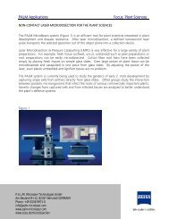
Download PDF - Carl Zeiss - Carl Zeiss, Inc.
Download PDF - Carl Zeiss - Carl Zeiss, Inc.
Download PDF - Carl Zeiss - Carl Zeiss, Inc.
- No tags were found...

You also want an ePaper? Increase the reach of your titles
YUMPU automatically turns print PDFs into web optimized ePapers that Google loves.
Innovation<br />
The Magazine from <strong>Carl</strong> <strong>Zeiss</strong><br />
ISSN 1431-8059<br />
10<br />
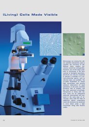
See the difference<br />
with META<br />
■ Microscopy and Prion Research<br />
■ Minimally Invasive Surgery on the Spine<br />
■ Measuring Next to the Machine
Foreword<br />
Developmental Biology<br />
and Cell Biology:<br />
Two Disciplines Unite<br />
Prof. Kai Simons<br />
“Developmental<br />
biologists and cell<br />
biologists have long<br />
ploughed their separate<br />
furrows. But now these<br />
two disciplines are<br />
coming together in<br />
unexpected and exciting<br />
ways.”<br />
Pearson, H. (2001): ”Two<br />
become one”, Nature.<br />
2001 Sep 20.)<br />
Fig. 1 (above):<br />
Fibroblast cell labeled in<br />
three colors: the cell nucleus<br />
(blue), actin stress fibers<br />
(red), lipid rafts (glycophosphatidyl<br />
inositol-anchored<br />
GFP, green) (Photo: Derek<br />
Toomre).<br />
Fig. 2 (below):<br />
Eye of mouse, rod receptor<br />
cells (development stage).<br />
Plasma membrane protein<br />
Prominin (green), cell nuclei<br />
(red). (Photo: K. Röper,<br />
D. Corbeil).<br />
In his classic textbook ”The Cell in Development and Inheritance”<br />
published in 1896, E. B. Wilson, summarizing<br />
the years of biological research since Schleiden and<br />
Schwann formulated their cell theory in 1839, concluded<br />
that the key to all ultimate biological problems must ”in<br />
the last analysis be sought in the cell”. By analyzing a<br />
multitude of different organisms, the 19th century biologists<br />
had, at the turn of the century, already understood<br />
that the basic design of all living cells was similar. These<br />
startling insights were derived from careful observation of<br />
cellular behavior and structure using such simple tools as<br />
light microscopy combined with different staining methods.<br />
These discoveries set the scene for biological research<br />
in the 20th century and led to the biological revolution,<br />
which we are still witnessing<br />
today. Genetics played a key role in<br />
unraveling the molecular mechanisms<br />
responsible for inheritance and<br />
development. Genetic analysis was<br />
one of the few tools biologists had<br />
at their disposal to cut through the<br />
complexity of cellular processes and<br />
to identify the players involved. The<br />
final logic of genetic pathways became<br />
so powerful that the cellular context,<br />
i.e. where the gene products<br />
did their work, was almost blurred<br />
beyond focus. Typical of the success<br />
of the reductionist approach was<br />
that processes were often reduced to<br />
the effects of single genes.<br />
Today we know that several hundreds<br />
of genes are involved in controlling<br />
complex developmental processes such as limb<br />
development. Experimental strategies to unravel developmental<br />
mechanisms are therefore changing. Genetic screens<br />
are being complemented with novel assays that give<br />
insights into the cellular context where the proteins are<br />
localized and where they carry out their function. The<br />
cells in a tissue are no longer considered to be simple<br />
bags with nuclei. Developmental biologists are expanding<br />
their experimental tool kits with the kits of the molecular<br />
cell biologist.<br />
During the last 30 years molecular cell biologists have<br />
been trying to understand the internal workings of a cell<br />
by focusing on a few model cell types, yeast and a small<br />
number of mammalian cells such as fibroblasts, epithelial<br />
cells and neuronal cells. This repertoire is now expanding,<br />
and it is becoming obvious that although the basic design<br />
of different cell types is very similar, the machinery responsible<br />
for cell and tissue organization is specifically modulated<br />
during embryonic development of the organism.<br />
A key to understanding the capabilities and the working<br />
mechanisms of the cellular machinery is to analyze<br />
changes which take place during cell differentiation. The<br />
cooperation of cell and developmental biologists is<br />
contributing to this as well.<br />
Novel microscopic techniques have been transforming<br />
the field of cell biology and are now being increasingly<br />
used in developmental biology. Confocal microscopy led<br />
the way by giving a sharp three-dimensional view of cell<br />
architecture through the elimination of out-of-focus<br />
interference. Optical sectioning by image deconvolution is<br />
another way of obtaining images with high signal-to-noise<br />
ratios. The most recent newcomer is imaging by twophoton<br />
microscopy, which allows sectioning deep within<br />
a tissue and gives access to specimens that previously<br />
could not be imaged. The introduction of GFP, green fluorescent<br />
protein, has also sparked a revolution in cellular<br />
imaging. The use of these color markers has enabled cell<br />
and developmental biologists to use video microscopy to<br />
follow cellular dynamics. The combination of both epifluorescence<br />
and total internal reflection fluorescence<br />
microscopy has made it possible to trace the paths taken<br />
by proteins during transport from the Golgi complex to<br />
the cell surface with exquisite details of the kinetics. These<br />
methods are opening up fascinating vistas of cellular<br />
landscapes until recently not accessible to observation.<br />
Our new Max Planck Institute of Molecular Cell Biology<br />
and Genetics in Dresden, Germany is an example of<br />
the marriage between cell and developmental biology. 20<br />
research groups cover all the major animal models dominating<br />
developmental biology, Drosophila, C.elegans, zebrafish<br />
and mouse. The major goal of the research is to<br />
understand the molecular processes that cells use to form<br />
tissues.<br />
The joint research<br />
has just begun and little<br />
is understood of how<br />
the cells’ internal workings<br />
shape the development<br />
of an organism.<br />
Prof. Dr. Kai Simons, Director of the<br />
Max Planck Institute of Molecular Cell<br />
Biology and Genetics, Dresden, Germany.<br />
E-mail: kai.simons@mpi-cbg.de<br />
Net: www.mpi-cbg.de<br />
2<br />
Innovation 10, <strong>Carl</strong> <strong>Zeiss</strong>, 2001
Contents<br />
Foreword<br />
Developmental Biology and Cell Biology:<br />
Two Disciplines Unite 2<br />
Kai Simons<br />
Contents, Publisher’s Imprint 3<br />
From Users for Users<br />
Microscopy and<br />
Prion Research 4<br />
Interview with Adriano Aguzzi<br />
Thin Sections for<br />
Fascinating Colors 6<br />
Jakob Zbären, Heinz Gundlach<br />
What Fossilic Cyanobacteria Tell Us<br />
About Primeval Oceans 8<br />
Gernot Arp, Christian Böker<br />
Making Laser Scanning<br />
Microscopy More Colorful 10<br />
Sebastian Tille<br />
Minimally Invasive Surgery<br />
on the Spine 13<br />
Too Hot to Handle 16<br />
Products in Practice<br />
Always Knowing Precisely<br />
How Fast the Earth is Turning 18<br />
Measuring Next to the Machine 20<br />
Bernd Balle<br />
All Sights Set on Minimum Angles 22<br />
Around the Globe<br />
News from Switzerland 24<br />
<strong>Zeiss</strong> Microscopes at Yale University 25<br />
Reopening Celebration in Big Style 26<br />
Publisher’s Imprint<br />
Innovation<br />
The Magazine from <strong>Carl</strong> <strong>Zeiss</strong><br />
No. 10, November 2001<br />
“Innovation” appears at irregular internals in German and English.<br />
It was formerly called “<strong>Zeiss</strong> Information with Jena Review” (1992 to<br />
1996), previously “<strong>Zeiss</strong> Information” (1953 to 1991) and “Jena<br />
Review” (1956 to 1991).The issues of the magazine will be serially<br />
numbered, regardless of the year in question, beginning with<br />
No. 1/1996.<br />
Publisher: <strong>Carl</strong> <strong>Zeiss</strong>, Oberkochen, Corporate Communications,<br />
Marc Cyrus Vogel.<br />
Editors: Gudrun Vogel (editor-in-charge), <strong>Carl</strong> <strong>Zeiss</strong> Jena GmbH,<br />
D-07740 Jena, Phone: (+36 41) 64 27 70, Telefax (+36 41) 64 29 41,<br />
e-mail: g.vogel@zeiss.de and Dr. Dieter Brocksch, <strong>Carl</strong> <strong>Zeiss</strong>, D-73446<br />
Oberkochen, Phone: (+73 64) 20 34 08, Telefax (+73 64) 20 33 70,<br />
e-mail: brocksch@zeiss.de, Germany, Medien-Service Wissenschaft,<br />
Stuttgart., Widera Kommunikation, Cologne, Germany.<br />
Internet: http//www.zeiss.de<br />
Prizes • Awards • Anniversaries<br />
10th Birthday at <strong>Carl</strong> <strong>Zeiss</strong> Jena 27<br />
Lothar Janiak<br />
Red Dot Awarded Twice 28<br />
PRISMO No. 2000 for DaimlerChrysler 28<br />
Optics Gold Award 29<br />
Otto Schott Research Award 29<br />
<strong>Carl</strong> <strong>Zeiss</strong> Research Award Followed<br />
by Nobel Prize 30<br />
<strong>Carl</strong> Pulfrich Award 2001 30<br />
Prize-winning Micrograph of Hamster’s Eye 30<br />
Orders • Cooperation Ventures<br />
Cooperation with Nobel Foundation 31<br />
<strong>Carl</strong> <strong>Zeiss</strong> Acquires Metrology Division<br />
of HK-Technologies 31<br />
Lenses for the Digital Cinema 31<br />
In Short<br />
The World’s Most Modern Cine Camera 32<br />
An Old Telescope<br />
Advertises a New Financial Group 33<br />
HypoVereinsbank Puts Its<br />
Money on <strong>Zeiss</strong> 33<br />
Business Barometer<br />
Best Result Ever<br />
in the Company’s History 33<br />
Marc Cyrus Vogel<br />
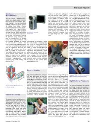
Product Report<br />
Light Microscopy, Ophthalmology,<br />
Surgical Products 34<br />
Electron Microscopy, Camera Lenses,<br />
Sports Optics, Ophthalmic Products 35<br />
Layout: Corporate Design, <strong>Carl</strong> <strong>Zeiss</strong>, Oberkochen;<br />
Manfred Schindler Werbeagentur, D-73431 Aalen.<br />
Setting: Typografie+Medien Werkstatt Hermann, D-73114 Schlat<br />
Printed in Germany by C. Maurer, Druck und Verlag,<br />
D-73312 Geislingen a. d. Steige.<br />
ISSN 1431-8059<br />
© 2001, <strong>Carl</strong> <strong>Zeiss</strong>, Oberkochen.<br />
Permission for the reproduction of individual articles and illustrations<br />
from “Innovation” - with due reference to the source – will gladly be<br />
granted after prior consultation with the editors.<br />
If readers have any inquiries about how the magazine can be obtained<br />
or if they wish to change their address (the customer number should<br />
be indicated, if applicable), we would kindly ask them to contact the<br />
editor.<br />
Picture sources: Unless otherwise specified, all photographs were<br />
contributed by the authors or originate in the archives of <strong>Carl</strong> <strong>Zeiss</strong>.<br />
Authors: If no information is given to the contrary, the authors of the<br />
articles can be contacted via the editor.<br />
Cover photo:<br />
Human chromosome 11 of<br />
intestinal cells (HT 29),<br />
dyed using multicolor<br />
banding. The LSM 510<br />
META permits considerably<br />
more dyes to be<br />
simultaneously used for<br />
marking than was possible<br />
in the past, and their<br />
fluorescence emission to be<br />
allocated precisely in spite<br />
of spectral overlapping.<br />
META provides more<br />
information at a single<br />
glance, i.e. chromosomal<br />
irregularities can be<br />
detected and genetically<br />
related diseases can be<br />
diagnosed at an early stage.<br />
The improved structural<br />
resolution increases the<br />
reliability of diagnosis.<br />
Specimen: Dr. Th. Liehr,<br />
Dr. V. Beensen, Institute of<br />
Human Genetics and<br />
Anthropology at Friedrich<br />
Schiller University in Jena,<br />
Germany (E-mail: i8lith@<br />
mti-n.mti.uni-jena.de).<br />
Micrograph using the LSM<br />
510 META: Dr. P. Ullmann,<br />
<strong>Carl</strong> <strong>Zeiss</strong>. (Please also see<br />
the article: Making Laser<br />
Scanning Microscopy More<br />
Colorful, pages 10 to 12).<br />
Outside back cover:<br />
Since October 2001, the<br />
world's largest and most<br />
precise ring laser has been<br />
sited deep under the earth<br />
at Wettzell in the Bavarian<br />
Forest mountains. <strong>Carl</strong><br />
<strong>Zeiss</strong> was responsible for the<br />
manufacture of the large<br />
ring laser.<br />
(Please also see the article:<br />
Always Knowing Precisely<br />
How Fast the Earth is<br />
Turning, pages 18 and 19).<br />
Innovation 10, <strong>Carl</strong> <strong>Zeiss</strong>, 2001 3
From Users for Users<br />
Microscopy and Prion Research<br />
“My<br />
dream:<br />
being able<br />
to see<br />
how prions<br />
move under<br />
the microscope”<br />
He is one of the leading researchers<br />
of the human variant of mad cow<br />
disease, i. e. the Creutzfeldt-Jakob<br />
disease: Professor Adriano Aguzzi<br />
considerably helped the world with<br />
his discoveries in the fight against the<br />
pernicious disease. Of course, the<br />
technical equipment he uses to<br />
examine the pathogens, known as<br />
prions, also plays an important role.<br />
“Innovation” talked with the renowned<br />
researcher in the Neuropathology<br />
Institute at Zurich University,<br />
Switzerland, of which he is the<br />
director.<br />
How did mad cow disease evolve<br />
We do not know exactly. There are<br />
two basic theories: one says that<br />
meat and bone meal containing scrapie-infected<br />
sheep brains was fed to<br />
animals. The other theory assumes<br />
spontaneous mutations in cattle. However,<br />
the real cause will most likely<br />
never be found.<br />
Mad cow disease now plays a<br />
considerably smaller role in the<br />
media than one year ago...<br />
That's right. But for us, this discussion<br />
never played a major role<br />
anyway. We just continued doing our<br />
job.<br />
...which mainly revolves around<br />
the new variant of the<br />
Creutzfeldt-Jakob disease.<br />
How does it differ from the<br />
conventional disease<br />
We assume that the new variant<br />
of the Creutzfeldt-Jakob disease in<br />
humans is identical to mad cow disease.<br />
The new variant mainly affects<br />
young people, often even teenagers.<br />
Furthermore, the course of the disease<br />
is slower in most cases. We<br />
reckon on one to two years, whereas<br />
the classical variant proceeds much<br />
faster.<br />
How many humans are affected<br />
with the new variant of the<br />
Creutzfeldt-Jakob disease<br />
We assume approx. 120 at present.<br />
How do you expect this<br />
number to develop<br />
That's hard to say. Of course, I<br />
hope the number will remain as low<br />
as possible.<br />
Are there any marked differences<br />
in the dangerousness of the<br />
variants<br />
No. Both variants are lethal. Only<br />
one variant is probably caused by the<br />
transmission of BSE.<br />
What actually is the morphologic<br />
difference between normal mad<br />
cow disease and the Creutzfeldt-<br />
Jakob disease<br />
There is, of course, quite a number<br />
of parameters. For example, the<br />
tissue patterns are very different, and<br />
there is markedly more plaque inside<br />
the tissue.<br />
Was microscopy a major tool for<br />
the discovery of the morphologic<br />
structures<br />
It is most definitely very important.<br />
However, I would say that we applied<br />
all the technical tools which are useful<br />
for us. Microscopy is one of them,<br />
no matter whether it is light microscopy,<br />
confocal microscopy or electron<br />
microscopy.<br />
A major part of your work now<br />
consists in detecting the<br />
pathogens known as prions.<br />
Will microscopy play a role here<br />
Certainly. Let us take the movement<br />
of prions, for example – here<br />
we do not yet have enough tools<br />
to be able to clarify this problem<br />
morphologically and functionally, although<br />
this is exactly what we will<br />
have to do in the end. For me, a<br />
dream would come true if I were able<br />
to see in the microscope how prions<br />
move, for example within the spleen<br />
or the nerve tissue. However, technology<br />
has not yet advanced enough<br />
for the equipment to be used in the<br />
required way. For example, the sensitivity<br />
of multiphoton techniques is still<br />
not good enough for our purposes.<br />
Does this mean you consider light<br />
microscopy in no way outdated<br />
True. I would never say that<br />
microscopy is outdated. Instead, I<br />
would say: microscopy is not yet advanced<br />
far enough.<br />
Does your dissatisfaction mean<br />
that the microscope does not<br />
enable you to resolve sufficient<br />
structures Or does it mean that<br />
there are not enough markers for<br />
prions to be able to detect them<br />
with the light microscope<br />
I guess both are right. But certainly<br />
sensitivity is the bigger problem<br />
at present.<br />
How did the microscope help you<br />
in the beginning Purely on the<br />
histology/morphology basis or<br />
also with suitable markers<br />
About five or six years ago we<br />
performed many morphology examinations.<br />
This was very important. After<br />
this, we laid more emphasis on molecular<br />
biology. But it would be good<br />
to return to morphology in the end.<br />
But then with living specimens<br />
Does this mean: living sections<br />
Yes, certainly, though not only, but<br />
also.<br />
The whole world is waiting for a<br />
reliable way of destroying the BSE<br />
pathogen to make sure that BSEfree<br />
foodstuff becomes available<br />
on the market. How can the BSE<br />
pathogen be destroyed<br />
And in what time can this be<br />
implemented on a large scale<br />
Various possibilities already exist<br />
today. For example, the use of very<br />
high temperatures or various chemicals.<br />
However, there are other areas<br />
where we are unable to destroy the<br />
BSE pathogen.<br />
4<br />
Innovation 10, <strong>Carl</strong> <strong>Zeiss</strong>, 2001
For example<br />
Blood, for example. Here it is not<br />
possible at present to achieve decontamination.<br />
And I would rather not<br />
make any forecasts about when this<br />
will be possible.<br />
After all, particular importance is<br />
attached to blood in the<br />
transmission of the new variant of<br />
the Creutzfeldt-Jakob disease.<br />
That's right. The problem in the<br />
future will be focused more on transmission<br />
between humans than from<br />
cattle to humans.<br />
Why<br />
Because all high-risk organs like<br />
the brain and spinal marrow have<br />
been removed from the human food<br />
chain by now. At the same time,<br />
however, many people carry the mad<br />
cow pathogen in their bodies. It is<br />
conceivable that it can be transmitted<br />
to other people by blood transfusions<br />
or by insufficiently sterilized instruments.<br />
How long will it then take for the<br />
disease to break out<br />
About 15 to 20 years.<br />
In animal experiments you found<br />
out that this period is almost<br />
exactly 200 days in mice. Can this<br />
period also be determined with<br />
similar precision for humans<br />
This is what we assume. For the<br />
time being, the exact incubation period<br />
is unfortunately not known. We<br />
are therefore unable to say when the<br />
disease will reach its climax in humans.<br />
Let us assume an incubation<br />
period of 15 to 20 years: is this the<br />
period of time required by prions<br />
to reach the human brain<br />
Yes, that's right. It seems that the<br />
prion only develops its adverse affect<br />
in the brain.<br />
How does this long period<br />
of time come about<br />
There are various stages<br />
which the prions must pass<br />
before they reach the brain.<br />
And it is most likely that they<br />
get held up along the way.<br />
Will all measures be too late once<br />
the prions have reached the<br />
brain<br />
Yes, most likely. But, of course,<br />
this is of interest to us, too. After all,<br />
we want to understand how the<br />
damage to the brain occurs.<br />
What do you envisage as a<br />
possible cure<br />
This is quite easy: We must prevent<br />
the prions from reaching the<br />
brain in the first place.<br />
You once mentioned that about<br />
100 million people had been in<br />
contact with the pathogen.<br />
What does this mean<br />
Let me say this much: it certainly<br />
does not mean that all these people<br />
have been infected. And the disease<br />
will not break out in all who have<br />
been infected. Here, many factors<br />
play a role, for example the genetic<br />
disposition which we know nothing<br />
at all about.<br />
There are also other theories<br />
about the Creutzfeldt-Jakob<br />
disease. There are people who<br />
support the virus theory, and<br />
some researchers consider<br />
chemicals as major co-factors.<br />
What do you think of these<br />
theories<br />
It's always good to have different<br />
approaches, and to check these<br />
again and again. Therefore, I can<br />
only welcome the various hypotheses<br />
from various scientists. However, the<br />
prion theory still seems to be most<br />
likely.<br />
Do you as a scientist still enjoy<br />
eating beef<br />
Certainly. After all, the meat itself<br />
was the minor problem. What worried<br />
us were the complete components<br />
contained in it, e.g. brain or separator<br />
meat. But this has fortunately<br />
been taken from the market by now.<br />
Adriano Aguzzi, Neuropathology Institute of<br />
the University of Zurich, Switzerland.<br />
E-mail: adriano@pathol.unizh.ch<br />
Net: www.neuroscience.unizh.ch/e/groups/<br />
aguzzi00.htm<br />
Fig. 1:<br />
Professor Dr. Adriano Aguzzi,<br />
Director of the Institute of<br />
Neuropathology at the<br />
University of Zurich and the<br />
National Swiss Reference<br />
Center for Prion Diseases<br />
and member of the Scientific<br />
BSE Advisory Committee of<br />
the British government and<br />
the EU Commission, in his<br />
laboratory using an<br />
Axioplan® 2 imaging<br />
research microscope with<br />
digital AxioCam®.<br />
(Photo: Jesper Dijohn).<br />
Innovation 10, <strong>Carl</strong> <strong>Zeiss</strong>, 2001 5
Thin Sections for Fascinating Colors<br />
Jakob Zbären, Heinz Gundlach<br />
Fig. 1:<br />
Kidneys of a rat, polymer<br />
section 0.75 µm, double<br />
marking with laminin<br />
(CY 3), filter FS 41007a,<br />
(10 s), cell nuclei (DAPI),<br />
filter 01 (16 s, gray filter).<br />
Fig. 2:<br />
Tongue of a rat, polymer<br />
section 0.75 µm, triple<br />
marking with CY 3<br />
cytokeratin, filter FS 41oo7a<br />
(exposure time: 15 s),<br />
vimentin (ALEXA 594), cell<br />
nuclei (DAPI). Filter 01<br />
(10 s, gray filter).<br />
Almost 100 years ago, August Köhler<br />
and Moritz von Rohr were able to<br />
observe fluorescence phenomena in<br />
a microscope with an ultraviolet illuminator<br />
for the first time. Today, fluorescence<br />
microscopy is a technique<br />
widely used in cell research, histology,<br />
genetics, and many other fields.<br />
Entirely new possibilities have become<br />
available to biomedical research<br />
thanks to the combination of fluorescence,<br />
confocal laser scanning<br />
microscopy, and high-performance<br />
digital image processing. Let us mention<br />
only a few key words: multicolor<br />
FISH, multicolor banding and – totally<br />
new – LSM 510 META, a system –<br />
introduced in this magazine – with<br />
a flexibility never achieved before.<br />
Polymer and<br />
antibodies<br />
However, further development has<br />
also taken place in traditional fluorescence<br />
microscopy. Here, progress in<br />
the past few years was achieved<br />
through multifluorescence techniques,<br />
the development of new dyes,<br />
the improvement of photomicrography<br />
techniques and the use of digital<br />
photography and image processing.<br />
Images of high information content<br />
and aesthetic beauty have become<br />
possible, not least because of new<br />
preparation techniques.<br />
One of these techniques is polymer<br />
sections. Compared with the traditional<br />
embedding of specimens in<br />
paraffin, the use of polymer enables<br />
section thicknesses which are 10 times<br />
smaller, extending down to less<br />
than 1 µm. After immunohistochemical<br />
staining of these specimens, it is<br />
impossible for the large antibody<br />
molecules as carriers of the fluorescence<br />
dye to penetrate the polymer,<br />
i.e. the antibodies only bind to the<br />
surface of the section. State-of-theart<br />
microscope systems such as the<br />
Axioplan ® 2 Imaging or Axiovert ®<br />
200, and highly sensitive detection<br />
methods permit even higher resolution<br />
of object details (Figs 1 and 2)<br />
than has been possible so far. It is<br />
readily comparable to the resolution<br />
of low-magnification electron microscopy.<br />
But also the Axioskop ® 2<br />
inverted microscope allows such<br />
fluorescence to be viewed and documented<br />
(Figs 3 and 4).<br />
From blue to red<br />
New dyes, called fluorochromes with<br />
the family name ALEXA, cover the<br />
entire spectrum from blue to red and<br />
provide color brilliance never achieved<br />
before. The high-precision filter<br />
technology of double and triple<br />
bandpass filters permits the simultaneous<br />
display and analysis of two or<br />
three of the fluorescence fluorochromes<br />
with only a single filter set. In<br />
6<br />
Innovation 10, <strong>Carl</strong> <strong>Zeiss</strong>, 2001
From Users for Users<br />
combination with high-resolution color<br />
films, the traditional method, i.e.<br />
classical photomicrography, permits<br />
up to four fluorescence types to be<br />
displayed simultaneously with high<br />
contrast and excellent resolution (Figs<br />
3 and 4).<br />
The Cy 5, Cy 5.5 and Cy 7 cyanin<br />
fluorochromes have extended the application<br />
possibilities in the red and<br />
infrared range, although they can<br />
only be displayed and evaluated<br />
using digital methods.<br />
Figs 3 and 4:<br />
Human endothelial cells.<br />
Fig. 3: Quadruple marking<br />
with actin (phalloidin<br />
ALEXA 594), of Willebrand<br />
factor (ALEXA 350),<br />
vinculin (ALEXA 488)<br />
mixed fluorochrome, cell<br />
nucleus (DAPI). Filter 01<br />
(24 s, gray filter), double<br />
bandpass filter 24 (28 s).<br />
Fig. 4: Quadruple marking:<br />
actin (phalloidin/TRITC),<br />
of Willebrand factor<br />
(ALEXA 350), tubulin<br />
(ALEXA 488), cell nucleus<br />
(DAPI). Double bandpass<br />
filter 23 (45 s), filter 01<br />
(15 s, gray filter).<br />
(Micrographs: Jakob Zbären<br />
using the Axioplan® 2<br />
Imaging, Plan-Apochromat<br />
20/0.75, (Figs 1 and 2) and<br />
Axioskop® 2,<br />
Plan-NEOFLUAR® 63/1.25,<br />
(3 and 4), MC 80, double<br />
exposure. All photographs<br />
were taken with<br />
Fujichrome Velvia ISO 50<br />
for professionals).<br />
Jakob Zbären, Thrombosis Laboratory at<br />
Inselspital Bern, Switzerland.<br />
E-mail: jakobzb@hotmail.com<br />
Dr. Heinz Gundlach, Research and<br />
Technology, <strong>Carl</strong> <strong>Zeiss</strong>.<br />
E-mail: gundlach@zeiss.de<br />
7
From Users for Users<br />
What Fossilic Cyanobacteria Tell Us<br />
About Primeval Oceans<br />
Gernot Arp, Christian Böker<br />
Cyanobacteria (blue-green algae)<br />
have existed for at least 2.7 billion<br />
years and therefore number among<br />
the oldest organisms on earth. Like<br />
plants which came into existence later,<br />
they already used photosynthesis<br />
at that time, a process by which carbon<br />
dioxide is assimilated and oxygen<br />
released. As a result, cyanobacteria<br />
created an oxygen-rich atmosphere<br />
in the Precambrian age between 3.8<br />
billion and 540 million years ago,<br />
without which the development of<br />
advanced forms of life in water and<br />
on land would never have been possible<br />
in the first place.<br />
Fig. 1:<br />
Contemporary<br />
cyanobacterial lime reefs<br />
(stromatolites) near the<br />
bank of Lake Thetis, West<br />
Australia. (Photo: J. Reitner,<br />
Göttingen).<br />
In the waterside areas of lakes and<br />
oceans, cyanobacteria and other<br />
microorganisms form what are<br />
known as biofilms which calcify in<br />
the appropriate conditions and can<br />
build meter-high, finely-layered reefs<br />
(Fig. 1). Enormous lime reefs, or stromatolites,<br />
were formed as far back as<br />
the Precambrian age and number<br />
among the earth's oldest fossils.<br />
Dr. Gernot Arp<br />
Dr. Christian Böker<br />
8<br />
Innovation 10, <strong>Carl</strong> <strong>Zeiss</strong>, 2001
From Users for Users<br />
Fig. 2:<br />
Biofilm with cyanobacteria<br />
(yellow, diameter: 5 µm),<br />
calcite crystals (green) and a<br />
nematode (green band)<br />
from a soda lake (Pyramid<br />
Lake, Nevada). The<br />
projection of 60 confocal<br />
image planes shows that the<br />
cyanobacteria are unable to<br />
precipitate calcium sheaths<br />
under the conditions<br />
prevailing in the lake, but<br />
that the calcite crystals are<br />
available in an unordered<br />
form in the biofilm. The<br />
micrograph was taken with<br />
an LSM 510 confocal laser<br />
scanning microscope from<br />
<strong>Carl</strong> <strong>Zeiss</strong>.<br />
Until now, scientists believed that cyanobacteria<br />
would change the chemical<br />
equilibrium in their immediate<br />
surrounding by photosynthetic carbon<br />
dioxide assimilation in such a way<br />
that calcium carbonate is precipitated<br />
in their slime sheaths, thus causing a<br />
stromatolithic reef formation. But not<br />
all fossilic cyanobacteria in the lime<br />
reefs have a lime sheath. Photomicrographs<br />
of contemporary mineralizing<br />
cyanobacteria show that the lime<br />
crystals are mostly formed irregularly<br />
in the slime matrix of the biofilms<br />
(Fig. 2) and are bound directly to the<br />
cyanobacteria only in exceptional cases.<br />
The geobiologists Gernot Arp,<br />
Andreas Reimer and Joachim Reitner<br />
from the Göttingen Geosciences<br />
Center have now been able to unseal<br />
the mystery about such exceptions.<br />
Their model calculations show that<br />
the cyanobacterial photosynthesis results<br />
in lime precipitation only if high<br />
concentrations of calcium and low<br />
concentrations of inorganic carbon<br />
are dissolved in water simultaneously.<br />
Calcareous cyanobacteria fossils permit<br />
us to find out in which millionyear<br />
periods this must have been the<br />
case. If the atmospheric carbon dioxide<br />
content of the air, which can be<br />
estimated for the geological ages by<br />
means of the stomatal density on<br />
Gingko leaves, for example, is taken<br />
into consideration, it is possible to<br />
calculate the minimum amount of<br />
calcium concentrations in primeval<br />
oceans for the first time. It then becomes<br />
evident that the calcium content<br />
fluctuated repeatedly between<br />
today's values and values which are<br />
up to three times higher.<br />
Since calcium plays an important<br />
role in the metabolic processes of organisms,<br />
more detailed knowledge<br />
about changes in the calcium concentrations<br />
in the ocean could permit,<br />
for example, conclusions to be<br />
made about the evolution of crustaceans<br />
and the skeletons of vertebrates.<br />
The renowned US “Science” magazine<br />
used the opportunity offered by<br />
the new insights into the calcium<br />
production of cyanobacteria to select<br />
a laser scanning micrograph of a biofilm<br />
with cyanobacteria as the cover<br />
photo of its 1 June 2001 issue and<br />
published the research findings of<br />
the Göttingen scientists in a special<br />
report.<br />
Dr. Gernot Arp is a member of the<br />
Göttingen Geosciences Center, Geobiology<br />
Department, Göttingen University, Germany.<br />
E-mail: garp@gwdg.de.<br />
Net: www.imgp.gwdg.de.<br />
Dr. Christian Böker is an applications<br />
specialist for laser scanning microscopy at<br />
<strong>Carl</strong> <strong>Zeiss</strong>.<br />
E-mail: c.boeker@zeiss.de<br />
Innovation 10, <strong>Carl</strong> <strong>Zeiss</strong>, 2001 9
From Users for Users<br />
Making Laser Scanning Microscopy<br />
More Colorful<br />
Sebastian Tille<br />
You do not always succeed in making<br />
coffee the same way every time. And<br />
when it does taste really good, the<br />
chances are you won’t remember<br />
how much coffee, water and milk<br />
you used – and whether you added<br />
one or two lumps of sugar. Wouldn’t<br />
it be great if we could analyze the<br />
composition of the coffee at a single<br />
glance, without any need to take the<br />
successful mixture apart. In biomedical<br />
research, the question would be:<br />
what are the components of an<br />
intact cell, and how are they linked<br />
with each other In addition to the<br />
structure, functional interrelations in<br />
living cells and organisms are also of<br />
interest, which leads to the second<br />
this to scientific experiments, this<br />
means: unambiguous and reliable<br />
results.<br />
Experience<br />
the future today<br />
We at <strong>Carl</strong> <strong>Zeiss</strong> certainly are no magicians<br />
either, but we use an entirely<br />
new approach to open up experimental<br />
possibilities for users of laser<br />
scanning microscopes (LSM) which<br />
were unthinkable until now. For the<br />
first time, the new LSM 510 META<br />
with its revolutionary Emission Fingerprinting<br />
technique permits the<br />
clean separation of several – even<br />
spectrally overlapping – fluorescence<br />
geneticists have completed the sequencing<br />
of the human genome,<br />
they are interested in the purpose of<br />
every single gene. Cell biologists not<br />
only want to know which proteins<br />
exist in a cell, but also what their<br />
functions are and with which other<br />
proteins they interrelate. Recording<br />
techniques such as FRET (Fluorescence<br />
Resonance Energy Transfer) or FRAP<br />
(Fluorescence Recovery After Photobleaching)<br />
allowing dynamic changes<br />
of the fluorescence emission to be<br />
followed are used very intensely for<br />
this purpose.<br />
The discovery of natural fluorescence<br />
dyes, i.e. the green fluorescent<br />
protein (GFP) and its variants (Figs 3a<br />
wish: being able to record active cells<br />
– like an entire day's vacation – in a<br />
single photo showing all details, even<br />
those which are not obvious at first.<br />
There is also a third wish, shared by<br />
children and parents when playing<br />
Memory: being able to always uncover<br />
the right combination of cards<br />
immediately – in other words, knowing<br />
instead of guessing! If we apply<br />
“This system<br />
provides a very easy<br />
way of performing<br />
FRAP experiments.<br />
I regret not having<br />
used this system<br />
earlier.”<br />
Prof. Yasushi Hiraoka,Kansai Advanced<br />
Research Center, Kobe, Japan.<br />
signals of a specimen. The number of<br />
dyes which can be used and detected<br />
in the experiment is almost unlimited.<br />
Thus, the new system overcomes the<br />
limits of existing detection methods<br />
and permits both qualitative and<br />
quantitative analyses, quickly and<br />
precisely, in vitro and in vivo.<br />
GFP and the life<br />
sciences<br />
Laser scanning microscopes are scientific<br />
tools mainly for use in the life<br />
sciences. They provide insights into<br />
cells and tissue. The fluorescence<br />
technique enables the visualization of<br />
cell components marked with various<br />
dyes and excited by laser light of different<br />
wavelengths. In the life sciences,<br />
the analysis of functional interrelations<br />
inside the cell is becoming<br />
increasingly important, in addition to<br />
the research of structures. Now that<br />
and 3b), was a major step forward<br />
for multifluorescence microscopy.<br />
This non-toxic dye can be produced<br />
by the cells themselves, thus permitting<br />
the long-time observation of<br />
living objects.<br />
However, limits have been set<br />
here, too. The now improved “living<br />
dyes” display spectral properties<br />
which make their simultaneous use<br />
more difficult. The problem is known<br />
as signal overlapping, or “crosstalk”.<br />
When several dyes are used, it is difficult,<br />
if not at all impossible, to find<br />
wavelength ranges where only one<br />
dye is guaranteed to emit, the signal<br />
of which can therefore be recorded<br />
using conventional bandpass detection.<br />
This problem could partly be<br />
solved using what is known as the<br />
multitracking technique provided by<br />
<strong>Zeiss</strong> LSM microscopes. However, no<br />
solution was available in cases where<br />
several dyes to be separated were ex-<br />
10<br />
Innovation 10, <strong>Carl</strong> <strong>Zeiss</strong>, 2001
From Users for Users<br />
Fig. 1:<br />
LSM 510 META – a<br />
confocal laser scanning<br />
fluorescence microscope<br />
opening doors<br />
to new experiments.<br />
2a<br />
2b<br />
Emission<br />
Intensity<br />
200<br />
150<br />
100<br />
50<br />
0<br />
FITC<br />
GFP<br />
500 520 540 560 580<br />
Wavelength [nm] 2c<br />
Figs 2a to 2c:<br />
Cultivated fibroblasts with<br />
expression of a GFP-<br />
Histon2B fusion protein,<br />
actin filaments marked with<br />
FITC phalloidin. Irradiation<br />
with 488 nm. Display via<br />
the new Emission<br />
Fingerprinting technique<br />
(2a) and by using the<br />
conventional bandpass filter<br />
between 505 and 530 nm<br />
(2b). Distance between<br />
emission maxima of GFP<br />
and FITC: 7 nm (2c).<br />
cited for fluorescence by a single laser<br />
wavelength, since separation by<br />
means of bandpass detection is not<br />
possible in this case.<br />
Emission<br />
Fingerprinting –<br />
the solution to<br />
the problem in<br />
multifluorescence<br />
applications<br />
Due to our ongoing cooperation with<br />
users, these problems were known to<br />
us, as was the necessity to pave new<br />
ways for future applications.<br />
The Emission Fingerprinting technique<br />
is based on a new multichannel<br />
detector onto which the entire<br />
emission spectrum is projected and<br />
which can change these signals to<br />
digital information thanks to its fast<br />
electronics unit. The emission signals<br />
of the various dyes can now be separated<br />
in three easy steps:<br />
1. Acquisition of a lambda stack,<br />
i.e. a stack of x-y images, to record<br />
the spectral distribution of fluorescence<br />
light as a parameter of each pixel<br />
in the examined object.<br />
2. Determination of the spectral<br />
Dr. Frank-D. Böhmer, Molecular Cell Biology,<br />
Friedrich Schiller University in Jena, Germany.<br />
“The new scan modes<br />
of the system offer<br />
a completely new<br />
quality of analysis.<br />
The interpretation of<br />
the data is far more<br />
reliable than with any<br />
conventional system<br />
based on filter sets<br />
and bandpass<br />
acquisition.”<br />
Innovation 10, <strong>Carl</strong> <strong>Zeiss</strong>, 2001 11
From Users for Users<br />
Figs 3a and 3b:<br />
Excitation and emission<br />
spectra of the GFP green<br />
fluorescent protein – of<br />
great importantance for the<br />
examination of living cells –<br />
and its variants CFP, YFP<br />
and DsRed, which – used<br />
simultaneously – could not<br />
be precisely separated with<br />
current techniques.<br />
(Source: Clontech).<br />
Excitation<br />
100<br />
80<br />
60<br />
40<br />
20<br />
CFP GFP YFP DsRed<br />
Emission<br />
100<br />
80<br />
60<br />
40<br />
20<br />
CFP GFP YFP DsRed<br />
0<br />
325 400 450 500 550 600<br />
Wavelength [nm]<br />
3a<br />
0<br />
325 425 475 525 575 625 675<br />
Wavelength [nm]<br />
3b<br />
Fig. 4:<br />
Clear separation of CFP-<br />
RanGAP1 (blue, proteins in<br />
cell plasma), GFP emerin<br />
(green, proteins in cell<br />
membrane) and YFP-<br />
SUMO1 expression (red,<br />
cell nuclei) in cultivated<br />
cells. (Prof. Y. Hiraoka,<br />
KARC, Kobe, Japan).<br />
Fig. 5:<br />
Zebrafish embryo, eye and<br />
part of the brain; cell<br />
adhesion molecule tag-1<br />
(Alexa Fluor 488, green),<br />
Tubulin (Cy3, red), sugar<br />
epitope PSA (Cy5, violet),<br />
cell nuclei (DAPI, blue).<br />
(Dr.M.Marx,Prof.M.Bastmeyer,<br />
Constance<br />
University, Germany).<br />
signatures in selected specimen<br />
areas, or loading of reference spectra<br />
of the used dyes from the database.<br />
3. Use of the Linear Unmixing algorithm,<br />
i.e. the digital separation of<br />
overlapping fluorescence signals in<br />
single image channels which only<br />
contain the intensities of one marker,<br />
clearly separated from each other.<br />
The Emission Fingerprinting technique,<br />
entirely integrated into the LSM<br />
software, is easy to operate. It provides<br />
the unique possibility of separating<br />
fluorescence signals which<br />
were excited by only a single laser<br />
line, e.g. in multiphoton microscopy.<br />
Furthermore, it is beneficial in many<br />
cases for the elimination of unwanted<br />
signals, such as background noise<br />
or autofluorescence. The new technique<br />
of the LSM 510 META solves all<br />
these problems thanks to knowledge<br />
”The LSM 510 META<br />
makes FRET imaging<br />
really easy because<br />
you get a spectral<br />
readout of both<br />
proteins. You see<br />
the contribution of<br />
both, acceptor and<br />
donor peak.”<br />
of the spectral properties of the specimen<br />
and the markers it contains –<br />
returning to our analogy with the<br />
Memory card game – this means that<br />
the right combination of cards is uncovered<br />
immediately without relying<br />
on luck. The recording of lambda<br />
stacks can be combined with 3D<br />
or/and time series (x, y, z, t, λ). Thanks<br />
to the electronic control of the multichannel<br />
detector, this can be done<br />
quickly and reproducibly. Combined<br />
with the information obtained about<br />
the entire emission spectrum, all<br />
events in the cells have thus been recorded<br />
– as would be the case in a<br />
single photo of a whole day's vacation.<br />
A wide spectrum of applications<br />
has already been tested successfully,<br />
and the users are enthusiastic about<br />
the method giving them a completely<br />
free hand in the selection and choice<br />
of fluorescence markers.<br />
The LSM 510 META will help researchers<br />
to further increase the efficiency<br />
and success of their work. This<br />
gives them more time during which<br />
they can be inspired by the extended<br />
possibilities of the system and discover<br />
further secrets of biology in<br />
new experiments while drinking a<br />
nice cup of coffee.<br />
4<br />
5<br />
Mary Dickinson, PhD, Biological Imaging<br />
Center, Caltech, Pasadena, USA.<br />
Sebastian Tille is Product Manager of Laser<br />
Scanning Microscopy at <strong>Carl</strong> <strong>Zeiss</strong>.<br />
E-mail: stille@zeiss.com<br />
Net: www.zeiss.de/lsm<br />
12<br />
Innovation 10, <strong>Carl</strong> <strong>Zeiss</strong>, 2001
From Users for Users<br />
Minimally Invasive Surgery<br />
on the Spine<br />
A step forward or<br />
backward<br />
At the symposium entitled ”Minimally<br />
Invasive Trends in the Spine Area” organized<br />
by <strong>Carl</strong> <strong>Zeiss</strong> during the 52nd<br />
Annual Convention of the German<br />
Society of Neurosurgery (DGN) in Bielefeld,<br />
Germany in May 2001, physicians<br />
and instrument manufacturers<br />
discussed the question: ”Is endoscopy<br />
a step forward or backward for neurosurgery”<br />
Although the participants<br />
in the symposium were impressed<br />
by the new possibilities available,<br />
some criticism was also voiced: ”We<br />
now have wonderful, new technology<br />
at our fingertips, but the question<br />
remains – who can do what with<br />
what” Some of the participating<br />
surgeons express their opinions below.<br />
Technology of the<br />
Finest<br />
Robert S. Bray<br />
In the operating room of the future,<br />
parallel, consecutive or alternating<br />
use will most certainly be made of<br />
minimally invasive techniques. This is<br />
already being practiced at the Cedars<br />
Sinai Hospital. The technical equipment<br />
ranges from state-of-the-art<br />
microscopes and navigation aids to<br />
3D endoscopes and voice-controlled<br />
robots. The possibility of transmitting<br />
operations live via the internet already<br />
exists, archival is fully digital,<br />
and the technical equipment in the<br />
OR such as lamps and cameras can<br />
be voice-controlled. Two experts in<br />
the hospital have specialized in disk<br />
surgery and thoracoscopy. They offer<br />
workshops in which the participants<br />
can learn the techniques step by<br />
step. For example, an anterior lumbar<br />
fusion is possible through four incisions;<br />
Robert S. Bray Jr., M.D.,<br />
Institute for Spinal<br />
Disorders at the Cedars-<br />
Sinai Medical Center in Los<br />
Angeles. With more than<br />
5500 microsurgical<br />
operations on the spine, he<br />
is a recognized specialist in<br />
this field.<br />
Fig. 1:<br />
The OPMI® Vario/NC 33<br />
system was developed<br />
exclusively for minimally<br />
invasive surgery in the<br />
spine area.<br />
2a<br />
Prof.Dr.med.Robert<br />
Schönmayr, Dr.-Horst-<br />
Schmidt-Kliniken GmbH,<br />
Hospital of the Provincial<br />
Capital Wiesbaden,<br />
Germany.<br />
2b<br />
Figs 2a and 2b:<br />
With its higher<br />
magnification and more<br />
powerful illumination,<br />
more anatomical details can<br />
be discerned in the surgical<br />
microscope (2a) than with<br />
loupes (2b).<br />
Innovation 10, <strong>Carl</strong> <strong>Zeiss</strong>, 2001 13
From Users for Users<br />
Fig 3:<br />
The ergonomic design of<br />
the surgical microscope<br />
allows the surgeon to work<br />
with the ultimate in<br />
comfort, even over long<br />
periods.<br />
their space requirements in the OR reduced.<br />
What does “Minimally<br />
Invasive” Mean<br />
Dr. Wolfgang Börm<br />
Senior Physician at the<br />
Bezirkskrankenhaus Günzburg,<br />
Neurosurgery, Germany<br />
the patients are hospitalized for only<br />
two days, even if the stabilization<br />
was performed over several verterbrae.<br />
Hospital stays for anterior and<br />
posterior stabilization total only 1 1 /2<br />
days in some individual cases.<br />
Every spine, with the exception of<br />
those with severe scoliosis, can be a<br />
case for endosurgery. Thanks to the<br />
development of modern technology,<br />
the physician will also be able to<br />
work in areas where there is no room<br />
for their hands. The major advantage<br />
of robots will be that they do not get<br />
tired. One example: via an incision<br />
measuring only 3 cm, new artificial<br />
intervertebral disks have already been<br />
successfully implanted in six patients.<br />
At the Cedars Sinai Hospital traditional<br />
surgery is also performed in<br />
addition to these procedures using<br />
sophisticated, high-tech equipment.<br />
Quo Vadis<br />
Prof. Dr. Robert Schönmayr<br />
It is not a matter of endoscope versus<br />
microscope: what we want is a combination<br />
of the two technologies.<br />
However, as I see it, the transition to<br />
the endoscope is a step backward. For<br />
surgeons like us who are accustomed<br />
to using a surgical microscope, it is a<br />
major drawback that the three-dimensionality<br />
is missing. The miniaturization<br />
of open surgery and the extension<br />
of percutaneous techniques have<br />
good prospects for the future. For the<br />
patient, it is of great psychological importance<br />
to have only a small incision;<br />
the more minimally invasive the procedure,<br />
the better. In the USA hospital<br />
stays after surgery are considerably<br />
shorter than, for example, in Germany<br />
due to the greater use of keyhole surgery.<br />
Further improvements should be<br />
made to navigation aids, robotics, and<br />
miniaturization. The mobility of the instruments<br />
must be enhanced and<br />
Minimally invasive – this is a very<br />
woolly term. Strictly speaking, only<br />
the incision is minimal; a lot of things<br />
also happen under it in minimally invasive<br />
procedures. For the success of<br />
the healing process it is not the<br />
length of the surface incision that is<br />
important, but the traumatization of<br />
the tissue under it. On the basis of<br />
the experience we have gained to<br />
date, we shall not use purely endoscopic<br />
techniques in future either.<br />
We prefer to operate with the visual<br />
control of the microscope. This is not<br />
as good in the two-dimensional endoscope<br />
image, and the aorta is very<br />
close.<br />
Preparation Under<br />
the Microscope<br />
Dr. Hans-J. Meisel<br />
Berufsgenossenschaftliche Kliniken,<br />
Bergmannstrost, Neurosurgery, Halle,<br />
Germany<br />
’Biological repair’ must be our top<br />
priority. What we have to do is prepare<br />
osseous components in such a<br />
way that bones can heal again and<br />
no layers of degenerated cartilage<br />
remain in the fusion zone. For this<br />
purpose, a technique must be used<br />
which allows surgery via as small an<br />
incision as possible either by open<br />
microsurgery using the microscope or<br />
with the aid of an endoscope in order<br />
to minimize traumatization and<br />
muscle destruction. In this way, decompression<br />
can be achieved in the<br />
area of the spinal canal and the measures<br />
required for stabilization imple-<br />
14<br />
Innovation 10, <strong>Carl</strong> <strong>Zeiss</strong>, 2001
From Users for Users<br />
mented. This allows minimization of<br />
what was seen as major surgery in<br />
the past.<br />
4a<br />
Today’s standard<br />
of care<br />
More and more people are now experiencing<br />
back problems at an earlier<br />
and earlier age. This is attributable<br />
to the increase in the number of<br />
sedentary jobs and to the fact that<br />
too much leisure time is spent sitting<br />
in the car or in the armchair. Nowadays,<br />
most of us have to cope with<br />
increasing stress and less exercise.<br />
Overweight in a rising percentage of<br />
people in the Western world and increased<br />
life expectancy are further<br />
contributory factors. These are all<br />
reasons why the number of spinal<br />
procedures is expected to rise in future.<br />
According to information received<br />
from leading neurosurgeons, these<br />
procedures will jump by as much as<br />
40% in the next 5 to 7 years. From<br />
surgeons, patients are expecting a<br />
higher quality of care with less pain,<br />
and hospital administrators are under<br />
increasing pressure to reduce patient<br />
stays. The medical world is rising to<br />
this challenge by the use of minimally<br />
invasive methods. Surgical microscopes<br />
are the ideal visualization tool for<br />
this purpose.<br />
optics, incredibly simple user guidance,<br />
flexible positioning and the impressive<br />
daylight quality illumination<br />
are all brought together in a very<br />
compact, stylish design.<br />
<strong>Carl</strong> <strong>Zeiss</strong>‘ visualization solutions<br />
have enabled surgeons to develop<br />
innovative, minimally invasive techniques<br />
that have improved patient<br />
outcomes and quality of life.<br />
E-mail: h.wolf@zeiss.de<br />
Net: www.zeiss.de/chirurgie<br />
4b<br />
Figs 4a and 4b:<br />
Ventral removal of cervical<br />
disks (schematic).<br />
Focus on results<br />
The importance of minimally invasive<br />
surgery on the spine is undisputed: it<br />
reduces patient strain and shortens<br />
recovery times. The use of microsurgical<br />
techniques reduced hospitalization<br />
from 4.6 to 1.4 days and costs<br />
by more than 50% (Quality Study of<br />
the Cedars Sinai Medical Center).<br />
The OPMI ® Vario/NC 33 system<br />
was specially developed to meet the<br />
needs of the spine market. The symmetrical<br />
configuration of the system<br />
and its outstanding ease of use<br />
make it the ideal partner for the spine<br />
surgeon. Brilliant, apochromatic<br />
Fig. 5:<br />
Removal of disk residue on<br />
the lumbar spine<br />
(schematic).<br />
Innovation 10, <strong>Carl</strong> <strong>Zeiss</strong>, 2001 15
From Users for Users<br />
Too Hot to Handle<br />
Fig. 1:<br />
During production the glass<br />
bottles pass by the on-line<br />
hot end inspection system<br />
with the telecentric<br />
measuring lenses. (Photo:<br />
ART-KON-TOR, Jena).<br />
The online inspection of the glass<br />
containers directly after production<br />
offers considerable advantages over<br />
traditional “cold end” inspection: the<br />
bottles are measured and sorted just<br />
a few seconds after manufacture.<br />
In addition, the hot end inspection<br />
system clearly allocates recognized<br />
errors to the manufacturing tools,<br />
allowing the causes to be quickly eliminated.<br />
The temperature of the hot<br />
bottle provides additional information<br />
on parameters relevant to the<br />
process. Therefore, hot end testing<br />
serves not only for quality sorting,<br />
but also for quality production.<br />
A real hotshot<br />
system<br />
However, inspection at the hot end<br />
of container glass production has to<br />
contend with extreme conditions: the<br />
containers cannot be subjected to<br />
any handling during their journey<br />
from the machine to the cooling line.<br />
This means they cannot be specially<br />
Fig. 2:<br />
Three cameras are used to<br />
photograph the bottle from<br />
three sides.<br />
Fig. 3:<br />
Hot ampoule in<br />
production. (Photos 2<br />
and 3: OTTO GmbH).<br />
During the production process bottles<br />
with defects are automatically removed.<br />
To date, however, this has not been done<br />
until the end of the production process<br />
prior to the packaging of the bottles and<br />
therefore a long time after the defect has<br />
actually occurred. Severe losses are the result,<br />
several 10,000 bottles leave the conveyor<br />
belt in the period before packaging.<br />
Now, with a ”hot end” inspection instrument<br />
developed by the firm OTTO GmbH,<br />
Jena, Germany, the red-hot glass containers<br />
can be inspected immediately after<br />
production. The centerpiece of this measuring<br />
technology is telecentric lenses from<br />
<strong>Carl</strong> <strong>Zeiss</strong>.<br />
2<br />
3<br />
16<br />
Innovation 10, <strong>Carl</strong> <strong>Zeiss</strong>, 2001
From Users for Users<br />
aligned or turned for the measurement.<br />
In addition, the space available<br />
for a measuring instrument is extremely<br />
small for technical reasons. Finally,<br />
the high temperatures of the<br />
bottles and the surrounding area<br />
make very exacting demands on any<br />
measuring technology used.<br />
It was for these reasons that the<br />
company OTTO GmbH developed a<br />
non-contact optical inspection technique<br />
on the basis of digital image<br />
processing. CCD matrix cameras with<br />
telecentric lenses generate two-dimensional<br />
images of the test object,<br />
from which the image processing<br />
software gathers dimensional information<br />
and compares it to target<br />
values and tolerance ranges. The<br />
glowing containers with temperatures<br />
of up to 500 °C travel past the<br />
lenses at a distance of 180 mm to<br />
250 mm! Specially coated glass<br />
protects them from the intense heat.<br />
4a<br />
4b<br />
Non-contact<br />
precision<br />
The containers leaving the machine<br />
have different distances from, and<br />
different orientations relative to the<br />
cameras and lenses. With traditional<br />
standard optics, this would result in<br />
systematic measuring errors which<br />
could lead to incorrect sorting. OTTO<br />
GmbH solved this problem through<br />
the use of telecentric VISIONMES ®<br />
measuring lenses from <strong>Carl</strong> <strong>Zeiss</strong>: Irrespective<br />
of the production-related<br />
distance of the bottles from the lenses,<br />
they are always imaged with the<br />
same, unaltered size. This allows<br />
measuring accuracies of up to 0.02<br />
mm, depending on the feature being<br />
measured, e.g. the bottle height, the<br />
diameter in various planes, the outer<br />
and core diameter of the thread, the<br />
bevel of the bottle mouth, and many<br />
more besides. Theoretically, the hot<br />
end inspection systems can be used<br />
to record, measure, evaluate and sort<br />
900 and more articles a minute. In<br />
practice, the machines in the container<br />
glass industry currently produce<br />
no more than 600 bottles a minute.<br />
Dr. Roland Fiedler of OTTO GmbH<br />
played a decisive role in the development<br />
of the measuring technology:<br />
”We currently know of no setups<br />
which can achieve equivalent qualitative<br />
and quantitative results in the<br />
hot area of container glass production.”<br />
OTTO GmbH Computer Vision Systems.<br />
E-mail: r.fiedler@otto-jena.de<br />
Net: www.otto-jena.de<br />
Fig. 5:<br />
VISIONMES®<br />
telecentric lens from<br />
<strong>Carl</strong> <strong>Zeiss</strong>.<br />
Figs 4a and 4b:<br />
Photographing objects at<br />
different distances from the<br />
lens. Without a telecentric<br />
lens (4a): the ratios of<br />
reproduction are different.<br />
With a telecentric lens(4b):<br />
the objects are imaged with<br />
the same reproduction<br />
ratio.<br />
In short<br />
In many areas of industrial metrology, automation technology,<br />
in-process inspection and control, and in quality management,<br />
the proportion of optical, non-contact processes<br />
now being used to identify, test and measure complex<br />
structures is increasing. Such advanced measuring<br />
and inspection tasks as the application<br />
described here can only be performed<br />
using high-resolution CCD camera<br />
systems and telecentric precision lenses.<br />
The exacting demands made on the<br />
measuring system are met<br />
by the virtually distortion-free<br />
VISIONMES ® lenses. Telecentric<br />
viewing and the measurement of<br />
relevant production processes<br />
eliminate the need for costly and<br />
time-consuming alignment and<br />
testing processes for the user,<br />
and higher quality is achieved in<br />
the entire production process and<br />
therefore also in the products<br />
themselves.<br />
VISIONMES ®<br />
Telecentric Components<br />
E-mail: petermann@zeiss.de<br />
Innovation 10, <strong>Carl</strong> <strong>Zeiss</strong>, 2001 17
Products in Practice<br />
Always Knowing Precisely<br />
How Fast the Earth is Turning<br />
Fig. 1:<br />
Schematic section of the<br />
underground laboratory<br />
for the large ring laser.<br />
(Drawing: ifag).<br />
drainage shaft<br />
electrical supply room<br />
drain<br />
Since October 2001, the world's largest<br />
and most precise ring laser has<br />
been sited deep under the earth at<br />
Wettzell in the Bavarian Forest mountains.<br />
Built by <strong>Carl</strong> <strong>Zeiss</strong> and implemented<br />
by the German Federal Cartography<br />
and Geodesy Office, the<br />
Technical University of Munich and<br />
the University of Canterbury, New<br />
Zealand, the large “G” ring laser<br />
measures the earth's rotation with<br />
unprecedented accuracy. Its data is<br />
used, for example, by satellite-based<br />
navigation systems such as GPS (Global<br />
Positioning System) which rely on<br />
global reference systems. The ring laser's<br />
measurements are also of interest<br />
to earthquake researchers.<br />
“G” for gigantic is indeed an accurate<br />
way of describing the new ring laser<br />
from <strong>Carl</strong> <strong>Zeiss</strong>: with its size of 4 x<br />
4 meters and its weight of more than<br />
10 tons, it rests on a 10-ton granite<br />
slab which is supported by a pier<br />
founded 11 meters deep – and it is<br />
able to measure variations in the<br />
earth's velocity of rotation within a<br />
range of only 0.1 milliseconds.<br />
This level of accuracy is vital for<br />
the implementation of a high-precision<br />
global reference system which<br />
takes into account even the minutest<br />
fluctuations in the earth's rotation.<br />
The cause of these fluctuations lies in<br />
the constant displacement of masses<br />
on the earth's surface (produced by<br />
7% ramp<br />
max. groundwater level, drill location 1<br />
sluice<br />
IT room<br />
foamed glass<br />
inner<br />
well ring<br />
outer<br />
e.g. tides, weather phases, lunar phases)<br />
and inside the earth (convection<br />
currents, continental drift, volcanic<br />
activity, etc.). For the exact determination<br />
of a position on the earth,<br />
even such slight fluctuations need to<br />
be included in the computation. One<br />
example: due to the earth's rotation,<br />
a point on the equator travels a distance<br />
of roughly 40,000 km within<br />
24 hours, or a distance of over 460<br />
meters within one second.<br />
To permit a point to be measured<br />
with centimeter accuracy – a requirement<br />
for advanced Global Positioning<br />
Systems – it must be possible to describe<br />
the earth's velocity of rotation<br />
to the nearest 20 millionth of a second.<br />
This is an accuracy level where<br />
even slight variations in the earth's<br />
rotation have a noticeable effect in<br />
the course of the day. With such critical<br />
accuracy requirements, the criteria<br />
to be applied to the measuring<br />
equipment and its direct environment<br />
are particularly stringent. Nothing<br />
must influence the measurement result.<br />
The geometry of the ring laser<br />
itself, for example, must not change<br />
in any way as a result of expansion or<br />
contraction of the material. Even<br />
changes totaling a mere millionth of<br />
a millimeter would affect the measurement<br />
result. This is the reason<br />
why the ring laser's centerpiece is a<br />
10 ton heavy disk of Zerodur ® ceramic<br />
glass manufactured by the comsurface<br />
water lining<br />
operation tank<br />
"monument"<br />
pier<br />
clay<br />
Styrodur<br />
deposit<br />
former ground surface<br />
max. groundwater level,<br />
drill location 6<br />
weathering layer<br />
uncased concrete pile<br />
solid rock<br />
pany Schott Glas in Mainz. Zerodur ®<br />
features virtually zero expansion,<br />
roughly 600 times less than steel and<br />
400 times less than optical glass.<br />
Further factors influencing the<br />
measurement result are the temperature<br />
and air pressure. Even fluctuations<br />
of more than one thousandth<br />
of a degree Celsius or more than 0.1<br />
hPa have a detrimental effect. To minimize<br />
the atmospheric pressure fluctuations<br />
caused by weather conditions<br />
to 0.1 hPa, the developers<br />
enclosed the complete ring laser by a<br />
steel tank whose internal pressure is<br />
kept constant at 1050 mbar by a<br />
control system. The temperature is<br />
kept stable by insulation of the laboratory<br />
room's concrete walls and ceiling<br />
using a thermal layer of Styrodur<br />
with a thickness of half a meter, followed<br />
by a one-meter thick layer of<br />
humid clay and again half a meter of<br />
Styrodur. In addition, the complete<br />
underground laboratory was covered<br />
by a 4 meter layer of soil.<br />
18<br />
Innovation 10, <strong>Carl</strong> <strong>Zeiss</strong>, 2001
Products in Practice<br />
The measuring<br />
principle:<br />
differences in<br />
frequency between<br />
two countercirculating<br />
laser<br />
beams<br />
Four highly reflective mirrors form a<br />
closed beam path running in what is<br />
called the resonator tube and enclosing<br />
a square surface (ring laser). The<br />
resonator tube is filled with an inert<br />
gas mixture of helium and neon. The<br />
laser or inert gas is excited by a highfrequency<br />
voltage in the radio wave<br />
range. Since no preference is given<br />
to any direction of circulation, the resulting<br />
red laser beam starts to circulate<br />
in both of the possible directions<br />
in the ring-shaped resonator. If the<br />
instrument was stationary, the frequencies<br />
of both the clockwise and<br />
counterclockwise beam would be<br />
identical. Due to the earth's rotation,<br />
however, the two laser beams travel<br />
on paths of different lengths. The resulting<br />
path difference is measured<br />
in frequencies (Sagnac effect). The<br />
Sagnac frequency is directly proportional<br />
to the earth's rotation, which<br />
means that any variation in the<br />
earth's rotation leads to a change in<br />
the path difference and therefore in<br />
the Sagnac frequency. The large ring<br />
laser permits frequency fluctuations<br />
of 10 -9 Hertz to be measured in one<br />
day.<br />
Dr. Ulrich Schreiber, Technical University<br />
of Munich, Research Institute of Satellite<br />
Geodesy,<br />
E-Mail: schreiber@wettzell.ifag.de<br />
Dr.-Ing. Wolfgang Schlüter, German Federal<br />
Cartography and Geodesy Office,<br />
Wettzell Fundamental Research Station,<br />
E-Mail: schlueter@wettzell.ifag.de,<br />
Net: www.ifag.de<br />
Hieronymus Weber, <strong>Carl</strong> <strong>Zeiss</strong>,<br />
Project Manager,<br />
E-mail: h.weber@zeiss.de<br />
Fig. 2:<br />
10 tons of Zerodur®<br />
ceramic glass, the carrier for<br />
the ring laser, suspended on<br />
the lifting gear of a specialpurpose<br />
crane on their way<br />
to the underground<br />
laboratory. In the background,<br />
the radio telescope<br />
of Wettzell fundamental<br />
research station which<br />
receives satellite signals<br />
from space for earth and<br />
ground measurements.<br />
Fig. 3:<br />
The large ring laser permits<br />
fluctuations in the earth's<br />
velocity of rotation to be<br />
measured with extremely<br />
high accuracy. Together<br />
with the instrument,<br />
Dr. Ulrich Schreiber,<br />
scientific director of the<br />
ring laser project at the<br />
Research Institute of<br />
Satellite Geodesy of the<br />
Technical University of<br />
Munich.<br />
Innovation 10, <strong>Carl</strong> <strong>Zeiss</strong>, 2001 19
Products in Practice<br />
Measuring Next to the Machine<br />
Bernd Balle<br />
Fig. 1:<br />
Optimized measuring<br />
range and enhanced loading<br />
capability with<br />
CenterMax®.<br />
Expectations on today's production<br />
measuring technology are largely focused<br />
on shopfloor application and<br />
the rough environmental conditions<br />
encountered there. The trend is obvious:<br />
measurement is being shifted<br />
from the metrology room into the<br />
manufacturing process, which considerably<br />
reduces reaction times.<br />
The field of production metrology<br />
ranges from the inspection of testpieces<br />
in the metrology room to statistical<br />
process control. Today, it is still<br />
the metrology engineer who uses a<br />
coordinate measuring machine in the<br />
metrology room for checking the features<br />
defined by the designer and<br />
produced by the machine operator,<br />
but production planners are now claiming<br />
that this quality assurance process<br />
should be implemented by using<br />
measuring machines integrated directly<br />
into the production line. In addition,<br />
quality engineering is characterized<br />
by an increasing trend toward not<br />
simply measuring and inspecting<br />
workpieces, but monitoring the overall<br />
manufacturing process in volume<br />
production, allowing early intervention,<br />
if necessary. Efficiency is the main<br />
reason for integrating coordinate<br />
measuring machines into the production<br />
process. The closer your measuring<br />
technology is located to the production<br />
facility, the more directly your<br />
process control can be implemented.<br />
ment. For this purpose, the foremost<br />
demand on a coordinate measuring<br />
machine is resistance to ambient<br />
conditions – much like a machining<br />
center.<br />
Just envisage the following scenario:<br />
it is hot, the machine is causing<br />
not only an incredible noise, but also<br />
floor vibrations. At times, temperature<br />
differences of up to 10 ° C prevail,<br />
workpieces are covered in lubricants<br />
and solvents. And it is in<br />
precisely these adverse conditions<br />
that high accuracy measurements<br />
need to be performed.<br />
CenterMax ® does precisely that<br />
– a coordinate measuring machine<br />
that can be used without an air-con-<br />
ditioned cabin.<br />
The new CenterMax ® production<br />
measuring center is based on the<br />
proven PRISMO ® series with the<br />
VAST ® sensor system. The machine<br />
was designed as a bridge construction<br />
to withstand the rough ambient conditions<br />
in production. Its body consists<br />
of a cast stone – a material which is<br />
ideal as a thermally and dynamically<br />
protective damping element. Like the<br />
human skeleton, CenterMax ® ’s TRF<br />
structure provides a thermally stable<br />
frame that guarantees the specified<br />
accuracy over the entire temperature<br />
range. Raised guideways additionally<br />
reduce moving masses and therefore<br />
enhance the stability and precision of<br />
Accurate and robust<br />
Apart from the relevant software, it is<br />
above all the coordinate measuring<br />
machines themselves which have to<br />
cope with constantly new challenges.<br />
<strong>Carl</strong> <strong>Zeiss</strong> launched ScanMax ® back<br />
in 1997 – a manual measuring machine<br />
for in-process inspection. The<br />
integration of the measuring machine<br />
right in the center of production<br />
provides quality information without<br />
any need for the metrology room detour.<br />
Measuring machines from <strong>Carl</strong><br />
<strong>Zeiss</strong> are used like production equip-<br />
20<br />
Innovation 10, <strong>Carl</strong> <strong>Zeiss</strong>, 2001
Products in Practice<br />
the coordinate measuring machine.<br />
An active vibration damping system<br />
makes CenterMax ® insensitive to the<br />
usual floor vibrations.<br />
Patented labyrinth sealings completely<br />
enclose the bearings and<br />
scales protecting them against dust,<br />
lubricants and solvents. This design<br />
also permits liquids such as oil or water<br />
to be collected underneath the<br />
workpiece base.<br />
To guarantee the optimum ratio<br />
between installation area and work<br />
range, the measuring volume was<br />
optimized. Neither the stylus changing<br />
magazine, the rotary table nor<br />
the loading system restrict the measuring<br />
volume. Therefore, the effective<br />
measuring volume corresponds to<br />
the nominal volume. A plate carrier<br />
frame, a granite plate or a rotary<br />
table can be optionally integrated<br />
into the machine bed of Center-<br />
Max ® . Due to its open design the<br />
machine can be loaded from three<br />
sides.<br />
The new TVA (Temperature Variable<br />
Accuracy) specification defined by<br />
<strong>Carl</strong> <strong>Zeiss</strong> provides CenterMax ® with<br />
guaranteed accuracy specifications<br />
graded according to the given temperature<br />
conditions. In the past, the<br />
accuracy specification of a coordinate<br />
measuring machine referred to the<br />
most favorable ambient conditions. If<br />
the temperature changed, you could<br />
never be sure exactly how accurate<br />
your machine was measuring. Now<br />
you can specify the appropriate accuracy<br />
as a function of the temperature<br />
at the site of installation.<br />
The system is equipped with a<br />
LCD touch screen for maximum ease<br />
of operation, even allowing the operator<br />
to wear gloves. CenterMax ®<br />
operates with the <strong>Zeiss</strong>-patented<br />
active scanning technology which<br />
yields information on dimension,<br />
form and location in just one measuring<br />
process. With stylus extensions<br />
up to 450 mm and stylus configurations<br />
up to 600 g, the VAST ®<br />
sensor system provides an answer to<br />
virtually all requirements. Compared<br />
to doghouse gauges, it is hence possible<br />
to verify not only one particular<br />
workpiece type, but all parts of your<br />
production program. Many gauges,<br />
multi-feature measuring devices or<br />
test configurations are no longer required.<br />
The result: CenterMax ® pays<br />
for itself in next to no time.<br />
Initial on-site<br />
experience<br />
Pilot installations of CenterMax ® on<br />
different users' premises substantiate<br />
the correctness of the design. At<br />
Grüner Systemtechnik GmbH in Bad<br />
Überkingen, a supplier to all renowned<br />
car manufacturers in Germany, a<br />
first machine is being used with great<br />
success in the middle of the production<br />
area. It has been installed exactly<br />
where the work needs to be done.<br />
Although exposed to rough manufacturing<br />
conditions, the machine has<br />
been operating faultlessly from day<br />
one.<br />
Peter Haller,<br />
Metrology Room Manager<br />
of Grüner Systemtechnik<br />
GmbH, Bad Überkingen,<br />
Germany.<br />
Bernd Balle, <strong>Carl</strong> <strong>Zeiss</strong> Industrial Measuring<br />
Technology, Business Strategy Marketing<br />
E-mail: b.balle@zeiss.de<br />
Net: www.zeiss.de/imt<br />
“We have compared<br />
CenterMax ® with<br />
PRISMO ® and found<br />
that we are actually<br />
getting the same<br />
results with<br />
CenterMax ® in the<br />
production area as<br />
with PRISMO ® in the<br />
metrology room. It<br />
measures with the<br />
same precision. This<br />
was revealed by a<br />
long-term test over a<br />
period of 6 months.”<br />
Fig. 2:<br />
CenterMax® in the center of<br />
production.<br />
Innovation 10, <strong>Carl</strong> <strong>Zeiss</strong>, 2001 21
Products in Practice<br />
All Sights Set on Minimum Angles<br />
Fig. 1:<br />
The refractive index<br />
measuring station is<br />
approx. 2 m x 2 m large<br />
and weighs approx. 2 tons<br />
due to its granite baseplate.<br />
One collimator weighs<br />
approx. 20 kg.<br />
The performance of computers is rising<br />
constantly, thanks to ever smaller<br />
structures on the microchips. To<br />
generate these finer structures using<br />
lithographic methods, the semiconductor<br />
industry is utilizing ever shorter<br />
wavelengths for exposure. To<br />
achieve this, however, the lenses<br />
which direct the light on to the chips<br />
must also constantly be improved.<br />
This requires that their optical properties<br />
be known more exactly. However,<br />
as the refractive indices of glass<br />
batches vary too much to allow<br />
the production of high-quality lenses,<br />
research staff must determine the refractive<br />
index of individual glass batches<br />
with extremely high precision.<br />
The measurement result is then used<br />
for computing the final figure of the<br />
lens element, ensuring the correct<br />
polishing of the element. <strong>Carl</strong> <strong>Zeiss</strong><br />
has now made this high-precision<br />
measurement possible for the vacuum<br />
UV wavelength of 157 nm.<br />
Take a circle and divide it into about<br />
4 million parts – this will give you<br />
some idea of the minute range in<br />
which the researchers of <strong>Carl</strong> <strong>Zeiss</strong><br />
perform their refractive index measurements.<br />
It is their goal next year to<br />
make the production of lens elements<br />
possible which will allow the<br />
critical dimensions on the chip to be<br />
reduced even further: The distance<br />
between the individual lines is to be<br />
clearly shorter than 120 nm. This is<br />
1/300th of the breadth of a human<br />
hair. In a few years, this distance is to<br />
shrink to only 70 nm (1/500th of a<br />
hair's breadth). Wavelengths of 157<br />
nm and shorter will then generate<br />
these finest features on the chips.<br />
High-precision<br />
refractive index<br />
measurements<br />
of vacuum UV<br />
transparent materials<br />
The development team of <strong>Carl</strong> <strong>Zeiss</strong><br />
looks back on three to four years of<br />
intensive engineering work and successful<br />
cooperation ventures with<br />
companies from various sectors of industry.<br />
It was a long way from the<br />
basic idea behind refractive index<br />
measurement to the finished measuring<br />
station. Dr. Martin Ross-Messemer<br />
of the <strong>Carl</strong> <strong>Zeiss</strong> Optical Research<br />
Division explains: “The idea of<br />
refractive index measurement was in<br />
fact developed by Fraunhofer as early<br />
as 1817. However, improving the<br />
measurement has only been possible<br />
by using electronics, industrial metrology<br />
and the intensive use of computers.”<br />
To achieve their goal, scientists<br />
and engineers had to perform calculations<br />
and measurements down to<br />
the very last detail and develop new<br />
components at many stages.<br />
As the 157 nm wavelength lies in<br />
the vacuum UV range and is absorbed<br />
by normal air, the measurement<br />
of the refractive index is performed in<br />
a nitrogen atmosphere. In addition,<br />
the ambient temperature and the<br />
pressure of the gas used must be<br />
exactly determined and taken into<br />
account in the computation. For a<br />
true comparison of different materials,<br />
the air pressure and the temperature<br />
must be kept constant. Deviations<br />
of as small as 0.1 hPa and<br />
0.1 °C will significantly change the<br />
result.<br />
The rotary stage on which the newly<br />
developed collimator is mounted can<br />
resolve angles of 0.3’’ (= 1.5 µrad).<br />
As the detector for the incident light<br />
consists of a highly developed camera<br />
system, a problem arose: One pixel of<br />
the camera corresponded to an angle<br />
of 15 µrad. To be able to approach<br />
the resolution provided by the collimator<br />
despite this handicap, the research<br />
staff developed the subpixel<br />
evaluation process, allowing them to<br />
measure even these small angles with<br />
this camera.<br />
The wide wavelength range from<br />
850 to 120 nm to be covered required<br />
a new optical concept for the<br />
collimators used in the measuring<br />
station, as they should be independent<br />
of the ambient temperature and<br />
the wavelength used. <strong>Zeiss</strong> found<br />
a solution to this problem in cooperation<br />
with the company Lichtenknecker:<br />
Using mirrors instead of the<br />
usual lens elements in the whole<br />
optical system, the researchers circumvented<br />
the problem of the different<br />
refraction of different light wavelengths,<br />
thus creating an achromatic<br />
system. The researchers used special<br />
steel (INVAR) to mount the<br />
mirrors, rendering the system<br />
athermal. In addition,<br />
the mirrors were<br />
specially coated for<br />
the vacuum UV<br />
range.<br />
22<br />
Innovation 10, <strong>Carl</strong> <strong>Zeiss</strong>, 2001
Products in Practice<br />
Autocollimator<br />
Camera<br />
Swing-in beam splitter<br />
Prism<br />
Spectral lamp<br />
Prism stage<br />
Viewing collimator<br />
Rotary stage<br />
Illuminating collimator<br />
Fig. 2:<br />
Sketch of the refractive<br />
measuring station.<br />
For the range between 850 nm to<br />
180 nm, <strong>Zeiss</strong> developed a new spectral<br />
lamp together with the company<br />
Heraeus. A different solution was<br />
again found for wavelengths below<br />
180 nm: An American company supplied<br />
a multiline source which provided<br />
a large number of overlapping<br />
emission lines. A computer-assisted<br />
procedure specially developed by<br />
<strong>Zeiss</strong> allows the individual emission<br />
lines to be identified like fingerprints<br />
and to be used for measurements. As<br />
the individual emission lines have different<br />
intensities, a problem arose regarding<br />
the light sensitivity of the camera<br />
used as the detector. Together<br />
with the company Proxitronic the device<br />
required was developed.<br />
This completed the number of<br />
units needed for the refractive index<br />
measuring station. The material to be<br />
examined was cut into prisms and<br />
placed on the measuring stage. – To<br />
create constant conditions for the<br />
measurements, the researchers designed<br />
a huge steel bell (similar to<br />
the tank of a milk truck) and in this<br />
way protected the whole system<br />
against ambient effects.<br />
Not only had all instruments<br />
including those determining the<br />
ambient conditions to be connected<br />
with each other, but also with a<br />
control unit – and then linked to a<br />
computer which performs the precise<br />
evaluation of the data. The computer,<br />
in turn, controls the measuring<br />
stage supporting the collimators – a<br />
real tour de force also regarding the<br />
programming work involved.<br />
The effort was worthwhile: the<br />
glass material can be measured with<br />
ultra-high precision. In addition, with<br />
its refractive index measuring station,<br />
<strong>Zeiss</strong> is involved in international projects<br />
in which only the best of the<br />
best participate. For instance, even<br />
the Physikalisch Technische Bundesanstalt<br />
(PTB, German National Metrology<br />
Laboratory) compares their measurement<br />
results with those obtained<br />
by <strong>Carl</strong> <strong>Zeiss</strong>. The measuring station<br />
can be used in many different areas:<br />
from the calibration of spectral lamps<br />
and the analysis of gas properties in<br />
the VUV range to the examination of<br />
the thermal behavior of specific glass<br />
types or newly developed materials<br />
such as synthetic crystals. This service<br />
is also available to <strong>Zeiss</strong> customers.<br />
Info: kalibrieren@zeiss.de<br />
Dr. Martin Ross-Messemer, Optics Research,<br />
<strong>Carl</strong> <strong>Zeiss</strong>, scientific adviser<br />
E-Mail: ross-messemer@zeiss.de<br />
Holger Wegendt, <strong>Carl</strong> <strong>Zeiss</strong> Measuring and<br />
Calibrating Center. Manager of the<br />
refractive index measuring station project<br />
E-mail: wegendt@zeiss.de<br />
Innovation 10, <strong>Carl</strong> <strong>Zeiss</strong>, 2001 23
Around the Globe<br />
News<br />
from Switzerland<br />
Fig. 1:<br />
Current headquarters of<br />
<strong>Carl</strong> <strong>Zeiss</strong> AG, Switzerland<br />
on the shores of Lake<br />
Zurich.<br />
Fig. 2:<br />
New customer service center<br />
for industrial measuring<br />
technology on modern<br />
premises in Feldbach.<br />
Fig. 3:<br />
Kreuzlingen Planetarium<br />
under construction. Planned<br />
opening: spring 2002.<br />
Fig. 4:<br />
Successful microscope sales<br />
by telephone.<br />
(Photos: <strong>Carl</strong> <strong>Zeiss</strong> AG,<br />
Switzerland).<br />
Traditionally, Germany’s southern<br />
neighbor has been a very good market<br />
for <strong>Carl</strong> <strong>Zeiss</strong>. As far back as<br />
spring 1914, when the new Zurich<br />
University building was being constructed,<br />
the first <strong>Zeiss</strong> products –<br />
microprojection instruments and reflected<br />
light projectors – were delivered<br />
to Switzerland. The firm Ganz &<br />
Co in Zurich represented <strong>Carl</strong> <strong>Zeiss</strong><br />
very successfully for over four decades.<br />
In 1958 it was transformed into<br />
the Ganz-Optar AG stock corporation,<br />
from which<br />
<strong>Carl</strong> <strong>Zeiss</strong> Zürich AG<br />
emerged as the<br />
Swiss subsidiary of<br />
<strong>Carl</strong> <strong>Zeiss</strong>, Oberkochen<br />
in 1970.<br />
Today, with a<br />
workforce of 80<br />
people, <strong>Carl</strong> <strong>Zeiss</strong><br />
AG in Switzerland<br />
generates sales totaling<br />
30 million Swiss<br />
franks. It is based in Feldbach on the<br />
shores of Lake Zurich, where the<br />
two sales organizations of <strong>Carl</strong> <strong>Zeiss</strong><br />
and Schott Glas operate from one<br />
building (Fig. 1). In 2000 two profit<br />
centers of <strong>Carl</strong> <strong>Zeiss</strong> AG, i.e. Industrial<br />
Measuring Technology and<br />
Medical Systems, were honored for<br />
their exceptional performance compared<br />
to other <strong>Zeiss</strong> European sales<br />
organizations.<br />
IMT in new customer<br />
service center<br />
In September 2001 the Industrial<br />
Measuring Technology division relocated<br />
from Wetzikon to the firm’s<br />
Feldbach headquarters. It opened a<br />
service center with five new measuring,<br />
demonstration and training<br />
rooms. Heinz J. Widmer, President of<br />
2<br />
<strong>Carl</strong> <strong>Zeiss</strong>, Switzerland and head of<br />
the IMT profit center: “Our goal is<br />
continued growth in the field of customer<br />
services. In future, we will not<br />
only train our customers, but will also<br />
perform measuring and programming<br />
tasks for them.” Small wonder<br />
that his staff have no reason to complain<br />
about a lack of work. A wide<br />
selection of courses, contract measurement<br />
and application programming<br />
for the creation of customer<br />
programs is being very well received<br />
by industry.<br />
Planetarium at<br />
Lake Constance<br />
Above the town of Kreuzlingen with<br />
a breathtaking view of Lake Constance,<br />
a <strong>Zeiss</strong> planetarium will be<br />
opened in May 2002. It is the third in<br />
Switzerland. In addition to a large<br />
star theater in Lucerne, a second,<br />
small planetarium was opened in<br />
Schwanden in the mountains surrounding<br />
Bern in October 2000.<br />
Audiences in the 70-seat Kreuzlingen<br />
Planetarium can see up to 7000<br />
stars on the dome. Meditative insights<br />
into the universe and a special<br />
children’s program round off the<br />
exciting program.<br />
3<br />
Microscope<br />
sales:<br />
Success by<br />
telephone<br />
One year ago <strong>Carl</strong> <strong>Zeiss</strong><br />
AG began an interesting<br />
experiment: simple microscopes<br />
for school, university,<br />
laboratory and<br />
hobby use, in particular the Stemi ®<br />
DV 4 stereomicroscope and the<br />
Axiostar ® transmitted-light microscope,<br />
were sold by telephone by a<br />
special trained team of office sales<br />
staff. Needless to say, it is not<br />
enough to simply wait for telephone<br />
calls. Direct mailings were sent to the<br />
appropriate target groups, and the<br />
DV 4 or Axiostar ® was placed at the<br />
disposal of opinion leaders and<br />
students on special terms and conditions.<br />
The result after one year: sales totaling<br />
around DM 1 million and an<br />
eight-fold increase in market share in<br />
the lower price segment of stereomicroscopy.<br />
50% of business is generated<br />
with microscopes, the other<br />
50% with colposcopes and lens analyzers.<br />
The office sales staff achieved<br />
this sales performance with existing<br />
personnel capacities and substantially<br />
eased the workload of field staff.<br />
E-mail: zeiss@zeiss.ch<br />
Net: www.zeiss.ch<br />
24<br />
Innovation 10, <strong>Carl</strong> <strong>Zeiss</strong>, 2001
Around the Globe<br />
<strong>Zeiss</strong> Microscopes<br />
at Yale University<br />
In New Haven, at the Yale University<br />
School of Medicine, an exhibit on<br />
”Microscopy: Tools of the Biomedical<br />
Sciences” was organized in honor of<br />
the university’s 300th anniversary.<br />
Opening day ceremonies were highlighted<br />
by a lecture on ”The Evolution<br />
of Infectious Disease” by Dr. Joshua<br />
Lederberg, Nobel Laureate for<br />
Medicine in 1958 and former president<br />
of the Rockefeller University,<br />
The exhibit was supported by an<br />
educational grant from the <strong>Carl</strong> <strong>Zeiss</strong><br />
Foundation. Not only historic instruments<br />
sent on loan to New Haven by<br />
the Optical Museum of <strong>Carl</strong> <strong>Zeiss</strong>,<br />
Oberkochen, but also state-of-the-art<br />
microscopes from <strong>Carl</strong> <strong>Zeiss</strong> were displayed.<br />
<strong>Carl</strong> <strong>Zeiss</strong>, <strong>Inc</strong>., Thornwood<br />
provided confocal and fluorescence<br />
and stereo microscopes, including<br />
the new PALM system with a laser<br />
scalpel. A look at the future of instrument<br />
development in the field of<br />
microscopy rounded off the exhibit.<br />
Fig. 2:<br />
Historical microscopes and<br />
state-of-the-art systems for<br />
biomedical research were<br />
exhibited at Yale University.<br />
Fig. 1:<br />
Dr. Norbert Gorny, Member<br />
of the Board at <strong>Carl</strong> <strong>Zeiss</strong><br />
and Executive Vice<br />
President and General Manager<br />
in the Microscopy<br />
business group, presents a<br />
replica of the <strong>Zeiss</strong> Stativ<br />
Microscope VII from 1880<br />
to Dr. Martin Gordon,<br />
director of the Yale Medical<br />
Library, and Dr. Joshua<br />
Lederberg (from left to<br />
right).<br />
Net: www.med.yale.edu/library/zeiss<br />
Fig. 3:<br />
Surgical microscopes and<br />
ophthalmic instrument<br />
systems rounded off the<br />
exhibition. (Photos: <strong>Carl</strong><br />
<strong>Zeiss</strong>, <strong>Inc</strong>., Thornwood).<br />
Innovation 10, <strong>Carl</strong> <strong>Zeiss</strong>, 2001 25
Around the Globe<br />
Reopening Celebration in Big Style<br />
Fig. 1:<br />
James McDonnell<br />
Planetarium in St. Louis.<br />
Fig. 2:<br />
Guests at the reopening<br />
ceremony included Wilfried<br />
Lang, Manager of the<br />
Planetarium Business Unit,<br />
and Dr. Franz-Ferdinand<br />
von Falkenhausen, President<br />
and CEO of <strong>Carl</strong> <strong>Zeiss</strong> Jena<br />
GmbH (from left).<br />
On the lectern:<br />
Douglas R. King,Director<br />
of the Science Center.<br />
(Photos: Ann Wagner).<br />
On June 22, 2001, the James Mc-<br />
Donnell Planetarium in St. Louis in<br />
the US state of Missouri reopened its<br />
doors with a totally new look. The<br />
complete revamping of the building<br />
also included the installation of a new<br />
Universarium Mark IX projector.<br />
This makes the St. Louis Science Center<br />
one of only four large planetariums<br />
worldwide boasting this type of<br />
projector.<br />
Major challenges await the Universarium<br />
in St. Louis. The presentation<br />
concept of this extremely successful<br />
science center is based on<br />
edutainment: the aim is to present<br />
the world of the stars in a way that<br />
appeals to the senses, thus achieving<br />
a greater understanding for it.<br />
Visitors will be surprised to find, for<br />
example, that there are no seats in<br />
the McDonnell Planetarium. They can<br />
watch the starlit sky either standing<br />
up or lying on their backs, or from a<br />
platform accessible with an elevator.<br />
All presentations lasting 15 to 20<br />
minutes are live shows during which<br />
the preprogrammed projections of<br />
the sky and astronomical phenomena<br />
are explained by the operator of the<br />
projector. The audience has an op-<br />
portunity to ask questions during the<br />
show. And that is not all: to answer<br />
the questions, the content of the<br />
projection can be instantaneously adapted<br />
to the issue discussed – one of<br />
the many benefits of the Universarium.<br />
Another unusual feature of the<br />
new facility is the lack of fixed program<br />
structures. The presentation<br />
room is always open and never completely<br />
darkened to make it easier for<br />
the audience to find their way about.<br />
Nevertheless, the <strong>Zeiss</strong> Universarium<br />
reproduces the stars with unprecedented<br />
brilliance.<br />
Roughly one million visitors every<br />
year have gained new insights into<br />
the laws and processes of astronomy<br />
in the James McDonnell Planetarium.<br />
This number may well increase with<br />
the new projector.<br />
Net: www.slsc.org<br />
Net: www.zeiss.de/planetariums<br />
26<br />
Innovation 10, <strong>Carl</strong> <strong>Zeiss</strong>, 2001
Prizes • Awards • Anniversaries<br />
10th Anniversary at <strong>Carl</strong> <strong>Zeiss</strong> Jena<br />
Lothar Janiak<br />
<strong>Carl</strong> <strong>Zeiss</strong> Jena GmbH has celebrated<br />
its 10th anniversary. This may be only<br />
a short time compared to the 150-<br />
year history of <strong>Carl</strong> <strong>Zeiss</strong> as a whole,<br />
but it has been an important period<br />
which has shown that, after the<br />
collapse of the ”combine” in former<br />
East Germany and German reunification,<br />
the company with a long, prestigious<br />
past also has a future in Jena.<br />
The short 10-year history of <strong>Carl</strong> <strong>Zeiss</strong><br />
Jena GmbH is a real success story. Today,<br />
the <strong>Carl</strong> <strong>Zeiss</strong> Group is recording<br />
a level of growth and income unequalled<br />
in the past five decades. Dr.<br />
Bernhard Vogel, the Premier of Thuringia,<br />
the German federal state in<br />
which Jena is located, summarized<br />
the situation as follows: ”A sad symbol<br />
of German partition has become<br />
a symbol of modernity with its focus<br />
firmly on the future.”<br />
The development<br />
of <strong>Carl</strong> <strong>Zeiss</strong> Jena<br />
GmbH was a major<br />
entrepreneurial<br />
challenge. The<br />
<strong>Zeiss</strong> spirit has<br />
proven to be an<br />
setting up of the new areas Molecular<br />
Medicine and Digital Projection<br />
Displays and the additional involvement<br />
of the Jena location in the successful<br />
lithography and laser optics<br />
business were all of key importance<br />
for the success that has been achieved.<br />
Growth in the new areas has led<br />
to today’s healthy earnings situation.<br />
Sales in the past fiscal year topped<br />
the € 250 million mark for the first<br />
time.<br />
Fig. 1:<br />
Thuringia’s Premier,<br />
Dr. Bernhard Vogel (center),<br />
was welcomed to the 10th<br />
anniversary celebrations by<br />
<strong>Carl</strong> <strong>Zeiss</strong> President and<br />
CEO, Dr. Dieter Kurz (left),<br />
and the President of <strong>Carl</strong><br />
<strong>Zeiss</strong> Jena GmbH, Dr. Franz<br />
von Falkenhausen.<br />
important propelling<br />
force in<br />
this critical situation.<br />
The years<br />
immediately after<br />
reunification<br />
were difficult.<br />
Full integration into the <strong>Carl</strong> <strong>Zeiss</strong><br />
Group cost much more money and<br />
lasted longer than expected. After<br />
years of running at a loss with the<br />
necessary personnel adjustments,<br />
problems in the Group as a whole in<br />
the mid-nineties further aggravated<br />
the situation in Jena. The turnaround<br />
came with the decision of the Board<br />
of Management in 1995 to give the<br />
Jena subsidiary more responsibility<br />
for its operations and provide it with<br />
more financial backing. The company’s<br />
portfolio was totally changed<br />
in the following years. The targeted<br />
transfer of future-oriented, high-tech<br />
products from the Group to Jena, the<br />
Lothar Janiak heads the Central<br />
Communications area at <strong>Carl</strong> <strong>Zeiss</strong> Jena.<br />
E-mail: janiak@zeiss.de<br />
Fig. 2:<br />
In his speech the<br />
Commissioner of the <strong>Carl</strong><br />
<strong>Zeiss</strong> Stiftung, Dr. Heinz<br />
Dürr, thanked everyone<br />
who had contributed to the<br />
success of the Jena<br />
company.<br />
Fig. 3:<br />
<strong>Carl</strong> <strong>Zeiss</strong> invited the<br />
residents of the region to<br />
attend an Open Day<br />
organized in celebration of<br />
the company’s 10th<br />
anniversary. (Photos: Peter<br />
Poser, Jena).<br />
Innovation 10, <strong>Carl</strong> <strong>Zeiss</strong>, 2001 27
Prizes • Awards • Anniversaries<br />
Red Dot Awarded Twice<br />
Fig. 1:<br />
CenterMax® coordinate<br />
measuring machine.<br />
Fig. 2:<br />
International “red dot”<br />
design award.<br />
Two new products from <strong>Carl</strong> <strong>Zeiss</strong><br />
were presented with the international<br />
“red dot” award of the Design<br />
Center of North Rhine-Westphalia for<br />
superb design quality.<br />
Firstly, it was awarded to the recently<br />
launched CenterMax ® coordinate<br />
measuring machine. In its development,<br />
acknowledged as “high<br />
design quality” by a prestigious jury,<br />
<strong>Carl</strong> <strong>Zeiss</strong> cooperated with the Henssler<br />
& Schultheiss design office. The<br />
synergy of state-of-the-art technology<br />
and good design was one of the<br />
reasons for the positive response<br />
during the product launch and was<br />
followed by multiple orders from<br />
major customers such as the car<br />
manufacturer Ford and the supplier<br />
Alsthom.<br />
The second instrument to be<br />
awarded the “red dot” was the S7<br />
floor stand for ophthalmic surgical<br />
microscopes. An international jury<br />
came to the conclusion that the<br />
S7 floor stand combines leading<br />
edge technology with a modern,<br />
elegant and durable design. Sealed<br />
and smooth surfaces are major benefits<br />
during cleaning at the end of<br />
a day's work in the OR.<br />
PRISMO No. 2000 for DaimlerChrysler<br />
Fig. 1:<br />
Presentation of the 2000th<br />
PRISMO® CMM with a<br />
protective full enclosure for<br />
use directly on the shopfloor<br />
under very harsh<br />
ambient conditions.<br />
From the left: Alexander<br />
Heeg, <strong>Carl</strong> <strong>Zeiss</strong>; Gerhard<br />
Müller, Gerd Becker, Armin<br />
Ilg, all DaimlerChrysler;<br />
Dr. Rainer Ohnheiser,<strong>Carl</strong><br />
<strong>Zeiss</strong> Manager for Bridgetype<br />
CMMs; Ralf Dieter,<br />
President and CEO of <strong>Carl</strong><br />
<strong>Zeiss</strong> Industrial Measuring<br />
Technology; Norbert Weis,<br />
DaimlerChrysler.<br />
Not many coordinate measuring machines<br />
can enjoy such overwhelming<br />
market success as the machines of<br />
the PRISMO ® series. In October<br />
2001, <strong>Carl</strong> <strong>Zeiss</strong> Industrial Measuring<br />
Technology delivered the 2000th unit<br />
of this series to the Hedelfingen plant<br />
of DaimlerChrysler – only one and<br />
a half years after the delivery of<br />
PRISMO ® No. 1000.<br />
PRISMO ® has set standards in<br />
industrial measuring technology: this<br />
extremely flexible, universal machine<br />
for cutting-edge accuracy requirements<br />
in production and process control<br />
is suited not only for the verification<br />
of individual features on a part,<br />
but also for the fast and highly precise<br />
inspection of complex workpiece<br />
geometries in their entirety. Its foremost<br />
success factor is the VAST ®<br />
scanning technology developed by<br />
<strong>Carl</strong> <strong>Zeiss</strong> 25 years ago and optimized<br />
since then on an ongoing basis. With<br />
the world’s most successful scanning<br />
probe, dimension, form and location<br />
can be measured in one chucking.<br />
28<br />
Innovation 10, <strong>Carl</strong> <strong>Zeiss</strong>, 2001
Prizes • Awards • Anniversaries<br />
Optics Gold Award<br />
In 2001 the Compendium Online received<br />
the “Optik-Award in Gold”<br />
and was declared Germany’s best<br />
industrial web site in the ophthalmic<br />
sector. The internet-supported reference<br />
work for all matters associated<br />
with ophthalmic optics and products<br />
is the first winner of this new award.<br />
After a nomination procedure in<br />
which over 1,600 surfers took part,<br />
the jury consisting of staff from<br />
leading German ophthalmic journals<br />
presented the Optics Gold Award to<br />
the <strong>Carl</strong> <strong>Zeiss</strong> editorial team.<br />
The Optics Award, an initiative of<br />
the ophthalmic internet portal Optik-<br />
Net (www.optik-net.de), is presented<br />
to web sites in the sector which excel<br />
through their creative design, ambi-<br />
tious content or the many benefits<br />
which they offer the ophthalmic sector.<br />
Patricia Perlitschke, editor of the<br />
German journal Deutsche Optikerzeitung<br />
DOZ and jury member, commented<br />
as follows on the presentation<br />
of the award to <strong>Carl</strong> <strong>Zeiss</strong>: “We<br />
were particularly impressed by the<br />
successful balancing act between a<br />
creative web design on the one hand<br />
and the clear, factual arrangement of<br />
information on the other. This offers<br />
exceptional benefits to the surfer.”<br />
The <strong>Carl</strong> <strong>Zeiss</strong> eyeglass lens compendium<br />
went online for the first<br />
time in May 2000. Since then the level<br />
of interest showed in it has continued<br />
to rise. It can be found in the internet<br />
at www.zeiss.de/compendium.<br />
Fig. 1:<br />
Since May 2000, the Lens<br />
Compendium Online has<br />
been presenting complex<br />
subject matter and<br />
answering special questions<br />
on the subject of<br />
ophthalmic optics in an<br />
easy-to-understand way<br />
with the aid of clear,<br />
explanatory illustrations<br />
and diagrams.<br />
What visitors to the compendium are saying:<br />
“A really super reference work<br />
with many interesting details.<br />
Just what I’ve always wanted.”<br />
“What I’ve seen till now has been<br />
very clearly presented and<br />
informative. Congratulations!”<br />
“...I’ll most certainly use it often,<br />
as at long last I’ve found a site<br />
that answers all my questions.”<br />
Otto Schott<br />
Research Award<br />
The chemical composition of glass<br />
determines its properties. But how<br />
are the various constituents interrelated<br />
And what effect do structural<br />
changes have on the properties In<br />
the past, time-consuming experiments<br />
were required to discover the<br />
relationship between new structures<br />
and their properties – no mean feat<br />
for glass materials comprising many<br />
components. Reliable results in a<br />
much shorter time are now promised<br />
by thermodynamic models, the in-<br />
ventors of which<br />
have received the<br />
Otto Schott Research<br />
Award 2001.<br />
Professor Dr.<br />
Reinhard Conradt<br />
of the RWTH Aachen,<br />
Germany<br />
was selected for<br />
the award “for<br />
his conception of<br />
a highly versatile approach to thermodynamic<br />
modeling of oxide melts<br />
and glass based on constitutional relations<br />
of equilibrium phases and for<br />
the pioneering results achieved by<br />
this approach in the evaluation of<br />
physical and chemical properties,<br />
particularly chemical resistance, of<br />
technical multi-component glass.”<br />
The Russian team, Dr. Natalia Mikhailovna<br />
Vedishcheva and Dr. Boris<br />
Anatoljevich Shakhmatkin, received<br />
the award “for the development of a<br />
rigorous thermodynamic model of<br />
oxide melts and glass based on compound<br />
equilibria and for the impressive<br />
results obtained by applying this<br />
model to the evaluation of physical<br />
properties of two-component glass<br />
forming systems”.<br />
Both models allow, on the one<br />
hand, glass properties such as volume,<br />
thermal expansion and thermal<br />
capacity to be derived solely from the<br />
glass synthesis and the thermodynamic<br />
data of the corresponding oxide<br />
systems. On the other hand, parameters<br />
that are very important to<br />
the glass-melting process can be<br />
predicted.<br />
The <strong>Carl</strong> <strong>Zeiss</strong> and Otto Schott<br />
Research Awards, to which the sum<br />
of €25,000 has been allocated, are<br />
each presented once every two years<br />
on an alternating basis. They were<br />
created to motivate primarily young<br />
scientists in recognition of outstanding<br />
work in the fields of glass, glass<br />
ceramics and optics.<br />
Fig. 1:<br />
Presentation of the Otto<br />
Schott Research Award in<br />
2001: award committee member<br />
Prof. Dr. Gerd Müller,<br />
Dr. Boris Anatoljevich<br />
Shakhmatkin and Dr. Natalia<br />
Mikhailovna Vedishcheva,<br />
Institute of Silicate Chemistry<br />
of the Russian Academy of<br />
Sciences, St. Petersburg,<br />
Prof. Dr. Reinhardt Conradt,<br />
Institute of Mineral<br />
Engineering, Chair of Glass<br />
and Ceramic Composites,<br />
RWTH Aachen, Germany,<br />
and Member of the Schott<br />
Board of Management,<br />
Dr. Udo Ungeheuer (from<br />
left to right).<br />
(Photo: Schott Glas).<br />
Innovation 10, <strong>Carl</strong> <strong>Zeiss</strong>, 2001 29
Prizes • Awards • Anniversaries<br />
<strong>Carl</strong> <strong>Zeiss</strong> Research Award<br />
Followed by Nobel Prize<br />
<strong>Carl</strong><br />
Pulfrich<br />
Award 2001<br />
Fig. 1:<br />
Eric A. Cornell, winner of<br />
the <strong>Carl</strong> <strong>Zeiss</strong> Research<br />
Award in 1996 and the<br />
Nobel Prize for Physics in<br />
2001.<br />
For the generation<br />
of a new state of aggregation<br />
of matter<br />
known as the Bose-<br />
Einstein condensate,<br />
the German physicist<br />
Wolfgang Ketterle,<br />
Massachusetts Institute<br />
of Technology,<br />
and the two US research<br />
scientists <strong>Carl</strong><br />
E. Wiemann and Eric<br />
A. Cornell, University<br />
of Colorado in Boulder,<br />
received the Nobel<br />
Prize for Physics<br />
in 2001. This condensate,<br />
which was<br />
already predicted in 1924 by Albert<br />
Einstein and the Indian physicist Satyendra<br />
Nath Bose, is the fifth form of<br />
matter to be discovered in addition<br />
to solid, liquid, gaseous and plasma.<br />
In 1995, the prize-winners succeeded<br />
in producing the Bose-Einstein<br />
condensate for the very first<br />
time. As long ago as 1996, Cornell<br />
received the <strong>Carl</strong> <strong>Zeiss</strong> Research<br />
Award for this achievement. After<br />
Ahmed Zewail, he is the second<br />
scientist to become a Nobel Laureate<br />
after winning this international<br />
award funded by the <strong>Carl</strong> <strong>Zeiss</strong> Foundation<br />
since 1988.<br />
Research is now being performed<br />
to find applications for the Bose-Einstein<br />
condensate. An atom laser<br />
which can generate extremely fine<br />
structures with atomic accuracy<br />
would permit considerable advances<br />
to be made in nanotechnology, while<br />
quantum computers point the way to<br />
a totally new quality of data transmission.<br />
Prize-winning Micrograph<br />
of a Hamster’s Eye<br />
Fig. 1:<br />
2nd prize in the light<br />
microscopy competition<br />
from Olympus and Nature:<br />
micrograph of a hamster's<br />
optic nerve papilla and the<br />
retinal regions surrounding<br />
it, taken with an Axioskop®<br />
FS, 10x objective Plan-<br />
NEOFLUAR®, SPOT-2<br />
camera.<br />
(Micrograph: E. Cho).<br />
The micrograph of a hamster’s retina<br />
taken with a <strong>Zeiss</strong> microscope by Professor<br />
Eric Cho from the Chinese University<br />
in Hong Kong won second prize<br />
in the worldwide Micrography<br />
Competition 2000 organized by<br />
Olympus and Nature magazine. Both<br />
the excellent technical quality of the<br />
micrograph and its interesting scientific<br />
background were the major factors<br />
for the jury’s decision. The micrograph<br />
shows the close interrelation<br />
between the retinal ganglia cells with<br />
their axons (red) directing the light<br />
signals to the brain, and the<br />
astrocytes (blue) and blood vessels<br />
which are recognizable by the processes<br />
of the astrocytes surrounding<br />
them. Cho obtained this interesting<br />
photo by using immunofluorescence<br />
to visualize the axon bundling in the<br />
optic nerve papilla and the progress<br />
of the blood vessels via the papilla<br />
into the retina and out of it.<br />
The <strong>Carl</strong> Pulfrich Award 2001 went<br />
to Dr. Claus Brenner. This was the<br />
first time after a 4-year interval that<br />
this award of major importance to<br />
the world of photogrammetry, geography<br />
and cartography has once<br />
again been conferred. Following an<br />
international invitation for entries, an<br />
independent international committee<br />
unanimously selected the winner<br />
from the proposed candidates in honor<br />
of his outstanding achievements<br />
Fig. 1:<br />
Presentation of the <strong>Carl</strong> Pulfrich Award 2001 to<br />
Dr. Claus Brenner by Rudolf Spiller, President and COO<br />
of Z/I Imaging GmbH. On the right: Prof. Dr. habil.<br />
Dieter Fritsch, President of the University of Stuttgart<br />
and Director of the Institute of Photogrammmetry.<br />
in the field of three-dimensional<br />
building reconstruction.<br />
The <strong>Carl</strong> Pulfrich Award, to which<br />
the sum of DM 10,000 has been allocated,<br />
was awarded for the 16th<br />
time this year, but for the first time<br />
by the <strong>Carl</strong> <strong>Zeiss</strong> joint venture company<br />
Z/I IMAGING, the successor to<br />
the <strong>Carl</strong> <strong>Zeiss</strong> Photogrammetry Division.<br />
The presentation of the award<br />
took place during the opening ceremony<br />
of the Photogrammetric Week.<br />
The <strong>Carl</strong> Pulfrich Award was created<br />
in 1969 to emphasize the importance<br />
of the industrial manufacture<br />
of surveying and photogrammetric<br />
instruments not only for this specialist<br />
field, but also for the economy of<br />
our country in general, and to promote<br />
the close links between science,<br />
practical application and industry.<br />
The award is named after Prof. Dr.<br />
<strong>Carl</strong> Pulfrich (1858-1927), who initiated<br />
the manufacture of surveying<br />
and photogrammetric instruments at<br />
<strong>Carl</strong> <strong>Zeiss</strong> in Jena around 1880.<br />
30<br />
Innovation 10, <strong>Carl</strong> <strong>Zeiss</strong>, 2001
Orders • Cooperation Ventures<br />
Cooperation with<br />
the Nobel Foundation<br />
In future <strong>Carl</strong> <strong>Zeiss</strong> and the Nobel<br />
Foundation, represented by the Nobel<br />
Foundation Museum Association, will<br />
cooperate in expanding the virtual<br />
Nobel-e-Museum (http://www.nobel.se).<br />
The Nobel-e-Museum aims to acquaint<br />
a wider public with the discoveries<br />
in science and research for<br />
which Nobel Prizes have been awarded.<br />
The Internet is the ideal medium<br />
for illustrating the complicated facts<br />
which lie behind discoveries that<br />
have received Nobel prizes. In some<br />
cases, practical examples and even<br />
games are used to explain the problems<br />
involved. To make the scientific<br />
achievements of the Nobel Prize<br />
winners readily understandable to<br />
everyone, easy-to-read texts, clearly<br />
laid-out graphics and video animations<br />
are offered.<br />
In July 2001, <strong>Carl</strong> <strong>Zeiss</strong> acquired the<br />
prestigious and successful metrology<br />
division of HK-Technologies Ltd. in<br />
England, formerly Hahn & Kolb. With<br />
this acquisition, <strong>Carl</strong> <strong>Zeiss</strong> will continue<br />
the successful 30-year business<br />
relationship on a new basis. New<br />
company facilities in Rugby will contain<br />
not only the demo center, but<br />
also a training center. The customers<br />
<strong>Carl</strong> <strong>Zeiss</strong> will support the production<br />
of Internet pages in the new section<br />
“Science & Technology” not only<br />
financially, but also by providing documentation.<br />
Here, the focus will be<br />
on discoveries made in optics and<br />
microscopy. For instance, a virtual<br />
electron microscope will allow surfers<br />
to learn by mouse click how to use<br />
the microscope and how a microscope<br />
of this type functions.<br />
The central subject of the new<br />
web sites will be the relationship between<br />
basic research and technical<br />
implementation. This applies in particular<br />
to the optical instruments<br />
which helped the Nobel Laureates<br />
make their pioneering discoveries in<br />
biology, physics and chemistry, or<br />
which were themselves developed on<br />
the basis of new discoveries. One example<br />
of this is the microscopic contrasting<br />
technique invented by the<br />
Dutchman Dr. Frits Zernike in 1933,<br />
for which he was awarded the Nobel<br />
Prize in Physics in 1953. In cooperation<br />
with the inventor, a prototype of<br />
the first phase contrast microscope<br />
was developed at <strong>Carl</strong> <strong>Zeiss</strong>. The Nobel-e-Museum<br />
will provide detailed<br />
information on this contrasting technique<br />
which is now indispensable in<br />
routine microscopy.<br />
<strong>Carl</strong> <strong>Zeiss</strong> Acquires<br />
Metrology Division of HK-<br />
Technologies Ltd., England<br />
of <strong>Carl</strong> <strong>Zeiss</strong> Industrial Measuring Technology<br />
now have a direct contact with<br />
the manufacturer in all questions regarding<br />
sales, training and service.<br />
<strong>Carl</strong> <strong>Zeiss</strong> Ltd.<br />
Division of Metrology<br />
6 Forum Drive Leicaster Road<br />
Rugby, CV21 1NY, United Kingdom<br />
This cooperation between <strong>Carl</strong><br />
<strong>Zeiss</strong> and the Nobel Foundation has<br />
been launched to celebrate the Centennial<br />
of the Nobel Prize, the first international<br />
award for outstanding<br />
scientific achievements. The agreement<br />
will last for several years.<br />
Lenses for<br />
the Digital<br />
Cinema<br />
<strong>Carl</strong> <strong>Zeiss</strong> and Band Pro have concluded<br />
a cooperation agreement on the<br />
development, production and marketing<br />
of high-performance lenses for<br />
digital high-definition cine cameras.<br />
In this cooperation, <strong>Carl</strong> <strong>Zeiss</strong>, the<br />
leading manufacturer of 35 mm lenses<br />
for cine cameras, will be responsible<br />
for the development and production<br />
of the lenses. Band Pro<br />
Film/Video of Burbank, California,<br />
will be responsible for global marketing<br />
and sales. For over 17 years,<br />
Band Pro has pioneered film-style<br />
electronic cinematography systems<br />
including the Sony CineAlta 24p<br />
HDCAM and is therefore familiar<br />
with this market. <strong>Carl</strong> <strong>Zeiss</strong>’ contribution<br />
to this partnership is its superior<br />
know-how in practically all important<br />
segments of the still and cine camera<br />
markets for which the company develops<br />
and manufactures lenses. In<br />
the next few years, it is expected that<br />
there will be a boom in digital cinema<br />
films, resulting in an accordingly<br />
high level of demand for high-performance<br />
lenses meeting the requirements<br />
of digital cinematography.<br />
Net: www.bandpro.com<br />
Innovation 10, <strong>Carl</strong> <strong>Zeiss</strong>, 2001 31
In Short<br />
The World’s Most Modern Cine Camera<br />
Fig. 1:<br />
The LDS ULTRA PRIME<br />
lenses from <strong>Carl</strong> <strong>Zeiss</strong> for<br />
the ARRICAM have been<br />
fitted with sensors which<br />
perform non-contact<br />
measurement of the focus,<br />
zoom and aperture settings<br />
and transmit this data to the<br />
camera via an electronic<br />
interface in the PL mount.<br />
(Photos: ARRI)<br />
The most modern cine camera comes<br />
from Germany. The lenses for this camera<br />
are supplied by <strong>Carl</strong> <strong>Zeiss</strong>. The<br />
ARRICAM launched by the company<br />
Arnold und Richter in Munich is a<br />
modular 35-mm camera system<br />
which features an exceptionally large<br />
number of innovations. In addition to<br />
the usual precision and reliability provided<br />
by ARRI, electronic help functions<br />
ensure extremely high operating<br />
convenience. The additional<br />
digital modules enable the cameraman<br />
to choose those which he really<br />
needs.<br />
For the very first time, a lens control<br />
system has been directly integrated<br />
into a camera which can<br />
record and control the settings of the<br />
mounted lens. In the new LDS<br />
ULTRA PRIME lenses (Lens Data System)<br />
from <strong>Carl</strong> <strong>Zeiss</strong>, sensors ensure<br />
the constant non-contact measurement<br />
of the focus, aperture and focal<br />
length settings of the zoom lens and<br />
feed them back to the camera.<br />
As the first professional studio cam-<br />
era, ARRICAM will provide an electronic<br />
focusing aid from fall 2002. Instead<br />
of the tape measure previously<br />
used, the distance between a handheld<br />
unit and the camera is measured<br />
at the press of a button using ultrasound.<br />
At the user’s request, the distance<br />
is also set immediately on the<br />
lens – only one example of the perfect<br />
system integration provided.<br />
The hallmarks of the ARRICAM<br />
Studio camera for work using a tripod<br />
are an especially low noise level<br />
and its enormous variety of functions.<br />
The ARRICAM Lite is<br />
ideal when weight,<br />
size and freedom<br />
of movement are<br />
major considerations,<br />
for example<br />
when a Steadicam<br />
or shoulder-mounting<br />
camera is used.<br />
Net: www.arri.com<br />
Fig. 2:<br />
Fred Schuler, ASC, shot the<br />
film GERMANIKUS in the<br />
Cinecitta studios in Rome<br />
in summer 2001 – this was<br />
the feature film premiere for<br />
the ARRICAM system with<br />
the newest LDS Ultra Primes<br />
from <strong>Carl</strong> <strong>Zeiss</strong>. The film<br />
will open in movie theaters<br />
in spring 2002.<br />
Fred Schuler, Director of Photography, ASC:<br />
“The ARRICAM system enables direct<br />
contact to the camera in all important areas<br />
and extends my creative possibilities. It is<br />
more compact, lighter and as a result better<br />
to handle.<br />
The LDS Ultra Prime lenses offer great<br />
advantages in focusing. You can work<br />
much more precisely and quickly because<br />
you can read all the relevant data at a<br />
glance In total, a comprehensive and<br />
complete system which covers all areas<br />
of use.”<br />
32<br />
Innovation 10, <strong>Carl</strong> <strong>Zeiss</strong>, 2001
Business Barometer<br />
An Old Telescope<br />
Advertises a New<br />
Financial Group<br />
Best Result Ever<br />
in the Company’s<br />
History<br />
Bringing the benefits of a large bank<br />
closer to the customer – this is the<br />
message conveyed by an advertisement<br />
of the HVB Group which shows<br />
the 600 mm reflecting telescope with<br />
a 300 mm tracking telescope installed<br />
at the Svábhegy Observatory in<br />
Budapest, Hungary. The telescope’s<br />
optical system and the dome were<br />
supplied by the company <strong>Carl</strong> <strong>Zeiss</strong><br />
back in 1928.<br />
Formed by the merger of Hypo-<br />
A photograph of the inspection<br />
microscope for microelectronics, the<br />
Axiotron ® 2, is shown in an ad with<br />
the headline ”Invest now in technology<br />
that deserves your money.” Activest,<br />
the investment group of the<br />
German bank HypoVereinsbank, is<br />
using this ad to promote the newly<br />
established fund Activest EuropeTech<br />
in major German newspapers, the<br />
Wirtschaftswoche, Managermagazin,<br />
Focus money and other magazines.<br />
This stock fund is focused on European<br />
technology stocks and invests in<br />
the most innovative and promising<br />
suppliers of European high technology.<br />
The selection of one of the innovative<br />
microscopes from the <strong>Carl</strong><br />
<strong>Zeiss</strong> Microelectronic Systems division<br />
Vereinsbank with Bank Austria, the<br />
HVB Group is one of the three largest<br />
banks in Europe and is also present<br />
all over the world. The financial<br />
group aims to be a modern, European<br />
network of regional banks. Size<br />
combined with proximity – this is<br />
symbolized by the old Budapest telescope<br />
to this very day: it gazes into<br />
the depths of the universe and brings<br />
information on distant objects down<br />
to us on earth.<br />
Fig. 1:<br />
This advertisement of the<br />
HVB Group showing an old<br />
<strong>Zeiss</strong> telescope appeared<br />
from May to November<br />
2001 in major German and<br />
European print media such<br />
as The Economist, Business<br />
Week, Financial Times<br />
Europe, Wall Street Journal<br />
Europe, Handelsblatt,<br />
Frankfurter Allgemeine<br />
Zeitung, etc.<br />
(Photo: HVB Group).<br />
HypoVereinsbank Puts<br />
Its Money on <strong>Zeiss</strong><br />
for the ad shows that these products<br />
are evidently ranked as the top of the<br />
line in leading-edge technology.<br />
Fig. 1:<br />
The photos for the Activest product ad were taken<br />
in the Ferdinand Braun Institute for Maximum<br />
Frequency Technology in the Berlin-Adlershof<br />
Technology Park, Germany. The photographer was<br />
Ralph Baiker. (Ad: Scholz & Friends, Berlin).<br />
At the end of the year 2001, <strong>Carl</strong><br />
<strong>Zeiss</strong> presented itself as an innovative<br />
company with strong growth and<br />
high profits. Innovative products and<br />
the systematic customer focus of all<br />
<strong>Zeiss</strong> activities resulted in increased<br />
sales in all product groups.<br />
With its six business groups, the<br />
<strong>Carl</strong> <strong>Zeiss</strong> Group is positioned in the<br />
four strategic markets Life Sciences<br />
and Health Care, Eye Care, Semiconductor<br />
and Optoelectronic Technology,<br />
and Industrial Solutions. In<br />
addition to the highly profitable<br />
Semiconductor Technology group,<br />
the business groups Microscopy, Medical<br />
Systems, Industrial Measuring<br />
Technology and Opto-Electronic Systems<br />
also contributed to the healthy<br />
result. The frontrunner in growth was<br />
the Microscopy business group in a<br />
marketplace that showed a high level<br />
of demand triggered by pharmaceutical<br />
and genetic research across the<br />
globe.<br />
Compared to the previous year,<br />
net income doubled to €110 million.<br />
Sales rose by 3% to €2.056 billion.<br />
The other figures also show<br />
a pleasing, positive trend. <strong>Inc</strong>oming<br />
orders rose by 5% to €2.191 billion,<br />
and the operating result improved<br />
from €120 million to €172 million.<br />
Equity was increased by over €100<br />
million. On the balance sheet date,<br />
the <strong>Carl</strong> <strong>Zeiss</strong> Group employed a<br />
global workforce of 14,200 people,<br />
including 10,100 in Germany.<br />
<strong>Carl</strong> <strong>Zeiss</strong> aims for growth through<br />
technology and innovation. Spending<br />
on research and development rose<br />
by 5% to €146 million which<br />
is equivalent to 7% of sales. Innovative<br />
strength is an important success<br />
factor for <strong>Carl</strong> <strong>Zeiss</strong>. In the 2000/01<br />
fiscal year, the company generated<br />
43% of its sales with products launched<br />
on the market in the past three<br />
years. The competence of <strong>Carl</strong> <strong>Zeiss</strong><br />
offers the company an opportunity<br />
for further business expansion.<br />
Marc Cyrus Vogel<br />
Vice President &<br />
General Manager,<br />
Corporate<br />
Communications,<br />
<strong>Carl</strong> <strong>Zeiss</strong><br />
Net: www.zeiss.de<br />
Innovation 10, <strong>Carl</strong> <strong>Zeiss</strong>, 2001 33
Product Report<br />
Light Microscopy<br />
The small Stemi ® DR and Stemi ®<br />
DV 4 stereomicroscopes provide uncompromisingly<br />
brilliant and clearly<br />
defined images, sturdy, hard-wearing<br />
technology and the very easy operation<br />
of all controls. Four basic models<br />
are available to the user: the Stemi ®<br />
DR 1040 with a 10x overview magnification<br />
and a 40x magnification<br />
for observing details, Stemi ® DR<br />
1663 with 16x and 63x magnifications,<br />
and the Stemi ® DV4 with a<br />
patented zoom system (magnification<br />
range between 8x and 32x). Combined<br />
with the new compact Stand C<br />
allowing the change between reflected,<br />
transmitted or mixed light at the<br />
press of a button for the first time,<br />
these instruments provide a new<br />
dimension of quality in the operation<br />
of modern stereomicroscopes. The<br />
Stemi ® DV4 SPOT additionally features<br />
a fiber-optic illuminator integrated<br />
into the front lens system and is<br />
therefore ideal for applications with<br />
special boom stands and in the OEM<br />
field.<br />
The product family of Stemi® DR and<br />
Stemi® DV4 stereomicroscopes.<br />
The digital AxioCam ® MRc and MRm<br />
color and Peltier-cooled monochrome<br />
high-performance cameras meet a<br />
wide variety of customer requirements<br />
with a cost/benefit ratio never<br />
achieved before. The 12-bit digitalization<br />
is performed directly in the camera<br />
head and permits image and color<br />
The AxioCam® MRc digital camera.<br />
quality far beyond the possibilities of<br />
standard video cameras. Furthermore,<br />
the AxioCam ® MR provides a large<br />
number of resolvable brightness levels<br />
and – a unique feature in this price<br />
category – a 36-bit color depth. In addition,<br />
the long, variable exposure<br />
times are not provided by standard<br />
video cameras at all, and only partly<br />
by other digital cameras in this price<br />
segment. The AxioCam ® MRm<br />
monochrome version is particularly<br />
beneficial in fluorescence applications.<br />
The photographic conditions<br />
can be reproduced quickly and easily,<br />
making this camera ideal for routine<br />
applications.<br />
Triple labeling of cultured cells (CHO<br />
Chinese Hamster Ovary cells) before and<br />
after 3D deconvolution.<br />
3D Deconvolution is innovative<br />
deconvolution software to improve<br />
image quality in fluorescence<br />
microscopy. Compared to previous<br />
deconvolution systems, this new software<br />
offers major benefits in both its<br />
functionality and operating convenience.<br />
The major functions include<br />
the automatic reading of all recording<br />
parameters from the image data, the<br />
preview or Region-of-Interest function<br />
(ROI), the calculation of the I-divergence<br />
and automatic calculation stop<br />
when the optimum image quality has<br />
been achieved, and the possibility of<br />
sequentially calculating all the fluorescence<br />
channels and times of timelapse<br />
imaging – either individually or<br />
as a whole.<br />
The AxioVision ® Viewer software for<br />
the viewing of photomicrographs taken<br />
and saved (zvi-format) with Axio-<br />
Vision ® software can now be downloaded<br />
free of charge in the Internet<br />
at http://www.zeiss.de/viewer. The<br />
software permits images and image<br />
details such as scale and recording pa-<br />
rameters saved in AxioVision ® image<br />
formats to be displayed, printed and<br />
exported. As with the AxioVision ®<br />
complete version, the entire information<br />
is also available without restriction<br />
during image viewing.<br />
Ophthalmology<br />
The FF 450plus fundus camera provides<br />
optimum performance for the<br />
observation and photography of the<br />
fundus, combining high-quality optical<br />
components with outstanding operating<br />
convenience. The three field<br />
angles are used to change the view of<br />
the field in steps, with the smallest<br />
field angle providing a markedly higher<br />
resolution and making even the<br />
finest details of the eye clearly visible.<br />
The fundus image can be easily adjusted<br />
without any reflections, and<br />
digital photography, archiving and<br />
FF 450plus fundus camera.<br />
processing open up new possibilities<br />
for displaying and documenting the<br />
findings. The video adapter is suitable<br />
for use with 3-CCD cameras, digital<br />
cameras and special viewfinder cameras.<br />
The newly introduced boost<br />
mode allows optimum light efficiency<br />
in late-stage photos taken in fluorescein<br />
or ICG angiography and thus reduces<br />
the flash energy – important<br />
for decreasing the patient’s exposure<br />
to light. One special benefit provided<br />
by the system: the findings are immediately<br />
available with high quality after<br />
examination of the patient's eye,<br />
allowing therapy to be immediately<br />
initiated.<br />
At the beginning of 2002, the VISU-<br />
LAS 532s will replace the VISULAS<br />
532. The main application of this<br />
solid-state laser is coagulation on the<br />
fundus of the eye (e.g. in diabetic<br />
retinopathy). The major benefits provided<br />
by the new laser are not only its<br />
“small size”,<br />
“low weight” and<br />
different setups,<br />
but also the<br />
VISULINK 532/U<br />
link system and<br />
a transport case.<br />
Customized<br />
solutions are available to all<br />
target groups for the setup, use<br />
and accessories of the laser.<br />
VISULAS 532s ophthalmic laser.<br />
Surgical Products<br />
The S7 floor stand for ophthalmic<br />
surgical microscopes is ideal for use<br />
in the ORs of hospitals and by officebased<br />
ophthalmologists. The userfriendly<br />
electronic system allows<br />
the instrument settings to be adapted<br />
to the needs of the ophthalmic<br />
surgeon – also during surgery. This<br />
stand eliminates time-consuming adjustments<br />
before every surgical procedure.<br />
And to top it all: Should the<br />
main lamp fail, a backup lamp is<br />
automatically swung into the beam<br />
path, allowing the surgical procedure<br />
to be continued uninterrupted. Cleaning<br />
of the floor stand is also facilitated,<br />
as the light guide and power<br />
supply cables are concealed by elegant,<br />
free-form surfaces.<br />
S7 floor stand for ophthalmic surgical<br />
microscopes.<br />
34<br />
Innovation 10, <strong>Carl</strong> <strong>Zeiss</strong>, 2001
Product Report<br />
Electron<br />
Microscopy<br />
The LEO 1560XB CrossBeam Workstation<br />
is a double-beam unit with<br />
two columns on one common specimen<br />
chamber. The electron-optical<br />
(GEMINI) and the ion-optical (Canion<br />
31 Mplus from Orsay Physics)<br />
columns have been mounted at a defined<br />
angle in such a way that both<br />
beams cross on the sample at a short<br />
working distance. Major applications<br />
for this new instrument combination<br />
include the high-resolution imaging<br />
of specimens using electrons and<br />
ions, the cross-sectioning of defined<br />
sample areas using an ion beam and<br />
the targeted deposition of metals<br />
or non-conducting materials. The<br />
LEO CrossBeam Workstation provides<br />
a number of outstanding features<br />
such as maximum resolution for<br />
imaging with electrons and ions,<br />
high ion current for rapid analysis,<br />
viewing during the simultaneous use<br />
of the electron beam and the ion<br />
beam, the examination of magnetic<br />
specimens, precise work with fully<br />
motorized, super-eucentric 6-axis<br />
specimen stage, high stability and<br />
easy operation.<br />
Sony DCR-IP7 camcorder.<br />
(Photo: Sony).<br />
The design of the DigiPrime lenses<br />
for high-definition cinematography<br />
fully utilizes the creative and technical<br />
potential of the best HD cameras featuring<br />
2/3” chips. Equipped with a B4<br />
mount, the mechanical interface for<br />
the camera, the DigiPrime lenses<br />
will provide special features for filmstyle<br />
cinematography using high-definition<br />
cameras. The first products<br />
will be fixed-focal-length high-performance<br />
lenses and are scheduled<br />
for delivery in late summer 2002.<br />
www.digiprimes.com.<br />
mostly on the same level as the game<br />
they are watching, these users will<br />
particularly appreciate the Diascope ®<br />
65 T* FL and 85 T* FL spotting scopes<br />
with eyepieces for straight viewing.<br />
The combination of T* multilayer<br />
coating and fluoride glass material<br />
provides the lenses of the Diascope ®<br />
spotting scopes with superachromatic<br />
properties. Two matching wide-angle<br />
eyepieces with fixed powers and one<br />
zoom eyepiece are available for all<br />
scopes – each with a bayonet mount<br />
for rapid change and with a safety<br />
catch.<br />
High performance, low weight and<br />
compactness are the hallmarks of the<br />
three riflescopes of the Conquest ®<br />
line. They were made possible by a redesign<br />
of the optical system and the<br />
use of glass material which is free<br />
from lead and arsenic. With a large<br />
eye relief, wide fields of view, peak<br />
transmission values and an attractive<br />
cost/benefit ratio, these new riflescopes<br />
meet hunters’ demands during<br />
long periods of stalking in wide open<br />
plains and for hunting in the mountains.<br />
The Conquest ® 3-9 x 40 MC is<br />
the classic multi-purpose riflescope<br />
with a power of 3x to 9x. Thanks to<br />
an objective lens diameter of 40 mm,<br />
the image provided is still bright and<br />
brilliant in poor light. The Conquest ®<br />
3.5-10 x 44 MC is a powerful all-round<br />
riflescope. Due to its 44 mm objective<br />
lens diameter, this riflescope is ideally<br />
suited for use at dawn and early dusk.<br />
The Conquest ® 4.5-14 x 44 MC is a<br />
variable-power riflescope with a high<br />
magnification and the riflescope of<br />
choice for hunting small game at long<br />
ranges.<br />
Victory Compact 8 x 20 B T* and 10 x 25 B T*<br />
binoculars.<br />
Sports Optics<br />
Setup of the GEMINI and FIB columns<br />
in the LEO 1560XB.<br />
Camera Lenses<br />
The world’s smallest and lightest camcorder<br />
is equipped with a 2.3 – 23 mm<br />
Vario-Sonnar ® f/1.7 lens. The DCR-<br />
IP5/DCR-IP7 models from Sony are<br />
10.3 cm high and tip the scales at<br />
a mere 310 g. They use mini-tape<br />
technology with Micro MV tapes<br />
which provide a recording time of up<br />
to 60 minutes and to which MPEG2<br />
data is written. Despite this, they<br />
meet the demands of even the most<br />
discerning user. www.world.sony.com<br />
High optical performance, compactness<br />
and low weight are the hallmarks<br />
of the Diascope ® spotting<br />
scopes which are available in four versions.<br />
Both the two compact scopes<br />
with 65 mm dia. objective lenses and<br />
the two high-power models with 85<br />
mm dia. objective lenses are available<br />
with eyepieces for straight or angled<br />
viewing. Discerning birdwatchers prefer<br />
the Diascope ® 65 T* FL or 85 T*<br />
FL models combined with an eyepiece<br />
for angled viewing. As hunters and<br />
enthusiastic nature observers are<br />
Diascope® spotting scope.<br />
The functional and elegant design of<br />
the new generation of pocket binoculars<br />
comprising the Victory Compact<br />
8 x 20 B T* and 10 x 25 B T* models<br />
immediately betrays their kinship with<br />
the large Victory binoculars. Like<br />
their big brothers, they feature matte,<br />
black rubber armoring which is pleasant<br />
to hold and ensures a secure grip<br />
in rainy or cold weather. The Victory<br />
Compact binoculars are dust-proof<br />
and waterproof. They are filled with<br />
nitrogen to prevent any fogging of<br />
the optics in the event of severe<br />
temperature fluctuations. The T* multilayer<br />
coating on all lens elements and<br />
prisms of the optical system guarantees<br />
a crystal-clear, razor-sharp image<br />
with maximum color fidelity. Special<br />
eyepieces with push-pull eyecups<br />
ensure that eyeglass wearers can also<br />
enjoy a full field of view. The shortest<br />
focusing distance, only 2.6 m in the<br />
8 x 20 B T* and 4.0 m in the 10 x 25<br />
B T*, turns these binoculars into telemagnifiers,<br />
allowing users to view<br />
insects and other small creatures<br />
close-up without disturbing them.<br />
Conquest® 3.5-10 x 44 MC Silver mounted on<br />
a Blaser all-weather rifle (nickel-plated).<br />
Ophthalmic Products<br />
The progressive eyeglass lens Gradal ®<br />
Individual is now also available in<br />
glass with the refractive index 1.6. In<br />
the design of this lens, not only the<br />
wearer’s refraction data, but also the<br />
various parameters obtained in the<br />
fitting process are taken into account<br />
in the surface computation. Such fitting<br />
parameters as the pantoscopic<br />
angle or tilt of the frame, back vertex<br />
distance, PD, frame dimensions and<br />
near object distance are all taken into<br />
consideration in the surface design of<br />
Gradal ® Individual. In addition, the<br />
progressive surface is calculated for<br />
each individual power. This means<br />
that substantially larger ranges of vision<br />
are possible in the near, intermediate<br />
and distance zones, providing<br />
the wearer with unparalleled visual<br />
comfort.<br />
Innovation 10, <strong>Carl</strong> <strong>Zeiss</strong>, 2001<br />
35
The earth’s rotation<br />
is fluctuating –<br />
and a subterranean<br />
giant notices it