

Dynamic Splinting for Hallux Valgus and Hallux Varus
Dynamic Splinting for Hallux Valgus and Hallux Varus
Dynamic Splinting for Hallux Valgus and Hallux Varus

Create successful ePaper yourself
Turn your PDF publications into a flip-book with our unique Google optimized e-Paper software.
The Foot <strong>and</strong> Ankle Online Journal Open Access Publishing<br />
<strong>Dynamic</strong> <strong>Splinting</strong> <strong>for</strong> <strong>Hallux</strong> <strong>Valgus</strong> <strong>and</strong> <strong>Hallux</strong> <strong>Varus</strong>:<br />
A Pilot Study<br />
by Mathew M. John, DPM 1 , F. Buck Willis, PhD 2<br />
The Foot <strong>and</strong> Ankle Online Journal 3 (1): 1<br />
Background: <strong>Hallux</strong> Abductovalgus (HAV) is a de<strong>for</strong>mity causing excessive angulation of the great toe<br />
towards the second toe, <strong>and</strong> this condition affects over 3.6 million Americans. Conversely hallux varus is<br />
excessive medial deviation <strong>and</strong> this pathology occurs secondary to procedures correcting hallux valgus <strong>and</strong> as<br />
a pediatric/congenital anomaly. The purpose of this pilot study was to report the benefits that <strong>Dynamic</strong><br />
<strong>Splinting</strong> (DS) had on reducing contracture in hallux varus <strong>and</strong> hallux valgus.<br />
Methods: Ten patients treated with DS were examined <strong>and</strong> these patients included six diagnosed with HAV <strong>and</strong><br />
four patients diagnosed with hallux varus. The outcome measures reported include changes in maximal, active<br />
range of motion (AROM) <strong>and</strong> resting alignment.<br />
Results: The patients treated <strong>for</strong> HAV regained a mean 10° active range of motion (AROM) in one month. The<br />
patients treated <strong>for</strong> hallux varus regained a mean 9° AROM in 3 months.<br />
Conclusions: <strong>Dynamic</strong> splinting was beneficial <strong>for</strong> all patients in this study. The HAV patients regained a mean<br />
10° of AROM (mean duration 1 month) <strong>and</strong> the hallux varus patients gained a mean 9° (mean duration 2<br />
months). The modality which delivered low-torque stretching <strong>for</strong> prolonged durations was effective in reducing<br />
these conditions without requiring surgery.<br />
Key words: Contracture reduction, Dynasplint, home therapy, rehabilitation.<br />
Accepted: December, 2009 Published: January, 2010<br />
This is an Open Access article distributed under the terms of the Creative Commons Attribution License. It permits unrestricted use, distribution, <strong>and</strong><br />
reproduction in any medium, provided the original work is properly cited. ©The Foot <strong>and</strong> Ankle Online Journal (www.faoj.org)<br />
allux Abductovalgus (HAV) is a bunion<br />
de<strong>for</strong>mity causing abnormal angulation of<br />
the great toe towards the second toe. The<br />
incidence rate of HAV is 1% of all Americans 1-4 H<br />
<strong>and</strong><br />
this includes 9% of women over the age of 60 years<br />
old. This pathology causes pain, inflammation, <strong>and</strong><br />
reduced or impaired functioning of the hallux in<br />
ambulation.<br />
Address correspondence to: University of Phoenix: Axia College, Adjunct<br />
Professor Health Sciences <strong>and</strong> Dynasplint Systems, Clinical Research<br />
Director.<br />
Email: BuckPhD@yahoo.com<br />
1<br />
Ankle & Foot Centers 2790 S<strong>and</strong>y Point Rd. #300 Marietta GA, 30066.<br />
(770) 977-3668.<br />
2<br />
University of Phoenix: Axia College, Adjunct Professor Health Sciences,<br />
Dynasplint Systems, Clinical Research Director , PO Box 1735 San Marcos<br />
TX 78667 (512) 297-1833<br />
ISSN 1941-6806 doi: 10.3827/faoj.2010.0301.0001<br />
The current st<strong>and</strong>ard of care in treating this condition<br />
includes nonsurgical treatment such as shoe<br />
modification followed by surgical management. 5-8<br />
Complications of surgical treatment are not without<br />
risk though. Osteotomies of the first metatarsal such<br />
as the Lapidus <strong>and</strong> distal chevron procedure have<br />
caused significant incidence of hallux varus.<br />
<strong>Hallux</strong> varus refers to excessive medial deviation of<br />
the great toe. In addition to the frequent iatrogenic<br />
postoperative variety, hallux varus occurs as a<br />
pediatric/congenital pathology <strong>and</strong> as a rheumatic or<br />
posttraumatic condition. 9,10 This connective tissue<br />
pathology is also currently only treated with surgical<br />
procedures. 3-6,11<br />
© The Foot <strong>and</strong> Ankle Online Journal, 2010
Volume 3, No. 1, January 2010 The Foot <strong>and</strong> Ankle Online Journal<br />
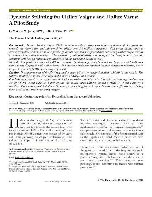
A B<br />
Figure 1A <strong>and</strong> 1B <strong>Hallux</strong> valgus (A) <strong>and</strong> hallux varus<br />
(B) Dynasplint.<br />
Similar pathologies have symptomatic contracture<br />
which is defined as the molecular shortening of the<br />
connective tissue <strong>and</strong> these pathologies occur from<br />
postoperative or posttraumatic arthrofibrosis 12-14 ,<br />
immobilization 15,16 , or occur secondarily to excessive<br />
neuromuscular tone. 16,17 A study by Usuba, et. al.,<br />
examined nonsurgical, therapeutic treatments <strong>for</strong><br />
contracture that was caused by surgical<br />
immobilization in rats. 15 C<br />
After 40 days of surgical<br />
immobilization the mean rat knee flexion contracture<br />
was -125° (n = 60). Usuba, et. al., then tested the<br />
interaction of four protocols: Stretching with high vs.<br />
low torque <strong>and</strong> stretching of prolonged duration vs.<br />
short duration. The only statistically significant<br />
difference seen between treatment protocols was<br />
found with combined protocols of low-torque<br />
stretching <strong>for</strong> prolonged durations.<br />
This combination of low-torque stretching <strong>for</strong><br />
prolonged durations is exactly what was used in the<br />
Dynasplint systems. A study by John, et. al.,<br />
examined efficacy of the Dynasplint modality <strong>for</strong><br />
reduction of contracture causing <strong>Hallux</strong> Limitus<br />
(HL). 14 In this study, 50 patients were enrolled after<br />
© The Foot <strong>and</strong> Ankle Online Journal, 2010<br />
diagnosis of HL which occurred following a<br />
bunionectomy or cheilectomy.<br />
The duration of this r<strong>and</strong>omized study was eight<br />
weeks, <strong>and</strong> experimental patients received low-torque,<br />
prolonged stretching in the metatarsal joint<br />
Dynasplint (MTD) <strong>for</strong> 60 minutes, three times per<br />
day.<br />
The dependent variable in Dr. John’s study was<br />
change in Active Range of Motion (AROM) <strong>and</strong> there<br />
was a significant change <strong>for</strong> the experimental patients<br />
following use of this home therapy modality (P <<br />
0.001, T = 4.224). Experimental patients in this study<br />
regained a mean 32° change in AROM, extension<br />
compared to only a mean 10° change in AROM <strong>for</strong><br />
control patients. Dr John’s r<strong>and</strong>omized, controlled<br />
trial showed conclusive efficacy of the MTD<br />
modality. 14 A retrospective study (N = 61) by Kalish<br />
<strong>and</strong> Willis showed comparable results in patients’<br />
regaining 73% dorsiflexion at the metatarsal joint<br />
after 4 weeks. 13 The purpose of this pilot study was<br />
to report the benefits that <strong>Dynamic</strong> <strong>Splinting</strong> (DS)<br />
had on reducing contracture in hallux varus <strong>and</strong><br />
hallux valgus.<br />
Methods<br />
Ten patients’ were treated with <strong>Dynamic</strong> <strong>Splinting</strong><br />
(DS) in this report, (six with HAV <strong>and</strong> four with<br />
hallux varus). The modality can be seen in Figure<br />
1AB <strong>and</strong> this unit delivers <strong>for</strong>ce <strong>and</strong> counter <strong>for</strong>ce to<br />
achieve elongation of connective tissue <strong>for</strong><br />
contracture reduction. The same unit may be used <strong>for</strong><br />
both lateral <strong>and</strong> medial stretching <strong>and</strong> this alteration is<br />
analogous to the Metatarsal Dynasplint that stretches<br />
both in plantarflexion 12 <strong>and</strong> dorsiflexion. 13,14
Volume 3, No. 1, January 2010 John, Willis<br />
The initial fitting <strong>for</strong> patients included customization<br />
of the unit (patient’s foot length, girth, <strong>and</strong> varying<br />
degrees of hallux edema), <strong>and</strong> training on donning<br />
<strong>and</strong> doffing of the devices. Patients also received<br />
instruction on safety, general wear <strong>and</strong> care, <strong>and</strong><br />
st<strong>and</strong>ardized tension setting goals. <strong>Dynamic</strong> splinting<br />
employs the protocol of low-load stretching <strong>for</strong><br />
contracture reduction through an appropriate<br />
biomechanical device which increases the joint’s time<br />
at end range (of motion). 12-14,16,17<br />
Each patient was instructed to wear the DS initially<br />
<strong>for</strong> 10 minutes, three times a day (tid) while seated,<br />
with an initial tension setting of #1 (0.10 foot pound<br />
of torque). Patients were instructed to sequentially<br />
increase the wearing time until they were com<strong>for</strong>table<br />
wearing the unit <strong>for</strong> 60 minutes, tid. This lowest<br />
intensity was used <strong>for</strong> becoming accustomed to the<br />
system, <strong>and</strong> the patients were instructed to increase<br />
tension on increment every two weeks after they were<br />
com<strong>for</strong>table wearing the unit <strong>for</strong> 60 minutes, tid.<br />
Results<br />
The outcome measurements in this study included<br />
changes in maximal AROM <strong>for</strong> all patients <strong>and</strong><br />
changes in hallux alignment measured in resting,<br />
weight bearing position. The patients treated <strong>for</strong><br />
HAV regained a mean 10° AROM (one month) <strong>and</strong><br />
the patients treated <strong>for</strong> hallux varus regained a mean<br />
9° AROM in 3 months. Measurement of hallux<br />
alignment was taken while resting (weight bearing).<br />
This variable yielded comparable gains of <strong>Hallux</strong><br />
abduction 10° (HAV) <strong>and</strong> 9° <strong>for</strong> adduction (hallux<br />
varus).<br />
Conclusion<br />
The purpose of this study was to report the benefits<br />
that dynamic splinting had on reducing contracture in<br />
hallux varus <strong>and</strong> hallux valgus. This examination of<br />
the new modality <strong>for</strong> contracture reduction was<br />
beneficial in restoring AROM <strong>and</strong> achieving a more<br />
optimal hallux alignment. The DS employed a<br />
proven protocol in using low-torque, prolonged<br />
stretching to reduce contracture without surgery. 13-17<br />
While surgical resolution of hallux varus <strong>and</strong> HAV are<br />
the current st<strong>and</strong>ard of care, therapeutic endeavors<br />
have been prescribed effectively <strong>for</strong> treatment of post<br />
operative rehabilitation 18 , <strong>and</strong> the DS used in this<br />
study answered the call <strong>for</strong> therapeutic treatment <strong>for</strong><br />
hallux contracture pathologies. 3,6,12-14,18<br />
The use of dynamic splinting in this pilot study<br />
caused no adverse events, <strong>and</strong> a future r<strong>and</strong>omized,<br />
controlled trial would determine if this new modality<br />
is effective in separate populations of patients with<br />
hallux abducto valgus <strong>and</strong> hallux varus.<br />
References<br />
1. Shima H, Okuda R, Yasuda T, Jotoku T, Kitano N, Kinoshita<br />
M: Radiographic measurements in patients with hallux valgus<br />
be<strong>for</strong>e <strong>and</strong> after proximal crescentic osteotomy. J Bone Joint<br />
Surg 91A (6): 1369 – 1376, 2009.<br />
2. Selner AJ, Selner MD, Cyr RP, Noiwangmuang W:<br />
Revisional Am Podiatr Med Assoc 4(4): 341 – 346, 2004.<br />
3. Miller JW: Acquired hallux varus: a preventable <strong>and</strong><br />
correctable disorder. J Bone Joint Surg 57A (2):183 – 188,<br />
1975.<br />
4. Lui TH: Technique tip: minimally invasive approach of<br />
tendon transfer <strong>for</strong> correction of hallux varus. Foot Ankle Int<br />
30(10): 1018 – 1021, 2009.<br />
5. Miller RJ, Rattan N, Sorto L: The geriatric bunion: correction<br />
of metatarsus primus varus <strong>and</strong> hallux valgus with the Swanson<br />
total joint implant. J Foot Surg 22 (3):263 – 270, 1983.<br />
6. Vanore JV, Christensen JC, Kravitz SR, Schuberth JM,<br />
Thomas JL, Weil LS, Zlotoff HJ, Mendicino RW, Couture SD;<br />
[Clinical Practice Guideline First Metatarsophalangeal Joint<br />
Disorders Panel of the American College of Foot <strong>and</strong> Ankle<br />
Surgeons]: Diagnosis <strong>and</strong> treatment of first metatarsophalangeal<br />
joint disorders. Section 3: <strong>Hallux</strong> varus. J Foot Ankle Surg 42 (3):<br />
137 – 142, 2003.<br />
7. Orzechowski W, Dragan S, Romaszkiewicz P, Krawczyk A,<br />
Kulej M, Morasiewicz L: Evaluation of follow-up results of<br />
McBride operative treatment <strong>for</strong> hallux valgus de<strong>for</strong>mity. Ortop<br />
Traumatol Rehabil 10(3): 261 – 273, 2008.<br />
8. Jahss MH: Disorders of the hallux <strong>and</strong> first ray. Disorders of the<br />
Foot <strong>and</strong> Ankle: Medical <strong>and</strong> Surgical Management. 2 nd<br />
ed. Philadelphia, Pa: WB Saunders Co, 1084 – 1089, 1991.<br />
9. Trnka HJ, Hofstaetter SG, Easley ME: Intermediate-term<br />
results of the Ludloff osteotomy in one hundred <strong>and</strong> eleven feet.<br />
Surgical technique. J Bone Joint Surg 91A (Suppl 2 Pt 1): 156 –<br />
168, 2009.<br />
10. Oloff LM, Bocko AP: Application of distal metaphyseal<br />
osteotomy <strong>for</strong> treatment of high intermetatarsal angle bunion<br />
de<strong>for</strong>mities. J Foot Ankle Surg 37(6): 481 – 489, 1998.<br />
© The Foot <strong>and</strong> Ankle Online Journal, 2010
Volume 3, No. 1, January 2010 The Foot <strong>and</strong> Ankle Online Journal<br />
11. Bilotti MA, Caprioli R, Testa J, Cournoyer R Jr, Esposito FJ:<br />
Reverse Austin osteotomy <strong>for</strong> correction of hallux varus. J Foot<br />
Surg 26 (1): 51 – 55, 1987.<br />
12. John MM, Willis FB, Portillo A: <strong>Dynamic</strong> splinting <strong>for</strong><br />
runner's toe: a case report with gait analysis. J Am Podiatric Med<br />
Assoc 99(4): 367 – 370, 2009.<br />
13. Kalish SA, Willis FB: <strong>Hallux</strong> limitus <strong>and</strong> dynamic splinting:<br />
a retrospective series. The Foot & Ankle Online Journal 2 (4):<br />
1, 2009.<br />
14. John MM, Kalish SR, Perns SV, Willis, FB. <strong>Dynamic</strong><br />
<strong>Splinting</strong> <strong>for</strong> <strong>Hallux</strong> Limitus: a R<strong>and</strong>omized, Controlled Trial.<br />
Journal American Podiatric Medical Assoc (In-Press)<br />
15. Usuba M, Akai M, Shirasaki Y, Miyakawa S: Experimental<br />
joint contracture correction with low torque -long duration<br />
repeated stretching. Clin Orthop Relat Res 456: 70 – 88, 2007.<br />
16. Willis FB: Post-TBI Gait rehabilitation. Applied Neurol 3(7):<br />
25 – 26, 2007.<br />
17. Lai J, Jones M, Willis B: Effect of dynamic splinting on<br />
excessive plantar flexion tone/contracture: A controlled, crossover<br />
study. Proceedings of the 16 th European Congress of<br />
Physical <strong>and</strong> Rehabilitation Medicine. Minerva Medica pubs,<br />
Italy, 106 – 109, August, 2008.<br />
18. Schuh R, Hofstaetter SG, Adams SB Jr, Pichler F, Kristen<br />
KH, Trnka HJ: Rehabilitation after hallux valgus surgery:<br />
importance of physical therapy to restore weight bearing of the<br />
first ray during the stance phase. Phys Ther 89(9): 934 – 945,<br />
2009.<br />
© The Foot <strong>and</strong> Ankle Online Journal, 2010