Introduction to Chordates & Fish Anatomy - De Anza College
Introduction to Chordates & Fish Anatomy - De Anza College
Introduction to Chordates & Fish Anatomy - De Anza College

You also want an ePaper? Increase the reach of your titles
YUMPU automatically turns print PDFs into web optimized ePapers that Google loves.
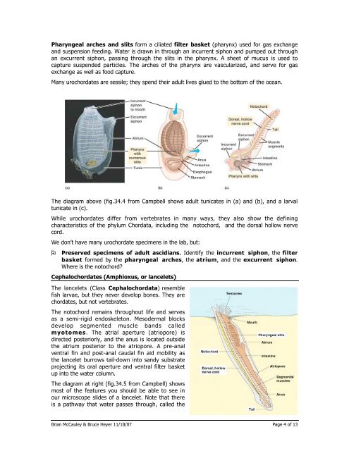
Pharyngeal arches and slits form a ciliated filter basket (pharynx) used for gas exchange<br />
and suspension feeding. Water is drawn in through an incurrent siphon and pumped out through<br />
an excurrent siphon, passing through the slits in the pharynx. A sheet of mucus is used <strong>to</strong><br />
capture suspended particles. The arches of the pharynx are vascularized, and serve for gas<br />
exchange as well as food capture.<br />
Many urochordates are sessile; they spend their adult lives glued <strong>to</strong> the bot<strong>to</strong>m of the ocean.<br />
The diagram above (fig.34.4 from Campbell shows adult tunicates in (a) and (b), and a larval<br />
tunicate in (c).<br />
While urochordates differ from vertebrates in many ways, they also show the defining<br />
characteristics of the phylum Chordata, including the no<strong>to</strong>chord, and the dorsal hollow nerve<br />
cord.<br />
We don’t have many urochordate specimens in the lab, but:<br />
Preserved specimens of adult ascidians. Identify the incurrent siphon, the filter<br />
basket formed by the pharyngeal arches, the atrium, and the excurrent siphon.<br />
Where is the no<strong>to</strong>chord<br />
Cephalochordates (Amphioxus, or lancelets)<br />
The lancelets (Class Cephalochordata) resemble<br />
fish larvae, but they never develop bones. They are<br />
chordates, but not vertebrates.<br />
The no<strong>to</strong>chord remains throughout life and serves<br />
as a semi-rigid endoskele<strong>to</strong>n. Mesodermal blocks<br />
develop segmented muscle bands called<br />
myo<strong>to</strong>mes. The atrial aperture (atriopore) is<br />
directed posteriorly, and the anus is located outside<br />
the atrium posterior <strong>to</strong> the atriopore. A pre-anal<br />
ventral fin and post-anal caudal fin aid mobility as<br />
the lancelet burrows tail-down in<strong>to</strong> sandy substrate<br />
projecting its oral aperture and ventral filter basket<br />
up in<strong>to</strong> the water column.<br />
The diagram at right (fig.34.5 from Campbell) shows<br />
most of the features you should be able <strong>to</strong> see in<br />
our microscope slides of a lancelet. Note that there<br />
is a pathway that water passes through, called the<br />
Brian McCauley & Bruce Heyer 11/18/07 Page 4 of 13