Paul Nghiem, MD, PhD - Merkel Cell Carcinoma
Paul Nghiem, MD, PhD - Merkel Cell Carcinoma
Paul Nghiem, MD, PhD - Merkel Cell Carcinoma
You also want an ePaper? Increase the reach of your titles
YUMPU automatically turns print PDFs into web optimized ePapers that Google loves.
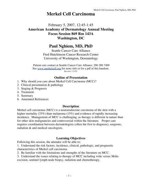
<strong>Merkel</strong> <strong>Cell</strong> <strong>Carcinoma</strong><br />
<strong>Merkel</strong> <strong>Cell</strong> <strong>Carcinoma</strong>, <strong>Paul</strong> <strong>Nghiem</strong>, <strong>MD</strong>, <strong>PhD</strong><br />
February 5, 2007, 12:45-1:45<br />
American Academy of Dermatology Annual Meeting<br />
Focus Session 869 Rm 143A<br />
Washington, DC<br />
<strong>Paul</strong> <strong>Nghiem</strong>, <strong>MD</strong>, <strong>PhD</strong><br />
Seattle Cancer Care Alliance<br />
Fred Hutchinson Cancer Research Center<br />
University of Washington, Dermatology<br />
Patient care contact at Seattle Cancer Care Alliance: 206 288 7400<br />
See www.merkelcell.org for more info or for a pdf of this handout.<br />
Revised: 1/31/07<br />
Outline of Presentation<br />
1. Why should you care about <strong>Merkel</strong> <strong>Cell</strong> <strong>Carcinoma</strong> (MCC)<br />
2. Clinical presentation & pathology<br />
3. Staging & Prognosis<br />
4. Treatment<br />
5. Summary<br />
6. Annotated References<br />
Description<br />
<strong>Merkel</strong> cell carcinoma (MCC) is a neuroendocrine carcinoma of the skin with a<br />
higher mortality (33%) than melanoma (15%) and evidence of rapidly increasing<br />
incidence. Management of MCC is challenging, as therapy is different in nature than<br />
for other skin malignancies and controversial within the literature. Proper care<br />
requires coordination between dermatologists (often the first to diagnose), surgeons,<br />
radiation & and medical oncologists.<br />
Learning Objectives<br />
Following this session, the attendee will be able to:<br />
1. Understand the risk factors, incidence, clinical, pathologic, and prognostic<br />
characteristics of <strong>Merkel</strong> cell carcinoma.<br />
2. Be familiar with the limitations and strengths of the literature on MCC.<br />
3. Understand the issues relating to therapy of MCC including wide versus Mohs<br />
excision, sentinel lymph node biopsy, radiation and chemotherapy.<br />
- 1 -
Part 1: Why should you care about MCC<br />
<strong>Merkel</strong> <strong>Cell</strong> <strong>Carcinoma</strong>, <strong>Paul</strong> <strong>Nghiem</strong>, <strong>MD</strong>, <strong>PhD</strong><br />
Fatality Rates:<br />
MCC 1 in 3<br />
Melanoma 1 in 6<br />
Sq <strong>Cell</strong> CA 1 in 50<br />
Basal <strong>Cell</strong> CA 65 yr<br />
Fair skin/ prolonged sun exposure/ PUVA therapy<br />
Profound immune suppression (HIV, solid organ transplant, CLL)<br />
13.4-fold increase among HIV+ pts.<br />
~10 fold increase after solid organ transplantation<br />
(Engels, et al 2002)<br />
(Miller, et al 1999 SEER)<br />
9% of MCC pts had HIV, CLL, Organ Solid Transplant among 141 in our<br />
series<br />
Controversy & bias is abundant<br />
Lack of balanced information due to no "owner" of MCC<br />
"Narrow" literatures are field/expertise biased:<br />
Derm/Mohs, Surg, Med Oncol, Rad Tx<br />
Few <strong>MD</strong>s are familiar with this disease or its management<br />
MCC management is often not optimal<br />
Underused therapies:<br />
Sentinel lymph node biopsy<br />
Radiation therapy<br />
Overused therapies:<br />
Over-aggressive surgery/amputation<br />
Scans (CT/MR/PET)<br />
Chemotherapy<br />
These issues will be detailed below<br />
- 2 -
<strong>Merkel</strong> <strong>Cell</strong> <strong>Carcinoma</strong>, <strong>Paul</strong> <strong>Nghiem</strong>, <strong>MD</strong>, <strong>PhD</strong><br />
Part 2: Clinical presentation and pathology<br />
Non-specific clinical presentation of MCC<br />
Firm, red to purple non-tender papule/nodule<br />
Rapid growth within prior 1-3 months<br />
Usually on a sun-exposed location (but not always)<br />
May rarely ulcerate<br />
At biopsy, most common presumed diagnosis was cyst/acneiform lesion<br />
Benign 57%<br />
Cyst/acneiform lesion 36%<br />
Lipoma 6%<br />
Dermatofibroma 5%<br />
Malignant 34%<br />
Non-melanoma skin CA 14%<br />
Lymphoma 9%<br />
Indeterminate 8%<br />
"Nodule/mass" 6%<br />
All others had 3 or fewer presumptive diagnoses: insect bite, abscess, chalazion, melanoma, neural tumor,<br />
appendage tumor. 72 of 138 cases stated a presumed diagnosis at biopsy. Total presumed diagnoses = 100<br />
12 pts had 2 presumed dx, 5 pts had 3 presumed dx, 2 pt had 4 dx. (Manuscript in preparation)<br />
Pathology<br />
<strong>Merkel</strong> cells are mechanoreceptors (fine touch) within basal epidermis<br />
Three histologic patterns (all with similar prognosis):<br />
Intermediate type<br />
most common type<br />
ddx: small blue cell tumors/melanoma/lymphoma<br />
Small cell type<br />
ddx: small cell lung CA (SCLC)<br />
Trabecular type<br />
ddx: metastatic carcinoid<br />
Immunohistochemistry panel:<br />
CK20 CK7 LCA S100<br />
<strong>Merkel</strong> cell CA + - - -<br />
Sm cell lung CA - + - -<br />
Lymphoma - - + -<br />
Melanoma - - - +<br />
Pathology Summary:<br />
"Peri-nuclear dot pattern of cytokeratin" is pathognomonic<br />
{favorite boards question!}<br />
Prior to CK20/CK7 (in early 1990s), many MCC cases were misdiagnosed as<br />
lymphoma, SCLC etc.<br />
If immunohistochemistry is done properly, diagnosis is definitive<br />
- 3 -
Part 3: Staging & Prognosis<br />
<strong>Merkel</strong> <strong>Cell</strong> <strong>Carcinoma</strong>, <strong>Paul</strong> <strong>Nghiem</strong>, <strong>MD</strong>, <strong>PhD</strong><br />
MCC Stages at Diagnosis per AJCC 6th Edition*: % Pts 3 yr survival**<br />
Stage I Localized disease, primary < 2 cm ~30% ~90%<br />
Stage II Localized disease, primary ≥ 2 cm ~30% ~70%<br />
Stage III Nodal disease ~30% ~60%<br />
Stage IV Metastatic disease ~10% CT Scan sensitivity<br />
No need to scan if small primary or if SLNB is negative.<br />
Scans useful for SLNB-positive patients to rule out distant spread<br />
- 4 -
Part 4: Treatment<br />
<strong>Merkel</strong> <strong>Cell</strong> <strong>Carcinoma</strong>, <strong>Paul</strong> <strong>Nghiem</strong>, <strong>MD</strong>, <strong>PhD</strong><br />
Can MCC be treated like BCC (no)<br />
Simple excision with 0.5 cm margins:<br />
100% recurrence in 38 pts (Meeuwissen, et al 1995)<br />
Can MCC be treated like SCC/Melanoma (no)<br />
Wide local excision >2.5 cm margins:<br />
49% regional recurrence/persistence<br />
41 pts (O'Connor, et al 1997)<br />
Is Mohs excision sufficient (no)<br />
Mohs excision +/- "safety margin" of 1 cm:<br />
16% recurrence in 25 patients (Boyer, et al 2002)<br />
Mohs + XRT:<br />
0% recurrence in 20 patients (Boyer, et al 2002)<br />
Can MCC be treated by XRT only (maybe)<br />
60 Gray (6000 cG) to primary site +/- node bed:<br />
0% recurrence in 9 patients with 3 yr f/u (Mortier, et al 2003)<br />
Effect of adding XRT to surgery:<br />
Event-Free Survival rate<br />
N 1 yr 5yrs HR P value<br />
Local recurrence<br />
Surgery only 418 71% 61% 1.00<br />
Surgery + RT 169 90% 88% 0.27
<strong>Merkel</strong> <strong>Cell</strong> <strong>Carcinoma</strong>, <strong>Paul</strong> <strong>Nghiem</strong>, <strong>MD</strong>, <strong>PhD</strong><br />
XRT as monotherapy<br />
Some patients may have inoperable disease.<br />
XRT monotherapy effective at controlling/curing extensive local disease<br />
(Multiple examples in our series and in the literature: Mortier, 2003)<br />
Adjuvant nodal therapy benefit depends on SLNB status<br />
Among SLNB-positive patients:<br />
Improved disease-free survival (p axillary > head/neck<br />
Chemotherapy<br />
Most commonly used agents: Carboplatin + Etoposide (VP-16)<br />
Useful in palliative setting for symptomatic disease:<br />
Most patients will have a response<br />
6 reasons we do not recommend adjuvant chemotherapy:<br />
• Mortality: 4-7% deaths due to adjuvant chemo in MCC<br />
(Tai, 2000; Voog, 1999)<br />
• Morbidity: neutropenia (60% of pts) fever and sepsis (40%)<br />
(Poulsen, 2001)<br />
• Decreased quality of life: fatigue, hair loss, nausea/vomiting<br />
• MCC that recurs after chemo is less responsive to later palliative chemo<br />
• Chemo suppresses immune function (important in fight against MCC)<br />
• Trend toward decreased survival among patients with nodal disease:<br />
Node Positive pts tx with MCC-specific survival<br />
No adjuvant Chemo (n=53) 60%<br />
Adjuvant Chemo (n=23) 40%<br />
(Allen, et al 2005; p=0.08, not a randomized trial, but certainly does not suggest a<br />
survival benefit!)<br />
- 6 -
<strong>Merkel</strong> <strong>Cell</strong> <strong>Carcinoma</strong>, <strong>Paul</strong> <strong>Nghiem</strong>, <strong>MD</strong>, <strong>PhD</strong><br />
Treatment bottom line:<br />
Current management of <strong>Merkel</strong> cell carcinoma tends to<br />
Underuse:<br />
Sentinel lymph node biopsy<br />
Radiation therapy<br />
Overuse:<br />
Over-aggressive surgery/amputation<br />
Scans (CT/MR/PET)<br />
Chemotherapy<br />
∗ Schematic of our recommended management:<br />
Biopsy of Primary Lesion Shows MCC<br />
Nodes Not Palpable<br />
Nodes Palpable<br />
Sentinel Lymph Node Biopsy (SLNB) &<br />
excision with negative margins<br />
Biospy of Palpable Nodes<br />
SLNB Negative<br />
SLNB Positive<br />
Biospy shows MCC<br />
Biopsy does not show MCC<br />
Radiotherapy* to Primary Site ±<br />
Draining Lymph Node Basin<br />
CT Scan of Chest, Abdomen & Pelvis<br />
Excision with negative margins<br />
+<br />
Radiotherapy* to Primary Site ±<br />
Draining Lymph Node Basin<br />
CT Scan Negative<br />
CT Scan Positive<br />
Excision with negative margins<br />
+<br />
Radiotherapy* to Primary Site &<br />
Draining Lymph Node Basin<br />
Further Evaluation and<br />
Palliative Surgery,<br />
Radiotherapy &/or<br />
Chemotherapy<br />
∗ Recommended Radiation Therapy dose (based on NCCN Guidelines for MCC 2006)<br />
45-50 Gy for: Primary site with negative excision margins<br />
Node bed with no palpable disease<br />
55-60 Gy for: Primary site with positive excision margins<br />
Node bed with palpable disease<br />
(XRT given in 2 Gy fractions, 5 times/week over 4-6 weeks)<br />
- 7 -
<strong>Merkel</strong> <strong>Cell</strong> <strong>Carcinoma</strong>, <strong>Paul</strong> <strong>Nghiem</strong>, <strong>MD</strong>, <strong>PhD</strong><br />
Part 5: Summary<br />
• MCC incidence is rising and it has a higher mortality than melanoma.<br />
• SLN bx, surgery and radiation are indicated in almost all cases.<br />
• CT Scans have poor sensitivity for nodal disease (20%) and poor<br />
specificity for distant disease (48%).<br />
• Over-aggressive surgery and adjuvant chemotherapy have high<br />
morbidity and no proven benefits.<br />
• The www.merkelcell.org website is a practical reference for patients &<br />
<strong>MD</strong>s in determining therapy and prognosis.<br />
(Easy to find...hit #2 of 240,000 for Google search of: <strong>Merkel</strong> cell carcinoma)<br />
- 8 -
Part 6: Annotated References<br />
(Most can be downloaded via www.merkelcell.org )<br />
<strong>Merkel</strong> <strong>Cell</strong> <strong>Carcinoma</strong>, <strong>Paul</strong> <strong>Nghiem</strong>, <strong>MD</strong>, <strong>PhD</strong><br />
Agelli M, Clegg LX.Epidemiology of primary <strong>Merkel</strong> cell carcinoma in the United States. Journal of the American<br />
Academy of Dermatology 2003;49:832-41.<br />
Largest study (1034 pts) of survival after MCC diagnosis via SEER data. Essentially all deaths due to<br />
MCC occur within three years of dx. No data on treatments included.<br />
Allen, P. J., Bowne, W. B. Jaques, D. P., Brennan, M. F., Busam, K., Coit, D. G. <strong>Merkel</strong> cell carcinoma: prognosis<br />
and treatment of patients from a single institution. Journal of Clinical Oncology 2005:23 (10); 2300-9.<br />
Study of 251 patients from Memorial Sloan-Kettering Cancer Center's MCC database, between 1970-<br />
2002. Conclusions: 1) Pathologic nodal staging identifies a group of patients with excellent long-term<br />
survival. 2) After margin-negative excision and pathologic nodal staging, local and nodal recurrence<br />
rates are low. 3) Adjuvant chemo for Stage III patients showed a trend (p=0.08) to decreased survival<br />
compared with Stage II patients that did not receive chemo.<br />
Gupta S, Wang L, <strong>Nghiem</strong> P. <strong>Merkel</strong> cell carcinoma: Information for patients and their physicians:<br />
www.merkelcell.org.<br />
A website dedicated to providing easily understood information on MCC causes, prongosis and therapy.<br />
20 page color pdf can be downloaded from the site.<br />
Gupta SG, Wang LC, Penas PF, Gellenthin M, Lee SJ, <strong>Nghiem</strong> P. Sentinel lymph node biopsy for evaluation and<br />
treatment of patients with <strong>Merkel</strong> cell carcinoma: The Dana-Farber experience and meta-analysis of the literature.<br />
Arch Dermatol 2006; 142: 771-4.<br />
Evaluation of 122 MCC patients (61 from the Dana-Farber and 92 from the literature). Findings:<br />
32% of patients with clinically local-only disease were found to have microscopic nodal disease by<br />
SLNB. Three-year recurrence rate was 3 times higher in this group (+SLNB vs - SLNB). Relapse free<br />
survival was improved with the use of adjuvant XRT in patients with a positive SLNB. CT scans had a<br />
low sensitivity and poor specificity for detecting nodal disease that was not readily clinically apparent.<br />
Mortier L, Mirabel X, Fournier C, Piette F, Lartigau E. Radiotherapy alone for primary <strong>Merkel</strong> cell carcinoma.<br />
Archives of Dermatology 2003;139:1587-1590.<br />
French study of stage I MCC showed excellent success (zero recurrences) in patients treated with<br />
radiation therapy alone (9 patients).<br />
Longo MI, <strong>Nghiem</strong> P. <strong>Merkel</strong> cell carcinoma treatment with radiation: a good case despite no prospective studies.<br />
Archives of Dermatology 2003;139:1641-1643.<br />
Editorial that accompanied Mortier, et al discussing the importance of adjuvant radiation therapy and<br />
a proposed algorithm for MCC treatment.<br />
Lewis K, Weinstock M, Weaver A, Otley C. Adjuvant local irradiation for merkel cell carcinoma. Archives of<br />
Dermatology 2006;142:693-700.<br />
Meta-analysis demonstrating reductions in local and regional MCC recurrence in patients treated with<br />
surgery plus XRT as compared to those treated with surgery alone.<br />
<strong>Nghiem</strong> P, McKee PH, Haynes HA. <strong>Merkel</strong> cell (cutaneous neuroendocrine) carcinoma, in Sober AJ, Haluska FG<br />
(ed): American Cancer Society Atlas of Clinical Oncology: Skin Cancer. Hamilton, Ontario, BC Decker Inc, 2001,<br />
pp 127-141.<br />
Comprehensive chapter on MCC in a multiauthored atlas of skin cancer.<br />
National Comprehensive Cancer Network (NCCN). <strong>Merkel</strong> cell <strong>Carcinoma</strong> Treatment Guidelines (updated<br />
annually). www.nccn.org.<br />
Consensus recommendations for MCC management from 20 different cancer centers across the US.<br />
- 9 -


