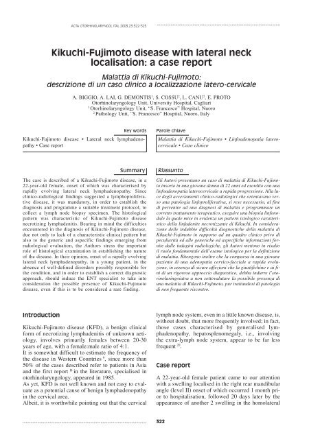
Kikuchi-Fujimoto disease with lateral neck localisation: a case report
Kikuchi-Fujimoto disease with lateral neck localisation: a case report
Kikuchi-Fujimoto disease with lateral neck localisation: a case report

You also want an ePaper? Increase the reach of your titles
YUMPU automatically turns print PDFs into web optimized ePapers that Google loves.
ACTA OTORHINOLARYNGOL ITAL 2003,23:322-325<br />
<strong>Kikuchi</strong>-<strong>Fujimoto</strong> <strong>disease</strong> <strong>with</strong> <strong>lateral</strong> <strong>neck</strong><br />
<strong>localisation</strong>: a <strong>case</strong> <strong>report</strong><br />
Malattia di <strong>Kikuchi</strong>-<strong>Fujimoto</strong>:<br />
descrizione di un caso clinico a localizzazione latero-cervicale<br />
A. BIGGIO, A. LAI, G. DEMONTIS 1 , S. COSSU 2 , L. CANU 2 , E. PROTO<br />
Otorhinolaryngology Unit, University Hospital, Cagliari<br />
1<br />
Otorhinolaryngology Unit, “S. Francesco” Hospital, Nuoro<br />
2<br />
Pathology Unit, “S. Francesco” Hospital, Nuoro, Italy<br />
Key words<br />
<strong>Kikuchi</strong>-<strong>Fujimoto</strong> <strong>disease</strong> • Lateral <strong>neck</strong> lymphadenopathy<br />
• Case <strong>report</strong><br />
Parole chiave<br />
Malattia di <strong>Kikuchi</strong>-<strong>Fujimoto</strong> • Linfoadenopatia laterocervicale<br />
• Caso clinico<br />
Summary<br />
The <strong>case</strong> is described of a <strong>Kikuchi</strong>-<strong>Fujimoto</strong> <strong>disease</strong>, in a<br />
22-year-old female, onset of which was characterised by<br />
rapidly evolving <strong>lateral</strong> <strong>neck</strong> lymphadenopathy. Since<br />
clinico-radiological findings suggested a lymphoproliferative<br />
<strong>disease</strong>, it was mandatory, in order to establish the<br />
diagnosis and programme a suitable treatment protocol, to<br />
collect a lymph node biopsy specimen. The histological<br />
pattern was characteristic of <strong>Kikuchi</strong>-<strong>Fujimoto</strong> <strong>disease</strong><br />
necrotizing lymphadenitis. Bearing in mind the difficulties<br />
encountered in the diagnosis of <strong>Kikuchi</strong>-<strong>Fujimoto</strong> <strong>disease</strong>,<br />
due not only to lack of a characteristic clinical pattern but<br />
also to the generic and aspecific findings emerging from<br />
radiological evaluation, the Authors stress the important<br />
role of histological examination in establishing the nature<br />
of the <strong>disease</strong>. In their opinion, onset of a rapidly evolving<br />
<strong>lateral</strong> <strong>neck</strong> lymphadenopathy, in a young patient, in the<br />
absence of well-defined disorders possibly responsible for<br />
the condition, and in order to establish a correct diagnostic<br />
approach, should induce the ENT specialist to take into<br />
consideration the possible presence of <strong>Kikuchi</strong>-<strong>Fujimoto</strong><br />
<strong>disease</strong>, even if this is to be considered a rare finding.<br />
Riassunto<br />
Gli Autori presentano un caso di malattia di <strong>Kikuchi</strong>-<strong>Fujimoto</strong><br />
insorto in una giovane donna di 22 anni ed esordito con una<br />
linfoadenopatia laterocervicale a rapida progressione. Alla luce<br />
degli accertamenti clinico-radiologici che orientavano verso<br />
una patologia linfoproliferativa, si rese necessario, al fine<br />
di pervenire ad una diagnosi di malattia e programmare un<br />
corretto trattamento terapeutico, eseguire una biopsia linfonodale<br />
la quale mise in evidenza un pattern istologico caratteristico<br />
della linfadenite necrotizzante di <strong>Kikuchi</strong>. In considerazione<br />
delle indubbie difficoltà diagnostiche della malattia di<br />
<strong>Kikuchi</strong>-<strong>Fujimoto</strong> in rapporto ad un quadro clinico privo di<br />
peculiarità ed alle generiche ed aspecifiche informazioni fornite<br />
dalle indagini radiologiche, gli Autori mettono in risalto<br />
il ruolo fondamentale dell’esame istologico per la definizione<br />
di malattia. Ritengono inoltre che la comparsa in una giovane<br />
paziente di una adenopatia cervico-facciale a rapida evoluzione,<br />
in assenza di sicure affezioni che la giustifichino e ai fini<br />
di un rigoroso approccio diagnostico, debba indurre l’otorinolaringoiatra<br />
a non sottovalutare la possibile presenza di<br />
una malattia di <strong>Kikuchi</strong>-<strong>Fujimoto</strong>, pur trattandosi di patologia<br />
di non frequente riscontro.<br />
Introduction<br />
<strong>Kikuchi</strong>-<strong>Fujimoto</strong> <strong>disease</strong> (KFD), a benign clinical<br />
form of necrotizing lymphadenitis of unknown aetiology,<br />
involves primarily females between 20-30<br />
years of age, <strong>with</strong> a female:male ratio of 4:1.<br />
It is somewhat difficult to estimate the frequency of<br />
the <strong>disease</strong> in Western Countries 9 , since more than<br />
50% of the <strong>case</strong>s described refer to patients in Asia<br />
and the first <strong>report</strong> 10 in the literature, specialised in<br />
otorhinolaryngology, appeared in 1985.<br />
As yet, KFD is not well known and not easy to evaluate<br />
as a potential cause of benign lymphadenopathy<br />
in the cervical area.<br />
Albeit, it is worthwhile pointing out that the cervical<br />
lymph node system, even in a little known <strong>disease</strong>, is,<br />
<strong>with</strong>out doubt, that more frequently involved; in fact,<br />
those <strong>case</strong>s characterised by generalised lymphadenopathy,<br />
hepatosplenomegaly, i.e., involving<br />
the extra-lymph node system, appear to be far less<br />
frequent 20 .<br />
Case <strong>report</strong><br />
A 22-year-old female patient came to our attention<br />
<strong>with</strong> a swelling localised in the right rear mandibular<br />
angle (level II) onset of which occurred 1 month prior<br />
to hospitalisation, followed 20 days later by the<br />
appearance of another 2 swelling in the homo<strong>lateral</strong><br />
322
KIKUCHI-FUJIMOTO DISEASE<br />
supraclavicular area (level V). The rapid evolution of<br />
the adenopathy, despite immediate treatment <strong>with</strong> anti-inflammatory<br />
drugs (oral flurbiprofen), led the primary<br />
care physician to hospitalise the patient.<br />
The physical examination revealed, in the absence of<br />
other clinical signs of importance, a voluminous<br />
swelling located in the right rear mandibular angle,<br />
the edges of which were irregular. The mass was of<br />
hard parenchymatous consistency, slightly painful<br />
upon palpation, hypomobile in the deeper levels,<br />
which had rapidly reached 6 cm in diameter, and a<br />
<strong>lateral</strong> <strong>neck</strong> polyadenitis more pronounced on the<br />
right, in correspondence to the supraclavicular area.<br />
Blood tests, carried out, upon admission, revealed<br />
only an increase in the Erythrocyte Sedimentation<br />
Rate (ESR), whereas tests to detect antibodies<br />
against Toxoplasma, Measles, Cytomegalovirus, Epstein-Barr<br />
virus and HIV, as well as the tuberculin<br />
skin test, gave negative results.<br />
Contrast enhanced computed tomography (CT) of the<br />
<strong>neck</strong> revealed the presence, in the bi<strong>lateral</strong> <strong>lateral</strong><br />
<strong>neck</strong> area, of “numerous hyperdense lesions, some of<br />
which colliquated, in part grouped together in a single<br />
mass <strong>with</strong> polycyclic margins, characteristic of lymphadenopathies”.<br />
The latter appeared to be ‘more numerous<br />
and voluminous in the right <strong>lateral</strong> <strong>neck</strong> site,<br />
where they reached from the rear mandibular angle<br />
region to the supraclavicular region’, thus confirming<br />
the clinical finding. The radiologist referred to a diagnosis<br />
of ‘suspected’ lympho-proliferative <strong>disease</strong>’.<br />
On the basis of clinical and instrumental findings, a<br />
fine needle aspiration biopsy (FNAB) was performed.<br />
Results failed to provide useful diagnostic<br />
information and it was, therefore, necessary to perform<br />
a lymph node biopsy in the right <strong>lateral</strong> <strong>neck</strong><br />
area.<br />
Histological examination of 4 adjacent lymph nodes<br />
revealed a well-preserved general architecture, in<br />
Fig. 2. Higher magnification reveals presence of intravenous<br />
fibrin deposits in necrotic areas [➛]. (Haematoxylin-eosin),<br />
240 X).<br />
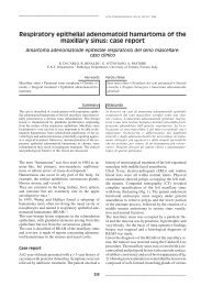
Fig. 3. Necrotic area <strong>with</strong> abundant nuclear debris, indicating<br />
karyorexis, and histiocytes [➛]. No granulocytes.<br />
Venule walls have thickened and intensely eosinophil appearance<br />
[↑] (Haematoxylin-eosin, 480 X).<br />
Fig. 1. Right <strong>lateral</strong> <strong>neck</strong> lymph node: areas of ischaemic<br />
necrosis in paracortical site [➛]. (Haematoxylin-eosin,<br />
120 X).<br />
Fig. 4. Presence of cells <strong>with</strong> clear, nucleolated nucleus,<br />
and abundant cytoplasm (plasmocytoid monocytes) <strong>with</strong>in<br />
an area of necrosis [➛]. (Haematoxylin-eosin, 480 X).<br />
323
A. BIGGIO ET AL.<br />
which multifocal areas of necrosis <strong>with</strong> a central deposit<br />
of fibrin and abundant nuclear debris (Fig. 1)<br />
were visible in the paracortical area (Fig. 2). No<br />
granulocytes were present (Fig. 3). The necrotic areas<br />
were surrounded by marked proliferation of plasmocytoid<br />
monocytes (Fig. 4) which, at immunohistochemical<br />
examination, revealed weak positivity for<br />
CD43 and CD68; histiocytes positive to myeloperoxidasis<br />
were also present.<br />
The characteristic absence of polymorphonucleates,<br />
together <strong>with</strong> the presence of a proliferation of plasmocytoid<br />
monocytes mixed <strong>with</strong> the necrosis in the<br />
paracortical site, prompted the definition of the lesion<br />
as necrotising non-suppurative lymphadenitis<br />
due to <strong>Kikuchi</strong>-<strong>Fujimoto</strong> lymphadenitis.<br />
The patient, therefore, underwent anti-inflammatory<br />
(oral nimesulide) and antibiotic (piperacilline 2 g<br />
b.i.d. i.m.) treatment and was monitored until the<br />
symptoms disappeared, about 20 days later.<br />
It was possible, at 18 months’ follow-up, to exclude<br />
recurrence of the <strong>disease</strong>.<br />
Discussion<br />
KFD, a <strong>disease</strong> frequently found in Oriental countries,<br />
was first <strong>report</strong>ed in the literature in 1972 in Japan 8 14 .<br />
Tanaka et al. 22 recently advanced the hypothesis that<br />
the higher incidence observed in Asiatics might be due<br />
to a genetic factor, corresponding to an allele of the<br />
histocompatibility HLA class II system detected <strong>with</strong><br />
a statistically significant frequency in the DNA of<br />
Japanese patients presenting the <strong>disease</strong>.<br />
The aetiology still remains to be defined, however,<br />
the hypothesis advanced, so far, suggests that a viral<br />
agent may be involved 5 13 15 . This would trigger a hyperimmune<br />
reaction resulting in polyclonal activation<br />
of T lymphocytes <strong>with</strong> a cytotoxic action 7 19 . Albeit,<br />
recent studies 11 12 have excluded the presence of<br />
a viral genoma in the cells of lymph nodes involved<br />
in KFD. Thus, it has been suggested that the triggering<br />
factor could be a not well-defined super-antigen<br />
of a proteic nature which, binding to the T lymphocyte<br />
receptors, would determine activation 4 .<br />
The pathogenetic role of impaired function of the immune<br />
system is, moreover, supported by the possible<br />
association of KFD <strong>with</strong> systemic erythematosus lupus<br />
6 or <strong>with</strong> other autoimmune <strong>disease</strong>s 2 .<br />
From a clinical point of view, the symptoms most<br />
frequently associated <strong>with</strong> lymphadenomegaly are<br />
asthenia, fever, sometimes a slight weight loss and,<br />
as far as concerns blood tests, neutropenia <strong>with</strong> lymphocytosis<br />
is often observed and, as in the <strong>case</strong> described<br />
here, an increase of ESR.<br />
These are, therefore, non specific, non pathognomonic<br />
symptoms which, together <strong>with</strong> the mode of<br />
onset which is common to various infectious and<br />
neoplastic <strong>disease</strong>s involving the lymph node system,<br />
account for the difficulties encountered in the<br />
diagnosis of KFD and stress the importance of differential<br />
diagnosis. In this respect, it is worthwhile<br />
stressing that adenopathy, in the cervical district,<br />
may be not only the site of metastases resulting from<br />
neoplastic lesions in the head and <strong>neck</strong> but also “early<br />
sentinels of neoplastic dissemination due to tumours<br />
situated in various, and sometimes distant, organs”<br />
18 .<br />
Bearing in mind these considerations, it becomes<br />
clear that it is necessary, in order to proceed <strong>with</strong> an<br />
appropriate therapeutic approach, to rapidly reach a<br />
diagnosis and, thus, avoid underestimating the condition,<br />
since KFD may, even if only rarely, have fatal<br />
consequences 3 .<br />
Moreover, it has been <strong>report</strong>ed that some patients<br />
presenting KFD have been submitted to chemotherapy<br />
following an erroneous diagnosis of lymphoma 17 .<br />
From a diagnostic viewpoint, lymph node biopsy is<br />
mandatory, in our opinion, and thus from the histological<br />
findings, which may include immuno-histochemical<br />
investigation in <strong>case</strong>s that are difficult to<br />
interpret, whilst FNAB does not always provide reliable<br />
data 16 23 24 .<br />
Symptomatic treatment is carried out using corticosteroids<br />
or non-steroidal anti-inflammatory drugs.<br />
Prognosis, which is constantly favourable, is characterised<br />
by regression of lymphadenopathy <strong>with</strong>in a<br />
few months, even if <strong>case</strong>s of recurrence have been <strong>report</strong>ed<br />
after a considerable period of time 1 21 .<br />
References<br />
1<br />
Blewitt RW, Kumar SN, Abraham JS. Recurrence of<br />
<strong>Kikuchi</strong>’s lymphadenitis after 12 years. J Clin Pathol<br />
2000;53:157-8.<br />
2<br />
Bousquet E, Tubèry M, Brousset P. Syndrome de <strong>Kikuchi</strong>,<br />
thyroidite de Hashimoto et sérologie lupique. Rev Méd Interne<br />
1996;17;836-8.<br />
3<br />
Chan JKC, Wong KC, Ng CS. A fatal <strong>case</strong> of multicentric<br />
<strong>Kikuchi</strong>’s histiocytic necrotizing lymphadenitis. Cancer<br />
1989;63:1856-62.<br />
4<br />
Correa H. <strong>Kikuchi</strong>-<strong>Fujimoto</strong> <strong>disease</strong>: An exuberant localized<br />
T cell activation arrested by histiocytes Medscape<br />
Womens Health 1996;1:5.<br />
5<br />
Dorfman RF, Berry GJ. <strong>Kikuchi</strong>’s histiocytic necrotizing<br />
lymphadenitis. An analysis of 108 <strong>case</strong>s <strong>with</strong> emphasis on<br />
differential diagnosis. Semin Diagn Pathol 1988;5:329-45.<br />
6<br />
Eisner MD, Amory J, Mullaney B, Tierney L Jr, Browner<br />
WS. Necrotizing lymphadenitis associated <strong>with</strong> systemic lupus<br />
erythematosus. Semin Arthritis Rheum 1996;26:477-<br />
82.<br />
7<br />
Felgar RE, Furth EE, Wasik MA, Gluckman SJ, Salhany<br />
324
KIKUCHI-FUJIMOTO DISEASE<br />
KE. Histiocytic necrotizing lymphadenitis (<strong>Kikuchi</strong>’s <strong>disease</strong>):<br />
in situ end-labeling, immunohistochemical and serologic<br />
evidence supporting cytotoxic lymphocyte-mediated<br />
apoptotic cell death. Mod Pathol 1997;10:231-41.<br />
8<br />
<strong>Fujimoto</strong> Y, Kojima Y, Yamaguchi K. Cervical subacute<br />
necrotizing lymphadenitis. Naika 1972;30:920-7.<br />
9<br />
Garcia CE, Girdhar-Gopal HV, Dorfman DM. <strong>Kikuchi</strong>-<strong>Fujimoto</strong><br />
<strong>disease</strong> of the <strong>neck</strong>. Update. Ann Otol Rhinol Laryngol<br />
1993;102:11-5.<br />
10<br />
Gleeson MJ, Siodlak MZ, Barbatis C, Salama NY. <strong>Kikuchi</strong>’s<br />
– A new cause of cervical lymphadenopathy. J Laryngol<br />
Otol 1985;99:935-9.<br />
11<br />
Hollingsworth HC, Peiper SC, Weiss LM, Raffeld M, Jaffe<br />
ES. An investigation of the viral pathogenesis of the<br />
<strong>Kikuchi</strong>-<strong>Fujimoto</strong> <strong>disease</strong>. Lack of evidence for Epstein-<br />
Barr virus or human herpesvirus type 6 as the causative<br />
agents. Arch Pathol Lab Med 1994;118:134-40.<br />
12<br />
Hudnall SD. <strong>Kikuchi</strong>-<strong>Fujimoto</strong> <strong>disease</strong>. Is Epstein-Barr the<br />
culprit Am J Clin Pathol 2000;113:761-4.<br />
13<br />
Huh J, Chi HS, Kim SS, Gong G. A study of the viral etiology<br />
of histiocytic necrotizing lymphadenitis (<strong>Kikuchi</strong>-<strong>Fujimoto</strong><br />
<strong>disease</strong>). J Korean Med Sci 1998;13:27-30.<br />
14<br />
<strong>Kikuchi</strong> M. Lymphadenitis showing focal reticulum cell hyperplasia<br />
<strong>with</strong> nuclear debris and phagocytes: a clinicopathological<br />
study. Nippon Ketsueki Gakkai Zasshi<br />
1972;35:379-80.<br />
15<br />
<strong>Kikuchi</strong> M, Yoshizumi M, Nakamura H. Necrotizing lymphadenitis:<br />
Positive acute toxoplasmic infection. Virchows<br />
Arch A Pathol Anat Histol 1977;376:247-53.<br />
16<br />
Mannarà GM, Boccato P, Rinaldo A, La Rosa F, Ferlito A.<br />
Histiocytic necrotizing lymphadenitis (<strong>Kikuchi</strong>-<strong>Fujimoto</strong><br />
Disease) diagnosed by fine needle aspiration biopsy. ORL J<br />
Otorhinolaryngol Relat Spec 1999;61:367-71.<br />
17<br />
Menasce LP, Banerjee SS, Edmondson D, Harris M. Histiocytic<br />
necrotizing lymphadenitis (<strong>Kikuchi</strong>-<strong>Fujimoto</strong> <strong>disease</strong>):<br />
continuing diagnostic difficulties. Histopathology<br />
1998;33:248-54.<br />
18<br />
Molinari R, Cantù G, Chiesa F, Podrecca S, Dilani F, Del<br />
Vecchio M. A statistical approach to detection of the primary<br />
cancer based on the site of <strong>neck</strong> lymph node metastases.<br />
Tumori 1977;63:267-82.<br />
19<br />
Ohshima K, Shimazaki K, Suzumiya J, Kanda M, Kumagawa<br />
M, <strong>Kikuchi</strong> M. Apoptosis of cytotoxic T-cells in histiocytic<br />
necrotizing lymphadenitis. Virchows Arch<br />
1998;433:131-4.<br />
20<br />
Rudniki C, Kessler E, Zarfati M, Turani H, Bar-Ziv Y, Zahavi<br />
I. <strong>Kikuchi</strong>’s necrotizing lymphadenitis: a cause of fever<br />
of unknown origin and splenomegaly. Acta Haematol<br />
1998;79:99-102.<br />
21<br />
Smith KG, Becker GJ, Busmanis I. Recurrent <strong>Kikuchi</strong>’s <strong>disease</strong>.<br />
Lancet 1992;340:124.<br />
22<br />
Tanaka T, Ohmori M, Yasunaga S, Ohshima K, <strong>Kikuchi</strong> M,<br />
Sasazuki T. DNA typing of HLA class II genes (HLA- DR, -<br />
DQ and -DP) in Japanese patients <strong>with</strong> histiocytic necrotizing<br />
lymphadenitis (<strong>Kikuchi</strong>’s <strong>disease</strong>). Tissue Antigens<br />
1999;54:246-53.<br />
23<br />
Tong TR, Chan OW. Lee KC. Diagnosing <strong>Kikuchi</strong> <strong>disease</strong><br />
on fine needle aspiration biopsy: a retrospective study of 44<br />
<strong>case</strong>s diagnosed by cytology and 8 by histopathology. Acta<br />
Cytol 2001;45:953-7.<br />
24<br />
Viguer JM, Jimènez-Heffernan JA, Perez P, Lopez-Ferrer P,<br />
Gonzalez-Peramato P, Vicandi B. Fine needle aspiration<br />
cytology of <strong>Kikuchi</strong>’s lymphadenitis: A <strong>report</strong> of ten <strong>case</strong>s.<br />
Diagn Cytopathol 2001;25:220-4.<br />
■ Received October 15, 2002.<br />
Accepted January 7, 2003.<br />
■ Address for correspondence: Dr. Andrea Biggio, Casa di<br />
Cura Polispecialistica “Sant’Elena”, viale Marconi 160,<br />
09045 Quartu S. Elena (CA), Italy. Fax: +39 070 837391.<br />
E-mail: biggioandrea@tiscalinet.it<br />
325